Preliminary characterization of race-specific elicitors fromPeronospora parasitica and their ability...
12
PHYTOPATHOLOGY/ MYCOLOGY S. Soylu and E.M. Soylu (2003) Phytoparasitica 31(4):381-392 Preliminary Characterization of Race-Specific Elicitors from Peronosporaparasitica and Their Ability to Elicit Phenolic Accumulation in Arabidopsis S. Soylu 1'* and E.M. Soylu 1 Intercellular washing fluid (IWF) obtained from the susceptible Arabidopsis accession Ws- edsl inoculated with Peronospora parasitica isolate Emoy-2, contained an elicitor of necrosis with ecotype specificity towards Arabidopsis accessions with particular resistance genes. This elicitor caused necrosis on the highly resistant accessions La-er, Nd-1 and partly on Col-5, but not on the susceptible accessions Ws-edsl and Oy-0. In resistant plants, injection of IWF caused hypersensitive reaction (HR)-like cell collapse which was associated with the accumulation of phenolics and lignin-like material in walls of cells undergoing cell death. The elicitor is sensitive to proteinase K and pronase enzymes, heating and autoclaving but insensitive to periodate oxidation, freezing and thawing, and is not dialyzable. Results suggest that the elicitor is a protein. Fractionation experiments using size-exclusion membranes revealed that elicitor activity has a molecular weight in excess of 100 kDa. KEY WORDS: Race-specific elicitor; Arabidopsis; Peronospora; resistance. INTRODUCTION In nature, plants are constantly challenged by an array of phytopathogenic microor- ganisms. Interactions between specific plant cultivars and defined races of potentially pathogenic microbes are categorized as compatible (host susceptible, pathogen virulent) or incompatible (host resistant, pathogen avirulent). Incompatibility often involves localized tissue necrosis, i.e., the hypersensitive reaction (HR) and manifold biochemical changes in the infected host tissue (10). These defense reactions, including the fortification of plant cell walls, accumulation of phenolics and biosynthesis of antimicrobial compounds (phytoalexins), are believed to stop the invading pathogen (2,20,22). The induction of defense reactions is presumed to be mediated by an initial recognition process between pathogen and host plant (5,17) which involves detection of certain unique structures of the incompatible pathogen (avr gene product) by recognition molecules (R gene product) in plants. In the molecular interpretation of this recognition process, each pathogen avirulence gene encodes for a specific elicitor which is recognized by the corresponding plant resistance gene and triggers the defense reactions. These genetic patterns have been observed in many plant/pathogen interactions, and are referred to as the gene-for-gene hypothesis (19). In a general model, elicitors of plant defense responses are classified into two categories depending on their source. Endogenous elicitors are of plant origin and arise as a ReceivedNov. 5, 2002; received in final formMarch 18, 2003; http://www.phytoparasitica.org postingJuly 13, 2003. 1 Dept. of Plant Protection, Faculty of Agriculture,University of Mustafa Kemal,31034 Antakya,Hatay, Turkey. *Corresponding author [Fax: +90 326 2455832;e-maih [email protected]]. Phytoparasitica 31:4, 2003 381
-
Upload
independent -
Category
Documents
-
view
3 -
download
0
Transcript of Preliminary characterization of race-specific elicitors fromPeronospora parasitica and their ability...

PHYTOPATHOLOGY/ MYCOLOGY S. Soylu and E.M. Soylu (2003) Phytoparasitica 31(4):381-392
Preliminary Characterization of Race-Specific Elicitors from Peronospora parasitica and Their Ability to Elicit
Phenolic Accumulation in Arabidopsis
S. Soylu 1'* and E.M. Soylu 1
Intercellular washing fluid (IWF) obtained from the susceptible Arabidopsis accession Ws- edsl inoculated with Peronospora parasitica isolate Emoy-2, contained an elicitor of necrosis with ecotype specificity towards Arabidopsis accessions with particular resistance genes. This elicitor caused necrosis on the highly resistant accessions La-er, Nd-1 and partly on Col-5, but not on the susceptible accessions Ws-edsl and Oy-0. In resistant plants, injection of IWF caused hypersensitive reaction (HR)-like cell collapse which was associated with the accumulation of phenolics and lignin-like material in walls of cells undergoing cell death. The elicitor is sensitive to proteinase K and pronase enzymes, heating and autoclaving but insensitive to periodate oxidation, freezing and thawing, and is not dialyzable. Results suggest that the elicitor is a protein. Fractionation experiments using size-exclusion membranes revealed that elicitor activity has a molecular weight in excess of 100 kDa. KEY WORDS: Race-specific elicitor; Arabidopsis; Peronospora; resistance.
INTRODUCTION
In nature, plants are constantly challenged by an array of phytopathogenic microor- ganisms. Interactions between specific plant cultivars and defined races of potentially pathogenic microbes are categorized as compatible (host susceptible, pathogen virulent) or incompatible (host resistant, pathogen avirulent). Incompatibility often involves localized tissue necrosis, i.e., the hypersensitive reaction (HR) and manifold biochemical changes in the infected host tissue (10). These defense reactions, including the fortification of plant cell walls, accumulation of phenolics and biosynthesis of antimicrobial compounds (phytoalexins), are believed to stop the invading pathogen (2,20,22). The induction of defense reactions is presumed to be mediated by an initial recognition process between pathogen and host plant (5,17) which involves detection of certain unique structures of the incompatible pathogen (avr gene product) by recognition molecules (R gene product) in plants. In the molecular interpretation of this recognition process, each pathogen avirulence gene encodes for a specific elicitor which is recognized by the corresponding plant resistance gene and triggers the defense reactions. These genetic patterns have been observed in many plant/pathogen interactions, and are referred to as the gene-for-gene hypothesis (19).
In a general model, elicitors of plant defense responses are classified into two categories depending on their source. Endogenous elicitors are of plant origin and arise as a
Received Nov. 5, 2002; received in final form March 18, 2003; http://www.phytoparasitica.org posting July 13, 2003. 1 Dept. of Plant Protection, Faculty of Agriculture, University of Mustafa Kemal, 31034 Antakya, Hatay, Turkey. *Corresponding author [Fax: +90 326 2455832; e-maih [email protected]].
Phytoparasitica 31:4, 2003 381

result of the interaction with an aggressor. Exogenous elicitors may be considered as the primary signals in plant-pathogen interaction. They originate from the pathogen or aggressors themselves and evoke a response in cells in the immediate vicinity of pathogens. These elicitors are divided into two categories, non-specific or race-specific, based on the specificity which has induced plant response.
Pathogen-derived non-specific elicitors induce various defense responses in a large variety of plant cultivars and species (9). In contrast to non-specific elicitors, certain bacterial and fungal avirulence (avr) genes have been shown to encode race-specific elicitors which efficiently trigger defense responses only in plant cultivars harboring the complementary resistance (R) gene in gene-for-gene interactions (14,16). Although several bacterial avr genes have been cloned, mostly from narrow host range pathovars of Pseudomonas and Xanthomonas species, only a few fungal avr genes have been cloned (5,14). These include the tomato leaf mold pathogen Cladosporiumfulvum, the barley leaf scald pathogen Rhynchosporium secalis, the cowpea rust fungus Uromyces vignae, and the soybean pathogen Phytophthora sojae (1,6-8,13,26,29,31 ).
The availability of purified elicitors that activate plant defense responses is a prereq- uisite for the analyses of signal transduction. Crude elicitors can be isolated from the pathogen cell wall or from the apoplast where the pathogen has grown (9). Intercellular washing fluids (IWF) are considered to be a source for elicitors released from intercellularly growing pathogens during pathogenesis. This was demonstrated by the detection of race- cultivar specific fungal peptide elicitors in the IWF extracted from infected plant leaves including Arabidopsis (25).
The obligate biotroph oomycete Peronospora parasitica is the causal agent of downy mildew of crucifers. The pathogen develops intercellularly and forms haustoria within mesophyll cells. The interaction between P. parasitica and Arabidopsis thaliana represents a good model system to dissect the molecular pathways leading to disease resistance (12). A number of physiological races of the pathogen and many Arabidopsis accessions carrying different genes for resistance have been described and a gene-for-gene relationship between them has been proven (11,23).
The objective of this study was to characterize ecotype specific elicitors (25) in the IWF obtained from susceptible host plant leaves of Arabidopsis colonized by a virulent isolate of P. parasitica. The elicitor molecule detected was partially characterized and its ability to elicit plant defense mechanisms such as the accumulation of phenolics and lignin was examined.
MATERIALS AND METHODS
Plant and pathogen Arabidopsis accessions, resistant and susceptible to P. parasitica isolate Emoy-2, were obtained from Dr. E. Holub (HRI, Wellesboume, UK). The isolate used in the study was maintained on susceptible plants as described previously (11). P. parasitica isolate Emoy-2 was inoculated weekly onto 4-6-week-old Ws-edsl plant leaves by spraying with a conidial suspension of 5x10⁵ spores m1-1. Inoculated plants were placed in a clear plastic box to retain sufficient relative humidity during the experiment. Plants were then transferred to growth rooms at 18-20~ under 16-h photoperiod (150- 200 #E m -2 s- l ) .
In order to bioassay elicitor activity in different accessions, 2-week-old seedlings were transplanted to individual pots (10 cm diam) and kept in a separate growth room at 18-
382 S. Soylu and E.M. Soylu
20~ Under such conditions plants developed large rosette leaves which were used in IWF injections.
Isolation of dicitor from intercellular washing fluid IWF was obtained based on a method modified from De Wit and Spikman (7). Seven days after inoculation (dai), 6- week-old Ws-edsl leaves bearing heavy sporulation were detached from the plants and gently washed in pre-chilled sterile distilled water (SDW) to remove the conidia and conidiophores. Leaves were immersed in a beaker containing SDW and vacuum-infiltrated (100 kPa) for a period of 15 min, during which the vacuum was broken and created every 3 min in order to achieve effective infiltration through tissues. Water-soaked leaves were then blotted with tissue paper and transferred to the barrel of a 20 ml plastic syringe which was placed into a 50 ml sterile centrifuge tube. The leaves were centrifuged at 1000 g for 10 rain at 4~ The extruded IWF was sterilized by membrane filtration (0.22 #m, Millipore, Billerica, MA, USA). IWF obtained from uninoculated plants placed under the same conditions in a separate growth chamber was used as a control in the study.
Bioassays for elicitor activity The technique used for injection of Arabidopsis leaves with IWF was essentially that of Rethage et al. (25), which involved infiltration of leaves using a blunt syringe without a needle. IWFs were infiltrated using 1 ml sterile syringes into the lower surface of 4--6-week-old expanded rosette leaves of plants which had not initiated formation of flowering structures. Approximately 20-40 #1 IWF Was sufficient to inoculate an Arabidopsis leaf. Up to four leaves from each plant were infiltrated, with at least five replicate plants used for each accession. Treated plants were incubated in the growth room. Plant reactions in infiltrated leaves were assessed at different time intervals and scaled according to four categories (Ph.D. thesis, senior author): rating 0 is assigned to leaves with no visible symptoms; 1: 5-25% of area showing weak HR with little chlorosis; 2: 26-50% of area showing HR with chlorosis; 3:51-75% of area showing confluent HR with chlorosis; 4: 76-100% of area showing complete necrosis with spreading chlorosis.
Microscopy was used to reveal the reactions of plant cells. IWF-injected leaves were detached from plants after visual observations and then decolorized in 100% methanol and cleared in chloral hydrate. Leaves were mounted in 50% glycerol and examined under a Nikon Optiphot microscope (Nikon Corp., Japan), equipped with differential interference contrast and epifluorescence optics. Phenolic compounds in unstained cleared tissue were detected by their autofluorescence under UV irradiation (2A, exciter filter EX 330-380, dichroic mirror DM 400, and barrier filter BA 420) or blue light irradiation (B-2A, exciter filter EX 450-490, dichroic mirror DM 510, and barrier filter BA 520). Wall-bound phenolics fluoresce green or yellow-green under blue light, or bright blue under UV light. Lignified structures were visualized using the phloroglucinol/HCl test according to Vallet et al. (28).
All experiments were arranged in a randomized block design and performed three times, with similar results. Data were subjected to analysis of variance and least significant differences were calculated at P<_0.01.
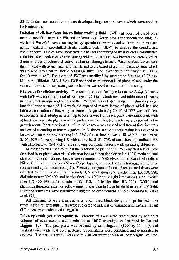
Polyacrylamide gel electrophoresis Proteins in IWF were precipitated by adding 9 volumes of cold acetone and incubating at -20~ overnight as described by Lu and Higgins (18). The precipitate was pelleted by centrifugation (1500 g, 15 min), and washed twice with 90% cold acetone. Supernatants were combined and evaporated to dryness. The residues were dissolved in distilled water at 50% of their original volume.
Phytoparasitica 31:4, 2003 383

Pellets were air-dried and dissolved in distilled water at the original volume, followed by centrifugation to remove insoluble material. The material was then electrophoresed on 10% (w/v) polyacrylamide slab gels containing 0.375 M Tris-HC1 (pH 8.8), 0.05% (w/v) SDS and 0.08% (w/v) N,N-methylenbis acrylamide. The stacking gel contained 6% (w/v) polyacrylamide, 0.250 M Tris-HC1 (pH 6.8), 0.05% (w/v) SDS and 1.6% (w/v) N,N-methylenbis acrylamide. Bromophenol blue was used as a marker. The electrophoresis buffer (pH 8.3) contained 0.025 M Tris, 0.250 M glycine and 0.5% (w/v) SDS. Electrophoretic separations were conducted at 200 volts for 2-3 h. A silver staining method was used to detect high molecular weight proteins (silver staining kit, Bio-Rad Laboratories, Herts., UK). For an estimation of the molecular weights, broad range protein markers (2-212 kDa, Sigma Chemical Co., St. Louis, IL, USA) were used.
Chemical, physical and enzymatic treatment of elicitor The stability of an elicitor to heat was tested by incubating IWF at 100~ for 20 min or by autoclaving at 121~ for 15 min. After cooling to room temperature, the resulting precipitate was removed by centrifugation and both pellet and supernatant were tested for biological activity. Stability to freezing-thawing was tested by (i) freezing (-20~ followed by thawing (room temperature), or (ii) refreezing followed by thawing (one or two cycles of freezing and thawing), and bioassayed. Sensitivity to proteases was tested by incubating the elicitor with pronase (final concentration 0.2 mg m1-1) at 37~ for 4 h or with proteinase K enzyme (final concentration 0.2 mg m1-1) at 37~ for 3 h. After incubation, treated IWF was immediately bioassayed. In controls, the enzymes mentioned were prepared in Tris buffer instead of the IWF. Periodate oxidation of the elicitor was carded out according to the procedure of Van den Ackerveken et al. (29). The IWF was mixed with an equal volume of 0.02 M NalO4 and incubated in the dark at 25~ overnight. The sample was then dialyzed against water to remove excess NalO4 and vacuum-concentrated to the original volume at 45~ The sample was bioassayed by infiltration into plant leaves.
Protein determination Protein was determined using a dye-based assay adapted from Bradford (3).
Molecular weight of the elicitor The molecular size of the elicitor in IWF was initially determined by ultramembrane filtration using Centricon concentrators (Amicon, Beverly, MA, USA). IWF was centrifuged at 1000 g for 30 min over 100 kDa cut-off ultramembrane. The molecules in the filtrate and those retained were assayed for bioactivity.
RESULTS
Race specificity of IWF Phenotypic and genotypic characterization of interactions between Arabidopsis accessions and Peronospora isolate Emoy-2, according to Holub et al. (11), is given in Table 1. Interaction between Oy-0 and Ws-edsl accessions and the Emoy-2 isolate was classed as compatible. Ws-edsl is a mutant line of the Ws-3 accession of A. thaliana and it is susceptible to all P parasitica isolates (24). Interactions between other accessions (Col-5, Nd-1 and La.-er) and Emoy-2 isolate were incompatible.
Six-week-old Ws-edsl plants were inoculated with the virulent Emoy-2 isolate. IWFs were harvested in SDW 7 dai and yielded 0.2--0.3 ml fluid containing 310 #g m1-1 soluble protein. The activities of the IWF from Ws-edsl caused a range of macroscopic symptoms in different accessions, which were scored and summarized in Table 2. The response of accessions to IWF was the same as the response caused by fungal isolates on the cotyledon
384 S. Soylu and E.M. Soylu

Fig. 1. Localization of phenolic (A and B) and lignin-like (C and D) compounds in IWF-injectect Arabidopsis leaves. Sites from susceptible accession Ws-edsl (A and C), and resistant accession La-er (B and D) 3 days after injection of IWE Infection sites were examined under blue (A and B) or visible (C and D) light. In A, note injected area does not display any specific fluorescence, indicating absence of phenolics at reaction site. B shows collapsed necrotic site (asterisk) with specific bright fluorescence (arrow), indicating accumulation of phenolics at reaction site. Arrows indicate the IWF did not reach beyond this point (healthy site). C and D show the site of lignin depositions as dark precipitates. Note the absence of specific staining in cell walls within the infiltrated region (C). In D, the typical dark staining within vascular tissue (asterisk) and faint staining on associated necrotic mesophyll cell wall (arrows) is indicative of the presence of lignin-like material at the lignified cell walls. A, B and D, bar= 50 #m; C, bar= 100 #m.
assay given in Table 1. Cultivar specificity, i.e., chlorosis and necrosis, was induced only in accessions resistant to the isolate. In La-er, first visible symptoms appeared as chlorosis at 2 dai. This symptom was followed by widespread yellowing and the appearance of necrotic patches by 5 dai. Injection of IWF into the leaves of susceptible accessions Ws-edsl and Oy-0 caused neither chlorosis nor necrosis within 7 dai. However, very faint chlorosis was observed after prolonged incubation (more than 10-14 dai). IWF obtained from healthy or
Phytoparasitica 31:4, 2003 385

Fig. 2. PAGE profile of IWF obtained from Arabidopsis leaves. Lane 1, molecular weight marker; lane 2, IWF obtained from uninoculated leaves; lane 3, IWF obtained from susceptible leaves inoculated with Emoy-2 isolate, 3 days after inoculation. Note that IWF from inoculated leaves contained protein bands 1, 2 and 3, which were not present in IWF obtained from uninoeulated leaves.
water-inoculated leaves did not induce any chlorosis or necrosis after injection in resistant and susceptible accession leaves.
To test the activity of IWF, dilution series of IWF were injected into leaves of resistant accessions and leaves were scored for the presence of necrosis or chlorosis. Results revealed that 1/60 dilution induced necrosis occasionally but 1/40 gave a more reproducible response.
Cytology of the plant reaction Following macroscopic observations and scoring, elicitor- injected leaves were examined by microscopy. Infiltration of IWF recovered from
386 -S. Soylu and E.M. Soylu

TABLE 1. Interaction phenotypes z observed on Arabidopsis accessions in response to the Emoy-2 isolate of Peronospora parasitica
Isolate Arabidopsis accessions Ws-edsl Oy-0 Col-5 Nd- 1 La-er
Emoy-2 S ~ (EH) S (EH) IR (FDL) R (PN) R (FN) Zlnteraction phenotypes within parentheses were characterized as follows: EH, _Early (3 dai) and Heavy (>20 conidiophore) sporulation; FDL, necrotic _Flecking and Delayed-Light parasite sporulation (< 10 conidiophore); PN, necrotic _Pitting and No sporulation; FN, minute necrotic Flecking and No sporulation. Phenotype designations are according to Holub et aL (11).
S, compatible interaction; IR, intermediate interaction; R, incompatible interaction.
TABLE 2. Mean bioassay response in three experiments of five different Arabidopsis accession leaves injected with IWF obtained from Emoy-2-inoculated leaves of Arabidopsis accession Ws-eds l
(scored for the presence of necrosis or chlorosis at different days after inoculation, dai)
Accessions Nec~sis r~ng z, DAI 1 2 3 5 7
Ws-edsl 0 ~ 0 a 0 a 0 a 0 a Oy-0 0a 0a 0a 0a 0a Col-5 0a 0a 0.26a 0.93b 2.1c Nd-1 0a 0a 0.93b 2.9d 3.73e La-er 0a 0.9b 2.8d 3.0d 3.93e
z0, no visible symptoms; I, 5-25% of injected area showing weak HR with littie chlorosis; 2, 26-50% of area showing HR with chlorosis; 3, 51-75% the area showing confluent HR with chlorosis; 4, 76-100% of area showing complete necrosis with spreading chlorosis. UValues followed by the same letter do not differ significantly at P<0.01, with LSD = 0.3735.
inoculated leaves into the intercellular spaces of leaves of resistant accessions resulted in autofluorescence of the mesophyll and epidermal cells and accumulation of lignin-
like material on cell wails (Figs. 1B, D). When the injected leaves were stained with aniline blue, clear bright blue staining was also observed under UV light, indicating callose
deposition in epidermal and mesophyll cell walls. Among the resistant accessions, reaction in La-er was evident 1 dai as scattered fluorescing cells in the interveinal areas. With time, responses in leaves of both resistant accessions were followed by a progressive increase in the number (Table 3) and intensity (Fig. 1B) of fluorescence within the injected
area. Reactions observed on Col-5 were less dramatic than those observed on La-er and Nd-1 (Table 3). In contrast, no autofluorescence and accumulation of lignin-like material appeared in susceptible leaves (Figs. 1A, C) apart from a low incidence of autofluorescence
TABLE 3. Mean autofluorescence ratings in three experiments of five different Arabidopsis accession leaves injected with IWF obtained from Emoy-2-inoculated leaves of Arabidopsis accession Ws-eds l at different days after inoculation (dai)
Accessions Autofluorescence rating z, DAI 1 2 3 5 7
Ws-edsl 0 aY 0 a 0 a 0 a 0 a Oy-0 0a 0a 0a 0a 0a Col-5 0 a 0 a 1.2 b 2.53 e 3.1 d Nd-1 0 a 1.13 b 2.2 c 3.86 e 4 e La-er 1.13 b 2.13 e 3.33 d 3.93 e 4 e
z0, no cell autofluorescence; 1, 5-25% of the cells autofluoresce in injected area; 2, 26-50% of the cells autofluoresce; 3, 51-75% of the cells autofluoresce; 4, 76-100% of the cells autofluoresce. UValues followed by the same letter do not differ significantly at P<0.01, with LSD = 0.4058.
Phytoparasitica 31:4, 2003 387

TABLE 4. Mean ratings z of the effects of denaturing treatments in three experiments on the eliciting activity in IWF. (Injected area of five leaves was scored for the presence of necrosis or chlorosis at 7 days after inoculation)
Treatments Arabidopsis accessions Ws-edsl Oy-0 Col-5 Nd- 1 La-er
Heating 0 a 0 a 0 a 0 a 0 a Autoclaving 0 a 0 a 0 a 0 a 0 a Freezing-Thawing
1 cycle 0 a 0 a 2.8 c 3.7 d 3.9 d 2 cycles 0 a 0 a 0 a 0 a 0 a
Pronase 0 a 0 a 0 a 0 a 0 a Proteinase K 0 a 0 a 0 a 0 a 0 a NalO4 0 a 0 a 1.7 b 3.7d 3.8 d Dialysis 0 a 0 a 1.6 b 3.6 d 3.8 d
zo, no visible symptoms; 1, 5-25% of the injected area showing weak HR with little chlorosis; 2, 26-50% of the area showing HR with chlorosis; 3, 51-75% of the area showing confluent HR with chlorosis; 4, 76-100% of the area showing complete necrosis with spreading chlorosis. UValues followed by the same letter do not differ significantly at P<0.01, with LSD = 0.343.
observed around the injection point. No autofluorescence of lignin-like material was found in accessions injected with water or with IWF obtained from uninoculated leaves.
Partial character izat ion of elicitor in I W F The elicitor found in IWF was subjected to several physical, enzymatic and chemical treatments and bioassayed by injection into resistant plants (Table 4).
Necrosis-inducing elicitor activity was completely abolished by proteinase K and pronase digestions, heating and autoclaving. The elicitor was insensitive to one cycle of freezing and thawing but not tolerant of a further cycle. It was insensitive to periodate oxidation and dialysis against water. These results provide evidence that the active component elicitor may be a protein. Buffers in which proteinase K or pronase were incubated without IWF, had no activity in the bioassay.
Fractionation experiments using size-exclusion membranes revealed that the elicitor activity was retained by a membrane with molecular weight cut-off of 100 kDa. This indicated that the active necrosis-inducing component(s) had a molecular weight over 100 kDa.
SDS-PAGE of IWF obtained from uninoculated and inoculated susceptible leaves contained a complex pattern of proteins as shown in Figure 2. IWF from inoculated leaves contained protein bands 1, 2, and 3, which were not present in IWF obtained from uninoculated leaves (Fig. 2).
DISCUSSION
The interaction between Arabidopsis and the oomycete pathogen P. parasitica was used as a model system to study the molecular basis of communication between plants and their pathogens. We have determined an elicitor active component in IWF of susceptible Arabidopsis leaves inoculated with a virulent isolate of the downy mildew fungus. Bioassay of the elicitor on resistant and susceptible Arabidopsis accessions indicated clearly that the elicitor present in IWF seems to be cultivar-specific. Cultivar specificity of Emoy- 2 previously reported by Holub et al. (11) was confirmed in this study. IWF obtained from compatible interactions induced chlorosis or necrosis only in accessions resistant to
388 S. Soylu and E.M. Soylu
Emoy-2 but not in cultivars susceptible to this isolate. Similar elicitor activity in IWF was demonstrated by Rethage et al. (25) for certain ArabidopsislPeronospora interactions, which were different from those used in our study. Similar race-specific activity has also been reported for a few host-fungal pathogen interactions (6). In the case of tomato leaves infected with the leaf mold fungus C. fulvum, a single peptide which induces necrosis only when injected into tomato leaves of cultivars bearing the corresponding resistance gene, has been isolated and characterized (7,15,29). Similar reports of race-specific elicitors have been presented for the interaction between barley and its fungal pathogen R. secalis. In barley plants with the resistance gene Rrsl, the race-specific elicitor NIP1 triggered several defense reactions (26). Results obtained by Sutherland and Deverall (27), however, were in contrast to our results. In their study a necrosis-inducing elicitor was obtained in IWF from wheat leaves infected with leaf rust fungus, but the results showed that the elicitor in IWF was non-specific.
Most of the isolated and characterized elicitors are macromolecules that vary widely in their chemical nature. Proteins, oligopolysaccharides, glycoproteins, fatty acids and derivates have been identified that can function as elicitors (9,13,21,31). In the Peronospora/Arabidopsis interaction, the necrosis-inducing elicitor present in IWF also appeared to be protein, being sensitive to heating and autoclaving, proteinase K and pronase digestions. By contrast, exposure of IWF to the periodate ion, which oxidizes sugar residues, had no apparent effect on activity of IWF in the bioassay (Table 4). Fractionation experiments using size-exclusion membranes indicated that the active necrosis-inducing component(s) had a molecular weight over 100 kDa. Rethage et al. (25) reported that SDS-PAGE of IWF obtained from NOCO- and WELA-isolates inoculated leaves did not reveal any race-specific differences in low Mr peptides.
Proteins with a molecular weight above 15-20 kDa do not pass freely though the cell wall and indeed the C. fulvum elicitors were small peptides (7,29). It is postulated that plants secrete hydrolytic enzymes (e.g. chitinases and glucanases) which may release the soluble fragment from the eliciting agent, e.g. from the fungal hypha, and that the component is then diffused to the cells and recognized by the plant cell surface receptors (9). In addition to cell wall components of pathogens directly recognized by the plant cells, microbial enzymes degrading the plant cell Walls can activate defense responses indirectly. The mode of action of microbial enzymes, especially that of c~-1,4-endopolygalacturonic acid lyases (PGA lyases), has often been suggested to be due to their catalytic activity, i.e., elicitor-active molecules are released from the plant cell walls (4,21). The mode of action of elicitor-active material identified in our study was not investigated. Therefore, we do not have any evidence that the elicitor-active material in IWF is enzymatic and might release endogenous elicitors from the cell walls. Further research is needed to clarify this issue.
Occurrence of the elicitor in the compatible interaction raises questions concerning its origin and function. Electrophoresis of apoplastic IWF revealed three proteins with molecular weights over 100 kDa present in inoculated leaves but not in uninoculated leaves. Bioassay of these IWFs also showed that necrosis was induced only by IWF from inoculated leaves. Taken together, these findings suggest that the elicitor in 1WF may be of pathogenic origin.
In resistant plants, production of the race-specific elicitor by an avirulent race of pathogen induces the HR, which is associated with the activation of other defense responses. The most prominent responses associated with the HR are the induction of
Phytoparasitica 31:4, 2003 389

phytoalexins, cell-wall alterations, PR proteins, and generation of active oxygen species (2). Infiltration of purified race-specific elicitors, AVR4 and AVR9, induced differential expression of acidic chitinase and 1,3-~-glucanase in tomato genotypes Cf4 and cfg, respectively (30). Similarly, treatment of resistant tomato cell-suspension cultures with the IWFs from incompatible races resulted in the activation of NADPH oxidase and ion fluxes (13,31). Such inductions, observed most abundantly in resistant genotypes, correlate well with the difference in gene expression of Cladosporium/tomato interactions. Specific HR-associated recognition of secreted proteins from C. fulvum was also reported to occur in both host and non-host plants (15). Becker et al. (1) have isolated four different isoforms of a protein elicitor from P. sojae (sojein 1-4). These proteins showed high homology to elicitins from other Phytophthora species. Purified sojein as well as recombinant sojein isoforms induced HR-like lesions in tobacco. All sojein isoforms induced defense-related genes like those encoding phenylalanine ammonia lyase, glutathione-S-transferase and chalcone synthase in tobacco and soybean plants and cell cultures.
In Arabidopsis, injection of IWF into resistant leaves caused an HR-like necrosis. Histochemical studies showed that development of HR in elicitor-treated resistant leaves was closely associated with the accumulation of phenolics and lignin. Enrichment of the cell walls and wall apposition (papilla) with phenolics or lignin-like polymers is likely to provide a further barrier to pathogen spread. Several studies have demonstrated that phenolic structures confer rigidity to host cell walls through peroxidase-mediated cross linking of constitutive wall carbohydrates, such as hemicellulose and pectin. Many phenolic compounds also have been reported to have antimicrobial activity. A further role for lignification in disease resistance has been proposed to involve effects on the fungal cell wall, so that tips of fungal hyphae in close contact with lignifying host materials might become lignified and lose the plasticity necessary for hyphal growth (22). In Arabidopsis, inhibition of lignin precursors suppressed lignification and caused a shift towards susceptibility to P. parasitica (20).
In conclusion, biochemical analysis of the interaction between plant and pathogens has con~buted to a greater understanding of the molecular basis for signal perception and transduction in plant cells. It is clear that recognition between host plant and pathogen mediated by receptors lies at the root of the gene-for-gene concept as observed in this study. The underlying biochemical mechanisms of elicitor perception and intracellular signal transduction leading to the activation of plant defense are, however, not yet well defined. Isolation and purification of the elicitor described in this study and its putative receptor (R gene) are the next steps required toward elucidating the molecular mechanisms by which changes in plant cellular metabolism are induced.
ACKNOWLEDGMENTS
We wish to thank Mark H. Bennett, Ian Brown and Conrad Stevens, Imperial College, University of London, UK, for valuable assistance and helpful discussions during characterization of elicitors, and Prof. John W. Mansfield for use of his laboratory facilities and critical review of the manuscript. This research was supported by a grant from the University of Mustafa Kemal, Hatay, Turkey.
REFERENCES
1. Becket, J., Nagel, S. and Tenhaken, R. (2000) Cloning, expression and characterization of protein elicitors from the soyabean pathogenic fungus Phytophthora sojae. Z Phytopathol. 148:161 - 167.
2. Benhamou, N. (1996) Elicitor-induced plant defense pathways. Trends Plant Sci. 1:233-240.
390 S. Soylu and E.M. Soylu

3. Bradford, M.M. (1976) A rapid and sensitive method for the quantification of microgram quantities of protein utilizing the principle of protein dye binding. Anal, Biochem. 72:248-257.
4. Davis, K.R. and Ausubel, EM. (1989) Characterization of elicitor-induced defense responses in suspension- cultured cells of Arabidopsis. Mol. Plant-Microbe Interact. 2:363-368.
5. De Wit, P.J.G.M. (1997) Pathogen avirulence and plant resistance: a key role for recognition. Trends Plant ScL 2:452-457.
6. DeWit•ELG.M.andJ••sten•M.H.A.J.(•999)Aviru•en•eandresistan•egenesintheClad•sp•riumfulvum- tomato interactions. Curr. Opin. Microbiol. 2:368-373.
7. De Wit, P.J.G.M. and Spikman, G. (1982) Evidence for the occurrence of race- and cultivar-specific elicitors of necrosis in intercellular fluids of compatible interactions of Cladosporium fulvum and tomato. Physiol. Plant Pathol. 21:1-11.
8. D'Silva, I. and Heath, M.C. (1997) Purification and characterization of two novel hypersensitive response- inducing specific elicitors produced by the cowpea rust fungus. J. Biol. Chem. 272:3924-3927.
9. Hahn, M.G. (1996) Microbial elicitors and their receptors in plants. Annu. Rev. Phytopathol. 34:387-412. 10. Hammond-Kosack, K.E., Jones, D.A. and Jones, J.D.G. (1996) Ensnaring microbes: the components of plant
disease resistance. New Phytol. 133:11-24. 11. Holub, E.B., Beynon, J.L. and Crute, I.R. (1994) Phenotypic and genotypic characterization of interactions
between isolates of Peronospora parasitica and accessions of Arabidopsis thaliana. Mol. Plant-Microbe Interact. 7:223-239.
12. Kunkel, B. (1996) A useful weed put to work: Genetic analysis of disease resistance in Arabidopsis thaliana. Trends Genet. 2:64-69.
13. Lam, C.-H.B., Xing, T., Higgins, V.J. and Blumwald, E. (1998) Effect of race-specific elicitors of Cladosporium fulvum on the tomato plasma membrane Ca 2+-ATPase. Physiol. Mol. Plant Pathol. 52:309- 321.
14. Laugt, R. and De Wit, P.J.G.M. (1998) Fungal avirulence genes: Structure and possible functions. Fungal Genet. Biol. 24:285-297.
15. Laug6 R., Goodwin, P.H., De Wit, P.J.G.M. and Joosten, M.H.A.J. (2000) Specific HR-associated recognition of secreted proteins from Cladosporiumfulvum occurs in both host and non-host plants. Plant J. 23:735-745.
16. Leach, J.E. and White, EE (1996) Bacterial avirulence genes. Annu. Rev. Phytopathol. 34:153-179. 17. Lindsay, W.P., Lamb, C.J. and Dixon, R.A. (1998) Microbial recognition and activation of plant defense
systems. Trends Microbiol. 4:181-187. 18. Lu, H. and Higgins, V.J. (1993) Partial characterizatioa of a non-proteinaceous suppressor of non-specific
elicitors from Cladosporium fulvum (syn. Fulvia fulva). Physiol. Mol. Plant Pathol. 42:427-439. 19. Mansfield, J.W., Bennett, M.H., Bestwick, C.S. and Woods-Tor, A.M. (1997) Phenotypic expression of gene-
for-gene interactions involving fungal and bacterial pathogens: variation from recognition to response, in: Crute, I.R. and Holub, E.B. [Eds.] The Gene-for-Gene Relationship in Host-Parasite Interactions. CAB International, London, UK. pp. 265-291.
20. Mauch-Mani, B. and Slusarenko, A.J. (1996) Production of salicylic acid precursors is a major function of phenylalanine ammonia-lyase in the resistance of Arabidopsis to Peronospora parasitica. Plant Cell 8:203- 212.
21. Moniz, de S.M., Subramaniam, R., Williams, EE. and Douglas, C.J. (1992) Rapid activation of phenyl- propanoid metabolism in elicitor treated hybrid poplar (Populus trichocarpa Torr. & Gray x Populus deltoides Marsh) suspension-cultured cells. Plant Physiol. 98:728-737.
22. Nicholson, R.L. and Hammerschmidt, R. (1992) Phenolic compounds and their role in disease resistance. Annu. Rev. Plant Pathol. 30:369-389.
23. Parker, J.E., Feys, B.J., Van Der Biezen, E.A., Noel, L., Aarts, N., Austin, M.J. et al. (2000) Unravelling R gene-mediated disease resistance pathways in Arabidopsis. Mol. Plant Pathol. 1:17-24.
24. Parker, J.E., Holub, E.B., Frost, L.N., Falk, A., Gunn, N.D. and Daniels, M.J. (1996) Characterization ofedsl, a mutation in Arabidopsis suppressing resistance to Peronospora parasitica specified by several different RPP genes. Plant Cell 8:2033-2046.
25. Rethage, J., Ward, P.I. and Slusarenko, A.J. (2000) Race-specific elicitors from the Peronospora parasit- icalArabidopsis thaliana pathosystem. Physiol. Mol. Plant Pathol. 56:179-184.
26. Rohe, M., Gierlich, A., Hermann, H., Hahn, M., Schmidt, B., Rosahl, S. et al. (1995) The race-specific elicitor, NIP1, from the barley pathogen, Rhynchosporium secalis determines avirulence on host plants of the Rrsl resistance genotype. EMBO J. 14:4168-4177.
27. Sutherland, M.W. and Deverall, B.J. (1990) The ubiquity of non-specific eliciting activity in intercellular washing fluids from rust-infected wheat leaves. Plant Pathol. 39:50-57.
28. Vallet, C., Chabbert, B., Czaninski, Y. and Monties, B. (1996) Histochemistry of lignin deposition during sclerenchyma differentiation in alfalfa stems. Ann. Bot. 78:625-632.
Phytoparasitica 31:4, 2003 391
29. Van den Ackerveken, G.F., Vossen, P. and De Wit, P.J.G.M. (1993) The AVR9 race-specific-elicitor of Cladosporiumfulvum is processed by endogenous plant proteases. Plant Physiol. 103:91-96.
30. Wubben J.P., Lawrence, C.B. and De Wit, P.J.G.M. (1996) Differential induction of chitinase and 1,3-~- glucanase gene expression in tomato by Cladosporium fulvum and its race-specific elicitors. Physiol. Mol. Plant Pathol. 48:105-.116.
31. Xing, T., Higgins, V.J. and Blumwald, E. (1997) Race-specific elicitors of Cladosporium fulvum promote translocation of cytosolic components of NADPH oxidase to the plasma membrane of tomato cells. Plant Cell 9:249-259.
392 S. Soylu and E.M. Soylu


![[PhD thesis] Activating Play: a design research study on how to elicit playful interaction from teenagers](https://static.fdokumen.com/doc/165x107/634540ab38eecfb33a067899/phd-thesis-activating-play-a-design-research-study-on-how-to-elicit-playful-interaction.jpg)