Unattended emotional faces elicit early lateralized amygdala–frontal and fusiform activations

Original Article
Does single cortical spreading depressionelicit pain behaviour in freely moving rats?
Cephalalgia
0(00) 1–12
! International Headache Society 2010
Reprints and permissions:
sagepub.co.uk/journalsPermissions.nav
DOI: 10.1177/0333102409360828
cep.sagepub.com
Didem Akcali1, Aslihan Sayin1, Yildirim Sara2 andHayrunnisa Bolay1
Abstract
Introduction: Behavioural animal studies are critical, particularly to translate results to human beings. Cortical spreading
depression (CSD) has been implicated in migraine pathogenesis. We aimed to investigate the effects of CSD on the
behaviour of freely moving rats, since available CSD models do not include awake animals.
Materials and methods: We developed a new model to induce single CSD by applying topical N-methyl-D-aspartate
(NMDA) and employed a combination of an automated behavioural analysis system, video camera and ultrasonic
vocalisation (USV) calls for the first time. Electrocorticograms were also studied during CSD in freely moving rats.
Behaviour associated with cephalic pain was assessed in a group of rats that received sumatriptan. Cortical c-fos
immunoreactivity was performed in order to confirm CSD.
Results: NMDA induced single CSD in ipsilateral cortex, evoked freezing behaviour (P< 0.01) and increased the number
of wet dog shakes (WDS; P< 0.01). Grooming, locomotion, eating, drinking, and circling were not significantly altered
among groups. Ultrasonic vocalisations compatible with pain calls (22–27 kHz) were only detected in 3 out of 25 rats.
Sumatriptan did not significantly reduce the freezing behaviour. CSD induced significant c-fos expression in ipsilateral
cerebral cortex and amygdala (P< 0.01).
Conclusions: CSD induces freezing behaviour by invoking anxiety/fear via amygdala activation in freely-moving rats. Single
CSD is unlikely to lead to severe pain in freely-moving rats, though the development of mild or vague pain cannot be
excluded. The relevance of rat behavioural responses triggered by CSD to migraine symptoms in humans needs further
evaluation.
Keywords
behaviour, cortical spreading depression, migraine, rat, sumatriptan
Date received: 30 May 2009; accepted: 18 December 2009
Introduction
Cortical spreading depression (CSD) indicates anextreme excitability state of the grey matter with mas-sive redistribution of ions. The hallmark of CSD is apropagating wave of brief and massive neuronal exci-tation followed by prolonged inhibition (1,2). Recoveryof spontaneous neuronal activity, as studied by electro-corticogram (ECoG), takes a few minutes, whereasrecovery of evoked/synaptic neuronal activity takesmuch longer (up to 30min) (1,2). As the inductionand propagation of CSD depends strictly on glutama-tergic neurotransmission, glutamate or N-methyl-D-aspartate (NMDA) can easily induce CSD (2,3).Although the existence of CSD in the human brainhas been questioned, studies over the last decade haveprovided data demonstrating the presence of CSD in
humans, and an association between CSD and migraineaura (4–6).
There has recently been an increased interest in CSDand a hypothesis that ‘CSD could also underliemigraine without aura attacks’ has been proposed(7,8). As the literature regarding CSD depends primar-ily on anaesthetised animal models, it is unclear
1Gazi University Faculty of Medicine, Neuropsychiatry Centre, Ankara,
Turkey.2Hacettepe University Faculty of Medicine, Ankara, Turkey.
Corresponding author:
Hayrunnisa Bolay, Professor of Neurology, Gazi University Faculty of
Medicine, Department of Neurology & Neuropsychiatry Centre,
Besevler, Ankara 06500, Turkey. Email: [email protected]
Cephalalgia OnlineFirst, published on March 31, 2010 as doi:10.1177/0333102409360828

whether the symptoms are fully comparable with thoseexperienced by humans (9). Bolay and colleagues pre-viously showed that CSD was a sufficient stimulus toactivate the ipsilateral trigeminovascular system in therodent brain by inducing neurogenic oedema and bloodflow increase in the dura mater, as well as c-fos expres-sion in the brainstem trigeminal nuclei (trigeminalnucleus caudalis, TNC) (10). A single event ofCSD-induced blood flow increase in the dura materwas a trigeminal nerve mediated brain stem reflexwhich lasted up to 60min in anaesthetised rats.The neurogenic blood flow increase in the middlemeningeal artery that peaks around 20min could reflectthe possible time course of head pain followingCSD (10). Depending on the previous data obtainedin anaesthetised animals, we hypothesised that anysymptom related to headache should be observablewithin 15–20min following induction of CSD in freelymoving rats.
As behavioural studies in animals are gainingimportance as research tools, consistent behaviouralcorrelates of headaches in rodents will improve thereliability of any migraine model. CSD in the gyren-cephalic brain cannot propagate through the entirehemisphere due to limitations by major sulci(11,12); therefore, symptoms due to the neuronal dys-function of the entire hemisphere are not seen as anaura in humans (4,13,14). This is one potential limi-tation to compare the behaviour of rodents havingCSD with symptoms in humans. Although the effectsof spreading depression in the cerebral cortex are wellcharacterised, its direct (propagation of CSD) or indi-rect (due to transient disconnection from cortex)influences on subcortical and limbic structures inawake rats remain unclear. The link between CSDand pain behaviour has not been extensively evalu-ated in freely moving rats. Koroleva and Bures (15)concluded that spreading depression was not anaversive experience, as awake hooded Long–Evansrats did not display avoidance behaviour of thedark compartment when multiple CSD waves wereinduced by KCl.
We aimed to investigate behavioural alterationscaused by CSD in order to address: (i) doesCSD cause behaviour, suggestive of head pain, whichcan be observed and quantified in freely movingrats; and (ii) are there any behavioural characteristicsof CSD in rats to employ for future migrainestudies? We have developed a novel experimentalmodel of single CSD in freely moving rats. Weinduced CSD by applying topical NMDA, andused a combination of an automated behaviouralanalysis system, video camera recording, and ultra-sonic vocalisation (USV) calls to record behaviouralchanges.
Materials and methods
Animals
The study was performed in the NeuroscienceLaboratory, Neuropsychiatry Centre, Gazi University.Ethical approval was obtained from Gazi UniversityAnimal Studies Ethical Committee. Male Wistar ratsweighing 200–250 g were used. Animals were housedin a controlled environment where the room tempera-ture was 22� 2�C and dark:light cycle was 12 h daylightand 12 h darkness. Rats received food and waterad libitum.
Induction of CSD
Animals were anaesthetised with ketamine (50mg/kg)and xylazine (8mg/kg). The depth of anaesthesia wasadjusted according to the heart rate and hind leg pinchreaction. During all the surgical procedures, body tem-perature was kept constant at 37� 0.5�C with a heatingpad. The animals were placed in a stereotaxic frame(Stoelting CO, USA). The skull was exposed by amedian incision of the scalp. A burr hole with a diam-eter of 2mm was opened over the right parietal cortex(3mm lateral and 7mm posterior to the bregma) bymeans of a mini-drill (FHC, USA). The dura materwas kept intact. Plastic tubing covering the burr holewas placed on the skull (8mm height, 2.5mm innerdiameter, 3.5mm outer diameter; Bicakcilar�, Turkey)and was fixed to the skull by dental acrylic (Meliodent,Heraus Kulzer�). To avoid drying of the dura mater,the tubing was always kept sealed with a cap. A stain-less steel screw (1mm diameter; PlasticsOne, USA) wasattached to the occipital bone (2mm posterior tolambda, 1mm lateral) in order to secure the dentalacrylic firmly. The animals were kept in the stereotaxicframe for another 30min until the preparation wasstable. A schematic illustration of the experimentalpreparation is shown in Figure 1. For CSD inductionor in control experiments, 20 ml of NMDA solution orisotonic saline, respectively, were injected into thetubing with a pipette, without touching the duramater. The animals were kept in their cage and werenot handled. Before the drug or saline application, thefluid accumulated in the tubing was removed using apiece of blotting paper. The cap was only removedduring NMDA or saline application, and the tubingwas gently filled with solution, taking care to avoidair bubbles. All behavioural experiments were con-ducted during the day-time (12:00 pm� 2:00 h) follow-ing 72� 5 h of recovery from surgery.
Behavioural experiments were conducted in threeseparate groups: (i) saline group (control, n¼ 9);(ii) NMDA group (n¼ 9); and (iii) sumatriptan succi-nate (SUMA)þNMDA group (n¼ 7).
2 Cephalalgia 0(00)

Electrocorticogram recordings
Electrophysiological recordings during CSD wereobtained in two different groups of rats. The firstgroup of experiments were performed on four rats todetermine the optimum NMDA dose for CSD induc-tion. Anaesthesia was induced (sodium thiopental,30mg/kg) and the animals placed in a stereotaxicframe. An anterior craniotomy was performed (2mmlateral and 2mm anterior to bregma) and a side-by-sidebipolar electrode (FHC, USA) was inserted in thecortex. The silver–silver chloride ground electrode wasplaced into the neck. Signal amplification and data ana-lysis were performed via an EEG amplifier and a dataacquisition system (Bioamplifier and Powerlab/8SP,AD Instruments, Australia).
Since the design of the automated behavioural ana-lysis set-up did not allow simultaneous ECoG record-ings, a second group of ECoG experiments wereconducted on awake rats (n¼ 5). To monitor the corti-cal activity, bipolar electrodes (MS303, PlasticsOne,USA) were implanted in the frontal lobe (2mm lateral,1–2mm anterior to bregma, 1mm deep into the cortex)in addition to the parietal tubing. The rats were electri-cally grounded via a stainless steel screw electrodesfixed in the occipital bone. The tubing for NMDA orsaline application was placed as in the behaviouralexperiments. Animals were connected to an AC ampli-fier (Biopac MP30, USA) with a thin cable that allowedthe animals to move freely in a transparent observationbox. The electrical signals from the electrodes were
filtered between 0.05Hz and 100Hz, and sampled at1 kHz using the Biopac MP30 system.
Drugs administered
NMDA was purchased from RBI, sumatriptan succi-nate was kindly provided by Glaxo Smith Kline. Suma-triptan succinate (300 mg/kg) was given subcutaneously30min before the topical NMDA administration.NMDA was dissolved in saline to a final concentrationof 10mM. The concentration of sumatriptan and topicalNMDA (16,17), in addition to the timing of sumatriptanadministration before the induction of CSD, were com-parable with the literature (10,18).
Behavioural analysis
In this study, we evaluated animal behaviour such aslocomotion, immobility, grooming, rearing, eating,drinking, purposeless chewing, clockwise (CW) andcounter clockwise (CCW) turning, head shakes (HS)and wet dog shakes (WDS). Rat behaviours were auto-matically recorded by a non-invasive behavioural ana-lysis system. This system is composed of a standardrat cage fixed on a platform with several force-displacement transducers, connected to a personal com-puter (Laboras�, Metris, The Netherlands). The plat-form detects and classifies the behaviours by using thevibrations created by movement (such as rearing, swal-lowing, circling, etc.) of the animal (19–21). Rats werefree to access food or water located as in standard ratcages, and needed to rear to feed or drink.
All experiments were simultaneously recorded by avideo-camera system in order to confirm the dataobtained from automated analysis system, and to dif-ferentiate freezing periods from immobility.
Ultrasonic vocalisation (USV) calls
Many vertebrates use vocalisations to communicate inmother–offspring interactions, mating, affective ormood status (fear, pain or aggression), the presence ofpredators or the location of food. Most small rodentspecies, including laboratory rats and mice, emit USVcalls (>20 kHz) (22). These vocalisations are inaudibleto humans without the use of specialised equipment.Ultrasonic sounds (within a range of 15–100 kHz) oflaboratory animals can be monitored and analyzed bya USV detector system (Sonotrack�, Metris, TheNetherlands). Sonotrack uses a hardware bandpassfilter (10th order Butterworth filter) with sharp cut-offsat 15 kHz and 100 kHz. This filter prevents aliasing andit also removes almost all environmental sounds. Thedata are presented without further filtering or smooth-ing. In Sonotrack, the dB (decibel) scale is relative to a
Burr hole and tubing(topical NMDA)
ECoGelectrodes
Lambda
Bregma
Screw
Figure 1. Schematic illustration of experimental rat preparation
showing the locations of the ECoG recording electrodes and the
burr hole for NMDA application. NMDA was applied into the
tubing placed over a burr hole on the posterior parietal cortex and
ECoG was recorded from 9 mm anterior to induction site.
Akcali et al. 3

1mV (RMS) signal. In the spectrogram, red indicatesthe strongest signal value (the 50mV or 35.3mV RMSor 31 dB) and black indicates the background noise(this is approximately 10mV in Sonotrack or 7mVRMS or 16 dB). The shift in the frequency at the begin-ning and at the end of the vocalisation is characteristicof a biological sound. The 22–27 kHz range of vocalisa-tions in juvenile and adult rats indicates a negativeaffective state and is seen during, pain, startling, dis-tressing events and exposure to predators. The 50 kHzvocalisations usually represent positive affective state.
Immunohistochemistry (IHC)
The animals were anaesthetised with a lethal dose ofthiopental sodium 2h after topical NMDA or salineapplication. They were perfused transcardially byheparinated saline, followed by 4% 0.1M paraformal-dehyde solution. The brains were prepared for c-fosimmunohistochemistry, as previously described (5),and cerebral cortical sections of the whole cortex,50 mm thick coronal sections at every 150 mm, were eval-uated for c-fos immunoreactivity. Wide-spread (in allposterior to anterior sections) and intense c-fos immu-noreactivity within all layers of cerebral cortex in theipsilateral hemisphere was considered a characteristicfeature of CSD. To test whether the freezing behaviouris related to activation of the amygdala, c-fos expres-sion was also studied in amygdala. c-fos positive cellswere counted in the amygdala (basolateral, central andmedial nucleus were all included) bilaterally frombregma �1.4 to �3.8mm, and expressed as the totalnumber of cells.
Statistical analysis
The results are presented as mean� SEM. Statisticalanalysis was performed using a one-way analysis ofvariance (ANOVA) and followed by a post-hoc(Tukey–Kramer) test, or a two-way ANOVA (locomo-tion) with repeated measures and Bonferroni post-hoctests (Figure 3B), as appropriate. P< 0.05 was consid-ered statistically significant.
Results
CSD induction by NMDA
NMDA in the 1 mM to 100mM range was applied tothe burr hole in the anaesthetised animals.Simultaneous electrophysiological recordings werealso obtained to confirm CSD. While NMDA up to1mM did not induce CSD at all, NMDA 10–100mMconsistently evoked CSD. The 10mM dose was chosento induce CSD in awake animals. NMDA always
induced a single CSD (Figure 2A), while KCl (3M)induced multiple CSDs in the ipsilateral cortex.
ECoG and behaviour
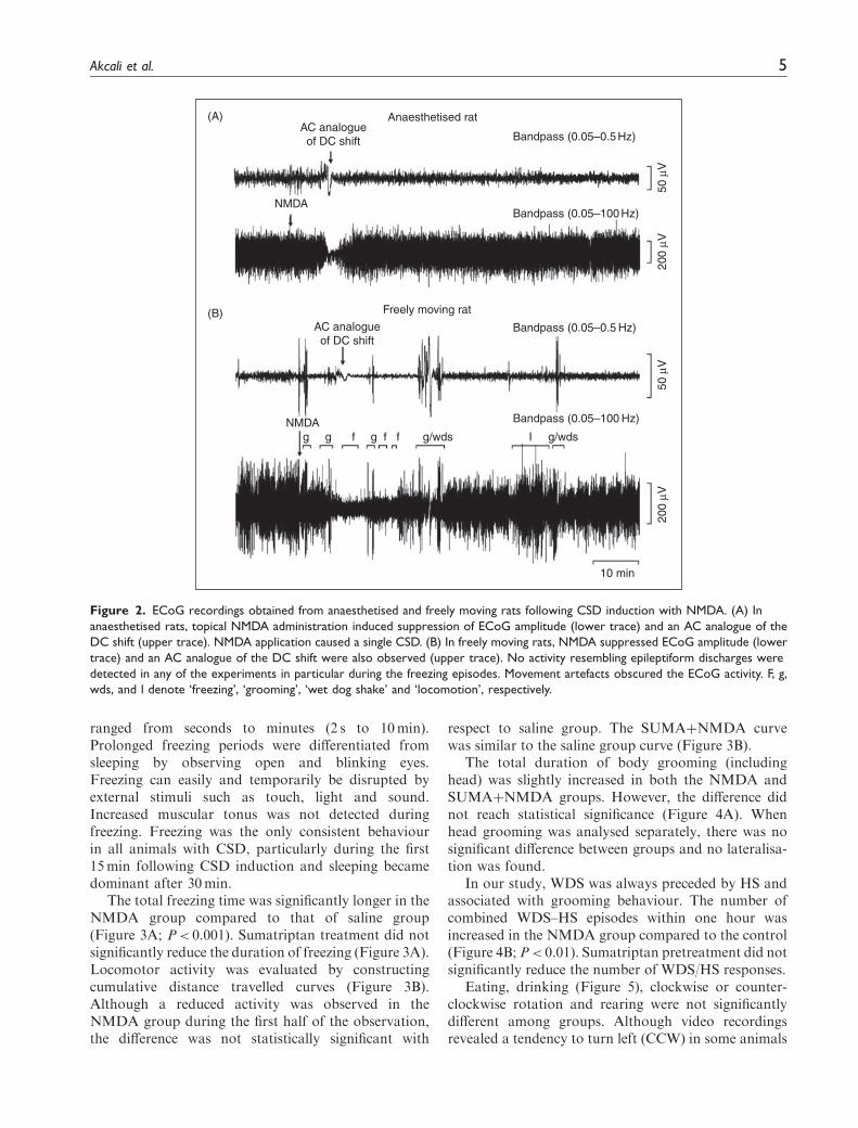
In the freely moving rats, topical NMDA applicationtemporarily suppressed the ECoG amplitude in5.1� 0.9min (Figure 2B). AC analogue of the DC shiftwas also observed by filtering the ECoG data with abandpass filter (0.05–0.5Hz; Figure 1B). The timeperiod for full recovery of ECoG amplitude was foundto be 14.2� 2.7min. In all animals studied,NMDA-induced reduction in ECoG activity occurredonly once. Freezing episodes were never observedbefore the NMDA application. The freezing episodesdid not coincide with the CSD-induced ECoG amplitudesuppression. Epileptiform discharges were not detectedin any of the recordings, including freezing periods. Sincebehaviour during freezing episodes displays similaritiesto absence seizures, we specifically analyzed ECoG dataduring these episodes. Spike-wave discharges of 7–11Hzspecific to the absence type of epilepsy were not detected.In the period of ECoG amplitude suppression, animalswere not paralysed, since they exhibited several motoractivities like walking, grooming, WDS, and rearing.When depression of ECoG amplitude was detected(9mm anterior to induction site), CSD had alreadyinvaded several cortical areas such as sensorimotor hin-dlimb and forelimb areas (bregma �3mm and bregmaþ1mm, respectively (21)) which are associated withbehavioural reactions such as grooming, WDS or freez-ing. Those movement artefacts mostly obscured back-ground neuronal activity and the AC analogue of DCshift in the ECoG (Figure 2B).
Behavioural results
CSD evoked a set of behavioural responses, of whichfreezing was the most prominent behavioural change.In another group of animals (n¼ 4), we prolongedobservation period up to 12 h following CSD induction.We did not find any significant behavioural alterationsbeyond 60min. Therefore, the experiments weredesigned to evaluate the first hour following NMDAapplication.
Freezing behaviour was recognised when rats sud-denly became immobile and developed a fixed stare(staring empty), the latter being particularly importantfor brief freezing episodes lasting 2–3 s. Freezing epi-sodes interrupted any on-going activity such as walk-ing, rearing, grooming or eating. Immediately after thecessation of the brief freezing episodes, animalsresumed their activity. The first freezing episode follow-ing NMDA application appeared at 3.9� 0.5min. Theduration of each freezing period was variable and
4 Cephalalgia 0(00)
ranged from seconds to minutes (2 s to 10min).Prolonged freezing periods were differentiated fromsleeping by observing open and blinking eyes.Freezing can easily and temporarily be disrupted byexternal stimuli such as touch, light and sound.Increased muscular tonus was not detected duringfreezing. Freezing was the only consistent behaviourin all animals with CSD, particularly during the first15min following CSD induction and sleeping becamedominant after 30min.
The total freezing time was significantly longer in theNMDA group compared to that of saline group(Figure 3A; P< 0.001). Sumatriptan treatment did notsignificantly reduce the duration of freezing (Figure 3A).Locomotor activity was evaluated by constructingcumulative distance travelled curves (Figure 3B).Although a reduced activity was observed in theNMDA group during the first half of the observation,the difference was not statistically significant with
respect to saline group. The SUMAþNMDA curvewas similar to the saline group curve (Figure 3B).
The total duration of body grooming (includinghead) was slightly increased in both the NMDA andSUMAþNMDA groups. However, the difference didnot reach statistical significance (Figure 4A). Whenhead grooming was analysed separately, there was nosignificant difference between groups and no lateralisa-tion was found.
In our study, WDS was always preceded by HS andassociated with grooming behaviour. The number ofcombined WDS–HS episodes within one hour wasincreased in the NMDA group compared to the control(Figure 4B; P< 0.01). Sumatriptan pretreatment did notsignificantly reduce the number of WDS/HS responses.
Eating, drinking (Figure 5), clockwise or counter-clockwise rotation and rearing were not significantlydifferent among groups. Although video recordingsrevealed a tendency to turn left (CCW) in some animals
Anaesthetised rat
Bandpass (0.05–0.5 Hz)
Bandpass (0.05–100 Hz)
Bandpass (0.05–100 Hz)
Bandpass (0.05–0.5 Hz)
Freely moving rat
NMDA
NMDAg g gf f f Ig/wds g/wds
10 min
200
μV20
0 μV
50 μ
V50
μV
AC analogueof DC shift
AC analogueof DC shift
(A)
(B)
Figure 2. ECoG recordings obtained from anaesthetised and freely moving rats following CSD induction with NMDA. (A) In
anaesthetised rats, topical NMDA administration induced suppression of ECoG amplitude (lower trace) and an AC analogue of the
DC shift (upper trace). NMDA application caused a single CSD. (B) In freely moving rats, NMDA suppressed ECoG amplitude (lower
trace) and an AC analogue of the DC shift were also observed (upper trace). No activity resembling epileptiform discharges were
detected in any of the experiments in particular during the freezing episodes. Movement artefacts obscured the ECoG activity. F, g,
wds, and l denote ‘freezing’, ‘grooming’, ‘wet dog shake’ and ‘locomotion’, respectively.
Akcali et al. 5

during the first 5–15min, or difficulty in raising forepaw during rearing or paw preference for gettingfood, none of those observations reached statistical sig-nificance amongst the groups. Behavioural data of eachanimal are shown in Figures. 3–5 as the variability washigh in groups. Additionally, the possible propagationof CSD with variable speed in awake animals, as shownby Koroleva et al. (24), may have influenced beha-vioural variability in our study.
Ultrasonic vocalisation calls
Distress or pain calls (22–27 kHz) were recorded in 3out of 25 animals (Table 1), which were emitted whenanimals were displaying freezing behaviour. None ofthe control animals exhibit distress calls and only tworats from the SUMAþNMDA group and one rat fromthe NMDA group emitted distress calls (Figure 6). Paincalls were long-lasting (808� 25ms) and the total
Freezing Locomotion
∗∗∗∗∗
(A)
30
Tota
l dur
atio
n (m
in)
20
10
SAL
NMDA
NMDA+
SUMA
0
(B)
8
6
4
Dis
tanc
e tr
avel
led
(m)
2
00 10 20 30
SAL
NMDA+SUMA
NMDA
Time (min)
40 50 60
Figure 3. Total freezing durations and cumulative distance travelled over an hour indicating the locomotor activity. (A) NMDA
application significantly increased total freezing period and sumatriptan pre-treatment did not alter NMDA-induced freezing. (B) All
animals were tested for locomotor activity following topical drug administration. Cumulative distances travelled in 60-min period did
not reveal any statistically significant difference. SAL, saline (control group). SUMA, sumatriptan pretreated group. Freezing data of
each animal are shown in grey symbols, and mean� SEM shown as dark lines. **P< 0.001, ***P< 0.0001.
20
Grooming(A) (B)
30
20
Num
ber
of W
DS
/ 60
min
10
0
Wet dog shake
*
Tota
l dur
atio
n (m
in) 15
10
5
0
SAL
NMDA
NMDA+
SUMA SAL
NMDA
NMDA+
SUMA
Figure 4. (A) Total duration of body grooming (including head grooming) is not significantly different among groups. (B) The total
number of head shake/wet dog shake (HS/WDS) episodes are significantly increased in the NMDA group. Sumatriptan pretreatment
did not significantly reduce the WDS number. SAL, saline (control group). SUMA, sumatriptan pretreated group. Data of each animal
are shown in grey symbols, and mean� SEM shown as dark lines. *P< 0.05.
6 Cephalalgia 0(00)

duration of pain calls lasted up to a minute (Table 1).The 50 kHz vocalisations reflect a positive affectivestate of the animal and with a frequency range of 32–96 kHz. The so-called ‘50 kHz vocalizations’ arereferred to as ‘chirps’ because of their brief duration(30–50ms) (25). USV calls reflecting positive affect(55� 9ms duration) were only recorded in control ani-mals during eating (Figure 6) and not detected in theNMDA or SUMAþNMDA groups. The sonotractsoftware also converts USV calls into frequencies audi-ble to humans; listening to these converted USV callsrevealed that 50 kHz vocalisations sounded like a whis-tle/chirp, while pain calls sounded like a scream.
c-fos immunochemistry
The c-fos immunoreactivity, a reliable marker of neu-ronal activity, was used to confirm CSD at the end ofeach experiment. An intense and wide-spread c-fosexpression was detected in all cortical layers and
pia-arachnoid membranes in the NMDA andSUMAþNMDA groups (Figure 7). The c-fos expres-sion in the anterior sections (bregma þ1mm) indicatedpropagation of CSD from posterior (bregma �7mm)cortical area where CSD was induced. As the animalswere awake and freely moving, some patchy c-fosexpression was also observed in the contralateralcortex, but the magnitude and the intensity was notcomparable to that of ipsilateral cortex. Three animalswere not included to the analysis as c-fos expressionwas not detected in the ipsilateral cortex. In the salinegroup, c-fos expression characteristic of CSD was notdetected in the cerebral cortex. c-fos expression in theamygdala was also analyzed in all groups. The meannumber of c-fos positive cells per section was signifi-cantly greater in the ipsilateral amygdala when com-pared to that of contralateral (non-CSD) amygdala inNMDA group (Figure 7; P< 0.01). Sumatriptan treat-ment did not affect the CSD-induced c-fos expressionpattern (Figure 7). The c-fos immunoreactivity in
20
Eating(A) (B)
10
8
2
4
6
0
Drinking
Tota
l dur
atio
n (m
in)
Tota
l dur
atio
n (m
in)15
10
5
0
SAL
NIMDA
NIMDA+
SUMA SAL
NIMDA
NIMDA+
SUMA
Figure 5. Total duration of eating and drinking are not significantly different among experimental groups. SAL, saline (control group).
SUMA, sumatriptan pretreated group. Data of each animal are shown in grey symbols, and mean� SEM shown as dark lines.
Table 1. Pain- or distress-induced ultrasonic vocalisations (USV) in experimental groups
USV calls (22–27 kHz)Onset Total Concurrent
Groups (NE) (E) (min) duration (s) behaviour
Saline 9 0 – – –
NMDA 8 1 2.17 47.5 Freezing
NMDAþSUMA 5 2 14.38 12.4 Freezing
2.34 33.5 Freezing
Number of animals emitting (E) or not emitting (NE) USV calls.
Onset, the time period between NMDA application and the first USV call.
Concurrent behaviour, animal behaviour accompanying the USV call.
Akcali et al. 7

100 ms
0
0
60
0
60
60
C
B
A
Freq
uenc
y (k
Hz)
Power spectral densities of USV calls
NMDA
SUMA+NMDA
SAL SAL20
30
40
Pow
er (dB)
Figure 6. Sonograms of ultrasonic vocalisations (USV calls) emitted by the rats. Frequency shifts at the beginning and at the end of
the vocalisations are characteristic of a biological sound. No statistically significant increase in the duration and/or number of pain calls
were detected in NMDA, SUMAþNMDA and SAL (control) groups. (A) USV pain call was detected in only one of the rats in NMDA
group, and this call coincided with freezing episode. (B) The panel demonstrates the pain/distress call emitted during freezing by a rat
in the SUMAþNMDA group. Both USV calls in the 22–27 kHz range are compatible with a negative affect state such as pain or anxiety.
(C) Example recordings exhibiting positive mood state obtained from the control animals during eating. Vocalisations with high
frequency and brief duration reflect a positive affective state (chirp sound). Y-axis denotes frequency spectrum of the USV calls and the
power of the spectrum was shown by using colour coding.
(A)
SAL
NMDA
NMDA+SUMA
(B)
(C)
400∗∗∗
∗∗
∗∗∗
300
200
Num
ber
of c
-fos
(+
) ce
lls
100
0
SAL-I
SAL-C
NMDA-I
NMDA-C
NMDA+S
UMA-I
NMDA+S
UMA-C
Bregma +1.0 mm Bregma –3.0 mm
Contralateral
Cingulate cortex
Ipsilateral
Corpus callosum
Piriform cortex
Amygdala
Amygdala
x40 x40
Figure 7. Immunohistochemical coronal sections showing c-fos expression in cerebral cortex and amygdala. (A) The c-fos immu-
noreactivity of cerebral cortical sections obtained from 8 mm anterior to NMDA application site. In contrast to topical saline
application (SAL), topical NMDA application induced wide-spread and intense c-fos expression in the ipsilateral cerebral cortex
(NMDA) as a characteristic feature of CSD. Sumatriptan pretreatment did not inhibit NMDA-induced c-fos expression pattern
(NMDAþSUMA). Scattered c-fos positive neurons are also seen in the contralateral hemisphere since animals are free to move, rear,
groom, eat or drink. (B) NMDA application significantly induced c-fos positive cell expression in ipsilateral amygdala nuclei (basolateral
and central nucleus are shown) with respect to that of saline group. Sumatriptan treatment did not alter the CSD induced c-fos
activation in amygdala. (C) Bar graphs show mean c-fos positive cell numbers (mean� SEM) in all amygdala nuclei. CSD induced
significant increase in c-fos cell number in ipsilateral (I) amygdala compared to that of contralateral (C) amygdala and saline group.
Sumatriptan did not reduce the CSD induced c-fos expression *P< 0.01, ***P< 0.0001.
8 Cephalalgia 0(00)
ipsilateral amygdala of the NMDA andSUMAþNMDA groups were significantly greaterwith respect to that of saline group (P< 0.0001 andP< 0.0001, respectively).
Discussion
In the present study, we investigated the behaviouralcorrelates of CSD/aura in freely moving rats. Tomimic pathophysiological conditions of migraineaura, we developed a single CSD model usingNMDA which is specific to the glutamatergic system,instead of non-selective depolarising agents such aspotassium. We employed an automated behaviouralanalysis system to record the behavioural responsesand USV vocalisations simultaneously during CSDfor the first time. Our data showed that: (i) a singleepisode of CSD, verified by ECoG suppression andipsilateral c-fos cortical expression, induced freezingepisodes and wet dog shakes; (ii) there was no associa-tion between CSD and grooming, eating, drinking,locomotion and ultrasonic vocalisations; and (iii) suma-triptan pretreatment did not change the total freezingduration or the number of wet dog shakes.
Freezing behaviour in rodents is usually related toanxiety reactions, and it is plausible that freezing couldreflect a fear or anxiety reaction induced by the invol-vement of amygdala during CSD (25,26). We observeda significant increase in c-fos expression in ipsilateralamygdala in rats with CSD and sumatriptan did notalter the CSD-induced c-fos activation. In a recentstudy, it was reported that, in amygdala–hippocam-pus–cortical slices, >75% of CSD waves invaded thelateral amygdale (27). The early onset of freezing(�4min) could support its relation to CSD propagationand ensuing anxiety in our study. Sensorial informationrelated to noxious stimuli enters the amygdala throughits lateral nucleus which, in turn, projects to otheramygdala areas to control defence reactions (25,26).Recent evidence suggests that the basolateral and cen-tral amygdala are involved in the expression of freezingbehaviour in the rat (28,29). The latter is also impli-cated in emission of USV distress calls (29).Significant c-fos expression in both basolateral and cen-tral nucleus (Figure 7) further supported the 3 notionthat freezing behaviour was probably mediated throughamygdala activation in freely moving rats. The activa-tion of amygdala by the invasion of the CSD waveseems a more likely explanation; however, this issueshould be confirmed by the simultaneous electrophysio-logical recordings from both amygdala and cortex. Theamygdala is the crucial structure in the affective assess-ment of sensorial inputs and the association of fear andanxiety responses (30). Scintillations and/or scotomasinterfering with visual perception, intense sensory loss
or hemiplegia cause fear and anxiety in migrainepatients (13). The functional interactions of the amyg-dala with other brain regions associated with vision,pain, emotion and autonomic centres could be impor-tant in the development of migraine symptoms.
An alternative explanation is that the freezing maybe a manifestation of basal ganglia involvement. CSDinvasion terminates synaptic activity in cortex and thiscould functionally disconnect cortical glutamatergicprojections to the cauda-putamen. CSD has beenshown to inhibit dopamine release in the cauda-putamen (31). Additionally, CSD may directly propa-gate into striatum with a variable rate depending on ratstrain, anaesthesia and application route of KCl (32–34). Although, we cannot exclude the contribution ofthe basal ganglia to freezing behaviour that occurred inrats with CSD, our observations of an unchanged mus-cular tonus and the temporary interruption of freezingby any sound or visual stimuli were not consistent withcataleptic freezing (35).
Freezing could also indicate pain perception due tothe trigeminovascular system activation by CSD. Inhumans, the throbbing pain during migraine attacksis aggravated by locomotion, so freezing might be adefence mechanism to limit head and body movementsof the animal. Sumatriptan has been shown to be selec-tively effective for cephalalgia in Wistar rats withoutany systemic analgesic activity (36). Therefore, weadministered sumatriptan, a 5-HT1D/B agonist, to inhi-bit activation of the trigeminovascular system, asdescribed previously (10,18). However, sumatriptandid not appreciably reduce the duration of freezingbehaviour in our study, nor significantly increase loco-motor activity. Therefore, it is unlikely that freezingbehaviour resulted from throbbing head pain aggra-vated by movement.
In addition, grooming, another stress or painresponse (37–39), was not significantly increased inthe NMDA group. We also observed increased WDSbehaviour, which was typically associated with groom-ing episodes. Although its relation to CSD is not clear,the WDS has been shown to be increased during phys-ical stress conditions and associated with 5-HT2Areceptor activation (40).
In order to evaluate further whether CSD causedhead pain, we analysed USV pain calls, reliable indica-tors of cephalalgia and somatic pain (36,41,42). Weassumed that, if CSD-induced changes were perceivedas painful, the rats would start emitting USV pain callsaround 15min, the peak time of increasedmiddle menin-geal artery flow (10). In the present study, typical pain ordistress calls (22–27 kHz) were recorded in only threerats out of 25: one from the NMDA group and theother two from the SUMAþNMDA group. However,USV calls indicating a positive affect were only detected
Akcali et al. 9

in the control animals. These results imply that theCSD-induced alterations were not painful. Increasedfeeding and drinking behaviour was reported to be themost prominent finding in previous studies investigatingCSD in awake Lister hooded rats (32,43,44). By con-trast, our data did not reveal any increase in eatingand drinking following CSD. This discrepancy mightbe explained by the differences in rat strains and exper-imental methods. Lister hooded rats display locomotorbursts and convulsions to acoustic stimuli, while our ratstrain responds with behavioural arrest (45,46).Moreover, in prior studies, several CSDs were inducedper day for a period of weeks (32,43,44).
The propagation of the CSD waves through onecerebral hemisphere should cause a temporary sensorialand motor dysfunction. However, we did notobserve any behaviour consistent with the hemiparesisduring CSD. In rodents, it is challenging to detect sen-sory motor deficits and it was demonstrated that therewas no neurological deficit following CSD in C57 miceby using the 5-point neurological scale (47). It is alsopossible that the freezing behaviour limited any move-ments of the animal and this, in turn, may have influ-enced the detection of paresis or circling behaviourin our study.
We mainly aimed to investigate behaviour, the ulti-mate outcome of the combination of perceived sensoryinputs, evoked senses such as pain and emotions.Behaviour is a very complex process and does not nec-essarily reflect the activation of lower brain centres suchas trigeminal nucleus caudalis. Therefore, the lack ofobservable pain behaviour in freely moving rats follow-ing single CSD does not exclude the possibility of neu-ronal activation in TNC or existence of mild pain. Thisstatement would be more important where the painbehaviour was not pronounced as in our study.However, it was shown that repetitive CSD was capableof activating ipsilateral c-fos expression in TNC underanaesthesia either by pinprick or KCl (10,18). On thecontrary, Ingvardsen et al. (48) found bilateral TNCactivation by injecting KCl ipsilaterally and hyperos-molar NaCl (1M) to the contralateral cortex, whichwas attributed to spill over of high amount of KCl tothe contralateral side (49). Lambert et al. (50) did notfind activation/sensitisation of upper cervical neuronsby pinprick-induced single or several CSDs in the cat.In another study, Ebersberger et al. (51) did not observeincreased neuronal firing rate in the brain stem trigem-inal nuclei following multiple CSDs; however, theyrecorded from deeper lamina (lamina V) in the rostralTNC where the c-fos activation following CSD highlyclustered in nociceptive lamina (I and II) of the caudalTNC (10,18). These conflicting results could be inter-preted that a single CSD is capable of activating TNC,though its impact can be subtle in the rodent brain.
Is there any relevance to the migraine headache ofhumans? Migraineurs experience moderate-to-severepulsatile pain aggravated by any movement thatincreases intracranial pressure. In addition, accom-panying symptoms of decreased appetite, nausea,vomiting, photophobia or phonophobia are common.Triptans are specific drugs that attenuate not onlyheadache but also these associated symptoms. In thepresent study, sumatriptan did not exert any significanteffect on CSD-induced behavioural parameters.Freezing, grooming and WDS behaviour independentof trigeminal activation, and the lack of USV pain calls,suggest that severe head pain does not follow CSD inrats. Moreover, repetitive CSDs were not experiencedas an aversive stimulus by rats (15).
Study limitations
The potential limitations of this study include:
1. We used a species with a lissencephalic cortex, whichmay limit extrapolation of our model to the gyren-cephalic human brain.
2. Species and sex differences that could influence beha-viour were not studied.
3. Techniques that may unmask mild or vague pain bystimulating sensory modalities were not employed.
Conclusions
Behavioural studies in experimental animals are impor-tant tools in developing treatments for humans. Thisstudy focused on species-specific behavioural patternsand vocalisations. Our results suggest that: (i) CSDinduces freezing behaviour by invoking fear and anxi-ety via amygdala activation; (ii) a single episode of CSDis unlikely to lead to severe pain in freely moving rats,though the development of mild or vague pain cannotbe excluded; and (iii) the relevance of CSD-inducedbehaviour in freely moving rats to migraine symptomsin humans needs further evaluation.
Acknowledgements
The study was supported by Gazi and Hacettepe University
scientific research grants (01-2003-19, 01-2009-41,05-01-101-012). The authors are thankful to GSK Turkeyfor kindly providing sumatriptan succinate and Dr PaulFirth for critically revising the manuscript. Part of the study
was presented at the European Headache and Migraine TrustInternational Congress, London, 2008.
References
1. Leao A. Spreading depression activity in the cerebral
cortex. J Neurophysiol 1944; 7: 391–463.
10 Cephalalgia 0(00)

2. Somjen GG. Mechanisms of spreading depression andhypoxic spreading depression-like depolarization.Physiol Rev 2001; 81: 1065–1096.
3. Marrannes R, Willems R, de Prins E, Wauquyer A. Evi-dence for a role of the N-methyl-D-aspartate (NMDA)receptor in cortical spreading depression. Brain Res1988; 457: 226–240.
4. Hadjikhani N, Sanchez Del Rio M, Wu O, et al.Mechanisms of migraine aura revealed by functionalMRI in human visual cortex. Proc Natl Acad Sci USA
2001; 98: 4687–4692.5. Dreier JP, Major S, Manning A, et al.; COSBID study
group. Cortical spreading ischaemia is a novel process
involved in ischaemic damage in patients with aneurys-mal subarachnoid haemorrhage. Brain 2009; 132:1866–1881.
6. Bolay H, Moskowitz MA. The emerging importanceof cortical spreading depression in migraine headache.Rev Neurol (Paris) 2005; 161: 655–657.
7. Granziera C, DaSilva AF, Snyder J, Tuch DS,
Hadjikhani N. Anatomical alterations of the visualmotion processing network in migraine with and withoutaura. PLoS Med 2006; 3: e402.
8. Woods RP, Iacoboni M, Mazziotta JC. Bilateral spread-ing cerebral hypoperfusion during spontaneous migraineheadache. N Engl J Med 1994; 331: 1689–1692.
9. Headache Classification Committee of the InternationalHeadache Society. The international classification ofheadache disorders, 2nd edn. Cephalalgia 2004;24(Suppl 1): 24–36.
10. Bolay H, Reuter U, Dunn A, Huang Z, Boas D,Moskowitz A. Intrinsic brain activity triggers trigeminalmeningeal afferents in a migraine model. Nat Med 2002;
8: 136–142.11. Smith JM, Bradley DP, James MF, Huang CL. Physio-
logical studies of cortical spreading depression. Biol Rev
Camb Philos Soc 2006; 81: 457–481.12. Strong AJ, Anderson PJ, Watts HR, et al. Peri-infarct
depolarizations lead to loss of perfusion in ischaemic gyr-
encephalic cerebral cortex. Brain 2007; 130: 995–1008.13. Cutrer FM, Huerter K. Migraine aura. Neurologist 2007;
13: 118–125.14. Eriksen MK, Thomsen LL, Olesen J. Implications of
clinical subtypes of migraine with aura. Headache 2006;46: 286–297.
15. Koroleva VI, Bures J. Rats do not experience cortical or
hippocampal spreading depression as aversive. NeurosciLett 1993; 149: 153–156.
16. Dijk SN, Francis PT, Stratmann GC, Bowen DM.
NMDA-induced glutamate and aspartate release fromrat cortical pyramidal neurones: evidence for modulationby a 5-HT1A antagonist. Br J Pharmacol 1995; 115:1169–1174.
17. Chi OZ, Chang Q, Weiss HR. Effects of topical N-methyl-D-aspartate on blood–brain barrier permeabilityin the cerebral cortex of normotensive and hypertensive
rats. Neurol Res 1997; 19: 539–544.18. Moskowitz MA, Nozaki K, Kraig RP. Neocortical
spreading depression provokes the expression of c-fos
protein-like immunoreactivity within trigeminal nucleus
caudalis via trigeminovascular mechanisms. J Neurosci1993; 13: 1167–1177.
19. Quinn LP, Stean TO, Trail B, et al. LABORAS: initial
pharmacological validation of a system allowing contin-uous monitoring of laboratory rodent behaviour.J Neurosci Methods 2003; 130: 83–92.
20. Quinn LP, Stean TO, Chapman H, et al. Further valida-
tion of LABORAS using various dopaminergic manipu-lations in mice including MPTP-induced nigro-striataldegeneration. J Neurosci Methods 2006; 156: 218–227.
21. Van de Weerd HA, Bulthuis RJ, Bergman AF, et al.Validation of a new system for the automatic registrationof behaviour in mice and rats. Behav Processes 2001; 53:
11–20.22. Portfors CV. Types and functions of ultrasonic vocaliza-
tions in laboratory rats and mice. J Am Assoc Lab Anim
Sci 2007; 46: 28–34.23. Bolay H, Gursoy-Ozdemir Y, Unal I, Dalkara T. Altered
mechanisms of motor-evoked potential generation aftertransient focal cerebral ischemia in the rat: implications
for transcranial magnetic stimulation. Brain Res 2000;873: 26–33.
24. Koroleva VI, Davydov VI, Roshchina GY. Properties of
spreading depression identified by EEG spectral analysisin conscious rabbits. Neurosci Behav Physiol 2009; 39:87–97.
25. LeDoux J, Cicchetti P, Xagoraris A, Romanski L.The amygdaloid nucleus: sensory interface of the amyg-dala in fear conditioning. J Neurosci 1990; 10: 1062–1069.
26. Pitkanen A, Savander V, LeDoux J. Organization of
intraamygdaloid circuits in the rat: an emerging frame-work for understanding functions of the amygdala.Trends Neurosci 1997; 20: 517–523.
27. Dehbandi S, Speckmann EJ, Pape HC, Gorji A. Corticalspreading depression modulates synaptic transmission ofthe rat lateral amygdala. Eur J Neurosci 2008; 27:
2057–2065.28. Power AE, McGaugh JL. Cholinergic activation of the
basolateral amygdala regulates unlearned freezing beha-
viour in rats. Behav Brain Res 2002; 134: 307–315.29. Choi JS, Brown TH. Central amygdala lesions block
ultrasonic vocalization and freezing as conditional butnot unconditional responses. J Neurosci 2003; 23:
8713–8721.30. Pape HC, Stork O. Genes and mechanisms in the amyg-
dala involved in the formation of fear memory. Ann NY
Acad Sci 2003; 985: 92–105.31. Yavich L, Ylinen A. Spreading depression in the cortex
differently modulates dopamine release in rat mesolimbic
and nigrostriatal terminal fields. Exp Neurol 2005; 196:47–53.
32. Jakobartl L, Huston JP. Circling and consumatory beha-viour induced by striatal and neocortical spreading
depression. Physiol Behav 1977; 19: 673–677.33. de Luca B, Bures J. Development of cortical spreading
depression and of its transition to the caudate nucleus in
rats. Dev Psychobiol 1977; 10: 289–297.34. Vinogradova LV, Koroleva VI, Bures J. Re-entry waves
of Leao’s spreading depression between neocortex and
caudate nucleus. Brain Res 1991; 538: 161–164.
Akcali et al. 11
35. Wadenberg ML. Serotonergic mechanisms in neurolep-tic-induced catalepsy in the rat. Neurosci Biobehav Rev1996; 20: 325–339.
36. Ottani A, Ferraris E, Giuliani D, et al. Effect of suma-triptan in different models of pain in rats. Eur JPharmacol 2004; 497: 181–186.
37. Yao D, Sessle BJ. Nitroglycerin facilitates calcitonin
gene-related peptide-induced behaviour. Neuroreport2008; 19: 1307–1311.
38. Xu M, Aita M, Chavkin C. Partial infraorbital nerve
ligation as a model of trigeminal nerve injury in themouse: behavioural, neural and glial reactions. J Pain2008; 9: 1036–1048.
39. Vos BP, Strassman AM, Maciewicz RJ. Behavioural evi-dence of trigeminal neuropathic pain following chronicconstriction injury to the rat’s infraorbital nerve.
J Neurosci 1994; 14: 2708–2723.40. Amano M, Suemaru K, Cui R, et al. Effects of physical
and psychological stress on 5-HT2A receptor-mediatedwet-dog shake responses in streptozotocin-induced dia-
betic rats. Acta Med Okayama 2007; 61: 205–212.41. Oliveira AR, Barros HM. Ultrasonic rat vocalizations
during the formalin test: a measure of the affective dimen-
sion of pain? Anesth Analg 2006; 102: 832–839.42. Martino G, Perkins MN. Tactile-induced ultrasonic vocali-
zation in the rat: a novel assay to assess anti-migraine thera-
pies in vivo. Cephalalgia 2008; 28: 723–733.43. Shibata M. Unilateral cortical spreading depression in
the rat: effects on feeding, drinking and other behaviours.Brain Res Bull 1978; 3: 395–400.
44. Siegfried B, Hefti F, Lichtensteiger W, Huston JP. Later-alized hunger: ipsilateral attenuation of cortical spreadingdepression-induced feeding after unilateral 6-OHDA
injection into the substantia nigra. Brain Res 1979; 160:327–340.
45. Commissaris RL, Palmer A, Neophytou S, Graham M,Beckett S, Marsden CA. Acoustically elicited behaviours
in Lister hooded and Wistar rats. Physiol Behav 2000; 68:521–531.
46. Nosek K, Dennis K, Andrus BM, et al. Context and
strain-dependent behavioural response to stress. BehavBrain Funct 2008; 4: 23.
47. Eikermann-Haerter K, Dilekoz E, Kudo C, et al. Genetic
and hormonal factors modulate spreading depression andtransient hemiparesis in mouse models of familial hemi-plegic migraine type 1. J Clin Invest 2009; 119: 99–109.
48. Ingvardsen BK, Laursen H, Olsen UB, Hansen AJ. Pos-sible mechanism of c-fos expression in trigeminal nucleuscaudalis following cortical spreading depression. Pain1997; 72: 407–415.
49. Moskowitz MA, Kraig R. Comment on Ingvardsen et al.,Pain, 72 (1997) 407–415. Pain 1998; 76: 265–267.
50. Lambert GA, Michalicek J, Storer RJ, Zagami AS.
Effect of cortical spreading depression on activity oftrigeminovascular sensory neurons. Cephalalgia 1999;19: 631–638.
51. Ebersberger A, Schaible HG, Averbeck B, Richter F.Is there a correlation between spreading depression,neurogenic inflammation, and nociception that mightcause migraine headache? Ann Neurol 2001; 49: 7–13.
12 Cephalalgia 0(00)
Copyright © 2022 FDOKUMEN