Chemistry of hot springs along the Eastern Lau Spreading Center
Upload
khangminh22Category
view
1download
0
Seediscussions,stats,andauthorprofilesforthispublicationat:https://www.researchgate.net/publication/8934805
AttachmentandspreadingoffibroblastsonanRGDpeptide-modifiedinjectablehyaluronanhydrogel
ArticleinJournalofBiomedicalMaterialsResearchPartA·February2004
ImpactFactor:3.37·DOI:10.1002/jbm.a.20002·Source:PubMed
CITATIONS
164
READS
138
7authors,including:
KaustabhGhosh
UniversityofCalifornia,Riverside
35PUBLICATIONS1,606CITATIONS
SEEPROFILE
LiuYanchun
34PUBLICATIONS2,202CITATIONS
SEEPROFILE
RichardAFClark
StonyBrookUniversity
267PUBLICATIONS16,607CITATIONS
SEEPROFILE
Availablefrom:RichardAFClark
Retrievedon:12May2016
Attachment and spreading of fibroblasts on an RGDpeptide–modified injectable hyaluronan hydrogel
Xiao Zheng Shu,1 Kaustabh Ghosh,2 Yanchun Liu,1 Fabio S. Palumbo,1,3 Yi Luo,1 Richard A. Clark,2,4
Glenn D. Prestwich1
1Department of Medicinal Chemistry, The University of Utah, 419 Wakara Way, Suite 205,Salt Lake City, Utah 84108-12572Department of Biomedical Engineering, The University at Stony Brook, Stony Brook, New York 11794-81653Dipartimento di Chimica e Tecnologie Farmaceutiche, Universita’ di Palermo, via Archirafi 32 90123, Palermo, Italy4Department of Dermatology, The University at Stony Brook, Stony Brook, New York 11794-8165
Received 5 May 2003; accepted 31 July 2003
Abstract: Hyaluronan (HA) hydrogels resist attachmentand spreading of fibroblasts and most other mammalian celltypes. A thiol-modified HA (3,3�-dithiobis(propanoic dihy-drazide) [HA-DTPH]) was modified with peptides contain-ing the Arg-Gly-Asp (RGD) sequence and then crosslinkedwith polyethylene glycol (PEG) diacrylate (PEGDA) to cre-ate a biomaterial that supported cell attachment, spreading,and proliferation. The hydrogels were evaluated in vitro andin vivo in three assay systems. First, the behavior of humanand murine fibroblasts on the surface of the hydrogels wasevaluated. The concentration and structure of the RGD pep-tides and the length of the PEG spacer influenced cell at-tachment and spreading. Second, murine fibroblasts wereseeded into HA-DTPH solutions and encapsulated via in situcrosslinking with or without bound RGD peptides. Cellsremained viable and proliferated within the hydrogel for 15days in vitro. Although the RGD peptides significantly en-hanced cell proliferation on the hydrogel surface, the cell
proliferation inside the hydrogel in vitro was increased onlymodestly. Third, HA-DTPH/PEGDA/peptide hydrogelswere evaluated as injectable tissue engineering materials invivo. A suspension of murine fibroblasts in HA-DTPH wascrosslinked using PEGDA plus PEGDA peptide, and theviscous, gelling mixture was injected subcutaneously intothe flanks of nude mice; gels formed in vivo following injec-tion. After 4 weeks, growth of new fibrous tissue had beenaccelerated by the sense RGD peptides. Thus, attachment,spreading, and proliferation of cells is dramatically en-hanced on RGD-modified surfaces but only modestly accel-erated in vivo tissue formation. © 2003 Wiley Periodicals, Inc.J Biomed Mater Res 68A: 365–375, 2004
Key words: glycosaminoglycan; fibronectin; Arg-Gly-Asp;in vitro cell growth; in vivo fibrous tissue engineering;crosslinked biomaterial; engineered extracellular matrix
INTRODUCTION
Hyaluronan (HA), a major constituent of extracellu-lar matrix (ECM), is a nonsulfated glycosaminoglycan(GAG) consisting of repeating disaccharide units (�-1,4-D -glucuronic acid and �-1,3-N-acetyl-D-glu-cosamine).1 This polyanionic GAG exhibits excellentbiocompatibility and biodegradability and has manyimportant biological functions, such as stabilizing andorganizing the ECM,2,3 regulating cell adhesion andmotility,3,4 and mediating cell proliferation and differ-
entiation.5 As a consequence, HA and its derivativeshave become widely used in clinical medicine.6–18
Hydrogels prepared from chemically modified HAhave significant potential in tissue regeneration. TheHA hydrogels can integrate the desirable biologicaland physiochemical properties. For example, thesematerials have high water content and physical char-acteristics similar to soft tissues, including high per-meability for oxygen, nutrients, and other water-solu-ble metabolites.19–22 However, the development ofHA hydrogels for cell growth and tissue remodelinghas been impeded by poor cell attachment. Proteindeposition and cell attachment are thermodynami-cally unfavorable because of the hydrophilic and poly-anionic properties of HA.23–29 To obtain an HA-de-rived material for tissue engineering, physical andchemical modifiers are required to produce a porousscaffold conducive to initial cell attachment, spread-
Correspondence to: G. D. Prestwich; e-mail: [email protected]
Contract grant sponsor: The University of UtahContract grant sponsor: National Institutes of Health; con-
tract grant numbers: DC04663, AR45883
© 2003 Wiley Periodicals, Inc.
ing, and migration, thus regulating cell function andsubsequent tissue formation both in vitro and in vivo.30
Recently, we prepared disulfide-crosslinked syn-thetic mimics of the ECM in which thiol-modified HA(3,3�-dithiobis(propanoic dihydrazide) [HA-DTPH])and thiol-modified gelatin were cocrosslinked. Bothhydrogels31 and sponges32 were prepared, and bothwere successfully employed for cell culture ex vivo andgrowth of new tissues in vivo. However, although thematerials could be preseeded with cells prior to air-induced crosslinking, surgical implantation of the pre-formed, cell-seeded scaffold was required. We there-fore sought an alternative protocol that would permitgelation in vivo of a biocompatible hydrogel. Thus, wehave developed two types of in situ crosslinkable hy-drogels based on thiolated HA for tissue regenera-tion.18,33 Murine L-929 fibroblasts were encapsulatedin situ in the hydrogel under physiological conditionsfollowing oxidation of thiols to disulfide by oxygen.Although crosslinking was slow, cells remained viableand proliferated in culture in vitro.18 For soft tissueregeneration, a more promising hydrogel would bebased on the conjugate addition reaction between HA-DTPH and the homobifunctional electrophile polyeth-ylene glycol diacrylate (PEGDA). This approach pro-vided a material suitable for injectable in vivo tissueengineering.33 Specifically, human tracheal scar fibro-blasts proliferated tenfold in this hydrogel during 4weeks in vitro culture and maintained the same phe-notype during this time. Furthermore, immunohisto-chemistry showed fibronectin-positive staining of theimplants of this cell-loaded hydrogel in nude mice,demonstrating that human fibroblasts could prolifer-ate and retain cell function in vivo.
Preliminary results thus suggest that these in situcrosslinkable HA-based hydrogels have considerablepromise for tissue engineering. However, similar toother HA hydrogels,34–36 these two in situ crosslink-able hydrogels also have extremely hydrophilic, poly-anionic surfaces, preventing cell attachment andlimiting their utility for cell growth and tissue remod-eling. This limitation can be overcome by providingbiological signals that can promote cell attachment to,and proliferation on, a given scaffold. With manynatural or synthetic polymeric scaffold materials, non-specific protein adsorption onto the surface of thematerials negatively impacts their applications in tis-sue engineering except some PEG-based hydro-gels.37–39 However, the hydrogels based on the HA-DTPH are largely resistant to protein adsorption.Thus, we hypothesized that incorporation of peptidesthat promote cell attachment would selectively en-hance the utility of HA-based hydrogels for tissueregeneration.
The fibronectin-derived tripeptide sequence Arg-Gly-Asp (RGD) is well known for its ability to pro-mote cellular attachment by binding to integrin recep-
tors. This interaction was also shown to play animportant role in cell growth, differentiation, andoverall regulation of cell functions.40–42 Herein, wefirst couple RGD peptides with N-terminal cysteineresidues to PEGDA via a conjugate addition reaction38
and then couple these peptide-modified PEGDA solu-tions that contain excess bifunctional crosslinker withHA-DTPH to produce a cytocompatible hydrogel. Us-ing two kinds of fibroblasts (human CF-31 and murineNIH 3T3), we investigated the influence of peptidestructure, concentration, and PEG linker length on cellattachment, spreading, and proliferation. In addition,a fibroblast-seeded hydrogel that formed in vivo byinjection of a partially gelled suspension of fibroblastsproduced new fibrous tissue in vivo in a nude mousemodel.
MATERIALS AND METHODS
Materials
Fermentation-derived HA (sodium salt, Mw 1.5 MDa) wasprovided by Clear Solutions Biotech, Inc. (Stony Brook, NY).1-Ethyl-3-[3-(dimethylamino)propyl]-carbodiimide (EDCI),acryloyl-chloride, PEG (3400 and 1000 Da), and 700-DaPEGDA 700 were purchased from Aldrich Chemical Co.(Milwaukee, WI). Dulbecco’s phosphate-buffered saline(DPBS) was obtained from Sigma Chemical Co. (St. Louis,MO). Dithiothreitol (DTT) was purchased from DiagnosticChemical Limited (Oxford, CT), and 5,5�-dithio-bis(2-nitro-benzoic acid) (DTNB) was purchased from Acros (Houston,TX). The DTPH-modified HA derivative (HA-DTPH) wassynthesized as described.18 The thiol pKa was 8.87; the freethiol content was 42 SH groups per 100 disaccharide units.The molecular weight (Mw) was measured by gel perme-ation chromatography (GPC) to be Mw 158 kDa, Mn 78 kDa,and polydispersity index 2.03. The PEGDA 3400 andPEGDA 1000 were synthesized from PEG (3400 and 1000 Da,respectively) as described.33 The degree of substitution forPEGDA 3400 was 95% and for PEGDA 1000, 93%. Threepeptides [Cys-Arg-Gly-Asp-Ser (CRGDS), Cys-Cys-Arg-Gly-Asp-Ser (CCRGDS), and Cys-Arg-Asp-Gly-Ser (CRDGS)]were synthesized or obtained as a gift from Dr. AlyssaPanitch (Arizona State University).
Analytical instrumentation
Proton nuclear magnetic resonance (NMR) spectral datawere obtained using a Varian INOVA 400 instrument at 400Hz. The UV–vis spectral data were recorded using a HewlettPackard 8453 UV–visible spectrophotometer (Palo Alto,CA). The GPC analysis was performed using the followingsystem: Waters 515 HPLC pump, Waters 410 differentialrefractometer, Waters™ 486 tunable absorbance detector,Ultrahydrogel 250 or 1000 columns (7.8 mm inside diameter� 130 cm; Milford, MA). The eluent was 200 mM phosphatebuffer (pH 6.5) and MeOH in an 80:20 (v/v) ratio. The flow
366 SHU ET AL.
rate was either 0.3 or 0.5 mL/min. The system was calibratedwith standard HA samples provided by Dr. U. Wik (Phar-macia, Uppsala, Sweden). Fluorescence images of viablecells were recorded using a confocal microscopy (LSM 510;Carl Zeiss Microimaging, Inc., Thornwood, NY). Cell prolif-eration was determined by MTS (Cell-Titer 96 ProliferationKit; Promega, Madison, WI) or MTT (Sigma) assay at 550nm, which was recorded on an OPTI Max microplate reader(Molecular Devices, Sunnyvale, CA).
Analysis conjugate addition reaction
The reaction kinetics of conjugate addition of Cys-contain-ing RGD peptides to PEGDA was followed using DTNB or2-nitro-5-thiosulfobenzoate (NTSB) reagents.43,44 EitherCRGDS (3.6 mg, 0.0067 mmol), CCRGDS (4.3 mg, 0.0067mmol), or CRDGS (3.6 mg, 0.0067 mmol) and PEGDA 3400(223.5 mg, 0.067 mmol), PEGDA 1000 (72 mg, 0.067 mmol),or PEGDA 700 (42.5 mg, 0.067 mmol)) were dissolved in 5 ml0.1N phosphate-buffered saline (PBS) pH 7.4, and the con-sumption of thiol groups was monitored by DTNB orNTSB.43,44
Hydrogel preparation
PEGDA solutions
Peptide-free stock solutions of each PEGDA crosslinkerwere prepared by dissolving PEGDA 3400 (224 mg, 0.067mmol) PEGDA 1000 (72 mg, 0.067 mmol), or PEGDA 700(42.5 mg, 0.067 mmol) in 5 mL DPBS solution. Next, peptide-containing stock solutions were prepared for each PEGDAlinker by dissolving CRGDS (3.6 mg, 0.0067 mmol),CCRGDS (4.3 mg, 0.0067 mmol,) or CRDGS (3.6 mg, 0.0067mmol) in the PEGDA stock solution and stirring for 4 h atroom temperature. In the peptide-linker stock solutions, themolar ratio of peptides to PEGDA was 1:10. AdditionalPEGDA solutions with lower peptide concentrations (pep-tide:PEGDA: 1:20, 1:100, and 1:500) were prepared by dilut-ing the stock solution with peptide-free PEGDA solution.Each solution was sterilized by filtration through a 0.45-�mfilter.
HA-DTPH solutions
A 1.25% (w/v) solution of HA-DTPH was prepared bydissolving HA-DTPH in serum-free Dulbecco’s modifiedEagle’s medium (DMEM)/F-12 medium or completeDMEM/F-12 medium supplied with 10% newborn calf se-rum (for NIH 3T3 fibroblast) or fetal bovine serum (FBS) (forCF-31 fibroblast). Next, 2 mM l-glutamine, 100 U/mL anti-biotic-antimycotic (GIBCO BRL, Life Technologies, GrandIsland, NY), and 50 �g/mL ascorbic acid (Sigma) wereadded, and the pH was adjusted to 7.4 by adding 1.0 MNaOH. The solutions were sterilized by filtration through a0.45-�m filter.
Hydrogel preparation
In a laminar flow hood, 1 mL of a PEGDA solution con-taining different concentrations of peptides was added to 4mL of HA-DTPH solution (thiol:acrylate ratio, ca. 2:1) andmixed for 30 s. Then, 0.3 mL of each mixture was injectedinto each well of a 24-well plate. The solutions usually gelledwithin 9 min. After 1 h, the hydrogels were used directly inthe following experiments or dried in the sterile hood for 2days to produce films.
Protein adsorption studies
Adsorption of serum proteins to HA-DTPH/PEGDA hy-drogels was evaluated by modifications of methods of Fau-cheaux et al.45 and Nuttelman et al.46 Thus, HA-DTPH–PEGDA hydrogels prepared in serum-free DMEM/F-12medium were rinsed four times with serum-free DMEM/F12 medium during 1 h. Next, 0.5 mL of newborn calf serum(GIBCO) was added on top of the hydrogel in a 24-well plateand incubated at 37°C for 45 min. Then the hydrogels wererinsed thoroughly with DPBS to remove the nonadsorbedproteins. The hydrogels were incubated with 1 mL gradedisopropanol solutions (10%, 30%, 50%, and 70% in distilledwater) at rt for 20 min to remove the absorbed proteins. Thewashes were decanted and transferred to 1-mL conicaltubes. The solvents removed by evaporation overnight andthen lyophilized for sodium dodecyl sulfate–polyacrylamidegel electrophoresis (SDS-PAGE) analysis.46
In vitro cell spreading and proliferation
Cell culture
Dermal fibroblasts from a 31-year-old Caucasian female(CF-31; Clonetics, San Diego, CA) and NIH 3T3 fibroblast(ATCC) were routinely cultured in complete DMEM/F-12medium supplied with 10% newborn calf serum (for NIH3T3 fibroblast) or fetal bovine serum (for CF-31 fibroblast), 2mM l-glutamine, 100 U/mL antibiotic-antimycotic (GIBCO),and 50 �g/mL ascorbic acid (Sigma) at 37°C in a humidified5% CO2 incubator. Monolayers of fibroblasts in their growthphase (ca. 90% confluence) were dissociated with trypsin/1mM ethylenediaminetetraacetic acid (EDTA) in DPBS, cen-trifuged, and resuspended in the serum-free medium orcomplete medium.
Cell attachment and spreading on the peptide-modified HA-DTPH–PEGDA hydrogel surfaces
The HA-DTPH–PEGDA hydrogels or dried films in a24-well plate were rinsed three times with serum-free me-dium or complete medium, and then CF-31 and NIH 3T3fibroblasts were seeded on the surface of the hydrogel or
INJECTABLE HYALURONAN HYDROGEL 367
dried film surface at a density of 4 � 104 cells/well. Then, 1mL of serum-free medium or complete medium was addedto each well, and the cells were cultured as described above.At 2 h, 4 h, 8 h, and 18 h postseeding, 0.4 mL of 10% formalinsolution (Sigma) was added to each well for 15 min to fix thecells. The cells were then stained at rt with 0.1% crystal violetin 200 mM boric acid (pH 8.0) for 15 min. Photomicrographsof the cytoplasmic-stained cells were obtained (100 � totalmagnification using an inverted Nikon microscope with aCCD camera) on a minimum of five random fields per well(n � 3 per composition). The bundled Spot 3.0 software(Diagnostic Instruments, Sterling Heights, MI) was used toclassify the cell morphology into three categories: round,partially spread, or fully spread.47 Cells with surface area �2000 pixels2 were defined as round. Cells with areas be-tween 2000 and 8000 pixels2 having few cytoplasmic exten-sions but were predominately round in appearance wereclassified as partially spread, and cells with surface areas �8000 pixels2 were classified as fully spread and had multiplecytoplasmic extensions in different directions.
Cell proliferation on the peptide-modified HA-DTPH-PEGDA hydrogel surfaces
Peptide-coupled HA-DTPH–PEGDA hydrogels or hydro-gel films in 24-well plates were rinsed three times withcomplete medium, and then CF-31 and NIH 3T3 fibroblastswere seeded on the surface of the hydrogel or hydrogel filmsurface at a density of 1 � 104 cells/well. Then, 1 mL ofcomplete medium was added to each well, and the cellswere cultured at 37°C. The medium was changed daily. Atdifferent time points (day 1, day 2, day 3, and day 4), the cellnumber was determined by MTS assay (Cell-titer 96 prolif-eration kits; Promega, Madison, WI) as previously de-scribed,18,48 and the absorption recorded at 550 nm with anOPTI Max microplate reader (Molecular Devices). The ab-sorbance was converted into cell numbers using a standardcurve.
Cell proliferation inside the peptide-modified HA-DTPH-PEGDA hydrogels
The NIH 3T3 fibroblasts were suspended in 1.25% (w/v)HA-DTPH solution in complete medium (pH 7.4) at a den-sity of 1 � 106 cells/mL. Next, 1 mL of stock peptide–coupled PEGDA or peptide-free PEGDA solution (see hy-drogel preparation) was added to 4 mL of the cell/HA-DTPH mixture solution. After mixing and before gelation,200 �L of the HA-DTPH–PEGDA-fibroblast suspension wasinjected into each well of the 24-well plates. After 1 h, 1.0 mLof complete medium was added to each well and incubatedin 37°C 5% CO2 incubator. The medium was changed daily,taking care not to damage the hydrogel. The viability of NIH3T3 fibroblast inside the hydrogel at day 1, day 5, and day 15of culture in vitro was determined by a double-stainingprocedure using fluorescein diacetate (F-DA) and pro-pidium iodide (PI).49 Briefly, cell–hydrogel constructs wererinsed twice with DPBS buffer, stained with F-DA (0.02
mg/mL) (Molecular Probes, Eugene, OR) and PI (0.2 �g/mL) (Sigma) at rt for 3 min, rinsed twice with DPBS buffer,stored on ice, and observed using a Zeiss Axioplan 2 Imag-ing System, LSM 5 Pa confocal microscope.
The cell proliferation inside the hydrogels following invitro culture for day 1, day 5, day 10, and day 15 wasdetermined by the MTT (3-[4,5-dimethylthiazol-2-yl]-2,5-di-phenyltetrazolium bromide) assay (Sigma). A 1% MTT so-lution in serum-free media solution was added to each wellcontaining the cell–hydrogel construct. Active mitochondriametabolize the tetrazolium salt to form an insoluble forma-zin dye. After 8 h of incubation, the constructs were placedin 10-mL glass vials and 2 mL 0.04 N HCl in isopropanol wasadded to extract the formazin. The absorbance was recordedat 550 nm with an OPTI Max microplate reader (MolecularDevices) and was converted into cell numbers.
In vivo studies
Animal experiments were performed according to NIHguidelines for the care and use of laboratory animals (NIHpublication #85-23, Rev. 1985). Male nude mice (n � 12)(Simonsen Laboratories Inc., Gilroy, CA) 4–6 weeks oldwere anesthetized with 2.5% isoflurane using an inhalationanesthesia system (VetEquip, Pleasanton, CA). The NIH 3T3fibroblasts were suspended in 1.25% (w/v) HA-DTPH solu-tion in complete medium (pH 7.4) at a density of 50 � 106
cells/mL, and then 1 mL stock CRGDS-coupled PEGDA(peptide:PEGDA � 1:10) or blank PEGDA (see “Hydrogelpreparation” section, above) was added to 4 mL of cell/HA-DTPH suspension and mixed. Each nude mouse receivedfour 300-�L subcutaneous dorsal injections by means of an18-gauge needle under the dorsal panniculus carnosus. Twoof the 300-�L injections contained suspended cells, whereasthe two control injections did not contain cells. At each timepoint (4 weeks and 8 weeks postimplantation), two nudemice were sacrificed, and the implants with the surroundingtissues were removed from the mice, fixed in 10% bufferedformalin solution (Sigma) for 24 h, embedded in paraffin, cutinto 5-�m sections, and mounted onto slides.
The slides were deparaffinized and rehydrated, and thenwashed with Tris-buffered saline (TBS), and then incubatedin 3% hydrogen peroxide for 5 min. After being rinsed withTBS, samples were treated with Proteinase-K enzyme for 5min, rinsed again with TBS for 5 min, and followed by 15min incubation with antiprocollagen (1:100, Chemicon Inter-national, Inc., Temecula, CA). The samples were then treatedwith the DAKO-LSAB kit (Dako, Tokyo, Japan): biotin 10min, rinsed with TBS, and incubated with StreptAvidin 10min. The slides were then rinsed again with TBS and treatedfor 5 min with DAB substrate solution (Research Diagnos-tics), rinsed with water and cover-slipped with hematoxylin(Dako).
Statistical analysis
Statistical analysis was performed using a two-tailed, un-paired Student t test. Values of p � 0.05 were considered tobe significant.
368 SHU ET AL.
RESULTS AND DISCUSSION
Conjugate addition reaction
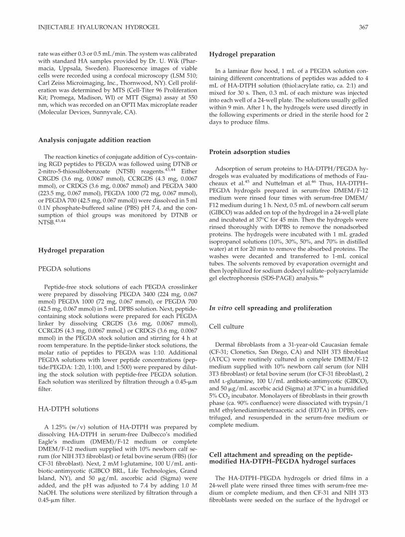
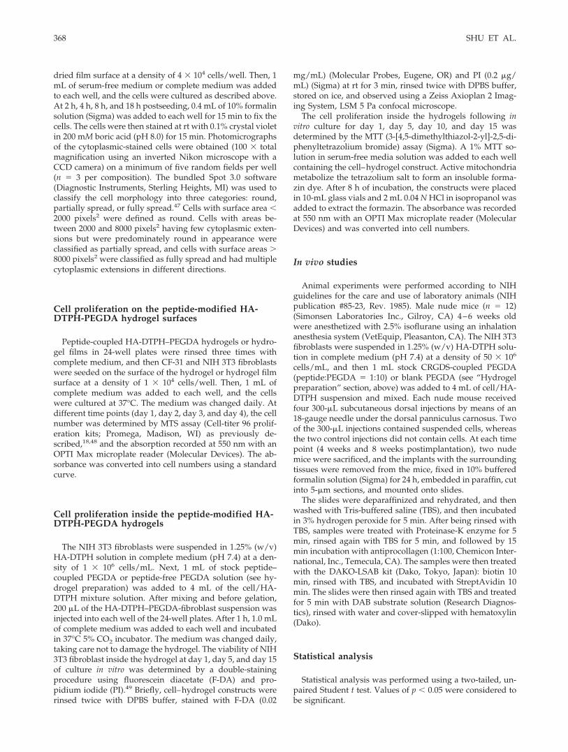
A novel hydrogel prepared by conjugate additionof HA-DTPH with PEGDA meets most of the re-quirements for an injectable, in situ-crosslinkablematerial for in vivo tissue engineering,33 except thatthis hydrogel is a poor substrate for cell attachment.We reasoned that attachment of fibronectin-likepeptide sequences would promote integrin-medi-ated cell attachment to the biocompatible hydrogels.To test this hypothesis, peptides containing the RGDsequence or a control sequence were cocrosslinkedinto the hydrogel by conjugate addition38 of a pep-tide Cys thiol onto one end of a bifunctional PEGDAcrosslinker. Thus, RGD peptides with either one ortwo cysteine residue (CRGDS and CCRGDS) wereconjugated to an excess of PEGDA to give a mixtureof unreacted homobifunctional PEGDA plus mono-function peptide–PEG acrylate. A nonsense peptide(CRDGS) was used as the control (Fig. 1). Duringthe first conjugate addition of peptide to PEGDA,the molar ratio of thiol to acrylate functionalitieswas maintained at 1:20 for CRGDS and CRDGS and1:10 for CCRGDS. As seen in Figure 2, the reactionswere complete within 5 min. These conditions favorthe singly modified PEDGA addition product, leav-ing one acrylate for attachment to HA-DTPH and anexcess of unmodified PEGDA for crosslinking of theHA-DTPH–peptide constructs into a hydrogel. Us-ing this protocol, it was unnecessary to isolate themonovalent CRGDS-PEG acrylate, CRDGS-PEGacrylate, or divalent CCRGDS bis(PEG acrylate)present in the crosslinking mixture.
Cell spreading and proliferation
Under sterile conditions, HA-DTPH–PEGDA hy-drogels containing different peptide concentrationswere fabricated by adding 1 mL of peptide-coupledPEGDA solution to 4 mL of HA-DTPH solution (1.25%w/v), maintaining a twofold molar ratio of thiol toacrylate functionalities. This ratio ensures completereaction of the PEGDA and the peptide–PEG acrylatederivative. The GPC analysis indicated that little freePEGDA remained (data not shown). Thus, the RGDpeptide–PEG acrylate was successfully coupled intothe hydrogel.
To estimate protein adsorption, an HA-DTPH–PEGDA hydrogel prepared in serum-free DMEM/F-12 medium was treated with newborn calf serum(GIBCO) and incubated at 37°C for 45 min. Afterremoving the nonadsorbed proteins by thorough rins-ing with DPBS, the absorbed proteins were collectedby elution with an isopropanol-water step gradient(10%, 30%, 50%, and 70%). Each isopropanol rinsesolution was then lyophilized for SDS-PAGE analysis.The results indicated that only very low amounts ofprotein were adsorbed onto the hydrogel (data notshown).
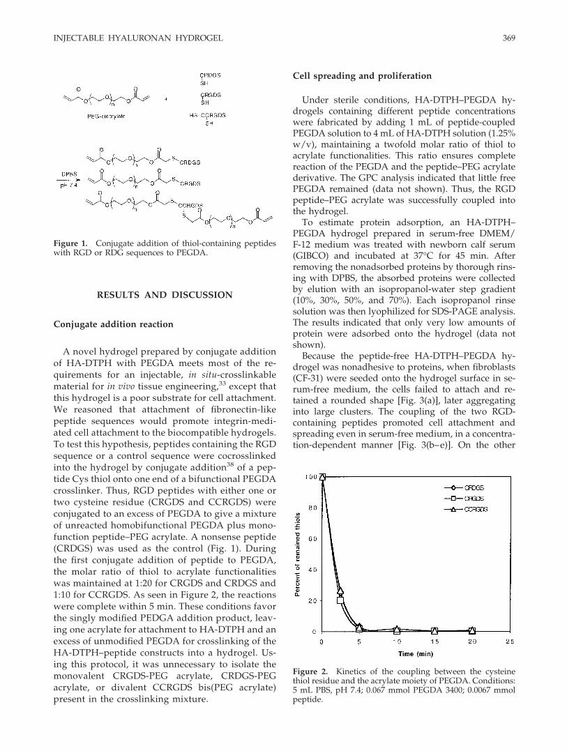
Because the peptide-free HA-DTPH–PEGDA hy-drogel was nonadhesive to proteins, when fibroblasts(CF-31) were seeded onto the hydrogel surface in se-rum-free medium, the cells failed to attach and re-tained a rounded shape [Fig. 3(a)], later aggregatinginto large clusters. The coupling of the two RGD-containing peptides promoted cell attachment andspreading even in serum-free medium, in a concentra-tion-dependent manner [Fig. 3(b–e)]. On the other
Figure 2. Kinetics of the coupling between the cysteinethiol residue and the acrylate moiety of PEGDA. Conditions:5 mL PBS, pH 7.4; 0.067 mmol PEGDA 3400; 0.0067 mmolpeptide.
Figure 1. Conjugate addition of thiol-containing peptideswith RGD or RDG sequences to PEGDA.
INJECTABLE HYALURONAN HYDROGEL 369
hand, the scrambled peptide (CRDGS) failed to pro-mote cell spreading, and all cells were rounded at allconcentrations in serum-free medium (data notshown). The results indicate that the cell attachmentand spreading occurred specifically in response to theRGD sequence.
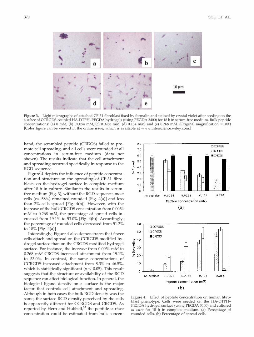
Figure 4 depicts the influence of peptide concentra-tion and structure on the spreading of CF-31 fibro-blasts on the hydrogel surface in complete mediumafter 18 h in culture. Similar to the results in serum-free medium (Fig. 3), without the RGD sequence, mostcells (ca. 58%) remained rounded [Fig. 4(a)] and lessthan 2% cells spread [Fig. 4(b)]. However, with theincrease of the bulk CRGDS concentration from 0.0054mM to 0.268 mM, the percentage of spread cells in-creased from 19.1% to 53.0% [Fig. 4(b)]. Accordingly,the percentage of rounded cells decreased from 51.2%to 18% [Fig. 4(a)].
Interestingly, Figure 4 also demonstrates that fewercells attach and spread on the CCRGDS-modified hy-drogel surface than on the CRGDS-modified hydrogelsurface. For instance, the increase from 0.0054 mM to0.268 mM CRGDS increased attachment from 19.1%to 53.0%. In contrast, the same concentrations ofCCRGDS increased attachment from 8.3% to 46.5%,which is statistically significant (p � 0.05). This resultsuggests that the structure or availability of the RGDsequence can affect biological function. In general, thebiological ligand density on a surface is the majorfactor that controls cell attachment and spreading.Although in both cases the bulk RGD density was thesame, the surface RGD density perceived by the cellsis apparently different for CCRGDS and CRGDS. Asreported by Hern and Hubbell,37 the peptide surfaceconcentration could be estimated from bulk concen-
Figure 3. Light micrographs of attached CF-31 fibroblast fixed by formalin and stained by crystal violet after seeding on thesurface of CCRGDS-coupled HA-DTPH–PEGDA hydrogels (using PEGDA 3400) for 18 h in serum-free medium. Bulk peptideconcentrations: (a) 0 mM, (b) 0.0054 mM, (c) 0.0268 mM, (d) 0.134 mM, and (e) 0.268 mM. (Original magnification �100.)[Color figure can be viewed in the online issue, which is available at www.interscience.wiley.com.]
Figure 4. Effect of peptide concentration on human fibro-blast phenotype. Cells were seeded on the HA-DTPH–PEGDA hydrogel surface (using PEGDA 3400) and culturedin vitro for 18 h in complete medium. (a) Percentage ofrounded cells. (b) Percentage of spread cells.
370 SHU ET AL.
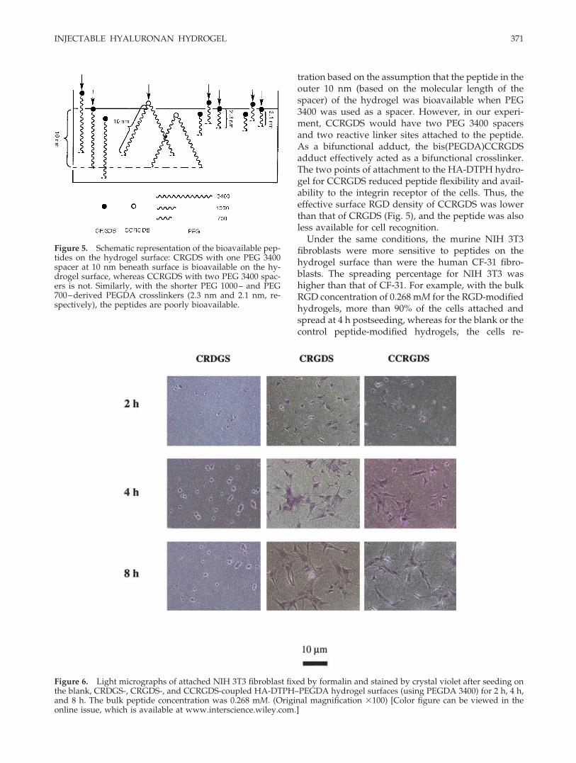
tration based on the assumption that the peptide in theouter 10 nm (based on the molecular length of thespacer) of the hydrogel was bioavailable when PEG3400 was used as a spacer. However, in our experi-ment, CCRGDS would have two PEG 3400 spacersand two reactive linker sites attached to the peptide.As a bifunctional adduct, the bis(PEGDA)CCRGDSadduct effectively acted as a bifunctional crosslinker.The two points of attachment to the HA-DTPH hydro-gel for CCRGDS reduced peptide flexibility and avail-ability to the integrin receptor of the cells. Thus, theeffective surface RGD density of CCRGDS was lowerthan that of CRGDS (Fig. 5), and the peptide was alsoless available for cell recognition.
Under the same conditions, the murine NIH 3T3fibroblasts were more sensitive to peptides on thehydrogel surface than were the human CF-31 fibro-blasts. The spreading percentage for NIH 3T3 washigher than that of CF-31. For example, with the bulkRGD concentration of 0.268 mM for the RGD-modifiedhydrogels, more than 90% of the cells attached andspread at 4 h postseeding, whereas for the blank or thecontrol peptide-modified hydrogels, the cells re-
Figure 5. Schematic representation of the bioavailable pep-tides on the hydrogel surface: CRGDS with one PEG 3400spacer at 10 nm beneath surface is bioavailable on the hy-drogel surface, whereas CCRGDS with two PEG 3400 spac-ers is not. Similarly, with the shorter PEG 1000– and PEG700–derived PEGDA crosslinkers (2.3 nm and 2.1 nm, re-spectively), the peptides are poorly bioavailable.
Figure 6. Light micrographs of attached NIH 3T3 fibroblast fixed by formalin and stained by crystal violet after seeding onthe blank, CRDGS-, CRGDS-, and CCRGDS-coupled HA-DTPH–PEGDA hydrogel surfaces (using PEGDA 3400) for 2 h, 4 h,and 8 h. The bulk peptide concentration was 0.268 mM. (Original magnification �100) [Color figure can be viewed in theonline issue, which is available at www.interscience.wiley.com.]
INJECTABLE HYALURONAN HYDROGEL 371
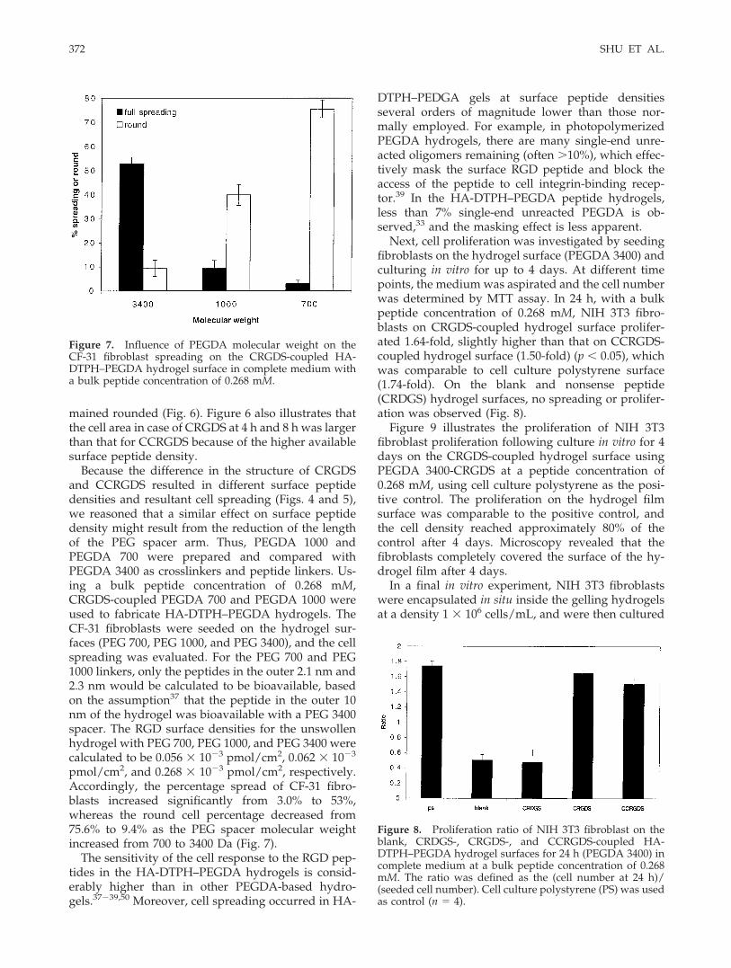
mained rounded (Fig. 6). Figure 6 also illustrates thatthe cell area in case of CRGDS at 4 h and 8 h was largerthan that for CCRGDS because of the higher availablesurface peptide density.
Because the difference in the structure of CRGDSand CCRGDS resulted in different surface peptidedensities and resultant cell spreading (Figs. 4 and 5),we reasoned that a similar effect on surface peptidedensity might result from the reduction of the lengthof the PEG spacer arm. Thus, PEGDA 1000 andPEGDA 700 were prepared and compared withPEGDA 3400 as crosslinkers and peptide linkers. Us-ing a bulk peptide concentration of 0.268 mM,CRGDS-coupled PEGDA 700 and PEGDA 1000 wereused to fabricate HA-DTPH–PEGDA hydrogels. TheCF-31 fibroblasts were seeded on the hydrogel sur-faces (PEG 700, PEG 1000, and PEG 3400), and the cellspreading was evaluated. For the PEG 700 and PEG1000 linkers, only the peptides in the outer 2.1 nm and2.3 nm would be calculated to be bioavailable, basedon the assumption37 that the peptide in the outer 10nm of the hydrogel was bioavailable with a PEG 3400spacer. The RGD surface densities for the unswollenhydrogel with PEG 700, PEG 1000, and PEG 3400 werecalculated to be 0.056 � 10�3 pmol/cm2, 0.062 � 10�3
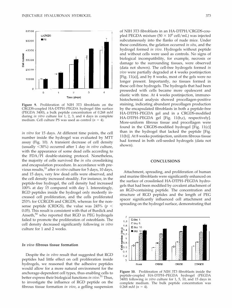
pmol/cm2, and 0.268 � 10�3 pmol/cm2, respectively.Accordingly, the percentage spread of CF-31 fibro-blasts increased significantly from 3.0% to 53%,whereas the round cell percentage decreased from75.6% to 9.4% as the PEG spacer molecular weightincreased from 700 to 3400 Da (Fig. 7).
The sensitivity of the cell response to the RGD pep-tides in the HA-DTPH–PEGDA hydrogels is consid-erably higher than in other PEGDA-based hydro-gels.37�39,50 Moreover, cell spreading occurred in HA-
DTPH–PEDGA gels at surface peptide densitiesseveral orders of magnitude lower than those nor-mally employed. For example, in photopolymerizedPEGDA hydrogels, there are many single-end unre-acted oligomers remaining (often �10%), which effec-tively mask the surface RGD peptide and block theaccess of the peptide to cell integrin-binding recep-tor.39 In the HA-DTPH–PEGDA peptide hydrogels,less than 7% single-end unreacted PEGDA is ob-served,33 and the masking effect is less apparent.
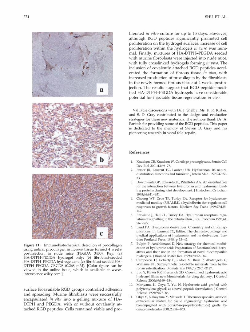
Next, cell proliferation was investigated by seedingfibroblasts on the hydrogel surface (PEGDA 3400) andculturing in vitro for up to 4 days. At different timepoints, the medium was aspirated and the cell numberwas determined by MTT assay. In 24 h, with a bulkpeptide concentration of 0.268 mM, NIH 3T3 fibro-blasts on CRGDS-coupled hydrogel surface prolifer-ated 1.64-fold, slightly higher than that on CCRGDS-coupled hydrogel surface (1.50-fold) (p � 0.05), whichwas comparable to cell culture polystyrene surface(1.74-fold). On the blank and nonsense peptide(CRDGS) hydrogel surfaces, no spreading or prolifer-ation was observed (Fig. 8).
Figure 9 illustrates the proliferation of NIH 3T3fibroblast proliferation following culture in vitro for 4days on the CRGDS-coupled hydrogel surface usingPEGDA 3400-CRGDS at a peptide concentration of0.268 mM, using cell culture polystyrene as the posi-tive control. The proliferation on the hydrogel filmsurface was comparable to the positive control, andthe cell density reached approximately 80% of thecontrol after 4 days. Microscopy revealed that thefibroblasts completely covered the surface of the hy-drogel film after 4 days.
In a final in vitro experiment, NIH 3T3 fibroblastswere encapsulated in situ inside the gelling hydrogelsat a density 1 � 106 cells/mL, and were then cultured
Figure 8. Proliferation ratio of NIH 3T3 fibroblast on theblank, CRDGS-, CRGDS-, and CCRGDS-coupled HA-DTPH–PEGDA hydrogel surfaces for 24 h (PEGDA 3400) incomplete medium at a bulk peptide concentration of 0.268mM. The ratio was defined as the (cell number at 24 h)/(seeded cell number). Cell culture polystyrene (PS) was usedas control (n � 4).
Figure 7. Influence of PEGDA molecular weight on theCF-31 fibroblast spreading on the CRGDS-coupled HA-DTPH–PEGDA hydrogel surface in complete medium witha bulk peptide concentration of 0.268 mM.
372 SHU ET AL.
in vitro for 15 days. At different time points, the cellnumber inside the hydrogel was evaluated by MTTassay (Fig. 10). A transient decrease of cell density(usually �30%) occurred after 1 day in vitro culture,with the appearance of some dead cells according tothe FDA/PI double-staining protocol. Nonetheless,the majority of cells survived the in situ crosslinkingand encapsulation procedure. In accordance with pre-vious results,33 after in vitro culture for 5 days, 10 days,and 15 days, very few dead cells were observed, andthe cell density increased steadily. For instance, in thepeptide-free hydrogel, the cell density had increased100% at day 15 compared with day 1. Interestingly,RGD peptides inside the hydrogel only modestly in-creased cell proliferation, and the cells proliferated253% for CCRGDS and CRGDS, whereas for the non-sense peptide (CRDGS), the value was 245% (p �0.05). This result is consistent with that of Burdick andAnseth,50 who reported that RGD in PEG hydrogelsfailed to promote the proliferation of osteoblasts. Thecell density decreased significantly following in vitroculture for 1 and 2 weeks.
In vivo fibrous tissue formation
Despite the in vitro result that suggested that RGDpeptides had little effect on cell proliferation insidehydrogels, we reasoned that the adhesive peptideswould allow for a more natural environment for theanchorage-dependent cell types, thus enabling cells tobetter express their biological functions in vivo.51 Thus,to investigate the influence of RGD peptide on thefibrous tissue formation in vivo, a gelling suspension
of NIH 3T3 fibroblasts in an HA-DTPH/CRGDS-cou-pled PEGDA mixture (50 � 106 cell/mL) was injectedsubcutaneously into the flanks of nude mice. Underthese conditions, the gelation occurred in situ, and thehydrogel formed in vivo. Hydrogels without peptideand without cells were used as controls. No signs ofbiological incompatibility, for example, necrosis ordamage to the surrounding tissues, were observed(data not shown). The cell-free hydrogels formed invivo were partially degraded at 4 weeks postinjection[Fig. 11(a)], and by 8 weeks, most of the gels were nolonger present. Importantly, no tissues formed inthese cell-free hydrogels. The hydrogels that had beenpreseeded with cells became more opalescent andelastic with time. At 4 weeks postinjection, immuno-histochemical analysis showed procollagen-positivestaining, indicating abundant procollagen productionby the encapsulated fibroblasts in both a peptide-freeHA-DTPH–PEGDA gel and in a CRGDS-modifiedHA-DTPH–PEGDA gel [Fig. 11(b,c), respectively].More-uniform fibrous tissue and procollagen werefound in the CRGDS-modified hydrogel [Fig. 11(c)]than in the hydrogel that lacked the peptide [Fig.11(b)]. At 8 weeks postinjection, uniform fibrous tissuehad formed in both cell-seeded hydrogels (data notshown).
CONCLUSIONS
Attachment, spreading, and proliferation of humanand murine fibroblasts were significantly enhanced onthe surface of crosslinked HA-DTPH–PEGDA hydro-gels that had been modified by covalent attachment ofan RGD-containing peptide. The concentration andstructure of RGD peptides and the length of PEGspacer significantly influenced cell attachment andspreading on the hydrogel surface, demonstrating that
Figure 10. Proliferation of NIH 3T3 fibroblasts inside thepeptide-coupled HA-DTPH–PEGDA hydrogel (PEGDA3400) following in vitro culture for 1, 5, 10, and 15 days incomplete medium. The bulk peptide concentration was0.268 mM (n � 4).
Figure 9. Proliferation of NIH 3T3 fibroblasts on theCRGDS-coupled HA-DTPH–PEGDA hydrogel film surface(PEGDA 3400), a bulk peptide concentration of 0.268 mMduring in vitro culture for 1, 2, 3, and 4 days in completemedium. Cell culture PS was used as control (n � 4).
INJECTABLE HYALURONAN HYDROGEL 373
surface bioavailable RGD groups controlled adhesionand spreading. Murine fibroblasts were successfullyencapsulated in situ into a gelling mixture of HA-DTPH and PEGDA, with or without covalently at-tached RGD peptides. Cells remained viable and pro-
liferated in vitro culture for up to 15 days. However,although RGD peptides significantly promoted cellproliferation on the hydrogel surfaces, increase of cellproliferation within the hydrogels in vitro was mini-mal. Finally, mixtures of HA-DTPH–PEGDA seededwith murine fibroblasts were injected into nude mice,with fully crosslinked hydrogels forming in vivo. Theinclusion of covalently attached RGD peptides accel-erated the formation of fibrous tissue in vivo, withincreased production of procollagen by the fibroblastsin the newly formed fibrous tissue at 4 weeks postin-jection. The results suggest that RGD peptide–modi-fied HA-DTPH–PEGDA hydrogels have considerablepotential for injectable tissue regeneration in vivo.
Valuable discussions with Dr. J. Shelby, Ms. K. R. Kirker,and S. D. Gray contributed to the design and evaluationstrategies for these new materials. The authors thank Dr. A.Panitch for providing some of the RGD peptides. This paperis dedicated to the memory of Steven D. Gray and hispioneering research in vocal fold repair.
References
1. Knudson CB, Knudson W. Cartilage proteoglycans. Semin CellDev Biol 2001;12:69–78.
2. Fraser JR, Laurent TC, Laurent UB. Hyaluronan: its nature,distribution, functions and turnover. J Intern Med 1997;242:27–33.
3. Dowthwaite GP, Edwards JC, Pitsillides AA. An essential rolefor the interaction between hyaluronan and hyaluronan bind-ing proteins during joint development. J Histochem Cytochem1998;46:641–651.
4. Cheung WF, Crue TF, Turley EA. Receptor for hyaluronan-mediated motility (RHAMM), a hyaladherin that regulates cellresponses to growth factors. Biochem Soc Trans 1999;27:135–142.
5. Entwistle J, Hall CL, Turley EA. Hyaluronan receptors: regu-lators of signalling to the cytoskeleton. J Cell Biochem 1996;61:569–577.
6. Band PA. Hyaluronan derivatives: Chemistry and clinical ap-plications. In: Laurent TC, Editor. The chemistry, biology andmedical applications of hyaluronan and its derivatives. Lon-don: Portland Press; 1998. p 33–42.
7. Bulpitt P, Aeschlimann D. New strategy for chemical modifi-cation of hyaluronic acid: Preparation of functionalized deriv-atives and their use in the formation of novel biocompatiblehydrogels. J Biomed Mater Res 1999;47:152–169.
8. Campoccia D, Doherty P, Radice M, Brun P, Abatangelo G,Williams DF. Semisynthetic resorbable materials from hyalu-ronan esterification. Biomaterials 1998;19:2101–2127.
9. Luo Y, Kirker KR, Prestwich GD. Cross-linked hyaluronic acidhydrogel films: new biomaterials for drug delivery. J ControlRelease 2000;69:169–184.
10. Moriyama K, Ooya T, Yui N. Hyaluronic acid grafted withpoly(ethylene glycol) as a novel peptide formulation. J ControlRelease 1999;59:77–86.
11. Ohya S, Nakayama Y, Matsuda T. Thermoresponsive artificialextracellular matrix for tissue engineering: hyaluronic acidbioconjugated with poly(N-isopropylacrylamide) grafts. Bi-omacromolecules 2001;2:856–863.
Figure 11. Immunohistochemical detection of procollagenusing antirat procollagen in fibrous tissue formed 4 weekspostinjection in nude mice (PEGDA 3400). Key: (a)HA-DTPH-PEGDA hydrogel only; (b) fibroblast-seededHA-DTPH–PEGDA hydrogel; and (c) fibroblast-seeded HA-DTPH–PEGDA–CRGDS (0.268 mM). [Color figure can beviewed in the online issue, which is available at www.interscience.wiley.com.]
374 SHU ET AL.
12. Pouyani T, Prestwich GD. Functionalized derivatives of hyal-uronic acid oligosaccharides: drug carriers and novel bioma-terials. Bioconjugate Chem 1994;5:339–347.
13. Prestwich GD, Vercruysse KP. Therapeutic applications of hy-aluronic acid and hyaluronan derivatives. Pharm Sci TechnolToday 1998;1:42–43.
14. Prestwich GD, Marecak DM, Marecek JF, Vercruysse KP, Zie-bell MR. Controlled chemical modification of hyaluronic acid:synthesis, applications, and biodegradation of hydrazide de-rivatives. J Control Release 1998;53:93–103.
15. Prestwich GD, Luo Y, Ziebell MR, Vercruysse KP, Kirker KR,MacMaster JS. Chemically-modified hyaluronan: new bioma-terials and probes for cell biology. In: Abatangelo G, editor.New frontiers in medical sciences: redefining hyaluronan. Lon-don: Portland Press; 2000. p 181–194.
16. Vercruysse KP, Prestwich GD. Hyaluronate derivatives indrug delivery. Crit Rev Ther Drug Carrier Syst 1998;15:513–555.
17. Kirker KR, Luo Y, Nielson JH, Shelby J, Prestwich GD. Glycos-aminoglycan hydrogel films as bio-interactive dressings forwound healing. Biomaterials 2002;23:3661–3671.
18. Shu XZ, Liu Y, Luo Y, Robert MC, Prestwich GD. Disulfidecrosslinked hyaluronan hydrogel. Biomacromolecules 2002;3:1304–1311.
19. Peppas NA. Hydrogels in medicine and pharmacy. Florida:CRC Press; 1987.
20. Griffith LG. Polymeric biomaterials. Acta Mater 2000;48:263–277.
21. Peppas NA, Bures P. Hydrogels in pharmaceutical formula-tion. Eur J Pharm Biopharm 2000;50:27–46.
22. Hoffman AS. Hydrogels for biomedical application. Adv DrugDel Rev 2002;43:3–12.
23. Miller JA, Ferguson RL, Powers DL, Burns JW, Shalaby SW.Efficacy of hyaluronic acid0/nonsteroidal anti-inflammatorydrug systems in preventing postsurgical tendon adhesions.J Biomed Mater Res (Appl Biomater) 1997;38:25–33.
24. Jackson JK, Skinner KC, Burgess L, Sun T, Hunter WL, BurtHM. Paclitaxel-loaded crosslinked hyaluronic acid films for theprevention of postsurgical adhesion. Pharm Res 2002;19:411–417.
25. Piacquadio D, Jarcho M, Goltz R. Evaluation of hylan b gel asa soft-tissue augmentation implant material. J Am Acad Der-matol 1997;36:544–549.
26. Leach RE, Burns JW, Dawe EJ, Smith B, Diamond MP. Reduc-tion of postsurgical adhesion formation in the rabbit uterinehorn model with use of hyaluronate/carboxy-methylcellulosegel. Fertility Sterility 1998;69:415–418.
27. Sawada T, Hasegawa K, Tsukada K, Kawakami S. Adhesionpreventative effect of hyaluronic acid after intraperitoneal sur-gery in mice. European Soc Hum Reprod Embryol 1999;14:1470–1472.
28. Yoldemir T, Sagol S, Adakan S, Oztekin K, Ozsener S, Karada-das N. Comparison of the reduction of postoperative adhe-sions by two barriers, one solution, and two pharmacologicagents in the rat uterine model. Fertility Sterility 2002;78:335–339.
29. Coelho AM, Berumen L, Avrameas S. Properties of proteinpolymers as substratum for cell growth in vitro. J Cell Physiol1974;83:379–388.
30. Hubbell JA. Biomaterials in tissue engineering. Bio/technology1995;13:565–576.
31. Shu XZ, Liu Y, Palumbo F, Prestwich GD. Disulfide-crosslinked hyaluronan-gelatin hydrogel films: a covalent
mimic of the extracellular matrix for in vitro cell growth. Bio-materials 2003;24:3825–3834.
32. Liu Y, Shu XZ, Gray SD, Prestwich GD. Disulfide-crosslinkedhyalruonan-gelatin sponges: growth of fibrous tissue in vivo.J Biomed Mater Res. Forthcoming.
33. Shu XZ, Liu Y, Palumbo F, Luo Y, Prestwich GD. In-situcrosslinkable hyaluronan hydrogel for tissue engineering. Bio-materials 2004;25:1139–1348.
34. Ramamurthi A, Vesely I. Smooth muscle cell adhesion oncrosslinked hyaluronan gels. J Biomed Mater Res 2002;60:196–205.
35. Hu M, Sabelman EE, Tsai C, Tan J, Hentz VR. Improvement ofSchwann cell attachment and proliferation on modified hyal-uronic acid strands by polylysine. Tissue Eng 2000;6:585–593.
36. Park YD, Tirelli N, Hubbell JA. Photopolymerized hyaluronicacid-based hydrogels and interpenetrating networks. Biomate-rials 2003;24:893–900.
37. Hern DL, Hubbell JA. Incorporation of adhesion peptides intononadhesive hydrogels useful for tissue resurfacing. J BiomedMater Res 1998;39:266–276.
38. Elbert DL, Hubbell JA. Conjugate addition reactions combinedwith free-radical crosslinking for the design of materials fortissue engineering. Biomacromolecules 2001;2:430–441.
39. Shin H, Jo S, Mikos AG. Modulation of marrow stromal osteo-blast adhesion on biomimetic oligo[poly(ethylene glycol) fu-marate] hydrogels modified with Arg-Gly-Asp peptides and apoly(ethylene glycol) spacer. J Biomed Mater Res 2002;61:169–179.
40. Ruoslahti E. RGD and other recognition sequences for inte-grins. Annu Rev Cell Dev Biol 1996;12:697–715.
41. Hynes RO. Integrins: versatility, modulation, and signalling incell adhesion. Cell 1992;69:11–25.
42. Quirk RA, Chan WC, Davies MC, Tendler SJB, Shakesheff KM.Poly(l-lysine)-GRGDS as a biomimetic surface modifier forpoly(lactic acid). Biomaterials 2001;22:865–872.
43. Ellman GL. A colorimetric method for determining low con-centrations of mercaptans. Arch Biochem Biophys 1958;74:443–450.
44. Thannhauser TW, Konishi Y, Scheraga HA. Analysis for disul-fide bonds in peptides and proteins. Meth Enzymol 1987;143:115–119.
45. Faucheaux N, Haye B, Nagel MD. Activation of the cyclic AMPpathway in cells adhering to biomaterials: Regulation by vitro-nectin- and fibronectin-integrin binding. Biomaterials 2000;21:1031–1038.
46. Nuttelman CR, Mortisen DJ, Henry SM. Attachment of fi-bronectin to poly(vinyl alcohol) hydrogels promotes NIH 3T3cell adhesion, proliferation, and migration. J Biomed Mater Res2001;57:217–223.
47. Neff JA, Caldwell KD, Tresco PA. A novel method for surfacemodification to promote cell attachment to hydrophobic sub-strates. J Biomed Mater Res 1998;40:511–519.
48. Lin VS, Lee MC, O’Neal S, McKean J, Sung K-LP. Ligamenttissue engineering using synthetic biodegradable fiber scaf-folds. Tissue Eng 1999;5:443–451.
49. Jones KH, Senft JA. An improved method to determine cellviability by simultaneous staining with fluorescein diacetate-propidium iodide. J Histochem Cytochem 1985;33:77–79.
50. Burdick JA, Anseth KS. Photoencapsulation of osteoblasts ininjectable RGD-modified PEG hydrogels for bone tissue engi-neering. Biomaterials 2002;23:4315–4323.
51. Mann BK, Tsai AT, Scott-Burden T, West JL. Modification ofsurfaces with cell adhesion peptides alters extracellular matrixdeposition. Biomaterials 1999;20:2281–2286.
INJECTABLE HYALURONAN HYDROGEL 375
Copyright © 2022 FDOKUMEN