SYNTHESIS OF NEW RGD PEPTIDOMIMETIC-DRUG ...
172
Doctoral School in Chemical Sciences and Technologies PhD Course in Chemistry, XXXI Cycle SYNTHESIS OF NEW RGD PEPTIDOMIMETIC-DRUG CONJUGATES TARGETING α v β 3 INTEGRIN Arianna PINA R11392 CHIM/06 Organic Chemistry Tutor: Prof. Dr. Cesare GENNARI Academic Co-Tutor: Dr. Luca PIGNATARO Industrial Co-Tutor: Dr. Michele CARUSO (Nerviano Medical Sciences) Coordinator: Prof. Dr. Emanuela LICANDRO Milan, January 2019
-
Upload
khangminh22 -
Category
Documents
-
view
3 -
download
0
Transcript of SYNTHESIS OF NEW RGD PEPTIDOMIMETIC-DRUG ...

Doctoral School in Chemical Sciences and Technologies PhD Course in Chemistry, XXXI Cycle
SYNTHESIS OF NEW
RGD PEPTIDOMIMETIC-DRUG CONJUGATES
TARGETING αvβ3 INTEGRIN
Arianna PINA
R11392
CHIM/06 Organic Chemistry
Tutor: Prof. Dr. Cesare GENNARI
Academic Co-Tutor: Dr. Luca PIGNATARO
Industrial Co-Tutor: Dr. Michele CARUSO (Nerviano Medical Sciences)
Coordinator: Prof. Dr. Emanuela LICANDRO
Milan, January 2019

The present work was led by: Prof. Dr. C. Gennari, Dr. L. Pignataro and Dr. M. Caruso
Doctoral Final Oral Examination: January, 25th 2019
Examination Committee: Chairperson: Prof. Dr. U. Piarulli
Università degli studi dell'Insubria (I)
Second Member: Prof. Dr. S. Sattin
Università degli Studi di Milano (I)
Third Member: Prof. Dr. J. Codee
Leiden University (NL)
The work herein described was performed at the University of Milan at the Department of Chemistry, in
collaboration with Nerviano Medical Sciences, in the period from October 2015 to October 2018 under the
supervision of Prof. Cesare Gennari.

“One never notices what has been done;
one can only see what remains to be done”
Marie Curie


Table of Contents
General Introduction ............................................................................................................................. 6
Chapter 1 ................................................................................................................................................ 8
1. DRUG TARGETING TO CANCER ...............................................................................................................8
1.1. INTRODUCTION ....................................................................................................................................8
1.2. CHEMOTHERAPY AND DRUG DELIVERY .......................................................................................9
1.2.1. Antibody-Drug Conjugates (ADCs) ....................................................................................................14
1.2.2. Small Molecule-Drug Conjugates (SMDCs) .......................................................................................17
1.3. CONTRIBUTION OF OUR GROUP: PREVIOUS WORKS IN αvβ3 INTEGRIN-DRUG
CONJUGATES FIELD .................................................................................................................................30
Chapter 2 .............................................................................................................................................. 36
2.1. THERANOSTIC RGD-CPT CONJUGATES ............................................................................................36
2.1.1. Synthesis of RGD-Naph-SS-CPT conjugates ......................................................................................39
2.1.2. Synthesis of an “uncleavable” RGD-CPT conjugate ...........................................................................40
2.2. BIOLOGICAL EVALUATIONS ...............................................................................................................41
2.2.1. Integrin Receptor Competitive Binding Assays...................................................................................41
2.2.2. Confocal microscopy analysis and quantitative imaging (ArrayScan) ................................................41
2.2.3. Stability and cell viability assays .........................................................................................................43
2.3. DISCUSSION AND CONCLUSIONS .......................................................................................................44
Chapter 3 .............................................................................................................................................. 47
3. ACTIVE TUMOR TARGETING USING RGD-BEARING LIGANDS ......................................................47
3.1. NEGATIVE CONTROLS FOR BIOLOGICAL INVESTIGATIONS ..................................................47
3.2. SYNTHESIS OF CONTROL COMPOUND cyclo(DKP-RβAD) .........................................................52
3.3. BIOLOGICAL EVALUATION OF CONTROL COMPOUND cyclo(DKP-RβAD) ............................53
3.4. MULTIVALENCY .................................................................................................................................57

Chapter 4 .............................................................................................................................................. 63
4. LINKER-DRUG MODULE: A KEY COMBINATION ...............................................................................63
4.1. SYNTHESIS OF 2nd GENERATION RGD-CPT CONJUGATE BEARING DISULFIDE LINKER ..66
4.2. BIOLOGICAL EVALUATIONS ...........................................................................................................68
4.2.1. Integrin Receptor Competitive Binding Assays...................................................................................68
4.2.2. Stability and cell viability assays .........................................................................................................69
Chapter 5 .............................................................................................................................................. 71
5. CONTRIBUTION OF OUR GROUP: NEW GENERATION OF RGD-DRUG CONJUGATES ...............71
Chapter 6 .............................................................................................................................................. 77
6. CONCLUSIONS AND FUTURE PERSPECTIVES.....................................................................................77
Chapter 7 .............................................................................................................................................. 80
7. EXPERIMENTAL SECTION .......................................................................................................................80
7.1. BIOLOGICAL ASSAYS ........................................................................................................................82
7.2. SYNTHESIS OF THERANOSTIC RGD-CAMPTOTHECIN CONJUGATES ...................................86
7.3. SYNTHESIS OF cyclo(DKP-RβAD) AS NEGATIVE CONTROL FOR BIOLOGICAL
INVESTIGATIONS ......................................................................................................................................93
7.4. SYNTHESIS OF FLUORESCENT COMPOUNDS FOR ACTIVE TUMOR TARGETING ..............99
7.5. SYNTHESIS OF 2nd GENERATION RGD-SS-CPT CONJUGATE ..................................................115
7.6. HPLC TRACES OF FINAL PRODUCTS ...........................................................................................123
7.7. MASS, 1H-NMR AND 13C-NMR SPECTRA ......................................................................................128
References .......................................................................................................................................... 161

Abbreviations
Aba Azabicycloalkane
Ac Acetyl
ADC Antibody-drug conjugate
Ampro 4-Aminoproline
aq. Aqueous solution
APT Attached proton test
Asc Ascorbate
Bn Benzyl
Boc tert-Butyloxycarbonyl
Bu Butyl
Bz Benzoyl
CA Carbonic anhydrase
Cy5 Sulfo-Cyanine5
CPT Camptothecin
CuAAC Copper-Catalyzed Azide-Alkyne
Cycloaddition
DAVB Desacetyl vinblastine
DEPT Distortionless Enhancement by
Polarization Transfer
DHFR Dihydrofolate reductase
DCC N,N'-dicicloesilcarbodiimmide
DIC Diisopropylcarbodiimide
DKP Diketopiperazine
DMAP 4-Dimethylaminopyridine
DMF N,N-Dimethylformamide
DMSO Dimethyl sulfoxide
DNA Deoxyribonucleic acid
DOX Doxorubicin
DTT Dithiothreitol
ECM Extracellular Matrix
EDC 1-Ethyl-3-(3-
dimethylaminopropyl)carbodiimide
EEDQ N-Ethoxycarbonyl-2-ethoxy-1,2-
dihydroquinoline
EPR Enhanced permeability and retention
equiv. Equivalents
ESI Electrospray ionisation
Et Ethyl
FAK Focal adhesion kinase
FACS Fluorescence Assisted Cell Sorting
FH4 Tetrahydrofolic acid
Fmoc 9-Fluorenylmethoxycarbonyl
GSH Glutathione
FR Folate Receptor
HATU O-(7-azabenzotriazol-1-yl)-
tetramethyl-uronium
hexafluorophosphate
Her/neu Receptor tyrosine-protein kinase erbB-
2
HOAt 1-Hydroxy-7-azabenzotriazole
HPLC High performance liquid
chromatography
HRMS High resolution mass spectrometry
Igs Immunoglobulins
iPr2NEt N,N-Diisopropylethylamine (DIPEA)
J Scalar coupling constants
Kd Dissociation Constant
MTT 3-(4,5-dimethylthiazol-2-yl)-2,5-
diphenyltetrazolium bromide

mAb Monoclonal antibody
MALDI Matrix-assisted laser desorption
ionization
MC/SD Monte Carlo/Stochastic Dynamics
Me Methyl
MED Minimum Effective Dose
MMAE Monomethyl auristatin E
MMAF Monomethyl auristatin F
MMP Matrix metalloproteinase
MS Mass spectroscopy
MTD Maximum tolerated dose
MW Molecular weight
Naph Naphthalimide
NMR Nuclear Magnetic Resonance
NHS N-Hydroxysuccinimide
PABC para-amino benzyl carbamate
PBS Phosphate-buffered saline
PCa Prostate cancer
PEG Polyethylene glycol
ppm Part per million
PSMA Prostate specific membrane antigen
PTX Paclitaxel
quant. Quantitative
RAFT Regioselectively-Addressable
Functionalized Template
Rf Retention factor
SAR Structure–activity relationship
RT Room temperature
SIP Small Immune Proteins
SMAC Second mitochondrial activator of
caspases
SMDC Small molecue-drug conjugate
SPECT Single-photon emission computed
tomography
SSTR Somatostatin receptor
S Selectivity
RNA Ribonucleic acid
RAFT Regioselectively Addressable
Functionalized Templates
RP Relative Potency
TFA Trifluoroacetic acid
THF Tetrahydrofuran
TI Targeting Index
TMS Tetramethylsilane
tR Retention time
t1/2 Half-life time
unc Uncleavable
VEGFR Vascular Endothelial Growth Factor
Receptor
WHO World Health Organization
δ Chemical shift

~ 5 ~
One-letter code Amino acid Three-letter code
A Alanine Ala
C Cysteine Cys
D Aspartic Asp
E Glutamic acid Glu
F Phenylalanine Phe
G Glycine Gly
H Histidine His
I Isoleucine Ile
K Lysine Lys
L Leucine Leu
M Methionine Met
N Asparagine Asn
P Proline Pro
Q Glutamine Gln
R Arginine Arg
S Serine Ser
T Threonine Thr
W Tryptophan Trp
Y Tyrosine Tyr
V Valine Val

~ 6 ~
General Introduction
Nowadays, systemic administration of cytotoxic agents is one of the main keystones of modern anticancer
chemotherapy. However, many drugs do not selectively localize into solid tumors, as they normally accumulate
also in healthy organs (e.g. liver and kidney). As a result, severe side effects are caused by the suboptimal
biodistribution profile, in combination with the non-specific mode of action of cytotoxic agents, preventing the
drug administration at therapeutic doses. The targeted delivery of anticancer agents into solid tumors is emerging
as encouraging approach to overcome the intrinsic drawbacks of cytotoxic drugs. For example, some antibodies
with high affinity for accessible tumor markers have been used as vehicles for the targeted drug delivery
(Antibody-Drug Conjugates, ADCs). Since they may have limitations due to their high molecular weight, to their
possible immunogenicity and to their high production costs, the development of smaller ligands (e.g. vitamins,
peptides and peptidomimetics) capable of binding efficiently to tumor-overexpressed receptors, may show some
significant therapeutic advantages. Peptides and peptidomimetics with the Arg-Gly-Asp tripeptide (RGD) are
known to bind integrin αvβ3, a heterodimeric transmembrane receptor which shows low expression on healthy
tissues, while being upregulated in a variety of cancer cells. For these reasons, integrin ligands have been widely
explored as homing devices for the selective delivery of anticancer agents. In this PhD thesis, the synthesis and
the biological evaluation of new small molecule-drug conjugates (SMDCs) targeting αvβ3 integrin are described.
This work started with the preparation of a SMDC for the integrin-targeted delivery of Camptothecin, containing
a disulfide linker and a naphtalimide moiety for the real-time monitoring of the cellular uptake (Chapter II).
While the design of this complex molecular structure was carried out to reproduce literature data, the biological
evaluation of this compound revealed a complex scenario. For this reason, the biological properties of all the
three moieties (ligand, drug and linker) have been deeply investigated individually. Firstly, in Chapter III we
established the role and the behavior of the ligand, trying to understand its action mechanism. Moreover,
modifications of the ligand unit have been carried out to deface the affinity for the receptor and to
unambiguously evaluate the capability of the original ligand to selectively target cancer cells that (over)express

~ 7 ~
αvβ3. Later on, linker-related structural features (e.g. stability, linker cleavage experiments, kinetics of drug
release, etc.) have been studied and improved (Chapter IV). Finally, the development of other integrin-targeted
SMDC products featuring peptide linkers (with their biological evaluation) is reported in Chapter V. In Chapter
VII, all the experimental details of synthetic and biological procedures are included, together with spectroscopic
data and HPLC profiles of the newly synthesized compounds
.
~ 8 ~
Chapter 1
1. DRUG TARGETING TO CANCER
1.1. INTRODUCTION
Cancer is one of the main causes of death in the developed countries and 14.1 million new cases of cancer and
8.2 million cancer deaths were estimated in 2014.1 The number of incidences is expected to increase up to 22
million within the next two decades. Lung cancer is the most diagnosed and the one associated to the highest
mortality rate in humans; in women, breast cancer is the most diagnosed and responsible for the largest number
of deaths. While heredity plays a major role in only 5% of patients, 30% of cancers are associated to unhealthy
lifestyles: smoking, alcohol consumption, overweight, poor diet of fruits and vegetables and exposure to UV
rays. The different types of cancer treatment belong to three main categories:
• surgery (surgical removal of the primary tumor and, whenever possible, of the secondary lesions);
• radiotherapy (treatment with various radioactive sources);
• pharmacological interventions (i.e. chemotherapy, hormone therapy, immunotherapy).
Combinations of these three anticancer treatments are often used. In most cases, surgical removal is followed by
chemotherapy or radiotherapy to remove remaining cancer cells and prevent recurrence. However, the state of
the art of all these treatments is far from optimal, in terms of efficacy, tolerability and cost. In fact, surgery is a
very invasive approach, it requires hospitalization and, in some cases, residual tumor cells that may be
undetectable during surgery lead to a re-growth of the cancer mass. Radiotherapy and chemotherapy are known
to act not only against tumor cells, but also against some healthy tissues, leading to several side effects. In most
cases, the latter are serious enough to substantially limit the so-called “therapeutic window”, which is defined as
the range of drug dosage between the minimum effective dose and the maximum tolerated dose.2 The efficacy of
chemotherapy is further limited by the occurrence or development of drug resistance: tumor cells can be

~ 9 ~
regarded as a rapidly changing target because of their genetic instability, heterogeneity, and high rate of
mutation, leading to selection and overgrowth of a drug-resistant tumor cell population.3 Furthermore, it is also
important to note that each case of cancer is unique. Thus, no universal treatments are known at this moment and
the therapy is modulated for each patient, depending on his/her physical characteristics of and on the one of the
tumor.
In 1907, Paul Ehrlich (1854-1915), often considered the father of chemotherapy, developed the first
chemotherapeutic agent for the treatment of syphilis, Salvarsan. Moreover, he was awarded the Nobel Prize in
1908 for his research in immunology. His pioneering work provided a deeper understanding of the correlation
between the chemical structure of molecules and their biological effects towards the cells they interact with. By
observing the selectivity of certain dyes for certain organs, he introduced the idea of the "magic bullet", a
compound that would bring an active ingredient selectively to the diseased area. For the first time, Paul Ehrlich’s
research approach led to the connection of chemistry to the biology and medicine areas.4
“In order to use Chemotherapy successfully we must search for substances which have an affinity to the cells of
the parasites and a power of killing them greater than the damage such substances cause to the organism itself,
so that the destruction of the parasites will be possible without seriously hurting the organism. To do this, we
must learn to aim with chemical substances.”5
- Paul Ehrlich
Over a century after these first discoveries, the scientific community is still actively seeking a "magic bullet" that
would increase the effectiveness of treatments while reducing the side effects caused by current cancer
chemotherapy.
1.2. CHEMOTHERAPY AND DRUG DELIVERY
Chemotherapy is the main approach for cancer treatment and is based on the administration of drugs able to
interfere with fundamental cellular functions (e.g. DNA replication, cell division).
These cytotoxic agents exhibit anticancer activity by killing cells through different mechanisms of action:6,7,8
➢ DNA alkylating agents: these compounds contain electrophilic residues to crosslink DNA strands. For
instance, Mechloroetamine (compound 1, Scheme 1) was developed as a derivative of nitrogen mustard gas,
and can be considered the first compound of this class to be evaluated as anticancer agent: it is a
bifunctional alkylating agent, which reacts with guanine residues on the DNA sequence. Similar analogs
have been developed (e.g. Melphalan, Cyclophosphamide, Chloroambucil).
The action mechanism of these compounds consists in the loss of the chloride ion, resulting in the
aziridinium ion, which interacts with nucleophiles (e.g. Guanine residues) covalently.9 As a result, the
access to DNA strands is prevented to vital DNA-processing enzymes affording the cell death.

~ 10 ~
Scheme 1: List of nitrogen mustard analogs and their mechanism of action.
➢ DNA metallating agents: a variety of Pt(II)-complexes (e.g. Cisplatin, Carboplatin and Oxaliplatin, Figure
1) have been developed as anticancer agents.10 These metal complexes are able to irreversibly crosslink
DNA strands, reacting with the N7 position of guanine residues.
Figure 1: Molecular structures of DNA metallating agents.
➢ Antimetabolites and Nucleoside analogs: this strategy consists in the inhibition of key enzymes involved in
the synthesis of DNA and of its building blocks. Methotrexate (compound 8, Figure 2) is the most
representative example of this class.11 It is an antifolate drug, which inhibits DHFR (dihydrofolate
reductase) and prevents the formation of tetrahydrofolate, an essential intermediate of purine and
pyrimidine biosynthesis.12 In particular, interferences with FH4 (tetrahydrofolate cofactors, series of the
reduced form of folate) metabolism reduce the cellular capability to transfer one-carbon atom (i.e.
methylation reactions), that are essential for the biosynthesis of purine ribonucleotides and thymidine
monophosphate (TMP), thereby inhibiting DNA replication.
Figure 2: Molecular structure of a drug that impairs the function of folic acid.

~ 11 ~
➢ Topoisomerase Inhibitors: this class of anticancer drugs interferes with DNA synthesis by inhibiting
topoisomerase I and II proteins. These enzymes are involved in fundamental arrangements of the DNA
structure, being able to break the single- (topoisomerase I) or double-strand (topoisomerase II), then
rejoining DNA ends and repairing DNA damages.1314 For example, Camptothecin (compound 9, Figure 3)
and its analogs stabilize DNA-topoisomerase I cleavable complex, thus inhibiting the re-ligation step.15,16
Figure 3: Molecular structures of Camptothecin and anologues.
By contrast, topoisomerase II is one of the targets of Anthracyclines (e.g. Doxorubicin, Daunorubicin,
Epirubicin and Idarubicin, Figure 4), antibiotic agents based on daunosamine and tetrahydro-naphthacene
dione scaffolds.17 Anthracyclines are known to kill cells by additional mechanisms, such as the intercalation
between base pairs of DNA strands. This feature is given by the flat topology of the drugs’ anthraquinone
nucleus, which allows the intercalation into the DNA major groove. The amino sugar moiety is thought to
further stabilize this binding through its interaction with the sugar backbone of DNA (into the minor
groove).
Figure 4: Molecular structures of antibiotic-DNA intercalating agents.
➢ Antimitotic agents: this class of cytotoxic agents is able to induce apoptosis by interfering with
formation/disgregation of microtubules, which are crucial in cell division and mitosis. Microtubule
destabilizing agents, such as Vinca Alkaloids, Auristatines and Hemiasterein inhibit tubulin polymerization,
thus blocking the formation of microtubules. On the other hand, Taxanes (see Figure 5) are the most well-

~ 12 ~
known microtubule stabilizing agents: these anticancer agents, derived from natural sources, inhibit tubulin
depolymerization, thus blocking the formation of the free tubulin.18
Figure 5: Molecular structures of four antimitotic agents and their interactions with microtubules.
However, the pharmacokinetic properties of these cytotoxic agents are often far from optimal. For instance,
because of their low molecular weight, these anticancer drugs are rapidly eliminated from systemic circulation
(e.g. by means of renal filtration), and they do not accumulate significantly in the tumor mass. In addition, their
small size and their (generally) high lipophilicity result in a large volume of distribution, leading to considerable
side-toxicity against healthy tissues. In general, before an anticancer agent can elicit antitumor efficacy, a large
number of barriers have to be overcome (see Table 1), concerning the drug’s chemical structure and its
interactions with several biological systems.19
Table 1: Examples of barriers that limit the efficacy of low-molecular weight anticancer drugs .
Anatomical barriers Physiological barriers Chemical barriers Clinical barriers
Vascular endothelium Renal filtration Low solubility Low efficacy
Perivascular space Hepatic degradation Low stability High toxicity
Cellular membrane High tumor cell density Low molecular weight Need for hospitalization
Nuclear membrane High interstitial fluid
pressure
Large volume of
distribution Frequent administration
Blood brain barrier Drug efflux pumps Charge interactions Low cost-effectiveness
In order to improve the pharmacokinetic properties of the chemotherapeutics and to increase their accumulation
at the tumor site, several nanomedicines (i.e. submicrometer-sized carrier materials as liposomes, polymers,
micelles, nanoparticles and antibodies) have been designed (see Figure 6).

~ 13 ~
Figure 6: Examples of typical nanomedicines.
These particular formulations take advantage of particular molecular and histological features of the tumor mass.
Ideally broaden the therapeutic window of the administrated drug, by enhancing the payload accumulation at the
site of disease (i.e. “site-specific” drug delivery), and minimizing the drug release in healthy organs and tissues
(i.e. “site-avoidance” drug delivery). Upon the i.v. injection, the routinely used nanomedicines are designed to
work through three main strategies (see Figure 7):19
• passive drug targeting;
• active drug targeting (cancer cell and endothelial cell targeting);
• triggered drug delivery.
Figure 7: Strategies used for drug targeting to tumors, compared to the traditional chemotherapy approach (i.e. free drug).
The abnormal production of new blood vessels is one of the hallmarks of cancer cells, which require an
increased supply of nutrients.20 This altered metabolism leads to the formation of an irregular vasculature, with
fenestrated capillaries (large interstitial gaps between neighboring endothelial cells) that result in abnormal
molecular and fluid transport dynamics.21 Exploiting this biological phenomenon, many nanomedicine
formulations (mainly liposomes, polymers, micelles and nanoparticles) can passively accumulate into solid
tumors.19 The so-called EPR (Enhanced Permeability and Retention) effect has been described as the main
mechanism of action of these large molecules, improving the circulation time of the conjugated or entrapped
payload and promoting its selective accumulation in tumor tissues.22,23
The “triggered” drug delivery approach aims at maximizing the drug release at the pathological site by using
nanomedicine formulations in which the drug release is triggered by external stimuli. In particular, the drug
accumulation at the site of disease is followed by the application of heat, light, ultrasounds and magnetic fields
that promote the drug detachment from the macromolecular matrix and prevent damages to healthy tissues.

~ 14 ~
Finally, the “active targeting” strategies are based on the conjugation of anticancer drugs to targeting ligands
(e.g. antibodies, peptides, vitamins, substrate analogs) which are able to recognize and bind specific antigens,
whose expression in tumor tissues is sensibly higher than in normal cells. Due to the binding to the over-
expressed receptor, this class of nanomedicine is kept for a long period in the tumor environment, before re-
entering the circulatory system: this extended permanence in the diseased tissue is fundamental for the activation
of the payload and its therapeutic effects.
Depending on the localization of the specific tumor marker that is identified as suitable target, different active
drug targeting approaches have been investigated:19
➢ Active targeting to cancer cells: the nanomedicine is targeted to specific receptors expressed on the
membrane of tumor cells. In general, the transmembrane receptor promotes the drug internalization through
receptor-mediated endocytosis (RME, described in the following paragraph). This strategy is particularly
useful to improve the cellular uptake of otherwise poorly internalized macromolecular drugs (e.g. DNA and
siRNA);
➢ Active targeting to endothelial cells: ligands are used to target drugs to tumor blood vessels, overcoming the
need for extravasation and penetration into the tumor mass. As a result, the nanomedicines find, bind and
kill their target cells (i.e. endothelial cells), depriving tumors of oxygen and nutrients. Moreover, upon
binding to tumor blood vessels, the payload can also be released within the tumor vasculature, thus enabling
low-molecular-weight drugs to penetrate deeply into the tumor environment.
➢ “Noninternalizing” tumor targeting: in this strategy, the ligand allows the nanomedicine accumulation in
the tumor environment, by binding to non-internalizing transmembrane receptors or to antigen expressed in
the tumor extracellular matrix. While in the “cancer cells” active targeting the linker cleavage occurs after
the receptor-mediated endocytosis, in this new approach the drug release takes place in the extracellular
space. The drug can then enter tumor cells or other cellular targets (e.g. tumor endothelial cells) by different
mechanisms (e.g. passive diffusion).24
1.2.1. Antibody-Drug Conjugates (ADCs)
Monoclonal antibodies (mAbs) have been extensively exploited for the development of active-targeting
nanomedicines.
In nature, antibodies are large, typically Y-shape proteins (see Figure 8) produced by plasma cells that are used
by the immune system to identify and neutralize pathogens such as bacteria and viruses. The antibody recognizes
a unique molecule of the harmful agent, called antigen. Each tip of the "Y" of an antibody contains a specific site
for one particular epitope of an antigen, allowing the molecular recognition.

~ 15 ~
Figure 8: Schematic structures of a monoclonal antibody (mAb) and an Antibody-Drug Conjugate (ADC).
Synthetic mAbs have been used as therapeutics against different indications, including cancer. Their application
in oncology derives from the ability of these macromolecules to induce the cancer cells death by several
mechanisms, as a) immune-mediated functions, such as antibody dependent cellular cytotoxicity (ADCC), b)
complement dependent cytotoxicity (CDC), c) antibody-dependent phagocytosis, d) interference with tumor-cell
signaling pathways, often achieved through receptor blockage, e) depletion of circulating tumor cells by direct
binding to the antibody, f) apoptosis and g) immune modulation of T-cell function.25,26
Owing to their ability to virtually interact with any target protein of interest, mAbs have been studied over the
last 30 years as attractive homing devices for the targeted drug delivery. In general, the anticancer drug is
covalently bound to the antibody through a linker, whose cleavage enables the cytotoxic agent activation. The
so-called antibody drug conjugates (ADCs) have been designed to target specific antigens that are
(over)expressed on the surface of cancer cells. Indeed, four ADCs are currently on the market (approved by the
FDA) for cancer therapy (Figure 9): i) the CD30-targeting brentuximab vedotin (Adcetris®) for the treatment of
Hodgkin lymphoma and anaplastic large cell lymphoma, ii) HER2-targeting ado-trastuzumab emtansine
(Kadcyla®) for the treatment of metastatic breast cancer, iii) CD22(inotuzumab)-targeting ozogamicin
(Besponsa®) to treat relapsed or refractory B-cell precursor acute lymphoblastic leukemia , iv) CD33-targeting
calicheamicins (Mylotarg™) to treat acute myeloid leukemia (see Figure 9).27,28 Moreover, 30 ADCs are now
running the clinical trials, while a large number of drug candidates are being investigated at preclinical level.29
Most of the ADC products developed so far were raised against transmembrane receptor, capable of internalizing
the conjugate, into the cancer cell through receptor-mediated endocytosis. In particular, the ADC binding to the
receptor triggers the internalization of the macromolecule through the formation of vesicles that fuse with
intracellular compartments (e.g. endosomes, lysosomes, see Figure 10).

~ 16 ~
Figure 9: Molecular structures of the FDA-approved ADCs Adcetris® (20), Kadcyla® (21), Besponsa® (22) and
Mylotarg™ (23).
Here, most of the ADCs are designed to release the free drug under specific conditions of the endocytic pathway
(e.g. acidic pH, over-expression of enzymes and antioxidants). However, ADCs featuring non-cleavable linkers
have been successfully developed, due to their ability to release the cytotoxic cargo upon intracellular
degradation of the mAb structure. In general, the premature drug release in the blood circulation would lead to
systemic toxicity and a lower therapeutic index. For this reason, the linker plays a key role: ideally, this moiety
should be stable in circulation for days and be efficiently cleaved upon the receptor-mediated endocytosis.30
Figure 10: Internalization of an ADC through receptor-mediated endocytosis followed by subsequent drug release.
Although ADCs have been designed to overcome the limitations of traditional chemotherapy, the efficient
delivery of the cytotoxic agent by an antibody is limited by a variety of factors:29,31,32

~ 17 ~
• the big size of antibodies delays the ADC extravasation and the diffusion into the cancer tissues. It has
been shown that only a little fraction of the conjugates penetrates into the tumor mass, being immediately
trapped by antigens located on perivascular tumor cells;
• the large-scale assembly of ADCs is a challenging and expensive process, requiring providers to
simultaneously handle biologic materials in sterile conditions and manipulate highly potent cytotoxic
compounds;29
• antibodies can be immunogenic. In particular, the immune response could alter the pharmacokinetic
properties and neutralize the therapeutic effects of ADCs.
Considering these drawbacks of the ADC technology, the development of other active targeting devices,
featuring better pharmacokinetic properties, is now gaining increasing interest among the pharmaceutical
industry.
1.2.2. Small Molecule-Drug Conjugates (SMDCs)
While ADC products have now reached the market and represent the state-of-the-art of recent active targeting
strategies, the development of smaller ligands for tumor targeting is being investigated at the clinical and pre-
clinical levels as a valid alternative to ADCs. Small molecule-drug conjugates (SMDCs) that bind with high
affinity to tumor-overexpressed receptors have been proved to extravasate more rapidly and diffuse more
homogenously in the tumor site than ADC therapeutics.33 Moreover, it is conceivable that the industrial
production of these small molecules is more sustainable than the preparation of clinical-grade ADCs.
In general, SMDCs are composed of the following fundamental moieties (see Figure 11):34
• a small organic molecule (ligand), capable of binding to receptors (over)expressed on the surface of
cancer cells;
• a linker, which is responsible for the drug release at the diseased site;
• a cytotoxic agent;
• additional spacers can be present at both sides of the linker, in order to adjust the conjugate’s physico-
chemical properties (e.g. solubility, flexibility) or to improve the kinetics of drug release.
Figure 11: General structure of a SMDC.
Similarly to ADCs, the targeted receptor governs the mechanism of drug release: a variety of SMDCs are
internalized into cancer cells by receptor-mediated endocytosis (see Figure 10), upon interaction between the
ligand and the biomarker on the surface of the tumor cell.35

~ 18 ~
Several linkers have been developed to release the drug, taking advantage from different conditions of the
tumor-site:36
➢ Hydrolytically labile linkers: functional groups such as esters and hydrazones allow the drug release
through a hydrolytic mechanism, which is accelerated by the acidic tumor environment.37 However, due to
their physiological instability, these linkers often result in aspecific drug release in the blood stream,
potentially leading to severe side-toxicities.
Scheme 2: Examples of acid-labile linkers.
➢ Enzymatically labile linkers: this kind of linkers are composed of specific peptide sequences or sugar
moieties that are selectively recognized and cleaved by a variety of enzymes (i.e. proteases or glycosidases),
which are generally active at acidic pH. Depending on the localization of these enzymes, the linker cleavage
can take place at different sites of the cancer cell (e.g. extracellular environment, cell membrane, lysosomes
and other intracellular compartments). Enzymatically labile linkers are hydrolytically stable and they
generally show high stability in plasma. A self-immolative spacer is often installed to reduce the steric
hindrance close to the enzymatic substrate (peptide sequence), thus triggering a more efficient enzymatic
action. The precursor activation then generates nucleophilic species (e.g. hydroxy, amino, or thiol groups),
conjugated in the proximity of a leaving group, and the spacer undergoes self-immolation through different
mechanisms (e.g. cyclization and electronic cascade over conjugated π-systems), releasing the free drug
(see Scheme 3).

~ 19 ~
Scheme 3: Drug release upon linker cleavage and elimination mechanism of a common self-immolative spacer.38
➢ Reducible linkers: the reducing environment of the tumor intracellular compartments and the increased
expression of antioxidants (e.g. thioredoxin, nicotinamide adenine dinucleotides, cysteine, peroxiredoxins,
and reduced glutathione) in cancer cells,39,40 inspired the development of delivery systems containing
reducible moieties, such as disulfide linkers. The drug release from disulfide bonds mainly occurs in the
cytoplasm upon endocytosis, through disulfide exchange reactions with glutathione and other cysteine-
containing proteins.41 The resulting reduced thiol group triggers the drug release through a cyclization onto
electrophilic functional groups (e.g. carbonates and esters, see Scheme 4).
Scheme 4: Mechanism of intracellular GSH- triggered, thiol-mediated cyclization.
Once the drug is released from the targeting ligands, it is free to diffuse within the cell and to exert its cytotoxic
activity by binding to its intracellular target (e.g. DNA, tubulin, etc.).
A variety of receptors have been investigated as targets for SMDCs. Some of these are described in the
following Paragraphs.
1.2.2.1. Vitamin-Based Drug-Delivery Systems
Vitamins are fundamental in many steps of the cell life. This strong requirement of vitamins and nutrients is
particularly pronounced in rapidly-growing tissues, such as cancers. Indeed, there is a high expression of vitamin
receptors in cancer cells due to their intense metabolic activity that arises from their fast growth.

~ 20 ~
For this reason, the conjugation of cytotoxic drugs to specific vitamins, leading to vitamin-drug conjugates,
results in the targeted delivery of cytotoxic agents to the cancer cells.42
In this context, the receptors of vitamin B12, biotin, riboflavin and folic acid, that are essential for the division of
all cells and in particular for tumor cells, have been recently evaluated as targeting agents.
➢ Folate Receptor
Among all vitamin receptors, FRs are the most studied as cancer biomarkers for SMDCs.43 Folic acid is a crucial
dietary factor that is converted by enzymatic reduction to a series of tetrahydrofolate (FH4) cofactors, providing
methyl groups for the synthesis of DNA precursors (e.g. thymines and purines) and RNA (e.g. purines). Due to
the importance of folic acid for fast-growing cells, FR is overexpressed on the surface of different cancer
cells.6,44,45
Recently, a potent folate-targeted Vinca Alkaloid conjugate, EC0489 (vintafolide analog, compound 24, Figure
12) was developed.46 Despite vintafolide (Vynfinit™) displayed considerable efficacy in preclinical trials for
solid tumors (i.e. ovarian, neck and colorectal), its clinical reports highlighted side effects such as fatigue and
constipation ascribable to the hepatic clearance and metabolism of the conjugate. To overcome these limitations,
EC0489 (35), containing a peptidoglycan-based spacer was designed and it is now undergoing Phase I clinical
trials.
EC0489 was composed of:
• folic acid as the ligand moiety;
• a hydrophilic peptide spacer, to improve the solubility in water of the SMDC;
• a disulfide linker;
• a self-immolative spacer;
• desacetylvinblastine hydrazide (DAVLBH) as cytotoxic agent.
Figure 12: Molecular structure of the SMDC Vintafolide analog EC0489 (24).
Encouraged by the results of these clinical trials, further studies have been performed to evaluate the dependence
of the antitumor activity on the combination of the spacer, the linker, or the drug moieties adopted and a large
variety of FR-targeted SMDCs are now being developed.47,48,4950,51

~ 21 ~
➢ Biotin Receptor
Similarly to FR, also the biotin receptor (BR) was found to be overexpressed in several cancer cell lines.
Biotin or coenzyme R, is a water-soluble B-vitamin (vitamin B7 or B8). It is composed of an ureido
(tetrahydroimidizalone) ring fused with a tetrahydrothiophene ring. A valeric acid substituent is attached to one
of the carbon atoms of the tetrahydrothiophene ring. Biotin is a coenzyme for carboxylase enzymes, which are
involved in the synthesis of fatty acids, isoleucine, and valine, and in gluconeogenesis.52
The sodium-dependent multivitamin transporter (SMVT) is the main transporter for biotin and it was found to be
overexpressed in several cancer cell lines (e.g. lung, leukemia, colon, breast and ovarian). Due to the essential
functions of biotin as a micronutrient for cell growth, BR is upregulated in rapidly growing tumors, and BR-
targeted cytotoxics have thus been studied for potential theranostic applications.53,54
Recently, Ojima and coworkers described the synthesis of a novel SMDC (compound 25, Figure 13), which
consists of biotin as tumor-targeting moiety, paclitaxel derivative as cytotoxic agent and disulfide linker.55
Fluorescein was also installed to exploit its fluorescent properties.
Figure 13: Molecular structure of a theranostic SMDC targeting biotin receptor.
Biotin conjugate exhibited efficient internalization in BR-expressing cancer cells via receptor-mediated
endocytosis (see Figure 14). Hence, these data indicated that the biotin-based SMDC was capable of interacting
selectively with the cells in which the biotin receptors were overexpressed. The conjugate was thus internalized
into the cell through RME and the drug was subsequently released.

~ 22 ~
Figure 14: Images of confocal experiments. A panel of cell lines was incubated with conjugate 25 (5 μM).55
The selectivity observed in this qualitative analysis is confirmed by cell antiproliferative assays against two BR−
(L1210 and WI38) and three BR+ cancer cell lines (L1210FR, ID8 and MX-1), compared with the free taxane.
The IC50 values are reported in Table 2: these data show that, while 25 is remarkably less potent than the
paclitaxel derivative against BR− cells (e.g. IC50 = 709 nM vs. 5.23 nM towards W138 cells), it exhibited a
cytotoxic activity similar to that of the free taxane against BR+ cells.55
Table 2: Evaluation of cytotoxic activity of compound 25 against BR+ (MX-1, ID8 and L1210FR) and BR- (L1210 and
WI38) cell lines.
Comp. MX-1 ID8 L1210FR L1210 WI38
PTX derivative 4.13 ± 2.59 0.17 ± 0.14 4.2 ± 1.8 7.05 ± 1.38 5.23 ± 0.27
25 21.2 ± 4.6 6.62 ± 0.86 14.7 ± 4.0 593 ± 123 709 ± 55
1.2.2.2. Prostate-Specific Membrane Antigen Drug-Delivery Systems
Prostate-Specific Membrane Antigen (PSMA, also known as folate hydrolase I or glutamate carboxypeptidase
II) is a plasma membrane protein, and it is the second most upregulated protein in prostate cancer (PCa).56 This
antigen represents an ideal cell surface protein for tumor-specific targeting. In addition to its expression on both
prostate cancer cells and on the neovascular tissue of other solid tumors, PSMA is present at low concentrations
in healthy tissues.57,58 Physiologically, this transmembrane glycoprotein cleaves glutamate residues from
biological substrates (e.g. N-acetylaspartyl glutamate) and, upon ligand binding, this cell-surface receptor
undergoes endocytosis through clathrin-coated pits, recycling then to the cell surface.59
Also in this case, basic research has been doing considerable efforts in PSMA-targeting SMDCs field. A variety
of analogues of N-acetylaspartyl glutamate have been prepared and linked to cytotoxic agents or radioisotopes.
For example, in EC0652 (compound 26, Figure 15) the ligand 2-[3-(1,3-dicarboxypropyl)ureido]pentanedioic
acid (DUPA, Fig. 11) was conjugated to the radioisotope technetium Tc-99m (99mTc), that can potentially be
used as a radio-imaging agent for PSMA-overexpressing tumor cells. After cell uptake and SPECT (i.e. single-
photon emission computed tomography) imaging, PSMA-positive tumor cells can be observed and identified.
Furthermore, PSMA-overexpression has been correlated to the targeting skills and the efficacy of certain PSMA-
targeting cytotoxic agents. Compound 26 showed remarkable tumor accumulation in seven patients without
reported toxicities, being now in phase I/II trials with a large number of patients.60 Another example of

~ 23 ~
successful radioligand is 177Lu-PSMA-617 (compound 27, Figure 15) developed by Endocyte and now in Phase
III clinical trials.[61] The DUPA ligand in 177Lu-PSMA-617, is chemically attached to the Lutetium-177 (177Lu)
therapeutic radioactive isotope which releases an energetically active β- particle as a cytotoxic radiation at the
site of disease (i.e. PSMA-positive cells). DUPA ligand was also conjugated to the potent antimitotic agent
tubulysin hydrazide through a disulfide linker (EC1169, compound 28). Treatment of compound 28 in a mouse
model in vivo (i.e. nude mice bearing subcutaneous LNCaP tumor) showed regression in all treated animals, with
2/7 achieving complete tumor eradication (i.e. no tumor regrowth until 90 days), with no apparent toxicities.
Compound 28 is now being evaluated in phase I clinical trials.60
Figure 15: Molecular structures of three examples of PSMA-targeted SMDCs. DUPA ligand is represented in green.
1.2.2.3. Drug-Delivery Systems Targeting αvβ3 Integrin
Another class of receptors that have been extensively studied in oncology are integrins.
In 1987, the name ‘integrin’ was first used to describe a family of cell-surface heterodimeric receptors, all
related through structure, function, and immunochemical properties. These integrins modulate interactions
between the extracellular matrix and the intracellular cytoskeleton.62 Their family is classified into subgroups
either based on their composition or on their ligand-binding ability. In the latter case, four different groups can
be identified (Figure 16):

~ 24 ~
• RGD binding
• Collagen binding
• Laminin binding
• Leukocite binding
Integrins are dication-dependent heterodimeric membrane glycoproteins composed of two non-covalently
associated subunits (α and β). This large family is composed of 18 α and 8 β subunits resulting in 24 identified
heterodimers.63 Each subunit is formed by a cytoplasmic region, a single transmembrane region and an
extracellular domain.64
Figure 17: Schematic representation of the 24-membered integrin family.
Each subunit consists of a large N-terminal extracellular domain (the head region), one transmembrane helix
region, and a short (∼30-40 amino acids) intracellular cytoplasmic tail region that is connected to cytoskeletal
elements through cytoplasmic adaptor proteins. The α and β subunits are different from each other, but there is
homology among α subunits and similarity between β subunits.
Figure 18: Schematic representation of the two integrin subunits.
In the inactive conformation, typical of integrins on resting cells, the headpiece is folded towards the leg
subunits, exhibiting a bent conformation.65 In this inactive state, integrin affinity for endogenous ligands is
normally low, due to unfavorable orientation of the ligand binding site.66 Upon integrin activation, the interface
Figure 16: Integrin classification based on their ligand-binding ability.

~ 25 ~
between headpiece and tailpiece is opened in a switchblade-like movement, resulting in an extended
conformation, in which the ligand binding headpiece has a favorable orientation for ligand binding.67 The
inactive bent conformation and the active extended conformation can interconvert when the division of the α-β
cytoplasmic and transmembrane domains is induced (Figure 19).68 During the activation process, along the
conversion from the bent conformation to the extended form, an extended form with closed headpiece and
intermediate affinity has been proposed as intermediate. Integrins are thus in dynamic equilibrium among
various conformational states, rather than locked in one specific state, due to intracellular signaling and ligand
binding. Both of these processes act by shifting the equilibrium and altering the population on the different
conformational states, allowing the cells to instantly respond to minimal environmental variations.
Figure 19: The three conformations of integrins: bent-closed conformation (low-affinity, closed headpiece), extended-
closed conformation (average affinity) and extended-open conformation (high-affinity, open headpiece).
Integrins are known to be bidirectional signaling machines due to their involvement in "outside-in" and "inside-
out" transduction processes. Both dynamic processes are used by the cell to spread chemical or physical signals
between the two sides of the plasma membrane. When integrins are involved in the "inside-out" signaling, the
signal starts from inside the cell after the binding of specific proteins with the integrin cytoplasmatic tails.69 In
the "outside-in" process, the interaction of the ligand in the extracellular environment with the active site induces
conformational rearrangements which provoke intracellular signals that control various cellular functions (e.g.
cell migration, immune responses, morphogenesis and vascular homeostasis).70,71 In general, these signaling
pathways are promoted by specific growth factors and cytokines and mediate the integrin interaction with a
variety of tyrosine kinase proteins (e.g. focal adhesion kinase and Src family kinases).72,73
Figure 20: Integrins and their role in an array of different functions in cancer cells.
The complexity of signals in which integrins are involved makes them playing vital roles in both healthy and
diseased tissues. For instance, it has been demonstrated that cancer cells enhance the expression of specific

~ 26 ~
integrins that favor their proliferation, survival and invasion. In particular, among these integrins, αvβ3 plays a
crucial role in the regulation of angiogenesis and is upregulated in some tumors.64,73 This integrin is strongly
involved not only in physiological processes (e.g. female reproductive cycle, embryogenesis, wound healing and
tissue remodeling), but also in the survival and growth of tumor masses. In particular, αvβ3 is involved in ECM
remodeling and degradation, which are key processes for tumor invasion and metastasis. These special roles of
αvβ3 integrin seem to arise from its capability to recruit and activate specific extracellular proteases (i.e. MMP2
and plasmin) which degrade different components of the extracellular and interstitial matrixes, thus promoting
the migration of cancer cells.72 As a result of these key roles in cell proliferation, survival and migration, αvβ3
integrin is not only upregulated in angiogenic endothelial cells, but it is also (over)expressed on the surface of
various tumor cells, such as glioblastoma,74 melanoma,75 breast,76 ovarian,77 pancreatic,78 cervical79 and prostate
carcinomas80.73
In 1984 Ruoslahti and colleagues reported that the cell-adhesion ability of fibronectin is due to the Arg-Gly-Asp
(RGD) peptide sequence (see Figure 21), included in one of the protein domains. Later on, this tripeptide has
been recognized as the minimal sequence of other naturally-occurring αvβ3 ligands (e.g. fibrinogen, osteopontin ,
vitronectin, laminin, plasminogen, MMP-2, prothrombin and thrombospondin).81,82,83
Figure 21: The RGD peptide sequence.
These findings prompted the incorporation of the RGD sequence in small peptides, in order to explore the
biological properties of integrin ligands. In particular, it was reported that the constrain of the RGD sequence in
small cyclic structures leads to high-affinity ligands, which show low conformational flexibility.84 The structural
rationale was assessed by X-ray analysis of the structure of integrin αvβ3 co-crystalized with Cilengitide
(compound 29, Figure 22), the most well-known integrin ligand.89,85,86 The crystal structure showed an extended
conformation of the RGD sequence in the binding pocket, with a 9-Å distance between C-β atoms of the Arg and
Asp residues: this folding allows the arginine side chain to interact with two anionic aspartic acid residues in the
α-subunit, whereas aspartic acid binds to asparagine and serine residues and to a divalent metal cation in the
metal ion-dependent adhesion site (MIDAS) region of the β-subunit.87 These pieces of structural information led
to the design of peptidomimetic and semi-peptidic ligands of αvβ3 integrin.88

~ 27 ~
Figure 22: Examples of RGD sequences and peptidomimetics.89
Due to their high-affinity to αvβ3 integrin, these potent integrin binders were initially developed as
antiangiogenic compounds, for application in oncology: besides the αvβ3 integrin-targeted mAb etaracizumab
(Abergrin), the small molecules Cilengitide and MK-0429 have been evaluated in clinical trials.90 However, the
results emerging from the administration of these compounds in patients stimulated a debate on their therapeutic
utility as anti-angiogenetic single drugs. Indeed, Cilengitide showed no improvements in patient with newly
diagnosed glioblastoma and methylated MGMT promoter status. Furthermore a paradoxical pro-angiogenic
activity when administered in certain doses was observed in in vitro studies.91 While the anti-angiogenic properties
of integrin ligands is still under debate, the ability of these compounds to recognize tumor-overexpressed integrins
prompted their investigation as carriers for the delivery of lyposomes,92 nanoparticles,93 imaging94 and cytotoxic
agents.34
In 1998, the first RGD-based SMDC was developed: cyclic RGD-bearing peptide (RGD4C) was conjugated to
doxorubicin, a DNA-intercalating agent. The resulting SMDC displayed lower toxicity than the free doxorubicin and
enhanced volume inhibition of human breast cancer in nude mice.95
Later on, several RGD-drug conjugates have been reported, composed of a variety of ligand-linker-drug
combinations.96 For instance, Kratz and co-workers prepared an RGD ligand-PTX conjugate, featuring a
cleavable succinyl ester linker (compound 34, Figure 23). However, this SMDC showed a moderately improved
antitumor effect over paclitaxel (PTX) in vivo, but no tumor regression was observed.97 The inefficacy of this
conjugate was attributed to hydrolysis of the ester bond at the 2′ position of paclitaxel, which causes premature
release of the cytotoxic agent and loss of the tumor-homing effect.

~ 28 ~
Figure 23: Molecular structure of the E[cyclo(RGDfK)]2-PTX SMDC, developed by Kratz and co-workers.
Finally, reducible Pt(IV) complexes were also investigated as linkers in the tumor-targeting.98 In 2015, Liu and
coworkers conjugated doxorubicin (DOX) to a RGD ligand through a Pt(IV) complex (compound 35, Figure
24).99 After internalization into αvβ3-expressing cells, the Pt(IV) reduction triggered the simultaneous releases of
the Pt(II) complex (i.e. cisplatin) and DOX.
Figure 24: Molecular structure of conjugate with reducible linker.34
Moreover, a tetraphenylene (TPE), derivative with fluorescent properties was included for a real-time controlling
of the SMDC endocytosis. Due to energy transfer to doxorubicin, the TPE fluorescence was quenched before
platinum reduction, whereas the linker cleavage and the chemical separation of the two fluorophores led to an
enhanced intensity of the TPE signal, allowing to monitor the endocytosis by Förster resonance energy transfer
(FRET) microscopy (see Figure 25). In addition, the release of doxorubicin from the vehicle was observed,
followed by its migration from the cytosol to the nuclei. On the other hand, the TPE fluorescence was not
observed in the nuclei, indicating that the RGD-TPE aggregate was not capable of diffusing from the endocytic
vesicle (Figure 25). The antiproliferative activity of compound 35, was measured against MDA-MB-231 (high

~ 29 ~
αvβ3-expressing), MCF-7 (lower αvβ3-expressing) cancer cell lines and normal 293T cells. This SMDC revealed
a stronger cytotoxicity compared to the free cisplatin and the doxorubicin.
Figure 25: Images related to confocal microscopy analysis of MDA-MB-231 cell line treated with compound 35 and
incubated for 1 h, 2 h, and 2 h followed by additional 4 h in fresh medium. Blue: TPE; green: DOX; red: nuclei.
In conclusion, despite the remarkable efforts made in this research area and the promising preclinical data
reported, αvβ3-targeted SMDCs are still far from being evaluated in the clinic.

~ 30 ~
1.3. CONTRIBUTION OF OUR GROUP: PREVIOUS WORKS IN αvβ3 INTEGRIN-
DRUG CONJUGATES FIELD
Since 2009, the Gennari and Piarulli group have been working on the development of cyclic peptidomimetics
bearing a bifunctional 2,5-diketopiperazine (DKP) moiety and the RGD peptide sequence as new integrin
ligands. Similarly to the RGD peptidomimetics described in the previous paragraph, the DKP 6-membered ring
(see Figure 26) has been incorporated in the structure to reduce the ligand susceptibility to proteolytic
degradation and to decrease the conformational flexibility of the linear RGD binding motif. Moreover, the
presence of groups that can act as donors (amide proton) or acceptors (amide carbonyl groups) of hydrogen
bonds allows for potentially favorable interactions with biological targets. The DKP ring can be derivatized by
introducing different functionalities in up to four positions (C3, C6, N1 and N4). Moreover, different
stereoisomers can be generated by varying the configuration of the stereogenic centers at C3 and C6.100,101 These
features make the DKP ring an ideal scaffold for the development of combinatorial libraries of peptidomimetics.
Figure 26: General structure of the DKP heterocyclic scaffold (36) and its positions in which structural diversity can be
introduced.
The Gennari and Piarulli group synthesized a library of DKP scaffolds, to be included in cyclic peptide
sequence, bearing both carboxylate and amine residues. A library of 8 RGD-cyclopeptides bearing the DKP
scaffold was prepared, and these members were subjected to binding affinity tests towards the purified integrins
αvβ3 and αvβ5. As shown in Figure 27, some of these molecules exhibited high affinity for αvβ3 integrin, with
low- and sub-nanomolar IC50 values.102 Notably, these compounds resulted more selective for αvβ3 than for αvβ5
integrin.

~ 31 ~
Figure 27: Cyclo(DKP-RGD) library and their relative affinity (IC50 values) to the isolated αvβ3 and αvβ5 receptors.
The presence of two benzyl groups in compound 41 and 43 prevented the interconversion between the two
forms, resulting in two diastereoisomers A and B. In particular, while the isolation of single diastereoisomers of
compound 42 was not possible, compounds 43A and 43B could be separated by HPLC. Interestingly, compound
43B was found to be the best αvβ3 ligand of the library, whereas 43A showed only sub-micromolar affinity.

~ 32 ~
Figure 28: Formation of two diastereoisomers, A and B, of compound 43 due to the hindered rotation.
This high in vitro integrin affinity was in agreement with docking experiments performed with the crystal
structure of the extracellular segment of integrin αvβ3. Compound 43B showed a perfect fit in the αvβ3 binding
pocket, overlapping with the crystal structure of Cilengitide, the well-known integrin ligand (see Figure 29).102
Figure 29: Docking best pose of compound 43B into the crystal structure of the extracellular domain of αVβ3 integrin (α
unit red and β unit blue wire representation) overlaid on the bound conformation of Cilengitide (green tube representation).
Only selected integrin residues involved in the interactions with the ligand are shown. The Mn++ ion is shown as a magenta
sphere.
Due to its synthetic accessibility and to its low-nanomolar affinity for αvβ3 integrin, supplementary in vitro
biological studies were performed with cyclo(DKP-RGD) 39: the tested compound efficiently inhibited
angiogenesis in HUVEC cells.103 Additionally, ligand 39 has been recently classified as a αvβ3 antagonist, due to
its inhibitory effect on integrin-mediated FAK/Akt transduction pathways and cell infiltration processes.104
Furthermore this compound was selected to be used as ligand in αvβ3-targeted SMDCs. A benzylamino group,
suitable for conjugation of different bioactive molecules, was then installed in the latter (compound 45).105,106,107
In 2012, compound 45 was conjugated to paclitaxel (PTX)108 through esterification of the 2’-hydroxyl group
(compound 46, Figure 30).105 This compound exhibited an excellent affinity for the αvβ3 integrin: the micromolar
IC50 values obtained from binding assays towards the αvβ5 receptor demonstrate the selectivity of 46 towards the
αvβ3 dimer.

~ 33 ~
Figure 30: Molecular structures of ligand cyclo[DKP-RGD]-CH2NH2 ligand (45) and SMDC cyclo[DKP-RGD]-PTX (46).
This SMDC exhibited low nanomolar ability to inhibit the biotinylated vitronectin binding to the isolated αvβ3
receptor and showed in vitro antiproliferative activity against a panel of cancer cell lines. The cyclo[DKP-RGD]-
conjugate was administered intravenously to athymic nude mice, xenografted with the ovarian carcinoma
IGROV-1/Pt1: although ester hydrolysis turned out to be a problem (half-life in murine plasma = 165 ± 2 min),
the SMDC showed a better tumor volume inhibition than the unconjugated paclitaxel, despite the lower (about
half) molar dosage used. While conjugate 46 showed stability for at least 7 days in physiological solution (Figure
31A), the ester linker proved to be less stable in plasma, and the release of free PTX occurred with half-lives of
143 min and 165 min in human and murine plasma respectively (Figure 31B).
Figure 31: Stability of conjugate 46 A) in physiological solution (1.28 mM); B) in murine plasma and C) biological
evaluation of 46 in vivo compared to PTX on IGROV-1/Pt1 ovarian carcinoma.
Plasma stability of this ester linker showed to be too low to exclude a premature drug release in circulation,
confirming that the linker system is a critical point for the efficacy of the conjugate and is essential for the
selective release of the cytotoxic agent within the tumor site. These findings prompted the development of
different linker systems.

~ 34 ~
Figure 32: Structure of cyclo[DKP-RGD]-Val-Ala-PTX (47) and its relative affinity (IC50 values) to the isolated αvβ3 and
αvβ5 receptors.
The first structural modification consisted in the use of protease-sensitive dipeptides:109 in particular, paclitaxel
was conjugated to compound 45 through the Val-Ala dipeptide and two different spacers (i.e. the spacer between
the dipeptide linker and PTX allowed the conjugation through a carbamate bond, which is known to be more
stable in plasma than esters or carbonates) resulting in compound 47, Figure 32.110 The Val-Ala linker was found
to be efficiently cleaved by lysosomal proteases, such as cathepsin B and the mechanism of drug release of this
construct is similar to the one described for enzyme-cleavable linkers (Paragraph 1.2.2., Scheme 3). Conjugate
47 showed a high affinity to the isolated αvβ3 receptor [αvβ3 IC50 = (13.3 ± 3.6) nM and αvβ5 IC50 = (924 ± 290)
nM]. It also displayed noticeable selectivity of cell killing towards over-expressing cell line CCRF-CEM αvβ3
(αvβ3+), with respect to the isogenic cell line CCRF-CEM (αvβ3-), devoid of the target receptor. The subclone
CCRF-CEM αvβ3 of the acute lymphoblastic leukemia cell line CCRF-CEM was generated by transfection of the
parental cell line with a DNA vector coding for the integrin receptor. Despite free PTX was found to be
significantly more potent against the integrin expressing cell line, its conjugation to the RGD sequence resulted
in an enhancement of this selectivity (S = 67). The Selectivity (S) was calculated as follows:
𝑆 = IC50(Cell Line−)
IC50(Cell line+)
Moreover, the Targeting Index (T.I.) was introduced in order to quantify the integrin-targeting effect, and it was
calculated as follows:
T. I. = 𝑆𝑒𝑙𝑒𝑐𝑡𝑖𝑣𝑖𝑡𝑦SMDC
𝑆𝑒𝑙𝑒𝑐𝑡𝑖𝑣𝑖𝑡𝑦Free Drug
Compound 47 resulted 9 times more selective than the free PTX. Such in vitro studies pointed out the advantage
of using cyclo(DKP-RGD) integrin ligands as targeting compound for the selective tumor-targeted delivery of
cytotoxic agents. Despite these promising results, a T.I. = 9.0 is much lower than those exhibited by some other
SMDCs (e.g. FR-targeted SMDC shows T.I. ca 224)47 and by ADCs (T.I. ca. 1000-2000).111
In order to find the right linker-drug combination that results in higher T.I., new cyclo(DKP-RGD)-drug
conjugates are currently being designed employing other payloads (e.g. the natural product α-amanitin)112 or
~ 35 ~
linkers (intracellular and extracellular cleavable linkers)113. Meanwhile, in order to attest the potential of RGD
peptidomimetics as tools for selective delivery of cytotoxic agents, cyclo(DKP-RGD) has been conjugated to
fluorescent probe (diagnostic strategy), to more potent cytotoxic payloads or with different mechanism of action
(therapeutic strategy) and to both fluorescent and cytotoxic agents combining diagnostic and therapeutic
properties (theranostic strategy). These topics will be discussed in the following Chapters, as aim of my PhD
thesis.

~ 36 ~
Chapter 2
2.1. THERANOSTIC RGD-CPT CONJUGATES
The choice of the linker moiety in SMDCs should be made by taking into account the internalization pathway
that is followed by the targeted transmembrane receptor, upon ligand binding.31 Owing to their fair stability in
blood plasma and their selective cleavage in the intracellular environment, disulfide linkers have been widely
used in vitamin-based SMDCs (e.g. compound 24 and 25 described in the previous Chapter).44,114 Surprisingly,
disulfide bonds have been poorly investigated as linkers in integrin-targeted SMDCs.
Figure 33: Structure of cyclo(RGDyK) (48) and its conjugate cyclo[RGDyK]-Naph-SS-CPT (49).
In 2012, Kim and coworkers developed a RGD-based drug delivery system (compound 49, Figure 33)115
composed of:
• the cyclic peptide ligand cyclo(RGDyK) (compound 48, Figure 33), capable of interacting with αvβ3
integrin receptor;116
• a naphthalimide fluorescent probe to monitor the SMDC’s internalization into the cell;
• a disulfide linker, able to intracellularly release the drug in its active form after cleavage by antioxidant
agents expressed inside the cell;

~ 37 ~
• Camptothecin (CPT) as the cytotoxic warhead.117
This particular drug design aimed at the real-time monitoring of the SMDC internalization and intracellular
linker cleavage. In particular, upon disulfide cleavage, cyclization mechanisms of the free thiol groups lead to
the release of CPT and to an intense red-shifted fluorescence signal of the naphthalimide moiety (i.e. emission
bands at 370 and 473 nm shift to 430 and 535 nm), as described in Scheme 5. Indeed, upon treating the
conjugate with GSH, the amount of CPT released was found to correlate with an increase in fluorescence
intensity at 535 nm. This signal has thus been used to detect the amount of CPT released after disulfide bond
cleavage.
Scheme 5: Drug release mechanism and simultaneous variation of fluorescent emission after intracellular disulfide
reduction.
SMDC 49 was tested in vitro for its ability to penetrate into two tumor cell lines expressing αvβ3 at different
levels: U87 (αvβ3+) and C6 (αvβ3-).118,119,120 This compound was found to efficiently recognize αvβ3-expressing
cell lines. While in the case of the C6 cells only a weak fluorescence signal was revealed, a strong rise in the
fluorescence intensity at 535 nm (ascribable to the cleaved linker and thus to the CPT released) was observed in
the case of the U87 cells (see Figure 34). To provide further evidence about the endocytic process in U87 cells,
the cellular uptake of compound 49 was found to be inhibited by endocytosis inhibitor, such as okadaic acid.
Overall, these sets of control experiments demonstrated that conjugate 49 selectively penetrated into αvβ3-
expressing tumor cells through αvβ3 receptor-mediated endocytosis.

~ 38 ~
Figure 34: Drug delivery mechanism of SMDC 49 (a) and images related to confocal microscopy analysis of U87 and C6
cells in presence of compound 49 (b).
With the aim of providing an in-depth in vitro evaluation of the disulfide linker-CPT combination, my first year
PhD work focused on the synthesis of conjugate 49 (see Figure 33) and of two SMDCs analogs (compounds 50
and 51, Figure 35). This group of compounds was used to perform an extensive analysis of the targeting features
of RGD-CPT conjugates.121
Figure 35: Molecular structures of Camptothecin-bearing SMDCs (50 and 51).

~ 39 ~
2.1.1. Synthesis of RGD-Naph-SS-CPT conjugates
Conjugate 50 differs from the cyclo[RGDyK]-Naph-SS-CPT 49 for the RGD ligand: in compound 50 the
integrin ligand is the cyclo[DKP-RGD]-CH2NH2 compound (45), developed by the Gennari group.
The two SMDCs were obtained following the synthetic route shown in Scheme 6.
Scheme 6: Synthesis of RGD-SS-CPT conjugates 49 and 50. REAGENTS AND CONDITIONS: a) β-alanine tert-butyl
ester hydrochloride, Et3N, EtOH, reflux, 3 h, Y.: 90%; b) SnCl2, EtOH, r.t., 1 h, Y.: quantitative; c) i. triphosgene, iPr2NEt,
toluene, r.t., 2 h; ii. 2,2’-dithioethanol, THF/CH2Cl2, r.t., overnight, Y.: 38%; d) 4-nitrophenyl chloroformate, DMAP,
CH2Cl2, 0 °C, 4 h, Y.: 92%;122 e) DMAP, CH2Cl2, r.t., overnight, Y.: 58%; f) TFA, CH2Cl2, r.t., 45 min, Y.: 45%; g) i. DIC,
NHS, DMF, r.t., overnight; ii. 48, MeCN/PBS/DMF (1:1:0.5; pH 7.5), r.t., overnight (49), Y.: 30% or 45, MeCN/PBS/DMF
(1:1:0.5; pH 7.5), r.t., overnight (50) Y.: 30%.
The original synthetic strategy of compound 49115 was optimized in different steps. Naphtalimide 53 was
obtained with high yields coupling commercially available β-alanine tert-butyl ester to 4-nitro-1,8-naphthalic
anhydride 52 under basic conditions. Our attempts (e.g. catalytic hydrogenation, iron powder) to reduce the nitro
group of compound 53 led to a significant formation of byproducts. The clean amine 54 was quantitatively
obtained using an excess of tin(II) chloride, after filtration over silica gel. The use of triphosgene in presence of
an organic base and subsequent treatment with an excess of 2,2’-dithiodiethanol (see Scheme 7), led to the
desired carbamate 55. All attempts made to improve the 38% yield obtained in this two-step transformation
proved to be unsuccessful.

~ 40 ~
Scheme 7: Reagents and conditions for the preparation of carbamate 55
The reaction between the primary alcohol 55 and the previously activated (4-nitrophenoxycarbonyl)-
camptothecin (compound 56),122 afforded carbonate 57 in good yields. The tert-butyl ester protecting group was
then removed in presence of TFA in order to afford the free acid 58. This latter was transformed into an
electrophilic N-hydroxysuccinimidyl ester and coupled (under pH controlled reaction) with both cyclo[DKP-
RGD]-CH2NH2 (45) and cyclo(RGDyK) (48), affording SMDCs 50 and 49 respectively. All the intermediates
were isolated and characterized by mass and NMR spectroscopy. Furthermore, compounds 49 and 50 were
purified by HPLC and freeze-dried before being subjected to biological tests.
Carbamate and carbonate groups, present at both sides of the disulfide, can be considered as parts of self-
immolative spacers: these groups are disassembled after the cyclizations of the two free thiol groups. While the
thiol closure onto the carbonate group allows the drug release in the active form, the cyclization onto the
carbamate group releases the free amine, leading to the change in fluorescent emission (as shown in Scheme 5).
2.1.2. Synthesis of an “uncleavable” RGD-CPT conjugate
A third SMDC (compound 51), featuring a more stable (or “uncleavable”) linker (amide/tertiary ester) has been
also prepared as negative control for the in vitro evaluation of the disulfide bond-bearing SMDCs 49 and 50.
Indeed, the linker of this conjugate was designed to be less prone to either enzymatic or reductive cleavage, even
though undesired hydrolysis of the tertiary ester should not be excluded.
The synthetic strategy followed for the preparation of conjugate 51 is shown in Scheme 8. CPT derivative 60
was prepared by reacting an amino-Camptothecin derivative (compound 59) with glutaric anhydride, according
to a previously reported methodology.123 The corresponding N-hydroxysuccinimidyl ester (compound 61) was
obtained by treatment of the carboxylic acid 60 with DIC and NHS. The isolated compound 61 was finally
coupled to cyclo[DKP-RGD]-CH2NH2 (45) at controlled pH. SMDC 51 was obtained in 85% yield after HPLC
purification.
Moreover, before submission to biological assays, the purity of the SMDCs were evaluated by HPLC (LC
profiles are shown in the Experimental Section).

~ 41 ~
Scheme 8: Synthesis of RGD-CPT conjugates 51. REAGENTS AND CONDITIONS: a) glutaric anhydride, Et3N, DMF,
r.t., 30 min, Y: 93%; b) DIC, NHS, 5:1 THF/MeCN, r.t., overnight, Y.: 63%; c) cyclo[DKP-RGD]-CH2NH2 (45),
MeCN/PBS pH 7,5 1:1, r.t., overnight, Y.: 85%.
2.2. BIOLOGICAL EVALUATIONS
2.2.1. Integrin Receptor Competitive Binding Assays
The ability of cyclo[DKP-RGD]-CPT conjugates 49, 50 and 51 to inhibit biotinylated vitronectin binding to the
isolated αvβ3 integrin was examined in vitro.
Table 3: IC50 values of conjugates 39, 49-51 in integrin receptor competitive binding assays.
Comp. Structure αvβ3 IC50 (nM)
39 cyclo(DKP-RGD) 4.5 ± 1.1
50 cyclo[DKP-RGD]-Naph-SS-CPT 21.4 ± 2.3
49 cyclo[RGDyK]-Naph-SS-CPT 5.3 ± 0.5
51 cyclo[DKP-RGD]-unc-CPT 7.4 ± 1.1
This in vitro experiment showed that, despite the remarkable steric bulk of the conjugates, their affinity for the
purified αvβ3 receptor is comparable to that of the free ligand 39.
In conclusion, unlike what we observed with other SMDCs developed by our group,110 the conjugation to a
payload-linker system did not impair the interaction between the ligand and its receptor.
2.2.2. Confocal microscopy analysis and quantitative imaging (ArrayScan)
Recently, in collaboration with Nerviano Medical Sciences (NMS), our group developed a new cellular model,
derived from the αvβ3-expressing (αvβ3+) glioblastoma cells U87.121 This cell line was genetically manipulated
through CRISPR/Cas9 technique in order to knockout the β3 integrin gene, affording the clone U87 β3-KO αvβ3-,

~ 42 ~
as non αvβ3-expressing cells (αvβ3-).124 The two cell lines were analyzed by fluorescence microscopy and flow
cytometry in order to confirm absence of integrin β3 protein expression (MFI: Median of Fluorescence Intensity).
Figure 36: Absence of integrin αvβ3- and αvβ5-expression confirmed by flow cytometry analysis in U87 β3-KO and U87
cells.
Hence, the selectivity of compounds 49 and 50 in terms of cell internalization was evaluated towards the parental
U87 cancer cell line and the clone U87 β3-KO (αvβ3-) through quantitative (ArrayScan) and qualitative (confocal
microscopy) analysis.
Figure 37: A. Resulting images from confocal microscopy analysis. Compound 49 (A) or 50 (B) (5 µM) were added to both
parental (αvβ3+, left) and non αvβ3-expressing clone (αvβ3-, right) cell lines, and the cells were incubated for 3 h. Green:
fluorescent RGD-CPT conjugates; blue: nuclei (Hoechest).
Confocal analysis experiments revealed the internalization of the two conjugates in both cell lines, regardless to
the integrin expression. Different experimental conditions (i.e. cell density125 and metal ions medium
composition126 reported to influence the internalization) have been investigated, but no significant differences
have been observed. The diffuse intracellular and intranuclear fluorescence signal indicated the non-selective
dye internalization, not associated with endocytic vesicles. Despite the absence of the targeted αvβ3 integrin,
A
B

~ 43 ~
ArrayScan analysis revealed not more than 12% reduction of the total fluorescence in the U87 β3-KO clone,
compared to the parental U87 cells.
2.2.3. Stability and cell viability assays
In order to rationalize the observed activity on cells, the compounds were subjected to stability assays in cell
medium. The two isogenic cell lines were incubated for 6 h in a 96-well plate at 37 °C with fresh medium
containing compound 49, 50, and 51 (5 µM concentration). The cell media were removed and analyzed by RP-
HPLC. Under these experimental conditions, compounds 49 and 50 revealed a poor stability, even after a fairly
short incubation time (6 h). Indeed, disulfide linker showed fast cleavage rates, resulting in the premature release
of Camptothecin. On the contrary, the “uncleavable” linker of conjugate 51 proved to be stable in cell medium.
Figure 38: Stability test of compound 49 (A) and 50 (B) analyzed by UPLC before and after 6 h incubation in cell media.
Figure 39: Compound 51 stability analysis by UPLC before and after 6 h incubation in cell media.
To complete the test panel, cell antiproliferative assays were also performed. The two cancer cell lines were
incubated with increasing doses of the two RGD-SS-CPT conjugates (49 and 50) and CPT for 6 h, and additional
138 h, upon cell washout and incubation in fresh medium.

~ 44 ~
Table 4: Cell viability assays of conjugates 49-51 and free CPT in U87 and U87 β3-KO cell lines.
IC50 (nM)[a]
Comp. Structure U87 U87 β3-KO
S[b] T.I.[c]
9 Camptothecin 725 434 0.6 1
50 cyclo[DKP-RGD]-Naph-SS-CPT 220 170 0.8 1.3
49 cyclo[RGDyK]-Naph-SS-CPT 205 210 1.0 1.7
39 cyclo[DKP-RGD] >10000 >10000 - -
[a] IC50 values were calculated as the concentration of compound required for 50% inhibition of cell viability, as
estimated by GraphPad Prism software. All values are the arithmetic mean of two replicates. Cells were treated for 6
hours in U-bottom 96-w plates, then washed and incubated for 138 hours in compound-free medium in 96-w flat
bottom plates. [b] Selectivity (S): IC50(U87 β3-KO)/IC50(U87). [c] Targeting index (T.I.): selectivity/selectivity observed with free Camptothecin.
Under these experimental conditions, no significant selectivity was detected with conjugates 49 and 50 against
the αvβ3+ cell line compared to its αvβ3- clone (Targeting Index = 1.3-1.7). This result is in line with the
observations derived from the above-described studies (low stability of the α,α-unsubstituted disulfide bond in
the cell medium and internalization of the RGD moiety in both αvβ3+ and αvβ3- cell lines).
2.3. DISCUSSION AND CONCLUSIONS
In 2012, Kim and coworkers developed a theranostic SMDC (49), in which the activation of the fluorescent
probe occurs simultaneously with the linker cleavage and the release of the CPT warhead. Re-synthesis carried
out by our group confirmed that similar structures are chemically accessible and the activation mechanism is
efficient. However, in our hands the linker structure showed very high instability in cell medium, which
ultimately prevented the product from exhibiting a significant targeting of αvβ3-expressing cells, both in confocal
microscopy and in cell antiproliferative assays. From our perspective, available literature data in the field of
integrin-targeted anticancer drugs often lack important control experiments that would objectively highlight the
real potential of this pharmaceutical platform. In particular, some of these weaknesses can be found in the
Research Article of Kim and coworkers, as discussed here: i) the Authors reported the intracellular fluorescence
of conjugate 49 already at t = 0 min, which could be explained either by a very fast drug internalization and
activation, or by the low linker stability; ii) exhaustive data on the linker stability were not reported, with the
exception of the evaluation of probe activation at different pH conditions, in the presence or absence of GSH; iii)
the cell antiproliferative assays performed with the SMDC and its negative control do not help the reader to
appreciate any integrin-targeting effect: as a matter of facts, while the activity in αvβ3-negative cells is not
reported, the data relative to the activity against U87 cells do not highlight a striking difference between the two
compounds.

~ 45 ~
To our disappointment, despite the large number of reports on RGD-drug conjugates, the presentation of
biological data is often suboptimal. For instance, few examples of literature reports that, from our perspective, do
not provide a thorough presentation of biological data, are discussed in the paragraph below.127
In 2012, Cochran and coworkers developed a RGD-gemcitabine (nucleoside analogues that induces apoptosis by
means of blocking the elongation of DNA strands) conjugates, with the aim to bypass the drug resistance
developed by pancreatic cancer cells.128 Gemcitabine has been attached to an engineered integrin-targeting
knottin peptide, via different linkers (i.e. ester, carbamate, amide and dipeptide). Knottins or cystine-knot
miniproteins are relatively short peptides containing three disulfide bridges (between pairs of cysteine residues),
providing considerable structural stability. The Authors demonstrated that the engineered RGD bearing knottin
peptide can selectively deliver gemcitabine to cancer cells expressing a panel of integrin receptors.
Figure 40: Molecular structure of gemcitabine derivatives containing four different cleavable linkers.
The conjugates showed both receptor affinity and cytotoxic activity in the low nanomolar range against U87
glioblastoma cell line, except for conjugate 64 (containing the carbamate linker) due to its strong linker stability.
Additional in vitro experiments to identify the best linker for delivering the payload intracellularly were
performed blocking gemcitabine cellular uptake. While the Authors claimed the importance of the linker, no
further studies are reported aimed to evaluate the stability of the conjugates and to prove the cleavage of the
linkers. The cell antiproliferative properties of the selected conjugate (65) were evaluated also against a panel of
cancer cell lines in comparison with gemcitabine, although the αvβ3 integrin expression of the tested cells was
not assessed.129,130
Similarly, in 2010, Dal Pozzo and coworkers designed and synthetized five RGD-CPT conjugates bearing cyclic
peptide analogs of the integrin ligand cyclo(RGDfV).131 A CPT analog was coupled to the cyclopeptides through
an amide or hydrazone-based linker. Despite the hydrazone group was found to be poorly stable in aqueous

~ 46 ~
solution, releasing the free drug even at pH 7.5, the stability and the cleavage of the amide linker was not
investigated. All the conjugates displayed good binding affinity towards the isolated αv integrins. While the two
compounds bearing the acid-labile hydrazone group showed high cytotoxic activity (comparably to free CPT),
the conjugates bearing the amide linker were found less cytotoxic than the free drug against all treated cell lines.
These cell viability data were in agreement with the above-mentioned studies showing the premature cleavage of
the hydrazone group, which promotes the release of free Camptothecin. On the contrary, the amide bond was
arguably too stable to release the hydroxyl group of the CPT, which is crucial for the biological activity of the
drug. Also in this case, the absence of data concerning the αvβ3 integrin expression in the selected cells, does not
allow a clear understanding of the role played by the integrin ligand.
Figure 41: Molecular structure of conjugate 66 bearing a hydrolytically labile linker.
Besides these examples, our group recently reviewed all literature data available in the field of integrin-targeted
SMDCs, with particular emphasis on the need for a more exhaustive and systematic biological evaluation of
these therapeutics.34 For these reasons, different strategies to address the therapeutic potential of RGD-bearing
cytotoxics will be presented in the following Chapters. In particular, Chapter 3 focuses on the evaluation of the
potential of our ligand cyclo(DKP-RGD) as homing device for tumor targeting. In Chapter 4, the role of the
linker and its stability in SMDCs products is addressed. Finally, Chapter 5 concerns on the development of other
integrin-targeted SMDC products featuring a different linker type (peptide linker) to achieve a better therapeutic
window.

~ 47 ~
Chapter 3
3. ACTIVE TUMOR TARGETING USING RGD-BEARING LIGANDS
3.1. NEGATIVE CONTROLS FOR BIOLOGICAL INVESTIGATIONS
As discussed in the previous Chapter, in the last decade many research groups have been focused on the design
and development of αvβ3-targeted prodrugs, reporting interesting results in terms of synthetic accessibility,
affinity for αvβ3 integrin, stability and solubility properties. However, the actual therapeutic potential of these
compounds is still not widely established and, from our perspective, all available literature data in the field do
not provide a full picture of the integrin recognition by the RGD ligands and the subsequent selective anticancer
outcome. Ideally, to evaluate in vitro the ability of RGD-drug conjugates to selectively target v3+ tumor cells,
all compounds in analysis should be tested against cancer cell lines expressing the v3 integrin receptor on the
surface of the cell membrane. These comparative assays (negative controls) can be carried out in two different
ways:
• using a biological negative control with cancer cell lines that express v3 integrin at different levels:
besides the selection of proper cancer cell lines upon screening of the integrin expression by different
means (e.g. FACS), a different level of integrin expression can be also introduced by gene manipulation
(e.g. transfecting V3- cells with a new integrin gene or silencing the receptor expression in V3+
cells);
• using a chemical negative control: the latter consists of an analogous conjugate, showing almost
identical physico-chemical properties and the same linker-payload module, while exhibiting negligible
affinity for the integrin receptor as compared to the parental conjugate. In this approach, the ligand-drug
and the negative control-drug conjugates are tested in parallel against the same v3-expressing cell line.

~ 48 ~
Concerning the use of a “biological” negative control, this approach has been often applied to ADCs and to other
types of SMDCs.132,133 However, to the best of our knowledge, in the evaluation of RGD-drug conjugates this
concept was introduced for the first time by our group. In our studies, two isogenic cell lines were chosen
(CCRF-CEM and CCRF-CEM αvβ3), showing different levels of integrin v3 expression.110 In this case, the
native non-expressing cell line CCRF-CEM was manipulated by transfection with a DNA vector to give rise to a
subclone expressing integrin αvβ3, as confirmed by flow cytometry and by immunofluorescence analysis.
Interestingly, despite the gene homology between the two cell lines, the CCRF-CEM αvβ3 subclone showed a
slightly higher sensitivity to paclitaxel compared to the parental CCRF-CEM cells. With the introduction of the
Selectivity factor (S) and of the Targeting Index (T.I.), the ability of our SMDC products to selectively target
integrin-expressing cells was quantified by taking into account the intrinsic activity of the free payload against
each cell line.
Different cellular models have been also tested. For instance, as described in the previous Chapter, the ability of
cyclo[DKP-RGD]-Naph-SS-CPT conjugate (50, Paragraph 2.1., Chapter 2) to selectively recognize αvβ3 integrin
expressing tumor cells was evaluated using the human glioblastoma U87cells (αvβ3+), whereas a negative control
(αvβ3-) was generated from the same cell line through CRISPR/Cas9 technique in order to knockout the β3
integrin gene expression (U87 β3-KO).121 Moreover, a non-isogenic system was chosen, in which U87 cells
(αvβ3+) and HT-29 (human colorectal adenocarcinoma, αvβ3-)134 were selected to evaluate the targeting ability of
cyclo[DKP-RGD]-PTX conjugates bearing different lysosomally cleavable linkers (i.e. Val-Ala and Gly-Phe-
Leu-Gly).113
The research for the appropriate pair of cell lines is a cumbersome process, due to the fact that the integrin
expression on the cell membrane strongly depends on cells and tissue types, time point of studies, biological
environment and experimental protocols.135 Indeed the comparison of biological results determined at different
moments for the very same compounds could show very high deviations. For these reasons, the evaluation of the
conjugate in parallel with a “chemical” negative control against a single cell line could represent a faster and
more general approach. In principle, a ligand unable to interact with the receptor should prevent the receptor-
mediated endocytosis. Moreover, the similar chemical structures and identical physical-chemical properties of
the two tested compounds should minimize false positive observations, such as the conjugate internalization by a
receptor-independent fashion (e.g. passive diffusion through the cell membrane).
In 1994, Kessler and co-workers published an interesting Research Article, in which a variety of linear and
cyclic penta- and hexapeptides were investigated as integrin ligands.136 The ability of all the compounds to
inhibit the binding of vitronectin to both soluble and immobilized integrin αvβ3 was evaluated in vitro (Figure
42). Among the compounds synthesized and studied, cyclopeptides 67-72 (Figure 42) were selected as hit.

~ 49 ~
Figure 42: Selected cyclopeptides 67-72 bearing either the RGD or the RAD motif. Aminoacid residues in lowercase have
D-configuration.
A D- amino acid was introduced systematically along the backbone to evaluate the influence on the overall
ligand conformation and, therefore, on the integrin binding properties. Cyclo(RGDfV) 69 was found to be a
potent integrin ligand and its further optimization led to the clinical candidate Cilengitide (compound 29, Figure
22, Paragraph 1.2.2.3., Chapter 1), featuring a N-methylated amine at the valine residue.85,87 Replacement of
glycine with alanine resulted in weaker integrin binders: in particular, as compared to the corresponding RGD
peptide 69, the RAD analogue 72 showed two orders of magnitude loss of affinity towards the target receptor.
Similarly, 71 was reported to be the weakest binder of the series. Later on, literature reports confirmed that
compounds displaying the Arg-Ala-Asp sequence are unable to bind the integrin receptor, and therefore
replacement of the RGD motif with the RAD derivative has been exploited as negative control for both drug
targeting and imaging applications in oncology.137 Based on these results, we envisioned the inclusion of the
RAD or RaD sequence into a DKP peptidomimetic, with the aim to prepare a weak αvβ3 integrin ligand, to be
exploited in anti-proliferative assays as negative control compound (compound 73, Figure 43).

~ 50 ~
Figure 43: Molecular structure of non-binding ligand cyclo(DKP-RAD).
First of all, the non-binding ligand cyclo(DKP-RAD) (73) was prepared. During the synthesis of the latter,
epimerization at the alanine stereocenter occurred during the macrolactamization step, obtaining
diastereoisomers cyclo(DKP-RAD)epi1 and cyclo(DKP-RAD)epi2 in roughly 1:2 mixture. Racemization of α-
amino acids is a well-known concern in peptide chemistry and it is particularly frequent during the C-terminal
elongation of peptide fragments. Here, the derivatization of the carboxylic acid and the formation of an activated
ester lowers significantly the pKa of the α-proton, which can be partially extracted by an organic base present in
the reaction mixture (Figure 44). However, through HPLC purification, it was possible to isolate both
compounds with excellent purity. NMR experiments are still in progress to identify the absolute configuration of
the Ala residue in both compounds.
Figure 44: Reaction mechanism in which racemization occurs at the α-amino acid site.

~ 51 ~
The ability of both compounds to bind to the isolated v3 integrin receptor was evaluated as described
previously with the RGD compounds.138 As expected, the two epimers showed IC50 values 80 and 333 times
lower than that of the cyclo(DKP-RGD), respectively.
Table 5: IC50 values of compounds 29, 39 and 73 in integrin receptor competitive binding assays.
Comp. Structure v3 IC50 (nM)
29 Cilengitide 0.6 0.1
39 cyclo(DKP-RGD) 4.5 1.1
73epi1 cyclo(DKP-RAD) (tR 9.2 min) 360 40
73epi2 cyclo(DKP-RAD) (tR 11.0 min) 1500 500
This result confirms that the replacement of the RGD sequence with its RAD counterpart can be exploited as a
chemical negative control for different biological evaluations. However, while epi2 showed micromolar affinity
for the integrin receptor, epi1 proved to be 5-times more active. For this reason, we concluded that the
exploitation of epi2 as negative control would require a time-consuming synthetic optimization for the
stereoselective synthesis of this epimer, aimed at avoiding the waste of 50% of material in the production of by-
product epi1. To overcome this synthetic problem, we took inspiration from the work of Boturyn and
colleagues,139 and we designed a new negative control mutating the Ala residue into a β-alanine: compound
cyclo(DKP-RAD) 74, Figure 45.
Figure 45: Molecular structure of cyclo(DKP-RAD) compound 74.

~ 52 ~
3.2. SYNTHESIS OF CONTROL COMPOUND cyclo(DKP-RβAD)
Scheme 9: Synthesis of cyclo(DKP-RβAD) 74; REAGENTS AND CONDITIONS: a) benzyl alcohol, trimethylsilyl
chloride, 100°C, 4 h, Y.: 30%; b) HATU, HOAt, iPr2NEt, DMF, r.t., 16 h, Y.: 67%; c) TFA/CH2Cl2 (1:2), r.t., 2.5 h, Y.:
quantitative; d) pyrrolidine, PPh3, Pd(PPh3)4, CH2Cl2, 1 h, r.t., Y.: quantitative; e) HATU, HOAt, iPr2NEt, DMF, r.t., 16 h,
Y.: 56%; f) TFA/CH2Cl2 (1:2), r.t., 2 h, Y.: quantitative; g) Cbz-Asp(OtBu)-OH, HATU, HOAt, iPr2NEt, DMF, r.t., 16 h,
Y.: 69%; h) H2, 10% Pd/C, THF/H2O (1:1), r.t., 16 h, Y.: 91%; i) HATU, HOAt, iPr2NEt, DMF, r.t., 16 h, Y.: 50%; j) TFA
/thioanisol/EDT/anisole (90:5:3:2), 3.5 h, r.t., Y.: 45%. Synthesis of compound 78 was performed as reported in
literature.102
As a beginning of the preparation of peptidomimetic compound 74, H-Ala-OBn 75 was synthetized starting
from the commercially available H-Ala-OH, which was protected as benzyl ester. The protected AA 75 was
then coupled to Boc-Arg(Mtr)-OH using HATU, HOAt, and iPr2NEt. Upon Boc removal, dipeptide 77 was
reacted with the free carboxylic acid of the DKP scaffold 79, followed by treatment with TFA in
dichloromethane, affording the corresponding free amine 81. The latter was coupled with Cbz-Asp(OtBu)-OH,
previously activated with the condensing agents HATU and HOAt under basic conditions, affording the linear
peptidomimetic Cbz-Asp(OtBu)-DKP-Arg(Mtr)-Ala-OBn 82. Catalytic hydrogenation removed simultaneously
the protecting groups of Ala and Asp, affording amino acid 83. The macrolactamization was performed by
reacting the carboxylic acid of the Ala residue with the primary amine of Asp, in the presence of HATU and
HOAt. High dilution conditions (1.4 mM in DMF) were used to favor the intramolecular reaction and to prevent
the formation of dimers and oligomers. Finally, the fully protected cyclo[DKP-R(Mtr)AD(OtBu)] 84 was
treated with a mixture of TFA and scavengers to remove Mtr and tBu protecting groups. The final product 74
was purified by reverse phase HPLC, lyophilized and isolated as trifluoroacetate salt.

~ 53 ~
3.3. BIOLOGICAL EVALUATION OF CONTROL COMPOUND cyclo(DKP-RβAD)
3.3.1. Integrin Receptor Competitive Binding Assays
Cyclo(DKP-RGD), cyclo(DKP-RA(a)D), and cyclo(DKP-RAD) were subjected to in vitro binding affinity
tests, aimed at evaluating compounds’ capability (IC50 values) to bind to the isolated v3 integrin receptor (see
Table 6). The control compounds containing RAD, RaD, or RβAD sequences exhibited a remarkably lower
integrin affinity compared to the parent RGD-bearing ligand (IC50 values in the micromolar range). In particular,
the peptidomimetic bearing the β-Ala exhibited a 1000-fold lower integrin affinity compared to the RGD
analogue. The evaluation of conjugates’ affinity toward other integrin subclasses (e.g. v5 and v6) is currently
in progress.
Table 6: Competitive binding assay results for the free ligand and the three control compounds.
Comp. Structure v3 IC50 (nM)
39 cyclo(DKP-RGD) 4.5 1.1
73epi1 cyclo(DKP-RAD) (tR 9.2 min) 360 40
73epi2 cyclo(DKP-RAD) (tR 11.0 min) 1500 500
74 cyclo(DKP-RβAD) 3000 ± 2200
Moreover, to elucidate the conformational preferences of the compounds and explain the experimental results,
NMR studies and MC/SD simulations are ongoing.
3.3.2. Cellular uptake of cyclo(DKP-RGD) compared to its “control
compounds”
Competitive binding assays confirmed that the mutation of the Gly residue in RGD peptides could hamper the
integrin binding affinity of the ligand, resulting in useful tools for the understanding of the biological mechanism
of action of integrin binders. For this reason, we decided to synthesize the amino-functionalized versions of
compounds 73-74, followed by the generation of a small group of conjugates featuring the DKP scaffold
(connected either to the integrin-binding RGD sequence or to the non-binding sequences RAD and RβAD) and
the commercially available Sulfo-Cy5 near infrared (NIR) probe. This payload was chosen to investigate the
interactions of the cyclo(DKP-RGD) ligand and its non-binding analogs with cancer cells (i.e. cell membrane
binding and internalization).

~ 54 ~
Figure 46: Molecular structures of the fluorescent conjugates bearing RGD, RAD, RaD or RβAD sequence.
The synthetic route (Scheme 10) for the preparation of functionalized derivatives of the four ligands closely
resembles that of compound 74, differing only in the use of a derivative of the DKP scaffold (compound 91),
featuring an aminomethyl moiety suitable for dye conjugation.
The integrin binding affinity of the newly prepared ligand-Cy5 conjugates were tested, confirming the trend
observed with the free peptidomimetic compounds (Table 7).
Table 7: Competitive binding assay results for the four Cy5-conjugates.
Comp. Structure v3 IC50 (nM)
85 cyclo[DKP-RGD]-Cy5 7.8 ± 1.5
86 cyclo[DKP-RβAD]-Cy5 1200 ± 650
87epi2 cyclo[DKP-RAD]-Cy5 ca. 10000
87epi1 cyclo[DKP-RAD]-Cy5 2280 ± 690

~ 55 ~
Scheme 10: Synthesis of cyclo[DKP-RGD]-Cy5 85, cyclo[DKP-RβAD] 86 and cyclo[DKP-RAD] 87. REAGENTS AND
CONDITIONS: a) HATU, HOAt, iPr2NEt, DMF, r.t., 16 h, Y.: 96%, (X = Gly), 55% (X = βAla), 81% (X = Ala); b)
TFA/CH2Cl2 (1:2), r.t., 2.5 h, Y.: quantitative; c) pyrrolidine, PPh3, Pd(PPh3)4, CH2Cl2, 1 h, r.t., Y.: 60%; d) HATU, HOAt,
iPr2NEt, DMF, r.t., 16 h, Y.: 40%, (X = Gly), 65% (X = βAla), 48% (X = Ala); e) TFA/ CH2Cl2 (1:2), r.t., 2 h, Y.:
quantitative; f) Cbz-Asp(OtBu)-OH, HATU, HOAt, iPr2NEt, DMF, r.t., 16 h, Y.: 55%, (X = Gly), 74% (X = βAla), 56% (X
= Ala); g) H2, 10% Pd/C, THF/H2O (1:1), r.t., 16 h, Y.: 85%-quantitative; h) HATU, HOAt, iPr2NEt, DMF, r.t., 16 h, Y.:
70%, (X = Gly), 60% (X = βAla), 56% (Ala+ala); i) TFA/TMSBr/thioanisol/EDT/phenol (70:14:10:5:1), 2 h, r.t., Y.: 83%,
(X = Gly), 66% (X = βAla), 72% (Ala:ala=3:2); j) sulfoCy5-NHS, iPr2NEt, DMF, r.t., 4 h, Y: 40% (X = Gly), 50% (X =
βAla), 40-46% (X = Ala/ala). Synthesis of compound 91 was performed as reported in literature.105
Moreover, the compounds were subjected to confocal microscopy analysis and their cellular uptake was
evaluated using human glioblastoma U87 cells (αvβ3+).
At the beginning, U87 cells (αvβ3+) were treated with the fluorescent compounds and incubated for 3 h, then
washed with PBS, fixed with paraformaldehyde and analyzed by confocal microscopy. Since no traces of
compounds were detected neither inside nor outside of the cells (data not shown), a new strategy was adopted.
After 24 h, the cell medium was withdrawn and fresh medium containing compounds 85-87 (1µM) was added;
cells were then incubated for 2 h and analyzed by confocal microscopy without neither washout nor fixation.

~ 56 ~
Under these experimental conditions, it was possible to detect a modest accumulation of RGD-bearing conjugate
(compound 85) on the cell membrane, but not in intracellular compartments (Figure 47).
Figure 47: Images of live U87 (αvβ3+) treated for 2 h with compounds (A) cyclo[DKP-RGD]-Cy5 85, (B) cyclo[DKP-
RAD]-Cy5 86, (C) cyclo[DKP-RAD]-Cy5 87epi2, (D) cyclo[DKP-RaD]-Cy5 87epi1 and (E) Cy5 89 (1 µM
concentration). Blue: nuclei (Hoechst); red: fluorescent compound.
Indeed, a more intense red signal was observed around the cell membrane indicating an interaction between the
RGD moiety of compound 85 and the integrin receptors expressed on the cell surface. As expected, the negative
control compounds displayed a significantly lower membrane accumulation compared to the RGD-bearing
conjugate, consistent with the observed affinity towards the purified integrin receptor. While all compounds 86-
87 showed no significant interaction with the cell surface, as mentioned above, the cyclo(DKP-RβAD)
peptidomimetic compound was chosen as preferential control compound for the following biological
evaluations, due to its synthetic accessibility. Ideally, a direct comparison between the RGD- and RβAD-drug
conjugates in cell-viability assays (using tumor cells overexpressing integrin αvβ3) might attest the potential of
RGD peptidomimetics as tools for selective delivery of cytotoxic agents.
While peptides and peptidomimetic compounds bearing the RGD sequence have been reported as internalizing
ligands, our microscopy analysis showed that our cyclo(DKP-RGD) ligand is not efficiently internalized by the
tumor cell, despite the low nanomolar affinity for integrin αvβ3. The inefficient internalization of this compound

~ 57 ~
could be explained by its prominent antagonist activity towards αvβ3 integrin. In particular, small integrin ligands
can have different effects on the integrin conformation, as mentioned in Paragraph 1.2.2.3., Chapter 1. In
particular, it has been observed that the clinical candidate Cilengitide may induce receptor activation under
certain concentration values, which could trigger potential agonist-like pharmacological effects, such as
induction of apoptosis process in tumors.140 In this case, the ligand binding would result in a shift of the targeted
receptor in its active form, causing the internalization of the ligand-receptor complex. On the other hand, ligands
stabilizing the integrin receptor in its bent conformation have been associated to antagonist-like properties.
Among them, our cyclo(DKP-RGD) ligand was reported to inactivate focal-adhesion kinase (FAK, a
cytoplasmic tyrosine kinase) and to inhibit the integrin- mediated cell infiltration processes in human U373
glioblastoma cells.104 FAK promotes various cellular processes (e.g. cell motility, survival, proliferation) and its
activation, regulated by integrin clustering, plays a fundamental role in tumor growth, progression, and
metastasis. For this reason, FAK appears to be a suitable intracellular integrin-linked effector to investigate the
activation of RGD-binding integrin transduction pathway. After treatment of U373 cells with cyclo(DKP-RGD)
ligand, Western blot analysis was performed on the cell extracts. The decrease of the phosphorylated signal was
indicative of an integrin antagonist activity.
The correlation between agonist effect and the promotion of receptor-mediated internalization is still under
debate among the researchers in the integrin field. For instance, while some researchers say that no
internalization can occur using integrin antagonists,141 it has been also reported that some RGD-bearing ligands
showing efficient integrin-mediated internalization do actually behave as antagonists of the integrin
receptor.142,143
According to literature data, the use of multimeric ligands could stimulate the internalization into cancer cells
even when antagonist ligands are used. My research activity, carried out during my Erasmus+ Placement at
University of Grenoble Alpes (Grenoble, France) in collaboration with the research group of Dr. Boturyn,
focused on the understanding of the effects of the multimerization of our integrin ligands on the uptake into
tumor cells.
3.4. MULTIVALENCY
Multivalent interactions are used by nature to generate stronger binding between two units (e.g. ligand/receptor).
Essentially, the interesting feature of multivalent interactions is the ability of enhancing the avidity of
multivalent species, resulting in an apparent enhancement of the binding affinity of each individual binding
unit.144,145,146 The enhancement of multivalent ligand affinity for a multivalent receptor can be explained by three
mechanisms that can operate simultaneously or not: chelation, statistical rebinding and protein aggregation
(Figure 48).147 Chelation is achieved through concomitant binding of (at least) two binding sites. A spacer of
proper length with a good balance between rigidity and flexibility is required. Statistical rebinding is a
consequence of the increased local concentration of ligand, which enables high rebinding rate of one binding site

~ 58 ~
upon dissociation. Protein aggregation may occur when a ligand simultaneously binds to binding sites belonging
to distinct receptors triggering their migration.
Figure 48: a) binding to a cluster of receptors; b) statistical rebinding; c) chelation to a divalent protein with the increase of
ligand concentration, enhancing the rebinding rate and, consequently, the binding strength.147
As an application of this concept to tumor-targeted therapy or diagnosis, multivalency could improve the tumor-
targeting performance of SMDCs, potentially approaching the well-known selectivity and binding strength of
ADCs.148 To date, a variety of radiotracers for tumor imaging have been conjugated to multimeric RGD peptides,
exibiting a higher cell adhesion and a more efficient receptor-mediated
endocytosis.88,149,150,151,152,153,154,155,156,157,158 In particular, a large contribution in this field has been given by
Boturyn and co-workers,159 who developed a RAFT (Regioselectively Addressable Functionalized Template)160
cyclodecapeptide as scaffold for the use of multipresentation in RGD constructs.161,162,163 RAFT allows a spatial
separation among the targeting and the “drug-delivery” domains, decreasing the risk that one moiety sterically
affects the conformation and accessibility of the other.164 Notably, it was demonstrated that a tetrameric
compound labeled with a fluorescent probe - compound 100 (cyclo[RGDfK])4-RAFT-Cy5) - displayed higher
binding affinity for the isolated αvβ3 receptor when compared to monomeric analog (respectively Kd = 3.87 nM
and Kd = 41.70 nM).165 Interestingly, when the RGD ligand was replaced by a non-targeting RβAD ligand -
(cyclo[RβADfK])4-RAFT-Cy5, the construct resulted in a dramatic loss of affinity for the receptor (Kd > 10000
nM). The Authors also proved that the tetrameric construct 100 is efficiently internalized in HEK293 cells (i.e.
β3-transfected human embryonic kidney cell line), with a 12% more intense signal than the monomeric analogue,
upon 10 min incubation.
This study proved that multimeric αvβ3-targeting systems can exhibit higher binding affinity to the integrin when
compared to monomeric analogues, showing increased tumor uptake and reduced site accumulation to other
organs.166,167

~ 59 ~
Figure 49: Molecular structure of (cyclo[RGDfK])4-RAFT-Cy5 (100) and confocal analysis of HEK293(β3) cells incubated
(10 min) with (A) 1 µM of tetrameric 100 or (B) 1 µM of the corresponding monomeric compound. Cells were analyzed at
633 nm.
3.4.1 Synthesis of multimeric conjugate bearing four cyclo(DKP-RGD) ligands
and the NIR-probe Cy5
As a follow-up of the literature data presented above, a multivalent version of conjugate 85 [with four
cyclo(DKP-RGD) ligands, compounds 101] has been designed in collaboration with the research group of Dr.
Boturyn.
Four cyclo[DKP-RGD] were linked on the RAFT scaffold through a highly efficient chemoselective ligation,
resulting in the formation of oxime bonds.159 Besides the chemoselective preparation, the oxime linkage is
hydrolytically stable in vitro and in vivo. The RAFT scaffold has been developed by Boturyn and co-workers,
and it has been extensively explored in terms of synthesis,166 functionalization168 and applications.169

~ 60 ~
Figure 50: Molecular structure of (cyclo[DKP-RGD])4-RAFT-Cy5 (101).
On one side, using four Lys residues modified at the ε-amino group with a spacer containing an hydroxylamine
moiety masked as an oxime of ethyl acetate (Acim, Scheme 11),170 we could install four RGD moieties; on the
other side, the Lys Boc-protected was useful for the conjugation to the fluorescent probe.
Scheme 11: Synthesis of modified Lys. REAGENTS AND CONDITIONS: a) Ethyl N-hydroxyacetimidate, NaOH
40%v/v, H2O, 80°C, 4.5 h, Y.: 79%; b) NHS, DCC, AcOEt/Dioxane (1:1), r.t., overnight, Y.: quantitative; c) HCl, Dioxane,
r.t., 2 h, Y.: quantitative; d) iPr2EtN, DMF, r.t., 2 h, Y.: 94%.
The protected RAFT scaffold 107 was prepared through solid-phase peptide synthesis (SPPS, Fmoc/tBu
strategy) starting from 2-Cl-trityl resin (Scheme 12). The cleavage step was performed with a mildly-acidic
mixture of AcOH and trifluoroethanol in dichloromethane: this unusual reagent mixture was chosen to prevent
cleavage of the Acim group, sensitive to acidic conditions. The preference for peptides containing Pro-Gly
segment to adopt β-turn structures,171 helped the macrocyclization reaction leading to the fully protected
cyclodecapeptide with quantitative yields. The latter was then used in the synthesis of (cyclo[DKP-RGD])4-
RAFT-Cy5 conjugate, following the synthetic route represented in Scheme 13.

~ 61 ~
Scheme 12: Synthesis of RAFT scaffold 107. REAGENTS AND CONDITIONS: SPPS (Fmoc/tBu strategy); Cleavage
(TFE/AcOH/CH2Cl2); Macrocyclization (PyBOP, iPr2EtN, DMF, r.t., 1 h, Y.: 94%).
Scheme 13: Synthesis of (cyclo[DKP-RGD])4-RAFT-Cy5 conjugate 101. REAGENTS AND CONDITIONS: a) NHS,
DCC, AcOEt/Dioxane (1:1), r.t., overnight, Y.: quantitative; b) 45, iPr2NEt, DMF, r.t., 3 h, Y.: quantitative; c) TFA/H2O
(95:5), r.t., 2 h, Y.: quantitative; d) NaIO4, H2O, r.t., 20 min, Y.: 74%; e) 107, TFA/H2O (7:3), r.t., 30 min, Y.: 44%; f)
sulfoCy5-NHS, iPr2NEt, DMF, r.t., 4 h, Y.: 62%.
Thus, Boc-Ser(OtBu)-OH was activated as N-hydroxysuccinimidyl ester and coupled with cyclo(DKP-RGD)
(compound 45, Figure 30). Both protecting groups were removed with TFA and the resulting deprotected amino-

~ 62 ~
alcohol 110 was oxidized using sodium periodate, affording aldheyde 111.172 The Acim/Boc deprotection of
RAFT 107 and the oxyme bond formation with aldehyde 111 were carried out under acidic conditions in one
pot. Later on, the desired product 112 was coupled with the N-hydroxysuccinimidyl activated ester Cy5,
affording compound 101.
3.4.2. Integrin Receptor Competitive Binding Assays
The ability of intermediate 112 - in terms of affinity to the isolated v3 integrin receptor - was evaluated, as
reported for the previous conjugates.
Table 8: Competitive binding assay result for the multimeric scaffold.
Comp. Structure v3 IC50 (nM)
39 cyclo(DKP-RGD) 4.5 1.1
112 (cyclo[DKP-RGD])4-RAFT-NH2 0.98 ± 0.27
In particular, the tetrameric compound showed a high affinity for αvβ3 receptor, with IC50 value in the
subnanomolar range, resulting a more potent binder than the free ligand (IC50 4.5 nM). This result demonstrates
that multipresentation leads to an increased ligand’s affinity toward the target receptor.
The efficacy of 85 and 101- in terms of cell internalization - is under evaluation through confocal analysis
towards a panel of cells [i.e. U87 (αvβ3+), HEK293(β3) human embryonic kidney cells (αvβ3+) and compared to
the HEK293(β1) cell line (αvβ3-)]. Moreover, compound 85 will be also subjected to qualitative biodistribution
analyses in tumor-bearing mice, which would hopefully provide useful information on the role of the DKP-RGD
recognition unit in vivo.

~ 63 ~
Chapter 4
4. LINKER-DRUG MODULE: A KEY COMBINATION
As discussed in Paragraph 1.2.2., many types of linkers can be considered for the development of new SMDCs.
The nature of the linker directly impacts on the efficacy and stability of the construct. Several parameters, such
as the type of bond connection to the targeting ligand (e.g. ester, carbonate, carbamate, oxime), polarity,
hydrophobicity and drug release mechanisms, may contribute to the SMDC performance.173,174
The linker moiety should be stable enough in plasma to reach the diseased site and, once there, it should be
intracellularly or extracellularly cleaved by the specific conditions of the tumor environment. From one side,
upon ligand binding, the target antigen-conjugate complex can be internalized through receptor-mediated
endocytosis.31,175 The specific features of the intracellular environment (e.g. presence of active lysosomal
proteases, endosomal reducing agents, acidic pH) would then trigger the linker cleavage and the release of the
cytotoxic cargo.176 On the other hand, other approaches have been investigated, where the drug release takes
place in the extracellular space (i.e. “non-internalizing” tumor targeting). In this strategy, the targeting moiety
promotes the drug accumulation into the tumor mass where the linker cleavage takes place and the activated
antitumor agent can act against tumor cells, penetrating the membrane of cancer cells by passive diffusion.177,24
The linker choice should be made by taking into account the internalization properties of targeted receptor. For
instance, it has been reported that folic acid-SMDCs bearing cleavable peptide-based linkers (i.e. substrates of
enzymes) did not exhibit any activity, possibly because the folate receptor does not reach the degradative
lysosomal compartments during the receptor-mediated endocytosis.178
Moreover, the functional groups in anticancer agents that may act as suitable anchoring points for linker
structures are usually restricted to accessible hydroxyl or amine groups. This limits the derivatization of these
anchoring points to a small number of groups (e.g. esters, carbonates, carbamates, amides) that may possess

~ 64 ~
different stability in solution and in circulation. For this reason, groups such as tertiary esters and carbamates are
usually preferred to more labile carbamates and primary esters.
Taking these concepts into account, we aimed at improving the targeting properties of the cyclo[DKP-RGD]-SS-
CPT conjugates previously described (compounds 49 and 50, Paragraph 2.1., Chapter 2) by introducing
structural modifications of the linker structure.
Figure 51: Cartoon of conjugates 49 and 50.
In particular, the poor stability in cell medium of compounds 49 and 50 (Paragraph 2.2.3., Chapter 2) could be
ascribed to two main weaknesses of the linker structure: the disulfide and the carbonate bonds. Since several
examples of CPT conjugates bearing the tertiary carbonate group are now running clinical trials (e.g.
Sacituzumab Govitecan),179 we focused on the reducible disulfide connection. A significant amount of literature
data reports derivatizations of this moiety, aimed at increasing its plasma stability.
Wender and co-workers described an oligoarginine transporter linked through disulfide bond to a fluorescent
cargo (Luciferin) for the real-time monitoring of transporter-conjugate uptake, receptor interaction and probe
release in luciferase-transfected cells.180 The product stability was found to increase in direct proportion to the
distance between the disulfide bond and the carbonyl bond of the carbonate group. Moreover, all conjugates
showed fast cleavage in the presence of DTT (half-lives in the order of minutes). In particular, while in the case
of compounds 113a and 113b only free luciferin was formed and no intermediates were detected, with elongated
product 113c, the intermediate 114c (t1/2 3 min) was observed, suggesting a rapid disulfide cleavage and a slow
cyclization step, forming the kinetically-disfavored seven-membered cycle.
Figure 52: Release mechanism of Luciferin after reduction of disulfide bond.

~ 65 ~
Beyond the length chain, another crucial point is the steric hindrance in the proximity of the disulfide bond, as
researchers at ImmunoGen pointed out.181 A library of antibody-maytansinoid conjugates differing in steric
hindrance around the disulfide bond has been synthetized (Figure 53) and the resulting ADCs were subjected to
stability under different conditions and cytotoxicity assays. The conjugates bearing the α gem-dimethyl group
exhibited superior properties in terms of equilibrium between chemical stability and biological activity.
Moreover, the Authors concluded that conjugates with steric hindrance introduced between the S-S bond and the
cytotoxic agent were more active compared to the corresponding conjugates with the same level of hindrance on
the other side of the linker. This was ascribed to the double-effect given by the steric bulk on the SS-drug site:
besides the increased stability, the self-elimination mechanism of the free thiol was kinetically favored by the so-
called Thorpe-Ingold effect,182 resulting in a faster activation of the cytotoxic agent.
Figure 53: Molecular structures of huC242-SS-Mayt conjugates with different steric hindrance around sulfur α positions.
The effect of the steric hindrance around the disulfide linker was also recently investigated by researchers at
Genentech, who introduced different aliphatic systems (i.e. methyl, cyclopropyl or cyclobutyl group) in the α
position of the S-S bond.183
Figure 54: Molecular structure of CD22-SS-PBD conjugates.
In particular, the Authors synthetized three ADCs using the pyrrolo[2,1-c][1,4]benzodiazepine (PBD) dimer as
anticancer agent (a DNA alkylators). Conjugates bearing the cyclobutyl- and methyl-substituent in the α position

~ 66 ~
of the disulfide bond similarly acted in terms of disulfide stability, payload release, and anticancer effects. On
the contrary, cyclopropyl substitution prevented linker immolation leading to poor cytotoxicity.
Another interesting study about disulfide linker cleavage was recently carried out by Caruso and co-workers.184
Here, nanoparticles based on mitochondria-disrupting KLAK peptides were functionalized with doxorubicin,
conjugated through different disulfide linkers. The cytotoxicity of the nanoparticles was tuned using different
aliphatic chains between the sulfur atom and the carbonyl group, which allowed the Authors to control the
kinetic of disulfide bond cleavage.
Figure 55: Aliphatic chains used for functionalizing the mesoporous silica nanoparticles.
4.1. SYNTHESIS OF 2nd GENERATION RGD-CPT CONJUGATE BEARING
DISULFIDE LINKER
Inspired by these studies, we designed compound 118, in which a gem-dimethyl group in the α position of the
disulfide bond was installed to improve the linker stability. An analogous conjugate (compound 117), devoid of
the gem-dimethyl group, has been prepared to evaluate and directly compare the kinetics of disulfide bond
cleavage and the corresponding cytotoxicity.
Figure 56 Molecular structures of Camptothecin-bearing SMDCs (117, 118 and 119).

~ 67 ~
Compound 119, bearing the RβAD sequence in the ligand moiety, was also included in the group, to evaluate in
vitro the ability of the RGD-SMDCs to selectively recognize αvβ3-expressing cancer cells. Similarly to the data
reported in Chapter 3, the RβAD-bearing ligand did not display significant affinity for the receptor, allowing the
direct comparison between the RGD- and RβAD-drug conjugates in cell-viability assays (using tumor cells
overexpressing αvβ3 integrin) and providing significant information about our tumor-targeting platform.
SMDC 117 was obtained through the synthetic strategy shown in Scheme 14:
Scheme 14: Synthesis of cyclo[DKP-RGD]-SS-CPT conjugate 117. REAGENTS AND CONDITIONS: a) i. 4-nitrophenyl
chloroformate, iPr2NEt, CH2Cl2, r.t., overnight; ii. β-alanine tert-butyl ester hydrochloride, iPr2NEt, CH2Cl2, r.t., overnight,
Y.: 63%; b) (4-nitrophenoxycarbonyl)-camptothecin, DMAP, CH2Cl2, r.t., overnight, Y.: 58%; c) TFA, CH2Cl2, r.t., 90 min,
Y.: 75%; d) i. DIC, NHS, DMF, r.t., overnight; ii. cyclo[DKP-RGD] (45), MeCN/PBS pH 7,5 1:1, r.t., overnight; Y.: 43%.
An excess of 2,2’-dithiodiethanol was reacted with 4-nitrophenyl chloroformate under basic conditions and β-
alanine tert-butyl ester was subsequently added, leading carbamate 120. Compared to the troublesome formation
of carbamate 55 in the synthesis of compounds 49 and 50, the features of carbamate 120 allowed an easier
experimental procedure. Indeed, unlike “naphtyl-aniline” 54, the high nucleophilicity of the amino moiety in β-
alanine tert-butyl ester allowed the nucleophilic attack onto the preformed 4-nitrophenyl carbonate, affording
carbamate 120 in good yields (63% over two steps). Intermediate 120 was reacted with (4-
nitrophenoxycarbonyl)-camptothecin,122 yielding carbonate 121. Finally, the carboxyl group was deprotected,
leading to compound 122. Upon esterification of the latter with DIC and NHS, an amide bond was formed at
controlled pH conditions with the cyclo[DKP-RGD]-CH2NH2 peptidomimetic (45). HPLC purification and
lyophilization finally afforded SMDC 117 in good yields.
For the synthesis of compounds 118 and 119, as shown in Scheme 15, compound 124 was prepared by reacting
isobutyraldehyde with sulfur monochloride at 60 °C for 16 h and then reducing the resulting disulfide 123 with
LiAlH4. In a parallel synthetic route, 2-mercaptoethanol was reacted with an excess of 2’-aldithiol affording the
desired disulfide 125. The hydroxy group was activated as 4-nitrophenoxycarbonate and β-alanine tert-butyl
ester was subsequently added, affording carbamate 126. The disulfide exchange was performed in the presence

~ 68 ~
of thiol 124 in MeOH/pyridine (2.5%). Intermediate 127 was reacted with (4-nitrophenoxycarbonyl)-
camptothecin 56 (Paragraph 2.1.1., Chapter 2), yielding carbonate 128. Finally, the carboxyl group was
deprotected, transformed into the corresponding N-hydroxysuccinimidyl ester and then reacted with cyclo[DKP-
RGD] 45 or cyclo[DKP-RβAD] 99 under controlled pH. HPLC purification and lyophilization finally afforded
SMDCs 118 and 119.
Scheme 15: Synthesis of cyclo[DKP-RGD]-SS(Me)2-CPT 118 and cyclo[DKP-RβAD]-SS(Me)2-CPT 119 conjugates.
REAGENTS AND CONDITIONS: a) S2Cl2, CCl4, 50-65 °C, 16 h, Y.: quantitative; b) LiAlH4, THF, r.t., 1 h, Y.:
quantitative; c) 2’-aldithiol, MeOH, r.t., 2 h, Y.: 58%; d) i. 4-nitrophenyl chloroformate, iPr2NEt, CH2Cl2, r.t., overnight; ii.
β-alanine tert-butyl ester hydrochloride, iPr2NEt, CH2Cl2, r.t., overnight, Y.: 70%; e) 124, MeOH, pyridine, r.t., overnight,
Y.: 80%; f) 56, DMAP, CH2Cl2, r.t., overnight, Y.: 50%; g) TFA, CH2Cl2, r.t., 1.5 h, Y.: 60%; h) i. DIC, NHS, DMF, r.t.,
overnight; ii. cyclo[DKP-RXD] (45) or cyclo[DKP-RβAD] (99), MeCN/PBS (1:1; pH 7.5), r.t., overnight, Y.: 28% (118) or
38% (119).
4.2. BIOLOGICAL EVALUATIONS
4.2.1. Integrin Receptor Competitive Binding Assays
The ability of CPT conjugates 117, 118 and 119 to bind to the isolated v3 integrin receptor was examined in
vitro.

~ 69 ~
Table 9: IC50 values of compounds 117-119 and free CPT in integrin receptor competitive binding assays.
Comp. Structure αvβ3 IC50 (nM)
39 cyclo(DKP-RGD) 4.5 ± 1.1
117 cyclo[DKP-RGD]-SS-CPT 15.4 ± 0.8
118 cyclo[DKP-RGD]-SS(Me)2-CPT 5.7 ± 0.6
119 cyclo[DKP-RβAD]-SS(Me)2-CPT 2970 ± 710
This in vitro experiment confirmed that, while the parent RGD-bearing conjugate has high affinity for the v3
integrin, the control compound containing RβAD sequence exhibited a remarkably lower integrin affinity.
4.2.2. Stability and cell viability assays
Conjugates 117, 118 and 119 were subjected to cell viability assays, aimed at evaluating the impact of the linker
modification on the conjugates’ activity. Increasing compound concentrations were added to U87 cells, that were
then incubated for 144 h.
Table 10: Antiproliferative activity of CPT conjugates in U87 cells.
IC50 (nM)
Comp. Structure U87
9 CPT 25
117 cyclo[DKP-RGD]-SS-CPT 58
118 cyclo[DKP-RGD]-SS(Me)2-CPT 122
119 cyclo[DKP-RβAD]-SS(Me)2-CPT 94
IC50 values were calculated as the concentration of compound required for 50% inhibition of cell viability, as estimated by GraphPad Prism software.
All values are the arithmetic mean of two replicates. Cells were treated and
then incubated for 144 h.
Figure 57: Curves related to antiproliferation assay of cyclo[DKP-RGD]-SS-CPT (117), cyclo[DKP-RGD]-SS(Me)2-CPT
(118), cyclo[DKP-RβAD]-SS(Me)2-CPT (119) and CPT.
The antiproliferative activity of conjugate 117 was comparable to that of free Camptothecin: this outcome
indicates a very fast cleavage of the linker, which is probably not stable enough in the cell medium, and this low
stability may not be suitable for tumor targeting applications. On the other hand, conjugate 118 exhibited a 2-
fold lower cytotoxicity compared to analogue 117, in line with its higher stability. This may confirm that a

~ 70 ~
favorable equilibrium between stability and cytotoxicity can be reached when a gem-dimethyl group is present in
proximity of the disulfide bond. Moreover, the similar cytotoxic activity of compounds 118 and 119 is in line
with the lack of ligand internalization (as described in the previous Chapter) and with the use of the same linker
in the two SMDCs. Kinetic studies of linker cleavage under reducing conditions are currently in progress, and
they will be useful to complete the definition of the ideal compromise between linker stability and anticancer
activity.

~ 71 ~
Chapter 5
5. CONTRIBUTION OF OUR GROUP: NEW GENERATION OF RGD-
DRUG CONJUGATES
Besides the research activity described so far, during my PhD work I actively contributed to the development of
other integrin-targeted SMDC products featuring peptide linkers. This work is briefly described in this Chapter.
As mentioned in Paragraph 1.3., the SMDC cyclo[DKP-RGD]-VA-PTX (47), developed by our group in 2015,
showed a 66.9-fold higher cytotoxic activity against αvβ3-expressing cells, compared to antigen-negative
isogenic cells, resulting in a T.I. = 9.0.110 Encouraged by this result, a new conjugate bearing a PEG4 spacer and
the same linker-drug module, was synthetized: cyclo[DKP-RGD]-PEG4-VA-PTX (compound 130, Figure 58).113
The PEG4 chain was introduced to enhance the hydrophilicity as well as the flexibility of the conjugate, which
may facilitate the RGD ligand binding to the integrin receptor on the cell membrane.185,186,187
In cell viability assays against human glioblastoma U87 cell line (αvβ3+) and human colorectal adenocarcinoma
HT29 cell line (αvβ3-), the conjugate, in comparison with compound 47 and PTX, showed the best Targeting
Index of the series (TI = 533, see Table 11). However, the activity of the conjugate was found to be significantly
lower than the one of the free drug. In particular, rather than analyzing the difference in activity of the compound
for each cell line (Selectivity value, see Paragraph 1.3., Chapter 1), we introduced the calculation of the Relative
Potency (RP = IC50 PTX/IC50 conjugate, calculated for each cell line), to better quantify the diminished activity
of the conjugates compared to the parent drug. The Targeting Index value was then extrapolated by comparing
the RP values obtained with the two cancer cell lines.

~ 72 ~
Figure 58: Molecular structures of free PTX and conjugates 47 and 130 and their relative affinity (IC50 values) to the
purified αvβ3 integrin receptor.
Table 11: Cell viability assays of conjugates 47, 130 and free PTX in U87 and HT29 cell lines.
The observed low absolute potency of our constructs, together with the confirmation by confocal microscopy of
the lack of ligand internalization mediated by the integrin (Chapter 3), prompted the development of two new
strategies: i) the use of multivalency to increase the avidity for the integrin receptor (promoting, according to
literature data, a more efficient receptor-mediated internalization); ii) trigger the extracellular payload release
and overcoming the need for antigen internalization by using different type of extracellularly-cleavable linkers.
As discussed in Paragraph 3.4. (Chapter 3), multivalent interactions are used to generate stronger binding
between ligand and receptor. It has been reported that multimerization of integrin ligands stimulates the receptor-
mediated endocytosis.159,188 Thus, we synthesized three new multimeric (cyclo[DKP-RGD]-PEG4)n-VA-PTX
conjugates (n = 2-4) and we compared their biological activity with the monomeric analog (see Figure 59).189
IC50 (nM)[a]
Comp. Structure U87 (αvβ3+) HT29 (αvβ3-) RPU87[b] RPHT29
[c] TI[d]
16 PTX 1.8 ± 1.9 32.7 ± 21.8 1 1 1
47 cyclo[DKP-RGD]-VA-PTX 2686 ± 589 6452 ± 1723 0.01216 0.0002821 43
130 cyclo[DKP-RGD]-PEG4-VA-PTX 432.6 ± 129.3 12840 ± 2730 0.07550 0.0001417 533
[a] IC50 values were calculated as the concentration of compound required for 50% inhibition of cell viability. Both cell lines were treated with different concentrations
of PTX and compounds 47 and 130 for 96 hours. The samples were measured in triplicate; [b] Relative Potency in U87 cell line (RPU87): IC50 PTX in U87/ IC50
Conjugate in U87; [c] Relative Potency in HT29 cell line (RPHT29): IC50 PTX in HT29/ IC50 Conjugate in HT29; [d] Targeting Index (TI): RPU87/RPHT29.
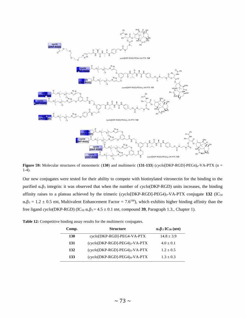
~ 73 ~
Figure 59: Molecular structures of monomeric (130) and multimeric (131-133) (cyclo[DKP-RGD]-PEG4)n-VA-PTX (n =
1-4).
Our new conjugates were tested for their ability to compete with biotinylated vitronectin for the binding to the
purified αvβ3 integrin: it was observed that when the number of cyclo(DKP-RGD) units increases, the binding
affinity raises to a plateau achieved by the trimeric (cyclo[DKP-RGD]-PEG4)3-VA-PTX conjugate 132 (IC50
αvβ3 = 1.2 ± 0.5 nM, Multivalent Enhancement Factor = 7.6190), which exhibits higher binding affinity than the
free ligand cyclo(DKP-RGD) (IC50 αvβ3 = 4.5 ± 0.1 nM, compound 39, Paragraph 1.3., Chapter 1).
Table 12: Competitive binding assay results for the multimeric conjugates.
Comp. Structure v3 IC50 (nM)
130 cyclo[DKP-RGD]-PEG4-VA-PTX 14.8 ± 3.9
131 (cyclo[DKP-RGD]-PEG4)2-VA-PTX 4.0 ± 0.1
132 (cyclo[DKP-RGD]-PEG4)3-VA-PTX 1.2 ± 0.5
133 (cyclo[DKP-RGD]-PEG4)4-VA-PTX 1.3 ± 0.3

~ 74 ~
Figure 60: Curves related to ability of compounds 39, 130-133 to bind to the isolated αvβ3 integrin receptor. X-axis:
compound concentration (Log scale); Y-axis: % of inhibition.
Later on, antiproliferative assays with all conjugates (47, 130-133) were carried out against two cancer cell lines,
expressing the integrin receptor at different levels: the human glioblastoma U87 cell line (αvβ3+) and human
colorectal adenocarcinoma HT29 cell line (αvβ3-). While all conjugates exhibited reduced potency when
compared to free PTX (for U87 and HT29 cell lines), the increasing valency of the conjugates leads to a
progressive loss of cytotoxicity. This trend may be ascribed to the increased steric bulk around the linker system,
which hinders the enzymatic cleavage. Synthetic optimizations are now in progress to confirm this hypothesis.
Our following efforts focused on the development of new SMDCs bearing an extracellularly-cleavable linker to
evaluate the release of PTX in the extracellular environment of cancer cells. This mechanism, often referred to a
“bystander effect”, may have important therapeutic benefits, since the payload release in the tumor
microenvironment would be free to diffuse in the surrounding areas, killing cancer cells that may be antigen-
negative or cancer-related host cells (e.g. endothelial cells), potentially resulting in a more efficient treatment.
We focused on a tripeptide linker cleaved by elastase, a tumor-associated enzyme. Elastase is a serine protease
secreted from activated neutrophils upon inflammation and, for this reason, it is known to be involved in the
progress of certain inflammatory diseases (e.g. cystic fibrosis and arthritis) and of certain solid tumors (e.g.
breast, lung and skin cancers).191 The pro-inflammatory nature of these tumors is characterized by a high number
of tumor-invading leucocytes, such as neutrophils, resulting in a high expression of elastase in the tumor
stroma.192 Hence, elastase can be employed as extracellular trigger for αvβ3 integrin-targeted SMDC, aimed at
releasing the free drug in the vicinity of tumor cells, rather than in intracellular compartments. Among the few
elastase tripeptide cleavable linkers that have been used to conjugate anticancer agents,193 we focused on the
Asn-Pro-Val (NPV) tripeptide sequence described by researchers at Bayer.194
Three different cyclo[DKP-RGD]-PTX conjugates have been synthesized, and their molecular structures are
reported in Figure 61.195 In addition to conjugate cyclo[DKP-RGD]-PEG4-NPV-PTX (134), bearing the elastase-
cleavable linker, cyclo[DKP-RGD]-PEG4-NPv-PTX conjugate (135), featuring the [D]-valine residue, was also
prepared as negative control for cleavage experiments. Furthermore, a third conjugate (compound 136) was
prepared, in which the tripeptide moiety was replaced by a tertiary amide bond.

~ 75 ~
Figure 61: Molecular structures of cyclo[DKP-RGD]-PEG4-NPV-PTX 134, cyclo[DKP-RGD]-PEG4-NPv-PTX 135 and
cyclo[DKP-RGD]-PEG4-unc-PTX 136 and their relative affinity (IC50 values) to the isolated αvβ3 integrin receptor.
In order to deeply investigate the activity and specificity of the linker, the compounds were subjected to stability
assays in presence of activated elastase. While conjugates 134 and 135 were stable under these experimental
conditions, the test confirmed the cleavage of conjugate 134 at the Val C-terminus and the subsequently drug
release. Further interesting results emerged from the in vitro investigation of 134: besides showing good binding
affinity for the αvβ3 isolated receptor, conjugate 134 displayed a 256-fold increment of antiproliferative activity
(IC50 = 19.6 ± 4.1 nM) against 786-O renal cancer cell line, in the presence of elastase (see Table 13).
Table 13: Cell viability assays of conjugates 134, 135, 136 and free PTX in 786-O renal cancer cell line (in presence or
absence of elastase).
v3 IC50 (nM)
Comp. Structure Added
Elastase -
16 PTX 35.77 ± 16.71 29.52 ± 7.64
134 cyclo[DKP-RGD]-PEG4-NPV-PTX 19.56 ± 4.16 >5000
135 cyclo[DKP-RGD]-PEG4-NPv-PTX > 5000 > 5000
136 cyclo[DKP-RGD]-PEG4-unc-PTX > 5000 > 5000
These results proved that the release of the drug within the tumor stroma can be a valuable tool to raise the
potency of our constructs. Given that the enzyme effector is present at significantly high concentrations at the
diseased site, conjugate 134 is an attractive candidate for in vivo antitumor therapy experiments, which will be
important to evaluate the anticancer agent accumulation at the diseased site mediated by the cyclo(DKP-RGD)
ligand. For this reason, in vivo studies are currently ongoing with cyclo[DKP-RGD]-PEG4-NPV-PTX conjugate

~ 76 ~
in comparison to an analogous compound bearing the RβAD sequence as non-recognition motif (cyclo[DKP-
RβAD]-PEG4-NPV-PTX, compound 137, Figure 62) aimed at evaluating the potential of our ligand cyclo(DKP-
RGD) as homing device for drug delivery. Hopefully, the direct comparison between the RGD- and RβAD-drug
conjugates in vivo would ultimately validate the potential of RGD peptidomimetics as tools for selective delivery
of cytotoxic agents.
Figure 62: Molecular structure of cyclo[DKP-RβAD]-PEG4-NPV-PTX 137 and its affinity (IC50 value) to the isolated αvβ3
integrin receptor.

~ 77 ~
Chapter 6
6. CONCLUSIONS AND FUTURE PERSPECTIVES
The work presented in this thesis mainly focused on the potential applications of the peptidomimetic integrin
ligand cyclo(DKP-RGD) as targeting device, for specific release of different payloads into integrin-expressing
cancer cells. The chemical properties of such cyclo(DKP-RGD) ligand allowed its covalent connection to a wide
range of chemical compounds and, due to the hydrophilic nature of the RGD peptide, the final conjugates
exhibited better solubility in aqueous solutions compared to highly lipophilic anticancer drugs. Generally, the
attachment of the RGD peptidomimetic to the N-termini of such di- or tripeptide linkers was found to affect
neither the affinity of the RGD moiety for the isolated αvβ3 receptor, nor the enzymatic cleavage at the linkers’
C-termini. A previous SMDC developed in our group, the cyclo[DKP-RGD]-VA-PTX conjugate (47), exhibited
a moderately selective anticancer activity. The work described herein reports the synthesis of a new SMDC,
endowed with a different linker type: a disulfide bond, which is selectively cleaved by endogenous reducing
agents expressed in intracellular compartments. Motivated by the results reported by Kim and colleagues
concerning the synthesis and biological evaluation of the theranostic cyclo[RGDyK]-Naph-SS-CPT (49), the
analogous conjugate cyclo[DKP-RGD]-Naph-SS-CPT 50 was prepared, in which the cyclo(DKP-RGD) ligand
(compound 39) was used as targeting moiety. Ideally, the presence of the naphtalimide fluorescent probe in
compounds 49 and 50 should allow the real-time monitoring of the internalization into the αvβ3-expressing cells.
The RGD-CPT conjugates were evaluated in vitro for their ability to bind to the isolated αvβ3 integrin, displaying
high affinity for the receptor (low nanomolar IC50 values). Hence, in collaboration with Nerviano Medical
Sciences (NMS), quantitative (ArrayScan) and qualitative (confocal microscopy) analysis were carried out to
evaluate the selective internalization of compounds 49 and 50 into αvβ3-expressing cells. To perform these
experiments, a new cellular model was developed, in which the glioblastoma U87 cell line was selected as αvβ3-
expressing cells (αvβ3+). To generate an isogenic non αvβ3-expressing cell line (αvβ3-), the parental U87 cells

~ 78 ~
were genetically modified by CRISPR/Cas9 technique in order to knockout the β3 integrin gene. The
internalization of the two conjugates was evaluated and confirmed by confocal microscopy in both the parental
αvβ3-expressing cell (αvβ3+) and the non αvβ3-expressing clone (αvβ3-). Despite the absence of the targeted αvβ3
integrin, only 12% reduction of the total fluorescence in the clone compared to the parental U87 cells was
observed by ArrayScan analysis. Moreover, compounds 49 and 50 exhibited poor stability in cell medium, as the
linker was found to be completely cleaved after 6 h incubation (fairly short time). Conjugates 49 and 50 were
also subjected to cell antiproliferative assays, aimed at evaluating their ability to selectively target αvβ3-
expressing cells. Consistent with the results described above, no significant selectivity was detected with
conjugates 49 and 50 against the αvβ3+ cancer cell line compared to the αvβ3- cells.
Despite the large number of RGD-drug conjugates described in the literature, the thorough evaluation of the
actual ability of these constructs to target αvβ3-positive cancer cells in vitro is often missing. Indeed, my
following research activity focused on the structural optimization of RGD-drug conjugates, together with the
preparation of new tools for investigating the therapeutic potential of this new technology.
To prove the ability of RGD-SMDCs to selectively kill αvβ3+ tumor cells in vitro, we devised new
peptide/peptidomimetic-drug conjugates showing almost identical physico-chemical properties and the same
linker-payload modules of the original RGD-based SMDCs, while exhibiting negligible affinity for the integrin
receptor. Following this approach, new peptidomimetic compounds were prepared, consisting in a similar
chemical structure to the one of the integrin ligand cyclo(DKP-RGD), albeit with replacement of the Gly residue
of the RGD sequence with an alanine or a β-alanine residue (compound 73 and 74). As detected by binding
assays in the presence of the purified integrin receptor, the ligand bearing the β-alanine residue resulted in a
1000-fold loss of binding affinity, as compared to the original cyclo(DKP-RGD) ligand. The newly synthesized
compound cyclo(DKP-RβAD) and the parental cyclo(DKP-RGD) were coupled to a fluorescent probe (Sulfo-
Cy5, see Figure 46), in order to investigate the interaction of our integrin ligand cyclo(DKP-RGD) with its target
receptor and the integrin-mediated internalization process. From confocal microscopy analysis on U87 cells, it
was possible to detect a moderate accumulation of conjugate cyclo[DKP-RGD]-Cy5 85 on the cell membrane.
On the contrary, cyclo[DKP-RβAD]-Cy5 86 displayed no membrane adhesion, consistent with the observed poor
affinity towards the purified integrin receptor. The direct comparison between the RGD- and RβAD-drug
conjugates highlighted the potential of our cyclo(DKP-RGD) ligand as tool for selective delivery of cytotoxic
agents. The analysis of multimeric fluorescently-labeled conjugates is still in progress, and it will hopefully
confirm that the ligand multimerization and its locally increased effective concentration around cancer cells
result in better cellular uptake.
The nature of the linker strongly affects efficacy and stability of the SMDC construct: the linker must be
sufficiently stable in plasma to reach the diseased site, and then be cleaved under the specific conditions present
in the tumor environment. Inspired by literature studies, I designed and synthetized compound 118, in which a

~ 79 ~
gem-dimethyl group in the α position of the disulfide bond was installed to improve the linker stability. An
analogous conjugate (compound 117), devoid of the gem-dimethyl group, was prepared to evaluate and directly
compare the kinetics of disulfide bond cleavage and the effects on the cytotoxicity. Both conjugates were
subjected to cell viability assays, aimed at evaluating the impact of the linker modification on the conjugates’
activity. Conjugate 118 exhibited lower cytotoxicity (yet still in the nanomolar range), in line with its higher
stability. This might suggest that a favorable equilibrium between stability and cytotoxicity is reached when a
gem-dimethyl group is present in proximity of the disulfide bond. To deeply investigate this aspect, kinetic
studies of linker cleavage and subsequent drug release are currently in progress.
Overall, the data presented herein, together with results missing from experiments in progress, will give a
complete picture of the use of our integrin ligand for tumor targeting applications. From my perspective,
important issues of this research area still need to be addressed: from the understanding of the different
biological properties of integrin agonists and antagonists, to the binding of RGD ligands to multiple integrin
isoforms and their expression in cancer and healthy cells. However, the development of small molecule-drug
conjugates represents an attractive strategy for a number of tumor-associated antigens, and it may help to
overcome some significant limitations of ADC products. Therefore, I believe our work contributed, at least in
part, to the development of this research area.

~ 80 ~
Chapter 7
7. EXPERIMENTAL SECTION
Materials and Methods
All manipulations requiring anhydrous conditions were carried out in flame-dried glassware, with magnetic
stirring and under a nitrogen atmosphere. All commercially available reagents were used as received. Anhydrous
solvents were purchased from commercial sources and withdrawn from the container by syringe, under a slight
positive pressure of nitrogen. Peptide cyclo(RGDyK) (48) and SulfoCy5-NHS were purchased respectively from
FutureChem Co., Ltd and Lumiprobe GmbH. Camptothecin (CPT) was kindly provided by Nerviano Medical
Sciences (NMS). Compound 78,102 compound 91,105 cyclo[DKP-RGD] (45)105 and (4-nitrophenoxycarbonyl)-
camptothecin 56 122 were prepared according to literature procedures, and their analytical data were in agreement
with those already published. Reactions were monitored by analytical thin-layer chromatography (TLC) using
silica gel 60 F254 pre-coated glass plates (0.25 mm thickness). Visualization was accomplished by irradiation
with a UV lamp and/or staining with a potassium permanganate alkaline solution or ninhydrin. Flash column
chromatography was performed according to the method of Still and co-workers196 using Chromagel 60 ACC
(40-63 µm) silica gel. Proton NMR spectra were recorded on a spectrometer operating at 400.16 MHz. Proton
chemical shifts are reported in ppm (δ) with the solvent reference relative to tetramethylsilane (TMS) employed
as the internal standard (CDCl3 δ = 7.26 ppm; CD2Cl2, δ = 5.32 ppm; D2O, δ = 4.79 ppm; [D]6DMSO, δ = 2.50
ppm; CD3OD, δ = 3.33 ppm).197 The following abbreviations are used to describe spin multiplicity: s = singlet, d
= doublet, t = triplet, q = quartet, m = multiplet, bs = broad signal, dd = doublet of doublet, ddd = doublet of
doublet of doublet, ddt = doublet of doublet of triplet. Carbon NMR spectra were recorded on a spectrometer
operating at 100.63 MHz, with complete proton decoupling. Carbon chemical shifts are reported in ppm (δ)
relative to TMS with the respective solvent resonance as the internal standard (CDCl3, δ = 77.16 ppm; CD2Cl2, δ
= 54.00 ppm; [D]6DMSO, δ = 39.51 ppm; CD3OD, δ = 49.05 ppm).

~ 81 ~
ESI-MS spectra were recorded on the ion trap mass spectrometer Finnigan LCQ Advantage or Micro Waters Q-
Tof (ESI source). MALDI-TOF-MS spectra were recorded on the instrument Bruker Microflex™ LT, supporting
the sample on the 2,5-dihydroxybenzoic acid (DHB), α-cyano-4-hydroxycinnamic acid (HCCA) and sinapinic
acid (SIN) matrices. The peptide calibration standard (300-6000 Da range), which consisted of Angiotensin II,
Angiotensin I, Substance P, Bombesin; ACTH clip 1-17, ACTH clip 18-39, Somatostatin 28, was purchased
from Bruker Daltonics® and used to calibrate the MALDI-TOF-MS instrument. The sample was mixed in equal
volumes with the matrix solution: a small amount (1 μL) of this mixture was spotted on the target surface. The
target matrix was dried at room temperature and then analyzed.
High-resolution mass spectra (HRMS) were performed with a Fourier Transform Ion Cyclotron Resonance (FT-
ICR) Mass Spectrometer APEX II & Xmass software (Bruker Daltonics) – 4.7 T Magnet (Magnex) equipped
with ESI source, available at CIGA (Centro Interdipartimentale Grandi Apparecchiature), University of Milan.
HPLC purifications and HPLC traces of final products were performed on Dionex Ultimate 3000 equipped with
Dionex RS Variable Wavelength Detector (column: Atlantis Prep T3 OBDTM 5 m 19 × 100 mm; flow 15
ml/min unless stated otherwise). The crude reaction mixture was dissolved in H2O or, if the compound was
insoluble in water, adding first DMF, then diluting slowly with H2O until reaching a 1:1 mixture DMF/H2O
(ultrasonic sonicator was used to assist the dissolution). The solution so obtained was filtered (polypropylene,
0.45 μm, 13 mm ø, PK/100) and injected in the HPLC, affording purified products. Purity analyses were carried
on a Dionex Ultimate 3000 instrument equipped with a Dionex RS Variable Wavelength detector (column:
Waters Atlantis® 5 μm 21 mm × 10 cm column). 1 mg of analyte was dissolved in 1 ml of H2O and was injected
using the same gradient used in the purification step. The analysis of the integrals and the relative percentage of
purity was performed with the software Cromeleon 6.80 SR11 Build 3161.
Freeze-drying: The products were dissolved in water and frozen with dry ice. Later on, the freeze-drying was
carried out at least for 48 h at -50 °C using the instrument 5Pascal Lio5P DGT.
General Procedures
GENERAL PROCEDURE FOR Boc DEPROTECTION REACTIONS:
To a 0.03 M CH2Cl2 solution of the N-Boc-protected compound half volume of TFA was added, and the reaction
was stirred at r.t. for 1 h. The solvent was removed under vacuum and then for five times CH2Cl2 was added to
the residue followed by evaporation under vacuum, to afford the corresponding amine as TFA salt.
GENERAL PROCEDURE FOR tBu DEPROTECTION REACTIONS:
A solution of tert-butyl ester 0.03 M in dry CH2Cl2 was cooled to 0 °C under a nitrogen atmosphere and half
volume of TFA was added. The mixture was then allowed to reach room temperature and stirred for 90 min. The
solvent was removed under vacuum and then for five times CH2Cl2 was added to the residue followed by
evaporation under vacuum, to afford the corresponding amine as TFA salt.

~ 82 ~
7.1. BIOLOGICAL ASSAYS
SOLID-PHASE RECEPTOR BINDING ASSAY:
Recombinant human integrin αvβ3 receptor (R&D Systems, Minneapolis, MN, USA) was diluted to 0.5 µg/ml in
coating buffer containing 20 mM Tris-HCl (pH 7.4), 150 mM NaCl, 1 mM MnCl2, 2 mM CaCl2, and 1 mM
MgCl2. An aliquot of diluted receptor (100 µl/well) was added to 96-well microtiter plates (Nunc MaxiSorp) and
incubated overnight at 4 °C. The plates were then incubated with blocking solution (coating buffer plus 1%
bovine serum albumin) for 2 h at r.t. to block nonspecific binding. After washing 2 times with blocking solution,
plates were incubated shaking for 3 h at r.t., with various concentrations (10-5-10-12 M) of test compounds in the
presence of 1 µg/ml biotinylated vitronectin (Molecular Innovations, Novi, MI, USA). Biotinylation was
performed using an EZ-Link Sulfo-NHS-Biotinylation kit (Pierce, Rockford, IL, USA). After washing 3 times,
the plates were incubated shaking for 1 h at r.t., with streptavidin-biotinylated peroxidase complex (Amersham
Biosciences, Uppsala, Sweden). After washing 3 times with blocking solution, plates were incubated with 100
µl/well of Substrate Reagent Solution (R&D Systems, Minneapolis, MN, USA) for 30 min, in the dark, with
shaking. After stopping the reaction with the addition of 50 µl/well 2 N H2SO4, absorbance at λ = 415 nm was
read in a SynergyTM HT Multi-Detection Microplate Reader (BioTek Instruments, Inc.). Each data point
represents the average of triplicate wells; data analysis was carried out by nonlinear regression analysis with
GraphPad Prism software (GraphPad Prism, San Diego, CA, USA). Each experiment was repeated in duplicate.
DEPLETION OF INTEGRIN β3 GENE (ITGB3) BY CRISPR/CAS9 TECHNIQUE:
The U87 human glioblastoma cell line was purchased from ATCC (U-87 MG, cat. # HBT-14) and maintained in
EMEM medium supplemented with 2 mm l-glutamine, 1% non-essential amino acids (NEAA) and 10% fetal
calf serum (FCS). The cell line was authenticated by Short Tandem Repeats (STR) analysis,198 resulting in a
profile 94.4% identical with that deposited in the ATCC database.
To disrupt the ITGB3 gene, U87 cells were transfected with the Integrin β3 Double Nickase Plasmid (sc-400216-
NIC, Santa Cruz Biotechnology) following provider recommendations; cell pool was subjected to one-week
puromycin selection (1 µg/ml) followed by dilution cloning. Integrin β3-negative individual clones were
identified by immunofluorescence using the murine anti-αvβ3 antibody (clone LM609, Millipore MAB 1976) and
subsequently expanded. The U87 β3-KO cell line was established from the clone D4 and further characterized by
immunofluorescence and flow cytometry.
IMMUNOFLUORESCENCE ANALYSIS OF INTEGRIN αvβ3 EXPRESSION:
The U87 β3-KO cell line was analyzed by fluorescence microscopy and flow cytometry in order to confirm
absence of integrin β3 protein expression (see Figure 63).

~ 83 ~
For microscopy analysis, parental U87 and U87 β3-KO cells were seeded in 96-well plates (c/w = 10000) in
complete EMEM + 10% FCS and fixed with paraformaldehyde 3.7% solution after 48 h. Cells were
permeabilized for 30 min with PBS containing 1% BSA and 0.05% Triton X-100, then incubated at 4 °C
overnight with anti-αvβ3 antibody (clone LM609, Millipore MAB 1976). The solution was removed, then cells
were washed with PBS, followed by immunostaining with the secondary antibody (Cy5-conjugated anti-mouse
IgG, GE Healthcare PA45002) and counterstaining with Hoechst 33342 (Sigma-Aldrich). Samples were washed
twice with PBS and cell photomicrographs were acquired with an ArrayScan high-content screening reader in
the blue channel (Hoechst) and in the far-red channel (anti-αvβ3).
Figure 63: Images of parental U87 and U87 β3-KO cells immunostained with anti-αvβ3 monoclonal antibody. ArrayScan
fields, objective 10×. To note, integrin αvβ3 receptors are mainly localized to membrane ruffles and lamellipodia in U87
cells.
For flow cytometry analysis, U87 and U87 β3-KO cells seeded in T75 flasks were detached with Trypsin/EDTA
solution, washed with PBS and counted. 500,000 cells per sample were re-suspended in 100 µl of BSA 1%
solution in PBS containing anti-αvβ3 (clone LM609-Millipore MAB 1976) or anti-αvβ5 (clone P1F6-Millipore
MAB 1961) murine monoclonal antibody at 1:50 dilution. “Secondary Ab only” samples were prepared by
incubating the cells in the same buffer without the primary antibody. Cells were incubated for 30 min at 4 °C,
then washed with PBS and incubated for 30 min at 4 °C with the secondary antibody (CF488A-goat anti-mouse
IgG, Biotium 20011) diluted 1:300. After washing with PBS, samples were acquired by a FacsCalibur flow
cytometer (Becton Dickinson) and analyzed by CellQuest software (Figure 64).

~ 84 ~
Figure 64: Flow cytometry analysis of integrin αvβ3 and integrin αvβ5 in U87 and U87 β3-KO cell lines. MFI: Median of
Fluorescence Intensity.
CONFOCAL MICROSCOPY ASSAYS OF CONJUGATES 49 AND 50:
The U87 human glioblastoma cells (αvβ3+) and the corresponding non αvβ3-expressing clone (U87 β3-KO) were
incubated with the fluorescent RGD-CPT conjugates (5 mM). After incubation for 3 h, the cells were washed
with phosphate buffer solution (PBS), fixed with paraformaldehyde in the presence of 1 mg/ml Hoechst 33342
(Sigma Aldrich) and analyzed by confocal microscopy (confocal laser scanning microscope SP2, Leica)
equipped with a 633 objective (oil immersion).
STABILITY AND CELL VIABILITY ASSAYS OF CONJUGATES 49, 50 AND 51:
The stability of compounds 49-51 was determined by ultraperformance liquid chromatography (UPLC) on a
Waters Acquity UPLC BEH C18 column (2.1 × 50 mm, 1.7 µm) by using a Waters Acquity UPLC system
equipped with a 2996 Aquity photodiode array detector and a Micromass ZQ 2000 single-quadrupole mass
spectrometer, equipped with an electrospray ion source (ESI). Mobile phase A was composed of 0.05%
trifluoroacetic acid (TFA) in water, and mobile phase B was 0.05% TFA in MeCN. Spectral analysis from 210–
400 nm; a flow rate of 0.6 ml/min; an injection volume of 10 µl; full scan, mass range from 200-2000 amu.
Compounds 49-51 (0.2 mM each) were incubated in cell media for 6 h at 37 °C.
Cells were seeded in 96-well plates with the appropriate complete medium (Rosewell Park Memorial Institute
1640 medium with 2 mM l-glutamine, 10% fetal calf serum, 10 mM 2-[4-(2-hydroxyethyl)-1-
piperazinyl]ethanesulfonic acid, and 1 mM sodium pyruvate). 48 h after seeding, the medium was replaced with
medium containing serial dilutions of the test compounds. The cells were incubated for 6 h at 37 °C, then
washed and re-incubated in fresh medium for an additional 138 h at 37 °C. Cell viability was assessed with the
CellTiter-Glo luciferase-based ATP detection assay (Promega) by following manufacturer’s instructions. Growth
inhibitory activity was evaluated at the end of incubation by using GraphPad Prism software. Experimental data
were normalized versus untreated control samples and interpolated by nonlinear regression analysis with
GraphPad Prism software to generate dose-response curves. IC50 values were calculated by using sigmoidal
interpolation curve fitting.

~ 85 ~
CONFOCAL MICROSCOPY ASSAYS OF CONJUGATES 85-87:
The U87 human glioblastoma cells (purchased from ATCC) were cultured in T75 flasks in EMEM (Gibco Life
Sciences, 41090) in presence of 1% HEPES and 10% Fetal Calf Serum (FCS). The cells were then harvested at
50-70% confluence by trypsinization (trypsin/EDTA solution, Sigma Aldrich T4049) for 5 min at 37 °C. Cell
count was performed with Vi-CELL (Beckman Coulter) and the viability was assessed. Cells were seeded in
cover glass chambers (200,000 cells/chamber in 1 ml, Sarstedt 94.6190.402) and incubated overnight at 37 °C,
5% CO2. The cell medium was replaced with fresh medium containing the tested compounds (1µM
concentration) and the cells were incubated for 2 h. Confocal microscopy analysis using confocal laser scanning
microscope SP2, Leica equipped with a 633 objective (oil immersion) was performed with the live cells.
CELL VIABILITY ASSAYS OF CONJUGATES 117-119:
Cells were seeded in 96-well plates with the appropriate complete medium (Rosewell Park Memorial Institute
1640 medium with 2 mM l-glutamine, 10% fetal calf serum, 10 mM 2-[4-(2-hydroxyethyl)-1-
piperazinyl]ethanesulfonic acid, and 1 mM sodium pyruvate). 48 h after seeding, the medium was replaced with
fresh medium containing serial dilutions of the test compounds. The cells were incubated for 144 h at 37 °C. Cell
viability was assessed with the CellTiter-Glo luciferase-based ATP detection assay (Promega) by following
manufacturer’s instructions. Growth inhibitory activity was evaluated at the end of incubation by using
GraphPad Prism software. Experimental data were normalized versus untreated control samples and interpolated
by nonlinear regression analysis with GraphPad Prism software to generate dose-response curves. IC50 values
were calculated by using sigmoidal interpolation curve fitting.

~ 86 ~
7.2. SYNTHESIS OF THERANOSTIC RGD-CAMPTOTHECIN CONJUGATES
7.2.1. cyclo[RGDyK]-Naph-SS-CPT (49) and cyclo[DKP-RGD]-Naph-SS-CPT (50)
Scheme 16: Synthesis of RGD-SS-CPT conjugates 49 and 50. REAGENTS AND CONDITIONS: a) β-alanine tert-butyl
ester hydrochloride, Et3N, EtOH, reflux, 3 h, Y.: 90%; b) SnCl2, EtOH, r.t., 1 h, Y.: quantitative; c) i. triphosgene, iPr2NEt,
toluene, r.t., 2 h; ii. 2,2’-dithioethanol, THF/CH2Cl2, r.t., overnight, Y.: 38%; d) 4-nitrophenyl chloroformate, DMAP,
CH2Cl2, 0 °C, 4 h, Y.: 92%; e) DMAP, CH2Cl2, r.t., overnight, Y.: 58%; f) TFA, CH2Cl2, r.t., 45 min, Y.: 45%; g) i. DIC,
NHS, DMF, r.t., overnight; ii. 48, MeCN/PBS/DMF (1:1:0.5; pH 7.5), r.t., overnight (49), Y.: 30%; 45, MeCN/PBS/DMF
(1:1:0.5; pH 7.5), r.t., overnight (50) Y.: 30%.
tButyl 3-(4-nitro-1,8-naphthalimide)propanoate (53)
A solution of β-alanine tert-butyl ester hydrochloride (217 mg, 1.2 mmol, 1.5 eq), 4-nitro-1,8-naphthalic
anhydride (200 mg, 0.8 mmol, 1 eq) and Et3N (0.176 ml, 1.28 mmol, 1.6 eq) in dry EtOH (4 ml) was heated to
reflux and stirred for 3 h under a nitrogen atmosphere. The reaction mixture was diluted with AcOEt (100 ml)
and washed with a 1 M aqueous solution of KHSO4 (3 20 ml) and brine (1 20 ml). The organic phase was

~ 87 ~
dried and concentrated. The crude was purified by flash chromatography on silica gel (0.5% MeOH in CH2Cl2)
affording imide 53, as a yellow solid (260 mg, 90% yield).
Rf= 0.33 (2% MeOH in CH2Cl2); 1H NMR (400 MHz, CDCl3) 8.83 (dd, J = 8.8, 1.1 Hz, 1H), 8.73 (dd, J = 7.3,
1.0 Hz, 1H), 8.69 (d, J = 8.0 Hz, 1H), 8.40 (d, J = 8.0 Hz, 1H), 7.98 (dd, J = 8.7, 7.3 Hz, 1H), 4.46 (d, J = 15.0
Hz, 2H), 2.72 – 2.66 (m, 2H), 1.42 ppm (s, 9H); 13C NMR (101 MHz, CDCl3) 170.3, 163.1, 162.3, 149.7,
132.6, 130.0, 129.9, 129.5, 129.1, 126.9, 124.0, 123.7, 122.9, 81.1 ppm.
tButyl 3-(4-amino-1,8-naphthalimide)propanoate (54)
To a suspension of 53 (100 mg, 0.27 mmol, 1 eq) in dry EtOH (3 ml), SnCl2 (213 mg, 1.12 mmol, 4.2 eq) was
added portionwise over a period of 1 h under a nitrogen atmosphere at room temperature. The color of the
reaction mixture changed from yellow to red and as the reaction progresses and it slowly turned limpid. The
solution was diluted with AcOEt (100 ml) and a saturated aqueous solution of NaHCO3 (20 ml) was added. The
white precipitate was filtered over cotton and then the two layers were separated. The organic phase was further
washed with a saturated aqueous solution of NaHCO3 (2 20 ml) and brine (1 25 ml). The organic phase was
dried and concentrated. The crude was purified over a pad of silica (eluent: 4:6 hexane/AcOEt), affording amine
54, as a red solid (90 mg, quantitative yield).
Rf= 0.27 (6:4 AcOEt/hexane); 1H NMR (400 MHz, MeOD) 8.52 (dd, J = 7.3, 1.0 Hz, 1H), 8.43 (d, J = 8.4 Hz,
1H), 8.32 (dd, J = 8.4, 1.0 Hz, 1H), 7.66 (dd, J = 8.4, 7.3 Hz, 1H), 7.34 (d, J = 8.4 Hz, 1H), 4.38 (t, J = 7.3 Hz,
2H), 2.63 (t, J = 7.3 Hz, 2H), 1.37 ppm (s, 9H); 13C NMR (101 MHz, MeOD) 172.6, 165.9, 165.4, 154.5,
135.4, 132.1, 130.5, 129.1, 125.9, 123.3, 120.1, 112.1, 106.6, 82.0, 37.1, 35.1, 28.2 ppm.
tButyl 3-[4-(2-hydroxyethyl-SS)-1,8-naphthalimide]propanoate (55)
Triphosgene (130 mg, 0.441 mmol, 3 eq) was added to a solution of amine 54 (50 mg, 0.147 mmol, 1 eq) in dry
toluene (1 ml) under a nitrogen atmosphere. The reaction mixture was cooled to 0 °C and iPr2NEt (75 µl, 0.441
mmol, 3 eq) was added dropwise. The mixture was allowed to reach room temperature and stirred for 2 h. A

~ 88 ~
solution of 2,2’-dithioethanol (89 l, 0.735 mmol, 5 eq) in a 1:1 CH2Cl2/THF mixture (1 ml) was added and the
reaction was stirred overnight at room temperature. AcOEt (100 ml) was added and the solution was washed
with a 1 M aqueous solution of KHSO4 (1 20 ml). The organic phase was dried and concentrated, then the
crude was purified by a Grace Reveleris system (column: Reveleris Silica HP 12 g, dry load, flow rate: 27
ml/min, ramp from 100% hexane to 70% AcOEt in 15 min) affording carbamate 55, as a yellow solid (29 mg,
38% yield).
Rf= 0.27 (6:4 AcOEt/hexane); 1H NMR (400 MHz, MeOD) 8.44 – 8.39 (m, 2H), 8.33 (d, J = 8.3 Hz, 1H), 8.12
(d, J = 8.3 Hz, 1H), 7.69 (dd, J = 8.6, 7.3 Hz, 1H), 4.53 (t, J = 6.5 Hz, 2H), 4.32 (t, J = 7.4 Hz, 2H), 3.83 (t, J =
6.4 Hz, 2H), 3.10 (t, J = 6.5 Hz, 2H), 2.91 (t, J = 6.4 Hz, 2H), 2.62 (t, J = 7.4 Hz, 2H), 1.39 ppm (s, 9H); 13C
NMR (101 MHz, MeOD) 172.5, 165.3, 164.8, 155.6, 142.1, 133.1, 132.2, 129.9, 129.6, 127.5, 125.1, 123.7,
118.9, 118.4, 82.1, 64.8, 61.3, 42.3, 38.4, 37.2, 34.9, 28.2 ppm; MS (ESI+): m/z calcd for [C24H28N2NaO7S2]+:
543.12 [M+Na]+; found: 543.2.
Camptothecin-SS-naphthalimide(tbutyl 3-aminopropanoate) (57)
A solution of alcohol 55 (33 mg, 0.063 mmol, 1.5 eq) in dry CH2Cl2 (3 ml) was cooled to 0 °C under a nitrogen
atmosphere, then (4-nitrophenoxycarbonyl)-camptothecin 56 (22 mg, 0.042 mmol, 1eq) and DMAP (10 mg,
0.084 mmol, 2 eq) were added. The mixture was allowed to reach room temperature and stirred overnight. The
reaction mixture was diluted with AcOEt (70 ml) and washed with a 1 M aqueous solution of KHSO4 (3 20 ml)
and brine (1 20 ml). The organic phase was dried and concentrated, the crude was then purified by a Grace
Reveleris system (column: Reveleris Silica HP 12 g, dry load, flow rate: 25 ml/min, ramp from 100% hexane to
100% AcOEt in 20 min) to afford carbonate 57 as a yellow solid (22 mg, 58% yield).
Rf = 0.32 (9:1 AcOEt/hexane); 1H NMR (400 MHz, CD2Cl2) 8.51 (d, J = 7.2 Hz, 1H), 8.42 (t, J = 6.3 Hz, 1H),
8.33 (d, J = 14.3 Hz, 1H), 8.29 (d, J = 8.5 Hz, 1H), 8.12 (dd, J = 19.5, 11.9 Hz, 2H), 7.92 (d, J = 8.0 Hz, 1H),
7.80 (t, J = 7.5 Hz, 1H), 7.65 (dt, J = 9.9, 7.7 Hz, 2H), 7.27 (s, 1H), 5.43 – 5.18 (m, 2H), 5.07 (dd, J = 48.3, 19.0
Hz, 2H), 4.54 (m, 1H), 4.47 – 4.23 (m, 5H), 3.02 (dt, J = 17.3, 5.9 Hz, 3H), 2.62 (dd, J = 16.4, 8.9 Hz, 2H), 2.25
– 2.01 (m, 2H), 1.40 (d, J = 4.4 Hz, 9H), 0.96 ppm (t, J = 7.3 Hz, 3H); 13C NMR (101 MHz, CD2Cl2) 170.9,
167.7, 164.2, 163.7, 157.4, 154.2, 153.3, 152.7, 149.2, 147.0, 145.9, 139.9, 132.4, 131.6, 131.4, 131.0, 129.8,
129.2, 129.1, 128.7, 128.7, 128.4, 127.2, 126.8, 123.5, 123.4, 120.3, 118.0, 116.8, 96.0, 81.0, 78.8, 67.3, 66.9,

~ 89 ~
63.6, 50.4, 38.1, 37.2, 36.5, 34.2, 32.0, 30.1, 28.2, 7.8 ppm; MS (ESI+): m/z calcd for [C45H43N4O12S2]+: 895.23
[M+H]+; found: 895.5.
Camptothecin-SS-naphthalimide(3-aminopropionic acid) (58)
A solution of tert-butyl ester 57 (35 mg, 0.04 mmol) in dry CH2Cl2 (2 ml) was cooled to 0 °C under a nitrogen
atmosphere and TFA (1 ml) was added. The mixture was allowed to reach room temperature and stirred for 45
min. The crude was purified by flash chromatography on silica gel [gradient from (99:1 CH2Cl2/MeOH + 1%
HCOOH) to (9:1 CH2Cl2/MeOH + 1% HCOOH)] affording carboxylic acid 58, as a yellow solid (16 mg, 45%
yield).
Rf= 0.46 (9:1 CH2Cl2/MeOH); 1H NMR (400 MHz, DMSO-d6) 8.59 (t, J = 7.0 Hz, 2H), 8.43 (m, 1H), 8.37 (dd,
J = 8.0, 5.9 Hz, 1H), 8.14 – 8.00 (m, 3H), 7.77 (ddd, J = 34.1, 17.7, 9.5 Hz, 2H), 7.65 (dd, J = 10.0, 5.0 Hz, 1H),
7.07 (m, 1H), 5.74 (t, J = 2.1 Hz, 1H), 5.51 (s, 2H), 5.28 – 5.13 (m, 2H), 4.46 – 4.30 (m, 4H), 4.29 – 4.18 (m,
2H), 3.07 (dd, J = 13.1, 7.1 Hz, 3H), 2.24 – 2.10 (m, 2H), 0.96 – 0.87 ppm (m, 3H); 13C NMR (101 MHz,
DMSO-d6) 167.1, 163.4, 162.8, 156.5, 153.7, 152.8, 152.1, 147.8, 146.2, 144.8, 140.5, 131.5, 130.8, 130.4,
129.6, 129.3, 129.0, 128.5, 128.2, 128.0, 127.7, 126.2, 123.9, 122.2, 119.2, 118.4, 117.1, 94.5, 78.0, 66.4, 62.8,
50.2, 36.8, 36.4, 33.8, 30.3, 29.0, 7.5 ppm. MS (ESI+): m/z calcd for [C41H35N4O12S2]+: 839.17 [M+H]+; found:
839.30.
cyclo[RGDyK]-Naph-SS-CPT (49)

~ 90 ~
To a solution of carboxylic acid 58 (11.3 mg, 13.5 × 10⁻³ mmol, 1.5 eq) in dry DMF (500 l) under a nitrogen
atmosphere, NHS (2.1 mg, 18.0 × 10⁻³ mmol, 2 eq) was added and the reaction mixture was cooled to 0 °C. DIC
(2.79 l, 18.0 × 10⁻³ mmol, 2 eq) was added and the mixture was then stirred overnight at room temperature.
Approximately half of the solvent was removed under high vacuum. Then MeCN (600 l) and a solution of
cyclo[RGDyK] (48, 7.6 mg, 9.0 × 10⁻³ mmol, 1 eq) in pH 7.5 phosphate buffer solution (600 l) were added. The
reaction mixture was stirred overnight at room temperature; during the first 3 h the pH value was kept near 7.3-
7.5 adding 0.2 M aqueous NaOH when necessary. The crude was then purified by semipreparative-HPLC
[Waters Atlantis 21 mm 10 cm column, gradient: 90% (H2O + 0.1% CF3COOH) / 10% (MeCN + 0.1%
CF3COOH) to 15% (H2O + 0.1% CF3COOH) / 85% (MeCN + 0.1% CF3COOH) in 10 min; tR (product): 7.25
min]. The purified product was then freeze dried to give 49 as a yellow solid (5 mg, 34% yield).
MS (ESI+): m/z calcd for [C68H74N13O19S2]+: 1440.47 [M+H]+; found: 1440.6; MS (MALDI): m/z calcd for
[C68H74N13O19S2]+: 1441.52 [M+H]+; found: 1441.1 (HCCA matrix).
cyclo[DKP-RGD]-Naph-SS-CPT (50)
To a solution of carboxylic acid 58 (10 mg, 11.9 × 10⁻³ mmol, 1 eq) in dry DMF (600 l) under a nitrogen
atmosphere, NHS (1.8 mg, 15.5 × 10⁻³ mmol, 1.3 eq) was added and the reaction mixture was cooled to 0 °C.
DIC (2.4 L, 15.5 × 10⁻³ mmol, 1.3 eq) was added and the mixture was then stirred overnight at room
temperature. Approximately half of the solvent was removed under high vacuum. Then MeCN (700 l) and a
solution of cyclo[DKP-RGD]-CH2NH2 (45, 11 mg, 13.1 × 10⁻³ mmol, 1.1 eq) in pH 7.5 phosphate buffer
solution (700 l) were added. The reaction mixture was stirred overnight at room temperature; during the first 3
h the pH value was kept near 7.3-7.5 adding 0.2 M aqueous NaOH when necessary. The crude was then purified
by semipreparative-HPLC [Waters Atlantis 21 mm 10 cm column, gradient: 90% (H2O + 0.1% CF3COOH) /
10% (MeCN + 0.1% CF3COOH) to 15% (H2O + 0.1% CF3COOH) / 85% (MeCN + 0.1% CF3COOH) in 10 min;
tR (product): 7.1 min]. The purified product was then freeze dried to give 50 as a yellow solid (5.5 mg, 25%
yield).

~ 91 ~
MS (ESI+): m/z calcd for [C68H71N14O19S2]+: 1451.45 [M+H]+; found: 1451.5; MS (MALDI): m/z calcd for
[C68H71N14O19S2]+: 1452.50 [M+H]+; found: 1452.2 (HCCA matrix); HRMS (ESI+): m/z calcd for
[C68H71N14O19S]+: 1451.4456 [M+H]+; found 1451.4458; m/z calcd for [C68H72N14O19S2]2+: 726.2265 [M+2H]2+;
found 726.2243.
7.2.2. Uncleavable cyclo[DKP-RGD]-unc-CPT (51)
Scheme 17: Synthesis of RGD-CPT conjugates 51. REAGENTS AND CONDITIONS: a) glutaric anhydride, Et3N, DMF,
r.t., 30 min, Y: 93%; b) DIC, NHS, 5:1 THF/MeCN, r.t., overnight, Y.: 63%; c) cyclo[DKP-RGD]-CH2NH2 (45),
MeCN/PBS pH 7,5 1:1, r.t., overnight, Y.: 85%.
cyclo[DKP-RGD]-unc-CPT (51)
DIC (7 l, 0.044 mmol, 2 eq) and NHS (5 mg, 0.044 mmol, 2 eq) were added to a solution of 60 (12 mg, 0.022
mmol, 1 eq) in a 5:1 mixture of dry THF and MeCN (0.6 ml). The mixture was stirred overnight at room
temperature and the white solid formed was filtered off. Evaporation of the filtrate and crystallization with
isopropyl alcohol afforded the activated ester form of compound 60. This latter (8.6 mg, 14.0 ×10⁻³ mmol, 1.2
eq) was dissolved in MeCN (1 ml) and a solution of 45 (10 mg, 11.6 ×10⁻³ mmol, 1 eq) in pH 7.5 phosphate
buffer solution (1 ml) was added. The reaction mixture was stirred overnight at room temperature; during the

~ 92 ~
first 3 h the pH value was kept near 7.3-7.5 adding 0.2 M aqueous NaOH when necessary. The crude was then
purified by semipreparative-HPLC [Waters Atlantis 21 mm 10 cm column, gradient: 80% (H2O + 0.1%
CF3COOH) / 20% (MeCN + 0.1% CF3COOH) to 29% (H2O + 0.1% CF3COOH) / 71% (MeCN + 0.1%
CF3COOH) in 6.5 min; tR (product): 6.2 min]. The purified product was then freeze dried to give the desired
compound 51 as a yellow solid (13 mg, 89% yield).
1H NMR (400 MHz, D2O + CD3CN) 8.35 (s, 1H), 8.06 (d, J = 8.0 Hz, 1H), 7.90 – 7.76 (m, 2H), 7.60 (m, 1H),
7.44 (s, 1H), 7.23 (d, J = 7.9 Hz, 2H), 7.19 (d, J = 7.9 Hz, 2H), 5.83 (d, J = 16.4 Hz, 1H), 5.66 (d, J = 16.4 Hz,
1H), 5.13 (d, J = 15.1 Hz, 1H), 5.04 (t, J = 7.0 Hz, 1H), 4.69 (dd, J = 8.4, 5.2 Hz, 2H), 4.62 (d, J = 18.2 Hz, 1H),
4.51 (d, J = 14.7 Hz, 1H), 4.47 (d, J = 15.6 Hz, 1H), 4.32 (dd, J = 9.7, 4.9 Hz, 1H), 4.27 (s, 2H), 4.16 – 4.06 (m,
3H), 3.79 (d, J = 17.1 Hz, 1H), 3.67 (dd, J = 14.0, 5.4 Hz, 1H), 3.40 (t, J = 6.8 Hz, 2H), 3.15 – 2.89 (m, 4H),
2.79 (dd, J = 14.0, 4.7 Hz, 1H), 2.59 – 2.51 (m, 2H), 2.45 (d, J = 0.9 Hz, 3H), 2.44 – 2.34 (m, 2H), 2.19 (m, 1H),
2.14 – 2.04 (m, 2H), 2.00 (m, 1H), 1.90 – 1.76 (m, 2H), 1.24 ppm (t, J = 7.3 Hz, 3H). 13C NMR (101 MHz, D2O
+ CD3CN) 174.9, 174.0, 172.9, 172.6, 172.0, 171.7, 169.9, 169.3, 169.2, 168.5, 167.8, 156.6, 155.9, 149.8,
146.1, 144.6, 137.2, 133.1, 131.6, 130.2, 127.6, 127.4, 127.1, 126.8, 96.6, 76.6, 65.8, 58.3, 53.2, 51.1, 49.3, 48.4,
48.1, 46.4, 41.7, 41.5, 40.2, 39.9, 38.2, 37.1, 34.0, 33.8, 33.7, 29.9, 25.0, 24.0, 21.0, 6.4 ppm. MS (ESI+): m/z
calcd for [C54H62N13O15]+: 1132.45 [M+H]+; found: 1132.70; MS (MALDI): m/z calcd for [C54H62N13O15]+:
1133.15 [M+H]+; found: 1133.7 (HCCA matrix).

~ 93 ~
7.3. SYNTHESIS OF cyclo(DKP-RβAD) AS NEGATIVE CONTROL FOR
BIOLOGICAL INVESTIGATIONS
Scheme 18: Synthesis of cyclo(DKP-RβAD) 74; REAGENTS AND CONDITIONS: a) benzyl alcohol, trimethylsilyl
chloride, 100 °C, 4 h, Y.: 30%; b) HATU, HOAt, iPr2NEt, DMF, r.t., 16 h, Y.: 67%; c) TFA/ CH2Cl2 (1:2), r.t., 2.5 h, Y.:
quantitative; d) pyrrolidine, PPh3, Pd(PPh3)4, CH2Cl2, 1 h, r.t., Y.: quantitative; e) HATU, HOAt, iPr2NEt, DMF, r.t., 16 h,
Y.: 56%; f) TFA/ CH2Cl2 (1:2), r.t., 2 h, Y.: quantitative; g) Cbz-Asp(OtBu)-OH, HATU, HOAt, iPr2NEt, DMF, r.t., 16 h,
Y.: 69%; h) H2, 10% Pd/C, THF/H2O (1:1), r.t., 16 h, Y.: 91%; i) HATU, HOAt, iPr2NEt, DMF, r.t., 16 h, Y.: 50%; j) TFA
/thioanisol/EDT/anisole (90:5:3:2), 3.5 h, r.t., Y.: 45%.
H-βAla-OBn (75)199
To a solution of H-βAla-OH (200 mg, 2.25 mmol, 1 eq) in benzyl alcohol (1.2 ml, 11.2 mmol, 5 eq) under N2
atmosphere, chlorotrimethylsilane (0.46 ml, 3.59 mmol, 1.6 eq) was added and the reaction was stirred at 100 °C
for 4 h. The reaction mixture was cooled to room temperature and washed 3 times with cold Et2O, and
centrifuged. The resulting precipitate was isolated and dried under vacuum, affording the benzyl protected β-
alanine 75 (120 mg, 30% yield).
1H NMR (400 MHz, MeOD) δ 7.38 (m, 1H), 5.20 (s, 1H), 3.22 (t, J = 6.5 Hz, 1H), 2.79 (t, J = 6.5 Hz, 1H).

~ 94 ~
Boc-Arg(Mtr)-βAla-OBn (76)
To a solution of Boc-Arg(Mtr)-OH (297 mg, 0.61 mmol, 1 eq) in dry DMF (10 ml) at 0 °C under N2 atmosphere,
HATU (232 mg, 0.61 mmol, 1 eq), HOAt (83 mg, 0.61 mmol, 1 eq) and iPr2NEt (0.3 ml, 3.054 mmol, 3 eq)
were added. After stirring the mixture for 1 h at 0 °C, a solution of 75 in dry DMF (4 ml) was added followed by
iPr2NEt (0.2 ml, 3.05 mmol, 2 eq). The reaction mixture was then warmed to room temperature and stirred
overnight. The mixture was diluted with AcOEt (50 ml) and consecutively washed with 1 M aqueous solution of
KHSO4 (4 × 30 ml), a saturated aqueous solution of NaHCO3 (4 × 30 ml) and brine (1 × 75 ml), dried over
Na2SO4 and the solvent removed under reduced pressure to afford the crude product. The residue was purified
with flash chromatography on silica gel (AcOEt: Hex 8:2, solid load) to afford the desired product 76 (290 mg,
67% yield).
Rf = 0.32 (AcOEt: Hex 9:1); 1H NMR (400 MHz, CDCl3) δ 7.33 (m, 5H), 6.53 (s, 1H), 5.10 (s, 2H), 4.12 (d, J =
7.1 Hz, 1H), 3.82 (s, 3H), 3.54 (m, 2H), 3.26 (m, 2H), 2.68 (s, 3H), 2.61 (s, 3H), 2.12 (s, 3H), 1.93 - 1.67 (m,
4H), 1.41 ppm (s, 9H). MS (ESI+): m/z calcd for [C31H43N5O8S]-: 645.28 ion type [M-H]-; found: 646.12; m/z
calcd for [C31H44N5O8SNa]+: 670.28 ion type [M+Na]+; found: 670.65.
Boc-DKP-Arg(Mtr)-βAla-OBn (80)
To a solution of deprotected DKP 79 (64 mg, 0.16 mmol, 1 eq) in dry DMF (1.2 ml) at 0 °C under N2
atmosphere, HATU (73 mg, 0.19 mmol, 1.2 eq), HOAt (26 mg, 0.19 mmol, 1.2 eq) and iPr2NEt (0.11 ml, 0.64
mmol, 4 eq) were added. After stirring the mixture for 30 min at 0 °C, the trifluoroacetate salt of deprotected
dipeptide H-Arg(Mtr)-βAla-OBn 77 (140 mg, 0.18 mmol, 1.5 eq) in dry DMF (0.6 ml) was added. The reaction
mixture was stirred at 0 °C for 1 h and room temperature overnight. The solvent was removed and the mixture
was diluted with AcOEt (50 ml) and consecutively washed with 1 M aqueous solution of KHSO4 (3 × 25 ml), a
saturated aqueous solution of NaHCO3 (3 × 25 ml) and brine (1 × 30 ml), dried over Na2SO4 and the solvent
removed under reduced pressure to afford the crude product. The residue was purified with flash

~ 95 ~
chromatography on silica gel (CH2Cl2/MeOH 96:4, solid load) to afford the desired product 80 as a yellow foam
(83 mg, 56% yield).
Rf=0.45 (CH2Cl2: MeOH 9:1); 1H NMR (400 MHz, MeOD) δ 7.44 – 7.13 (m, 10H), 6.63 (s, 1H), 5.38 (d, J =
15.4 Hz, 1H), 5.08 (d, J = 9.0 Hz, 2H), 4.53 (t, J = 5.4 Hz, 1H), 4.29 (dd, J = 8.4, 5.0 Hz, 1H), 4.04 (d, J = 15.5
Hz, 1H), 3.92 – 3.70 (m, 5H), 3.50 – 3.33 (m, 3H), 3.22 – 3.03 (m, 2H), 2.86 (ddd, J = 21.8, 15.6, 5.5 Hz, 2H),
2.67 (s, 3H), 2.60 (s, 3H), 2.54 (t, J = 6.8 Hz, 2H), 2.09 (s, 3H), 1.76 (m, 1H), 1.67 – 1.47 (m, 3H), 1.41 ppm (s,
9H); 13C NMR (101 MHz, MeOD) δ 174.2, 173.0, 172.0, 168.5, 159.9, 158.1, 139.5, 137.9, 137.4, 137.1, 129.9,
129.5, 129.2, 128.9, 128.8, 125.7, 112.8, 80.7, 67.4, 61.4, 56.0, 54.3, 52.5, 41.5, 38.7, 36.4, 34.7, 30.1, 28.8,
24.4, 18.8, 12.1 ppm. MS (ESI-): m/z calcd for [C45H59N8O11S]-: 919.40 ion type [M-H]-; found: 919.52.
Cbz-Asp(OtBu)-DKP-Arg(Mtr)-βAla-OBn (82)
To a solution of Cbz-L-Asp(OtBu)-OH (48 mg, 0.149 mmol, 1.5 eq) in dry DMF (1.6 ml) at 0 °C under N2
atmosphere, HATU (60 mg, 0.158 mmol, 1.6 eq), HOAt (21.5 mg, 0.158 mmol, 1.6 eq) and iPr2NEt (0.07 ml,
0.396 mmol, 4 eq) were added. After stirring the mixture for 30 min at 0°C, the trifluoroacetate salt of
deprotected Boc-DKP-Arg(Mtr)-βAla-OBn 81 (92.56 mg, 0.099 mmol, 1 eq) in dry DMF (0.8 ml) was added.
The reaction mixture was stirred at 0 °C for 1 h and then room temperature overnight. The mixture was diluted
with AcOEt (50 ml) and consecutively washed with 1 M aqueous solution of KHSO4 (3 × 30 ml), a saturated
aqueous solution of NaHCO3 (2 × 25 ml) and brine (1 × 30 ml), dried over Na2SO4 and the solvent removed
under reduced pressure to afford the crude product. The residue was purified with flash chromatography on silica
gel (CH2Cl2/MeOH 95:5, solid load) to afford the desired product 82 as yellow foam (77 mg, 69% yield).
Rf= 0.43 (CH2Cl2: MeOH 95:5); 1H NMR (400 MHz, MeOD) δ 7.43 – 7.14 (m, 15H), 6.61 (s, 1H), 5.33 (d, J =
15.4 Hz, 1H), 5.20 – 4.92 (m, 4H), 4.63 (m, 1H), 4.50 (m, 1H), 4.30 (m, 1H), 4.11 (d, J = 15.5 Hz, 1H), 3.85 –
3.78 (m, 5H), 3.63 (m, 1H), 3.31 (s, 2H), 3.20 - 3.10 (m, 2H), 2.96 – 2.71 (m, 3H), 2.66 (s, 3H), 2.58 (s, 3H),
2.56 - 2.50 (m, 3H), 2.07 (s, 3H), 1.78 (m, 1H), 1.64 – 1.46 (m, 3H), 1.37 ppm (s, 9H); 13C NMR (101 MHz,
MeOD) δ 174.1, 174.0, 172.9, 171.9, 171.3, 168.8, 168.5, 159.7, 158.2, 158.0, 139.4, 137.8, 137.7, 137.3, 137.0,
134.7, 129.8, 129.4, 129.4, 129.1, 129.1, 129.0, 128.9, 128.8, 128.7, 125.6, 112.7, 82.3, 67.9, 67.3, 60.7, 55.9,

~ 96 ~
54.1, 53.2, 52.2, 40.4, 38.3, 38.2, 36.3, 34.6, 30.0, 28.2, 24.3, 18.8, 12.1 ppm. MS (ESI-): m/z calcd for
[C56H70N9O14S]-: 1125.27 ion type [M-H]-; found: 1125.54.
H-Asp(OtBu)-DKP-Arg(Mtr)-βAla-OH (83)
A solution of Cbz-Asp(OtBu)-DKP-Arg(Mtr)-βAla-OBn 82 (77 mg, 0.068 mmol, 1 eq) in THF/H2O 1:1 (6.8 ml)
was treated with 10% Pd/C (1.45 mg, 0.014 mmol, 0.2 eq) and the flask was purged three times with vacuum/H2.
The mixture was stirred at room temperature overnight under H2 atmosphere, then filtered through a pad of
Celite and then was washed thoroughly with MeOH. The solvents were removed under vacuum to give the crude
product as white solid (56 mg, 91% yield), which was used without further purification.
Rf= 0.05 (9:1CH2Cl2/MeOH).
cyclo[DKP-Arg(Mtr)-βAla-Asp(OtBu)] (84)
To a solution of H-Asp(OtBu)-DKP-Arg(Mtr)-βAla-OH 83 (56 mg, 0.062 mmol, 1 eq) in dry DMF (44 ml,
0.0014 M), under nitrogen atmosphere and at 0 °C, HATU (94.3 mg, 0.248 mmol, 4 eq), HOAt (33.8 mg, 0.248
mmol, 4 eq) and iPr2NEt (0.065 ml, 0.372 mmol, 6 eq) were added. The reaction was warmed up to room
temperature and stirred overnight. DMF was then removed under reduced pressure. The residue was dissolved in
AcOEt (70 ml) and consecutively washed with 1 M aqueous solution of KHSO4 (3 × 25 ml), brine (1 × 30 ml),
dried over Na2SO4 and the solvent removed under reduced pressure to afford the crude product. The residue was

~ 97 ~
purified with flash chromatography on silica gel (CH2Cl2/MeOH 95:5, solid load) to afford the desired product
84 as a pale yellow solid (27 mg, 50% yield).
Rf= 0.40 (9:1 CH2Cl2/MeOH); 1H NMR (400 MHz, MeOD) δ 7.47 – 7.07 (m, 5H), 6.65 (s, 1H), 5.52 (d, J = 15.5
Hz, 1H), 4.93 (t, J = 7.4 Hz, 1H), 4.70 (dd, J = 9.0, 5.2 Hz, 1H), 4.07 - 3.98 (m, 4H), 3.88 (t, J = 5.6 Hz, 1H),
3.82 (s, 3H), 3.73 (dt, J = 13.4, 3.6 Hz, 1H), 3.53 (dd, J = 14.4, 5.6 Hz, 1H), 3.21 - 3.16 (m, 3H), 2.89 (m, 1H),
2.67 – 2.40 (m, 10H), 2.28 (dd, J = 14.0, 3.1 Hz, 1H), 2.12 (s, 3H), 1.99 (m, 1H), 1.85 (m, 1H), 1.68 – 1.50 (m,
2H), 1.44 ppm (s, 9H); 13C NMR (101 MHz, MeOD) δ 174.7, 174.7, 173.6, 172.7, 170.9, 170.1, 169.3, 159.9,
139.4, 137.9, 137.4, 134.9, 129.9, 129.0, 128.9, 125.7, 112.8, 82.7, 61.0, 56.0, 55.3, 52.3, 51.7, 39.5, 38.4, 37.8,
36.9, 35.5, 28.3, 27.9, 24.3, 18.8, 12.1 ppm; MS (ESI+): m/z calcd for [C41H56N9O11S]-: 882.38 ion type [M-H]-;
found: 882.86.
cyclo(DKP-RAD) (74)
To a solution of cyclo[DKP- Arg(Mtr)- βAla-Asp(OtBu)] 84 (27 mg, 0.0306 mmol, 1 eq), a cleavage cocktail of
thioanisole (0.15 ml), ethanedithiol (0.09 ml) and anisole (0.06 ml) in TFA (3 ml) was added at 0 °C under N2
atmosphere, the flask was opened and the mixture warmed up to room temperature and stirred for 2 h. All
volatiles were then removed under vacuum and the crude was dissolved in a mixture of iPr2O/H2O (1:1, 10
ml).The aqueous phase was washed with iPr2O (3 × 5 ml) and then concentrated under reduced pressure to give
the crude compound, which was purified by semipreparative-HPLC [Water’s Atlantis 21 mm × 10 cm column;
gradient 90% (H2O + 0.1% CF3COOH)/(10% MeCN + 0.1% CF3COOH) to 72% (H2O + 0.1% CF3COOH)/28%
(MeCN + 0.1% CF3COOH) in 12 min; tR (product): 8 min] to give the desired compound 74 as trifluoroacetate
salt (10 mg, 45% yield), after freeze-drying.
1H NMR (400 MHz, D2O) δ 7.49 – 7.30 (m, 5H), 5.34 (d, J = 15.5 Hz, 1H), 4.80 - 4.76 (m, 2H), 4.20 – 3.99 (m,
5H), 3.75 – 3.66 (m, 2H), 3.25 - 3.20 (m, 3H), 3.02 (dd, J = 15.4, 6.5 Hz, 1H), 2.93 – 2.66 (m, 3H), 2.50 – 2.46
(m, 2H), 2.03 (m, 1H), 1.87 (m, 1H), 1.70 – 1.65 ppm (m, 2H); 13C NMR (101 MHz, D2O) δ 174.3, 173.9, 173.8,
173.0, 172.2, 169.2, 168.0, 163.1, 162.8, 135.1, 129.1, 128.1, 127.6, 127.6, 117.8, 114.9, 59.6, 53.9, 51.3, 50.2,

~ 98 ~
48.5, 40.6, 38.9, 36.9, 35.8, 35.3, 33.9, 25.9, 24.7; MS (ESI+): m/z calcd for [C27H38N9O8]+: 616.28 ion type
[M+H]+; found: 616.21.

~ 99 ~
7.4. SYNTHESIS OF FLUORESCENT COMPOUNDS FOR ACTIVE TUMOR
TARGETING
7.4.1. Fluorescent compounds cyclo[DKP-RXD]-Cy5 (85, 86, and 87)
Scheme 19: Synthesis of cyclo[DKP-RGD]-Cy5 85, cyclo[DKP-RβAD] 86 and cyclo[DKP-RAD] 87. REAGENTS AND
CONDITIONS: a) HATU, HOAt, iPr2NEt, DMF, r.t., 16 h, Y.: 96%, (X = Gly), 55% (X = βAla), 81% (X = Ala); b)
TFA/CH2Cl2 (1:2), r.t., 2.5 h, Y.: quantitative; c) pyrrolidine, PPh3, Pd(PPh3)4, CH2Cl2, 1 h, r.t., Y.: 60%; d) HATU, HOAt,
iPr2NEt, DMF, r.t., 16 h, Y.: 40%, (X = Gly), 65% (X = βAla), 48% (X = Ala); e) TFA/CH2Cl2 (1:2), r.t., 2 h, Y.:
quantitative; f) Cbz-Asp(OtBu)-OH, HATU, HOAt, iPr2NEt, DMF, r.t., 16 h, Y.: 55%, (X = Gly), 74% (X = βAla), 56% (X
= Ala); g) H2, 10% Pd/C, THF/H2O (1:1), r.t., 16 h, Y.: 85%-quantitative; h) HATU, HOAt, iPr2NEt, DMF, r.t., 16 h, Y.:
70%, (X = Gly), 60% (X = βAla), 56% (Ala+ala); i) TFA/TMSBr/thioanisol/EDT/phenol (70:14:10:5:1), 2 h, r.t., Y.: 83%,
(X = Gly), 66% (X = βAla), 72% (Ala:ala=3:2); j) SulfoCy5-NHS, iPr2NEt, DMF, r.t., 4 h, Y: 40% (X = Gly), 50% (X =
βAla), 40-46% (X = Ala/ala).

~ 100 ~
Boc-Arg(Mtr)-Ala-OBn (89)
To a solution of Boc-Arg(Mtr) (1.5 g, 3.08 mmol, 1 eq) in dry DMF (51.6 ml) at 0 °C under inert atmosphere,
HATU (1.2 g, 3.07 mmol, 1 eq), HOAt (424 mg, 3.1 mmol, 1 eq) and iPr2NEt (2.7 ml, 15.4 mmol, 5 eq) were
added. After stirring the mixture for 30 min at 0 °C, a solution of Ala-OBn in dry DMF (10 ml) was added. The
reaction mixture was stirred at 0°C for 1 h and r.t. overnight. The mixture was diluted with AcOEt (300 ml) and
washed with 1 M aqueous solution of KHSO4 (4 × 150 ml), a saturated aqueous solution of NaHCO3 (4 × 150
ml) and brine (1 × 200 ml). The organic phase was dried over Na2SO4 and concentrated. The crude was purified
by flash chromatography (8:2 AcOEt/Hex) to afford the desired product 89 as white foam (1.61 g, 81% yield).
Rf = 0.3 (9:1 AcOEt/Hex); 1H NMR (400 MHz, CDCl3) δ 7.41 (d, J = 6.9 Hz, 1H), 7.37 – 7.26 (m, 5H), 6.52 (s,
1H), 6.31 (broad s, 2H), 5.52 (d, J = 7.5 Hz, 1H), 5.21 – 4.99 (m, 2H), 4.54 (m, 1H), 4.27 (s, 1H), 3.81 (s, 3H),
3.19 (s, 2H), 2.68 (s, 3H), 2.61 (s, 3H), 2.12 (s, 3H), 1.81 (s, 1H), 1.57 (s, 3H), 1.40 ppm (s, 12H); 13C NMR
(101 MHz, CDCl3) δ 173.0, 172.4, 158.7, 156.5, 156.1, 138.8, 136.9, 135.5, 128.7, 128.5, 128.2, 125.0, 111.9,
80.1, 67.3, 55.6, 53.4, 48.5, 40.7, 30.6, 28.5, 25.2, 24.3, 18.5, 17.4, 12.1 ppm.
Boc-DKP-Arg(Mtr)-X-OBn (93)
X=Ala
To a solution of DKP intermediate 92 (188 mg, 0.32 mmol, 1 eq) in dry DMF (3.2 ml) at 0 °C under inert
atmosphere, HATU (158 mg, 0.4 mmol, 1.3 eq), HOAt (57 mg, 0.4 mmol, 1.3 eq) and iPr2NEt (212 μl, 1.28
mmol, 4 eq) were added. The mixture was stirred for 30 min at 0 °C and then the trifluoroacetate salt of
deprotected dipeptide H-Arg(Mtr)-Ala-OBn 90 (188 mg, 0.32 mmol, 1 eq) in dry DMF (3.2 ml) was added. The
reaction mixture was stirred at 0 °C for 1 h and then at r.t. overnight. The mixture was diluted with AcOEt (100
ml) and washed with 1 M aqueous solution of KHSO4 (3 × 90 ml), a saturated aqueous solution of NaHCO3 (3 ×
90 ml) and brine (1 × 100 ml). The organic phase was dried over Na2SO4 and concentrated. The crude was
purified by flash chromatography (95:5 CH2Cl2/MeOH) to afford the desired product 93 as white foam (246,7
mg, 71% yield).

~ 101 ~
Rf = 0.4 (9:1 CH2Cl2/MeOH); 1H NMR (400 MHz, MeOD) δ 7.32 (m, 5H), 7.18 – 7.06 (m, 4H), 6.68 (s, 1H),
6.67 (s, 1H), 5.30 (d, J = 15.3 Hz, 1H), 5.18 – 5.03 (m, 2H), 4.51 (s, 1H), 4.44 (q, J = 7.3 Hz, 1H), 4.38 (s, 1H),
3.99 (s, 2H), 3.95 (d, J = 15.3 Hz, 1H), 3.85 (s, 3H), 3.84 (s, 1H), 3.82 (s, 3H), 3.70 (m, 2H), 3.41 (d, J = 12.0
Hz, 1H), 3.11 (br, 2H), 2.90 (m, 1H), 2.72 (m, 1H), 2.67 (s, 3H), 2.60 (s, 6H), 2.49 (s, 3H), 2.10 (s, 3H), 2.08 (s,
3H), 1.76 (s, 1H), 1.58 (s, 3H), 1.42 (s, 9H), 1.40 ppm (d, J = 7.3 Hz, 3H); 13C NMR (101 MHz, MeOD) δ 174.0,
173.8, 171.8, 168.4, 160.7, 159.9, 158.2, 158.1, 139.9, 139.5, 138.5, 137.9, 137.2, 136.2, 134.9, 130.9, 129.6,
129.4, 129.3, 129.3, 129.0, 126.1, 125.7, 113.2, 112.8, 80.7, 68.0, 61.3, 56.2, 56.0, 54.0, 52.6, 48.0, 46.9, 41.4,
39.0, 30.7, 30.4, 28.8, 24.5, 24.4, 18.9, 18.2, 17.2, 12.1 ppm. MS (ESI+): m/z calcd for [C56H75N9O14S2Na]+:
1184.48; [M+Na]+ found: 1185.06; MS (ESI-): m/z calcd for [C56H74N9O14S2]-: 1160.48; [M-H]- found: 1160.84.
X=βAla
To a solution of functionalised DKP 92 (95 mg, 0.15 mmol, 1 eq) in dry DMF (1 ml) at 0 °C under N2
atmosphere, HATU (76 mg, 0.2 mmol, 1.3 eq), HOAt (27 mg, 0.2 mmol, 1.3 eq) and iPr2NEt (0.104 ml, 0.6
mmol, 4 eq) were added. After stirring the mixture for 30 min at 0 °C, the trifluoroacetate salt of deprotected
dipeptide H-Arg(Mtr)-βAla-OBn 90 (200 mg, 0.3 mmol, 2 eq) in dry DMF (0.7 ml) was added. The reaction
mixture was stirred at 0 °C for 1 h and room temperature overnight. The solvent was removed and the mixture
was diluted with AcOEt (50 ml) and consecutively washed with 1 M aqueous solution of KHSO4 (3 × 25 ml), a
saturated aqueous solution of NaHCO3 (3 × 25 ml) and brine (1 × 30 ml), dried over Na2SO4 and the solvent
removed under reduced pressure to afford the crude product. The residue was purified with flash
chromatography on silica gel (CH2Cl2/MeOH, starting at 96:4 and finishing at 92:8, solid load) to afford the
desired product 93 as pale yellow foam (114 mg, 65% yield).
Rf= 0.35 (CH2Cl2: MeOH 96:4); 1H NMR (400 MHz, MeOD) δ 7.36 – 7.25 (m, 5H), 7.10 (dd, J = 19.5, 8.2 Hz,
4H), 6.69 (s, 1H), 6.65 (s, 1H), 5.30 (d, J = 15.5 Hz, 1H), 5.07 (s, 3H), 4.50 (m, 1H), 4.28 (m, 1H), 4.00 – 3.94
(m, 3H), 3.84 - 3.71 (m, 9H), 3.55 – 3.35 (m, 8H), 3.15 (m, 2H), 2.85 (m, 2H), 2.66 (s, 3H), 2.60 (s, 6H), 2.53 (t,
J = 6.8 Hz, 2H), 2.48 (s, 3H), 2.08 (s, 3H), 2.06 (s, 3H), 1.77 (m, 1H), 1.62 – 1.49 (m, 3H), 1.41 ppm (s, 9H); 13C
NMR (101 MHz, MeOD) δ 174.2, 173.1, 172.0, 168.5, 160.7, 159.8, 139.9, 139.5, 138.5, 137.9, 137.5, 136.2,
134.8, 130.9, 129.6, 129.6, 129.4, 129.3, 129.3, 129.2, 128.8, 126.1, 125.7, 113.2, 112.9, 112.8, 67.4, 61.4, 59.0,
56.2, 56.0, 54.3, 52.5, 46.8, 36.4, 34.7, 30.9, 30.1, 28.8, 24.5, 24.4, 18.9, 18.2, 12.1, 12.1, 12.1 ppm; MS (ESI):
m/z calcd for [C56H76N9O14S2]-: 1161.49 ion type [M-H]-; found: 1160.77.

~ 102 ~
Cbz-Asp(OtBu)-DKP-Arg(Mtr)-X-OBn (95)
X=Ala
To a solution of Cbz-Asp(OtBu)-OH (160 mg, 0.42 mmol, 1.5 eq) in dry DMF (4.0 ml) at 0 °C under inert
atmosphere, HATU (183 mg, 0.42 mmol, 1.5 eq), HOAt (70 mg, 0.42 mmol, 1.5 eq) and iPr2NEt (223 μl, 1.1
mmol, 4 eq) were added. The mixture was stirred for 30 min at 0 °C and then then the trifluoroacetate salt of
deprotected H-DKP-Arg(Mtr)-Ala-OBn 94 (327 mg, 0.278 mmol, 1 eq) in dry DMF (2.5 ml) was added. The
reaction mixture was stirred at 0 °C for 1 h and then at r.t. overnight. The mixture was diluted with AcOEt (130
ml) and washed with 1 M aqueous solution of KHSO4 (3 × 90 ml), a saturated aqueous solution of NaHCO3 (3 ×
90 ml) and brine (1 × 130 ml). The organic phase was dried over Na2SO4 and concentrated. The crude was
purified by flash chromatography [eluent: 95% CH2Cl2/ 5% MeOH] to afford the desired product 95 as white
solid (228 mg, 72 % yield).
Rf = 0.4 (9:1 CH2Cl2/MeOH); 1H NMR (400 MHz, MeOD) δ 7.35 – 7.26 (m, 10H), 7.16 (d, J = 7.9 Hz, 2H),
7.09 (d, J = 8.1 Hz, 2H), 6.67 (s, 1H), 6.63 (s, 1H), 5.24 (d, J = 15.5 Hz, 1H), 5.19 – 4.96 (m, 4H), 4.58 (t, J =
5.4 Hz, 1H), 4.47 (m, 1H), 4.44 (m, 1H), 4.39 (m, 1H), 4.06 (d, J = 15.2 Hz, 1H), 3.98 (s, 2H), 3.84 (s, 3H), 3.80
(s, 3H), 3.78 (d, J = 5.1 Hz, 2H), 3.60 (m, 1H), 3.08 (br, 2H), 2.91 (dd, J = 15.8, 5.0 Hz, 1H), 2.79 (dd, J = 5.4,
3.1 Hz, 1H), 2.75 (d, J = 4.4 Hz, 1H), 2.67 (s, 3H), 2.60 (s, 3H), 2.60 (s, 3H), 2.55 (dd, J = 16.4, 8.9 Hz, 2H),
2.48 (s, 3H), 2.09 (s, 3H), 2.08 (s, 3H), 1.76 (s, 1H), 1.55 (s, 3H), 1.40 (s, 9H), 1.38 ppm (d, J = 7.4 Hz, 3H); 13C
NMR (101 MHz, MeOD) δ 174.1, 171.7, 168.9, 168.5, 160.7, 159.9, 139.9, 139.5, 138.5, 137.9, 137.2, 136.3,
129.6, 129.5, 129.4, 129.2, 129.1, 113.2, 112.9, 68.0, 60.6, 56.2, 56.0, 53.9, 53.3, 52.3, 46.9, 40.5, 38.6, 38.4,
30.4, 28.3, 24.5, 24.4, 18.9, 18.2, 17.2, 12.1 ppm. MS (ESI+): m/z calcd for [C67H86N10O17S2Na]+: 1389.55;
[M+Na]+ found: 1390.56; MS (ESI-): m/z calcd for [C56H85N10O17S2]-: 1365.55; [M-H]- found: 1365.67.
X=βAla
To a solution of Cbz-L-Asp(OtBu)-OH (48 mg, 0.147 mmol, 1.5 eq) in dry DMF (1.4 ml) at 0 °C under N2
atmosphere, HATU (60 mg, 0.157 mmol, 1.6 eq), HOAt (21 mg, 0.157 mmol, 1.6 eq) and iPr2NEt (0.07 ml,
0.396 mmol, 4 eq) were added. After stirring the mixture for 30 min at 0 °C, the trifluoroacetate salt of
deprotected H-DKP-Arg(Mtr)-βAla-OBn 94 (115 mg, 0.098 mmol, 1 eq) in dry DMF (0. 6ml) was added. The
reaction mixture was stirred at 0 °C for 1 h and room temperature overnight. The mixture was diluted with

~ 103 ~
AcOEt (50 ml) and consecutively washed with 1 M aqueous solution of KHSO4 (3 × 30 ml), a saturated aqueous
solution of NaHCO3 (2 × 25 ml) and brine (1 × 30 ml), dried over Na2SO4 and the solvent removed under
reduced pressure to afford the crude product. The residue was purified with flash chromatography on silica gel
(CH2Cl2/MeOH starting at 97:3 and finishing at 95:5, solid load) to afford the desired product 95 as a yellow
foam (99 mg, 74% yield).
Rf = 0.26 (CH2Cl2: MeOH 96:4); 1H NMR (400 MHz, MeOD) δ 7.38 – 7.21 (m, 10H), 7.11 (dd, J = 25.3, 8.1
Hz, 2H), 6.71 (s, 1H), 6.59 (s, 1H), 5.26 (d, J = 15.4 Hz, 1H), 5.19 – 4.99 (m, 4H), 4.60 (t, J = 5.3 Hz, 1H), 4.48
(dd, J = 8.6, 5.1 Hz, 1H), 4.29 (m,, 1H), 4.05 (d, J = 15.4 Hz, 1H), 3.97 (s, 2H), 3.89 – 3.71 (m, 8H), 3.61 (m,
1H), 3.36 (m, 2H), 3.23 – 3.02 (m, 2H), 2.94 – 2.70 (m, 4H), 2.66 (s, 3H), 2.62 (s, 3H), 2.58 (s, 3H), 2.53 (t, J =
7.6 Hz, 2H), 2.48 (s, 3H), 2.17 (m, 6H), 1.84 – 1.44 (m, 4H), 1.40 ppm (s, 9H); 13C NMR (101 MHz, MeOD) δ
174.4, 173.3, 172.2, 169.0, 160.9, 160.1, 140.1, 139.7, 138.7, 138.1, 136.4, 129.8, 129.7, 129.6, 129.5, 129.4,
129.3, 129.2, 129.1, 128.8, 128.2, 126.3, 113.4, 113.0, 82.6, 68.2, 67.7, 60.9, 56.4, 56.2, 54.4, 53.5, 52.5, 40.7,
38.6, 36.6, 34.9, 31.0, 30.3, 28.6, 28.5, 24.7, 24.6, 19.1, 18.4, 12.3 ppm; MS (ESI): m/z calcd for
[C67H87N10O17S2]-: 1365.55 ion type [M-H]-; found: 1365.91.
cycloDKP-Arg(Mtr)-X-Asp(OtBu) (97)
X=Ala
To a solution of H-Asp(OtBu)-DKP-Arg(Mtr)-Ala-OH 96 (74 mg, 0.065 mmol, 1 eq) in dry DMF (46 ml), under
inert atmosphere, HATU (37 mg, 0.1 mmol, 1.5 eq), HOAt (13,3 mg, 0.1 mmol, 1.5 eq) and iPr2Net (46 μL, 0.26
mmol, 4 eq) were added at 0 °C. The reaction was stirred at r.t. overnight. The solvent was then removed under
reduced pressure and the residue was dissolved in AcOEt (100 ml) and washed with washed with 1 M aqueous
solution of KHSO4 (3 × 30 ml) and brine (1 × 30 ml). The organic phase was dried over Na2SO4 and
concentrated. The crude was purified by flash chromatography [eluent: from 95% CH2Cl2 / 5% MeOH to 94%
CH2Cl2 / 6% MeOH] to afford the desired product 97 as a white solid (41.4 mg, 47 % yield).
Rf= 0.4 (9:1 CH2Cl2/MeOH); 1H NMR (400 MHz, MeOD) δ 7.20 (d, J = 7.9 Hz, 2H), 7.13 (d, J = 7.6 Hz, 2H),
6.68 (s, 1H), 6.63 (s, 1H), 5.44 (d, J = 16.1 Hz, 1H), 5.14 (d, J = 15.1 Hz, 1H), 4.60-4.56 (m, 2H), 4.55 – 4.48

~ 104 ~
(m, 1H), 4.42 (m, 1H), 4.03 (s, 2H), 3.98 (s, 2H), 3.85 (s, 3H), 3.83 (s, 2H), 3.80 (s, 3H), 3.77 (s, 1H), 3.64 (s,
2H), 3.44 (m, 1H), 3.15 (m, 1H), 2.92 (m, 1H), 2.82 (s, 2H), 2.68 (s, 1H), 2.65 (s, 3H), 2.60 (s, 3H), 2.57 (s, 3H),
2.50 (s, 3H), 2.09 (s, 3H), 2.08 (s, 3H), 1.82 (s, 1H), 1.60 (s, 3H), 1.42 (s, 3H), 1.39 ppm (s, 9H). MS (ESI+): m/z
calcd for [C52H72N10O14S2Na]+: 1147.46; [M+Na]+ found: 1148.64.
X=βAla
To a solution of H-Asp(OtBu)-DKP-Arg(Mtr)-βAla-OH 96 (82 mg, 0.072 mmol, 1 eq) in dry DMF (51.4 ml),
under nitrogen atmosphere and at 0 °C, HATU (110 mg, 0.288 mmol, 4 eq), HOAt (39 mg, 0.288 mmol, 4 eq)
and iPr2Net (0.075 ml, 0.432 mmol, 6 eq) were added. The reaction stirred at 0 °C for 1.5 h and then at room
temperature overnight. The solvent was then removed under reduced pressure. The residue was dissolved in
AcOEt (100 ml) and consecutively washed with 1 M aqueous solution of KHSO4 (3 × 50 ml), and brine (1 × 60
ml), dried over Na2SO4 and the solvent removed under reduced pressure to afford the crude product. The residue
was purified with flash chromatography on silica gel (CH2Cl2/MeOH starting at 95:5 and finishing at 93:7, solid
load) to afford the desired product 97 as pale yellow solid (48.8 mg, 60% yield).
Rf= 0.4 (9:1 CH2Cl2/MeOH); 1H NMR (400 MHz, MeOD) δ 7.09 (s, 4H), 6.68 (s, 1H), 6.65 (s, 1H), 5.43 (d, J =
15.5 Hz, 1H), 4.89 (t, J = 7.4 Hz, 1H), 4.69 (dd, J = 9.0, 5.2 Hz, 1H), 4.10 – 3.94 (m, 4H), 3.92 (d, J = 15.5 Hz,
1H), 3.84 (s, 3H), 3.83 – 3.79 (m, 4H), 3.73 (m, 1H), 3.51 (dd, J = 14.4, 5.6 Hz, 1H), 3.23 – 3.12 (m, 3H), 2.89
(dd, J = 15.3, 7.6 Hz, 1H), 2.72 – 2.53 (m, 12H), 2.50 – 2.39 (m, 4H), 2.28 (m, 1H), 2.11 (s, 3H), 2.08 (s, 3H),
2.01 (m, 1H), 1.86 (m, 1H), 1.59 (m, 2H), 1.44 ppm (s, 9H); 13C NMR (101 MHz, MeOD) δ 174.7, 174.7, 173.6,
172.7, 170.9, 170.0, 169.2, 160.7, 159.9, 139.9, 139.5, 138.6, 137.9, 136.5, 129.5, 129.5, 129.5, 129.4, 129.4,
129.4, 128.9, 128.8, 126.1, 125.7, 113.2, 112.8, 82.6, 61.0, 56.2, 56.2, 56.1, 56.0, 56.0, 55.3, 52.3, 51.7, 39.5,
38.4, 37.8, 36.8, 35.5, 30.7, 28.6, 28.3, 28.3, 27.8, 27.3, 24.4, 24.3, 18.8, 18.2, 18.2, 12.1, 12.1, 12.1 ppm; MS
(ESI): m/z calcd for [C52H71N10O14S2]-: 1124.47 ion type [M-H]-; found: 1123.83.
cyclo[DKP-RA(a)D] (98)
Compound cyclo[DKP-Arg(Mtr)-Ala-Asp(OtBu)] 97 (41 mg, 0.036 mmol, 1 eq) was dissolved in the cleavage
cocktail TFA/thioanisol/EDT/phenol (4.1 ml/615 l/305 l/61.5 mg) at 0°C under inert atmosphere.
Trimethylsilylbromide (820 l) was added and the mixture was stirred at r.t. under air for 2 h. All volatiles were

~ 105 ~
then removed under vacuum and the crude was dissolved in a mixture of H2O/iPrO2 (1:1, 10 ml).The aqueous
phase was washed with iPrO2 (3 × 10 ml) and then concentrated. The crude was purified by semipreparative
HPLC [Waters Atlantis 21 mm 10 cm column, gradient: 100% (H2O + 0.1% CF3COOH) / 0% (MeCN + 0.1%
CF3COOH) to 78% (H2O + 0.1% CF3COOH) / 22% (MeCN + 0.1% CF3COOH) in 15 min; tR (product): 11 and
12 min]. The purified products were then freeze-dried to give compounds 98epi1 and 98epi2 as white solids [5.5
mg (tR: 11 min) and 8 mg (tR: 12 min), 43% yield].
tR: 11 min (epi1): 1H NMR (400 MHz, D2O) δ 7.47 (d, J = 8.1 Hz, 2H), 7.38 (d, J = 8.1 Hz, 2H), 5.16 (d, J =
15.4 Hz, 1H), 4.86 (d, J = 7.3 Hz, 2H), 4.57 – 4.43 (m, 2H), 4.24 (dd, J = 9.5, 5.4 Hz, 1H), 4.20 (m, 4H), 4.02
(dd, J = 14.5, 1.2 Hz, 1H), 3.63 (dd, J = 15.0, 5.7 Hz, 1H), 3.24 (t, J = 6.8 Hz, 2H), 2.97 – 2.86 (m, 2H), 2.78
(dd, J = 16.9, 7.4 Hz, 1H), 2.68 (dd, J = 14.1, 6.6 Hz, 1H), 2.01 (br, 1H), 1.81 (br, 1H), 1.66 (br, 2H), 1.36 ppm
(d, J = 7.0 Hz, 3H).
tR: 12 min (epi2): 1H NMR (400 MHz, D2O) δ 7.48 (d, J = 8.2 Hz, 2H), 7.42 (d, J = 8.2 Hz, 2H), 5.35 (d, J =
15.8 Hz, 1H), 4.68 (t, J = 4.8 Hz, 1H), 4.62 (t, J = 6.3 Hz, 1H), 4.44 (dd, J = 8.4, 5.2 Hz, 1H), 4.26 (d, J = 8.2
Hz, 1H), 4.24-4.20 (m, 3H), 4.09 (t, J = 3.1 Hz, 1H), 3.91 (dd, J = 14.5, 4.1 Hz, 1H), 3.80 (dd, J = 14.8, 2.8 Hz,
1H), 3.18 (t, J = 7.0 Hz, 2H), 3.09 (dd, J = 15.9, 5.0 Hz, 1H), 2.98-2.90 (m, 3 H), 1.91 (br, 1H), 1.76 (br, 1H),
1.64 (br, 2H), 1.42 ppm (d, J = 7.2 Hz, 3H); 13C NMR (101 MHz, D2O) δ 174.6, 174.2, 173.8, 173.2, 171.2,
167.9, 167.8, 163.1, 162.8, 162.4, 156.7, 136.0, 132.3, 129.4, 128.4, 117.8, 114.9, 59.3, 53.1, 51.1, 50.5, 50.4,
47.2, 42.7, 40.6, 39.5, 37.4, 34.8, 28.9, 24.5, 16.3 ppm.
MS (ESI+): m/z calcd for [C28H41N10O8]+: 645.31; [M+H]+ found: 645.65.
cyclo[DKP-RβAD] (99)
To a solution of cyclo[DKP-Arg(Mtr)-βAla-Asp(OtBu)] 97 (49 mg, 0.044 mmol, 1 eq), a cleavage cocktail of
phenol (74 mg), thioanisole (0.734 ml) and ethanedithiol (0.368 ml) in TFA (4.9 ml) was added.
Trimethylsilylbromide (0.98 ml) was added at 0 °C under N2 atmosphere, the flask was opened and the mixture
warmed up to room temperature and stirred for 2 h. All volatiles were then removed under vacuum and the crude
was dissolved in a mixture of H2O/iPrO2 (1:1, 60 ml). The aqueous phase was washed with iPrO2 (3 × 30 ml)
and then concentrated under reduced pressure to give the crude compound, which was purified by

~ 106 ~
semipreparative-HPLC [Water’s Atlantis 21 mm × 10 cm column; gradient 95% (H2O + 0.1% CF3COOH)/(5%
MeCN + 0.1% CF3COOH) to 78% (H2O + 0.1% CF3COOH)/22% (MeCN + 0.1% CF3COOH) in 10 min; tR
(product): 6.5 min] to give the desired compound 99 as trifluoroacetate salt as a white solid (25 mg, 66% yield),
after freeze-drying from water.
1H NMR (400 MHz, D2O) δ 7.34 (m, 4H), 5.18 (m, 1H), 4.73 (dd, J = 11.0, 4.0 Hz, 1H), 4.64 (dd, J = 8.5, 5.5
Hz, 1H), 4.23 (m, 1H), 4.14 – 3.92 (m, 5H), 3.70 – 3.54 (m, 2H), 3.17 (m, 2H), 2.99 – 2.61 (m, 4H), 2.39 (dd, J
= 8.3, 3.7 Hz, 2H), 1.94 (m, 1H), 1.77 (m, 1H), 1.64 – 1.52 ppm (m, 2H); 13C NMR (101 MHz, D2O) δ 174.2,
173.9, 173.8, 173.0, 172.2, 169.3, 168.2, 136.1, 132.3, 129.4, 128.3, 128.1, 59.8, 58.2, 53.8, 52.5, 51.8, 51.2,
50.3, 49.9, 48.4, 42.7, 40.5, 40.4, 38.8, 36.8, 35.8, 35.4, 34.0, 26.1, 24.7, 24.5 ppm; MS (ESI): m/z calcd for
[C28H39N10O8]-: 643.30 ion type [M-H]-; found: 643.76; m/z calcd for [C28H41N10O8]+: 645.31 ion type [M+H]-;
found: 645.66.
cyclo[DKP-RXD]-Cy5
X = Gly (85)
To a solution of compound 45 (2.4 mg, 0.0028 mmol, 1 eq) in DMF (400 µl), iPr2NEt (2 µl, 0.0112 mmol, 4 eq)
was added adjusting the pH at 8-9; then SulfoCy5 (3 mg, 0.0039 mmol, 1.5 eq) was added and the reaction
mixture was stirred for 4 h at room temperature in the darkness. Then the mixture was directly purify by
semipreparative-HPLC [gradient: 90% (H2O + 0.1% CF3COOH) / 10% (MeCN + 0.1% CF3COOH) to 40%
(H2O + 0.1% CF3COOH) / 60% (MeCN + 0.1% CF3COOH) in 9 min; tR (product): 7 min]. The purified product
was then freeze-dried to give compound 85 as a blue solid (1.5 mg, 40% yield).
HRMS (ESI+): m/z calcd for [C59H75N12O15S2]+: 1255.4911 [M]+; found: 1255.4932 - m/z calcd for
[C59H75N12O15S2Na]2+: 639.2404 [M+Na]2+; found: 639.2391.
X = βAla (86)
To a solution of compound 99 (4 mg, 0.0046 mmol, 1 eq) in DMF (600 µl), iPr2NEt (3 µl, 0.0184 mmol, 4 eq)
was added adjusting the pH at 8-9; then SulfoCy5 (5.3 mg, 0.0069 mmol, 1.5 eq) was added and the reaction
mixture was stirred for 4 h at room temperature in the darkness. Then the mixture was directly purify by
semipreparative-HPLC [gradient: 90% (H2O + 0.1% CF3COOH) / 10% (MeCN + 0.1% CF3COOH) to 40%

~ 107 ~
(H2O + 0.1% CF3COOH) / 60% (MeCN + 0.1% CF3COOH) in 9 min; tR (product): 7 min]. The purified product
was then freeze-dried to give compound 86 as a blue solid (3 mg, 50% yield).
HRMS (ESI-): m/z calcd for [C60H75N12O15S2]-: 1267.4922 [M-2H]-; found: 1267.4928 - m/z calcd for
[C60H74N12O15S2]2-: 633.2424 [M-3H]2-; found: 633.2426.
X = Ala or ala (87epi2)
To a solution of compound 98epi2 (1.9 mg, 0.0022 mmol, 1 eq) in DMF (300 µl), iPr2NEt (2 µl, 0.0088 mmol, 4
eq) was added adjusting the pH at 8-9; then SulfoCy5 (2.5 mg, 0.0033 mmol, 1.5 eq) was added and the reaction
mixture was stirred for 4 h at room temperature in the darkness. Then the mixture was directly purify by
semipreparative-HPLC [gradient: 90% (H2O + 0.1% CF3COOH) / 10% (MeCN + 0.1% CF3COOH) to 40%
(H2O + 0.1% CF3COOH) / 60% (MeCN + 0.1% CF3COOH) in 9 min; tR (product): 7 min]. The purified product
was then freeze-dried to give compound 87epi2 as a blue solid (1.2 mg, 40% yield).
X = Ala or ala (87epi1)
To a solution of compound 98epi1 (2 mg, 0.0023 mmol, 1 eq) in DMF (300 µl), iPr2NEt (2 µL, 0.0092 mmol, 4
eq) was added adjusting the pH at 8-9; then sulfoCy5 (2.7 mg, 0.0034 mmol, 1.5 eq) was added and the reaction
mixture was stirred for 4 h at room temperature in the darkness. Then the mixture was directly purify by
semipreparative-HPLC [gradient: 90% (H2O + 0.1% CF3COOH) / 10% (MeCN + 0.1% CF3COOH) to 40%
(H2O + 0.1% CF3COOH) / 60% (MeCN + 0.1% CF3COOH) in 9 min; tR (product): 7 min]. The purified product
was then freeze-dried to give compound 87epi1 as a blue solid (1.5 mg, 46% yield).
HRMS (ESI-): m/z calcd for [C60H75N12O15S2]-: 1267.4922 [M-2H]-; found: 1267.4930 - m/z calcd for
[C60H74N12O15S2]2-: 633.2424 [M-3H]2-; found: 633.2429.

~ 108 ~
7.4.2. Multimeric conjugate (cyclo[DKP-RGD])4-RAFT-Cy5 (101)
7.4.2.1. RAFT scaffold (107)
Scheme 20: Synthesis of modified Lys. REAGENTS AND CONDITIONS: a) Ethyl N-hydroxyacetimidate, NaOH 40%v/v,
H2O, 80°C, 4.5 h, Y.: 79%; b) NHS, DCC, AcOEt/Dioxane (1:1), r.t., overnight, Y.: quantitative; c) HCl, Dioxane, r.t., 2 h,
Y.: quantitative; d) iPr2NEt, DMF, r.t., 2 h, Y.: 94%.
(Z)-2-(((1-ethoxyethylidene)amino)oxy)acetic acid (102)
To a solution of iodoacetic acid (9 g, 48.4 mmol, 1 eq) in H2O (19 ml) at 0°C, NaOH (40% v/v, 3 ml) was added.
The mixture was warmed to room temperature and then a solution of ethyl N-hydroxyacetimidate (6 g, 58.1
mmol, 1.2 eq) and NaOH (40%v/v, 4.5 ml) in H2O (19 ml) was added. The pH was adjusted to pH >12 and the
reaction was stirred at 80 °C for 4.5 h. Then the reaction mixture was diluted with H2O (70 ml) and washed with
CH2Cl2 (3 × 50 ml). The aqueous phase was acidified until pH 2-3 with HCl 1M and extracted with AcOEt (5 ×
70 ml) (N.B. after each extraction, the pH was adjusted to pH 2-3). The resulting organic phases were collected,
washed with brine (1 × 100 ml), dried and concentrated affording the desired compound 102 as a yellow pale oil
(6.15 g, 79% yield).
UPLC-MS: tR = 1.20 min (5-100% B in 2.3min); MS (ESI+): m/z calcd for [C6H12NO4]+: 162.08 [M+H]+; found:
162.04.
(Z)-2-(((1-ethoxyethylidene)amino)oxy)acetic-NHS (103)
To a solution of compound 102 (6.15 g, 38.14 mmol, 1 eq) in AcOEt/Dioxane (1:1, 130 ml), NHS (4.39 g, 38.14
mmol, 1 eq) was added and the reaction mixture was cooled to 0 °C for 15 min. DCC (7.87 g, 38.14 mmol, 1 eq)
was added and the mixture was then stirred overnight at room temperature. The reaction mixture was filtered
through a Celite pad and the collected filtrate was concentrated under high vacuum. The reaction mixture was
diluted with AcOEt (300 ml) and washed with water (1 × 75 ml) and brine (1 × 100 ml). Then the organic phase
was dried and concentrated affording the activated compound 103 as a yellow pale foam (9.9 g, quantitative
yield).

~ 109 ~
UPLC-MS: tR = 1.74 min (5-100% B in 2.3min); MS (ESI+): m/z calcd for [C10H15N2O6]+: 259.09 [M+H]+;
found: 259.18.
Fmoc-Lys-OH
To a solution of Fmoc-Lys(Boc)-OH (25 g, 53.36 mmol, 1 eq) in Dioxane (200 ml), HCl (37%, 100 ml) was
added dropwise over a period of 10 min and the reaction mixture was stirred at room temperature for 2 h. The
solvent was removed by high vacuum and the product was then freeze dried three times to give the desired
product as a white solid (20.5 g, 95% yield).
UPLC-MS: tR = 1.36 min (5-100% B in 2.3min); MS (ESI+): m/z calcd for [C21H25N2O4]+: 369.18 [M+H]+;
found: 369.96.
Fmoc-Lys(Acim)-OH (104)
To a solution of Fmoc-Lys-OH (15.6 g, 38.52 mmol, 1 eq) and iPr2NEt (7 ml, 39.9 mmol, 1.02 eq) in DMF (229
ml), compound 103 (9.9 g, 38.52 mmol, 1 eq) and iPr2NEt (10 ml, 57.78 mmol, 1.5 eq) dissolved in DMF (92
ml) was added dropwise over a period of 20 min. The pH was adjusted to pH 8-9 and the reaction mixture was
stirred at room temperature for 2 h. The solvent was removed and the crude was dissolved in AcOEt (250 ml)
and washed with a 10% aqueous solution of citric acid (2 × 70 ml), water (2 × 70 ml) and brine (2 × 70 ml).
Then the organic phase was dried and concentrated affording compound 104 as a yellow pale foam (18.5 g, 94%
yield).
UPLC-MS: tR = 2.06 min (5-100% B in 2.3min); MS (ESI+) m/z calcd for [C27H34N3O7]+: 512.24 [M+H]+;
found: 512.20.

~ 110 ~
Scheme 21: Synthesis of RAFT scaffold 107. REAGENTS AND CONDITIONS: SPPS (Fmoc/tBu strategy); Cleavage
(TFE/AcOH/CH2Cl2); Macrocyclization (PyBOP, iPr2EtN, DMF, r.t., 1 h, Y.: 94%).
SPPS (107)
SPPS was carried out using Fmoc/tBu strategy and the coupling reactions were performed using, relative to the
resin loading, 3 eq of N-Fmoc-AA-OH activated in situ with 3 eq of PyBOP and 6 eq of iPr2NEt in DMF (4 ml).
Manual synthesis was controlled by TNBS test. Fmoc protecting groups were removed by treatment with a
piperidine/DMF solution (1:4). The deprotection was verified by UV absorption of the washing solutions at 299
nm. The cleavage step was carried out with AcOH/TFE/CH2Cl2 (10:20:70). The combined washings were
concentrated under reduced pressure, and the white solid peptide was obtained by precipitation from ether. No
further purification was performed. The linear peptide (1 eq, 0.5 mM) was dissolved in DMF, and the pH was
adjusted to 8-9 by addition of iPr2NEt. PyBOP (1.2 eq) was added, and the solution was stirred at room

~ 111 ~
temperature for 1 h. Solvent was removed under reduced pressure, and the residue dissolved in the minimum of
CH2Cl2. Et2O was added to precipitate the peptide. The latter was then triturated and washed 3 times with Et2O
to yield crude material without further purification.
UPLC-MS: tR = 2.10 min (5-100% B in 2.3min); MS (ESI+): m/z calcd for [C76H130N19O24]+: 1692.95 [M+H]+;
found: 1692.94 - m/z calcd for [C76H131N19O24]2+: 846.98 [M+2H]2+; found: 847 - m/z calcd for
[C71H123N19O22]2+: 796.96 [M-Boc+2H]2+; found: 797.07.
7.4.2.2. Multimeric conjugate (101)
Scheme 22: Synthesis of (cyclo[DKP-RGD])4-RAFT-Cy5 conjugate 101. REAGENTS AND CONDITIONS: a) NHS,
DCC, AcOEt/Dioxane (1:1), r.t., overnight, Y.: quantitative; b) 45, iPr2NEt, DMF, r.t., 3 h, Y.: quantitative; c) TFA/H2O
(95:5), r.t., 2 h, Y.: quantitative; d) NaIO4, H2O, r.t., 20 min, Y.: 74%; e) 107, TFA/H2O (7:3), r.t., 30 min, Y.: 44%; f)
SulfoCy5-NHS, iPr2NEt, DMF, r.t., 4 h, Y.: 62%.
Boc-Ser(OtBu)-NHS (108)
To a solution of Boc-Ser(OtBu)-OH (5 g, 11.1 mmol, 1 eq) in AcOEt/Dioxane (1:1, 110 µl), NHS (1.37 g, 11.9
mmol, 1.1 eq) was added and the reaction mixture was cooled to 0 °C for 15 min. DCC (2.46 g, 11.9 mmol, 1.07
eq) was added and the mixture was then stirred overnight at room temperature. The reaction mixture was filtered
through a Celite pad and the collected filtrate was concentrated under high vacuum. The reaction mixture was
diluted with AcOEt (200 ml) and washed with a 5% aqueous solution of NaHCO3 (2 × 70 ml), water (2 × 70 ml)
and brine (2 × 100 ml). Then the organic phase was dried and concentrated affording the activated ester 108 as a
white foam (4 g, quantitative yield).

~ 112 ~
UPLC-MS: tR = 1.96 min (5-100% B in 2.3 min); MS (ESI+): m/z calcd for [C16H26N2O7Na]+: 381.16 [M+Na]+;
found: 381.10 - m/z calcd for [C12H24NO4]+: 246.17 [M-ONHS+H]+; found: 246.97 - m/z calcd for [C7H11N2O5]+:
203.07 [M-Boc-tBu+H]+; found: 202.92.
cyclo[DKP-RGD]-Ser(OtBu)-Boc (109)
To a solution of compound 45 (80 mg, 0.093 mmol, 1 eq) in DMF (110 ml), activated ester 108 (43 mg, 0.121
mmol, 1.3 eq) and iPr2NEt (65 µl, 0.373 mmol, 4 eq) were added and the reaction mixture was stirred for 2 h at
room temperature. The solvent was removed under high vacuum and the peptide was precipitated by adding
diethyl ether (80 mg, quantitative yield).
HPLC: tR = 4.6 min (5-100% B in 15 min); MS (ESI-) m/z calcd for [C39H58N11O12]-: 872.43 [M-H]-; found:
872.50.
cyclo[DKP-RGD]-Ser (110)
Compound 109 (80 mg, 0.093 mmol, 1 eq) was dissolved in TFA/H2O mixture (95:5, 3 ml) and the reaction was
stirred for 2 h at room temperature. The solvent was removed under high vacuum and the peptide 110 was
precipitated by adding Et2O (85 mg, quantitative yield). The crude was used in the next reaction without further
purifications.
HPLC: tR = 1 min (5-100% B in 15 min); MS (ESI+): m/z calcd for [C30H44N11O10]+: 718.33 [M+H]+; found:
718.4 - m/z calcd for [C30H45N11O10]2+: 359.66 [M+2H]2+; found: 359.70.

~ 113 ~
cyclo[DKP-RGD]-CHO (111)
To a solution of compound 110 (85 mg, 0.093 mmol, 1 eq) in H2O (500 µl), NaIO4 (200 mg, 0.93 mmol, 10 eq)
was added and the reaction mixture was stirred for 20 min at room temperature. Then the mixture was directly
purified by semipreparative-HPLC [gradient: 95% (H2O + 0.1% CF3COOH) / 5% (90% MeCN + 10% H2O +
0.1% CF3COOH) to 46% (H2O + 0.1% CF3COOH) / 54% (90% MeCN + 10% H2O + 0.1% CF3COOH) in 11
min; tR (product): 8.8 min]. The purified product was then freeze dried to give compound 109 as a white solid
(54.7 mg, 74% yield).
HPLC: tR = 1 min (5-100% B in 15 min); MS (ESI-): m/z calcd for [C29H37N10O10]-: 685.27 [M-H]-; found:
685.30.
(cyclo[DKP-RGD])4-RAFT-NH2 (112)
Compounds 107 (12 mg, 0.0067 mmol, 1 eq) and 111 (32 mg, 0.04 mmol, 6 eq) were dissolved in TFA/H2O
(7:3, 1.4 ml) and the reaction mixture was stirred for 30 min at room temperature. Then the mixture was directly
purified by semipreparative-HPLC [gradient: 80% (H2O + 0.1% CF3COOH) / 20% (90% MeCN + 10% H2O +
0.1% CF3COOH) to 70% (H2O + 0.1% CF3COOH) / 30% (90% MeCN + 10% H2O + 0.1% CF3COOH) in 6 min
and then 70% (H2O + 0.1% CF3COOH) / 30% (90% MeCN + 10% H2O + 0.1% CF3COOH) for 6 min; tR
(product): 8 min]. The purified product was then freeze dried to give compound 112 as a white solid (12 mg,
44% yield).
HPLC: tR = 3.9 min (5-100% B in 15 min); MS (ESI+): m/z calcd for [C171H246N59O54]5+: 797.97 [M+5H]5+;
found: 798.28 - m/z calcd for [C171H245N59O54]4+: 997.21 [M+4H]4+; found: 997.61.

~ 114 ~
(cyclo[DKP-RGD])4-RAFT-Cy5 (101)
To a solution of compound 112 (5.5 mg, 1.2 µmol, 1 eq) in DMF (1 ml), iPr2EtN (0.9 µl, 4.9 µmol, 4 eq) and
SulfoCy5 (1.8 mg, 2.4 µmol, 2 eq) were added and the reaction mixture was stirred for 4 h at room temperature
in the darkness. Then the mixture was directly purified by semipreparative-HPLC [gradient: 85% (H2O + 0.1%
CF3COOH) / 15% (90% MeCN + 10% H2O + 0.1% CF3COOH) to 35% (H2O + 0.1% CF3COOH) / 65% (90%
MeCN + 10% H2O + 0.1% CF3COOH) in 30 min; tR (product): 13 min]. The purified product was then freeze
dried to give compound 101 as a blue solid (3.7 mg, 62% yield).
HPLC: tR = 5.6 min (5-100% B in 15 min); MS (ESI+): m/z calcd for [C203H282N61O61S2Na2]6+: 776.67
[M+4H+2Na]6+; found: 776.69 - m/z calcd for [C203H282N61O61S2K]5+: 930.60 [M+4H+K]5+; found: 930.70 - m/z
calcd for [C203H282N61O61S2]4+: 1153.51 [M+4H]4+; found: 1153.64.

~ 115 ~
7.5. SYNTHESIS OF 2nd GENERATION RGD-SS-CPT CONJUGATE
7.5.1. cyclo[DKP-RGD]-SS-CPT (117)
Scheme 23: Synthesis of cyclo[DKP-RGD]-SS-CPT conjugate 117. REAGENTS AND CONDITIONS: a) i. 4-nitrophenyl
chloroformate, iPr2NEt, CH2Cl2, r.t., overnight; ii. β-alanine tert-butyl ester hydrochloride, iPr2NEt, CH2Cl2, r.t., overnight,
Y.: 63%; b) (4-nitrophenoxycarbonyl)-camptothecin, DMAP, CH2Cl2, r.t., overnight, Y.: 58%; c) TFA, CH2Cl2, r.t., 90 min,
Y.: 75%; d) i. DIC, NHS, DMF, r.t., overnight; ii. cyclo[DKP-RGD] (45), MeCN/PBS pH 7,5 1:1, r.t., overnight; Y.: 43%.
2-hydroxyethyl-SS-(tert-butyl 3-aminopropanoate) (120)
To a solution of 2,2’-dithioethanol (139 mg, 0.9 mmol, 3 eq) in CH2Cl2 (15 ml), iPr2NEt (56 µl, 0.33 mmol, 1.1
eq) was added. 4-nitrophenyl chloroformate (60 mg, 0.3 mmol, 1 eq) was added at 0 °C and the reaction mixture
was stirred overnight at room temperature. A solution of β-alanine tert-butyl ester hydrochloride (120 mg, 0.66
mmol, 2.2 eq) and iPr2NEt (133 µl, 0.78 mmol, 2.6 eq) in CH2Cl2 (12 ml) was then added and the reaction was
stirred overnight at room temperature. AcOEt (100 ml) was added and the solution was washed with a 1 M
aqueous solution of KHSO4 (1 20 ml). The organic phase was dried over Na2SO4 and concentrated. The crude
was purified by a Grace Reveleris system (column: Reveleris Silica 12 g, dry load, flow rate: 30 ml min−1; ramp
from 100% Hex to 100% AcOEt in 20 min) to afford 2-hydroxyethyl-SS-(tert-buthyl 3-aminopropanoate) 120 as
a pale yellow solid (62 mg, 63% yield).
Rf= 0.29 (6:4 AcOEt/Hex); 1H NMR (400 MHz, MeOD) δ 4.29 (t, J = 6.5 Hz, 2H), 3.80 (t, J = 6.5 Hz, 2H), 3.34
(t, J = 6.8 Hz, 2H), 2.95 (t, J = 6.5 Hz, 2H), 2.87 (t, J = 6.5 Hz, 2H), 2.45 (t, J = 6.8 Hz, 2H), 1.47 ppm (s, 9H);
13C NMR (101 MHz, MeOD) δ 172.8, 158.5, 81.9, 63.9, 61.2, 42.2, 38.6, 37.8, 36.6, 28.4 ppm.

~ 116 ~
Camptothecin-SS-(tert-butyl 3-aminopropanoate) (121)
To a solution of CPT derivative 56 (56 mg, 0.11 mmol, 1 eq) in dry CH2Cl2 (8 ml) under a nitrogen atmosphere,
alcohol 120 (81 mg, 0.25 mmol, 2.2 eq) and DMAP (47 mg, 0.385 mmol, 3.5 eq) were added at 0 °C and the
mixture was stirred overnight at room temperature. The reaction mixture was diluted with AcOEt (75 ml) and
washed with a 1 M aqueous solution of KHSO4 (2 20 ml) and brine (1 25 ml). The organic phase was dried
over Na2SO4 and concentrated. The crude was purified by a Grace Reveleris system (column: Reveleris Silica
HP 12 g, dry load, flow rate: 25 ml min−1, ramp from 100% Hex to 100% AcOEt in 20 min) to afford compound
carbonate 121 as a pale yellow solid (45 mg, 58% yield).
Rf= 0.31 (100% AcOEt); 1H NMR (400 MHz, CD2Cl2) δ 8.42 (s, 1H), 8.19 (d, J = 8.4 Hz, 1H), 7.97 (d, J = 7.5
Hz, 1H), 7.84 (ddd, J = 8.4, 6.9, 1.4 Hz, 1H), 7.68 (ddd, J = 7.5, 6.9, 1.4 Hz, 1H), 7.28 (s, 1H), 5.65 (d, J = 17.1
Hz, 1H), 5.36 (d, J = 17.1 Hz, 1H), 5.28 (bs, 2H), 4.37 (t, J = 6.5 Hz, 2H), 4.18 (dt, J = 6.3 Hz, 2H), 3.32 (dt, J =
6.3 Hz, 2H), 2.94 (t, J = 6.5 Hz, 2H), 2.88 (t, J = 6.3 Hz, 2H), 2.39 (t, J = 6.3 Hz, 2H), 2.25 (dq, J = 15, 7.5 Hz,
1H), 2.15 (dq, J = 15, 7.5 Hz, 1H), 1.42 (s, 9H), 1.00 ppm (t, J = 7.5 Hz, 3H); 13C NMR (101 MHz, CD2Cl2) δ
171.8, 167.7, 157.6, 153.8, 152.9, 149.2, 147.0, 145.8, 131.6, 130.9, 129.9, 129.2, 128.7, 128.7, 128.3, 120.4,
95.9, 81.1, 78.5, 67.5, 66.9, 62.8, 50.4, 38.3, 37.2, 37.1, 35.9, 32.1, 30.1, 28.2, 7.8 ppm. MS (ESI+): m/z calcd
for [C33H37N3NaO10S2]+: 722.18 [M+Na]+; found: 722.31.
cyclo[DKP-RGD]-SS-CPT (117)
DIC (8 µl, 5.2 10−2 mmol, 4 eq) and NHS (6 mg, 5.2 10−2 mmol, 4 eq) were added to a solution of carboxylic
acid 122 (30 mg, 4.7 10−2 mmol, 3.5 eq) in dry DMF (1 ml). The resulting solution was stirred overnight under
a nitrogen atmosphere. Approximately half of the solvent was removed under high vacuum. Then acetonitrile

~ 117 ~
(600 µl) was added and the mixture was cooled to 0 °C. A solution of cyclo[DKP-RGD]-CH2NH2 (45, 11 mg,
1.3 10−2 mmol, 1 eq) in PBS (600 µl; pH 7.5) was then added to the acetonitrile solution, and the pH value was
adjusted to 7.3-7.6 with NaOH aq. (0.2 M). The resulting solution was warmed to room temperature and stirred
overnight. During the first 5 h, the pH value was kept near to 7.3 by adding 0.2 M aqueous NaOH if necessary.
The solution was concentrated and the crude residue was purified by semipreparative HPLC [Waters Atlantis 21
mm 10 cm column, gradient: 90% (H2O + 0.1% CF3COOH) / 10% (MeCN + 0.1% CF3COOH) to 100%
(MeCN + 0.1% CF3COOH) in 13 min; tR (product): 7.3 min]. The purified product was then freeze dried to give
117 as a yellow solid (7 mg, 43% yield).
1H NMR (500 MHz, DMSO-d6 + D2O) δ 8.67 (s, 1H), 8.14 (d, J = 8.7 Hz, 1H), 8.10 (d, J = 7.0 Hz, 1H), 7.86
(ddd, J = 8.7, 7.0, 1.1 Hz, 1H), 7.71 (ddd, J = 8.1, 7.0, 1.1 Hz, 1H), 7.19 (d, J = 8.2 Hz, 2H), 7.15 (d, J = 8.2 Hz,
2H), 7.12 (s, 1H), 5.50 (s, 2H), 5.29 (s, 2H), 4.98 (d, J = 15.1 Hz, 1H), 4.79 (dd, J = 9.2, 5.8 Hz, 1H), 4.37 (m,
1H), 4.34 – 4.27 (m, 2H), 4.22 (d, J = 17.1 Hz, 1H), 4.20 (s, 2H), 4.12 – 4.03 (m, 2H), 3.85 (d, J = 15.1 Hz, 1H),
3.80 – 3.75 (m, 2H), 3.72 (d, J = 13.7 Hz, 1H), 3.47 (dd, J = 14.5, 7.1 Hz, 1H), 3.35 (d, J = 17.1 Hz, 1H), 3.16 (t,
J = 7.2 Hz, 2H), 3.10 (dt, J = 6.9, 1.9 Hz, 2H), 2.97 (dt, J = 6.0, 1.9 Hz, 2H), 2.86 (t, J = 6.2 Hz, 2H), 2.79 (dd, J
= 16.6, 9.3 Hz, 1H), 2.58 (dd, J = 13.5, 9.9 Hz, 1H), 2.43 (dd, J = 13.5, 3.7 Hz, 1H), 2.35 (dd, J = 16.6, 5.7 Hz,
1H), 2.28 (t, J = 7.2 Hz, 2H), 2.15 (dq, J = 14.7, 7.2 Hz, 1H), 2.15 (dq, J = 14.7, 7.2 Hz, 1H), 1.97 (m, 1H), 1.69
(m, 1H), 1.54 – 1.39 (m, 2H), 0.90 ppm (t, J = 7.4 Hz, 3H); 13C NMR (126 MHz, DMSO-d6 + D2O) δ 171.7,
171.6, 171.3, 171.1, 170.8, 169.6, 169.6, 169.1, 167.5, 157.0, 156.7, 156.1, 153.2, 152.4, 148.2, 146.5, 145.3,
139.1, 134.8, 132.1, 130.9, 130.0, 129.2, 128.9, 128.4, 128.2, 128.1, 128.0, 119.4, 95.1, 78.3, 66.7, 66.6, 62.0,
58.8, 53.9, 51.2, 50.7, 48.4, 46.7, 42.4, 42.0, 40.7, 38.3, 37.6, 37.4, 37.2, 36.6, 35.7, 35.4, 30.6, 26.3, 26.0, 7.8
ppm. MS (ESI+): m/z calcd for [C56H66N13O17S2]+: 1256.41 [M+H]+; found: 1256.61; m/z calcd for
[C56H65N13NaO17S2]+: 1278.40 [M+Na]+; found: 1278.59; m/z calcd for [C56H67N13O17S2]2+: 628.71 [M+2H]2+;
found: 628.80; HRMS (ESI+): m/z calcd for [C56H66N13O17S2]+: 1256.4136 [M+H]+; found 1256.4139; m/z calcd
for [C56H67N13O17S2]2+: 628.7105 [M+2H]2+; found 628.7082.

~ 118 ~
7.5.2. cyclo[DKP-RGD]-SS(Me)2-CPT (118) and cyclo[DKP-RβAD]-SS(Me)2-CPT
(119)
Scheme 24: Synthesis of cyclo[DKP-RGD]-SS(Me)2-CPT 118 and cyclo[DKP-RβAD]-SS(Me)2-CPT 119 conjugates.
REAGENTS AND CONDITIONS: a) S2Cl2, CCl4, 50-65 °C, 16 h, Y.: quantitative; b) LiAlH4, THF, r.t., 1 h, Y.:
quantitative; c) 2’-aldithiol, MeOH, r.t., 2 h, Y.: 58%; d) i. 4-nitrophenyl chloroformate, iPr2NEt, CH2Cl2, r.t., overnight; ii.
β-alanine tert-butyl ester hydrochloride, iPr2NEt, CH2Cl2, r.t., overnight, Y.: 70%; e) 124, MeOH, pyridine, r.t., overnight,
Y.: 80%; f) 56, DMAP, CH2Cl2, r.t., overnight, Y.: 50%; g) TFA, CH2Cl2, r.t., 1.5 h, Y.: 60%; h) i. DIC, NHS, DMF, r.t.,
overnight; ii. cyclo[DKP-RXD] (45) or cyclo[DKP-RβAD] (99), MeCN/PBS (1:1; pH 7.5), r.t., overnight, Y.: 28% (118) or
38% (119).
2,2'-disulfanediylbis(2-methylpropanal) (123)200
To a solution of isobutylaldheyde (2.24 ml, 24.6 mmol, 2 eq) in CCl4 (10 ml) under a nitrogen atmosphere at 50-
55 °C, S2Cl2 (1 ml, 12.3 mmol, 1 eq) was added dropwise. After a short lag phase (15 min), evolution of HCl gas
started. After HCl evolution ceased, the mixture was stirred at 60-65 °C for 2 h, then at 50-55 °C for 16 h. CCl4
was evaporated under N2 flow affording a yellow solid/oil. The crude was purified by flash chromatography
[gradient: from 100% Hex to 80% Hex / 20% AcOEt] affording 123 (2.4 g, 96% yield).

~ 119 ~
Rf= 0.42 (8:2 Hex/AcOEt). The analytical data were in agreement with those already published.
2-mercapto-2-methylpropan-1-ol (124)201
To a solution of 123 (1.2 g, 5.82 mmol, 1 eq) in dry THF (13 ml) under a nitrogen atmosphere, a 1 M solution of
LiAlH4 (663 mg, 17.5 mmol, 3 eq) in dry THF (17 ml) was added dropwise at 0 °C. The mixture was stirred at
room temperature for 1 h, then cooled to 0 °C and HCl 4 M was added dropwise until pH 1-2. The acid solution
was extracted with CH2Cl2 (5 100 ml). The organic phase was dried over Na2SO4 and concentrated affording
124 (800 mg, 67% yield).
Rf= 0.11 (9:1 Hex/AcOEt). The analytical data were in agreement with those already published.
2-methyl-2-(pyridin-2-yldisulfanyl)propan-1-ol (125)202
To a solution of 2’-aldithiol (845 mg, 3.84 mmol, 3 eq) in dry MeOH (3.8 ml) under a nitrogen atmosphere, 2-
mercaptoethanol (100 mg, 1.28 mmol, 1 eq) was added dropwise. The mixture was stirred at room temperature
for 2 h in the darkness. The solvent was removed under vacuum and the crude was purified by flash
chromatography [gradient: from 100% Hex to 70% Hex / 30% AcOEt] affording 125 (136 mg, 58% yield).
Rf= 0.51 (8:2 CH2Cl2/AcOEt). The analytical data were in agreement with those already published.
tbutyl 3-(((2-(pyridin-2-yldisulfanyl)ethoxy)carbonyl)amino)propanoate (126)
To a solution of 125 (135 mg, 0.72 mmol, 1 eq) in CH2Cl2 (36 ml), iPr2NEt (162 µl, 0.72 mmol, 1 eq) was
added. 4-nitrophenyl chloroformate (218 mg, 1.1 mmol, 1.5 eq) was added at 0 °C and the reaction mixture was
stirred overnight at room temperature. A solution of β-alanine tert-butyl ester hydrochloride (144 mg, 0.79
mmol, 1.1 eq) and iPr2NEt (163 µl, 0.94 mmol, 1.3 eq) in CH2Cl2 (14 ml) was then added and the reaction was
stirred overnight at room temperature. AcOEt (50 ml) was added and the solution was washed with a 1 M
aqueous solution of KHSO4 (2 15 ml) and brine (1 25 ml). The organic phase was dried over Na2SO4 and

~ 120 ~
concentrated. The crude was purified by flash chromatography [gradient: from 100% CH2Cl2 to 50% AcOEt /
50% CH2Cl2] affording 126 (181 mg, 70% yield).
Rf= 0.20 (9:1 CH2Cl2/AcOEt); 1H NMR (400 MHz, CD2Cl2) δ 8.43 (d, J = 4.1 Hz, 1H), 7.68 (m, 2H), 7.11 (m,
1H), 5.19 (s, 1H), 4.26 (t, J = 6.3 Hz, 2H), 3.35 (q, J = 6.2 Hz, 2H), 3.02 (t, J = 6.3 Hz, 2H), 2.41 (t, J = 6.2 Hz,
2H), 1.57 (s, 2H), 1.44 ppm (s, 11H); 13C NMR (101 MHz, CD2Cl2) δ 171.9, 160.3, 156.2, 150.0, 137.4, 121.2,
119.9, 81.2, 62.9, 38.4, 37.1, 35.9, 28.2 ppm. MS (ESI+): m/z calcd for [C15H22N2O4S2Na]+: 381.09; [M+Na]+
found: 381.29.
tbutyl 3-(((2-((1-hydroxy-2-methylpropan-2-yl)disulfanyl)ethoxy)carbonyl)amino)propanoate
(127)
Compound 126 (180 mg, 0.5 mmol, 1 eq) and alcohol 124 (54 mg, 0.5 mmol, 1 eq) were dissolved in dry MeOH
(3.9 ml) under a nitrogen atmosphere and pyridine (100 µl, 2.5% v/v) was added. The mixture was stirred
overnight at room temperature. The solvent was removed and the crude purified by flash chromatography
[gradient: from 80% Hex / 20% AcOEt to 40% Hex / 60% AcOEt] affording 127 as an uncolorless oil (140 mg,
80% yield).
Rf= 0.19 (6: 4 Hex/AcOEt); 1H NMR (400 MHz, CD2Cl2) δ 4.26 (t, J = 6.7 Hz, 2H), 3.46 (s, 2H), 3.36 (q, J = 6.2
Hz, 2H), 2.88 (t, J = 6.7 Hz, 2H), 2.42 (t, J = 6.2 Hz, 2H), 1.44 (s, 9H), 1.28 (s, 6H); 13C NMR (101 MHz,
CDCl3) δ 171.8, 156.3, 81.3, 69.7, 68.8, 63.2, 52.8, 38.4, 36.8, 35.6, 28.2, 24.3 ppm. MS (ESI+): m/z calcd for
[C14H27NO5S2Na]+: 376.12; [M+Na]+ found: 376.42.
Camptothecin-SS-(tbutyl 3-aminopropanoate) (128)
To a solution of CPT derivative 56 (55 mg, 0.11 mmol, 1 eq) in dry CH2Cl2 (7 ml) under a nitrogen atmosphere,
alcohol 127 (83 mg, 0.24 mmol, 2.2 eq) and DMAP (33 mg, 0.27 mmol, 2.5 eq) were added at 0 °C and the
mixture was stirred for 48 h at room temperature. The reaction mixture was diluted with AcOEt (70 ml) and
washed with a 1 M aqueous solution of KHSO4 (2 20 ml) and brine (1 30 ml). The organic phase was dried
over Na2SO4 and concentrated. The crude was purified by flash chromatography [eluent: 70% CH2Cl2/ 30%
AcOEt] affording 128 as a yellow pale solid (140 mg, 80% yield).

~ 121 ~
Rf= 0.33 (6:4 CH2Cl2/AcOEt); 1H NMR (400 MHz, CDCl3) δ 8.39 (s, 1H), 8.20 (d, J = 7.5 Hz, 1H), 7.93 (d, J =
7.5 Hz, 1H), 7.82 (t, J = 7.5 Hz, 1H), 7.65 (t, J = 7.5 Hz, 1H), 7.33 (s, 1H), 5.52 (dd, J = 121.8, 17.2 Hz, 2H),
5.28 (s, 2H), 4.21 – 4.05 (m, 4H), 3.33 (q, J = 6.1 Hz, 2H), 2.79 (m, 2H), 2.39 (t, J = 6.1 Hz, 2H), 2.34 – 2.10
(m, 2H), 1.41 (s, 9H), 1.31 (d, J = 3.3 Hz, 6H), 1.00 ppm (t, J = 7.4 Hz, 3H); 13C NMR (101 MHz, CDCl3) δ
171.6, 167.4, 157.4, 156.0, 153.7, 152.3, 148.9, 146.5, 145.8, 131.4, 130.9, 129.7, 128.6, 128.3, 128.3, 128.2,
120.4, 96.2, 81.1, 78.1, 74.3, 67.2, 62.8, 50.2, 49.1, 49.0, 49.0, 39.4, 36.8, 35.6, 32.0, 29.8, 28.3, 28.3, 28.2, 28.2,
28.2, 28.1, 28.1, 28.1, 28.1, 28.1, 28.1, 28.1, 28.0, 28.0, 28.0, 24.3, 24.3, 24.3, 7.8 ppm. MS (ESI+): m/z calcd
for [C35H41N2O10S2Na]+: 750.21; [M+Na]+ found: 750.74.
cyclo[DKP-RGD]-SS(Me)2-CPT (118)
DIC (17 µl, 0.11 mmol, 4 eq) and NHS (12 mg, 0.11 mmol, 4 eq) were added to a solution of carboxylic acid
129 (18 mg, 2.7 10−2 mmol, 3.5 eq) in dry DMF (540 µl). The resulting solution was stirred overnight under a
nitrogen atmosphere. The solvent was removed under high vacuum and then MeCN (500 µl) was added and the
mixture was cooled to 0 °C. A solution of cyclo[DKP-RGD]-CH2NH2 (45, 7 mg, 0.8 10−2 mmol, 1 eq) in PBS
(500 µl; pH 7.5) was then added to the acetonitrile solution, and the pH value was adjusted to 7.3-7.6 with
NaOH aq. (0.2 M). The resulting solution was warmed to room temperature and stirred overnight. During the
first 5 h, the pH value was kept near to 7.3 by adding 0.2 M aqueous NaOH if necessary. The solution was
concentrated, and the crude residue was purified by semipreparative HPLC [Waters Atlantis 21 mm 10 cm
column, gradient: 90% (H2O + 0.1% CF3COOH) / 10% (MeCN + 0.1% CF3COOH) to 100% (MeCN + 0.1%
CF3COOH) in 15 min; tR (product): 8.5 min]. The purified product was then freeze-dried to give 118 as a yellow
pale solid (3 mg, 28% yield).
MS (MALDI, HCCA matrix): m/z calcd for [C58H70N13O17S2]+: 1285.38 [M+H]+; found: 1285.30; HRMS
(ESI+): m/z calcd for [C58H70N13O17S2]+: 1284.4454 [M+H]+; found 1284.4449.

~ 122 ~
cyclo[DKP-RβAD]-SS-CPT (119)
DIC (2.7 µl, 1.5 10−2 mmol, 1.5 eq) and NHS (1.7 mg, 1.5 10−2 mmol, 1.5 eq) were added to a solution of
carboxylic acid 129 (6.5 mg, 9.7 10−3 mmol, 3.5 eq) in dry DMF (140 µl). The resulting solution was stirred
overnight under a nitrogen atmosphere. The solvent was removed under high vacuum and then MeCN (200 µl)
was added and the mixture was cooled to 0 °C. A solution of cyclo[DKP-RβAD]-CH2NH2 (99, 5 mg, 0.6 10−2
mmol, 1 eq) in PBS (200 µl; pH 7.5) was then added to the acetonitrile solution, and the pH value was adjusted
to 7.3-7.6 with NaOH aq. (0.2 M). The resulting solution was warmed to room temperature and stirred overnight.
During the first 5 h, the pH value was kept near to 7.3 by adding 0.2 M aqueous NaOH if necessary. The solution
was concentrated, and the crude residue was purified by semipreparative HPLC [Waters Atlantis 21 mm 10 cm
column, gradient: 90% (H2O + 0.1% CF3COOH) / 10% (MeCN + 0.1% CF3COOH) to 100% (MeCN + 0.1%
CF3COOH) in 15 min; tR (product): 8.5 min]. The purified product was then freeze-dried to give 119 as a yellow
pale solid (3 mg, 38% yield).
MS (MALDI, HCCA matrix): m/z calcd for [C59H72N13O17S2]+: 1299.41 [M+H]+; found: 1299.00; HRMS
(ESI+): m/z calcd for [C59H72N13O17S2]+: 1298.4611 [M+H]+; found 1298.4624.

~ 123 ~
7.6. HPLC TRACES OF FINAL PRODUCTS
cyclo[RGDyK]-Naph-SS-CPT (49)
Waters Atlantis 21 mm 10 cm column, gradient: 90% (H2O + 0.1% CF3COOH) / 10% (MeCN + 0.1%
CF3COOH) to 15% (H2O + 0.1% CF3COOH) / 85% (MeCN + 0.1% CF3COOH) in 10 min.
cyclo[DKP-RGD]-Naph-SS-CPT (50)
Waters Atlantis 21 mm × 10 cm column, gradient: 90% (H2O + 0.1% CF3COOH) / 10% (MeCN + 0.1%
CF3COOH) to 15% (H2O + 0.1% CF3COOH) / 85% (MeCN + 0.1% CF3COOH) in 10 min.
cyclo[DKP-RGD]-unc-CPT (51)
Waters Atlantis 21 mm 10 cm column, gradient: 80% (H2O + 0.1% CF3COOH) / 20% (MeCN + 0.1%
CF3COOH) to 29% (H2O + 0.1% CF3COOH) / 71% (MeCN + 0.1% CF3COOH) in 6.5 min.
cyclo(DKP-RGD) (74)
Waters Atlantis 21 mm 10 cm column, gradient: 90% (H2O + 0.1% CF3COOH) / 10% (MeCN + 0.1%
CF3COOH) to 72% (H2O + 0.1% CF3COOH) / 28% (MeCN + 0.1% CF3COOH) in 12 min.

~ 124 ~
cyclo[DKP-RβAD] (99)
Waters Atlantis 21 mm 10 cm column, gradient: 95% (H2O + 0.1% CF3COOH) / 5% (MeCN + 0.1%
CF3COOH) to 78% (H2O + 0.1% CF3COOH) / 22% (MeCN + 0.1% CF3COOH) in 10 min.
cyclo[DKP-RGD]-Cy5 (85)
Waters Atlantis 21 mm 10 cm column, gradient: 90% (H2O + 0.1% CF3COOH) / 10% (MeCN + 0.1%
CF3COOH) to 40% (H2O + 0.1% CF3COOH) / 60% (MeCN + 0.1% CF3COOH) in 9 min.
cyclo[DKP-RβAD]-Cy5 (86)
Waters Atlantis 21 mm 10 cm column, gradient: 90% (H2O + 0.1% CF3COOH) / 10% (MeCN + 0.1%
CF3COOH) to 40% (H2O + 0.1% CF3COOH) / 60% (MeCN + 0.1% CF3COOH) in 9 min.

~ 125 ~
cyclo[DKP-RAD]-Cy5 (87epi2)
Waters Atlantis 21 mm 10 cm column, gradient: 90% (H2O + 0.1% CF3COOH) / 10% (MeCN + 0.1%
CF3COOH) to 40% (H2O + 0.1% CF3COOH) / 60% (MeCN + 0.1% CF3COOH) in 9 min.
cyclo[DKP-RAD]-Cy5 (87epi1)
Waters Atlantis 21 mm 10 cm column, gradient: 90% (H2O + 0.1% CF3COOH) / 10% (MeCN + 0.1%
CF3COOH) to 40% (H2O + 0.1% CF3COOH) / 60% (MeCN + 0.1% CF3COOH) in 9 min.
(cyclo[DKP-RGD])4-RAFT-Cy5 (101)
Waters Atlantis 21 mm 10 cm column, gradient: 90% (H2O + 0.1% CF3COOH) / 10% (MeCN + 0.1%
CF3COOH) to 10% (H2O + 0.1% CF3COOH) / 90% (MeCN + 0.1% CF3COOH) in 15 min.

~ 126 ~
cyclo[DKP-RGD]-SS-CPT (117)
Waters Atlantis 21 mm 10 cm column, gradient: 90% (H2O + 0.1% CF3COOH) / 10% (MeCN + 0.1%
CF3COOH) to 100% (MeCN + 0.1% CF3COOH) in 13 min.
cyclo[DKP-RGD]-SS(Me)2-CPT (118)
Waters Atlantis 21 mm 10 cm column, gradient: 90% (H2O + 0.1% CF3COOH) / 10% (MeCN + 0.1%
CF3COOH) to 100% (MeCN + 0.1% CF3COOH) in 15 min.
cyclo[DKP-RβAD]-SS(Me)2-CPT (119)
Waters Atlantis 21 mm 10 cm column, gradient: 90% (H2O + 0.1% CF3COOH) / 10% (MeCN + 0.1%
CF3COOH) to 100% (MeCN + 0.1% CF3COOH) in 15 min.

~ 127 ~

~ 128 ~
7.7. MASS, 1H-NMR AND 13C-NMR SPECTRA
tButyl 3-(4-nitro-1,8-naphthalimide)propanoate (53)
1H NMR (400 MHz, CDCl3)
13C NMR (101 MHz, CDCl3)

~ 129 ~
tButyl 3-(4-amino-1,8-naphthalimide)propanoate (54)
1H NMR (400 MHz, MeOD)
13C NMR (101 MHz, MeOD)

~ 130 ~
tButyl 3-[4-(2-hydroxyethyl-SS)-1,8-naphthalimide]propanoate (55)
1H NMR (400 MHz, MeOD)
13C NMR (101 MHz, MeOD)

~ 131 ~
Camptothecin-SS-naphthalimide(tbutyl 3-aminopropanoate) (57)
1H NMR (400 MHz, CD2Cl2)
13C NMR (101 MHz, CD2Cl2)

~ 132 ~
Camptothecin-SS-naphthalimide(3-aminopropionic acid) (58)
1H NMR (400 MHz, DMSO-d6)
13C NMR (101 MHz, DMSO-d6)

~ 133 ~
cyclo[DKP-RGD]-unc-CPT (51)
1H NMR (400 MHz, D2O+CD3CN)

~ 134 ~
Boc-DKP-Arg(Mtr)-βAla-OBn (80)
1H NMR (400 MHz, MeOD)
13C NMR (101 MHz, MeOD)

~ 135 ~
Cbz-Asp(OtBu)-DKP-Arg(Mtr)-βAla-OBn (82)
1H NMR (400 MHz, MeOD)
13C NMR (101 MHz, MeOD)

~ 136 ~
cyclo[DKP-Arg(Mtr)-βAla-Asp(OtBu)] (84)
1H NMR (400 MHz, MeOD)
13C NMR (101 MHz, MeOD)

~ 137 ~
cyclo(DKP-RAD) (74)
1H NMR (400 MHz, D2O)
13C NMR (101 MHz, D2O)

~ 138 ~
Boc-Arg(Mtr)-Ala-OBn (89)
X=Ala
1H NMR (400 MHz, CDCl3)
13C NMR (101 MHz, CDCl3)

~ 139 ~
Boc-DKP-Arg(Mtr)-X-OBn (93)
X=Ala
1H NMR (400 MHz, MeOD)
13C NMR (101 MHz, MeOD)

~ 140 ~
X=βAla
1H NMR (400 MHz, MeOD)
13C NMR (101 MHz, MeOD)

~ 141 ~
Cbz-Asp(OtBu)-DKP-Arg(Mtr)-X-OBn (95)
X=Ala
1H NMR (400 MHz, MeOD)
13C NMR (101 MHz, MeOD)

~ 142 ~
X=βAla
1H NMR (400 MHz, MeOD)
13C NMR (101 MHz, MeOD)

~ 143 ~
cycloDKP-Arg(Mtr)-X-Asp(OtBu) (97)
X=Ala
1H NMR (400 MHz, CD2Cl2)

~ 144 ~
X=βAla
1H NMR (400 MHz, MeOD)
13C NMR (101 MHz, MeOD)

~ 145 ~
cycloDKP-RAD (98epi1)
1H NMR (400 MHz, D2O)

~ 146 ~
cycloDKP-RAD (98epi2)
1H NMR (400 MHz, D2O)
13C NMR (101 MHz, D2O)

~ 147 ~
cycloDKP-RβAD (99)
1H NMR (400 MHz, D2O)
13C NMR (101 MHz, D2O)

~ 148 ~
cycloDKP-RGD-Cy5 (85)
HRMS
cycloDKP-RβAD-Cy5 (86)
HRMS

~ 149 ~
cycloDKP-RAD-Cy5 (87)
HRMS

~ 150 ~
RAFT (107)
Boc-Ser(OtBu)-NHS (108)

~ 151 ~
cyclo[DKP-RGD]-Ser(OtBu)-Boc (109)
cyclo[DKP-RGD]-Ser (110)

~ 152 ~
cyclo[DKP-RGD]-CHO (111)
(cyclo[DKP-RGD])4-RAFT-NH2 (112)

~ 153 ~
(cyclo[DKP-RGD])4-RAFT-Cy5 (101)

~ 154 ~
2-hydroxyethyl-SS-(tert-butyl 3-aminopropanoate) (120)
1H NMR (400 MHz, MeOD)
13C NMR (101 MHz, MeOD)

~ 155 ~
Camptothecin-SS-(tert-butyl 3-aminopropanoate) (121)
1H NMR (400 MHz, CD2Cl2)
13C NMR (101 MHz, CD2Cl2)

~ 156 ~
cyclo[DKP-RGD]-SS-CPT (117)
1H NMR (400 MHz, DMSO-d6)
13C NMR (101 MHz, DMSO-d6)

~ 157 ~
tbutyl 3-(((2-(pyridin-2-yldisulfanyl)ethoxy)carbonyl)amino)propanoate (126)
1H NMR (400 MHz, CD2Cl2)
13C NMR (400 MHz, CD2Cl2)

~ 158 ~
tbutyl 3-(((2-((1-hydroxy-2-methylpropan-2-yl)disulfanyl)ethoxy)carbonyl)amino)propanoate
(127)
1H NMR (400 MHz, CD2Cl2)
13C NMR (400 MHz, CD2Cl2)

~ 159 ~
Camptothecin-SS-(tbutyl 3-aminopropanoate) (128)
1H NMR (400 MHz, CDCl3)
13C NMR (400 MHz, CDCl3)

~ 160 ~
cyclo[DKP-RGD]-SS(Me)2-CPT (118)
HRMS
cyclo[DKP-RβAD]-SS(Me)2-CPT (119)
HRMS

~ 161 ~
References
[1] WHO Global Cancer report 2014. http://www.who.int/cancer/en/ (Accessed March 18th, 2018).
[2] E. Frei III, Cancer Res. 1972, 32, 2593-2607.
[3] H. J. Broxterman, J. Lankelma, K. Hoekman, Drug Resist. Updates. 2003, 6, 111-127.
[4] K. Strebhardt, A. Ullrich, Nat. Rev. Cancer 2008, 8, 473-480.
[5] A. Piro, A. Tagarelli, G. Tagarelli, P. Lagonia, A. Quattrone, International Reviews of Immunology 2008, 27, 1-17.
[6] L. L. Brunton, Goodman and Gilman's The Pharmacological Basis of Therapeutics McGraw-Hill, New York, 2011, 2084 pp.
[7] T. L. Lemke, D. A. Williams, V. F. Roche, S. W. Zito, Foye’s Principles of Medicinal Chemistry Lippincott Williams & Wilkins
Wolters Kluwer, 2012, 1520 pp.
[8] G. L. Patrick, An introduction to Medicinal Chemistry OUP Oxford, 2009, 776 pp.
[9] D. J. Mann, J. Phys. Chem. A 2010, 114, 4486-93.
[10] M. G. Apps, E. H. Y. Choi, N. J. Wheate, Endocrine-related Cancer 2015, 22, 219-233.
[11] K. Cheung-Ong, G. Giaever, C. Nislow, Chem Biol. 2013, 20, 648-659.
[12] N. Gonen, Y. G. Assaraf , Drug Resist. Update. 2012, 15, 183-210.
[13] L. G. Dezhenkova, V. B. Tsvetkov, A. A. Shtil, Russ. Chem Rev. 2014, 83, 82-94.
[14] M. F. Braña, A. Sánchez-Migallón, Clin. Transl. Oncol. 2006, 8, 717-728.
[15] R. P. Hertzberg, M. J. Caranfa, S. M. Hecht, Biochemistry 1989, 28, 4629-4638.
[16] Y. H. Hsiang, L. F. Liu, M. E. Wall, M. C. Wani, A. W. Nicholas, G. Manikumar, S. Kirschenbaum, R. Sil-Ber, M. Potmesil, Cancer
Res. 1989, 49, 4385-4389.
[17] J. B. Chaires, N. Dattagupta, D. M. Crothers, Biochemistry 1982, 21, 3933-40.
[18] J. P. Snyder, J. H. Nettles, B. Cornett, K. H. Downing, E. Nogales, Proc. Natl. Acad. Sci. 2001, 98, 5312-5316.
[19] T. Lammers, F. Kiessling, W. E. Hennink, G. Storm, J. Control. Release 2012, 161, 175-187.
[20] D. Hanahan, R.A. Weinberg, Cell 2011, 144, 646-674.
[21] S. K. Hobbs, W. L. Monsky, F. Yuan, W. G. Roberts, L. Griffith, V. P. Torchilin, R. K. Jain, Proc. Natl. Acad. Sci. U. S. A. 1998, 95,
4607-4612.
[22] V. P. Torchilin, Drug Delivery Handbook of Experimental Pharmacology 2010, 197, 3-53.
[23] P. A. Vasey, S. B. Kaye, R. Morrison, C. Twelves, P. Wilson, R. Duncan, A. H. Thomson, L. S. Murray, T. E. Hilditch, Y. Murray,
S. Burtles, D. Fraier, E. Frigerio, J. Cassidy, Cancer Res. 1999, 5, 83-94;
[24] G. Casi, D. Neri, Mol. Pharmaceutics 2015, 12, 1880-1884.
[25] R. K. Oldham, R. O. Dillman, J. Clin. Oncol. 2008, 26, 1774-1777.
[26] A. M. Scott, J. D. Wolchok, L. J. Old, Nat. Rev. Cancer 2012, 12, 278-287.
[27] H. E. Vezina,, M. Cotreau, T. H. Han, M. Gupta, J. Clin. Pharmacol. 2017, 57, S11-S25.
[28] R. V. J. Chari, M. L. Miller, W. C. Widdison, Angew. Chem. Int. Ed. 2014, 53, 3796-3827.
[29] A. M. Thayer, C&EN 2014, 92, 13-21.
[30] J. Lu, F. Jiang, A. Lu, G. Zhang, Int. J. Mol. Sci. 2016, 17, 561-583.
[31] N. Krall, J. Scheuermann, D. Neri, Angew. Chem. Int. Ed. 2013, 52, 1384-1402.
[32] P. A. Van Schouwenburg, G. M. Bartelds, M. H. Hart, L. Aarden, G. J. Wolbink, D. Wouters, J. Immunol. Methods 2010, 362, 82-
88.
[33] S. Cazzamalli, A. Dal Corso, F. Widmayer, D. Neri, J. Am. Chem. Soc. 2018, 140, 1617-1621.
[34] A. Dal Corso, L. Pignataro, L. Belvisi and C. Gennari, Curr. Top. Med. Chem. 2016, 16, 314-329.
[35] M. Srinivasarao, C. V. Galliford, P. S. Low, Nat. Rev. Drug Disc. 2015, 14, 203-219.
[36] P. T. Wong, S. K. Choi, Chem. Rev. 2015, 115, 3388-3432.

~ 162 ~
[37] a) D. Neri, C. T. Supuran, Nat. Rev. Drug Disc. 2011, 10, 767-777; b) M. Dyba, N. I. Tarasova, C. J. Michejda, Curr. Pharm. Des.
2004, 10, 2311-2334.
[38] A. Alouane, R. Labruère, T. Le Saux, F. Schmidt, L. Jullien, Angew. Chem. Int. Ed. 2015, 54, 7492-7509.
[39] D. J. Philips, M. I. Gibson, Antioxid. Redox Signaling 2014, 21, 786-803.
[40] A. Meister, Cancer Res. 1994, 54, 1969-1975.
[41] J. Yang, H. Chen, I.R. Vlahov, J.-X. Cheng, P. S. Low, Proc. Natl. Acad. Sci. U. S. A. 2006, 103, 13872-13877.
[42] G. Tripodo, D. Mandracchia, S. Collina, M. Rui, D. Rossi, J. Med. Chem. 2014, DOI:10.4172/2161-0444;
[43] S. Wang, P. S. Low, J. Control. Release 1998, 53, 39-48.
[44] C. P. Leamon, I. R. Vlahov, J. A. Reddy, M. Vetzel, H. K. R. Santhapuram, F. You, A. Bloomfield, R. Dorton, M. Nelson, P.
Kleindl, J. F. Vaughn, E. Westrick, Bioconjugate Chem. 2014, 25, 560-568.
[45] P. S. Low, W. A. Henne, D. D. Doorneweerd. Acc. Chem. Res. 2008, 41, 120-129.
[46] M. Srinivasarao, P.S. Low, Chem. Rev. 2017, 117, 12133-12164.
[47] M. Edelman, J. Sachdev, W. Harb, A. Armour, D, Wang, L. Garland, J Thorac Oncol. 2017, 12, 1288-1289.
[48] T. Legigan, J. Clarhaut, I. Tranoy-Opalinski, A. Monvoisin, B. Renoux, M. Thomas, A. Le Pape, S. Lerondel, S. Papot, Angew.
Chem. Int. Ed. 2012, 51, 11606-11610.
[49] W. A. Henne, S. A. Kularatne, J. Hakenjos, J. D. Carron, K. L. Henne, Bioorg. Med. Chem. Lett. 2013, 23, 5810-5813.
[50] Y. Singh, M. Palombo, P. J. Sinko. Recent, Curr. Med. Chem. 2008, 15, 1802-1826.
[51] C. Weiss, B. Sammet, N. Sewald, Nat. Prod. Rep. 2013, 30, 924-940.
[52] F. Finkenwirtha, F. Kirscha, T. Eitinger, Bioengineered 2014, 5, 129-132.
[53] D. Jung, S. Maiti, J. H. Lee, J. H. Leeb, J. S. Kim, Chem. Commun. 2014, 50, 3044-3047.
[54] G. Russell-Jones, K. McTavish, J. McEwan, J. Rice, D. Nowotnik, J. Inorg. Biochem. 2004, 98, 1625-1633.
[55] J. G. Vineberg, T. Wang, E. S. Zuniga, I. Ojima, J. Med. Chem. 2015, 58, 2406-2416.
[56] X. Warg, L. Yin, P. Rao, R. Stein, K. M. Harsch, Z. Lee, W. D. W. Heston, J. Cell. Biochem. 2007, 102, 571-579.
[57] S. A. Kularatne, K. Wang, H.-K. R. Santhapuram, P. S. Low, Mol. Pharmaceutics 2009, 6, 780-789.
[58] S. A. Kularatne, C. Venkatesh, H.-K. R. Santhapuram, K. Wang, B. Vaitilingam, W. A. Henne, P. S. Low, J. Med. Chem. 2010, 53,
7767-7777.
[59] A. Ghosh, W. D. W. Heston, J. Cell. Biochem. 2004, 91, 528-539.
[60] C. S. Kue, A. Kamkaew, K. Burgess, L. V. Kiew, L. Y. Chung, H. B. Lee, Med. Res. Rev. 2016, 36, 494-575.
[61] W. Roll, A. Bode, M. Weckesser, M. Bögemann, K. Rahbar, Clin. Nucl. Med. 2017, 42, 152-153.
[62] R.O. Hynes, Cell, 48, 549-550.
[63] D. Cox, M. Brennan, N. Moran, Nat. Rev. Drug Discov. 2010, 9, 804-820.
[64] F. Danhier, A. Le Breton, V. Préat, Mol. Pharmaceutics 2012, 9, 2961-2973.
[65] M. Rocco, C. Rosano, J.W. Weisel, D. A. Horita, and R. R. Hantgan, Structure, 2008, 16, 954-964.
[66] E.J. Park, M. Shimaoka, E.J. Park, Y. Yuki, H. Kiyonoa, H. Kiyono, J. Biomed. Sci. 2015, 22, 51-60.
[67] X. Chen, C. Xie, N. Nishida, Z. Li, T. Walz, T. A Springer, Proc. Natl. Acad. Sci 2010, 107, 14727-14732.
[68] M. Kim , C. V. Carman , T. A. Springer, Science 2003, 301, 1720-1725.
[69] A. Banno, M.H. Ginsberg, Biochem. Soc. Trans. 2008, 36, 229-234.
[70]J.P. Xiong, T. Stehle, R. Zhang, A. Joachimiak, M. Frech, S.L. Goodman, M.A. Arnaout, Science 2002, 296, 151-155.
[71] T. P. Caswell, S. Vadrevu, J. C. Norman, Nat. Rev. Mol. Cell Biol. 2009, 10, 843-853.
[72] W. Guo, F. G. Giancotti, Nat. Rev. Mol. Cell Biol. 2004, 5, 816-826.
[73] J.S. Desgrosellier, D.A. Cheresh, Nat. Rev. Cancer 2010, 10, 9-22.
[74] a) O. Schnell, B. Krebs, E. Wagner, A. Romagna, A.J Beer, S.J. Grau, N. Thon, C. Goetz, H.A. Kretzschmar, J.-C. Tonn, R.H.
Goldbrunner, Brain Pathol. 2008, 18, 378-386; b) D.A. Reardon, B. Neyns, M. Weller, J.C. Tonn, L.B. Nabors, R. Stupp, Future
Oncol. 2011, 7, 339-354; c) L. Bello, M. Francolini, P. Marthyn, J. Zhang, R.S. Carroll, D.C. Nikas, J.F. Strasser, R. Villani, D.A.
Cheresh, P.M. Black, Neurosurgery 2001, 49, 380-390.
[75] F. Mitjans, T. Meyer, C. Fittschen, S. Goodman, A. Jonczyk, J.F. Marshall, G. Reyes, J. Piulats, Int. J. Cancer 2000, 87, 716-723.
[76] B. Felding-Habermann, T.E. O’Toole, J.W. Smith, E. Fransvea, Z.M. Ruggeri, M.H. Ginsberg, P.E. Hughes, N. Pampori, S.J. Shattil,
A. Saven, B.M. Mueller, Proc. Natl Acad. Sci. USA 2001, 98, 1853-1858.
[77] a) D. Lössner, C. Abou-Ajram, A. Benge, U. Reuning, Int. J. Biochem. Cell Biol. 2008, 40, 2746-2761; b) Z Ruseva, P.X. Geiger, P.
Hutzler, M. Kotzsch , B. Luber , M. Schmitt , E. Gross, U. Reuning, Exp. Cell Res. 2009, 315, 1759-1771; c) C.N. Landen, T.J.
Kim, Y.G. Lin, W.M. Merritt, A.A. Kamat, L.Y. Han, W.A. Spannuth, A.M. Nick, N.B. Jennnings, M.S. Kinch, D. Tice, A.K.
Sood, Neoplasia 2008, 10, 1259-1267.
[78] R. Hosotani, M. Kawaguchi, T. Masui, T. Koshiba, J. Ida, K. Fujimoto, M. Wada, R. Doi, M. Imamura, Pancreas 2002, 25, e30–e35.
[79] G. Gruber, J. Hess, C. Stiefel, D.M. Aebersold, Y. Zimmer, R.H. Greiner, U. Studer, H.J. Altermatt, R. Hlushchuk, V. Djonov, J.
Cancer 2005, 92, 41-46.
[80] N.P. McCabe, S. De, A. Vasanji, J. Brainard, T.V. Byzova, Oncogene 2007, 26, 6238-6243.
[81] H. M. Sheldrake, L. H. Patterson, Curr. Cancer Drug Targets 2009, 9, 519-540.
[82] M. D. Pierschbacher, E. Ruoslahti., Nature 1984, 309, 30-33.
[83] E. Ruoslahti, M. D. Pierschbacher, Science 1987, 238, 491-497.
[84] R. S. McDowell, T. R. Gadek, J. Am. Chem. Soc. 1992, 114, 9245-9253.
[85] K.E. Gottschalk, H. Kessler, Angew. Chem. Int. Ed. 2002, 41, 3767-3774.
[86] J.P. Xiong, T. Stehle, R. Zhang, A. Joachimiak, M. Frech, S. L. Goodman, M. A. Arnaout, Science 2002, 296, 151-155.

~ 163 ~
[87] C. Mas-Moruno, J.G. Beck, L. Doedens, A.O. Frank, L. Marinelli, S. Cosconati, E. Novellino, H. Kessler, Angew. Chem. Int. Ed.
2011, 50, 9496.
[88] L. Auzzas, F. Zanardi, L. Battistini, P. Burreddu, P. Carta, G. Rassu, C. Curti, G. Casiraghi, Curr. Med. Chem. 2010, 17, 1255-1299.
[89] Compound 29: M. Aumailley, M. Gurrath, G. Müller, J. Calvete, R. Timpl, H. Kessler. FEBS Lett. 1991, 291, 50-54; compound 30:
F. Sladojevich, A. Trabocchi, A. Guarna, J. Org. Chem. 2007, 72, 4254-4257; compound 31: F. Zanardi P. Burreddu, G. Rassu, L.
Auzzas, L. Battistini, C. Curti, A. Sartori, G. Nicastro, G. Menchi, N. Cini, A. Bottoncetti, S. Raspanti, G. Casiraghi, J. Med. Chem.
2008, 51, 1771-1782; compound 32: L. Manzoni, L. Belvisi, D. Arosio, M. Civera, M. Pilkington-Miksa, D. Potenza, A. Caprini, E.
Araldi, E Monferrini, M. Mancino, E Podestà, C Scolastico, ChemMedChem. 2009, 4, 615-632; compound 33: C. Mas‐Moruno, R.
Fraioli, F. Rechenmacher, S. Neubauer, T. G. Kapp, H. Kessler, Angew. Chem. Int. Ed. 2016, 55, 7048-7067.
[90] C. C. Suna, X. J. Qua, Z. H. Gao, Anticancer Drugs 2014, 25, 1107-1121.
[91] G. Eisele, A. Wick, A.-C. Eisele, P. Clément, J. Tonn, G. Tabatabai, A. Ochsenbein, U. Schlegel, B. Neyns, D. Krex, M. Simon, G.
Nikkhah, M. Picard, R. Stupp, W. Wick, M. Weller, J. Neurooncol. 2014, 117, 141-145.
[92] L. Battistini, P. Burreddu, A. Sartori, D. Arosio, L. Manzoni, L. Paduano, G. D’Errico, R. Sala, L. Reia, S. Bonomini, G. Rassu, F.
Zanardi, Mol. Pharmaceutics 2014, 11, 2280-2293.
[93] Y. Zhong, F. Meng, C. Deng, Z. Zhong, Biomacromolecules 2014, 15, 1955-1969.
[94] K. Cheng, S.-R. Kothapalli, H. Liu, A. L. Koh, J. V. Jokerst, H. Jiang, M. Yang, J. Li, J. Levi, J. C. Wu, S. S. Gambhir, Z. Cheng, J.
Am. Chem. Soc. 2014, 136, 3560-3571.
[95] W. Arap, R. Pasqualini, E. Ruoslahti, Science 1998, 279, 377-380.
[96] S. Katsamakas, T. Chatzisideri, S. Thysiadis, V. Sarli, Future Med Chem. 2017, 9, 579-604.
[97] C. Ryppa, H. Mann-Steinberg, M. L. Biniossek, R. Satchi-Fainaro, F. Kratz, Int. J. Pharm. 2009, 368, 89-97.
[98] K. R. Barnes, A. Kutikov, S. J. Lippard, Chem. Biol. 2004, 11, 557-564.
[99] Y. Yuan, R. T. Kwok, R. Zhang, B. Z. Tang, B. Liu, Chem. Commun. 2014, 50, 11465-11468.
[100] D. Pantaloni, C. Le Clainche, M. F. Carlier, Science 2001, 292, 1502-1506.
[101] T.D. Pollard, G. G. Borisy, Cell 2003, 112, 453-465.
[102] M. Marchini, M. Mingozzi, R. Colombo, I. Guzzetti, L. Belvisi, F. Vasile, D. Potenza, U. Piarulli, D. Arosio, C. Gennari, Chem.
Eur. J. 2012, 18, 6195-6207.
[103] R. Fanelli, L. Schembri, U. Piarulli, M. Pinoli, E. Rasini, M. Paolillo, M. C. Galiazzo, M. Cosentino, F. Marino, Vasc. Cell 2014, 6,
11-20.
[104] S. Panzeri, S. Zanella, D. Arosio, L. Vahdati, A. Dal Corso, L. Pignataro, M. Paolillo, S. Schinelli, L. Belvisi, C. Gennari, U.
Piarulli, Chem. Eur. J. 2015, 21, 6265-6271.
[105] R. Colombo, M. Mingozzi, L. Belvisi, D. Arosio, U. Piarulli, N. Carenini, P. Perego, N. Zaffaroni, M. De Cesare, V. Castiglioni, E.
Scanziani C. Gennari, J. Med. Chem. 2012, 55, 10460-10474.
[106] M. Mingozzi, L. Manzoni, D. Arosio, A. Dal Corso, M. Manzotti, F. Innamorati, L. Pignataro, D. Lecis, D. Delia, P. Seneci, C.
Gennari, Org. Biomol. Chem. 2014, 12, 3288-3302.
[107] S. Zanella, M. Mingozzi, A. Dal Corso, R. Fanelli, D. Arosio, M. Cosentino, L. Schembri, F. Marino, M. De Zotti, F. Formaggio, L.
Pignataro, L. Belvisi, U. Piarulli, C. Gennari, ChemistryOpen 2015, 4, 633-641.
[108] M.C. Wani, H.L. Taylor, M.E. Wall, P. Coggon, A.T. McPhail, J. Am. Chem. Soc. 1971, 93, 2325-2327.
[109] K. Y. Choi, M. Swierczewska, S. Lee, X. Chen, Theranostics 2012, 2, 156-178.
[110] A. Dal Corso, M. Caruso, L. Belvisi, D. Arosio, U. Piarulli, C. Albanese, F. Gasparri, A. Marsiglio, F. Sola, S. Troiani, B.
Valsasina, L. Pignataro, D. Donati, C. Gennari, Chem. Eur. J 2015, 21, 6921-6929.
[111] X. Wang, D. Ma, W. C. Olson, W. D. W. Heston, Mol. Cancer Ther. 2011, 10, 1728-1739.
[112] P. López Rivas, L. Bodero, B. Korsak, T. Hechler, A. Pahl, C. Müller, D. Arosio, L. Pignataro, C. Gennari, U. Piarulli, Beilstein J.
Org. Chem. 2018, 14, 407-415.
[113] P. López Rivas, I. Ranđelović, A. Raposo Moreira Dias, A. Pina, D. Arosio, J. Tovari, G. Mező, A. Dal Corso, L. Pignataro, C.
Gennari, Eur. J. Org. Chem. 2018, 2902-2909.
[114] J. G. Vineberg, T. Wang, E. S. Zuniga, I. Ojima, J. Med. Chem. 2015, 58, 2406-2416.
[115] M. H. Lee, J. Y. Kim, J. H. Han, S. Bhuniya, J. L. Sessler, C. Kang, J.S. Kim, J. Am. Chem. Soc. 2012, 134, 12668-12674.
[116] R. Haubner, R. Gratias, B. Diefenbach, S.L. Goodman, A. Jonczyk, H. Kessler, J. Am. Chem. Soc. 1996, 118, 7461-7472.
[117] M.E. Wall, M.C. Wani, C.E. Cook, K. H. Palmer, A.T. Mcphail, G.A. Sim, J. Am. Chem. Soc. 1966, 88, 3888-3890.
[118] W. Cai, Y. Wu, K. Chen, Q. Cao, D. A. Tice, X. Chen, Cancer Res. 2006, 66, 9673-9681.
[119] X. Zhang, Z. Xiong, Y. Wu, W. Cai, J. R. Tseng, S. S. Gambhir, X. Chen, J. Nucl. Med. 2006, 47, 113-121.
[120] H. Wang, X. Chen, Expert Opin. Drug Delivery 2009, 6, 745-768.
[121] A. Pina, A. Dal Corso, M. Caruso, L. Belvisi, D. Arosio, S. Zanella, F. Gasparri, C. Albanese, U. Cucchi, I. Fraietta, A. Marsiglio,
L. Pignataro, D. Donati, C. Gennari, ChemistrySelect 2017, 2, 4759-4766.
[122] A. G. Cheetham, Y.-C. Ou, P. Zhang, H. Cui, Chem. Commun. 2014, 50, 6039-6042.
[123] Z. X. Dong, Y. Shen, J. M. Comstock, S. H. Kim, EP 1624884 A2, 2004.
[124] M. Kirchner, S. Schneider, Angew. Chem. Int. Ed. 2015, 54, 13508-13514.
[125] D. Moncelet, V. Bouchaud, P. Mellet, E. Ribot, S. Miraux, J. M. Franconi, P. Voisin, PLOS ONE 2013, 8, 1-9.
[126] K.L. Dao, R.R. Sawant, J.A. Hendricks, V. Ronga, V.P. Torchilin, R. N. Hanson, Bioconjugate Chem. 2012, 23, 785-95.
[127] For further examples, see: a) X. Chen, C. Plasencia, Y. Hou, N. Neamati, J. Med. Chem. 2005, 48, 1098-1106; b) D.J. Burkhart,
B.T. Kalet, M.P. Coleman, G.C. Post, T.H. Koch, Mol. Cancer Ther. 2004, 3, 1593-1604.
[128] N. Cox, J.R. Kintzing, M. Smith, G.A. Grant, J. R. Cochran, Angew.Chem.Int. Ed. 2016, 55, 9894-9897.

~ 164 ~
[129] S.L. Goodman, H.J. Grote, C. Wilm, Biology Open 2012, 1, 329-340.
[130] C.T. Mierke1, B. Frey, M. Fellner, M. Herrmann, B. Fabry, Journal of Cell Science 2010, 124, 369-383.
[131] A. Dal Pozzo, M.H. Ni, E. Esposito, S. Dallavalle, L. Musso, A. Bargiotti, C. Pisano, L. Vesci, F. Bucci, M. Castorina, R. Foderà,
G. Giannini, C. Aulicino, S. Penco, Bioorganic Med. Chem. 2010, 18, 64-72.
[132] T. Legigan, J. Clarhaut, I. Tranoy-Opalinski, A. Monvoisin, B. Renoux, M. Thomas, A. Le Pape, S. Lerondel, S. Papot, Angew.
Chem. Int. Ed. 2012, 51, 11606-11610.
[133] X. Wang, D. Ma, W. C. Olson, W. D. W. Heston, Mol. Cancer Ther. 2011, 10, 1728-1739.
[134] S.L. Goodman, H.J. Grote, C. Wilm, Biology Open 2012, 1, 329-340.
[135] T.G. Kapp, F. Rechenmacher, S. Neubauer, O.V. Maltsev, E.A. Cavalcanti-Adam, R. Zarka, U. Reuning, J. Notni, H.-J. Wester, C.
Mas-Moruno, J. Spatz, B. Geiger, H. Kessler, Sci. Rep. 2017, 7, 39805-39818.
[136] M. Pfaff, K. Tangemann, B. Muller, M. Gurrath, G. Miiller, H. Kessler, R. Timpl, J. Engel, J. Biol. Chem. 1994, 269, 20233.
[137] Selected examples of RAD sequence exploited as negative control in drug targeting and imaging: a) J. Morlieras, S. Dufort, L.
Sancey, C. Truillet, A. Mignot, F. Rossetti, M. Dentamaro, S. Laurent, L.Vander Elst, R. N. Muller, R. Antoine, P. Dugourd, S.
Roux, P. Perriat, F. Lux, J. Coll, and O. Tillement, Bioconjugate Chem 2013, 24, 1584; b) Y. Guo, H. Yuan, W. L. Rice, A. T. N.
Kumar, C. J. Goergen, K. Jokivarsi, and L. Josephson, J. Am. Chem. Soc. 2012, 134, 19338; c) A. Eldar-Boock, K. Miller, J.
Sanchis, R. Lupu, M. J. Vicent, and R. Satchi-Fainaro, Biomaterials, 2011, 32, 3862; d) D. Polyak, C. Ryppa, A. Eldar-Boock, P.
Ofek, A. Many, K. Licha, F. Kratz, and R. Satchi-Fainaro, Polym. Adv. Technol 2011, 22, 103; e) C. W. Kessinger, C. Khemtong,
O. Togao, M. Takahashi, and B. D. Sumer, J. Gao, Exp Biol Med, 2010, 235, 957; f) C. Ryppa, H. Mann-Steinberg, M. L.
Biniossek, R. Satchi-Fainaro, and F. Kratz, Int J. Pharm, 2009, 368, 89.
[138] M. Civera, D. Arosio, F. Bonato, L. Manzoni, L. Pignataro, S. Zanella, C. Gennari, U. Piarulli, L. Belvisi, Cancers 2017, 9, 128-
141.
[139] E. Garanger, D. Boturyn, Z. Jin, P. Dumy, M.C. Favrot, J.L. Coll, Molecular Therapy 2005, 12, 1168-1175.
[140] R.J.D. Hatley, S.J.F. Macdonald, R.J. Slack, J. Le, S.B. Ludbrook, P.T. Lukey, Angew. Chem. Int. Ed. 2017, 2-26.
[141] A. Tolomelli , P. Galletti, M. Baiula, D. Giacomini Cancers 2017, 9, 78-96.
[142] M. Nahrwold, C. Weiß, T. Bogner, F. Mertink, J. Conradi, B. Sammet, R. Palmisano, S.R. Gracia, T. Preuße, N. Sewald, J. Med.
Chem. 2013, 56, 1853-1864.
[143] V. Maggi, F. Bianchini, E. Portioli, S. Peppicelli, M. Lulli, D. Bani, R. Del Sole, F. Zanardi, A. Sartori, R. Fiammengo, Chem. Eur.
J. 2018, 24, 12093-12100.
[144] A. Barnard, D. K. Smith, Angew. Chem. Int. Ed. 2012, 51, 6572-6581.
[145] S. M. Deyev, E. N. Lebedenko, Bioessays 2008, 30, 904-918.
[146] E. Mahon, M. Barboiu, Org. Biomol. Chem. 2015, 13, 10590-10599.
[147] S. Ordanini, N. Varga, V. Porkolab, M. Thepaut, L. Belvisi, A. Bertaglia, A. Palmioli, A. Berzi, D. Trabattoni, M. Clerici, F.
Fieschi, A. Bernardi, Chem. Commun. 2015, 51, 3816-3819.
[148] a) R. Mahato, W. Tai, K. Cheng, Adv. Drug Delivery Rev. 2011, 63, 659-670; b) T. Lammers, F. Kiessling, W. E. Hennink, G.
Storm, J. Controlled Release 2012, 161, 175-187; c) N. Krall, J. Scheuermann, D. Neri, Angew. Chem. Int. Ed. 2013, 52, 1384-
1402; d) G. Casi, D. Neri, J. Med. Chem. 2015, 58, 8751-8761; e) P. T. Wong, S. K. Choi, Chem. Rev. 2015, 115, 3388-3432.
[149] C. Zhai, G. M. Franssen, M. Petrik, P. Laverman, D. Summer, C. Rangger, R. Haubner, H. Haas, C. Decristoforo, Mol. Imaging
Biol. 2016, 18, 758-767.
[150] C. Imberti, S. Y. A. Terry, C. Cullinane, F. Clarke, G. H. Cornish, N. K. Ramakrishnan, P. Roselt, A. P. Cope, R. J. Hicks, P. J.
Blower, M. T. Ma, Bioconjugate Chem. 2017, 28, 481-495.
[151] Z. H. Jin, T. Furukawa, T. Ohya, M. Degardin, A. Sugyo, A. B. Tsuji, Y. Fujibayashi, M. R. Zhang, T. Higashi, D. Boturyn, P.
Dumy, T. Saga, Nucl. Med. Commun. 2017, 38, 347-355.
[152] A. Raposo Moreira Dias, A. Pina, A. Dal Corso, D. Arosio, L. Belvisi, L. Pignataro, M. Caruso, C. Gennari, Chem. Eur. J. 2017,
23, 14410-14415
[153] S. Liu, Bioconjug. Chem. 2015, 26, 1413-1438.
[154] X. Chen, C. Plasencia, Y. Hou, N. Neamati, J. Med. Chem. 2005, 48, 1098-1106.
[155] M. Janssen, W. J. Oyen, L. F. Massuger, C. Frielink, I. Dijkgraaf, D. S. Edwards, M. Radjopadhye, F. H. Corstens, O. C. Boerman,
Cancer Biother. Radiopharm. 2002, 17, 641-646.
[156] Z. Q. Zhao, Y. Yang, W. Fang, S. Liu, Nucl. Med. Biol. 2016, 43, 661-669.
[157] A. Bianchi, D. Arosio, P. Perego, M. De Cesare, N. Carenini, N. Zaffaroni, M. De Matteo,L. Manzoni, Org. Biomol. Chem. 2015,
13, 7530-7541.
[158] M. Pilkington-Miksa, D. Arosio, L. Battistini, L. Belvisi, M. De Matteo, F. Vasile, P. Burreddu, P. Carta, G. Rassu, P. Perego, N.
Carenini, F. Zunino, M. De Cesare, V. Castiglioni, E. Scanziani, C. Scolastico, G. Casiraghi, F. Zanardi, L. Manzoni, Bioconjug.
Chem. 2012, 23, 1610-1622.
[159] D. Boturyn, J. L. Coll, E. Garanger, M. C. Favrot, P. Dumy. J. Am. Chem. Soc. 2004, 126, 5730-5739.
[160] D. Boturyn, E. Defrancq, G. T. Dolphin, J. Garcia, P. Labbe, O. Renaudet, P. Dumy, J. Pept. Sci. 2008, 14, 224-240.
[161] Z. H. Jin, V. Josserand, J. Razkin, E. Garanger, D. Boturyn, M. C. Favrot, P. Dumy, J. L. Coll, Mol Imaging 2006, 5, 188-197.
[162] Z. H. Jin, V. Josserand, S. Foillard, D. Boturyn, P. Dumy, M. C. Favrot, J. L. Coll, Mol. Cancer 2007, 6, 41-50.
[163] L. Sancey, V. Ardisson, L. M. Riou, M. Ahmadi, D. Marti-Batlle, D. Boturyn, P. Dumy, D. Fagret, C. Ghezzi, J. P. Vuillez, Eur. J.
Nucl. Med. Mol. Imaging 2007, 34, 2037-2047.
[164] P. Dumy, I. Eggleston, S. Servigni, U. Sila, X. Sun, M. Mutter, Tetrahedron Lett. 1995, 36, 1255-1258.

~ 165 ~
[165] L. Sancey, E. Garanger, S. Foillard, G. Schoehn, A. Hurbin, C. Albiges-Rizo, D. Boturyn, C. Souchier, A. Grichine, P. Dumy, J. L.
Coll, Mol. Ther. 2009, 17, 837-843.
[166] A. Grassin, M. Jourdan, P. Dumy, D. Boturyn, Chembiochem 2016, 17, 515-520.
[167] S. Foillard, P. Dumy, D. Boturyn, Org. Biomol. Chem. 2009, 7, 4159-4162.
[168] M. Galibert, O. Renaudet, P. Dumy, D. Boturyn, Angew. Chem. Int. Ed. 2011, 50, 1901-1904.
[169] Selected examples: a) A. Karageorgis, M. Claron, R. Jugé, C. Aspord, F. Thoreau, C. Leloup, J. Kucharczak, J. Plumas, M. Henry,
A. Hurbin, P. Verdié, J. Martinez, G. Subra, P. Dumy, D. Boturyn, A. Aouacheria, J.-L. Coll, Mol. Ther. 2017, 25, 235-246; b)
C.H.F. Wenk, F. Ponce, S. Guillermet, C. Tenaud, D. Boturyn, P. Dumy, D. Watrelot-Virieux, C. Carozzo, V. Josserand, J.-L. Coll,
Cancer Letters 2013, 334, 188-195; c) S. Ulrich, D. Boturyn, A. Marra, O. Renaudet, P. Dumy, Chem. Eur. J. 2014, 20,34-41.
[170] S. Foillard, M.O. Rasmussen, J. Razkin, D. Boturyn, P. Dumy, J. Org. Chem. 2008, 73, 983-991.
[171] Y.K. Kang, H.S. Park, New J. Chem. 2016,40, 8565-8578.
[172] J.R. Clamp and L. Hough, Biochem. J. 1966, 101, 120-126.
[173] F. Kratz, I. A. Müller, C. Ryppa, A. Warnecke, Chem. Med. Chem. 2008, 3, 20-53.
[174] S. C. Alley, N. M. Okeley, P. D. Senter, Curr. Opin. Chem. Biol. 2010, 14, 529-537.
[175] G. Casi, D. Neri, J. Control. Release 2012, 161, 422-428.
[176] E. L. Sievers, P.D. Senter, Annu. Rev. Med. 2013, 64, 15-29.
[177] a) E. Perrino, M. Steiner, N. Krall, G. J. Bernardes, F. Pretto, G. Casi, D. Neri, Cancer Res. 2014, 74, 2569-2578.
[178] I.R. Vlahov, C.P. Leamon, Bioconjugate Chem. 2012, 23, 1357-1369.
[179] P.A. Traila, G.M. Dubowchik, T.B. Lowinger, Pharmacol. Ther. 2018, 181, 126-142.
[180] L.R. Jones, E.A. Goun, R. Shinde, J.B. Rothbard, C.H. Contag, P.A. Wender, J. Am. Chem. Soc. 2006, 128, 6526-6527.
[181] B.A. Kellogg, L. Garrett, Y. Kovtun, K.C. Lai, B. Leece, M. Miller, G. Payne, R. Steeves, K.R. Whiteman, W. Widdison, H. Xie,
R. Singh, R.V.J. Chari, J.M. Lambert, R.J. Lutz, Bioconjug. Chem. 2011, 22, 717-727.
[182] M.E. Jung, G. Piizzi, Chem. Rev. 2005, 105, 1735-1766.
[183] D. Zhang, T.H. Pillow, Y. Ma, J. dela Cruz-Chuh, K.R. Kozak, J.D. Sadowsky, G.D.L. Phillips, J. Guo, M. Darwish, P. Fan, J.
Chen, C. He, T. Wang, H. Yao, Z. Xu, J. Chen, J. Wai, Z. Pei, C.E.C.A. Hop, S.C. Khojasteh, P.S. Dragovich, ACS Med. Chem.
Lett. 2016, 7, 988-993.
[184] T. Suma, J. Cui, M. Mullner, S. Fu, J. Tran, K. Fung Noi, Y. Ju, F. Caruso, J. Am. Chem. Soc. 2017, 139, 4009-4018.
[185] a) I. R. Vlahov, H. K. R. Santhapuram, F. You, Y. Wang, P. J. Kleindl, S. J. Hahn, J. F. Vaughn, D. S. Reno, C. P. Leamon, J. Org.
Chem. 2010, 75, 3685-3691; b) S. Ordanini, N. Varga, V. Porkolab, M. Thépaut, L. Belvisi, A. Bertaglia, A. Palmioli, A. Berzi, D.
Trabattoni, M. Clerici, Chem. Commun. 2015, 51, 3816-3819.
[186] R. Mahato, W. Tai and K. Cheng, Adv. Drug Deliv. Rev. 2011, 63, 659-670.
[187] P. Rejmanová, J. Pohl, M. Baudys, V. Kostka and J. Kopecek, Makromol Chem 1983, 184, 2009-2020.
[188] D. S. Choi, H.-E. Jin, S. Y. Yoo, S.-W. Lee, Bioconjugate Chem. 2014, 25, 216-223.
[189] A. Raposo Moreira Dias, A. Pina, A. Dal Corso, D. Arosio, L. Belvisi, L. Pignataro, M. Caruso, C. Gennari, Chem. Eur. J. 2017,
23, 14410-14415.
[190] To estimate if the enhancement in binding is truly synergistic or only statistical, the Multivalent Enhancement Factor of the
multivalent conjugates were calculated dividing the relative potency (IC50 of the monovalent derivative 130 / IC50 of the multivalent
derivative) by the valency (n) of the multivalent derivatives. True multivalent effect is observed if the Multivalent Enhancement
Factor is higher than 1, see: D. Alvarez-Dorta, D.T. King, T. Legigan, D. Ide, I. Adachi, D. Deniaud, J. Désiré, A. Kato, D.
Vocadlo, S. G. Gouin, Y. Blériot, Chem. Eur. J. 2017, 23, 9022–9025.
[191] K. Achilles, P. J. Bednarski, Biol. Chem. 2003, 384, 817-824.
[192] a) K. Yamaguchi, S. Shimada, S. Tashima, M. Ogawa, Oncol. Rep. 2000, 7, 1017-1038; b) T. Sato, S. Takahashi, T. Mizumoto, M.
Harao, M. Akizuki, M. Takasugi, T. Fukutomi, J. Yamashita, Surg. Oncol. 2006, 15, 217-222.
[193] R. Firestone, L. Telan (Boehringer Ingelheim Pharmaceuticals Inc), US 6855689 B2, 2005.
[194] H. G. D. Lerchen, J. D. Baumgarten, A. D. Schoop, M. D. Albers (Bayer HealthCare AG), EP 1372732 A1, 2002.
[195] A. Raposo Moreira Dias, A. Pina, A. Dean, H.-G. Lerchen, M. Caruso, F. Gasparri, I. Fraietta, S. Troiani, D. Arosio, L. Belvisi, L.
Pignataro, A. Dal Corso, C. Gennari, Chem. Eur. J. DOI: 10.1002/chem.201805447.
[196] W. C. Still, M. Kahn, A. Mitra, J. Org. Chem. 1978, 43, 2923-2925.
[197] G. R. Fulmer, A. J. M. Miller, N. H. Sherden, H. E. Gottlieb, A. Nudelman, B. M. Stoltz, J. E. Bercaw, K. I. Goldberg,
Organometallics 2010, 29, 2176-2179.
[198] For authentication of cell lines by STR fingerprinting, see: A. Somaschini, N. Amboldi, A. Nuzzo, E. Scacheri, G. Ukmar, D.
Ballinari, J. Malyszko, L. Raddrizzani, A. Landonio, F. Gasparri, A. Galvani, A. Isacchi, R. Bosotti, Genet Test Mol Biomarkers
2013, 17, 254-259.
[199] F. Büttner, A.S. Norgren, S. Zhang, S. Prabpai, P. Kongsaeree, P.I. Arvidsson, Chem Eur J. 2005, 11, 6145-6158.
[200] B. Roy, A. M. d’Hardemare, M. Fontecave, J. Org. Chem. 1994, 59, 7019-7026.
[201] U. K. Bandarage, L. Chen, X. Fang, D. S. Garvey, A. Glavin, D. R. Janero, L. G. Letts, G. J. Mercer, J. K. Saha, J. D. Schroeder,
M. J. Shumway, S. W. Tam, J. Med. Chem. 2000, 43, 4005-4016.
[202] L.R. Jones, E.A. Goun, R. Shinde, J.B. Rothbard, C.H. Contag, P.A. Wender, J. Am. Chem. Soc. 2006, 128, 6526-6527.


ACKNOWLEDGEMENTS
Prima di tutto vorrei ringraziare profondamente i miei supervisor, il professor Gennari, Luca e
Michele, per avermi dato la possibilità di lavorare ad un progetto così stimolante e multidisciplinare,
per il loro sostegno costante e per il loro contributo alla mia educazione scientifica. Mi rivolgo con
gratitudine anche allo staff di Fabio (NMS) e a tutti i nostri collaboratori, senza i quali lo svolgimento
di questo progetto non sarebbe stato possibile.
Questi tre anni sono stati impegnativi e bellissimi. Ho imparato tanto, sia sulla chimica sia sulla vita;
sono cresciuta molto, soprattutto come persona. Durante questo percorso, mi hanno accompagnato
molte persone ed ognuna di loro ha lasciato un segno.
Innanzitutto le mie amiche storiche Marta, Mamma Ele e Mengozzi, le quali riescono sempre a farmi
sentire la loro vicinanza (nonostante tutti i vari impegni), sostegno ed amicizia..insomma a
migliorarmi le giornate. Giorgia e Ludo, senza le quali la montagna non avrebbe lo stesso valore.
Maggy per le pause caffe e i momenti pettegolezzo. E come tralasciare i miei fedeli compagni di uni
con cui ho condiviso questi indimenticabili OTTO anni della mia vita: Olly, Gio e Jhonny..almeno
un pranzo al mese siamo riusciti ad organizzarlo?!
Un ringraziamento speciale è per tutti i chimici che ho incontrato in lab durante questa avventura, con
cui ho condiviso esperienze e che sono stati come una seconda famiglia. Come le fanciulle del
corridoio (Laura, Anna, Donatella, Sara, Francesca e Monica), o gli storici come la Simo, la Fra, Rob,
Dani e Marc (che sono e saranno sempre nei miei pensieri), o le nuove leve come Valentina, Toza
(dov’è l’occhio della tigre?), e Laura (con tutta la sua energia).
Un grazie a chi mi ha instradato, Zanelli sempre con una battuta pronta; a chi ho giusto incrociato
durante questo percorso come Marika, Laure ed Estelle; a chi ha iniziato come me, Alice (a cui ho
fatto tante di quelle domande stupide!), a chi ha condiviso con me la strada, Andrè sei stato un ottimo
compagno di viaggio. Amelia, Fra e Andre vi ricorderò sempre come i miei “bimbi”. Ed grazie per le
chiacchierate..e le sciate (la caduta nel cannellone e la maschera impanata rimarranno nella storia).
Cinzia sei stata e sarai la mia spalla, un riferimento per la chimica e per la vita.
E infine i miei tre moschettieri: Giulio, Mat e Albi. Ognuno di voi, a suo modo, mi hai insegnato
tanto, sulla chimica, sul laboratorio, su come risolvere i problemi e su come affrontare la vita. Siete
stati sostegni preziosi nei momenti difficili e fenomenali compagni di baldoria. Vi voglio bene.
Un ringraziamento particolare va a papà e mamma, i quali con il loro incondizionato sostegno e
incoraggiamento, hanno reso possibile il raggiungimento di questo traguardo. A Silvia, la mia
sorellina per la quale spero di essere una figura di riferimento e di cui sempre mi prenderò cura.
Filippo..non ci sono molte parole per esprimere quanto tu sia stato e sia importante per me. Hai saputo
sopportare i miei scleri, la mia euforia smisurata e tutti i miei immancabili cambi d’umore; hai saputo
sostenermi in tutte le occasioni, dandomi forza e serenità. Grazie.