Coated magnetic carrier particles for targeted drug delivery
Upload
independentCategory
view
4download
0
REVIEW ARTICLE Shubhrajit Mantry et.al / IJIPSR / 2 (10), 2014, 2596-2635
Department of Pharmaceutics ISSN (online) 2347-2154
Available online: www.ijipsr.com September Issue 2596
TARGETED DRUG DELIVERY SYSTEM
1Shubhrajit Mantry*,
2M.Bhagyalaxmi,
3S.Anil Kumar
Department of Pharmaceutics, Kottam Institute of Pharmacy, Mahaboobnagar, Telangana,
INDIA
Corresponding Author:
M.Bhagyalaxmi
Department of Pharmaceutics,
Kottam Institute of Pharmacy,
Mahaboobnagar - 509125, Telangana, INDIA
Email: [email protected]
Mobile: +91 9704916563
International Journal of Innovative
Pharmaceutical Sciences and Research www.ijipsr.com
Abstract
Targeted drug delivery is anadvanced method of delivering drugs to the patients in such a targeted
sequences that increases the concentration of delivered drug to the targeted body part of interest only
(organs/tissues/ cells) which in turn improves efficacy of treatment by reducing side effects of drug
administration. Basically, targeted drug delivery is to assist the drug molecule to reach preferably to
the desired site.The concept of targeted drugs is not new, but dates back to 1906 when Ehrlich1 first
postulated the ‘magic bullet’. The durability of this concept is a strong indication of its appeal, but
the ‘magic bullet’ continues to be a challenge to implement in the clinic. The challenge has been on
three fronts: finding the proper target for a particular disease state; finding a drug that effectively
treats this disease; and finding a means of carrying the drug in a stable form to specific sites while
avoiding the immunogenic and nonspecific interactions that efficiently clear foreign material from
the body. Now, in days the Liposomal topical formulations are more effectively and give the safe
therapeutic efficacy. Nanoparticles are potentially useful as carriers of active drugs and, when
coupled with targeting ligands, may fulfill many attributes of a ‘magic bullet’. The use of resealed
erythrocytes looks promising for a safe and sure delivery of various drugs for passive and active
targeting.
Key words: Targeted Drug Delivery, Liposome, Nanoparticle, Resealed Erythrocytes.

REVIEW ARTICLE Shubhrajit Mantry et.al / IJIPSR / 2 (10), 2014, 2596-2635
Department of Pharmaceutics ISSN (online) 2347-2154
Available online: www.ijipsr.com September Issue 2597
INTRODUCTION
Target means specific organ or a cell or group of cells, which in chronic or acute condition need
treatment [1]. Targeted drug delivery is a kind of smart drug delivery system which is miraculous
in delivering the drug to a patient. This conventional drug delivery system is done by the
absorption of the drug across a biological membrane, whereas the targeted release system is that
drug is released in a dosage form Targeted drug delivery system is based on a method that
delivers a certain amount of a therapeutic agent for a prolonged period of time to a targeted
diseased area within the body. This helps maintain the required plasma and tissue drug levels in
the body, therefore avoiding any damage to the healthy tissue via the drug. The drug delivery
system is highly integrated and requires various disciplines, such as chemists, biologist and
engineers, to join forces to optimize this system.When implementing a targeted release system,
the following design criteria for the system need to take into account: the drug properties, side
effects of the drugs, the route taken for the delivery of the drug, the targeted site, and the disease
[2]. For many decades, medication of an acute disease or chronic illness has been accomplished
by delivering drugs to patients via various dosage forms like tablets, capsules, pills, creams,
ointments, liquids, aerosols, injectables and suppositories etc. Even today these drug delivery
systems are still the primary pharmaceutical products. But these conventional drug delivery
systems do not ensure maximum therapeutic responses. To achieve and then to maintain the
concentration of drug at the site of action, it is of necessary to take conventional type of delivery
system several times a day. This results in a fluctuating drug level, premature biodegradation of
the drug, drug toxicity, inability to attain effective drug concentration and patient compliance [1].
Properties of ideal Targeted drug delivery [1]
It should be nontoxic, biocompatible, biodegradable, and physicochemical stable in vivo
and invitro.
Restrict drug distribution to target cells or tissue or organ or should have uniform capillary
distribution.
Controllable and predictable rate of drug release.
Drug release should not affect the drug distribution.
Therapeutic amount of drug release.
Minimal drug leakage during transit

REVIEW ARTICLE Shubhrajit Mantry et.al / IJIPSR / 2 (10), 2014, 2596-2635
Department of Pharmaceutics ISSN (online) 2347-2154
Available online: www.ijipsr.com September Issue 2598
Carrier used must be biodegradable or readily eliminated from the body without any
problem and no carrier should induce modulation of diseased state.
The preparation of drug delivery system should be easy or reasonably simple, reproductive
and cost effective.
ADVANTAGES OF TARGETED DRUG DELIVERY SYSTEM [3]
1. Drug administration protocols may be simplified;
2. Drug quantity may be greatly reduced as well as the cost of therapy.
3. Drug concentration in the required sites can be sharply increased without negative effects
on non-target compartments.
DISADVANTAGES OF TARGETED DRUG DELIVERY SYSTEM [3]
1. Rapid clearance of targeted systems.
2. Immune reactions against intravenous administered carrier systems.
3. Insufficient localization of targeted systems into tumour cells.
4. Diffusion and redistribution of released drugs.
5. THE CONCEPTS OF TARGETING [4]
6. The concept of designing specified delivery system to achieve selective drug targeting has
been originated from the perception of Paul Ehrlich, who proposed drug delivery to be as
a 'magic bullet'. It was the very first report published on targeting (Paul Ehrlich, 1902)
describing targeted drug delivery as an event where, a drug-carrier complex/conjugate,
delivers drug(s) exclusively to the preselected target cells in a specific manner. Bangham's
observation on phospholipid hexagonal liquid crystals, that they are permselective to the
ions in a manner similar to biomembrane, led to discovery of artificial vesicular system
based on phospholipid amphiphiles (Bangham, 1965). Gregoriadis, 1981 described drug
targeting using novel drug delivery systems as 'old drugs in new cloths'. Targeted therapy,
as Ehrlich, 1902 proposed remains an unachieved goal yet, however the idea stimulated a
long series of experiments that propounded the philosophy of targeting of drugs and genes
and attracted present generation of researchers towards the problems and prospects
associated with the concept. It is pertinent to discuss the concept and components, which
are utilized in the targeting of drug(s). A number of essential aspects which should be
considered for the designing of drug delivery systems to achieve this goal include target,
carrier, ligand(s) and physically modulated components. Targeted drug delivery implies
REVIEW ARTICLE Shubhrajit Mantry et.al / IJIPSR / 2 (10), 2014, 2596-2635
Department of Pharmaceutics ISSN (online) 2347-2154
Available online: www.ijipsr.com September Issue 2599
for selective and effective localization of pharmacologically active moiety at preidentified
(preselected) target(s) in thera- peutic concentration, while restricting its access to non-
target normal cellular linings, thus minimizing toxic effects and maximizing therapeutic
index (Gregoriadis and Florence, 1993).
TYPES OF TARGETED DRUG DELIVERY SYSTEM [2]
Targeting drug to a specific area is not only increases the therapeutic efficacy of drugs also it aims
to decreases the toxicity associated with drug to allow lower doses of the drug to be used in
therapy. For the fulfilment of such conditions, two approaches are used extensively which also
known as classification of drug istargeting.
1. PASSIVE TARGETING
It refers to the accumulation of drug or drugcarrier system at a specific site such as anti-cancerous
drug whose explanation may be attributed to physicochemical or pharmacological factors of the
disease. Hence, in case of cancer treatment the size and surface properties of drug delivery nano-
particles must be controlled specifically to avoid uptake by the reticulo-endothelial system (RES)
to maximize circulation times and targeting ability.The bottom line is called passive targeting as
misnomer which is simple drug delivery system via blood circulation. Drug release or drug
actions are limited to selective sites within the body such as a tumour but not the liver.Other
examples include targeting of antimalarial drugs for treatment of leishmiansis, brucellosis,
candiadsis.
2. ACTIVE TARGETING
Active targeting means a specific ligand–receptor type interaction for intracellular localization
which occurs only after bloodcirculation and extravasations.This active targeting approach can be
further classified into three different levels of targeting which are:
a) First order targeting refers to restricted distribution of the drug carrier systems to the
capillary bed of a predetermined target site, organ or tissue e.g. compartmental targeting in
lymphatics, peritoneal cavity, plural cavity, cerebral ventricles and eyes, joints.
b) 2) Second order targeting refers to selective delivery of drugs to specific cell types such as
tumour cells and not to the normal cells e.g. selective drug delivery to kupffer cells in the
liver.

REVIEW ARTICLE Shubhrajit Mantry et.al / IJIPSR / 2 (10), 2014, 2596-2635
Department of Pharmaceutics ISSN (online) 2347-2154
Available online: www.ijipsr.com September Issue 2600
c) 3) Third order targeting refers to drug delivery specifically to the intracellular site of
targeted cells e.g. receptor based ligand mediated entry of a drug complex into a cell by
endocytosis
Fig.1: Types of Targeted Drug Delivery System
Fig.2: Types of Targeted Drug Delivery System (Passive & Active Targeting)

REVIEW ARTICLE Shubhrajit Mantry et.al / IJIPSR / 2 (10), 2014, 2596-2635
Department of Pharmaceutics ISSN (online) 2347-2154
Available online: www.ijipsr.com September Issue 2601
TARGETED DRUG DELIVERY SYSTEM USING LIPOSOMES
INTRODUCTION
Liposomes are microscopic vesicles composed of one or more lipid bilayers arranged in
concentric fashion enclosing an equal number of aqueous compartments [5]. The name liposome
is derived from two Greek words: 'Lipos' meaning fat and 'Soma' meaning body. A liposome can
be formed at a variety of sizes as uni-lamellar or multi-lamellar construction, and its name relates
to its structural building blocks, phospholipids, and not to its size. A liposome does not
necessarily have lipophobic contents, such as water, although it usually does. Liposomes are
artificially prepared vesicles made of lipid bilayer. Liposomes can be filled with drugs, and used
to deliver drugs for cancer and other diseases. Liposomes can be prepared by disrupting biological
membranes, for example by sonication. Liposomes are micro particulate or colloidal carriers,
usuallay 0.05- 5.0 μm in diameter which form spontaneously when certain lipids are hydrated in
aqueous media [6].
Fig. 3: Liposomes Drug Delivery Fig.4: Liposome
HISTORY OF LIPOSOME [12]
The story of success of liposomes was initiated by Bangham and his colleagues in the early 1960s
who observed that smears of egg lecithin reacted with water to form quite intricate structures.
They were analyzed by electron microscopy showing that a multitude of vesicles were formed
spontaneously. These more or less homogenous lipid vesicles were first called smectic
mesophases. Later on, a colleague of Bangham termed them more euphoniously liposomes. The
physiochemical characterization of liposomes had been carried out in 1968-75. Moreover, thin
lipid film hydration method had been developed to prepare multilamellar vesicles (MLVs).
Liposomes were widely used to study the nature of biological membrane because of close
resemblance of bilayered membrane with the biological membrane. During the late 1970s and
early ‘80s, liposomes were re-engineered to maintain their stability so they could circulate in the

REVIEW ARTICLE Shubhrajit Mantry et.al / IJIPSR / 2 (10), 2014, 2596-2635
Department of Pharmaceutics ISSN (online) 2347-2154
Available online: www.ijipsr.com September Issue 2602
blood for longer periods of time. While this was accomplished and stealth™ liposomes ideal for
delivering pharmaceutical drugs directly to cells - were developed, theyremained very difficult to
produce on a large scale. In 1975 – 85 Liposome’s utility was improved following basic research
that increased the understanding of their stability and interaction characteristic within the system.
This period also dealt with the discovery of various alternative methods for the preparation of
liposomes. Also, due to the availability of vast knowledge about the physio-chemical properties of
liposomes, their behavior within the body, their interaction with the cells, attempts had been made
to improve their performance as drug carrier systems. The development of liposomal drugs with
clinical utilityrelied on the development of techniques, which allowed the rapid generation of
homogeneous small liposomes and efficient accumulation of drugs into liposomes. This was made
possible by the extrusion technique and the pH gradient loading techniques, which were
developed in the late 1980s and early 1990s. The first liposomal drug formulation on the US
market was the anticancer drug doxorubicin encapsulated in sterically stabilised liposomes
(Doxil®). Doxil® was approved by the FDA in 1995. It should be noted that it can take between
5 - 10 years and 50 - 100 million US dollars to bring a liposomal drug from the research and
development stage to the market. Today, liposomes are used successfully in various scientific
disciplines, including mathematics and theoretical physics (topology of two-dimensional surfaces
floating in a three dimensional continuum), biophysics (properties of cell membranes and
channels), chemistry (catalysis, energy conversion, photosynthesis), colloid science (stability,
thermodynamic of finite systems), biochemistry (function of membrane proteins) and biology
(excretion, cell function, trafficking and signaling, gene delivery and function). AmbisomeTM, a
parenteral amphotericin-B based liposomal product was first in the race, followed by number of
other products which are either at the stage of clinical trials or are already in the market.
ADVANTAGES OF LIPOSOMES [7]
1. Liposomes are biocompatible, completely bioderaable, non-toxic and immunogenic.
2. Suitable for delivery of hydrophobic, amphipathic and hydrophilic drug.
3. Protect the encapsulated drug from the external environment.
4. Reduce toxicity and incresed stability as therapeutic activity of chemotherapeutic agents
can be improved through liposome encapsulation.
5. Reduce exposure of sensitive tissue to toxic drugs,
DISADVANTAGES OF LIPOSOMES [7]
1. Production cost is high.

REVIEW ARTICLE Shubhrajit Mantry et.al / IJIPSR / 2 (10), 2014, 2596-2635
Department of Pharmaceutics ISSN (online) 2347-2154
Available online: www.ijipsr.com September Issue 2603
2. Leakage and fusion of encapsulated drug or molecules.
3. Short half-life.
CLASSIFICATION OF LIPOSOMES [8]
1. BASED ON COMPOSITION AND MODE OF DRUG DELIVERY
a) Conventional liposomes
These types of liposomes arecomposed of neutral or negatively charged phospholipids and
cholesterol. It is useful for E.E.S targeting; rapid and saturable uptake by R.E.S; short circulation
half life, dose dependent pharmacokinetics.
b) pH sensitive liposomes
These types of liposomes arecomposed of phospholipids such as phosphatidyl ethanolamine,
dioleoyl phosphatidyl ethanolamine. These are subjected to coated pit endocytosis at low pH, fuse
with cell or endosomes membrane and release their contents in cytoplasm; suitable for intra
cellular delivery of weak base and macromolecules. Biodistribution and pharmacokinetics are
similar to conventional liposomes.
c) Cationic Liposomes
These types of liposomes arecomposed of cationic lipids. These are mainly suitable for delivery
of negatively charged macromolecules (DNA, RNA); ease of formation, structurally unstable;
toxic at high dose, mainly restricted to local administration
d) Temperature or heat sensitive liposomes
These types of liposomes arecomposed of dipalmitoyl phosphotidyl choline. These are vesicles
showed maximum release at 41?C, the phase transition temperature of dipalmitoyl phosphotidyl
choline. Liposomes release the entrapped content at the target cell surface upon a brief heating to
the phase transition temperature of the liposome membrane.
e) Immuno liposomes
These are conventional or stealth liposomes with attached antibody or recognition sequence.
These are subjected to receptor mediated endocytosis. It has cell specific binding (targeting) and
can release contents extra cellularly near the target tissue and drugs diffuse through plasma
membrane to produce their effects.
f) Long circulating or stealth liposomes
These types of liposomes arecomposed of neutral high transition temperature lipid, cholesterol
and 5-10% of PEG-DSPE. These are subjected to hydrophilic surface coating, low opsonisation

REVIEW ARTICLE Shubhrajit Mantry et.al / IJIPSR / 2 (10), 2014, 2596-2635
Department of Pharmaceutics ISSN (online) 2347-2154
Available online: www.ijipsr.com September Issue 2604
and thus low rate of uptake by R.E.S. So, it has long circulating half life (40 hrs) and dose
independent Pharmacokinetics.
g) Magnetic Liposomes
These types of liposomes arecomposed of phosphotidyl choline, cholesterol and small amount of
a linear chain aldehyde and colloidal particles of magnetic iron oxide. These are liposomes that
indigenously contain binding sites for attaching other molecules like antibodies on their exterior
surface. These can be made use by an external vibrating magnetic field on their deliberate, on site,
rapture and immediate release of their components.
2. BASED ON SIZE AND NUMBER OF LAMELLAE
a) Multi Lamellar Vesicles (M.L.V.)
Multi lamellar vesicles have more than one bilayer; moderate aqueous volume to lipid ratio 4: 1
mole lipid. Greater encapsulation of lipophilic drug, mechanically stable upon long term storage,
rapidly cleared by R.E.S, useful for targeting the cells of R.E.S, simplest to prepare by thin film
hydration of lipids in presence of an organic solvent.
Oligo lamellar vesicles or Paucilamellar vesicles: Intermediate between L.U.V. & M.L.V.
Multi vesicular liposomes: Separate compartments are present in a single M.L.V.
Stable Pluri lamellar vesicles: Have unique physical and biological properties due to
osmotic compression.
b) Large Unilamellar Vesicles (L.U.V.)
Large unilamellar vesicles have single bilayer, high aqueous volume to lipid ratio (7: 1 mole
lipid), useful for hydrophilic drugs, high capture of macro molecules; rapidly cleared by R.E.S.
Prepared by detergent dialysis, ether injection, reverse phase evaporation or active loading
methods.
c) Small Unilamellar Vesicles (S.U.V.)
Single bilayer, homogeneous in size, thermodynamically unstable, susceptible to aggregation and
fusion at low or no charge, limited capture of macro molecules, low aqueous volume to lipid ratio
(0.2 : 1.5 : 1 mole lipid) prepared by reducing the size of M.L.V. or L.U.V. using probe sonicator
or gas extruder or by active loading or solvent injection technique.
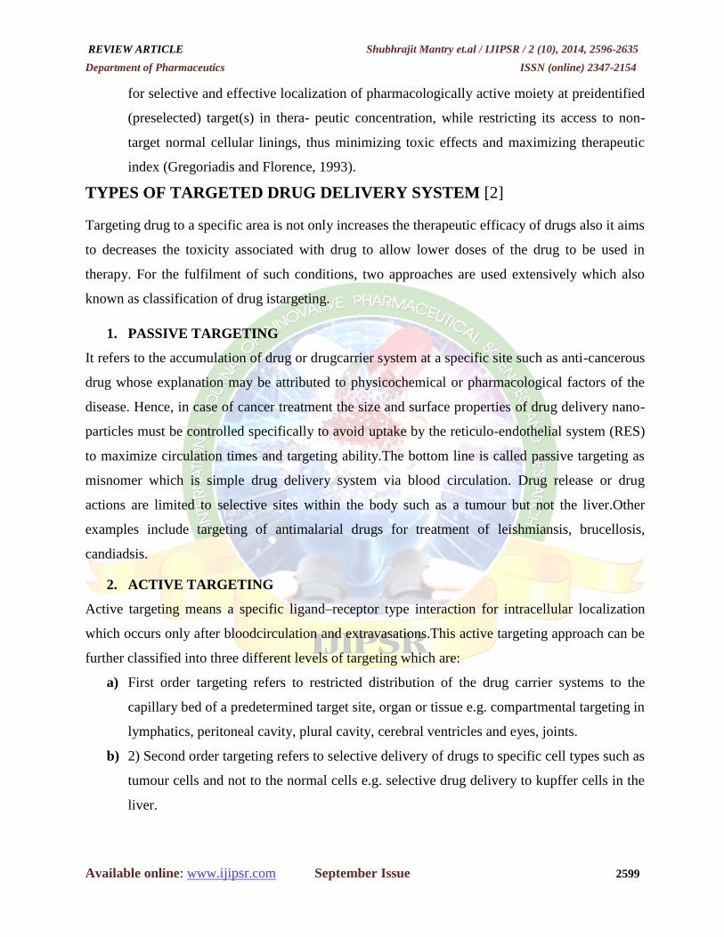
METHOD USED IN THE PREPARATION OF LIPOSOME [9]
The preparation of all types of vesicular systems requires the input of energy. Generally all the
methods of liposome preparation involve three basic stages
REVIEW ARTICLE Shubhrajit Mantry et.al / IJIPSR / 2 (10), 2014, 2596-2635
Department of Pharmaceutics ISSN (online) 2347-2154
Available online: www.ijipsr.com September Issue 2605
1. Drying down of mixture of lipids from an organic solvent.
2. Dispersion of lipids in aqueous media.
3. Separation and purification of resultant liposomes
Fig.5: Flow Chart - Preparation of Liposomes
EVALUATION OF LIPOSOME [11]
Liposomal formualtion and processing for specified purpose are characterized to ensure their
pedictable in vito an in vivo peformance. The characterization parameter for purpose of
Method of Liposomes Preparation
Passive Loading Technique Active Loading Technique
Proliposome
Lyophilization

REVIEW ARTICLE Shubhrajit Mantry et.al / IJIPSR / 2 (10), 2014, 2596-2635
Department of Pharmaceutics ISSN (online) 2347-2154
Available online: www.ijipsr.com September Issue 2606
evaluation could be classified into three board categories which include physical, chemical and
biological parameters.
Physical Charaterization
Physical Charaterization evaluates various parameters including Size, Shape, Surface features,
Lamellarty, Phase behaviour and Drug release profile.
Chemical Charaterization
Chemical charaterization includes those studies which establish the purity and potency of various
lipophillic constituens.
Biological Charaterization
Biological Charaterization parameters are helpful in establishing the safety and suitability of
formualation for therapeutic application.
Same of parameters are:
1) Vesicle shape and lamellarity
Wade shape can be assessed using Electron Microscopic Techniques. Lamellarity of vesicles ie.
Number of bilayers presents in liposomes is determined using Freeze-Fracture Electron
Microscopy and P-31 Nuclear Magnetic Resosance Analysis.
2) Vesicle size and size distribution
Various techniques are described in literature for determination of size and size Distribution.
These include Light Microscopy, Fluorescent Microscopy, Electron Microscopy (specially
Transmission Electron Microscopy), Laser light scaterring Photon Correlation Spectroscopy,
Field Flow fractionation, Gel permeation and Gel Exclusion. The most precise method of
determine size of liposome is Electron Microscopy since it permit one to view each individual
liposome and to obtain exact information about profile of liposome population over the whole
range of sizes Unfortunately, it is very time consuming and require equipments that may not
always be immediately to hand. In contrast, laser light scattering method is very simple and lapid
to perform but having disadvantage of measuring an average property of bulk of liposomes. All
these methods require costly equipments. Another more recently developed microscopic
Technique known as atomic force microscopy has been utilized to study liposome morphology,
size, and stability. Most of methods used in size, shape and distribution analysis can be grouped
into various caterories namely microscopic, diffraction, scaterring and hydrodynamic techniques.
a) Microscopic Techniques
i. Optical Microscopy

REVIEW ARTICLE Shubhrajit Mantry et.al / IJIPSR / 2 (10), 2014, 2596-2635
Department of Pharmaceutics ISSN (online) 2347-2154
Available online: www.ijipsr.com September Issue 2607
The microscopic method includes use of Bright-Field, Phase Contrast Microscope and
Fluorescent Microscope and is useful in evaluating vesicle size of large vesiscle.
ii. Negative Stain TEM
Electron Microscopic Techniques used to assess liposome shape and size are mainly negative-
stain TEM and Scanning Electron Microscopy. The latter technique is less preferred. Negative
stain Electron Microscopy visualizes bright areas against dark background (hense termed as
negative in nature).
The negative stains used in TEM analysis are ammonium molybdate or Phosphotungstic acid
(PTA) or uranyl acetate.
iii. Cryo-Transmission Electron Microscopy Techniques (cryo-TEM)
This technique has been used to elucidate the surface morphology and size of vesicles.
b) Diffaction and Scattring Techniques
i. Laser Light Scattering
Photon correlaion spectroscopy (PCS) is analysis of time dependence of intensity fluctuation in
scattered laser light due to Brownian motion of particles in solution / suspension. Since small
particles diffuse more rapidly than large particle, the rate of fluctuation of scattered light intensity
varies accordingly. Thus, the translational diffusion coeffiecient (D) can be measured, which in
turn can be used to determine the mean hydrodyamic radius (Rh) of particles using the Stoke-
Einstein equation. Using this technique one can measure particles in range of about 3nm.
3) Encapsulation Efficiency and Trapped volume
These determine amount and rate of entrapment of water soluble agents in aqueous compartment
of liposomes.
a) Encapsulation Efficiency
It describe the percent of the aqueous phase and hence percent of water soluble drug that become
ultimately entrapped during preparation of liposomes and is usually expressed as % entrapment /
mg lipid.
b) Trapped Volume
It is an important parpameter that governs morphology of vesicles. The trapped or internal volume
is aqueous entrapped volume per unit quantity of lipids. This can vary from 0.5 to 30
microlitre/micro mol. various material including spectroscopically inert fluid, radioactive marker
and fluroscent markers are used to determine trapped/internal volume.
4) Phase Response and Transitional Behaviour

REVIEW ARTICLE Shubhrajit Mantry et.al / IJIPSR / 2 (10), 2014, 2596-2635
Department of Pharmaceutics ISSN (online) 2347-2154
Available online: www.ijipsr.com September Issue 2608
Liposome and lipid bilayers exhibit various phase transitions that are studied for their role in
triggered drug release or stimulus mediated fusion of liposomal constituents with target cell. An
understanding of phase transitions and fluidity of phospholipids membranes is important both
manufacture and exploitation of liposomes since phase behaviour of liposomal membrane
determine such properties such as permeability, fusion, aggregation and protein binding.
5) Drug Release
The mechanism of drug release from liposomes can be assessed by use of well calibrated invtro
diffuion cell. The liposome based formulation can be assisted by employing in vitro assays to
predict pharmacokinetics and bioavailability of drug before employing costly and time consuming
in vivo studies. The dilution indued drug release in buffer and plasma was employed as predictor
for pharmacokinetic performance of liposomal formulation and another assay which determined
intracellular drug release induced by liposomes degradation in presence of mouse liver lysosome
lysate was used to assess the bioavailability of drug.
APPLICATION OF LIPOSOME [10]
1. THERAPEUTIC APPLICATIONS
Anticancer Therapy
Toxicity of anticancer drugs can be reduced by using them as liposomal formulations. As we
know that drugs of Anthracyclin group are very toxic and kill rapidly dividing cell including
normal ones so these toxicities can be reduced by 50% by using encapsulated liposomal
preparations but the efficiency may be compromised due to bioavailability variations in some
cases. While in some cases efficacy may be enhanced by encapsulation of liposomes due to its
continuous release.
Ocular Drug Delivery
There are three mechanisms for eye protection and these act as a barriers for drug penetration. So
the encapsulated liposomes were first reported to overcome this problem and enhance penetration
of drug in eye.
Pulmonary Drug Delivery
Administration of antibiotics, antiasthmatics and anti-allergic drug through systemic route is
replaced by liposomal preparation of these drugs through pulmonary route. Good solubilization
capacity of liposomes make them a useful tool for the delivery of drug through pulmonary route.

REVIEW ARTICLE Shubhrajit Mantry et.al / IJIPSR / 2 (10), 2014, 2596-2635
Department of Pharmaceutics ISSN (online) 2347-2154
Available online: www.ijipsr.com September Issue 2609
Pulmonary persistence time is prolonged due to its biodegradability without causing any adverse
effect.
Topical Drug Delivery
Liposomes have proved to be useful as a topical agent for drug delivery. They enhance skin
permeability for various drugs and also abolish the side effects of some drugs as lower doses are
needed. Increased Drug transport is due to the lipophilicity of the vesicles, and these vesicles
serves as drug carriers.
Infectious Therapy
Encapsulated liposomes are used for delivery of Antimicrobial agents for two reasons:
1. Protect certain drugs from enzymatic degradation e.g. Penicillins & Cephalosporins
2. Antibiotic cellular uptake is increased by the microorganisms due to lipophilicity of vesicles.
Also reduce dose of drug and toxicity as well.
Diagnostic Applications
Liposomes are also being used as radiopharmaceuticals for diagnostic purposes. It also has
immunodiagnostic applications.
2. GENETIC APPLICATIONS
Gene Delivery
In 1979 liposomes were explored as system for DNA delivery. Plasmid DNA encapsulation in
liposomes and RNA of polio virus was introduced in cell with the help of liposomes. Genetic
Vaccination
Vaccines based on liposomes have shown promising results in clinical trials and further trials on
humans are in progress
Commercial & Industrial Applications
Cosmetic & Dermatology: In dermal preparations liposomes have foung great applications. They
are used in the form of hydrogels or solutions in the preparations for skin care.Appropriate
thickening agents for hydrogels are the Hydrophilic polymers.
Enzyme Immobilization & Bioreactor Technology
Liposomes are used for immobilization of enzymes and also in the bioreactor technology.
Miscellaneous Applications: Modified Release System: Sustained release liposomal preparations
can be administered locally or systemically. By the mechanism of site-avoidance toxicity of some
vital organs can be reduced.
3. MISCELLANEOUS APPLICATIONS

REVIEW ARTICLE Shubhrajit Mantry et.al / IJIPSR / 2 (10), 2014, 2596-2635
Department of Pharmaceutics ISSN (online) 2347-2154
Available online: www.ijipsr.com September Issue 2610
Modified Release System
Sustained release liposomal preparations can be administered locally or systemically. By the
mechanism of site-avoidance toxicity of some vital organs can be reduced. Solubility Enhancers:
Amphiphilic & lipophilic drugs solubility is improved by the liposomes. This is done by the
following two ways:
1. Drug precipitation
2. Gel formation within liposomes
Penetration Enhancers
Tissue penetration of drug is improved by applying liposomal preparations dermally.
Targeting to Specific Site
By attaching ligands on the surface of liposomes we can target specific sites. Liposomes can be
used for the passive targeting especially to target the immune cells. Encapsulated Liposomes:
Encapsulation of liposomes minimizes toxicity by preventing the drug accumulation in the
organs.
TARGETED DRUG DELIVERY SYSTEM USING NANOPARTICLE
INTRODUCTION
Nanoparticles are defined as particulate dispersions or solid particles with a size in the range of
10-1000nm. The drug is dissolved, entrapped, encapsulated or attached to a nanoparticle matrix
[13]. Nanoparticles are used for drug targeting both active and passive. The relatively small size
of these systems limits their use, as only small quantities of material can be encapsulated. Other
types of (non-biodegradable) nanoparticle systems include colloidal sulfur and colloidal gold.
Colloidal sulfur is used as a diagnostic agent (labeled with 99m
TC). It is usually protected from
aggregation by the addition of gelations as a polymeric stabilizer. Colloidal gold is also used as a
diagnostic (198
Au) and as a therapeutic agent [14].
Fig.6: Drug Loaded Nanoparticle Fig.7: Targeted delivery using Nanoparticle
REVIEW ARTICLE Shubhrajit Mantry et.al / IJIPSR / 2 (10), 2014, 2596-2635
Department of Pharmaceutics ISSN (online) 2347-2154
Available online: www.ijipsr.com September Issue 2611
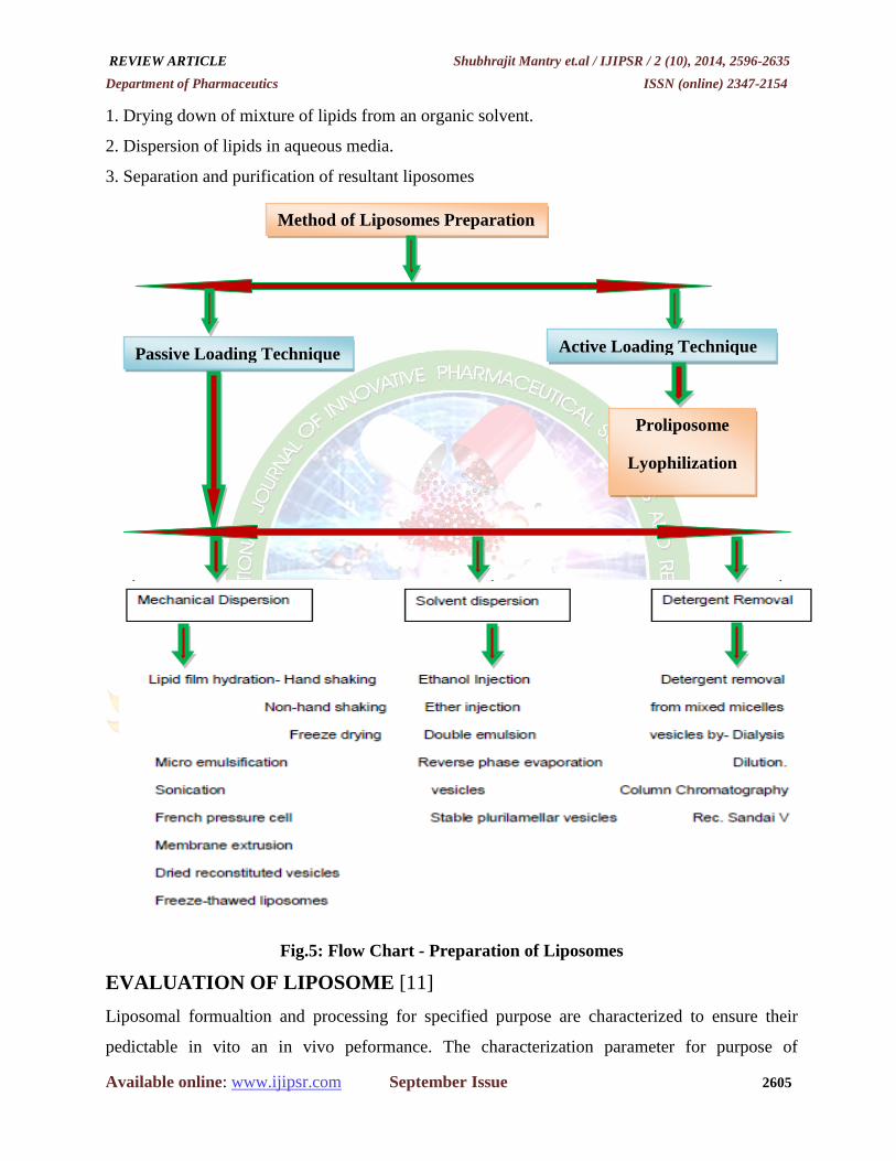
HISTORY OF NANOPARTICLE [15]
The history of nanoparticle research is long and the use of these particles dates back to the 9th
century in Mesopotamia when artisans used these to generate a glittering effect on the surface of
pots. This lustre or glitter over pottery from the middle Ages and Renaissance is due to a metallic
film that was applied to the transparent surface of a glazing. The lustre can still be visible if the
film has resisted atmospheric oxidation and other weathering. The lustre is within the film itself
which contained silver and copper nanoparticles dispersed homogeneously in the glassy matrix of
the ceramic glaze. Artisans created the nanoparticles by adding copper and silver salts and oxides
together with vinegar, ochre and clay, on the surface of previously-glazed pottery. Then the pots
were placed into a kiln and heated to about 600 °C in a reducing atmosphere. With the heat the
glaze would soften, causing the copper and silver ions to migrate into the outer layers of the glaze.
Michael Faraday provided the first description, in scientific terms, of the optical properties of
nanometer-scale metals in his 1857 paper.
Fig.8: Nanoparticle drug delivery systems with relation to other scales
ADVANTAGES OF NANOPARTICLES [16]
Nanoparticles have dimensions below the critical wavelength of light renders them
transparent, a property which makes them very useful for applications in packagings,
cosmetics and coatings.
Particle size and surface characteristics of nanoparticles can be easily manipulated to
achieve both active and passive targeting.

REVIEW ARTICLE Shubhrajit Mantry et.al / IJIPSR / 2 (10), 2014, 2596-2635
Department of Pharmaceutics ISSN (online) 2347-2154
Available online: www.ijipsr.com September Issue 2612
Release of the drug can be controlled or sustained so as to achieve increase in therapeutic
efficacy of drug and reduction in side-effects.
They are capable of being stored for a period of upto 1 year and hence have longer shelf
stability.
They have the ability to incorporate both hydrophilic and hydrophobic drug molecules.
They have higher carrier capacity and drugs can be incorporated without any chemical
reaction and hence preserving the drug activity.
The system can be administered via different routes including oral, nasal, parenteral etc.
These have the potential to increase the bioavailability of drugs.
They have longer clearance time.
Site-specific targeting can be achieved by attaching targeting ligands to surface of
particles or by using magnetic guidance.
DISADVANTAGES OF NANOPARTICLES [16]
It involves higher manufacturing costs which may in turn lead to increase in the cost of
formulation.
These have low encapsulation efficiency.
Water-soluble drugs can be rapidly leaked out in the presence of blood components.
Their small size and large surface area can lead to particle-particle aggregation, making
physical handling of nanoparticles difficult in dry and liquid forms.
They may trigger immune response and allergic reaction.
It may involve use of harsh toxic solvents in the preparation process.
IDEAL PROPERTIES OF NANO PARTICLES [16]
Nanoparticles can be considered as a realizable method for drug delivery, following ideal
properties of the system are required:
Stable in blood
Non toxic
Non thrombogenic
Non immunogenic
Non inflammatory
No activation of neutrophils
Biodegradable

REVIEW ARTICLE Shubhrajit Mantry et.al / IJIPSR / 2 (10), 2014, 2596-2635
Department of Pharmaceutics ISSN (online) 2347-2154
Available online: www.ijipsr.com September Issue 2613
Avoidance of the reticulo-endothelial system
Applicable to various molecules, such as small molecules, proteins, peptides or nucleic
acids (platform technology)
Scalable and inexpensive manufacturing process
FORMULATION OF NANOPARTICLE [17]
Preparation of nanoparticle
In the preparation of nanoparticles different types of matrix material are used such as
Polysaccharides, synthetic polymer and proteins. Various factors are involved in selection of
matrix material to be used in preparations which are
(i) Required nanoparticle size.
(ii) Permeability and surface charge of nanoparticle.
(iii) Level of biodegradability and biocompatibility must be optimum.
(iv) Material must not be toxic.
(v) Solubility profile and stability of drug should not be affected.
(vi) It should show desired drug release profile.
(vii) Must not be immnunogenic.
Table 1: Polymer used for the preparation of nanoparticle [17]
Technique Candidate drug Polymer used
Heat denaturation and cross
linking in w/o emulsion Hydrophilic Hydrophilic Albumin ,Gelatin
Desolvation and cross linking in
water
Hydrophilic and protein
affinity Hydrophilic Albumin ,Gelatin
Cross-linking in water Hydrophilic and protein
affinity Hydrophilic Alginates and chitosan
Polymer precipitation in an
organic solvent Hydrophilic Hydrophilic Dextran
Emulsion polymerization Hydrophilic Hydrophobic Poly(alkylcyanoacrylates)
Interfacial O/W polymerization Hydrophobic Hydrophobic Poly(alkylcyanoacrylates)
Solvent extraction evaporation
Hydrophilic and
Hydrophobic
Soluble in polar solvent
Polyesters Poly (lactic acid), poly(
caprolactone)
Solvent displacement
Hydrophilic and
Hydrophobic
Soluble in polar solvent
Poly (lactic acid), Poly (lactide-co-
glycolide),
Salting out Soluble in polar solvent Polyesters Poly (lactic acid),
Poly (lactide-co-glycolide).

REVIEW ARTICLE Shubhrajit Mantry et.al / IJIPSR / 2 (10), 2014, 2596-2635
Department of Pharmaceutics ISSN (online) 2347-2154
Available online: www.ijipsr.com September Issue 2614
FOLLOWING ARE METHODS WHICH ARE USED IN FORMULATION OF
NANOPARTICLES
1. Dispersion of preformed polymers.
2. Polymerization method.
3. Coacervation or ionic gelatin method.
4. Supercritical fluid technology
1. DISPERSION OF PREFORMED POLYMERS
For the preparation of biodegradable nanoparticles from polymers such as poly (lactic acid)
(PLA); poly (D, L-glycolide), PLG; poly (D, L-lactide-co-glycolide) (PLGA) and Poly-
(cyanoacrylate) (PCA), dispersion of preformed polymer method is used [13]. This technique can
be used in various ways as described below.
Solvent evaporation method
In this method, there is conventional formation of o/w emulsion between a partially water
miscible solvent containing the polymer and the drug, and an aqueous phase containing the
stabilizer. In this polymer is dissolved in an organic solvent such as dichloromethane, chloroform
or ethyl acetate. Oil in water (o/w) emulsion is prepared by emusification of drug and polymer
mixture in aqueous solution which contain emulsifying agent, which result in formation of stable
emulsion. After that by using pressure reduction method or continuous stirring, organic solvent is
evaporated. The homogenizer speed, nature and stabilizer concentration along with the property
of polymer effect size of nanoparticle. Usually high speed homogenizer or ultrsonication had been
used to reduce the size of nanoparticle to an optimum size.
Spontaneous emulsification or solvent diffusion method
Also known as modified version of solvent evaporation method. In this method, two phase
solvent is used, one is water miscible and other is water immiscible i.e. organic in nature which
act as oil phase. In this method interfacial turbulence is created, by immediate diffusion between
two solvents (which are differing in phase) which lead to the formation of small particles. A
reduction in particle size can be gained by increasing the concentration of water miscible solvent
both the above described method can be used for preparation of hydrophilic and hydrophobic
drugs.
Salting out
It is one of commonly used method used for preparation of nanoparticle. This method involves

REVIEW ARTICLE Shubhrajit Mantry et.al / IJIPSR / 2 (10), 2014, 2596-2635
Department of Pharmaceutics ISSN (online) 2347-2154
Available online: www.ijipsr.com September Issue 2615
the mixing of saturated aqueous solution of polyvinyl alcohol (PVA) into an acetone solution of
the polymer under magnetic stirring resulting in the formation of o/w emulsion. The precipitation
of the polymer occurs when sufficient amount of water is added to external phase to allow
complete diffusion of the acetone from internal phase into aqueous phase.
2. POLYMERIZATION METHOD
Polmerization of monomers in an aqueous solution form the basis of this method. Two different
techniques are used for the preparation in aqueous solution.
Emulsion polymerization
This method involves emulsification of monomer in non-solvent phase.
Dispersion polymerization
This method involves dispersion of monomer in non-solvent phase.
Incorporation of drug in nanoparticle can be achieved either by dissolving the drug in
polymerization medium or by adsorption onto nanoparticle. Suspension of nanoparticles is
formed, which contain surfactants and stabilizers that are used in polymerization which has to be
removed by method like ultracentrifugation or by suspending them in isotonic medium which is
free of surfactant. Polybutylcyanoacrylate or poly (alkylcyanoacrylate) nanoparticles are been
prepared by this method. The polmer particle size had been affected by concentration of stabilizer
and surfactant involved in preparation.
.3. COACERVATION OR IONIC GELATION METHOD
Chitosan, sodium alginate and gelatin are hydrophilic biodegradable polymers which are used for
the preparation of nanoparticles by coacervation method. Preparation of hydrophilic chitosan
nanoparticles by ionic gelation was developed by Calvo and Co-worker. This method involves a
preparation of two aqueous phases, of which one is the polymer chitosan, adi-block co-polymer
ethylene oxide or propylene oxide (PEO-PPO) and the other is a polyanion sodium
tripolyphosphate which are mixed, due to mixing positively charged amino group of chitosan
interacts with negative charged tripolyphosphate to form coacervates with a size in the range of
nanometer. when electrostatic interaction take place between two aqueous phases coacervates are
formed, and when two molecules interact due to ionic force, resulting in transition from liquid
phase to gel phase at room temperature this is known as ionic gelation method.

REVIEW ARTICLE Shubhrajit Mantry et.al / IJIPSR / 2 (10), 2014, 2596-2635
Department of Pharmaceutics ISSN (online) 2347-2154
Available online: www.ijipsr.com September Issue 2616
4. SUPERCRITICAL FLUID TECHNOLOGY
Various conventional approaches like solvent diffusion, solvent extraction-evaporation and
organic phase separtion require the use of organic solvent are hazardous to the environment as
well as the physiological systems. Supercritical fluid technology thus has been invested as an
alternative to prepare biodegradable micro and Nanoparticles. Solvent which remain fluid in a
single phase regardless of pressure above critical temperature are known as supercritical fluid
.Super critical CO2 is the most widely used supercritical fluid. The most common processing
techniques involves supercritical fluids are supercritical Antisolvent (SAS) and rapid expansion of
critical solution (RESS). RESS diffuse from SAS process in that its solute in dissolved in super
critical fluid .Thus with solvent power of super critical fluid decrease and the solute eventually
precipitate.
EVLUATION OF NANOPARTICLE [16]
1. Particle Size Determination
2. Static Light Scattering
3. Scanning Electron Microscopy (SEM)
4. Transmission Electron Microscopy (TEM)
5. Atomic Force Microscopy
6. X-Ray Diffraction (Power X-ray Diffraction)
7. Nuclear Magnetic Resonance Spectroscopy
8. Fourier Transform Infrared Spectroscopy
9. Differential Scanning Calorimetry (DSC)
10. Surface Charge/ Surface Properties of Nanoparticles
11. Zeta Potential
12. Electrophoresis
13. Drug Loading and In-Vitro Release Profile Of Nanoparticles
a) In-vitro release profile of lipophilic drug
b) In-vitro release profile of hydrophilic drugs
1. PARTICLE SIZE DETERMINATION
Particle size and size distribution are the most important characteristics of nanoparticle systems.
They determine the in vivo distribution, biological fate, toxicity and the targeting ability of
nanoparticle systems. In addition, they can also influence the drug loading, drug release and

REVIEW ARTICLE Shubhrajit Mantry et.al / IJIPSR / 2 (10), 2014, 2596-2635
Department of Pharmaceutics ISSN (online) 2347-2154
Available online: www.ijipsr.com September Issue 2617
stability of nanoparticles. Many studies have demonstrated that nanoparticles of sub-micron size
have a number of advantages over microparticles as a drug delivery system.
2. STATIC LIGHT SCATTERING
Dynamic light scattering (DLS) is the name that covers different techniques for measurement of
particle size from the dynamic changes of the scattered light intensity. Photon correlation
spectroscopy (PCS) is at present the most widely used name. It relates to the correlation technique
that is most frequently applied in instruments. Quasi-elastic light scattering (QELS) was used as a
name often in the past. This term relates to the type of interaction between particles and light. It is
a rapid method for determining the mean size, the size distribution and the polydispersity index
(PdI) of a sample. In the DLS technique, the intensity of the scattered light by an ensemble of
particles is measured at a given angle (90º) as a function of time. The Brownian motion of the
dispersed particles determines the rate of change of the scattered light intensity. The temporal
intensity changes are converted to a mean translational diffusion coefficient. Fast intensity
changes are related to a rapid decay of the correlation function and a large diffusion coefficient.
The diffusion coefficient is then converted into particle size by means of the Stokes–Einstein
equation. DLS measurement range is about 0.005–1 and time required for measurement is
typically about 0.5–10 min. For measurement of large particles (larger than about 0.5 μm), three
problems are generally encountered are:
Particles may settle out of the measurement zone to the bottom of the cell and thus will
gradually become out of reach for measurement.
Few particles suffice to reach the maximum allowable concentration in view of multiple
scattering and thus changes of their number concentration will bias the sizing result.
Brownian motion is very slow, especially in liquids of increased viscosity. Thus, long
measurement times have to be applied, during which both instrument and suspension
should remain stable. Typically, the particulate concentration during measurement is
around 10-2 –10-3 % (v/v).
3. SCANNING ELECTRON MICROSCOPY (SEM)
The scanning electron microscope (SEM) is a type of electron microscope that images the sample
surface by scanning it with a high-energy beam of electrons in a raster scan pattern. The electrons
interact with the atoms that make up the sample producing signals that contain information about
the sample's surface topography, composition and other properties such as electrical conductivity.
The types of signals produced by an SEM include secondary electrons, back-scattered electrons

REVIEW ARTICLE Shubhrajit Mantry et.al / IJIPSR / 2 (10), 2014, 2596-2635
Department of Pharmaceutics ISSN (online) 2347-2154
Available online: www.ijipsr.com September Issue 2618
(BSE), characteristic X-rays, light (cathodoluminescence), specimen current and transmitted
electrons. Secondary electron detectors are common in all SEMs, but it is rare that a single
machine would have detectors for all possible signals. The signals result from interactions of the
electron beam with atoms at or near the surface of the sample. In the most common or standard
detection mode, secondary electron imaging or SEI, the SEM can produce very high-resolution
images of a sample surface, revealing details about less than 1 to 5 nm in size. Due to the very
narrow electron beam, SEM micrographs have a large depth of field yielding a characteristic
three-dimensional appearance useful for understanding the surface structure of a sample. This is
exemplified by the micrograph of pollen shown to the right. A wide range of magnifications is
possible, from about 10 times (about equivalent to that of a powerful hand-lens) to more than
500,000 times, about 250 times the magnification limit of the best light microscopes. For the
same reason, BSE imaging can image colloidal gold immuno-labels of 5 or 10 nm diameter which
would otherwise be difficult or impossible to detect in secondary electron images in biological
specimens. Characteristic X-rays are emitted when the electron beam removes an inner shell
electron from the sample, causing a higher energy electron to fill the shell and release energy.
4. TRANSMISSION ELECTRON MICROSCOPY (TEM)
Transmission electron microscope is analogous to a slide projector, with illumination from an
electron beam rather than light. When an electron beam is impinged upon a sample, a black and
white TEM image is formed from the passage of some electrons through the sample untouched,
alongside the combination of interactions between other electrons and sample atoms (e.g.,
inelastic/elastic scattering, diffraction). If the undiffracted beam is selected to form the image, it is
referred to as bright-field imaging; in contrast, selection of strongly diffracting regions of the
sample, which would appear brighter than the transmitted beam, is known as dark-field imaging.
It should be noted that electrons may also be absorbed by molecules containing large atoms, or by
surface contamination (e.g., dust, grease). The absorption of a high density of electrons in a
specific region will cause a buildup of heat, leading to sample destruction and poor image quality.
5. ATOMIC FORCE MICROSCOPY
In this technique, a probe tip with atomic scale sharpness is raftered across a sample to produce a
topological map based on the forces at play between the tip and the surface. The probe can be
dragged across the sample (contact mode), or allowed to hover just above (noncontact mode),
with the exact nature of the particular force employed serving to distinguish among the sub
techniques. That ultrahigh resolution is obtainable with this approach, which along with the

REVIEW ARTICLE Shubhrajit Mantry et.al / IJIPSR / 2 (10), 2014, 2596-2635
Department of Pharmaceutics ISSN (online) 2347-2154
Available online: www.ijipsr.com September Issue 2619
ability to map a sample according to properties in addition to size, e.g., colloidal attraction or
resistance to deformation, makes AFM a valuable tool. However, size and shape has been the
most common application to date. The need to raster the probe renders the method very time-
consuming and the size of the sample actually observed is small. Nanoparticles are typically
presented as an evaporated suspension on smooth silicon or mica surface, though not without the
possibility of deformation. Application of various forms of AFM to nanoparticles characterization
represents an area of active research.
6. X-RAY DIFFRACTION (POWER X-RAY DIFFRACTION)
The geometric scattering of radiation from crystal planes within a solid allow the presence or
absence of the former to be determined thus permitting the degree of crys` tallinity to be assessed.
In one example; the crystallization of interior lipids could be tracked. Application of the method
is little different from that for bulk powders, though broadening of the diffraction pattern’s peaks
is observed for particles less than 100nm in diameter. For nanoparticles, order on the smaller scale
can be investigated by reducing the wavelength and angle of incident radiation. Using electron or
neutron beams allows reduction of the former parameter due to the shorter De-Broglie
wavelengths of such particles.
7. NUCLEAR MAGNETIC RESONANCE SPECTROSCOPY Nuclear magnetic resonance (NMR) can be used to determine both the size and the qualitative
nature of nanoparticles. The selectivity afforded by chemical shift complements the sensitivity to
molecular mobility to provide information on the physicochemical status of components within
the nanoparticle. For example; the mobility of Miglyol 812 within solid lipid nanoparticles
confirmed the liquid-like nature of the interior, though it was more limited than the same oil in an
o/w emulsion. Pulsed field gradient methods allow diffusivity of the entire particle to be
quantified and compared to produce 2-D, diffusion ordered plots in which colloidal behavior and
chemical speciation are leveraged simultaneously.
8. FOURIER TRANSFORM INFRARED SPECTROSCOPY Fourier transform spectroscopy is a measurement technique whereby spectra are collected based
on measurements of the coherence of a radiative source, using time-domain or space-domain
measurements of the electromagnetic radiation or other type of radiation. It can be applied to a
variety of types of spectroscopy including optical spectroscopy, infrared spectroscopy (FT-IR,
FT-NIRS), Fourier transform (FT) nuclear magnetic resonance, mass spectrometry and electron
spin resonance spectroscopy. There are several methods for measuring the temporal coherence of

REVIEW ARTICLE Shubhrajit Mantry et.al / IJIPSR / 2 (10), 2014, 2596-2635
Department of Pharmaceutics ISSN (online) 2347-2154
Available online: www.ijipsr.com September Issue 2620
the light, including the continuous wave Michelson or Fourier transform spectrometer and the
pulsed Fourier transform spectrograph (which is more sensitive and has a much shorter sampling
time than conventional spectroscopic techniques, but is only applicable in a laboratory
environment).
9. DIFFERENTIAL SCANNING CALORIMETRY (DSC)
Another method that is a little different from its implementation with bulk materials, DSC can be
used to determine the nature and speciation of crystallinity within nanoparticles through the
measurement of glass and melting point temperatures and their associated enthalpies. A
complement to X-ray diffraction, this method is regularly used to determine the extent to which
multiple phases exist in the interior or to which the various constituents, including the drug,
interact.
10. SURFACE CHARGE/ SURFACE PROPERTIES OF NANOPARTICLES
When nanoparticles are administered intravenously, they are easily recognized by the body
immune systems, and are then cleared by phagocytes from the circulation. Apart from the size of
nanoparticles, their surface hydrophobicity determines the amount of adsorbed blood components,
mainly proteins (opsonins). This in turn influences the in vivo fate of nanoparticles. Binding of
these opsonins onto the surface of nanoparticles called opsonization acts as a bridge between
nanoparticles and phagocytes. The association of a drug to conventional carriers leads to
modification of the drug biodistribution profile, as it is mainly delivered to the mononuclear
phagocytes system (MPS) such as liver, spleen, lungs and bone marrow. Indeed, once in the blood
stream, surface non-modified nanoparticles (conventional nanoparticles) are rapidly opsonized
and massively cleared by the macrophages of MPS rich organs. Generally, it is IgG, compliment
C3 components that are used for recognition of foreign substances, especially foreign
macromolecules. Hence, to increase the likelihood of the success in drug targeting by
nanoparticles, it is necessary to minimize the opsonization and to prolong the circulation of
nanoparticles in vivo. This can be achieved by (a) surface coating of nanoparticles with
hydrophilic polymers/surfactants; (b) formulation of nanoparticles with biodegradable
copolymers with hydrophilic segments such as polyethylene glycol (PEG), polyethylene oxide,
poloxamer, poloxamine and polysorbate 80 (Tween 80). Studies show that PEG conformation at
the nanoparticles surface is of utmost importance for the opsonins repelling function of the PEG
layer. PEG surfaces in brush-like and intermediate configurations reduced phagocytosis and

REVIEW ARTICLE Shubhrajit Mantry et.al / IJIPSR / 2 (10), 2014, 2596-2635
Department of Pharmaceutics ISSN (online) 2347-2154
Available online: www.ijipsr.com September Issue 2621
complement activation whereas PEG surfaces in mushroom-like configuration were potent
complement activators and favored phagocytosis.
11. ZETA POTENTIAL
Zeta potential is used as a surrogate for surface change, and is often measured by observing the
oscillations in signal that result from light scattered by particles located in an electric field, though
there are other approaches. There are a number of instrumental configurations by which this is
achieved, mostly using a Doppler shift, and the user should familiarize them with the particular
approach implemented in their equipment. Instrumentation concerns aside, the need for dilution
begs the question of what is an appropriate diluent, because its choice can profoundly influence
the surface chemistry and thus the results. One approach is to use a particle-free supernatant to
dilute the sample. This will not account for concentration effects, however, and obtaining such a
diluent is nontrivial as the particle size drops. Electro acoustic methods should in principal
eliminate or reduce the need for dilution and its inevitable consequences. Nonpolar media and the
combination of low mobility with high ionic strength are also problematic; however, phase
analysis light scattering, a newer method in which a phase delay shift rather than a frequency shift
is observed, addresses these issues.
12. ELECTROPHORESIS
The body’s response to the introduction of nanoparticles into circulation is such that within a
short period of time their surface is festooned with lipoproteins and related species. This process
will determine the clearance and biodistribution of the colloid, so evaluating the exact nature of
the surface coverage is required to achieve a useful understanding. The small size of nanoparticles
allows their electrophoretic behavior to be observed using bio-analytical tools such as iso-electric
focusing and 2-D poly acryl-amide gel electrophoresis. As with any ex vivo approach, the
investigator needs to take into account the effect that sample preparation may have on the
experimental observations. Similar information has been derived by electrophoresis of serum
proteins desorbed from incubated nanoparticles.
13. DRUG LOADING AND IN-VITRO RELEASE PROFILE OF NANOPARTICLES
Lipophilic or poorly soluble drugs are often incorporated in a nanocapsule or nanoparticles using
hydrophobic polymers. The selection of organic solvent which is to used in reservoir of system
depends on the solubility of the drug. Depending on the drug hydrophobicity, it may require
highly non-polar solvents like chloroform andmethylene chloride. The release of incorporated
drug depends on partitioning behaviour between capsularreservoir and dispersion phase sink. In

REVIEW ARTICLE Shubhrajit Mantry et.al / IJIPSR / 2 (10), 2014, 2596-2635
Department of Pharmaceutics ISSN (online) 2347-2154
Available online: www.ijipsr.com September Issue 2622
hydrophilic drugs, the aqueous phase contains the drug molecules which are embedded in the
polymeric cross-linked matrix. Drug-polymer affinity and interaction may be the critical factors
that determine and regulate drug payload or percent drug incorporation and drug release. The
encapsulation of drug in hydrophilic polymers requires some appropriate organic solvent
depending on the procedural and formulation factors.
In-vitro release profile of lipophilic drugs
Inner structure of polymeric colloidal system largely affects the in-vitro release behaviour of
lipophilic compounds. Drug release from nanocapsules mainly occurs by the drug partition from
the colloidal suspension to the external sink solution and thus in turn depends on the solubility of
the drug in the oily core and external receptor medium. On the other hand, the in-vitro release
characteristics of the lipophilic compounds from the nanoparticles are dominated by the polymer
erosion and in most of the cases a biphasic release pattern results. The first phase (burst release) is
due to the release of the drug adsorbed on the particles surface and the second phase is due to
drug diffusion out of the polymer matrix.
In-vitro release profile of hydrophilic drugs
A hydrophilic compound can be adsorbed on to preformed nanospheres or entrapped within a
polymer matrix composed of natural macromolecules or synthetic polymers. Generally, in the
first case, the release of hydrophilic drugs from polymeric nanoparticles occurs relatively fast (1-2
days) and the release rate reflects the affinity of the drug for the polymer. In the second case
however, the polymer degradation rate and inner structure of nanoparticle in combination may
influence the drug release profile.
THERAPEUTIC APPLICATIONS OF NANOPARTICLES [16]
Nanoparticles have been widely employed for different therapeutic applications, some of which
are listed below:
a) For intracellular targeting of anti-effective drugs to combat the difficult to treat’
intracellular infections of the human body.
b) For targeting of cytostatic drugs to reduce toxicity and increase therapeutic activity.
c) For specific targeting of anti-inflammatory drugs to areas of inflammation, by which the
side-effects of these drugs can be minimized.
d) For ocular delivery systems, to deliver pilocarpine and other miotic drugs.
e) As carriers for radionucleotides for diagnostic purposes in nuclear medicines.

REVIEW ARTICLE Shubhrajit Mantry et.al / IJIPSR / 2 (10), 2014, 2596-2635
Department of Pharmaceutics ISSN (online) 2347-2154
Available online: www.ijipsr.com September Issue 2623
f) To improve the solubility and bioavailability of poorly soluble drugs and protects from
gastrointestinal enzymes and hence, helps in peroral absorption.
g) For skin and hair care in the form of solid nanoparticles, wherein the oily core contains a
wide variety of different cosmetic oils and lipophilic agents.
h) To deliver drugs across the blood brain barrier (BBB).
i) To formulate sustained release preparations.
j) For the controlled delivery of disinfectants or algicide into large bodies of water such as
insect pest feed .
k) For targeted delivery of proteins and peptides.
l) As adjuvants to render antigens potent enough to be useful for vaccines.
m) Have prolonged systemic drug effect due to prolonged systemic circulation and hence,
uptake by reticuloendothelial system can be avoided.
TARGETED DRUG DELIVERY SYSTEM USING RESEALED ERYTHROCYTES
INTRODUCTION
The Drug carrier system including liposomes, nanoparticles, niosomes, resealed erythrocytes etc
act on specific target, promote therapeutic effect of the drug, decrease toxic effect (by increasing
drug level and persistence in vicinity of target cells, hence decreasing the drug exposure to non-
target cells) and finally increases the dose effectiveness also [18].
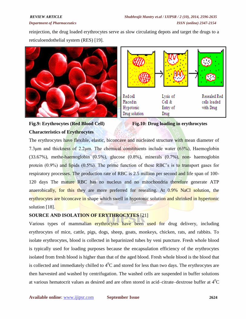
Erythrocytes
Red blood cells (also referred to as erythrocytes) are the most common type of blood cells and the
vertebrate organism's principal means of delivering oxygen (O2) to the body tissues via the blood
flow through the circulatory system. They take up oxygen in the lungs or gills and release it while
squeezing through the body's capillaries. These cells' cytoplasm is rich in hemoglobin, an iron-
containing bimolecule that can bind oxygen and is responsible for the blood's red color. In
humans, mature red blood cells are flexible biconcave disks that lack a cell nucleus and most
organelles. 2.4 million New erythrocytes are produced per second [19].
Resealed Erythrocytes
Such drug-loaded carrier erythrocytes are prepared simply by collecting blood samples from the
organism of interest, separating erythrocytes from plasma, entrapping drug in the erythrocytes,
and resealing the resultant cellular carriers. Hence, these carriers are called resealed erythrocytes.
The overall process is based on the response of these cells under osmotic conditions. Upon
REVIEW ARTICLE Shubhrajit Mantry et.al / IJIPSR / 2 (10), 2014, 2596-2635
Department of Pharmaceutics ISSN (online) 2347-2154
Available online: www.ijipsr.com September Issue 2624
reinjection, the drug loaded erythrocytes serve as slow circulating depots and target the drugs to a
reticuloendothelial system (RES) [19].
Fig.9: Erythrocytes (Red Blood Cell) Fig.10: Drug loading in erythrocytes
Characteristics of Erythrocytes
The erythrocytes have flexible, elastic, biconcave and nucleated structure with mean diameter of
7.3μm and thickness of 2.2μm. The chemical constituents include water (63%), Haemoglobin
(33.67%), methe-haemoglobin (0.5%), glucose (0.8%), minerals (0.7%), non- haemoglobin
protein (0.9%) and lipids (0.5%). The prime function of these RBC’s is to transport gases for
respiratory processes. The production rate of RBC is 2.5 million per second and life span of 100-
120 days The mature RBC has no nucleus and no mitochondria therefore generate ATP
anaerobically, for this they are more preferred for resealing. At 0.9% NaCl solution, the
erythrocytes are biconcave in shape which swell in hypotonic solution and shrinked in hypertonic
solution [18].
SOURCE AND ISOLATION OF ERYTHROCYTES [21]
Various types of mammalian erythrocytes have been used for drug delivery, including
erythrocytes of mice, cattle, pigs, dogs, sheep, goats, monkeys, chicken, rats, and rabbits. To
isolate erythrocytes, blood is collected in heparinized tubes by veni puncture. Fresh whole blood
is typically used for loading purposes because the encapsulation efficiency of the erythrocytes
isolated from fresh blood is higher than that of the aged blood. Fresh whole blood is the blood that
is collected and immediately chilled to 40C and stored for less than two days. The erythrocytes are
then harvested and washed by centrifugation. The washed cells are suspended in buffer solutions
at various hematocrit values as desired and are often stored in acid–citrate–dextrose buffer at 40C

REVIEW ARTICLE Shubhrajit Mantry et.al / IJIPSR / 2 (10), 2014, 2596-2635
Department of Pharmaceutics ISSN (online) 2347-2154
Available online: www.ijipsr.com September Issue 2625
for as long as 48hrs before use. Jain and Vyas have described a well-established protocol for the
isolation of erythrocytes.
Fig. 11: Source and Isolation of Erythrocytes
ADVANTAGES OF RESEALED ERYTHROCYTES [20]
1. Biocompatible, particularly when autologous cells are used hence no possibility of
triggered immune response.
2. Biodegradability with no generation of toxic products.
3. Considerable uniform size and shape of carrier.
4. Relatively inert intracellular environment can be encapsulated in a small volume of cells.
5. Isolation is easy and large amount of drug can be loaded.
6. Prevention of degradation of the loaded drug from inactivation by endogenous chemical.
7. Entrapment of wide variety of chemicals can be possible.
8. Entrapment of drug can be possible without chemical modification of the substance to be
entrapped.
9. Possible to maintain steady-state plasma concentration, decrease fluctuation in
concentration.
10. Protection of the organism against toxic effect of drug.
11. Targeting to the organ of the RES.
12. Ideal zero-order drug release kinetic.
13. Prolong the systemic activity of drug by residing for a longer time in the body.
DISADVANTAGES RESEALED ERYTHROCYTES
1. They have a limited potential as carrier to non-phagocyte target tissue.
2. Possibility of clumping of cells and dose dumping may be there.

REVIEW ARTICLE Shubhrajit Mantry et.al / IJIPSR / 2 (10), 2014, 2596-2635
Department of Pharmaceutics ISSN (online) 2347-2154
Available online: www.ijipsr.com September Issue 2626
METHOD USED IN THE PREPARATION OF RESEALED ERYTHROCYTES [21]
Several methods can be used to load drugs or other bioactive compounds in erythrocytes,
including physical (e.g., electrical pulse method) osmosis-based systems, and chemical methods
(e.g., chemical perturbation of the erythrocytes membrane).
Hypotonic Hemolysis
This method is based on the ability of erythrocytes to undergo reversible swelling in a hypotonic
solution. Erythrocytes have an exceptional capability for reversible shape changes with or without
accompanying volume change and for reversible deformation under stress. An increase in volume
leads to an initial change in the shape from biconcave to spherical. This change is attributable to
the absence of superfluous membrane; hence, the surface area of the cell is fixed. The cells
assume a spherical shape to accommodate additional volume while keeping the surface area
constant. The volume gain is 25-50%. The cells can maintain their integrity up to a tonicity of 150
mosm/kg, above which the membrane ruptures, releasing the cellular contents. At this point (just
before cell lysis), some transient pores of 200–500 Å are generated on the membrane. After cell
lysis, cellular contents are depleted. The remnant is called an erythrocyte ghost the
principle of using these ruptured erythrocytes as drug carriers is based on the fact that the
ruptured membranes can be resealed by restoring isotonic conditions. Upon incubation, the cells
resume their original biconcave shape and recover original impermeability.
Use of Red cell Loader:
Magnani et al. developed a novel method for entrapment of non diffusible drugs into erythrocytes.
They developed a piece of equipment called a “red cell loader”. With as little as 50 mL of a blood
sample, different biologically active compounds were entrapped into erythrocytes within a period
of 2 h at room temperature under blood banking conditions. The process is based on two
sequential hypotonic dilutions of washed erythrocytes followed by concentration with a hemo
filter and an isotonic resealing of the cells. There was 30% drug loading with 35-50% cell
recovery. The processed erythrocytes had normal survival in vivo. The same cells could be used
for targeting by improving their recognition by tissue macrophages.
Hypotonic Dilution
Hypotonic dilution was the first method investigated for the encapsulation of chemicals into
erythrocytes and is the simplest and fastest. In this method, a volume of packed erythrocytes is
diluted with 2–20 volumes of aqueous solution of a drug. The solution tonicity is then restored by

REVIEW ARTICLE Shubhrajit Mantry et.al / IJIPSR / 2 (10), 2014, 2596-2635
Department of Pharmaceutics ISSN (online) 2347-2154
Available online: www.ijipsr.com September Issue 2627
adding a hypertonic buffer. The resultant mixture is then centrifuged, the supernatant is discarded,
and the pellet is washed with isotonic buffer solution). The major drawbacks of this method
include a low entrapment efficiency and a considerable loss of hemoglobin and other cell
components this reduces the circulation half life of the loaded cells. These cells are readily
phagocytosed by RES macrophages and hence can be used for targeting RES organs hypotonic
dilution is used for loading enzymes such as galactosidase and glucosidase asparginase and
arginase. As well as bronchodilators such as salbutamol.
Hypotonic Preswelling
In the process, an isotonic, buffered suspension of erythrocytes with a hematocrit value of 70–80
is prepared and placed in a conventional dialysis tube immersed in 10- 20 volumes of a hypotonic
buffer. The medium is agitated slowly for 2 h. The tonicity of the dialysis tube is restored by
directly adding a calculated amount of a hypertonic buffer to the surrounding medium or by
replacing the surrounding medium by isotonic buffer. The drug to be loaded can be added by
either dissolving the drug in isotonic cell suspending buffer inside a dialysis bag at the beginning
of the experiment or by adding the drug to a dialysis bag after the stirring is complete. The use of
standard hemodialysis equipment for loading a drug in erythrocytes was reported by Roper et al.
In this method, the erythrocyte suspension and the drug to be loaded were placed in the blood
compartment and the hypotonic buffer was placed in a receptor compartment. This led to the
concept of “continuous flow dialysis,” which has been used by several other researchers. The
loaded cells exhibit the same circulation half life as that of normal
cells. Also this method has high entrapment efficiency on the order of 30–50% cell recovery of
70–80%, high-loading capacity, and is amenable to automation with control of process variables.
The drawbacks include a long processing time and the need for special equipment.
Isotonic Osmotic Lysis
This method, also known as the osmotic pulse method, involves isotonic hemolysis that is
achieved by physical or chemical means. The isotonic solutions may or may not be isotonic. If
erythrocytes are incubated in solutions of a substance with high membrane permeability, the
solute will diffuse into the cells because of the concentration gradient. This Process is followed by
an influx of water to maintain osmotic equilibrium. Chemicals such as urea solution, polyethylene
glycol, and ammonium chloride have been used for isotonic hemolysis. However, this method
also is not immune to changes in membrane structure composition. The suspension was diluted
with an isotonicbuffered drug solution. After the cells were separated, they were resealed at 370C.

REVIEW ARTICLE Shubhrajit Mantry et.al / IJIPSR / 2 (10), 2014, 2596-2635
Department of Pharmaceutics ISSN (online) 2347-2154
Available online: www.ijipsr.com September Issue 2628
Chemical Perturbation of the membrane
This method is based on the increase in membrane permeability of erythrocytes when the cells are
exposed to certain chemicals. In 1973, Deuticke et al. showed that the permeability of
erythrocytic membrane increases upon exposure to polyene antibiotic such as amphotericin B. In
1980, this method was used successfully by Kitao and Hattori to entrap the antineoplastic drug
daunomycin in human and mouse erythrocytes. Lin et al used halothane for the same purpose.
However, these methods induce irreversible destructive changes in the cell membrane and hence
are not very popular.
Electroinsertion or Electro encapsulation
In 1973, Zimmermann tried an electrical pulse method to encapsulate bioactive molecules. Also
known as electroporation, the method is based on the observation that electrical shock brings
about irreversible changes in an erythrocyte membrane. In 1977, Tsong and Kinosita suggested
the use of transient electrolysis to generate desirable membrane permeability for drug loading.
The erythrocyte membrane is opened by a dielectric breakdown. Subsequently, the pores can be
resealed by incubation at 370C in an isotonic medium. The procedure involves suspending
erythrocytes in an isotonic buffer in an electrical discharge chamber. A capacitor in an external
circuit is charged to a definite voltage and then discharged within a definite time interval through
cell suspension to produce a square wave potential. The optimum intensity of an electric field is
between 1-10 kw/cm and optimal discharge time is between 20-160s. An inverse relationship
exists between the electric-field intensity and the discharge time. The compound to be entrapped
is added to the medium in which the cells are suspended from the commencement of the
experiment. The characteristic pore diameter created in the membrane depends upon the intensity
of electric field, the discharge time, and the ionic strength of suspending medium. The colloidal
macromolecules contents of the cell may lead to cell lysis because of the increase in osmotic
pressure. This process can be prevented by adding large molecules (e.g., tetrasaccharide stachyose
and bovine serum albumin) and ribonucleose. One advantage of this method is a more uniform
distribution of loaded cells in comparison with osmotic methods. The main drawbacks are the
need for special instrumentation and the sophistication of the process. Entrapment efficiency of
this method is 35% and the life span of the resealed cells in circulation is comparable with that of
normal cells. Various compounds such as sucrose, urease, methotrexate, isoniazid, human
glycophorin, DNA fragments, and latex particles of diameter 0.2m can be entrapped within
erythrocytes by this method.

REVIEW ARTICLE Shubhrajit Mantry et.al / IJIPSR / 2 (10), 2014, 2596-2635
Department of Pharmaceutics ISSN (online) 2347-2154
Available online: www.ijipsr.com September Issue 2629
Entrapment by Endocytosis
Endocytosis involves the addition of one volume of washed packed erythrocytes to nine volumes
of buffer containing 2.5 mM ATP, 2.5 mM MgCl2, and 1mM CaCl2, followed by incubation for
2 min at room temperature. The poress created by this method are resealed by using 154 mM of
NaCl and incubation at 370C for 2 min. The entrapment of material occurs by endocytosis. The
vesicle membrane separates endocytosed material from cytoplasm thus protecting it from the
erythrocytes and viceversa. The various candidates entrapped by this method include primaquine
and related 8 - aminoquinolines, vinblastine, chlorpromazine and related phenothiazines,
hydrocortisone, propranolol, tetracaine and vitamin A.
Loading by Electric cell fusion
This method involves the initial loading of drug molecules into erythrocyte ghosts followed by
adhesion of these cells to target cells. The fusion is accentuated by the application of an electric
pulse, which causes the release of an entrapped molecule. An example of this method is loading a
cellspecific monoclonal antibody into an erythrocyte ghost). An antibody against a specific
surface protein of target cells can be chemically cross-linked to drug-loaded cells that would
direct these cells to desired cells.
Loading by Lipid fusion
Lipid vesicles containing a drug can be directly fused to human erythrocytes, which lead to an
exchange with a lipid-entrapped drug. This technique was used for entrapping inositol
monophosphate to improve the oxygen carrying capacity of cells. However, the entrapment
efficiency of this method is very low (~1%). Numerous applications have been proposed for the
use of resealed erythrocytes as carrier for drugs, enzyme replacement therapy etc. Resealed
erythrocyte has a safe and sure delivery of various drugs for passive and active targeting.
However, the concept needs further optimization to be come a routine drug delivery system.
EVALUATION OF RESEALED ERYTHROCYTES [20]
After loading of therapeutic agent on erythrocytes, the carrier cells are exposed to physical,
cellular as well as biological evaluations
1. Shape and Surface Morphology
The morphology of erythrocytes decides their life span after administration. The morphological
characterization of erythrocytes is undertaken by comparison with untreated erythrocytes using

REVIEW ARTICLE Shubhrajit Mantry et.al / IJIPSR / 2 (10), 2014, 2596-2635
Department of Pharmaceutics ISSN (online) 2347-2154
Available online: www.ijipsr.com September Issue 2630
either transmission (TEM) or Scanning electron microscopy (SEM). Other methods like phase
contrast microscopy can also be used.
2. Drug Content
Drug content of the cells determines the entrapment efficiency of the method used. The process
involves deproteinization of packed, loaded cells (0.5 mL) with 2.0 mL acetonitrile and
centrifugation at 2500 rpm for 10 min. The clear supernatant is analyzed for the drug content by
spectrophotometrically.
3. Cell Counting and Cell Recovery
This involves counting the number of red blood cells per unit volume of whole blood, usually by
using automated machine it is determined by counting the no. of intact cells per cubic mm of
packed erythrocytes before and after loading the drug.
4. Turbulence Fragility
It is determined by the passage of cell suspension through needles with smaller internal diameter
(e.g., 30 gauges) or vigorously shaking the cell suspension. In both cases, haemoglobin and drug
released after the procedure are determined. The turbulent fragility of resealed cells is found to be
higher.
5. Erythrocyte sedimentation rate (ESR)
It is an estimate of the suspension stability of RBC in plasma and is related to the number and size
of the red cells and to relative concentration of plasma protein, especially fibrinogen and α, β
globulins. This test is performed by determining the rate of sedimentation of blood cells in a
standard tube. Normal blood ESR is 0 to 15 mm/hr. higher rate is indication of active but obscure
disease processes.
6. Determination of entrapped magnetite
Atomic absorption spectroscopic method is reported for determination of the concentration of
particular metal in the sample. The HCl is added to a fixed amount of magnetite bearing
erythrocytes and content are heated at 600C for 2 hours, then 20 %w/v trichloro acetic acid is
added and supernatant obtained after centrifugation is used to determine magnetite concentration
using atomic absorption spectroscopy.
7. In vitro stability
The stability of the loaded erythrocytes is assessed by means of the incubation of the cells in the
autologous plasma or in an isoosmotic buffer, setting hematocrit between 0.5% and 5% at
temperatures of 40C and 37
0C.

REVIEW ARTICLE Shubhrajit Mantry et.al / IJIPSR / 2 (10), 2014, 2596-2635
Department of Pharmaceutics ISSN (online) 2347-2154
Available online: www.ijipsr.com September Issue 2631
8. Haemoglobin release
The content of haemoglobin of the erythrocytes may be diminished by the alterations in the
permeability of the membrane of the red blood cells during the encapsulation procedure.
Furthermore, the relationship between the rate of haemoglobin and rate of drug release of the
substance encapsulated from the erythrocytes. The haemoglobin leakage is tested using a red cell
suspension by recording absorbance of supernatant at 540nm on a spectrophotometer.
9. In-vitro drug release and Hb content
The cell suspensions (5% hematocrit in PBS) are stored at 40C in ambered colour glass container.
Periodically clear supernatant are drawn using a hypodermic syringe equipped with 0.45 are filter,
deproteined using methanol and were estimated far drug content. The supernatant of each sample
after centrifugation collected and assayed, %Hb release may be calculated using formula % Hb
release=A540 of sample-A540 of background A540 of 100% Hb.
10. Osmotic shock
For osmotic shock study, erythrocytes suspension (1 ml 10% hct) was diluted with distilled water
(5 ml) and centrifuge at 300 rpm for 15 minutes. The supernant was estimated for % haemoglobin
release analytically.
11. Miscellaneous
Resealed erythrocyte can also be characterized by cell sizes, mean cell volume, energy
metabolism, lipid composition, membrane fluidity, rheological properties, and density gradient
separation
APPLICATIONS OF RESEALED ERYTHROCYTES [19]
Resealed erythrocytes have several possible applications in various fields of human and
veterinary medicine. Such cells could be used as circulating carriers to disseminate a drug within
a prolonged period of time in circulation or in target-specific organs, including the liver, spleen,
and lymph nodes. A majority of the drug delivery studies using drug-loaded erythrocytes are in
the preclinical phase. In a few clinical studies, successful results were obtained.
Slow drug release
Erythrocytes have been used as circulating depots for the sustained delivery of antineoplastics,
antiparasitics, veterinary antiamoebics, vitamins, steroids, antibiotics, and
Cardiovascular drugs. The various mechanisms proposed for drug release include:
Passive diffusion

REVIEW ARTICLE Shubhrajit Mantry et.al / IJIPSR / 2 (10), 2014, 2596-2635
Department of Pharmaceutics ISSN (online) 2347-2154
Available online: www.ijipsr.com September Issue 2632
Specialized membrane associated carrier transport
Phagocytosis of resealed cells by macrophages of RES, subsequent accumulation of drug
into the macrophage interior, followed by slow release.
Accumulation of erythrocytes in lymph nodes upon subcutaneous administration followed
by hemolysis to release the drug.
Drug targeting
Ideally, drug delivery should be site-specific and target-oriented to exhibit maximal therapeutic
index with minimum adverse effects. Resealed erythrocytes can act as drug carriers and targeting
tools as well. Surface-modified erythrocytes are used to target organs of mononuclear phagocytic
system / reticuloendothelial system because the changes in the membrane are recognized by
macrophages. However; resealed erythrocytes also can be used to target organs other than those
of reticuloendothelial (RES).
Targeting reticuloendothelial system (RES) organs
Damaged erythrocytes are rapidly cleared from circulation by phagocytic Kupffer cells in liver
and spleen. Resealed erythrocytes, by modifying their membranes, can therefore be used to target
the liver and spleen. The various approaches to modify the surface
Characteristics of erythrocytes include.
Targeting the liver Enzyme deficiency/replacement therapy
Many metabolic disorders related to deficient or missing enzymes can be treated by injecting
these enzymes. However, the problems of exogenous enzyme therapy include a shorter circulation
half-life of enzymes, allergic reactions, and toxic manifestations. These problems can be
successfully overcome by administering the enzymes as resealed erythrocytes. The enzymes used
include-glucosidase, glucoronidase, galactosidase. The disease caused by an accumulation of
glucocerebrosides in the liver and spleen can be treated by glucocerebrosidaseloaded erythrocytes.
Treatment of hepatic tumors:
Hepatic tumors are one of the most prevalent types of cancer. Antineoplastic drugs such as
methotrexate, bleomycin, asparaginase, and adriamycinhave been successfully delivered by
erythrocytes. Agents such as daunorubicin diffuse rapidly from the cells upon loading and hence
pose a problem. This problem can be overcome by covalently linking daunorubicin to the
erythrocytic membrane using glutaraldehyde or cisaconitic acid as a spacer. The resealed
erythrocytes loaded with carboplatin show localization in liver.

REVIEW ARTICLE Shubhrajit Mantry et.al / IJIPSR / 2 (10), 2014, 2596-2635
Department of Pharmaceutics ISSN (online) 2347-2154
Available online: www.ijipsr.com September Issue 2633
Treatment of parasitic diseases
The ability of resealed erythrocytes to selectively accumulate within RES organs make them
useful tool during the delivery of antiparasitic agents. Parasitic diseases that involve harboring
parasites in the RES organs can be successfully controlled by this method. Results were favorable
in studies involving animal models for erythrocytes loaded with antimalarial, antileishmanial, and
antiamoebic drugs.
Delivery of antiviral agents
Several reports have been cited in the literature about antiviral agents entrapped in resealed
erythrocytes for effective delivery and targeting. Because most antiviral drugs are nucleotides or
nucleoside analogs, their entrapment and exit through the membrane needs careful consideration.
Nucleosides are rapidly transported across the membrane whereas nucleotides are not and thus
exhibiting prolonged release profiles. The release of nucleotides requires conversion of these
moieties to purine or pyrimidine bases. Resealed erythrocytes have been used to deliver
deoxycytidine derivatives, recombinant herpes simplex virus type 1 (HSV-1) glycoprotein B,
azidothymidine derivatives, azathioprene, acyclovir, and fludarabine phosphate.
Enzyme therapy
Enzymes are widely used in clinical practice as replacement therapies to treat diseases associated
with their deficiency (e.g.,Gaucher’s disease, galactosuria), degradation of toxic compounds
secondary to some kind of poisoning (cyanide, organophosphorus), and as drugs. The problems
involved in the direct injection of enzymes into the body have been cited. One method to
overcome these problems is the use of enzyme-loaded erythrocytes. These cells then release
enzymes into circulation upon hemolysisact as a “circulating bioreactors” in which substrates
enter into the cell, interact with enzymes, and generate products or accumulate enzymes in RES
upon hemolysis for future catalysis.
CONCLUSION
Many problems which appeared during the development of drug targeting strategies for clinical
application for different types of therapies have been identified, analyzed and solved.A specific
area of which belongs in the treatment of cancer therapy. Several such preparations have entered
the phases of clinical testing and/or have now been marketed. However, such strategies should be
subjected to continuous evaluation in the light of advances in the understanding of the numerous
processes occurring in response to administration of the carriers and/or the drugs. Liposomes are

REVIEW ARTICLE Shubhrajit Mantry et.al / IJIPSR / 2 (10), 2014, 2596-2635
Department of Pharmaceutics ISSN (online) 2347-2154
Available online: www.ijipsr.com September Issue 2634
one of the unique drug delivery system; they can be use in controlling and targeting drug delivery.
Now, in days the Liposomal topical formulations are more effectively and give the safe
therapeutic efficacy. These are also used in the cosmetic and hair Technologies, diagnostic
purpose and good carrier in gene delivery. Liposomes are giving a good and encouraging result in
the anticancer therapy and human therapy. Nanoparticles are the colloidal particles whose size
ranges in the nanometric size which are mainly employed through parenteral route, hence,
requiring careful development of test methods and acceptance criteria for the specifications. The
use of resealed erythrocytes looks promising for a safe and sure delivery of various drugs for
passive and active targeting. However, the concept needs further optimization to become a routine
drug delivery system. The same concept also can be extended to the delivery of
biopharmaceuticals and much remains to be explored regarding the potential of resealed
erythrocytes. For the present, it is concluded that erythrocyte carriers are “golden eggs in novel
drug delivery systems”.
ACKNOWLEDGEMENT
The authors are thankful to Mr. Shubhrajit Mantry, Dept. of Pharmaceutics, Kottam Institute of
Pharmacy, for his encouragement for carrying out this review work.
REFERENCE
1. International Journal of Pharmaceutical Sciences Review and Research, Volume 8, Issue
2, May – June 2011; Article-020.
2. Rani K et al., Sch. J. App. Med. Sci., 2014; 2(1C):328-331.
3. http://punkhardy.wordpress.com/2012/11/13/drug-targeting-basic-concepts-and-drug-
carrier-systems-a-review.
4. Book- Targeted & controlled drug delivery, Author- Vyas / Khar.
5. Vol. 1 (1) Jan – Mar 2012 www.ijpcsonline.com
6. Mansoori and Agrawal, IJARPB, 2012; Vol.2 (4):453-464.
7. Kant Shashi et al. IRJP 2012, Volume 3 – Issue 7, Page No.-10
8. http://www.pharmatutor.org/articles/liposomes-novel-drug-delivery-carrier?page=0,2
9. Vol. 1 (3) Jul-Sep 2012, www.ijpcsonline.com,Page No. 1103-1113
10. Rukhsana Yusaf wt al. Structural Components of liposomes and Characterization Tools.
Indo American Journal of Pharm Research,2014, 4(08).
11. Kant Shashi et al. IRJP 2012, Volume 3 – Issue 7, Page No.- 13-14

REVIEW ARTICLE Shubhrajit Mantry et.al / IJIPSR / 2 (10), 2014, 2596-2635
Department of Pharmaceutics ISSN (online) 2347-2154
Available online: www.ijipsr.com September Issue 2635
12. http://www.pharmatutor.org/articles/liposome-novel-drug-delivery-system
13. Tropical Journal of Pharmaceutical Research, June 2006; 5 (1): 561-573
14. Encyclopedia of Pharmaceutical Technology, 2nd
Edition edited by Elias Fattal, Christine
Vauhier. Published by, Marcel Dekker, Inc. New York, 2002; pg (1864).
15. http://www.news-medical.net/health/Nanoparticles-What-are-Nanoparticles.aspx
16. Davinder et al Journal of Drug Delivery & Therapeutics; 2013, 3(2), 169-175 17. Abhishek Garg et al Der Pharmacia Sinica, 2011, 2 (2):17-26 18. Gill, IJPSR, 2012; Vol. 3(2): 383-397
19. Ashok Kumar*, Mansi Verma, K. K. Jha, Vol. 1 No. 2 2012 www.thepharmajournal.com
20. Raut Deepika et al. IJRPC 2013, 3(2), Page No.- 198- 207
21. Int. J. of Pharm. & Research Sci. (IJPRS), Vol. 2, Issue 5: May: 2013, 718-738.
Copyright © 2022 FDOKUMEN