Journal of Drug Delivery and Therapeutics
18
Saxena et al Journal of Drug Delivery & Therapeutics. 2019; 9(3-s):802-819 ISSN: 2250-1177 [802] CODEN (USA): JDDTAO Available online on 15.06.2019 at http://jddtonline.info Journal of Drug Delivery and Therapeutics Open Access to Pharmaceutical and Medical Research © 2011-18, publisher and licensee JDDT, This is an Open Access article which permits unrestricted non-commercial use, provided the original work is properly cited Open Access Review Article The Drug Discovery Development for Treatment of Tuberculosis Nisha Saxena 1 *, Noopur Srivastava 2 , Poonam Shukla 3 , Govind Kumar Tripathi 4 1 * Medicinal and Green Sciences Laboratory, Department of Chemistry, M.R.M. College, Lalit Narayan Mithila University, Bihar, India 2 Department of Chemistry and Biochemistry, School of Basic Sciences & Research (SBSR), Sharda University, Greater Noida, UP, India, 201306 3 Department of Chemistry, Veer Kunwar Singh University, Ara, Bhojpur Bihar, India 4 The International Union Against Tuberculosis and Lung Diseases, The Union, South East Asia office, Qutub Institutional Area, Delhi, India ABSTRACT Since decades Tuberculosis (TB) has been a foremost cause of mortality and morbidity with more than one-third of the world population infected with latent TB. Recent fight with an age old disease continuously smack with a dawdling approach toward its treatment. In spite of extensive researches in this field for combating the disease we are lacking behind in race with its causing agent Microbacterium Tuberculosis. Multidrug (MDR) and extensively drug resistant (XDR) Mycobacterium tuberculosis creates the worldwide open threat to human welfare. Thus there is a need of swift researches for its combat. Here in this review we are giving a brief description towards various chemical agents which have been used for its therapy and new families arrived as a potential drug candidate till date. Keywords: Tuberculosis (TB), Mycobacterium tuberculosis, Multidrug resistance (MDR), Extensively drug resistance (XDR), Directly Observed Treatment (DOTs), Minimum Inhibitory Concentration (MIC), Short-course, Nanoparticles, Drug delivery Article Info: Received 17 April 2019; Review Completed 20 May 2019; Accepted 24 May 2019; Available online 15 June 2019 Cite this article as: Saxena N, Srivastava N, Shukla P, Tripathi GK, The Drug Discovery Development for Treatment of Tuberculosis, Journal of Drug Delivery and Therapeutics. 2019; 9(3-s):802-819 http://dx.doi.org/10.22270/jddt.v9i3-s.2777 *Address for Correspondence: Nisha Saxena, Medicinal and Green Sciences Laboratory, Department of Chemistry, M.R.M. College, Lalit Narayan Mithila University, Bihar, India CONTENTS 1. Introduction 1.1. The Biology of Mycobacterium tuberculosis 1.2. Multidrug resistant tuberculosis (MDR-TB) 1.3. Extensively drug resistant tuberculosis (XDR-TB) 1.4. Pathogenesis 1.5. Diagnosis 2. Currently available drugs for the treatment of tuberculosis 2.1. First-line antitubercular drugs 2.1.1. Isoniazid (1) 2.1.2. Rifampicin (2) 2.1.3. Streptomycin (3) 2.1.4. Pyrazinamide (4) 2.1.5. Ethambutol (5) 2.2. Second-line antitubercular drugs 2.2.1. D-Cycloserine (6) 2.2.2. Ethionamide (7) 2.2.3. p-aminosalicylic acid (8) 2.2.4. Thiacetazone, (9) 2.2.5. Capreomycin, (10) 2.2.6. Fluoroquinolines (11) 2.2.7. Rifabutin (12) 2.2.8. Clofazimine (13) 2.2.9. Macrolides (14) 3. Antituberculars derived from natural products 3.1. Pseudopteroxazole (15) 3.2. Pseudopteroxazole (16). 3.3. Erogorgiaene (17) and 3.4. 7-Hydroxyerogorgiaene (18). 3.5. Elisapterosin B (19) 4. Compounds with anti-TB activity in preclinical trials 4.1. CS-940 (20) 4.2. PD 161148 (21) 4.3. Calanolide A, (22) 4.4. Calanolide B, (23) 4.5. CGI 17341 (24) 4.6. Poloxamer 315 (CRL-1072) (25) 4.7. Linezolid (U-100766) (26) 4.8. PNU-100480 (27) 4.9. B4157 (28) 4.10. Miconazole (29) 4.11. Niclosamide (30) 4.12. CGP 7040 (31) 5. Compounds with anti-TB activity in clinical trials 5.1. Moxifloxacin (BAY12-8039) (32) 5.2. Sitafloxacin (DU-6859a) (33) 5.3. Gemifloxacin (34) 5.4. T-3811ME (35) 5.5. Rifametane (SPA-S-565) (36) 5.6. Rifalazil KRM-1648 (37) 5.7. MiKasome (38) 5.8. Aconiazide (39) 5.9. SRL 172 (40) 5.10. Diarylquinoline R207910 (41) 5.11. Sudoterb (LL-3858) (42) 5.12. PA 824 (43) 5.13 OPC67683 (44) 5.14. TMC207 (45) 5.15. Quinalones (46) 6. Newly tested molecules for Anti-TB activity 7. Disease burden and the job ahead 8. The future opportunity of TB drug development 9. Conclusion 10. Acknowledgements 11. References
-
Upload
khangminh22 -
Category
Documents
-
view
2 -
download
0
Transcript of Journal of Drug Delivery and Therapeutics

Saxena et al Journal of Drug Delivery & Therapeutics. 2019; 9(3-s):802-819
ISSN: 2250-1177 [802] CODEN (USA): JDDTAO
Available online on 15.06.2019 at http://jddtonline.info
Journal of Drug Delivery and Therapeutics Open Access to Pharmaceutical and Medical Research
© 2011-18, publisher and licensee JDDT, This is an Open Access article which permits unrestricted non-commercial use, provided the original work is properly cited
Open Access Review Article
The Drug Discovery Development for Treatment of Tuberculosis
Nisha Saxena1*, Noopur Srivastava2, Poonam Shukla3, Govind Kumar Tripathi4
1* Medicinal and Green Sciences Laboratory, Department of Chemistry, M.R.M. College, Lalit Narayan Mithila University, Bihar, India 2 Department of Chemistry and Biochemistry, School of Basic Sciences & Research (SBSR), Sharda University, Greater Noida, UP, India, 201306 3 Department of Chemistry, Veer Kunwar Singh University, Ara, Bhojpur Bihar, India
4 The International Union Against Tuberculosis and Lung Diseases, The Union, South East Asia office, Qutub Institutional Area, Delhi, India
ABSTRACT
Since decades Tuberculosis (TB) has been a foremost cause of mortality and morbidity with more than one-third of the world population infected with latent TB. Recent fight with an age old disease continuously smack with a dawdling approach toward its treatment. In spite of extensive researches in this field for combating the disease we are lacking behind in race with its causing agent Microbacterium Tuberculosis. Multidrug (MDR) and extensively drug resistant (XDR) Mycobacterium tuberculosis creates the worldwide open threat to human welfare. Thus there is a need of swift researches for its combat. Here in this review we are giving a brief description towards various chemical agents which have been used for its therapy and new families arrived as a potential drug candidate till date.
Keywords: Tuberculosis (TB), Mycobacterium tuberculosis, Multidrug resistance (MDR), Extensively drug resistance (XDR), Directly Observed Treatment (DOTs), Minimum Inhibitory Concentration (MIC), Short-course, Nanoparticles, Drug delivery
Article Info: Received 17 April 2019; Review Completed 20 May 2019; Accepted 24 May 2019; Available online 15 June 2019
Cite this article as:
Saxena N, Srivastava N, Shukla P, Tripathi GK, The Drug Discovery Development for Treatment of Tuberculosis, Journal of Drug Delivery and Therapeutics. 2019; 9(3-s):802-819 http://dx.doi.org/10.22270/jddt.v9i3-s.2777
*Address for Correspondence:
Nisha Saxena, Medicinal and Green Sciences Laboratory, Department of Chemistry, M.R.M. College, Lalit Narayan Mithila University, Bihar, India
CONTENTS
1. Introduction 1.1. The Biology of Mycobacterium
tuberculosis 1.2. Multidrug resistant tuberculosis
(MDR-TB) 1.3. Extensively drug resistant
tuberculosis (XDR-TB) 1.4. Pathogenesis 1.5. Diagnosis 2. Currently available drugs for the treatment of tuberculosis 2.1. First-line antitubercular drugs 2.1.1. Isoniazid (1) 2.1.2. Rifampicin (2) 2.1.3. Streptomycin (3) 2.1.4. Pyrazinamide (4) 2.1.5. Ethambutol (5) 2.2. Second-line antitubercular drugs 2.2.1. D-Cycloserine (6) 2.2.2. Ethionamide (7) 2.2.3. p-aminosalicylic acid (8) 2.2.4. Thiacetazone, (9) 2.2.5. Capreomycin, (10) 2.2.6. Fluoroquinolines (11) 2.2.7. Rifabutin (12)
2.2.8. Clofazimine (13) 2.2.9. Macrolides (14) 3. Antituberculars derived from
natural products
3.1. Pseudopteroxazole (15) 3.2. Pseudopteroxazole (16). 3.3. Erogorgiaene (17) and 3.4. 7-Hydroxyerogorgiaene (18). 3.5. Elisapterosin B (19) 4. Compounds with anti-TB activity
in preclinical trials 4.1. CS-940 (20) 4.2. PD 161148 (21) 4.3. Calanolide A, (22) 4.4. Calanolide B, (23) 4.5. CGI 17341 (24) 4.6. Poloxamer 315 (CRL-1072) (25) 4.7. Linezolid (U-100766) (26) 4.8. PNU-100480 (27) 4.9. B4157 (28) 4.10. Miconazole (29) 4.11. Niclosamide (30) 4.12. CGP 7040 (31)
5. Compounds with anti-TB activity in clinical trials
5.1. Moxifloxacin (BAY12-8039) (32) 5.2. Sitafloxacin (DU-6859a) (33) 5.3. Gemifloxacin (34) 5.4. T-3811ME (35) 5.5. Rifametane (SPA-S-565) (36)
5.6. Rifalazil KRM-1648 (37) 5.7. MiKasome (38)
5.8. Aconiazide (39) 5.9. SRL 172 (40) 5.10. Diarylquinoline R207910 (41) 5.11. Sudoterb (LL-3858) (42) 5.12. PA 824 (43) 5.13 OPC67683 (44) 5.14. TMC207 (45) 5.15. Quinalones (46) 6. Newly tested molecules for Anti-TB activity 7. Disease burden and the job ahead 8. The future opportunity of TB drug development 9. Conclusion 10. Acknowledgements 11. References

Saxena et al Journal of Drug Delivery & Therapeutics. 2019; 9(3-s):802-819
ISSN: 2250-1177 [803] CODEN (USA): JDDTAO
1. INTRODUCTION
Tuberculosis (TB), an airborne communicable disease and one of the three World Health Organization (WHO) priority infectious diseases, is caused by transmission of aerosolized droplets of Mycobacterium tuberculosis organism. Drugs for treating tuberculosis have been available for over half a century, and yet the incidence of disease worldwide continues to rise year by year.
Statics
In 2002, the statistics available estimated that 24,000 people developed active disease and close to 5,000 people died from TB every day.
1 Co-infection with immunodeficiency virus is driving the increase in incidence2,3 and the cause of death in 31% of AIDS cases can be attributed to TB in the African region.4,5 When coupled with the emergence of multidrug-resistant strains of Mycobacterium tuberculosis (MDR-TB),6 the scale of the problem becomes clear, as it will inevitably become even more difficult to treat TB in the future. It is now more than a decade since the World Health Organization declared TB “a global health emergency”.7
The reasons for these problems are numerous.8,9
Compliance with even the best available regimen is poor, and treatment failure is all too common. The need for new drugs is to extend the range of TB treatment options is acute. New chemical entities with novel mechanisms of action will most likely possess activity against MDR-TB.10 However, these alone will not provide the breakthrough that is needed. The key to improving therapy is to develop new agents with potent sterilizing activity that will lead to a shortening of the duration of chemotherapy.11
1.1. The biology of Mycobacterium tuberculosis
M. tuberculosis, the agent of human TB, was discovered in 1882 by Robert Koch and for a long time called after his name (the Koch bacillus). Mycobacterium genus are Gram-positive bacteria that share the property of acid-fastness (Ziehl-Neelsen staining), due to their mycolic acid rich cell wall structure.
Mycobacterium is a genus of bacteria, which are slow growing, aerobic and distinguished by
acid-fast staining and are obligate aerobes, characterized as gram-positive. The genus mycobacterium comprises the exceedingly pathogenic organisms that cause tuberculosis, Mycobacterium tuberculosis (M. tuberculosis) and sometimes M. bovis and M. leprae (leprosy).
Tuberculosis (TB) is immensely infectious disease it reaches almost exclusively by airborne transmission. Although the disease can affect any site in the body, it most often affects the lungs. When pulmonary TB infected persons cough, their cough contains miniature droplet nuclei that enclose TB bacteria. These droplets remain suspended in the air for prolong duration. If someone who breathes in this contaminated air, can develop or be infected with TB.
The cell wall of Mycobacterium species is essential for its growth and for continued existence in the infected host it is the most essential structural and functional component. In fact, this is the one of the most important drug target for some of the most efficient anti-mycobacterial drug molecules e.g. isoniazid and ethambutol. These chemical entities are known inhibitors of the biogenesis of cell wall which is in turn subjugated by covalently associated mycolic acids, few related arabinogalactan and peptidoglycan (AGP), based mycolic acids are accoladed by glycolipids such as α,α-
trehalose monomycolate (TMM).12 This permeability barrier based on mycolic acid screens the organism from environmental stress and enhance the contributes in disease perseverance and the refractoriness of M. tuberculosis to many known and potent antibiotics. A large amount of important macromolecular entities of mycobacterial cell wall is arabinan, it is a common constituent in arabinogalactan (AG) and lipoarabinomannan (LAM) both. In the chemical infrastructure of the mycolylarabino galactan-peptidiglycan complex, AG constitutes an integral part of proper cell wall, while LAM, foundation for a phosphatidylinositol anchor, apparently exists in a state of flux. LAM is an essential part of the cell envelope, which lacks covalent association with the cell wall core. Anchored in the cell membrane and traversing the cell wall, as well appearing as an excretory product, LAM has been implicated as a key surface molecule in host-pathogen interactions. The biosynthetic pathways leading to formation of the key mycobacterial cell wall components AG and mycolic acids, are the targets for the rational design of new antitubercular agents.
The determination of the complete genome sequence of M. tuberculosis13 has had a profound effect on researchers in the TB field. New approaches are being followed that depend upon the availability of this information. These include detailed comparative genomics using bioinformatics, functional genomics, proteomics, transcriptomics, and structural genomics. Combined new tools for manipulating the genome of M. tuberculosis, offers a better understanding of the complex biology of this pathogen.
1.2. Multidrug resistant tuberculosis (MDR-TB)
It refers to the simultaneous to at least Isoniazid (INH) and Rifampicin (RIF) with or without resistance to other drugs. Multidrug-resistance arises from the sharing of genes between different species or genera generally mediated by small pieces of extra-chromosomal DNA known as transposons or plasmids.14 Some antibiotics can actually induce the transfer of these resistance genes.15 Alternatively, as with the problematic multidrug-resistant M. tuberculosis (MDR-TB) strains accumulation of multiple point mutations in the chromosomal DNA can take place.16 Contamination of some commercial antibiotic preparations with the DNA (containing the inherent resistance genes) of the organisms that produce the antibiotic has been implicated as a source of drug resistance genes. The presence of DNA encoding drug resistance in antibiotic preparations has been proposed as a factor in the rapid development of multidrug resistance in bacteria.17
1.3. Extensively drug resistant tuberculosis (XDR-TB)
XDR-TB, defined as extensively drug-resistant tuberculosis are cases of TB disease in persons whose M. tuberculosis isolates were resistant to isoniazid and rifampin and at least three of the six main classes of second-line drugs (aminoglycosides, polypeptides, fluoroquinolones, thioamides, cycloserine and p-aminosalicylic acid).18 XDR-TB is related to the poor management of multidrug resistant tuberculosis cases (which in turn is the consequence of poorly managed susceptible TB).19,20 As per the new definition of XDR-TB it is defined as the MDR-TB that is resistant to quinolones and also to any one of kanamycin, capreomycin or amikacin.21 The principles of treatment of MDR-TB and for XDR-TB are the same. The main difference is that XDR-TB is associated with a much higher mortality rate than MDR-TB, because of a reduced number of effective treatment options. The epidemiology of XDR-TB is currently not well studied, but it is believed that XDR-TB does not transmit easily in healthy populations, but is capable of

Saxena et al Journal of Drug Delivery & Therapeutics. 2019; 9(3-s):PageNumber
ISSN: 2250-1177 [804] CODEN (USA): JDDTAO
causing epidemics in populations which are already stricken by HIV and therefore more susceptible to TB infection.22
1.4 Pathogenesis
M. tuberculosis is an intracellular pathogenic bacterium, which has developed sophisticated mechanisms to survive inside host monocellular phagocytic cells and thus evade the immune system. TB bacilli usually multiply first in the macrophages in the lung alveoli and alveolar ducts and in draining lymph nodes. Infected macrophages eventually get killed, progressively creating a primary tubercle. Delayed cutaneous hypersensitivity develops and together with other cellular immune reactions leads to the caseous necrosis of the primary complex. CD4+ T-cells accumulate in great numbers in the early granulomatous lesions, where they are later joined by CD8+T-cells. Bacilli eventually spread to many parts of the body such as liver, spleen, meninges, bones, kidneys and lymph nodes, where they can either be a source of over disseminated TB or, more commonly, remain dormant. CD4+ T-cells play a major role in containment of infection: progressive TB is usually associated with a Th2 T-cell response, whereas a pure Th1 response mediates protection. Th1 type cytokines, notably IFN-γ and TNF-α, are instrumental in walling off M. tuberculosis inside granulomatous lesions and controlling the evolution of the disease.23 In addition, T cells expressing a γδ T-cell receptor with specificity for small phosphorylated ligands and T-cells with specificity for glycolipids are stimulated.24,25 Individuals with deficient IFN-γ signaling suffer from rapid evolution of the disease. Treatment with anti-TNF-α antibodies readily leads to tuberculosis reactivation in patients with rheumatoid arthritis.26 Granulomas persist for years and efficiently contain M. tuberculosis in a state of dormancy, as long as the host remains immunocompetent.27 Occasional decline in cell-mediated immunity leads to reactivation tuberculosis, most frequently seen in adults as a
pulmonary disease with infiltration or cavity in the apex of the lung. This is the most infectious form of TB.28
1.5. Diagnosis
Different diagnostic techniques have been adopted for identify the bacterium these are
1. Tuberculin skin test (Mantoux tuberculin skin test)
2. Interferon-gamma release assays (IGRA)
3. Rapid sputum tests for tuberculosis (TB)
4. New diagnostic technologies and tools
2. CURRENTLY AVAILABLE DRUGS FOR THE TREATMENT OF TUBERCULOSIS
To date, many drugs are available, which are classified into two categories. First-line therapy includes five medications: isoniazid (INH), pyrazinamide (PZA), ethambutol (EMB), rifampicin (RIF) and streptomycin.29,30 This regimen comprises daily isoniazid, rifampin, pyrazinamide, and ethambutol treatment for 2 months followed by 4 months of daily doses of INH and RIF.
Second-line therapy, which is used exceptionally in the cases of drug resistance, includes cycloserine, capreomycin, fluoroquinolones, ethionamide, PAS, thioacetazone, rifabutin, tetramethyl piperidine substituted phenazines (TMP phenazines) and some macrolides.31 The second-line of drugs is clinically less effective and sometimes causes severe side effects; therefore, they are not often used in therapy.
2.1. First-line antitubercular drugs
The first-line of drugs namely isoniazid, rifampicin, pyrazinamide, ethambutol and streptomycin are highly effective for short-term therapy. Figure 1 shows the site of action of first line drugs. (Table: 1)
Figure 1: An outline of Patent Protected drug targets of Mycobacterium tuberculosis.
2.1.1. Isoniazid (1) (INH, 1952), is a highly potent drug for the treatment of tuberculosis. It is highly effective, inexpensive, well tolerated and readily available. It is very selective to mycobacteria and produces side effects in
only 5% of patients. INH contains a pyridine ring and a hydrazide group and both moieties are essential for its high activity for M. tuberculosis. INH inhibits the synthesis of mycolic acids (long chain α-branched β-hydroxylated fatty acids) in M. tuberculosis by affecting the enzyme mycolate synthatase, which is unique for mycobacteria.32,33
N
HN
NH2
O
(1)

Saxena et al Journal of Drug Delivery & Therapeutics. 2019; 9(3-s):PageNumber
ISSN: 2250-1177 [805] CODEN (USA): JDDTAO
Isoniazid is a prodrug which requires activation by the mycobacterial catalase peroxidase enzyme (kat G) to generate a range of both reactive radicals (hydrogen peroxide, superoxide, peroxynitrite and hydroxyl radical) and reactive organic radicals such as isonicotinic acyl radical. These radicals then attack multiple targets (e.g. mycolic acid synthesis, DNA damage, lipid peroxidation and NAD metabolism) in the tubercle bacillus.34 It is orally active and exhibits bacteriostatic action on resting bacilli and is highly active against the M. tuberculosis complex (M. bovis, M. africanum and M. microti). It has very low MICs (0.02-0.06 µg/mL)35 against these pathogens. INH enters the organism by diffusion and oxygen dependent active transport36,37 and is reported to have an effect on almost every aspect of mycobacterial metabolism. A mutation within the mycobacterial INH-A gene was shown to confer resistance to both INH and ethionamide in M. smegmatis and in M. bovis, suggesting that the INH-A is the likely target of this drug and related reagents.38
The patented targets against which antitubercular drugs can be developed are shown in boxes. These targets are known to control various cellular processes required for survival or virulence of M. tuberculosis. The site of action of first-line drugs (Rifampicin, Isoniazid, Pyrazinamide, Ethambutol and Streptomycin) are shown by arrows. (Adapted from : Vohra R., Gupta M., Chaturvedi R., Singh Y., Recent patents on Anti-Infective Drug Discovery, 2006, 1, 95-106.)
2.1.2. Rifampicin (2) (RIF, 1965), belongs to rifamycin group of semisynthetic antibiotics isolated from Streptomyces mediterrani,39 characterized by a natural ansa structure (chromophoric naphthohydroquinone group spanned by a long aliphatic bridge), are potent inhibitors
of prokaryotic DNA-dependent RNA polymerase, an enzyme necessary for RNA synthesis.40 RIF acts on β-subunit of this enzyme resulting in the formation of a stable complex. This in turn, causes inhibition of bacterial RNA synthesis and has no effect on mammalian enzymes. RIF specifically inhibits the transition from synthesis of short oligoribonucleotides to full length transcripts. Isolating RNA polymerase from M. smegmatis, it has been demonstrated that RIF specifically inhibited the elongation of full-length transcripts and had virtually no effect on the initiation of transcription.41 The lipohilic properties of the molecule are important for the binding of the drug to the polymerase and aid in the penetration of the drug across the mycobacterial cell wall. To avoid rapid development of bacterial resistance, RIF is recommended in combination with other first-line agents either isoniazid or ethambutol. However, a combination of INH and RIF may increase a risk of hepatotoxicity. RIF is effective against M. tuberculosis with MIC ranging from 0.1 to 0.2 μg/mL.42
2.1.3. Streptomycin (3) (1944), an aminoglycoside antibiotic derived from Streptomyces griseus made up of three components streptidine, streptose and N-methyl-L-glucosamine, poorly absorbed from the gastrointestinal tract, is administered intramuscularly. Streptomycin was the first really effective drug against TB, and dihydrostreptomycin has almost
the same antibacterial activity as the parent compound. It has an MIC value of 1 µg/mL with 50-60% plasma protein bound and a half life of 5-7 hr. It penetrates the inner membrane of M.tuberculosis and binds to the 30S subunit of the ribosome.4343 Mutations in the rpsL gene of the ribosomal S12 protein of mycobacteria or base substitutions in the 16S rRNA region confers resistance to streptomycin. Different synthetic derivatives of streptomycin have been synthesized and evaluated against M. tuberculosis.44,45,46 Due to the toxic effects on peripheral, central nervous system at higher dosage and hypersensitivity reaction, streptomycin is not a popular choice for treating tuberculosis.
2.1.4. Pyrazinamide (4) (PZA, 1970) a structural analogue of nicotinamide, is first-line drug of short-course TB therapy. It is also active against semidormant bacilli not affected by any other drug, has strong synergy with INH and RIF and shortens the therapy period to 6 months.47, 48 The drug has no significant bactericidal effect and is thought to
act by sterilizing effect.49 The activity of PZA depends on the presence of bacterial amidase which converts PZA to pyrazinoic acid, the active anti-TB molecule. Resistance to PZA is usually accompanied by loss of pyrazinamidase activity in M.
tuberculosis. The gene pncA encoding the M. tuberculosis pyrazinamidase has been sequenced, and mutations in pncA were found in smaller number of PZA-resistant M. tuberculosis strains.50 Mutations in pncA have been identified in PZA–resistant strains, and transformation of these strains with a functional pncA gene restored
pyrazinamidase and PZA susceptibility.51
2.1.5. Ethambutol (5) (EMB, 1968) a synthetic amino alcohol (ethylene diamino-di-1-butanol) 52, 53, 54 with profound antimycobacterial activity, is
orally effective bacteriostatic agent that is active against most strains of Mycobacterium. Activity of EMB is stereospecific as dextro isomer exhibited maximum antitubercular activity (S,S form is 600 times more active than R,R). The target of ethambutol lies in the pathway for the biosynthesis of cell wall arabinogalactan. EMB specifically inhibits arabinosyl transfer, suggesting that arabinosyl transferase is primary cellular target for EMB. In the cell wall biosynthesis arabinosyl transferase III is responsible for the polymerization of arabinose into arabinan of arabinogalactan. The genes embAB of M. avium encode the drug target for ethambutol, the arabinosyl transferase responsible for the polymerisation of arabinose into the arabinan of arabinogalactan and overproduction of this ethambutol-sensitive target leads to ethambutol resistance.55
2.2. Second-line antitubercular drugs
Second-line antitubercular drugs are used due to drug-resistance or to the non-availability of first-line drugs. (Table: 1)
2.2.1. D-Cycloserine (6) (1955), chemically defined as D-4-amino-3-isooxazolidone, D-cycloserine is derived from Streptomyces orchidaceus and is active against a broad spectrum of bacteria, including M. tuberulosis.56 It is a structural analog of the amino acid-
alanine, possesses activity against a wide range of bacteria, and inhibits M. tuberculosis at concentrations of 5-20 µg/mL.
ONH
OH2N
(6)
NH
OOH
O
NN
NOH
OO
CH3
CH3O
H3C
O
CH3
HO
H3CO
CH3
OHH3C
CH3
CH3
CH3
HO
(2)
O
OH
OHHN
O
HN
OHNH
OHHONH2
NH
H2N
NH
O OH3C
OH
CH3
OHH
O
(3)
N
N CONH2
(4)
HO
H3C
NH
HN
CH3
OH
(5)

Saxena et al Journal of Drug Delivery & Therapeutics. 2019; 9(3-s):PageNumber
ISSN: 2250-1177 [806] CODEN (USA): JDDTAO
D-cycloserine is well absorbed and distributed throughout the body following oral administration. As a mechanism based inhibitor it blocks PG biosynthesis by inhibiting the enzymes D-alanine racemase and D-alanilyl D-alanine ligase necessary for the synthesis of UDP-muramyl-pentapeptide. Which of these two enzymes is the main target of D-cycloserine causing M. tuberculosis growth inhibition remains the matter of research.57, 58, 59, Microorganisms treated with cycloserine accumulate a muramic-uridine-nucleotide-peptide, which differs from that produced by mycobacteria in the absence of terminal D-alanine dipeptide. 60, 61 Cycloserine produces severe side effects in the central nervous system that can also generate psychotic states with suicidal tendencies and epileptic convulsion. The drug is stable in alkaline solution but is rapidly destroyed when exposed to neutral or acidic pH.
2.2.2. Ethionamide (7) (ETH, 1966) a derivative of isonicotinic acid, is bacteriostatic in nature. Ethionamide is useful for treating drug-resistant tuberculosis, but it causes frequent toxic side effects such as anorexia, vomiting, dysgeusia, neurological
reactions and reversible hepatitis. In the last six years it was confirmed that this compound is a prodrug and is oxidized by ETH A (a flavoprotein monooxygenase).62, 63, 64, 65 Oxidation of ethionamide by ETH A enzyme leads to a sulfinic acid which is likely to be further transformed to an amide and alcohol.63 Moreover, it had been known that the sulfinic acid produced was as active on mycobacterial growth in vitro as ethionamide.66, 67, 68, 69, 70
2.2.3. The antitubercular activity of p-aminosalicylic acid (8) (PAS) was reported in 1946, although it was synthesized long before.71 It is available in the form of sodium and calcium salt. p-aminosalicylic acid acts as an inhibitor of M. tuberculosis by impairing folate synthesis. Following DOTS (directly observed therapy, short course), it is rarely
used today. However, it is occasionally used in the regimens for the treatment of TB caused by MDR-TB.72 The mode of action of this drug is still unclear, but previously it was suggested that it interferes with the salicylate-dependent biosynthesis of the iron chelating mycobactins involved in iron assimilation.73,74, 75, Recently, some of the M. tuberculosis strains resistant to this compound were found to have developed a reduced thymidylate synthase activity which interferes in fine with folate levels. But this study needs further investigation as direct inhibition of any enzyme has not yet been demonstrated and seven out of the ten clinical M. tuberculosis mentioned above did not have a reduced thymidylate synthase activity. 76
2.2.4. Thiacetazone, (9) widely used drug in developing countries because of its easy availability and low cost, is sometimes used in combination with isoniazid
for the treatment of tuberculosis. Thiacetazone is bacteriostatic and more toxic than other drugs, with fatal skin and gastrointestinal reactions being its more severe adverse effects. Thiacetazone like ethionamide suppresses the repression of the expression of ETH A which is at the source of this resistance. 77,78 In this case a sulfenic acid derivative and the carbodiimide were identified.64,65
2.2.5. Capreomycin, (10) a complex cyclic peptide antibiotic derived from Streptomyces capreolus, capreomycin is
administered intramuscularly. In addition, capreomycin has a mode of action, as well as pharmalocogical and toxicity
profiles, similar to those of streptomycin.
Capreomycin is the injectable drug of choice for the treatment of tuberculosis. It inhibits the protein synthesis through modification of ribosomal
structures at the 16S rRNA.
2.2.6. Fluoroquinolines (11) (FQ) have been used as therapeutic alternatives in MDR-TB. These compounds have a good distribution throughout the body tissues and fluids following oral administration. The main effects of
fluoroquinolines are the inhibition of DNA supercoiling and damage to DNA, whose synthesis is rapidly interrupted. The action of quinolines on gyrase was determined from enzymatic and binding assays employing the purified E. coli enzyme as
well as from examination of the sites in the genome that had undergone mutation when strains of E. coli became resistant to fluoroquinolones.79
The fluoroquinolines such as ciprofloxacin are totally synthetic antibacterial agents. Derivatives recognized with activity against mycobacteria are ofloxacin, ciprofloxacin, sparfloxacin, levofloxacin, lomefloxacin etc. MICs of levofloxacin, ofloxacin and ciprofloxacin of about 1 µg/mL for M. tuberculosis have been reported.80 Moxifloxacin (BAY 12-8039) is an 8-methoxyquinolone and is one of the most active quinolones against bacteria with resistance to penicillins and macrolides, including Streptococcus pneumoniae, Haemophilus influenzae, Moraxella catarrahalis and M. tuberculosis.81, 82,83, 84,85
2.2.7. Rifabutin (12) is a bactericidal antibiotic drug primarily used in the treatment of tuberculosis. The drug is a semi-synthetic derivative of rifamycin. Its effect is based on blocking the DNA-dependent RNA-polymerase of the bacteria. The use of rifabutin is a matter of debate as, although in vitro, some M. tuberculosis strains resistant to rifampin are
still sensitive to rifabutin, its routine clinical use in MDR cases has not demonstrated any further usefulness.
2.2.8. Clofazimine (13) was first synthesized in 1954 as an anti-tuberculosis drug. In 1959 the drug was found effective against M. leprae and its use in the treatment of tuberculosis has been suggested.86 However, no specific mechanism of action has been established but a recent study suggests a generalized membrane-disrupting effect.87 Its analogs tetramethyl piperidine substituted phenazines (TMP phenazines) are found to possess
N
NH2H3C
S
(7)
N
HN NH2
SNH
H3C
O
(9)
NH
OO
O
OO
CH3
H3CO
H3C
O
CH3
OH
H3CO
H3C OHH3C
CH3
CH3
HO
NNH
N CH3
CH3(12)
N
N N
NH
Cl
Cl
(13)
HN
N N
OH
OO
R3
R4
R1
F
R2
(11)
NH2
OH
OHO
(8)
NH
NH
OHN
HN
HN
HN
O
NH
O
NH
NH
O
O
H2N
R
NH
NH2
NH2O
NH2
O
(10)
Saxena et al Journal of Drug Delivery & Therapeutics. 2019; 9(3-s):PageNumber
ISSN: 2250-1177 [807] CODEN (USA): JDDTAO
significantly more activity against M. tuberculosis, including MDR clinical strains than clofazimines and the intracellular accumulation in mononuclear phagocytic cells, anti-inflammatory activity, a low incidence of drug resistance and slow metabolic elimination rate, make clofazimines attractive candidate for the treatment of mycobacterial infections. The compounds of this series are active in vivo also.88, 89,90 One of the phenazines, B4169, potently inhibited the bacterium with MIC value of 0.015 µg/mL; the corresponding value for clofazimine was 0.06 µg/mL. Another phenazine B4128, showed significant intracellular activity (60% inhibition of growth) at 0.001 µg/mL against M. tuberculosis-infected monocyte derived macrophages and were superior to both clofazimine and rifampicin. B4157 a phenazinamine derivative displayed MIC 0.12 µg/mL as against 1.0 µg/mL for clofazimine.
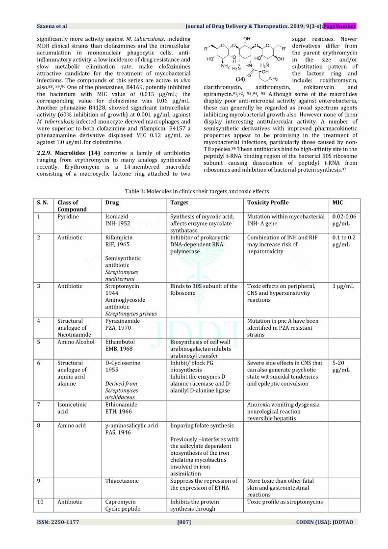
2.2.9. Macrolides (14) comprise a family of antibiotics ranging from erythromycin to many analogs synthesized recently. Erythromycin is a 14-membered macrolide consisting of a macrocyclic lactone ring attached to two
sugar residues. Newer derivatives differ from the parent erythromycin in the size and/or substitution pattern of the lactose ring and include: roxithromycin,
clarithromycin, azithromycin, rokitamycin and spiramycin.91,92, 93,94, 95 Although some of the macrolides display poor anti-microbial activity against enterobacteria, these can generally be regarded as broad spectrum agents inhibiting mycobacterial growth also. However none of them display interesting antitubercular activity. A number of semisynthetic derivatives with improved pharmacokinetic properties appear to be promising in the treatment of mycobacterial infections, particularly those caused by non-TB species.96 These antibiotics bind to high-affinity site in the peptidyl t-RNA binding region of the bacterial 50S ribosome subunit causing dissociation of peptidyl t-RNA from ribosomes and inhibition of bacterial protein synthesis.97
Table 1: Molecules in clinics their targets and toxic effects
S. N. Class of Compound
Drug Target Toxicity Profile MIC
1 Pyridine Isoniazid INH-1952
Synthesis of mycolic acid, affects enzyme mycolate synthatase
Mutation within mycobacterial INH- A gene
0.02-0.06 μg/mL
2 Antibiotic Rifampicin RIF, 1965 Semisynthetic antibiotic Streptomyces mediterrani
Inhibitor of prokaryotic DNA-dependent RNA polymerase
Combination of INH and RIF may increase risk of hepatotoxicity
0.1 to 0.2 μg/mL
3 Antibiotic Streptomycin 1944 Aminoglycoside antibiotic Streptomyces griseus
Binds to 30S subunit of the Ribosome
Toxic effects on peripheral, CNS and hypersensitivity reactions
1 μg/mL
4 Structural analogue of Nicotinamide
Pyrazinamide PZA, 1970
Mutation in pnc A have been identified in PZA resistant strains
5 Amino Alcohol Ethambutol EMB, 1968
Biosynthesis of cell wall arabinogalactan inhibits arabinosyl transfer
6 Structural analogue of amino acid -alanine
D-Cycloserine 1955 Derived from Streptomyces orchidaceus
Inhibit/ block PG biosynthesis Inhibit the enzymes D-alanine racemase and D-alanilyl D-alanine ligase
Severe side effects in CNS that can also generate psychotic state wit suicidal tendencies and epileptic convulsion
5-20 μg/mL
7 Isonicotinic acid
Ethionamide ETH, 1966
Anorexia vomiting dysgeusia neurological reaction reversible hepatitis
8 Amino acid p-aminosalicylic acid PAS, 1946
Imparing folate synthesis Previously –interferes with the salicylate dependent biosynthesis of the iron chelating mycobactins involved in iron assimilation
9 Thiacetazone Suppress the repression of the expression of ETHA
More toxic than other fatal skin and gastrointestinal reactions
10 Antibiotic
Capromycin Cyclic peptide
Inhibits the protein synthesis through
Toxic profile as streptomycins
O O O OR
OH
H2NHN
OHHO
OH
R'
NH2
HO
NH2
OHO
H2N
(14)

Saxena et al Journal of Drug Delivery & Therapeutics. 2019; 9(3-s):PageNumber
ISSN: 2250-1177 [808] CODEN (USA): JDDTAO
antibiotic Streptomyces capreolus
modification of ribosomal structures at the 16S rRNA
11 Fluoroquinolines
Ciprofloxacin Ofloxacin Levofloxacin
Inhibition of DNA supercoiling and damage to DNA
1 μg/mL
12 Antibiotic Rifabutin Semi-synthhetic derivative of rifamycin
Blocking the DNA-dependent RNA-polymerase of the bacteria
13 Clofazimine 1959 structural analogues Tetramethyl piperidine Substituted phenazines
Membrane disrupting effect 0.06 μg/mL
14 Macrolides Antibiotic
Erythromycin Roxithromycin Clarithromycin Azithromycin Spiramycin
Inhibit mycobacterial growth
3. ANTITUBERCULARS DERIVED FROM NATURAL PRODUCTS
Naturally occurring pure compounds as well as extracts from higher and lower forms of plants, microorganisms and marine organisms have indicated that inhibitory activity against MTB is widespread in nature. Many compounds isolated using preliminary functional assays have been provided from investigators interested in phytochemical biodiversity. Usually their potential pharmaceutical worth remains unknown since data to show that these compounds are adversely affecting mycobacterial survival mechanisms in humans, or have been derived from medicinal plants, is lacking. (Table: 2)
3.1. & 3.2. Diterpenes continue to be valuable natural sources of antibacterials. Antituberculous agents from West Indian sea whip Pseudopterogorgia elisabethae led to the discovery of two benzoxazole alkaloids, pseudopteroxazole (15) and seco-pseudopteroxazole (16).98 Pseudopteroxazole was found to effect potent inhibitory activity (97%) against M. tuberculosis H37Rv at a concentration of 12.5 µg/mL and seco-pseudopteroxazole inhibited 66% of mycobacterial growth. The potent activity
was attributed, at least in part, to the benzoxazole functionality.
3.3. & 3.4. Two novel serrultane diterpenes having antimycobacterial activity has also been isolated from the West Indian sea whip coral, Pseudopterogorgia elisabethae, namely, erogorgiaene (17) and 7-hydroxyerogorgiaene (18).99 Erogorgiaene induced 96% growth inhibition for M. tuberculosis H37Rv at a concentration of 12.5µg/mL and 7-hydroxyerogorgiaene inhibited 77% of mycobacterial growth at a concentration of 6.25 µg/mL, respectively.
3.5. Novel diterpene, elisapterosin B (19), possessing cage-like elisapterane carbon skeleton, isolated from the hexane solubles of West Indian sea whip Pseudopterogorgia elisabethae Bayer displays strong in vitro antituberculosis activity. The tetracyclic carbon skeleton of the elisapterosins constitutes a new class of C20 rearranged diterpenes. In vitro studies on elisapterosin B showed strong inhibitory activity (79%) against M. tuberculosis H37Rv at a concentration 12.5µg/mL.100,101
H3C CH3
CH3
CH3
H3CH
H
H3C CH3
CH3
CH3
H3CH
H
OH
(17) (18)
NO
H
CH3
H3C CH3
H
H3C
CH3
H
NO
H
CH3
H3C CH3
H
H3C
CH3
H
(15) (16)

Saxena et al Journal of Drug Delivery & Therapeutics. 2019; 9(3-s):PageNumber
ISSN: 2250-1177 [809] CODEN (USA): JDDTAO
Table: 2 Natural products as antituberculars
S.No.
Class of compound
Drug Isolaion MIC
1 Diterpenes Benzoxazole alkaloids Pseudopteroxazole (15)
H37Rv 97% inhibition at 12.5 μg/mL
Pseudopteroxazole (16) H37Rv 66% inhibition at 12.5 μg/mL
2 Serrultane Diterpenens
Erogorgiaene (17) West Indian sea whip coral Pseudopterogorgia elisabethae
H37Rv 97% inhibition at 12.5 μg/mL
7-Hydroxy erogorgiaene (18) H37Rv 77% inhibition at 6.25 μg/mL
3 Diterpene Elisapterosin B (19) West Indian sea whip Pseudopterogorgia elisabethae
4. COMPOUNDS WITH ANTI-TB ACTIVITY IN PRECLINICAL TRIALS
4.1. Fluoroquinolones, recently developed anti-TB compounds are being considered to be better in the treatment of MDR-TB infections. Among them CS-940 (20) and sparfloxacin showed the greatest antimycobacterial activities with inhibition of 50% of all the isolates at the concentrations 0.25-0.5 µg/mL. In vitro antimicrobial activity of CS-940 was tested against 761 clinical isolates.102
4.2. PD 161148 (21) is a third generation fluoroquinolone selected from analysis of structure activity relationship (SAR), having potent activity against M.
tuberculosis. It was observed that a C-8 methoxyl group enhances the bactericidal activity of quinolones with N1-cyclopropyl substitution. 103 However, there is no information on the frequent side effects of fluoroquinolones such as complexation with metallic ions, phototoxicity and P450 inhibition nor on its rarer but life-threatening side-effects such as renal, hepatic and cardiac toxicity. PD 161148 and CS-940 were synthesized by Parke-Davis (originally but now Warner-Lambert) and Sankyo respectively. Both PD 161148 and CS-940 are amongst the most active of all third-generation quinolones. But unfortunately there are no in vivo tuberculosis data in the public domain which would allow the two compounds to be further separated.
4.3. Calanolide A, (22) is a naturally occurring pyranocoumarin104 of considerable interest because of its dual activity against TB and HIV infections.105, 106 The compound, an inhibitor of HIV-1 reverse transcriptase,107 is
being progressed by Sarawak Medichem Pharmaceuticals and is in phase I/II developmental stage for the HIV indication. It also displays good in vitro activity towards M. tuberculosis. (+)-Calanolide A and (-)-Calanolide A demonstrated inhibitory antimycobacterial activity against M. tuberculosis H37Rv with 96 and 98% growth inhibition, respectively. The actual MIC for (+)-calanolide A was found to be 3.13 µg/mL. In a preliminary assessment of its activity, calanolide A was comparable to the positive control isoniazid and remained effective against rifampin- and streptomycin-resistant TB strains.105 Sarawak has developed a process for the large scale synthesis of calanolide A thus reducing the dependency upon obtaining the material from scarce natural sources.105 Other analogues have been obtained either from plant extracts or by synthesis108 and some have been patented for their antimycobacterial properties.109
4.4. In addition, Calanolide B, (23) which unlike calanolide A, is readily available in substantial quantities from renewable natural sources, e.g. from calophyllum seed oil,110 is claimed to have a similar spectrum of activity to calanolide A against mycobacteria and may be a more cost-effective treatment.111
4.5. The Ciba-Geigy 5-nitroimidazole derivative CGI 17341 (24) showed considerable potential for the treatment of tuberculosis in preclinical studies. In vitro, at 0.04 to 0.3 µg/mL, the compound inhibited both drug-susceptible and
multidrug-resistant strains of M. tuberculosis and showed no cross resistance with isoniazid, rifampicin, streptomycin or ethambutol. Against M. tuberculosis in vitro, its activity was comparable to that of isoniazid and rifampicin and superior to streptomycin, ciprofloxacin,
norfloxacin and the oxazolidinone DuP 721. In M. tuberculosis - infected mice, oral treatment with CGI 17341 on days 11 and 12 post infection resulted in an ED50 of 7.7 mg/kg and a significant dose-dependent increase in survival time.112
4.6. Poloxamer 315 (CRL-1072) (25) is a methyloxirane surfactant polymer from the CytRx corporation that appears to disrupt the cell membranes of microbes or their intracellular components. The highly purified polymer has been shown to be active against both M. tuberculosis113 and M. avium.114 In vitro studies against M. tuberculosis in broth culture show MIC values of 3.1-6.2 µg/mL whilst, in a macrophage assay, these drop to 0.92 to 1.25 µg/mL.115 The compound was active against strains of M. tuberculosis resistant to isoniazid, streptomycin and

Saxena et al Journal of Drug Delivery & Therapeutics. 2019; 9(3-s):PageNumber
ISSN: 2250-1177 [810] CODEN (USA): JDDTAO
rifampin. In vivo, 2 mg/kg/day of 315 administered intravenously three times a week for three weeks allowed survival of M. tuberculosis infected mice and reduced CFU (colony forming unit) counts in lungs and spleens by one to two log units. An IND (investigational new drug) has been obtained for this compound in the USA but as yet no plans have been made to progress the compound clinically.113
4.7. The oxazolidones are a promising new class of synthetic antimicrobial agents with a unique mechanism of action in inhibiting protein synthesis.116 One compound, linezolid, has already reached the market place and other members of this class are in varying stages of development.117 The oxazolidones have activity against Mycobacteriun tuberculosis with linezolid (U-100766) (26) inhibiting multi-resistant isolates in vitro at 2 µg/mL.118 This compound is amongst the very few fully artificial antibacterials targeting the 50S ribosomal subunit.119, 120,121,122 However, the long term use of linezolid may be plagued with forbidding side effects.123,124
4.8. PNU-100480 (27) an oxazolidone containing thiomorpholine moiety was active against M. tuberculosis
with MICs of 0.125 µg/mL.125 The Pharmacia Upjohn compound PNU-100480, was tested in a murine model against ten viable strains of M. tuberculosis in comparison to linezolid and isoniazid. When treatment was started one-day post infection and the compounds given by gavage for four weeks, PNU-100480 proved comparable
to isoniazid and more active than linezolid.126
4.9. B4157127 (28) is phenazinamine derivative, closely related to the antileprosy drug clofazimine, which has been investigated at the University of Illinois as a
potential treatment of tuberculosis.128 In vitro B4157 was tested against 20 strains of M. tuberculosis, including 16 drug-resistant strains and all were found to be susceptible to B4157. The MIC of B4157 at which 90% of strains were inhibited were 0.12 μg/mL. In addition it prevented mortality and caused significant reduction of CFUs in the lungs and spleens. The animals treated with B4157 showed less pigmentation than those receiving clofazimine.
4.10. Miconazole (29) is a well-established antifungal agent which has been reported to have anti-TB activity in vitro (MIC 2 µg/mL against M. tuberculosis H37Ra). The strength of the compound is that, as well as inhibiting
replicating bacteria, it also has some effect on stationary phase bacilli129 Unfortunately, miconazole is not orally active and hence is of little further interest for progressing further. However, there are many 2nd and 3rd generation anti-fungal azoles which are clinically efficacious by the oral route, including the anti-fungal fluconazole which is much used in AIDS patients.130
4.11. The anthelmintic drug niclosamide (30) was found to have anti-TB activity in vitro (MIC 0.5-1 µg/mL) against M. tuberculosis H37Ra. Besides being active against growing cells, it has the interesting property of acting against stationary phase non-replicating bacterial cells.129 However, although niclosamide has been extremely useful for the treatment of human tapeworm infections, it is not absorbed to any significant extent from the intestine. This explains why no systemic pharmacological effects are observed although the compound has been reported to be mutagenic in vitro and to have effects on sperm morphology in animals.131 This pharmacokinetic profile, along with its mutagenic capability, considerably blights the potential of this compound for TB chemotherapy.
4.12. Rifamycin derivative CGP 7040 (31), has been compared in vitro to rifampicin against rifampicin-sensitive and rifampicin-resistant strains of M. tuberculosis and M. avium / intracellulare / scrofulaceum (MAIS) complex. The compounds had MICs 4 to 8 times lower than those of rifampicin against sensitive M. tuberculosis strains.132 Overall CGP7040 was more active than rifabutin and rifampicin against M. avium and was superior to rifampicin towards M. tuberculosis. In addition it was found to be considerably more stable than rifampicin.133 However, preclinical studies in collaboration with the University of Colorado were terminated and the compound is not available for licensing.134

Saxena et al Journal of Drug Delivery & Therapeutics. 2019; 9(3-s):PageNumber
ISSN: 2250-1177 [811] CODEN (USA): JDDTAO
Table: 3 Compounds with anti-TB activity in preclinical trials
S. No.
Class of compound Molecule S. N. Class of compound Molecule
1 Fluoroquinolones CS-940 (21) 8 Oxazolidones Linezolid (U-100766) (27)
2 IIIrd generation fluoroquinolone PD 161148 (22)
9 Oxazolidones PNU-100480 (28)
3 Pyranocoumarin Calanolide A, (23) 10 Phenazinamine B4157129 (29) 4 Pyranocoumarin Calanolide B, (24) 11 Imidazole
5 5-nitroimidazole CGI 17341 (25) 12 Amide Niclosamide (31) 6 5-nitroimidazole PA 824 (26), 13 Rifamycin derivative CGP 7040 (32) 7 Methyloxirane surfactant
polymer Poloxamer 315 (CRL-1072)
5. COMPOUNDS WITH ANTI-TB ACTIVITY IN CLINICAL TRIALS
Regardless of the severe occurrence of TB along with the rise of MDR strains, progression to discover a new vaccine or improvement of current BCG vaccine has been very slow.135 In recent drug discovery program there are several new drug candidates developed, which have arrived at early stages of clinical trials (Table 4).20 Among many active profiles discovered only a couple of the compounds are in phase I or II clinical trials and few of the compounds have entered phase III clinical trials. Following discussion will provide the detailed information of each clinical candidate.
5.1. The Baeyer quinolone moxifloxacin (BAY12-8039)136 (32) is the newest member of this 4th generation
class of antibiotic progress through the clinical pipeline. It has recently been launched in Germany (1999) for the treatment of respiratory tract infections.137 The drug has been shown to be active against M. tuberculosis in vitro and in
vivo in various test systems.138, 139 Against M. tuberculosis CSU93, a highly virulent, recently isolated clinical strain, the MIC of moxifloxacin was 0.25 µg/mL. Oral administration of drug to mice at 100 mg/kg produced peak serum concentrations of 7.8 µg/mL within 15 minutes of dosing. On this basis, mice were infected with a sublethal inoculum of M. tuberculosis CSU93 and then treated with moxifloxacin at 100 mg/kg per day for 8 weeks. This resulted in a significant decrease in the log 10 CFU counts in the organs of treated, compared to untreated, mice – 0.6 + 0.2 versus 5.65 + 0.3 in the lungs and 1.5 + 0.7 versus 4.9 + 0.5 in the spleens, respectively (p < 0.001 in both organs).140 In other studies in M. tuberculosis H37Rv-infected mice, orally administered moxidectin at 100 mg/kg/d given six times weekly was as bactericidal as isoniazid at 25 mg/kg over a similar dosing schedule.138 It was also demonstrated that 8 weeks of treatment with moxifloxacin (100 mg/kg/d) or with moxifloxacin plus isoniazid (100 mg/kg and 25 mg/kg, respectively per day) sterilized the lungs in seven of eight and eight of eight mice, respectively. Furthermore, the elimination half life of the drug in man, mean value 12 hours140 (compare isoniazid – 1 to 2 hours) supports the possibility of once-a-day treatment.
5.2. The Daiichi quinolone Sitafloxacin (DU-6859a)141
(33) is in Phase III trials in both Japan and the USA.142 The compound has outstanding activity against a broad range of bacteria. When compared to a number of other quinolones – ciprofloxacin, levofloxacin, gatifloxacin and moxifloxacin – sitafloxacin was the most active against 3344 gram-positive cocci and 406 anaerobes, and against
5046 gram-negative bacteria it either equalled or bettered clinafloxacin.143 The mechanistic basis for this potency is believed to reside with sitafloxacin’s ability to equally inhibit both DNA gyrase and topoisomerase IV, and its IC50s against these enzymes were amongst the lowest of the quinolones.144 Sitafloxacin was equipotent with gatifloxacin and sparfloxacin, and more active than levofloxacin and ofloxacin, when tested against M. tuberculosis - MICs at which 90% of strains of M. tuberculosis inhibited (MIC90s) were 0.2 µg/mL.145, 146 No data appear to have been published for the activity of the compound against M. tuberculosis in vivo. In clinical studies of sitafloxacin in healthy volunteers oral bioavailability was at least 70%; it was well tolerated with no serious adverse effects and the elimination half life was 4.4 to 5 hours. A study in which the postantibiotic effect was determined for sitafloxacin (and several other quinolones) against various bacteria concluded that this factor would permit dosing on a once every 24 hours basis.147
5.3. Gemifloxacin (34) is another quinolone in the late stages of development and it is being progressed by SmithKline Beecham on license from LG Chem. Following various Phase III trials, it has now been submitted for approval in the USA for the treatment of respiratory infections.148, 149 In healthy volunteers,
oral bioavailability is approximately 70%, it is well tolerated and has a mean elimination half life of 7.4 hours. These characteristics, coupled with its potent antibacterial activity suggest its suitability for a once-daily dosing regimen.150 Data on the anti-TB potential of gemifloxacin appear to be limited to a report comparing it activity to five other quinolones against 250 clinical isolates of M. tuberculosis susceptible or resistant to first-line antituberculosis drugs.

Saxena et al Journal of Drug Delivery & Therapeutics. 2019; 9(3-s):PageNumber
ISSN: 2250-1177 [812] CODEN (USA): JDDTAO
In these assays, gemifloxacin showed a MIC90 value of 8 µg/mL, compared with 1 µg/mL for levofloxacin, trovafloxacin and grepafloxacin.151
5.4. T-3811ME (35) is unique amongst other broad-spectrum quinolones as it lacks the presence of a fluorine atom at the 6-position of the ring. But shows similar broad spectrum and potent activity against
bacteria as the best of the fluoroquinolones.152 Against ten strains of M. tuberculosis, T-3811ME has an MIC90 value of 0.0625 µg/mL – comparable with ciprofloxacin and levofloxacin, and more active than trovafloxacin. No other TB relevant data appear to have been published but the compound is being advanced for other bacterial indications. It is being progressed in the USA in collaboration with BristolMeyers Squib, with Phase II/III trials shortly to commence.153
Rifametane (SPA-S-565) (36) is a new semi-synthetic rifamycin being progressed by Societa Prodotti Antibiotici (SPA), Milan, Italy. It has a bactericidal spectrum and potency similar to that of rifampicin, but with much better pharmacokinetic properties.154 This is reflected in the fact that, although the MIC90 values of the two compounds were the same against 20 strains of M. tuberculosis, in TB-infected mice rifametane proved to be the more effective orally.155 In
the healthy male volunteers, the pharmacokinetic and safety of a 300 mg single oral dose of rifametane were compared to a 300 mg dose of rifampicin.156 The elimination half life for rifametane was 10.58 hours compared with 1.89 hours for rifampicin, and the mean residence time was 10.58 hours for rifametane and 3.93 hours for rifampicin, and the mean residence time was 18.05 hours for rifametane and 3.93 hours for rifampicin. In another Phase I trial carried out in collaboration with Glaxo India, a single oral dose of 150 mg was administered and the half-life and area under curve (AUC) were some 6 or 7-fold that of rifampicin. Encouragingly, serum drug levels above the MIC for M. tuberculosis were maintained for up to 48 hours after drug administration.157 Currently SPA are collaborating with Glaxo India in preparing to advance rifametane into Phase II trials.158
5.5. Rifalazil KRM-1648, 3’-hydroxy-5’-(4-isobutyl-1-piperazinyl)benzoxazinorifamycin159 (37) was found to be more potent than RIF and shortens the duration of treatment.160, 161, 162 It was synthesized at the Kaneka Corporation and shown to have superior activity to rifampicin against M. tuberculosis in vitro and in vivo.163 The compound was progressed clinically in association with PathoGenesis through Phase I trials in the USA, and into a Phase II trial in Brazil using pulmonary TB patients.164,165 However, due to severe side-effects in the four-day Phase II trial, the development of rifalazil has been terminated.166
5.6. The anti-mycobacterial activity of liposome-encapsulated drug, MiKasome (38), has been found to be effective against M. avium infections in vitro and in animal models.167 In preclinical studies,168 48 hr after delivery to the lungs via liposome, over half of the antibiotic remained in this tissue. In animals, pharmacokinetic data showed that MiKasome produced 7-fold higher peak plasma levels compared to free drug (amikacin) administered intravenously. Additionally, the AUC was 150-fold higher with the liposomal material and a single dose of liposomal amikacin produced therapeutic levels of antibiotic for more
than 72 hr.168 In preliminary clinical study, a patient with tuberculosis treated with MiKasome for 49 days exhibited negative culture.169 Pilot Phase II studies showed that MiKasome was able to resolve M. tuberculosis infections in adults and children who failed conventional therapies.
5.7. Aconiazide (39) a prodrug of isoniazid which was designed to be less toxic than the parent drug. The latter is metabolized to hydrazine and acetylhydrazine, which have both been implicated in the toxicity of isoniazid. Because
aconiazide is converted to isoniazid and 2-formylphenoxy acetic acid, it was expected that the acid would bind to the isoniazid metabolites and so lower toxicity.170 This proved to be the case and aconiazide is indeed less
toxic than the parent drugs and lacks carcinogenicity.170 In healthy patients it was found to produce proportionately lower levels of isoniazid in serum than the parent molecule itself.171 For sometime, further progression of the compound was delayed due to problems with its commercial synthesis. However, additional toxicology data are now being submitted to the FDA by Lincoln Diagnostics in order to conduct clinical trials in tuberculosis patients, and the compound has been granted Orphan Drug status in the US.172
5.8. SRL 172 (40) is a preparation containing heat-killed Mycobacterium vaccae and comprises Th1 adjuvant with bacterial antigens to induce host protective immunity. Extensive clinical studies on this immunomodulator are being conducted by SR Pharma for various indications including tuberculosis. In one study in Argentina it was added to routine chemotherapy in the treatment of pulmonary tuberculosis.173 Patients receiving SRL 172 (plus drugs) were found to have reduced sputum smear positivity

Saxena et al Journal of Drug Delivery & Therapeutics. 2019; 9(3-s):PageNumber
ISSN: 2250-1177 [813] CODEN (USA): JDDTAO
of AFB, increased weight gain and shortened time to becoming apyrexial, compared to drug treatment alone. It was concluded that the results indicated a switch to Th1 immunological status and improved clinical status. Another study in 100 cases with relapsed or chronic tuberculosis showed SRL 172 to keep 97% of patients disease-free six months after treatment had finished, compared with 70% who received placebo. In other trials in tuberculosis patients, SRL 172 showed little difference, if any to those
receiving placebo.174
5.9. A diarylquinoline R207910 (41) has been identified as a potent anti-TB compound that inhibits both drug sensitive and drug resistant M. tuberculosis in vitro having
MIC 0.06 µg/mL.175 The investigation picked up this compound from the 20 interesting drug candidates which were identified after optimization by synthetic chemistry.176
5.10. Sudoterb (LL-3858) (42) belongs to a class of
compounds as pyrroles which are plant alkaloids. Sudoterb has been reported to have potent anti-TB activity in vitro and in vivo. In vitro sudoterb has bactericidal activity similar to isoniazid and is synergistic with rifampin. The combination of sudoterb with isoniazid, rifampin and pyrazinamide led to complete sterilization of both sensitive and MDR-TB strains in mice within two months and in combination with rifampin and pyrazinamide cured TB in all animals after three months. Sudoterb exhibits good oral bioavailability with once daily dosing. LL-4858 is the combination of LL-3858, isoniazid, rifampin and pyrazinamide.177
5.11. One compound PA 824 (43), displayed MIC values ranging from 0.015 to 0.25 µg/mL against cultured replicating M. tuberculosis pan sensitive and rifampin mono-
resistant clinical isolates. It showed impressive efficacy when administered orally to both M. tuberculosis – infected mice and (25mg/kg) and guinea pigs (40 mg/kg), and was comparable to isoniazid at 25 mg/kg.178 In addition, it displayed low levels of toxicity with an cute toxic threshold value of >500 mg/kg daily for 28 days. A bioreductive activation of PA 824 by a combination of the
low redox potential F420-dependent glucose 6-phosphate dehydrogenase and a previously unstudied protein (Rv3547) acting as the electron transfer mediator has been suggested.179
5.12. Another nitroimidazole OPC67683 (Delamanid) (44) has successfully finished its phase II clinical studies and it demonstrates excellent in vitro activity for both resistant and nonresistant drug strains of M. tb in addition to this there is no cross-resistance to the first-line antitubercular drugs, consequently occasional and low dosing of the compound may be effective.180,181 This molecule has already completed a placebo-controlled phase II trial further the safety and pharmacokinetics properties in MDR refractive TB have also been done. 182, 183
5.13. TMC207 (45) is bactericidal and it is tolerated very well during its initial clinical studies.60 Based on the promising phase I clinical trials, the compound TMC207 has entered into the phase II clinical trials.184 Tibotec Medicinal Compound 207 (TMC207) has emerged as a lead molecule out of this work and currently this compound is under phase II clinical assessment (Fig. 2). Detailed mechanistic study revealed that oligomeric (F ATPase) and proteolipic (V ATPase) subunit c of ATP synthase of mycobacteria is the target of this compound.79, 80 TMC207 is effective for resistant and nonresistant strains of M. tb at MIC 0.03 μg/mL.79 The results of its clinical trials show that TMC207 may shorten the treatment of TB and be effective in its treatment.185
5.14. Quinolones have been classified as first generation (nalidixic acid, oxolinic acid, and cinoxacin), second generation (ciprofloxacin, ofloxacin, enoxacin, lomefloxacin, and norfloxacin), third generation gatifloxacin, sparfloxacin, and levofloxacin), and fourth generation (moxifloxacin and trovafloxacin).186

Saxena et al Journal of Drug Delivery & Therapeutics. 2019; 9(3-s):PageNumber
ISSN: 2250-1177 [814] CODEN (USA): JDDTAO
Table: 4 Compounds with anti-TB activity in clinical trials
S. No.
Class of Compound
Name of Active agent Target Stage of Development Funded agency
1 4th generation class of antibiotic
Baeyer quinolone moxifloxacin (BAY12-8039
MIC of moxifloxacin was 0.25 µg/mL.
III Global TB Alliance/ Bayer
2 Fluoroquinolone
Gatifloxacin M. tuberculosis DNA topoisomerase II inhibitor
III EU/ TDR
3 Quinolone Sitafloxacin (DU-6859a) inhibit both DNA gyrase and topoisomerase IV
(MIC90s) were 0.2 µg/mL
Phase III trials Daiichi
4 Quinolone Gemifloxacin MIC90 value of 8 µg/mL submitted for approval in the USA for the treatment of respiratory infections
SmithKline Beecham on license from LG Chem
5 Quinolones T-3811ME MIC90 value of 0.0625 µg/mL
Phase II/III trials BristolMeyers Squib
6 Semi-synthetic Rifamycin
Rifametane (SPA-S-565) Phase II Societa Prodotti Antibiotici (SPA), Milan, Italy
7 Rifalazil KRM-1648, 3’-hydroxy-5’-(4-isobutyl-1-piperazinyl)benzoxazinorifamycin160
due to severe side-effects in the four-day Phase II trial, the development of rifalazil has been terminated
PathoGenesis
8 MiKasome Phase II 9 Prodrug of
isoniazid Aconiazide Orphan Drug status in
the US
10 SRL 172 SR Pharma 11 Diarylquinoline R207910 12 Pyrrole
plant alkaloids Sudoterb (LL-3858)
13 Nitroimidazoles OPC67683 Mycolic acid synthesis inhibitor (prodrug and requires activation
III Otsuka
14 Nitroimidazoles PA824 Protein synthesis Inhibition of Lipids
II Global TB Alliance
15 Diarylquinoline TMC207 ATP synthase subunit c proton pump
II Tibotec J & J
16 Ethylene diamine
SQ109 Involved in cell wall synthesis inhibition
II Scquella
6. NEW ENTITIES DISCOVERED AND BIOEVALUATED FOR ATI-TUBERCULAR ACTIVITY
Many different class of compounds are under inestigation for their bioactive potetial against antibacterial, specially anti-TB activity. Structural optimization of many diverge
range of derivatives of following compounds (Table 5) by various groups across the globe has led to the identification and successfully synthesized for potent anti-TB activity profiling some of the compounds exhibiting significant activity against drug-sensitive strains of M. tb. Following are the major classes identified for the drug discovery venture.
Table: 5 New entities discovered and bioevaluated for ati-tubercular activity
Quinoline derivatives Fluoroquinolone derivetives
Diamine derivatives
Pyrimidine derivatives
Quinone derivatives
Nitroimidazole derivative
Terpenoids derivatives
Purine derivatives
Quinolone derivatives
Oxazolidinones
Isonicotinyl derivatives
Sugar hybrids of aromatic and heteroaromatic aminoesters

Saxena et al Journal of Drug Delivery & Therapeutics. 2019; 9(3-s):802-819
ISSN: 2250-1177 [815] CODEN (USA): JDDTAO
7. DISEASE BURDEN AND THE JOB AHEAD
Rising trends in the cases of tuberculosis infection have prompted a search for newer, more effective, less toxic drugs against drug-susceptible and resistant strains of tuberculosis. Despite most patients’ clearing their sputum of live bacteria two months after they are placed on oral therapy, the full six-month course is required to prevent relapse with active disease after therapy is discontinued.187, 188 Decades of poor compliance with such a prolonged and complex regimen has had two unfortunate consequences: first, treatment of the disease is often unsuccessful, and tuberculosis continues to spread and cause immense mortality; and second, there is an expanding epidemic of drug resistance that threatens TB control programs worldwide.189, 190 Drug resistancein detection in such cases is not only challenging but time taking too – the method can take up to 35 days–and the wait means patients can be exposed to suboptimal treatment, and this ends up with MDR-TB. This is the major setback in controlling tuberculosis. Currently, at least 50 million people are estimated to be affected with MDR-TB. A little MDR strains of M. tuberculosis were establish as the resistant for several first-line chemotherapeutic agents as well as for few of the second-line drugs.191 Moreover, the high rate of coinfection with human immunodeficiency virus (HIV) presented a challenge to the existing chemotherapies.
The ability of mycobacteria to survive in the environment and to persist in the host cell for prelong period of time makes it a successful pathogen. The cell-wall guards the bacteria and helps to promote their intracellular perseverance by inhibiting phagosome–lysosome fusion, keeping them sequestered away from terminal endocytic organelles.192 Latency too is an significant facet of TB pathogenesis. Mycobacteria release peripheral cell-wall lipids into their host cells, which induces the formation of granulomas that wall off the infection and limit its spread while restricting macrophage killing of the bacteria.193
One such approach is the development of optimized compounds that are active against current targets. This strategy requires in-depth knowledge about the mode of action of the drug so that appropriate modifications in structure could lead to new active compounds. The second approach is based on the identification of newer targets, a strategy that would overcome the problem of multidrug resistance and offer effective control of drug-sensitive cases of tuberculosis as well. The basic requirement of the third strategy is to identify a crucial metabolic step whose interruption at any stage would make survival of the parasite very difficult. For example, enzymes involved in the cell wall biosynthesis pathway could provide a novel target for future drug discovery.
Another potential route to decreasing the length of treatment would be to improve the potency of the currently used anti-tuberculosis agents. Such a strategy would allow higher effective dosing of patients and could thereby improve the therapeutic effect of the agent by maintaining the drug concentration above the MIC for longer periods of time or by enhancing the ratio of the peak concentration to the MIC. Drugs which affect the cell wall of the bacteria are known to have concentration-dependent cidal effects in vitro that may be achievable in vivo with enhanced potency.
Since few years, at hand a remarkable progress is observed in understanding the biochemistry of M. tuberculosis, the disease progression, and the process by which a drug resistance mechanism works, and lots more in ascertaining the value of DOTS strategy in preventing treatment malfunctioning . Study in China has shown that cure rates as
high as 95% can be achieved through DOTS implementation194 but treatment is labor intensive, making it difficult to deliver unless substantial infrastructure is in place. However, scarcity of appropriate efforts to develop new efficiently active chemical entities or rapid diagnostic procedures, and their significance to the global TB control is a point to be questioned.195
Therefore, there is a serious requirement for the development of novel drugs or chemical entities that can act alongside of both i.e. actively growing and dormant bacteria with novel mode of action. The efforts for drug development are being coordinated by Global Alliance for TB Drug Development (www.tballiance.org), an organization which is involved in developing public-private partnerships since a long time to fetch out new faster-acting and reasonably priced drugs effective as antituberculars. The Alliance launched in 2000, aims to conquer the natural barriers to TB drug development procedure. Through working in joint venture with miscellaneous organizations such as academic institutions, government research organisations, non-governmental bodies, the pharmaceutical manufacturing units and contract research companies, the GATB is supposed to plug gaps in the R&D pipeline. The efforts and policy will also encourage TB drug development by providing a outline in which the various elements of the process may be brought together.
Rational development of a novel antitubercular agent actually calls for the exploration of new means to understanding of the genetics and physiology of M. tuberculosis. In this stare, the accessibility of the genome sequence of M. tuberculosis196 and potent genetic tools for manipulating mycobacteria have facilitated the valuable information about the potential targets. In addition to this, the information presented from X-ray crystallographic studies of numerous of these targets has assist for designing novel chemotherapeutic representative.
Our research group has explored the chemistry of various heterocycles and their sugar hybrids and receieved overwhelming response in this direction, though many obstacle The present state of knowledge provided vivid advances in understanding the biology, detailed biochemistry of the causing agent and intracellular lifestyle of mycobacterium, this precious wealth of information is supportive to undertake different targets for synthesizing new chemical entities which will be effective chemotherapeutics in drug development program against MDR-TB. The new authenticated and selective targets, attuned to high throughput assays against all forms of TB should be designed and synthesised for drug vulnerability testing, baseline screening (to discover new chemical leads), early drug resistance, and to direct retreatment plans for therapy. To get an early lead for developing new chemotherapeutics, idea of combinatorial and virtual libraries may prove very helpful, further in silico screening of the compounds must be on top priorities.13, 197
Specifically, there are four areas where this extensive collaboration can supplementary advanced in moderanizing TB treatment ; in order to achieve the best results firstly, identifying new chemical candidates – the TB pipeline needs to be cherished and reinforced with additional promising chemical entities. Identifying and validating new drug targets, predominantly those related with the persistent state of Mycobacterium tuberculosis, is vital to screen and select novel compounds for development. Such compounds may already exist in libraries or may come from basic research discovery. Secondly, expanding clinical trial capacity – there is an urgent need to expand and improve TB

Saxena et al Journal of Drug Delivery & Therapeutics. 2019; 9(3-s):PageNumber
ISSN: 2250-1177 [816] CODEN (USA): JDDTAO
drug clinical trial capacity to evaluate new multi-drug regimens. The recent commitment by the European Union to fund clinical trial networks for AIDS, TB and malaria is an example of how donor countries can contribute, and endemic countries can work side-by-side with investigators to prepare and sustain the infrastructure necessary for ongoing clinical trials. Thirdly, advancing new technologies – the successful use of surrogate markers in the evaluation of HIV/AIDS therapeutics has demonstrated that valid surrogate and biomarkers can significantly reduce the length of clinical drug trials. Such markers must be developed for TB. Fourthly, ensuring regulatory harmonization – to avoid delays in the approval of new therapies and to accelerate the adoption of new TB drug regimens, TB-specific regulatory guidelines and their global harmonization are essential.
Without new medicines, TB will only grow as a global threat, driven by its deadly synergy with HIV/AIDS, complicated by multi-drug resistant strains, and amplified by the consequences of poverty. When introduced alongside other advancements, such as diagnostics and vaccines, new drugs will expand the scope of current TB control and redefine public health targets.198,199, 200
8. THE FUTURE OPPORTUNITY OF TB DRUG DEVELOPMENT
The current regimens for treatment of drug sensitive TB are greatly uncomplicated when compared with the previously deviced TB treatment plan. It has been shortened from two years to six months but it still required a optimal regimen. The prolong exposure of a patient with immune suppressive condition cause severe harmful effects thus there is an urgent need to develop a two to three month regimen, the regimen must include once weekly dosing of three to four drugs. this can alter the results and will reduce the duration of treatment from 28 weeks to 8-12 to twelve weeks, moreover approximately 130 doses of a combination regimen will drop to 10 doses. This modification will appreciably pose a positive impact on treatment by improving patient adherence, and on development of drug resistance via improving treatment completion rates.201, 202
Nanotechnology offers a unique opportunity in drug delivery system. Out of two major approaches the nanoparticle based technique is seems to be most promising approach.203, 204, 205, In fact Nanoparticles tool emerged as valuable means in drug delivery owe to their high stability, viability of incorporation of both hydrophilic and hydrophobic components. The simplicity of the process involves parenteral administration in addition to oral and inhalational administration capability or probability. The competence of the treatment also proves when complete bacterial clearance from the organs is observed in M. tuberculosis infected mice treated with nanoparticles bound drugs. Free drugs cause bacterial clearance only after
administration of 46 doses daily.206 Nanoparticle based drug delivery is is particularly useful for targeted therapies intending to deliver drugs selectively to intracellular sites, like within macrophages, reticuloendothelial system or in monocytes, or in the areas where dormant persistent organisms accountable for prolonged treatment durations may be lodging.207, 208,209, 210, 211
Liposomal formulations are also a novel delivery systems of TB drugs, but the prospective elasticity with nanoparticles poses superiority over liposome-encapsulated drugs. Another approach includes dry powder inhalers and nebulizers.212, 213 Aerosols of PA-824 based dry porous particle have been tested in low and high dosages which are eight-fold higher in concentration than its oral dose and the dose effects were compared with PA-824 oral administration for the treatment of guinea pigs tuberculosis. The results demonstrate a lower degree of inflammation and tissue damage. therefore this technique has a potential future in the treatment of tuberculosis.214203 In fact second line drug capreomycin formulation being used in the large porous particles form is being tested in human volunteers as an inhalational product, this chemical entity is in a Phase I study.210 Several other first-line drugs and few investigational agents have also been formulated and tested in an inhalational delivery system. Inhalational method distributes a large amount of drug dose to the lung, but the histological localization of increased delivery is yet too investigated.
9. CONCLUSION
In few decades, the investigation, research, and drug delivery approaches for the control of TB in under extensive progress. Loads of work regarding enrichment of the anti- TB activity or in finding new leads particularly active against MDR and XDR i.e. resistant mycobacterium strains is under exploration. All the efforts in this area are overwhelming and welcomed but still there is a need of novel persuasive drug targets and chemical entities which have strong, synergistic and complementary actions against a range of M. tuberculosis subpopulations which not only shorten TB treatment but also have high efficacy and low toxicity and in addition to this must be effective against MDRTB/XDR-TB and without doubt administered in concurrence with HIV.
10. ACKNOWLEDGEMENTS
N. Saxena acknowledges to the University Grant Commission, New Delhi, India for providing DSK Fellowship and in part, the University of Delhi, Delhi, India for the assistance and providing important information in the form of research articles. N. Srivastava is thankful to the Sharda University, Greater Noida, India for providing essential support in making of this article.
11. REFERENCES
1. World Health Organization 2004. Fact sheet no. 104, revised March 2000 World Health Organization, Geneva, Switzerland. 2. Nakata, K., Honda Y., Tanaka N., Weiden M., Keicho N., Kekkaku, 2000, 75, 547- 556. 3. Williams, B. G., C. Dye, Science, 2003, 301, 1535-1537. 4. Corbett, E. L., Watt C. J., Catherine J., Walker N., Maher D., Williams B. G., Raviglione M. C., Dye C., Arch. Intern. Med., 2003, 163, 1009-1021. 5. Septkowitz A., Raffalli J., Riley L., Kiehn T. E., Armstrong D., Clin. Microbiol. Rev., 1995, 180-199. 6. World Health Organization. 2004. Global tuberculosis control: surveillance, planning, financing. W.H.O. report 2004. World Health
Organization, Geneva, Switzerland. 7. World Health Organization. 2004. Stop TB annual report. W.H.O. report W.H.O./ CDS /STB/ 2002.17. W.H.O. report 2004. World Health
Organization, Geneva, Switzerland. 8. Davies P.D., Arch. Chest Dis., 1999, 54, 168-171. 9. Gandy M., Zumbla A., Soc. Sci. Med., 2002, 55, 385-396. 10. Blanchard J.S., Annu. Rev. Biochem., 1996, 65, 215-239.

Saxena et al Journal of Drug Delivery & Therapeutics. 2019; 9(3-s):PageNumber
ISSN: 2250-1177 [817] CODEN (USA): JDDTAO
11. Duncan K., Barry C. E. III, Curr Opin. Microbiol., 2004, 7, 1-6. 12. Brennan P. J., Nikaido H., Annu. Rev. Biochem. 1995, 64, 29. 13. Cole S. T., Brosch R., Parkhill J., Garnier T., Churcher C., et al, Nature, 1998, 393, 537; Erratum in Nature, 1998, 396, 190. 14. Tomasz A., New. Eng. J. Med., 1994, 330, 1247. 15. Mazodier P., Davies J., Annu. Rev. Genet., 1991, 25, 147. 16. Courvalin P., J. Antimicrob. Chemother., 1996, 37, 855. 17. Webb V., Davies J., Antimicrob. Agents Chemother., 1993, 37, 2379. 18. Center for disease Control, MMWR weekly, 2006, 55 (11): 301-305. 19. Md J. F., Lancet, 2006, 368, 954-964. 20. Marris E., Nature, 2006, 443, 131. 21. World Health Organization, 2006, Fact sheet no., 10-21. 22. McGregor S., Reuters, 2006, 9-17. 23. Kaufmann S. H., Nat. Rev. Immunol. 2001, 1(1), 20–30. 24. Fischer K., Scotet E., Niemeyer M., Koebernick H., Zerrahn J., Maillet S., et al. Proc. Natl. Acad. Sci. USA., 2004, 101(29), 10685–90. 25. Kaufmann S.H., Proc. Natl. Acad. Sci. USA., 1996, 93(6), 2272–2279. 26. Maini R., St Clair E.W., Breedveld F., Furst D., Kalden J., Weisman M., et al. Lancet, 1999, 354(9194), 1932–9. 27. Flynn C. J., J. Clin. Immunol., 2004,110(1), 2–12. 28. Smith K.C., Starke J.R., Bacille Calmette-Gu´erin Vaccine. In: Plokin SA, Orenstein WA, editors. Vaccines. 4th ed. Philadelphia: Saunders, 2004. 29. Gilman, A. G., Pergamon Press, New York, 1990, 1061-1062. 30. Goldberger M.J., Med. Clin. North Am., 1988, 72, 661-8. 31. Iseman M.D., N. Engl. J. Med., 1993, 329, 784-91. 32. Heym B., Alzari P. M., Honore N., Cole S. T., Mol. Microbiology, 1995, 15, 235- 245. 33. Mdluli K., Slayden R.A., Zhu Y., Ramaswamy S., Pan X., Mead D., Crane DD, Musser J.M., Barry C.E .III., Science, 1998, 280, 1608-16110. 34. Molecular genetics of bacteria: Genetics of drug resistance in M. tuberculosis, Chapter 15, 2000, 235. 35. Youatt J., Am. Rev. Respir. Dis., 1969, 99, 729. 36. (a) Tsukamura M., Mizuno S., Kekkaku, 1962, 37, 29 (b) Tsukumara M., Tsukumara S., Nakano E., Am. Rev. Respir. Dis., 1963, 87, 269. 37. Wimpenny J. W., J. Gen. Microbiol., 1967, 47, 389. 38. Bannerjee A., Dubnau E., Quemard A., Balasubramanian V., Um K. S., Wilson T., Collins D., Lisle G. de, Jacobs W. R., Science, 1994, 263, 227. 39. Parneti F., Lancini G., Rifamycins. In: O’ Grady F, Lambert HP, Finch RG, Greenwood D, editors. Antibiotic and Chemotherapy, 7th edition.
Edinburg: Churchill Livingston, 1997, 453-459. 40. Gale E.F., Cundliffe E., Reynolds P.E., Richmond M.H., Waring M.J., The Molecular Basic of Antibiotic Action , 2nd edition. London: John Wiley
and Sons, 1981. 41. Levin M. E., Hatfull G. F., Mol. Microbiol., 1993, 8, 277. 42. Saito H., Tomioka H., Sato K., Emori M., Yamane T., Yamashita K., Hosoe K., Hidaka T. Antimicrobial Agents Chemother., 1991, 35, 542-547. 43. Blanchard J.S., Annu. Rev. Biochem., 1996, 65, 215-239. 44. Philips I., Shannon K.P., Aminoglycosides and Aminocyclitols. In: O’ Grady F, Lambert HP, Finch RG, Greenwood D, editors. Antibiotic and
Chemotherapy, 7th edition. Edinburgh: Churchill Livingstone, 1997, 164-210. 45. “Burger’s Medicinal Chemistry and Drug Discovery: Therapeutic Agents”, ed. Wolff M. E., Wiley-Interscience, New York, 1996, Vol.2. 46. Finken M., Kirschner P., Meier A., Wrede A., Bottger E. C., Mol. Microbiol. 1993, 9, 1239. 47. McCune R.M., Feldman F.M., Mc Dermott W., Microbial persistence II., J. Exp. Med., 1966, 123, 468-486. 48. Konno K., Feldmann F.M., Mc Dermott W., Am. Rev. Resp. Dis., 1967, 95, 461-469. Heifets L. B., Lindholm-Levy P. J., Am. Rev. Respir. Dis., 1992,
145, 1223. 49. Heifets L. B., Lindholm-Levy P. J., Am. Rev. Respir. Dis., 1992, 145, 1223. 50. Scorpio A., Lindholm-Levy P., Heifets L., Gilman R., Siddiqi S., Cynamon M., Zhang Y., Antimicrob. Agents Chemother., 1997, 41, 540. 51. Scorpio A., Zhang Y., Nat. Med., 1996, 2, 662. 52. Rastogi N., Labrousse V., Antimicrob. Agents Chemother. 1991, 35, 462-470. 53.Inderlied C.B., Barbara-Burnham L., Wu M., Young L.S., Bermudez L.E.M., Antimicrob. Agents Chemother., 1994, 38, 1838-1843. 54. Mikusova K. Slayeden R.A., Besra G.S., Brennan P.J., Antimicrob. Agents Chemother., 1995, 39, 2484-2489. 55. Belanger A. E., Besra G. S., Ford M. E., Mikusova K., Belisle J. T.,. Brennan P. J, Inamine J. M., Proc. Natl. Acad. Sci., 1996, 93, 11919. 56. Otten H., Handbook of Experimental Pharmacology, Vol. 44, Berlin: Springer-Verlog, 1998, 158-166. 57. Gale E..F, Cundliffe E., Reynolds P.E., Richmond M.H., Waring M.J. The Molecular Basic of Antibiotic Action , 2nd edition, London: John Wiley
and Sons, 1981. 58. David H. L., Takayama K., Goldman D. S., Am. Rev. Respir. Dis., 1969, 100, 579–581. 59. Caceres N. E., Harris N. B., Wellehan J. F., Feng Z., Kapur V., Barletta R. G., J. Bacteriol., 1997, 179, 5046–5055. 60. Chacon O., Feng Z., Harris N. B., Caceres N. E., Adams L. G., Barletta R. G. Antimicrob. Agents Chemother., 2002, 46, 47–54. 61. Feng Z., Barletta R. G., Antimicrob. Agents Chemother., 2003, 47, 283–291. 62. Baulard A. R., Betts J. C., Engohang-Ndong, J., Quan S., McAdam R. A., Brennan P. J., Locht C., Besra G. S., J. Biol. Chem., 2000, 275, 28326–
28331. 63. DeBarber A. E., Mdluli K., Bosman M., Bekker L. G., Barry, C. E., 3rd. Proc. Natl. Acad. Sci. U.S.A., 2000, 97, 9677–9682. 64. Vannelli T. A., Dykman A., Ortiz de Montellano P. R., J. Biol. Chem., 2002, 277, 12824–12829. 65. Qian L., Ortiz de Montellano P. R., Chem. Res. Toxicol., 2006, 19, 443–449. 66. Bieder A., Brunel P., Roquet-Ghaye J., Kries B. Rev. Fra. Etud., Clin. Biol. 1966,11, 419–423. 67. Johnston J. P., Kane P. O., Kibby M. R., J. Pharm. Pharmacol., 1967, 19, 1–9. 68. Grunert M., Iwainsky H., Arzneim. Forsch., 1967, 17, 411–415. 69. Prema K., Gopinthan K. P., J. Indian Inst. Sci., 1976, 58, 16–27, Chem. Abstr., 84, 159454. 70. Bonicke R., Beitr. Klin. Erforsch., Tuberk. Lungenkr., 1965, 132, 311–314. 71. Offe H,A., In: Bartmann K, editor. Antituberculosis drugs, Handbook of Experimental Pharmacology, Vol. 84. Berlin: Springer-Verlag, 1988, 1-
30. 72. Trinka L, Misson P., In: Batmann K, editor. Antituberculosis drugs, Handbook of Experimental Pharmacology, Vol.84. Berlin: Springer-Verlag,
1988, 51-68. 73. Brown K. A., Ratledge C., Biochem. Biophys. Acta, 1975, 385, 207–220. 74. Adilakshmi T., Ayling P. D., Ratledge C., J. Bacteriol., 2000, 182, 264–271. 75. Ratledge C., Tuberculosis, 2004, 84, 110–130. 76. Rengarajan J., Sassetti C. M., Naroditskaya V., Sloutsky A., Bloom B. R., Rubin E., J. Mol. Microbiol., 2004, 53, 275–282. 77. Fre´nois F., Baulard A. R., Villeret, V., Tuberculosis, 2006, 86, 110–114. 78. Fre´nois F., Engohang-Ndong, J., Locht C., Baulard A. R., Villeret, V., Mol. Cell, 2004, 16, 301–307. 79. Ferrero L., Cameron B., Manse B., Lagneaux D., Crouzet J., Famechon A., Blanche, Mol. Microbiol., 1994, 13, 641.

Saxena et al Journal of Drug Delivery & Therapeutics. 2019; 9(3-s):PageNumber
ISSN: 2250-1177 [818] CODEN (USA): JDDTAO
80. Miyazaki E., Miyazaki M., Chem J. M., Chaisson R. E., Bishai W. R., Antimicrob. Agents Chemother., 1999, 43, 85. 81. Cardenosa O., Soto-Hernandez J. L., Chemotherapy, 2000, 46, 379. 82. Del’Alamo L., Sampaio J., Miranda E. A., Sader H. S., Braz. J. Infect. Dis., 1999, 3, 215. 83. Gillepsie S. H., Billington O., J. Antimicrob. Chemother., 1999, 44, 393. 84. Baohong J. I., Lounis N., Maslo C., Truffot-Pernot C., Bonnafous P., Grosset J., Antimicrob. Agents Chemother., 1998, 42, 2066. 85. Woodcock J. M., Andrews J. M., Boswell F. J., Brenwald N. P., Wise R., Antimicrob. Agents Chemother., 1997, 41, 101. 86. Adams, L. B., Sinha, I., Franzblau, G. S., Krahenbuhl J.L., Mehta R. T., Antimicrob. Agents Chemother., 1999, 43, 1638–1643. 87. Oliva B., O’Neill A. J., Miller K., Stubbings W., Chopra I. J., Antimicrob. Agents Chemother., 2004, 53, 435–440. 88. Field S.K., Cowie R.L., Chest, 2003, 124(4), 1482-1786. 89. Reddy V.M., Srinivasan S., Gangadharam P.R., Tuber. Lung Dis., 1994, 75(3), 208-212. 90. Reddy V.M., Nadadhur G., Daneluzzi D., O’ Sullivan J.F., Gangadharam P.R., J. Antimicrob. Agents Chemother., 1996, 40(3), 633-636. 91. Watt B., J. Antimicrob. Agents Chemother., 1997, 39, 567-574. 92. Bryskier A., Butzler J.P., Antibiotic and Chemotherapy, 7th edition, 1997, 377-393. 93. Casal M., Guitierezz J., Gonzalez J., Ruiz P., Tubercle, 1987, 68, 141-143. 94. Brown B.A., Wallace R.J., Onyi G.O., De R. V., Wallace R.J., J. Antimicrob. Agents Chemother., 1992, 36, 180-184. 95. Rastogi N., Goh K.S., Bryskier A., J. Antimicrob. Agents Chemother. 1993, 37, 1560-1562. 96. Heifets L.B., Tuber. Lung Dis., 1996, 77, 19-26. 97. Bottger E.C., Trends Microbio., 1994, 2, 416-421. 98. Rodriguez A. D., Ramirez C., Rodriguez I. I., Gonzalez E., Org. Lett., 1999, 1, 527. 99. Rodriguez A. D., Ramirez C., J. Nat. Prod., 2001, 64, 100. 100. Rodriguez A. D., Ramirez C., Rodriguez I. I., Barnes C. L., J. Org. Chem., 2000, 65, 1390. 101. Roy B. N., Karnik M. A., Shankaran R., J. Ind. Chem. Soc., 2002, 79, 320-335. 102. Biedenbach D. J., Sutton L. D., Jones R. N., Antimicrob. Agents Chemother., 1995, 39, 2325-2330. 103. Zhao B. Y., Pine R., Domagala J., Drlica K., Antimicrob. Agents Chemother., 1999, 43, 661-666. 104. McKee T.C. et al, J. Nat. Prod., 1998, 61(10), 1252-1256. 105. Pharma-Transfer, 2000, pub.4/4, Abstract ID infepta59-190400.htm(http:/www.pharma-transfer.com/pharmat/welcome). 106. Currens M.J. et al, J. of Pharmacol. & Exp. Ther., 1996, 279(2), 645-651. 107. Dharmaratne H.R. et al, Planta Medica, 1998, 64(5), 460-461. 108. Galinis D.L. et al, J. Med. Chem., 1996, 39(22), 4507-4510. 109. Xu Z-Q, Lin Y-M, Flavin M., Pat.WO 200021514-A2. 110. Spino C., Dodier M., Sotheeswaran S., Bioorg. Med. Chem.. Lett., 1998, 8(24),3475-3478. 111. Personal Communications, Sarawak Medichem Pharmaceuticals representative to Drs T. Imamura and C. Morel. 112. Ashtekar D.R. et al, Antimicrobial Agents and Chemotherapy, 1993, 37(2), 183-186. 113. R & D Focus, update date19970310. 114. Jagannath C., Emanuele M.R., Hunter R.L., Antimicrob. Agents and Chemother., 1999, 43(12), 2898-2903. 115. Personal communications, CytRx Corporation representative to Dr. T. Imamura. 116. Diekema D.J., Jones R.N., Drugs, 2000, 59(1), 7-16. 117. Corti G., Cinelli R., Paradisi F., International Journal of Antimicrobial Agents, 2000, 16(4), 527-530. 118. Zurenko G.E., Yagi B. H., Allison J. W. et al, Antimicrobial Agents and Chemotherapy, 1996, 40, 839-845. 119. Swaney S. M., Ayoki H., Ganoza C., Shinabarger D. L., Antimicrob. Agents Chemother., 1998, 42, 3251-3255. 120. Exp. Opin. Investig. Drugs, 1999, 8, 1195-1202. 121. Aoki H., Ke L., Poppe S. M. et al, Antimicrob. Agents Chemother., 2002, 46, 1080-1085. 122. Colca J. R., McDonald W. G., Waldon D. J., et al , J. Biol. Chem., 2003, 278, 21972-21979. 123. Hutchinson D. K., Curr. Top. Med. Chem., 2003, 3, 1021-1042. 124. Nilius A. M., Curr. Opin. Investig. Drugs, 2003, 4, 149-155. 125. Barbachyn M.R. et al, J. Med. Chem., 1996, 39(3), 680-656. 126. Cyanamon M.H. et al, Antimicrobial Agents and Chemotherapy, 1999, 39(3), 1189- 1191. 127. Reddy V.M., Nadadhur G., Daneluzzi D., O’ Sullivan J. F. et al, Antimicrobial Agents and Chemotherapy, 1996, 40(3), 633-636. 128. R & D Focus, update date 1995/02/27. 129. Sun Z., Zhang Y., Tubercle and Lung disease, 1999, 79(5), 319-320. 130. Groll A.H., Walsh T.J., Current Opinion in Infectious Diseases, 1997, 10, 449-458. 131. Cruthers L.R., Linenheimer W.H., Maplesden D.C., Am. J. of Vet. Res., 1979, 40(5), 676-678. 132. Dickinson J.M., Mitchison D.A., Tubercle, 1987, 68, 177-182. 133. Dickinson J.M., Mitchison D.A., Tubercle, 1987, 68, 183-193. 134. R & D Focus, update date 1997/01/27 135.Williams A, Hall Y, Orme IM. Evaluation of new vaccines for tuberculosis in the guinea pig model. Tuberculosis 2009;89:389–397. 136. Barman Balfour J.A., Lamb H.M., Drugs, 2000, 59(1), 115-139. 137. Reported by Pireh D., ID Weekly Highlights, 2000, October, 32-33. 138. Ji B., Lounis N., Maslo C. et al, Antimicrobial Agents and Chemotherapy, 1998, 42(8), 2066-2069. 139. Miyazaki E., Miyazaki M., Chen J. M. et al, Antimicrobial Agents and Chemotherapy, 1999, 43(1), 85-89. 140. Sullivan J.T. et al, Antimicrobial Agents and Chemotherapy, 1999, 43(11), 2793-2797. 141. Nakashima M. et al, Antimicrobial Agents and Chemotherapy, 1995, 39(1), 170-174. 142. Personal communication, Daiichi Pharmaceutical Co. representative to Dr. T. Imamura. 143. Milatovic D. et al, Antimicrobial Agents and Chemotherapy, 2000, 44(4), 1102-1107. 144. Milatovic D. et al, Antimicrobial Agents and Chemotherapy, 2000, 44(4), 1102-1107. 145. Tomioka H. et al, Antimicrobial Agents and Chemotherapy, 1999, 43(12), 3001-3004. 146. Saito H. et al, Antimicrobial Agents and Chemotherapy, 1994, 38(12), 2877-2882. 147. Houston A.K., Jones R.N., Diagnostic Microbiology and Infectious Disease, 1994, 18(1), 57-59. 148. ID Weekly Highlights, August 2000, 43. 149. R & D Focus, update date, 20000207. 150. Allen A. et al, Antimicrobial Agents and Chemotherapy, 2000, 44(6), 1604-1608. 151. Ruiz-Serrano M.J. et al, Antimicrobial Agents and Chemotherapy, 2000, 44(9), 2567-2568. 152. Takhata M. et al, Antimicrobial Agents and Chemotherapy, 1999, 43(5), 1077-1084. 153. Personal communication, Toyoma representative to Dr. T. Imamura. 154. Bruzzese T. et al, Arzneimittel-Forschung, 2000, 50(1), 60-71. 155. Strippoli V. et al, Il, Farmaco, Edizione Scientifica, 1988, 43(7-8), 619-625. 156. Potkar C. et al, Chemotherapy, 1999, 45(3), 147-153.

Saxena et al Journal of Drug Delivery & Therapeutics. 2019; 9(3-s):PageNumber
ISSN: 2250-1177 [819] CODEN (USA): JDDTAO
157. Kelkar M.S. et al, International Journal of Clinical Pharmacology Research, 1998, 18(3), 137-143. 158. Personal communication, SPA representative to Dr. T. Imamura. 159. Saito H., Tomioka H., Sato K., Emori M., Yamane T., Yamashita K., Hosoe K., Hidaka T., Antimicrobial Agents and Chemotherapy, 1991, 35,
542-547. 160. Robin P.M., Reznik T., Kutlin A., Hammerschlag M.R., Antimicrob. Agents Chemother. 2003, 47(3), 1135-1136. 161. Tomioka H., J. Infect. Chemother., 2000, 6(1), 8-20. 162. (a) Dhople A.M., Arzeimittelforschung, 2001, 51(6), 501-505. (b) Dhople A.M., Int. J. Antimicrob. Agents 2001, 17(1) 57-61. (c) Tomioka H.,
Arch. Immunol. Ther. Exp. (Warsz), 2000, 48(3), 183-188. 163. Yamane T. et al, Chem. and Pharm. Bull., Tokyo, 1993, 41(1), 148-155. 164. Hidaka T., Kekkaku, 1999, 74(1), 53-61. 165. R & D Focus, update date 19990315. 166. Personal communication, Kaneka representation to Dr. T. Imamura 167. Ehlers S. et al, Zentralblatt fur Bakteriologie, 1996, 284(2-3), 218-2131. 168. R & D Focus, update date 1999/08/16. 169. Whitehead T.C. et al, Europeon Journal of Clinical Microbiology and Infectious Diseases, 1998, 17(11), 794-797. 170. Held H. R., Landi S., Journal of Pharmaceutical Sciences, 1980, 69(11), 1284-1287. 171. Peloquin C. A. et al, Pharmacotherapy, 1994, 14(4), 415-23. 172. R & D focus, update date 1994/09/05. 173. Dlugovitzky D. et al, Respiratory Medicine, 1999, 93(8), 557-562. 174. R & D Focus, update date 2000/04/10. 175. Andries K., Verhasselt et al, Science, 2005, 307, 223-227. 176. Cole, S. T. Alzari, P. M., Science, 2005, 307, 214-21. 177. (a) Chauhan L. S., Arora V. K., Indiand. Pediatr., 2004, 41(9), 901-905 (b) Smart et al, Abstr. No. 106, 13th conference on Retroviruses &
Opportunistic Infections (CROI), Denver, Co, 5-8 Feb. 2006. 178. Stover C.K. et al, Nature, 2000, 405(6789), 962-966 179. Manjunatha U. H., Boshoff H., Dowd C. S., Zhang L., Albert T. J., Norton J. E.,Daniels L., Dick T., Pang S. S., Barry C. E. , 3rd. Proc. Natl. Acad. Sci.
U.S.A., 2006, 103, 431–436. 180. Matsumoto M, Hashizume H, Tomishige T, Kawasaki M, Tsubouchi H, Sasaki H, Shimokawa Y, Komatsu M. OPC67683, a nitro-dihydro-
imidazooxazole derivative with promising action against tuberculosis in vitro and in mice. PLoS Med 2006;3:2131–2144. http://www.clinicaltrials.gov/ct/show/NCT00401271.
181. Safety, efficacy and pharmacokinetics of OPC-67683 in patients with pulmonary tuberculosis. 2007 182. A placebo-controlled, phase 2 trial to evaluate OPC 67683 in patients with pulmonary sputum culture-positive, multidrug-resistant
tuberculosis (TB). 2011 http://clinicaltrials.gov/ct2/show/NCT00685360 183. Safety and pharmacokinetics (PK) in multidrug-resistant (MDR) refractive tuberculosis. 2011,
http://clinicaltrials.gov/ct2/show/NCT01131351. 184. Rustomjee R, Diacon AH, Allen J, Venter A, Reddy C, Patientia RF, Mthiyane TC, De Marez T, Heeswijk R,KerstensR,Koul A, De BeuleK,
Donald PR, McNeeleyDF. Early bactericidal activity and pharmacokinetics of the diarylquinoline TMC207 in treatment of pulmonary tuberculosis. Antimicrob Agents Chemother 2008;52:2831–2835.
185. Dhillon J, Andries K, Phillips PPJ, Mitchison DA. Bactericidal activity of the diarylquinoline TMC207 against Mycobacterium tuberculosis outside and within cells. Tuberculosis 2010;90: 301– 305.
186. De Souza MVN, Vasconcelos TRA, de AlmeidaMV,Cardoso SH. Fluoroquinolones:Animportant class of antibiotics against tuberculosis. Curr Med Chem 2006;13: 455–463.
187. Mitchison, D. A., Int. J. Tuberc. Lung Dis., 1998, 2, 863-865 188. Mitchison, D. A., Int. J. Tuberc. Lung Dis., 1998, 2, 10-15. 189. Mitchison, D. A., Tubercle, 1985, 66, 219-225. 190. Farmer P., Bayona J., Becerra M., Furin J., Henry C. Hiatt, H.,Kim, J. Y., Mitnick C., Nardell E., Shin S., Int. J. Tuberc. Lung Dis., 1998, 2, 869-876. 191. Pablos-Mendez A., Raviglione M. C., Laszlo A., Binkin N., Rieder H. L., Bustreo F., Cohn D. L., Lambregts-Van Weezenbeek, C. S., Kim, S. J., Chaulet, P.,Nunn, P. N. Engl. J. Med., 1998, 338, 1641-1649. 192. Chan-Tack, K. M., N. Engl. J. Med., 1998, 339, 1079, discussion 1080. 193. (a) Sharma A.K., Khuller G.K., DNA vaccines: Immunol. Cell. Biol., 2110, 79(6) 537-546. (b) Loddenkemper R., Sagebiel D., Brendel A., Eur.
Respir. J., 2002, 36(Suppl.) 66-77. 194. Deretic V., Fratti R.A., Mol. Microbiol., 1999, 31(6), 1603–1609. 195. Russell D.G., J. Vet. Med. Educ., 2003, 30, 140–142 196. Roth A., Schaberg T., Mauch H., Eur. Respir. J., 1997, 10(8), 877-891. 197. Srivastava, N., Anil Kumar, K. S., Sinha, S., Srivastava, R., and Dikshit D.K., Anti-Infective Agents, 2012, 10, 6-14. 198. Freire Marie C., Global Forum update on Research for Health, Volume 2, 171-173. 199. Tripathi, R. P., Tiwari, V. K., Tiwari, N., Katiyar, D., Saxena, N., Sinha, S., Gaikwad, A., Srivastava, A., Chaturvedi, V., Manju, Y. K., Srivastava R., and Srivastava. B. S., Bioorganic & Medicinal Chemistry 13 (2005) 5668–5679. 200. Tripathi, R. P., Saxena, N., Tiwari, V. K., Verma, S. S., Chaturvedi, V., Manju, Y. K., Srivastava, A. K., Gaikwad A., and Sinha. S., Bioorganic & Medicinal Chemistry 14 (2006) 8186–8196. 201. Laurenzi, M., Ginsberg, A., Spigelman, M., Infectious Disorders - Drug Targets, 2007, 7, 105-119. 202. Anand, N., Upadhyaya, K., Tripathi, R. P., Chemistry & Biology Interface, 2015, 5, 2, 84-127 203. Pandey R., Khuller GK., J. Antimicrob. Chemother. 2004, 54, 266-268. 204. Gelperina S., Kisich K., Iseman MD. et al. Am. J. Respir. Crit. Care Med. 2005, 172, 1487-1490. 205. Pandey R., Zahoor A, Sharma S. et al. Tuberculosis (Edinb.) 2003, 83, 373-378. 206. Nuermberger EL., Spigelman MK., Yew WW., Respirology., 2010 15, 764-778. 207. Anisimova YV., Gelperina S., Peloquin C. et al. J. Nanoparticle Res., 2000, 2, 165- 171. 208. Barrow EL., Winchester GA., Staas JK. et al. Antimicrob. Agents Chemother., 1998, 42, 2682-2689. 209. Fawaz F., Bonini F., Maugein J., et al., Int. J. Pharm., 1998, 168, 255-259. 210. Grosset J., Antimicrob. Agents Chemother., 2003, 47, 833-836. 211. Pandey R., Khuller GK., J. Antimicrob. Chemother., 2005, 55, 430-435. 212. Fiegel J., Garcia-Contreras L., Thomas M. et al. Pharm. Res., 2008, 25, 805-811. 213. Garcia-Contreras L., Sung JC., Muttil P,. et al. Antimicrob. Agents Chemother., 2010, 54, 1436-1442. 214. Garcia-Contreras L., Fiegel J., Telko MJ., et al.. Antimicrob. Agents Chemother. 2007, 51, 2830-2836.