DNA Nanodevice-Based Drug Delivery Systems - MDPI
30
biomolecules Review DNA Nanodevice-Based Drug Delivery Systems Chaoyang Guan 1 , Xiaoli Zhu 1,2, * and Chang Feng 1, * Citation: Guan, C.; Zhu, X.; Feng, C. DNA Nanodevice-Based Drug Delivery Systems. Biomolecules 2021, 11, 1855. https://doi.org/10.3390/ biom11121855 Academic Editor: Maoquan Chu Received: 15 November 2021 Accepted: 6 December 2021 Published: 10 December 2021 Publisher’s Note: MDPI stays neutral with regard to jurisdictional claims in published maps and institutional affil- iations. Copyright: © 2021 by the authors. Licensee MDPI, Basel, Switzerland. This article is an open access article distributed under the terms and conditions of the Creative Commons Attribution (CC BY) license (https:// creativecommons.org/licenses/by/ 4.0/). 1 Center for Molecular Recognition and Biosensing, School of Life Sciences, Shanghai University, Shanghai 200444, China; [email protected] 2 Department of Clinical Laboratory Medicine, Shanghai Tenth People’s Hospital of Tongji University, Shanghai 200072, China * Correspondence: [email protected] (X.Z.); [email protected] (C.F.) Abstract: DNA, a natural biological material, has become an ideal choice for biomedical applica- tions, mainly owing to its good biocompatibility, ease of synthesis, modifiability, and especially programmability. In recent years, with the deepening of the understanding of the physical and chemical properties of DNA and the continuous advancement of DNA synthesis and modification technology, the biomedical applications based on DNA materials have been upgraded to version 2.0: through elaborate design and fabrication of smart-responsive DNA nanodevices, they can respond to external or internal physical or chemical stimuli so as to smartly perform certain specific func- tions. For tumor treatment, this advancement provides a new way to solve the problems of precise targeting, controllable release, and controllable elimination of drugs to a certain extent. Here, we review the progress of related fields over the past decade, and provide prospects for possible future development directions. Keywords: drug delivery; nanodevice; DNA framework; stimuli response; controllable release 1. Introduction Cancer is regarded as one of the diseases challenged by modern medicine. Of note, although it is extremely ambitious to develop its specific drugs, we can adopt to optimize the pharmacokinetics and biodistribution of less specific drugs, especially the targeted delivery of tumors [1]. Therefore, stimulus-responsive drug delivery systems (DDS), which are exploited for smart medical treatment, have caused deeply concerned in twenty years. Due to the peculiarity of tumor microenvironment (TME), the concentration of most functional reagents delivered to tumor sites in the past few decades has been shown to be low. Some biological molecular drugs, such as antibodies, microRNA (microRNA), and small interfering RNA (siRNA), are easily degraded by enzymes or it is difficult to penetrate the cell membrane into the cells, greatly reducing their anticancer function; in addition, even adverse side effects and toxicity occurred [2–6]. By this token, the safety and accuracy of drug delivery are considerably critical for treating sophisticated and heterogeneous diseases such as cancer. Furthermore, an ideal drug carrier is consistently desired to defend the drug from degradation, to load one or more drugs, and to target specific delivery and release. As a novel drug delivery system, DNA nanomaterials harbor tremendous potential in delivering multiple drugs to the ultimate target to implement combined therapy. Self- assembled DNA can be constructed into distinct geometric shapes, enter the tumor region, and pass through the cell membrane owning to its programmable structure and strict base complementary pairing principle. In addition, the DNA structure can also modify multiple functional ligands, such as targeting aptamers, gene sequences, imaging probes, etc., and can even be programmed to be capable of responding to external stimuli (such as light, heat, and magnetism) [7–10], or responding to the stimulus of pathological environment (such as pH value and enzyme concentration), being activated and releasing the carried drugs as Biomolecules 2021, 11, 1855. https://doi.org/10.3390/biom11121855 https://www.mdpi.com/journal/biomolecules
-
Upload
khangminh22 -
Category
Documents
-
view
5 -
download
0
Transcript of DNA Nanodevice-Based Drug Delivery Systems - MDPI

biomolecules
Review
DNA Nanodevice-Based Drug Delivery Systems
Chaoyang Guan 1, Xiaoli Zhu 1,2,* and Chang Feng 1,*
�����������������
Citation: Guan, C.; Zhu, X.; Feng, C.
DNA Nanodevice-Based Drug
Delivery Systems. Biomolecules 2021,
11, 1855. https://doi.org/10.3390/
biom11121855
Academic Editor: Maoquan Chu
Received: 15 November 2021
Accepted: 6 December 2021
Published: 10 December 2021
Publisher’s Note: MDPI stays neutral
with regard to jurisdictional claims in
published maps and institutional affil-
iations.
Copyright: © 2021 by the authors.
Licensee MDPI, Basel, Switzerland.
This article is an open access article
distributed under the terms and
conditions of the Creative Commons
Attribution (CC BY) license (https://
creativecommons.org/licenses/by/
4.0/).
1 Center for Molecular Recognition and Biosensing, School of Life Sciences, Shanghai University,Shanghai 200444, China; [email protected]
2 Department of Clinical Laboratory Medicine, Shanghai Tenth People’s Hospital of Tongji University,Shanghai 200072, China
* Correspondence: [email protected] (X.Z.); [email protected] (C.F.)
Abstract: DNA, a natural biological material, has become an ideal choice for biomedical applica-tions, mainly owing to its good biocompatibility, ease of synthesis, modifiability, and especiallyprogrammability. In recent years, with the deepening of the understanding of the physical andchemical properties of DNA and the continuous advancement of DNA synthesis and modificationtechnology, the biomedical applications based on DNA materials have been upgraded to version 2.0:through elaborate design and fabrication of smart-responsive DNA nanodevices, they can respondto external or internal physical or chemical stimuli so as to smartly perform certain specific func-tions. For tumor treatment, this advancement provides a new way to solve the problems of precisetargeting, controllable release, and controllable elimination of drugs to a certain extent. Here, wereview the progress of related fields over the past decade, and provide prospects for possible futuredevelopment directions.
Keywords: drug delivery; nanodevice; DNA framework; stimuli response; controllable release
1. Introduction
Cancer is regarded as one of the diseases challenged by modern medicine. Of note,although it is extremely ambitious to develop its specific drugs, we can adopt to optimizethe pharmacokinetics and biodistribution of less specific drugs, especially the targeteddelivery of tumors [1]. Therefore, stimulus-responsive drug delivery systems (DDS),which are exploited for smart medical treatment, have caused deeply concerned in twentyyears. Due to the peculiarity of tumor microenvironment (TME), the concentration of mostfunctional reagents delivered to tumor sites in the past few decades has been shown tobe low. Some biological molecular drugs, such as antibodies, microRNA (microRNA),and small interfering RNA (siRNA), are easily degraded by enzymes or it is difficult topenetrate the cell membrane into the cells, greatly reducing their anticancer function; inaddition, even adverse side effects and toxicity occurred [2–6]. By this token, the safetyand accuracy of drug delivery are considerably critical for treating sophisticated andheterogeneous diseases such as cancer. Furthermore, an ideal drug carrier is consistentlydesired to defend the drug from degradation, to load one or more drugs, and to targetspecific delivery and release.
As a novel drug delivery system, DNA nanomaterials harbor tremendous potentialin delivering multiple drugs to the ultimate target to implement combined therapy. Self-assembled DNA can be constructed into distinct geometric shapes, enter the tumor region,and pass through the cell membrane owning to its programmable structure and strict basecomplementary pairing principle. In addition, the DNA structure can also modify multiplefunctional ligands, such as targeting aptamers, gene sequences, imaging probes, etc., andcan even be programmed to be capable of responding to external stimuli (such as light, heat,and magnetism) [7–10], or responding to the stimulus of pathological environment (suchas pH value and enzyme concentration), being activated and releasing the carried drugs as
Biomolecules 2021, 11, 1855. https://doi.org/10.3390/biom11121855 https://www.mdpi.com/journal/biomolecules

Biomolecules 2021, 11, 1855 2 of 30
needed, which solves the problem that the drug cannot be released after premature leakageor reaching tumor cells in the delivery process. Ultimately, DNA nanocarriers will bedegraded and eliminated in vivo [11–13], reducing the potential risk of biotoxicity. In termsof clinical application, the latest evaluation results on drug delivery by DNA nanostructuresindicate that the barriers to the development of DNA nanostructure-based drug delivery arelikely to be primary technical, regulatory, and ethical rather than financial [14]. Therefore,solving the related technical problems is given priority.
For decades, researchers have designed and constructed various DNA or DNA-basednanomaterial structures by virtue of using the intrinsic properties of DNA itself [15–27].Compared with other nanomaterials, due to its good controllability, biocompatibility, easymodification, and programmability, the drug delivery efficiency of DNA nanomaterials inthe tumor field undoubtedly provides a new prospect for constructing nano drug carriersfrom bottom to top [28–33]. In short, from targeted drug delivery, cell uptake to stimulus-responsive release, a large number of studies have confirmed that DNA nanomaterialscan effectively load drugs with the help of some functional elements, and accomplishefficient delivery. However, due to the discrepancy in structure and function, distincttypes of stimulus-responsive nanomaterials harbor different application fields and effects.This review is based on the development of DNA nanodevices and, as shown in Figure 1in recent years, lists several kinds of DNA nanostructures used for drug carriers, andsummarizes the applications of DNA nanodevices responding to physical, chemical, andbiological stimuli in the fields of tumor treatment and biological imaging, and looksforward to the future development, with a purpose towards the vigorous developmentof DNA nanotechnology involving multi-disciplinary fields and its early application inclinical practice.
Biomolecules 2021, 11, x FOR PEER REVIEW 2 of 31
light, heat, and magnetism) [7–10], or responding to the stimulus of pathological environ-ment (such as pH value and enzyme concentration), being activated and releasing the carried drugs as needed, which solves the problem that the drug cannot be released after premature leakage or reaching tumor cells in the delivery process. Ultimately, DNA nanocarriers will be degraded and eliminated in vivo [11–13], reducing the potential risk of biotoxicity. In terms of clinical application, the latest evaluation results on drug delivery by DNA nanostructures indicate that the barriers to the development of DNA nanostruc-ture-based drug delivery are likely to be primary technical, regulatory, and ethical rather than financial [14]. Therefore, solving the related technical problems is given priority.
For decades, researchers have designed and constructed various DNA or DNA-based nanomaterial structures by virtue of using the intrinsic properties of DNA itself [15–27]. Compared with other nanomaterials, due to its good controllability, biocompatibility, easy modification, and programmability, the drug delivery efficiency of DNA nano-materials in the tumor field undoubtedly provides a new prospect for constructing nano drug carriers from bottom to top [28–33]. In short, from targeted drug delivery, cell uptake to stimulus-responsive release, a large number of studies have confirmed that DNA na-nomaterials can effectively load drugs with the help of some functional elements, and ac-complish efficient delivery. However, due to the discrepancy in structure and function, distinct types of stimulus-responsive nanomaterials harbor different application fields and effects. This review is based on the development of DNA nanodevices and, as shown in Figure 1 in recent years, lists several kinds of DNA nanostructures used for drug carri-ers, and summarizes the applications of DNA nanodevices responding to physical, chem-ical, and biological stimuli in the fields of tumor treatment and biological imaging, and looks forward to the future development, with a purpose towards the vigorous develop-ment of DNA nanotechnology involving multi-disciplinary fields and its early application in clinical practice.
Figure 1. Schematic illustration of different types associated with DNA nanodevices.
2. DNA Nanostructures for Drug Carriers Initially inspired by the upward and downward sliding of the Holliday intermediate
along a pair of homologous DNA chains, about 30 years after the mysteries of the DNA double helix were solved, Nadrian C. Seeman [34] initiated the historical development of DNA nanostructures based on the principle of base complementary pairing. They ex-ploited building blocks to construct tile and design connections for loading some biologi-cal macromolecules. In 2006, after the founder Rothemund [35] developed the landmark DNA origami, scientists studied DNA origami mainly with the desire that it could be used
Figure 1. Schematic illustration of different types associated with DNA nanodevices.
2. DNA Nanostructures for Drug Carriers
Initially inspired by the upward and downward sliding of the Holliday intermediatealong a pair of homologous DNA chains, about 30 years after the mysteries of the DNAdouble helix were solved, Nadrian C. Seeman [34] initiated the historical developmentof DNA nanostructures based on the principle of base complementary pairing. They ex-ploited building blocks to construct tile and design connections for loading some biologicalmacromolecules. In 2006, after the founder Rothemund [35] developed the landmark DNAorigami, scientists studied DNA origami mainly with the desire that it could be used asa raw material to build nano-models, rather than a carrier of genetic information. Howto take advantage of DNA self-assembly nature to design and construct more complexDNA-based 2D or 3D nanostructures has attracted substantial interest from researchers.

Biomolecules 2021, 11, 1855 3 of 30
From 2D smiley faces to 3D geometric objects and letter building blocks, origami technol-ogy is becoming more and more advanced. Over the past few decades, resulting from theemerging medical technology requiring personalized treatment with accurate diagnosticdata, the demand for molecules that have a high degree of specificity and affinity for theirtarget molecules has been increasing [36]. The emerging DNA nanobots [37–39] haveachieved dynamic mechanical functions on the basis of DNA origami technology, whichcan carry drugs along the designed path to accurately reach the position of the lesion foraccurate drug administration, providing a potential application prospect for the adjustableloading and release of drugs.
In this case, DNA can simultaneously load multiple drugs with the characteristics ofnanoscale size and base complementary pairing, as well as load cargo of distinct sizes andtypes according to different circumstances. The high addressability of DNA nanostruc-tures enables accurate control of the valence state and position of the loaded molecules.Functional DNA shows nanostructures consisting of three-dimensional complex struc-tures, including tetrahedral, cubic, spherical, and polyhedral structures. However, inaddition to being used as a biomaterial alone, it can be combined with or cooperated withother nanomaterials such as metal nanoparticles, nanorods, and get access to maximizetherapeutic effects.
2.1. DNA Framework2.1.1. DNA Origami Structure
DNA origami refers to the binding and spreading of a long single strand of DNAinto a certain shape like folding a piece of paper, which may be known as “DNA ropefolding”. Rothemund [35] made the single-stranded DNA of M13mp18 bacteriophage asthe nucleating chain, known as the scaffold chain or skeleton chain, and the short chainused for binding was also known as the staple chain or auxiliary chain. DNA origamiestablishes the pattern by folding the entire chain back and forth in a raster-filled fashionso that it is full of the designed pattern. The average molecule is carried on a “staple”because of each DNA nanostructure containing about 200 staples and can accurately carrythe “cargo”. The method greatly enlarges that structural functionality of DNA, and has ahigh assembly success rate and yield. Nonetheless, each design requires the synthesis ofhundreds of different DNA chains, which intensively expands the complexity and cost ofthe experiment and limits the large-scale production and application.
Doxorubicin (Dox) is a regularly operational anti-tumor drug to treat various tumorsby inhibiting DNA synthesis. Due to the structural specificity of inserting DNA double-stranded G-C base pair, Dox is usually used to test the delivery efficiency of drugs carriedby DNA nanocarriers. Although Dox is one of the most effective chemotherapy drugsapproved by FDA, it can effectively kill cancer cells while damaging normal cells, andits side effects and low selectivity have resulted in dose-limited Dox-based treatment.For instance, Dox was non-covalently attached to DNA origami nanostructures throughintercalation [40], and Ding and his colleagues present a novel drug carrier system basedon self-assembled, spatially addressable DNA origami nanostructures. The results showedthat the DNA origami/Dox complex not only had significant cytotoxicity to normal humanbreast cancer cells (MCF7), but more importantly, had significant cytotoxicity to Dox-resistant MCF7 cells, resulting from the increased uptake of Dox by cells using origamivectors and the redistribution of Dox at the action site. In a follow-up project, Ding and hiscolleagues studied the therapeutic effects of the origami/Dox system in vivo, particularlyin multidrug-resistant (MDR) cancer cells. Hogberg’s research group [41] tested two DNAorigami nanostructures with different degrees of distortion on three different breast cancercell lines (MDA-MB-231, MDA-MB-468, and MCF-7) to regulate the drug encapsulationefficiency and release rate. This demonstrated the feasibility of controlling drug loadingand release by modulating the DNA origami nanostructures, providing a new avenue forobtaining more tailored pharmacokinetic DNA nanocarriers. Wu et al. [42] reported a DNAorigami nanostructure (DON) modified with targeted ligands (Figure 2a), which could be

Biomolecules 2021, 11, 1855 4 of 30
used as an antibody-drug conjugate (ADC)-like carrier for targeted treatment of prostatecancer. Dox was loaded onto DON by embedding dsDNA. Liu et al. [43,44] built up aseries of DNA nanostructures as a co-delivery vector of RNA interference (RNAi)/p53 andchemodrugs for combined therapy, to combat multidrug-resistant tumor (MCF-7R) in vitroand in vivo without apparent systemic toxicity. In addition, the carrier DNA structure alsoprovides a large number of dense insertion sites for the low-molecular mouse carbazolederivative BMEPC in tumor cells.
In addition to Dox loading, CpGs (unmethylated cytosine-phosphate-guanine dinu-cleotides) located in the microbial genome can be used as a safe and effective adjuvant forimmunotherapeutic vaccines. Nucleic acid sequences with unique biomedical functionslike CpG also include antisense nucleic acids, siRNA, and miRNA, which can be designedas part of DNA nano-devices [45]. Bacterial DNA containing CpG motifs can be identifiedby the innate immunity of mammals and trigger strong immune responses [46,47]. How-ever, single CpG dinucleotides harbor poor uptake ability by cells and are easily degradedby nucleases [48]. Scientists have formulated a variety of schemes to solve this problem.The Liedl team [49] tested the immune response induced by 30 spiral tubular DNA origamiloaded with 62 CpG sequences in spleen cells. CpG DON was anchored on the surfaceof designed hollow DNA nanotubes. They found that the origami tube modified withCpG could trigger higher immune stimulation than a standard carrier system (such asliposome). The complex was shown to be localized to the endosome and to promotecytokine production with no detectable toxicity or impact on splenic cell activity. This sug-gested that the stability and compactness of the DNA nanotube skeleton effectively resisteddegradation of CpG DON by nucleases. Furthermore, Castro et al. [50] also assembled arod-shaped DNA origami carrier controllably loaded with daunorubicin drugs, which alsobelong to anthracyclines, for resisting a certain dose of daunorubicin resistance in leukemiacell models. This robust drug delivery method can increase drug retention in cells andreduce drug resistance of leukemia cells, providing a theoretical basis for the design andtransformation of DNA origami to examine other drug delivery systems for clinical bloodcirculation tumors.
Triangular DNA nanostructures offer unique advantages over square and tubularstructures with reasonable geometry and a size of about 120 nm on each side. In the workof Pei et al. [51], triangular DNA origami was also exploited as the targeted DDS for cancertreatment, showing favorable biocompatibility and stability in cell culture medium within24 h. In addition, the DNA origami structure conjugated with the multivalent aptamer canefficiently deliver the anticancer drug Dox to targeted cancer cells because of its targetingfunction, and the system also illustrates a prominent therapeutic effect in vitro. In orderto further develop the targeting efficiency of DNA origami nanostructures, folic acidfunctionalized triangular DNA origami can also effectively target Dox to breast cancer cellsoverexpressed by folate receptor (FOLR1) [52], which selectively ingests folic acid-modifiedDNA nanostructures through receptor-mediated endocytosis, and Dox is released into thenucleus to induce cytotoxicity and cell death. Ding research group [53] not only provedthat the triangular origami structure showed the optimal passive targeted accumulation oftumors, but also DNA origami containing Dox showed significant anti-tumor efficacy andno side effects in nude mice with breast tumors labeled with green fluorescent protein insitu. It has been proved that functionalized DNA origami, as an excellent biocompatibleand targeted drug delivery carrier, has great potential in the treatment of cancer. However,there is still a lack of relevant research to utilize the unique potential of DON to controldrug space tissue, which may be very important for optimizing the efficacy and enhancingthe safety of drugs [54].

Biomolecules 2021, 11, 1855 5 of 30Biomolecules 2021, 11, x FOR PEER REVIEW 5 of 31
Figure 2. Role of nanostructures constructed based on DNA origami in tumor targeted therapy. (a) ADC-like nanocom-posites: principles of construction and process of operation, reproduced with permission from Reference [42], Copyright 2020 John Wiley and Sons; (b) The Dox/DNA origami complexes injected into the tail are transported by blood circulation and accumulate in nude mouse mammary tumors due to the EPR effect, reproduced with permission from Reference [53], Copyright 2014 American Chemical Society.
Nanobot is one of the largest applications of DNA origami technology. At present, nanobots have revealed great potential in drug delivery and disease treatment. In 2018, Yan team and researchers from Chinese National Center for Nanotechnology jointly de-veloped a typical DNA nanorobot delivery system based on DNA origami technology [37]. The outside of the nanorobot is a DNA aptamer combined with nucleolin (nucleolin is mainly expressed on the surface of tumor cells), and the lumen is loaded with thrombin that can lead to thrombosis and kill the tumor (Figure 2b). Externally, via recognizing the tumor microenvironment signals, the nano-robot can accurately deliver the drugs to the blood vessels near the tumor, so that the drugs can be accurately released to form throm-bus near the tumor and block the supply of the tumor, leading to tumor necrosis and in-hibition of tumor growth. This year, Nie Zhou’s team [55] reported for the first time that DNA nanobots, which can walk on the interface of mobile cell membrane and have the function of driving cells, could regulate cell rearrangement by constantly activating recep-tor signaling pathways and super-sensitively control cell migration behavior. Addition-ally, in this design, by customizing different DNA enzyme-based driving modules, the conditions of different selective environments can be controlled for the operation of DNA robots, and then the cell behaviors are controlled orthogonally, which further verifies the multi-power of this design strategy. In conclusion, this research provides a new strategy to transform nano-scale molecular manipulation of DNA robots into micro-scale behavior of living cells, which is extremely hopeful to promote the development of cell-based nano-scale precision medicine in the future.
2.1.2. DNA Tetrahedral Nanostructures DNA tetrahedral nanostructure (TDN) is the simplest DNA cube synthesized by
Turberfield et al. [56] in 2005, and consists of four DNA oligonucleotides. This three-di-mensional nanostructure has high mechanical rigidity, nuclease degradation resistance and no side effects, and the cytoplasmic membrane is permeable to its typical spatial struc-ture, making it a recognized candidate for a drug carrier. Studies have shown that Dox carried by DNA tetrahedrons can effectively avoid cellular efflux and reverse the drug tolerance of tumor cells. This simple and efficient DNA regular tetrahedron drug delivery
Figure 2. Role of nanostructures constructed based on DNA origami in tumor targeted therapy. (a) ADC-like nanocom-posites: principles of construction and process of operation, reproduced with permission from Reference [42], Copyright2020 John Wiley and Sons; (b) The Dox/DNA origami complexes injected into the tail are transported by blood circulationand accumulate in nude mouse mammary tumors due to the EPR effect, reproduced with permission from Reference [53],Copyright 2014 American Chemical Society.
Nanobot is one of the largest applications of DNA origami technology. At present,nanobots have revealed great potential in drug delivery and disease treatment. In 2018, Yanteam and researchers from Chinese National Center for Nanotechnology jointly developeda typical DNA nanorobot delivery system based on DNA origami technology [37]. Theoutside of the nanorobot is a DNA aptamer combined with nucleolin (nucleolin is mainlyexpressed on the surface of tumor cells), and the lumen is loaded with thrombin that canlead to thrombosis and kill the tumor (Figure 2b). Externally, via recognizing the tumormicroenvironment signals, the nano-robot can accurately deliver the drugs to the bloodvessels near the tumor, so that the drugs can be accurately released to form thrombus nearthe tumor and block the supply of the tumor, leading to tumor necrosis and inhibitionof tumor growth. This year, Nie Zhou’s team [55] reported for the first time that DNAnanobots, which can walk on the interface of mobile cell membrane and have the function ofdriving cells, could regulate cell rearrangement by constantly activating receptor signalingpathways and super-sensitively control cell migration behavior. Additionally, in this design,by customizing different DNA enzyme-based driving modules, the conditions of differentselective environments can be controlled for the operation of DNA robots, and then the cellbehaviors are controlled orthogonally, which further verifies the multi-power of this designstrategy. In conclusion, this research provides a new strategy to transform nano-scalemolecular manipulation of DNA robots into micro-scale behavior of living cells, which isextremely hopeful to promote the development of cell-based nano-scale precision medicinein the future.
2.1.2. DNA Tetrahedral Nanostructures
DNA tetrahedral nanostructure (TDN) is the simplest DNA cube synthesized byTurberfield et al. [56] in 2005, and consists of four DNA oligonucleotides. This three-dimensional nanostructure has high mechanical rigidity, nuclease degradation resistanceand no side effects, and the cytoplasmic membrane is permeable to its typical spatialstructure, making it a recognized candidate for a drug carrier. Studies have shown thatDox carried by DNA tetrahedrons can effectively avoid cellular efflux and reverse thedrug tolerance of tumor cells. This simple and efficient DNA regular tetrahedron drugdelivery system is considered to be able to improve the relatively complex construction ofthe existing DNA origami drug delivery system [57].

Biomolecules 2021, 11, 1855 6 of 30
In 2016, Kim and Lee et al. [58] developed a self-assembled mirror DNA tetrahedralstructure, which greatly enhanced the stability of DNA vectors. l-DNA is a mirror imageof natural d-DNA, and both have the same thermodynamic properties. However, mirrorimage DNA can solve the problem of poor serum stability of natural d-DNA, which hassymbolic effects on pharmacokinetics and biological distribution. Their research illustratesthat the mirror DNA nanostructures can selectively deliver anti-cancer drugs to specifictumor cells and tissues to enhance the permeability of tumor cells and tissues, displayinga stronger anti-cancer result than the traditional methods. A year later, their researchgroup [59] explored the in vivo and in vitro behavior of DNA tetrahedrons as drug carriers(Figure 3a). The laser scanning confocal microscope (LSCM) was used to observe thechange of cell uptake over time in vitro, and pharmacokinetic studies were conductedin vivo. It was found that, compared with free Dox, the clearance rate of Dox loaded onDNA tetrahedron modified by folic acid was decreased, and the initial concentration, bloodcirculation half-life, and circulation time were increased. This low dose administration viaDNA nanostructures in particular enhances the accumulation of drug at the tumor andreduces cardiotoxicity.
In 2011, Fan et al. [60] developed a polyvalent DNA tetrahedron with unmethylatedCpG motifs added, demonstrating that this DNA nanostructure complex was resistantto nuclease degradation and could maintain its intact structure in fetal bovine serumand cells for at least several hours. Notably, the complex was also able to efficientlyand non-invasively enter macrophage-like cell RAW264.7 in the absence of transfectionreagent. After cell uptake, TLR9 recognized CpG motifs and then activated the downstreampathway to induce immune stimulation [61], resulting in high-level secretion of variouspro-inflammatory factors such as TNF-α, IL-6, and L-12. In addition, the more CpG onthe DNA tetrahedral structure, the stronger the stimulation activity. Furthermore, at lowconcentrations of TDNS (usually <250 nM), various living cells (such as RAW264.7 cellsand L929 fibroblast-like cells) showed no obvious cytotoxicity or adverse reactions [62–64],and even higher concentrations promoted cell proliferation or migration activities, furtherproving that TDNs had favorable biocompatibility [65,66].
Biomolecules 2021, 11, x FOR PEER REVIEW 7 of 31
tumors and kidneys. This indicates that DNA tetrahedrons, assisted by functional ele-ments, are expected to become universal platforms for siRNA delivery. Among the strat-egies proposed by Chen et al. [69] for cancer treatment using tetrahedral DNA cage-loaded ruthenium polypyridyl complexes (RUPOP), the binding of biotin enables the sys-tem to enhance specific cellular uptake, drug retention, and cytotoxicity against HepG2 cells. The RuPOP-DNA cage was internalized and then entered the nucleus, the DNase response triggered the release of drugs and induced ROS-mediated apoptosis. In the nude mouse model, the system specifically gathered at the tumor site and effectively inhibited tumor growth.
Figure 3. Structural assembly and functional characterization of tetrahedral DNA. (a) Laser scan-ning confocal microscopy (LSCM) was used to observe the uptake of L-DNA tetrahedrons (L-TDs) of different sizes by living cells over time and to test the potential of L-TDs to deliver low-dose Dox in mice, reproduced with permission from Reference [59], Copyright 2017 Elsevier. (b) AS1411 ap-tamers are attached to multiple overhangs on the tetrahedral edge of DNA to target nucleolin re-ceptors on the plasma membrane of cancer cells, reproduced with permission from Reference [67], Copyright 2014 American Chemical Society.
Cisplatin and platinum-based drugs are widely used in clinical practice, and the an-titumor mechanism is mainly the interaction with DNA base pairs. However, patients need to endure a series of adverse reactions such as nephrotoxicity, neurotoxicity, and ototoxicity during chemotherapy, and the delivery of platinum drugs may be conducive to reducing drug toxicity and improving the treatment effect. The Ding team [70] embed-ded platinum-based drug 56MESS into a double-stranded DNA tetrahedron by the inter-calation method, and coupled with a nano antibody targeting the inhibition of epidermal growth factor receptor (EGFR, one of tumor markers), in order to implement multi-drug combination therapy. We found that the DNA nanoplatform combined with the nanobod-ies not only showed outstanding selectivity for tumor treatment, but also showed no pal-pable cytotoxicity. It is well known that tumor cells have tumor antigens, and the immune system of the body can distinguish tumor cells from normal cells by recognizing tumor antigens. Tumor vaccines inhibit tumor development by expressing specific and immu-nogenic tumor antigens (such as polypeptides, nucleic acids, and so on) and activating the body’s anti-tumor immunity with the assistance of adjuvants. In this case of co-delivery based on DNA nanostructure platform, Chang et al. [71] also assembled the model antigen (streptavidin molecule STV) and CpG adjuvant (mouse-specific ODN-1826) into a nano-scale complex, which could generate a strong, specific, and durable immune response
Figure 3. Structural assembly and functional characterization of tetrahedral DNA. (a) Laser scanningconfocal microscopy (LSCM) was used to observe the uptake of L-DNA tetrahedrons (L-TDs) ofdifferent sizes by living cells over time and to test the potential of L-TDs to deliver low-dose Dox inmice, reproduced with permission from Reference [59], Copyright 2017 Elsevier. (b) AS1411 aptamersare attached to multiple overhangs on the tetrahedral edge of DNA to target nucleolin receptors onthe plasma membrane of cancer cells, reproduced with permission from Reference [67], Copyright2014 American Chemical Society.

Biomolecules 2021, 11, 1855 7 of 30
The combined application of TDNs and various other small molecules have beenelaborated by multiple research institutes in recent years, which solves the key problemsin the development of small molecule drugs, such as poor membrane permeability, notargeting function, and instability in physiological environments. Charoenphole et al. [67]introduced AS1411 aptamers (AS1411 nucleic acid aptamers capable of specifically bindingto nucleolin) into DNA tetrahedral nanostructures to enhance cell uptake and specificallyinhibit cancer cell growth in the absence of transfection reagents (Figure 3b). The Linteam [68] presented the multifunctional TDN-based delivery system that can transportantisense PNA (asPNA) in their overview, DNA aptamers, and small-molecular-weightdrugs into bacteria or targeted living cells. In their scheme, TDN-based delivery systemsare established via four different modification approaches: using antisense peptide nucleicacid instead of single-stranded short sequence; attaching an aptamer at the tip; directlyincubating with small molecular drugs; and a coating protective agent. Undoubtedly,these TDNs modified complexes have broad prospects in targeted therapy, anticancer andantibacterial activities, and promotion of uptake.
In addition, siRNA (small interfering RNA) can be investigated to treat RNA inter-ference (RNAi) in eukaryotic cells by targeting the induction of complementary mRNAdegradation. Anderson et al. [13] showed that self-assembled DNA tetrahedral nanopar-ticles with a well-defined size can deliver siRNAs into cells and silence target genes intumors. They demonstrated that gene silencing occurs only when the ligands are assem-bled with proper spatial localization, and superlative delivery occurs when the three folicacid molecules are modified tetrahedrally to maximize local density. In addition, in vivotests showed that these nanoparticles exhibited longer blood circulation time, mainly intumors and kidneys. This indicates that DNA tetrahedrons, assisted by functional elements,are expected to become universal platforms for siRNA delivery. Among the strategiesproposed by Chen et al. [69] for cancer treatment using tetrahedral DNA cage-loadedruthenium polypyridyl complexes (RUPOP), the binding of biotin enables the system toenhance specific cellular uptake, drug retention, and cytotoxicity against HepG2 cells. TheRuPOP-DNA cage was internalized and then entered the nucleus, the DNase response trig-gered the release of drugs and induced ROS-mediated apoptosis. In the nude mouse model,the system specifically gathered at the tumor site and effectively inhibited tumor growth.
Cisplatin and platinum-based drugs are widely used in clinical practice, and the anti-tumor mechanism is mainly the interaction with DNA base pairs. However, patients needto endure a series of adverse reactions such as nephrotoxicity, neurotoxicity, and ototoxicityduring chemotherapy, and the delivery of platinum drugs may be conducive to reducingdrug toxicity and improving the treatment effect. The Ding team [70] embedded platinum-based drug 56MESS into a double-stranded DNA tetrahedron by the intercalation method,and coupled with a nano antibody targeting the inhibition of epidermal growth factor recep-tor (EGFR, one of tumor markers), in order to implement multi-drug combination therapy.We found that the DNA nanoplatform combined with the nanobodies not only showedoutstanding selectivity for tumor treatment, but also showed no palpable cytotoxicity. It iswell known that tumor cells have tumor antigens, and the immune system of the body candistinguish tumor cells from normal cells by recognizing tumor antigens. Tumor vaccinesinhibit tumor development by expressing specific and immunogenic tumor antigens (suchas polypeptides, nucleic acids, and so on) and activating the body’s anti-tumor immunitywith the assistance of adjuvants. In this case of co-delivery based on DNA nanostructureplatform, Chang et al. [71] also assembled the model antigen (streptavidin molecule STV)and CpG adjuvant (mouse-specific ODN-1826) into a nano-scale complex, which couldgenerate a strong, specific, and durable immune response against the antigen withoutaffecting the DNA nano-scaffold. As shown in Figure 4, Setyawati et al. [72] have producedstoichiometric DNA tetrahedral nanostructures that can be utilized to co-deliver Dox andspecific EGFR-targeting cetuximab. The apices of each etrahedron can carry 1–3 cetuximab.As the number of carried antibodies increases, its lethality to tumor cells will also increase.When carrying three antibodies, the other vertex is used to bind to the fluorescent probe,

Biomolecules 2021, 11, 1855 8 of 30
but in theory a fourth antibody can also be conjugated. This synergistic effect of dual im-munotherapy once again demonstrates the infinite potential of DNA nanostructures for thesafe delivery and maximization of drug efficacy, as well as the construction of a commonplatform for a variety of vaccines. In the delivery treatment of natural protein, Li et al. [73]designed a fully enclosed divalent aptamer tetrahedral DNA framework (asdTDF) deliverysystem, in which a protein drug was reversibly wrapped in the framework cavity, and theauxiliary blocking of ligase enabled TDF to resist nuclease degradation, thus enhancingits stability. The participation of bivalent aptamers enables them to have targeting andefficient transmission ability, and the natural therapeutic protein will undergo triggerrelease followed by the endogenous glutathione cutting the chemical bond.
Biomolecules 2021, 11, x FOR PEER REVIEW 8 of 31
against the antigen without affecting the DNA nano-scaffold. As shown in Figure 4, Setya-wati et al. [72] have produced stoichiometric DNA tetrahedral nanostructures that can be utilized to co-deliver Dox and specific EGFR-targeting cetuximab. The apices of each et-rahedron can carry 1–3 cetuximab. As the number of carried antibodies increases, its le-thality to tumor cells will also increase. When carrying three antibodies, the other vertex is used to bind to the fluorescent probe, but in theory a fourth antibody can also be conju-gated. This synergistic effect of dual immunotherapy once again demonstrates the infinite potential of DNA nanostructures for the safe delivery and maximization of drug efficacy, as well as the construction of a common platform for a variety of vaccines. In the delivery treatment of natural protein, Li et al. [73] designed a fully enclosed divalent aptamer tet-rahedral DNA framework (asdTDF) delivery system, in which a protein drug was revers-ibly wrapped in the framework cavity, and the auxiliary blocking of ligase enabled TDF to resist nuclease degradation, thus enhancing its stability. The participation of bivalent aptamers enables them to have targeting and efficient transmission ability, and the natural therapeutic protein will undergo trigger release followed by the endogenous glutathione cutting the chemical bond.
Figure 4. Structure formation diagram of DNA tetrahedral nanocomposites. DNA TH was self-as-sembled by four ssDNAs in the previously reported way and could be directly loaded with Dox to form THD, or coupled to cetuximab (anti-EGFR antibody) via the sulfo-SMCC to form THC3, which could then be loaded with Dox to form THDC3. When the A chain is replaced by the Cy3-A chain, Cy3-TH and Cy3-THC3 are generated in steps so that these structures can be detected, reproduced with permission from Reference [72], Copyright 2016 John Wiley and Sons.
It is not difficult to see that the preparation process of the DNA tetrahedron is simple, the yield is high, the size can be dynamically adjusted, and the DNA tetrahedron has good biocompatibility and penetrability so as to stand out in the manufacture of nano devices. Although DNA tetrahedral nanostructures do represent an ideal and simple three-dimen-sional DNA nanomaterial, its stability in vivo remains to be strengthened.
2.1.3. Other DNA Framework Structures There are a variety of DNA nano-frame structures that can be investigated for drug
delivery. In addition to the several structures above, the most famous one is that Ma et al. [74] designed a DNA icosahedron responsive to telomerase, which is composed of two pyramidal DNA cages connected with telomerase primers and telomere repeat sequences, with platinum nano-drugs encapsulated in the DNA structure. The system can respond
Figure 4. Structure formation diagram of DNA tetrahedral nanocomposites. DNA TH was self-assembled by four ssDNAs in the previously reported way and could be directly loaded with Dox toform THD, or coupled to cetuximab (anti-EGFR antibody) via the sulfo-SMCC to form THC3, whichcould then be loaded with Dox to form THDC3. When the A chain is replaced by the Cy3-A chain,Cy3-TH and Cy3-THC3 are generated in steps so that these structures can be detected, reproducedwith permission from Reference [72], Copyright 2016 John Wiley and Sons.
It is not difficult to see that the preparation process of the DNA tetrahedron is simple,the yield is high, the size can be dynamically adjusted, and the DNA tetrahedron hasgood biocompatibility and penetrability so as to stand out in the manufacture of nanodevices. Although DNA tetrahedral nanostructures do represent an ideal and simplethree-dimensional DNA nanomaterial, its stability in vivo remains to be strengthened.
2.1.3. Other DNA Framework Structures
There are a variety of DNA nano-frame structures that can be investigated for drug de-livery. In addition to the several structures above, the most famous one is that Ma et al. [74]designed a DNA icosahedron responsive to telomerase, which is composed of two pyrami-dal DNA cages connected with telomerase primers and telomere repeat sequences, withplatinum nano-drugs encapsulated in the DNA structure. The system can respond totelomerase in tumors and perform accurate release to enhance the anticancer effect whilereducing toxicity for normal tissues.
In 2012, the Chruch team developed a logic gate-controlled DNA nanorobot [75] fortargeted transport of molecular payloads, including antibodies. Complex-shaped structuresformed by manipulation of long DNA chains by binding to shorter “backbone” chainsthat can transmit payloads, such as gold nanoparticles or fluorescently labeled antibody

Biomolecules 2021, 11, 1855 9 of 30
fragments. They used origami technology to design a hexagonal DNA barrel that couldrespond to various switches via aptamer-encoded logic gates. These nanobots are designedto open in response to specific cell surface proteins, releasing molecules that trigger cellsignals. In 2014, Sleiman’s team produced a 3D DNA prism integrating gene silencing ofantisense oligonucleotide chain (ASO) at locuses 1, 2, 4, and 6 [76]. Specifically, the additionof antisense strand makes it easier to induce gene silence and maintain gene knockoutlevel in mammalian cells, which we know is attributed to that intensive stability of DNAnanostructure. Later, the research group further designed, synthesized, and optimizedDNA “nanosuitcases” [77], which can encapsulate siRNA and release it after recognizingmRNA or microRNA, demonstrating the potential of dual or synergistic treatment. Theyfound that the DNA framework protects cargo from site-specific cleavage and nucleasedegradation, releasing cargo on demand while maintaining biological conditions.
Previously, Huang’s research group [78] created MUC1-aptamer-DNA icosahedralusing a five-or six-point star-like motif connected by sticky ends. The G-C base pair of Doxembedded in the icosahedral can be controlled released by environmental pH, and afterincubating with MUC1 positive cells, the intracellular internalization rate and cytotoxicitycan be significantly improved. Lately, Tan [79] has adopted a layered self-assembly strategyand created a core-shell nanostructure consisting of a DNA octahedral framework andCA4-functionalized Sgc8c-aptamer (CA4-FS). CA4-FS can be loaded onto each side ofthe DNA frame, and a different number of CA4-FS can be accurately loaded onto theDNA octahedron by reasonable design (Figure 5a). The results of the comparative studyshowed that CA4-Oct had sufficient biological safety and was the most effective anti-cancertherapy (Figure 5b). This indicates that the strong permeability and retention of DNAnanostructures, together with the active targeting of Sgc8c aptamers, allow highly selectivechemical drug delivery and effectiveness in vivo imaging and therapy.
2.2. Spherical Nucleic Acid
Spherical nucleic acid (SNA) is a new nanomaterial composed of oligonucleotidechains which can be densely packed and highly oriented in nanoparticle templates, with ahead group attached to the nanonucleus and a tail group extending into solution, and thegeneral size is 10–50 nm. In the chemical synthesis process, gold (Au), silver (Ag), Fe3O4,quantum dots, platinum, SiO2, and liposomes were adopted as the nanoparticle nuclei, andthe dispersed nucleic acids included DNA, siRNA, micro RNA, peptide nucleic acid (PNA),locked nucleic acid (LNA), etc. Since AuNPs can be easily synthesized in different particlesize ranges with high extinction coefficients, they can be functionalized with a variety ofchemical reagents and display a clear catalytic performance, they are often viewed as corematerials [80]. It is well known that SNA can resist the degradation of nucleases with goodstability and strong binding force, and can be used for intracellular probe, drug delivery,gene regulation, molecular diagnosis, intracellular imaging, tumor treatment, and the like.
Despite siRNA being a promising drug in various diseases, there are still some chal-lenges in its delivery into cells. Owing to the fact that SNA can naturally and rapidlyenter more than 50 cells, the use of SNA as a single entity transfection agent to modifythe siRNA double strand has been used for the treatment of numerous diseases, yet themechanism leading to gene silencing is still ambiguous. In general, gene silencing refers tothe regulation of the expression of relatively large genes through a small segment of nucleicacid molecules, such as siRNA, ribozymes, DNA enzymes, antisense oligonucleotides(ASO), CpG, and aptamers. However, we can block the gene expression that causes canceroccurrence through gene silencing of “spherical nucleic acids” to fulfill the purpose oftreating tumors. In light of the mutated gene, dozens of small RNA molecules are designed.If these small RNA molecules are sent to tumor cells, the cancer-causing genes will bedegraded and the normal genes of the human body will not be affected. This achieves thepurpose of precise tumor treatment, from this perspective, such drugs can generally treatmajor tumors. Ruan et al. [81] formulated an excellent biocompatible spherical nucleicacid conjugate by grafting siRNA onto the surface of a nucleus consisting of spherical

Biomolecules 2021, 11, 1855 10 of 30
DNA nanostructure (DNA nanoclew, DC). After being ingested by cancer cells, the SNAnanoparticles release siRNAs through Dicer enzyme cleavage. In vitro experiments haveproved that the SNA exhibits potent gene knockout at both mRNA and protein levelswith negligible cytotoxicity. Yamankurt et al. [82] conducted research on the cytoplasmicprocess of siRNA-SNAs and how they lead to gene knockout. Dicer cleaves the modifiedsiRNA duplex from the surface of the nanoparticle, and the liberated siRNA subsequentlyfunctions in a way that is dependent on the canonical RNA interference mechanism. Basedon this mechanism, the researchers designed a structure with siRNA density one orderof magnitude higher, and the results showed that the increase in nucleic acid content canreduce the cytotoxicity of SNA without reducing the bioactivity of siRNA. Interestingly,the recent work of Deng et al. [83] has turned SNA synthesis into a simple task to fulfill theinstantaneous (in seconds) synthesis of SNAs with record high DNA density, enabling us toexplore the physical, chemical, and biological effects of SNA with ultra-high DNA densityin the future. Previous studies have found that the stability of DNA-AuNPs is highlydependent on the density of modified DNA, by virtue of the densely arranged AuNPshaving higher local salt concentration than the loosely arranged AuNPs, and high-densityDNA-AuNPs also perform better gene silencing than low-density DNA-AuNPs [36].
Biomolecules 2021, 11, x FOR PEER REVIEW 9 of 31
to telomerase in tumors and perform accurate release to enhance the anticancer effect while reducing toxicity for normal tissues.
In 2012, the Chruch team developed a logic gate-controlled DNA nanorobot [75] for targeted transport of molecular payloads, including antibodies. Complex-shaped struc-tures formed by manipulation of long DNA chains by binding to shorter “backbone” chains that can transmit payloads, such as gold nanoparticles or fluorescently labeled an-tibody fragments. They used origami technology to design a hexagonal DNA barrel that could respond to various switches via aptamer-encoded logic gates. These nanobots are designed to open in response to specific cell surface proteins, releasing molecules that trigger cell signals. In 2014, Sleiman’s team produced a 3D DNA prism integrating gene silencing of antisense oligonucleotide chain (ASO) at locuses 1, 2, 4, and 6 [76]. Specifically, the addition of antisense strand makes it easier to induce gene silence and maintain gene knockout level in mammalian cells, which we know is attributed to that intensive stability of DNA nanostructure. Later, the research group further designed, synthesized, and op-timized DNA “nanosuitcases” [77], which can encapsulate siRNA and release it after rec-ognizing mRNA or microRNA, demonstrating the potential of dual or synergistic treat-ment. They found that the DNA framework protects cargo from site-specific cleavage and nuclease degradation, releasing cargo on demand while maintaining biological condi-tions.
Previously, Huang’s research group [78] created MUC1-aptamer-DNA icosahedral using a five-or six-point star-like motif connected by sticky ends. The G-C base pair of Dox embedded in the icosahedral can be controlled released by environmental pH, and after incubating with MUC1 positive cells, the intracellular internalization rate and cytotoxicity can be significantly improved. Lately, Tan [79] has adopted a layered self-assembly strat-egy and created a core-shell nanostructure consisting of a DNA octahedral framework and CA4-functionalized Sgc8c-aptamer (CA4-FS). CA4-FS can be loaded onto each side of the DNA frame, and a different number of CA4-FS can be accurately loaded onto the DNA octahedron by reasonable design (Figure 5a). The results of the comparative study showed that CA4-Oct had sufficient biological safety and was the most effective anti-cancer ther-apy (Figure 5b). This indicates that the strong permeability and retention of DNA nanostructures, together with the active targeting of Sgc8c aptamers, allow highly selec-tive chemical drug delivery and effectiveness in vivo imaging and therapy.
Figure 5. Schematic illustration of targeted delivery of CA4-FS by DNA octahedron wireframe. (a) Six DNA tiles are layered and self-assembled to form an octahedral framework; after the nucleic Figure 5. Schematic illustration of targeted delivery of CA4-FS by DNA octahedron wireframe.(a) Six DNA tiles are layered and self-assembled to form an octahedral framework; after the nucleicacid aptamer is functionalized, single-chain handles connected on each side of the framework canbe combined with a synthesized CA4-FS module; and with designing a sequence of the single-chain handles, different numbers of CA4-FS can be connected. The DNA nanocarrier in core-shellmode is finally formed, which is called CA4-Oct, reproduced with permission from Reference [79],Copyright 2020 American Chemical Society; (b) structural advantages: The outer three-dimensionalspatial distribution of multivalent Sgc8c aptamers allows more precise binding to the tumor markerPTK7 receptor; enhanced permeability and retention (EPR) of passive targets improves deliveryefficiency, reducing direct exposure of CA4 to avoid premature biotoxicity; the dense DNA octahedralframework facilitates the insertion of flexible CA4-FS into solid tumors to release the drug, reproducedwith permission from Reference [79], Copyright 2020 American Chemical Society.
Glioblastoma (GBM) is a typical primary malignant brain tumor. The existence of theblood–brain barrier (BBB) is vital to restrain harmful substances from entering the brainfrom the blood. However, the BBB also prevents the transfer of most small molecule drugsand macromolecules, severely limiting the treatment of central nervous system (CNS)

Biomolecules 2021, 11, 1855 11 of 30
diseases. Therefore, it has been difficult to prescribe a drug that can break the blood–brainbarrier to treat glioblastoma in the past decade. In 2017, Sita et al. [84] developed thenanoconjugate SNA for blood–brain barrier/blood tumor barrier penetration to deliversiRNA and miRNA to intracranial glioblastoma tumor sites. Their model O6-methylguard-DNA-methyltransfer (MGMT) can vertically and non-invasively measure the responseof MGMT gene knockout to MGMT-specific SNA treatment in vivo, and identify themost resultful siMGMT-SNA protocol by optimizing the SNA carrying MGMT siRNAspecificity in animals, as monotherapy and in combination with temozolomide (TMZ).Analysis of the biological distribution and pharmacokinetics of the SNA showed that rapidabsorption and significant retention within the tumor increased the antitumor activityof coadministered TMZ. In addition, it is very noteworthy that Kumthekar et al. [85]of Northwestern University in the United States recently developed an SNA drug (NU-0129) consisting of siRNA arranged around gold nanoparticles (AuNP). These siRNAsspecifically target the mRNA of Bcl-2L12 genes and reduce the expression of Bcl-2L12 geneby infecting RNA. The research group has been studying spherical nucleic acid drugs since1996 and has conducted toxicological and toxicokinetic studies in non-human primates.Since the implementation of the Phase 0 clinical trial in 2017 (the Phase 0 clinical trialis the first trial in which drugs are applied to a very marginal number of subjects afteranimal trials), the eight patients enrolled in the trial were given a small dose of NU-0129intravenously one day before surgery. After the surgery, scientists observed whetherthe NU-0129 drug passed the blood–brain barrier and was swallowed by tumor cells.Clinical trials have demonstrated that NU-0129 penetrates the blood–brain barrier, inhibitsoncogene expression levels, and leads to tumor cell apoptosis or programmed cell death,even at small doses. The results of this research are very meaningful, and it is the firsttime that nanotherapies penetrate the blood–brain barrier through intravenous infusionand change the genetic mechanism of tumors to cause the death of tumor cells. Althoughthis novel SNA drug platform is currently only an early clinical trial, it represents thedelivery of a revolutionary new drug, and it is expected that the drug will soon carry outa phase I clinical trial, which is expected to be applied to nervous system diseases suchas Alzheimer’s disease, Parkinson’s disease, and Huntington’s disease in the future. Wemay compare this drug to another “PD-1”, which can be made into many drugs againstmany cancer-related genes. Thus, for complicated tumor gene mutations, we may considerachieving a better controlled delivery effect by using a drug combination.
From the above, we have found that normal linear nucleic acids generally cannot entercells or cross the blood–brain barrier, but this spherical nucleic acid can achieve the aboveprocess. The key to the success of SNA drugs is that it has a nano-3D spherical structureand the density of nucleic acids. RNA chains are tightly adsorbed and surrounded like aprotective shell on gold nanoparticles with a diameter of only 13 nm. Nucleic acids canbe densely wrapped and form micro-spherical structures. In view of the fact that someanticancer drugs may cross the blood–brain barrier and have significant side effects on thecentral nervous system, the spherical nucleic acid system described by Bousmail [86] inanother article is used to deliver the anticancer drug BKM120 for the treatment of chroniclymphocytic leukemia (CLL), with minimal leakage through the blood–brain barrier. TheBKM120-loaded DNA nanoparticles [86] can promote the apoptosis of lymphocytes inpatients with primary CLL and serve as sensitizers for other anti-tumor drugs (such asDox) without causing non-specific inflammation, and have a long circulation time (upto 24 h), systemic distribution, and accumulation at the tumor site. The Li team [87] hasrecently reported a DNA biosensor for the detection of flap endonuclease 1 (FEN1, one ofthe biomarkers of tumors) and in vivo and in vitro imaging. This DNA nanostructure hasgood stability, sensitivity, and specificity for FEN1 detection, and is expected to be used asa precise therapeutic agent for space-time controlled targeted drug administration. Theoligonucleotides designed on the spherical nucleic acids can encode the substrate chain ofFEN1 for fluorescence sensing, the aptamer sequence of AS1411 for cancer cell recognitionand targeting, and the GC-rich sequence for loading the anticancer drug Dox. Thus, the

Biomolecules 2021, 11, 1855 12 of 30
fabricated spherical nucleic acid can accurately target cancer cells and tumors and explicitlyshow FEN1 levels for early tumor diagnosis.
In addition to taking AuNP as the core, the research group of Ke Zhang from North-eastern University [88] reported a self-assembled spherical nucleic acid vector capableof reducing skin pigmentation, which demonstrated enhanced ability to penetrate theepidermis and dermis after skin administration. The carrier contains a double-functionaloligonucleotide amphiphilic chain consisting of an antisense oligonucleotide targetingMC1R and a tyrosinase inhibitor prodrug, forming a spherical micelle and a compactDNA shell (Figure 6a), wherein the two synergists promote oligonucleotide cell absorption,improve drug solubility, and promote skin penetration and the like. The granule [88]has the capacity to decrease the melanin content in B16F10 melanoma cells and has aneffective anti-melanin production effect in a mouse model of hyperpigmentation inducedby ultraviolet B. This group also previously reported an SNA [89] using drugs as the core,which utilizes amphiphilic DNA- anticancer drug paclitaxel (PTX) to self-assemble intomicelle nanoparticles with a structure similar to SNAs in solution by covalent attachment.The nucleic acid component in the SNA is not only used as a delivery carrier for drugcomponents, but also as a payload for intracellular gene regulation and treatment. Thenucleic acid component enters cells 100 times faster than free DNA, and has enhancednuclease resistance. Compared with free drugs, it has little discrepancy in cytotoxicity. Theresults undoubtedly suggest that people can only use the payload itself to achieve geneand drug targeting without necessarily requiring other carrier systems. Differing from thedrug core, Sinegra et al. [90] synthesized lipid-core lipid nanoparticles SNA(LNP-SNA)for the delivery of DNA and RNA to targeted sites in the cytoplasm. Its surface DNAaccumulation, G sequence enrichment, and core lipid content increase can all lead to theenhancement of LNP-SNA activity. The optimized LNP-SNA can reduce the siRNA con-centration required for silencing mRNA by two orders of magnitude. Their experimentfound that the LNP-SNA structure was different from the conventional liposome SNA inefficacy and biological distribution (Figure 6b) and therefore could be used to target tissues.
Biomolecules 2021, 11, x FOR PEER REVIEW 13 of 31
Figure 6. Different spherical nucleic acid (SNA) structures for drug targeted delivery. (a) SNA-based ASO-drug conjugates exert anti-melanoma effects as the skin depigmentation agent, reproduced with permission from Reference [88], Copyright 2021 American Chemical Society; (b) optimization of the composition of the lipid nanoparticle SNA (LNP-SNA) and changes in the biodistribution and therapeutic properties of the structure in mice compared to conventional LNP, reproduced with permission from Reference [90], Copyright 2021 American Chemical Society.
2.3. DNA and Other Nanomaterial Complex So far, we have introduced the unique structure based on DNA, and these structures
show unique biological and chemical properties; it is not difficult to see that scientists have realized a high degree of programming of DNA structure design. While functional inorganic nanomaterial such as gold, quantum dots and fluorescent nanoparticle have been studied for various forms of hybridization with DNA origami in order to exploit that beneficial property of DNA to explore applications in the biomedical field, including bio-sensors and drug delivery vehicles. Of note, when these functional DNA are combined with nanoparticles, they maintain their structure in an extremely stable manner, which allows DNA-conjugated nanoparticles to be applied as potential materials for biological applications.
Gold nanoparticles have good biocompatibility, stability, special size structure effect, and local surface plasmon resonance (LSPR) characteristics, and are used frequently to build a variety of nano-devices. DNA-AuNPs not only possess the optical and physico-chemical properties of AuNP, but have the self-assembly ability and programmability of DNA. Some of the DNA structures mentioned in the above section, especially SNAs, are products of the covalent conjugation of DNA to AuNP. Moreover, fluorescent dyes ordi-narily occupy an irreplaceable position in cell analysis. With the maturity of DNA synthe-sis technology, it has been considered to modify various fluorescent dyes on DNA chains for subsequent use of fluorescence microscopy to track the circulation, distribution, and lifetime of DNA nanodevices in living cells. Typically, fluorescent nanoparticles are com-bined with other functional molecules to bind to DNA nanostructures for simultaneous imaging and delivery. Tan et al. [91] created aptamerally linked fluorescence resonance
Figure 6. Different spherical nucleic acid (SNA) structures for drug targeted delivery. (a) SNA-basedASO-drug conjugates exert anti-melanoma effects as the skin depigmentation agent, reproducedwith permission from Reference [88], Copyright 2021 American Chemical Society; (b) optimizationof the composition of the lipid nanoparticle SNA (LNP-SNA) and changes in the biodistributionand therapeutic properties of the structure in mice compared to conventional LNP, reproduced withpermission from Reference [90], Copyright 2021 American Chemical Society.

Biomolecules 2021, 11, 1855 13 of 30
SNAs typically embody an emerging nucleic acid structure that can enter cells in largeamounts without the need for transfection reagents, and even cross the epidermis, BBB. Inrecent years, SNAs has been used as a multifunctional material, which has great potentialin molecular diagnosis and gene therapy by combining with various small molecules anddrugs. It is possible that oligonucleotide-based therapy can be customized for the diagnosisand treatment of many diseases, and in the future, SNAs may be used simultaneously asan individualized on-demand therapy for different diseases. At the same time, consideringthe special environment in vivo, the manufacture of biocompatible SNA nanodevices tomaximize the efficacy in vivo still needs to be resolved urgently.
2.3. DNA and Other Nanomaterial Complex
So far, we have introduced the unique structure based on DNA, and these structuresshow unique biological and chemical properties; it is not difficult to see that scientistshave realized a high degree of programming of DNA structure design. While functionalinorganic nanomaterial such as gold, quantum dots and fluorescent nanoparticle havebeen studied for various forms of hybridization with DNA origami in order to exploitthat beneficial property of DNA to explore applications in the biomedical field, includ-ing biosensors and drug delivery vehicles. Of note, when these functional DNA arecombined with nanoparticles, they maintain their structure in an extremely stable man-ner, which allows DNA-conjugated nanoparticles to be applied as potential materials forbiological applications.
Gold nanoparticles have good biocompatibility, stability, special size structure effect,and local surface plasmon resonance (LSPR) characteristics, and are used frequently tobuild a variety of nano-devices. DNA-AuNPs not only possess the optical and physic-ochemical properties of AuNP, but have the self-assembly ability and programmabilityof DNA. Some of the DNA structures mentioned in the above section, especially SNAs,are products of the covalent conjugation of DNA to AuNP. Moreover, fluorescent dyesordinarily occupy an irreplaceable position in cell analysis. With the maturity of DNAsynthesis technology, it has been considered to modify various fluorescent dyes on DNAchains for subsequent use of fluorescence microscopy to track the circulation, distribution,and lifetime of DNA nanodevices in living cells. Typically, fluorescent nanoparticles arecombined with other functional molecules to bind to DNA nanostructures for simultaneousimaging and delivery. Tan et al. [91] created aptamerally linked fluorescence resonance en-ergy transfer (FRET) nanoflowers (NF) based on rolling ring amplification for multicellularimaging and traceable targeted drug delivery. Here, DNA can be covalently combined withthree FRET fluorescent dyes to facilitate NF that can emit different fluorescence emissionswith uniform performance, high fluorescence intensity, and high photostability. Differingfrom the traditional assembly method, the NF assembly does not depend on the templatesequence after using the rolling circle amplification (RCA) method. RCA is an isothermalenzymatic reaction that can rapidly synthesize DNA, avoiding the original complex designof DNA building blocks that are assembled into nanostructures through base pairing [91].
Zhu et al. designed a double-targeted and cascade-enhanced intelligent response nu-cleic acid therapy for Bcl-2 gene silencing [92], using graphene oxide (GO) as the nanocarrierto load an elaborately designed DNAzyme that can target and silence Bcl-2 mRNA. WhenDNAzyme binds to Bcl-2mRNA, the enzyme activity activates and cleaves the substrateoligonucleotide to produce anti-nucleolin aptamer AS1411. With the help of cofactor K+,AS1411 is allowed to form a stable dimer, G-quadruplex, to bind to and inactivate nucleolin,and inactivate Bcl-2mRNA via cascade reaction. The results demonstrated that, even with-out any chemotherapy drugs, after the nucleic acid drugs were administered, the apoptosisof tumor cells could be observed at the cell level, and a visible shrink of the tumor in vivocould be observed without robust side effects [92]. Therefore, the novel nano compositematerial is feasible to be applied as an intelligent response therapy tool for synergistictreatment and imaging of various cancers in the future.

Biomolecules 2021, 11, 1855 14 of 30
The above case studies in this chapter demonstrate the immense potential of thecombination of programmable functional DNA with functional nanoparticles as a com-bination therapeutic agent. With the in-depth research on assembling DNA nanostruc-tures, tremendous hopes are placed on the further development of this technology andbiomedical applications.
3. Intelligent-Responsive DNA Nanodevice
Inspired by the structure and function of natural macromolecules with stimulationresponse characteristics in biological systems, people have attempted to design, build,and operate similar nano systems or devices at the molecular level to enable them torespond in time to complete the task under subtle environmental changes. Among them,DNA molecules play an important role from sensing to activation to response due to theirunique programmability, predictability, and biocompatibility. In recent years, functionalDNA molecules with favorable biological recognition ability have been used to designintelligent medical nano-devices, in order to expect to complete specific high-precisiontasks in vivo, such as biosensing and imaging, molecular information calculation, andcontrollable drug transport. Intelligent responsive DNA nano delivery system also arisesat the historic moment. By designing nucleic acid nanocarriers that release drugs inresponse to specific stimulation factors, researchers can solve the problem that drugscannot be released after premature leakage or reaching tumor cells during the deliveryprocess. Compared with the traditional drug administration, drug stimulation responseand controlled release have the active targeting feature, which is conducive to improvingthe efficacy of the drug and reducing the toxic and side effects, and has great significancein elevating clinical medication.
At present, the receptor targets recognized by most nano-devices are not exclusive todisease tissues. The nano-devices mentioned above can generally target a variety of tumordiseases and do not have time controllability in the drug delivery process, thus weakeningthe specificity and accuracy of diagnostic and therapeutic applications. Therefore, how toregulate the biological recognition function of nano-devices in time and space to proceedwith more accurate biological applications has attracted much attention. In the followingsections, we summarize the development of intelligent responsive DNA nanodevices inrecent years from physical, chemistry, and biology according to different types of stimu-lation factors, in order to provide a reference basis for more sophisticated device designin the future. The stimulation factors of the intelligent response-type DNA nano-drugdelivery system studied in depth mainly include acidic pH in the tumor region or lysosome,near-infrared light, temperature, specific enzymes, and intracellular glutathione [36].
3.1. Physically Responsive DNA Nanodevices for Drug Carriers
Light stimulation does not need to meditate the shifts of complex physiological envi-ronment in vivo. It is a clean, non-invasive, and efficient type of external stimulation. Thephotoresponse wavelengths of DNA were mainly in the ultraviolet/visible (UV/Vis) andnear-infrared (NIR) regions. The ultraviolet light was controlled to release mainly throughphotolysis, chain scission, and pH change, while the near-infrared light was convertedby the photothermal method to release controllably. In addition, photodynamic therapy(PDT), a clinical treatment, mainly relies on the production of cytotoxic reactive oxygenspecies (ROS) under the irradiation of photosensitizers (PSs) [93]. ROS can oxidize lipids,amino acids, and protein, cause irreversible damage to cell membranes and importantorganelles, and induce cell death through apoptosis, necrosis, or autophagy [94].
Usually for the photolysis denaturation system, the vast majority of studies adoptan azobenzene molecule with a light-sensitive bond wedged into the DNA chain. Whenirradiating by UV light (300–380 nm) and cis-visible light, that azobenzene group (and itsderivatives) can undergo a trans-to cis-reversible molecular transformation result in light-mediated controlled drug release. For example, Tan et al. [95] used photoactive azobenzeneas a crosslinking agent in combination with DNA chains to construct a hydrogel that can
Biomolecules 2021, 11, 1855 15 of 30
store and release molecules and nanoparticles based on the rheological changes of thehydrogel, including dissolution of the hydrogel under UV irradiation to release drugs.Zhang et al. [96] grafted one end of DNA combined with an azobenzene group onto thesurface of mesoporous silica nanoparticles (MSN) and modified the other end with AuNP.Local melting and hybridization studies using FRET under alternating UV/Vis illuminationcontrolled the blocking and opening of the orifices by AuNP to control release. When thephotosensitizer was inserted into DNA, it could lead to photo-chain scission. When Quet al. [97] performed light irradiation on the constructed novel light-controlled carrier, thephotosensitizer would generate ROS, leading to the end-capping of G-quadruplex DNAto be cleaved, thereby releasing the goods. This undoubtedly laid a foundation for thedevelopment of drug carrier systems for synergistic association treatment of chemotherapyand PDT, especially for cancer treatment with space/time control [97]. Photo-inducedpH shift refers to the pH shift caused by the reaction of photosensitive substances underthe light condition, thus transforming the structure of DNA. Zhao et al. [98] fixed thephotosensitive substance malachite green (MGCB) at the nanochannels of MSN, and thei-motif DNA structure was grafted onto the surface of MSN to seal the orifice; i-motif wasa special DNA four-chain structure, and only when two cytosine-rich double-chains wereinserted antiparallel in an acidic environment could i-motif be formed [99,100]. Under UVlight, MGCB was dissociated into malachite green (MG) cation and OH- ion so that the pHof the solution increased, followed by the i-motif structure being converted into a singlechain and the guest molecule being released [98].
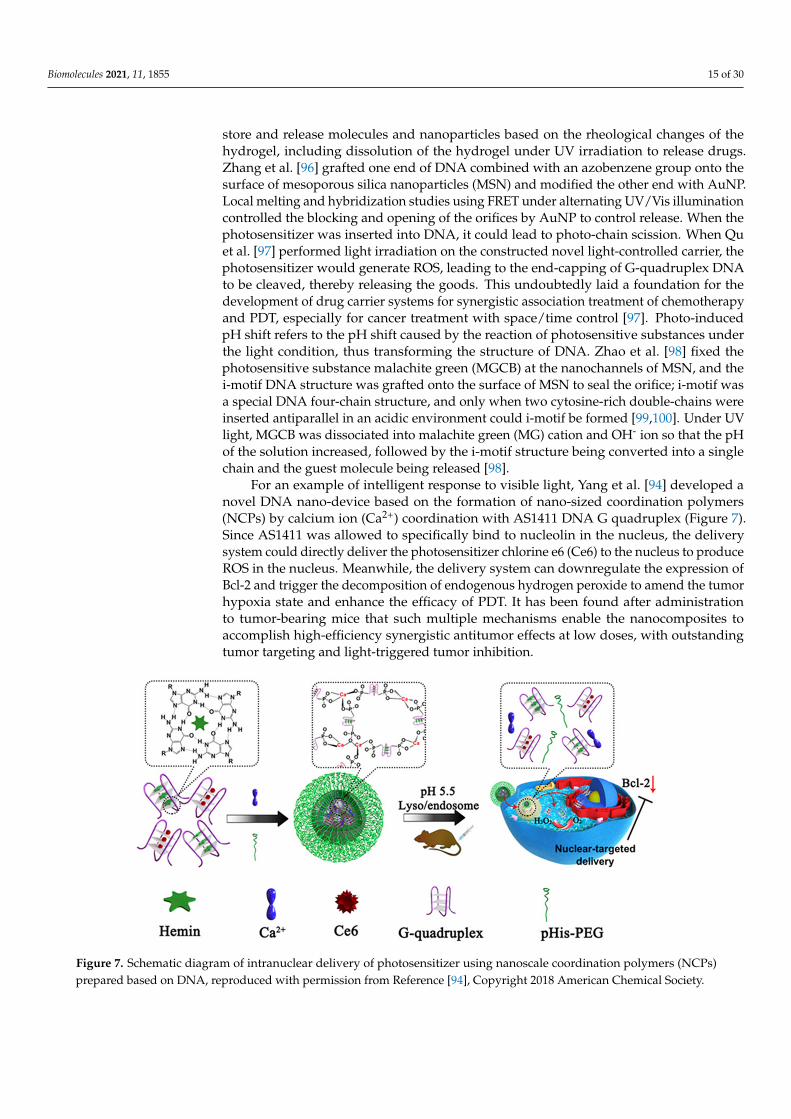
For an example of intelligent response to visible light, Yang et al. [94] developed anovel DNA nano-device based on the formation of nano-sized coordination polymers(NCPs) by calcium ion (Ca2+) coordination with AS1411 DNA G quadruplex (Figure 7).Since AS1411 was allowed to specifically bind to nucleolin in the nucleus, the deliverysystem could directly deliver the photosensitizer chlorine e6 (Ce6) to the nucleus to produceROS in the nucleus. Meanwhile, the delivery system can downregulate the expression ofBcl-2 and trigger the decomposition of endogenous hydrogen peroxide to amend the tumorhypoxia state and enhance the efficacy of PDT. It has been found after administrationto tumor-bearing mice that such multiple mechanisms enable the nanocomposites toaccomplish high-efficiency synergistic antitumor effects at low doses, with outstandingtumor targeting and light-triggered tumor inhibition.
Biomolecules 2021, 11, x FOR PEER REVIEW 16 of 31
Figure 7. Schematic diagram of intranuclear delivery of photosensitizer using nanoscale coordination polymers (NCPs) prepared based on DNA, reproduced with permission from Reference [94], Copyright 2018 American Chemical Society.
So far, its application is still limited due to the low tissue transmittance of ultravio-let/visible light and the lack of spatial control of PSs [101]. However, it is gratifying that the near-infrared light has a deep penetration into the tissue, and can be used for irradia-tion in vivo, which is expected to realize efficient photodynamic therapy (PDT). In 2016, Zhuang et al. [102] devised a triangular DNA nanostructure based on DNA origami for the delivery of a low-molecular carbazole derivative BMEPC in tumor cells. BMEPC is a one-and two-photon imaging agent and photosensitizer that can be fully excited by the near-infrared light to destroy targets under anaerobic conditions. However, the aggrega-tion of BMEPC in the water environment leads to quenching, which limits its application [102]. After binding to the vector DNA containing the largest dense insertion site, the wa-ter solubility, photodynamic efficiency, and intracellular photobleaching of BMEPC were significantly improved. The triangular DNA nanostructures loaded with BMEPCs could be successfully ingested by human breast cancer cell MCF-7. Under the irradiation, BMEPCs would generate free radicals and be released due to DNA photo-cleavage, and the generated ROS would eventually lead to cell death. The complex can simultaneously play the roles of an imaging agent and a photosensitizer in cells, and provides new tactics for intracellular imaging and photodynamic therapy.
In addition, the lanthanide-doped upconversion nanomaterials have individual light conversion capability, which can be used to absorb two or more low-energy photons and release a high-energy photon to construct a near-infrared responsive nanoplatform [103]. In recent years, a series of cooperations between the research group of Li Lele, a researcher at the National Center for Nano Science of Chinese Academy of Sciences, and the research group of Zhao Yuliang, an academic at the Chinese Academy of Sciences, has promoted the development of DNA nano medical devices. In the preliminary work, with the pur-pose of constructing a DNA sensing device that can be activated at a required time under remote stimulation, the Li research group [104] proposed for the first time the remote ma-nipulation of biosensing using up-conversion luminescence. The nano device not only can efficiently deliver the aptamer probe to living cells, but also can time-control the ATP flu-orescence sensing activity by in vitro and in vivo near-infrared light irradiation. After that, they expanded and realized the “time-space” controllable and accurate detection and di-agnostic applications of a variety of important biomarkers including miRNA and mRNA [105–107]. On the basis of these studies, they constructed a DNA nano-device driven by orthogonal near-infrared light [108] (Figure 8a) in this work to achieve higher spatial-tem-poral accuracy. The nano device combines an ultraviolet light activatable aptamer module and a photosensitizer with up-conversion nanoparticles (UCNPs) [108], and the UCNPs
Figure 7. Schematic diagram of intranuclear delivery of photosensitizer using nanoscale coordination polymers (NCPs)prepared based on DNA, reproduced with permission from Reference [94], Copyright 2018 American Chemical Society.

Biomolecules 2021, 11, 1855 16 of 30
So far, its application is still limited due to the low tissue transmittance of ultravi-olet/visible light and the lack of spatial control of PSs [101]. However, it is gratifyingthat the near-infrared light has a deep penetration into the tissue, and can be used forirradiation in vivo, which is expected to realize efficient photodynamic therapy (PDT). In2016, Zhuang et al. [102] devised a triangular DNA nanostructure based on DNA origamifor the delivery of a low-molecular carbazole derivative BMEPC in tumor cells. BMEPC isa one-and two-photon imaging agent and photosensitizer that can be fully excited by thenear-infrared light to destroy targets under anaerobic conditions. However, the aggregationof BMEPC in the water environment leads to quenching, which limits its application [102].After binding to the vector DNA containing the largest dense insertion site, the watersolubility, photodynamic efficiency, and intracellular photobleaching of BMEPC were sig-nificantly improved. The triangular DNA nanostructures loaded with BMEPCs could besuccessfully ingested by human breast cancer cell MCF-7. Under the irradiation, BMEPCswould generate free radicals and be released due to DNA photo-cleavage, and the gen-erated ROS would eventually lead to cell death. The complex can simultaneously playthe roles of an imaging agent and a photosensitizer in cells, and provides new tactics forintracellular imaging and photodynamic therapy.
In addition, the lanthanide-doped upconversion nanomaterials have individual lightconversion capability, which can be used to absorb two or more low-energy photons andrelease a high-energy photon to construct a near-infrared responsive nanoplatform [103].In recent years, a series of cooperations between the research group of Li Lele, a researcherat the National Center for Nano Science of Chinese Academy of Sciences, and the researchgroup of Zhao Yuliang, an academic at the Chinese Academy of Sciences, has promotedthe development of DNA nano medical devices. In the preliminary work, with the purposeof constructing a DNA sensing device that can be activated at a required time underremote stimulation, the Li research group [104] proposed for the first time the remotemanipulation of biosensing using up-conversion luminescence. The nano device not onlycan efficiently deliver the aptamer probe to living cells, but also can time-control the ATPfluorescence sensing activity by in vitro and in vivo near-infrared light irradiation. Afterthat, they expanded and realized the “time-space” controllable and accurate detectionand diagnostic applications of a variety of important biomarkers including miRNA andmRNA [105–107]. On the basis of these studies, they constructed a DNA nano-device drivenby orthogonal near-infrared light [108] (Figure 8a) in this work to achieve higher spatial-temporal accuracy. The nano device combines an ultraviolet light activatable aptamermodule and a photosensitizer with up-conversion nanoparticles (UCNPs) [108], and theUCNPs generate orthogonal ultraviolet light and visible light under the excitation of twonear-infrared light beams with different wavelengths to activate the biological recognitionactivity of the aptamer and excite the photosensitizer to generate active oxygen, respectively,thereby realizing space-time controllable target recognition and photodynamic therapy(Figure 8b). Moreover, the DNA nanodevice [108] can be integrated with an immunecheckpoint blocking therapy to inhibit the untreated distal tumor by promoting tumorinfiltration of cytotoxic T cells and causing synergistic and distal effects in tumor-bearingmice. This work has taken a new step towards the employment of exogenous tools tocontrol the function and behavior of DNA nanodevices in vivo.

Biomolecules 2021, 11, 1855 17 of 30
Biomolecules 2021, 11, x FOR PEER REVIEW 17 of 31
generate orthogonal ultraviolet light and visible light under the excitation of two near-infrared light beams with different wavelengths to activate the biological recognition ac-tivity of the aptamer and excite the photosensitizer to generate active oxygen, respec-tively, thereby realizing space-time controllable target recognition and photodynamic therapy (Figure 8b). Moreover, the DNA nanodevice [108] can be integrated with an im-mune checkpoint blocking therapy to inhibit the untreated distal tumor by promoting tu-mor infiltration of cytotoxic T cells and causing synergistic and distal effects in tumor-bearing mice. This work has taken a new step towards the employment of exogenous tools to control the function and behavior of DNA nanodevices in vivo.
Figure 8. Photothermal-responsive DNA nanodevices for drug carrier. (a) UCNPs (light modulator) are used to convert NIRs of two different wavelengths (808 and 980 nm) into orthogonal ultraviolet light and up-converted light (UCL), re-spectively, and (b) the nanodevice recognizes nucleolin on the surface of tumor cells under local trigger of 808 nm NIR light and then generates ROS under irradiation of 980 nm NIR light for PDT, reproduced with permission from Reference [108], Copyright 2020 The Authors; (c) the encapsulation and release of HRP by a covalently closed three-dimensional DNA cage device are controlled by temperature, reproduced with permission from Reference [108], Copyright 2013 Amer-ican Chemical Society.
Taken together, although ultraviolet light is easily obtained, it will cause damage to biological samples and has insufficient transmission ability, which can be blocked by the skin to reach the lesion site. Nevertheless, near-infrared light can reach a depth of nearly 10 cm in the body and has no damage to normal cells. Therefore, UV is only suitable for in vitro research and is not conducive to clinical application. NIR is a relatively ideal light source for photoresponsive control of drug release due to its strong transmission ability, marginalex absorption by the skin and tissue, and no damage. Under the action of external light irradiation or magnetic field, the inorganic nanomaterials can generate resonance absorption and transfer heat to the surroundings, so that the local temperature increases to cause DNA melting or decomposition, and the drugs stored or blocked by DNA chains are set free. It is possible to design a magnetic response system in addition to the above-mentioned optical response system. For example, Vallet-Regi et al. [109] added superpar-amagnetic iron oxide nanoparticles to MSN for conjugation with DNA, which enclosed MSN pores when two DNA chains hybridized, and the double-stranded DNA was grad-ually melted by an external magnetic field (up to a high temperature of 42–47 °C) to result in decapsulation and drug release. This remotely triggered drug delivery is capable of
Figure 8. Photothermal-responsive DNA nanodevices for drug carrier. (a) UCNPs (light modulator) are used to convert NIRsof two different wavelengths (808 and 980 nm) into orthogonal ultraviolet light and up-converted light (UCL), respectively,and (b) the nanodevice recognizes nucleolin on the surface of tumor cells under local trigger of 808 nm NIR light and thengenerates ROS under irradiation of 980 nm NIR light for PDT, reproduced with permission from Reference [108], Copyright2020 The Authors; (c) the encapsulation and release of HRP by a covalently closed three-dimensional DNA cage device arecontrolled by temperature, reproduced with permission from Reference [108], Copyright 2013 American Chemical Society.
Taken together, although ultraviolet light is easily obtained, it will cause damage tobiological samples and has insufficient transmission ability, which can be blocked by theskin to reach the lesion site. Nevertheless, near-infrared light can reach a depth of nearly10 cm in the body and has no damage to normal cells. Therefore, UV is only suitablefor in vitro research and is not conducive to clinical application. NIR is a relatively ideallight source for photoresponsive control of drug release due to its strong transmissionability, marginalex absorption by the skin and tissue, and no damage. Under the actionof external light irradiation or magnetic field, the inorganic nanomaterials can generateresonance absorption and transfer heat to the surroundings, so that the local temperatureincreases to cause DNA melting or decomposition, and the drugs stored or blocked byDNA chains are set free. It is possible to design a magnetic response system in additionto the above-mentioned optical response system. For example, Vallet-Regi et al. [109]added superparamagnetic iron oxide nanoparticles to MSN for conjugation with DNA,which enclosed MSN pores when two DNA chains hybridized, and the double-strandedDNA was gradually melted by an external magnetic field (up to a high temperatureof 42–47 ◦C) to result in decapsulation and drug release. This remotely triggered drugdelivery is capable of warming that surrounds medium, which may have unexpectedeffects if applied to the delivery of chemotherapeutic drugs. The principle of temperature-controlled conformational change can also be accessed to design temperature-responsiveDNA nanodevices. The Juul team [110] used covalently closed three-dimensional DNAcage structure to encapsulate horseradish peroxidase (HRP) and chose whether to releaseit under the control of temperature. This structure of 12 double-stranded B-DNA helicesforming the edges, each interrupted by a short single-stranded linker forming six “horns”between them [110], allowed the cargo to enter or be released at 37 ◦C while ensuring thatthe cargo remained in the central cavity of the cage at 4 ◦C (Figure 8c). The enzyme trappedwithin the DNA cage has catalytic activity and the DNA cage device helps reversibly and

Biomolecules 2021, 11, 1855 18 of 30
controllably encapsulate and release the enzyme cargo without any form of covalent ornon-covalent attachment to the cargo.
3.2. Chemical-Responsive DNA Nanodevices for Drug Carriers
The tumor microenvironment (TME) is featured by being slightly acidic, hypoxic, andrich in tumor cell secretase. Simultaneously, tumor cells also contain high concentrationsof reactive oxygen species (ROS), glutathione (GSH), and other biochemical indicatorsdifferent from those of normal cells [111]. TME-responsive nano-drug carriers need toshow outstanding performance in achieving tumor targeting, removal of carrier protectiveshell, and controlled release of drugs at tumor sites.
pH change is one of the traits of pathological tissues in the human body. Due to theaggressive proliferation of tumor cells and rapid generation of irregular blood vessels,nutrition and oxygen in the tumor site are rapidly lacking, and lactic acid metabolitesproduced by glycolysis in tumor cells are accumulated in the tumor stroma, resulting inthe pH value of the tumor extracellular environment reduced to 6.5–7.2 [112], while the pHvalues of inclusions and lysosomes in tumor cells are further reduced to 4.0–6.0 [113]. Onthis basis, a variety of pH-responsive drug delivery systems have been designed to stabilizethe vehicle under normal physiological conditions. Ruling out the pH-responsive MUC1-aptamer-DNA icosahedron constructed by Huang’s research group mentioned above, Liuet al. [114] reported a DNA tetrahedral device triggered by an acidic environment, in whichthe key element was the formation/dissociation of short and cytosine (C)-containing DNAtriplexes at pH 5.0–8.0, leading to the assembly or disassembly of DNA tetrahedrons. First,they constructed a three-star motif with three sticky ends through base complementarypairing, and self-assembled into a tetrahedron at pH 5.0. The motif occupied the vertexof the tetrahedron, and the branches of the motif became the sides of the tetrahedron.By dissociating the triad at pH 8.0, the tetrahedron can be restored to its original struc-ture. Therefore, it can also be termed a strategy for reversibly assembling/disassemblingDNA nanocages using the DNA nanomachine concept [114]. Recently, Ruan et al. [115]also adopted tetrahedral DNA framework (TDF) as the solid structure framework of thebiosensor and DNA i-motif structure consisting of different sequences as the pH responseconversion module structure, and developed a programmable pH sensor (Figure 9a). Theobvious advantages of this sensor [115] lie in its durative response to biological pH be-tween 5.0 and 7.5, prone to anchoring on the cell membrane and programmability in pHsensing (Figure 9b), demonstrating its potential for dynamically adjustable pH sensing,microenvironment in situ imaging, and pH-related disease diagnosis. In light of the foldingsynergy, He et al. [116] developed a robust pH sensor with high response sensitivity usingdimer DNA i-motif as the module. The DNA framework provided a stable structuralframework to manipulate the folding behavior of the responsive nucleic acid module andexert its performance in cells. Dimer DNA i-motif is confined to the vertex of TDF, andthe DNA framework promotes the correct assembly of i-motif structure, thus enhancingtheir folding cooperation; this high-response pH sensor [116] has different sensing rangesfrom pH 5.0 to 8.0 for monitoring tiny pH changes in cells. The pre-tissue strategy used inthis study would be applied to the recent emergence of sensors and molecular switchesbased on other bimolecular receptors, such as the bimolecular nucleic acid aptamer and theisolated G-quadruplex [117–119]. Thereafter, Zhu et al. [120] have devised a DNA nano-hydrogel system for delivering mRNA drugs, which can stably express the target proteinin cells. The elaborately designed pH-responsive i-motif “X” DNA scaffold and mRNAwere crosslinked to form dense nanospheres for effortless entry into the cells throughendocytosis. In lysosomes, with the formation of i-motif, the hydrogel disintegrates andmRNA is released into the cytoplasm encoding protein. Since no other external syntheticmaterials are used in the system construction process, the system undoubtedly has goodbiocompatibility, and considering the sophistication of mRNA, the nano hydrogel systemis a promising candidate for providing a new way for the delivery of functional mRNAin vivo.

Biomolecules 2021, 11, 1855 19 of 30
Distinct from the device frameworks in which the abovementioned motifs participatein the construction, the pH-responsive SNA nanodevices based on AuNP designed byLiang team [121] have adjustable pH sensitivity, rapid, and reversible allosteric effects, andeven the capability to image lysosomes in living cells. They hybridized a long thioatedchain with a short reporter (green) labeled with pH-insensitive Alexa-647 dye, and a tail(red) with a specific protonation site was added at the end of the thioated chain to driveDNA deformation when pH varied [121]. Under the basic condition, the DNA chain wasin a linear conformation, and the fluorescence emission of Alexa-647 was quenched. Underthe acidic conditions, protonation of the tail would preferentially drive the DNA chain tofold into a loop-stem (green-blue) structure, with the tail region and stem region (blue)forming a three-chain structure through parallel Hoogsteen interactions, with the reporterchain released and followed by generation of measurable fluorescence signals. To confirmits applicability, their team [121] next designed a pH-triggered Dox-SNA conjugate thatcan transport Dox molecules to cancer cells to promote pH-dependent drug release. TheDox-SNA conjugate can arise by mixing Dox with SNA solution, and thereafter the Dox-SNA compound is internalized by HeLa cells [121]. The results manifested that, also in theacidic environment of late endosome or lysosome, the protonation of the tail (red) forcedthe chain to adopt the cyclic stem (blue-black) structure, and the Dox loading region (blue)unfolded. When pH was further reduced, the release of Dox was significantly accelerated.In particular, the cumulative release rate of SNA nanocarrier at pH 4.6 reached ~92%within 80 min, illustrating a higher efficiency in drug delivery and cell viability reductioncompared with free Dox [121]. Zhu et al. [122] also exhibited a stimulation-responsivemultiple drug delivery system capable of accurately responding to pH changes within therange of 5.0–7.0 (Figure 9c) based on a three-strand DNA nano-switch in 2019. Two DNAchains were bound to AuNP. The first DNA chain was an anti-MUC1 aptamer serving asthe targeting ligand, and the other was called switchable DNA, which assumed a linearconformation under neutral or basic conditions and self-folded into three chains underacidic conditions. By binding three drugs to this switchable DNA: Dox, cisplatin, andantisense DNA (asDNA), tumor cells undergo a linear to three-chain conformational changeof this nanoswitch through endocytosis, leading to the intelligent release of the combineddrug. The stimulation-responsive drug delivery system not only has biocompatibility, butalso can simultaneously provide multiple therapeutic drugs to improve curative effects.In the tumor-bearing mouse model, we have indicated the effective gene silencing andsignificant tumor growth inhibition after intravenous injection of the intelligent nano-switch, supplying an opportunity for combined treatment of cancer [122].
In the delivery of tumor vaccine, Ding team [123] attempted to manufacture a tubularDNA nanodevice vaccine capable of delivering antigens and adjuvants, in the hope ofspecifically stimulating tumor T-cell reaction for a long time to cause tumor regression.Interestingly, the tubular complex was switched by a DNA lock, in a double-stranded stateat pH 7.5 and closed along the long sides of the rectangle into a tubular nanostructure witha diameter of about 19 nm, and in a three-stranded state at pH 5.5 resulted in the release ofthe quencher-labeled pcSW-16 and increased fluorescence intensity. The antigen/adjuvant-loaded DNA nanorobot structure in the lumen can be transported to the draining lymphnode (DLN) without the influence of extracellular nucleases. When localized to DLNantigen-presenting cells (APCs), the co-assembled responsive DNA lock is capable ofmechanically exposing the drug in response to pH changes, thereby activating the TLRpathway and antigen peptide presentation. The results indicated that the therapeuticvaccination carefully designed by the Ding team could not only affect the primary tumor,but also prevent metastasis and recurrence [123], showing good therapeutic potential andpromising to promote the development of personalized cancer vaccine production. Thedual-response device engineered by Siwy et al. [124] responds to both pH and electricalsignals: lowering the pH to 5.5 will combine positively charged DNA molecules withother chains to form an electrostatic network, thus sealing the pore channels and achievingan unprecedented high resistance of tens of millions of ohms; at a neutral pH value, the

Biomolecules 2021, 11, 1855 20 of 30
voltage switch causes the DNA chain to conduct nano mechanical movement for separation.This pH-dependent reversible closure mechanism proved robust, not only enabling closureand release as a switch, but dominating the drug release rate as a valve.
Biomolecules 2021, 11, x FOR PEER REVIEW 20 of 31
Figure 9. pH-responsive DNA nanodevices for drug carrier. (a) The TDF-based programmable pH sensor using DNA i-motif as the proton recognition probe provides a continuously distributed pH response over the range of 5.0 to 7.5. and (b) the i-motif-TDF nanosensor was anchored to the cell surface for in situ pH analysis, reproduced with permission from Reference [115], Copyright 2021 American Chemical Society. (c) The triplex-DNA nanoswitch-based system responds ac-curately to pH changes over the 5.0-7.0 range. In an extracellular neutral space, a linear DNA nanoswitch is used for fixing a plurality of therapeutic drugs; After entering the intracellular acidic space, the DNA nanodevice changed from linear to triple to release the drug, reproduced with permission from Reference [122], Copyright 2019 American Chemical Society.
In the delivery of tumor vaccine, Ding team [123] attempted to manufacture a tubular DNA nanodevice vaccine capable of delivering antigens and adjuvants, in the hope of specifically stimulating tumor T-cell reaction for a long time to cause tumor regression. Interestingly, the tubular complex was switched by a DNA lock, in a double-stranded state at pH 7.5 and closed along the long sides of the rectangle into a tubular nanostructure with a diameter of about 19 nm, and in a three-stranded state at pH 5.5 resulted in the release of the quencher-labeled pcSW-16 and increased fluorescence intensity. The anti-gen/adjuvant-loaded DNA nanorobot structure in the lumen can be transported to the draining lymph node (DLN) without the influence of extracellular nucleases. When local-ized to DLN antigen-presenting cells (APCs), the co-assembled responsive DNA lock is capable of mechanically exposing the drug in response to pH changes, thereby activating the TLR pathway and antigen peptide presentation. The results indicated that the thera-peutic vaccination carefully designed by the Ding team could not only affect the primary tumor, but also prevent metastasis and recurrence [123], showing good therapeutic po-tential and promising to promote the development of personalized cancer vaccine pro-duction. The dual-response device engineered by Siwy et al. [124] responds to both pH and electrical signals: lowering the pH to 5.5 will combine positively charged DNA mol-ecules with other chains to form an electrostatic network, thus sealing the pore channels and achieving an unprecedented high resistance of tens of millions of ohms; at a neutral pH value, the voltage switch causes the DNA chain to conduct nano mechanical move-ment for separation. This pH-dependent reversible closure mechanism proved robust, not
Figure 9. pH-responsive DNA nanodevices for drug carrier. (a) The TDF-based programmable pH sensor using DNAi-motif as the proton recognition probe provides a continuously distributed pH response over the range of 5.0 to 7.5. and(b) the i-motif-TDF nanosensor was anchored to the cell surface for in situ pH analysis, reproduced with permission fromReference [115], Copyright 2021 American Chemical Society. (c) The triplex-DNA nanoswitch-based system respondsaccurately to pH changes over the 5.0–7.0 range. In an extracellular neutral space, a linear DNA nanoswitch is used for fixinga plurality of therapeutic drugs; After entering the intracellular acidic space, the DNA nanodevice changed from linear totriple to release the drug, reproduced with permission from Reference [122], Copyright 2019 American Chemical Society.
3.3. Biological Responsive DNA Nanodevices for Drug Carriers
Using biological molecules as the stimulation source is a common type of intelligentresponsive controlled release, and one of the most commonly used biological molecules isadenosine triphosphate (ATP). ATP is formed by the connection of adenine, ribose, andthree phosphate groups. More energy is released during hydrolysis; it is the most direct en-ergy source in organisms, and plays a vital role in major biological processes. Including thecommon muscle contraction, synthesis and degradation of intracellular compounds, andactive transportation, etc., the infinite proliferation of tumor cells demands a large amountof energy to maintain growth and metabolism, resulting in different ATP concentrationsinside and outside of the tumor cells. The intracellular ATP concentration is markedlyhigher than that outside the cell. Studies have found that this property has a palpableeffect on the drug resistance of cancer cells. Such systems usually connect the aptamer tothe carrier as the control switch: in the absence of ATP, the aptamer or the structure con-structed based on the aptamer blocks the drug; in the presence of ATP, the aptamer bindsto it closely, leading to the structural transformation of the aptamer and direct realizationof drug release [125]. Classical examples include the sandwich-shaped ATP-responsive

Biomolecules 2021, 11, 1855 21 of 30
controlled release system composed of MSN capped with aptamers designed by Tanget al. [126]. In this system, in the absence of ATP, ATP aptamers hybridized with twosingle chains to form a sandwich DNA structure, which was grafted onto the surface ofMSN by click chemistry, accompanied by pore blockage and inhibition of release. In thepresence of ATP, ATP binds to the ATP aptamer and leaves the pore canal, and the loadedmolecule is released. Subsequent study demonstrated not only high selectivity of thisATP-responsive behavior for ATP analog, but also that the amount of molecular leakagein the absence of ATP could be negligible. Similarly, Gu et al. [127] demonstrated that,when the environment was rich in ATP, the polymer nanocarrier functionalized with DNAmotifs bound by ATP aptamers could selectively release the embedded Dox through con-formational transformation. On the other hand, the controlled release of ATP-responsivenanogel drug-loading system in tumor-bearing mice was further studied. The system notonly effectively inhibited the activity of tumor cells, but also did not affect the body weightand normal cell metabolism of mice [127], which was denoted as a momentous step for itsfeasibility study in clinical application.
Compared with the controlled release of ATP response, there is also an adenosineresponse system. Cyclic guanosine monophosphate (cdGMP, second messenger) moleculeswith intracellular information transmission can also be used for controllable release [128]to integrate cdGMP aptamers into DNA icosahedron. In the presence of cdGMP, theaptamers are changed in structure through chain replacement, thereby destroying the DNAicosahedron structure and opening the capsule to release the drugs therein. Meanwhile,the fluorescence spectrum was used to monitor the release of the goods, further realizingthe rigorous control on the opening of the DNA icosahedron and the release of the goods.
As we all know, many reducing molecules (such as glutathione and reductase) intumor cells generate large redox potential differences inside and outside those cells. Mutualtransformation of glutathione (GSH) and glutathione disulfide (GSSG) is a major redoxpathway in mammalian cells that is primarily responsible for preventing ROS-induced cellinjury, maintaining the stability of thiol-containing enzymes, and preserving the integrityof cell membranes. The rapid proliferation of tumor cells results in the GSH content intumor tissues being at least four times higher than the normal tissue content, reaching0.5–10 mM [129]. This significant difference in GSH level can be utilized to trigger thecontrolled release of redox-sensitive nanodrugs in tumor cells. The routinely used struc-tures for constructing reduction-responsive carriers, such as disulfide bonds (S-S) andselenium bonds (Se-Se), can be rapidly cleaved by high concentrations of GSH. ROS, as aclass of molecules with strong oxidizing ability, is a by-product of aerobic metabolism inorganisms, and is a special cell signal transcription factor. Hydrogen peroxide (H2O2), themost typical ROS, has been widely explored as an activation condition for drug release dueto its catalytic activity in multiple reactions [130,131]. Compared with normal cells, tumorcells will produce higher concentrations of H2O2 [132]. The high biotoxicity of H2O2 islargely due to its easy conversion to various types of active free radicals, which disruptthe redox metabolic balance. In 2018, Ding et al. [44] created a novel drug delivery systembased on DNA nanostructure (called TODP), the linear tumor treatment gene p53 waslinked using disulfide bonds, and Dox was inserted into the base pair of DNA doublechains. Through GSH response, specific release of Dox and p53 in tumor cells was achieved,as well as efficient gene transfer. The system illustrated an effective inhibitory effect ontumor growth in vivo and in vitro, without obvious systemic toxicity. These structural andchemical consensus nanocarriers not only realize the combination treatment of gene andchemotherapy, but offer a new strategy for gene therapy of various diseases. Then, theDing research group [133] developed a nano-platform based on branched DNA structureto efficiently load the sgRNA/Cas9/antisense complex targeting the tumor-related genePLK1 through DNA self-assembly for co-delivery of gene editing and gene silencing tools.The gene editing tool sgRNA/Cas9 can be targeted for DNA in the nucleus and the genesilencing tool antisense can be used for mRNA in the cytoplasm, thereby attaining syner-gistic treatment on tumors in vivo and in vitro. As the structure introduces the aptamer for

Biomolecules 2021, 11, 1855 22 of 30
targeted delivery and the influenza hemagglutinin peptide for endogenous escape, andcan effectively release drugs in response to GSH and RNase H, the structure displays high-efficiency antitumor activity without causing visible adverse reactions. This first instanceof a nanoplatform based on branched DNA offers new proposals for the development ofgene therapy.
Researchers have shown that the pathology of neoplastic diseases is associated withenzyme dysfunction or dysregulation of its expression. Among them, the expression andactivity of proteases and phospholipases in tumor tissue are significantly higher than thosein normal tissue, which has an important impact on the occurrence and development oftumors [134]. Based on this feature, the design and development of enzyme-responsiveantitumor nano-drug delivery systems are of great priority, contributing to the controlledrelease of drugs at tumor sites [69,135]. Apart from the above-mentioned cases where DNAdevices realized the release of drugs in response to enzyme [81,82,133], the conformationallyswitchable intelligent nano-probe designed by Li research group [136] contains a denseshell of hairpin DNA labeled with gold nanoparticles (AuNP) and 5- carboxyfluorescein(FAM). Under the influence of high-activity telomerase, the conformation of hairpin DNAis transformed, in order to facilitate the drug Dox loaded by AuNP and responding totelomerase activity being released in cancer cells. Analogously, functional DNA tetrahedralprobes designed by Yue et al. [137] based on FRET can also respond to changes in telomeraseactivity, and the probes loaded with telomerase primers and molecular beacons MB canimage telomerase in living cells and distinguish between normal cells and tumor cells,with the potential to be utilized for the delivery of tumor drugs. Enzyme molecules maywell interact with other responsive factors, and therapeutic effects may be stronger. Zhuet al. [138] then synthesized a biological porous nanosphere (PRS) using RNA as a structuralunit and cyclodextrin as a binder. RNA contained EpCAM aptamer for targeted deliveryand siRNA (gene drug) for EpCAM gene silencing. Cyclodextrin could load sorafenib(chemotherapy drug) for targeted liver cancer treatment through its hydrophobic cavity.As shown in Figure 10a, Dicer enzymes in liver cancer cells could intelligently respond toand degrade the complex and release drugs for synergistic treatment. The measurementson the cell, subcutaneous and in situ levels of tumor-bearing mice show that the compositecan significantly inhibit tumor growth, promote apoptosis (Figure 10b), and is safe andnontoxic for organs to be treated.
Most single-stimulus-responsive nanosystems are susceptible to interference, yet thecombination of endogenous and exogenous stimuli often emerges unexpected effects onspatio-temporal control. In addition to the multiple response device cases mentioned above,the matrix metalloproteinase (MMP)-responsive AuNP probe (Figure 10c) designed by Yanget al. [139] can be used for tumor-targeted photothermal therapy and drug delivery. Underthe action of the MMP generated by the tumor tissue, the DNA ligated on the surface of thegold nanoparticles can hybridize to enable the complex to be in an aggregation state fromdispersion, and under the irradiation of in vitro near infrared light, the material generateslocal high temperature to induce the release of Dox drug, and the combined treatmentof enzymatic reaction and near infrared light is successful. The magnetic DNA nanogelconstructed by Yao et al. [140] made use of magnetic nanoparticles as the core, and a DNAnanogel layer is synthesized through rolling cycle amplification for loading anticancerdrugs. Under the guidance of an external magnetic field, the magnetic DNA nanogel canachieve high-efficiency targeting to tumor cells. Furthermore, the special DNA nanogelcan intelligently respond to various stimulation factors such as temperature, pH, andnuclease, thereby operating the release of anticancer drugs. The system cleverly combinesthe magnetically controlled delivery with the intelligent response release to further thedevelopment of precision medicine. The mutual echo of this exogenous stimulus andendogenous stimulus strikingly enhanced the accuracy of the work of the nano-devices,which might be the development direction of space-time controllable nano-devices inthe future.

Biomolecules 2021, 11, 1855 23 of 30
Biomolecules 2021, 11, x FOR PEER REVIEW 23 of 31
spatio-temporal control. In addition to the multiple response device cases mentioned above, the matrix metalloproteinase (MMP)-responsive AuNP probe (Figure 10c) de-signed by Yang et al. [139] can be used for tumor-targeted photothermal therapy and drug delivery. Under the action of the MMP generated by the tumor tissue, the DNA ligated on the surface of the gold nanoparticles can hybridize to enable the complex to be in an ag-gregation state from dispersion, and under the irradiation of in vitro near infrared light, the material generates local high temperature to induce the release of Dox drug, and the combined treatment of enzymatic reaction and near infrared light is successful. The mag-netic DNA nanogel constructed by Yao et al. [140] made use of magnetic nanoparticles as the core, and a DNA nanogel layer is synthesized through rolling cycle amplification for loading anticancer drugs. Under the guidance of an external magnetic field, the magnetic DNA nanogel can achieve high-efficiency targeting to tumor cells. Furthermore, the spe-cial DNA nanogel can intelligently respond to various stimulation factors such as temper-ature, pH, and nuclease, thereby operating the release of anticancer drugs. The system cleverly combines the magnetically controlled delivery with the intelligent response re-lease to further the development of precision medicine. The mutual echo of this exogenous stimulus and endogenous stimulus strikingly enhanced the accuracy of the work of the nano-devices, which might be the development direction of space-time controllable nano-devices in the future.
Figure 10. Enzyme-responsive DNA nanodevices for drug carrier. (a) The enzymatic RNA generated by rolling circle transcription (RCT) was self-assembled into PRS by using cyclodextrin as a complexing agent, and SF was further carefully loaded to form porous RNA nanospheres (PRS@SF), and (b) after intravenous injection of PRS@SF, the PRS@SF was trans-ported in blood vessels and swallowed by target cells, and the drug SF and siRNA were released for synergistic treatment after being digested by cytoplasmic Dicer enzyme, reproduced with permission from Reference [138], Copyright 2020 Elsevier. (c) MMPs induced specific and rapid assembly of PEG-Dox-attached AuNPs in vivo to enhance tumor inhibition, reproduced with permission from Reference [139], Copyright 2019 Elsevier.
Figure 10. Enzyme-responsive DNA nanodevices for drug carrier. (a) The enzymatic RNA generated by rolling circletranscription (RCT) was self-assembled into PRS by using cyclodextrin as a complexing agent, and SF was further carefullyloaded to form porous RNA nanospheres (PRS@SF), and (b) after intravenous injection of PRS@SF, the PRS@SF wastransported in blood vessels and swallowed by target cells, and the drug SF and siRNA were released for synergistictreatment after being digested by cytoplasmic Dicer enzyme, reproduced with permission from Reference [138], Copyright2020 Elsevier. (c) MMPs induced specific and rapid assembly of PEG-Dox-attached AuNPs in vivo to enhance tumorinhibition, reproduced with permission from Reference [139], Copyright 2019 Elsevier.
4. Conclusions and Perspectives
Since DNA has been found to have highly programmable and predictable properties,it has been used as a fascinating functional material to design and assemble variousnanostructures to exert unique advantages in medical diagnosis and treatment. In additionto being constrained by the principle of base complementary pairing, changes in DNAnanostructure abnormalities under endogenous and exogenous stimuli also suggest thepossibility of the development of smart responsive nanodevices. In this review, we initiallyintroduced the concept of DNA nanomaterials and their applications in the medical field,and summarized the classification of DNA nanostructures for drug carriers; especially, therecent accomplishments in the design and application of DNA origami, DNA tetrahedralstructure, and spherical nucleic acid were described. Afterwards, we then focused onintroducing various intelligent responsive DNA nanodevices for targeted therapy from thephysical, chemical, and biological perspectives according to the differences in stimulationsources, confirming that both static and dynamic DNA nanocarriers can be designedto actively and passively release loading substances at specific sites. Collectively, DNAnanostructures, whether as nanocarriers or incorporated in intelligent responsive controlledrelease systems, have demonstrated good stability and controllability, providing an infiniteprospect for their application in drug therapy systems.
However, as a novel thing in the field of drug delivery, DNA nanostructures are facingmany challenges and opportunities. Particularly in practical applications, the circulation,distribution, metabolism, efficacy, and degradation of drugs in the body are crucial for

Biomolecules 2021, 11, 1855 24 of 30
studying the interaction between DNA and cell behavior. Although researchers havedesigned a variety of nanoparticles to transport drugs to solve the problems of toxic andside effects and drug resistance of antitumor drugs, as shown in Figure 11, there are stillpotential risks [13,49,141]. (1) DNA itself, as a deoxynucleotide polymer, is easily degradedin the blood circulation to reach the target, or generates a certain degree of immunogenicitydue to self-modification, thereby interfering with the function of the immune system ofthe body. How to reasonably design DNA nanostructures to avoid the special hazards ofnanomaterials needs to be explained in more detailed research. (2) Due to the small sizeeffect and biocompatibility of DNA nanomaterials, after crossing the cell membrane, someregions of DNA devices may bind to single-stranded RNA such as mRNA and genes inthe nucleus to regulate gene expression, resulting in gene expression disorder and proteindysfunction. Therefore, the timely clearance mechanism of DNA nanomaterials plays anirreplaceable role. (3) Some DNA nanomaterials need light, magnetic or other externalassistance to be effective, and the safety effect on normal cells around the tumor remainsto be proved. Therefore, DNA sensors that require the application of external conditionsoften need to be verified multiple times to ensure that toxic and side effects are minimized.
Biomolecules 2021, 11, x FOR PEER REVIEW 25 of 31
Figure 11. Potential risks of DNA nanotechnology for drug delivery systems.
At present, most of the studies are limited to the level of cells, tissues, or tumor-bear-ing mice. To extend this drug delivery system to human trials, there are still many prob-lems to be solved, such as population control, energy supply, and material selection. We anticipate that, in the next two decades, it will develop into a higher clinical stage, even in small-scale disease applications. However, FDA approval could be more difficult because the range of interests affected by this technological revolution cannot be underestimated. Scientists still have a long way to go.
Author Contributions: Conceptualization, C.G. and X.Z.; literature review and resources, C.G.; writing—original draft preparation, C.G.; writing—review and editing, C.G., X.Z. and C.F.; figure preparation and editing, C.G. and C.F.; supervision, C.F., X.Z. All authors have read and agreed to the published version of the manuscript.
Funding: This work was supported by the National Natural Science Foundation of China (Grant No. 22074090), the Natural Science Foundation of Shanghai (Grant No. 19ZR1474200).
Institutional Review Board Statement: Not applicable.
Informed Consent Statement: Not applicable.
Data Availability Statement: Not applicable.
Conflicts of Interest: The authors declare no conflict of interest.
References 1. Wang, J.; Li, Y.; Nie, G. Multifunctional biomolecule nanostructures for cancer therapy. Nat. Rev. Mater. 2021, 6, 766–783,
https://doi.org/10.1038/s41578-021-00315-x. 2. Zhang, R.R.; Schroeder, A.B.; Grudzinski, J.J.; Rosenthal, E.L.; Warram, J.M.; Pinchuk, A.N.; Eliceiri, K.; Kuo, J.; Weichert, J.P.
Beyond the margins: Real-time detection of cancer using targeted fluorophores. Nat. Rev. Clin. Oncol. 2017, 14, 347–364, https://doi.org/10.1038/nrclinonc.2016.212.
3. Thakor, A.S.; Gambhir, S.S. Nanooncology: The future of cancer diagnosis and therapy. CA-Cancer J. Clin. 2013, 63, 395–418, https://doi.org/10.3322/caac.21199.
4. Torchilin, V. Tumor delivery of macromolecular drugs based on the EPR effect. Adv. Drug Deliv. Rev. 2011, 63, 131–135, https://doi.org/10.1016/j.addr.2010.03.011.
5. Fang, J.; Nakamura, H.; Maeda, H. The EPR effect: Unique features of tumor blood vessels for drug delivery, factors involved, and limitations and augmentation of the effect. Adv. Drug Deliv. Rev. 2011, 63, 136–151, https://doi.org/10.1016/j.addr.2010.04.009.
Figure 11. Potential risks of DNA nanotechnology for drug delivery systems.
Undoubtedly, benefiting from the biocompatibility and degradability of DNA nano-materials, in the future, functional DNA will be developed into more complicated andadvanced forms to achieve more efficient medical applications, such as multi-responsivedelivery of nano-robots. However, because of the late research in this field, most of thestudies so far have been limited to simply revealing the operations at the laboratory level,and no related drugs have been marketed. Therefore, considering the actual starting point,the next stage of technical research should also include standardization converted to theclinical level. Additionally, the high-purity preparation and large-scale production of DNAnanostructures are also great challenges impacting their commercial applications, whichare necessary conditions for practical biomedical applications. Last but not least, the priceof DNA nanostructure synthesis has also received a high degree of attention. Complexnanocarrier synthesis means that more DNA chains are desired, so reducing the cost ofsynthesizing nanostructures has become the focus of research. It is believed that, withthe rapid development of intelligent DNA nanocarriers, customized personalized DNAnanocarriers will provide a powerful security means for the effective treatment of tumors,and have more extensive applications in the field of precision biomedicine.

Biomolecules 2021, 11, 1855 25 of 30
At present, most of the studies are limited to the level of cells, tissues, or tumor-bearingmice. To extend this drug delivery system to human trials, there are still many problems tobe solved, such as population control, energy supply, and material selection. We anticipatethat, in the next two decades, it will develop into a higher clinical stage, even in small-scaledisease applications. However, FDA approval could be more difficult because the range ofinterests affected by this technological revolution cannot be underestimated. Scientists stillhave a long way to go.
Author Contributions: Conceptualization, C.G. and X.Z.; literature review and resources, C.G.;writing—original draft preparation, C.G.; writing—review and editing, C.G., X.Z. and C.F.; figurepreparation and editing, C.G. and C.F.; supervision, C.F., X.Z. All authors have read and agreed tothe published version of the manuscript.
Funding: This work was supported by the National Natural Science Foundation of China (Grant No.22074090), the Natural Science Foundation of Shanghai (Grant No. 19ZR1474200).
Institutional Review Board Statement: Not applicable.
Informed Consent Statement: Not applicable.
Data Availability Statement: Not applicable.
Conflicts of Interest: The authors declare no conflict of interest.
References1. Wang, J.; Li, Y.; Nie, G. Multifunctional biomolecule nanostructures for cancer therapy. Nat. Rev. Mater. 2021, 6, 766–783.
[CrossRef]2. Zhang, R.R.; Schroeder, A.B.; Grudzinski, J.J.; Rosenthal, E.L.; Warram, J.M.; Pinchuk, A.N.; Eliceiri, K.; Kuo, J.; Weichert,
J.P. Beyond the margins: Real-time detection of cancer using targeted fluorophores. Nat. Rev. Clin. Oncol. 2017, 14, 347–364.[CrossRef] [PubMed]
3. Thakor, A.S.; Gambhir, S.S. Nanooncology: The future of cancer diagnosis and therapy. CA-Cancer J. Clin. 2013, 63, 395–418.[CrossRef] [PubMed]
4. Torchilin, V. Tumor delivery of macromolecular drugs based on the EPR effect. Adv. Drug Deliv. Rev. 2011, 63, 131–135. [CrossRef][PubMed]
5. Fang, J.; Nakamura, H.; Maeda, H. The EPR effect: Unique features of tumor blood vessels for drug delivery, factors involved,and limitations and augmentation of the effect. Adv. Drug Deliv. Rev. 2011, 63, 136–151. [CrossRef]
6. Chen, H.; Zhang, W.; Zhu, G.; Xie, J.; Chen, X. Rethinking cancer nanotheranostics. Nat. Rev. Mater. 2017, 2, 17024. [CrossRef][PubMed]
7. Seeman, N.C.; Sleiman, H.F. DNA nanotechnology. Nat. Rev. Mater. 2017, 3, 17068. [CrossRef]8. Li, J.; Fan, C.; Pei, H.; Shi, J.; Huang, Q. Smart Drug Delivery Nanocarriers with Self-Assembled DNA Nanostructures. Adv. Mater.
2013, 25, 4386–4396. [CrossRef] [PubMed]9. Pinheiro, A.V.; Han, D.; Shih, W.M.; Yan, H. Challenges and opportunities for structural DNA nanotechnology. Nat. Nanotechnol.
2011, 6, 763–772. [CrossRef]10. Song, C.; Wang, Z.-G.; Ding, B. Smart Nanomachines Based on DNA Self-Assembly. Small 2013, 9, 2382–2392. [CrossRef]11. Mei, Q.; Wei, X.; Su, F.; Liu, Y.; Youngbull, C.; Johnson, R.; Lindsay, S.; Yan, H.; Meldrum, D. Stability of DNA Origami Nanoarrays
in Cell Lysate. Nano Lett. 2011, 11, 1477–1482. [CrossRef]12. Jiang, D.; Sun, Y.; Li, J.; Li, Q.; Lv, M.; Zhu, B.; Tian, T.; Cheng, D.; Xia, J.; Zhang, L.; et al. Multiple-Armed Tetrahedral DNA
Nanostructures for Tumor-Targeting, Dual-Modality in Vivo Imaging. ACS Appl. Mater. Interfaces 2016, 8, 4378–4384. [CrossRef]13. Lee, H.; Lytton-Jean, A.; Chen, Y.; Love, K.T.; Park, A.I.; Karagiannis, E.; Sehgal, A.; Querbes, W.; Zurenko, C.S.; Jayaraman,
M.; et al. Molecularly self-assembled nucleic acid nanoparticles for targeted in vivo siRNA delivery. Nat. Nanotechnol. 2012, 7,389–393. [CrossRef]
14. Coleridge, E.L.; Dunn, K.E. Assessing the cost-effectiveness of DNA origami nanostructures for targeted delivery of anti-cancerdrugs to tumours. Biomed. Phys. Eng. Express 2020, 6, 065030. [CrossRef]
15. Chen, J.; Seeman, N.C. Synthesis from DNA of a molecule with the connectivity of a cube. Nature 1991, 350, 631–633. [CrossRef]16. Ding, B.; Seeman, N.C. Operation of a DNA Robot Arm Inserted into a 2D DNA Crystalline Substrate. Science 2006, 314, 1583–1585.
[CrossRef] [PubMed]17. Douglas, S.M.; Chou, J.J.; Shih, W.M. DNA-nanotube-induced alignment of membrane proteins for NMR structure determination.
Proc. Natl. Acad. Sci. USA 2007, 104, 6644–6648. [CrossRef]18. Angelin, A.; Weigel, S.; Garrecht, R.; Meyer, R.; Bauer, J.; Kumar, R.K.; Hirtz, M.; Niemeyer, C.M. Multiscale Origami Structures as
Interface for Cells. Angew. Chem. Int. Ed. Engl. 2015, 54, 15813–15817. [CrossRef]

Biomolecules 2021, 11, 1855 26 of 30
19. Dietz, H.; Douglas, S.M.; Shih, W.M. Folding DNA into Twisted and Curved Nanoscale Shapes. Science 2009, 325, 725–730.[CrossRef] [PubMed]
20. Douglas, S.; Dietz, H.; Liedl, T.; Högberg, B.; Graf, F.; Shih, W.M. Self-assembly of DNA into nanoscale three-dimensional shapes.Nature 2009, 459, 414–418. [CrossRef] [PubMed]
21. Bruetzel, L.K.; Walker, P.U.; Gerling, T.; Dietz, H.; Lipfert, J. Time-Resolved Small-Angle X-ray Scattering Reveals MillisecondTransitions of a DNA Origami Switch. Nano Lett. 2018, 18, 2672–2676. [CrossRef] [PubMed]
22. Kuzyk, A.; Schreiber, R.; Fan, Z.; Pardatscher, G.; Roller, E.-M.; Högele, A.; Simmel, F.C.; Govorov, A.O.; Liedl, T. DNA-basedself-assembly of chiral plasmonic nanostructures with tailored optical response. Nat. Cell Biol. 2012, 483, 311–314. [CrossRef]
23. Marras, A.; Zhou, L.; Su, H.; Castro, C.E. Programmable motion of DNA origami mechanisms. Proc. Natl. Acad. Sci. USA 2015,112, 713–718. [CrossRef]
24. Tan, S.J.; Campolongo, M.J.; Luo, D.; Cheng, W. Building plasmonic nanostructures with DNA. Nat. Nanotechnol. 2011, 6, 268–276.[CrossRef]
25. Pei, H.; Liang, L.; Yao, G.; Li, J.; Huang, Q.; Fan, C. Reconfigurable three-dimensional DNA nanostructures for the construction ofintracellular logic sensors. Angew. Chem. Int. Ed. Engl. 2012, 51, 9020–9024. [CrossRef]
26. Zhou, C.; Duan, X.; Liu, N. A plasmonic nanorod that walks on DNA origami. Nat. Commun. 2015, 6, 8102. [CrossRef] [PubMed]27. Yehl, K.; Mugler, A.; Vivek, S.; Liu, Y.; Zhang, Y.; Fan, M.; Weeks, E.R.; Salaita, K. High-speed DNA-based rolling motors powered
by RNase H. Nat. Nanotechnol. 2015, 11, 184–190. [CrossRef]28. Okholm, A.H.; Kjems, J. DNA nanovehicles and the biological barriers. Adv. Drug Deliv. Rev. 2016, 106, 183–191. [CrossRef]
[PubMed]29. Pei, H.; Zuo, X.; Zhu, D.; Huang, Q.; Fan, C. Functional DNA Nanostructures for Theranostic Applications. Acc. Chem. Res. 2013,
47, 550–559. [CrossRef] [PubMed]30. Jones, M.; Seeman, N.C.; Mirkin, C.A. Nanomaterials. Programmable materials and the nature of the DNA bond. Science 2015,
347, 1260901. [CrossRef]31. Linko, V.; Ora, A.; Kostiainen, M.A. DNA Nanostructures as Smart Drug-Delivery Vehicles and Molecular Devices. Trends
Biotechnol. 2015, 33, 586–594. [CrossRef]32. Angell, C.; Xie, S.; Zhang, L.; Chen, Y. DNA Nanotechnology for Precise Control over Drug Delivery and Gene Therapy. Small
2016, 12, 1117–1132. [CrossRef] [PubMed]33. Lee, D.S.; Qian, H.; Tay, C.Y.; Leong, D.T. Cellular processing and destinies of artificial DNA nanostructures. Chem. Soc. Rev. 2016,
45, 4199–4225. [CrossRef] [PubMed]34. Seeman, N.C. Nucleic acid junctions and lattices. J. Theor. Biol. 1982, 99, 237–247. [CrossRef]35. Rothemund, P.W.K. Folding DNA to create nanoscale shapes and patterns. Nature 2006, 440, 297–302. [CrossRef]36. Kim, J.; Jang, D.; Park, H.; Jung, S.; Kim, D.H.; Kim, W.J. Functional-DNA-Driven Dynamic Nanoconstructs for Biomolecule
Capture and Drug Delivery. Adv. Mater. 2018, 30, e1707351. [CrossRef]37. Li, S.; Jiang, Q.; Liu, S.; Zhang, Y.; Tian, Y.; Song, C.; Wang, J.; Zou, Y.; Anderson, G.; Han, J.-Y.; et al. A DNA nanorobot functions
as a cancer therapeutic in response to a molecular trigger in vivo. Nat. Biotechnol. 2018, 36, 258–264. [CrossRef] [PubMed]38. Zhang, X.; Liu, N.; Zhou, M.; Zhang, T.; Tian, T.; Li, S.; Tang, Z.; Lin, Y.; Cai, X. DNA Nanorobot Delivers Antisense Oligo-
nucleotides Silencing c-Met Gene Expression for Cancer Therapy. J. Biomed. Nanotechnol. 2019, 15, 1948–1959. [CrossRef]39. Yang, L.; Zhao, Y.; Xu, X.; Xu, K.; Zhang, M.; Huang, K.; Kang, H.; Lin, H.C.; Yang, Y.; Han, D. An Intelligent DNA Nano-robot for
Autonomous Anticoagulation. Angew. Chem. Int. Ed. Engl. 2020, 59, 17697–17704. [CrossRef]40. Jiang, Q.; Song, C.; Nangreave, J.; Liu, X.; Lin, L.; Qiu, D.; Wang, Z.-G.; Zou, G.; Liang, X.; Yan, H.; et al. DNA Origami as a
Carrier for Circumvention of Drug Resistance. J. Am. Chem. Soc. 2012, 134, 13396–13403. [CrossRef]41. Zhao, Y.-X.; Shaw, A.; Zeng, X.; Benson, E.; Nyström, A.; Högberg, B. DNA Origami Delivery System for Cancer Therapy with
Tunable Release Properties. ACS Nano 2012, 6, 8684–8691. [CrossRef] [PubMed]42. Ge, Z.; Guo, L.; Wu, G.; Li, J.; Sun, Y.; Hou, Y.; Shi, J.; Song, S.; Wang, L.; Fan, C.; et al. DNA Origami-Enabled Engineering of
Ligand–Drug Conjugates for Targeted Drug Delivery. Small 2020, 16, e1904857. [CrossRef]43. Liu, J.; Song, L.; Liu, S.; Zhao, S.; Jiang, Q.; Ding, B. A Tailored DNA Nanoplatform for Synergistic RNAi-/Chemotherapy of
Multidrug-Resistant Tumors. Angew. Chem. Int. Ed. Engl. 2018, 57, 15486–15490. [CrossRef] [PubMed]44. Liu, J.; Song, L.; Liu, S.; Jiang, Q.; Liu, Q.; Li, N.; Wang, Z.-G.; Ding, B. A DNA-Based Nanocarrier for Efficient Gene Delivery and
Combined Cancer Therapy. Nano Lett. 2018, 18, 3328–3334. [CrossRef]45. Liu, J.; Wang, Z.; Zhao, S.; Ding, B. Multifunctional nucleic acid nanostructures for gene therapies. Nano Res. 2018, 11, 5017–5027.
[CrossRef]46. Lai, C.; Duan, S.; Ye, F.; Hou, X.; Li, X.; Zhao, J.; Yu, X.; Hu, Z.; Tang, Z.; Mo, F.; et al. The enhanced antitumor-specific immune
response with mannose- and CpG-ODN-coated liposomes delivering TRP2 peptide. Theranostics 2018, 8, 1723–1739. [CrossRef]47. Li, J.; Li, J.; Aipire, A.; Luo, J.; Yuan, P.; Zhang, F. The combination of Pleurotus ferulae water extract and CpG-ODN enhances
the immune responses and antitumor efficacy of HPV peptides pulsed dendritic cell-based vaccine. Vaccine 2016, 34, 3568–3575.[CrossRef] [PubMed]
48. Zhao, H.; Zhao, B.; Wu, L.; Xiao, H.; Ding, K.; Zheng, C.; Song, Q.; Sun, L.; Wang, L.; Zhang, Z. Amplified Cancer Immuno-therapyof a Surface-Engineered Antigenic Microparticle Vaccine by Synergistically Modulating Tumor Microenvironment. ACS Nano2019, 13, 12553–12566. [CrossRef]

Biomolecules 2021, 11, 1855 27 of 30
49. Schüller, V.J.; Heidegger, S.; Sandholzer, N.; Nickels, P.C.; Suhartha, N.A.; Endres, S.; Bourquin, C.; Liedl, T. Cellular immunostim-ulation by CpG-sequence-coated DNA origami structures. ACS Nano 2011, 5, 9696–9702. [CrossRef]
50. Halley, P.D.; Lucas, C.; McWilliams, E.M.; Webber, M.J.; Patton, R.A.; Kural, C.; Lucas, D.M.; Byrd, J.C.; Castro, C.E. Daunorubicin-Loaded DNA Origami Nanostructures Circumvent Drug-Resistance Mechanisms in a Leukemia Model. Small 2015, 12, 308–320.[CrossRef]
51. Cao, M.; Sun, Y.; Xiao, M.; Li, L.; Liu, X.; Jin, H.; Pei, H. Multivalent Aptamer-modified DNA Origami as Drug Delivery Systemfor Targeted Cancer Therapy. Chem. Res. Chin. Univ. 2019, 36, 254–260. [CrossRef]
52. Pal, S.; Rakshit, T. Folate-Functionalized DNA Origami for Targeted Delivery of Doxorubicin to Triple-Negative Breast Can-cer.Front. Chem. 2021, 9, 721105. [CrossRef] [PubMed]
53. Zhang, Q.; Jiang, Q.; Li, N.; Dai, L.; Liu, Q.; Song, L.; Wang, J.; Li, Y.; Tian, J.; Ding, B.; et al. DNA Origami as an In Vivo DrugDelivery Vehicle for Cancer Therapy. ACS Nano 2014, 8, 6633–6643. [CrossRef] [PubMed]
54. Weiden, J.; Bastings, M.M. DNA origami nanostructures for controlled therapeutic drug delivery. Curr. Opin. Colloid Interface Sci.2020, 52, 101411. [CrossRef]
55. Li, H.; Gao, J.; Cao, L.; Xie, X.; Fan, J.; Wang, H.; Wang, H.H.; Nie, Z. A DNA Molecular Robot Autonomously Walking on the CellMembrane to Drive the Cell Motility. Angew. Chem. Int. Ed. Engl. 2021, 60, 26087–26095. [CrossRef] [PubMed]
56. Goodman, R.P.; Schaap, I.A.; Tardin, C.F.; Erben, C.M.; Berry, R.M.; Schmidt, C.F.; Turberfield, A.J. Rapid chiral assembly of rigidDNA building blocks for molecular nanofabrication. Science 2005, 310, 1661–1665. [CrossRef]
57. Kim, K.R.; Kim, D.R.; Lee, T.; Yhee, J.Y.; Kim, B.S.; Kwon, I.C.; Ahn, D.R. Drug delivery by a self-assembled DNA tetrahedron forovercoming drug resistance in breast cancer cells. Chem. Commun. 2013, 49, 2010–2012. [CrossRef] [PubMed]
58. Kim, K.-R.; Kim, H.Y.; Lee, Y.-D.; Ha, J.S.; Kang, J.H.; Jeong, H.; Bang, D.; Ko, Y.T.; Kim, S.; Lee, H.; et al. Self-assembled mirrorDNA nanostructures for tumor-specific delivery of anticancer drugs. J. Control. Release 2016, 243, 121–131. [CrossRef]
59. Kang, J.H.; Kim, K.-R.; Lee, H.; Ahn, D.-R.; Ko, Y.T. In vitro and in vivo behavior of DNA tetrahedrons as tumor-targetingnanocarriers for doxorubicin delivery. Colloids Surf. B Biointerfaces 2017, 157, 424–431. [CrossRef] [PubMed]
60. Li, J.; Pei, H.; Zhu, B.; Liang, L.; Wei, M.; He, Y.; Chen, N.; Li, D.; Huang, Q.; Fan, C. Self-Assembled Multivalent DNANanostructures for Noninvasive Intracellular Delivery of Immunostimulatory CpG Oligonucleotides. ACS Nano 2011, 5,8783–8789. [CrossRef]
61. Hemmi, H.; Takeuchi, O.; Kawai, T.; Kaisho, T.; Sato, S.; Sanjo, H.; Matsumoto, M.; Hoshino, K.; Wagner, H.; Takeda, K.; et al. AToll-like receptor recognizes bacterial DNA. Nature 2000, 408, 740–745. [CrossRef] [PubMed]
62. Peng, Q.; Shao, X.-R.; Xie, J.; Shi, S.-R.; Wei, X.-Q.; Zhang, T.; Cai, X.-X.; Lin, Y.-F. Understanding the Biomedical Effects of theSelf-Assembled Tetrahedral DNA Nanostructure on Living Cells. ACS Appl. Mater. Interfaces 2016, 8, 12733–12739. [CrossRef]
63. Zhang, Q.; Lin, S.; Shi, S.; Zhang, T.; Ma, Q.; Tian, T.; Zhou, T.; Cai, X.; Lin, Y. Anti-inflammatory and Antioxidative Effects ofTetrahedral DNA Nanostructures via the Modulation of Macrophage Responses. ACS Appl. Mater. Interfaces 2018, 10, 3421–3430.[CrossRef] [PubMed]
64. Xia, K.; Kong, H.; Cui, Y.; Ren, N.; Li, Q.; Ma, J.; Cui, R.; Zhang, Y.; Shi, J.; Li, Q.; et al. Systematic Study in Mammalian CellsShowing No Adverse Response to Tetrahedral DNA Nanostructure. ACS Appl. Mater. Interfaces 2018, 10, 15442–15448. [CrossRef]
65. Qin, X.; Li, N.; Zhang, M.; Lin, S.; Zhu, J.; Xiao, D.; Cui, W.; Zhang, T.; Lin, Y.; Cai, X. Tetrahedral framework nucleic acids preventretina ischemia-reperfusion injury from oxidative stress via activating the Akt/Nrf2 pathway. Nanoscale 2019, 11, 20667–20675.[CrossRef]
66. Liu, N.; Zhang, X.; Li, N.; Zhou, M.; Zhang, T.; Li, S.; Cai, X.; Ji, P.; Lin, Y. Tetrahedral Framework Nucleic Acids Promote CornealEpithelial Wound Healing in Vitro and in Vivo. Small 2019, 15, e1901907. [CrossRef] [PubMed]
67. Charoenphol, P.; Bermudez, H. Aptamer-Targeted DNA Nanostructures for Therapeutic Delivery. Mol. Pharm. 2014, 11, 1721–1725.[CrossRef] [PubMed]
68. Zhang, T.; Tian, T.; Zhou, R.; Li, S.; Ma, W.; Zhang, Y.; Liu, N.; Shi, S.; Li, Q.; Xie, X.; et al. Design, fabrication and applications oftetrahedral DNA nanostructure-based multifunctional complexes in drug delivery and biomedical treatment. Nat. Protoc. 2020,15, 2728–2757. [CrossRef]
69. Huang, Y.; Huang, W.; Chan, L.; Zhou, B.; Chen, T. A multifunctional DNA origami as carrier of metal complexes to achieveenhanced tumoral delivery and nullified systemic toxicity. Biomaterials 2016, 103, 183–196. [CrossRef] [PubMed]
70. Wu, T.; Liu, J.; Liu, M.; Liu, S.; Zhao, S.; Tian, R.; Wei, D.; Liu, Y.; Zhao, Y.; Xiao, H.; et al. A Nanobody-Conjugated DNANanoplatform for Targeted Platinum-Drug Delivery. Angew. Chem. Int. Ed. Engl. 2019, 58, 14224–14228. [CrossRef]
71. Liu, X.; Xu, Y.; Yu, T.; Clifford, C.; Liu, Y.; Yan, H.; Chang, Y. A DNA nanostructure platform for directed assembly of syntheticvaccines. Nano Lett. 2012, 12, 4254–4259. [CrossRef]
72. Setyawati, M.I.; Kutty, R.V.; Leong, D.T. DNA Nanostructures Carrying Stoichiometrically Definable Antibodies. Small 2016, 12,5601–5611. [CrossRef]
73. Li, D.; Li, X.; Yang, F.; Yuan, R.; Xiang, Y. Targeted Delivery of DNA Framework-Encapsulated Native Therapeutic Protein intoCancer Cells. ACS Appl. Mater. Interfaces 2020, 12, 54489–54496. [CrossRef] [PubMed]
74. Ma, Y.; Wang, Z.; Ma, Y.; Han, Z.; Zhang, M.; Chen, H.; Gu, Y. A Telomerase-Responsive DNA Icosahedron for Precise De-liveryof Platinum Nanodrugs to Cisplatin-Resistant Cancer. Angew. Chem. Int. Ed. Engl. 2018, 57, 5389–5393. [CrossRef]
75. Douglas, S.M.; Bachelet, I.; Church, G.M. A Logic-Gated Nanorobot for Targeted Transport of Molecular Payloads. Science 2012,335, 831–834. [CrossRef]

Biomolecules 2021, 11, 1855 28 of 30
76. Fakhoury, J.J.; McLaughlin, C.K.; Edwardson, T.; Conway, J.W.; Sleiman, H.F. Development and Characterization of GeneSilencing DNA Cages. Biomacromolecules 2013, 15, 276–282. [CrossRef]
77. Bujold, K.E.; Hsu, J.C.C.; Sleiman, H.F. Optimized DNA “Nanosuitcases” for Encapsulation and Conditional Release of siRNA. J.Am. Chem. Soc. 2016, 138, 14030–14038. [CrossRef] [PubMed]
78. Chang, M.; Yang, C.-S.; Huang, D.-M. Aptamer-Conjugated DNA Icosahedral Nanoparticles as a Carrier of Doxorubicin forCancer Therapy. ACS Nano 2011, 5, 6156–6163. [CrossRef]
79. Wang, D.; Peng, R.; Peng, Y.; Deng, Z.; Xu, F.; Su, Y.; Wang, P.; Li, L.; Wang, X.-Q.; Ke, Y.; et al. Hierarchical Fabrication of DNAWireframe Nanoarchitectures for Efficient Cancer Imaging and Targeted Therapy. ACS Nano 2020, 14, 17365–17375. [CrossRef]
80. Mokhtarzadeh, A.; Vahidnezhad, H.; Youssefian, L.; Mosafer, J.; Baradaran, B.; Uitto, J. Applications of Spherical Nucleic AcidNanoparticles as Delivery Systems. Trends Mol. Med. 2019, 25, 1066–1079. [CrossRef]
81. Ruan, W.; Zheng, M.; An, Y.; Liu, Y.; Lovejoy, D.B.; Hao, M.; Zou, Y.; Lee, A.; Yang, S.; Lu, Y.; et al. DNA nanoclew tem-platedspherical nucleic acids for siRNA delivery. Chem. Commun. 2018, 54, 3609–3612. [CrossRef] [PubMed]
82. Yamankurt, G.; Stawicki, R.J.; Posadas, D.M.; Nguyen, J.Q.; Carthew, R.W.; Mirkin, C.A. The effector mechanism of siRNAspherical nucleic acids. Proc. Natl. Acad. Sci. USA 2020, 117, 1312–1320. [CrossRef]
83. Hao, Y.; Li, Y.; Song, L.; Deng, Z. Flash Synthesis of Spherical Nucleic Acids with Record DNA Density. J. Am. Chem. Soc. 2021,143, 3065–3069. [CrossRef]
84. Sita, T.L.; Kouri, F.M.; Hurley, L.A.; Merkel, T.J.; Chalastanis, A.; May, J.L.; Ghelfi, S.T.; Cole, L.E.; Cayton, T.C.; Barnaby, S.N.;et al. Dual bioluminescence and near-infrared fluorescence monitoring to evaluate spherical nucleic acid nanoconjugate activityin vivo. Proc. Natl. Acad. Sci. USA 2017, 114, 4129–4134. [CrossRef]
85. Kumthekar, P.; Ko, C.H.; Paunesku, T.; Dixit, K.; Sonabend, A.M.; Bloch, O.; Tate, M.; Schwartz, M.; Zuckerman, L.; Lezon, R.; et al.A first-in-human phase 0 clinical study of RNA interference–based spherical nucleic acids in patients with recurrent glioblastoma.Sci. Transl. Med. 2021, 13. [CrossRef]
86. Bousmail, D.; Amrein, L.; Fakhoury, J.J.; Fakih, H.H.; Hsu, J.C.C.; Panasci, L.; Sleiman, H.F. Precision spherical nucleic acids fordelivery of anticancer drugs. Chem. Sci. 2017, 8, 6218–6229. [CrossRef] [PubMed]
87. Li, S.; Jiang, Q.; Liu, Y.; Wang, W.; Yu, W.; Wang, F.; Liu, X. Precision Spherical Nucleic Acids Enable Sensitive FEN1 Imaging andControllable Drug Delivery for Cancer-Specific Therapy. Anal. Chem. 2021, 93, 11275–11283. [CrossRef] [PubMed]
88. Fang, Y.; Lu, X.; Wang, D.; Cai, J.; Wang, Y.; Chen, P.; Ren, M.; Lu, H.; Union, J.; Zhang, L.; et al. Spherical Nucleic Acids forTopical Treatment of Hyperpigmentation. J. Am. Chem. Soc. 2021, 143, 1296–1300. [CrossRef]
89. Tan, X.; Lu, X.; Jia, F.; Liu, X.; Sun, Y.; Logan, J.K.; Zhang, K. Blurring the Role of Oligonucleotides: Spherical Nucleic Acids as aDrug Delivery Vehicle. J. Am. Chem. Soc. 2016, 138, 10834–10837. [CrossRef]
90. Sinegra, A.J.; Evangelopoulos, M.; Park, J.; Huang, Z.; Mirkin, C.A. Lipid Nanoparticle Spherical Nucleic Acids for Intra-cellularDNA and RNA Delivery. Nano Lett. 2021, 21, 6584–6591. [CrossRef]
91. Hu, R.; Zhang, X.; Zhao, Z.; Zhu, G.; Chen, T.; Fu, T.; Tan, W. DNA Nanoflowers for Multiplexed Cellular Imaging and TraceableTargeted Drug Delivery. Angew. Chem. Int. Ed. Engl. 2014, 53, 5821–5826. [CrossRef] [PubMed]
92. Ren, L.; Chen, X.; Feng, C.; Ding, L.; Liu, X.; Chen, T.; Zhu, X. Visualized and cascade-enhanced gene silencing by smartDNAzyme-graphene nanocomplex. Nano Res. 2020, 13, 2165–2174. [CrossRef]
93. Dolmans, D.E.; Fukumura, D.; Jain, R.K. Photodynamic therapy for cancer. Nat. Rev. Cancer 2003, 3, 380–387. [CrossRef] [PubMed]94. Yang, Y.; Zhu, W.; Feng, L.; Chao, Y.; Yi, X.; Dong, Z.; Yang, K.; Tan, W.; Liu, Z.; Chen, M. G-Quadruplex-Based Nanoscale
Coordination Polymers to Modulate Tumor Hypoxia and Achieve Nuclear-Targeted Drug Delivery for Enhanced PhotodynamicTherapy. Nano Lett. 2018, 18, 6867–6875. [CrossRef] [PubMed]
95. Kang, H.; Liu, H.; Zhang, X.; Yan, J.; Zhu, Z.; Peng, L.; Yang, H.; Kim, Y.; Tan, W. Photoresponsive DNA-cross-linked hydrogelsfor controllable release and cancer therapy. Langmuir 2011, 27, 399–408. [CrossRef] [PubMed]
96. Wen, Y.; Xu, L.; Wang, W.; Wang, D.; Du, H.; Zhang, X. Highly efficient remote controlled release system based on light-drivenDNA nanomachine functionalized mesoporous silica. Nanoscale 2012, 4, 4473–4476. [CrossRef]
97. Chen, C.; Zhou, L.; Geng, J.; Ren, J.; Qu, X. Photosensitizer-incorporated quadruplex DNA-gated nanovechicles for light-triggered,targeted dual drug delivery to cancer cells. Small 2013, 9, 2793–2800, 2653. [CrossRef]
98. He, D.; He, X.; Wang, K.; Cao, J.; Zhao, Y. A Photon-Fueled Gate-Like Delivery System Using i-Motif DNA FunctionalizedMesoporous Silica Nanoparticles. Adv. Funct. Mater. 2012, 22, 4704–4710. [CrossRef]
99. Benabou, S.; Ruckebusch, C.; Sliwa, M.; Aviñó, A.; Eritja, R.; Gargallo, R.; de Juan, A. Study of conformational transitions of i-motifDNA using time-resolved fluorescence and multivariate analysis methods. Nucleic Acids Res. 2019, 47, 6590–6605. [CrossRef]
100. Du, Y.; Peng, P.; Li, T. DNA Logic Operations in Living Cells Utilizing Lysosome-Recognizing Framework Nucleic AcidNanodevices for Subcellular Imaging. ACS Nano 2019, 13, 5778–5784. [CrossRef]
101. Mitsunaga, M.; Ogawa, M.; Kosaka, N.; Rosenblum, L.T.; Choyke, P.L.; Kobayashi, H. Cancer cell–selective in vivo near infraredphotoimmunotherapy targeting specific membrane molecules. Nat. Med. 2011, 17, 1685–1691. [CrossRef] [PubMed]
102. Zhuang, X.; Ma, X.; Xue, X.; Jiang, Q.; Song, L.; Dai, L.; Zhang, C.; Jin, S.; Yang, K.; Ding, B.; et al. A Photosensitizer-Loaded DNAOrigami Nanosystem for Photodynamic Therapy. ACS Nano 2016, 10, 3486–3495. [CrossRef]
103. Dong, H.; Du, S.R.; Zheng, X.Y.; Lyu, G.M.; Sun, L.D.; Li, L.D.; Zhang, P.Z.; Zhang, C.; Yan, C.H. Lanthanide Nanoparticles: FromDesign toward Bioimaging and Therapy. Chem. Rev. 2015, 115, 10725–10815. [CrossRef] [PubMed]

Biomolecules 2021, 11, 1855 29 of 30
104. Zhao, J.; Gao, J.; Xue, W.; Di, Z.; Xing, H.; Lu, Y.; Li, L. Upconversion Luminescence-Activated DNA Nanodevice for ATP Sensingin Living Cells. J. Am. Chem. Soc. 2018, 140, 578–581. [CrossRef] [PubMed]
105. Zhao, J.; Chu, H.; Zhao, Y.; Lu, Y.; Li, L. A NIR Light Gated DNA Nanodevice for Spatiotemporally Controlled Imaging ofMicroRNA in Cells and Animals. J. Am. Chem. Soc. 2019, 141, 7056–7062. [CrossRef]
106. Chu, H.; Zhao, J.; Mi, Y.; Zhao, Y.; Li, L. Near-Infrared Light-Initiated Hybridization Chain Reaction for Spatially and Tem-porallyResolved Signal Amplification. Angew. Chem. Int. Ed. Engl. 2019, 58, 14877–14881. [CrossRef] [PubMed]
107. Chu, H.; Zhao, J.; Mi, Y.; Di, Z.; Li, L. NIR-light-mediated spatially selective triggering of anti-tumor immunity via upconversionnanoparticle-based immunodevices. Nat. Commun. 2019, 10, 2839. [CrossRef] [PubMed]
108. Di, Z.; Liu, B.; Zhao, J.; Gu, Z.; Zhao, Y.; Li, L. An orthogonally regulatable DNA nanodevice for spatiotemporally controlledbiorecognition and tumor treatment. Sci. Adv. 2020, 6, eaba9381. [CrossRef]
109. Ruiz-Hernández, E.; Baeza, A.; Vallet-Regí, M. Smart Drug Delivery through DNA/Magnetic Nanoparticle Gates. ACS Nano2011, 5, 1259–1266. [CrossRef]
110. Juul, S.; Iacovelli, F.; Falconi, M.; Kragh, S.L.; Christensen, B.; Frøhlich, R.; Franch, O.; Kristoffersen, E.L.; Stougaard, M.; Leong,K.W.; et al. Temperature-Controlled Encapsulation and Release of an Active Enzyme in the Cavity of a Self-Assembled DNANanocage. ACS Nano 2013, 7, 9724–9734. [CrossRef]
111. Ran, M.; Zhen, G.J.M.T. Tumor microenvironment and intracellular signal-activated nanomaterials for anticancer drug delivery.Mater. Today 2015, 19, 274–283.
112. Stubbs, M.; McSheehy, P.M.; Griffiths, J.R.; Bashford, C. Causes and consequences of tumour acidity and implications for treatment.Mol. Med. Today 2000, 6, 15–19. [CrossRef]
113. Fan, J.; Li, R.; He, X.; Seetho, K.; Zhang, F.; Zou, J.; Wooley, K.L. Construction of a versatile and functional nanoparticle platformderived from a helical diblock copolypeptide-based biomimetic polymer. Polym. Chem. 2014, 5, 3977–3981. [CrossRef]
114. Liu, Z.; Li, Y.; Tian, C.; Mao, C. A smart DNA tetrahedron that isothermally assembles or dissociates in response to the solutionpH value changes. Biomacromolecules 2013, 14, 1711–1714. [CrossRef]
115. Liu, J.; Li, W.; Li, R.; Yin, X.; He, S.; Hu, J.; Ruan, S. Programmable DNA Framework Sensors for In Situ Cell-Surface pH Analysis.Anal. Chem. 2021, 93, 12170–12174. [CrossRef] [PubMed]
116. He, S.; Liu, M.; Yin, F.; Liu, J.; Ge, Z.; Li, F.; Li, M.; Shi, J.; Wang, L.; Mao, X.; et al. Programming folding cooperativity of thedimeric i-motif with DNA frameworks for sensing small pH variations. Chem. Commun. 2021, 57, 3247–3250. [CrossRef]
117. Luo, Y.; Yu, H.; Alkhamis, O.; Liu, Y.; Lou, X.; Yu, B.; Xiao, Y. Label-Free, Visual Detection of Small Molecules Using HighlyTarget-Responsive Multimodule Split Aptamer Constructs. Anal. Chem. 2019, 91, 7199–7207. [CrossRef]
118. Connelly, R.P.; Verduzco, C.; Farnell, S.; Yishay, T.; Gerasimova, Y.V. Toward a Rational Approach to Design Split G-QuadruplexProbes. ACS Chem. Biol. 2019, 14, 2701–2712. [CrossRef] [PubMed]
119. Zhou, X.; Su, X.; Pathak, P.; Vik, R.; Vinciguerra, B.; Isaacs, L.; Jayawickramarajah, J. Host–Guest Tethered DNA Transducer:ATP Fueled Release of a Protein Inhibitor from Cucurbit [7]uril. J. Am. Chem. Soc. 2017, 139, 13916–13921. [CrossRef]
120. Fu, X.; Chen, T.; Song, Y.; Feng, C.; Chen, H.; Zhang, Q.; Chen, G.; Zhu, X. mRNA Delivery by a pH-Responsive DNANano-Hydrogel. Small 2021, 17, e2101224. [CrossRef] [PubMed]
121. Li, H.; Zhou, X.; Yao, D.; Liang, H. pH-Responsive spherical nucleic acid for intracellular lysosome imaging and an effective drugdelivery system. Chem. Commun. 2018, 54, 3520–3523. [CrossRef]
122. Chen, X.; Chen, T.; Ren, L.; Chen, G.; Gao, X.; Li, G.; Zhu, X. Triplex DNA Nanoswitch for pH-Sensitive Release of MultipleCancer Drugs. ACS Nano 2019, 13, 7333–7344. [CrossRef]
123. Liu, S.; Jiang, Q.; Zhao, X.; Zhao, R.; Wang, Y.; Wang, Y.; Liu, J.; Shang, Y.; Zhao, S.; Wu, T.; et al. A DNA nanodevice-basedvaccine for cancer immunotherapy. Nat. Mater. 2020, 20, 421–430. [CrossRef]
124. Buchsbaum, S.; Nguyen, G.; Howorka, S.; Siwy, Z.S. DNA-Modified Polymer Pores Allow pH- and Voltage-Gated Control ofChannel Flux. J. Am. Chem. Soc. 2014, 136, 9902–9905. [CrossRef] [PubMed]
125. Asadpour, F.; Mazloum-Ardakani, M.; Hoseynidokht, F.; Moshtaghioun, S.M. In situ monitoring of gating approach on meso-porous silica nanoparticles thin-film generated by the EASA method for electrochemical detection of insulin. Biosens. Bioelectron.2021, 180, 113124. [CrossRef] [PubMed]
126. He, X.; Zhao, Y.; He, D.; Wang, K.; Xu, F.; Tang, J. ATP-Responsive Controlled Release System Using Aptamer-FunctionalizedMesoporous Silica Nanoparticles. Langmuir 2012, 28, 12909–12915. [CrossRef] [PubMed]
127. Mo, R.; Jiang, T.; Disanto, R.; Tai, W.; Gu, Z. ATP-triggered anticancer drug delivery. Nat. Commun. 2014, 5, 3364. [CrossRef]128. Banerjee, A.; Bhatia, D.; Saminathan, A.; Chakraborty, S.; Kar, S.; Krishnan, Y. Controlled Release of Encapsulated Cargo from a
DNA Icosahedron using a Chemical Trigger. Angew. Chem. Int. Ed. 2013, 52, 6854–6857. [CrossRef]129. Kuppusamy, P.; Li, H.; Ilangovan, G.; Cardounel, A.J.; Zweier, J.L.; Yamada, K.-I.; Krishna, M.C.; Mitchell, J.B. Noninvasive
imaging of tumor redox status and its modification by tissue glutathione levels. Cancer Res. 2002, 62, 307–312.130. Ma, X.; Jannasch, A.; Albrecht, U.R.; Hahn, K.; Miguel-López, A.; Schäffer, E.; Sánchez, S. Enzyme-Powered Hollow Meso-porous
Janus Nanomotors. Nano Lett. 2015, 15, 7043–7050. [CrossRef] [PubMed]131. Ma, X.; Hortelão, A.C.L.; Patino, T.; Sánchez, S. Enzyme Catalysis to Power Micro/Nanomachines. ACS Nano 2016, 10, 9111–9122.
[CrossRef]
Biomolecules 2021, 11, 1855 30 of 30
132. Doskey, C.M.; Buranasudja, V.; Wagner, B.A.; Wilkes, J.G.; Du, J.; Cullen, J.J.; Buettner, G.R. Tumor cells have decreased abilityto metabolize H2O2: Implications for pharmacological ascorbate in cancer therapy. Redox Biol. 2016, 10, 274–284. [CrossRef][PubMed]
133. Liu, J.; Wu, T.; Lu, X.; Wu, X.; Liu, S.; Zhao, S.; Xu, X.; Ding, B. A Self-Assembled Platform Based on Branched DNA forsgRNA/Cas9/Antisense Delivery. J. Am. Chem. Soc. 2019, 141, 19032–19037. [CrossRef] [PubMed]
134. Callmann, C.E.; Barback, C.V.; Thompson, M.P.; Hall, D.J.; Mattrey, R.F.; Gianneschi, N.C. Therapeutic Enzyme-ResponsiveNanoparticles for Targeted Delivery and Accumulation in Tumors. Adv. Mater. 2015, 27, 4611–4615. [CrossRef] [PubMed]
135. Sun, L.; Zhu, B.; Su, Y.; Dong, C.-M. Light-responsive linear-dendritic amphiphiles and their nanomedicines for NIR-triggereddrug release. Polym. Chem. 2014, 5, 1605–1613. [CrossRef]
136. Shi, H.; Gao, T.; Shi, L.; Chen, T.; Xiang, Y.; Li, Y.; Li, G. Molecular imaging of telomerase and the enzyme activity-triggered drugrelease by using a conformation-switchable nanoprobe in cancerous cells. Sci. Rep. 2018, 8, 16341. [CrossRef]
137. Yue, X.; Qiao, Y.; Gu, D.; Wu, Z.; Zhao, W.; Li, X.; Yin, Y.; Zhao, W.; Kong, D.; Xi, R.; et al. Reliable FRET-ON imaging of telomerasein living cells by a tetrahedral DNA nanoprobe integrated with structure-switchable molecular beacon. Sens. Actuators B Chem.2020, 312, 127943. [CrossRef]
138. Chen, X.; Chen, T.; Zhang, L.; Wang, Z.; Zhou, Q.; Huang, T.; Ge, C.; Xu, H.; Zhu, M.; Zhao, F.; et al. Cyclodextrin-mediatedformation of porous RNA nanospheres and their application in synergistic targeted therapeutics of hepatocellular carcinoma.Biomaterials 2020, 261, 120304. [CrossRef]
139. Yang, K.; Liu, Y.; Wang, Y.; Ren, Q.; Guo, H.; Matson, J.B.; Chen, X.; Nie, Z. Enzyme-induced in vivo assembly of gold nanoparticlesfor imaging-guided synergistic chemo-photothermal therapy of tumor. Biomaterials 2019, 223, 119460. [CrossRef]
140. Yao, C.; Yuan, Y.; Yang, D. Magnetic DNA Nanogels for Targeting Delivery and Multi-stimuli Triggered Re-lease of AnticancerDrugs. ACS Appl. Bio Mater. 2018, 1, 2012–2020. [CrossRef]
141. Ouyang, X.; Li, J.; Liu, H.; Zhao, B.; Yan, J.; Ma, Y.; Xiao, S.; Song, S.; Huang, Q.; Chao, J.; et al. Rolling circle amplification-basedDNA origami nanostructrures for intracellular delivery of immunostimulatory drugs. Small 2013, 9, 3082–3087. [CrossRef][PubMed]