Recombinant collagen and gelatin for drug delivery
21
Recombinant collagen and gelatin for drug delivery David Olsen a, * , Chunlin Yang a , Michael Bodo a , Robert Chang a , Scott Leigh a , Julio Baez a , David Carmichael a , Maritta Pera ¨la ¨ b , Eija-Riitta Ha ¨ma ¨la ¨inen b , Marko Jarvinen b , James Polarek a a FibroGen, Inc., 225 Gateway Boulevard, South San Francisco, CA 94080, USA b FibroGen Europe, Medipolis, FIN-90230 Oulu, Finland Received 16 July 2003; accepted 26 August 2003 Abstract The tools of recombinant protein expression are now being used to provide recombinant sources of both collagen and gelatin. The primary focus of this review is to discuss alternatives to bovine collagen for biomedical applications. Several recombinant systems have been developed for production of human sequence collagens. Mammalian and insect cells were initially used, but were thought to be too costly for commercial production. Yeast have been engineered to express high levels of type I homotrimer and heterotrimer and type II and type III collagen. Co-expression of collagen genes and cDNAs encoding the subunits of prolyl hydroxylase has lead to the synthesis of completely hydroxylated, thermostable collagens. Human types I and III collagen homotrimers have been expressed in transgenic tobacco plants, while transgenic mice have been engineered to produce full-length type I procollagen homotrimer as well as a a2 (I) homotrimeric mini-collagen. Most recently, a transgenic silkworm system was used to produce a fusion protein containing a collagenous sequence. Each of these transgenic systems holds great promise for the cost-effective large-scale production of recombinant human collagens. As seen in other recombinant expression systems, transgenic silkworms, tobacco, and mice lack sufficient endogenous prolyl hydroxylase activity to produce fully hydroxylated collagen. In mice and tobacco, this was overcome by over-expression of prolyl hydroxylase, analogous to what has been done in yeast and insect cell culture. In addition to recombinant alternatives to bovine collagen, other sources such as fish and sponge collagen are discussed briefly. Recombinant gelatin has been expressed in Pichia pastoris and Hansenula polymorpha in both non-hydroxylated and hydroxylated forms. Pichia was shown to be a highly productive system for gelatin production. The recombinant gelatins produced in yeast are of defined molecular weight and physio-chemical properties and represent a new biomaterial not previously available from animal sources. Genetic engineering has made great progress in the areas of recombinant collagen and gelatin expression, and there are now several alternatives to bovine material that offer an enhanced safety profile, greater reproducibility and quality, and the ability of these materials to be tailored to enhance product performance. D 2003 Elsevier B.V. All rights reserved. Keywords: Recombinant collagen; Recombinant gelatin; Yeast; Transgenic animals and plants; Prolyl hydroxylase; Hydroxyproline 0169-409X/$ - see front matter D 2003 Elsevier B.V. All rights reserved. doi:10.1016/j.addr.2003.08.008 * Corresponding author. Tel.: +1-650-866-7376; fax: +1-650-866-7255. E-mail address: [email protected] (D. Olsen). www.elsevier.com/locate/addr Advanced Drug Delivery Reviews 55 (2003) 1547 – 1567
Transcript of Recombinant collagen and gelatin for drug delivery
www.elsevier.com/locate/addr
Advanced Drug Delivery Reviews 55 (2003) 1547–1567
Recombinant collagen and gelatin for drug delivery
David Olsena,*, Chunlin Yanga, Michael Bodoa, Robert Changa, Scott Leigha,Julio Baeza, David Carmichaela, Maritta Peralab, Eija-Riitta Hamalainenb,
Marko Jarvinenb, James Polareka
aFibroGen, Inc., 225 Gateway Boulevard, South San Francisco, CA 94080, USAbFibroGen Europe, Medipolis, FIN-90230 Oulu, Finland
Received 16 July 2003; accepted 26 August 2003
Abstract
The tools of recombinant protein expression are now being used to provide recombinant sources of both collagen and
gelatin. The primary focus of this review is to discuss alternatives to bovine collagen for biomedical applications. Several
recombinant systems have been developed for production of human sequence collagens. Mammalian and insect cells were
initially used, but were thought to be too costly for commercial production. Yeast have been engineered to express high levels of
type I homotrimer and heterotrimer and type II and type III collagen. Co-expression of collagen genes and cDNAs encoding the
subunits of prolyl hydroxylase has lead to the synthesis of completely hydroxylated, thermostable collagens. Human types I and
III collagen homotrimers have been expressed in transgenic tobacco plants, while transgenic mice have been engineered to
produce full-length type I procollagen homotrimer as well as a a2 (I) homotrimeric mini-collagen. Most recently, a transgenic
silkworm system was used to produce a fusion protein containing a collagenous sequence. Each of these transgenic systems
holds great promise for the cost-effective large-scale production of recombinant human collagens. As seen in other recombinant
expression systems, transgenic silkworms, tobacco, and mice lack sufficient endogenous prolyl hydroxylase activity to produce
fully hydroxylated collagen. In mice and tobacco, this was overcome by over-expression of prolyl hydroxylase, analogous to
what has been done in yeast and insect cell culture. In addition to recombinant alternatives to bovine collagen, other sources
such as fish and sponge collagen are discussed briefly. Recombinant gelatin has been expressed in Pichia pastoris and
Hansenula polymorpha in both non-hydroxylated and hydroxylated forms. Pichia was shown to be a highly productive system
for gelatin production. The recombinant gelatins produced in yeast are of defined molecular weight and physio-chemical
properties and represent a new biomaterial not previously available from animal sources. Genetic engineering has made great
progress in the areas of recombinant collagen and gelatin expression, and there are now several alternatives to bovine material
that offer an enhanced safety profile, greater reproducibility and quality, and the ability of these materials to be tailored to
enhance product performance.
D 2003 Elsevier B.V. All rights reserved.
Keywords: Recombinant collagen; Recombinant gelatin; Yeast; Transgenic animals and plants; Prolyl hydroxylase; Hydroxyproline
0169-409X/$ - see front matter D 2003 Elsevier B.V. All rights reserved.
doi:10.1016/j.addr.2003.08.008
* Corresponding author. Tel.: +1-650-866-7376; fax: +1-650-866-7255.
E-mail address: [email protected] (D. Olsen).
D. Olsen et al. / Advanced Drug Delivery Reviews 55 (2003) 1547–15671548
Contents
1. Introduction . . . . . . . . . . . . . . . . . . . . . . . . . . . . . . . . . . . . . . . . . . . . . . . . . . . . 1548
1.1. Characteristics of collagens . . . . . . . . . . . . . . . . . . . . . . . . . . . . . . . . . . . . . . . . 1548
1.2. Prolyl hydroxylase and collagen synthesis . . . . . . . . . . . . . . . . . . . . . . . . . . . . . . . . 1549
2. Recombinant collagens . . . . . . . . . . . . . . . . . . . . . . . . . . . . . . . . . . . . . . . . . . . . . . 1550
2.1. Expression in yeast . . . . . . . . . . . . . . . . . . . . . . . . . . . . . . . . . . . . . . . . . . . . 1550
2.1.1. Characterization of recombinant collagen from P. pastoris. . . . . . . . . . . . . . . . . . . . 1551
2.2. Transgenic systems for expression of recombinant collagen . . . . . . . . . . . . . . . . . . . . . . . 1553
2.2.1. Expression in tobacco . . . . . . . . . . . . . . . . . . . . . . . . . . . . . . . . . . . . . . 1554
2.2.2. Expression in mice . . . . . . . . . . . . . . . . . . . . . . . . . . . . . . . . . . . . . . . . 1555
2.2.3. Expression in silkworms . . . . . . . . . . . . . . . . . . . . . . . . . . . . . . . . . . . . . 1555
2.3. Expression in E. coli . . . . . . . . . . . . . . . . . . . . . . . . . . . . . . . . . . . . . . . . . . . 1556
3. Formulations of collagen for drug delivery . . . . . . . . . . . . . . . . . . . . . . . . . . . . . . . . . . . . 1556
3.1. Collagen sponges . . . . . . . . . . . . . . . . . . . . . . . . . . . . . . . . . . . . . . . . . . . . . 1556
3.2. Collagen membranes . . . . . . . . . . . . . . . . . . . . . . . . . . . . . . . . . . . . . . . . . . . 1558
3.3. Collagen stents and vascular graft coatings . . . . . . . . . . . . . . . . . . . . . . . . . . . . . . . . 1558
4. Non-recombinant alternatives to bovine collagen . . . . . . . . . . . . . . . . . . . . . . . . . . . . . . . . . 1558
4.1. Fish collagen . . . . . . . . . . . . . . . . . . . . . . . . . . . . . . . . . . . . . . . . . . . . . . . 1558
4.2. Sponge collagen . . . . . . . . . . . . . . . . . . . . . . . . . . . . . . . . . . . . . . . . . . . . . . 1559
4.3. Non-recombinant human collagen . . . . . . . . . . . . . . . . . . . . . . . . . . . . . . . . . . . . . 1559
5. Characteristics of gelatins . . . . . . . . . . . . . . . . . . . . . . . . . . . . . . . . . . . . . . . . . . . . . 1559
5.1. Recombinant gelatins . . . . . . . . . . . . . . . . . . . . . . . . . . . . . . . . . . . . . . . . . . . 1559
5.2. Production of gelatins in plants . . . . . . . . . . . . . . . . . . . . . . . . . . . . . . . . . . . . . . 1562
5.3. Gelatins as substrates for cell attachment . . . . . . . . . . . . . . . . . . . . . . . . . . . . . . . . . 1563
5.4. Gelatins as coatings for microcarriers . . . . . . . . . . . . . . . . . . . . . . . . . . . . . . . . . . . 1563
Acknowledgements. . . . . . . . . . . . . . . . . . . . . . . . . . . . . . . . . . . . . . . . . . . . . . . . . . . 1564
References . . . . . . . . . . . . . . . . . . . . . . . . . . . . . . . . . . . . . . . . . . . . . . . . . . . . . . . 1564
1. Introduction
Collagens and gelatins are widely used in drug
delivery and pharmaceutical applications, as well as
in many medical devices [1–3]. Today, these materials
come from animal sources. In excess of 50,000 metric
tons of collagen and gelatin are used in medical
applications annually. These materials serve a variety
of functions as implants, hemostats, device coatings,
and stabilizers for biologics. There are increasing
concerns with the continued use of animal-derived
collagens and gelatins [4]. The concerns have several
facets, including issues of biocompatibility, the ability
of tissue-derived collagens and gelatins to transmit
pathogenic vectors including prions, and finally, prod-
uct homogeneity and the degree to which collagen and
gelatins can be considered ‘‘well-characterized’’ bio-
logical materials is questionable [2,5]. These concerns
have spurred interest in the development of alternate
materials including the development of new, non-
animal sources of collagens and gelatins.
1.1. Characteristics of collagens
The collagen family has been well described (for a
review, see Prockop and Kivirikko [6]). This section
provides a brief discussion of collagens, focusing on
their physical structure and properties. In humans, 25
distinct collagen types have been identified to date on
the basis of protein and/or DNA sequence informa-
tion. A list of collagen types and some tissues in
which they are found is provided in Table 1.
Medical applications of collagens utilize prepa-
rations containing certain types of collagen in a
ratio that reflects the composition of the tissue from
which the collagen was isolated. For example, a
commercial scale process has been developed for
the extraction of collagen from bovine hides [7].
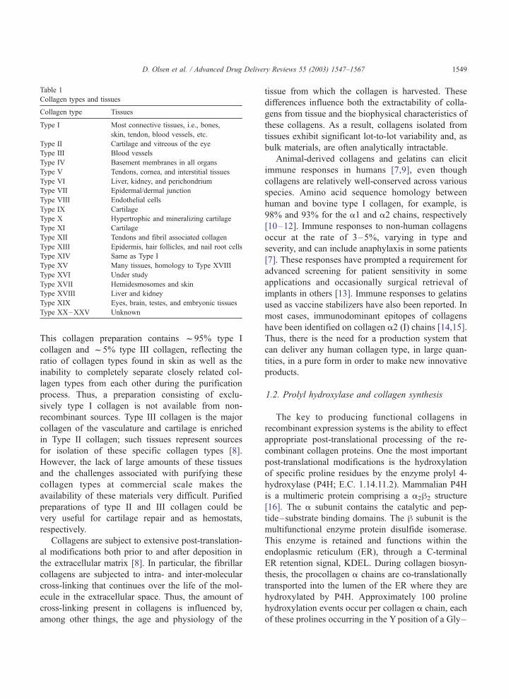
Table 1
Collagen types and tissues
Collagen type Tissues
Type I Most connective tissues, i.e., bones,
skin, tendon, blood vessels, etc.
Type II Cartilage and vitreous of the eye
Type III Blood vessels
Type IV Basement membranes in all organs
Type V Tendons, cornea, and interstitial tissues
Type VI Liver, kidney, and perichondrium
Type VII Epidermal/dermal junction
Type VIII Endothelial cells
Type IX Cartilage
Type X Hypertrophic and mineralizing cartilage
Type XI Cartilage
Type XII Tendons and fibril associated collagen
Type XIII Epidermis, hair follicles, and nail root cells
Type XIV Same as Type I
Type XV Many tissues, homology to Type XVIII
Type XVI Under study
Type XVII Hemidesmosomes and skin
Type XVIII Liver and kidney
Type XIX Eyes, brain, testes, and embryonic tissues
Type XX–XXV Unknown
D. Olsen et al. / Advanced Drug Delivery Reviews 55 (2003) 1547–1567 1549
This collagen preparation contains f95% type I
collagen and f5% type III collagen, reflecting the
ratio of collagen types found in skin as well as the
inability to completely separate closely related col-
lagen types from each other during the purification
process. Thus, a preparation consisting of exclu-
sively type I collagen is not available from non-
recombinant sources. Type III collagen is the major
collagen of the vasculature and cartilage is enriched
in Type II collagen; such tissues represent sources
for isolation of these specific collagen types [8].
However, the lack of large amounts of these tissues
and the challenges associated with purifying these
collagen types at commercial scale makes the
availability of these materials very difficult. Purified
preparations of type II and III collagen could be
very useful for cartilage repair and as hemostats,
respectively.
Collagens are subject to extensive post-translation-
al modifications both prior to and after deposition in
the extracellular matrix [8]. In particular, the fibrillar
collagens are subjected to intra- and inter-molecular
cross-linking that continues over the life of the mol-
ecule in the extracellular space. Thus, the amount of
cross-linking present in collagens is influenced by,
among other things, the age and physiology of the
tissue from which the collagen is harvested. These
differences influence both the extractability of colla-
gens from tissue and the biophysical characteristics of
these collagens. As a result, collagens isolated from
tissues exhibit significant lot-to-lot variability and, as
bulk materials, are often analytically intractable.
Animal-derived collagens and gelatins can elicit
immune responses in humans [7,9], even though
collagens are relatively well-conserved across various
species. Amino acid sequence homology between
human and bovine type I collagen, for example, is
98% and 93% for the a1 and a2 chains, respectively
[10–12]. Immune responses to non-human collagens
occur at the rate of 3–5%, varying in type and
severity, and can include anaphylaxis in some patients
[7]. These responses have prompted a requirement for
advanced screening for patient sensitivity in some
applications and occasionally surgical retrieval of
implants in others [13]. Immune responses to gelatins
used as vaccine stabilizers have also been reported. In
most cases, immunodominant epitopes of collagens
have been identified on collagen a2 (I) chains [14,15].
Thus, there is the need for a production system that
can deliver any human collagen type, in large quan-
tities, in a pure form in order to make new innovative
products.
1.2. Prolyl hydroxylase and collagen synthesis
The key to producing functional collagens in
recombinant expression systems is the ability to effect
appropriate post-translational processing of the re-
combinant collagen proteins. One the most important
post-translational modifications is the hydroxylation
of specific proline residues by the enzyme prolyl 4-
hydroxylase (P4H; E.C. 1.14.11.2). Mammalian P4H
is a multimeric protein comprising a a2h2 structure
[16]. The a subunit contains the catalytic and pep-
tide–substrate binding domains. The h subunit is the
multifunctional enzyme protein disulfide isomerase.
This enzyme is retained and functions within the
endoplasmic reticulum (ER), through a C-terminal
ER retention signal, KDEL. During collagen biosyn-
thesis, the procollagen a chains are co-translationally
transported into the lumen of the ER where they are
hydroxylated by P4H. Approximately 100 proline
hydroxylation events occur per collagen a chain, each
of these prolines occurring in the Y position of a Gly–
D. Olsen et al. / Advanced Drug Delivery Reviews 55 (2003) 1547–15671550
X–Y triplet. For example, in type I human collagen,
the ratio of hydroxyproline residues to total proline
residues plus hydroxyproline residues is approximate-
ly 0.46 [17]. In the absence of proline hydroxylation,
the essential triple helical conformation of collagen is
thermally unstable at well below physiological tem-
peratures [18]. The production of thermally stable
recombinant collagen for use in humans requires a
system that enables high-level collagen expression
along with sufficient P4H activity.
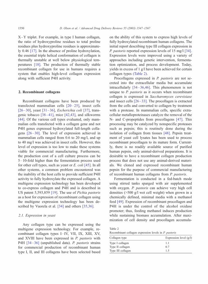
Table 2
Recombinant collagen expression levels in P. pastoris
Collagen type Expression level (g/l)
Type I collagen 1.1
Type II collagen 0.7
Type III collagen 1.5
2. Recombinant collagens
Recombinant collagens have been produced by
transfected mammalian cells [20–25], insect cells
[26–30], yeast [31–36], Escherichia coli [37], trans-
genic tobacco [38–41], mice [42,43], and silkworms
[44]. Of the various cell types evaluated, only mam-
malian cells transfected with a collagen gene and not
P4H genes expressed hydroxylated full-length colla-
gens [26–30]. The level of expression achieved in
mammalian cells ranged from 0.6 to 20 mg/l, and up
to 40 mg/l was achieved in insect cells. However, this
level of expression is too low to make these systems
viable for commercial manufacturing. Furthermore,
the production cost of a cell culture process can be
5–10-fold higher than the fermentation process used
for other cell types, such as yeast or E. coli [45]. In all
other systems, a common problem encountered was
the inability of the host cells to provide sufficient P4H
activity to fully hydroxylate the expressed collagen. A
multigene expression technology has been developed
to co-express collagen and P4H and is described in
US patent 5,593,859 [19]. The use of Pichia pastoris
as a host for expression of recombinant collagen using
the multigene expression technology has been de-
scribed by Vuorela et al. [34] and others [35,36].
2.1. Expression in yeast
Any collagen type can be expressed using the
multigene expression technology. For example, re-
combinant collagen types I–IV, VII, IX, XIII, XV,
and XVIII have been expressed in P. pastoris with
P4H [34–36] (unpublished data). P. pastoris strains
for commercial production of recombinant human
type I, II, and III collagens have been selected based
on the ability of this system to express high levels of
fully hydroxylated recombinant human collagens. The
initial report describing type III collagen expression in
P. pastoris reported expression levels of 15 mg/l [34].
Expression levels were improved using a variety of
approaches including genetic intervention, fermenta-
tion optimization, and process development. Today,
yields in excess of 1 g/l have been achieved for certain
collagen types (Table 2).
Procollagens expressed in P. pastoris are not se-
creted into the extracellular media but accumulate
intracellularly [34–36,46]. This phenomenon is not
unique to P. pastoris as it occurs when recombinant
collagen is expressed in Saccharomyces cerevisiase
and insect cells [26–33]. The procollagen is extracted
from the cells and converted to collagen by treatment
with a protease. In mammalian cells, specific extra-
cellular metalloproteinases catalyze the removal of the
N- and C-propeptides from procollagens [47]. This
processing may be catalyzed by nonspecific proteases
such as pepsin; this is routinely done during the
isolation of collagen from tissues [48]. Pepsin treat-
ment of yeast cell lysates has been used to process
recombinant procollagen to its mature form. Current-
ly, there is no readily available source of purified
human pepsin, only animal-derived preparations. It is
desirable to have a recombinant collagen production
process that does not use any animal-derived materi-
als. We cloned and expressed recombinant human
pepsin for the purpose of commercial manufacturing
of recombinant human collagens from P. pastoris.
Fermentation is conducted in a fed-batch mode
using stirred tanks sparged with air supplemented
with oxygen. P. pastoris can achieve very high cell
densities (>500 g/l wet cell weight) when grown in a
chemically defined, minimal media with a methanol
feed [49]. Expression of recombinant procollagen and
P4H is under the control of the alcohol oxidase
promoter; thus, feeding methanol induces production
while sustaining biomass accumulation. After maxi-
mization of cell density and procollagen accumula-
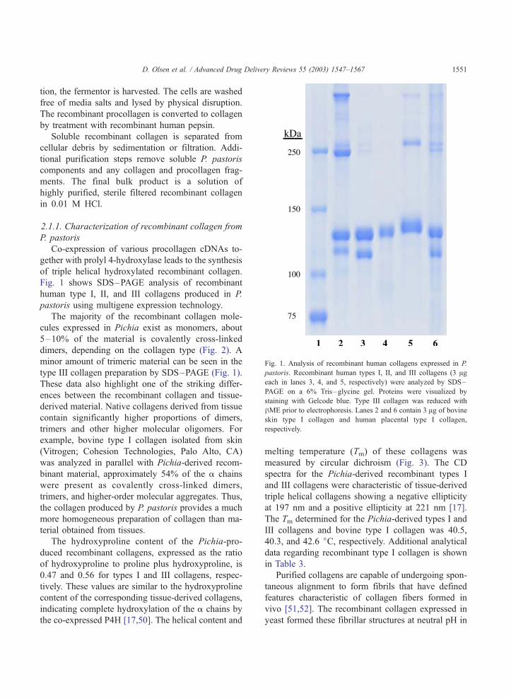
Fig. 1. Analysis of recombinant human collagens expressed in P.
pastoris. Recombinant human types I, II, and III collagens (3 Ageach in lanes 3, 4, and 5, respectively) were analyzed by SDS–
PAGE on a 6% Tris–glycine gel. Proteins were visualized by
staining with Gelcode blue. Type III collagen was reduced with
hME prior to electrophoresis. Lanes 2 and 6 contain 3 Ag of bovine
skin type I collagen and human placental type I collagen,
respectively.
D. Olsen et al. / Advanced Drug Delivery Reviews 55 (2003) 1547–1567 1551
tion, the fermentor is harvested. The cells are washed
free of media salts and lysed by physical disruption.
The recombinant procollagen is converted to collagen
by treatment with recombinant human pepsin.
Soluble recombinant collagen is separated from
cellular debris by sedimentation or filtration. Addi-
tional purification steps remove soluble P. pastoris
components and any collagen and procollagen frag-
ments. The final bulk product is a solution of
highly purified, sterile filtered recombinant collagen
in 0.01 M HCl.
2.1.1. Characterization of recombinant collagen from
P. pastoris
Co-expression of various procollagen cDNAs to-
gether with prolyl 4-hydroxylase leads to the synthesis
of triple helical hydroxylated recombinant collagen.
Fig. 1 shows SDS–PAGE analysis of recombinant
human type I, II, and III collagens produced in P.
pastoris using multigene expression technology.
The majority of the recombinant collagen mole-
cules expressed in Pichia exist as monomers, about
5–10% of the material is covalently cross-linked
dimers, depending on the collagen type (Fig. 2). A
minor amount of trimeric material can be seen in the
type III collagen preparation by SDS–PAGE (Fig. 1).
These data also highlight one of the striking differ-
ences between the recombinant collagen and tissue-
derived material. Native collagens derived from tissue
contain significantly higher proportions of dimers,
trimers and other higher molecular oligomers. For
example, bovine type I collagen isolated from skin
(Vitrogen; Cohesion Technologies, Palo Alto, CA)
was analyzed in parallel with Pichia-derived recom-
binant material, approximately 54% of the a chains
were present as covalently cross-linked dimers,
trimers, and higher-order molecular aggregates. Thus,
the collagen produced by P. pastoris provides a much
more homogeneous preparation of collagen than ma-
terial obtained from tissues.
The hydroxyproline content of the Pichia-pro-
duced recombinant collagens, expressed as the ratio
of hydroxyproline to proline plus hydroxyproline, is
0.47 and 0.56 for types I and III collagens, respec-
tively. These values are similar to the hydroxyproline
content of the corresponding tissue-derived collagens,
indicating complete hydroxylation of the a chains by
the co-expressed P4H [17,50]. The helical content and
melting temperature (Tm) of these collagens was
measured by circular dichroism (Fig. 3). The CD
spectra for the Pichia-derived recombinant types I
and III collagens were characteristic of tissue-derived
triple helical collagens showing a negative ellipticity
at 197 nm and a positive ellipticity at 221 nm [17].
The Tm determined for the Pichia-derived types I and
III collagens and bovine type I collagen was 40.5,
40.3, and 42.6 jC, respectively. Additional analyticaldata regarding recombinant type I collagen is shown
in Table 3.
Purified collagens are capable of undergoing spon-
taneous alignment to form fibrils that have defined
features characteristic of collagen fibers formed in
vivo [51,52]. The recombinant collagen expressed in
yeast formed these fibrillar structures at neutral pH in
D. Olsen et al. / Advanced Drug Delivery Reviews 55 (2003) 1547–15671552
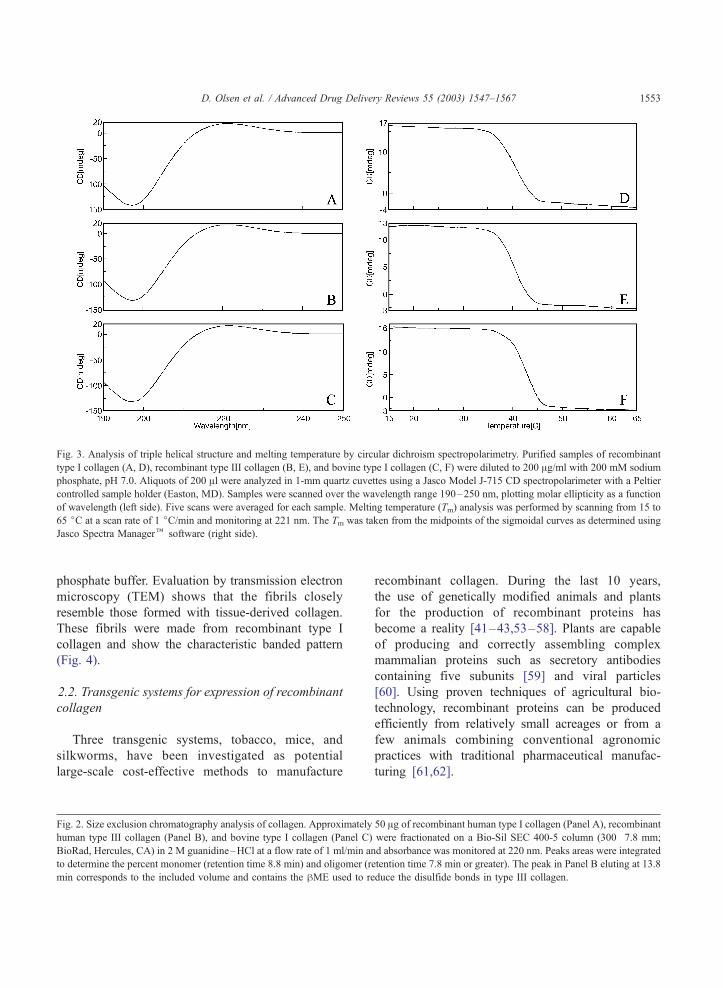
Fig. 3. Analysis of triple helical structure and melting temperature by circular dichroism spectropolarimetry. Purified samples of recombinant
type I collagen (A, D), recombinant type III collagen (B, E), and bovine type I collagen (C, F) were diluted to 200 Ag/ml with 200 mM sodium
phosphate, pH 7.0. Aliquots of 200 Al were analyzed in 1-mm quartz cuvettes using a Jasco Model J-715 CD spectropolarimeter with a Peltier
controlled sample holder (Easton, MD). Samples were scanned over the wavelength range 190–250 nm, plotting molar ellipticity as a function
of wavelength (left side). Five scans were averaged for each sample. Melting temperature (Tm) analysis was performed by scanning from 15 to
65 jC at a scan rate of 1 jC/min and monitoring at 221 nm. The Tm was taken from the midpoints of the sigmoidal curves as determined using
Jasco Spectra Managerk software (right side).
D. Olsen et al. / Advanced Drug Delivery Reviews 55 (2003) 1547–1567 1553
phosphate buffer. Evaluation by transmission electron
microscopy (TEM) shows that the fibrils closely
resemble those formed with tissue-derived collagen.
These fibrils were made from recombinant type I
collagen and show the characteristic banded pattern
(Fig. 4).
2.2. Transgenic systems for expression of recombinant
collagen
Three transgenic systems, tobacco, mice, and
silkworms, have been investigated as potential
large-scale cost-effective methods to manufacture
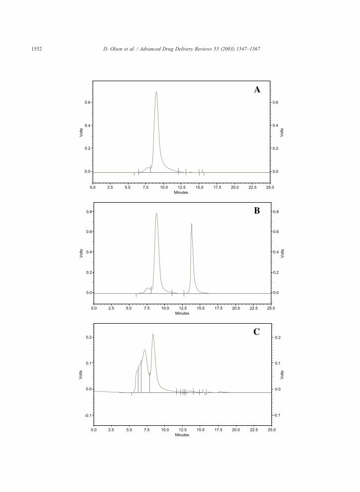
Fig. 2. Size exclusion chromatography analysis of collagen. Approximately
human type III collagen (Panel B), and bovine type I collagen (Panel C)
BioRad, Hercules, CA) in 2 M guanidine–HCl at a flow rate of 1 ml/min an
to determine the percent monomer (retention time 8.8 min) and oligomer (r
min corresponds to the included volume and contains the hME used to re
recombinant collagen. During the last 10 years,
the use of genetically modified animals and plants
for the production of recombinant proteins has
become a reality [41–43,53–58]. Plants are capable
of producing and correctly assembling complex
mammalian proteins such as secretory antibodies
containing five subunits [59] and viral particles
[60]. Using proven techniques of agricultural bio-
technology, recombinant proteins can be produced
efficiently from relatively small acreages or from a
few animals combining conventional agronomic
practices with traditional pharmaceutical manufac-
turing [61,62].
50 Ag of recombinant human type I collagen (Panel A), recombinant
were fractionated on a Bio-Sil SEC 400-5 column (300�7.8 mm;
d absorbance was monitored at 220 nm. Peaks areas were integrated
etention time 7.8 min or greater). The peak in Panel B eluting at 13.8
duce the disulfide bonds in type III collagen.
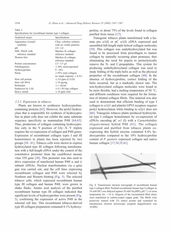
Fig. 4. Transmission electron micrograph of recombinant human
type I collagen fibril. Purified recombinant human type I collagen in
10 mM HCl was dialyzed against 20 mM Na2HPO4 pH 7.2 at room
temperature for f18 h. Aliquots of the reconstituted fibril slurry
were transferred to carbon-coated copper grids. The fibrils were
positively stained with 2% uranyl acetate and examined by
transmission electron microscopy (original magnification was
36,000).
Table 3
Specifications for recombinant human type I collagen
Analytical assays Specifications
Appearance (visual)/
solubility
Clear, colorless solution
with no visible particles
pH 2.0–2.4
SDS–PAGE with
reference standard
Matches rh collagen
reference standard
Western blot Matches rh collagen
reference standard
Protein concentration 2.5–5.5 g/l
Fibrillogenesis V 30% unincorporated
Hydroxyproline 9–10%
Purity z 95% total collagen,
no single impurity z 0.5%
Host cell protein V 2.0 ppm (L.O.D)
Host cell DNA V 10 pg/mg
Bioburden V 1 cfu/ml
Endotoxin by LAL V 0.1 EU/mg collagen
Heavy metals V 10 ppm total
D. Olsen et al. / Advanced Drug Delivery Reviews 55 (2003) 1547–15671554
2.2.1. Expression in tobacco
Plants are known to synthesize hydroxyproline-
containing proteins [63]. However, the prolyl hydrox-
ylase that is responsible for synthesis of hydroxypro-
line in plant cells does not exhibit the same substrate
sequence specificity as mammalian P4H [64,65].
Thus, production of collagen containing hydroxypro-
line only in the Y position of Gly–X–Y triplets
requires the co-expression of collagen and P4H genes.
Expression of recombinant collagen types I and III
homotrimers in plants has been reported by two
groups [38–41]. Tobacco cells were shown to express
hydroxylated type III collagen following transforma-
tion with a full-length cDNA under the control of the
constitutive promoter from the cauliflower mosaic
virus 35S gene [38]. This promoter was also used to
drive expression of transfected human P4H a and hsubunit cDNAs. Nuclear transformation via a gene
gun was carried out, and the cell lines expressing
recombinant collagen and P4H were selected by
Northern and Western blotting (Fig. 5). The selected
tobacco cells, which expressed recombinant human
type III collagen and human P4H, were grown in
shake flasks. Amino acid analysis of the purified
recombinant human type III collagen indicated that
significant levels of hydroxyproline were present (Fig.
5), confirming the expression of active P4H in the
selected cell line. This recombinant tobacco-derived
type III collagen preparation contained 8.1% hydroxy-
proline, or about 75% of the levels found in collagen
purified from tissue [17].
Transgenic tobacco plants transformed with a hu-
man pro a1(I) or pC a1(I) cDNA expressed and
assembled full-length triple helical collagen molecules
[39]. This collagen was underhydroxylated but was
found to be processed from procollagen to mature
collagen by naturally occurring plant proteases, thus
eliminating the need for pepsin to proteolytically
remove the N- and C-propeptides. This system for
producing underhydroxylated collagen was used to
study folding of the triple helix as well as the physical
properties of the recombinant collagen [40]. In the
absence of hydroxyproline, correct folding of the
helix occurred, but at a markedly slower rate. The
non-hydroxylated collagen molecules were found to
be more flexible, had a melting temperature of 30 jC,and different conditions were required for the forma-
tion of striated collagen fibrils. This material was also
used to demonstrate that efficient binding of type I
collagen to a1h1 and platelet GPVI receptors requires
prolyl hydroxylation while binding to a2h1 does not
[66]. Transgenic tobacco plants produced hydroxylat-
ed type I collagen homotrimers by co-expression of
cDNAs encoding pC a1 (I) with a Caenorhabditis
elegans/mouse hybrid P4H [41]. The collagen
expressed and purified from tobacco plants co-
expressing this hybrid enzyme contained 8.4% hy-
droxyproline compared to the 10% hydroxyproline
content of P. pastoris expressed collagen and native
human collagen [17,34,35,41].
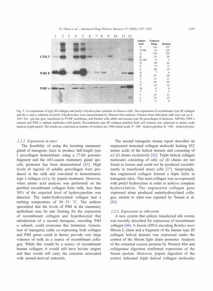
Fig. 5. Co-expression of type III collagen and prolyl 4-hydroxylase subunits in tobacco cells. The expression of recombinant type III collagen
and the a and h subunits of prolyl 4-hydroxylase were demonstrated by Western blot analysis. Extracts from individual calli were run on 4–
20% Tris–glycine gels, transferred to PVDF membrane, and blotted with rabbit anti-human type III procollagen (Chemicon, AB764), P4H hsubunit and P4H a subunit antibodies (left panel). Recombinant type III collagen purified from cell extracts was subjected to amino acids
analysis (right panel). The results are expressed as number of residues per 1000 amino acids. P–OH—hydroxyproline; K–OH—hydroxylysine.
D. Olsen et al. / Advanced Drug Delivery Reviews 55 (2003) 1547–1567 1555
2.2.2. Expression in mice
The feasibility of using the lactating mammary
gland of transgenic mice to produce full-length type
I procollagen homotrimers using a 37-kb genomic
fragment and the aS1-casein mammary gland spe-
cific promoter has been demonstrated [43]. High
levels (8 mg/ml) of soluble procollagen were pro-
duced in the milk and converted to homotrimeric
type I collagen (a1)3 by pepsin treatment. However,
when amino acid analysis was performed on the
purified recombinant collagen from milk, less than
50% of the expected level of hydroxyproline was
detected. The under-hydroxylated collagen had a
melting temperature of 30–31 jC. The authors
speculated that the levels of P4H in the mammary
epithelium may be rate limiting for the expression
of recombinant collagen and hypothesized that
introduction of a second transgene, encoding P4H
a subunit, could overcome this limitation. Genera-
tion of transgenic cattle co-expressing both collagen
and P4H genes could in theory provide very large
volumes of milk as a source of recombinant colla-
gen. While this would be a source of recombinant
human collagen, it would still have bovine origins
and thus would still carry the concerns associated
with animal-derived materials.
The second transgenic mouse report describes an
engineered truncated collagen molecule lacking 852
amino acids of the helical domain and consisting of
a2 (I) chains exclusively [42]. Triple helical collagen
molecules consisting of only a2 (I) chains are not
found in tissues and could not be produced recombi-
nantly in transfected insect cells [27]. Surprisingly,
this engineered collagen formed a triple helix in
transgenic mice. This mini-collagen was co-expressed
with prolyl hydroxylase in order to achieve complete
hydroxylation. The engineered collagen gene
expressed alone produced underhydroxylated colla-
gen, similar to what was reported by Toman et al.
[43].
2.2.3. Expression in silkworms
A new system that utilizes transfected silk worms
was recently described for expression of recombinant
collagen [44]. A fusion cDNA encoding Bombyx mori
fibroin L-chain and a fragment of the human type III
collagen helical domain was expressed under the
control of the fibroin light chain promoter. Analysis
of the extracted cocoon proteins by Western blot and
collagenase digestion confirmed expression of the
fusion protein. However, pepsin digestion of the
extract indicated triple helical collagen molecules
D. Olsen et al. / Advanced Drug Delivery Reviews 55 (2003) 1547–15671556
were not assembled in the silk glands. The authors
speculated this was due to low levels of endogenous
P4H activity in gland cells. Theoretically, this prob-
lem could be solved by co-expression of collagen and
P4H subunits, similar to what has been done in P.
pastoris, insect cells, and transgenic mice.
The potential of this system for high-level eco-
nomical expression is tremendous in that silk glands
synthesize large amounts of protein. According to the
authors, a single cocoon produces approximately 70
mg of total protein. The fusion protein containing the
collagenous sequence was found to represent 3.6% of
the total extracted protein. The authors calculated that
a 300 m2 factory containing 1.5 million silkworms
could produce about 5 kg of recombinant protein.
The finding that over-expression of P4H is re-
quired for the production of fully hydroxylated colla-
gen in such varied systems further highlights the
importance of the multigene technology for making
thermostable recombinant collagen.
2.3. Expression in E. coli
A novel approach to the production of hydroxyl-
ated collagen not involving P4H co-expression was
reported [37]. This study demonstrated the co-trans-
lational incorporation of trans-4 hydroxyproline into
a fragment of collagen, full-length a1 (1) and a2 (I)
chains and other non-collagenous recombinant pro-
teins, during over-expression in E. coli. This was
achieved by supplementing the cultures with hy-
droxyproline while growing the cells in hyperos-
motic media. Hydroxyproline was incorporated at
all proline codons in a 192-amino-acid collagen
fragment. It is also of significance to note that
efficient synthesis of collagen could only be
achieved when a synthetic collagen gene, optimized
for E. coli codon usage, was expressed. This recom-
binant collagen is biochemically distinct from tissue-
derived collagen in that hydroxyproline was present
at both X- and Y-positions of the Gly–X–Y triplets.
However, it was intriguing that the over-hydroxylat-
ed collagen fragment, consisting of 64 Gly–X–Y
repeats, assembled into a triple helix, based on
circular dichroism analysis. The authors further dem-
onstrated that they could affect the level of hydroxy-
proline incorporation by supplementing the cultures
with a mixture of hydroxyproline and proline. The
remaining challenge would be to affect the specific-
ity of hypdroxyproline incorporation and the authors
speculated this may be possible using genetic
approaches.
3. Formulations of collagen for drug delivery
Collagen molecules in physiological solutions will
spontaneously self-assemble into higher-order struc-
tures [51,52]. The fibril-forming collagens form high-
ly ordered thread-like aggregates solely on the basis of
their biophysical properties. In these fibrils, the axis of
the triple helix in the collagen monomer is parallel to
the axis of the fibrils. In tissues, the size and organi-
zation of the fibrils varies according to the function of
the tissue, associated proteins, degree of cross-linking,
and other factors [67,68].
As described above, recombinant collagens self-
assemble into ordered biological structures or fibrils
(Fig. 4). Thus, any desired physical and structural
forms (e.g., porous matrices, films, gels, or monofila-
ments) that can be fabricated from tissue-derived
collagens can also be produced using recombinant
collagens. Drugs may be incorporated into or added
after the construction of the final forms. Among the
advantages of using recombinant collagens in these
applications is the opportunity to begin with uniform
homogeneous monomolecular collagen. The subse-
quent processes may, therefore, be standardized for
consistent formulation output.
3.1. Collagen sponges
Most commercially available collagen sponges are
made of insoluble collagen derived from a variety of
animal species, such as cows, horses, and pigs.
Animal tissues are treated enzymatically and chemi-
cally to remove the majority of non-collagenous
proteins. The insoluble collagen is then resuspended,
in many cases, in a dilute organic acid, and lyophi-
lized into sponges. To increase the mechanical integ-
rity and handling properties of the material, the
sponges are cross-linked with glutaraldehyde, formal-
dehyde vapor, diisocyanate, or thermal dehydration
[69]. The resulting collagen sponges are porous and
absorbable. These absorbable collagen sponges were
developed primarily for use as hemostats.
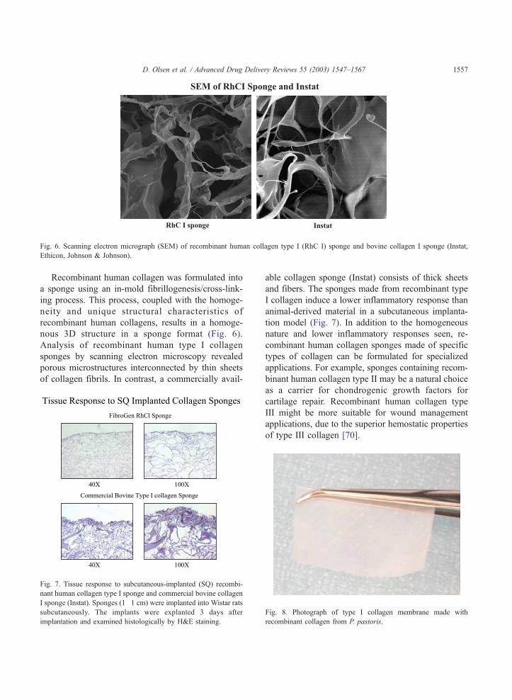
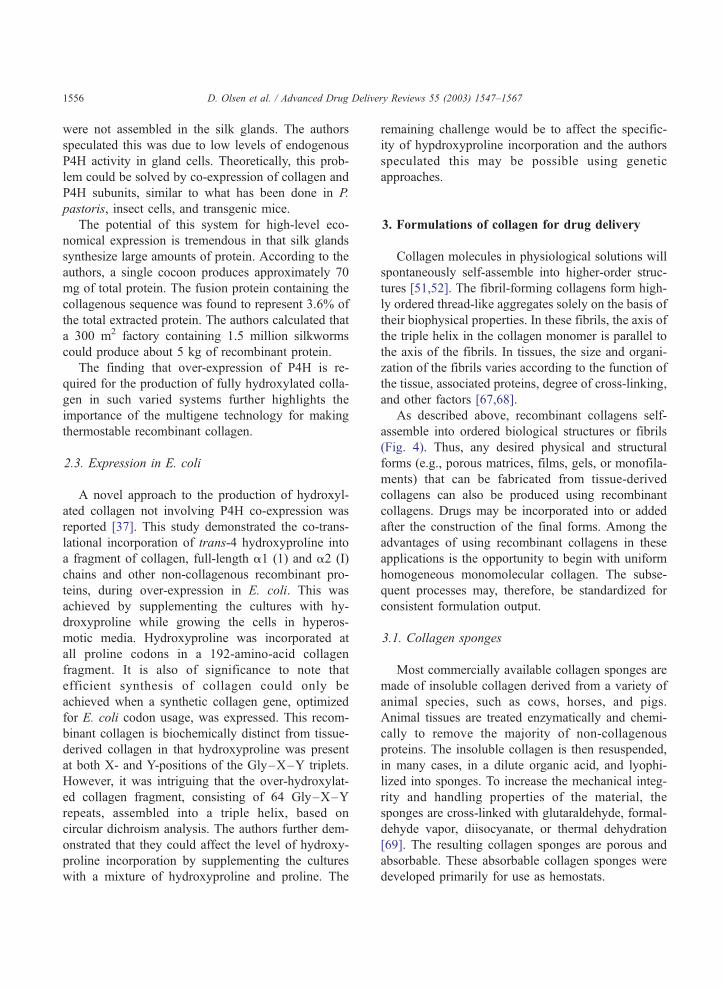
Fig. 6. Scanning electron micrograph (SEM) of recombinant human collagen type I (RhC I) sponge and bovine collagen I sponge (Instat,
Ethicon, Johnson & Johnson).
D. Olsen et al. / Advanced Drug Delivery Reviews 55 (2003) 1547–1567 1557
Recombinant human collagen was formulated into
a sponge using an in-mold fibrillogenesis/cross-link-
ing process. This process, coupled with the homoge-
neity and unique structural characteristics of
recombinant human collagens, results in a homoge-
nous 3D structure in a sponge format (Fig. 6).
Analysis of recombinant human type I collagen
sponges by scanning electron microscopy revealed
porous microstructures interconnected by thin sheets
of collagen fibrils. In contrast, a commercially avail-
Fig. 7. Tissue response to subcutaneous-implanted (SQ) recombi-
nant human collagen type I sponge and commercial bovine collagen
I sponge (Instat). Sponges (1�1 cm) were implanted into Wistar rats
subcutaneously. The implants were explanted 3 days after
implantation and examined histologically by H&E staining.
able collagen sponge (Instat) consists of thick sheets
and fibers. The sponges made from recombinant type
I collagen induce a lower inflammatory response than
animal-derived material in a subcutaneous implanta-
tion model (Fig. 7). In addition to the homogeneous
nature and lower inflammatory responses seen, re-
combinant human collagen sponges made of specific
types of collagen can be formulated for specialized
applications. For example, sponges containing recom-
binant human collagen type II may be a natural choice
as a carrier for chondrogenic growth factors for
cartilage repair. Recombinant human collagen type
III might be more suitable for wound management
applications, due to the superior hemostatic properties
of type III collagen [70].
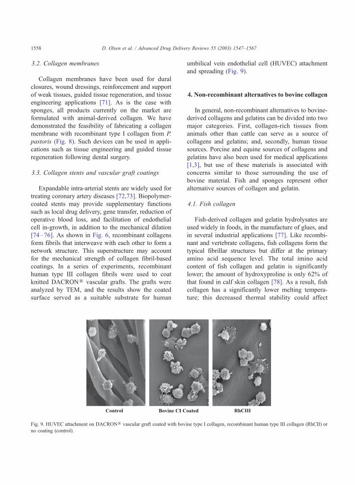
Fig. 8. Photograph of type I collagen membrane made with
recombinant collagen from P. pastoris.
elivery Reviews 55 (2003) 1547–1567
3.2. Collagen membranes
Collagen membranes have been used for dural
closures, wound dressings, reinforcement and support
of weak tissues, guided tissue regeneration, and tissue
engineering applications [71]. As is the case with
sponges, all products currently on the market are
formulated with animal-derived collagen. We have
demonstrated the feasibility of fabricating a collagen
membrane with recombinant type I collagen from P.
pastoris (Fig. 8). Such devices can be used in appli-
cations such as tissue engineering and guided tissue
regeneration following dental surgery.
3.3. Collagen stents and vascular graft coatings
Expandable intra-arterial stents are widely used for
treating coronary artery diseases [72,73]. Biopolymer-
coated stents may provide supplementary functions
such as local drug delivery, gene transfer, reduction of
operative blood loss, and facilitation of endothelial
cell in-growth, in addition to the mechanical dilation
[74–76]. As shown in Fig. 6, recombinant collagens
form fibrils that interweave with each other to form a
network structure. This superstructure may account
for the mechanical strength of collagen fibril-based
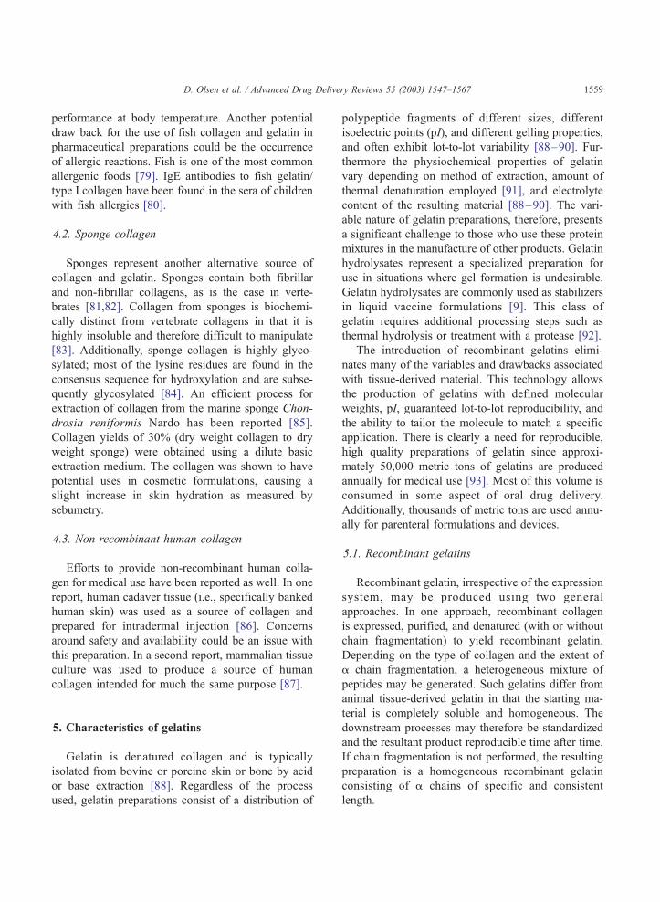
coatings. In a series of experiments, recombinant
human type III collagen fibrils were used to coat
knitted DACRONR vascular grafts. The grafts were
analyzed by TEM, and the results show the coated
surface served as a suitable substrate for human
D. Olsen et al. / Advanced Drug D1558
Fig. 9. HUVEC attachment on DACRONR vascular graft coated with bov
no coating (control).
umbilical vein endothelial cell (HUVEC) attachment
and spreading (Fig. 9).
4. Non-recombinant alternatives to bovine collagen
In general, non-recombinant alternatives to bovine-
derived collagens and gelatins can be divided into two
major categories. First, collagen-rich tissues from
animals other than cattle can serve as a source of
collagens and gelatins; and, secondly, human tissue
sources. Porcine and equine sources of collagens and
gelatins have also been used for medical applications
[1,3], but use of these materials is associated with
concerns similar to those surrounding the use of
bovine material. Fish and sponges represent other
alternative sources of collagen and gelatin.
4.1. Fish collagen
Fish-derived collagen and gelatin hydrolysates are
used widely in foods, in the manufacture of glues, and
in several industrial applications [77]. Like recombi-
nant and vertebrate collagens, fish collagens form the
typical fibrillar structures but differ at the primary
amino acid sequence level. The total imino acid
content of fish collagen and gelatin is significantly
lower; the amount of hydroxyproline is only 62% of
that found in calf skin collagen [78]. As a result, fish
collagen has a significantly lower melting tempera-
ture; this decreased thermal stability could affect
ine type I collagen, recombinant human type III collagen (RhCII) or
D. Olsen et al. / Advanced Drug Delivery Reviews 55 (2003) 1547–1567 1559
performance at body temperature. Another potential
draw back for the use of fish collagen and gelatin in
pharmaceutical preparations could be the occurrence
of allergic reactions. Fish is one of the most common
allergenic foods [79]. IgE antibodies to fish gelatin/
type I collagen have been found in the sera of children
with fish allergies [80].
4.2. Sponge collagen
Sponges represent another alternative source of
collagen and gelatin. Sponges contain both fibrillar
and non-fibrillar collagens, as is the case in verte-
brates [81,82]. Collagen from sponges is biochemi-
cally distinct from vertebrate collagens in that it is
highly insoluble and therefore difficult to manipulate
[83]. Additionally, sponge collagen is highly glyco-
sylated; most of the lysine residues are found in the
consensus sequence for hydroxylation and are subse-
quently glycosylated [84]. An efficient process for
extraction of collagen from the marine sponge Chon-
drosia reniformis Nardo has been reported [85].
Collagen yields of 30% (dry weight collagen to dry
weight sponge) were obtained using a dilute basic
extraction medium. The collagen was shown to have
potential uses in cosmetic formulations, causing a
slight increase in skin hydration as measured by
sebumetry.
4.3. Non-recombinant human collagen
Efforts to provide non-recombinant human colla-
gen for medical use have been reported as well. In one
report, human cadaver tissue (i.e., specifically banked
human skin) was used as a source of collagen and
prepared for intradermal injection [86]. Concerns
around safety and availability could be an issue with
this preparation. In a second report, mammalian tissue
culture was used to produce a source of human
collagen intended for much the same purpose [87].
5. Characteristics of gelatins
Gelatin is denatured collagen and is typically
isolated from bovine or porcine skin or bone by acid
or base extraction [88]. Regardless of the process
used, gelatin preparations consist of a distribution of
polypeptide fragments of different sizes, different
isoelectric points (pI), and different gelling properties,
and often exhibit lot-to-lot variability [88–90]. Fur-
thermore the physiochemical properties of gelatin
vary depending on method of extraction, amount of
thermal denaturation employed [91], and electrolyte
content of the resulting material [88–90]. The vari-
able nature of gelatin preparations, therefore, presents
a significant challenge to those who use these protein
mixtures in the manufacture of other products. Gelatin
hydrolysates represent a specialized preparation for
use in situations where gel formation is undesirable.
Gelatin hydrolysates are commonly used as stabilizers
in liquid vaccine formulations [9]. This class of
gelatin requires additional processing steps such as
thermal hydrolysis or treatment with a protease [92].
The introduction of recombinant gelatins elimi-
nates many of the variables and drawbacks associated
with tissue-derived material. This technology allows
the production of gelatins with defined molecular
weights, pI, guaranteed lot-to-lot reproducibility, and
the ability to tailor the molecule to match a specific
application. There is clearly a need for reproducible,
high quality preparations of gelatin since approxi-
mately 50,000 metric tons of gelatins are produced
annually for medical use [93]. Most of this volume is
consumed in some aspect of oral drug delivery.
Additionally, thousands of metric tons are used annu-
ally for parenteral formulations and devices.
5.1. Recombinant gelatins
Recombinant gelatin, irrespective of the expression
system, may be produced using two general
approaches. In one approach, recombinant collagen
is expressed, purified, and denatured (with or without
chain fragmentation) to yield recombinant gelatin.
Depending on the type of collagen and the extent of
a chain fragmentation, a heterogeneous mixture of
peptides may be generated. Such gelatins differ from
animal tissue-derived gelatin in that the starting ma-
terial is completely soluble and homogeneous. The
downstream processes may therefore be standardized
and the resultant product reproducible time after time.
If chain fragmentation is not performed, the resulting
preparation is a homogeneous recombinant gelatin
consisting of a chains of specific and consistent
length.

D. Olsen et al. / Advanced Drug Delivery Reviews 55 (2003) 1547–15671560
A second approach to recombinant gelatin produc-
tion is to intentionally direct the recombinant system
to generate a specified fragment of any selected
collagen a chain. Werten et al. [94] discusses the
Fig. 10. Schematic representation of recombinant human gelatin fragmen
(Panel B). Panel A: Bars representing different recombinant human gelati
helical domain of the human a1 (I) chain, similarly colored bar share the s
the calculated isoelectric point, based on the amino acid sequence. The nu
the fragment. Panel B: Conditioned media from Pichia strains expressin
Tricine gel and stained with Gelcode blue. *Gelatin made by thermal hyd
utility of P. pastoris as an expression system for
recombinant gelatin. Fragments of rat type III and
mouse type I collagen, ranging in size from 21 to 74
kDa, were expressed and secreted into the extracellu-
ts expressed in P. pastoris (Panel A) and analysis by SDS–PAGE
n fragments indicate the relative location of the fragment within the
ame N-terminal sequence. The numbers to left of each bar represent
mbers to the right of each bar indicate the number of amino acids in
g various fragments were analyzed by SDS–PAGE on a 10–20%
rolysis from recombinant type I collagen expressed in P. pastoris.
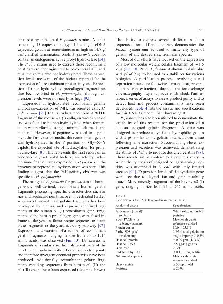
Table 4
Specifications for 8.5 kDa recombinant human gelatin
Analytical assays Specifications
Appearance (visual)/
solubility
White solid, no visible
particles
SDS–PAGE with
reference standard
Matches rh gelatin
reference standard
Protein content 80.0–105.0%
Purity-SDS gels/
densitometry
z 95% total gelatin, no
single impurity z 0.5%
Host cell protein V 0.05 ppm (L.O.D)
Host cell DNA V 5 pg/mg gelatin
Bioburden 20 cfu
Endotoxin by LAL V 0.1 EU/mg gelatin
N-terminal sequence Matches rh gelatin
reference standard
Heavy metals V 50 ppm total
Moisture V 20.0%
D. Olsen et al. / Advanced Drug Delivery Reviews 55 (2003) 1547–1567 1561
lar media by transfected P. pastoris strains. A strain
containing 15 copies of rat type III collagen cDNA
expressed gelatin at concentrations as high as 14.8 g/
l of clarified fermentation broth. P. pastoris does not
contain an endogenous active prolyl hydroxylase [34].
The Pichia strains used to express these recombinant
gelatins were not engineered to co-express P4H, and,
thus, the gelatin was not hydroxylated. These expres-
sion levels are some of the highest reported for the
expression of a recombinant protein in yeast. Expres-
sion of a non-hydroxylated procollagen fragment has
also been reported in H. polymorpha, although ex-
pression levels were not nearly as high [95].
Expression of hydroxylated recombinant gelatin,
without co-expression of P4H, was reported using H.
polymorpha, [96]. In this study, a recombinant 28 kDa
fragment of the mouse a1 (I) collagen was expressed
and was found to be non-hydroxylated when fermen-
tation was performed using a minimal salt media and
methanol. However, if peptone was used to supple-
ment the fermentation media, the recombinant gelatin
was hydroxylated in the Y position of Gly–X–Y
triplets, the expected site of hydroxylation for prolyl
hydroxylase [8]. This represents the first report of an
endogenous yeast prolyl hydroxylase activity. When
the same fragment was expressed in P. pastoris in the
presence of peptone, no hydroxylation was seen. This
finding suggests that the P4H activity observed was
specific to H. polymorpha.
The utility of P. pastoris for production of homo-
geneous, well-defined, recombinant human gelatin
fragments possessing specific characteristics such as
size and isoelectric point has been investigated further.
A series of recombinant gelatin fragments has been
developed by cloning and expressing defined seg-
ments of the human a1 (I) procollagen gene. Frag-
ments of the human procollagen gene were fused in-
frame to the yeast a factor prepro sequence to direct
these fragments to the yeast secretory pathway [97].
Expression and secretion of a number of recombinant
gelatin fragments, ranging in size from 56 to 1014
amino acids, was observed (Fig. 10). By expressing
fragments of similar size, from different parts of the
a1 (I) chain, gelatins with different isoelectric points
and therefore divergent chemical properties have been
produced. Additionally, recombinant gelatin frag-
ments encoding sequences from human a1 (II) and
a1 (III) chains have been expressed (data not shown).
The ability to express several different a chain
sequences from different species demonstrates the
Pichia system can be used to make any type of
gelatin, of any desired size, from any species.
Most of our efforts have focused on the expression
of a low molecular weight gelatin fragment of f8.5
kDa (Fig. 10, Panel A, fragment shown in dark blue
with pI of 9.4), to be used as a stabilizer for various
biologics. A purification process involving a cell
separation procedure following fermentation, precipi-
tation, solvent extraction, filtration, and ion exchange
chromatography steps has been established. Further-
more, a series of assays to assess product purity and to
detect host and process contaminants have been
developed. Table 4 lists the assays and specifications
for this 8.5 kDa recombinant human gelatin.
P. pastoris has also been utilized to demonstrate the
suitability of this system for the production of a
custom-designed gelatin fragment. A gene was
designed to produce a synthetic, hydrophilic gelatin
with a pI similar to the gelatin obtained from tissue
following lime extraction. Successful high-level ex-
pression and secretion was achieved, demonstrating
the ability of Pichia to produce designed gelatins [98].
These results are in contrast to a previous study in
which the synthesis of designed collagen-analog pep-
tides was attempted in E. coli with only limited
success [99]. Expression levels of the synthetic gene
were low due to degradation and gene instability
issues. More recently fragments of the bovine a2 (I)
gene, ranging in size from 93 to 245 amino acids,
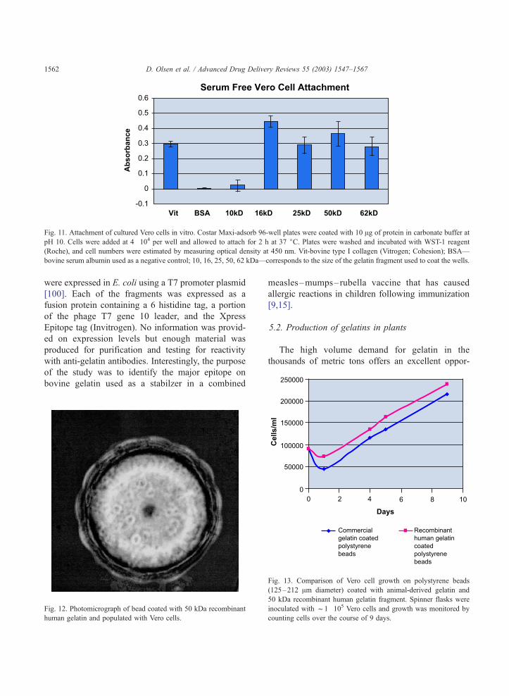
Fig. 11. Attachment of cultured Vero cells in vitro. Costar Maxi-adsorb 96-well plates were coated with 10 Ag of protein in carbonate buffer at
pH 10. Cells were added at 4�104 per well and allowed to attach for 2 h at 37 jC. Plates were washed and incubated with WST-1 reagent
(Roche), and cell numbers were estimated by measuring optical density at 450 nm. Vit-bovine type I collagen (Vitrogen; Cohesion); BSA—
bovine serum albumin used as a negative control; 10, 16, 25, 50, 62 kDa—corresponds to the size of the gelatin fragment used to coat the wells.
D. Olsen et al. / Advanced Drug Delivery Reviews 55 (2003) 1547–15671562
were expressed in E. coli using a T7 promoter plasmid
[100]. Each of the fragments was expressed as a
fusion protein containing a 6 histidine tag, a portion
of the phage T7 gene 10 leader, and the Xpress
Epitope tag (Invitrogen). No information was provid-
ed on expression levels but enough material was
produced for purification and testing for reactivity
with anti-gelatin antibodies. Interestingly, the purpose
of the study was to identify the major epitope on
bovine gelatin used as a stabilzer in a combined
Fig. 12. Photomicrograph of bead coated with 50 kDa recombinant
human gelatin and populated with Vero cells.
measles–mumps–rubella vaccine that has caused
allergic reactions in children following immunization
[9,15].
5.2. Production of gelatins in plants
The high volume demand for gelatin in the
thousands of metric tons offers an excellent oppor-
Fig. 13. Comparison of Vero cell growth on polystyrene beads
(125–212 Am diameter) coated with animal-derived gelatin and
50 kDa recombinant human gelatin fragment. Spinner flasks were
inoculated with f1�105 Vero cells and growth was monitored by
counting cells over the course of 9 days.
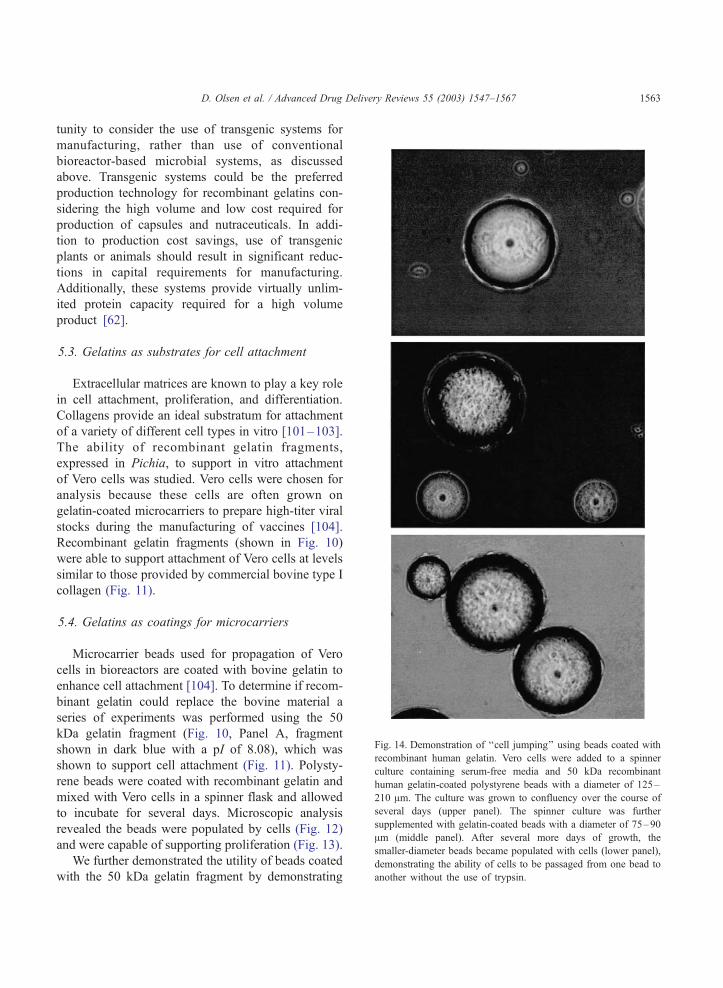
Fig. 14. Demonstration of ‘‘cell jumping’’ using beads coated with
recombinant human gelatin. Vero cells were added to a spinner
culture containing serum-free media and 50 kDa recombinant
human gelatin-coated polystyrene beads with a diameter of 125–
210 Am. The culture was grown to confluency over the course of
several days (upper panel). The spinner culture was further
supplemented with gelatin-coated beads with a diameter of 75–90
Am (middle panel). After several more days of growth, the
smaller-diameter beads became populated with cells (lower panel),
demonstrating the ability of cells to be passaged from one bead to
another without the use of trypsin.
D. Olsen et al. / Advanced Drug Delivery Reviews 55 (2003) 1547–1567 1563
tunity to consider the use of transgenic systems for
manufacturing, rather than use of conventional
bioreactor-based microbial systems, as discussed
above. Transgenic systems could be the preferred
production technology for recombinant gelatins con-
sidering the high volume and low cost required for
production of capsules and nutraceuticals. In addi-
tion to production cost savings, use of transgenic
plants or animals should result in significant reduc-
tions in capital requirements for manufacturing.
Additionally, these systems provide virtually unlim-
ited protein capacity required for a high volume
product [62].
5.3. Gelatins as substrates for cell attachment
Extracellular matrices are known to play a key role
in cell attachment, proliferation, and differentiation.
Collagens provide an ideal substratum for attachment
of a variety of different cell types in vitro [101–103].
The ability of recombinant gelatin fragments,
expressed in Pichia, to support in vitro attachment
of Vero cells was studied. Vero cells were chosen for
analysis because these cells are often grown on
gelatin-coated microcarriers to prepare high-titer viral
stocks during the manufacturing of vaccines [104].
Recombinant gelatin fragments (shown in Fig. 10)
were able to support attachment of Vero cells at levels
similar to those provided by commercial bovine type I
collagen (Fig. 11).
5.4. Gelatins as coatings for microcarriers
Microcarrier beads used for propagation of Vero
cells in bioreactors are coated with bovine gelatin to
enhance cell attachment [104]. To determine if recom-
binant gelatin could replace the bovine material a
series of experiments was performed using the 50
kDa gelatin fragment (Fig. 10, Panel A, fragment
shown in dark blue with a pI of 8.08), which was
shown to support cell attachment (Fig. 11). Polysty-
rene beads were coated with recombinant gelatin and
mixed with Vero cells in a spinner flask and allowed
to incubate for several days. Microscopic analysis
revealed the beads were populated by cells (Fig. 12)
and were capable of supporting proliferation (Fig. 13).
We further demonstrated the utility of beads coated
with the 50 kDa gelatin fragment by demonstrating
D. Olsen et al. / Advanced Drug Delivery Reviews 55 (2003) 1547–15671564
passage of Vero cells from one bead to another
without the use of trypsin, an enzyme typically
employed in passaging cells (Fig. 14). The culture
containing attached Vero cells was mixed with 50 kDa
gelatin-coated beads of smaller diameter (75–90 Am)
and incubated for 48 h. Microscopic evaluation of the
culture demonstrated cells on the surface of both the
original beads (125–210 Am diameter beads) and the
new beads of smaller diameter.
These studies demonstrate the effectiveness of
recombinant human gelatin as a microcarrier coating,
as well as a method of passaging cells in a bioreactor
without using an animal-derived protease, such as
trypsin.
Acknowledgements
The authors would like to thank Elaine Lee for help
in preparation of this manuscript. This work was
funded in part by NIH grant R01 AR45879.
References
[1] W. Friess, Collagen-biomaterial for drug delivery, Eur. J.
Pharmacol. 54 (1998) 54113–54136.
[2] L. Lachman, H.A. Liverman, J.L. Kaing (Eds.), The Theory
and Practice of Industrial Pharmacy, Lea & Febiger, Phila-
delphia, 1986.
[3] C.H. Lee, A. Singla, Y. Lee, Biomedical applications of
collagen, Int. J. Pharm. 221 (2001) 1–22.
[4] D.M. Asher, The transmissible spongiform encephalopathy
agents: concerns and responses of the United States regula-
tory agencies in maintaining the safety of biologics, Dev.
Biol. Stand. 100 (1999) 103–118.
[5] J.W. Wilesmith, J.B.M. Ryan, M.J. Atkinson, Bovine spongi-
form encephalopathy: epidemiological studies on the origin,
Vet. Rec. 128 (1991) 199–203.
[6] D.J. Prockop, K.I. Kivirikko, Collagens: molecular biology,
diseases, and potentials for therapy, Annu. Rev. Biochem. 64
(1995) 403–434.
[7] L. Cooperman, D. Michaeli, The immunogenicity of inject-
able collagen: I. A 1-year prospective study, J. Am. Acad.
Dermatol. 10 (1984) 638–646.
[8] K.I. Kivirikko, Collagen biosynthesis: a mini-review cluster,
Matrix Biol. 16 (1998) 355–356.
[9] M. Sakaguchi, S. Inouye, Systemic allergic reactions to gel-
atin included in vaccines as a stabilizer, Jpn. J. Infect. Dis. 53
(2000) 189–195.
[10] M.P. Bernard, M.L. Chu, J.C. Meyers, F. Ramirez, E.F.
Eikenberry, D.J. Prockop, Nucleotide sequences of compli-
mentary deoxyribonucleic acids for the proa1 chain of hu-
man type I procollagen. Statistical evaluation of structures
that are conserved during evolution, Biochemistry 22 (1983)
213–223.
[11] T. Shirai, S. Hattori, M. Sakaguchi, S. Inouye, A. Kimura, T.
Ebihara, S. Irie, Y. Nagai, H. Hori, The complete cDNA
coding sequence for the bovine proalpha2(I) chain of type
I procollagen, Matrix Biol. 17 (1998) 85–88.
[12] W. de Wet, M. Bernard, V. Benson-Chanda, M.L. Chu, L.
Dickson, D. Weil, F. Ramirez, Organization of the human
pro-a2(I) collagen gene, J. Biol. Chem. 262 (1987) 32–36.
[13] Zyderm Collagen Implant, Package Insert, Collagen Aes-
thetics (International), Palo Alto, CA, 2002.
[14] H. Lindsley, M. Mannik, P. Bornstein, The distribution of
antigenic determinants in rat skin collagen, J. Exp. Med. 133
(1971) 1309–1324.
[15] M. Sakaguchi, H. Hori, S. Hattori, S. Irie, A. Imai, M. Ya-
nagida, H. Miyazawa, M. Toda, S. Inouye, IgE reactivity to
alpha1 and alpha2 chains of bovine type 1 collagen in chil-
dren with bovine gelatin allergy, J. Allergy Clin. Immunol.
104 (1999) 695–699.
[16] J. Myllyharju, K.I. Kivirikko, Identification of a novel pro-
line rich peptide-binding domain in prolyl 4-hydroxylase,
EMBO J. 18 (1999) 306–312.
[17] E.J. Miller, S. Gay, The collagens: an overview and update,
Methods Enzymol. 82 (1982) 3–32.
[18] R.A. Berg, D.J. Prockop, Thermal transition of a non-hy-
droxylated form of collagen. Evidence for a role for hydroxy-
proline in stabilizing the triple-helix of collagen, Biochem.
Biophys. Res. Commun. 52 (1973) 115–120.
[19] D.J. Prockop, L. Ala-Kokko, A. Fertala, A. Sieron, K.I. Ki-
virikko, A. Geddis, T. Pihlajaniemi, inventors; Thomas Jef-
ferson University and FibroGen, assignees. Synthesis of
human procollagens and collagens in recombinant DNA sys-
tems. US Patent 5 593 859. January 14, 1997.
[20] A.E. Geddis, D.J. Prockop, Expression of human COL1A1
gene in stably transfected HT1080 cells, Matrix 13 (1993)
399–405.
[21] A. Fertala, A.L. Sieron, A. Ganguly, S.W. Li, L. Ala-Kokko,
K.R. Anumula, D.J. Prockop, Synthesis of recombinant hu-
man procollagen II in a stably transfected tumor cell line
(HT1080), Biochem. J. 298 (1994) 31–37.
[22] A. Fichard, E. Tillet, F. Delacoux, R. Garrone, F. Ruggiero,
Human recombinant a1(V) collagen chain-homotrimeric as-
sembly and subsequent processing, J. Biol. Chem. 272 (1997)
30083–30087.
[23] S. Frischholz, F. Beier, I. Girkontaite, K. Wagner, E. Poschl,
J. Turnay, U. Mayer, K. von der Mark, Characterization of
human type X procollagen and its NC-1 domain expressed as
recombinant proteins in HEK293 cells, J. Biol. Chem. 273
(1998) 4547–4555.
[24] M. Chen, F.K. Costa, C.R. Lindvay, Y.-P. Han, D.T. Wood-
ley, The recombinant expression of full-length type VII col-
lagen and characterization of molecular mechanisms
underlying dystrophic epidermolysis bullosa, J. Biol. Chem.
277 (2002) 2118–2124.
[25] E. Tillet, H. Wiedemann, R. Golbik, T.-C. Pan, R.-Z. Zhang,
D. Olsen et al. / Advanced Drug Delivery Reviews 55 (2003) 1547–1567 1565
K. Mann, M.-L. Chu, R. Timpl, Recombinant expression and
structural and binding properties of a1(VI) and a2(VI)
chains of human collagen type VI, Eur. J. Biochem. 221
(1994) 177–185.
[26] A. Lamberg, T. Helaakoski, J. Myllyharju, S. Peltonen, H.
Notbohm, T. Pihlajaniemi, K.I. Kivirikko, Characterization
of human type III collagen expressed in a baculovirus sys-
tem. Production of a protein with a stable triple helix re-
quires coexpression with the two types of recombinant
prolyl 4-hydroxylase subunit, J. Biol. Chem. 271 (1996)
11988–11995.
[27] J. Myllyharju, A. Lamberg, H. Notbohm, P.P. Fietzek, T.
Pihlajaniemi, K.I. Kivirikko, Expression of wild-type and
modified proalpha chains of human type I procollagen in
insect cells leads to the formation of stable [alpha1(I)]2al-
pha2(I) collagen heterotrimers and [alpha1(I)]3 homotrimers
but not [alpha2(I)]3 homotrimers, J. Biol. Chem. 272 (1997)
21824–21830.
[28] M. Tomita, T. Kitajima, K. Yoshizato, Formation of re-
combinant human procollagen I heterotrimers in a baculo-
virus expression system, J. Biochem. (Tokyo) 121 (1997)
1061–1069.
[29] M. Tomita, N. Ohkura, M. Ito, T. Kato, P.M. Royce, T.
Kitajima, Biosynthesis of recombinant human pro-alpha
1(III) chains in a baculovirus expression system: production
of disulphide-bonded and non-disulphide-bonded species
containing full-length triple helices, Biochem. J. 312 (1995)
847–853.
[30] M. Nokelainen, T. Helaakoski, J. Myllyharju, H. Notbohm,
T. Pihlajaniemi, P.P. Fietzek, K.I. Kivirikko, Expression and
characterization of recombinant human type II collagens with
low and high contents of hydroxylysine and its glycosylated
forms, Matrix Biol. 16 (1998) 329–338.
[31] P.D. Toman, G. Chisholm, H. McMullin, L.M. Giere, D.R.
Olsen, R.J. Kovach, S.D. Leigh, B.E. Fong, R. Chang, G.A.
Daniels, R.A. Berg, R.A. Hitzeman, Production of recom-
binant human type I procollagen trimers using a four-gene
expression system in the yeast Saccharomyces cerevisiae,
J. Biol. Chem. 275 (2000) 23303–23309.
[32] D.R. Olsen, S.D. Leigh, R. Chang, H. McMullin, W.
Ong, E. Tai, G. Chisholm, D.E. Birk, R.A. Berg, R.A.
Hitzeman, P.D. Toman, Production of human type I collagen
in yeast reveals unexpected new insights into the molecular
assembly of collagen trimers, J. Biol. Chem. 276 (2001)
24038–24043.
[33] P.R. Vaughn, M. Galanis, K.M. Richards, T.A. Tebb, J.A.
Ramshaw, J.A. Werkmeister, Production of recombinant hy-
droxylated human type III collagen fragment in Saccharo-
myces cerevisiae, DNA Cell Biol. 17 (1998) 511–518.
[34] A. Vuorela, J. Myllyharju, R. Nissi, T. Pihlajaniemi, K.I.
Kivirikko, Assembly of human prolyl 4-hydroxylase and
type III collagen in the yeast Pichia pastoris: formation of
a stable enzyme tetramer requires coexpression with collagen
and assembly of a stable collagen requires coexpression with
prolyl 4-hydroxylase, EMBO J. 16 (1997) 6702–6712.
[35] M. Nokelainen, H. Tu, A. Vuorela, H. Notbohm, K.I. Ki-
virikko, J. Myllyharju, High-level production of human
type I collagen in the yeast Pichia pastoris, Yeast 18
(2001) 797–806.
[36] J. Myllyharju, M. Nokelainen, A. Vuorela, K.I. Kivirikko,
Expression of recombinant human type I-III collagens in
the yeast Pichia pastoris, Biochem. Soc. Trans. 28 (2000)
3532–3557.
[37] D.D. Buechter, D.N. Paolella, B.S. Lelie, M.S. Brown, K.A.
Mehos, E.A. Gruskin, Co-translational incorporation of
tran-4-hydroxyproline into recombinant proteins in bacteria,
J. Biol. Chem. 278 (2003) 645–650.
[38] C. Yang, M. Bodo, R. Chang, M. Perala-Heape, E. Hama-
lainen, D. Russell, J. Polarek, Development of recombinant
gelatin by expression of hydroxylated collagen in yeast and
plants, AAPS Annual Meeting (1999) Abstract Number:
3722.
[39] F. Ruggiero, J.Y. Exposito, P. Bournat, V. Gruber, S.
Perret, J. Comte, B. Olagnier, R. Garrone, M. Theisen,
Triple helix assembly and processing of human collagen
produced in transgenic tobacco plants, FEBS Lett. 469
(2000) 132–136.
[40] S. Perret, C. Merle, S. Bernocco, P. Berland, R. Garrone,
D.J.S. Hulmes, M. Theisen, F. Ruggiero, Unhydroxylated
triple helical collagen I produced in transgenic plants pro-
vides new clues on the role of hydroxyproline in collagen
folding and fibril formation, J. Biol. Chem. 276 (2001)
43693–43698.
[41] C. Merle, S. Perret, T. Lacour, V. Jonval, S. Hudaverdian, R.
Garrone, F. Ruggiero, M. Theisen, Hydroxylated human
homotrimeric collagen I in Agrobacterium tumefaciens-
mediated transient expression and in transgenic tobacco
plants, FEBS Lett. 515 (2002) 114–118.
[42] D.C. John, R. Watson, A.J. Kind, A.R. Scott, K.E. Kadler,
N.J. Bulleid, Expression of an engineered form of recombi-
nant procollagen in mouse milk, Nat. Biotechnol. 17 (1999)
385–389.
[43] P.D. Toman, F. Pieper, N. Sakai, C. Karatzas, E. Platenburg,
I. de Wit, C. Samuel, A. Dekker, G.A. Daniels, R.A. Berg,
G.J. Platenburg, Production of recombinant human type I
procollagen homotrimer in the mammary gland of transgenic
mice, Transgenic Res. 8 (1999) 415–427.
[44] M. Tomita, H. Munetsuna, T. Sato, T. Adachi, R. Hino, M.
Hayashi, K. Shimizu, N. Nakamura, T. Tamura, K. Yoshi-
zato, Transgenic silkworms produce recombinant human
type III procollagen in cocoons, Nat. Biotechnol. 21
(2003) 52–56.
[45] R.V. Datar, T. Cartwright, C.-G. Rosen, Process economics
of animal cell and bacterial fermentations: a case study anal-
ysis of tissue plasminogen activator, Bio/technology 11
(1993) 349–357.
[46] I. Keizer-Gunnink, A. Vuorela, J. Myllyharju, T. Pihlaja-
niemi, K.I. Kivirikko, M. Veenhuis, Accumulation of prop-
erly folded human type III procollagen molecules in specific
intracellular membranous compartments in the yeast Pichia
pastoris, Matrix Biol. 19 (2000) 29–36.
[47] D.J. Prockop, A.L. Sieron, S.W. Li, Procollagen N-protei-
nase and procollagen C-proteinase. Two unusual metallopro-
teinases that are essential for collagen processing probably
D. Olsen et al. / Advanced Drug Delivery Reviews 55 (2003) 1547–15671566
have two important roles in development and cell signaling,
Matrix Biol. 16 (1998) 399–408.
[48] E.J. Miller, R.K. Rhodes, Preparation and characterization of
the different types of collagen, Methods Enzymol. 82 (1982)
33–64.
[49] G.H. Wegner, Emerging applications of the methylotrophic
yeasts, FEMS Microbiol. Rev. 7 (1990) 279–283.
[50] E. Chung, E.J. Miller, Collagen polymorphism: characteriza-
tion of molecules with the chain composition [a1(III)3] in
human tissues, Science 183 (1974) 1200–1201.
[51] B.R. Williams, R.A. Gelman, D.C. Poppke, K.A. Piez,
Collagen fibril formation. Optimal in vitro conditions
and preliminary kinetic results, J. Biol. Chem. 253
(1978) 6578–6585.
[52] R.A. Gelman, D.C. Poppke, K.A. Piez, Collagen fibril for-
mation in vitro. The role of the nonhelical terminal regions,
J. Biol. Chem. 254 (1979) 11741–11745.
[53] H.S. Mason, H. Warzecha, T. Mor, C.J. Arntzen, Edible plant
vaccines: applications for prophylactic and therapeutic mo-
lecular medicine, Trends Mol. Med. 8 (2002) 324–329.
[54] Y. Yamamoto, N. Rajbhandari, X. Lin, B. Bergmann, Y.
Nishimura, A. Stomp, Genetic transformation of duckweed,
In Vitro Cell Dev. Biol. 37 (2001) 349–353.
[55] C.L. Cramer, J.G. Boothe, K.K. Oishi, Transgenic plants for
therapeutic proteins: linking upstream and downstream strat-
egies, Curr. Top. Microbiol. Immunol. 240 (1999) 95–118.
[56] M.B. Hein, Y. Tang, D.A. McLeod, K.D. Janda, A. Hiatt,
Evaluation of immunoglobulins from plant cells, Biotechnol.
Prog. 7 (1991) 455–461.
[57] M. Young, Production of biopharmaceutical proteins in milk,
Biopharm 10 (1998) 34–38.
[58] L.M. Houdebine, Transgenic animal bioreactors, Transgenic
Res. 9 (2000) 305–320.
[59] J.K. Ma, A. Hiatt, M. Hein, N.D. Vine, F. Wang, P. Stabila,
C. von Dolleweerd, K. Mostov, T. Lehner, Generation and
assembly of secretory antibodies in plants, Science 268
(1995) 716–719.
[60] L.J. Richter, Y. Thanavala, C.J. Arntzen, H.S. Mason, Pro-
duction of hepatitis B surface antigen in transgenic plants for
oral immunization, Nat. Biotechnol. 18 (2000) 1167–1171.
[61] L. Miele, Plants as bioreactors for biopharmaceuticals: regu-
latory considerations, Trends Biotechnol. 15 (1997) 45–50.
[62] D. Mison, J. Curling, The industrial production costs of re-
combinant therapeutic proteins expressed in transgenic corn,
Biopharm 13 (2000) 48–54.
[63] D.T.A. Lamport, The isolation and partial characterization
of hydroxyproline-rich glycopeptides obtained by enzymic
degradation of primary cell walls, Biochemist 8 (1969)
1155–1163.
[64] M. Tanaka, K. Sato, T. Uchida, Plant prolyl hydroxylase
recognizes poly(L-proline) II helix, J. Biol. Chem. 256
(1981) 11397–11400.
[65] R. Hieta, J. Myllyharju, Cloning and characterization of a
low molecular weight prolyl 4-hydroxylase from Arabidopsis
thaliana. Effective hydroxylation of proline-rich, collagen-
like, and hypoxia-inducible transcription factor a-like pepti-
des, J. Biol. Chem. 277 (2002) 23965–23971.
[66] S. Perret, J.A. Eble, P.R. Siljander, C. Merle, R.W. Farndate,
M. Theisen, F. Ruggiero, Prolyl hydroxylation of collagen
type I is required for efficient binding to integrin alpha 1 beta
1 and platelet GPVI but not to alpha 2 beta 1, J. Biol. Chem.
278 (2003) 29873–29879.
[67] D.E. Birk, F.H. Silver, R.L. Trelstad, Matrix assembly, in:
E.D. Hay (Ed.), Cell Biology of the Extracellular Matrix,
Plenum, New York, 1991, pp. 221–253.
[68] D.F. Holmes, C.J. Gilpin, C. Baldock, U. Ziese, A.J. Koster,
K.E. Kadler, Corneal collagen fibril structure in three dimen-
sions: structural insights into fibril assembly, mechanical
properties, and tissue organization, Proc. Natl. Acad. Sci.
U. S. A. 98 (2001) 7307–7312.
[69] M.E. Nimmi, D. Cheung, B. Strates, M. Kodama, K. Skeikh,
Chemically modified collagen: a biomaterial for tissue re-
placement, J. Biomed. Mater. Res. 21 (1987) 741–771.
[70] L. Balleisen, S. Gay, R. Marx, K. Kuhn, Comparative inves-
tigation on the influence of human and bovine collagen types
I, II, and III on aggregation of human platelets, Klin. Wo-
chenschr. 53 (1975) 903–905.
[71] M. Maeda, K. Kadota, M. Kajihara, A. Sano, K. Fujioka,
Sustained release of human growth hormone (hGH) from
collagen film and evaluation of effect on wound healing in
db/db mice, J. Control. Release 77 (2001) 261–272.
[72] W. Serruys, P. de Jaegere, F. Kiemeneji, C. Macaya, W.
Rutsch, G. Heyndrickx, H. Emanuelsson, J. Marco, V.
Legrand, P. Materne, A comparison of balloon expandable
stent implantation with balloon angioplasty in patients with
coronary artery disease. Benestent Study Group, N. Engl.
J. Med. 331 (1994) 489–495.
[73] L. Maillard, M. Hamon, K. Khalife, P.G. Steg, F. Bey-
gui, J.L. Guermonprez, C.M. Spaulding, J.M. Boulenc, J.
Lipiecki, A. Lafont, P. Brunel, G. Grollier, R. Koning, P.
Coste, X. Favereau, B. Lancelin, E. Van Belle, P. Ser-
ruys, J.P. Monassier, P. Raynaud, A comparison of sys-
temic stenting and conventional balloon angioplasty during
primary percutaneous transluminal coronary angioplasty for
acute myocardial infarction. STENTIM-2, J. Am. Coll. Car-
diol. 34 (1999) 1498–1506.
[74] Y. Nakayama, J.Y. Kim, N. Shogo, H. Ueno, M. Takehisa,
Development of high-performance stent: gelatinous photo-
gel-coated stent that permits drug delivery and gene transfer,
J. Biomed. Mater. Res. 57 (2001) 559–566.
[75] J. Yuan, R. Gao, R. Shi, L. Song, J. Tang, Y. Li, Intravascular
local gene transfer mediated by protein-coated metallic stent,
Chin. Med. J. (Engl.) 114 (2001) 1043–1045.
[76] M.J. Wissink, R. Beernink, A.A. Poot, G.H. Engbers, T.
Beugeling, W.G. van Aken, J. Feijen, Improved endothelial-
ization of vascular grafts by local release of growth factor
from heparinized collagen matrices, J. Control. Release 64
(2000) 103–114.
[77] J. Soper, Method of encapsulating food or flavor particles
using warm water fish gelatin and capsules produced there-
from, US Patent 5,603,952, 1997.
[78] K.A. Piez, J. Gross, The amino acid composition of some
fish collagens: the relationship between composition and
structure, J. Biol. Chem. 235 (1960) 995–1000.
D. Olsen et al. / Advanced Drug Delivery Reviews 55 (2003) 1547–1567 1567
[79] K. Asa, Studies of hypersensitivity to fish: a clinical study,
Int. Arch. Allergy Appl. Immunol. 29 (1989) 990–994.
[80] M. Sakaguchi, M. Toda, T. Ebihara, S. Irie, H. Hori, A.
Imai, M. Yanagida, H. Miyazawa, H. Ohsuna, Z. Ikezawa,
S. Inouye, IgE antibody to fish gelatin (type I collagen) in
patients with fish allergy, J. Allergy Clin. Immunol. 106
(2000) 579–584.
[81] J.-Y. Exposito, D. Le Guellec, Q. Lu, R. Garrone, Short chain
collagens in sponges are encoded by a family of closely
related genes, J. Biol. Chem. 266 (1991) 21923–21928.
[82] J.-Y. Exposito, R. Garrone, Characterization of a fibrillar
collagen gene in sponges reveals the evolutionary appear-
ance of two collagen gene families, Proc. Natl. Acad. Sci.
U. S. A. 87 (1990) 6669–6673.
[83] R. Garrone (Ed.), Phylogenesis of Connective Tissue, Mor-
phological Aspects and Biosynthesis of Sponge Intercellular
Matrix, Karger, Basel, 1978.
[84] S. Junqua, L. Robert, R. Garrone, M. Pavans de Ceccatty, J.
Vacelet, Biochemical and morphological studies on collagen
of horny sponges. Ircinia filaments compared to sponginess,
Connect. Tissue Res. 2 (1974) 193–203.
[85] D. Swatschek, W. Schatton, J. Kellermann, W.E.G. Mueller,
J. Kreuter, Marine sponge collagen: isolation, characteriza-
tion and effects on the skin parameters surface-pH, moisture
and sebum, Eur. J. Pharm. Biopharm. 53 (2002) 107–113.
[86] A.P. Sclafani, T. Romo III, A. Parker, S.A. McCormick, R.
Cocker, A. Jacono, Homologous collagen dispersion (dermal-
ogen) as a dermal filler: persistence and histology compared
with bovine collagen, Ann. Plast. Surg. 49 (2002) 181–188.
[87] G.K. Naughton, From lab bench to market: critical issues
in tissue engineering, Ann. N.Y. Acad. Sci. 961 (2002)
372–385.
[88] A. Asghar, R.L. Henrickson, Chemical, biochemical, func-
tional, and nutritional characteristics of collagen in food sys-
tems, Adv. Food Res. 28 (1982) 231–372.
[89] C.A. Farrugia, I.V. Farrugia, M.J. Groves, Comparison of the
molecular weight distribution of gelatin fractions by size-
exclusion chromatography and light scattering, Pharm. Phar-
macol. Commun. 4 (1998) 559–562.
[90] J.M. Saddler, P.J. Horsey, The new generation gelatins. A
review of their history, manufacture and properties, Anes-
thesiology 42 (1987) 998–1004.
[91] A. Courts, The N-terminal amino acid residues of gelatin: 2.
Thermal degradation, Biochem. J. 58 (1954) 74–79.
[92] Y. Sakai, R. Yamato, M. Onuma, T. Kikuta, M. Watanabe, T.
Nakayama, Non-antigenic and low allergenic gelatin pro-
duced by specific digestion with an enzyme-coupled matrix,
Biol. Pharm. Bull. 21 (1998) 330–334.
[93] R.W. Moskowitz, Role of collagen hydrolysate in bone and
joint disease, Semin. Arthritis Rheum. 30 (2000) 87–99.
[94] M.W.T. Werten, T.J. van den Bosch, R.D. Wind, H. Mooi-
broek, F.A. de Wolf, High-yield secretion of recombinant
gelatins by Pichia pastoris, Yeast 15 (1999) 1087–1096.
[95] E.C. de Bruin, F.A. de Wolf, N.C. Lane, Expression and
secretion of human alpha 1 (I) procollagen fragment by Han-
senula polymorpha as compared to Pichia pastoris, Enzyme
Microb. Technol. 26 (2000) 640–644.
[96] E.C. de Bruin, M.W.T. Werten, C. Laane, F.A. de Wolf,
Endogenous prolyl 4-hydroxylation in Hansenula polymor-
pha and its use for the production of hydroxylated recombi-
nant gelatin, FEMS Yeast Res. 1 (2002) 291–298.
[97] A.J. Brake, J.P. Merryweather, D.G. Coit, U.A. Heberlein,
F.R. Masiarz, G.T. Mullenbach, M.S. Urdea, P. Valenzuela, P.
Barr, Alpha-factor-directed synthesis and secretion of mature
foreign proteins in Saccharomyces cerevisiae, Proc. Natl.
Acad. Sci. U. S. A. 81 (1984) 4642–4646.
[98] M.W.T. Werten, W.H. Wisselink, T.J. Jansen-van den Bosch,
E.C. de Bruin, F.A. de Wolf, Secreted production of a cus-
tom-designed, highly hydrophilic gelatin in Pichia pastoris,
Protein Eng. 14 (2001) 447–454.
[99] I. Goldberg, A.J. Salerno, T. Patterson, J.I. Williams, Cloning
and expression of a collagen-analog-encoding synthetic gene
in Escherichia coli, Gene 80 (1989) 305–314.
[100] H. Hori, S. Hattori, S. Inouye, A. Kimura, S. Irie, H.
Miyazawa, M. Sakaguchi, Analysis of the major epitope of
the a2 chain of bovine type I collagen in children with bo-
vine gelatin allergy, J. Allergy Clin. Immunol. 110 (2002)
652–657.
[101] T. Nakagawa, P. Pawelek, F. Grimmel, Extracellular matrix
organization modulates fibroblast growth and growth factor
responsiveness, Exp. Cell Res. 182 (1989) 572–582.
[102] S.L. Schor, Cell proliferation and migration on collagen sub-
strata in vitro, J. Cell Sci. 41 (1980) 159–175.
[103] D. Carey, Control of growth and differentiation of vascular
cells by extracellular matrix proteins, Annu. Rev. Physiol. 53
(1991) 161–177.
[104] B. Montagnon, J.C. Vincent-Falquet, B. Fanget, Thousand
litre scale microcarrier culture of Vero cells for killed polio
virus vaccine: promising results, Dev. Biol. Stand. 55 (1984)
37–38.