Development, characterization, and toxicity evaluation of amphotericin B–loaded gelatin...
10
Original Article: Toxicology Development, characterization, and toxicity evaluation of amphotericin B–loaded gelatin nanoparticles Manoj Nahar, MPharm, Dinesh Mishra, MPharm, Vaibhav Dubey, MPharm, Narendra Kumar Jain, PhD ⁎ Pharmaceutics Research Laboratory, Department of Pharmaceutical Science, Dr. H.S. Gour University, Sagar, India Abstract Our aim in the present investigation was to develop a nanoparticulate carrier of amphotericin B (AmB) for controlled delivery as well as reduced toxicity. Nanoparticles of different gelatins (GNPs) (type A or B) were prepared by two-step desolvation method and optimized for temperature, pH, amount of cross-linker, and theoretical drug loading. AmB-loaded GNPs were characterized for size, polydispersity index (PI), shape, morphology, surface charge, drug release, and hemolysis. The developed GNPs (GNP A300 ) were found to be of nanometric size (213 ± 10 nm), having low PI (0.092 ± 0.015) and good entrapment efficiency (49.0 ± 2.9%). All GNPs showed biphasic release characterized by an initial burst followed by controlled release. The in vivo hematological toxicity results suggest nonsignificant reduction (P N .05) in hemoglobin concentration and hematocrit. Nephrotoxicity results showed that there was a nonsignificant (P N .05) increase in blood urea nitrogen and serum creatinine levels. The results confirm that developed GNPs could optimize AmB delivery in terms of cost and safety, and type A gelatin with bloom number 300 was found suitable for such preparation. © 2008 Elsevier Inc. All rights reserved. Key words: Nanoparticles; Gelatin; Amphotericin B; Desolvation; Nephrotoxicity Administration of amphotericin B (AmB), a potent fungal agent, is limited by its pronounced side effects (e.g., chills, fever, nausea, hemolytic toxicity, and nephrotoxicity). 1 Conventional micellar solutions of AmB with deoxycholate (Fungizone) has been reported to cause serious nephro- toxicity. 2 The alternate novel lipid-based nanoparticulate formulations, including liposomes (AmBisome), AmB-lipid complex (Abelcet), and AmB colloidal dispersion (Ampho- cil), have been developed to enhance the therapeutic index and to reduce the hemolytic and nephrotoxic side effects, but are quite expensive. 3 Recently, low-priced AmB disc formulations containing cationic lipid dioctadecyldimethyl have been developed, but such formulations have been reported to possess low drug-loading capacity. 4,5 Moreover, AmB is the drug of choice for diseases such as visceral leishmaniasis wherein long-term infusion is needed and that require hospitalization, increased cost, and poor patient compliance. All these factors warrant the need of developing an alternative drug carrier, which may provide safe, effective, and controlled delivery of AmB. Nanoparticulate carriers have always been attractive on account of their size and capacity for spatial and temporal controlled delivery of bioactives. 6 Nanoparticles of various polymers like poly(lactide-co- glycolide), poly(ɛ-caprolactone), and more recently chito- san-dextran with AmB have been investigated. 7-10 Gelatin nanoparticles (GNPs) have recently been developed to offer one good viable option because of their low cost, biocompatibility, biodegradability, low antigenicity, and applications in several parenteral formulations. 11 GNPs Available online at www.sciencedirect.com Nanomedicine: Nanotechnology, Biology, and Medicine 4 (2008) 252 – 261 www.nanomedjournal.com Received 11 December 2007; accepted 24 March 2008. The authors are thankful to the Indian Council of Medical Research, New Delhi for financial support. ⁎ Corresponding author. Department of Pharmaceutical Sciences, Dr. H. S. Gour University, Sagar 470003, India. E-mail address: [email protected] (N.K. Jain). 1549-9634/$ – see front matter © 2008 Elsevier Inc. All rights reserved. doi:10.1016/j.nano.2008.03.007 Please cite this article as: M. Nahar, D. Mishra, V. Dubey, N.K. Jain, Development, characterization, and toxicity evaluation of amphotericin B–loaded gelatin nanoparticles. Nanomedicine: NBM 2008;4:252-261, doi:10.1016/j.nano.2008.03.007.
-
Upload
independent -
Category
Documents
-
view
0 -
download
0
Transcript of Development, characterization, and toxicity evaluation of amphotericin B–loaded gelatin...
Available online at www.sciencedirect.com
Nanomedicine: Nanotechnology, Biology, and Medicine 4 (2008) 252–261www.nanomedjournal.com
Original Article: Toxicology
Development, characterization, and toxicity evaluationof amphotericin B–loaded gelatin nanoparticles
Manoj Nahar, MPharm, Dinesh Mishra, MPharm,Vaibhav Dubey, MPharm, Narendra Kumar Jain, PhD⁎
Pharmaceutics Research Laboratory, Department of Pharmaceutical Science, Dr. H.S. Gour University, Sagar, India
Abstract Our aim in the present investigation was to develop a nanoparticulate carrier of amphotericin B
Received 11 DeceThe authors are th
New Delhi for financi⁎Corresponding au
S. Gour University, SE-mail address: jn
1549-9634/$ – see frodoi:10.1016/j.nano.20
Please cite this articgelatin nanoparticles
(AmB) for controlled delivery as well as reduced toxicity. Nanoparticles of different gelatins (GNPs)(type A or B) were prepared by two-step desolvation method and optimized for temperature, pH,amount of cross-linker, and theoretical drug loading. AmB-loaded GNPs were characterized for size,polydispersity index (PI), shape, morphology, surface charge, drug release, and hemolysis. Thedeveloped GNPs (GNPA300) were found to be of nanometric size (213 ± 10 nm), having low PI(0.092 ± 0.015) and good entrapment efficiency (49.0 ± 2.9%). All GNPs showed biphasic releasecharacterized by an initial burst followed by controlled release. The in vivo hematological toxicityresults suggest nonsignificant reduction (P N .05) in hemoglobin concentration and hematocrit.Nephrotoxicity results showed that there was a nonsignificant (P N .05) increase in blood ureanitrogen and serum creatinine levels. The results confirm that developed GNPs could optimize AmBdelivery in terms of cost and safety, and type A gelatin with bloom number 300 was found suitablefor such preparation.© 2008 Elsevier Inc. All rights reserved.
Key words: Nanoparticles; Gelatin; Amphotericin B; Desolvation; Nephrotoxicity
Administration of amphotericin B (AmB), a potent fungalagent, is limited by its pronounced side effects (e.g., chills,fever, nausea, hemolytic toxicity, and nephrotoxicity). 1
Conventional micellar solutions of AmB with deoxycholate(Fungizone) has been reported to cause serious nephro-toxicity. 2 The alternate novel lipid-based nanoparticulateformulations, including liposomes (AmBisome), AmB-lipidcomplex (Abelcet), and AmB colloidal dispersion (Ampho-cil), have been developed to enhance the therapeutic indexand to reduce the hemolytic and nephrotoxic side effects, butare quite expensive. 3 Recently, low-priced AmB disc
mber 2007; accepted 24 March 2008.ankful to the Indian Council of Medical Research,al support.thor. Department of Pharmaceutical Sciences, Dr. H.agar 470003, [email protected] (N.K. Jain).
nt matter © 2008 Elsevier Inc. All rights reserved.08.03.007
le as: M. Nahar, D. Mishra, V. Dubey, N.K. Jain, Develop. Nanomedicine: NBM 2008;4:252-261, doi:10.1016/j.nano
formulations containing cationic lipid dioctadecyldimethylhave been developed, but such formulations have beenreported to possess low drug-loading capacity. 4,5 Moreover,AmB is the drug of choice for diseases such as visceralleishmaniasis wherein long-term infusion is needed and thatrequire hospitalization, increased cost, and poor patientcompliance. All these factors warrant the need of developingan alternative drug carrier, which may provide safe,effective, and controlled delivery of AmB.
Nanoparticulate carriers have always been attractive onaccount of their size and capacity for spatial and temporalcontrolled delivery of bioactives. 6
Nanoparticles of various polymers like poly(lactide-co-glycolide), poly(ɛ-caprolactone), and more recently chito-san-dextran with AmB have been investigated. 7-10 Gelatinnanoparticles (GNPs) have recently been developed to offerone good viable option because of their low cost,biocompatibility, biodegradability, low antigenicity, andapplications in several parenteral formulations. 11 GNPs
ment, characterization, and toxicity evaluation of amphotericin B–loaded.2008.03.007.

253M. Nahar et al / Nanomedicine: Nanotechnology, Biology, and Medicine 4 (2008) 252–261
have been previously reported for delivery of different drugslike methotrexate, 12,13 doxorubicin, 14 cycloheximide, 15
paclitaxel, 16 and chloroquine phosphate 17as well as forgene delivery. 18-21 Furthermore, antibody-modified GNPswere used to target leukemic cells and primary Tlymphocytes. 22,23 Various methods, including nano-encapsulation, 24 emulsification-solvent evaporation, 13
desolvation, 25 and coacervation-phase separation, 16 havebeen used to prepare GNPs. GNPs prepared by many ofthese methods were found to be large in size and have ahigh PI due to heterogeneity in molecular weight of thegelatin polymer. Variation in size and PI of novelnanoparticulate carrier systems may affect drug release,drug loading, and alternation in in vivo parameters likeuptake by macrophages. Moreover, poor aqueous solubilityand self-aggregation of AmB present a great challenge tothe development of an effective and safe carrier system.These challenges prompted us to design a nanoparticulatedelivery system using a low-cost polymer like gelatin, withproper control of the process parameters that may developa carrier with small size, low PI, and maximum entrapmentefficiency (EE), which can effectively deliver AmB withfewer or no side effects.
Therefore, in the present investigation we attempted todevelop an alternative delivery system that may overcomethe severe side effects of existing AmB formulations byreducing self-aggregation, improving patient compliance bycontrolled delivery, and providing safe and effective deliveryat low cost as an alternative to lipid-based formulations.
Methods
Materials
Gelatin type A from porcine skin (300, 175 bloom),gelatin type B from bovine skin (75 bloom), glutaraldehydegrade I (25% v/v aqueous solution), trypsin from bovinepancreas, HEPES buffer, and amberlite XAD 16 resin werepurchased from Sigma Chemical Co. (St Louis, MO).Acetone, ethanol, dimethyl sulfoxide (DMSO), and acetoni-trile were purchased from Central Drug House (New Delhi,India). AmB was a gift from Dabur India Ltd. (Ghaziabad,UP, India). All other chemicals were of analytical grade andused as received. Triple-distilled deionized water was usedfor all experiments.
Preparation of GNPsGNPs were prepared by a two-step desolvation method
reported by Coester et al 25 with slight modifications. Briefly,200 mg gelatin (A or B) was dissolved in distilled water(10 mL) under constant heating at 40° ± 1°C. Acetone(10 mL) was added to the gelatin solution as a desolvatingagent to precipitate the high-molecular-weight (HMW)gelatin. The supernatant was discarded, and the HMWgelatin was redissolved by adding distilled water (10 mL)with stirring at 600 rpm (Remi, Mumbai, India) under
constant heating. The pH of the gelatin solution at the seconddesolvation step was adjusted (between 2 and 12). AmB(dissolved in 500 μL of DMSO) was added in aqueouspolymer phase, followed by dropwise addition of acetone(30 mL) to form GNPs. At the end of the process,glutaraldehyde solution (25% v/v aqueous solution) wasadded as a cross-linking agent, and the solution was stirredfor 12 hours at 600 rpm. The unentrapped AmB wasremoved by adding 10 mL of adsorbent polymer beads (2%w/v), amberlite XAD 16 followed by filtration 7 (1-μm filter;Whatman Japan KK, Tokyo, Japan). DMSO was removedwith repeated mild washing with distilled water. The GNPsobtained were lyophilized with trehalose (5% w/v solution)for further investigations. Effect of parameters like pH,temperature, amount of glutaraldehyde, and theoretical drugloading on the size, PI, and EE of the GNPs was studied.
Determination of EE, yield, and actual drug loading
Entrapment efficiencies of GNPs were determined by themethod reported by Leo et al 26 Briefly, AmB-loadednanoparticles (10 mg) were dispersed in an aqueous solution(10 mL; NaCl, 0.9%, w/v and 5% v/v DMSO) containingtrypsin (200 μg/mL) at a trypsin-to-nanoparticles ratio of 1:5(w/w). The dispersion was kept for 5 hours at 37° ± 1°C inthe dark under magnetic agitation; a clear solution wasobtained, and free AmB was quantified using high-performance liquid chromatography by the methoddescribed by Echevarrya et al, 27 with minor modifications.The estimation was carried out using a C18 column (250 ×34.6 mm, 5 mm particle size) (Shimadzu, Kyoto, Japan),preceded by a guard column (45 × 34.6 mm). The mobilephase was composed of acetonitrile–acetic acid (1%)–water(41:43:16) at a flow rate of 1.5 mL/min. The chromato-graphy was carried out at room temperature (25° ± 1°C). Theultraviolet (UV) detection was performed at 405 nm using anSPD-M10Avp diode array UV detector (Shimadzu, Kyoto,Japan). The retention time was found to be at 4.3 minutes.The EE was calculated as follows:
Drug entrapment efficiency EEð Þ kð Þ¼ mass of drug in GNPs=mass of drug used in formulationð Þ
� 100
ð1ÞThe purified nanosuspension was ultracentrifuged (L-8
60M; Beckman, Buckinghamshire, UK) at 31,000 g for 1hour at 4° ± 1°C. The supernatant was discarded and thepellet was freeze-dried. The yields of various GNPs werecalculated using Eq. (2), whereas the actual drug contents ofGNPs were calculated using Eq. (3).
Nanoparticles yield kw=wð Þ¼ mass of recovered GNPs=total mass of polymer and drug addedð Þ
� 100
ð2ÞActual drug loading kw=wð Þ¼ mass of drug in GNPs=mass of GNPs recoveredð Þ
� 100 ð3Þ

254 M. Nahar et al / Nanomedicine: Nanotechnology, Biology, and Medicine 4 (2008) 252–261
Particle size, shape, surface charge, and PI
The particle size, surface charge, and PI weredetermined by using a Malvern Zetasizer (NanoZS;Malvern Instruments Inc., Worcestershire, UK). Particlesize and PI of GNPs were determined by light-scatteringmethod based on laser diffraction at an angle of 135degrees. Typically, GNPs were suspended in aqueousmedium (1.5 mL) and placed in a cuvette at a concentra-tion of 0.3 mg/mL at 25° ± 1°C. The viscosity andrefractive index of the continuous phase were set to thosespecific to water. GNPs surface charge was determined bylaser doppler anemometry. Briefly, GNPs were suspendedin 1 mM HEPES buffer and adjusted to pH 7.4 by 0.1 MHCl so as to maintain a constant ionic strength.
Transmission electron microscopy (TEM) (H7500; Hita-chi Ltd., Tokyo, Japan) was used for determination of shapeand size of GNPs. The aqueous dispersion (one drop) wasplaced over a 400-mesh carbon-coated copper grid followedby negative staining with phosphotungstic acid solution (3%w/v, adjusted to pH 4.7 with KOH) and placed at theaccelerating voltage of 95 kV.
UV-visible spectral study
UV-visible (UV-vis) spectra of pure AmB as well aslyophilized AmB-loaded GNPs were obtained using UV-visspectrophotometer (Shimadzu1601; Kyoto, Japan). Briefly,AmB-loaded lyophilized GNPs were dispersed in 5.0 mL ofdeionized water (containing 5% v/v DMSO) to give a finalAmB concentration of 10 μg/mL. The suspension is thenfiltered with a 0.22-μm membrane filter and scanned in therange 200-550 against plain nanoparticles as blank.
In vitro release kinetics
In vitro release kinetics was evaluated by equilibriumdialysis membrane, and quantification was carried out byhigh-performance liquid chromatography. Briefly, 10 mgAmB equivalent of various formulations of GNPs weresuspended in 2 mL of phosphate-buffered saline (PBS, pH7.4) containing 5% v/v of DMSO in a dialysis bag(molecular weight cutoff 5 kD; AnaSpec, Inc., San Jose,CA) and dialyzed against 50 mL of PBS/DMSO (95%:5%v/v) at a speed of 50 rpm. Samples (500 μL) were collectedat known intervals and volumes and were replenished byPBS/DMSO (95%:5% v/v) while maintaining strict sinkconditions throughout the experiment.
In vitro hemolytic study
Whole human blood was collected in HiAnticlot blood-collecting vials (Himedia, Mumbai, India). The red bloodcells (RBCs) were separated by centrifugation and resus-pended in normal saline solution to a 10% hematocritvalue. 28 After the centrifugation (1500 g, 5 minutes at 4° ±1°C) the supernatant was removed and the RBCs pellet waslysed with sterile water (considered as 100% hemolysis) andnormal saline (taken as blank for spectrophotometric
estimation at 540 nm). Suitably diluted AmB-loaded GNPs(0.5 mL; GNPA300, GNPA175, and GNPB75) were addedseparately to 4.5 mL of normal saline and allowed to interactwith the RBCs suspension. Similarly, 0.5 mL of drugsolution and 0.5 mL of blank gelatin GNPs were mixed with4.5 mL of normal saline and allowed to interact with theRBCs suspension. The AmB and GNPs were taken inseparate tubes at equivalent concentrations. Thus, thehemolysis data of plain drug, blank GNPs, and AmB-loadedGNPs were compared. Percentage hemolysis was deter-mined for each sample as follows:
% Hemolysis ¼ABs=AB100 � 100 ð4Þwhere ABs is the absorbance of the sample and AB100 is theabsorbance of the control (without formulation).
In vivo toxicity studies
In vivo studies were conducted following the protocol bythe Institutional Animals Ethical Committee of the University.Proper humane care of animals was taken during study period.Various blood biochemical parameters like hemoglobinconcentration, white blood cell (WBC) count, and hemocritpercentage were determined to study hematological toxicityassociated with AmB-loaded nanoparticles using adult maleSwiss albino mice weighing 30 to 50 gm. RBCs, leukocytes,and platelets were counted by standard methods. 29 Thenephrotoxicity associated with the newly developed systemwas also determined in the blood samples obtained fromsurvivor mice at day 14 by two measures of kidney function:blood urea nitrogen (BUN) and serum creatinine.
Animals were divided into five groups at six animals pergroup (n = 6). Treatment was commenced from day 1. Allgroups except control (group 1) received a daily dose of1 mg/kg body weight of AmB up to day 7. The AmB group(group 2) received daily 1 mg/kg body weight of AmB asa solution in 2% w/v in DMSO. The blank GNPs group(group 3) received 100 μL of blank nanoparticles intra-venously. The drug GNPs group (group 4) received AmB-loaded GNPs, whereas the AmBisome group (group 5)received liposome market preparation of AmB.
Statistical analysis
Statistical analysis was performed with Graph Pad InstatSoftware (Version 3.00; Graph Pad Software, San Diego,CA) by one-way analysis of variance followed by Tukey-Kramer test for multiple comparison. Difference with P b .05was considered statistically significant.
Results
GNPs were prepared by a two-step desolvation method asdescribed before. The effects of various process variables onformulation parameters, including amount of glutaraldehyde,temperature, pH, and theoretical drug loading, wereoptimized. Glutaraldehyde solution (25% v/v in water) was
Table 1Effect of amount of glutaraldehyde on particle size, PI, and EE (at 40°C and pH 3 and 11 for type A and type B, respectively, at 2% w/w theoretical drugloading)⁎
Amount ofglutaraldehyde(μL)
Size (nm), (PI) EE (%)
GNPA300 GNPA175 GNPB75 GNPA300 GNPA175 GNPB75
50 412 ± 17 (0.434 ± 0.062) 498 ± 23 (0.503 ± 0.070) 514 ± 31 (0.321 ± 0.043) 43.6 ± 2.3 41.1 ± 2.1 17.9 ± 0.8
100 303 ± 12 (0.283 ± 0.095) 374 ± 34 (0.454 ± 0.082) 413 ± 49 (0.412 ± 0.070) 44.0 ± 1.4 41.9 ± 2.3 18.8 ± 1.2
200 214 ± 12 (0.066 ± 0.010) 272 ± 23 (0.189 ± 0.013) 316 ± 26 (0.228 ± 0.019) 44.9 ± 1.7 42.1 ± 2.8 20.0 ± 1.8
300 161 ± 19 (0.095 ± 0.017) 198 ± 32 (0.199 ± 0.014) 271 ± 43 (0.274 ± 0.018) 45.0 ± 2.1 42.7 ± 2.2 21.8 ± 2.2
400 47 ± 9 (0.271 ± 0.062) 80 ± 12 (0.302 ± 0.187) 99 ± 11 (0.327 ± 0.083) 45.7 ± 3.0 43.1 ± 2.4 23.4 ± 2.8
⁎Values represent mean ± SD (n = 3).
Table 2Effect of temperature on particle size, PI, and EE (200 μL glutaraldehyde, pH 3 and 11 for type A and type B, respectively at 2% w/w theoretical drug loading)⁎
Temperature(°C)
Size (nm), (PI) EE (%)
GNPA300 GNPA175 GNPB75 GNPA300 GNPA175 GNPB75
40 214 ± 13 (0.064 ± 0.090) 254 ± 22 (0.132 ± 0.021) 382 ± 27 (0.243 0.021) 47.8 ± 1.8 44.0 ± 2.3 22.6 ± 1.4
50 958 ± 57 (0.204 ± 0.031) 1016 ± 98 (0.323 ± .0.03) 1150 ± 110 (0.403 ± 0.024) 45.8 ± 2.2 41.3 ± 1.9 19.4 ± 1.6
60 1095 ± 30 (0.330 ± 0.045) 1160 ± 67 (0.400 ± 0.022) 1219 ± 44 (0.556 ± 0.014) 43.9 ± 2.1 40.0 ± 2.2 18.8 ± 1.4
⁎Values represent mean ± SD (n = 3).
Table 3Effect of pH on particle size, PI, and EE (at 40°C and 200 μL glutaraldehyde at 2% w/w theoretical drug loading)⁎
pH Size (nm), (PI) EE (%)
GNPA300 GNPA175 GNPB75 GNPA300 GNPA175 GNPB75
Type A 2.0 233 ± 16 (0.140 ± 0.081) 244 ± 12 (0.232 ± 0.051) – 45.0 ± 2.7 44.8 ± 2.4 –
3.0 213 ± 15 (0.065 ± 0.092) 230 ± 25 (0.103 ± 0.031) – 49.8 ± 2.5 42.6 ± 2.1 –
4.0 220 ± 26 (0.110 ± 0.014) 260 ± 23 (0.11± 0.042) – 41.6 ± 2.5 40.7 ± 1.9 –
Type B 10.0 – – 400 ± 30 (0.340 ± 0.028) – – 20.6 ± 2.2
11.0 – – 310 ± 23 (0.140 ± 0.021) – – 24.4 ± 1.9
12.0 – – 349 ± 20 (0.235 ± 0.018) – – 17.6 ± 1.7
⁎Values represent mean ± SD (n = 3).
255M. Nahar et al / Nanomedicine: Nanotechnology, Biology, and Medicine 4 (2008) 252–261
used as cross-linker for preparation of GNPs. The amount ofglutaraldehyde was varied from 50 to 400 μL, while keepingother formulation variables constant (at temperature 40°Cand pH 3 and 11 for type A and type B, respectively, at 2%w/w theoretical drug loading) to achieve optimized GNPswith nanometric size, low PI, and maximum EE. The resultsshowed that reduction in particle size was directly related tothe amount of glutaraldehyde used (Table 1). There wasapproximately five to ten times reduction in particle size, asthe amount of glutaraldehyde varied from 50 to 400 μL in allGNPs (Table 1). Similarly, temperature was varied from 40°± 1°C to 60° ± 1°C (at 200 μL glutaraldehyde, pH 3 and 11for type A and type B, respectively, at 2% w/w theoreticaldrug loading). The result suggested that 40° ± 1°C was theminimum as well as the optimum temperature resulting in
GNPs with nanometric size, narrow size distribution (lowPI), but no significant difference in EE (Table 2).Additionally, formulation pH at the second desolvationwas also varied, and specific pH, in this case pH 3 for type Aor pH 11 for type B, was found to be optimum, because atthese pH values, strength of electrostatic interactions couldbe maximized, resulting in small particles with low PI andmaximum EE while keeping other formulation variablesconstant (Table 3). Furthermore, theoretical drug loadingwas varied from to 1% to 8% w/w so as to observe the effecton size, PI, and drug EE (at a temperature of 40°C, 200 μLglutaraldehyde, and pH 3 and 11 for type A and type B,respectively). Our results revealed that 2% w/w drug loadingwas considered optimum, in that nanometric GNPs withmaximum EE were observed and after 2% w/w there was no

Table 4Effect of theoretical drug loading on particle size, PI, and EE (at 40°C, 200 μL glutaraldehyde, and pH 3 and 11 for type A and type B, respectively)⁎
Theoreticaldrug loading(% w/w)
Size (nm), (PI) EE (%)
GNPA300 GNPA175 GNPB75 GNPA300 GNPA175 GNPB75
1 201 ± 11 (0.11 ± 0.018) 244 ± 12 (0.120 ± 0.016) 309 ± 13 (0.134 ± 0.027) 40.8 ± 1.8 40.5 ± 1.32 12.25 ± 2.1
2 213 ± 10 (0.092 ± 0.015) 249 ± 14 (0.149 ± 0.052) 313 ± 12 (0.240 ± 0.061) 49.0 ± 2.9 45.1 ± 2.6 19.6 ± 1.9
4 219 ± 21 (0.104 ± 0.09) 266 ± 21 (0.180 ± 0.076) 322 ± 10 (0.254 ± 0.013) 49.9 ± 3.1 45.9 ± 2.7 20.9 ± 1.8
8 225 ± 32 (0.114 ± 0.021) 270 ± 34 (0.149 ± 0.034) 339 ± 23 (0.249 ± 0.026) 51.0 ± 3.2 46.9 ± 3.1 21.8 ± 2.2
⁎Values represent mean ± SD (n = 3).
Table 5Characterization of blank and AmB-loaded (2% w/w) GNPs⁎
Formulations Size(nm) (PI) EE (% w/w) Actual drug loading (% w/w) Nanoparticles yield (% w/w) Zeta potential (mV)
GNPA300 Blank 204 ± 12 (0.104 ± 0.013) – – 66.25 ± 2.42 20.6 ± 0.24
AmB-loaded 213 ± 10 (0.092 ± 0.015) 49.0 ± 2.9 1.67 ± 0.32 64.6 ± 1.20 21.89 ± 0.76
GNPA175 Blank 234 ± 13 (0.320 ± 0.096) – – 63.14 ± 3.26 10.32 ± 0.60
AmB-loaded 249 ± 14 (0.149 ± 0.012) 45.1 ± 2.6 1.40 ± 0.21 62.4 ± 2.10 11.94 ± 0.86
GNPB75 Blank 304 ± 23 (0.234 ± 0.042) – – 50.29 ± 3.10 –10.23 ± 1.10
AmB-loaded 313 ± 12 (0.240 ± 0.023) 19.6 ± 1.9 0.87 ± 0.14 48.64 ± 2.60 –12.26 ± 0.98
⁎Values represent mean ± SD (n = 3).
256 M. Nahar et al / Nanomedicine: Nanotechnology, Biology, and Medicine 4 (2008) 252–261
significant (P N .05) increase in all three parameters as weincreased drug loading to 8% (Table 4). The yield, practicaldrug loading, and zeta potential values of various blank andAmB-loaded GNPs are presented in Table 5.
The TEM characterization revealed that the GNPs werespherical in shape (Figure 1). However, some variation insize distribution (PI) was observed in the TEM image, whichmight be attributed to an uncontrolled charge neutralizationprocess involved between oppositely charged chains occur-ring during the formation of nanoparticles at specific pH,which is consistent with reports. 30,31
The UV-vis spectra of AmB-loaded GNPs and pure AmBwere recorded. The AmB-loaded GNPs spectrum showed apeak with a slight shift at λmax (showed peak at 406.8, 363.2,380 nm in methanol solution) but with decreased intensities.
In vitro release of the AmB-loaded GNPA300, GNPA175,and GNPB75 for 196 hours at pH 7.4 was studied. It wasobserved that the drug released from the GNPA300,GNPA175, and GNPB75 were characterized by an initialburst of 39.3 ± 3.40%, 32.65 ± 1.49%, and 24.65 ± 2.56%,respectively, in the first 4 hours, followed by controlledrelease. The biphasic pattern of drug release observed fromGNPs was similar to that reported by other researchgroups. 12,15 The AmB released over 196 hours fromformulations GNPA300, GNPA175, and GNPB75 were90.30% ± 1.20%, 85.24% ± 2.34%, and 74.25% ±2.65%, respectively (Figure 2). A significantly higher (Pb .05) AmB release was obtained with the GNPA300formulation in comparison to GNPA175 and GNPB75formulations at various time points (after 4 hours).
The in vitro hemolytic toxicity of formulations wascompared with plain drug. The results depicted a 10 timesreduction in hemolysis of formulations in comparison toplain AmB. The percentage hemolysis observed in variousdrug-loaded formulations GNPA300, GNPA175, and GNPB75were 2.4% ± 0.6%, 3.6% ± 0.24%, and 4.89% ± 1.1%,respectively (Figure 3).
Various hematological parameters like RBC counts,hemoglobin content, percentage hematocrit, and plateletcount were evaluated in a mouse model at AmB equivalentto 1 mg/kg, 5 mg/kg, and 10 mg/kg for AmB solution,AmBisome, and GNPs (Table 6). The results indicate thatthere were significant differences (P b .05) in all bloodparameters (except WBCs) for plain AmB solution (Table 6)compared with control. The AmB-loaded GNPA300 andAmBisome showed no significant decrease (P N .05) in anyof these parameters except platelet count. Similarly, blankGNPs showed no significant change (P N .05) in any of theseparameters except platelet count.
To investigate the renal toxicity, mice were treated withAmBisome or GNPA300 (equivalent to 1 mg/kg, 5 mg/kg,and 10 mg/kg AmB). On day 8, serum from surviving micewas collected and analyzed for BUN and serum creatinineconcentrations. Mice injected with all doses of plain AmBhad significantly (P b .001) increased BUN and serumcreatinine concentrations (Figure 4, A and B). Thissignificant increase in these two parameters confirmed therenal toxicity caused after plain AmB administration. Onthe contrary, BUN and serum creatinine levels of micereceiving AmB-loaded GNPA300 or marketed formulation
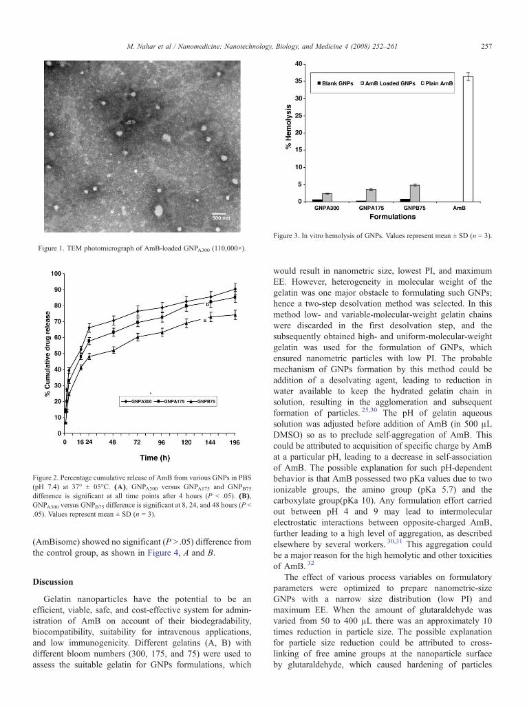
Figure 2. Percentage cumulative release of AmB from various GNPs in PBS(pH 7.4) at 37° ± 05°C. (A), GNPA300 versus GNPA175 and GNPB75difference is significant at all time points after 4 hours (P b .05). (B),GNPA300 versus GNPB75 difference is significant at 8, 24, and 48 hours (P b.05). Values represent mean ± SD (n = 3).
Figure 3. In vitro hemolysis of GNPs. Values represent mean ± SD (n = 3).
Figure 1. TEM photomicrograph of AmB-loaded GNPA300 (110,000×).
257M. Nahar et al / Nanomedicine: Nanotechnology, Biology, and Medicine 4 (2008) 252–261
(AmBisome) showed no significant (P N .05) difference fromthe control group, as shown in Figure 4, A and B.
Discussion
Gelatin nanoparticles have the potential to be anefficient, viable, safe, and cost-effective system for admin-istration of AmB on account of their biodegradability,biocompatibility, suitability for intravenous applications,and low immunogenicity. Different gelatins (A, B) withdifferent bloom numbers (300, 175, and 75) were used toassess the suitable gelatin for GNPs formulations, which
would result in nanometric size, lowest PI, and maximumEE. However, heterogeneity in molecular weight of thegelatin was one major obstacle to formulating such GNPs;hence a two-step desolvation method was selected. In thismethod low- and variable-molecular-weight gelatin chainswere discarded in the first desolvation step, and thesubsequently obtained high- and uniform-molecular-weightgelatin was used for the formulation of GNPs, whichensured nanometric particles with low PI. The probablemechanism of GNPs formation by this method could beaddition of a desolvating agent, leading to reduction inwater available to keep the hydrated gelatin chain insolution, resulting in the agglomeration and subsequentformation of particles. 25,30 The pH of gelatin aqueoussolution was adjusted before addition of AmB (in 500 μLDMSO) so as to preclude self-aggregation of AmB. Thiscould be attributed to acquisition of specific charge by AmBat a particular pH, leading to a decrease in self-associationof AmB. The possible explanation for such pH-dependentbehavior is that AmB possessed two pKa values due to twoionizable groups, the amino group (pKa 5.7) and thecarboxylate group(pKa 10). Any formulation effort carriedout between pH 4 and 9 may lead to intermolecularelectrostatic interactions between opposite-charged AmB,further leading to a high level of aggregation, as describedelsewhere by several workers. 30,31 This aggregation couldbe a major reason for the high hemolytic and other toxicitiesof AmB. 32
The effect of various process variables on formulatoryparameters were optimized to prepare nanometric-sizeGNPs with a narrow size distribution (low PI) andmaximum EE. When the amount of glutaraldehyde wasvaried from 50 to 400 μL there was an approximately 10times reduction in particle size. The possible explanationfor particle size reduction could be attributed to cross-linking of free amine groups at the nanoparticle surfaceby glutaraldehyde, which caused hardening of particles

Table 6Hematological parameters on post-injection effects at day 8 after seven doses (1, 5, and 10 mg/kg/day of AmB)
Parameters † Doses (mg/kg/day) Plain AmB solution AmB-loaded GNPA300 AmBisome Control Blank GNPA300
RBCs (× 10 6/μL) 1 6.03 ± 0.4⁎ 7.08 ± 1.8 7.31 ± 1.3 8.57 ± 0.2 8.62 ± 1.11
5 5.12 ± 0.68⁎ 6.58 ± 1.1⁎ 7.06 ± 1.6
10 4.24 ± 0.45⁎ 6.10 ± 0.81⁎ 6.56 ± 1.0⁎
Hemoglobin (g/dL) 1 13.0 ± 0.8⁎ 14.6 ± 2.7 16.4 ± 0.9 17.6 ± 1.3 17.5 ± 0.6
5 10.4 ± 0.3⁎ 12.4 ± 1.4⁎ 16.1 ± 1.4
10 09.8 ± 0.2⁎ 11.0 ± 0.8⁎ 15.8 ± 1.1
WBCs (× 10 3/μL) 1 6.0 ± 0.7 6.4 ± 1.3 6.8 ± 2.7 7.1 ± 1.1 6.0 ± 1.0
5 5.8 ± 0.8 6.2 ± 0.8 6.5 ± 1.2
10 5.5 ± 0.9 5.7 ± 0.4 6.2 ± 1.5
Hematocrit (%) 1 35. ± 3.6⁎ 42.1 ± 4.6 43.9 ± 3.8 48.4 ± 3.4 46.7 ± 1.56
5 32.1 ± 1.3⁎ 39.4 ± 3.2⁎ 43.1 ± 3.2
10 30.4 ± 2.4⁎ 36.5 ± 2.8⁎ 42.1 ± 2.9
Platelet (× 10 3/μL) 1 125 ± 16⁎ 240 ± 65⁎ 342 ± 41⁎ 547 ± 34 129 ± 45⁎
5 123 ± 11⁎ 235 ± 44⁎ 312 ± 12⁎
10 116 ± 19⁎ 228 ± 38⁎ 300 ± 21⁎
RBCs, Red blood cells; WBCs, white blood cells.⁎Significant (P b 0.05, all groups compared with control).†All parameters were expressed as mean values ± SD (n = 6).
258 M. Nahar et al / Nanomedicine: Nanotechnology, Biology, and Medicine 4 (2008) 252–261
leading to reduction in size. As we increased the amountof cross-linker more groups were cross-linked andsubsequently caused a higher degree of reticulation, assuggested by Leo et al 14 In contrast, the PI was found tobe minimum at 200 μL of glutaraldehyde. The probablereason could be that this amount was sufficient to causeuniform reduction in particle size for the number of GNPsformed. The deviation to either higher or lower amountsmay have caused disproportionate availability of cross-linker for the same number of GNPs formed, leading to ahigher PI. The EEs were not significantly affected by theamount of glutaraldehyde used (P N .05). However, type Agelatin showed approximately double the EE of type B,which might be due to formation of the larger size particlesleading to smaller surface area and consequently low EE.Thus, a 200-μL concentration yielding GNPs with thedesired particle size, minimum PI, and maximum EE wasconsidered as optimum. Similarly, temperature was oneimportant formulation variable, which had a pronouncedeffect on size and PI while affecting EE to a lesser extent.A higher temperature was found to be preferable for theformation of GNPs because of the high viscosity of gelatinat room temperature. The possible reason for this effect isthe triple-helical structure of gelatin, which uncoiled astemperature rose in a controlled manner. However, athigher temperatures (50° ± 1°C and 60° ± 1°C) anunexpected increase in particle size was observed,probably due to complete uncoiling of gelatin chains.The results were consistent with the earlier findings ofAzarmi et al 30 Likewise, pH at the second desolvation step
was significant to obtain GNPs of desired size and low PI,because formation of GNPs probably was associated witha higher degree of electrostatic interactions causing chargeneutralization and consequently formation of GNPs onadding desolvating agent to polymer solution. 33 Such pH-dependent behavior of gelatin could be attributed to itspolyelectrolyte nature (contains both amino and carbox-ylate-terminated chains at its isoelectric point: type A, pH7-9; type B, pH 4-5), and as the pH was shifted to theacidic or basic range there was a predominance of NH3
+ orCOO – ions depending upon the type of gelatin. 33
Drug EE is an important index for drug deliverysystems. The problem associated with AmB to load in anycolloidal drug delivery system is its poor solubility in mostof the solvents. For the EE study, drug was dissolved in aspecified quantity of DMSO and mixed with an aqueoussolution of polymer with continuous stirring in a mechan-ical stirrer. The entrapment of AmB in GNPs could beexplained on the basis of preferential localization of druginside the nanoparticulate core, which was less hydrophilicas compared with the outer aqueous environment; more-over, as desolvation removed water from the core, EE forthis drug was further improved. Low entrapment of AmBin type B gelatin GNPs could be further explained by thelow surface-to-volume ratio for the number of nanoparti-cles formed, which could be correlated with the greatersize of GNPs formed. Although gelatin and drug showedpH-dependent charge interactions evident by significantchange in zeta potential values (P b .05), these chargeinteractions do not contribute to AmB loading in GNPs;
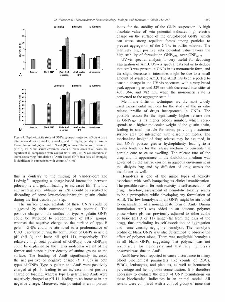
Figure 4. Nephrotoxicity study of GNPA300 on post-injection effects at day 8after seven doses (1 mg/kg, 5 mg/kg, and 10 mg/kg per day of AmB).Concentrations of (A) serum BUN and (B) serum creatinine were measured(n = 6). BUN and serum creatinine levels of plain AmB at all doses aresignificant in comparison with control (P b .001). BUN concentration inanimals receiving formulation of AmB-loaded GNPs in a dose of 10 mg/kgis significant in comparison with control (P b .05).
259M. Nahar et al / Nanomedicine: Nanotechnology, Biology, and Medicine 4 (2008) 252–261
this is contrary to the finding of Vandervoort andLudwig 34 suggesting a charge-based interaction betweenpilocarpine and gelatin leading to increased EE. This lowand average yield obtained in GNPs could be ascribed todiscarding of some low-molecular-weight gelatin chainsduring the first desolvation step.
The surface charge attribute of these GNPs could besuggested by their corresponding zeta potential. Thepositive charge on the surface of type A gelatin GNPscould be attributed to predominance of NH3
+ groups,whereas the negative charge on the surface of type Bgelatin GNPs could be attributed to a predominance ofCOO –, acquired during the formulation of GNPs in acidicpH (pH 3) and basic pH (pH 11), respectively. Therelatively high zeta potential of GNPA300 over GNPA175could be explained by the higher molecular weight of theformer and hence higher density of amine groups at thesurface. The loading of AmB significantly increasedthe net positive or negative charge (P b .05) in bothtypes of GNPs. Type A gelatin and AmB were positivelycharged at pH 3, leading to an increase in net positivecharge on loading, whereas type B gelatin and AmB werenegatively charged at pH 11, leading to an increase in netnegative charge. Moreover, zeta potential is an important
index for the stability of the GNPs suspension. A highabsolute value of zeta potential indicates high electriccharge on the surface of the drug-loaded GNPs, whichcan cause strong repellent forces among particles toprevent aggregation of the GNPs in buffer solution. Therelatively high positive zeta potential value favors thehigh stability of formulation GNPA300 over GNPA175.
UV-vis spectral analysis is very useful for deducingaggregation of AmB. UV-vis spectral data led us to deducethat AmB was present in GNPs in its monomeric form, andthe slight decrease in intensities might be due to a smallamount of available AmB. The AmB has been reported tocause a change in the UV-vis spectrum, with a very broadpeak appearing around 329 nm with decreased intensities at405, 364, and 382 nm, when the monomeric state isconverted to the aggregate state. 11
Membrane diffusion techniques are the most widelyused experimental methods for the study of the in vitrorelease profile of drugs incorporated in GNPs. Thepossible reason for the significantly higher release ratein GNPA300 is its higher bloom number, which corre-sponds to a higher molecular weight of the gelatin chain,leading to small particle formation, providing maximumsurface area for interaction with dissolution media. Themechanistic insight of drug release may be assumed, inthat GNPs possess greater hydrophilicity, leading to agreater tendency for the release medium to penetrate theparticle core to cause swelling. The release rate of thedrug and its appearance in the dissolution medium wasgoverned by the matrix erosion in aqueous environment inthe dialysis bag and by diffusion of drug across themembrane as well.
Hemolysis is one of the major types of toxicityassociated with AmB hampering its clinical manifestation.The possible reason for such toxicity is self-association ofdrug. Therefore, assessment of hemolytic toxicity seemsto be a prerequisite while developing any formulation ofAmB. The low hemolysis in all GNPs might be attributedto encapsulation of a nonaggregate form of AmB. Duringformulation AmB was added in an aqueous polymerphase whose pH was previously adjusted to either acidicor basic (pH 3 or 11) range (far from the pKa of thedrug), thus precluding its self-association or aggregationand hence causing negligible hemolysis. The hemolyticprofile of blank GNPs was also determined to observe theeffect of polymer alone. There was negligible hemolysisin all blank GNPs, suggesting that polymer was notresponsible for hemolysis and that any hemolysisobserved was due to AmB.
AmB have been reported to cause disturbance in manyblood biochemical parameters like counts of RBCs,WBCs, leukocytes, and platelets, as well as hematocritpercentage and hemoglobin concentration. It is thereforenecessary to evaluate the effect of GNP formulations onthese biochemical indicators in an animal model. Allresults were compared with a control group of mice that
260 M. Nahar et al / Nanomedicine: Nanotechnology, Biology, and Medicine 4 (2008) 252–261
received no formulation. GNP formulation was comparedwith a marketed liposome formulation (AmBisome) toestablish its market potential. One group of animalsreceived blank GNP formulations to establish whether thedisturbance in these parameters was due to gelatin orAmB. At day 8 animals were anesthetized with etherand bled from the retro-orbital plexus. A significantdecrease in platelet count was observed in all formulationsas compared with control, but this decrease could notbe explained.
Different gelatins with various bloom numbers wereused and evaluated for GNPs preparation, and type Agelatin with bloom number 300 was found to be suitablefor such preparation because of its nanometric size, low PI,good EE, and low hemolytic profile. Further detailedhematological profiling of AmB-loaded GNPA300 proved itspotential as a safe delivery system. Also, there was nosignificant increase in either BUN or serum creatininelevels up to day 7 of GNPA300 administration, thusadvocating the safety of the proposed system for indica-tions like visceral leishmaniasis or some systemic fungalinfections. The drug release characteristics exhibitedcontrolled delivery of AmB for longer duration, whichmay certainly improve patient compliance. Moreover,formulation and drug loading in GNPs were carried outat pH values far from the pKa of the drug. This is a majoradvance reported in this work to reduce the self-aggrega-tion of AmB.
It can be concluded that GNPs may be a viable, effective,and cheap alternative to other carriers for the controlled andsafe delivery of AmB. We present a cost-effective drugdelivery system for AmB that is easy to prepare and haspotential for further development.
Acknowledgment
Manoj Nahar expresses his gratitude to SAIF, PunjabUniversity, Chandigarh, India, for providing TEM facilities.
References
1. Gallis HA, Drew RH, Picard WW. Amphotericin B: 30 years of clinicalexperience. Rev Infect Dis 1990;12:308-29.
2. Kleinberg M. What is the current and future status of conventionalamphotericin B? Int J Antimicrob Agents 2006;27S:S12-6.
3. Boswell GW, Buell D, Bekersky I. AmBisome (liposomalAmphotericin B): a comparative review. J Clin Pharmacol 1998;38:583-92.
4. Lincopan N, Mamizuka E, Carmona-Ribeiro A. Low nephrotoxicity ofan effective amphotericin B formulation with cationic bilayerfragments. J Antimicrob Chemother 2005;55:727-34.
5. Vieira DB, Carmona-Ribeiro AM. Synthetic bilayer fragments forsolubilization of amphotericin B. J Colloid Interface Sci 2001;244:427-31.
6. Nahar M, Dutta T, Murugesan S, Asthana A, Mishra D, Rajkumar V,et al. Functional polymeric nanoparticles: an efficient and promisingtool for active delivery of bioactives. Crit Rev Ther Drug Carrier Syst2006;23:259-318.
7. Venier-Julienne M, Benoit J. Preparation, purification and morphologyof polymeric nanoparticles as drug carriers. Pharm Acta Helv 1996;71:121-8.
8. Espuelas MS, Legrand P, Campanero M, Appel M, Cheron M, GamazoC, et al. Polymeric carriers for amphotericin B: in vitro activity, toxicityand therapeutic efficacy against systemic candidiasis in neutropenicmice. J Antimicrob Chemother 2003;52:419-27.
9. Espuelas MS, Legrand P, Irache JM, Gamazo C, Orecchioni AM,Devissaguet JPh, et al. Poly (e-caprolacton) nanospheres as an alterna-tive way to reduce amphotericin B toxicity. Int J Pharm 1997;158:19-27.
10. Tiyabbonchai W, Limpeanchob N. Formulation and characterization ofAmphotericin B–chitosan–dextran sulfate nanoparticles. Int J Pharm2007;329:142-9.
11. Lee CH, Singla A, Lee Y. Biomedical applications of collagen. Int JPharm 2001;221:1Q22.
12. Narayami R, Panduranga RK. Controlled release of anticancer drugmethotrexate from biodegradable gelatin microspheres. J Microencapsul1994;11:69-77.
13. Cascone MG, Lazzeri L, Carmignani C, Zhu Z. Gelatin nanoparticlesproduced by a simple W/O emulsion as delivery system formethotrexate. J Mater Sci Mater Med 2002;13:523-6.
14. Leo E, Vandelli MA, Cameroni R, Forni F. Doxorubicin-loaded gelatinnanoparticles stabilized by glutaraldehyde: involvement of the drug inthe cross-linking process. Int J Pharm 1997;12:75-82.
15. Verma AK, Sachin K, Saxena A, Bohidar HB. Release kinetics frombio-polymeric nanoparticles encapsulating protein synthesis inhibitor–Cycloheximide, for possible therapeutic applications. Curr PharmBiotechnol 2005;6:121-30.
16. Yeh TK, Lu Z, Wientjes MG, Au JLS. Formulating paclitaxel innanoparticles alters its disposition. Pharm Res 2005;22:867-74.
17. Bajpai AK, Choubey J. Design of gelatin nanoparticles as swellingcontrolled delivery system for chloroquine phosphate. J Mater Sci MaterMed 2006;17:345-58.
18. Sham JO, Zhang Y, Finlay WH, Roa WH, Lobenberg R. Formu-lation and characterization of spray-dried powders containingnanoparticles for aerosol delivery to the lung. Int J Pharm 2004;28:457-67.
19. Truong-Le VL, Walsh SM, Schweibert E, Mao HQ, Guggino WB,August JT, et al. Gene transfer by DNA-gelatin nanospheres. ArchBiochem Biophys 1999;361:47-56.
20. Kaul G, Amiji M. Cellular interactions and in vitro DNA transfec-tion studies with poly(ethylene glycol)–modified gelatin nanoparticles.J Pharm Sci 2005;94:184-98.
21. Kaul G, Amiji M. Tumor-targeted gene delivery using poly(ethyleneglycol)–modified gelatin nanoparticles: in vitro and in vivo studies.Pharm Res 2005;22:951-61.
22. Balthasar S, Michaelis K, Dinauer N, von Briesen H, Kreuter J, LangerK. Preparation and characterisation of antibody modified gelatinnanoparticles as drug carrier system for uptake in lymphocytes.Biomaterials 2005;26:2723-32.
23. Dinauer N, Balthasar S, Weber C, Kreuter J, Langer K, von BriesenH. Selective targeting of antibody-conjugated nanoparticles toleukemic cells and primary T-lymphocytes. Biomaterials 2005;26:5898-906.
24. Li JK, Wang N, Wu XS. Gelatin nanoencapsulation of protein/peptidedrugs using an emulsifier-free emulsion method. J Microencapsul 1998;15:163-72.
25. Coester CJ, Langer K, Van Briesen H, Kreuter J. Gelatin nanoparticlesby two-step desolvation—a new preparation method, surface modifica-tions and cell uptake. J Microencapsul 2000;17:187-93.
26. Leo E, Cameroni R, Forni F. Dynamic dialysis for the drug releaseevaluation from doxorubicin-gelatin nanoparticles conjugates. Int JPharm 1999;180:23-30.
27. Echevarrya I, Barturen C, Renedo MJ, Dios-Vieitez MC. High-performance liquid chromatographic determination of amphotericin B
261M. Nahar et al / Nanomedicine: Nanotechnology, Biology, and Medicine 4 (2008) 252–261
in plasma and tissue application to pharmacokinetic and tissuedistribution studies in rats. J Chromatogr A 1998;819:171-6.
28. Singhai AK, Jain S, Jain NK. Evaluation of an aqueous injection ofketoprofen. Die Pharmazie 1997;52:149-54.
29. Marcondes MC, Borelli P, Yoshida N, Russo M. Acute Trypanosomacruzi infection is associated with anemia, thrombocytopenia, leukope-nia, and bone marrow hypoplasia: reversal by nifurtimox treatment.Microbes Infect 2000;2:347-52.
30. Azarmi S, Huang Y, Chen H, McQuarrie S, Abrams D, Roa W,et al. Optimization of a two-step desolvation method forpreparing gelatin nanoparticles and cell uptake studies in 143Bosteosarcoma cancer cells. J Pharm Pharmaceut Sci 2006;9:124-32.
31. Mazerski J, Gryzbowska J, Borowski E. Influence of net charge on theaggregation and solubility on aggregation and solubility behaviour ofamphotericin B and its derivatives in aqueous media. Eur Biophys J1990;18:1-6.
32. Legrand P, Romero EA, Cohen BE, Bolard J. Effect of aggregation andsolvent on the toxicity of amphotericin B to human erythrocytes.Antimicrob Agents Chemother 1992;36:2518-22.
33. Saxena A, Sachin K, Bohidar HB, Verma AK. Effect of molecularweight heterogeneity on drug encapsulation efficiency of gelatin nano-particles. Coll Surf B: Biointerfaces 2005;45:42-8.
34. Vandervoort J, Ludwig A. Preparation and evaluation of drug loadedgelatin nanoparticles for topical ophthalmic use. Eur J Pharm Biopharm2004;57:251-61.