Loaded/Folic Acid-Targeted Chitosan - Research Square
23
Page 1/23 Biodegradable and Biocompatible Subcutaneous Implants Consisted of Ph-Sensitive Mebendazole- Loaded/Folic Acid-Targeted Chitosan Nanoparticles for Murine Triple-Negative Breast Cancer Treatment Amirhosein Kefayat Isfahan University of Medical Sciences Maryam Hosseini Amirkabir University of Technology Fatemeh Ghahremani Arak University of Medical Sciences mohammad raenia ( [email protected] ) Isfahan University of Medical Sciences Research Article Keywords: Mebendazole, Chitosan nanoparticles, subcutaneous implants, Folic acid, Triple-negative breast cancer Posted Date: July 30th, 2021 DOI: https://doi.org/10.21203/rs.3.rs-736440/v1 License: This work is licensed under a Creative Commons Attribution 4.0 International License. Read Full License
-
Upload
khangminh22 -
Category
Documents
-
view
0 -
download
0
Transcript of Loaded/Folic Acid-Targeted Chitosan - Research Square

Page 1/23
Biodegradable and Biocompatible SubcutaneousImplants Consisted of Ph-Sensitive Mebendazole-Loaded/Folic Acid-Targeted Chitosan Nanoparticlesfor Murine Triple-Negative Breast Cancer TreatmentAmirhosein Kefayat
Isfahan University of Medical SciencesMaryam Hosseini
Amirkabir University of TechnologyFatemeh Ghahremani
Arak University of Medical Sciencesmohammad ra�enia ( m_ra�[email protected] )
Isfahan University of Medical Sciences
Research Article
Keywords: Mebendazole, Chitosan nanoparticles, subcutaneous implants, Folic acid, Triple-negativebreast cancer
Posted Date: July 30th, 2021
DOI: https://doi.org/10.21203/rs.3.rs-736440/v1
License: This work is licensed under a Creative Commons Attribution 4.0 International License. Read Full License

Page 2/23
AbstractBackground: Mebendazole (MBZ) is a well-known anti-parasite drug with signi�cant anti-cancerproperties. However, MBZ exhibits low solubility, limited absorption e�cacy, extensive �rst-pass effect,and low bioavailability. Therefore, multiple oral administration of high dose MBZ is required daily forachieving the therapeutic serum level which can cause severe side effects and patients’ non-compliance.
Method: In the present study, MBZ-loaded/folic acid-targeted chitosan nanoparticles (CS-FA-MBZ) weresynthesized, characterized, and used to form cylindrical subcutaneous implants for 4T1 triple-negativebreast tumor (TNBC) treatment in BALB/c mice. The therapeutic e�cacy of the CS-FA-MBZ implants wasinvestigated after subcutaneous implantation in comparison with Control (no-treatment), MBZ (40 mg/kg,oral administration, twice a week for 2 weeks), and CS-FA implants, according to 4T1 tumors’ growthprogression, metastasis, and tumor-bearing mice survival time. Also, their biocompatibility was evaluatedby blood biochemical analyzes and histopathological investigation of vital organs.
Results: The CS-FA-MBZ implants were completely degraded 15 days after implantation and causedabout 73.3%, 46.5%, 37.3% decrease in the mean tumors’ volume in comparison with the Control(1050.5±120.7 mm3), MBZ (561.7±70.3 mm3), and CS-FA (658.3±88.1 mm3) groups, respectively.Average liver metastatic colonies’ number per microscope �eld at the CS-FA-MBZ (8.6±1.9) group wassigni�cantly (P<0.05) lower than the Control (24.5±4.1), MBZ (14.1±2.5), and CS-FA (15.7±3.1) groups. Inaddition, the CS-FA-MBZ treated mice exhibited about 51%, 24%, and 17% more survival time (days) afterthe cancer cells injection in comparison with the Control, MBZ, and CS-FA groups, respectively. Moreover,the CS-FA-MBZ implants were completely biocompatible based on histopathology and blood biochemicalanalyzes.
Conclusion: Taking together, CS-FA-MBZ implants were completely biodegradable and biocompatible withhigh therapeutic e�cacy in a murine TNBC model.
1. BackgroundBreast cancer is the most common malignancy among women and the leading cause of cancer-relateddeaths in the female gender [1]. About 20% of all diagnosed breast cancers are categorized as triple-negative breast cancer (TNBC). This acronym simply means that the tumor doesn’t express estrogen,progesterone, and human epidermal growth factor-2 receptors on its cells’ surface [2]. TNBC issigni�cantly more aggressive and invasive than other subtypes of breast cancer and exhibits higherrelapse rates and shorter recurrence period [3]. Therefore, due to the absence of well-de�ned moleculartargets, currently, the only therapeutic approach for TNBC patients is chemotherapy [4, 5]. However, theoutcome of chemotherapy with currently approved drugs isn’t satisfying, and poor therapeutic response,severe side effects, and development of multidrug resistance are still big challenges in TNBCchemotherapy [6, 7].

Page 3/23
Mebendazole (MBZ) is a well-known antihelminthic drug, with high biocompatibility and low price whichhas been repurposed for anti-neoplastic treatment [8]. Many studies have reported anti-proliferative, pro-apoptotic, and anti-metastatic effects of MBZ on different cancer cell lines including chemoresistantcancer cell lines [9, 10]. MBZ treatment causes a decrease or complete arrest of tumor growth, signi�cantinhibition of tumor metastasis, and an increase of tumor-bearing mice survival time in different animalmodels of cancer [11–16]. Anti-neoplastic activities of MBZ can be attributed to inhibition of tubulinpolymerization, decrease of tumor angiogenesis, blocking of pro-survival pathways, inhibition of matrixmetalloproteinases function, and multi-drug resistance protein transporters activity [8, 17–19]. However,MBZ has limited bioavailability after oral administration as only 20% of the dosage reaches the systemiccirculation. This can be related to the low solubility of MBZ, limited absorption e�cacy, and extensive�rst-pass effect following oral administration [20, 21]. Therefore, a high dose of MBZ is required forachieving the therapeutic serum level which can cause severe adverse effects and patients’ non-compliance [22].
Drug-releasing implants have gained lots of attention for controlled drug release in the long-termtreatments, especially cancer chemotherapy [23, 24]. Implants that contain chemotherapy drugs haveexhibited many advantages over intravenous or oral drug administration routes including the eliminationof daily multiple injections and maintenance of the steady-state concentration of the drug [25–29].Biodegradable polymers are the most utilized materials for developing these implants [30]. Chitosan (CS)is a well-known biodegradable biopolyaminosaccharide with a natural origin. The chitosan-based drugcarriers and implants exhibit high biodegradability, low toxicity, and appropriate biocompatibility. Inaddition, chitosan per se has considerable anti-proliferative, pro-apoptotic, anti-angiogenetic, and anti-metastatic effects on different types of tumors [31–33]. Different forms of chitosan-based drug deliverysystems have been used and chitosan nanoparticles are one of the most advanced ones for theenhancement of anti-cancer drugs e�cacy. They can be de�ned as spherical, biocompatible, andbiodegradable nanostructures with high drug-loading e�cacy [34–36]. Chitosan nanoparticles can beused for controlled release of different types of drugs and enhancement of tumor drug delivery e�cacy[37–41]. In addition, chitosan nanoparticles decorating with targeting ligands that can bind to themalignant cells’ surface receptors have received lots of attention for enhancement of tumor drug deliverye�cacy [42, 43]. One of the most e�cient targeting agents for cancer cells speci�c targeting is folic acid(FA) due to overexpression of the folate receptor on their membrane in comparison with normal cells. FAhas many advantages over other targeting agents, including small molecular weight, simple chemicalproperties, receptor-medicated endocytosis of the folic acid decorated-carriers, extremely lowimmunogenicity, overexpression on cancer cells membrane, and limited expression on normal cells’surface [44–47]. Although TNBC cells are known due to the lack of different molecular targets on theirmembrane, FA is a highly overexpressed receptor on their surface [48–50]. Therefore, FA-modi�ed drugdelivery systems can increase the drug concentration at the TNBC tumors site and decline its side effectsat normal tissues [51].
In the present study, MBZ-loaded/folic acid-targeted chitosan nanoparticles (CS-FA-MBZ) were used toform cylindrical implants for 4T1 triple-negative breast tumor treatment in BALB/c mice. The therapeutic

Page 4/23
e�cacy of the CS-FA-MBZ implants was investigated after subcutaneous implantation in comparisonwith Control, MBZ (40 mg/kg, oral administration, twice a week for 2 weeks), and CS-FA implantsaccording to 4T1 tumors’ growth progression, metastasis, and tumor-bearing mice survival time. Also, theCS-FA-MBZ implant's biocompatibility was evaluated by blood biochemical analyzes and vital organshistopathological investigations. In this study, 4T1 murine triple-negative mammary carcinoma was usedas an experimental animal model with high similarity to human TNBC. This cell line is a highlytumorigenic and invasive cancer cell line that can spontaneously metastasize from the primary tumor inthe mammary gland to multiple distant sites. Also, its metastasis pattern is very similar to that of humanbreast cancer [52–55].
2. Materials And Methods
2.1. MaterialMedium-molecular weight chitosan (190 to 310 kDa, MMW), Sodium tripolyphosphate (TPP), 1-ethyl-3-(3-dimethylaminopropyl) carbodiimide (EDC), N-hydroxysuccinimide (NHS), folic acid (97%, FA), Tween-80(a non-ionic surfactant), mebendazole (MBZ), ammonia (25%, NH4OH), acetic acid, dimethyl sulfoxide(DMSO), phosphate-buffered saline powder (pH = 7.4 PBS), methanol and acetone were purchased fromSigma-Aldrich and used without further puri�cation unless stated otherwise. RPMI 1640, fetal bovineserum (FBS), phosphate buffer saline (PBS), penicillin/streptomycin, ethanol (96%, v/v), trypsin-EDTA(0.25%) were purchased from Merck (Germany).
2.2. Conjugation of folic acid to chitosan (CS-FApreparation)CS-FA was fabricated as reported in the literature [56]. Brie�y, 0.5 g of FA and 0.2 g of EDC were initiallydissolved in anhydrous DMSO (20 mL) under constant stirring at room temperature (2 h). Then, thesolution was dropped into the CS solution 0.5% (w/v) prepared in acetate buffer (0.1 M, pH 4.7) and atroom temperature in dark for 16 h. Thereafter, the pH of the solution was adjusted to 9.0 by the additionof NaOH (1.0 M). The resulting precipitate was collected by centrifugation, then puri�ed by dialysisagainst phosphate-buffered saline (PBS, pH 7.4) for 2 days and against water for another 4 days. Finally,yellow-colored CS-FA products were collected and freeze-dried.
2.3. Preparation of the CS-FA-MBZ nanoparticlesCS-FA-MBZ nanoparticles were synthesized according to the previously reported method with somemodi�cations [57]. Initially, CS-FA (0.1 g) was dissolved in a solution containing acetic acid (1% v/v, 20mL), then left under stirring at room temperature in dark for 16 h to prepare a solution of CS-FA (0.5%w/v). The pH of the solution was adjusted to 4.8 by the addition of NaOH (1.0 M). Afterward, 250 µL ofTween-80 was added dropwise and left for 2 h under stirring at 45°C. In the next step, 0.01 g MBZ wasdissolved in 0.5 M methanolic hydrochloride [58] and then added to the former solution and stirred for 30min. Finally, 10 mL TPP aqueous solution (0.5% w/v) was dropped to the CS-FA solution slowly under

Page 5/23
magnetic stirring (800 rpm) at room temperature for 1 h then the nanoparticles were collected bycentrifugation (12000 rpm, 30 min). The resulting CS-FA-MBZ nanoparticles were lyophilized and stored.
2.4. MBZ loadingMBZ was loaded during the formation of CS-FA nanoparticles as reported in the literature [57]. Tocalculate MBZ encapsulation e�cacy (EE%) and loading capacity (LC%), unloaded MBZ content in thesupernatant of the last step was determined through a calibration curve of MBZ standard solution byUV–Visible spectroscopy at 234 nm [58]. The MBZ loading ratio of the nanoparticles was calculated bythe following equations (1 and 2):
2.5. In vitro drug release pattern from the CS-FA-MBZnanoparticlesTo evaluate the release behavior of MBZ from CS-FA-MBZ nanoparticles, 5 mg of the CS-FA-MBZnanoparticles were immersed in PBS solution containing Tween-80 (0.1% w/v) at pH values of 5.5, 6.8,and 7.4. The release pro�le was assessed at 37°C in dark under shaking at 100 rpm for up to 1 week [59].At each predetermined time interval, the nanoparticles were centrifuged (10,000 rpm for 15 min) and thereleased medium was collected and replaced with equivalent fresh PBS solution. The cumulativepercentage of released MBZ was determined by UV–Visible spectrophotometry at 234 nm.
2.6. Nanoparticles characterization and implants fabricationTo assess the structure and interaction of CS, CS-FA, and CS-FA-MBZ, Fourier Transform InfraredSpectroscopy (FTIR) was used by a Bruker Equinox 55 spectrometer with the KBr pellets method. Toevaluate the size and morphology of nanoparticles, scanning electron microscopy (SEM) was acquiredusing FEI ESEM QUANTA 200, MIRAII, and MIRAIII Tescan. The average size and size distribution ofparticles were determined by measuring the diameter of 100 particles of SEM images using ImageJsoftware. The ultraviolet-visible (UV-vis) spectra were recorded by a PerkinElmer Lambda 950spectrophotometer (wavelength range: 200–800 nm). The zeta potential, size distribution, andpolydispersity of the prepared nanoparticles were measured by Dynamic Light Scattering (DLS) (MalvernInstruments). A well-dispersed aqueous suspension of the prepared nanoparticles was applied. Eachexperiment was carried out in triplicate and data were presented as means ± standard deviations. Forfabricating an implant, adequate mass of the synthesized CS-FA-MBZ nanoparticles (according to themouse body weight and its needed dosage of MBZ) were pressed in a steel die at 1500 psi to fromcylindric implants (usually 6 mm diameters and 3 mm height).

Page 6/23
2.1. Animals ethics, care, and handlingAll animal experiments complied with the ARRIVE guidelines and were conducted according to theguidelines of the European Communities Council Directive (2010/63/UE) and the Isfahan University ofMedical Sciences for the care and use of laboratory animals. Likewise, all the procedures, protocols, andsteps were approved by the ethics committee of the Isfahan University of Medical Sciences(IR.MUI.RESEARCH.REC.1399.125). Female BALB/c mice (weight: 25 ± 2 g) were purchased from thePasteur Institute of Tehran, Iran. The mice were acclimatized to the laboratory environment (24 ± 2 ºCtemperature, 50 ± 10% relative humidity, and 12 h light/12 h dark cycles) for 14 days before involving intothe experiments. All mice were fed sterilized standard mouse chow and water ad libitum. Overdose ofKetamine-Xylazine (KX) solution through intraperitoneal injection was used for the mice sacri�ce.
2.2. Tumor implantation4T1 cancer cells (murine mammary carcinoma) were purchased from the Pastor Institute of Tehran, Iran.The cells were cultured in RPMI 1640 medium containing 10% fetal bovine serum (FBS). The cells wereincubated at 37 ºC in a humidi�ed incubator in 5% CO2 atmosphere. When the cells reached adequatenumbers, were harvested from culture �asks by trypsin and washed three times with PBS. The mice wereinjected with 2 × 10⁶ cells suspended in 50 µL of FBS-free DMEM-F12, subcutaneously (s.c.) into the left4th abdominal mammary fat pad.
2.3. Tumor-bearing mice grouping and therapeuticapproachesFor this experiment 32 tumor-bearing mice were used. When the tumors’ volume reached 50–70 mm3 (3rdday after the cancer cells injection), mice were divided into four groups (n = 8) including (1) Control, (2)MBZ, (3) CS-FA implants, (4) CS-FA-MBZ implants. The tumor-bearing mice in the 2nd group were treatedwith oral administration )p.o.( of MBZ (40 mg/kg, twice a week for 2 weeks according to previous studies[14]) from the 3rd day after the cancer cells injection. In the 3rd and 4th groups, the tumor-bearing micewere anesthetized with intraperitoneally injection of Ketamine-Xylazine (KX) solution (Ketamine: 100mg/kg, Xylazine: 10 mg/kg). The left �ank was shaved and scrubbed with betadine. The scrub solutionwas wiped away from the surgical site with alcohol 70 % and covered with a sterile drape. Then, a small(~ 1 cm) incision was made and the implant was embedded under sterile conditions, and the skin wasstitched with nylon (4 − 0). All the operations were done under complete anesthesia. To manage post-surgical pain, ketoprofen (5 mg/kg) was administered subcutaneously until the next 72 h. The mice weremonitored daily for prolonged signs of pain, weight loss, or surgical site infections. If any signs of pain,wounds infection, massive necrosis, and hemorrhage, diffuse metastasis were observed during any stepsof the study, the mice were sacri�ced by KX overdose. In the Control group, one incision was made at theleft �ank of the tumor-bearing mice and sutured without implantation of any implants. To determinetumors’ growth progression, the greatest longitudinal diameter (length) and the greatest transversediameter (width) of the tumors were measured every 3 days until the 18th after cancer cells injection.
Page 7/23
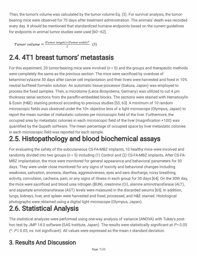
Then, the tumor’s volume was calculated by the tumor volume Eq. (3). For survival analysis, the tumor-bearing mice were observed for 70 days after treatment administration. The animals’ death was recordedevery day. It should be mentioned that standardized humane endpoints based on the current guidelinesfor endpoints in animal tumor studies were used [60–62].
2.4. 4T1 breast tumors’ metastasisFor this experiment, 20 tumor-bearing mice were involved (n = 5) and the groups and therapeutic methodswere completely the same as the previous section. The mice were sacri�ced by overdose ofketamine/xylazine 30 days after cancer cell implantation and their livers were harvested and �xed in 10%neutral buffered formalin solution. An automatic tissue processor (Sakura, Japan) was employed toprocess the �xed samples. Then, a microtome (Leica Biosystems, Germany) was utilized to cut 4 µmthickness serial sections from the para�n-embedded blocks. The sections were stained with Hematoxylin& Eosin (H&E) staining protocol according to previous studies [55, 63]. A minimum of 10 randommicroscopic �elds was observed under the 10× objective lens of a light microscope (Olympus, Japan) toreport the mean number of metastatic colonies per microscopic �eld of the liver. Furthermore, theoccupied area by metastatic colonies in each microscopic �eld of the liver (magni�cation ×100) wasquanti�ed by the Qupath software. The mean percentage of occupied space by liver metastatic coloniesin each microscopic �eld was reported for each sample.
2.5. Histopathology and blood biochemical assaysFor evaluating the safety of the subcutaneous CS-FA-MBZ implants, 10 healthy mice were involved andrandomly divided into two groups (n = 5) including (1) Control and (2) CS-FA-MBZ implants. After CS-FA-MBZ implantation, the mice were monitored for general appearance and behavioral parameters for 30days. They were under close monitored for any signs of toxicity and behavioral changes includingweakness, salivation, anorexia, diarrhea, aggressiveness, eyes and ears discharge, noisy breathing,activity, convulsion, cachexia, pain, or any signs of illness in each group for 30 days [64]. On the 30th day,the mice were sacri�ced and blood urea nitrogen (BUN), creatinine (Cr), alanine aminotransferase (ALT),and aspartate aminotransferase (AST) levels were measured in the discarded serums [65]. In addition,lungs, kidneys, liver, and spleen were harvested and �xed, processed, and H&E stained. Histologicalphotographs were obtained using a digital light microscope (Olympus, Japan).
2.6. Statistical AnalysisThe statistical analyzes were performed using one-way analysis of variance (ANOVA) with Tukey's post-hoc test by JMP 14.0 software (SAS Institute, Japan). The results were statistically signi�cant at P < 0.05(*: P 0.05, ns: not signi�cant). All values were expressed as the mean ± standard deviation.
3. Results And Discussion

Page 8/23
3.1. Fabrication and characterization of the CS-FA-MBZ nanoparticles
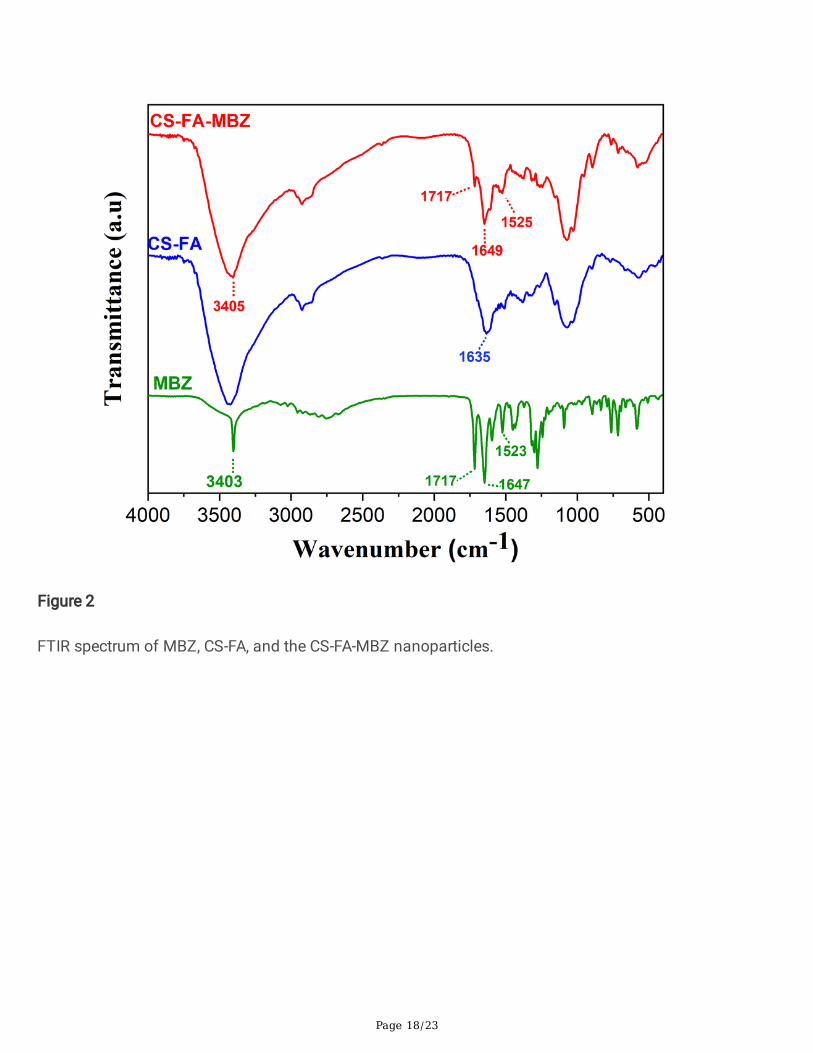
For fabrication of the CS-FA-MBZ nanoparticles, at �rst, folic acid was conjugated to chitosan in thepresence of EDC as carboxyl activating agent to produce CS-FA (Figure S1). The activated carboxylmoiety of FA was covalently linked to amine groups of CS [66]. In the next step, the cross-linking reactionbetween CS-FA and TPP lead to the formation of nanoparticles in which the negatively charged TPP waselectrostatically adsorbed to positively charged free protonated amine groups of CS-FA and MBZ wasencapsulated during the synthesis process (Figure 1). The successful introduction of folate to CS chainswas evaluated by FTIR analysis. The FTIR spectra of CS, FA, and CS-FA were shown in Figure S2. Thecharacteristic bands of CS located at 3422 cm-1 assigned to O-H stretching vibration overlapped with N-Hstretching mode. The bands observed at 2920 and 2880 cm-1 were attributed to the C-H stretchingvibrations of CS. Moreover, the peaks that appeared at 1656 and 1605 cm-1 corresponded to the C-Ostretching vibration of amide I and N-H bending vibration of amide II, respectively [67]. FTIR spectrum ofFA showed characteristic bands located at 1696, 1607, and 1486 cm-1 related to the C=O amidestretching vibration of the carboxyl group, N-H bending vibration of CONH, and the stretching vibration ofC=C of the phenyl ring of FA, respectively [68]. In the CS-FA spectrum, the absorption peaks at 1635 and1031 cm-1 could be attributed to the vibration of C-N [67]. The amid band at 1656 cm-1 of CS shifts to1635 cm-1 because of overlap with the newly formed amide bond, and also a new N-H bending vibrationlocated at 1520 cm-1 con�rmed the successful coupling of FA to CS [69]. After the formation of the CS-FA-MBZ nanoparticles, FTIR analysis was carried out to �nd the drug-polymer compatibility (Figure 2). Thecharacteristic absorption peaks of pure MBZ at 3403, 1717, 1647, and 1523 cm-1 were also observed forCS-FA-MBZ. The stretching vibration of amide I of the carbamate group of MBZ (1717 cm-1) was alsoobserved at the CS-FA-MBZ nanoparticles spectrum in the same location [70]. The N-H stretching andCNH vibration were observed at 3403 and 1523 cm-1 for MBZ, 3405, and 1525 cm-1 for CS-FA-MBZ.Hence, no interaction was observed from FTIR detection.
Figures 3a and b show SEM images of the CS-FA-MBZ nanoparticles at different magni�cations, in whichthe nanoparticles were uniform and exhibited a spherical morphology. The corresponding particle sizedistribution histograms (dry state) were obtained by measuring the size of 100 nanoparticles in SEMimages by the “Image J” software (Figure 3c). A narrow size distribution with a mean size of 153.3 ± 18.4nm was measured. Moreover, the particle size distribution of the CS-FA-MBZ was determined by DLSmeasurement (wet state) and the mean size (diameter) of the nanoparticles was measured 182 ± 12.1nm with a low polydispersity index (PDI<0.2), illustrating a narrow size distribution and con�rmed theresult obtained from SEM as well (Figure 3d). Due to the measuring of hydrodynamic radius, the DLSanalysis showed a slightly larger size for the nanoparticles as compared to SEM photographs [71]. Thezeta potential values showed that the positive charge of chitosan due to the presence of the aminegroups decreased after FA conjugation (Figure 2e). It could be related to the interaction of FA moleculeswith these amine groups of CS, leading to the neutralization of the potential value [72]. Additionally, theresults depicted that the encapsulation of MBZ into the CS-FA nanoparticles could not induce anoticeable zeta value change in the �nal nanoparticles with the preserved positive value of +27 mV.

Page 9/23
3.2. MBZ loading
MBZ, as a hydrophobic drug, was loaded into the CS-FA nanoparticles. The UV-Vis spectrum of MBZ(Figure 4a) showed the characteristic absorption band at 234 nm which also could be observed for CS-MBZ. This band along with the absorption band of FA at 280 nm with a slight shift to longer wavelengthscould be observed in the spectrum of CS-FA-MBZ, indicating the appropriate MBZ loading into the CS-FAnanoparticles. The encapsulation e�cacy (EE) and loading capacity (LC) of MBZ were calculated byplotting the standard calibration curve with a linear curve �t equation (Figure S3.). The value of EE and LCwere achieved 57.7% and 10.5% respectively, demonstrating the valuable capability of the target systemfor MBZ loading.
3.3. In vitro MBZ release study
The release behavior of MBZ from the CS-FA-MBZ nanoparticles was evaluated at different pH values of5.5, 6.8, and 7.4 to mimic tumor microenvironment, physiological conditions (pH =7.4 like bloodstream)and endocytic compartments conditions (pH= 5.5) [59]. As Figure 4b illustrates, the CS-FA-MBZnanoparticles exhibited a continuous and sustained release pro�le at different pH values started with aburst release within 6 h followed by a gradual release up to 1 week. The initial fast release could berelated to the absorbed MBZ on the surface of nanoparticles [68]. Moreover, a pH-responsive behavior ofMBZ was observed. After 1 week, the release of MBZ was around 62 %, 49 %, and 38 % at pH values of5.5, 6.8, and 7.4, respectively. This behavior could be assigned to the high swelling ability of CS whichwas induced by protonation of the amine groups of the polymer in an acidic environment [68].Considering the acidic microenvironment of cancerous tissues and intracellular organelles such asendosomes and lysosomes, the pH-sensitive behavior of the CS-FA-MBZ nanoparticles improves theircapability for tumor-speci�c drug delivery and reduces the probable adverse effects to normal tissues.
3.2 The CS-FA-MBZ implants effect on the 4T1 breast tumors’ growth progression
As Figure 5a illustrates, the CS-FA and CS-FA-MBZ implants were s.c. implanted in the left �ank of thetumor-bearing mice inside a small incision on the 3rd day after the cancer cell injection. The implantswere completely palpable even after suturing the incision. The implants completely degraded until the18th day in both CS-FA and CS-FA-MBZ groups and nothing was palpable at the implantation site. Thismeans that the implants were dissociated and release their composing agents means CS-FA-MBZnanoparticle.
The therapeutic effect of the CS-FA-MBZ implants on inhibition of 4T1 tumors’ growth progression wasevaluated by serial measurement of tumor’s diameters and compared with the Control, MBZ (40 mg/kg,oral administration, twice a week for two weeks), and CS-FA groups (Figure 5b). All treatments wereinitiated from the 3rd day after cancer cells injection. As Figure 5b illustrates, the CS-FA-MBZ implantscould signi�cantly inhibit the breast tumors growth progression in comparison with all other groups. Onthe last day of tumors’ volume monitoring (18th day after cancer cells injection), the mean tumors’
Page 10/23
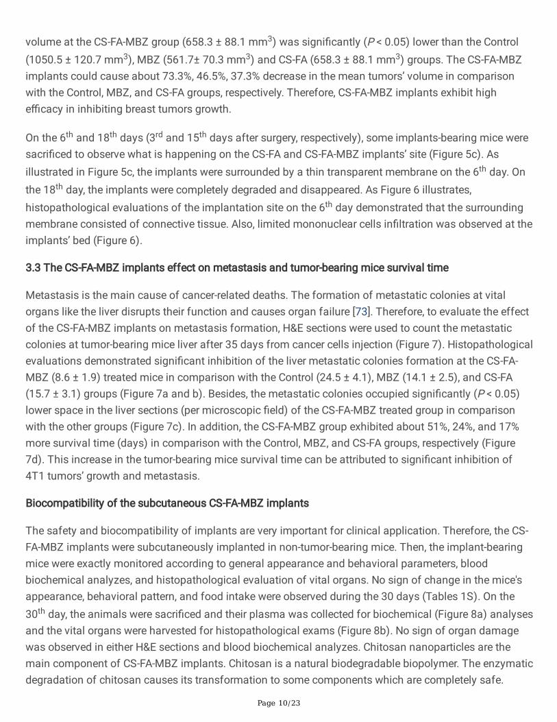
volume at the CS-FA-MBZ group (658.3 ± 88.1 mm3) was signi�cantly (P < 0.05) lower than the Control(1050.5 ± 120.7 mm3), MBZ (561.7± 70.3 mm3) and CS-FA (658.3 ± 88.1 mm3) groups. The CS-FA-MBZimplants could cause about 73.3%, 46.5%, 37.3% decrease in the mean tumors’ volume in comparisonwith the Control, MBZ, and CS-FA groups, respectively. Therefore, CS-FA-MBZ implants exhibit highe�cacy in inhibiting breast tumors growth.
On the 6th and 18th days (3rd and 15th days after surgery, respectively), some implants-bearing mice weresacri�ced to observe what is happening on the CS-FA and CS-FA-MBZ implants’ site (Figure 5c). Asillustrated in Figure 5c, the implants were surrounded by a thin transparent membrane on the 6th day. Onthe 18th day, the implants were completely degraded and disappeared. As Figure 6 illustrates,histopathological evaluations of the implantation site on the 6th day demonstrated that the surroundingmembrane consisted of connective tissue. Also, limited mononuclear cells in�ltration was observed at theimplants’ bed (Figure 6).
3.3 The CS-FA-MBZ implants effect on metastasis and tumor-bearing mice survival time
Metastasis is the main cause of cancer-related deaths. The formation of metastatic colonies at vitalorgans like the liver disrupts their function and causes organ failure [73]. Therefore, to evaluate the effectof the CS-FA-MBZ implants on metastasis formation, H&E sections were used to count the metastaticcolonies at tumor-bearing mice liver after 35 days from cancer cells injection (Figure 7). Histopathologicalevaluations demonstrated signi�cant inhibition of the liver metastatic colonies formation at the CS-FA-MBZ (8.6 ± 1.9) treated mice in comparison with the Control (24.5 ± 4.1), MBZ (14.1 ± 2.5), and CS-FA(15.7 ± 3.1) groups (Figure 7a and b). Besides, the metastatic colonies occupied signi�cantly (P < 0.05)lower space in the liver sections (per microscopic �eld) of the CS-FA-MBZ treated group in comparisonwith the other groups (Figure 7c). In addition, the CS-FA-MBZ group exhibited about 51%, 24%, and 17%more survival time (days) in comparison with the Control, MBZ, and CS-FA groups, respectively (Figure7d). This increase in the tumor-bearing mice survival time can be attributed to signi�cant inhibition of4T1 tumors’ growth and metastasis.
Biocompatibility of the subcutaneous CS-FA-MBZ implants
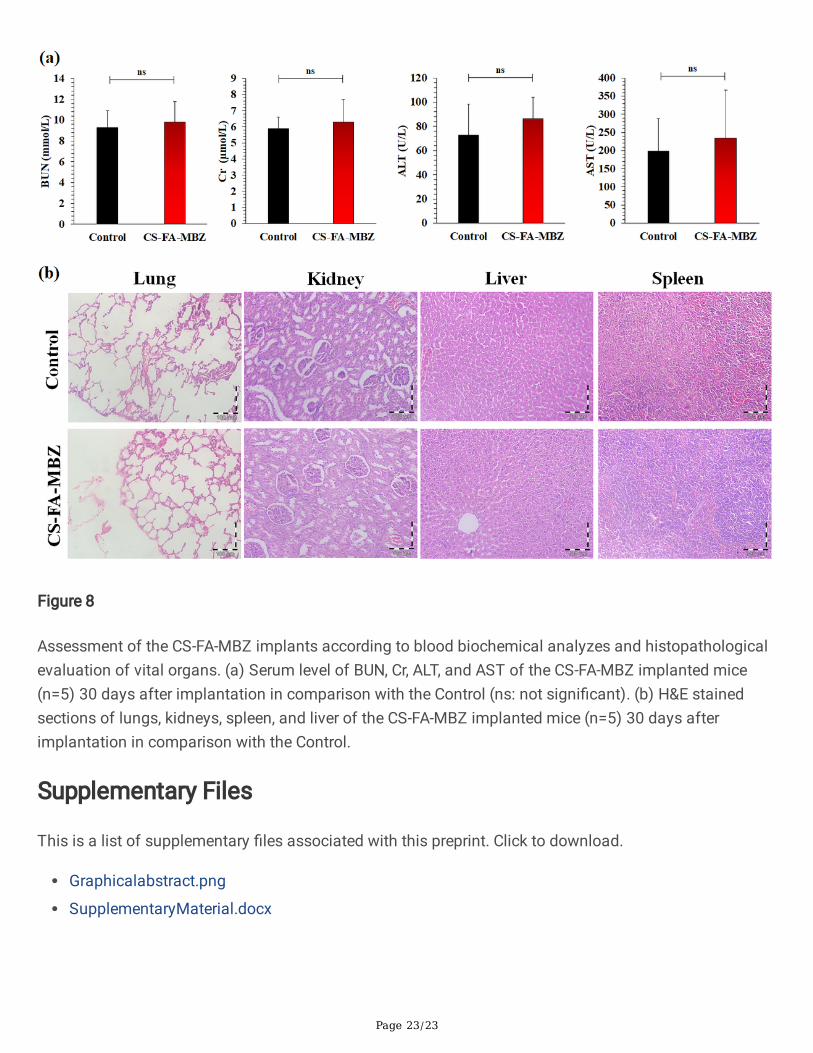
The safety and biocompatibility of implants are very important for clinical application. Therefore, the CS-FA-MBZ implants were subcutaneously implanted in non-tumor-bearing mice. Then, the implant-bearingmice were exactly monitored according to general appearance and behavioral parameters, bloodbiochemical analyzes, and histopathological evaluation of vital organs. No sign of change in the mice'sappearance, behavioral pattern, and food intake were observed during the 30 days (Tables 1S). On the30th day, the animals were sacri�ced and their plasma was collected for biochemical (Figure 8a) analysesand the vital organs were harvested for histopathological exams (Figure 8b). No sign of organ damagewas observed in either H&E sections and blood biochemical analyzes. Chitosan nanoparticles are themain component of CS-FA-MBZ implants. Chitosan is a natural biodegradable biopolymer. The enzymaticdegradation of chitosan causes its transformation to some components which are completely safe.

Page 11/23
Many enzymes have the ability to degrade chitosan and the most well-known one is lysozyme as a non-speci�c protease. This enzyme which presents in all mammalian tissues and �uids, plays a key role indegradation of chitosan-based implants in vivo. It targets the acetylated residues of chitosan polymerand degrades chitosan to non-toxic oligosaccharides which can be excreted or incorporated toglycosaminoglycans and glycoproteins [30, 74-76]. Also, eight human chitinases (in the glycosidehydrolase 18 family) have been identi�ed, three of which have shown enzymatic activity [77].
ConclusionChemotherapy drugs loaded implants have exhibited higher e�cacy in comparison with common routesof drug administration. In the present study, folic acid-targeted chitosan nanoparticles were used as acarrier to increase MBZ therapeutic e�cacy in the triple-negative breast cancer-bearing BALB/c model. Ade�nite amount of MBZ-loaded CS-FA nanoparticles were compressed to form cylindrical CS-FA-MBZimplants which were subcutaneously implanted at mice’s �ank. The CS-FA-MBZ implants couldsigni�cantly inhibit 4T1 breast tumor growth and metastasis. Therefore, these biodegradable andbiocompatible implants can be an appropriate choice for further experiments in breast cancer and othercancers treatment
AbbreviationsTriple-negative breast cancer: TNBC
Mebendazole: MBZ
Folic acid: FA
Chitosan: CS
KX: Ketamine-Xylazine
DeclarationsEthics approval and consent to participate
All animal experiments complied with the ARRIVE guidelines and were conducted according to theguidelines of the European Communities Council Directive (2010/63/UE) and the Isfahan University ofMedical Sciences for the care and use of laboratory animals. Likewise, all the procedures, protocols, andsteps were approved by the ethics committee of the Isfahan University of Medical Sciences(IR.MUI.RESEARCH.REC.1399.125).
Consent for publication
All authors have read and agreed to publish this manuscript.

Page 12/23
Availability of data and material
All datasets are available upon reasonable request.
Competing interests
The authors declared no potential con�icts of interest with respect to the research, authorship, and/orpublication of this article.
Funding
This research was supported by Isfahan University of Medical Sciences.
Authors' contributions
The study was designed by A. Kefayat. In vivo assessments were carried out by A. Kefayat, F.Ghahremani, and M. Ra�enia. The nanoparticles synthesis and characterization were done by M.Hosseini. The manuscript writing and revisions were done by A. Kefayat, M. Hosseini, F. Ghahremani, andM. Ra�enia. All the authors read and approved the manuscript before submission.
References1. DeSantis, C.E., et al., Breast cancer statistics, 2017, racial disparity in mortality by state. 2017. 67(6):
p. 439-448.
2. Perou, C.M., et al., Molecular portraits of human breast tumours. 2000. 406(6797): p. 747-752.
3. Kumar, P., R.J.A.o.g. Aggarwal, and obstetrics, An overview of triple-negative breast cancer. 2016.293(2): p. 247-269.
4. Wang, J., et al., Adjuvant chemotherapy and radiotherapy in triple-negative breast carcinoma: aprospective randomized controlled multi-center trial. 2011. 100(2): p. 200-204.
5. Sorolla, A., et al., Triple-hit therapeutic approach for triple negative breast cancers using docetaxelnanoparticles, EN1-iPeps and RGD peptides. 2019. 20: p. 102003.
�. Al-Mahmood, S., et al., Metastatic and triple-negative breast cancer: challenges and treatmentoptions. 2018. 8(5): p. 1483-1507.
7. Nedeljković, M. and A.J.C. Damjanović, Mechanisms of Chemotherapy Resistance in Triple-NegativeBreast Cancer—How We Can Rise to the Challenge. 2019. 8(9): p. 957.
�. Guerini, A.E., et al., Mebendazole as a candidate for drug repurposing in oncology: an extensivereview of current literature. 2019. 11(9): p. 1284.
9. Coyne, C., et al., Gemcitabine-(C4-amide)-[anti-HER2/neu] anti-neoplastic cytotoxicity in dualcombination with mebendazole against chemotherapeutic-resistant mammary adenocarcinoma.2013. 2(2).
Page 13/23
10. Doudican, N., et al., Mebendazole induces apoptosis via Bcl-2 inactivation in chemoresistantmelanoma cells. 2008. 6(8): p. 1308-1315.
11. Larsen, A.R., et al., Repurposing the antihelmintic mebendazole as a hedgehog inhibitor. 2015. 14(1):p. 3-13.
12. Bai, R.-Y., et al., Brain penetration and e�cacy of different mebendazole polymorphs in a mousebrain tumor model. 2015. 21(15): p. 3462-3470.
13. He, L., et al., Mebendazole exhibits potent anti-leukemia activity on acute myeloid leukemia. 2018.369(1): p. 61-68.
14. Mukhopadhyay, T., et al., Mebendazole elicits a potent antitumor effect on human cancer cell linesboth in vitro and in vivo. 2002. 8(9): p. 2963-2969.
15. Pantziarka, P., et al., Repurposing Drugs in Oncology (ReDO)—mebendazole as an anti-cancer agent.2014. 8.
1�. Martarelli, D., et al., Mebendazole inhibits growth of human adrenocortical carcinoma cell linesimplanted in nude mice. 2008. 61(5): p. 809-817.
17. Pinto, L.C., et al., Mebendazole, an antiparasitic drug, inhibits drug transporters expression inpreclinical model of gastric peritoneal carcinomatosis. 2017. 43: p. 87-91.
1�. Sung, S.J., et al., Autophagy is a potential target for enhancing the anti-angiogenic effect ofmebendazole in endothelial cells. 2019. 27(1): p. 117.
19. Bai, R.-Y., et al., Effective treatment of diverse medulloblastoma models with mebendazole and itsimpact on tumor angiogenesis. 2015. 17(4): p. 545-554.
20. Dawson, M., R. Allan, and T.J.B.j.o.c.p. Watson, The pharmacokinetics and bioavailability ofmebendazole in man: a pilot study using [3H]‐mebendazole. 1982. 14(3): p. 453-455.
21. Dawson, M., et al., The pharmacokinetics and bioavailability of a tracer dose of [3H]‐mebendazole inman. 1985. 19(1): p. 79-86.
22. de la Torre-Iglesias, P.M., et al., Enhanced bioavailability and anthelmintic e�cacy of mebendazole inredispersible microparticles with low-substituted hydroxypropylcellulose. 2014. 8: p. 1467.
23. Belz, J.E., et al., Sustained Release Talazoparib Implants for Localized Treatment of BRCA1-de�cientBreast Cancer. Theranostics, 2017. 7(17): p. 4340.
24. Park, C.-W., et al., In vitro/in vivo evaluation of NCDS-micro-fabricated biodegradable implant. 2010.33(3): p. 427-432.
25. Arias, J.L., Novel strategies to improve the anticancer action of 5-�uorouracil by using drug deliverysystems. Molecules, 2008. 13(10): p. 2340-2369.
2�. Weinberg, B.D., E. Blanco, and J. Gao, Polymer implants for intratumoral drug delivery and cancertherapy. Journal of pharmaceutical sciences, 2008. 97(5): p. 1681-1702.
27. Talebian, S., et al., Biopolymers for antitumor implantable drug delivery systems: recent advancesand future outlook. 2018. 30(31): p. 1706665.

Page 14/23
2�. Zhang, Z., et al., Sandwich‐Like Fibers/Sponge Composite Combining Chemotherapy andHemostasis for E�cient Postoperative Prevention of Tumor Recurrence and Metastasis. 2018.30(49): p. 1803217.
29. Kuang, G., et al., Biphasic drug release from electrospun polyblend nano�bers for optimized localcancer treatment. 2018. 6(2): p. 324-331.
30. Kefayat, A. and S.J.I.j.o.b.m. Vaezifar, Biodegradable PLGA implants containing doxorubicin-loadedchitosan nanoparticles for treatment of breast tumor-bearing mice. 2019. 136: p. 48-56.
31. Nam, K.-S., Y.-H.J.J.o.m. Shon, and biotechnology, Suppression of metastasis of human breastcancer cells by chitosan oligosaccharides. 2009. 19(6): p. 629-633.
32. Li, Y., et al., Chitosan sulfate inhibits angiogenesis via blocking the VEGF/VEGFR2 pathway andsuppresses tumor growth in vivo. 2019. 7(4): p. 1584-1597.
33. Adhikari, H.S. and P.N.J.I.J.o.B. Yadav, Anticancer activity of chitosan, chitosan derivatives, and theirmechanism of action. 2018. 2018.
34. Unsoy, G., et al., Synthesis of Doxorubicin loaded magnetic chitosan nanoparticles for pH responsivetargeted drug delivery. 2014. 62: p. 243-250.
35. Tiyaboonchai, W.J.N.U.J.S. and Technology, Chitosan nanoparticles: a promising system for drugdelivery. 2013. 11(3): p. 51-66.
3�. Wang, J.J., et al., Recent advances of chitosan nanoparticles as drug carriers. 2011. 6: p. 765.
37. Min, K.H., et al., Hydrophobically modi�ed glycol chitosan nanoparticles-encapsulated camptothecinenhance the drug stability and tumor targeting in cancer therapy. 2008. 127(3): p. 208-218.
3�. Nagpal, K., et al., Chitosan nanoparticles: a promising system in novel drug delivery. 2010. 58(11): p.1423-1430.
39. Sinha, V., et al., Chitosan microspheres as a potential carrier for drugs. 2004. 274(1-2): p. 1-33.
40. Pan, Y., et al., Bioadhesive polysaccharide in protein delivery system: chitosan nanoparticles improvethe intestinal absorption of insulin in vivo. 2002. 249(1-2): p. 139-147.
41. Mitra, S., et al., Tumour targeted delivery of encapsulated dextran–doxorubicin conjugate usingchitosan nanoparticles as carrier. 2001. 74(1-3): p. 317-323.
42. Sahu, S.K., et al., In vitro evaluation of folic acid modi�ed carboxymethyl chitosan nanoparticlesloaded with doxorubicin for targeted delivery. 2010. 21(5): p. 1587-1597.
43. Yang, S.-J., et al., Folic acid-conjugated chitosan nanoparticles enhanced protoporphyrin IXaccumulation in colorectal cancer cells. 2010. 21(4): p. 679-689.
44. Kularatne, S.A. and P.S. Low, Targeting of nanoparticles: folate receptor, in Cancer Nanotechnology.2010, Springer. p. 249-265.
45. Xu, L., et al., Folate-mediated chemotherapy and diagnostics: an updated review and outlook. 2017.252: p. 73-82.
4�. Kefayat, A., et al., Ultra-small but ultra-effective: Folic acid-targeted gold nanoclusters forenhancement of intracranial glioma tumors' radiation therapy e�cacy. 2019. 16: p. 173-184.

Page 15/23
47. Kefayat, A., et al., Investigation of different targeting decorations effect on the radiosensitizinge�cacy of albumin-stabilized gold nanoparticles for breast cancer radiation therapy. 2019. 130: p.225-233.
4�. O’Shannessy, D.J., et al., Folate receptor alpha (FRA) expression in breast cancer: identi�cation of anew molecular subtype and association with triple negative disease. 2012. 1(1): p. 22.
49. Zhang, Z., et al., Folate receptor α associated with triple-negative breast cancer and poor prognosis.2014. 138(7): p. 890-895.
50. Necela, B.M., et al., Folate receptor-α (FOLR1) expression and function in triple negative tumors.2015. 10(3).
51. Paulmurugan, R., et al., Folate receptor–targeted polymeric micellar nanocarriers for delivery oforlistat as a repurposed drug against triple-negative breast cancer. 2016. 15(2): p. 221-231.
52. Kaur, P., et al., A mouse model for triple-negative breast cancer tumor-initiating cells (TNBC-TICs)exhibits similar aggressive phenotype to the human disease. 2012. 12(1): p. 120.
53. Pulaski, B.A. and S.J.C.p.i.i. Ostrand‐Rosenberg, Mouse 4T1 breast tumor model. 2000. 39(1): p.20.2. 1-20.2. 16.
54. Ghahremani, F., et al., AS1411 aptamer conjugated gold nanoclusters as a targeted radiosensitizerfor megavoltage radiation therapy of 4T1 breast cancer cells. 2018. 8(8): p. 4249-4258.
55. Ghahremani, F., et al., AS1411 aptamer-targeted gold nanoclusters effect on the enhancement ofradiation therapy e�cacy in breast tumor-bearing mice. 2018. 13(20): p. 2563-2578.
5�. Dehghani, S., et al., Multifunctional MIL-Cur@ FC as a theranostic agent for magnetic resonanceimaging and targeting drug delivery: in vitro and in vivo study. 2020. 28(6): p. 668-680.
57. Liu, Y., et al., Novel albendazole–chitosan nanoparticles for intestinal absorption enhancement andhepatic targeting improvement in rats. 2013. 101(6): p. 998-1005.
5�. Parakh, D.R., et al., Development and validation of Spectrophotometric method for estimation ofmebendazole in bulk and pharmaceutical formulation. 2015. 4: p. 2223-35.
59. Nejadsha�ee, V., et al., Magnetic bio-metal–organic framework nanocomposites decorated with folicacid conjugated chitosan as a promising biocompatible targeted theranostic system for cancertreatment. 2019. 99: p. 805-815.
�0. Care, O.o.A. and Use, Guidelines for Endpoints in Animal Study Proposals. 2011.
�1. Workman, P., et al., Guidelines for the welfare and use of animals in cancer research. 2010. 102(11):p. 1555-1577.
�2. Wallace, J., Humane endpoints and cancer research. ILAR journal, 2000. 41(2): p. 87-93.
�3. Ibrahim, K., et al., Histopathology of the Liver, Kidney, and Spleen of Mice Exposed to GoldNanoparticles. 2018. 23(8): p. 1848.
�4. Rajeh, M.A.B., et al., Acute toxicity impacts of Euphorbia hirta L extract on behavior, organs bodyweight index and histopathology of organs of the mice and Artemia salina. 2012. 4(3): p. 170.

Page 16/23
�5. Zhang, X.D., et al., Enhanced Tumor Accumulation of Sub‐2 nm Gold Nanoclusters for CancerRadiation Therapy. 2014. 3(1): p. 133-141.
��. Beidokhti, H.R.N., et al., Preparation, characterization, and optimization of folic acid-chitosan-methotrexate core-shell nanoparticles by box-behnken design for tumor-targeted drug delivery. 2017.18(1): p. 115-129.
�7. Devendiran, R.M., et al., Green synthesis of folic acid-conjugated gold nanoparticles with pectin asreducing/stabilizing agent for cancer theranostics. 2016. 6(35): p. 29757-29768.
��. Esfandiarpour-Boroujeni, S., et al., Fabrication and study of curcumin loaded nanoparticles based onfolate-chitosan for breast cancer therapy application. 2017. 168: p. 14-21.
�9. Cheng, L., et al., Synthesis of folate‐chitosan nanoparticles loaded with ligustrazine to target folatereceptor positive cancer cells. 2017. 16(2): p. 1101-1108.
70. Gunasekaran, S. and D.J.A.J.o.C. Uthra, Vibrational spectra and qualitative analysis of albendazoleand mebendazole. 2008. 20(8): p. 6310.
71. Souza, T.G., V.S. Ciminelli, and N.D.S. Mohallem. A comparison of TEM and DLS methods tocharacterize size distribution of ceramic nanoparticles. in Journal of physics: conference series.2016. IOP Publishing.
72. Song, H., et al., Folic acid-chitosan conjugated nanoparticles for improving tumor-targeted drugdelivery. 2013. 2013.
73. Welch, D.R. and D.R.J.C.r. Hurst, De�ning the hallmarks of metastasis. 2019. 79(12): p. 3011-3027.
74. Kurita, K., et al., Enzymatic degradation of β-chitin: susceptibility and the in�uence of deacetylation.2000. 42(1): p. 19-21.
75. Szymańska, E. and K.J.M.d. Winnicka, Stability of chitosan—a challenge for pharmaceutical andbiomedical applications. 2015. 13(4): p. 1819-1846.
7�. Vårum, K.M., et al., In vitro degradation rates of partially N-acetylated chitosans in human serum.1997. 299(1-2): p. 99-101.
77. Funkhouser, J.D. and N.N.J.B.e.b. Aronson, Chitinase family GH18: evolutionary insights from thegenomic history of a diverse protein family. 2007. 7(1): p. 96.
Figures

Page 17/23
Figure 1
Sequential steps for preparation of the CS-FA-MBZ nanoparticles.
Page 18/23
Figure 2
FTIR spectrum of MBZ, CS-FA, and the CS-FA-MBZ nanoparticles.

Page 19/23
Figure 3
(a, b) SEM images of the CS-FA-MBZ nanoparticles with different magni�cations. (c) Size distribution ofthe CS-FA-MBZ nanoparticles determined by ImageJ software considering 100 particles in correspondingSEM images. (d) The CS-FA-MBZ nanoparticles hydrodynamic size distribution according to DLSmeasurements. (e) Zeta potential of the Cs, CS-FA, and CS-FA-MBZ nanoparticles.
Figure 4

Page 20/23
(a) UV–Vis spectra of MBZ, CS-FA, CS-MBZ, and CS-FA-MBZ. (b) Release pro�les of MBZ from CS-FA-MBZ nanoparticles at pH = 5.5, 6.8, and 7.4. Data are expressed as mean ± S.D (n = 3).
Figure 5
(a) Subcutaneous implantation of the CS-FA-MBZ implants. A small incision was made at the left �ank ofmice and the implant was placed inside it. The incision was sutured. The implantation site on the 18thday after cancer cells injection (15th day after surgery). (b) Tumors’ growth progression in differentgroups including Control, MBZ, CS-FA, and CS-FA-MBZ from the 3rd to 18th day after cancer cellsinjection (not signi�cant: ns, *: P <0.05). (c) Harvested skin of the implants-bearing mice at the 3rd and15th days after implantation which equates the 6th and 18th days after cancer cells injection,respectively.

Page 21/23
Figure 6
Mouse body response to the subcutaneously implanted CS-FA-MBZ at 3rd day after implantationaccording to histopathology analysis. (a) A close-up view of a CS-FA-MBZ implant 3rd day afterimplantation and histopathological evaluation of (b) the implant-surrounding membrane, (c) tissues faraway from the implantation site, and (d) tissues at the bed of the implantation site. The two head arrows,asterisk, and one-head arrows indicate derma, the implant’s surrounding membrane, and in�ltratingimmune cells, respectively.

Page 22/23
Figure 7
Metastatic burden and survival time of the tumor-bearing mice at different treatment groups. (a) Onemicroscopic �eld of H&E stained sections of livers in each group was illustrated as the sample. (b) Theaverage number of metastasis colonies per microscopic �eld of the tumor-bearing mice’s liver 30 daysafter the cancer cells injection. The yellow arrows indicate a liver metastatic colony. (c) Mean percentageof the occupied space per microscopic �eld of the tumor-bearing mice’s liver 30 days after the cancercells injection. (d) The survival time of tumor-bearing mice in different groups. (not signi�cant: ns, *: P<0.05)
Page 23/23
Figure 8
Assessment of the CS-FA-MBZ implants according to blood biochemical analyzes and histopathologicalevaluation of vital organs. (a) Serum level of BUN, Cr, ALT, and AST of the CS-FA-MBZ implanted mice(n=5) 30 days after implantation in comparison with the Control (ns: not signi�cant). (b) H&E stainedsections of lungs, kidneys, spleen, and liver of the CS-FA-MBZ implanted mice (n=5) 30 days afterimplantation in comparison with the Control.
Supplementary Files
This is a list of supplementary �les associated with this preprint. Click to download.
Graphicalabstract.png
SupplementaryMaterial.docx