Macrophage polarization following chitosan implantation
8
Macrophage polarization following chitosan implantation Daniela P. Vasconcelos b , Ana C. Fonseca a, b , Madalena Costa a, c , Isabel F. Amaral b , Mário A. Barbosa a, b , Artur P. Águas a, c , Judite N. Barbosa a, b, * a ICBAS e Instituto de Ciências Biomédicas Abel Salazar, Universidade do Porto, Rua de Jorge Viterbo Ferreira n. 228, 4050-313 Porto, Portugal b INEB e Instituto de Engenharia Biomédica, NEWTherapies Group, Universidade do Porto, Rua do Campo Alegre 823, 4150-180 Porto, Portugal c UMIB e Unit for Multidisciplinary Biomedical Research of ICBAS e Instituto de Ciências Biomédicas Abel Salazar, Universidade do Porto, Rua de Jorge Viterbo Ferreira n. 228, 4050-313 Porto, Portugal article info Article history: Received 12 July 2013 Accepted 4 September 2013 Available online 25 September 2013 Keywords: Macrophage polarization Inflammation Chitosan In vivo studies abstract Macrophages are a key cell in the host response to implants and can be polarized into different phe- notypes capable of inducing both detrimental and beneficial outcomes in tissue repair and remodeling, being important in tissue engineering and regenerative medicine. The objective of this study was to evaluate the macrophage response to 3D porous chitosan (Ch) scaffolds with different degrees of acet- ylation (DA, 5% and 15%). The M1/M2 phenotypic polarization profile of macrophages was investigated in vivo using a rodent air-pouch model. Our results show that the DA affects the macrophage response. Ch scaffolds with DA 5% induced the adhesion of lower numbers of inflammatory cells, being the M2 the predominant phenotypic profile among the adherent macrophages. In the inflammatory exudates F4/ 80 þ /CD206 þ cells (M2 macrophages) appeared in higher numbers then F4/80 þ /CCR7 þ cells (M1 mac- rophages), in addition, lower levels of pro-inflammatory cytokines together with higher levels of anti- inflammatory cytokines were found. Ch scaffolds with DA 15% showed opposite results, since M1 were the predominant macrophages both adherent to the scaffold and in the exudates, together with high levels of pro-inflammatory cytokines. In conclusion, Ch scaffolds with DA 5% induced a benign M2 anti- inflammatory macrophage response, whereas Ch scaffolds with DA 15% caused a macrophage M1 pro- inflammatory response. Ó 2013 Elsevier Ltd. All rights reserved. 1. Introduction Implantation of a biomaterial causes injury that will lead to the onset of inflammation which is a prerequisite for efficient tissue repair; nonetheless, successful repair after tissue injury requires resolution of the inflammatory response. Persistence of an in- flammatory stimulus results in chronic inflammation that is a hallmark of the non-healing wound. Therefore, successful bioma- terial integration requires the coordinate expression of both inflammation, at first, and resolution of inflammation, latter on [1]. Leukocytes are central players in directing host inflammatory and immune processes; thus, their response to biomaterials is important in the understanding of materialehost interactions. Monocytes are recruited to the implant site, undergo maturation into macrophages, and persist at implant surfaces. Macrophages are the dominant infiltrating cells that respond rapidly to biomaterial implantation in soft and hard tissues. Macrophages may play a bimodal role: by the release of pro-inflammatory cytokines, participate in the initiation of an acute inflammatory response, and by the release of anti-inflammatory cytokines, pro-angiogenic fac- tors and matrix metalloproteases (MMPs), participate in the biodegradation of bioresorbable implants, playing an important role in tissue regeneration and healing. By phagocytosis, macro- phages clear the way for tissue ingrowths, secrete a spectrum of cytokines and growth factors to regulate cell recruitment, prolif- eration and differentiation, leading to effective tissue regeneration and angiogenesis. Thus, macrophages have been suggested to be the cells that orchestrate both the inflammatory and the repair phase of tissues around implants [2,3]. Macrophages display remarkable plasticity and can change their physiology in response to environmental cues, giving rise to different cell populations with distinct functions. This is reflected in the division of macrophages into two major phenotypes (M1 and M2). The pro-inflammatory, cytotoxic macrophage phenotype, labeled as M1 (classically activated), promotes pathogen killing and * Corresponding author. INEB e Instituto de Engenharia Biomédica, NEW- Therapies Group, Universidade do Porto, Rua do Campo Alegre 823, 4150-180 Porto, Portugal. Tel.: þ351 22 6074982; fax: þ351 22 6094567. E-mail addresses: [email protected], [email protected] (J.N. Barbosa). Contents lists available at ScienceDirect Biomaterials journal homepage: www.elsevier.com/locate/biomaterials 0142-9612/$ e see front matter Ó 2013 Elsevier Ltd. All rights reserved. http://dx.doi.org/10.1016/j.biomaterials.2013.09.012 Biomaterials 34 (2013) 9952e9959
-
Upload
independent -
Category
Documents
-
view
0 -
download
0
Transcript of Macrophage polarization following chitosan implantation

lable at ScienceDirect
Biomaterials 34 (2013) 9952e9959
Contents lists avai
Biomaterials
journal homepage: www.elsevier .com/locate/biomateria ls
Macrophage polarization following chitosan implantation
Daniela P. Vasconcelos b, Ana C. Fonseca a, b, Madalena Costa a, c, Isabel F. Amaral b,Mário A. Barbosa a, b, Artur P. Águas a, c, Judite N. Barbosa a, b, *
a ICBAS e Instituto de Ciências Biomédicas Abel Salazar, Universidade do Porto, Rua de Jorge Viterbo Ferreira n.� 228, 4050-313 Porto, Portugalb INEB e Instituto de Engenharia Biomédica, NEWTherapies Group, Universidade do Porto, Rua do Campo Alegre 823, 4150-180 Porto, Portugalc UMIB e Unit for Multidisciplinary Biomedical Research of ICBAS e Instituto de Ciências Biomédicas Abel Salazar, Universidade do Porto, Rua de JorgeViterbo Ferreira n.� 228, 4050-313 Porto, Portugal
a r t i c l e i n f o
Article history:Received 12 July 2013Accepted 4 September 2013Available online 25 September 2013
Keywords:Macrophage polarizationInflammationChitosanIn vivo studies
* Corresponding author. INEB e Instituto de EnTherapies Group, Universidade do Porto, Rua do CampPortugal. Tel.: þ351 22 6074982; fax: þ351 22 60945
E-mail addresses: [email protected], judite@ibmc.
0142-9612/$ e see front matter � 2013 Elsevier Ltd.http://dx.doi.org/10.1016/j.biomaterials.2013.09.012
a b s t r a c t
Macrophages are a key cell in the host response to implants and can be polarized into different phe-notypes capable of inducing both detrimental and beneficial outcomes in tissue repair and remodeling,being important in tissue engineering and regenerative medicine. The objective of this study was toevaluate the macrophage response to 3D porous chitosan (Ch) scaffolds with different degrees of acet-ylation (DA, 5% and 15%). The M1/M2 phenotypic polarization profile of macrophages was investigatedin vivo using a rodent air-pouch model. Our results show that the DA affects the macrophage response.Ch scaffolds with DA 5% induced the adhesion of lower numbers of inflammatory cells, being the M2 thepredominant phenotypic profile among the adherent macrophages. In the inflammatory exudates F4/80þ/CD206þ cells (M2 macrophages) appeared in higher numbers then F4/80þ/CCR7þ cells (M1 mac-rophages), in addition, lower levels of pro-inflammatory cytokines together with higher levels of anti-inflammatory cytokines were found. Ch scaffolds with DA 15% showed opposite results, since M1 werethe predominant macrophages both adherent to the scaffold and in the exudates, together with highlevels of pro-inflammatory cytokines. In conclusion, Ch scaffolds with DA 5% induced a benign M2 anti-inflammatory macrophage response, whereas Ch scaffolds with DA 15% caused a macrophage M1 pro-inflammatory response.
� 2013 Elsevier Ltd. All rights reserved.
1. Introduction
Implantation of a biomaterial causes injury that will lead to theonset of inflammation which is a prerequisite for efficient tissuerepair; nonetheless, successful repair after tissue injury requiresresolution of the inflammatory response. Persistence of an in-flammatory stimulus results in chronic inflammation that is ahallmark of the non-healing wound. Therefore, successful bioma-terial integration requires the coordinate expression of bothinflammation, at first, and resolution of inflammation, latter on [1].
Leukocytes are central players in directing host inflammatoryand immune processes; thus, their response to biomaterials isimportant in the understanding of materialehost interactions.Monocytes are recruited to the implant site, undergo maturationintomacrophages, and persist at implant surfaces. Macrophages are
genharia Biomédica, NEW-o Alegre 823, 4150-180 Porto,67.up.pt (J.N. Barbosa).
All rights reserved.
the dominant infiltrating cells that respond rapidly to biomaterialimplantation in soft and hard tissues. Macrophages may play abimodal role: by the release of pro-inflammatory cytokines,participate in the initiation of an acute inflammatory response, andby the release of anti-inflammatory cytokines, pro-angiogenic fac-tors and matrix metalloproteases (MMPs), participate in thebiodegradation of bioresorbable implants, playing an importantrole in tissue regeneration and healing. By phagocytosis, macro-phages clear the way for tissue ingrowths, secrete a spectrum ofcytokines and growth factors to regulate cell recruitment, prolif-eration and differentiation, leading to effective tissue regenerationand angiogenesis. Thus, macrophages have been suggested to bethe cells that orchestrate both the inflammatory and the repairphase of tissues around implants [2,3].
Macrophages display remarkable plasticity and can change theirphysiology in response to environmental cues, giving rise todifferent cell populations with distinct functions. This is reflected inthe division of macrophages into two major phenotypes (M1 andM2). The pro-inflammatory, cytotoxic macrophage phenotype,labeled as M1 (classically activated), promotes pathogen killing and

D.P. Vasconcelos et al. / Biomaterials 34 (2013) 9952e9959 9953
is associated with classic signs of active inflammation, particularlywith chronic inflammation. The anti-inflammatory macrophagephenotype, labeled as M2 (alternatively activated), promotesimmunoregulation tissue repair and constructive tissue remodel-ing. M1 and M2 macrophages can be identified and distinguishedaccording to their cell surface markers and their cytokine and geneexpression profiles [4e6].
Chitosan, a biodegradable polysaccharide obtained by N-deacetylation of chitin, is a natural polymer that is currently underinvestigation for several biomedical applications, namely as scaf-folds and as drug and gene delivery system for regenerative med-icine [7,8]. Previous results of our team have demonstrated that thedegree of acetylation of Ch affects the behavior of osteoblasts [9]and also modulates the in vivo biological response, namely in-flammatory cell recruitment and fibrous capsule formation aroundimplants [10].
The ability of the host innate immune system to resolve apolarized macrophage response (M1 or M2) resulting from animplant is of key importance to determine the downstream func-tional success of implanted biomaterials. However, few studieshave evaluated macrophage polarization specifically with respectto biomaterials implantation [11]. The effects of macrophagephenotype upon tissue remodeling outcome following implanta-tion of a biomaterial are largely unknown. However, the recogni-tion of the predominant phenotypic profile may provide a tool bywhich a constructive and functional tissue remodeling outcome canbe predicted and/or promoted [12].
The objective of the present study was to determine the effect ofimplantation of 3D chitosan scaffolds on the phenotypic profile ofthe macrophages participating in the host response, and also therelationship between the degree of acetylation of chitosan andmacrophage polarization.
2. Materials and methods
2.1. Chitosan (Ch) purification and characterization
Squid pen Ch (ref. 114, Batch No. S4; DAw2%) was supplied byMahtani ChitosanPvt. Ltd. and subsequently purified by filtration of chitosan acidic solution andsubsequent alkali precipitation. The purified Ch was characterized in terms of DAand average molecular weight by Fourier transform infrared spectroscopy (FT-IR)and size exclusion chromatography, respectively, as previously described [9]. A DA of5.02 � 0.66 (n ¼ 6), with weight-average molecular weight (Mw) 8.9 � 1.0 � 105 andpolydispersity index (PDI) 1.2� 0.0 were found. Chitosanwith DA 15% was preparedby N-acetylation of the former, according to Vachoud L et al. [13], in a water/aceticacid/1,2-propanediol solution, using acetic anhydride as reactive. Subsequent anal-ysis revealed a DA of 15.71 � 0.52 (n ¼ 6), Mw 8.3 � 0.9 � 105 and a PDI of 1.3 � 0.1.Chitosan endotoxin levels were measured in water extracts, using the LimulusAmebocyte Lysate (QCL-1000� test, Cambrex), for the chromogenic quantitation ofGram-negative bacterial endotoxin. Briefly, Ch extracts were prepared using 40 mLendotoxin-free water/g of Ch, incubating Ch suspensions for 24 h at 50 �C undercontinuous shaking (250 rpm), as described elsewhere [14]. The resultant extractswere filtered through a 0.45 mm syringe filter, and used for endotoxin quantification.The two polymers revealed endotoxin levels below 0.1 EU/mL, respecting the USDepartment of Health and Human Services guidelines for implantable devices.
2.2. Preparation and characterization of Ch 3D scaffolds
The 3D porous scaffolds were prepared from degassed 2% w/v Ch solutions in0.2 M acetic acid via thermally induced phase separation (�20 �C) and subsequentsublimation of the ice crystals. Following lyophilization (�85 �C; 0.2 mbar), theresultant scaffolds were cut in discs with 8.5 mm in diameter and 2 mm thickness.Scaffolds microstructure was analyzed by scanning electron microscopy (SEM) intransversal and longitudinal cross-sections of the lyophilized scaffolds. In accor-dance to previous results Ch scaffolds revealed a highly porous and homogeneousmicrostructurewith interconnected poreswith diameters in the range of 100 mm, forboth the DAs used [10].
2.3. Mouse animal model
The procedures involved in the animal model were submitted and evaluated bythe in-house ethics committee and also by the Portuguese official authority on
animal welfare and experimentation (DGV), and were approved before the experi-ments were performed.
For each experimental group, 6 male BALB/c mice (Charles River, Spain) wereused at 7 weeks of age. Air pouches were generated according to the method ofSedwick et al. [15] as adapted by Castro et al. [16]. Anaesthetized by intramuscularinjection of ketamine (Ketalar, Parke-Davis Co., Spain; 4.0e8.0 mg/kg of weight) andxilazine (Rompum, Bayer Co., Portugal; 0.8e1.6 mg/kg) mice were injected subcu-taneously in the dorsal area with 5 mL of sterile air that caused the formation of anair pouch. A reinforcement of the air pouch was performed 5 days latter through asecond subcutaneous injection of 3 mL of sterile air. A single scaffold was implantedin each animal.
2.4. Implantation of the chitosan 3D scaffolds
One day after the second subcutaneous injection, the mice were anaesthetizedby intramuscular injection of ketamine (Ketalar, Parke-Davis Co., Spain; 4.0e8.0 mg/kg of weight) and xilazine (Rompum, Bayer Co., Portugal; 0.8e1.6 mg/kg) and theskin covering the air pouch area was shaved and cleaned with betadine. A surgicalincision was made, the materials were placed inside the air pouch, and the incisionwas sutured. Three different experimental groups were used: One group with Chscaffolds with a DA 5%, another group with Ch scaffolds with DA 15% and sham-operated animals (animals submitted to the same technique with no scaffoldimplanted). For each experimental group 6 animals were used.
2.5. Inflammatory exudates
The exudates were recovered from the mouse air-pouches 1 and 4 days after theimplantation. The mice were anaesthetized and sacrificed. Harvesting of inflam-matory exudates was done bywashing the air pouch cavities with 2mL of phosphatebuffered saline (PBS, Merck) followed by recovery of the lavage fluid.
2.6. Explants
Explantation occurred immediately after the recovery of the inflammatory ex-udates, 1 and 4 days after implantation. The sutures were cut, and the wound edgesseparated; the scaffolds were then carefully removed from the pouches and fixedwith 4% paraformaldehyde (PFA, Merck) in PBS for 1 h, for confocal microscopy.
2.7. Confocal microscopy
Chitosan scaffolds were rinsed in PBS three times in order to remove any tracesof PFA. Non-specific binging sites were blocked with 1% BSA (bovine serum albumin,Gibco), 10% FBS (fetal bovine serum, Gibco) and 5% NGS (normal goat serum, Invi-trogen) and permeabilized with 0.1% Triton-X (Sigma) in PBS for 1 h at room tem-perature. Samples were incubated overnight at 4 �Cwith ratmonoclonal antibody a-F4/80 (1:200), mouse monoclonal antibody a-Mannose Receptor CD206 (1:100) andrabbit monoclonal antibody a-CCR7 (1:350), all from Abcam. After washing threetimes with PBS, the scaffolds were incubated for 1 h at room temperature withsecondary antibodies Alexa Fluor� 488 rabbit a-rat (1:1000), Alexa Fluor� 568 goata-mouse (1:1000) and Alexa Fluor� 647 goat a-rabbit (1:1000), all from Invitrogen.For F-actin staining, scaffolds were prepared as above and stained with Alexa Fluor�
488 Phalloidin (1:100, Invitrogen). All scaffolds were mounted with Vectashield�
with DAPI (Vector). Imageswere obtainedwith a laser scanning confocal microscopeLeica TCS SP5 II (Leica Microsystems, Germany). An average of 5 horizontal imagesections with a z-step of 50 mm were obtained per site and a total of 5 sites perscaffold were visualized. In order to render the 3D image into a 2D projection, theMaximum Intensity Projection was performed. All images were processed with Fijisoftware. Confocal images of M1 and M2 macrophage colonization of the implantedscaffolds were further scored into three categories: Present (þ); Frequent (þþ);Abundant (þþþ).
2.8. Flow cytometry
The exudates were filtered through 40 mm nylon mesh (BD Biosciences) toremove cell clumps and spun at 1200 rpm for 5 min at 4 �C. Supernatants wereremoved and cell pellets re-suspended in 1 mL staining buffer (PBS/0.5% BSA/0.1%azide). Single-cell suspensions were pre-incubated with Fc-receptor-blocking anti-bodies (Miltenyi Biotec) for 10 min at 4 �C. Labeling was performed in a final volumeof 50 ml with the indicated fluorescently conjugated antibodies for 30 min at 4 �C inthe dark. Cells were thenwashed three times with staining buffer and transferred toFACS tubes for flow cytometry analysis. The following antibodies were used in thisstudy: phycoerythrin (PE)-labeled anti-mouse F4/80 (clone 521204, 4:50 ml), fluo-rescein isothiocyanate (FITC)-labeled anti-mouse CD3 (clone 17A2, 3:50 ml), allo-phycocyanin (APC)-labeled anti-mouse CCR7 (clone 4B12 7:50 ml), Alexa Fluor�488labeled anti-mouse MMR/CD206 (3:50 ml), all from R&D Systems and APC-labeledanti-mouse CD11b (Clone M1/70.15 2:50 ml) from Immunotools. The isotype con-trols PE-labeled IgG2A (clone 54447), FITC-labeled IgG2B (clone 141945), APC-labeledIgG2A (54447) and Alexa Fluor�488-labeled IgG, all from R&D Systems and APC-labeled IgG2b from Immunotools, were used as negative controls to define

D.P. Vasconcelos et al. / Biomaterials 34 (2013) 9952e99599954
background staining at corresponding concentrations. Fluorescence was measuredusing FACS Calibur flow cytometer (BD Biosciense) with Cell Quest software, and10.000 events were collected per sample. Analysis was performed using FlowJosoftware.
2.9. Cytokine production
The concentrations of TNF-a, IL-6 and IL-4 in the inflammatory exudates weredetermined using commercially available ELISA kits (R&D Systems) according to themanufacturer recommendations. Cytokine concentrations were determinedcomparing the absorbance values with a standard calibration curve, and normalizedfor the total number of cells present in the corresponding inflammatory exudate.
2.10. Statistical analysis
Statistical analysis was performed using the non-parametric test ManneWhit-ney with Graph Pad v6.02 for Windows. A value of p < 0.05 was considered sta-tistically significant: *p < 0.05; **p < 0.01; ***p < 0.001. Data are presented showingindividual groups and respective mean � SEM.
3. Results
3.1. Confocal microscopy
The implanted Ch scaffolds were retrieved from the airpouches of mice 1 and 4 days after implantation and analyzed byconfocal microscopy, in order to investigate the relative amountof adherent cells on the scaffolds, and also the phenotypic profileof adherent macrophages. The scaffolds were initially stained forF-actin and DNA and the images obtained are depicted in Fig. 1.Both scaffolds presented adherent inflammatory cells at theirsurfaces, although being present in higher numbers in the Chscaffolds with DA 15% at both time points studied (images AeD).We have also performed transversal cuts in the scaffolds to assesswhether or not the cells migrated through the scaffolds andcolonized them. The results show higher numbers of inflamma-tory cells within the Ch scaffolds with the higher DA, withcells migrating through the whole scaffold. In the case of Ch
Fig. 1. Confocal microscopy images of inflammatory cells adherent to the implanted 3D porosurface (AeD) and also cells migrating through the scaffold and colonizing its interior (EeH
scaffolds with DA 5% the cells were observed closer to the surface(images EeH).
The scaffolds were then stained with specific macrophagemarkers: F4/80 (pan macrophage marker); CD206 (M2 marker)and CCR7 (M1 marker) to assess the phenotypic profile ofadherent macrophages (Fig. 2). Cells were stained for F4/80 inorder to identify the adherent macrophages, and doubled stainedwith an additional marker for M1 or M2 macrophages. Cells F4/80þ and CD206þ are M2 macrophages whereas cells F4/80þ andCCR7þ are M1 macrophages. The arrows in the images indicateexamples of cells that presented double staining and the coloc-alization is shown in yellow. Images with staining for M2 mac-rophages (F4/80/CD206) are presented on the left side of Fig. 2.After 1 and 4 days of implantation, Ch scaffolds with DA 5%presented higher numbers of M2 macrophages (images C and F)when compared with Ch scaffolds with DA 15% (images I and L).Images with staining for M1 macrophages (F4/80/CCR7) areprovided on the right side of Fig. 2. At both time points Chscaffolds with the higher DA showed higher numbers of M1macrophages (images U and X) in comparison with Ch scaffoldswith the lower DA (images O and R). A scoring system was usedin order to perform a semi-quantitative evaluation of the M1 andM2 macrophages present in the implanted scaffolds, as is shownin Table 1.
3.2. Flow cytometry
The inflammatory exudates induced by the presence of theimplanted Ch scaffolds were collected from the mouse air pouchesand analyzed by flow cytometry 1 and 4 days after implantation.Macrophages were identified using CD11b and F4/80, twocommonly used murine macrophage markers and CCR7 and CD206as specific M1 and M2 markers respectively. Dot plots from arepresentative experiment are presented in Fig. 3. The resultspresented in Fig. 4a revealed that Ch with DA 15% induced higher
us chitosan scaffolds, 1 and 4 days after implantation. Images show adherent cells at the). Ch scaffolds were stained for F-actin/DNA. (Scale bar: 25 mm).

Fig. 2. Confocal images of immunoflourescent staining of the implanted 3D porous chitosan scaffolds, 1 and 4 days after implantation. F4/80 (green fluorophore); CD206 (redfluorophore); CCR7 (red fluorophore); nuclei are revealed by DAPI staining (blue). The arrows indicate examples of cells that presented double staining: F4/80þ/CD206þ cells(yellow: images C, F, I, L) that are M2 macrophages; whereas F4/80þ/CCR7þ cells (yellow: images O, R, U, X) are M1 macrophages. (Scale bar: 25 mm). (For interpretation of thereferences to color in this figure legend, the reader is referred to the web version of this article.)
D.P. Vasconcelos et al. / Biomaterials 34 (2013) 9952e9959 9955
numbers of recruited inflammatory cells, when compared with Chwith DA 5%, and sham-operated animals. Although Ch with thehigher DA induced the recruitment of higher numbers of inflam-matory cells to the implant site, these exudates presented a lowerpercentage of F4/80þ cells in comparison with Ch with the lowerdegree of acetylation. This was observed for both time pointsinvestigated (Fig. 4b).
The use of specific macrophage phenotypic markers showedthat the inflammatory exudates collected from air pouches withimplanted Ch scaffolds with DA 15% presented higher percentage ofCCR7þ cells, together with a lower percentage of CD206þ cells thatindicate an M1 macrophage phenotype. In contrast, inflammatoryexudates retrieved from air pouches with implanted Ch scaffoldswith DA 5% showed a higher percentage of CD206þ cells, and alower percentage of CCR7þ cells, when compared with Ch with DA15%, indicating an M2 macrophage phenotypic profile. The
Table 1Semi-quantitative analysis of M1 and M2 macrophages present in implanted 3Dporous chitosan scaffolds, based on the immunofluorescent confocal images, 1 and 4days after implantation. M1 and M2 phenotypes were scored as: Present (þ);Frequent (þþ); Abundant (þþþ).
Ch 5% Ch 15%
Day 1 Day 4 Day 1 Day 4
M1 macrophages þ þþ þþþ þþþM2 macrophages þþ þþþ þ þþ
differences obtained between the different Ch scaffolds were al-ways statistically significant (Fig. 4c and d).
3.3. Cytokine production
The exudates recovered from the air-pouches 1 and 4 daysafter the implantation of the different scaffolds were also eval-uated regarding cytokine production; the results are depicted inFig. 5. Higher concentrations of IL-6 were detected in the in-flammatory exudates induced by the presence of Ch scaffoldswith DA 15%, both at 1 and 4 days after implantation, whencomparing with Ch implants with the lower degree of acetylation(Fig. 5a). The TNF-a concentration was also higher in the in-flammatory exudates recovered from air pouches with Ch im-plants with the higher DA, being this difference statisticalsignificant only 4 days after implantation (Fig. 5b). Higher levelsof the anti-inflammatory cytokine IL-4 were found on the in-flammatory exudates induced by the presence of the Ch scaffoldswith a DA 5%, when comparing with the Ch scaffolds with thehigher DA, being these differences statistically significant bothfor 1 and 4 days after implantation (Fig. 5c). Taken together,these results indicate that Ch scaffolds with a lower DA inducethe production of lower levels of pro-inflammatory cytokines,together with higher levels of anti-inflammatory cytokines, thatis characteristic of an M2 macrophage response, being theopposite observed in the case of Ch scaffolds with the higher DA,that induce an M1 macrophage response.

Fig. 3. Expression of cell surface markers on macrophages from the inflammatory exudates recovered from the mouse air pouches. Representative dot plots showing (a) aspectratio; (b, c) expression of F4/80, CD11b, CD206 and CCR7 antigens. Gates were drawn from isotype control staining. (b) F4/80 and CD11b double staining. Macrophages were gatedon the basis of size and positive double staining (F4/80þ and CD11bþ). (c) Percentage of F4/80 positive cells expressing CD206 (M2 marker) and/or CCR7 (M1 marker) antigens. (FSC:forward scatter; SSC: side scatter).
D.P. Vasconcelos et al. / Biomaterials 34 (2013) 9952e99599956
4. Discussion
Until very recently, the host response to biomaterials wasviewed as detrimental and with unintended but unavoidablenegative implications. Recently, the paradigm of macrophage acti-vation and polarization, and its relevance to biomaterials in thecontext of regenerative medicine, has changed this point of view.
The host macrophage response is an essential element of aconstructive tissue remodeling process that follows the implanta-tion of biomaterials. Macrophage polarization has been acknowl-edged to be an important modulator of the tissue remodelingprocess [17].
The purpose of this investigation was to assess in vivo themacrophage phenotypic profile following the implantation of

Fig. 4. Characterization of the inflammatory exudates recovered from the mouse air pouches by flow cytometry. Cells were obtained by air pouch lavage with 2 mL of PBS. Resultsare presented as average � s.e.m. (a) Total number of cell recruited to the air pouches. (b) Percentage of F4/80 positive cells. (c) Cells expressing both F4/80þ and CD206þ. (d) Cellsexpressing both F4/80þ and CCR7þ. (Statistically significant differences: *p < 0.05; **p < 0.01.
D.P. Vasconcelos et al. / Biomaterials 34 (2013) 9952e9959 9957
chitosan (Ch) 3D porous scaffolds with different degrees of acety-lation (5 and 15%). We have analyzed the macrophages that arerecruited to the implant site, both those present in the inflamma-tory exudates and those adherent to the implanted scaffolds. Theinflammatory exudates were studied by flow cytometry usingspecific macrophage markers, and also by the assessment of cyto-kine production. The Ch scaffolds were analyzed by confocal mi-croscopy using the same macrophage markers employed in flowcytometry.
The 3D porous Ch scaffolds were characterized in terms of theDA, average molecular weight, porosity, pore size, inter-connectivity and the endotoxin levels were measured in Ch waterextracts, being the results described and discussed in detail else-where [9,10]. Briefly, for both DAs, the scaffolds are highly porouswith an interconnected microstructure, which is very important inthe development of scaffolds for regenerative medicine applica-tions, since it allows the seeding of numerous cells and theirmigration into the bulk with an appropriate supply of nutrients[8,18].
In this investigation, we have used the mouse air-pouch modelof inflammation. This model involves the formation of a sterilesubcutaneous cavity that can be used to insert a biomaterial and tostudy the inflammatory reaction caused by the implant, providing alocalized environment in which to study cell trafficking and theinflammatory response through the infiltration of inflammatorycells and the production of cytokines/chemokines [19,20].
In previous investigations, we have concluded that Ch scaffoldswith a higher DA caused a more severe inflammatory response byinducing the recruitment of higher numbers of inflammatory cellsto the implant site, with also higher numbers of adherent
inflammatory cells and the formation of a thick fibrous capsulearound the implanted scaffolds [10]. In the current investigation,we have focus our attention on M1 and M2 macrophage sub-populations since these cells are known to play key role ininflammation after the implantation of a biomedical device and inaddition, can play both beneficial and detrimental roles in tissuerepair and regeneration [11]. Macrophages change their physiologyin response to environmental cues and give rise to different pop-ulations of cells with distinct functions. Macrophages have beencharacterized as having an M1 or M2 phenotype based on receptorexpression, cytokine and effector molecule production and func-tion. M1 macrophages produce pro-inflammatory cytokines, lead-ing to immunostimulation; M2 macrophages produce anti-inflammatory cytokines, pro-angiogenic factors and matrix metal-loproteases, promoting angiogenesis and tissue repair [5,6].
F-actin and DNA staining of Ch implants revealed highernumbers of adherent inflammatory cells in Ch scaffolds with DA15%, which is in accordance with our previous results [10]. More-over, after performing transversal cuts in the scaffolds, our resultsshowed higher numbers of inflammatory cells within the Ch scaf-folds with the higher degree of acetylation with cells migratingthrough the whole scaffold, when compared with data from Chscaffolds with the lower DA. The scaffolds were stained withmacrophage specific markers in order to identify M1 (F4/80þ/CCR7þ cells) and M2 macrophages (F4/80þ/CD206þ cells). After 1and 4 days of implantation Ch scaffolds with DA 5% presentedhigher numbers of M2 macrophages when compared with Chscaffolds with DA 15%. At both time points investigated Ch scaffoldswith the higher DA showed higher numbers of M1 macrophages, incomparison with Ch scaffolds with the lower DA.
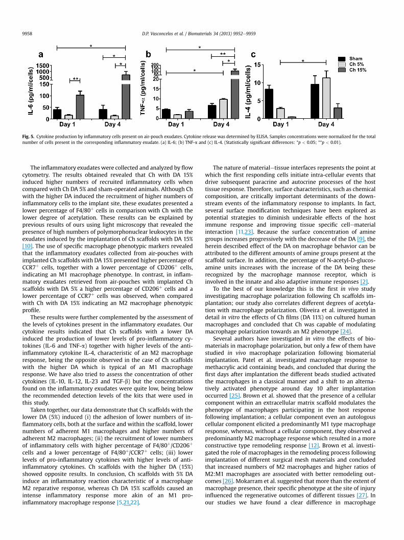
Fig. 5. Cytokine production by inflammatory cells present on air-pouch exudates. Cytokine release was determined by ELISA. Samples concentrations were normalized for the totalnumber of cells present in the corresponding inflammatory exudate. (a) IL-6; (b) TNF-a and (c) IL-4. (Statistically significant differences: *p < 0.05; **p < 0.01).
D.P. Vasconcelos et al. / Biomaterials 34 (2013) 9952e99599958
The inflammatory exudates were collected and analyzed by flowcytometry. The results obtained revealed that Ch with DA 15%induced higher numbers of recruited inflammatory cells whencompared with Ch DA 5% and sham-operated animals. Although Chwith the higher DA induced the recruitment of higher numbers ofinflammatory cells to the implant site, these exudates presented alower percentage of F4/80þ cells in comparison with Ch with thelower degree of acetylation. These results can be explained byprevious results of ours using light microscopy that revealed thepresence of high numbers of polymorphonuclear leukocytes in theexudates induced by the implantation of Ch scaffolds with DA 15%[10]. The use of specific macrophage phenotypic markers revealedthat the inflammatory exudates collected from air-pouches withimplanted Ch scaffolds with DA 15% presented higher percentage ofCCR7þ cells, together with a lower percentage of CD206þ cells,indicating an M1 macrophage phenotype. In contrast, in inflam-matory exudates retrieved from air-pouches with implanted Chscaffolds with DA 5% a higher percentage of CD206þ cells and alower percentage of CCR7þ cells was observed, when comparedwith Ch with DA 15% indicating an M2 macrophage phenotypicprofile.
These results were further complemented by the assessment ofthe levels of cytokines present in the inflammatory exudates. Ourcytokine results indicated that Ch scaffolds with a lower DAinduced the production of lower levels of pro-inflammatory cy-tokines (IL-6 and TNF-a) together with higher levels of the anti-inflammatory cytokine IL-4, characteristic of an M2 macrophageresponse, being the opposite observed in the case of Ch scaffoldswith the higher DA which is typical of an M1 macrophageresponse. We have also tried to assess the concentration of othercytokines (IL-10, IL-12, IL-23 and TGF-b) but the concentrationsfound on the inflammatory exudates were quite low, being belowthe recommended detection levels of the kits that were used inthis study.
Taken together, our data demonstrate that Ch scaffolds with thelower DA (5%) induced (i) the adhesion of lower numbers of in-flammatory cells, both at the surface and within the scaffold, lowernumbers of adherent M1 macrophages and higher numbers ofadherent M2 macrophages; (ii) the recruitment of lower numbersof inflammatory cells with higher percentage of F4/80þ/CD206þ
cells and a lower percentage of F4/80þ/CCR7þ cells; (iii) lowerlevels of pro-inflammatory cytokines with higher levels of anti-inflammatory cytokines. Ch scaffolds with the higher DA (15%)showed opposite results. In conclusion, Ch scaffolds with 5% DAinduce an inflammatory reaction characteristic of a macrophageM2 reparative response, whereas Ch DA 15% scaffolds caused anintense inflammatory response more akin of an M1 pro-inflammatory macrophage response [5,21,22].
The nature of materialetissue interfaces represents the point atwhich the first responding cells initiate intra-cellular events thatdrive subsequent paracrine and autocrine processes of the hosttissue response. Therefore, surface characteristics, such as chemicalcomposition, are critically important determinants of the down-stream events of the inflammatory response to implants. In fact,several surface modification techniques have been explored aspotential strategies to diminish undesirable effects of the hostimmune response and improving tissue specific cellematerialinteraction [11,23]. Because the surface concentration of aminegroups increases progressively with the decrease of the DA [9], theherein described effect of the DA on macrophage behavior can beattributed to the different amounts of amine groups present at thescaffold surface. In addition, the percentage of N-acetyl-D-glucos-amine units increases with the increase of the DA being theserecognized by the macrophage mannose receptor, which isinvolved in the innate and also adaptive immune responses [2].
To the best of our knowledge this is the first in vivo studyinvestigating macrophage polarization following Ch scaffolds im-plantation; our study also correlates different degrees of acetyla-tion with macrophage polarization. Oliveira et al. investigated indetail in vitro the effects of Ch films (DA 11%) on cultured humanmacrophages and concluded that Ch was capable of modulatingmacrophage polarization towards an M2 phenotype [24].
Several authors have investigated in vitro the effects of bio-materials in macrophage polarization, but only a few of them havestudied in vivo macrophage polarization following biomaterialimplantation. Patel et al. investigated macrophage response tomethacrylic acid containing beads, and concluded that during thefirst days after implantation the different beads studied activatedthe macrophages in a classical manner and a shift to an alterna-tively activated phenotype around day 10 after implantationoccurred [25]. Brown et al. showed that the presence of a cellularcomponent within an extracellular matrix scaffold modulates thephenotype of macrophages participating in the host responsefollowing implantation; a cellular component even an autologouscellular component elicited a predominantly M1 type macrophageresponse, whereas, without a cellular component, they observed apredominantly M2 macrophage response which resulted in a moreconstructive type remodeling response [12]. Brown et al. investi-gated the role of macrophages in the remodeling process followingimplantation of different surgical mesh materials and concludedthat increased numbers of M2 macrophages and higher ratios ofM2:M1 macrophages are associated with better remodeling out-comes [26]. Mokarram et al. suggested that more than the extent ofmacrophage presence, their specific phenotype at the site of injuryinfluenced the regenerative outcomes of different tissues [27]. Inour studies we have found a clear difference in macrophage
D.P. Vasconcelos et al. / Biomaterials 34 (2013) 9952e9959 9959
response with regards to different degrees of acetylation, even atvery short periods after implantation of the chitosan scaffolds (1day). This suggests that Ch with lower degrees of acetylation willmore likely have the ability to improve outcomes in regenerativemedicine, since with these scaffolds we observed an M2 reparativeresponse. Alternatively activated M2 macrophages are hypores-ponsive to pro-inflammatory stimuli, are involved in debris scav-enging, angiogenesis, tissue remodeling as well as resolution ofinflammation, being therefore, inducers of repair functions [28].
Promoting polarization to an M2 anti-inflammatory, pro-tissuehealing phenotype is likely to be a useful strategy to mitigate theadverse effects of biomaterial induced inflammation. In conclu-sion, approaches which provide control of macrophage phenotypemay constitute a promising route in regenerative medicine ap-plications [11].
5. Conclusions
Macrophage response to Ch 3D porous scaffolds with differentDAs was evaluated. Phenotypic and functional polarization ofmacrophages in response to the implanted scaffolds was assessedaccording to their cell surface markers and their cytokine expres-sion profiles. The results demonstrate that Ch scaffolds with thelower DA (5%) induce an inflammatory reaction that is character-istic of a macrophage M2 reparative response, defined mainly byhigher numbers of M2 macrophages adherent to the implantedscaffolds, higher percentage of these cells present in the inflam-matory exudates, and lower levels of pro-inflammatory cytokines,together with higher levels of anti-inflammatory cytokines. Chscaffolds with the higher DA (15%) caused an intense inflammatoryresponse mediated by M1 pro-inflammatory macrophages, sincehigher numbers of M1 macrophages were seen both adherent atthe scaffold surface and in the inflammatory exudates, togetherwith high levels of pro-inflammatory cytokines. These results areinstructive for the design of biomaterials for regenerative medicine,since the manipulation of macrophage effector mechanisms can bea potential strategy for promoting constructive tissue remodeling.
Acknowledgments
This work was financed by FEDER funds through the ProgramaOperacional Factores de Competitividade e COMPETE and Portu-guese funds through FCT e Fundação para a Ciência e Tecnologia inthe framework of project PTDC/SAU-BMA/113030/2009.
References
[1] Eming SA, Krieg T, Davidson JM. Inflammation in wound repair: molecular andcellular mechanisms. J Invest Dermatol 2007;127(3):514e25.
[2] Xia Z, Triffitt JT. A review on macrophage responses to biomaterials. BiomedMater 2006;1(1):R1e9.
[3] Kou PM, Babensee JE. Macrophage and dendritic cell phenotypic diversity inthe context of biomaterials. J Biomed Mater Res A 2011;96(1):239e60.
[4] Duffield JS. The inflammatory macrophage: a story of Jekyll and Hyde. Clin Sci(Lond) 2003;104(1):27e38.
[5] Mosser DM, Edwards JP. Exploring the full spectrum of macrophage activa-tion. Nat Rev Immunol 2008;8(12):958e69.
[6] Mantovani A, Sica A, Sozzani S, Allavena P, Vecchi A, Locati M. The chemokinesystem in diverse forms of macrophage activation and polarization. TrendsImmunol 2004;25(12):677e86.
[7] VandeVord PJ, Matthew HWT, DeSilva SP, Mayton L, Wu B, Wooley PH.Evaluation of the biocompatibility of a chitosan scaffold in mice. J BiomedMater Res 2002;59(3):585e90.
[8] Kim IY, Seo SJ, Moon HS, Yoo MK, Park IY, Kim BC, et al. Chitosan and itsderivatives for tissue engineering applications. Biotechnol Adv 2008;26(1):1e21.
[9] Amaral IF, Sampaio P, Barbosa MA. Three-dimensional culture of humanosteoblastic cells in chitosan sponges: the effect of the degree of acetylation.J Biomed Mater Res A 2006;76(2):335e46.
[10] Barbosa JN, Amaral IF, Aguas AP, Barbosa MA. Evaluation of the effect of thedegree of acetylation on the inflammatory response to 3D porous chitosanscaffolds. J Biomed Mater Res A 2010;93(1):20e8.
[11] Brown BN, Ratner BD, Goodman SB, Amar S, Badylak SF. Macrophage polari-zation: an opportunity for improved outcomes in biomaterials and regener-ative medicine. Biomaterials 2012;33(15):3792e802.
[12] Brown BN, Valentin JE, Stewart-Akers AM, McCabe GP, Badylak SF. Macro-phage phenotype and remodeling outcomes in response to biologic scaffoldswith and without a cellular component. Biomaterials 2009;30(8):1482e91.
[13] Vachoud L, Zydowicz N, Domard A. Formation and characterisation of aphysical chitin gel. Carbohydr Res 1997;302:169e77.
[14] Nakagawa Y, Murai T, Hasegawa C, Hirata M, Tsuchiya T, Yagami T, et al.Endotoxin contamination in wound dressings made of natural biomaterials.J Biomed Mater Res B 2003;66(1):347e55.
[15] Sedwick AD, Sin YM, Edwards JC, Willoughby DA. Increased inflammatoryreactivity in newly formed lining tissue. J Pathol 1983;141(4):483e95.
[16] Castro AG, Esaguy N, Macedo PM, Aguas AP, Silva MT. Live but not heat-killedmycobacteria cause rapid chemotaxis of large numbers of eosinophils in vivoand are ingested by the attracted granulocytes. Infect Immun 1991;59(9):3009e14.
[17] Brown BN, Badylak SF. Expanded applications, shifting paradigms and animproved understanding of host-biomaterial interactions. Acta Biomater2013;9(2):4948e55.
[18] Suh JKF, Matthew HWT. Application of chitosan-based polysaccharide bio-materials in cartilage tissue engineering: a review. Biomaterials 2000;21(24):2589e98.
[19] Hooper KA, Nickolas TL, Yurkow EJ, Kohn J, Laskin DL. Characterization of theinflammatory response to biomaterials using a rodent air pouch model.J Biomed Mater Res 2000;50(3):365e74.
[20] Wooley PH, Morren R, Andary J, Sud SA, Yang SY, Mayton L, et al. Inflam-matory responses to orthopaedic biomaterials in the murine air pouch. Bio-materials 2002;23(2):517e26.
[21] Mantovani A, Garlanda C, Locati M. Macrophage diversity and polarization inatherosclerosis a question of balance. Arterioscler Thromb Vasc Biol2009;29(10):1419e23.
[22] Sica A, Mantovani A. Macrophage plasticity and polarization: in vivo veritas.J Clin Invest 2012;122(3):787e95.
[23] Brown BN, Sicari BM, Turner NJ, Kukla KA, Badylak SF. Local macrophagepolarization and tissue remodeling following bilateral implantation of ECMscaffold materials in the abdominal wall. J Tissue Eng Regen Med 2012;6:217e8.
[24] Oliveira MI, Santos SG, Oliveira MJ, Torres AL, Barbosa MA. Chitosan drivesanti-inflammatory macrophage polarisation and pro-inflammatory dendriticcell stimulation. Eur Cell Mater 2012;24:136e53.
[25] Patel RJ, Sefton MV. Some aspects of the host response to methacrylic acidcontaining beads in a mouse air pouch. J Biomed Mater Res A 2012;100(8):2054e62.
[26] Brown BN, Londono R, Tottey S, Zhang L, Kukla KA, Wolf MT, et al. Macro-phage phenotype as a predictor of constructive remodeling following theimplantation of biologically derived surgical mesh materials. Acta Biomater2012;8(3):978e87.
[27] Mokarram N, Merchant A, Mukhatyar V, Patel G, Bellamkonda RV. Effect ofmodulating macrophage phenotype on peripheral nerve repair. Biomaterials2012;33(34):8793e801.
[28] Eming SA, Hammerschmidt M, Krieg T, Roers A. Interrelation of immunity andtissue repair or regeneration. Semin Cell Dev Biol 2009;20(5):517e27.