Saponin-loaded chitosan nanoparticles and their cytotoxicity to cancer cell lines in vitro
10
Carbohydrate Polymers 84 (2011) 407–416 Contents lists available at ScienceDirect Carbohydrate Polymers journal homepage: www.elsevier.com/locate/carbpol Saponin-loaded chitosan nanoparticles and their cytotoxicity to cancer cell lines in vitro N. Sanoj Rejinold a , M. Muthunarayanan a , K. Muthuchelian b , K.P. Chennazhi a , Shanti V. Nair a , R. Jayakumar a,∗ a Amrita Centre for Nanosciences and Molecular Medicine, Amrita Institute of Medical Sciences & Research Centre, Amrita Vishwa Vidyapeetham University, Cochin 682041, India b Department of Bioenergy, School of Energy, Environment and Natural Resources, Madurai Kamaraj University, Madurai 625021, India article info Article history: Received 30 September 2010 Received in revised form 23 November 2010 Accepted 28 November 2010 Available online 3 December 2010 Keywords: Saponin Chitosan Anti cancer saponin delivery Nanosaponin Specific toxicity abstract In this work we developed a nanoformulation for anticancer saponin with chitosan for an enhanced and sustained release. The saponin loaded chitosan nanoparticles showed a particle size of 65 ± 7 nm. The synthesized nanoparticles were analyzed by FTIR, TG/DTA, SEM and AFM. The cytotoxicity of the nanoparticles was analyzed on L929, NIH-3T3, KB and PC3 which showed particles are non-toxic in a concentration range of 0.1–1.0 mg/ml whereas the nanosaponin showed specific toxicity on PC3 and KB cell lines. The internalization of the nanosaponin on L929 and PC3 was confirmed by Rhodamine conju- gation with the nanoparticles. Our preliminary studies support that nanosaponin could be an efficient therapeutic agent for cancer. © 2010 Elsevier Ltd. All rights reserved. 1. Introduction Saponins are naturally occurring amphiphilic compounds present in many foods of plant origin. Legumes, in particular, are rich sources of dietary saponin. Although practically non-toxic to man upon oral ingestion, saponins are powerful hemolytic agents when injected into the bloodstream. Saponins are lipase inhibitors (Basalingappa & Chandravadan, 1971; Fenwick & Oakenfull, 1983; Francis, Kerem, Harinder Makkar, & Becker, 2002; Nair, Kalariya, & Chanda, 2005; Oakenfull & Sihdu, 1990; Oakenfull, 1981; Yoshizumi et al., 2006; Zhao et al., 2005). Populations consuming diets high in legumes and their products are therefore exposed to a highlevel of saponin. There is an increased interest in the importance of different micronutrients as well as of non-nutritive compounds present in vegetables in the prevention of chronic dis- eases such as cardio vascular heart disease and cancer (Shibata, 2001; Sung, Kendall, & Rao, 1995; Yibing, Yichun, & Biao, 2007). Saponin possesses important biological activities, including hypoc- holesterolaemic, immune-stimulatory (Potter, 1993; Wu et al., ∗ Corresponding author. Tel.: +91 484 2801234; fax: +91 484 2802020. E-mail addresses: [email protected], [email protected] (R. Jayakumar). 1990) and antitumorigenic (Yu et al., 1992) effects. However the insolubility of these saponins limits their wide spectrum actions in medical field. Chitosan is a well known biopolymer having many applica- tions in tissue engineering, wound healing (Muzzarelli, 2009; Sudheesh Kumar et al., 2010), drug delivery (Elzatahry & Mohy, 2008; Kashappa & Hyun, 2005; Kofuji, Ito, Murata, & Kawashima, 2001; Thanou, Verhoef, & Junginger, 2001), and also in gene delivery (Jayakumar, Chennazh, et al, 2010; Muzzarelli, 1988, 2009, 2010; Richardson, Kolbe, and Duncan, 1999). Chitosan nanoparticles used for the delivery of polypep- tides such as insulin, tetanus toxoid, and diphtheria toxoids are widely explored (Calvo, Remunan, Vila, & Alonso, 1997a; Calvo, Remunan, Vila, & Alonso, 1997b; Huang, Khor, & Lim, 2004; Janes & Alonso, 2003; Vander et al., 2003; Xu & Du, 2003). Nano sized drug delivery vehicles formulated from biocompat- ible chitosan and biodegradable polymers constitute an evolving approach to saponin delivery and tumor targeting (Jayakumar, Deepthy, Manzoor, Nair, & Tamura, 2010). Biodegradable saponin carriers are being purposely engineered and constructed with nanometer dimensions. Such approaches made it possible to develop smart materials like nanosaponin as a better therapeutics. In this paper we are describing about synthesis, characterization and anticancer activities of nanosaponin in detail. 0144-8617/$ – see front matter © 2010 Elsevier Ltd. All rights reserved. doi:10.1016/j.carbpol.2010.11.056
-
Upload
independent -
Category
Documents
-
view
0 -
download
0
Transcript of Saponin-loaded chitosan nanoparticles and their cytotoxicity to cancer cell lines in vitro

Si
NSa
Cb
a
ARR2AA
KSCANS
1
prmw(F&Ydtice2Sh
(
0d
Carbohydrate Polymers 84 (2011) 407–416
Contents lists available at ScienceDirect
Carbohydrate Polymers
journa l homepage: www.e lsev ier .com/ locate /carbpol
aponin-loaded chitosan nanoparticles and their cytotoxicity to cancer cell linesn vitro
. Sanoj Rejinolda, M. Muthunarayanana, K. Muthuchelianb, K.P. Chennazhia,hanti V. Naira, R. Jayakumara,∗
Amrita Centre for Nanosciences and Molecular Medicine, Amrita Institute of Medical Sciences & Research Centre, Amrita Vishwa Vidyapeetham University,ochin 682041, IndiaDepartment of Bioenergy, School of Energy, Environment and Natural Resources, Madurai Kamaraj University, Madurai 625021, India
r t i c l e i n f o
rticle history:eceived 30 September 2010eceived in revised form3 November 2010ccepted 28 November 2010
a b s t r a c t
In this work we developed a nanoformulation for anticancer saponin with chitosan for an enhancedand sustained release. The saponin loaded chitosan nanoparticles showed a particle size of 65 ± 7 nm.The synthesized nanoparticles were analyzed by FTIR, TG/DTA, SEM and AFM. The cytotoxicity of thenanoparticles was analyzed on L929, NIH-3T3, KB and PC3 which showed particles are non-toxic in aconcentration range of 0.1–1.0 mg/ml whereas the nanosaponin showed specific toxicity on PC3 and KB
vailable online 3 December 2010eywords:aponinhitosannti cancer saponin delivery
cell lines. The internalization of the nanosaponin on L929 and PC3 was confirmed by Rhodamine conju-gation with the nanoparticles. Our preliminary studies support that nanosaponin could be an efficienttherapeutic agent for cancer.
© 2010 Elsevier Ltd. All rights reserved.
anosaponinpecific toxicity
. Introduction
Saponins are naturally occurring amphiphilic compoundsresent in many foods of plant origin. Legumes, in particular, areich sources of dietary saponin. Although practically non-toxic toan upon oral ingestion, saponins are powerful hemolytic agentshen injected into the bloodstream. Saponins are lipase inhibitors
Basalingappa & Chandravadan, 1971; Fenwick & Oakenfull, 1983;rancis, Kerem, Harinder Makkar, & Becker, 2002; Nair, Kalariya,
Chanda, 2005; Oakenfull & Sihdu, 1990; Oakenfull, 1981;oshizumi et al., 2006; Zhao et al., 2005). Populations consumingiets high in legumes and their products are therefore exposedo a highlevel of saponin. There is an increased interest in themportance of different micronutrients as well as of non-nutritive
ompounds present in vegetables in the prevention of chronic dis-ases such as cardio vascular heart disease and cancer (Shibata,001; Sung, Kendall, & Rao, 1995; Yibing, Yichun, & Biao, 2007).aponin possesses important biological activities, including hypoc-olesterolaemic, immune-stimulatory (Potter, 1993; Wu et al.,∗ Corresponding author. Tel.: +91 484 2801234; fax: +91 484 2802020.E-mail addresses: [email protected], [email protected]
R. Jayakumar).
144-8617/$ – see front matter © 2010 Elsevier Ltd. All rights reserved.oi:10.1016/j.carbpol.2010.11.056
1990) and antitumorigenic (Yu et al., 1992) effects. However theinsolubility of these saponins limits their wide spectrum actions inmedical field.
Chitosan is a well known biopolymer having many applica-tions in tissue engineering, wound healing (Muzzarelli, 2009;Sudheesh Kumar et al., 2010), drug delivery (Elzatahry &Mohy, 2008; Kashappa & Hyun, 2005; Kofuji, Ito, Murata,& Kawashima, 2001; Thanou, Verhoef, & Junginger, 2001),and also in gene delivery (Jayakumar, Chennazh, et al, 2010;Muzzarelli, 1988, 2009, 2010; Richardson, Kolbe, and Duncan,1999). Chitosan nanoparticles used for the delivery of polypep-tides such as insulin, tetanus toxoid, and diphtheria toxoidsare widely explored (Calvo, Remunan, Vila, & Alonso, 1997a;Calvo, Remunan, Vila, & Alonso, 1997b; Huang, Khor, & Lim,2004; Janes & Alonso, 2003; Vander et al., 2003; Xu & Du,2003).
Nano sized drug delivery vehicles formulated from biocompat-ible chitosan and biodegradable polymers constitute an evolvingapproach to saponin delivery and tumor targeting (Jayakumar,Deepthy, Manzoor, Nair, & Tamura, 2010). Biodegradable saponin
carriers are being purposely engineered and constructed withnanometer dimensions. Such approaches made it possible todevelop smart materials like nanosaponin as a better therapeutics.In this paper we are describing about synthesis, characterizationand anticancer activities of nanosaponin in detail.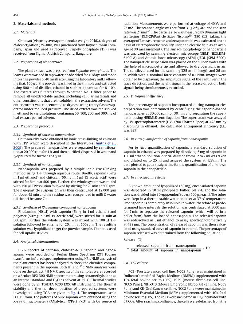
4 rate P
2
2
Npr
2
liiuTroeoil
2
2
w2tl
2
miswTft
2
p5ssf
2
attndoawsiiX
08 N.S. Rejinold et al. / Carbohyd
. Materials and methods
.1. Materials
Chitosan (viscosity average molecular weight 20 kDa, degree of-deacetylation (75–80%) was purchased from Koyochitosan Com-any, Japan and used as received. Tripoly phosphate (TPP) waseceived from Sigma–Aldrich Company, Bangalore.
.2. Preparation of plant extract
The plant extract was prepared from Sapindus emarginatus. Theeaves were washed in tap water, shade dried for 10 days and madento a fine powder of 40 mesh size using the laboratory mill. Follow-ng that, 100 g of the powder was filled in the thimble and extractedsing 500 ml of distilled ethanol in soxhlet apparatus for 8–10 h.he extract was filtered through Whatman No. 1 filter paper toemove all unextractable matter, including cellular materials andther constitutions that are insoluble in the extraction solvent. Thentire extract was concentrated to dryness using rotary flash evap-rator under reduced pressure. The dried extract was redissolvedn ethanol to yield solutions containing 50, 100, 200 and 300 mg ofeaf extract per ml solvent.
.3. Preparation protocols
.3.1. Synthesis of chitosan nanoparticlesChitosan-NPs were obtained by ionic cross-linking of chitosan
ith TPP, which were described in the literatures (Anitha et al.,009). The prepared nanoparticles were separated by centrifuga-ion at 20,000 rpm for 1 h, and then purified, dispersed in water andyophilized for further analysis.
.3.2. Synthesis of nanosaponinNanosaponin was prepared by a simple ionic cross-linking
ethod using TPP through aqueous route. Briefly, saponin (5 mgn 1 ml ethanol) and chitosan (50 mg in 5 ml 1% acetic acid) weretirred for 5 min at 500 rpm. Further, the whole system was mixedith 150 �l TPP solution followed by stirring for 20 min at 500 rpm.
he nanoparticle suspension was then centrifuged at 12,000 rpmor about 45 min and the residue was resuspended in milli Q waterill the pH became 7.4.
.3.3. Synthesis of Rhodamine conjugated nanosaponinRhodamine (40 �l) with saponin (5 mg in 1 ml ethanol) and
olymer (50 mg in 5 ml 1% acetic acid) were stirred for 20 min at00 rpm. Further the whole system was mixed with 100 �l TPPolution followed by stirring for 20 min at 500 rpm. The resultingolution was lyophilized to get the powder sample. Then it is usedor cell uptake studies.
.4. Analytical determinations
FT-IR spectra of chitosan, chitosan-NPs, saponin and nanos-ponin were recorded on Perkin Elmer Spectrum RX1 Fourierransforms infrared spectrophotometer using KBr. NMR analysis ofhe plant extract has been analyzed to check the chemical compo-ents present in the saponin. Both H1 and 13C NMR analyses wereone on the extract. 1H NMR spectra of the samples were recordedn a Bruker DPX 300 NMR spectrometer using tetramethylsilane asn internal standard and D2O as solvent at 25 ◦C. Thermal studies
ere done by SII TG/DTA 6200 EXSTAR instrument. The thermaltability and thermal decomposition of prepared systems werenvestigated using TGA are given in Fig. 4. The temperature scales 10 ◦C/min. The patterns of pure saponin were obtained using the-ray diffractometer (PANalytical X’Pert PRO) with Cu source of
olymers 84 (2011) 407–416
radiation. Measurements were performed at voltage of 40 kV and25 mA. The scanned angle was set from 3◦ ≤ 2� ≥ 40◦ and the scanrate was 2◦ min−1. The particle size was measured by Dynamic lightscattering (DLS-ZP/Particle Sizer NicompTM 380 ZLS) taking theaverage of 3 measurements and zeta potential was estimated on thebasis of electrophoretic mobility under an electric field as an aver-age of 30 measurements. The surface morphology of nanoparticlewas analyzed by scanning electron microscope (SEM) (JEOLJSM-6490LA) and Atomic force microscopy (AFM) (JEOL JSPM-5200).The nanoparticle suspension was placed on the silicon wafer withthe help of micropipette tip and allowed to dry overnight in air.The cantilever used for the scanning 325 �m in length and 26 �min width with a nominal force constant of 0.1 N/m. Images wereobtained by displaying the amplitude signal of the cantiliver in thetrace direction, and the height signal in the retrace direction, bothsignals being simultaneously recorded.
2.5. Entrapment efficiency
The percentage of saponin incorporated during nanoparticlespreparation was determined by centrifuging the saponin-loadednanoparticles at 30,000 rpm for 30 min and separating the super-natant using HERMLE centrifugation. The supernatant was assayedby UV spectrophotometer (UV-1700 Pharma Spec) at 428 nm bydissolving in ethanol. The calculated entrapment efficiency (EE)was 92%.
2.6. In vitro quantification of saponin from nanosaponin
For in vitro quantification of saponin, a standard solution ofsaponin in ethanol was prepared by dissolving 5 mg of saponin in100 ml ethanol solution. A serial dilution from 0.2 to 2 ml was takenand diluted up to 25 ml and assayed the system at 428 nm. Thedata plotted to get a straight line for the quantification of unknownsaponin in the nanoparticle.
2.7. In vitro saponin release
A known amount of lyophilized (50 mg) encapsulated saponinwas dispersed in 10 ml phosphate buffer, pH 7.4, and the solu-tion was divided into 30 epppendorf tubes (500 �l each). The tubeswere kept in a thermo stable water bath set at 37 ◦C temperature.Free saponin is completely insoluble in water; therefore at prede-termined time intervals the solution was centrifuged at 5000 rpmfor 7 min to separate the released saponin (which will be in apellet form) from the loaded nanosaponin. The released saponinwas redissolved in 3 ml ethanol to assay spectrophotometricallyat 428 nm. The concentration of released saponin was then calcu-lated using standard curve of saponin in ethanol. The percentage ofsaponin released was determined from the following equation:
Release (%)
= released saponin from nanosaponintotal amount of saponin in nanosaponin
× 100
2.8. Cell culture
PC3 (Prostate cancer cell line, NCCS Pune) was maintained inDulbecco’s modified Eagles Medium (DMEM) supplemented with10% fetal bovine serum (FBS). L929 (mouse fibroblast cell line,
NCCS Pune), NIH-3T3 (Mouse Embryonic fibroblast cell line, NCCSPune) and KB (Oral Cancer cell line, NCCS Pune) were maintained inMinimum Essential Medium (MEM) supplemented with 10% fetalbovine serum (FBS). The cells were incubated in CO2 incubator with5% CO2. After reaching confluency, the cells were detached from the
N.S. Rejinold et al. / Carbohydrate Polymers 84 (2011) 407–416 409
stry o
fl3f
2
cd2oocf1tficn(arw(oT(
Fig. 1. Hypothesized loading chemi
ask with trypsin–EDTA. The cell suspension was centrifuged at000 rpm for 3 min and then re-suspended in the growth mediumor further studies.
.9. Cytotoxicity studies
For cytotoxicity experiments, L929, NIH3T3, KB and PC3ells, respectively were seeded on a 96 well plate with aensity of 10,000 cells/cm2. MTT [3-(4,5-dimethylthiazole-2-yl)-,5-diphenyl tetrazolium] assay was used to evaluate cytotoxicityf the prepared nanoparticles and this is a colorimetric test basedn the selective ability of viable cells to reduce the tetrazoliumomponent of MTT in to purple colored formazan crystals. Six dif-erent concentrations of the nanoparticles (0.1, 0.2, 0.4, 0.6, 0.8 andmg/ml) were prepared by dilution with the media. The concen-
ration of saponin in the nanoformulations was varied as follows inve concentrations. (0.1 mg/ml) of nanoformulation has saponinoncentration as {[saponin]}= 0.95 �g, likewise (0.2 mg/ml) ofanoformulation contains 1.9 �g, (0.4 mg/ml) {[saponin]}= 3.8 �g,0.6 mg/ml) {[saponin]}= 4.6 �g, (0.8 mg/ml) {[saponin]}= 7.6 �gnd a higher concentration as (1 mg/ml) {[saponin]}= 8.6 �g,espectively. After reaching 90% confluency, the cells were washed
ith PBS buffer and different concentrations of the nanoparticles100 �l) were added and incubated. Cells in media alone devoidf nanoparticles acted as negative control and wells treated withriton X-100 as positive control for a period of 24 h. 5 mg of MTTSigma) was dissolved in 1 ml of PBS and filter sterilized. 10 �l
f saponin in chitosan nanoparticles.
of the MTT solution was further diluted to 100 �l with 90 �l ofserum-free phenol red free medium. The cells were incubated with100 �l of the above solution for 4 h to form formazan crystals bymitochondrial dehydrogenases. 100 �l of the solubilisation solu-tion (10% Triton X-100, 0.1 N HCl and isopropanol) was added ineach well and incubated at room temperature for 1 h to dissolve theformazan crystals. The optical density of the solution was measuredat a wavelength of 570 nm using a Beckmann Coulter Elisa platereader (BioTek Power Wave XS). Triplicate samples were analyzedfor each experiment.
2.10. Cellular uptake studies
Acid etched cover slips kept in 24 well plates were loaded withL929 and PC3 cells with a seeding density of 5000 cells per coverslip and incubated for 24 h for the cells to attach well. After the24 h incubation the media were removed and the wells were care-fully washed with PBS buffer. Then the particle at a concentrationof 1 mg/ml was added along with the media in triplicate to thewells and incubation for 4 h. Thereafter the media with samplewere removed and the cover slips with well attached cells wereprocessed for fluorescent microscopy. The processing involved
washing the cover slips with PBS and thereafter fixing the cellsin 3.7% para formaldehyde (PFA) followed by a final PBS wash. Thecover slips were air dried and mounted on to glass slides with DPXas the mountant medium. The slides were then viewed under thefluorescent microscope.
410 N.S. Rejinold et al. / Carbohydrate P
2
yw
3
3
p
3e
t
3
eit1i
3
tarcmtp
Fig. 2. (A) 1H NMR and (B) 13C NMR spectrum of saponin extract.
.11. Statistics
Statistical analysis of the data was performed via one-way anal-sis of variance (ANOVA) using origin software; a value of p < 0.05as considered significant (n = 3).
. Results and discussion
.1. Preparation of extract
The plant extract was prepared from S. emarginatus and theresence of saponin was confirmed through NMR and FTIR.
.2. Nuclear magnetic resonance spectroscopic analysis of plantxtract
The H1 NMR and 13C NMR analysis of the plant extract analyzedo check the chemical structure of the saponins.
.2.1. 1H1 NMRFrom Fig. 2A, the singlet peak at 7.2 (ppm) represents the pres-
nce of aromatic functional groups. The methylene protons aredentified at 0.88 ppm and the sugar methylene protons were iden-ified at 1.06 ppm (Jangwan, Dhobhal, & Naveen, 2010; Han et al.,999). The H-3, H-12 and H-18 protons in the saponin have been
dentified at sharp peaks at 3.36, 5.44 and 3.14 ppm, respectively.
.2.2. 13C NMR spectra of plant extract13C NMR spectra of the saponin analysis done in order to confirm
he presence of functional groups such as trisaccharides, carboxyliccids, aliphatic acids, esters and amides, saponin, glycosides. Fig. 2B
13
epresents the C NMR spectra of the extract, saponin, whichonfirmed the above said functionalities in the extract. The sugaroieties have been identified at 79.6 ppm with a sharp peak andhe carbon skeleton has been determined as it showed a prominenteak at 28.3 ppm, which is characteristic for the C-23 skeleton of the
olymers 84 (2011) 407–416
saponin. The FT-IR analysis also confirmed the presence of aromaticrings in the saponin molecule (Kizu & Tomimori, 1982).
3.3. Synthesis of nanosaponin
The saponin loaded chitosan nanoparticles were prepared byTPP cross-linking. The nanoparticles are dried and used for fur-ther characterization studies. The loading chemistry is shown inFig. 1. The expected loading chemistry is hydrogen bonding inter-action between the saponin and chitosan nanoparticles. Chitosanis having active –NH2 groups which can easily react with the –OHgroups of the saponin. Moreover, the sugar moieties in the saponinscould be utilized for the weak interactions with carrier chitosanmolecules. The hydrogen bonding is enough to hold saponin on thechitosan nanoparticles. The new peak at 1560 cm−1 indicates thereis a possibility for amide linking between saponin and chitosan.Since the –NHCOCH3 in chitosan is very less, there would not beany hydrogen bonding interaction between –OH and –NHCOCH3groups.
3.4. Particle size and topography
The particle size analysis done with dynamic light scattering waspresented below (Fig. 3), bare chitosan-NPs showed particles size inthe range of 35 ± 7 nm whereas the nano saponin showed increasedsize of 55 ± 7 nm. The surface morphology of nano saponin wasshown in Fig. 3D. The SEM and AFM analyses give a good evidencefor the entrapment of the saponin on the chitosan nanoparticlessine the nano saponin has higher particle size.
3.5. FTIR studies
Chitosan-NPs were prepared by ionic gelation technique usingTPP as cross-linker in aqueous medium. To confirm the nanofor-mulation of chitosan, FTIR analysis was done, characteristic peaksof bare chitosan are located at 1644, 980 and 3436 cm−1 whichcorresponds to amide I, anhydro glucosidic ring (Fig. 4A) andprimary amine, respectively. These observed peaks get shiftedfrom higher wave number region to lower wave number regionas 1644–1639 cm−1. The reduction in stretching frequency couldbe attributed to the TPP interaction with the amine function-ality; thereby the bond length of amine would be disturbedor increased. As the bond length increased, the stretching fre-quency would be decreased, thereby wave number shifts fromhigher frequency regions to lower frequency region (Sanoj Rejinold,Muthunarayanan, et al., 2010; Sanoj Rejinold, Chennazhi, Nair,Tamura, & Jayakumar, R, 2011). The saponin loading was confirmedby the presence of a new peak at 1560 cm−1 corresponds to amidelinkage between saponin and chitosan nanoparticles. There wasa sharp peak at 3450 cm−1 region which assures that interactionof saponin with chitosan is more of hydrogen bonding than ionicinteractions.
3.6. Thermal analysis
The thermal stability and thermal decomposition of preparedsystems were investigated by TG and are given in Fig. 4B. It showsthat the initial degradation temperature of chitosan is very closeto 280 ◦C, slow weight loss starting from 140 to 200 ◦C due to thedecomposition of polymer with low molecular weight, followedby more obvious loss of weight starting from 200 to 310 ◦C, which
could be attributed to a complex process including dehydration ofthe anhydro glucosidic ring (Devika &Varsha, 2006; Radhakumary,Nair, Suresh, & Nair, 2005). The degradation profile of chitosan-NPsseems to be different compared to chitosan, but stabler than chi-tosan which proves that the system is of amorphous nature. The
N.S. Rejinold et al. / Carbohydrate Polymers 84 (2011) 407–416 411
nin; (C
stTitn
pNPscdmmaiheer
wtDtt(
ptooi
Fig. 3. DLS spectra of (A) chitosan-NPs; (B) nanosapo
aponin alone has lower stability compared to the chitosan (con-rol) and chitosan-NPs that show comparatively higher stability.he saponin alone showed a sharp degradation at 100 ◦C whichndicates the melting point is at this temperature range. Howeverhe nanosaponin has greater stability even at 500 ◦C and 34% rem-ant at the same temperature.
Similarly, the differential thermal analysis of all the systems wasreformed to understand the behavior of the chitosan, chitosan-Ps, saponin and nanosaponin on application of thermal energy.olysaccharides usually have a strong affinity for water and in solidtate these macromolecules may have disordered structures thatan be easily hydrated. The hydration properties of polysaccharidesepend on primary and supramolecular structures. The endother-ic peak related to evaporation of water is expected to reflect theolecular changes brought in after cross-linking. Thus, chitosan
nd modified systems had different varied water-holding capac-ty. In chitosan, the bound water molecules are associated withydrophilic hydroxyl groups. The thermogram of chitosan showedndothermic peak at 95 ◦C. The saponin alone showed a sharpndothermic peak at 100 ◦C which is shifted to higher temperatureegion in the nanosaponin.
The heat capacity of chitosan-NPs was found to be less comparedith that of chitosan. The cross-linking reaction via TPP modifies
he crystalline nature of chitosan and also the nanosaponin. TheTA analysis showed saponin has started to melt at 100 ◦C wherein
he loaded nanosaponin, the same endothermic peak was shiftedo 350 ◦C which assures the amorphous nature of the nanosaponinFig. 4C).
On the basis of these results it can be stated that increase in the
olar groups and reduction in crystalline domains caused reduc-ion in heat capacity/thermal stability. The second thermal eventbserved was the presence of exotherms due to the decompositionf the polymer. Owing to the differences in the chemical character-stics, changes in the exothermic peak of chitosan and cross-linked) SEM of nanosaponin and (D) AFM of nanosaponin.
chitosan were also observed. Characterization of cross-linked chi-tosan polymer and its saponin loaded nanoparticles were analyzedby FTIR, which provided the evidence of reduction in crystallinityafter cross-linking with TPP. The whole TG/DTA analysis confirmsthe more amorphous nature for the cross-linked nanosaponin.
3.7. Nature of saponin in chitosan nanoparticles (XRD studies)
The characteristic peaks of saponin exhibited as shown in Fig. 5and can be inferred to traits of a high crystalline structure. Thecharacteristic peak of saponin 2� = 16◦ (Fig. 5A) was observed butthe intensity was less when entrapped into nanoparticles (Fig. 5B),possibly due to formation of an amorphous complex with theintermolecular interaction occurring within the matrix. A similarphenomenon has been observed in the literature providing evi-dence that the crystalline structure of saponins was converted toan amorphous state (Abdelwahed, Degobert, Stainmesse, & Fessi,2006).
3.8. In vitro saponin release
The saponin EE of chitosan-NPs was found to be 95%. The in vitrosaponin release was studied with PBS at pH 7.4. The percentage ofsaponin released from chitosan-NPs at predetermined time inter-vals was calculated using the standard curve prepared for saponin.Fig. 5C shows the in vitro saponin release profile of saponin. Releaseoccurs by a combination of diffusion of the saponin out of the par-ticles into the external environment and also by the degradationof the polymer. The decrease of the saponin release at longer time
suggested the importance of the diffusion process in the releasekinetics (Dev, Jithin, Nair, & Jayakumar, 2010). The in vitro drugrelease profile shows that 32% of saponin was released within 12 h.After the initial burst, a slow release was observed. A sustainedand controlled release has been observed till 72 h and 75% of the
412 N.S. Rejinold et al. / Carbohydrate Polymers 84 (2011) 407–416
F (d) na(
litsntt
3
ctcaneub
s(M
ig. 4. (A)FT-IR spectra of (a) chitosan (control), (b) chitosan-NPs, (c) saponin, andb) chitosan nanoparticles, (c) saponin and (d) nanosaponin.
oaded saponin released during this period. The initial burst releases expected due to the saponin molecules attached on the surface ofhe chitosan-NPs, and the sustained release is from the entrappedaponin. The observed sustained release after initial burst was sig-ificant because controlled release is required in the field of cancerherapy. These results indicated that the nanosaponin is useful con-rolled delivery system for cancer treatment.
.9. Cytotoxicity studies
The synthesized chitosan-NPs were analyzed by MTT for its toxi-ity on normal as well as cancer cells. As determined by MTT assay,he chitosan-NPs were non-toxic on L929, NIH-3T3, PC3 and KBells (Fig. 6A). The nanosaponin showed specific toxicity on prostatend oral cancer cells (Fig. 6B) while it did not show any toxicity onormal L929, and NIH-3T3 cells (Fig. 6B). The cytotoxicity differ-nce between normal and cancer cells by nanosaponin however isnknown, whereas the saponins specific action on cancer cells has
een well explored.Saponins isolated from different plants and animals have beenhown to specifically inhibit the growth of cancer cells in vitroKonoshima et al., 1998; Kuznetzova, Anisimov, & Popov, 1982;
arino, Iorizzi, Palagiano, Zollo, & Roussakis, 1998; Mimaki,
nosaponin. Thermal analysis: (B) TG and (C) DTA profile for (a) chitosan (control),
Kuroda, Kameyama, Yokosuka, & Sashida, 1998; Podolak, Elas, &Cieszka, 1998; Rao & Sung, 1995). The pursuit of natural sub-stances capable of controlling malignancies has led to considerableresearch on this property of saponins. The previous research workon isolated saponin from Gymnema sylvestre leaves have required50 �g/ml to show toxicity on Hela cells (human cervical carcinoma)where as it was non toxic to Vero cells (Khanna & Kannabiran,2009). Similarly saponins isolated from Paris polyphylla var. Yun-nanensis have a good anti tumor action (Yan, Zhang, Gao, Man, &Wang, 2009). It throws light on the fact that there is some molecu-lar level mechanism for saponin to show specific toxicity on cancercells. Further studies on this aspect are required emergently for thebetter understanding of the specific anticancer action of saponin.
3.10. Cell uptake studies
Systematic study for cellular uptake of Rhodamine 123 con-jugated nanosaponin by L929 cells and PC3 was performed by
visualizing the fluorescence of Rhodamine 123 using fluorescentmicroscopy. Rhodamine 123 is a green fluorescent dye, so the cellswith nanoparticle uptake would typically appear bright green. Flu-orescent microscopic images taken after 24 h of incubation revealedthat, there was significant internalization and retention of nanopar-
N.S. Rejinold et al. / Carbohydrate Polymers 84 (2011) 407–416 413
Fig. 5. XRD spectra of (A) saponin, (B) nanosaponin, and (C) in vitro release of saponin from nanosaponin (values reported are mean ± SD; n = 3).
Fig. 6. (A) MTT assay of chitosan-NPs and (B) nanosaponin on L929, KB, NIH3T3 and PC3 cells (values reported are mean ± SD; n = 3).

414 N.S. Rejinold et al. / Carbohydrate Polymers 84 (2011) 407–416
F ) brig
tast
4
ahocc
ts
tin1obib
ig. 7. Fluorescent microscopic images of nanosaponin on (A) L929 and (B) PC3: (a & d
icles inside the cells (Fig. 7A and B). Images of control cells withoutny particles did not show fluorescence, which further validates thetudy. Additional experiments are underway to give more insighto cellular uptake and sub-cellular localization of nanosaponin.
. Discussion
In recent years considerable attention has been given todvanced biomaterials field including nanobiomaterials, smartydrogels, etc. This study presents the preparation and evaluationf saponin loaded chitosan nanoparticles (nanosaponin) as a can-er therapeutic agent. The methodology is simple and less timeonsuming.
Saponin isolated from different plants are well known forheir potential anticancer activity, however, the reported value foraponin is 50 �g/ml to show toxicity against cancer cells.
The anticancer action of saponins on cancer cells may take placehrough diverse and complex mechanisms. The destructive activ-ty of saponins against cells such as erythrocytes was, however,ot related to cytostatic activity against cancer cells (Mimaki et al.,
998). The selective inhibition of the growth of tumor has beenbserved by triterpenoid saponins (avicins from Acacia victoriae)y cell cycle arrest in human breast cancer cell line and apoptosisn leukaemia cell line (Mujoo et al., 2001) which in turn reducedoth tumor incidence and multiplicity in a murine skin carcino-
ht field, (b & f) fluorescent images, and (c & e) control cells at different magnifications.
genesis model (Hanausek et al., 2001). Saponin (triterpenoidorsteroid)-induced apoptosis is primarily caused by stimulating thecytochrome c–caspase 9–caspase 3 pathway in the human can-cer and other cell lines (Cai, Liu, Wang, & Ju, 2002; Haridas,Arntzen, & Gutterman, 2001; Liu, Xu, & Che, 2000; Yui et al.,2001), a property that is shared by a ginseng saponin derivative(20-O-(b-d-glucopyranosyl)-20(S)-protopanaxadiol) produced inintestine by bacteria and absorbed into blood (Lee, Ko, et al., 2000;Lee, Sohn, Park, Kim, and Jung, 2000). The specific toxic activityagainst macrophage colony-stimulating factor-induced growth ofmacrophages by terpenoids (securiosides) having specific struc-tural features such as presence of dimethoxycinnamoyl group hasbeen detected by Yui et al. (2001). Depending on the structuralfunctionalities saponins can induce a cell cycle arrest mediatedby inhibition of the phosphatidylinositol-3-kinase–protein kinasesignalling pathway (Mujoo et al., 2001) or direct suppressionof protein kinase complex genes (Liu et al., 2000) is stimulatedby saponins either along with the apoptotic pathway (Mujooet al., 2001) or independently (Oh & Sung, 2001). The inhibi-tion of the phosphatidylinositol-3-kinase–protein kinase pathway
is considered important in apoptosis, given the role of proteinkinases in inactivating the caspases (apoptotic enzymes). Saponinsalso reduce occurrence of reactive oxygen species such as H2O2(Haridas et al., 2001; Pawar, Gopalakrishnan, & Bhutani, 2001),probably by enhancing its breakdown by activation of peroxire-
rate P
d1p2
dcatt
tO(ssdNtac
vsbmncan
5
pTrtcsewtbicdtcanaaeelg
A
paNlC
N.S. Rejinold et al. / Carbohyd
oxins and catalase, and/or glutathioneperoxidase (Deng & Zhang,991) as well as by suppressing its production by inhibiting thehosphatidylinositol-3-kinase signalling pathway (Haridas et al.,001).
Our study clearly demonstrated that nanosaponin can induceose-dependent cancer cell death with lower toxicity on normalells. Moreover, only less than 10 �g is required to show toxicitygainst cancer cells like PC3 and KB in our study. This is an impor-ant observation because cancer cell death has been considered aarget of chemotherapeutic agents in a variety of cancer research.
Apoptosis is a process of an essential tissue homeostasis andhus being regarded as the ideal way to inhibit cancer cell growth.ur data is focusing about the anticancer activity of nanosaponin
Fig. 6) and drug release (Fig. 5) which is clearly showing that austained release could be possible to cancer cell lines. The bareaponin would completely expire after causing sudden death of aefinite population of cancer cell as seen in the previous studies.anosaponin would release the drug in controlled fashion to cause
oxicity to that population of cells seeded for an MTT assay as wells still hold a potentially toxic quantity of drugs that would haveome to use had the population of cells been higher.
Therefore, nanosaponin inducing cancer cell death can beiewed as a new potential generation of cancer treatment. Manytudies demonstrated that the induction of cell death in cancer cellsy anticancer saponins appeared in a dose and time-dependentanner. Our data is in agreement with this fact showing that the
anosaponin with higher concentrations of saponin can induce can-er cell death within a short-time period of 24 h. Additional studiesre required to clarify the efficacy and the mechanism by which theanosaponin induce apoptotic cell death.
. Conclusions
In conclusion, the extract from S. emarginatus was taken andreliminary phytochemical analysis of the leaf extract was done.he saponin loaded chitosan nanoparticles “nanosaponin”, as nevereported before, were prepared via a simple cross-linking reac-ion with TPP for enhanced, controlled and sustained delivery toancer cell lines. The DLS, SEM and AFM studies confirmed theize of the prepared nanoparticles to be 40–60 nm, which meansven after the saponin incorporation, the particle size can be tunedithin the optimal range for saponin delivery applications. The
hermal studies indicated that nanosaponin has more thermal sta-ility than saponin alone. FT-IR studies confirmed the potential
nteraction of the saponin with the chitosan nanoparticles. Theellular uptake studies of nanosaponin using L929 and PC3 cellsemonstrated significant internalization and retention of nanopar-icles inside the cells, suggesting that these nanoparticle systemsan be used for delivering saponin directly into the cells. The nanos-ponin has showed specific toxicity on cancer cells while they areon-toxic to normal cells. These preliminary results could serve asgood platform for future experimentations with nanosaponin onppropriate experimental animal models with relevance to differ-nt human cancers. Our preliminary study thus provide convincingvidence of nanotechnology based saponin delivery to cancer cellines in vitro to enhance the therapeutic efficacy of this well knownreen chemotherapeutic agent.
cknowledgments
The Department of Biotechnology, Government of India sup-
orted this work, under a center grant of the Nanosciencend Nanotechnology Initiative Program (Ref. No. BT/PR10850/NT/28/127/2008). One of the authors (Dr. K. Muthuche-ian), would like to gratefully acknowledge University Grantsommission-University with Potential for Excellence, MKU, for
olymers 84 (2011) 407–416 415
funding this study. This work is also partly supported by DST, mon-itored by Dr. C.N.R. Rao. The authors are also thankful to Mr. SajinP. Ravi, Mr. C.M. Girish, Mr. Sudheesh Kumar P.T and Ms. Sreeja VNair for their help in SEM, AFM, TG/DTA and XRD studies.
References
Abdelwahed, W., Degobert, G., Stainmesse, S., & Fessi, H. (2006). Freeze drying ofnano particles: Formulation, process and storage considerations. Advanced DrugDelivery Reviews, 58, 1688–1713.
Anitha, A., Divya Rani, V. V., Krishna, R., Sreeja, V., Selvamurugan, N., Nair, S. V.,et al. (2009). Synthesis, characterization, cytotoxicity and antibacterial studiesof chitosan, O-carboxymethyl and N, O-carboxymethyl chitosan nanoparticles.Carbohydrate Polymers, 78, 672–677.
Basalingappa, L. H., & Chandravadan, H. P. (1971). A survey of plants in Gujarat,India, for alkaloids, saponin, and tannins. In U.S.D.A. Forest Service Research Paper,NE-201 (pp. 1–11).
Cai, J., Liu, M., Wang, Z., & Ju, Y. (2002). Apoptosis induced by dioscinin Hela cells.Biological and Pharmaceutical Bulletin, 25, 193–196.
Calvo, P., Remunan, L. C., Vila, J. C. L., & Alonso, M. J. (1997a). Novel hydrophilicchitosan-polyethylene oxide nanoparticles as protein carriers. Journal of AppliedPolymer Science, 63, 125–132.
Calvo, P., Remunan, L. C., Vila, J. C. L., & Alonso, M. J. (1997b). Chitosan and chi-tosan/ethylene oxide-propylene oxide block copolymer nanoparticles as novelcarriers for proteins and vaccines. Pharmaceutical Research, 14, 1431–1436.
Deng, H. L., & Zhang, J. T. (1991). Anti-lipid peroxilative effect of ginsenosideRb1 andRg1. Chinese Medical Journal, 104, 395–398.
Dev, A., Jithin, C. M., Nair, S. V., & Jayakumar, R. (2010). Novel carboxymethyl chitinnanoparticles for cancer drug delivery applications. Carbohydrate Polymers, 79,1073–1079.
Devika, R. B., & Varsha, P. (2006). Studies on Effect of pH on cross-linking of chitosanwith sodium tripolyphosphate: A Technical Note. AAPS Pharmaceutical Scienceand Technology, 7, E1–E6.
Elzatahry, A., & Mohy, E. (2008). Preparation and characterization of metronida-zole loaded chitosan nanoparticles for saponin delivery application. Polymersfor Advanced Technologies, 19, 1787–1791.
Fenwick, D. E., & Oakenfull, D. (1983). Saponin content of food plants and someprepared foods. Journal of Science and Food Agriculture, 34, 186–191.
Francis, G., Kerem, Z., Harinder Makkar, P. S., & Becker, K. (2002). The biologicalaction of saponin in animal systems: A review. British Journal of Nutrition, 88,587–605.
Han, X. W., Yu, H., Liu, X. M., Bao, X., Yu, B., Li, C., et al. (1999). Complete 1H and 13CNMR assignments of diosgenyl saponins. Magnetic Resonance in Chemistry, 37,140–144.
Hanausek, M., Ganesh, P., Walaszek, Z., Arntzen, C. J., Slaga, T. J., & Gutterman, J.U. (2001). Avicins, a family of triterpenoid saponins from Acacia victoriae (Ben-tham), suppress H-ras mutations and aneuploidy in a murine skin carcinogenesismodel. Proceedings of the National Academy of Sciences of the United States ofAmerica, 98, 11551–11556.
Haridas, V., Arntzen, C. J., & Gutterman, J. U. (2001). Avicins, a family of triterpenoidsaponins from Acacia victoriae (Bentham), inhibit activation of nuclear factor-kappa B by inhibiting both its nuclear localization and ability to bind DNA.Proceedings of the National Academy of Sciences of the United States of America,98, 11557–11562.
Huang, M., Khor, E., & Lim, L. Y. (2004). Uptake and cytotoxicity of chitosan moleculesand nanoparticles: Effects of molecular weight and degree of deacetylation.Pharmaceutical Research, 21, 344–353.
Janes, K. A., & Alonso, M. J. (2003). Depolymerized chitosan nanoparticles for proteindelivery: Preparation and characterization. Journal of Applied Polymeric Science,88, 2766–2779.
Jangwan, J. S., Dhobhal, M., & Naveen, K. (2010). New cytotoxic saponin of Albizzialebbeck. Indian Journal of Chemistry, 49B, 123–126.
Jayakumar, R., Chennazhi, K. P., Muzzarelli, R. A. A., Tamura, H., Nair, S. V., & Selva-murugan, N. (2010). Chitosan conjugated DNA nanoparticles in gene therapy.Carbohydrate Polymers, 79, 1–8.
Jayakumar, R., Deepthy, M., Manzoor, K., Nair, S. V., & Tamura, H. (2010). Biomed-ical applications of chitin and chitosan based nanomaterials-A short review.Carbohydrate Polymers, 82, 227–232.
Kashappa, G. H. D., & Hyun, J. P. (2005). Preparation and characterization ofsaponin-loaded chitosan-tripolyphosphate micro spheres by spray drying. DrugDevelopment Research, 64, 114–128.
Khanna, V. G., & Kannabiran, K. (2009). Anti cancer-cytotoxic activity of saponinsisolated from leaves of Gymnea sylvestre and Eclipta prostrate on Hela cells.International Journal of Green Pharmacy, 56280, doi:4103/0973-8528.
Kizu, H., & Tomimori, T. (1982). Studies on the constituents of Clematis species. IV.On the saponins of the root of Clematis chinensis Osbeck. Chemical and Pharma-ceutical Bulletin, 30, 859–865.
Kofuji, K., Ito, T., Murata, Y., & Kawashima, S. (2001). Biodegradation and saponin
release of chitosan gel beads in subcutaneous air pouches of mice. Biological andPharmaceutical Bulletin, 24, 205–208.Konoshima, T., Takasaki, M., Tokuda, H., Nishino, H., Duc, N. M., Kasai, R., et al. (1998).Anti-tumor-promoting activity of majonoside-R2 from Vietnamese ginseng.Panax vietnamensis HA et GRUSHV. (I). Biological and Pharmaceutical Bulletin,21, 834–838.

4 rate P
K
L
L
L
M
M
M
M
M
M
N
OO
O
P
P
P
R
R
16 N.S. Rejinold et al. / Carbohyd
uznetzova, T. A., Anisimov, M. M., & Popov, A. M. (1982). A comparative studyin vitro of physiological activity of triterpene glycosides of marine invertebratesof echinoderm type. Comparative Biochemistry and Physiology, 73C, 41–43.
ee, S. J., Ko, W. G., Kim, J. H., Sung, J. H., Lee, S. J., Moon, C. K., et al. (2000).Induction of apoptosis by a novel intestinal metabolite of ginseng saponin viacytochrome c-mediated activation ofcaspase-3 protease. Biochemical Pharma-cology, 60, 677–685.
ee, K. T., Sohn, I. C., Park, H. J., Kim, D. W., & Jung, G. O. (2000). Essential moietyfor antimutagenic and cytotoxic activity of hederagenin monodesmosides andbisdesmosides isolated from the stem bark of Kalopanax pictus. Planta Medica,66, 329–332.
iu, W. K., Xu, S. X., & Che, C. T. (2000). Anti-proliferative effect of ginseng saponinson human prostate cancer cell line. Life Sciences, 67, 1297–1306.
arino, S. D., Iorizzi, M., Palagiano, E., Zollo, F., & Roussakis, C. (1998). Star fishsaponins. 55. Isolation, structure elucidation, and biological activity of steroidoligoglycosides from an antarctic starfish of the family Asteriidae. Journal ofNatural Products, 61, 1319–1327.
imaki, Y, Kuroda, M., Kameyama, A., Yokosuka, A., & Sashida, Y. (1998). Steroidalsaponins from the rhizomes of Hosta sieboldiiand their cytostatic activity onHL-60 cells. Phytochemistry, 48, 1361–1369.
ujoo, K., Haridas, V., Hoffmann, J. J., Wachter, G. A., Hutter, L. K., Lu, Y., et al.(2001). Triterpenoid saponins from Acacia victoriae (Bentham) decrease tumorcell proliferation and induce apoptosis. Cancer Research, 61, 5486–5490.
uzzarelli, R. A. A. (1988). Carboxymethylated chitin and chitosans. CarbohydratePolymers, 8, 1–21.
uzzarelli, R. A. A. (2009). Chitins and chitosans for the repair of wounded skin,nerve, cartilage and bone. Carbohydrate Polymers, 76, 167–182.
uzzarelli, R. A. A. (2010). Chitosans: New vectors for gene therapy. In R. Ito, & Y.Matsuo (Eds.), Handbook of carbohydrate polymers: Development, properties andapplications (pp. 583–604). Hauppauge, NY, USA: Nova Publ.
air, R., Kalariya, T., & Chanda, S. (2005). Antibacterial activity of some selectedIndian medicinal flora. Turkish Journal of Biology, 29, 41–47.
akenfull, D. G. (1981). Saponin in food—A review. Food Chemistry, 6, 19–40.akenfull, D. G., & Sihdu, G. S. (1990). Could saponin be a useful treatment for
hypercholesterolemia. European Journal of Clinical Nutrition, 44, 79–88.h, Y. J., & Sung, M. K. (2001). Soybean saponins inhibit cell proliferation by sup-
pressing PKC activation and induce differentiation of HT-29 human colon adenocarcinoma cells. Nutrition and Cancer, 39, 132–138.
awar, R., Gopalakrishnan, C., & Bhutani, K. K. (2001). Dammaranetriterpene saponinfrom Bacopa monniera as the superoxide inhibitor in polymorphonuclear cells.Planta Medica, 67, 752–754.
odolak, I., Elas, M., & Cieszka, K. (1998). In vitro antifungal and cytotoxic activityof triterpene saponosides and quinoid pigments from Lysimachia vulgaris L.Phytotherapy Research, 12, S70–S73.
otter, J. D. (1993). Colon cancer—Do the nutritional epidemiology, the gut physi-
ology and the molecular biology tell the same story? Journal of Nutrition, 123,418–423.adhakumary, C., Nair, P. D., Suresh, M., & Nair, C. P. R. (2005). Trends in Pharmaco-logical Sciences. Trends Biomateials and Artifial organs, 30, 117–124.
ao, A. V., & Sung, M. K. (1995). Saponins as anticarcinogens. Journal of Nutrition,125, 717S–724S.
olymers 84 (2011) 407–416
Richardson, S. C. W., Kolbe, H. V. J., & Duncan, R. (1999). Potential of low molecularmass chitosan as a DNA delivery system: Biocompatibility, body distributionand ability to complex and protect DNA. International Journal of Pharmaceutics,178, 231–243.
Sanoj Rejinold, N., Muthunarayanan, M., Deepa, N., Chennazhi, K. P., Nair, S. V., &Jayakumar, R. (2010). Development of novel fibrinogen nanoparticles by twostep coacervation method. International Journal of Biological Macromolecules, 7,37–43.
Sanoj Rejinold, N., Chennazhi, K. P., Nair, S. V., Tamura, H., & Jayakumar, R. (2011).Biodegradable and thermo-sensitive chitosan-g-poly (N-vinylcaprolactam)nanoparticles asa5-fluorouracilcarrier. Carbohydrate Polymers, 83, 776–786.
Shibata, S. (2001). Chemistry and cancer preventing activities of Ginseng saponinand some related triterpenoid compounds. Journal of Korean Medical Sciences,16, S28–S37.
Sudheesh Kumar, P. T., Abhilash, S., Manzoor, K., Nair, S. V., Tamura, H., & Jayaku-mar, R. (2010). Preparation and characterization of novel �-chitin/nanosilvercomposite scaffolds for wound dressing applications. Carbohydrate Polymers, 80,761–767.
Sung, M. K., Kendall, C. W. C., & Rao, A. V. (1995). Effect of soybean saponin andgypsophila saponin on morphology of colon carcinoma cells in culture. Food andChemical Toxicology, 33, 357–366.
Thanou, M., Verhoef, J. C., & Junginger, H. E. (2001). Oral saponin absorption enhance-ment by chitosan and its derivatives. Advanced Drug Delivery Reviews, 52,117–126.
Vander, L. I. M. V., Kersten, G., Fretz, M. M., Beuvery, C., Verhoef, J. C., & Junginger, H.E. (2003). Chitosan micro particles for mucosal vaccination against diphtheria:Oral and nasal efficacy studies in mice. Vaccine, 21, 1400–1408.
Wu, R. T., Chiang, H. C., Fu, W. C., Chien, K. Y., Chung, Y. M., & Horng, L. Y. (1990).Formosanin-C, an immunomodulator with antitumor activity. International Jour-nal of Immuno Pharmacology, 12, 777–786.
Xu, Y., & Du, Y. (2003). Effect of molecular structure of chitosan on protein delivery.International Journal of Pharmaceutics, 250, 215–226.
Yan, L., Zhang, Y. J., Gao, W. Y., Man, S. L., & Wang, Y. (2009). In vitro and in vivoanticancer activity of steroid saponins of Paris polyphylla var. Yunnanensis.Experimental Oncology, 31, 27–32.
Yibing, W., Yichun, Z., & Biao, Y. (2007). The cytotoxicity of saponin correlates withtheir cellular internalization. ChemMedChem, 2, 288–291.
Yoshizumi, K., Hirano, K., Ando, H., Hirai, Y., Ida, Y., Tsuji, T., et al. (2006). Lupane-typesaponins from leaves of Acanthopanax sessiliflorus and their inhibitory activityon pancreatic lipase. Journal of Agricultural and Food Chemistry, 54, 335–341.
Yu, L., Ma, R., Wang, Y., Nishino, N., Takayasu, J., He, W., et al. (1992). Potent anti-tumorigenic effect of tubeimoside i isolated from the bulb of Bolbostemmapaniculatum (Maxim) Franquet. International Journal of Cancer, 50, 635–638.
Yui, S., Ubukata, K., Hodono, K., Kitahara, M., Mimaki, Y., Kuroda, M., et al. (2001).Macrophage-oriented cytotoxic activity of novel triterpene saponins extracted
from roots of Securidaca inappendiculata. International Immunopharmacology,1, 1989–2000.Zhao, H. L., Sim, J. S., Shim, S. H., Ha, Y. W., Kang, S. S., & Kim, Y. S. (2005). Antiobeseand hypolipidemic effects of platycodin saponins in diet-induced obese rats:Evidences for lipase inhibition and calorie intake restriction. International Journalof Obesity, 29, 983–990.