Antitubercular, Cytotoxicity, and Computational Target ... - MDPI
Upload
khangminh22Category
view
3download
0
SYNTHESIS, DNA INTERACTIONS AND
CYTOTOXICITY OF NOVEL
CHLOROETHYLAMINOANTHRAQUINONES
Zennia Paniwnyk B.Se (Hons)
A thesis submitted for the degree of Doctor of Philosophy
at De Montfort University, Leicester.
February 2000
School of Pharmacy and Pharmaceutical Sciences,
De Montfort University, Leicester.

For my parents
Hryhorij and Kateryna Paniwnyk
ii

ABSTRACT
The design of DNA directed nitrogen mustards has led to the development of compounds with increased specificity for DNA and enhanced cytotoxicity compared to similar untargeted mustards. By linking an intercalating anthraquinone chromophore to a nitrogen mustard function it was anticipated that such compounds would also have the potential to irreversibly inhibit the topoisomerase II (topo II) enzyme. Fourteen anthraquinones containing various hydroxyethylaminoalkylamino side chains (hydroxyethylaminoanthraquinones) were synthesised, and ten were reacted further to give a series of mono, 5,8-dihydro or 5,8-dihydroxy nitrogen mustard anthraquinones (chloroethylaminoanthraquinones) substituted with alkylating side chains at the 1 or 1,4 position of the chromophore. The ability of hydroxyethylaminoanthraquinones to associate with DNA and to stabilize the helix to thermal denaturation was investigated. The increasing order of drughelix stability was found to be I-monosubstituted anthraquinone < 1,4-disubstituted-5,8-dihydroanthraquinone < 1,4-disubstituted-5,8-dihydroxyanthraquinone. In contrast, in the same DNA denaturation assay the chloroethylaminoanthraquinones showed apparent weak affinity for DNA, the exception being unsymmetrically substituted ZP281 which stabilised the helix by 1 DoC. Four chloroethylaminoanthraquinones ZP281, ZP282, ZP285 and ZP289 were selected for DNA covalent binding studies. These revealed that the compounds were excellent DNA inter-strand cross linking agents that prevented strand separation at concentrations of InM. Furthermore, when compared to the non-targeted mustard mechlorethamine, the chloroethylaminoanthraquinones demonstrated an altered pattern of guanine alkylation which coincided with a preference for reaction at guanine sites with a pyrimidine 5' (5'PyG). This suggested that the chromophore influences the site of DNA alkylation and enhances the selectivity of the drug for DNA. To determine the cellular mechanism of the novel anthraquinones, a topo II inhibition study was performed in a yeast strain, Saccharomyces cereviciae (Sc. ce), transfected with human topo IIa or p, or yeast to po II. Of the seven compounds assayed three alkylating anthraquinones, ZP281, ZP285 and ZP289 inhibited yeast cell growth and may target both topo IIa and p as a mechanism of cytotoxic action. ZP285 was the most potent compound requiring only 4.51lM to achieve 50% yeast cell kill. In vitro cytotoxicity assays were performed with ovarian carcinoma cell lines; A2780, CHI; cisplatin resistant cell lines A2780cisR, CHlcisR, SKOV-3; doxorubicin resistant cell line 2780AD; and mismatch repair chromosome transfer modified cell line Bl. Seven compounds ZP257, ZP265, ZP275 (hydroxyethylaminoanthraquinones) and ZP281, ZP282, ZP285 and ZP289 (chloroethylaminoanthraquinones) were extremely potent in both wild type and resistant cells giving ICso values below 0.05 ).lM. Furthermore, compound ZP281 displayed a relative lack of cross resistance in all resistant cell lines tested. The data in this thesis therefore indicates that ZP281 is a good candidate for further study as a potential covalent topoisomerase II inhibitor that may circumvent multi-drug resistance.
iii

ACKNOWLEDGEMENTS
I would like to thank the Association of International Cancer Research for
personal funding throughout my three years of research. I am also grateful to the
Department of Pharmaceutical Science at De Montfort University, Leicester for
allowing me to carry out research within their laboratories. Most importantly I would
like to thank Professor Laurence Patterson and Dr Paul Teesdale-Spittle for all their
help and encouragement to the completion of this thesis. Special thanks go to my sister
Dr L Paniwnyk for her constant motivation, and Yvonne Giles for her help in the topo II
assay and for our discussion sessions. Additional thanks to Professor l.A Hartley and
Simon McAdam at the Oncology Department, University College London, Dr 1.
lenkins at the MRC Toxicology Unit, Leicester, Dr L Kelland at the CRC Centre for
Cancer Therapeutics, Sutton and Dr. l. Plumb at the Cancer Research Campaign
Beatson Laboratories, University of Glasgow for allowing me to use their expertise and
resources. Not to forget, Sandra Forbes and Ketan Ruparelia, John Lamb at the MRC,
Leicester University for FAB-MS analysis and Dr Mike Needham for NMR analysis.
iv

Title Dedication Abstract Aclmowledgements Contents Tables List Figures List List of synthesis Abbreviations
CONTENTS
ii iii IV
V
X
Xl
xiv XVI
SectionNo. PageNo.
CHAPTER 1 INTRODUCTION
1. 1. Cancer and its treatment
1. 2. The topoisomerase II enzyme and its role in drug action 2
1. 2. 1. Cellular role and structure 2
1. 2. 2. The catalytic cycle of topoisomerase II 5
1. 2. 3 Topoisomerase II as a potential drug target 6
1.3 Development of chemotherapeutic drugs 9
1. 3. 1. Anthracyc1ines 9
1. 3. 2 Anthraquinones in the treatment of cancer 11
1. 3. 2. 1. Mitoxantrone 11
1. 3. 2. 2. Structure and binding 13
1. 3. 2. 3. Topo II inhibition and cellular mechanisms of action 14
1. 3. 2.4. Modified mitoxantrone analogues 15
1. 3. 2. 5. Asymmetric anthraquinones 16
1. 4 Nitrogen Mustards and anti-tumour drug development 17
1. 4. 1. The early origins of cancer therapy 17
1. 4. 2. Classification and mode of action 18
1. 4. 3. DNA sequence specificity of nitrogen mustards 19
v

1. 4. 4. DNA targeted alkylating agents 21
1. 4. 4. 1. Acridine linked mustards 21
1. 4. 4. 2. Morpholino doxorubicin analogues as alkylating agents 23
1. 5. Multi-drug resistance mechanisms associated with anthracycline and nitrogen mustard anti-cancer agents
1. 5. 1. The multi drug resitance (MDR) phenotype
25
25
1. 5. 1. 1. MDRI and MRP mediated resistance 26
1. 5. 1. 2. Altered topoisomerase II 28
1. 5. 1. 3. Detoxification by glutathione and its role in resistance 29
1. 5. 1.4. DNA repair 31
1. 6. Toxicity and resistance modulators
1. 7. Aims
CHAPTER 2 THE SYNTHESIS AND ANALYSIS OF SUBSTITUTED ANTHRAQUINONES
2. 1. Introduction
2. 2. Chemicals and reagents
2.3. Sample analysis
2.4. Experimental (see List of Synthesis)
2.5. Results
2.6. Discussion
34
37
39
40
40
41
59
64
2. 6. 1. Synthesis of hydroxyethylalkylamino sidechains 64
2. 6. 2. Chromophore substitution reactions 66
2. 6. 3 Synthesis of chloroethylaminoanthraquinones 69
2. 6. 4. Attempted synthesis of chloroethylaminoanthraquinone N-oxide 72 hydrochloride
vi

CHAPTER 3 DNA BINDING STUDIES
3. 1. Introduction 77
3.2. Chemicals and reagents 77
3.3. Methods 78
3.3. 1. Determination of the druglDNA isosbestic point 78
3.3.2. Determination of the binding constant of drug with DNA 78
3.3.3. Determination of the thermal denaturation of DNA 78
3.3.4. Determination of the thermal denaturation of DNA in 79 the presence of hydroxyethylaminoanthraquinones
3.3.5. Determination of the thermal denaturation of DNA in 79 the presence of chloroethylaminoanthraquinone
3.4. Results 80
3.4.1. Determination of the druglDNA isosbestic point 80
3.4.2. Spectrophotometric titration studies 85
3.4.3. DNA thermal denaturation temperature studies 87
3.5. Discussion 91
3.5. 1. Isosbestic behaviour of the novel anthraquinones 91
3.5.2. Analysis of DNA binding constant (KJ ) for 93 the hydroxyethylaminoanthraquinones
3.5.3 Effect of the hydroxyethylaminoanthraquinones on stabilisation 95 of the DNA helix to thermal denaturation
3.5.4. Effect of the chloroethylaminoanthraquinones on the stabilisation 96 of the DNA helix to thermal denaturation
CHAPTER 4 DNA ALKYLATION STUDIES
4.1. Introduction 98
4.2. Chemicals and reagents 98
4.3. Methods 99
vii
4.3.1. Detennination of DNA sequence specific alkylation using 99 the polymerase chain reaction (PCR)
4.3.2. Detennination of the fonnation of drug-DNA inter-strand 100 crosslinks
4.4. Results 101
4.4.1. DNA sequence specific alkylation studies 101
4.4.2. DNA inter-strand crosslink fonnation studies 104
4.5. Discussion 106
4. s. 1. Identification of chloroethylaminoanthraquinone-DNA alkylation 106 sites
4. s. 2. Effect of chloroethylaminoanthraquinones on the inhibition of 109 DNA strand separation
CHAPTER 5 TOPOISOMERASE II INHIBITION STUDIES
s. 1. Introduction 112
S.2. Chemicals and reagents 112
5.3. Characterisation of topo IIa and f3 yeast strains 113
S.4. Methods 113
5.4.1. Detennination of the effect of novel anthraquinones on yeast 113 strains containing human a and f3. and Sc. ce topo II
5.5 Results 114
5.5. 1. Effect of novel anthraquinones on yeast cell growth inhibition 114
S.6. Discussion 118
CHAPTER 6 CYTOTOXICITY STUDIES
6. 1. Introduction 123
6.2. Methods 123
6.2.1. Detennination of the cytotoxicity of novel anthraquinones in 123
viii
wild type and cisplatin resistant human ovarian cancer cell lines
6.2.2. Comparitive study of the cytotoxicity of novel anthraquinones in 124 wild type, cisplatin resistant and anthracycline resistant human ovarian cancer cell lines
6.3. Results 124
6.3. l. Effect of novel anthraquinones on A2780, A2780CisR, CHI, 124 CH 1 CisR and SKOV -3 cell line viability
6.3.2. Effect of novel anthraquinones on A2780, A2780CisR (2780CP), 131 A2780AD and B 1 cell line viability
6.4. Discussion 133
CHAPTER 7 SUMMARY, FUTURE WORK AND CONCLUSION 138
Bibliography 145 Appendix 1 Standard experimental procedures 171 Appendix 2 Preparation of buffers and solutions 174 Appendix 3 HPLC analysis 177
ix

TABLES LIST
Table No. Page No.
1. O. Base specificity of anti-cancer drug interaction with topo II 8
3.1. Effect on visible absorbance following the addition of 82 hydroxyethylaminoanthraquinones to calf thymus DNA.
3.2. The affinity ofhydroxyethylaminoanthraquinones for calf thymus DNA 87
3.3. The effect of the hydroxyethylaminoanthraquinones on the 90 denaturation temperature of calf thymus DNA.
3.4. The effect of the chloroethylaminoanthraquinones on the 91 denaturation temperature of calf thymus DNA.
4. 1. Most intense sites of DNA alkylation following reaction with 104 chloroethylaminoanthraquinones and mechlorethamine.
4.2. Summary of the prefered DNA alkylation sites of chromophore linked 108 nitrogen mustards
5. 1. The effect of the chloroethylaminoanthraquinones on the inhibition of 117 yeast cell growth.
6.1. The effect of 24h drug exposure on the growth of ovarian cancer cell 126 lines A2780 and A2780CisR.
6.2. The effect of 24h drug exposure on the growth of ovarian cancer cell 127 lines CHI, CHICisR and SKOV-3
6.3. The cell growth inhibition concentration of the seven most potent 128 anthraquinones following 24 hr exposure to A2780 and A2780CisR cell lines.
6.4. The cell growth inhibition concentration of the seven most potent 129 anthraquinones following 24 hr exposure to ovarian carcinoma cell lines CHI, CHICisR and SKOV-3.
6.5. The effect of short term (4hr) drug exposure on the growth of human 130 ovarian cancer cell lines A2780 and A2780CisR.
6.6. The effect of short term (4hr) drug exposure on the growth of human 130 ovarian carcinoma cell lines CHI, CHICisR and SKOV-3.
6.7. The effect of drug exposure on the growth of human ovarian 132
carcinoma cell lines A2780 and A2780AD.
x

6.8. The effect of drug exposure on the growth of human ovarian carcinoma cell lines A2780CisR (2780CP) and B 1
FIGURES LIST
Figure No.
CHAPTER 1
133
Page No.
1. 1. A restricted length of topo II, amino acids 410-1202. 4
1. 2. The effects of different types of inhibitor on specific stages of the 6 catalytic cycle of topo II.
1.3. The anthracyclines doxorubicin and daunorubicin. 9
1. 4. A comparison of B-DNA before and after intercalation. 10
1. 5. The anthraquinones mitoxantrone and ametantrone. 11
1. 6. MH2 and further reaction to the diimino metabolite MH2 + 12
1. 7. Chromophore parallel and perpendicular mode of DNA intercalation 14
1. 8. Modified positions on the anthraquinone chromophore and side chains 15
1.9. AQ6 17
1. 10. SN2 reaction of mechlorethamine via aziridinium ion formation 18
1. 11. 2-methoxy-6-chloro-9[ -3-(N-ethyl-N-2-chloroethylamino )propyl]aminoacridine dihydrochloride
1. 12. Quinacrine intercalated in between DNA base pairs.
1. 13. Morpholino doxorubicin derivatives MRA-CN and MMDX.
1. 14. Proposed ring opening reaction of cyano morpholino doxorubicin to alkylate DNA via Schiffs base formation.
1. 15. DNA repair mechanisms.
CHAPTER 2
2. 1. Structure of the alkyl amino side chains.
xi
21
22
24
25
32
59

2.2. General reaction scheme for the synthesis of mono anthraquinones 61
2.3. General reaction scheme for the synthesis of 1,4 disubstituted 62 anthraquinones.
2.4. General reaction scheme for the synthesis of unsymmetrical 63 1, 4 disubstituted anthraquinones.
2.5. The protection of a primary amine with ethyl acetate 64
2.6. Reaction of the amine with ethylene oxide to form the hydroxyethyl 65 side arms
2.7. The deprotection of the primary amine under acid conditions 65
2.8. The protection of a primary amine with benzophenone. 66
2.9. The formation of mono anthraquinones 67
2. 10 SNi reaction of thionyl chloride with a hydroxyl group 70
2. 11. Synthetic route for the conversion ofhydroxyethylaminoanthraquinones 71 to chloroethylaminoanthraquinones
2.12. AQ4N 72
2.13. Proposed rearrangement of aliphatic bis-mustard N-oxides 73
2.14. Attempted synthetic route for the formation of a mustard N-oxide 74
CHAPTER 3
3.1. Spectral shift in visible absorbance following the addition of increasing 82 concentrations of calf thymus DNA to ZP242.
3.2. Spectral shift in visible absorbance following the addition of increasing 83 concentrations of calf thymus DNA to ZP275.
3.3. Spectral shift in visible absorbance following the addition of increasing 83 concentrations of calf thymus DNA to ZP274.
3.4. Effect on visible absorbance following the addition of increasing 84 concentrations of calf thymus DNA to ZP285. Determined in high ionic strength buffer.
3.5. The visible absorbance spectra of ZP285 in dHzO 84
3.6. Scatchard determination of the binding affinity of ZP265 with calf 86
thymus DNA.
xii

3.7. Effect of increasing temperature on the UV absorbance of calf thymus 89 DNA in the presence of ZPI, ZP242 or ZP265.
3.8. The chromophore side chain substitution pattern ofZPI, ZP242 and 89 ZP265.
3.9. The effect of increasing temperature on the UV absorbance of calf thymus 90 DNA in the presence of chloroethylaminoanthraquinone ZP281.
CHAPTER 4
4. 1. Gel electrophoresis autoradiograph of the most frequently alkylated DNA 103 sites by four chloroethylaminoanthraquinones.
4.2. Autoradiograph of the effects of chloroethylaminoanthraquinones 105 on DNA strand separation inhibition.
4.3. Structure of the chloroethylaminoanthraquinones ZP281, ZP282 106 ZP285 and ZP289
4.4. A representation of ZP285 intercalated at a 5 'CG base pair sequence 110
CHAPTER 5
5. 1. Effect of ZP281 on the survival of yeast cells (Sc. ce) transfected with human 115 topo lIa, topo 1If3, or yeast topo II.
5.2. Effect ofZP285 on the survival of yeast cells (Sc. ce) transfected with human 116 topo lIa, topo IIf3, or yeast topo II.
5.3. Effect of ZP289 on the survival of yeast cells (Sc. ce) transfected with human 117 topo lIa, topo IIf3, or yeast topo II.
5.4. Proposed mechanism of topo II inhibition by the chloroethylamino- 119 anthraquinones.
5.5. Structure ofhydroxyethylaminoanthraquinones ZP257, ZP265 121 and ZP275
CHAPTER 7
7. I. Structure of chloroethylaminoanthraquinone ZP281 143
Xlll

LIST OF SYNTHESIS
Compound No. Page No.
2. 4. 1. SIDE CHAIN PREPARATION 41
(1) N,N-Bis(2-hydroxyethyl)ethylenediamine (2) N-(2-Hydroxyethyl)-N-methylethylenediamine (3) N-(2-H ydroxyethyl )-N-methylpropanediamine (4) N-(2-Hydroxyethyl)-N-phenylethylenediamine
2. 4. 2. SYNTHESIS OF MONO SUBSTITUTED HYDROXYETHYLAMINO- 45 ANTHRAQUINONES
(5) 1-[ N-[2-[N-(2-Hydroxyethyl)-N-methylamino ]ethyl]amino]- 9,10-anthracenedione (ZPl)
(6) 1-[N-[3-[N-(2-Hydroxyethyl)-N-methylamino ]propyl]amino ]-9,1 O-anthracenedione (ZP150)
(7) 1-[N-[3-[N'-N' '-Bis(2-hydroxyethyl)amino ]propyl]amino ]-9,1 O-anthracenedione (ZP233)
2.4. 3. SYNTHESIS OF 1,4 DI-SUBSTITUTED- 5,8-DIHYDROHYDROXYETHYLAMINOANTHRAQUINONES
(8) 1 ,4-Bis-[ N-[2-[N'-(2-hydroxyethyl)-N'-methylamino]ethyl]amino]-9, to-anthracenedione (ZP242)
(9) 1 ,4-Bis-[ N-[3-[N',N'-bis(2-hydroxyethyl)amino ]ethyl] amino]- 9,10-anthracenedione (ZP232)
(10) 1 ,4-Bis-[N-[3-[N',N'-bis(2-hydroxyethyl)amino ]propyl] amino ]-9,1 O-anthracenedione (ZP240)
(11) 1 ,4-Bis-[ N-[2-[N' -(2-hydroxyethyl)-N' -phenylamino] ethyl]amino ]-9,1 O-anthracenedione (ZP245)
2. 4. 4. SYNTHESIS OF 1,4-DISUBSTITUTED-S,8-DIHYDROXYHYDROXYETHYLAMINOANTHRAQUINONES
(12) 1 ,4-Bis[ N-[2-[ N '-(2-hydroxyethyl)-N'-methylamino] ethyl]amino ]-5,8-dihydroxy-9, 10-anthracenedione (ZP265)
(13) 1,4-Bis[N-[3-[N'-(2-hydroxyethyl)-N'-methylamino] propyl]amino-5,8-dihydroxy-9,10-anthracenedione (ZP273)
(14) 1 ,4-Bis[N-[2-[N',N'-bis(2-hydroxyethyl)amino] ethyl]amino-S,8-dihydroxy-9, 1 O-anthracenedione (ZP274)
(IS) 1 ,4-Bis[ N-[3 [N ',N' -bis(2-hydroxyethyl)amino] propyl] amino ]-S ,8-dihydroxy-9, 1 O-anthracenedione (ZP255)
(16) 1 ,4-Bis[N-[2-[N'-(2-hydroxyethy)-N'-phenylamino] ethyl] amino ]-5,8-dihydroxy-9, 1 O-anthracenedione (ZP254)
xiv
47
49

2.4.5. UNSYMMETRICAL 1,4 DI-SUBSTITUTED-5,8-DlliYDROXYHYDROXYETHYLAMINOANTHRAQUINONES
(17) 1-[ N-[2-[ N'-(2-Hydroxyethyl)-N' -methylamino] ethyl] amino ]-4-[N" -[2-[N" ',N" '-dimethylamino] ethyl]amino ]-5,8-dihydroxy-9, 1 O-anthracenedione (ZP257)
(18) 1-[ N-[2-[ N ',N' -Bis(2-hydroxyethyl)amino] ethyl]amino]-4-[N' '-[2-[N" ',N" '-dimethyl amino] ethyl]amino ]-5,8-dihydroxy-9, 1 O-anthracenedione (ZP275)
51
2. 4. 6. SYNTHESIS OF CHLOROETHYLAMINOANTHRAQUINONES 52
(19) 1-[N-[2-[N-(2-Chloroethyl)-N-methylamino ]ethyl] amino ]-9,1 O-anthracenedione hydrochloride (ZP293)
(20) 1-[ N-[3-[N-(2-Chloroethyl)-N-methylamino ]propyl] amino ]-9,1 O-anthracenedione hydrochloride (ZP288)
(21) 1-[ N-[3-[ N,N-Bis(2-chloroethyl)amino ]propyl] amino]-9,10- anthracenedione hydrochloride (ZP284)
(22) 1 ,4-Bis[N-[2-[ N'-(2-chloroethyl)-N' -methylamino ]ethyl] amino ]-9,1 O-anthracenedione dihydrochloride (ZP286)
(23) 1,4-Bis[N-[2-[N',N'-bis(2-chloroethyl)amino]ethyl] amino ]-9,1 O-anthracenedione dihydrochloride (ZP290)
(24) 1,4-Bis[N-[3-[N',N'-bis(2-chloroethyl)amino]propyl] amino ]-9,1 O-anthracenedione dihydrochloride (ZP289)
(25) 1 ,4-Bis[ N-[2-[ N' -(2-chloroethyl)-N' -methy lamino] ethyl]amino ]-5,8-dihydroxy-9, 10-anthracenedione dihydrochloride (ZP285)
(26) 1 ,4-Bis[N-[2-[N',N '-bis(2-chloroethyl)amino] ethyl]amino ]-5,8-dihydroxy-9, lO-anthracenedione dihydrochloride (ZP280)
(27) 1 ,4-Bis[ N-[3 -[ N'N' -bis(2-chloroethyl)amino] propyl]amino ]-5,8-dihydroxy-9, 1 O-anthracenedione dihydrochloride (ZP282)
(28) 1-[N-[2-[N',N'-Bis(2-chloroethyl)amino] ethyl] amino ]-4-[N" -[2-[ N'" -dimethylamino ]ethyl]amino] -5,8-dihydroxy-9, 10-anthracenedione dihydrochloride (ZP281)
2.4.7 ATTEMPTED PURIFICATION OF SYNTHESISED ANTHRAQUINONES 57
(29) 1 ,4-Bis[N-[2-[N'-(2-chloroethyl)-N' -phenyl amino ] ethyl]amino ]-5,8-dihydroxy-9, 1 O-anthracenedione dihydrochloride (ZP292)
(30) 1-[N-[2-[N-(2-Chloroethyl)-N-methylamino] ethyl] amino]-9,1 O-anthracenedione- N-oxide hydrochloride (ZP180)
xv

ATP
y32 P-ATP
APS
BAP
dH20
DMSO
EDTA
FAB-MS
I. R.
m.p.
NADPH
N.M.R.
PCR
Sc. ce
Taq polymerase
Topo
TEA
TeoA
Tris
TBE
UVI Vis
ABBREVIATIONS
Adenosine Triphosphate
Radiolabelled Adenosine Triphosphate
Ammonium Persulphate
Bacterial Alkaline Phosphatase
Double distilled water
Dimethylsulfoxide
Ethylenediaminetetraacetic acid
Fast Atom Bombardment Mass Spectroscopy
Infrared Spectroscopy
Melting Point
Reduced Nicotinamide Adenine Dinucleotide Phosphate
Nuclear Magnetic Resonance Spectroscopy
Polymerase Chain Reaction
Saccharomyces cereviciae yeast strain
Thermus aquaticus DNA polymerase
Topoisomerase enzyme
TrislEDT AI Acetic Acid
Triethanolamine
Tris(hydroxymethyl)aminomethane
TrislBoric acidlEDT A
Ultraviolet and/or Visible Spectroscopy
XVI
Chapter 1 INTRODUCTION
1. 1 CANCER AND ITS TREATMENT
Cancer is the second major cause of death in the UK. More than a quarter of a
million people are diagnosed with it each year and based on national incidence rates it is
estimated that at least 1 in 3 people will develop the disease during their lifetime.
Survival rates vary enormously and depend on many factors including the type of cancer
and at which stage it is treated. In the USA, the five year survival rate for women with
early diagnosed breast cancer is over 80 per cent, whereas in the UK, the five year
survival rate varies between 40-60% and is dependent on the patient's area of residence
in addition to the initial stage of diagnosis and subsequent treatment. The probability of
developing a paricular type of cancer may in some cases be related to an individual's
country of origin and their diet and lifestyle. This was evident when a survey revealed
that the cummulative incidence rate (ages 0-74) of breast cancer varied more than six
fold over the world, and immigrants living in the USA that originated from countries
with low risk breast cancer had an increased chance of developing the disease (Cancer
Research Campaign Annual Report,1997)
Cancer is defined as the uncontrolled proliferation of cells in which the
physiological factors governing normal cell growth and tissue development become
ineffective. Death in cancer patients commonly results from metastasis, which occurs
when cancer cells invade surrounding tissues and the lymphatic system. Cancer cells can
thus be carried to take residence as secondary tumours distant from the primary tumour.
The new tumour can continue to grow by stimulating the development of its own
vasculature (angiogenesis)(reviewed by Vaupel et al., 1989; Brown et al., 1998) and,
depending on the site of growth, death will eventually occur.
To treat cancer with maximum effect a combination of surgery, if appropriate,
with radiotherapy and/or chemotherapy is used. Chemotherapy is usually administered
over a period of weeks or months and generally comprises of a number of cytotoxic
drugs with different modes of action and activity in tumours. Because chemotherapeutic
drugs are also toxic towards nonnal dividing cells, it is important to compare both the
long and short tenn toxicities of each drug individually in order to balance the clinical
benefit to the patient against potential side-effects. Often chemotherapy results in severe
systemic toxicities which can be dose limiting and can produce side effects including
nausea, vomiting, myelosuppression, reproductive sterility and alopesia. Tumours can
also acquire resistance to drug therapy. This commonly occurs if repeated small doses of
drug are given over a long period of time. Therefore, in some cases where it is
detennined that the patient is able to withstand such treatment, it can be beneficial to
administer an initial high dose of the drug in order to maximise cell kill.
Although there are problems associated with chemotherapy, certain tumours
such as Hodgkins disease, acute lymphoblastic leukaemia in children, and teratoma of
the testis in adults are curable. However, many cancers are still non-responsive to
available treatments and can be alleviated only temporarily. Thus there is an
overwhelming need to develop new drugs that can be targeted more effectively towards
tumour cells and do not display the toxic side effects associated with most existing
cancer treatments.
1. 2 THE TOPOISOMERASE II ENZYME AND ITS ROLE IN
DRUG ACTION
1. 2. 1. CELLULAR ROLE AND STRUCTURE
DNA exists as a highly complex structure packaged tightly within chromosomes
as loops, knots, supercoils and catenanes. In order for the cell to function correctly, the
regulation of DNA topology is of critical importance. Eukaryotic topoisomerases are
enzymes that achieve this by altering the confonnational state of DNA. At least two
fonns of topoisomerase exist, type I and type II, refered to as topo I and topo II
respectively. These enzymes are able to relax supercoiled DNA by introducing transient
single (topo I) or double (topo II) strand breaks into the helix of either different DNA
molecules or to different regions of the same DNA molecule, and then by passing a
second intact helix through the strand break (Liu et a/ ., 1980; Gellert, 1981; Liu, 1983;
2

Stemglanz et al., 1989). Topo II is known to be involved with DNA transcription and
replication (Brill et al., 1987) and its close association with nuclear processes implies
that it has a number of important cellular functions. For example, it has been identified
as the major non-histone protein which links the basis of the radial chromatin loops to
the central axis of mitotic chromosome fiber, and it is involved with chromosome
condensation and separation of DNA daughter chromatids at the end of replication (Di
Nardo et al., 1984; Earnshaw et al., 1985; Uemura et al., 1986; Adachi et al., 1991).
More recently, it is thought to function as part of the DNA repair process (Larson et al.,
1998). The enzyme topo I is predominantly located in the nucleolar region in
proliferating cells and is associated with genes and regions which are undergoing active
transcription by RNA polymerase I or IT (Muller et al.,1985; Capranico et al., 1992).
Topo I is continuously expressed throughout the cell cycle and therefore may he an
effective target in slow growing tumours that are currently resistant to chemotherapy
(Heck et al., 1988). Recently a newly identified topo III has been isolated as a single
copy gene located at chromosome 17p 11.2-12. The function of topo III has has yet be
fully described, although it was shown to successfully substitute for topo I in yeast cells
lacking endogenous topo I (Hanai et al .,1996).
Eukaryotic DNA topo II exists as two isoforms, a and p, with a molecular
weight of 170kD and 180kD, respectively. Each isoform is coded on separate
chromosomes and is regulated differently during the cell cycle by enzyme specific
phosphorylation (Tsai-Pflugfelder et al .,1988, Jenkins et al., 1992, Tan et al., 1992;
Corbett et al., 1992 and 1993). The a isoenzyme is under strict cell cycle contol. It is
minimally expressed in Gland in quiescent cells, but goes on to increase exponentially
through the S phase, eventually reaching maximal expression in late G2 phase (Chow e/
al., 1987). In contrast, topo lIP is expressed in low constant concentration throughout
all phases of the cell cycle (Woessner et al., 1991). Because of these differences in cell
cycle distribution it is generally considered that each isoenzyme is targeted separately by
topo IT inhibitors. Topo ITa particularly is suggested to be the major target of inhibitors
that arrest cells at G2IM phases (Chen et al., 1995; Qui et al., 1996b). Furthermore,
because both are expressed differentially in proliferating and non-proliferating tissue,
and are located at different sites within the cell nucleus, for example, topo ITP has been
3

reported to be nucleolar, although this has more recently been disputed (Meyer et al.,
1997), growing evidence suggests that the isoenzymes also perform separate functions
within the cell (Capranico et ai., 1992a; Zini et ai., 1994).
Fig. 1. 1 A restricted length of topo II, amino acids 410-1202. The active tyrosine is represented as a ball and stick configuration.
Both of the human topo IIa. and ~ cDNAs have been cloned and their respective
proteins can be divided into three conserved consensus sequences. Namely, an N
terminal ATP-ase binding domain, a central breakage reunion domain (which contains
the reactive tyrosine, amino acid 804) and a C-terminal domain containing a leucine
zipper motif. The isoenzymes show high nucleotide sequence homology (approximately
72%) until the C-terminal domain where only approximately 35% sequence homology
exists (Austin et al., 1993). Crystal structure analysis of yeast topo II shows that it
consists of two crescent shaped monomers joined together to form a heart shaped dimer
which surrounds a large hydrophobic central hole. At the DNA cleavage site the
hydrophobic centre of the enzyme is accessible by the unpaired nucleotides from each
DNA strand (Fig. 1. 1) (Roca et ai.,1992; Berger et ai., 1996; Roca et ai. , 1996,
Capranico et ai., 1997).
4

Topo II cleavage sites are not randomly distributed along DNA, but are restricted
to regions encompassing loosely defined nucleotide sequences of approximately 15 base
pairs (Spitzner et a/.. 1988). Preferential cleavage has been identified at alternating
purine-pyrimidine bases, independent of DNA confonnation (Spitzner et a/ .. 1989; Choi
et a/.,1995). Furthennore, the initial recognition of DNA by topo II is thought not to be
restricted to sites on both DNA strands as the two subunits may act cooperatively to
effect cleavage. Hence, only one molecule of drug is required to be associated with the
DNA-enzyme complex to create a double strand break in DNA (Bigioni et a/., 1994).
1. 2. 2. THE CATALYTIC CYCLE OF TOPOISOMERASE II
The catalytic activity of topo II is manifest through a DNA double strand
passage two gate mechanism for which an ATP modulated molecular clamp has been
proposed (Roca et a/., 1992). Initially each enzyme monomer binds non-covalently to
each DNA strand producing a species known as the 'non-cleavable complex'and
establishing a DNA cleavage/ religation equilibrium. A trans-esterification reaction
between an enzyme tyrosine group with each strand of DNA produces a pair of 5' end
covalently linked DNA-protein phospho diester bonds and a DNA break (G-segment)
four base pairs apart. This is known as the 'cleavable complex' (Liu et al.. 1983). The
binding of ATP and the presence of a divalent metal ion co-factor creates a
confonnational change in the enzyme. This induces the protein to capture a second
DNA strand (T-segment) which enters through one gate (N-gate) in the enzyme, passes
through the 'G-segment' and finally out of a second gate in the enzyme (C-gate) which
lies opposite to the site of entrance (Roca et a/ .• 1996). A second trans-esterification to
release the original bound DNA results in a post-strand passage cleavage/ religation
equilibrium and is followed by hydrolysis of ATP to regenerate the original
confonnational state of the enzyme and to trigger enzyme turnover. This mechanism has
been defined by its ready conversion to a protein-linked DNA strand break on exposure
to alkali or detergent, both strong protein denaturants (Liu et a/., 1983).
5

1. 2. 3. TOPOISOMERASE II AS A POTENTIAL DRUG TARGET
A T-segment
N-gate
NJP~ C)
/"ATP
It B)
Fig. 1. 2 The effect of different types of inhibitor on specific stages of the catalytic cycle of to poi some rase II: A) 'DNA binding inhibitors'prevent the enzyme from binding to DNA. B) 'Topo II poisons'stabilize the cleavable complex and hinder the resealing of strand breaks C) 'Enzyme binding inhibitors' - block protein conformational transitions. The black oblong/circle represent the sites of various drug action. From Capranico et al., 1997.
Topo II is the cellular target for a wide variety of anti-neoplastic agents.
Intercalating drugs such as the anthracyclines and anthraquinones (see section 1. 3. 1
and 1. 3. 2), and also the non-intercalating epipodophyllotoxins e.g etoposide (VP-16)
and teniposide (VM-26), primarily act by stabilizing the cleavable complex and shifting
the rate of equilibrium towards its formation (see Fig. 1. 2B» (Nelson et ai., 1984;
Robinson et al.,1990). Both these type of drug are commonly refered to as 'topo II
poisons'as they convert the enzyme into a DNA damaging agent by increasing the
probability of DNA double strand break production which is potentially a lethal event.
Although double strand breaks are known to initiate cell death, the exact
mechanism of inhibitor cytotoxicity is yet to be established. Cleavable complex
stabilising drugs are generally most cytotoxic to cells in the S phase of the cell cycle i.e
6

during DNA synthesis. However, they cause the greatest number of strand breaks in the
G2 1M phase, where topo II expression is maximal (Chow et al., 1987). Furthermore,
once an inhibitor is removed from its site of action the DNA strand breaks are reversed
(Tewey et al., 1984; Hsiang et al., 1989). These observations signify that the drug-topo
II-DNA ternary complex itself is not cytocidal and that other cellular processes such as
ongoing DNA replication may be required to develop the cytotoxic effects of these
agents. Early studies with the topol inhibitor camptothecin demonstrated that lethal
lesions may be generated if transient DNA breaks are converted to permanent lesions
when replication forks attempt to traverse the protein-bound blockages in the DNA
(Hsiang et al., 1988). Direct evidence of this as a mechanism of cytotoxicity arising
from topo IT inhibition was established in Human Leukaemia CEM cells by (Catapano et
al., 1997) who showed that replication fork (RF) arrest inhibited chain elongation, and
in the c-myc oncogene occurred specifically at the site of to po II mediated cleavage (Qui
et al., 1996a).
More recently, topo IT inhibitor related DNA-strand breaks have been associated
with the onset of apoptosis. The addition of etoposide to immature rat thymocytes,
caused a concentration dependent fragmentation and laddering of DNA, a clear
indication of the self destruction of the cell and programmed cell death (apoptosis) (Sun
et al.,1994). Whether or not this was a consequence of replication fork arrest is
unknown.
The cytotoxicity of an inhibitor can in most cases be correlated with the
frequency of DNA strand breaks that it produces (Long et al., 1984). When this
relationship is compared between individual inhibitors at a known concentration of
drug, it becomes evident that each DNA break does not result in the same cytocidal
potency. For example, the anthracycline doxorubicin creates fewer lesions yet is more
cytotoxic than etoposide (Paoletti et al., 1993). This may be explained by the rate of
lesion repair following the removal of drug from the target site (Ross et at., 1982) or the
duration of exposure of tumour cells to the drug induced cleavable complex. (Fox et al.,
1990; Smith et al., 1990).
Further studies using DNA sequence specific analysis identified that drugs of the
same class produce a consistent, characteristic cleavage in the presence of topo II. Some
7

sites were found to be identical to those created by the enzyme alone, whilst others were
entirely new. At the most intense sites the base located either 5' or 3' of the DNA break
Table 1. 0 Base specificity of anti-cancer drug interaction with topoisomerase II
Drug
Doxorubicin
m-AMSA
VM26, VP 16
Mitoxantrone
Prefered base location*
- 1
C(T)
C(T)
+1
A
A
References
Capranico et al., 1990a
Pommier at al .. 1991a
Pommier et al .. 1991a
Capranico et al.. 1993
* Denotes base position in relation to DNA break (-1 = 5', + 1 = 3 ')
was unique to the inhibitor (Table 1. 0). From this evidence it was postulated that
effective drug interactions are likely to depend on the local DNA sequence (Paoletti et
al., 1993). This principle was extended to relate the exact location of the complexes in
the genome with drug potency. The high affinity of m-AMSA for the c-myc oncogene
promoter region was shown shown to coincide with enhanced topo II-DNA cleavage
(Pommier et al., 1992). Similarly, in a study of anthracyclines, the most active
compounds induced prominent cleavage in only a small region of SV 40 DNA
(Capranico et al., 1990b).
A lack of drug sensitivity has in some cases been linked to low levels of topo II
enzyme within certain tumour types. For example, cancers such as chronic lymphocytic
leukaemia and colon carcinoma are poorly responsive to these agents (Potmesil et
al.,1988). Attempts have therefore been made to enhance inhibitor cytotoxicity by
stimulating the topo II content of tumour cells. Recent studies have demonstrated that
the addition of topotecan, a topol inhibitor, to athymic mice bearing SW 480 human
colon carcinoma xenografts increased topo IIa. levels and sensitivity to the topo II
inhibitor etoposide (Utsugi et al., 1997). Others have found that the addition of Tumour
Necrosis Factor together with either the topol inhibitor camptothecin or topo II
inhibitors such as doxorubicin produced a dose-dependent synergistic cytotoxicity
8

against murine L929 fibrosarcoma cells. This combination was identified with a rapid
and transient increase in the respective topoisomerase which resulted in an increased
number of strand breaks and cytotoxicity (Utsugi et al., 1990).
1. 3 DEVELOPMENT OF CHEMOTHERAPEUTIC DRUGS
1.3.1 ANTHRACYCLINES
Doxorubicin: R = CH20H
Daunorubicin: R = H
Fig. 1. 3 The anthracyclines doxorubicin and daunorubicin.
An important advance in tumour therapy was the discovery and isolation of
daunorubicin, the first antibiotic of its class to show activity against acute lymphoblastic
leukaemia in man (Jacquillat et al., 1966). Doxorubicin, (see Fig. 1.3) an analogue of
daunorubicin, exhibited the same kind of inhibitory effect on tumour growth, but was
generally more potent and has since become one of the most established and widely
used drugs in the clinic today. It is effective in the treatment of breast cancer and
leukaemia, and has a broad spectrum of activity against a number of solid tumours
(Arcamone et al., 1985). As with most anti-cancer drugs a disadvantage in the use of
anthracyclines is their toxicity towards non-tumour cells. Prolonged use of doxorubicin
can lead to the acute dose limiting effect of myelosuppression, and to cumulative dose
related cardiotoxicity which is manifest through drug related free radical generation
(Olson et al., 1990). This limits the total dose of drug that can be given to a patient in
their lifetime and hinders the utility of these agents in single or in combination therapy.
9

Whilst early studies also correlated the cytotoxicity of the anthracyclines with
the induction of free radical mediated tissue damage in vitro and in vivo (Lown et al.,
1982; Kappus, 1986), further investigation suggested the contrary. Research by Fisher et
al., (1989 and 1992) demonstrated that the drug mitoxantrone which also includes a
quinone chromophore did not undergo redox cycling and despite this, was more
cytotoxic than doxorubicin to MCF7 human breast cancer cells. By analogy, it was
proposed that free radical production may only playa small role in the cytotoxicity of
doxorubicin.
The chemical structure of both doxorubicin and daunomycin comprises of an
aglycone ring coupled to a daunasamine sugar (see Fig 1. 3) which allows the drug to
bind to DNA via the reversible process of intercalation (see Fig 1. 4). The mechanism
A B
Fig 1. 4 Comparison of a) B-DNA with b) B-DNA following intercalation. The drug is represented by the shaded areas of the helix. A model proposed by Lerman, 1961.
of intercalation was first proposed by Lerman in (1961) who hypothesised that the base
pairs of DNA were co-planar but could be separated by an additional 3.4 angstroms to
accommodate a planar acridine chromophore. The close contact between 7t-orbitals of
the drug molecule and the base pairs stabilise the complex by stacking interactions,
which are essentially van der Waals forces, hydrophobic interactions and charge transfer
forces. As the drug becomes intercalated in between the base pairs, the DNA begins to
10

forces. As the drug becomes intercalated in between the base pairs, the DNA begins to
locally unwind and stiffen, bend and elongate which leads to a disruption of the regular
helix stucture (Reinert et al., 1983). At high drug concentration the consequence of this
is directly to affect DNA-protein interactions (see section 1. 3.2. 3).
At the site of intercalation the long axis of the anthracycline chromophore is
seen to lie almost perpendicular with that of the DNA base pairs. Rings B and C overlap
with adjacent base pairs, whilst the A ring passes through the intercalation site into the
minor groove. The daunosamine sugar also lies in the minor groove (Wang et al., 1987).
Studies have indicated that anthracyclines have an intercalation sequence preference at
GCA or GeT triplet base pairs and have strong binding affinity with DNA (Pullman,
1991). Despite this, intercalative binding has proved to be insufficient for antitumour
activity, instead, the cytotoxicity of anthracyclines is manifest through the stabilisation
of the DNNTopo II cleavable complex (Tewey et al., 1984a; Capranico et al., 1989)
(see section 1.2). This was supported by the observation that a doxorubicin derivative
with a weaker DNA binding chromophore (idarubicin) was more effective at inducing
topo II related cleavage, implicating the chromophore and not the sugar moiety in topo
II inhibition (Capranico et al., 1989 and 1990b).
1.3.2. ANTHRAQUINONES IN THE TREATMENT OF CANCER
1.3.2.1 MITOXANTRONE
R o
R o
~~~OH H
~~OH I H R = H: Ametantrone
R = OH: Mitoxantrone
Fig. 1. 5 The anthraquinones Mitoxantrone and Ametantrone
11

In an attempt to identify compounds which lacked the toxicities associated with
doxorubicin but at the same time retained its spectrum of activity, research led to the
synthesis of two lead compounds: Mitoxantrone, 1,4-Bis{N-[2-[N'-(2-hydroxyethyl)
amino ] ethyl] amino ]-5,8-dihydroxy-9,1O-anthracenedione and its 5,8-dihydro derivative
ametantrone (see Fig. 1.5) (Zee-Cheng et al., 1978; Johnson et al., 1979; Murdock et
al., 1979). Both compounds comprise of a tricyclic aromatic quinone chrompohore
disubstituted on the 1 and 4 positions with an ethanolamine containing side chain. The
anthraquinones retain only one feature from the anthracycline structure, that is, a planar
aromatic chromophore as it is essential for drug intercalation into DNA (Kapuscinski et
al., 1981, Lown et al., 1985). Since its isolation in 1979 mitoxantrone has proved to be
effective in the clinic and is currently used in the treatment of breast cancer, lymphoma
and acute leukaemia (Koeller et al., 1988). In addition to having a broad spectrum of
activity it also has reduced cardiotoxicity in comparison to doxorubicin (Herman et al.,
1997).
The first studies of mitoxantrone showed that it inhibited DNA synthesis and
RNA processing (Johnson et al., 1979; Kapusczinski et al., 1981; Foye et al., 1982;
Cheng et al., 1983). Its cytotoxicity was found to coincide with the aggregation and
compaction of chromatin (Kapusczinski et al., 1986) and the creation of protein
associated DNA single and double strand breaks which correspond with the drug
stabilisation of both DNA-topo I and II enzyme complexes (see section 1. 3. 2. 3)
(Tewey et al., 1984b; Crespi et al., 1986).
OlI 0 H1('I
~OH
H'(l ~OH ...
"'
OH 0 HN~~OH I H
OH 0 HN~~OH + I
H
MH2
Fig. 1. 6 MH2 and its rearrangement to the diimino metabolite, MH22+ •
12

The detection of mitoxantrone metabolites following the analysis of human urine
samples established that it is capable of undergoing oxidative activation to a number of
highly electrophilic species. These include the product of intramolecular rearrangement,
hexahydronaphtho{2,3-f}quinoxaline-7,12-dione (MH2) (see Fig. 1. 6) (Rezka et al.,
1986; Chiccarelli et al., 1986). MH2 can bind DNA covalently and is thought to
contribute to the non-protein associated DNA single strand breaks created following
drug treatment (Reska et al., 1989; Panoussis et at., 1995).
1. 3. 2. 2. STRUCTURE AND BINDING
Mitoxantrone binds to DNA biphasically, by intercalation and by the weaker
electrostatic interaction of its basic amino side chains with the anionic phosphate groups
of the DNA helix (Kapuczinski et at., 1981; Collier et at., 1988; Pohle et at., 1990).
With the use of computer modelling three intercalative binding modes for mitoxantrone
have been proposed. In the first mode the drug lies parallel to the long axis of the DNA
base pairs with one sidechain in each groove, 'straddling'the intercalation site. The
second model shows the chromophore lying perpendicular to the DNA base pairs with
both side chains lying in the major groove (Fig. 1.7b) (Islam et al., 1985; Collier et aI.,
1988; Tanious et al., 1992). Recently a third mode was proposed (Mazerski et al.,
1998). This arrangement requires both sidechains of the drug to thread through to the
minor groove with the chromophore lying perpendicular to the long axis of the DNA.
Once intercalation has occured, it is suggested that the small width of the minor groove
slows the rate of drug dissengagement from DNA and promotes longer retention times
which can be directly related to mitoxantrone's high potency. Despite this evidence, the
general agreement is that the perpendicular mode is the most favourable binding
configuration for the 1,4 di-substituted anthraquinones and that it is the 5,8 hydroxyl
groups of mitoxantrone that slow its dissengagement from the minor groove (Denny et
al., 1990).
To further attempt to understand the high affinity of mitoxantrone for DNA
evidence of its sequence selectivity and base pair preference have been determined. The
computer modelling of specific DNA nucleotide sequences with mitoxantrone have
13

a Minor Groove
...•... Maior Groove
b Minor Groove
Maior Groove
Fit: 1. 7 Shows chromophore a) parallel and b) perpendicular intercalation into DNA. Figure obtained from Islam et al., 1985.
reported preferential intercalation at GC sites (Fox et al., 1986; Chen et al., 1986).
Whilst further work with DNase footprinting studies show preferential binding at
5'(AlT)CG and 5' (AlT)CA base pairs (Panoussis et al., 1994). In a comparitive DNase
footprinting study, mitoxantrone but not ametantrone was found to select for the
5'(AlT)CG triplet sequence suggesting that in this case the chromophore hydroxyl
groups may influence the site of intercalation (Bailly et al., 1996). Although
mitoxantrone has demonstrated some preference for GC base pairs, it has been
suggested that a slow disengagement from GC sites may infact be due to their reduced
rate of opening compared to AT sites (Malhotra et al., 1980).
1. 3. 2. 3. TOPO IT INHmITION AND CELLULAR MECHANISMS OF
ACTION
The most important mechanism of mitoxantrone cytotoxicity is the inhibition of
topo IT. At low concentrations mitoxantrone is a potent topo II inhibitor (see section 1.
14

2. 3). Its cytotoxicity corresponds with G2 cell cycle arrest and the long term trapping of
the topo II cleavable complex (Roberts et al., 1989; Fox et al., 1990; Smith et aI., 1990).
Conversely, at high drug concentration (> 1 JlM) mitoxantrone loses its cell cycle
specificity and a dose dependent decrease in DNA strand breaks is observed through
intercalation mediated distortion of the helix. At this point mitoxantrone becomes an
enzyme catalytic inhibitor. Topo II is prevented from binding DNA because the
potential sites of cleavable complex formation are saturated with drug (see Fig.!. 2A)
(Smith et at., 1990). Confocal spectral imaging of K562 erythroleukaemia cells to
localise the cellular effects of mitoxantrone established that it binds in monomeric and
aggregate form to both nuclear and cytoplasmic structures and its persistent drug action
is goverened by the dissociation of the cytoplasmic aggregates to the nucleus (Feofanov
et al., 1997, Smith et aI., 1990 and 1992). Furthermore, as a direct effect of high
mitoxantrone concentration the initiation of apoptosis and cell death has been observed,
indicating an enhanced mode of cytotoxicity, probably through drug saturation of the
cellullar target (Feofanov et al., 1997; Bhalla et aI., 1993).
1. 3. 2. 4. MITOXANTRONE ANALOGUES
A wide range of anthraquinone analogues have been synthesised and evaluated
including compounds that have undergone bis-substitution onto the 2 and 6, and 1 and 8
Fig 1. 8 Modified positions on the anthraquinone chromophore and side chains
positions of the chromophore (Murdock et a/., 1979; Johnson et a/., 1979; Collier et
al.,1988, Agbanjie et al., 1992). These studies revealed that the ideal requirement for
potent anti-tumour activity is the substitution of two side chains onto the 1 and 4
positions and also hydroxyl groups onto the 5 and 8 positions (Rt = OH) of the
15

chromophore. Additionally, each side chain should retain its cationic nature and
contain an ethylene (C2) alkyl spacer (n = 2) between its two amino groups (see Fig. 1.
8) (Denny et al., 1990). Although this is exemplified by mitoxantrone which remains
amongst the most potent within its class of drug, comparitive studies into the effects of
varying the sidechain length (n ~ 2) and substituents (R2 and R3) of a number of
anthraquinone derivatives has not revealed a clear correlation between structure and
biological activity. For example, the sequential addition of a methylene group to
increase side chain length coincided with a decline in tumour activity, despite the
increased lipophilicity of the compounds (Johnson et al., 1979; Cheng et al., 1983).
Increasing the number of hydroxyl ethyl groups on the sidechain from two to four (R2 =
R3 = -CH2CH20H) weakens the affinity of the drug for DNA and correlates with a
decline in cytotoxicity through increased drug-water solubility (Murdock et al., 1979).
In contrast, the synthesis of I, 4- bis{N-[2-N,N'-(dimethylamino)ethyl]amino}-5,8-
dihydroxyanthracene-9,10-dione (AQ4) which contains two tertiary amino sidechain
substituted with four N-methyl groups (R2 = R3 = CH3) produced a highly lipophilic
compound which binds to DNA more efficiently than mitoxantrone, evident by its
higher thermal melting temperature and DNA association constant (Murdock et al.,
1979; Patterson, 1993). Hence, although drug structures can give a good indication of
anti-tumour activity they cannot always be relied upon to give an optimum
determination of drug potency.
1. 3. 2. 5. ASYMMETRIC ANTHRAQUINONES
In a study to evaluate the influence of sidechains on DNA binding, a number of
unsymmetrically substituted 5, 8 dihydro anthraquinones were prepared. Krapcho et
al.,(1986) found that although some showed excellent activity in vitro they were mostly
inactive in vivo. Subsequent work resulted in the synthesis of I-[N-[2-(N, N'
dimethylamino )ethyl]amino ]-4-[ N-[2-(N" 'hydroxyethylamino )ethyl] amino ]-5,8-
dihydroxyanthracene-9,IO-dione also known as (AQ6) (see Fig.I. 9). This proved to be
the most active analogue and was found to be more cytotoxic than mitoxantrone and
16

HO o HN~~OH I H
Fig 1. 9 AQ6
doxorubicin in both sensitive and resistant human colon carcinoma sub lines (Krapcho et
ai., 1990). More recently, the cytotoxicity of AQ6 has been identified with S phase cell
cycle kill and linked to the inhibition of the enzyme topo IT (Smith et ai., 1997). The
rationale behind the unexpectedly high activity of AQ6 was thought to be because the
optimal hydrophobic-lipophilic balance had been achieved with this drug structure. It
was also concluded that identical sidechains are not essential for anti-tumour action as
the level of activity depends on the nature of the second ann (Stefanska et at., 1990).
1. 4 NITROGEN MUSTARDS AND ANTI-TUMOUR DRUG
DEVELOPMENT
1. 4. 1 THE EARLY ORIGINS OF CANCER THERAPY
The origin of cancer chemotherapy can be traced back to the First World War
and to the use of mustard gas (sulphur mustard) in the battlefields. At that time the
ability of mustard gas to induce specific action on the bone marrow was attributed to its
production of hydrochloric acid in intracellular regions presumed to be hypersensitive to
acidity. Once its promising anti-tumoural effect was realised, the difficulty in containing
sulfur mustard was overcome by the development of a compound with similar chemical
and biological properties, nitrogen mustard. This compound could be stored as a stable
hydrochloride salt and could easily be neutralised to produce an active nitrogen mustard
17

free base. As a result, the nitrogen mustards became the first drugs to be used in the
trials of systemic therapy. In 1946 Gilman and Philips were first to describe the
alkylation chemistry of the sulphur and nitrogen mustards and to summarise their effect
on biological systems. Since then, the simplicity of the nitrogen mustard structure and
its potential for chemical variation has led to the synthesis of hundreds of derivatives
including cyclophosphamide (Brock et aZ., 1963), melphalan (Bergel et aZ., 1954), and
chlorambucil (Everett et aZ.,1953), three drugs that continue to playa major role in
current anti-cancer chemotherapy.
1.4.2. CLASSIFICATION AND MODE OF ACTION
Nitrogen mustards are a member of the alkylating, anti-neoplastic agents which
have an ability to react with biologically important macromolecules through the
formation of highly electrophilic intermediates. Alkylation can occur by two
nucleophilic substitution mechanisms: SN 1 or SN2 and is dependent on the nature of the
substituents attached to the alkylating moiety (reviewed by Niculescu-Duvaz et aZ.,
1991). In the case of mechlorethamine
CI ;--/CI H3CL -~. H3C-~ _
~ Nu Nu-
Fig 1. 10 SN2 reaction of mechlorethamine via aziridinium ion formation.
(also known as nitrogen mustard) the SN2 mechanism predominates and reaction occurs
via the formation of a positively charged aziridinium ion intermediate (see Fig 1. 10).
Because the bases of DNA are susceptible to alkylation at physiological pH it was
believed that the biological effects of alkylating agents result from such reactivities
(Brookes et aZ., 1960). Specific alkylation sites on DNA were identified by Pullman et
aZ., (1981) by comparing the molecular electrostatic (MEP) of each DNA base. The
18
most negative site on DNA and hence the most reactive site was found to be at the
guanine N7 position with less reactive sites at the 0 6 of guanine and N1 and N3 adenine.
The detection of crosslink formation emerged from the isolation of nitrogen
mustard linked guanine dimers (Lawley et aI., 1965). Since then, covalent binding to
DNA has been defined by inter-strand crosslinks (alkylation at two bases each on
opposite DNA strands), intra-strand crosslinks (alkylation of two bases on the same
DNA strand) and mono adducts (a single base alkylation). The proportion of each
adduct formed can vary between agents. For mechlorethamine the majority of the
adducts appear to be monoalkylations at guanine N7, with inter-strand crosslinks
between GN 7 -GN 7 accounting for 4-7% of total adduct formation (Kohn et al., 1966).
Adducts at adenine N3 have also been observed following reaction with
mechlorethamine and melphalan, although to a lesser extent (Osborne et al., 1993 and
1995). Studies to identify base pair sites of nitrogen mustard inter-strand crosslink
formation have demonstrated that complementary 5'GN*C (N*=any base) sequences
are favoured with 5'GC sites being less prevalent (Rink et al., 1993; Rink et al., 1995b).
A mechlorethamine 5'GN*C inter-strand crosslink has been shown by computer
modelling to bend the DNA duplex by 12.4-16.8° per lesion thus altering the structure
of DNA and in tum potentially modulating its affinity for DNA binding proteins (Rink
et al., 1995a).
Generally, the formation of inter-strand cross links is considered to be the most
effective way to create lethal lesions as intra-strand crosslinks are more likely to
undergo error free repair (see section 1. 5. 1. 4) (Lawley et al.. 1996). Whilst
monoadducts are described as being a determinant of mutagenic damage rather than
cytotoxic effect (Povirk et al., 1994). The importance of bifunctional crosslinking
agents is their potential to create inter-strand di-adducts with DNA and despite the fact
that these cross links are less abundant they are generally considered to be the major
cytotoxic lesion as they may lead to DNA double strand breaks and cell death.
1. 4. 3. DNA SEQUENCE SPECIFICITY OF NITROGEN MUSTARDS
Nitrogen mustards are known to have a characteristic nucleotide alkylation
preference for guanine bases, more particularly, Pullman et al., (1981) found that a
19

guanine situated within a run of three guanines would be expected to be more reactive
than the other two guanines. This was confirmed by Kohn et al., (1987) who established
a relationship between molecular electrostatic potential (MEP) and the reaction intensity
of a number of nitrogen mustards. For example, mustards such as mechlorethamine
which contain a high positive charge, react intensely at guanine N7 positions. In
comparison, chlorambucil which also contains a negative charge within its structure
rendering it electrically neutral reacts to a lesser extent but still at the same sites. Not all
alkylating agents follow this principle, in some cases the substituent attached to the
alkylating moiety can introduce distinct sequence preferences for reaction. Uracil
mustard was seen to prefer 5 'PyGC sites over runs of guanines and mustards attached to
intercalating chromophores were more selective towards alkylation at particular
guanines (Mattes et al., 1986a; KOhn et al., 1987; Ponti et al., 1991).
Agents that modify DNA covalently can block, at specific sites, the progress of
both DNA and RNA polymerases (Ponti et al., 1991; Gray et al., 1991). The processing
of these induced blockages by either DNA repair (see section 1. 5. 1. 4) or the activation
of cell death can determine the outcome of cytotoxicity. In murine leukaemia cells
exposed to cisplatin (a platinating agent which can inter-strand crosslink DNA), long
standing arrest in G2 and subsequent cell death coincided with the appearance of DNA
double strand breaks (Sorensen et al., 1988). Others have postulated that due to their
sequence selectivity, nitrogen mustards may specifically target GC rich regions of the
genome. These include the 5' flank control regions of a number of protooncogenes
(Hartley et al., 1988). Analysis ofalkylated sites within the the 5' flank of the c-Ha-ras
oncogene showed that these were prefered sites of damage by a number of guanine N7
specific alkylating agents (Mattes et al.,1988). Furthermore, mechlorethamine was able
to inhibit the binding of transcription factors such as NFkB and SPI to their GC rich
consensus sequences but not those that bind to AT rich sequences (D'Inca1ci et a/.,
1993). More recently, studies have indicated that actively transcribing regions/strands of
DNA are modified preferentially by DNA binding agents because of their increased
accessibility, however this may be dependent on the nature of the lesions and their
location in the genome (Larminat et al., 1993; Wasserman et al., 1994a; and 1994b).
20

Nitrogen mustards are used in the treatment of many cancers such as Hodgkins
disease, breast cancer and ovarian cancer (reviewed by Black et al., 1990 and 1990a).
Their short half-life and high reactivity results in a rapid removal from the site of
administration and the non-specific targeting of rapidly growing tissues. Overall, only a
low concentration of drug reaches its target and is often found secreted within the liver
and kidneys. Consequently, nitrogen mustards are particularly subject to dose related
toxicity (Connors et al., 1986). As many have been classified as potential mutagens and
potential human carcinogens, it is important to establish a balance between therapeutic
efficacy (cell killing) and unwanted side-effects (mutation induction).
1. 4. 4. DNA TARGETED ALKYLATING AGENTS
1. 4. 4. 1. ACRIDINE LINKED MUSTARDS
Cl 2HCI
Fie 1. 11 2-methoxy-6-chloro-9-[N-3-(N' -ethyl-eN"~ -2-chloroethyl)amino )propyl]aminoacridine dihydrochloride
Bifunctional alkylating agents are highly reactive compounds with low
specificity for DNA. One way to overcome this lack of selectivity is to attatch a DNA
affinic chromophore to the alkylating moiety. Although initial studies proved successful,
over the years relatively few groups have focused on the development of DNA targeted
nitrogen mustards.
In 1959, Peck et al synthesised a number of monofunctional alkylating agents
attached to various heterocyclic chromophores. One particular mustard, 2-methoxy-6-
chloro-9-[ N-3 -(N-ethyl-N" -2-chloroethylamino )propyl] aminoacridinedihydrochloride
(see Fig. 1. 11) showed good activity by prolonging the survival rate of mice bearing
ascites tumours. Furthermore, it also exhibited mutagenic capability in Drosophila. As
21

the corresponding bis mustards also displayed highly improved cytotoxicity the
heterocyclic nucleus and 2-chloroethyl groups were identified as being of critical
importance. A further fifty mono and bifunctional derivatives with varying sidechain
length and acridine chromophore were synthesised and tested. From this research it was
concluded that a DNA affinic chromophore could influence the activity of a nitrogen
mustard group by directing it towards and intercalating into DNA (Peck et al., 1961,
Preston et al., 1964). Further studies by the same group confirmed that a tricyclic or
tetracyclic aromatic chromophore was required for high antitumour activity and as a
result a mechanism was proposed suggesting that initial intercalation was followed by
the alkylation of DNA bases by the chloroethyl group (see Fig 1.12) (Creech et aI.,
1972).
More recently, Ferguson et al., (1989) synthesised a number of mono and
bifunctional aniline mustards attached to a 9-aminoacridine chromophore by a linker
chain of varying length. By also varying the substituent attached immediately to the
aniline ring, the reactivity of the aromatic mustard with DNA could be altered. When
the cytotoxicity of these compounds was determined in DNA repair proficient and
deficient strains the compounds were found to be considerably more potent in the repair
deficient strains (>10 fold) suggesting that DNA crosslinking was the major mechanism
of action.
51 3'
G- C
T- - A
A- T
3' 5'
Fig 1. 12 Quinacrine intercalated in between DNA base pairs. The circled N7 bases indicate potential sites for alkylation and inter-strand crosslink formation. The shaded rectangle represents the acridine chromophore. From Prakash et al., (1990).
22

In a study comparing the cytotoxicities of 9-aminoacridine mustards against
untargeted mustards in murine leukaemia P388 and AA8 and UV 4 CHO derived cell
lines up to a 100 fold increase in potency was observed for the targeted mustards. These
compounds also showed good in vivo activity in mice bearing the p388 leukaemia cell
line (Ferguson et al., 1989; Valu et al., 1990; Gourdie et al., 1990).
Other groups have linked aliphatic nitrogen mustards to acridine chromophores.
The linker chain of 9-[N-[n-[N-(2-chloroethyl)-N-methylamino ] alkyl] amino ]-2-
methoxy-acridine dihydrochloride was varied in length (alkyl (CH2)n; (n) = 2-6). When
the cytotoxicity of the targeted mustards was compared to the parent compound
mechlorethamine in human colon carcinoma HT -29 cells a 10-100 fold increase in
potency was observed for the targeted mustards (Kohn et al., 1994).
Attempts have also been made to improve the low crosslink to mono adduct
ratio by synthesizing agents that could potentially alkyl ate both grooves. A series of 9-
aminoacridine-4-carboxamide derivatives substituted with an aniline mono-mustard at
the 4- and 9-position of the chromophore were instead found to preferentially form
monoadducts at guanine-N7 (Gourdie et al., 1991). Other examples of targeted
bifunctional cross linking agents are the platinating acridine derivatives (Palmer et ai"
1990; Mikata et al., 1998) and aniline mustard amsacrine derivatives (Fan et al., 1997).
1.4.4.2 MORPHOLINO DOXORUBICIN ANALOGUES AS ALKYLATING
AGENTS
One of the more promising series of drugs to emerge recently are the morpholino
doxorubicin derivatives. These compounds were originally synthesised by Acton et aI"
(1984) in an attempt to overcome the toxicities and drug resistance associated with
doxorubicin. They vary from doxorubicin in structure by a substituted morpholino ring
at the 3' position (R) of the sugar moiety (see Fig. 1.13). In vitro analysis of the
morpholino derivatives revealed that they are mostly non-cardiotoxic at effective anti
tumour doses (Sikic et al., 1985) and display a lack of cross-resistance in doxorubicin
resistant cell lines exhibiting MDR1 (drug resistance through the overexpression of the
membrane glycoprotein, Pgp) and at-MDR (drug resistance through an altered
23

OH
R\ = CN; R2 = H; Cyanomorpholino-(MRA-CN)
R2 = OCH3; R\ = H; Methoxymorpholino-(MMDX)
Fig. 1. 13 Morpholino doxorubicin derivatives MRA-CN and MMDX
topoisomerase II enzyme) (see section 1. 5. 1. 1. and 1. 5.1. 2.), and in cisplatin resistant
cell lines (Streeter et ai., 1986, Bielack et ai., 1995; van der Graaf et ai., 1995). MRA
CN in particular is extremely cytotoxic, demonstrating between 100-1500 fold potency
over doxorubicin (Johnston et ai., 1983; Bielack et ai., 1995). It has been postulated that
the substituted hydrophobic morpholino ring increases the lipophililic character of these
compounds and facilitates their transport through the cell membrane. MDRI is avoided
by effectively altering the drug affinity for P-glycoprotein (Pgp), a drug efflux pump,
and so a greater concentration of drug is accummulated and retained in both sensitive
and resistant cells (Acton et ai., 1984; Johnston et al., 1987; Coley et ai., 1993). Despite
this, in doxorubicin resistant cell line P388/ADR, the addition of verapamil (a Pgp
inhibitor) restored not only the cytotoxicity of doxorubicin but also enhanced the
cellular uptake of 4-Methoxymorpholino doxorubicin (MMDX) and 3'-deamino-3'-(3-
cyano-4-morpholinyl) doxorubicin (MRA-CN) although to a considerably lesser extent.
These observations suggest that MDRI is not completely circumvented by these agents
(Streeter et ai.,1986). MRA-CN is a DNA alkylating agent with the potential to
crosslink DNA. Unlike MMDX does not require metabolic activation (Scudder et ai.,
1988; Jesson et ai., 1989). X-ray diffraction analysis demonstrated that the aglycone of
24

MRA-CN is intercalated between CG base steps with its morpholino ring lying in the
minor groove (Gao et al., 1995). Site specific alkylation studies have revealed that
R
I ~CN
C~H- ~ .... "" Fig. 1. 14 Proposed ring opening reaction of cyano morpholino to alkylate
DNA via Schiffs base fonnation
MRA-CN has a preference for runs of guanines at 5'GG sequences which are the sites
of intra-strand crosslink fonnation, with less selectivity for 5'GC base pairs (Cullinane
et al., 1991). More recently a mechanism by which MRA-CN alkylates DNA has been
proposed whereby the drug undergoes structural rearrangement with the loss of the
cyano group and the opening of the morpholino ring to alkylate DNA via an iminium
ion intennediate (see Fig. 1.14).
Whilst doxorubicin is known to inhibit topo II, studies indicate that MRA-CN
has a cytotoxicity profile which mirrors that of camptothecin a topol inhibitor,
particularly at high concentration (Wassennan et al., 1990). This may in part explain
the ability ofMRA-CN to avoid at-MDR.(Bielack et al., 1995)
1. 5 MULTI DRUG RESISTANCE MECHANISMS ASSOCIATED
WITH ANTHRAQUINONE AND NITROGEN MUSTARD ANTI
CANCER AGENTS
1. 5.1. THE MULTI DRUG RESISTANCE (MDR) PHENOTYPE
Multi drug resistance (MDR) is a major hindrance to the successful treatment of
neoplastic disease. It is the process whereby the exposure to one drug induces cross
resistance to a variety of agents to which the cell has not yet been exposed. MDR can be
intrinsic, confering a cell's natural resistance to cytotoxic agents, or it can be acquired
25

through drug exposure. Generally, it is intrinsically resistant cells are the most difficult
to treat as these often develop into a subpopulation of cells that maintain the resistance
phenotype.
In vitro studies have shown that MDR cells may differ from their drug sensitive
counterparts by a number of factors: 1) Decreased accumulation of cytotoxic drugs and
net drug binding; 2) changes in the expression of particular cellular proteins and 3)
enhanced repair mechanism. These differences can occur individually or, as in many
cases in combination (multifactorial resistance). It is therefore essential to understand
how a tumour cell develops strategies to evade the cytotoxic effects of anticancer drugs
in order to overcome the problem ofMDR.
1. 5. 1. 1 MDRI AND MRP MEDIATED RESISTANCE
To date there are two major mechanisms of resistance, MDRI and MRP, that are
associated with the overexpression of membrane glycoproteins. Resistance has been
shown to vary from in vitro to in vivo clinical settings, and has been linked in some
cases to the overexpression or amplification of the MDRJ gene which encodes for P
glycoprotein (Pgp) (Holzmayer et al., 1992) and/or of the MRP J gene which encodes
the multi drug resistance-associated protein (MRP) (Kavallaris, 1997). Pgp and MRP
are both energy dependent efflux pumps which act by binding and exporting drug from
the cell in order to protect it from cytotoxic damage. The detrimental effect of this is to
prevent sufficient quantities of chemotherapeutic drug from reaching its target.
Despite only a 15% homology between their structures they are members of the
same ATP-binding cassette superfamily of membrane proteins (Cole et al., 1992) and are
capable of confering resistance to a similar though not identical, multiple range of
chemotherapeutic drugs. These include natural products such as the
epipodophyllotoxins, the anthracyclines and the vinca alkaloids (Grant et al., 1994).
Their apparent identical function does not make them mutually exclusive as studies
with the small cell lung cancer cell line, H69, have indicated that MRP and MDRJ may
co-exist in the same cell type (Brock et al.,1995).
Pgp has been isolated as an ATP-driven, trans membrane transporter protein
with a molecular weight of 170kD (Endicott et ai., 1989). In vitro levels of MDRJ gene
26

expression have shown correlation with the degree of resistance within a cell type and
transfection of MDRI into drug sensitive cells is sufficient to confer the MDRI
phenotype (Veda et al., 1987). However, where no initial overexpression of Pgp has
been observed it has been suggested that exposure of cells to cytotoxic drugs can induce
the MDRI phenotype (Baas et al., 1990).
The MRP 1 gene was cloned from a non-Pgp mediated multi drug resistant small
cell lung cancer cell line and was found to code for an 190kDa membrane protein (Cole
et al., 1992). It is expressed in a wide variety of normal and tumour tissues at both RNA
and protein level (Stride et al., 1996). Low MRP mRNA expression has been detected
in the brain, liver and small intestine, with moderate to high levels in lung, skeletal
muscle, testis and ovary (Kruh et al., 1995). Despite this, the role of MRP in the failure
of chemotherapy is poorly defined and so evidence of its true influence on MDR has yet
to be established.
However, recent studies of resistance have identified a possible role for MRP as
the coordinator of an ATP-dependent glutathione S-conjugate carrier (GS-X pump),
which is responsible for transporting glutathione and glucuronide conjugates out of the
cell. Evidence for this mechanism was acquired by comparing levels of expression of
MRP 1 with the ATP-dependent transport of glutathione conjugated compounds
(Jedlitschky et al., 1996; Muller et al., 1994) and further substantiated by the finding
that MRP expressing resistant cells accumulated more drug following treatment with
buthionine sulfoximine (8S0), an agent that inhibits glutathione synthesis
(Versantvoort et al., 1995). Others have found that the supression of MRP 1 expression
results in hypersensitivity to natural cytotoxins but surprisingly not to alkylating agents
which are specific substrates for intracellular glutathione conjugation (Lorico et al.,
1997). In support of this, Morrow et al., (1998) demonstrated that MRP alone failed to
confer resistance to the nitrogen mustard chlorambucil, however, the combined
expression of MRP 1 with glutathione-S-transferase isoenzyme A 1-1 (a-GST) acted in
synergy to protect cells from the same bifunctional crosslinking agent. Hence, it has
been postulated that MRP may participate in both drug detoxification and glutathione
metabolism.
27

1. 5.1. 2 ALTERED TOPOISOMERASE II
Overexpression of drug efflux pumps results in the removal of drugs from the
cell before they reach their cellular target. Many topo II inhibitors are substrates for
these efflux pumps and consequently resistance develops through decreased interaction
with the enzyme topo II. However, alterations in the topo II enzyme itself may also
induce resistance and in some cases the altered topoisomerase (at-MDR) mechanism has
been shown to exist without MDRl and MRP 1 expression. In the absence of efflux
pumps the drug is effectively excluded from the nucleus and is found sequestered in
cytoplasmic structures away from its site of action (Zwelling et al., 1990; Whithoff et
al.,1996; Consoli et al., 1997).
Cell lines confering the at-MDR phenotype have been shown to form a reduced
number of cleavable complexes which in tum leads to a lower frequency of strand
breaks and a decrease in cytotoxicity (Harker et al., 1991; Fry et al., 1991). Others have
demonstrated that differences in sensitivity between drug classes in resistant cells may
be dependent on the ratio of topo II a to P in the cell, and to the isoenzyme preference
of a compound. For example, cells made resistant to topo II inhibitor VM26 correlated
with altered topo IIa levels (Mirski et al., 1993). Whilst low sensitivity to mitoxantrone
has identified with a decrease in topo lIP, other groups have disputed this preference
(Harker et al., 1991; Hammonds et al., 1998).
The topo II status of a cell is also an important determinant of resistance. Slow
growing tumours, which are notoriously difficult to treat, generally reside in the GO/G 1
phase of the cell cycle where tapa IIa levels are mostly undetectable (Potmeisil et al .,
1988; Woessner et al., 1991). As a result, several stress conditions such as hypoxia and
glucose starvation have been linked to poor drug response through low topo II levels
(Harris et al., 1992).
at-MDR can occur as a function of reduced catalytic activity, reduced levels of
the enzyme, and mutations and deletions (Deffie et al.,1989; Yu et al., 1997). In HL-
60IMX2 cells, resistance to mitoxantrone was defined by a decrease in topo II catalytic
activity, undetectable amounts of topo lIP and the isolation of a novel 160kDa top a IIa
protein located in the cytoplasm (Harker et al., 1991). Further studies identified that the
novel protein was associated with a truncated tapa IIcr mRNA causing a disruption of
28

the carboxy tenninal of the enzyme and altering its cellular localisation to the
cytoplasm (Harker et al., 1995a and 1995b, Mirski et al., 1995). Hence, drug cleavable
complexes were fonned only with the reduced levels of topo IIa. in the nucleus. In
support of this, it has been postulated that cytoplasmic topo II may also act as a drug
'sump' delaying the drug from entering the nucleus (Whithoff et al., 1996). A number
of mutations associated with drug resistance have been identified within the topo IIa.
gene. These include alterations at the ATP binding domain which may impair the
binding of ATP to the enzyme, and those near the active tyrosine which may prevent
drug and/or topo II from binding to DNA. It is unknown whether these mutations are
located within the drug interaction domain of topo II as many mutant enzymes display
different and contradictory profiles of drug resistance (Osheroff et al., 1994).
Furthennore, whilst in vitro experiments demonstrate the involvement of atMDR in
drug resistance, there is little evidence to support gene mutation as a mechanism
resistance in clinical specimens (Nitiss et al., 1996; Hsiung et al., 1996).
Studies of topo II cellular levels and sensitivity to alkylating agents have
generally shown an inverse correlation (Campling et al., 1997). For example, the
increased resistance of Burkitt lymphoma cells to mechlorethamine coincided with
reduced levels of DNA inter-strand crosslink fonnation and an approximate 4-fold
increase in topo II activity compared to the sensitive cell line (Tan et al., 1987). Whilst
in cisplatin resistant L 1210 murine leukaemia cells an increase in sensitivity towards
topo II inhibitor amsacrine correlated with a 3-fold increase in topo II activity.
Following the sequential addition of the two drugs an additive effect was seen in the
sensitive cell line and a supra additive effect in the resistant cells (Barret et al., 1994).
These results imply a causal relationship between topo II expression and resistance
towards alkylating agents and is suggested to be linked to topo IIa. involvement with the
damage recognition and repair of DNA (Larsen et al., 1998).
1. 5.1. 3 DETOXIFCATION BY GLUTATHIONE AND ITS ROLE IN
RESISTANCE
The glutathione-S-transferases (GST's) are a family of cytosolic proteins that
can catalyse the conjugation of xenobiotics and their electrophilic metabolites with the
29

endogenous tripeptide glutathione (aSH) (reviewed by Tew et at., 1994). For some
nitrogen mustards the conjugation reaction with glutathione can also take place non
enzymatically, the rate of the reaction increasing with pH (Bolton et at., 1993). As
indicated in section 1. 5. 1. 1, GSH affects the efficacy and action of a variety of
antineoplastic agents mainly through nucleophilic thioether formation or oxidation
reduction reactions and thus plays a role in the detoxification and repair of cellullar
injury. Mammalian GST isoenzymes have been grouped into three major classes a, Il
and 7t on the basis of structural and functional characteristics (Armstrong et at., 1991).
Early detection of resistance to mechlorethamine demonstrated that reduced inter-strand
crosslinking in resistant Chinese Hamster Ovary (CHO) cells correlated with a three
fold overexpression of GST activity in the absence of deficient repair or reduced drug
accumulation (Robson et al., 1987). Elevated levels of aSH and aST were also found
to contribute to the decrease in sensitivity to mechlorethamine in 9L rat brain tumour
cells (Evans et al., 1987). More recently resistance to certain alkylating agents has been
linked to the overexpression of one or more GST isoenzymes. Increased amounts of a
GST have been observed in cell lines resistant to mechlorethamine (Lewis et al., 1988;
Clapper et at., 1989). In contrast, the transfection of a-GST into MCF7 human breast
cancer cells had no effect on sensitivity to chlorambucil, melphalan or cisplatin, infering
that this particular isoenzyme is not a contributing factor in the resistance to these drugs
(Leyland-Jones et al., 1991). These inconsistencies suggest that a loss of sensitivity may
perhaps depend on the cell type and the individual drug used, and has led to the true role
of GSH in the detoxification of alkylating agents to be questioned. When no GSH
melphalan (L-P AM) conjugates were detected in rat or human perfused liver samples it
was postulated that GSH may playa role in the repair of drug induced DNA damage
rather than act as a detoxifying agent (Vahrmeijer et al., 1996, Britten et al., 1991).
Glutathione is also involved in the detoxification and resistance to the
anthracyclines and the anthraquinones. It has been implicated in the detoxification of
mitoxantrone metabolites through conjugation with products from metabolic oxidation
of its sidechains and anthraquinone chromophore (Mewes et al., 1993). MDR cell lines
selected for doxorubicin resistance have displayed elevated or altered GSH levels
(Deffie et al., 1988; Dajani et at., 1995) and the overexpression of 7t-GST isoenzyme
30

(Lee at al., 1989). The cellular increase in GST is not always accompanied by
overexpression of Pgp (Lee et al.,I989; Cole et al.,I990) and because as it is not
uniform within MDR cell lines its contribution to anthracycline resistance is thought to
be secondary to that ofPgp MDR (Beck et al., 1990).
1. 5.1. 4 DNA REPAIR
The exact mechanism by which bifunctional DNA adducts are detrimental to the
cell is unknown. Recent studies have indicated that it is likely that abortive attempts to
replicate and/or repair damaged DNA leads to the formation of double-strand breaks or
single strand gaps in the DNA causing chromosome aberrations and breakage during
mitosis (Chaney et al., 1996). Conversely, another response to DNA damage is for the
cell to trigger a significant increase in proficient adduct repair, the outcome being the
creation of fewer cytotoxic lesions and resistance to chemotherapeutic agents. Lesions
identified with nitrogen mustard DNA damage are mostly single base alkylations which
produce monoadducts, and to a lesser extent, inter- and intra-strand DNA crosslinks (see
section 1. 4. 1. 1). The response to such lesions is to activate one or more of the repair
mechanisms available to the cell (see Fig 1.15).
A) In direct repair, the cytotoxic drug methylation of the 0 6 position of a
guanine (or 0 4 position of a thymine) base is directly repaired. That is, the cytotoxic
lesion is removed from the base by transferring the alkyl group to a specific cysteine
residue of the repair protein methylguanine-DNA methyltransferase (MGMT). The
damaged base is restored without any further modification (Mitra et al., 1993).
B) Base-Excision Repair (BER) and its role in repairing damage caused by
alkylating agents involves the enzyme methylpurine-DNA glycosylase (MPG) (Sancar,
1994). This enzyme is active against alkylated purines by removing the damaged base
from the DNA. Apurinic/apyrimidinic (AP) lyase followed by AP endonuclease then
act to release the deoxyribose sugar. The resulting gap is filled by DNA polymerase p (and to a lesser degree polymerase 3 and E) and closed by DNA ligase. BER, has been
identified by the enhanced removal of inter-strand crosslinks and elevated levels of
MPG, and has been implicated in the resistance of lymphocytes from CLL (chronic
lymophocytic leukaemia) patients receiving nitrogen mustard treatment (Bramson et al.,
31

A Direct Repair by MGMT
B)
N~x-CH3 N HN)OXN~ I~ I N) +MGMT-SH ----- I I '/ +MGMT- SCH3
H2NA N j' H NAN N p_ 2 -pJ'p-
Base Excision Repair
-P
NJx"' N~ ~N I N1
I j'P_ CH3 -P
(3-methyladenine)
661566666C5 Q9Q'Q9Ij1Q9 .1 Glycosylase
t5t5t5I5t5I5I5i5i5 1jIIjIIjI,IjI1jI91j19
lAP Lyase
15666666156 ...££.IjI ,Q1jI1jI1jI9
dR1 AP Endonuclease
66666666[5 -2..£2 1j191j11j11j1
1 Pol Ii, I), £ Ligase
151515615615615 1j11jI991jl991jl9
C) Nucleotide Excision Repair
NH3 NH3
" / Pt
/ " G G
opJ"PJ'PO (Clsplatin GG Diadduct)
[5[5[5[5[5[5[5[5[5 991j1@91j11j1Q
1jI!jl@999
XPA, XPC RPA, XPG TFIIH XPF-ERCC1 3' and S' nick
29 mer
666666666 -2-_29 nl_-2-
!POII), £ Ligase
1566666666 1jl91jl1jl1jl1jl1jl1jl1jl
D) Mismatch Repair
--G--
--T--
(G-T mismatch) or
(OB-MeG-T mismatch)
[5[5[5[5[5[5[5[5[5 1jI1jI1jI.91j11j191j1
!Mutsa, MutL( 3' or S' nick
6666666156 91j11j1'1jI1jl1jl92...
.NMe's 1 E,on",''''''
15666[5[5666 -2-_300-500 nl_-2-
lPOll>, £ Ligase
66[56666[56 Ijlljlljlljlljlljlljlljlljl
Fig 1. 15 DNA repair mechanisms a) Direct Repair b) Base Excision Repair c) Nucleotide Excision Repair d) Mismatch Repair. RPA = Replication repair factor; TFIIH = transcription/repair factor; XP A, XPC, XPG, XPF-ERCCI = products of genes corresponding to xerodenna pigmentosum (XP) or excision repair cross-complementaing (ERCC) groups; Pol = DNA Polymerase; dNMP's = deoxynucleoside monophosphate. From Chaney et al.,(l996).
32

1995; Geleziunas et al.,1991).
C) Nucleotide Excision Repair (NER) is dependent on ATP and the combination
of six repair factors to remove lesions from DNA. The DNA damage created is incised
on both sides of the lesion and excised as a 29-nucleotide long oligomer. The resulting
gap is filled with DNA polymerase 0 or f; and closed with DNA ligase (Sancar et al.,
1994; Mu et al., 1995). NER is suggested to be the most likely mechanism of repair of
bifunctional DNA damage because the creation of inter and intra-strand crosslinks
involves more than one base (Chaney et al., 1996). The overexpression of the repair
factor ERCC 1 (excision repair cross complementing group 1) was found to be
associated with resistance to mechlorethamine in CHO cells and also to have a negative
effect on sensitivity to cisplatin in human ovarian carcinoma cells (Bramson et al
.,1993; Li et al., 1998 and 1999).
D) Mismatch repair (MMR) plays a vital role in the postreplicative correction of
DNA polymerase errors that have escaped proofreading during DNA replication, and in
the control of the fidelity of genetic recombination (Modrich et al., 1996). It is different
from the other repair systems as the lesions removed are undamaged nuc1eotides.
Instead, the abnormality arises because the base is mispaired. For example, the
deamination of methyl-cytosine produces a G.T mispair which will be further converted
to a G.A mispair if a thymine is not correctly substituted for the cytosine prior to DNA
replication (Griffin et al., 1994). MMR involves the excision of a long tract of DNA
followed by synthesis of a repair patch of up to 1000 nucleotides. Exonuc1eolytic
degradation of DNA is thought to be initiated from a nick in the strand generated from
Okazaki fragments during the synthesis of the defective DNA strand (Modrich et al.,
1996). To date five genes that encode mismatch repair proteins have been identified. In
normal human cells mispaired bases are recognised by two complexes: hMutSa
(containing the hMSH2 subunit) and hMutS~ (human MutS homologue). These can
activate the hMutLa (the human MutL homologue) heterodimer which consists of the
proteins hMLH1(human MutL homologue 1) and hMPMS2 (human homologue of yeast
post meiotic segregation gene 2).
Unlike the previous examples gIven of nitrogen mustard resistance where
enhanced repair correlates with a decrease in sensitivity, a defect in mismatch repair
33

results in resistance to DNA damaging agents. Two possible explanations have been put
forward. In the first, the binding of the repair proteins to the DNA lesion may activate
signal transduction pathways leading to cell cycle arrest and/or cell death (Hawn et al..
1995; Nehme et al .• 1997). Secondly, it may be the case that futile cycles of synthesis
past the DNA lesion, followed by removal of the newly synthesised strand increases the
probability of generating a large gap or strand break in the DNA which eventually leads
to the induction of cell death (Fink et al .• 1996; Vaisman et al .• 1998).
Following the in vitro selection of a human ovarian carcinoma cell line for
cisplatin resistance, loss of mismatch repair activity arose through defects in the hMLHI
subunit. In addition, defects in cells with no prior exposure to cisplatin were linked to
loss of the hMSH2 subunit (Aebi et al., 1996). A number of topo II inhibiting agents
such as doxorubicin and etoposide have also displayed low level resistance in mismatch
repair deficient cell lines. Therefore, it has been suggested that MMR proteins may be
involved in the detection of cleavable complexes and thus act to promote DNA damage
(Drummond et ai., 1996). In contrast, no change in sensitivity was observed with the
bifunctional nitrogen mustard melphalan suggesting that DNA adducts produced by
these agents are not recognised by the MMR protein complex (Aebi et al., 1997).
The development of cross-resistance between anti-tumour agents reduces the
potential for maintaining reI event levels of chemotherapy. This is exemplified by the
agents mitoxantrone and doxorubicin where resistance associated with either one of
these agents tends to results in cross-resistance with the other. The development of
multi-factorial resistance can lead to reduced sensitivity to drugs with completely
different spectrums of activity and eventually hinders their use in combination therapy.
In respect of this, the search for compounds that display good anti-tumour activity
without the detrimental effects ofMDR continues.
1. 6 TOXICITY AND RESISTANCE MODULATORS
The resistance of cancer cells to chemotherapeutic agents can be caused by a
number of mechanisms which result in the poor outcome of treatment. In an attempt to
34

overcome resistance, research has focused on the development of drugs that selectively
inhibit the intracellular targets associated with resistance and hence potentiate the
cytotoxic action of anti-cancer agents.
One of the first resistance modulators to undergo trials against the MDRI gene
product Pgp, was the calcium antagonist verapamil. The rationale behind the use of
calcium channel inhibitors as modulators of resistance was that drug efflux is likely to
be related to calcium transport across cellular membranes and therefore modulation of
transport proteins within the membrane would also result in modulation of resistance.
Although the mechanism of action of verapamil is not fully defined, it has been shown
to inhibit Pgp by increasing its phosphorylated state (Endicott et al., 1989; Yusa et ai.,
1989). A number of groups have demonstrated that verapamil is an effective agent
against anthacycline resistance. For example, in CHrC5, a resistant CHO cell line
overexpressing Pgp, verapamil induced the redistribution of doxorubicin from the
cytoplasm to the nucleus enhancing its cellular accumulation and restoring the
cytotoxicity of the drug (Bellamy et al., 1988, Yusa et al., 1989). However, despite its
early in vitro success, trials of verapamil with doxorubicin against advanced epithelial
ovarian cancer demonstrated that the equivalent non-cytotoxic concentration required to
inhibit resitance in vitro was toxic in plasma levels in vivo. As a result verapamil
became less attractive as a resistance inhibitor because of its cardiotoxic potential at the
in vivo inhibitory concentration (Echizen et al., 1985). More recently the D-isomer of
verapamil (D-verapamil) which is 3-4 fold less cardiotoxic but is equipotent to racemic
verapamil has shown good response in pancreatic cancer and it is currently undergoing
clinical trials (Komek et al., 1995). Another example of a MDR modulator is
Cyclosporine A a drug that functions to inhibit more than one mechanism of resistance.
These include the inhibition of calmodulin, Pgp and the lowering of GSH levels
(Twentyman, 1988).
The development of inhibitors against alkylating agent resistance has mainly
focused on identifying drugs that exert their action by affecting drug detoxifying
conjugation. Examples of two such inhibitors are butathionine sulphoximine (BSO) and
ethacrynic acid (ER). The drug BSO functions to irreversibly inhibit the enzyme y
glutamyl cysteine synthetase (y-GCS) and thus decreases intracellular glutathione levels.
35

Consequently a reduced concentration of drug-GSH conjugates is formed and exported
from the cell. The administration of BSO in combination with melphalan to patients
with various cancers showed a good reduction in GSH levels with acceptable toxicity. In
view of favourable results it is currently undergoing phase I clinical trials (0' Dwyer et
at.,1992 and 1996). The diuretic ethacrynic acid reversibly inhibits the GST isoenzymes
and serves to prevent enzyme catalysed drug conjugation with GSH (Ahokas et ai.,
1985; Giaccio et at., 1991). It has also shown effectiveness in phase I trials (O'Dwyer et
at., 1991).
Although many chemical modulators have proved effective in vitro, they have
yet to show a significant reversal of MDR in the clinic. The poor response is partly
related to the toxicity of these agents and also to the existence of more than one
mechanism of resistance in cancer cells. Recently, a completely different approach to
toxicity and resistance modulation has come to the forefront and this involves the use
drug-containing liposomes. These microscopic capsules «100nm) are composed of a
lipid bylayer membrane enclosing an active drug within a central aqueous compartment
(Kim et at., 1993). By undergoing slow degradation the goal of the liposomes is to alter
the tissue distribution and pharmokinetics of the drug, improving its efficacy and
reducing its toxicity. Liposomes have no affinity for tumour cells but sequester within
the tumour microvasculature slowly releasing drug into the immediate environment
(Gabizon et at., 1994). Because they take longer than non-encapsulated drug to
eliminate from the body an increased concentration of drug is found at the target site.
This was exemplified by doxorubicin and mitoxantrone containing liposomes which
have both demonstrated prolonged cytotoxicity in vitro (Law et al., 1996; Wiles et ai.,
1997) and in vivo (Chang et ai., 1997). Hence, the positive evidence following the use
of liposomal drug increases the prospect for higher doses of drug to be administered
during treatment which should lead to a more effective clinical outcome with reduced
toxic side-effects.
36

1. 7. AIMS
The previous work carried out concerning the anthraquinone DNA intercalating
agents and bifunctional nitrogen mustard anti-cancer drugs has been summerised in the
introduction (sections 1. 3 and 1. 4). From the information gathered it may be concluded
that in order to achieve effective anti-tumour activity it is necessary to identify drugs that
target and bind to their biological goals more efficiently. Such compounds could potentially
improve drug potency and circumvent the problems associated with resistance (section 1.
5). Several attempts have been made to develop intercalating agents that bind to DNA
covalently and irreversibly inhibit the topo IT cleavable complex, an important cellular
target of many anticancer drugs. Much of this previous work has focused on the synthesis of
aliphatic and aniline nitrogen mustards linked to an acridine chromophore, an efficient
intercalator (see section 1. 4. 4. 1 ). The acridine mustards have demonstrated a
considerable increase in potency over non-targeted mustards and can therefore be
considered as a basis for further synthetic development. The long established anti-tumour
drug mitoxantrone a known topo II inhibitor, is one of the most effective drugs in clinical
use today (see section 1. 3. 2. 1). It comprises of a planar tri-cyclic aromatic chromophore
substituted on the 1,4 position with two basic alkyl amino side-chains. The ease of
manipulation of the sidechains and the aromatic chromophore of the anthraquinones makes
them ideal candidates for drug development as DNA targeted alkylating agents.
The aims of this study are, therefore, to investigate the hypothesis that alkylating
anthraquinones can irreversibly inhibit topo IT and evade the mechanisms of resistance
associated with both intercalating and alkylating anticancer agents. This will be achieved
by:-
[1] Synthesizing a number compounds consisting of an anthraquinone chromophore
substituted with at least one alkylating alkylamino side chain.
37

[2] Detennining the covalent binding interactions of selected alkylating anthraquinones
with DNA, in particular, their nucleotide sequence specificity and ability to crosslink DNA.
[3] Comparing the intercalating ability and cytotoxicity in vitro of the alkylating
anthraquinones to their non-alkylating anthraquinone analogues in resistant cell lines.
[4] Providing infonnation concerning the effect of a selected number of alkylating and
non-alkylating anthraquinone analogues on human topo lIa and p dependent yeast
Saccharomyces cereviciae.
38

Chapter 2 SYNTHESIS AND ANALYSIS OF SUBSTITUTED
ANTHRAQUINONES
2. 1 INTRODUCTION
The nitrogen mustards are a family of highly reactive antineoplastic agents which
covalently bind with nuc1eophiles. Although DNA is their primary cellular target, their lack
of specificity for it leads to diminished drug cytotoxicity and additional toxic side effects
(see section 1. 4). In view of this, a number of groups have explored the potential for
enhancing the specificity of the nitrogen mustards by combining their structure with a DNA
affinic chromophore such as acridine. More significantly, when comparing non-targeted
with targeted mustards, the increased cytotoxicity of the latter was determined to be a
consequence of enhanced target selection (see section 1. 4. 4).
The anthraquinone chromophore was chosen as the basis for the synthesis of the
following DNA directed aliphatic nitrogen mustards as it is an integral constituent of a
number of highly DNA affinic drugs such as mitoxantrone and AQ4, which exert their
cytotoxicity through the inhibition of the enzyme topo II (see section 1. 2. 3). It is
envisaged that a drug which comprises of an anthraquinone chromophore substituted with
alkylating alkyl amino sidechain(s) would have both DNA specificity and covalent binding
ability.
By utilising an anthraquinone chromophore and altering side chain length and
terminal substituent composition a series of mono substituted anthraquinones, 1,4 di
substituted 5,8-dihydroanthraquinones (ametantrone analogues) and 5,8-
dihydroxyanthraquinones (mitoxantrone analogues) with mono and bis-alkylating side
chains were synthesised. Furthermore, by making the terminal nitrogen on each side chain
tertiary it was anticipated that synthesis of an N-oxide prodrug might be feasible.
39

2. 2 CHEMICALS AND REAGENTS
All reagents purchased were of a minimum 95% purity. l-chloroanthraquinone,
benzophenone, lithium aluminium hydride, bromoacetonitrile, bromoacrylonitrile, N-2-
hydroxyethy-N-methylamine, N-methylaminoethylenediamine, N-phenylaminoethylene
diamine, boron triflouride etherate, N,N-dimethylethylenediamine, thionyl chloride,
triphenylphosphine, triflouro acetic acid and 2-methoxyethanol were all supplied by Sigma
Aldrich, Poole, Dorset. 2, 3-dihydroleucoquinizarin from Pfaltz and Bauer, UK. Ethylene
oxide was supplied by Fluka Chemicals, Gillingham, Dorset. N,N-2-
hydroxyethylpropylenediamine obtained from Tokyo Kasei Chemicals, Hadfield,
Derbyshire. Metachloroperoxybenzoic acid was obtained from Aldrich Rare Chemicals,
USA. Leuco-l,4,5,8-tetrahydroxyanthraquinone was prepared from the method of
(Greenhalgh et al., 1968) by Ketan Ruparelia, Pharmacy Dept, De Montfort University,
Leicester. Chromatography column silica: particle size 35-70~ and 20-35~, was supplied by
Fisher Scientific, Loughborough, England. Thin layer chromatography plates (Aluminium
backed) supplied by Merck, Poole, Dorset, England. Reverse phase C8 Hplc column
supplied by Bondapack, UK.
All solvents were of analytical (HPLC) grade: Dichloromethane, Carbon
tetrachloride, Chloroform, Ethanol, Methanol, Diethylether, Hexane, Toluene, Ethylacetate
and Acetonitrile were supplied by Fisher Scientific, Loughborough, England.
2. 3 SAMPLE ANALYSIS
The UVNis absorbance of the drugs was recorded on a Beckman DU70 UVNis
Spectrophotometer fitted with deuterium and tungsten lamps. The infra-red absorbance was
recorded on a Nicolet 205 FT-IR Spectrometer. Proton NMR spectra were reproduced on a
Brucker AC 250 MHz Nuclear Magnetic Resonance Spectrometer, by Dr M.Needham,
Department of Chemistry, De Montfort University, Leicester. Fast Atom Bombardment
(FAB+) Mass-Spectra sample identification was obtained on a V.G 70 SEQ Mass
Spectrometer (Manchester, England) by J. Lamb, MRC Toxicology Unit, Leicester
40

University, UK. HPLC analysis of 2Pl was perfonned on a Waters SOl twin pump system
linked to a gradient controller. The drug was eluted over 30 minutes under gradient control
at a flow rate of ImVmin. The mobile phase gradient was raised from 100% dH20 (0.1 %
TF A) at the start of analysis to 100% Methanol (0.1 % TF A) after 30 minutes sample
elution. Sample detection took place through a Waters 410 UVNis Spectrophotometer at a
wavelength of 2S4nm and all data was recorded on Millenium software.
2. 4 EXPERIMENTAL
2. 4. 1. SIDE CHAIN PREPARATION
(1) N-(2-Hydroxyethyl)-N-methylethylenediamine (Kohn et al., 1994)*
a) [N-(2-hydroxyethyl)-N-methylamino J acetonitrile. 2-Methylaminoethanol (1 Ocm3,
0.12Smol) was warmed under nitrogen at a temperature between 40-S0°C.
Bromoacetonitrile (4.0cm\ O.OSmol) was added dropwise to the flask and the reactants
stirred vigorously over a period of 1 hour. The temperature of the reaction was then
elevated to 110°C and the mixture allowed to stir for a further 2 hours. On cooling, water
(20cm3) was added and the mixture extracted into dichloromethane (3x20cm3). The
combined organic layers were dried over sodium SUlphate and the solvent removed under
reduced pressure. Distillation by Kugelrohr at 8S-9SoC/SmmHg afforded the product
impure as a colourless viscous oil (3.67g, 64.1 % yield). TLC (MeOHlCH2Ch I:S; Rr O. 6);
Lit values*: IH NMR (CDCb) 8: 2.2S (s, 3H, CH3N), 2.S (t, 2H, CH2CN), 2.S8 (t, 2H,
CH2N)' 3.6 (t, 2H, CH20H).
The product was used without further purification in the following procedure.
b) N-(2-Hydroxyethyl)-N-methylethylenediamine To a cooled suspension of lithium
aluminium hydride (S.7g, O.1Smol) in dry tetrahydrofuran (lSOcm3) was slowly added [N
(2-hydroxyethyl)-N-methylamino]acetonitrile (S.72g, O.OSmol) in dry tetrahydrofuran
(5.0cm\ The mixture was stirred for 1 hour at O°C and then refluxed for a further 2.S
41

hours. After the reaction mixture had cooled, the following were added in succession; ethyl
acetate (20cm\ distilled water (O.57cm\ 15% sodium hydroxide solution (O.57cm3) and
distilled water (l.71ml). The mixture was then filtered under vacuum to remove the
inorganic salts formed, the solvents removed under reduced pressure and the resulting
filtrate partitioned between distilled water (100cm3) and chloroform (3x 100cm3
).
Distillation by Kugelrohr at 95-105°CI5mmHg afforded the product as a golden viscous oil
of approximately 80 % purity (3.5g, 59.3% yield). TLC (MeOH/CH 2Ch 1:5; Rr 0.1); 'H
NMR (CDCh) <5: 2.3 (s, 3H, CH3N) , 2.5(t, 2H,CH2N), 2.58 (t, 2H, CH2N), 2.8 (t, 2H,
CH2NH2), 2.81 (bs, 2H, NH2CH2), 3.6 (t, 2H, CH20H).
(2) N-(2-Hydroxyethyl)-N-methylpropanediamine (Kohn et al., 1994).
a) [N-(2-HydroxyethyIJ-N-methyl-aminoJacrylonitrile The method and conditions
employed in the synthesis of this compound follow the procedure for the synthesis of [N-(2-
hydroxyethyl)-N-methylamino ]acrylonitrile. The concentration of reactants used were; 2-
methylaminoethanol (11.4cmJ, 0.14mol), acrylonitrile (6.2cm3
, 0.094mol). Kugelrohr
distillation at 1 15-120°CI 0.25mmHg afforded a clear viscous oil (5.57g, 46.3% yield). TLC
(MeOH/CH2Ch 1 :5; Rf o. 7); 'H NMR (CDCh) <5: 2.28 (s, 3H, CH3N) , 2.54 (t, 2H,
CH1CN), 2.65 (t, 4H,CH2N), 2.8 (t, 2H,CH2N), 3.65 (t, 2H, CH10H).
The product was used without further purification in the following procedure.
b) N-(2-Hydroxyethyl)-N-methylpropanediamine. The reaction procedure and conditions
follow those employed in the synthesis of N-(2-hydroxyethyl)-N-methylethylenediamine.
Concentration of reactants used were; [N-(2-hydroxyethyl)-N-methylamino ]acrylonitrile
(5.57g,0.049mol), lithium aluminium hydride (5.55g, 0.146mol). Distillation by Kugelrohr
at 160-165°C, 0.25 mmHg afforded the product as a golden viscous oil of 80% purity (4.8g,
74.2% yield). TLC (MeOHlCH2Ch 1:5; Rr O. 15); IH NMR (D20) <5: 1.8 (m, 2H,
CH2CH1CH2), 2.28 (s, 3H, CH3N), 2.4 (t, 2H, CH2NH2), 2.58 (t, 4H,CH2N), 3.62 (t, 2H,
CH20H).
42

(3) N,N-Bis(2-hydroxyethyl)ethylenediamine
a) Monoacetylethylenediamine (Aspinall et al., 1949). Ethyl acetate (528.3g, 6.3mol) and
ethylenediamine (1550.0g, 25.8 mol) were stirred together at room temperature for several
days until the mixture had become homogenous. The unreacted ethyl acetate was removed
under reduced pressure and the resulting product was collected by high vacuum distillation
at l1SoC-130°C/SmmHg to give (272.4g, 44.6% yield). TLC (MeOHINH3 (4 drops); Rf
0.6);
The product was utilised in the next step of the reaction without identification or further
purification.
b) N-Acetyl-N,N-bis(2-hydroxyethylJethylenediamine. (Peck et al., 1961).
Monoacetylethylenediamine (150.5g, 1.5mol) was dissolved into methanol (200cm3) and
cooled to O°C by keeping the reaction flask on ice and by fixing a liquid nitrogen cold trap
to the top of the condenser. To the flask was added ethylene oxide (194g, 4.4mol), the
mixture stirred at O°C for 2.5 hours, and then at room temperature for 18h once the cold trap
had been removed. Ethylene oxide (97.0g, 2.2mol) was again added at O°C using the same
procedure and the mixture stirred at this temperature for 1.5 hours then at reflux for a
further 6 hours. On cooling, the unreacted ethyl acetate was removed under reduced pressure
and the resulting product collected by high vacuum distillation at 220-230°CIl.25mmHg, to
give (265.2g, 52.3% yield). TLC (MeOHlCH2Ch, 1:1; Rf 0.5); IH NMR (CDCh) 0: 2.0
(bs, 3H, CH3NCO), 2.6 (bt, 6H, 3xCH]NH2), 3.3 (bq, 2H, CH]NH2), 3.5 (t, 4H, 2CH]OH),
3.7 (t, IH, NHCO), 4.5 (bs, IH, CH20H).
*The product was utilised in the next step of the reaction without further purification.
c) N,N-bis(2-Hydroxyethyl)ethylenediamine. To* N-acetyl-N,N-bis(2-hydroxyethyl)-
ethylenediamine (265.2g, 1.3mol) was added hydrochloric acid (5M, 180cm3) and the
mixture refluxed for 6 hours. On cooling, the mixture was made basic to pH 8 with aqueous
sodium hydroxide solution (1M), and the salt removed by precipitation with ethanol. All
solvents were removed under reduced pressure and the final product collected at
43

approximately 80% purity by high vacuum distillation at 252-260°CI 5mmHg to give
(81.3g, 39.4% yield). TLC (MeOHlCH2Ch 1:1; RrO. 1); IH NMR (CD30D) 8: 2.5 (t, 2H,
CH2NH2), 2.7 (t, 6H, 3CH2N), 3.7 (t, 4H, 2CH20H).
(4) N-(2-Hydroxyethyl)-N-phenylethylenediamine
a) N-Phenylaminoethyl-N'-diphenylimine. To N-phenylethylenediamine (20.1 g, 0.15mol) in
xylene (50cm3) was added benzophenone (2S.84g, O.l42mol) and a catalytic amount of
boron trifluoride etherate (O.lml). The reaction mixture was heated to reflux and the molar
equivalent of water formed removed by Dean and Stark azeotropic distillation. On cooling,
distilled water (2xSOcm3) was added to extract the unreacted amine from the organic layer
and following the removal of the organic solvent the product precipitated as a white
crystalline solid (2S.6g, 58.0% yield). TLC (EtOH/CH2Ch 1 :5; Rr O. 2).
The product was put forward to the next step of the reaction without identification or
further purification.
b) N-(2-Hydroxyethyl)-N-phenylaminoethyl-N'-diphenylimine. To a cooled solution of N
phenylaminoethyl-N'-diphenylimine (25.6g, 0.08mol) in methanol (200cm3) was added
ethylene oxide (11.3g, O.2Smol). The mixture was allowed to stir at O°C for 2.5 hours and
then at room temperature overnight. Following this, ethylene oxide (5.28g, 0.125mol) was
again added to the mixture at O°C for 1.5 hours and then the mixture further refluxed for 6
hours. On cooling, the solvent was removed and the product dried under vacuum to give a
white crystaline solid (21.5g, 78% yield). TLC (EtOHlCH2Ch 1 :5; Rr 0.7);
The product was put forward to the next step of the reaction without identification or
further purification.
c) N-(2-Hydroxyethyl)-N-phenylethylenediamin e. 2-[N-(2-hydroxyethyl)-N-phenylamino]
ethyl-N-diphenylimine (21.5 g, 0.06mol) was added to a mixture of hydrochloric acid (2M,
200cm3) and methanol (SOcm\ and allowed to stir at room temperature for 12 hours.
Following the removal of the methanol under vacuum, dichloromethane (2x200cm3) was
44

added to separate the cleaved benzophenone from the mixture and the product, a
hydrochloride, was extracted into the acidic water layer. The water layer was then made
basic to pH 8 with sodium hydroxide (5M), the water removed, and the inorganic salts
separated from the product by precipitation in ethanol. The product a goldenlbrown viscous
oil (B.pt. 225-235°C/ 5mmHg) of approximately 80% purity was isolated on removal of
solvent (I0.3g, 91.3% yield). TLC (MeOHlCH2Cb 1:1; RrO.5); IH NMR (CD30D) 8: 3.2
(t, 2H, CH2NH2), 3.6(t, 2H, CH20H) 3.8 (t, 4H, CH2N), 7.3 (t, 2H, Ar-H), 7.8 (t, IH, Ar
H), 8.0 (d, 2H, Ar-H).
2. 4. 2 SYNTHESIS OF MONO SUBSTITUTED HYDROXYETHYL-
AMINOANTHRAQUINONES
(5) 1-[N-[2-[N-(2-Hydroxyethyl)-N-methylamino] ethyl] amino]-
9,10- anthracenedione (ZP1)
To N-(2-hydroxyethyl)-N-methylethylenediamine (2,4g, 0.02mol) In 2-
methoxyethanol (IOcm3) was added l-chloroanthraquinone (0.5g, 2.05mmol) and the
mixture refluxed for 6 hours. On evaporation of the solvents the resulting waxy solid was
partitioned between dichloromethane (20cm3) and distilled water (3x20cm3
) to remove any
unreaeted amine. The separated organic layer was removed in vacuo and the crude product
lyophilised. Purification by flash column chromatography (CH2Ch: EtOH, 9:1; TLC Rr 0.8)
followed by precipitation from ethanol with dry diethylether gave on drying, the product as
a red/orange powder (IOOmg, 15.3% yield). m.p.t05.9 - t06.8°C; IH NMR (CDCI) 8: 2.3
(s, 3H, CH3N), 2.7 (t, 2H, CH2CH2N), 2.8 (t, 2H, CH2CH2N), 3,4 (t, 2H, CH2NHAr), 3.7 (t,
2H, CH20H), 7.0 (d, tH, Ar-H), 7.5 (m, 2H, Ar-H), 7.7 (m, 2H, Ar-H), 8.2 (d, IH, Ar-H),
8.3 (d, tH, Ar-H), 10.1 (t (broad), tH, NHAr); m/z FAB-MS (MeOH in 3-NBA) 325
(M+Ht; IR Vmax (KBr) em,l 3600-3300 (OH), 1650 (C=O), 1040 (N-H); E"
(MeOHIDMSO) = 4761cm,l; "'max (521nm)
45

(6) 1-[N-[3-[N-(2-Hydroxyethyl)-N-methylamino]propyl] amino ]
-9,lO-anthracenedione (ZP150)
For reaction procedure and conditions follow the method for the synthesis of ZPl.
The reactants used were: l-chloroanthraquinone (0.2g, 0.82mmol) and N-(2-hydroxyethyl)
N-methylpropanediamine (1.1g, 8.2mmol). Purification by flash column chromatography
(CH2Ch: EtOH, 9:1; TLC RF 0.8) gave the product, a red powder (47.2mg, 17.5% yield).
m.p. 98.S - 99.2°C; IH NMR(CDCh) 8: 1.8 (m, 2H, CH2CH2CH2), 2.3 (s, 3H, CH3N), 2.7
(t, 2H,CH2N), 2.8 (t, 2H, HNCH2CH2N), 3.4 (t, 2H, CH2NHAr), 3.7 (t, 2H, CH20H), 7.0
(d, 1H, Ar-H), 7.5 (m, 2H, Ar-H), 7.7 (m, 2H, Ar-H), 8.2 (d, 1H, Ar-H), 8.3 (d, IH, Ar-H),
10.1 (t(broad), 1H, NHAr); mJz FAB-MS (MeOH in 3-NBA) 325 (M+Ht; IR Vmax (KBr)
cm-I 3600-3300 (OH), 1650 (C=O), 1040 (N-H); Ex. (MeOHIDMSO) = 3514cm-1; "'max
(S04nm).
(7) 1-[N-[3-[N'-N"-Bis(2-hydroxyethyl)amino]propyl] amino]
-9,1 O-anthracenedione (ZP233)
To N.N-bis(2-hydroxyethyl)propanediamine (3.3g, 20.0mmol) in 2-methoxyethanol
(IOcm3) was added 1-chloroanthraquinone (0.5g, 2.0Smmol) and the mixture refluxed for 6
hours. On cooling the reaction mixture was partitioned between distilled water (3x20cm3)
and dichloromethane (20cm3) to remove any unreacted amine, and the organic layer
evaporated in vacuo. The product was dried under vacuum to give a solid shown by TLC
(CH2Ch: EtOH, 8:2; RF 0.8) to contain unreacted 1-chloroanthraquinone (RF 9.5). The
unreacted I-chloroanthraquinone was precipitated with methanol (IOcm3) and the required
product isolated from the filtrate on evaporation of the solvent. The product was further
purified by precipitation in methanol with dry ethereal hydrogenchloride, to give a red
powder (7S.3mg, 10.2% yield). m.p. 91.2 - 92.2 DC. IH NMR (CDC h) peaks: 8 1.8 (m,
2H, CH2CH2CH2), 2.3 (t, 6H, 3xCH2N), 3.4 (t, 2H, CH2NHAr), 3.7 (t, 4H, 2xCH20H), 7.0
(d, lH, ArH), 7.S (m, 2H, ArH)' 7.7 (m, 2H, ArH), 8.2 (d, 1H, ArH), 8.3 (d, lH, ArH), 10.1
(t(broad), 2H, NHAr); mJz FAB-MS (MeOH in 3-NBA) 369 (M+Ht; IR Vmax (KBr) cm-l
46

3600-3300 (OH), 1650 (C=O), 1040 (N-H); EA, (MeOHIDMSO) = 1961
(510nm).
2.4.3. SYNTHESIS OF 1,4 DISUBSTITUTED-5,8-
DIHYDRO-HYDROXYETHYLAMINOANTHRAQUINONES
(8) 1 ,4-Bis-[N-[2-[N'-(2-hydroxyethyl)-N'-methylamino] ethyl] amino]
-9,10-anthracenedione (ZP242)
Under nitrogen, leucoquinizarin (0.3g, 1.26mmol) was added to wanned N-(2-
hydroxyethyl)-N-methylethylenediamine (2.44g, 20.0mmol) and the reactants heated
together at 60°C until TLC (MeOH; Rf 0.4) showed that the reaction had gone to
completion (approximately 4 hours). Aqueous sodium hydroxide solution (15%, 0.2cm3)
was then added to the cooled mixture and a brownlblack to blue colour change was
observed. In order to fully oxidize, the reaction was left to stir in air overnight. To remove
the unreacted amine the mixture was partitioned between dichloromethane (80cm3) and
distilled water (3x80cm\ the organic layer separated and the solvent evaporated in vacuo
and the product dried under vacuum to give a waxy-solid. Purification by flash column
chromatography (MeOH: CH2Ch 1: 1; then MeOH). gave the product which was then
further purified by precipitation from methanol with dry diethyl-ether to give dark
bluelblack crystals (l05.7mg, 19.3% yield ).m.p.181.2-182.3 °C; !H NMR (DMSO) peaks:
(5 2.5 (bs, 6H, 2xCH3N), 2.8 (bt, 2H, CH2CH2N), 3.0 (bt, 2H, CH2CH2N), 3.4 (bt, 2H,
CH2NHAr), 3.7 (bt, 2H, CH20H), 7.5 (s, 2H, Ar-H), 7.7 (m, 2H, Ar-H), 8.2 (m, 2H, Ar
NH), 10.7 (t (broad), 2H, 2xNHAr); mJz FAB-MS (MeOH in 3-NBA) 441 (M+Ht; IR
Vmax (KBr) cm-! 3600-3300 (OH), 1590 (C=O), 1230, (N-H); EA, (dH20) = 7128 cm-! ;
Amax (626nm)
(9) 1,4-Bis-[N-[3-[N',N'-his(2-hydroxyethyl)amino] ethyl] amino]
-9,1 O-anthracenedione (ZP232)
Reaction conditions and procedure follow that for the synthesis of ZP242. The
reactants used were N,N-bis(2-hydroxyethyl)ethylenediamine (3.7g, 20.0mmol) and
47

leucoquinizarin (0.3g, 1.26mmol). Purification hy flash column chromatography
(MeOH:CH2Ch 1:1; RF 0.3) afforded the product as hluelblack crystals (132.3mg, 21.8%
yield). m.p. 177.5 - 178.9°C; IH NMR (DMSO) peaks: 0 3.4 (bt, 8H, 4xCH2N), 3.5 (ht, 4H,
2xCH2N), 3.85 (ht, 4H, 2xCH2NHAr), 4.0 (ht, 8H, 4xCH20H), 7.7 (s, 2H, Arl!), 7.85 (m,
2H, Arl!), 8.3 (m, 2H, Arl!), 10.4 (t(hroad), 2H, 2xNHAr); m1z F AB-MS (MeOH in 3-
NBA) 501 (M+Ht; IR Vmax (KBr) em-I 3600-3300 (OH), 1590 (C=O), 1230 (N-H); E).
(dH20) = 8060 em-I; Amax (624nm).
(10) 1,4-Bis-[N-[3-[N',N'-his(2-hydroxyethyl)amino]propyl]a mino]
-9,10-antbracenedione (ZP240)
The reaction conditions and procedure follow that for the synthesis of ZP242. The
reactants used were N,N-his(2-hydroxyethyl)propanediamine (3.64g, 20.0mmol) and
leucoqinizarin (O.3g, 1.26mmol). Purification hy flash column chromatography
(MeOH:CH2Ch, 3:7 then increase to 1:1; RF 0.4) afforded the product as hluelblack crystals
(I 13.0mg, 17.6% yield). m. p. 137.6 -138.6 °C; IH NMR (DMSO) peaks: 0 1.8 (m, 4H,
2xCH2CH2CH2), 2.3 (ht, 8H, 4xCH2N), 2.4 (ht, 4H, 2xCH2N), 3.4 (ht, 4H, 2xCH2NHAr),
3.7 (ht, 8H, 4xCH20H), 7.7 (s, 2H, Arl!), 7.85 (m, 2H, Arl!), S.3 (m, 2H, Arll), 10.7
(t(hroad), 2H, 2xNHAr); m1z FAB-MS (MeOH in 3-NBA) 529 (M+Ht; IR vrnax(KBr) em')
3600-3300 (OH), 1590 (C=O), 1230 (N-H); E). (dH20) = 9964 em-I; Arnax (630nm).
(11) 1,4-Bis-[N-[2-[N'-(2-hydroxyethyl)-N'-phenylamino]ethyl]amino]
-9,10-antbracenedione (ZP245)
The reaction conditions and procedure follow that for the synthesis of ZP242. The
reactants used were N-(2-hydroxyethyl)-N-phenylethylenediamine (3.6g, 20.0mmol) and
leucoquinizarin (0.3 g, 1.26mmol). To aid soluhility 2-methoxyethanol (1 Ocm3) was
employed as a reflux solvent during this reaction. Purification hy flash column
chromatography (EtOH: CH2Ch: Hexane 0.2: 8: 1.8; RF 0.45) afforded the product as a
hlue powder (195.5mg, 27.5% yield). TLC (EtOH:CH2Ch. 0.1: 9.9; Rf 0.8); m.p 161.9 -
162.5°C; IH NMR (DMSO) peaks: 03.35 (ht, SH, 4xCH2N), 3.5 (ht, 4H, 2xCH2N), 3.6
48
(bt, 4H, 2xCH2NHAr), 3.7 (bt, 8H, 4xCH20H), 6.6 (m, 2H, 2xArl!), 6.8 (m, 4H, 4xArl!),
7.2 (m, 4H, 4xArl!), 7.7 (s, 2H, Arl!), 7.85 (m, 2H, Arl!), 8.3 (m, 2H, Arl!), 10.7 (t(broad),
2H, 2xNHAr); mJz FAB-MS (MeOH in 3-NBA) 529 (M+Ht; IR Vmax (KBr) em-I 3600-
3300 (OH), 1590 (C=O), 1580, 1510 (Ar); 1230 (N-H); Ei.. (MeOHIDMSO) = (13121);
Amax (643nm).
2. 4. 4. SYNTHESIS OF 1,4 DISUBSTITUTED-S, 8-
DIHYDROXY-HYDROXYETHYLAMINOANTHRAQUINONES
(12) 1,4-Bis[N-[2-[N'-(2-hydroxyethyl)-N'-methylamino]ethyl]amino]-5,8-
dihydroxy-9,IO-anthracenedione (ZP265)
Under nitrogen, leuco-l,4,5,8-tetrahydroxyanthraquinone (O.2g, 0.7SmmoI) was
added to warmed N-(2-hydroxyethyI)-N-methylethylenediamine (1.3g, 12.0mmol) and the
mixture heated together at 60°C for 5 hours. Following the addition of aqueous sodium
hydroxide solution (2M, 0.2em3) the mixture was exposed to air and further stirred at room
temperature overnight. To remove impurities, flash column chromatography was performed
(MeOH: CH2Ch: NH3, O.S: 9.4: 0.1; TLC: RF 0.2S) and the column solvents removed in
vacuo. The resulting pure product was precipitated from methanol with dry diethylether and
dried under vacuum to give a dark blue powder (87.1mg, 24.6% yield). m. p. 211.3 - 213.S
DC; IH NMR(CDCh) 8: 2.3 (s, 6H, 2xCH3N), 2.7 (t, 4H, 2xCH2N), 2.8 (t, 4H, 2xCH2N),
3.6 (t, 4H, 2xCH2NAr), 3.7 (t, 4H, 2xCH20H), 7.2 (s, 2H, Arl!), 7.4 (s, 2H, Arl!),
10.6(t(broad), 2H, 2xNHArl!); mJz FAB-MS (MeOH in 3-NBA) 474 (M+Ht; IR V max
(KBr) cm-I 3600-3300 (OH), 1580 (C=O), 1230 (N-H); E;.. (dH20) = 14604 em-I; Amax
(608nm).
(13) 1,4-Bis[N-[3-[N'-(2-hydroxyethyl)-N'-methylamino]propyI]amino-
5,8-dihydroxy-9,IO-anthracenedione (ZP273)
The reaction procedure and conditions foIIow that for the synthesis of ZP26S. The
reactants used were; N-(2-hydroxyethyl)-N-methylpropanediamine (I.4Sg, 1O.0mmol) and
leuco-l,4,5,8-tetrahydroxyantracenedione (0.2g, 0.75mmol). Purification by flash column
49

chromatography (MeOH: CH2Ch: NH3, 0.25: 9.75: 0.05; TLC RF 0.5) afforded the product
as a dark blue powder (45.1mg, 12.0% yield). m. p. 157 - 158°C; !H NMR(CDCh) 8: 1.9
(m, 4H, 2xCH2CH2CH2), 2.35 (s, 6H, 2xCH3N), 2.7 (t, 4H, 2xCH2N), 2.8 (t, 4H, 2xCH2N),
3.5 (t, 4H, 2xCH2NAr), 3.7 (t, 4H, 2xCH20H), 7.15 (s, 2H, ArH), 7.35 (s, 2H, ArH), 10.5
(t(broad), 2H, 2xNHArH); mlz FAB-MS (MeOH in 3-NBA) 502 (M+Ht; IR Vmax (KBr)
em"! 3600-3300 (OH), 1580 (C=O), 1230 (N-H); Ei. (MeOHIDMSO) = 3688 cm-!; Amax
(676nm).
(14) 1,4-Bis[N-[2-[N',N'-his(2-hydroxyethyl)amino]ethyl]amino-5,8-dihydroxy-9,lO
anthracenedione (ZP274)
The reaction procedure and conditions follow that for the synthesis of ZP265. The
reactants used were; N,N-bis(2-hydroxyethyl)ethylenediamine (2.0g, 10.0mmol) and leuco-
1,4,5,8-tetrahydroxyanthracenedione (0.2g, 0.7Smmol). Purification by flash column
chromatography (MeOH: O.S% NH3; TLC RF 0.2S) afforded the product as a dark blue
powder (94.2mg, 23.6% yield); m. p. 218.5 - 219.5 °C; IH NMR(CDCh) 8: 2.7 (t, 8H,
4xCH2N), 2.9 (t, 4H, 2xCH2N), 3.5 (t, 4H, 2xCH2NAr), 3.7 (t, 8H, 4xCH20H), 7.1 (s, 2H,
ArH), 7.3 (s, 2H, ArH), 10.6 (t(broad), 2H, 2xNHAr); mlz FAB-MS (MeOH in 3-NBA)
533 (M+Ht; IR Ymax (KBr) cm"! 3600-3300 (OH), 1580 (C=O), 1230 (N-H); Ei. (dH20) =
8876 em"!; Amax (606nm).
(IS) 1,4-Bis[N-[3[N',N'-his(2-hydroxyethyl)amino]propyl]amino]
-S,8-dihydroxy-9,10-anthracenedione dihydrochloride (ZP2SS)
The reaction procedure and conditions follow that for the synthesis of ZP265. The
reactants used were; N,N-bis(2-hydroxyethyl)propanediamine (2.16g, 10.0mmol) and leuco-
1,4,S,8-tetrahydroxyanthracenedione (0.2g, 0.7Smmol). Purification by flash column
chromatography (MeOH: CH2Ch: NH3, 0.5: 9.S: Sdrops; TLC RF O.2S) afforded the product
as a dark blue powder (128.1mg, 30.5% yield). m. p. 243.4 - 245.2 °C; !H NMR(DMSO) 8:
1.75 (bt, 4H, 2x CH2CH2CH2), 2.5 (bt, SH, 4xCH2N), 2.6 (bt, 4H, 2xCH2N), 3.45 (bt, 4H,
2xCH2NAr), 3.6 (bt, 8H, 4xCH20H), 7.2 (s, 2H, ArH), 7.6 (s, 2H, ArH), 10.6 (t(broad),
50

2H, 2xNHAr); mlz FAB-MS (MeOH in 3-NBA) 561 (M+Ht; IR Vmax (KBr) cm-I 3600-
3300 (OH), 1580 (C=O), 1230 (N-H); Ei.. (dH20) = 9844 cm-I; Amax (612nm).
(16) 1 ,4-Bis [N-[2-[N'-(2-hydroxyethyl)-N'-phenylamino ]ethyl] amino]
-5,8-dihydroxy-9,10-anthracenedione (ZP254)
The reaction procedure and conditions follow that for the synthesis of ZP265 the
exception heing that the reaction mixture was heated at 100°C for 5 hours. The reactants
used were: N-(2-hydroxyethyl)-N-phenylethylenediamine (1.98g, 10.0mmol) and leuco-
1,4,5,8-tetrahydroxyanthracenedione (0.2g, 0.75mmol). Following purification hy column
chromatography (EtoAc: Hexane, 7.5: 2.5 increasing to 100% EtoAc; TLC RF 0.8) the
product was washed in diethylether and filtered to give a deep greenlblue powder (74.6mg,
16.7% yield). m. p. 178.5 - 179.5 °C; IH NMR (DMSO) peaks: 83.35 (ht, 8H, 4xCH2N),
3.5 (ht, 4H, 2xCH2N), 3.6 (ht, 4H, 2xCH2NHAr), 3.7 (ht, 8H, 4xCH20H), 6.65 (m, 2H,
2x ArH) , 6.8 (m, 4H, 4xArH), 7.2 (m, 4H, 4xArH), 7.2 (s, 2H, Arl!), 7.45 (s, 2H, ArH),
10.7 (t(hroad), 2H, 2xNHAr); mlz FAB-MS (MeOH in 3-NBA) 596 (M+H)+; IR Vmax
(KBr) em-I 3600-3300 (OH), 1590 (C=O), 1600, 1510 (Ar); 1230 (N-H); E"
(MeOHIDMSO) = 15319 cm-I; Amax (681nm).
2.4.5. SYNTHESIS OF UNSYMMETRICAL 1, 4-DISUBSTITUTED-5,8 DIHYDROXY
HYDROXYETHYLAMINOANTHRAQUINONES
(17) 1-[N-[2-[N'-(2-Hydroxyethyl)-N'-methylamino]ethyl] amino ]
-4-[N"-[2-[N"',N"'-dimethylamino]ethyl]amino]-S,S-dihydroxy-9,10-anthracenedione
(ZP257)
Under nitrogen, leuco-l,4,5,8-tetrahydroxyanthraquinone (O.2g, O.75mmol) was
added to warmed N-(2-hydroxyethyl)-N-methylethylenediamine (0.71g, 6.0mmol) and the
mixture allowed to stir for 30min. To this was then added N,N-(dimethyl)ethylenediamine
(O.194g, 2.2mmol) and the reaction allowed to proceed at 80°C for 5 hours. After cooling,
aqueous sodium hydroxide solution (15%, 0.2cm3) was added and the reaction exposed to
air and left to stir overnight at room temperature. The crude product was identified hy TLC,
51

isolated by flash column chromatography (MeOH: CH2Ch: NH3, 0.5: 9.5: 0.1; TLC
(MeOH: CH2Ch) RF 0.35) and, following the removal of the solvent in vacuo, lyophilised
to remove traces of water. Precipitation from methanol with dry diethylether and removal of
trace solvent under vacuum gave a dark-blue powder (34.8mg, 10.5% yield). m. p. 207.5 -
209.5 °C; IH NMR(CDCh) 8: 2.7 (s, 9H, 3xNCH3), 3.1 (t, 4H, 2xCH2N), 3.3 (t, 2H,
CH2N) , 3.9 (t, 4H, 2xCH2NHAr), 4.1 (t, 2H, CH20H), 7.1 (s, 2H, ArH), 7.3 (s, 2H, ArH),
10.8 (t(broad), 2xH, NHAr); mJz FAB-MS (MeOH in 3-NBA) 443.2 (M+Ht; IR V max
(KBr) em-I 3600-3300 (OH), 1580 (C=O), 1230 (N-H); E). = 7128 em-I; Amax (60Snm).
(IS) 1-[N-[2-[N',N'-Bis(2-hydroxyethyl)amino]ethyl]amino]-4-[N"-[2-
[N"',N"'-dimethylamino] ethyl] amino]-S,S-dihydroxy-9, 10-anthracenedione (ZP27S)
The reaction conditions and procedure follow that for the synthesis of ZP257. The
reactants used were; N,N-bis(2-hydroxyethyl)ethylenediamine (0.67g, 3.6mmol), N,N
(dimethyl)ethylenediamine (0. 1949, 2.2mmol) and leuco-l,4,5,S-tetra
hydroxyanthraquinone (0.2g, 0.75mmol). Following purification by column
chromatography (MeOH: CH2Ch : NH3, 0.5: 9.5: 0.1 increasing to 2: 8: 0.1; TLC RF 0.3)
the product was isolated as a dark blue powder (60.03mg, 19.5% yield). m.p. 204.2-
205.DoC; IH NMR(CDCh/CD30D) 8: 2.4 (s, 6H, 2xNCH3), 2.9 (t, 4H, 2xNCH2), 3.0 (t,
2H, CH2N), 3.6 (t, 4H, 2xCH2NHAr), 3.7 (t, 4H, 2xCH20H), 7.1(s, 2H, ArH), 7.3 (s, 2H,
ArH), 10.6 (t(broad), 2xH, NHAr); mJz FAB-MS (MeOH in 3-NBA) 473 (M+Ht; IR V max
(KBr) em-I 3600-3300 (OH), 15S0 (C=O), 1230 (N-H); E). = 7936 em-I; Amax (608nm)
2. 4. 6. SYNTHESIS OF CHLOROETHYLAMINOANTHRAQUINONES
(19) 1-[N-[2-[N-(2-Chloroethyl)-N-methylamino ]ethyl] amino]
-9,10-anthracenedione hydrochloride (ZP293)
Under nitrogen, triphenylphosphine (O.l6g, 0.6mmol) then carbon tetrachloride
(0.28g, 1.8mmol) was added to a stirred solution of ZP 1 (0.1 g, 0.31 mmol) in
dichloromethane (5.0cm3) and the suspension allowed to stir at room temperature for 24
hours. The crude product was precipitated by the addition of dry ethereal hydrogen chloride
52

and the precipitate filtered and dried under vacuum. To remove unreacted
triphenylphosphine, the product was dissolved into the minimum amount of
dichloromethane/methanol (I: I) then precipitated by the addition of an ethanol/ethylacetate
mixture (l: I) at 60°C and dried under vacuum. The product was isolated as a red powder
(93.5mg, 80% yield). TLC (EtOH: CH2Ch, 0.5: 9.5; Rr 0.8); m. p. 207.1 - 209.2 DC; IH
NMR (MeOD/CDCh) 3: 3.1 (s, 3H, CH3N) , 3.5 (bt, 2H, CH2CH2N), 3.7 (bt, 2H,
CH2CH2N), 3.9 (t, 2H, CH2NHAr), 3.95 (t, 2H, CH2CI), 7.3 (d, IH, Ar-H), 7.7 (m, 2H, Ar
H), 7.8 (m, 2H, Ar-H), 8.2 (d, IH, Ar-H), 8.3 (d, IH, Ar-H), 10.1 (t (broad), IH, NHAr);
m/z FAB-MS (MeOH in 3-NBA) 343 (M+Ht; IR Vmax (KEr) cm"l 3550-3310 (NRH), 1600
(C=O), 1230 (N-H); EJ.. (MeOHIDMSO) = 6963 cm"l; "'max (488nm).
(20) 1-[N-[3-[N-(2-Chloroethyl)-N-methylamino ]propyl] amino]-
9,10-anthracenedione hydrochloride (ZP288)
The reaction procedure and conditions follow that for the synthesis of ZP293. The
reagents used were; ZP150 (0.05g, O.15mmol), triphenylphosphine (O.078g, 0.3mmol) and
carbon tetrachloride (O.137g, 0.9mmol). The product was isolated as a red powder (35.5mg,
60.2%). TLC (EtOH: CH2Ch, 0.5: 9.5; Rr 0.9); m. p. 205 - 207°C; IH NMR (MeOD) 8:
2.25 (m, 2H, CH2CH2CH2), 3.1 (s, 3H, CH3N) , 3.4 (t, 2H, CH2CH2N), 3.54 (t, 2H,
CH2CH2N), 3.7 (t, 2H, CH2NHAr), 4.0 (t, 2H, CH2C1), 7.3 (d, IH, Ar-H), 7.7 (m, 2H, Ar
H), 7.8 (m, 2H, Ar-H), 8.2 (d, IH, Ar-H), 8.3 (d, IH, Ar-H), 10.1 (t (broad), IH, NHAr);
m/z FAB-MS (MeOH in 3-NBA) 357 (M+Ht; IR Vmax (KEr) cm"1 3550-3310 (NRH),
1600 (C=O), 1230 (N-H); EJ.. (MeOHIDMSO) = 1297.5 cm"l; "'max (498nm).
(21) 1-[N-[3-[N,N-Bis(2-chloroethyl)amino]propyl]amino]
-9,10-anthracenedione hydrochloride (ZP284)
The reaction procedure and conditions follow that for the synthesis of ZP293. The
reagents used were; ZP233 (0.05g, 0.136mmol); triphenylphosphine (0.071g, 0.27mmol)
and carbon tetrachloride (0.246g, 1.62mmol). The product was isolated as a red powder
(48.0mg, 80.3% yield). TLC (EtOH: CH2Ch, 0.5: 9.5; Rr 0.8); m. p. 194.5 - 196.5 °C; IH
53

NMR (MeOD) 8: 2.2 (m, 2H, CH2CH2CH2), 3.5 (t, 2H, CH2CH2N), 3.54 (t, 2H,
CH2CH2N), 3.65 (t, 2H, CH2CH2N), 3.7 (t, 2H, CH2NHAr), 4.0 (t, 4H, 2xCH2Cl), 7.3 (d,
IH, AI-H), 7.7 (m, 2H, AI-H), 7.8 (m, 2H, Ar-H), 8.2 (d, IH, Ar-H), 8.3 (d, IH, Ar-H); mlz
FAB-MS (MeOH in 3-NBA) 405 (M+H)+; IR Vrnax (KBr) cm"! 3550-3310 (NRH), 1600
(C=O), 1230 (N-H); E~ (MeOHIDMSO) = 7948 cm"l; Arnax (503nm).
(22) 1,4-Bis [N-[2-[N'-(2-chloroethyl)-N'-methylamino] ethyl] amino ]-9,10-
anthracenedione dihydrochloride (ZP286)
Under nitrogen, triphenylphosphine (O.086g, 0.33mmol), then carbon tetrachloride
(0.15g, 1.32mmol) were added to a stirred solution of ZP242 (O.05g, O.11mmol) in a
mixture of dichloromethane (4.0cm3) and acetonitrile (1.0cm\ and the suspension allowed
to stir at room temperature for 48 hours. The crude product was precipitated by the addition
of dry ethereal.hydrogenchloride and following the removal of solvent traces under vacuum,
triphenylphosphine impurities were removed by initially dissolving the compound into the
minimum amount of dichloromethane/methanol (1:1) and then precipitating with an
ethanollethylacetate mixture (1:1) at 60°C. The product was isolated as a dark blue
powder (47.4mg, 78.5% yield). TLC (EtOH: CH2Ch, 1: 9; Rr 0.6); m. p. 185.4 - 186.0 °C;
!H NMR (MeOD/CDCh) 8: 3.1 (s, 6H, CH3N) , 3.6 (bt, 4H, 2xCH2CH2N), 3.7 (bt, 4H,
2xCH2CH2N), 3.9 (t, 4H, 2xCH2NHAr), 3.95 (t, 4H, 2xCH2CI), 7.6 (s, 2H, AI-H), 7.8 (m,
2H, AI-H), 8.3 (m, 2H, Ar-H), 10.1 (t (broad), IH, NHAr); mlz FAB-MS (MeOH in 3-
NBA) 477 (M+Ht; IR Vrnax (KBr) cm"! 3550-3310 (NRH), 1600 (C=O), 1230 (N-H); E~
(MeOHIDMSO) = 12027 cm"!; Amax (638nm).
(23) 1,4-Bis[N-[2-[N',N'-bis(2-chloroethyl)amino]ethyl]amin0]-
9,10-anthracenedione dihydrochloride (ZP290)
The reaction conditions and procedure follow that for the synthesis of ZP286. The
reagents used were; ZP232 (O.05g, 0.09mmol), triphenylphosphine (O.144g, 0.5mmol) and
carbon tetrachloride (0.32g, 2.lmmol). The product was isolated as a dark blue powder
(35.4mg, 63.5% yield). TLC (EtOH: CH2Ch, 1: 9; Rr 0.8); m. p. 194 -196°C; !H NMR
54

(MeOD/CDCh) 0: 3.6 (t, 4H, 2xCH1CH2N), 3.64 (t, 8H, 4xCH1CH2N), 3.9 (t, 4H,
2xCH2NHAr), 4.0 (t, 8H, 4xCH2Cl), 7.3 (d, IH, Ar-H), 7.7 (m, 2H, Ar-H), 7.8 (m, 2H, Ar
H), 8.2 (d, IH, Ar-H), 8.3 (d, IH, Ar-H); rnIz FAB-MS (MeOH in 3-NBA) 575 (M+Ht; IR
Vrnax (KBr) em-I 3550-3310 (NRH), 1600 (C=O), 1230 (N-H); E).. (MeOHIDMSO) =
8570 em-I; Arnax (638nrn).
(24) 1,4-Bis[N-[3-[N',N'-his(2-chloroethyl)amino]propyl]amino]
-9,10-anthracenedione dihydrochloride (ZP289)
The reaction conditions and procedure follow that for the synthesis of ZP286. The
reagents used were; ZP240 (0.05g, 0.095mmol), triphenylphosphine (O.l5g, 0.57mmol);
and carbon tetrachloride (0.346g, 2.28mmol). The product was isolated as a dark blue
powder (44.9mg, 70.1% yield). TLC (EtOH:CH1CI1, 0.5:9.5; RfO.8); m. p. 187.2-188.9°C;
IH NMR(CDCh/CD30D) 0: 2.2 (m, 4H, 2xCH1CH2CH1), 3.5 (t, 4H, 2CH1N), 3.6 (t, 8H,
4xCH2N), 3.7 (t, 4H,CH2NAr), 3.9 (t, 8H, 4xCH2CI), 7.4 (s, 2H, ArH), 7.7 (m, 2H, ArH),
8.2(m, 2H, ArH); rnIz FAB-MS (MeOH in 3-NBA) 603 (M+Ht; IR Vrnax (KBr) em-I 3550-
3310 (NRH), 1600 (C=O), 1230 (N-H); E).. (MeOHIDMSO) = 2171 cm-I; Arnax (642nrn).
(25) 1,4-Bis[N-[2-[N'-(2-chloroethyl)-N'-methylamino] ethyl] amino]
-5,8-dihydroxy-9,10-anthracenedione dihydrochloride (ZP285)
The reaction conditions and procedure follow that for the synthesis of ZP286. The
reagents used were; ZP265 (0.05g, O.l1mmol); triphenylphosphine (O.08g, 0.32mmol) and
carbon tetrachloride (0.19g, 1.2mmol). The product was isolated as a dark blue powder
(52.5mg, 85.3% yield). m. p 201.2 - 203.1°C; TLC (EtOH: CH1C}z, 0.5: 9.5; Rf 0.8); IH
NMR (CDCh/CD30D) 0: 3.0 (s, 6H, 2xCH3N), 3.5 (t, 4H, 2 x CH2NCH2), 3.6 (t, 4H, 2 x
CH2NCH2), 3.9 (t, 4H, 2x CH2NHAr), 3.9 (t, 4H, 2 x CH2CI), 7.2 (s, 2H, ArH), 7.4 (s, 2H,
Arl!), 10.6 (t(broad), 2H, 2xNHAr); rnIz FAB-MS (MeOH in 3-NBA) 509 (M+Ht; IR Vrnax
(KBr) cm-I 3600-3300 (OH), 1580 (C=O), 1230 (N-H); E).. (MeOHIDMSO) = 27026 cm
I; Arnax (666nrn).
55

(26) 1,4-Bis [N-[2-[N',N'-bis(2-chloroethyl}amino] ethyl] amino ]-5,8-dihydroxy-9,1 0-
anthracenedione dihydrochloride (ZP280)
The reaction conditions and procedure follow that for the synthesis of ZP286. The
reagents used were; ZP274 (0.05g, 0.09mmol), triphenylphosphine (0.147g, 0.56mmol) and
carbontetrachloride (0.34g, 2.25rnmol). The product was isolated as a dark blue powder
(45.6mg, 75.3% yield). TLC (EtOH: CH2Ch, 0.5: 9.5, Rr 0.85); m. p. 19l.6 -192.0°C; IH
NMR (CDCh) i> 3.0 (m, 12H, 6xCH2N), 3.5 (t, 4H, 2xCH2NHAr), 3.7 (t, 8H, 2xCH2Cl),
7.1 (s, 2H, ArH), 7.3 (s, 2H, ArH), 10.4 (t(broad), 2H, 2xNHAr); m1z F AB-MS (MeOH in
3-NBA) 607 (M+Ht; IR Vmax (KBr) cm-l 3600-3300 (OH), 1580 (C=O), 1230 (N-H); EA
(MeOHIDMSO) = 8950 em-I; Amax (673nm).
(27) 1,4-Bis [N-[3-[N'N'-bis(2-chloroethyl}amino] propyl]amino ]
-5,8-dihydroxy-9,lO-anthracenedione dihydrochloride (ZP282)
The reaction conditions and procedure follow that for the synthesis of ZP286. The
reagents used were; ZP255 (0.05g, 0.79rnmol), triphenylphosphine (0.124g, 0.47mmol) and
carbon tetrachloride (0.288g, 1.89mmol). The product was isolated as a dark blue powder
(44.7mg, 80.7% yield); TLC (EtOH:CH2Ch , 0.5:9.5) Rr 0.85; m. p. 205.1 -206.5°C; IH
NMR (CDCh/CD30D) i>: 2.2 (t, 4H, 2xCH2CH2CH2), 3.5 (t, 8H, 4xCH2N), 3.6 (t, 4H,
2xCH2N), 3.7 (t, 4H, 2xCH2NHAr), 3.9 (t, 8H, 4xCH2CI), 7.1 (s, 2H, ArH), 7.3 (s, 2H,
ArH), 10.6 (t(broad), 2H, 2xNHAr); m1z FAB-MS (MeOH in NBA) 635 (M+Ht; IR V max
(KBr) cm- l 3600-3300 (OH), 1580 (C=O), 1230 (N-H); E). (MeOHIDMSO) = 9125 em-I;
Amax (675nm)
(28) 1-[N-[2-[N',N'-Bis(2-chloroethyl)amino]ethyl]amino]-4-[N"-[2-[N"'
dimethylaminoJethyIJamino]-5,8-dihydroxy-9,lO-anthracenedione dihydrochloride
(ZP281)
The reaction conditions and procedure follow that for the synthesis of ZP286.
ZP275 (0.05g, 0.10mmol); triphenylphosphine (O.11g, 0.32mmol) and carbon tetrachloride
(0.19g, 1.25mmol). The product was isolated as a dark blue powder ( 51.5mg, 83.2%); TLC
56

(EtOH: CH2Cb, 0.5: 9.5; Rf 0.75); m. p. 190.0 -192.l°C; IH NMR(CDCh/CD30D) 0:
3.0(s, 6H, 2xNCH3), 3.4 (t, 4H, 2xCH2N), 3.6 (t, 4H,2xCH2N), 3.85 (t, 4H, 2xCH2NHAr),
3.9 (t, 4H, 4xCH2CI), 7.1 (s, 2H, ArH), 7.3 (s, 2H, ArH), 10.6 (t(broad), 2H, 2xNHAr);
mlz FAB-MS (MeOH in 3-NBA) 509 (M+Ht; IR Vmax (KEr) em-I: 3600-3300 (OH), 1580
(C=O), 1230 (N-H); EA (MeOHIDMSO) = 14162 em-I; Amax (620nm)
2. 4. 7 ATTEMPTED SYNTHESIS AND PURIFICATION OF SUBSTITUTED
ANTHRAQUINONES
(29) 1,4-Bis[N-[2-[N'-(2-chloroethyl)-N'-phenylamino]ethyl]a mino]
-5,8-dihydroxy-9,10-anthracenedione dihydrochloride (ZP292)
The reaction conditions and procedure follow that for the synthesis of ZP286. The
reagents used were; ZP254 (O.06g, O.lmmol), triphenylphosphine (O.lOg, O.4mmol) and
carbontetrachloride (O.182g, 1.2mmol). A product was isolated as a blue-green powder
identified by NMR as cyclised and/or mono chlorinated product.
IH NMR (CDCh) peaks: impure () 3.6 (m, 8H, 4xCH2N), 3.8 (m, 8H, 2xCH2NAr;
2xCH2CI), 6.8 (m, 6H, 2xArH), 7.1 (s, 2H, ArH), 7.2 (s, 2H, ArH), 7.3 (m, 6H, ArNH) 10.5
(t(broad), 2H, 2xNHAr).
(30) 1-[N-[2-[N-(2-Chloroethyl)-N-methylamino] ethyl]amino]
-9,10-anthracenedione-N-oxide hydrochloride (ZP180)
To a solution of ZP293 hydrochloride (0.04g,O.lmmol) in choroform (3ml) at ODC
was added slowly a cooled solution of metachloroperoxybenzoicacid (mCPBA) (0.019g,
0.11mmol) in chloroform (Iml). After 1 hour TLC showed that the reaction had gone to
completion (approximately 80% conversion) and the resulting mixture was then
precipitated with dry ethereal hydrogenchloride to give the N-oxide hydrochloride.
Following flash column chromatography (100% chloroform) on neutral alumina the product
was isolated in solution as a single spot as shown by TLC. After the removal of the column
solvent under reduced pressure at a temperature of 40°C, TLC showed that the compound
had broken down to form an impurity. By washing with three aliquots of hexane some of
57

the impurity was removed, the product dried and isolated as red crystals of approximately
70% purity. IH NMR (DMSO) impure 0: 4.1 (s, 3H, CH3N), 4.4 (bt, 4H, 2xCH2CH]N),
4.55 - 4.65 (bm, 4H, CH2N and CH2Cl), 7.8 (d, 1H, Ar-H), 7.9 (d, 1H, Ar-H), 8.1 (m, IH,
Ar-H), 7.8 (m, 2H, Ar-H), 8.2 (d, 1R, Ar-H), 8.3 (d, IH, Ar-H), 10.1 (t (broad), IH, NHAr);
m1z FAB-MS (MeOH in 3-NBA) impure 359 (M+Ht.
58

2.5. RESULTS
Side chain :-(1) n = 2, RI = CH2CH20H, R2 = CH3
(2) n = 3, R\ = CH2CH20H, R2 = CH3 (3) n = 2, R\ = R2 = CH2CH20H (4) n = 2, RI = CH2CH20H, R2 = Ph
Fig. 2. 1 Structure of alkyl amino side chains
Two synthetic pathways were explored in the preparation of the vanous
hydroxyethylamino side chains. In the first (Kohn et al.,1994), bromoalkanonitrile (alkano
= (CH2)n; n = 2 or 3) was reacted with the secondary amine 2-(methylamino )ethanol to give
the (N-methyl-N-hydroxyethyl)alkanonitrile. The nitrile group was then reduced to a
primary amine with lithium aluminium hydride and the crude product distilled under high
vacuum to give side chains (1) or (2). A different synthetic strategy was applied for the
synthesis of sidechains (3) and (4). In the first method, ethylenediamine was monoprotected
with ethylacetate (Aspinall, 1949) and the free primary amine reacted with excess ethylene
oxide to give the acetyl protected N,N-bis-(hydroxyethyl)ethylenediamine (Peck et al.,
1959). Removal of the protecting group with dilute acid followed by high vacuum
distillation afforded the product (3) as an oil. In the synthesis of (4), the primary amine of 2-
(N-phenyl)ethylenediamine was protected with diphenylacetophenone. Alkylation at the
secondary amine with ethylene oxide and deprotection to isolate the product followed
similar reaction conditions to the synthesis of (3) (Peck et al., 1959 and 1961). All
sidechains were isolated with 60-80% purity and identified by HI NMR which
corresponded with previous literature values.
59

The substitution of the sidechains onto the anthraquinone chromophore to give the
mono (Fig. 2. 2), and the 1,4 disubstituted compounds as the 5,8-dihydro- and 5,8-
dihydroxyanthraquinones (Fig. 2. 3) followed the general synthetic procedure of Murdock
et al., (1979) and Johnson et al., (1979). Synthesis of the 5,8 dihydro- and 5,8
dihydroxyanthraquinones took place under a nitrogen atmosphere following the addition of
the appropriate amine to leucoquinizarin or 5,8-dihydroxyleucoquinizarin, respectively.
Monitoring the reaction by TLC showed the initial formation of a purple spot, the mono
substituted product (approximate RF 0.75) and this was followed by the formation of a
blue spot, the product (approximate RF 0.25). Air oxidation overnight, followed by column
chromatography and recrystallisation gave the product as a blue powder. Mono substituted
anthraquinones were isolated as a red/orange powder following recrystallization.
The synthesis of the mixed sidechain compounds (Fig 2.4) was achieved according
to the literature method of Krapcho et al., (1990) and Stefanska et al., (1989). Two side
chains were required for this reaction and TLC showed the formation of three blue spots of
approximate equal intensity, but with differing polarities. Isolation of the middle spot by
column chromatography and recrystallisation gave the product as a blue powder.
Conversion of the hydroxyethylaminoanthraquinones to chloroethylamino
anthraquinones required the reaction to proceed at room temperature III
triphenylphosphine/carbontetrachloride with dichloromethane as solvent. This method was
adapted from Slagle et al., (1981). Following recrystallization, the
chloroethylaminoanthraquinones were isolated in approximately 80% yield. By TLC the
product was observed as a single spot with non-polar properties (RF 0.8). In some cases a
difficulty was encountered when dissolving the starting material and therefore acetonitrile
was added to the reaction to increase solubility. All compounds were identified by HI
NMR, F AB+ MS, UV NIS and IR.
60

o
A)
B)
+
I Reflux in 2-• methoxyethano1l6hr
o
o ZPl = R\ = CH2CH20H; R2 = CH3; n =2 ZP150 = R\ = CH2CH20H; R2 = CH3; n =3 ZP233 = R\ = R2 = CH2CH20H; n= 3
o ZP293 = R. = CH2CH2Cl; R2 = CH3; n =2 ZP284 = R\ = CH2CH2Cl; R2 = CH3; n =3 ZP288 = R\ = R2 = CH2CH2Cl; n= 3
Fie. 2. 2 General reaction scheme for the formation of mono anthraquinones a) substitution to give the hydroxyethylaminoanthraquinone b) halogenation to give the chloroethylaminoanthraquinone.
61

R OH 0
R OH 0
5,8-dihydroxyhydroxyethylaminoanthraquinones:
R=OH ZP254= R. = CH2CHzOH; R2 = Ph; n =2 ZP255 = R. = R2 = CH2CH20H; n= 3 ZP265 = R. = CH2CHzOH; R2 = CH3; n =2 ZP273 = R. = CH1CH10H; R2 = CH3; n =3
A)
+
5,8-Dihydrohydroxyethylaminoanthraquinones:
R=H ZP232 = R. = R2 = CH1CHzOH; 0 =2
!heat for ZP240 = R. = Rz = CHzCHzOH; n =3 5hr ZP242= R. = CHzCHzOH; Rz = CH3; n=2
ZP245 = R( = CHzCHzOH; R2 = Ph; 0 =2
5,8-Dihydro-5,8-Dihydroxychloroethylaminoanthraquinones:
R=OH ZP280= R( = Rz = CHzCH1CI; n =2 ZP282= R( = Rz = CH2CH1CI; 0 =3 ZP285 = R( = CHzCHzCI; R2 = CH3;
! (Ph )P/CCI chloroethylaminoanthraquinones: 3 4 R=H
RT ZP286= R( = CHzCHzCI; Rz = CH3
0=2
0=2
ZP289 = R( = Rz = CH2CHzCJ; 0 =3 ZP290 = R( = Rz = CHzCH2CI; 0= 2
Fig. 2. 3
B)
General reaction scheme for the formation of 1,4 disubstituted anthraquinones a) hydroxyethylaminoanthraquinones b) chloroethylaminoanthraquinones
62
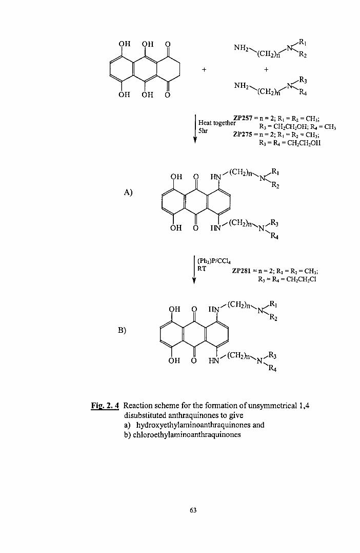
OH OH
+ + NH ,..,.,.....R3
Z'(CHZ)r("'l'~ OH OH 0
1 ZP257 = n = 2; R( = R2 = CH3;
Heat together R = CH CH OH' D = CH Shr 3 2 2 • ''''' 3
ZP275 = n = 2; R( = R2 = CH3;
R3 = R. = CH1CH10H
OH 0
A)
OH 0 HN/ (CH2)n'N/R3
'~
1 (Ph,)P/CC4 RT ZP281 = n = 2; R( = Rz = CH3;
R3 = ~ = CH2CH2CI
OH 0 HN/(CH2)n'N('R)
'Rz
B)
OH 0 HN/ (CH2)n'N/R3
'~
Fig. 2. 4 Reaction scheme for the formation of unsymmetrical 1,4 disubstituted anthraquinones to give a) hydroxyethylaminoanthraquinones and b) chloroethylaminoanthraquinones
63
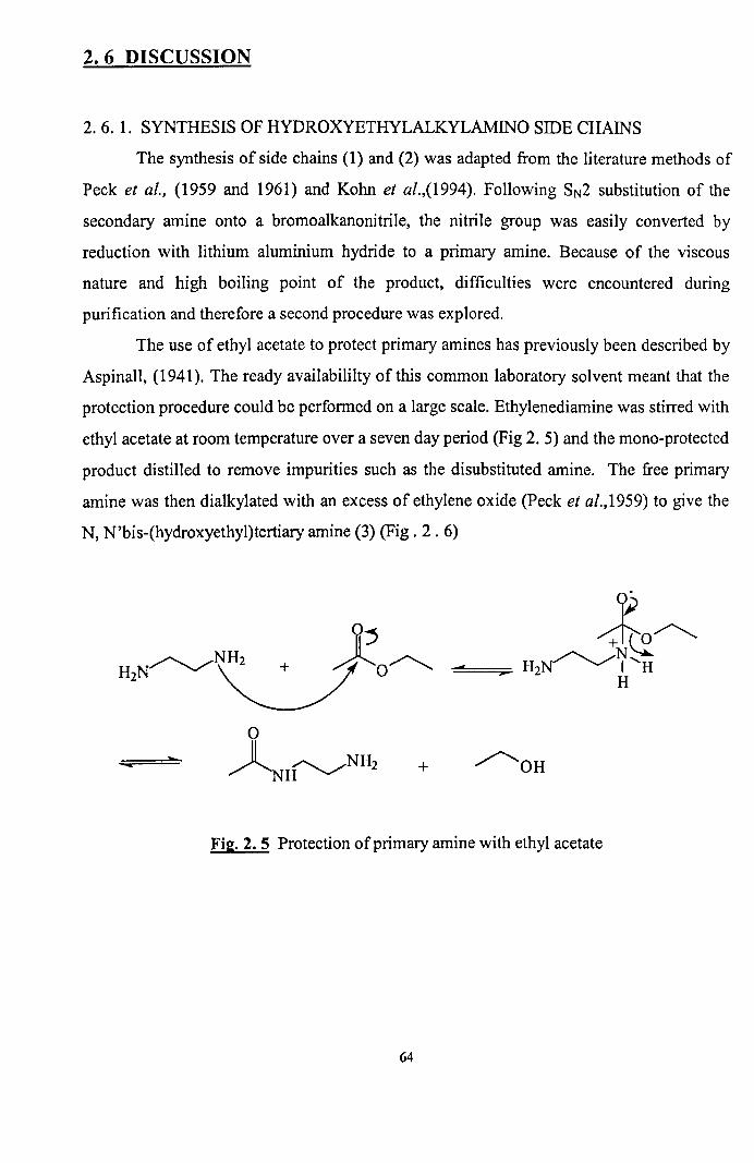
2. 6 DISCUSSION
2. 6. 1. SYNTHESIS OF HYDROXYETHYLALKYLAMINO SIDE CHAINS
The synthesis of side chains (1) and (2) was adapted from the literature methods of
Peck et al., (1959 and 1961) and Kohn et al.,(1994). Following SN2 substitution of the
secondary amine onto a bromoalkanonitrile, the nitrile group was easily converted by
reduction with lithium aluminium hydride to a primary amine. Because of the viscous
nature and high boiling point of the product, difficulties were encountered during
purification and therefore a second procedure was explored.
The use of ethyl acetate to protect primary amines has previously been described by
Aspinall, (1941). The ready availabililty of this common laboratory solvent meant that the
protection procedure could be performed on a large scale. Ethylenediamine was stirred with
ethyl acetate at room temperature over a seven day period (Fig 2. 5) and the mono-protected
product distilled to remove impurities such as the disubstituted amine. The free primary
amine was then dialkylated with an excess of ethylene oxide (Peck et al.,1959) to give the
N, N'bis-(hydroxyethyl)tertiary amine (3) (Fig. 2 . 6)
~j ~~ 112N~~O~ ~4===_;;::-. H2N~Z'H
o
~H~NH2 .... + ~OH -
Fig. 2. 5 Protection of primary amine with ethyl acetate
64

...
o 0
~"'C=~- ~ ~NH~ p ... ~H OH
o ~ ~OH ~H~N~
OH
Fig. 2. 6 Reaction of primary amine with ethylene oxide to fonn hydroxyethyl side ann.
H
H
+O~ ~OH ~H~N~ C, OH OH2
(1 ~OH r-/0H
.;;;:"'C=~- ~H~N~ ~"'=~ ... H2N~N~ /o~ OH (3) 0 OH
H + H H+ +,)l
OH
Fig. 2. 7 Deprotection ofthe primary amine under acid conditions
Although this synthetic route was successful, the length oftime required for the
first step of the reaction to reach completion led to the search for an alternative protecting
group for the synthesis of the N-2-(Hydroxyethyl)-N-phenylethylenediamine side chain (4).
When considering ketones as protecting agents, it is necessary for the carbonyl group to be
substituted with at least one aryl group so as to stabilise the protected product to
decomposition (March, 1992). The ketone benzophenone was found to be appropriate as it
65

is selective for the primary amino function and it can easily be displaced when required. As
N-Phenyl-ethylenediamine was the starting material for the reaction, the formation of the
di-protected amine was precluded. Primary amines react with ketones via nucleophilic
addition to the carbonyl group to form a tetrahedral intermediate (Fig. 2. 8) and this is
followed by the loss of water to form an imine as the protecting group. In order to increase
the rate of reaction, a catalyst, borontrifluoride etherate was employed and the water formed
as a byproduct was removed by azeotropic distillation to drive the reaction to completion.
R (
BF3 ,
C=O " ..... R
BF -..... 3 R , ..... O~
..... C, ..... H R N
" /+'
R H
R= --<Q)
R, r'- ..... BF3-
R/'\:+ ...
NH2R
H + BF3 , .....
R, to "" ..... C:{' H
R N I R
R, ..... C=NR + BF3 + H20
R
Fig 2.8 Protection of primary amine with benzophenone
2. 6. 2. CHROMOPHORE SUBSTITUTION REACTIONS
Much of the synthesis of monosubstituted and 1,4-disubstituted anthraquinones
revolves around the chemistry of aromatic nucleophilic substitution. Generally, compounds
such as the halobenzenes are resistant to this type of reaction due to the electron rich nature
of the aryl system. However, when an activating group such as a carbonyl or nitro group is
66

located ortho and/or para to the halogen, substitution can be rationalised by the SNAr
mechanism (March, 1992) which takes place in two steps. The attacking species forms a
bond with the substrate producing an intermediate complex and then the leaving group
departs. The intermediates formed are similar to Meisenheimer salts (Fig. 2. 9). By
comparison, the chemistry of mono-substitution onto the anthraquinone moiety is
dominated by the electron withdrawing property of the two quinone carbonyl groups which
generate a partial charge across the three rings and weaken the carbon-halogen bond,
activating it to nucleophilic substitution.
o o
+ Hel
o
Fit:. 2.9 Formation of mono anthraquinones
The synthesis of ZP1, ZP233 and ZP150 was achieved by treating 1-
chI oro anthraquinone with a ten fold excess of amine in the solvent 2-methoxyethanol, and
the mixture refluxed for 6 hours. At first attempt this reaction was performed by heating the
two reactants together as the amine, a viscous liquid, was thought suitable to act as both
nucleophile and solvent. Despite a long reflux time, 48 hours, no improvement in yield
above 10% could be achieved. The poor reactivity was initially attributed to the lack of
absolute purity of the side-chains and the possibility of a more reactive nucleophile
67

substituting onto the chromophore, however this was not evident by TLC. Instead,
visualisation under UV light revealed that a large amount of unreacted 1-
chloroanthraquinone was present in the reaction mixture indicating that the solubility of the
starting material was poor. In view of this, a solvent, 2-methoxyethanol was selected to
dissolve l-chloroanthraqinone at elevated temperatures (129°C) and allowed the reaction to
be controlled at reflux. Only a 5% increase in yield was attained by using this solvent and
so further modifications to the method were made.
When various alkylamino side chains were reacted with 9-chloro-3-
methoxyacridine, Kohn et al., (1994) found that the hydrogen chloride displaced as a by
product of the reaction was effectively protonating the primary amine side chain and
preventing it from substituting onto the chromophore. Consequently, the chlorine atom of
acridine was replaced with a phenoxy group which allowed couplings to proceed without
the need for an excess of amine. This procedure was followed for the 1-
chloroanthraquinone, however poor reactivity with the phenol prevented this route from
being further pursued. Instead, pyridine was added to complex the hydrogen chloride
expelled. The outcome was to increase the overall yield by only 2% which suggests that
protonation of the amine side chain was not a major concern regarding this reaction. As the
final yield of 17% seemed the best available and provided adequate product for subsequent
steps, no further work was done on this synthesis.
The preparation of 5,8-dihydro and 5,8-dihydroxyanthraquinones has been described
extensively in the literature (Zee-Cheng et al., 1978; Murdock et aI., 1979; Johnson et al.,
1979). Generally the reaction mechanism involves the condensation of leucoquinizarins
(Fig. 2. 3 and 2. 4) with an excess amount of the appropriate amine. The initial reaction of
the primary amine is at the quinone group on the I-position of the chromophore followed
by the loss of water which results in the formation of an imine. This is followed by a second
identical slow reaction at the 4-position. The intermediate imines (Schiff bases) are then
oxidized in air or by the addition of a few drops of base to secondary aryl amines via
internal rearrangement of the anthraquinone chromophore. The oxidation step can be
68

readilly monitored by TLC as a colour change from orangelbrown to blue is observed on
conversion to products.
When mitoxantrone is synthesised, reaction temperatures are maintained below
SO°C as the N-monosustituted ethylenediamine side chain can readily cyclise onto the 2-
position of the anthraquinone ring to form the hydroxyethylamino-l ,2,3,4-tetrahydronaptho
(2,3-f)-quinoxaline-7, 12-dione (see Fig. 1. 6) (Murdock et al., 1979). In contrast, the
tertiary nature of the hydroxyl ethyl amino anthraquinone side chains prevents a similar intra
molecular rearrangement from taking place.
It is generally accepted that the reaction of an amine with leucoquinizarin generates
a low quantity of product. As a consequence this has led to the pursuit for a more reactive
anthraquinone chromophore substituted with functional groups that can be displaced more
readily by the amine nucleophile. Krapcho et al., (1990) devised two methods to synthesise
1 ,4-difluoro-5,8-dihydroxy-9, 1 O-anthracenedione which on further reaction gave
mitoxantrone with a 78% yield. More recently Lee et al., (1999) described a five step
synthesis to isolate AQ4 from the starting product 3, 6 dichloropthalic anhydride. Whilst
being high yielding, the expense of these routes and the additional steps required in the
preparation of the starting material make them relatively unattractive.
2. 6. 3 SYNTHESIS OF CHLOROETHYLAMINOANTHRAQUINONES
In the conversion of an alcohol to an alkyl halide, the reaction can be initiated either
at the -C-O- bond or at the -O-H bond. As the hydroxyl is a poor leaving group it does not
undergo SN2 reactions readily. To overcome this, much use is made of inorganic halides
such as SOCh, PCls, PCb and POCb which form inorganic ester intermediates that can
easily be displaced by a halide ion (Fig. 2. 10 and see March, 1992 for references).
The reaction of an alcohol with thionyl chloride (acting as both solvent and
nucleophile) follows a second order rate equation (Rate = k[ROH][SOChD with retention
of configuration and proceeds via an SNi (internal nucleophilic substitution) mechanism
(Fig 2. 10). The rate at which the alkylchlorosulphite intermediate breaks down to the
product RCI is found to increase with increasing polarity of the solvent, and also with
69

increasing stability of the carbocation R + ,therefore, an ion pair, R+-OSOCI is thought to
be involved. Following collapse of the ion pair to products, attack is likely to occur on the
same side of R+ from which -OSOCI departed and as a result configuration is retained. If
the reaction is carried out in the presence of pyridine, the product undergoes inversion of
o +_I'"WII
R O.LS-CI
~
o II
R-O-S-CI ld
---:.~ RCI + HCI
Fit!. 2. 10 SNi reaction of thionyl chloride with a hydroxyl group
configuration. The HCI produced during the reaction complexes with the pyridine and C(
then attacks from the back as expected for an SN2 reaction.
Inorganic halides such as phosphorusoxychloride and thionyl chloride have been
successfully employed in the synthesis of substituted acridine anti-tumour drugs (Peck et
al., 1961; Kohn et al., 1994, Watanabe et al., 1989) and quinoline anti-malarial drugs (Peck
et aI1959), with thionyl chloride being the more favoured reagent. Following the method of
Kohn et al.. (1994), a 100 fold excess of thionyl chloride was reacted with the
hydroxyethylaminoanthraquinone in the absence of air to give a mixture from which the
product could not be isolated pure by recrystallisation. Purification by column
chromatography proved highly problematical as the product could not be separated from the
top of the silica column. In an effort to reduce impurities, attempted modifications to the
procedure such as increasing the temperature of reaction to 40°C and the use of additional
solvents such as dimethylformamide (Watanabe et al., 1989) and diethylether were
investigated without success. An increase in the quantity of product formed was observed
70
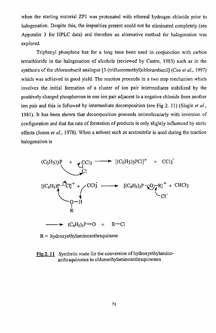
when the starting material ZPl was protonated with ethereal hydrogen chloride prior to
halogenation. Despite this, the impurities present could not be eliminated completely (see
Appendix 3 for HPLC data) and therefore an alternative method for halogenation was
explored.
Triphenyl phosphine has for a long time been used in conjunction with carbon
tetrachloride in the halogenation of alcohols (reviewed by Castro, 1983) such as in the
synthesis of the chlorambucil analogue [3-(trifluoromethyl)chlorambucil] (Coe et al., 1997)
which was achieved in good yield. The reaction proceeds in a two step mechanism which
involves the initial formation of a cluster of ion pair intermediates stabilized by the
positively charged phosphorous in one ion pair adjacent to a negative chloride from another
ion pair and this is followed by intermediate decomposition (see Fig 2. 11) (Slagle et al.,
1981). It has been shown that decomposition proceeds unimolecularly with inversion of
configuration and that the rate of formation of products is only slightly influenced by steric
effects (Jones et al., 1978). When a solvent such as acetonitrile is used during the reaction
halogenation is
R = hydroxyethylaminoanthraquinone
[(C6H5)3P~(R]+ + CHCl3
cr
Fig.2. 11 Synthetic route for the conversion of hydroxyethylaminoanthraquinones to chloroethylaminoanthraquinones
71

reported to proceed very rapidly and the enhanced polarity is effective enough to change the
kinetic order from unimolecular to bimolecular (Slagle et al.. 1981). This method of
chlorination proved to be the most effective. resulting in 70-85% yield following
recrystallisation for all compounds isolated. Only one compound ZP292 proved difficult to
halogenate because of its poor solubility in the solvent dichloromethane. The NMR of this
compound demonstrates that one side chain underwent chlorination to give the half mustard
whilst the remaining material was unreacted ZP254 (starting material).
Fig. 2. 11 describes the formation of the ion pair intermediate through nucleophilic
attack onto a chlorine atom of carbon tetrachloride. In an opposing view. Castro et al.,
(1983) suggests that this step could only take place if a highly nucleophilic reagent such as
aminophosphine replaced triphenylphosphine during reaction. as a strong nucleophile
would allow complete charge transfer to the trichloromethyl anion to occur. Instead, in an
amendment to the mechanism described in Fig. 2. 11, it was postulated that
triphenylphosphine attacks the tetrachloromethane carbon and forms an ion pair with the
displaced chloride anion.
2. 6. 4 ATTEMPTED SYNTHESIS OF CHLOROETHYLAMINOANTHRAQUINONE N
OXIDE HYDROCHLORIDE
OH 0
Fig. 2. 12 AQ4N
72

The drug AQ4N (Fig. 2. 12) was developed as a hypoxia selective bis-bioreductive
prodrug which undergoes cellular reduction at the N-oxide to give a DNA binding agent
that is cytotoxic through the inhibition of topo IT (Patterson, 1989, 1993). The inclusion of
a neutral quarternary N-oxide salt in the drug structure considerably decreases the attraction
of AQ4 for DNA and coincides with minimal prodrug cytotoxicity (Patterson, 1993). It was
therefore anticipated that a similar but much improved spectrum of activity would be
attained if a nitrogen mustard N-oxide could be linked to an anthraquinone chromophore.
Previous reports of aryl mustard N-oxide synthesis demonstrated that the compounds
generated were unstable. In acidic medium they reform to the original nitrogen mustard by
loss of the tertiary amine oxygen, and in basic media they rearrange to the half mustard and
a simple aldehyde (Degutis et al., 1972; Degutene et al., 1986, Tercel et al., 1995).
Regarding this, an attempt was made to synthesize an N-oxide of the anticancer drug
chlorambucil which was initially considered to be successful (Kirkpatrick et al., 1994).
However, an investigation into its decomposition revealed that a stable hydroxylamine was
formed as the major rearranged product in organic solvents and this had been isolated
instead (Tercel et al., 1995).
Fig. 2. 13 Proposed rearrangement of aliphatic bis-mustard N-oxides via a cyclic intermediate
Despite the futile attempts to isolate an aryl mustard N-oxide, an aliphatic
bifunctional mustard N-oxide of mechlorethamine (Nitromin) had been synthesised
previously. Although aliphatic bifunctional mustard N-oxides are known to undergo fast
rearrangement via a cyclic intermediate to give trisubstituted hydroxylamines (Fig. 2. 13),
73

nitromin has demonstrated increased stability and reduced cytotoxicity (30-40 fold) over
mechlorethamine with some selectivity for hypoxic cells (Connors, 1988; White et al.,
1989). Furthermore, an investigation into the decomposition of monofunctional mustard N
oxides in aqueous media revealed that they hydrolyse more slowly than their bifunctional
equivalents (Owari et al.,1953). Based on this evidence it was considered possible to
prepare a stable aliphatic chI oro ethyl amino anthraquinone N-oxide. The proposed starting
point was the isolation of a simple mono functional N-oxide by reacting mono substituted
chloroethylaminoanthraquinone ZP293 with mCPBA to form ZP180 (see Fig 2.14).
RHN (ZP293)
R = anthraquinone chromoohore
Fig. 2. 14 Attempted synthetic route for the formation ofa mustard N-oxide
This reaction followed a modified method of Patterson et al., (1989). The N-oxide
was isolated pure (observed by TLC) following column chromatography but decomposed
on solvent evaporation. Subsequent F AB+MS analysis showed that the mixture contained a
molecular ion [M]+ peak consistent with the relative molecular mass of the mustard N
oxide. However, it is unknown whether this was the desired product or the product of
rearrangement to the hydroxylamine, as both compounds would give identical MS spectra.
Because rearrangement of even the monofunctional anthraquinone mustard N-oxide proved
facile, no further attempts at N-oxidation were made.
To summarise, a number of hydroxyethylaminoalkylamino sidechains were
synthesised and substituted onto an anthraquinone chromophore to give a series of mono
substituted and 1, 4-disubstituted hydroxyethylaminoanthraquinones. The subsequent
74

coverslOn of the hydroxyethylaminoanthraquinones to chloroethylaminoanthraquinones
involved initial attempts at halogenation using thionyl chloride. However, the large number
of impurities produced from reaction and a difficulty in purification led to the choice of
milder chlorinating reagent triphenylphosphine with carbontetrachloride. This method
proved to be high yielding with few impurities and was therefore utilised to produce a series
of derivatives that were put forward for further study. An attempt was also made to isolate a
simple mono mustard anthraquinone N-oxide which, due its instability, led to the
abandonment of further synthesis.
75

AN INVESTIGATION INTO THE MECHANISM
OF ACTION OF ALKYLATING
ANTHRAQUINONES
76

Chapter 3 DNA-BINDING STUDIES
3.1. INTRODUCTION
DNA intercalating ligands have the capacity to bind reversibly to DNA by insertion
of an aromatic chromophore between DNA base pairs. One aspect of intercalative binding
that influences anti-tumour activity is the drug binding strength with DNA (reviewed by
Denny et ai, 1989). This principle is not always followed, as there are many intercalating
agents that bind tightly to DNA but have no activity (Feigon et ai., 1984). Despite this,
druglDNA binding which can be measured in terms of druglDNA association constant (K),
drug dissociation rate (kd) from DNA (Gandecha et ai., 1985; Denny et ai.,1989) or more
precisely the average residence time spent by the chromophore at a particular binding site
(Denny et ai., 1990), has often demonstrated a positive correlation with cytotoxicity within
a given class of compound (Denny et aI., 1989).
A senes of novel chloroethylaminoanthraquinones and hydroxyethyl
aminoanthraquinones were compared for their ability to intercalate into calf thymus DNA,
and their DNA association constants (K) and frequency of base pair intercalation (n)
determined. Futhermore, the effect of drug stabilisation of the DNA helix to thermal
denaturation (T m) was investigated in order to distinguish between covalent and non
covalent drug-DNA interactions.
3. 2. CHEMICALS AND REAGENTS
Calf thymus DNA was purchased from Sigma Chemicals, London, UK. Tris,
hydrochloric acid and sodium chloride were purchased from Fisher Scientific,
Loughborough, UK. All drug samples were stored as a dry powder and dissolved into
DMSO to give a 50llM stock solution prior to being further diluted to the specified
concentration in the appropriate buffer.
77

3.3 METHODS
For preparation of DNA solution and high and low ionic strength buffers see
appendix 1 and 2 respectively.
3.3.1. DETERMINATION OF DRUGIDNA ISOSBESTIC POINT
A series of DNAIDrug ratios (0, 0.2, 0.5, 1, 2, 5, 10, 15 to 1) were prepared by
varying the concentration of DNA from 0 to 2.25xlO-4M against a constant concentration of
drug 1.5x 1O-5M. To eight 10ml volumetric flasks was added drug (1.5x 1O-5M) and high
ionic strength buffer to give a final volume of 9ml. The approprate ratio of DNA was then
carefully added to each flask and the final solution accurately made up to 10ml with buffer
and mixed by single inversion. The spectra of each concentration ratio was recorded in the
visible region (350-700nm), and superimposed to give eight overlapping spectra. This
method was repeated using low ionic strength buffer.
3.3.2. DETERMINATION OF THE BINDING CONSTANT OF DRUG WITH DNA
A stock solution of DNA (l x 1 0-3M) in high ionic strength buffer was prepared
according to appendix 1. The drug (2.5xlO-5M) in buffer (3ml) was added to a lcm
pathlength quartz cuvette and its absorbance measured to determine the drug A.max• DNA
stock solution was serially pipetted into the drug solution at volumes of: 12x101lI, 6x20IlI,
8x40JlI and 4x 1 OOJl!' After each addition the solution was stirred and left to equilibrate for
10 minutes after which, the absorbance at the Amax of the drug was taken. All data were
reproduced in triplicate.
3.3.3 DETERMINATION OF THE THERMAL DENATURATION OF DNA
Low ionic strength buffer (3ml) and distilled (5.5ml) was added to a 10 ml
volumetric flask and degassed by sonication for 30 min. A solution of DNA (3.2x 1O-6M) in
low ionic strength buffer (0.6ml) was slowly added to the flask and the remaining volume
made up to 10 ml with dH20. The solution was mixed by a single inversion of the flask and
pipetted into two lcm pathlength quartz cells, the reference and sample cells. The
78

temperature of the reference cell was maintained at room temperature whilst the sample cell
temperature was raised to SO°C and the solution left to equilibrate for IS min. The
absorbance of the DNA was monitored at 260nm as the sample cell temperature was raised
at a rate of 1°C/min and readings taken at 1°C intervals from SO°C-85°C. All data were
reproduced in triplicate.
3.3.4 DETERMINATION OF THE THERMAL DENATURATION OF DNA IN THE
PRESENCE OF HYDROXYETHYLAMINOANTHRAQUINONE
Low ionic strength buffer (O.7Sml), dH20 (6mI) and drug (8.Sx 1 0·8M ) was added to
a 10ml volumetric flask and degassed by sonication for 30min. DNA (8.5x10·7 M) in low
ionic strength buffer (O.6ml) was slowly added to the flask and the volume made up
accurately to 10ml with dH20 to give a final DNA:drug ratio of 1 0: 1. The solution was
mixed by single inversion of the flask and pipetted carefully into two I cm pathlength quartz
cells, the reference and sample cells. The temperature of the reference cell was maintained
at room temperature whilst the sample cell temperature was raised to 60°C and the solution
left to equilibrate for 15 minutes. The absorbance of the DNA was monitored at 260nm as
the sample cell temperature was raised at a rate of 1°C/min and readings taken at 1 °C
intervals from 60-1 OO°C. All data were reproduced in triplicate.
3. 3. 5. DETERMINATION OF THE THERMAL DENATURATION OF DNA IN THE
PRESENCE OF CHLOROETHYLAMINOANTHRAQUINONE
Low ionic strength buffer (O.75ml) and dH20 water (6ml) were added to a 10ml
volumetric flask and degassed by sonication for 30min. To this was added the drug (8.Sx 1 O·
8 M) and then the DNA (8.5x10·7 M) and the volume made up accurately to 10ml with
dH20 water. The solution was mixed by single inversion of the flask and pipetted into two
lcm pathlength quartz cells, the reference and sample cells. The temperature of the
reference cell was maintained at room temperature whilst the sample cell temperature was
raised to 60°C and the solution left to equilibrate for 15 minutes. The absorbance of the
79

DNA was monitored at 260nm as the sample cell temperature was raised at a rate of
1°C/min and readings were taken at 1 °C intervals from 60-100°C.
All data were reproduced in triplicate.
3. 4. RESULTS
3. 4. 1 DETERMINATION OF ISOSBESTIC POINT
DNA intercalating agents such as the anthraquinones absorb in the visible region of
the electromagnetic spectrum to give characteristic absorption spectra. If the spectra derived
from the addition of varying concentrations of DNA to a single concentration of drug is
superimposed onto the spectrum of the drug alone, the absorbance wavelength at which all
the spectra cross is termed the isosbestic point. This may be achieved if there are only two
species in solution, that is, free drug and DNA bound drug. The point at which DNA is fully
bound with drug corresponds to no further decrease in the extinction coefficient at the "'max
of the free drug.
The isosbestic behaviour of eight hydroxyethylaminoanthraquinones was
determined in high ionic strength buffer (0.5M NaCl). Unlike the 1, 4-disubstituted
anthraquinones (Fig. 3.1 and Fig. 3. 2) the mono derivatives showed no apparent affinity for
calf thymus DNA. The 5, 8-dihydroanthraquinones could be distinguished from the 5, 8-
dihydroxy derivatives by the position of their isosbestic point, 639 nm and 618 nm
respectively (Table 3. 1). Furthermore, the spectral shifts for the 5, 8-
dihydroxyanthraquinones were up to 50% greater than for the 5,8-dihydroanthraquinones
indicating enhanced chromophore 7t- orbital interaction with the DNA base pairs.
In contrast to the other compounds tested ZP274, a 5,8-dihydroxyanthraquinone
substituted with two hydroxyethyl groups per side chain gave an isosbestic point at the four
lowest drug concentrations in low ionic strength buffer (0.05M) (Fig. 3. 3). This behaviour
has been observed previously (Kapusczinski et al., 1985) and may be related to the
80
preference of this compound at high druglDNA concentration to bind electrostatically to the
surface of DNA.
The isosbestic behaviour of ZP254 was not determined as drug precipitation
occured on addition to buffer. The bulky phenyl groups attached to the side chains of ZP254
increase its lipophilic character and therefore make it poorly soluble in water even if the
concentration ofDMSO in buffer is raised to 10%.
The spectra of ZP285 (Fig. 3. 4) is representative of the effect of the
chloroethylaminoanthraquinones on DNA binding. At each increasing DNA concentration
the drug absorbance showed no change in A.max but demonstrated a large change in its
visible region when compared to the 1,4-disubstituted-5,8-dihydroxy
hydroxyethylaminoanthraquinones (Fig. 3. 2). When the same concentration of drug was
added to distilled water in the absence of salt and buffer, the characteristic 1, 4-disubstituted
anthraquinone spectra was re-gained (Fig. 3. 5). This indicates that high ionic strength
buffer may adversely affect the intercalation of the chloroethylaminoanthraquinones and
reduce their affinity for DNA.
81

Table 3.1 The effect on visible absorbance following the addition of the h~drox~eth~anthraguinones to calf thymus DNA.
LP Shift in Amax
Compound (nm) (nm)
ZP232 639 10
ZP240 639 20
ZP242 636 17
ZP255 618 20
ZP257 618 18
ZP265 618 20
ZP274* 618 nJd
ZP275 618 17
LP = Isosbestic point (nm); shift in Amax (nm) from free drug to fully intercalated DNA. nJd= not determined, * isosbestic point determined in low ionic strength buffer (O.05M).
13 . 5
13 . 4
13 . 3
13 . 2
13. 1
e 0 ........ ~ ..... "N ....... ~ ...... N"" .. "' •• _ ••• M ••• "' W ...... _ ••••••• "M ..... _ ............. _ •• _, ....... _~.·._· .... _ .. ••• .. •••• .... •• • ............................ ., .............. ......................... •· •• .. ••·•• .. ······T··· .. ············,······
j5e.ee 4213 . 00 490 . 1313 560 . 00 630 . e0 71313 0
Fig 3. 1 Spectral shift in visible absorbance following the addition of increasing concentrations of calf thymus DNA to 5,8- dihydrohydroxyethylaminoanthraquinone ZP242. Determined in high ionic strength buffer.
82

0. 4
e . 0 ........ _ ................... _ ....... _ ......... _ .. ,_ ........... _._ ... -..................... __ ... , ...... _._ ..... .. 350. 00 420 . 0e 4900e 560 00 630 .eo 700 . 00
Fig 3. 2 Spectral shift in visible absorbance following the addition increasing concentrations of calf thymus DNA to 5,8-dihydroxyhydroxyethylaminoanthraquinone ZP275 . Determined in high ionic strength buffer.
8.40
032
490 .ee 630 eo 7BO 00
Fig 3. 3 Spectral shift in visible absorbance following the addition of increasing concentrations of calf thymus DNA to 5,8-dihydroxyhydroxyethylaminoanthraquinone ZP274. Determined in low ionic strength buffer.
83

0.5
o 4
o ~ ... -...... - ... ·.·-··.-.·-.-··,.·.·-.·---·-····--.-r ·.· .. - -.-----··.· .... -... , ...... -............................... , ................................................ , 35 0. 00 429 . 99 490 .99 560 . 09 630 .00 700 .00
Fig 3. 4 Effect on visible absorbance following the addition of increasing concentration of calf thymus DNA to chloroethylaminoanthraquinone ZP285. Determined in high ionic strength buffer.
9.5
0. 4
13 . 3
0.2
9. 1
----e . e .. ----.-.-.--.-.... --.... , ... -.-.. -.. -... --.... -.. - .. ,.----.-..... -... , ... -.-.. -............. -............ ,.-................................................... , 350 . 99 420 .90 490 .90 560 .oe 630 .00 7ee 00
Fig 3. 5 The visible absorbance spectrum of ZP285 in dH20
84

3. 4. 2 SPECTROPHOTOMETRIC TITRA nON STUDIES
In order to proceed with the spectrophotometric titration assay it is essential that
each drug produces an isosbestic point (sec section 3. 4. 1) and that both free and bound
drug obey Beer-Lamberts Law over the drug concentrations used.
Following the addition of an increasing concentration of DNA to a fixed
concentration of drug. intercalation can be identified by a decrease in absorbance at (Amux)
of the free drug and a visible absorbance shift to longer wavelengths as a result of
peturbation of the 1t and 1t* orbital energies on binding. The extent and nature of DNA
binding can be quantitated using the Scatchard equation which can be applied to
intercalating species. By taking absorbance readings at the Amax of the free drug and
plotting ric vs r, where r = (concentration of ligand bound/concentration of DNA) and c =
concentration of free ligand, the extent of DNNdrug association can be dctermined
(Plumbridge et al., 1978; Bell et al., 1989). The negative slope corresponds to the affinity
constant (Kd, whilst the fraction of drug molccule bound per DNA base pair (n) can be
determined at the x intercept (Fig. 3. 6)
All titrations were performed in triplicate and in high ionic strength buffer to
maintain the conditions of the drug isosbestic point determination where surface binding is
excluded. At low concentrations of DNA, intercalative binding was shown to be negligible.
Conversely, at high DNNlow drug concentration ratio intercalation predominates, and the
graph tends towards a steep negative slope. The KJ values for the 5,8-
dihydroxyanthraquinone analogues were in the order of 1 06M-J, whilst the KI values for the
5,8-dihydroanthraquinones were 106 _107M-I (Table 3.2).
The number of drug molecules bound per molecule of DNA (n) did not vary
significantly amongst compounds of the same substituted chromophore. The 5,8
dihydroanthraquinones intercalated at every 8.3 base pairs of DNA (n = 0.12) whilst the 5,8
dihydroxyanthraquinones intercalated at approximately every 6.6 base pairs (n = 0.15).
The DNA affinity of the chloroethylaminoanthraquinones was not determined as the
compounds did not show isosbestic behaviour (see section 3.4. 1 and 3.5).
85

120000
100000
80000
(j 60000 I:;
40000
20000
0 0 0.05 0.1 n 0.15 0.2
r
Fig. 3.6 Scatchard detennination of the binding affinity ofZP265 with calf thymus DNA. The data shown represents the average of triplicate experiments. K\ = drugIDNA association constant; n = fraction of drug molecule intercalated per base pair site
86

Table 3. 2 The affinity of the hydroxyethylaminoanthraquinones for calf thymus DNA.
Compound KI(M-1) n
ZP232* 3x106 0.12 (8.3)
ZP240* 7x106 0.12 (8.3)
ZP242* 1x107 0.12 (8.3)
ZP255 3x10⁶ 0.15 (6.6)
ZP257 2x10⁶ 0.175(5.7)
ZP265 3xl06 0.14 (7.1)
ZP274 2x10⁶ 0.15 (6.6)
ZP275 2xl06 0.15 (6.6)
Data determined at the Amax of drug. Kl represents the DNA intercalation association constant and is expressed as the mean of three replicates; n is the fraction of drug molecule bound per DNA base pair and is expressed in parenthesis as the frequency of drug-base pair intercalation .... denotes 5, 8-dihydroanthraquinone.
3.4.3 DNA THERMAL DENATURATION TEMPERATURE STUDIES
The DNA melting temperature (T m ) was assayed according to the method of Tong
et al.,( 1978). T m is defined as the temperature at which half of the concentration of DNA
has denatured to single strands and can be derived by plotting absorbance vs temperature.
By subtracting the T m for DNA alone from the T m for DNA intercalated with drug, the
degree of helix stabilisation towards denaturation can be determined as I1Tm•
All analysis was performed in triplicate in low ionic strength buffer. Calf thymus
DNA with no drug gave a T m value of 70.5 ± 0.5 °C.
The efficiency of helix stabilisation by the hydroxyethylaminoanthraquinones
followed the general trend (least efficient) monoanthraquinones < 5,8-
dihydroanthraquinones < 5,8-dihydroxyanthraquinones (Table 3. 3). The weakest drug
proved to be monosubstituted ZPl which showed poor binding with DNA, giving a I1Tm of
87
0.4 °C. In contrast, the 1,4-disubstituted-5,8-dihydroxyanthraquinone ZP265, was the most
effective drug, increasing DNA denaturation by 19.7 °C. Fig 3. 7 compares the effect of
increased chromophore substitution on helix stability by showing the T m plots for three
drugs ZPI, ZP242 and ZP265 substituted with the (N-(2-hydroxyethyl)-N
methylamino)ethylenediamine sidechain (Fig. 3. 8). The effect of increasing the number of
hydroxyethyl groups from one to two per side chain resulted in reduced DNA binding. This
coincides with data derived from the isosbestic behaviour of ZP274 and the assumption that
surface binding predominates with this compound. The evidence from the thermal
denaturation studies of the hydroxyethylaminoanthraquinones therefore indicates that the
chromophore 5,8 substituted hydroxyl groups and the structure of the side chain both affect
the extent of drug-DNA binding.
The chloroethylaminoanthraquinones showed a general trend of weak helix
interaction (Table 3. 4) suggesting that with the exception ofZP281 (L~Tm 10.1) (Fig 3. 11)
they appear to have poor affinity for DNA. A possible explanation for this is discussed in
(section 3. 5.)
Two compounds ZP254 and the chloroethylaminoanthraquinone ZP280 precipitated
on addition to buffer and so determination ofTm was not possible.
88

0.200 I-------~:;:;;;::;;:;; ... ~Hiifi-" 0.180
0.160
e 0.140
S 0.120 ~ CJ
~ 0.100 .c ~ 0.080 I:Il
~ 0.060
0.040
0.020
-+-ZPl
---ZP242
--k- ZP265
0.000 H1I1MRtG"'~4+++++-H-+-++++-++-i-++++~I-+-+-+++-1 g ~ ~ ~ ~ ~ ~ ~ ~ ~ g ~ ~ ~ ~ ...
Temperature °c
Fig. 3. 7 Demonstrates the effect of increasing temperature on the UV absorbance of calf thymus DNA (at A260) in the presence ofZPl, ZP242 and ZP265. The data shown for each compound is the average of triplicate experiments.
ZPl: R\; R2 = H; R3 = H ZP242: RJ = R2; R3 = H ZP265: RJ = R2; R3 = OH
Fig 3. 8 Shows the chromophore side chain substitution pattern of. ZPl, ZP242 and ZP265
89

Table 3. 3 The effect of hydroxyethylaminoanthraquinones on the denaturation temperature of calf thymus DNA.
Compound TmeC) IlTm(OC)
ZPI 70.9 (± 0.27) 0.4
ZP240 74.5 (± 0.21) 4.0
ZP242 81.7 (± 0.15) 11.3
ZP255 83.3 (± 0.35) 12.8
ZP257 80.1 (± 0.1) 10.5
ZP265 90.2 (± 0.22) 19.7
ZP274 81.5 (± 0.21) 11.0
ZP275 84.0 (± 0.32) 13.5
T m determined at the /...260 of calfthymus DNA and expressed as the mean triplicate value (± s.d)
0.16 ......--------------~
0.14
_ 0.12
e = ¥ 0.10 tJ
~ 0.08
of ~ 0.06
~ 0.04
0.02
0.00 ... ~~ .... ~++IH_+++++++IH_+++++++I~
$ ~ ~ $
Temperature °C
Fie 3. 9 The effect of increasing temperature on the UV absorbance of calf thymus DNA (/...260) in the presence of chloroethylaminoanthraquinone ZP281. The data shown is the average value of triplicate experiments.
90

Table 30 4 The effect of chloroethylaminoanthraquinones on the denaturation temperature of calf thymus DNA.
Compound
ZP293
ZP289
ZP286
ZP282
ZP285
ZP280
ZP281
TmeC)
72.2 (± 0.1)
76.2 (± 0.53)
77.7 (± 0.4)
73.8 (± 0.41)
75.9 (± 0.37)
pption
80.6 (± 0.41)
!1TmeC)
1.7
5.7
7.2
2.3
5.4
nJa
10.1
T m determined at the 1...260 DNA and expressed as the mean triplicate value (± s.d). nJa = not applicable.
30 50 DISCUSSION
3. 5. 1. ISOSBESTIC BEHAVIOUR OF THE NOVEL ANTHRAQUINONES
A property of substituted anthraquinones is to bind DNA by the process of
intercalation which is manifest by a characteristic drug visible absorbance shift to longer
wavelengths (bathochromic shift) and a decrease in the molar extinction at the Amax of the
free drug (hypochromic shift) (Waring et ai., 1981). When the drug becomes intercalated
into DNA, it is stabilised within the hydrophobic interior of the helix by charge transfer
forces, van der Waals and hydrophobic interactions. The effect of these are to alter the
electron density of the chromophore and its absorbance properties. Anthraquinones
containing basic amine side chains can also bind to DNA through ionic interaction. This is a
weaker mode of binding compared to intercalation and occurs at physiological pH between
the positively charged amino group on the sidechains and the anionic phosphate groups of
the DNA backbone (reviewed by Baguely 1991, Pohle et ai., 1990).
91

Studies of the novel hydroxyethylarninoanthraquinones showed that 1,4-
disubstituted 5,8-dihydro and the 5,8-dihydroxy but not the I-substituted compounds gave
isosbestic points in high ionic strength buffer (O.5M NaCl). The effect of high salt
concentration is to neutralise the anionic charge of the DNA phosphate backbone with
positively charged sodium ions and this discourages the drug from binding to the surface of
the DNA but at the same time allows intercalative binding to continue (Lown et al., 1985;
Bell et al., 1989). Evidence of electrostatic binding was apparent in low ionic strength
buffer as the majority of the drugs lost isosbestic behaviour suggesting that significant
surface binding had occured. In view of this, the DNA binding affinity of the novel
anthraquinones was determined in high ionic strength buffer. It is also worth noting that
others have shown that the presence of an isosbestic point can be dependent on the buffer
used (Kapusczinski et al., 1981; Lown et al., 1985).
ZP274, comprising of two hydroxyethyl groups per side chain would only give a
partial isosbestic point in low ionic strength buffer at the four lowest drug concentrations. It
has previously been stated that intercalation is encouraged at a low drug to DNA ratio and
the subsequent loss of an isosbestic point may be attributed to the ability of the drug to
surface bind DNA at high druglDNA ratios (Kapuczinski et al., 1985). The paucity of DNA
binding sites may also encourage drug aggregation. Surface binding encourages 'self
association' between drug molecules which stack on top of one another (dimerisation)
through chromophore hydrophobic interactions and reduce the number of accessible
intercalation sites. The outcome of this is to promote additional secondary binding and a
species with absorption properties which differ from the intercalated drug (Kapusczinski et
al., 1985). In the case of ZP274 it is possible that the partial isosbestic point occured as a
result DNA surface binding alone as at high ionic strength a complete loss of isosbestic
behaviour was observed. From this evidence it can be postulated that intercalation as a
single binding mode does not take place for this drug.
The mono substituted anthraquinones eg ZP1, ZP150 and ZP233 showed a lack of a
consistent single interaction with DNA. Because they possess only one side chain this
structural configuration allows for a number of intercalative binding modes to take place.
92

Islam et al., (1985) demonstrated that I-substituted anthraquinones can intercalate either
parallel or perpendicular to the DNA axis with the sidechain in the major or minor groove
of DNA and may therefore produce more than one DNA binding species in solution.
At high ionic strength the chloroethylaminoanthraquinones failed to show isosbestic
behaviour. Instead at each DNA concentration they gave identical spectra which in contrast
to the hydroxyethylaminoanthraquinones showed a large change in the visible region
coinciding with a shift to shorter wavelengths (Fig. 3. 4). By comparison, at the same
concentration of free drug in distilled water (no buffer or salt) the characteristic
anthraquinone spectra was observed (Fig. 3. 5). A similar shift in spectrum profile was
obtained in the theoretical calculation of mitoxantrone dimers (Kapusczinski et a!., 1985).
Considering this, it may be postulated that high ionic strength promotes drug chromophore
chromophore hydrophobic interactions. These may lead to drug dimerisation and reduced
DNA interaction, perhaps as a consequence of the high lipophilic nature of the
chloroethylaminoanthraquinones. As a further suggestion, the lack of crossover points for
spectra at high and low druglDNA ratios does not necessarily indicate that intercalation did
not take place because under certain conditions such as high ionic strength, free and bound
drug may differ significantly to never give an isosbestic point.
3. 5. 2 ANALYSIS OF THE DNA BINDING CONSTANT (K1) FOR THE
HYDROXYETHYLAMINOANTHRAQUINONES
The determination of DNA affinity constants for the
hydroxyethylaminoanthraquinones followed the method of Lown et a!., (1985); Bell et al.,
(1989). This assay involves reverse titration where the DNA is added to and initially
becomes saturated with drug. As DNA concentration increases, intercalation rather than
surface binding predominates. Kapuscinski et al., (1985) stated that this method can
promote DNA condensation and precipitation which compromises accuracy, however, this
effect was not observed in this study or that ofLown et al., (1985).
The DNA association constant values (K1) for the 1,4-disubstituted-5,8-
dihydroxyanthraquinones were in the range 106M-I at high ionic strength. This data is in
93

agreement with Lown et al., (1985) who found that mitoxantrone and its congeners bound
duplex DNA in the range (0.16-2.35)x 106M- t, whilst the 5,8-dihydroanthraquinone
derivatives such as ametantrone gave 10 fold lower values. The present study provided
contrary results when the two classes of drug were compared as the 5,8-
dihydroanthraquinones produced the higher (K1) values (Table 3. 2) which is consistent
with the data derived by the titration method of Kapuczinski et al., (1985). Because DNA
affinity is an equilibrium measure of the extent of drug attraction for DNA it may be
affected by the choice of buffer or ionic concentration (Chaires et al., 1982). This
inconsistency has led others to suggest that the dissociation rate of a drug from DNA is a
better indication of DNA affinity (Denny et al., 1990).
When a comparison was made between the 1,4-disubstituted-5, 8-dihydro- and the
5,8-dihydroxyanthraquinones and the frequency of drug base pair intercalation (n) the latter
were found to intercalate into DNA on average every 6.6 base pairs, an increase of
approximately two base pairs over the 5, 8-dihydroanthraquinones. Previous studies by
Lown et al., (1985); Kapuczinski et al., (1985) confirm this trend but demonstrated that
mitoxantrone intercalates into DNA at approximately every 2.9 base pairs consistent with
the neighbour exclusion model. This model suggests that the binding of one drug molecule
to one site influences the binding of subsequent molecules by preventing binding at nearby
sites either through physical blockage or by steric alterations of the DNA helix. Although
the novel dihydroxyanthraquinones would be expected to have a similar intercalative
binding frequency to mitoxantrone, it has been suggested that high ionic strength can reduce
the number of binding sites by lowering the affinity of the drug for DNA (Chaires et al.,
1982).
From the DNA-drug affinity data it can be shown that the chromophore hydroxyl
groups influence the equilibrium towards DNA-drug formation. This is particularly evident
when comparing compounds with identical sidechains such as ZP242 and ZP65, and can
been explained in a number of ways. The perpendiCUlar mode of intercalation which is
favoured by 1,4 disubstituted anthraquinones (see section 1. 3. 2. 2) orientates the
chromophore hydroxyl groups such that they are able to penetrate the helix through to the
94

minor groove. The effect of this is to cause the disengagement of the chromophore from
DNA to be energetically unfavourable and so considerably slower (Denny et al., 1989).
Prolonged chromophore retention times have also been attributed to the formation of
intramolecular hydrogen bonds between the chromophore 5,8-hydroxyl groups and the
quinone carbonyl groups. These bonding interactions increase the lipophilic character of
the drug and its affinity for the hydrophobic interior of DNA (Patterson, 1993).
Furthermore, the hydroxyl groups are suggested to introduce planarity to the chromophore,
which is in contrast to ametantrone where the unsubstituted ring is slightly buckled. As a
result the planarity of mitoxantrone has been suggested to enhance base pair interaction at
the intercalation site (Mazerski et al., 1998).
The consistency of data within each series of drugs i.e 5,8-dihydro or 5,8-
hydroxyanthraquinone, shows that sidechain interactions playa minor role (if any) during
the intercalation process at high ionic strength. The mode of action of the 5,8- dihydro and
5,8-dihydroxy-anthraquinones is therefore consistent with intercalative binding.
3. 5. 3. EFFECT OF THE HYDROXYETHYLAMINOANTHRAQUINONES ON
STABILISATION OF THE DNA HELIX TO THERMAL DENATURATION
The DNA molecule is made up of two complementary strands consisting of a sugar
phosphate backbone and purine/pyrimidine base pairs which are held together by hydrogen
bonding. On heating, the DNA inter-strand hydrogen bonds break causing denaturation of
the double helix which leads the formation of single-stranded DNA. Drug molecules that
reversibly intercalate into DNA stabilize the helix such that more energy (a higher
temperature) is required to cause denaturation. This effect can be measured by monitoring
the change in absorbance with temperature at 260nm, the absorbance maximum for duplex
DNA.
As all the drugs were assayed in low ionic concentration, it is therefore assumed that
electrostatic binding in addition to intercalation contributes to the results obtained. The
hydroxyethylaminoanthraquinones displayed a number of characteristics; drugs related by
sidechain such as ZP1, ZP242 and ZP265 gave approximately lOoC rise in temperature
95

with each substitution onto the chromophore (Fig. 3. 7). The increasing order of drug-DNA
stability followed the trend, mono substituted anthraquinone < 5,8-dihydroanthraquinone <
5,8-dihydroxyanthraquinone (both 1, 4-disubstituted). This is in agreement with the DNA
dissociation knietics of substituted anthraquinones (Gandecha et al., 1985; Denny et
al.,1990). Drugs with two hydroxyethyl groups per sidechain such as ZP274, ZP240 and
ZP255 demonstrated considerably reduced ~ T m values. It is likely that the hydroxyethyl
side chains of these compounds predispose them to increased hydrophilicity and
preferential electrostatic binding to DNA. Since these interactions are external to the DNA
duplex, low ~ T m values would be expected.
1,4 disubstituted anthraquinones comprising of the (N-(2-hydroxethyl)-N
methylamino )ethylenediamine sidechain gave the highest overall ~ T m values (Fig. 3. 8 and
Table 3.3). ZP265 stabilised the helix by 19.7°C. In comparison, under similar conditions
mitoxantrone stabilises DNA by 15.9 °c (Johnson et al., 1979). At physiological pH the
secondary amine side chains of mitoxantrone become protonated and interact with the
negatively charged DNA-phosphate groups. Additional hydrogen bonding may occur
between the drug side chain hydroxyl groups and the base pairs in the vicinity of the
intercalation site (Bailly et al., 1996; Mazerski et al., 1998). In view of this, it is likely that
ZP242 and ZP265 display a similar mode of base pair side chain interaction to
mitoxantrone. Furthermore, as the tertiary methyl amine side chains increase drug
lipophilicity they may essentially enhance DNA binding by making the drug more attractive
to the DNA helix.
3. 5. 4. EFFECT OF THE CHLOROETHYLAMINOANTHRAQUINONES ON
STABILISATION OF THE DNA HELIX TO THERMAL DENATURA lION
At first sight, the data from chloroethylaminoanthraquinone-DNA interaction
suggests that these compounds are weak, reversible DNA binding agents. This contrasts
with the DNA alkylation studies of Chapter 4 which demonstrated that they inhibit DNA
strand separation at nanomolar concentrations which is up to 100 fold less than in this
assay. In view of this, it is likely that the formation of drug-DNA inter-strand crosslinks
96
takes place but too few crosslinks are created to stabilise the helix effectively. The
consequence of drug crosslinking DNA is to prevent thermal denaturation (Hartley et al.,
1991), reducing the formation of single strand DNA rather than altering Tm and it can be
directly linked to the number of crosslinks fonned. Any hydrolysed drug would be expected
to bind DNA non-covalently and detennine the outcome of helix stability at the absorbance
wavelength stated. Studies with nitrogen mustards such as melphalan have shown that the
dihydroxyethylamine is the product of reaction under aqueous conditions (Bolton et al.,
1993). When fully hydrolysed the chloroethylaminoanthraquinones would be expected to
fonn hydroxyethylaminoanthraquinones which have generally engendered good helix
stability (Table 3. 3). These products of hydrolysis would probably be present in low
concentration and so would have little effect on the helix stability. Additionally, mono- and
perhaps intra-strand alkylations of the helix may take place (Osborne et al., 1995). This
fonn of covalent binding is unable to inhibit DNA strand separation and further to this may
orientate the drug in such as way as to prevent it from intercalating into DNA.
Of the chloroethylaminoanthraquinones, ZP281 gave the highest fl. T m (10.1 °C). The
unsymmetrical nature of this compound restricts bis-alkylation of DNA to one side chain as
its second arm is identical to the side chains substituted onto AQ4 (Fig. 2.12), a drug that
has demonstrated high affinity for DNA (fl.Tm 17.5°C) (Patterson, 1993). Because ZP281
creates fewer inter-strand crosslinks (demonstrated in Chapter 4) it is likely to have a
greater effect on DNA thennal melting temperature through the formation of a higher
concentration of single-strand alkylations that are enhanced by the non-covalent interaction
of its second side chain.
In summary, the hydroxyethylaminoanthraquinones showed good affinity for DNA
consistent with both intercalative binding and lome interactions. The
chloroethylaminoanthraquinones with the exception of ZP281, demonstrated poor binding
implying that intercalation may not be the primary mechanism by which these compounds
interact with DNA. Results in Chapter 4 will suggest that rather than being poor DNA
binders, the chloroethylaminoanthraquinones irreversibly alkylate DNA.
97

Chapter 4 DNA ALKYLATION STUDIES
4. 1 INTRODUCTION
Rational anticancer drug development faces a dilemma that most drugs have
multiple sites of action and it is not known which of these contribute to therapy or to
toxicity. It is therefore beneficial to attempt to select compounds with a restricted range of
action but without reduced anti-tumour activity. Bifunctional nitrogen mustards are
extremely cytotoxic through their ability to form covalent intra and/or inter-strand
crosslinks with DNA. However, their high reactivity and lack of target specificity often
leads to reaction with other cellular nucleophiles and to a reduced concentration of drug
reaching its target site (see section 1.4).
Nitrogen mustards linked to an acridine chromophore have previously demonstrated
improved selectivity for DNA and an altered drug base pair specificity compared to non
targeted mustards (Mattes et al., 1986). The novel chloroethylaminoanthraquinones are a
series of drugs which have the capacity to both intercalate and alkylate DNA. To establish
the extent of their covalent binding nature, the DNA base pair alkylation preference of these
compounds and their ability to inter-strand crosslink DNA was determined.
4. 2. CHEMICAI.JS AND REAGENTS
Plasmid DNA pBR322 (O.25u1JlI), T4 polynucleotide kinase (SulJlI), BAM HI
(10u/JlI) and Sal I (IOu/JlI) restriction enzymes were purchased from Northumbria
Biologicals Ltd. y32p_ATP (500Ci/mmol), primer (lOpmlJlI), dNTP mix (2.5mM), MgCh
(25mM), gelatin (0.2%), Bacterial Alkaline Phosphatase (BAP) (150u/JlI), Bacterial
Alkaline Phosphatase reaction buffer (BAP buffer 5x), lOx buffer, React 3 buffer (lOx),
DNA stop solution, Forward reaction buffer (5x), were obtained from Amersham Biotech
Ltd, St. Albans, Herts. Taq DNA polymerase (x50) (0.1 u/JlI) was purchased from Promega,
98

London BIO-Spin chromatography column and BIORAD 583 gel drier were obtained from
BIO-RAD Laboratories, Kent, UK. Ammonium persulphate (APS), N, N, N', N'
tetramethylethylenediamine (TEMED), sequagel 6, complete mix, mineral oil and
mechloroethamine (N-methyl-N,N-bis(2-chloroethyl)amine) were purchased from Sigma
Chemicals, London, UK. The control mechlorethamine was made up to the appropriate
concentration in 0.1 M hydrochloric acid and stored at -20°C prior to use. The agents ZP281,
ZP282, ZP285 and ZP289 were synthesised as described in section 2. 4, made up to the
appropriate stock solution concentration in DMSO and stored at -20°C prior to use.
u = unit activity.
Strand separation buffer, TEo A buffer, TEA buffer, TBE buffer and formamide dye were
all prepared according to appendix 2.
4.3. METHODS
See appendix 1 for preparation of DNA
4.3. 1 DETERMINATION OF DNA SEQUENCE SPECIFIC ALKYLATION USING
THE POLYMERASE CHAIN REACTION (PCR)
The following constituents were added to an eppendorf containing the labelled
primer (for preparation see appendix 1) and mixed: 0.2% gelatin (55J.lI), 25mM MgClz
(220J.ll), lOxbuffer (110J.l1), 2.SnM dNTP mix (110J.ll) and dH20 (SSJ.ll). The mixture (SOJ.l1)
was placed into a PCR tube containing the drug-treated DNA (see appendix 1) (40J.ll) and
the enzyme DNA Taq polymerase (xSO) (10J.ll) which was diluted into dH20 (1 in 50) prior
to use. Each tube was covered with mineral oil and the PCR set to cycle 36 times at 94°C
for 1 min, S8°C for 2 min, nOc for 1 min and an additional 1 min per cycle, a total of 3 h.
The ten samples were then transfered into sterile eppendorfs and the DNA precipitated and
dried as described in appendix 1. Each dried sample was re-suspended in formamide dye
(4J.lI), heated to 95°C for 2 min and cooled in an ice-bath to denature the DNA. The samples
99

were loaded onto the gel (see appendix 2) and electrophoresis was performed in TBE buffer
over 3 h at 3000V, 55°C using vertical glass electrophoresis plates. The resulting gel was
then transfered onto Whatman 3MM filter paper, covered and dried under pressure on a
BIO-RAD 583 gel drier for approximately two hours. Once dry, the gel was exposed onto a
Kodak X-ray film at -70°C for 24 h before developing.
4. 3. 2. DETERMINATION OF THE FORMATION OF DNA INTER STRAND
CROSSLINKS
To each sample was added: 32P-radiolabelled DNA (lOOng/lOJlI), drug: ZP281,
ZP282, ZP285 or ZP289 (1 JlI) in DMSO to give final assay drug concentrations of 0.1 nM,
InM, 10nM and 100nM, TEoA buffer (5Jll) and the total volume made up to 50JlI with
dH20. Following incubation at 37°C for lh the reaction was terminated by the addition of
DNA stop solution (lOJlI) to each sample. The DNA was precipitated by the addition of
95% ethanol (l80J.lI) and centrifuged at 1300rpm for 15 min. This precipitation process was
repeated and the resulting pellet dried by lyophilisation. Each dried sample including the
control sample of drug untreated DNA were resuspended in strand separation buffer (lOJll),
heated at 95°C for 2 min in order to denature the DNA, then immediately chilled in an ice
water bath to prevent the DNA strands rejoining. A sample of undenatured DNA, the
second control, was not heated, and instead was dissolved in formamide dye (lO/-1l). An
0.8% agarose gel approximately 1 cm thick was prepared in TEA buffer (also used as the
running buffer) and the samples subsequently loaded. Electrophoresis was performed
overnight using a horizontal gel kit at 40 V. The resultant gel was dried for 2 h at 9SoC onto
one layer of Whatman 3MM paper (covered in film wrap) in a BIO-RAD 583 gel drier
connected to a vacuum. Autoradiography was performed using Kodak Medical X-ray film
for 24 hat -70°C.
100

4. 4. RESULTS
4. 4. 1 DNA SEQUENCE SPECIFIC ALKYLATION STUDIES
A synthetic 20 base oligonucleotide primer complimentary to bases 621-640 of the
273 base pair BamH USal I fragment ofpBR3222 (bases 375-650) was 5' end labelled and
annealed to the DNA. Following extension with Taq DNA polymerase a full length
fragment of 263 base pairs was produced. The alkylating concentration of each compound
was determined by identifying the lowest level of modification per DNA fragment (usually
one base pair) in order to enable qualitative comparisons to be made (Ponti et al., 1991).
Four chloroethylaminoanthraquinones ZP281, ZP282, ZP285 and ZP289 were
examined for their ability to block the progress of DNA Taq polymerase. Fig 4. 1 shows the
result of drug alkylation at four assay concentrations 5nM, 10nM, 50nM and 100nM against
full length DNA and a control, mechlorethamine (2.5~M). The unmodified DNA showed
complete elongation with only a few sites of early termination. In contrast, the four
alkylating anthraquinones showed inhibition of DNA elongation consistent with DNA
alkylation at all concentrations tested.
All sites of alkylation occured at guanine bases, consistent with previous studies of
nitrogen mustards (Ponti, et al., 1991). When alkylation patterns were compared between
the targeted and non-targeted mustard mechlorethamine, a difference in guanine selectivity
was observed (Table 4. 1 and Fig. 4. 1). The chloroethylaminoanthraquinones demonstrated
a preference for guanines adjacent to a 5' pyrimidine (5'PyG), particularly isolated
5 'PyGPy sites, which are sites of weak alkylation by nitrogen mustards (Kohn et al., 1987).
At the following sequences 5'-CGGGGGA-3' (bases 534-539); 5'PyGG (bases 506, 579
and 582) the chlorethylaminoanthraquinones preferentially alkylated at the 5'guanine
base(s) whilst mechlorethamine selected the 3'side. At triplet guanine sequences 5'CGGGC
(bases 471-473 and 486-488), the middle guanine was the most intense for all compounds
and is consistent with the central guanine of a run of three being the most electronegative
base (Pullman et al., 1981).
101

Two distinguishing alkylation patterns separated the four compounds into two
groups on the grounds of side chain length. These occured at (bases 511-513 and 579-580).
In the first instance, at the sequence 5' GGGT, ZP282 and ZP289 (both containing
propylene side chains) alkylated preferentially at the 3' guanine, whilst no reaction was
observed for ZP281 and ZP285. At the second sequence, 5'GG, both guanines were
alkylated by ZP282 and ZP289, whilst ZP281 and ZP285 (containing an ethylene side
chain) selectively alkylated the 5'guanine. Both examples are sites of relatively weak
alkylation.
The concentration (5nM) of chloroethylaminoanthraquinone drug required to
alkylate DNA was found to be 500 fold less than that for mechlorethamine (2.5J.lM)
suggesting that the chloroethylaminoanthraquinones have high specificity for DNA.
Furthermore the selection of 5'PyG sites implies that they have the potential to form a high
degree of inter-strand crosslinks.
102

I 2 3 4 5 6 7 8 9 10 111213 14 151617 18
5 7
Fig 4. 1 Gel electrophoresis ofthe sites of most frequent alkylation by the chloroethylanthraquinones. Lane 1, unmodified DNA; Lane 2, Mechlorethamine (2.5J..tM); Lane 3-6, ZP281; Lane 7-10, ZP282; Lane 11-14, ZP285; Lane 15-18, ZP289. Increasing concentration of drug (left-right): 0.005,0.01,0.05 and O.lJ..tM
103

Table 4. 1 Sites of most intense alkylation by the chloroethylaminoanthraquinones and mechlorethamine at pBR322 DNA fragment (base pairs 375-650).
Alkylation Mechlor-
site (bp. no) etharnine ZP281 ZP282 ZP285 ZP289
471-473 5'CGGGC 5'CGGGC 5'CGGGC 5'CGGGC 5'CGGGC
511-513 5'GGGT nd 5'GGGT nd 5'GGGT
529 nd 5'CGT 5'CGT 5'CGT 5'CGT
535-539 5'GGGGG 5'GGGGG 5'GGGGG 5'GGGGG 5'GGGGG
546-548 5'TGGGC nd 5'TGGGC 5'TGGGC 5'TGGGC
550 5'CGC 5'CGC 5'CGC 5'CGC 5'CGC
565 nd 5'TGC 5'TGC 5'TGC 5'TGC
577 5'TGC 5'TGC 5'TGC 5'TGC 5'TGC
579-580 5'CGGC 5'CGGC 5'CGGC 5'CGGC 5'CGGC
582-583 5'CGGC 5'CGGC 5'CGGC 5'CGGC 5'CGGC
The highlighted 'G' denotes the sites of most reactivity by each compound. 'nd' denotes alkylation not detected. Bp.no (base pair number)
4. 4. 2 DNA INTER-STRAND CROSS LINKlNG STUDIES
The agarose gel assay is a reproducible method for the in vitro measurement of
DNA inter-strand crosslinks. The principle of the reaction is that double stranded/or drug
crosslinked DNA runs at a slower rate through agarose gel compared to denatured DNA
which effectively runs as two mobile single strands. The extent of crosslinking can be
calculated by quantification of the double and single-stranded DNA produced in each
sample lane (Hartley et al .. 1991).
Linearised plasmid pBR322 DNA was reacted with increasing concentrations of
drug O.lnM, lnM, 10nM, 100nM. By comparing drug treated DNA samples with the
untreated controls which were single stranded (denatured) DNA, and double stranded DNA,
the inhibition of DNA strand separation could be determined at each concentration for the
chloroethylaminoanthraquinones ZP28l, ZP282, ZP285 and ZP289. A dose dependent
104

increase in formation of inter-strand cross-links was observed with 100% cross-linking for
each compound at the highest concentration of 100nM. ZP282 and ZP285 proved to be the
most effective inter-strand cross-linking agents preventing over 50% strand separation at
the lowest concentration O.lnM. Whilst, ZP281 was the least effective creating mostly
single stranded DNA at O.lnM. The order of increasing reactivity was found to be ZP281 <
ZP289 < ZP285 = ZP282 (most reactive). The data from this assay indicates that at the
concentrations of drug used all of the compounds assayed have the ability to form inter
strand cross-links and may potentially be active as DNA cross-linking agents in vivo.
1 2 3 4 5 6 7 8 9 10 1112 13 14 15 16 17 18
Single Stranded DNA
Double Stranded DNA
Fig 4.2 Autoradiograph of the effects of chloroethylaminoanthraquinones on DNA strand separation inhibition. Lane 1, double stranded DNA, no drug; Lane 2, single stranded DNA; Lane 3-6, ZP281; Lane 7-10, ZP282; Lane 11-14, ZP285; Lane 15-18, ZP289. Increasing concentration of drug (left to right): O.l, 1.0, 10.0 and 100nM
105

4.5. DISCUSSION
4. 5. 1 IDENTIFICATION OF CHLOROETHYLAMINOANTHRAQUINONE-DNA
SEQUENCE SPECIFIC ALKYLATION SITES
Nitrogen mustards such as mechlorethamine alkyl ate DNA through the formation of
an aziridinium ion, a positively charged intermediate. By calculating the molecular
electrostatic potential (MEP) of each base, Pulman et al., (1981) identified that the most
electronegative site on DNA is the nitrogen at the seven position of guanine (G N7) in the
major groove. Furthermore, within contiguous guanines the central guanine was found to be
the most electronegative and therefore the likely target of these agents. By use of gel
electrophoresis techniques subsequent studies with various nitrogen mustards such as
mechlorethamine and L-phenylalanine mustard (L-P AM) were able to confirm this
evidence (Kohn et al., 1987; Mattes et al., 1986; Ponti et al., 1991).
ZP281 n = 2, R = OH, R\ = R2 = -CH2CH2CI, RJ = ~ = CH3 ZP282 n = 3, R = OH, R\ = R2 = RJ = ~ = -CH2CH2CI ZP285 n = 2, R = OH, R\ = R3 = -CH2CH2CI, R2 = ~ = CH3 ZP289 n = 3, R = H, R\ = R2 = R3 = ~ = -CH2CH2CI
Fig. 4. 3. The structure of chloroethylaminoanthraquinones ZP281, ZP282, ZP285 and ZP289
Reaction of the chloroethylaminoanthraquinones ZP281, ZP282, ZP285 and ZP289
(Fig. 4. 3) with DNA showed that alkylation took place at most guanine bases but was most
prominent at those adjacent to a 5'pyrimidine (cytosine or thymine). This was in contrast to
mechlorethamine which generally preferred to alkylate the 3' guanine within a run of
106

guanines. This difference in base pair selectivity may be attributed to the influence of the
DNA affinic anthraquinone chromophore. Altered sequence selectivity for DNA directed
alkylating agents has been observed before. The reactivity of quinacrine mustard (Fig. 1.12)
was found to depend on the two bases 3' to the reactive guanine and this is where
intercalation was suggested to take place (Kohn et al., 1987). This proposal was later
amended when Kohn et al., (1994) realised that such a model would impose major
structural distortion of the DNA. Instead it was postulated that aliphatic mustards linked to
a chromophore by a short spacer chain, typically 2-3 carbons, would most likely react with
an immediately adjacent guanine on either side of the intercalation site depending on the
flanking bases as these affect the reaction site conformation. By comparison the
chloroethylaminoanthraquinones also contain short aliphatic spacer chains. Their preference
for 5 'PyG sites implies that intercalation may occur directly in between these two bases,
with alkylation taking place at the 3' guanine.
To support the importance of the anthraquinone chromophore in DNA directed
covalent binding, the DNA intercalation preference of mitoxantrone has been identified
with 5'(NT)CG triplet base pairs (Bailly et al., 1996; Panousiss et al .. 1994). Two bases in
this sequence correspond to the 5'PyG sites found for the mustard anthraquinones. Studies
of acridine mustards have also suggested that the choice of chromophore may direct the site
of alkylation. For example, 9-aminoacridine shows preference for intercalation at 5'PyPu
base pair sites (Neidle et al., 1984) and this correlates with the prefered 5'GCPu sites of
alkylation by acridine mustard (Prakash et al., 1990). Furthermore, the proposed
mechanism of initial non-covalent binding followed by alkylation of nearby base pairs is
consistent with that first postulated by Creech et al., (1972).
Little variation existed between the alkylating patterns of the four novel drugs
except at two sites. In both cases the same two compounds could be placed together in
terms of sequence specificity, ZP282 and ZP289 containing three carbons and ZP281 and
ZP285 containing two carbons. Previous studies have demonstrated the importance of the
length of the side chain (see Table 4.2). For example, when aniline mustards linked to an
acridine chromophore contained side chain linkers greater than three methylene groups in
107

length the alkylation specificity of these compounds changed from guanine to adenine (see
Table 4. 2; Ferguson et al., 1992). A series of short chain aliphatic mustards linked to
acridine were found to be more DNA base pair specific than the longer side chain ~alogues
which alkylated additionally at 5'GT sites (Kohn et al., 1994). Short sidechains are thought
to confer a degree of rigidity in the intercalation mode and can therefore restrict the number
of accessible nucleophilic sites available for alkylation.
Table 4. 2 Summary of the prefered DNA alkylation sites determined for chromophore linked nitrogen mustards
Compound
Quinacrine mustard
(s)
(1)
9- aminoacridine
aniline mustard
(s)
(1)
Chloroethylamino
anthraquinones
Prefered base pair
alkylation site
5'GG
5'GT
5'GT
5'AC
5'PyG
Reference
Kohn et al., 1986
Ponti et al., 1991
Kohn et al., 1994
Prakash et al., 1990
Ferguson et al., 1992
(s) denotes compounds with short linker chains. (1) denotes compounds with long linker chains >3.
Although the chromophore is thought to directly influence the site of covalent
binding, it is likely that intercalation is not mandatory for alkylation to take place. Such
reactions with DNA may perhaps be found at sites that are consistent with intense
mechlorethamine binding.
Some of the most intense reactivity by the mustard anthraquinones occured at lone
guanines flanked either side by a pyrimidine base. Previous studies have demonstrated that
108

isolated 5'GC sequences are usually weakly reactive because the amino group of the
adjacent cytosine exerts a positive electrostatic field in the vicinity of GN7 rendering it less
electronegative (Kohn et al., 1987). To date only the non-intercalating drug uracil mustard
has shown selectivity for this site (Mattes et al., 1986). In respect of this, it may be possible
that the anthraquinone chromophore reduces the electrostatic influence of the adjacent
cytosines and reinstates the electronegativity of the guanine making it more reactive.
However, an alternative suggestion is that the high incidence of alkylation at these sites may
result from intercalation taking place either side of the reactive guanine.
ZP289, was the most weakly reactive compound and this can be related to the lack
of hydroxyl groups on the 5 and 8 position of its chromophore. These groups slow the rate
of chromophore disengagement from DNA (Denny et al., 1990) and consequently increase
the probability of alkylation taking place at nearby base pairs.
Based on the lowest effective chloroethylaminoanthraquinone drug concentration
used, the reaction concentration of quinacrine mustard (Ponti et al., 1991) and
mechlorethamine required to alkyl ate DNA was found to be 100 and 500 fold greater,
respectively. In the first example, this may be a reflection of the weaker DNA affinity of the
acridine chromophore compared to the anthraquinone chromophore (Feigon et al., 1984;
Denny et al., 1989). Nevertheless, the important evidence to arise from this study is that the
mustard anthraquinones have greatly enhanced selectivity for DNA over non-targeted
mustards. Furthermore, reaction specificity at 5'PyG and isolated 5'PyGPy sites suggests
that the chloroethylaminoanthraquinones are potentially selective for sites at which inter
strand crosslinks maybe formed, that is 5'CG base pairs.
4. 5. 2. EFFECT OF CHLOROETHYLAMINOANTHRAQUINONES ON THE
INHIBITION OF DNA STRAND SEP ARA nON
The anti-tumour activity of the nitrogen mustards is defined by their ability to form
inter-strand and intra-strand crosslinks with DNA. Of these lesions, inter-strand crosslinks
are considered to be the more relevant despite the fact that they make up only 4-7% of the
total number of adducts formed by mechlorethamine (see section 1. 4)(Kohn et al., 1966).
109

Fig. 4. 4 A representation of ZP285 intercalated into and forming an inter-strand crosslink between a 5'CG base pair
The mustard anthraquinones were able to inhibit DNA strand separation with
varying degrees down to sub-nanomolar concentrations. This confirms the earlier
observation (see section 4. 5. 1) that the anthraquinone chromophore plays an important
role in directing the mustard function towards DNA and potential sites of inter-strand
crosslink formation through 5'PyG base pair selectivity. Studies with aniline mustard
attached to various acridine chromophores have also demonstrated greatly improved
inhibition of strand separation over their respective untargeted parent mustard (Prakash et
at., 1990).
The order of increasing reactivity of the chloroethylaminoanthraquinones was found
to be ZP281 < ZP289 < ZP285 = ZP282. This reflects the fact that ZP281 contains only
one alkylating sidechain and so may preferentially react with one strand of DNA to form a
greater number of intra-strand crosslinks. These do not prevent strand separation. In
contrast, ZP282, ZP285 and ZP289 are capable of alkylating each DNA strand with separate
side chains which predisposes them to a greater range of nucleophilic sites on DNA and
110

hence an increased chance of inter-strand crosslink formation. Despite this, ZP281 is an
extremely effective inter-strand crosslinking agent suggesting that the bifunctional nature of
its mustard side chain is sufficient in length to alkylate across two strands of DNA.
Early studies of DNA sequence specific alkylation identified 5'GC sequences as
sites of inter-strand crosslink formation (Mattes et at., 1986). More recently, it has been
shown that 5'GNC (N=any base) complementary strands are the actual sites of crosslink
formation for nitrogen mustards such as mechlorethamine and L-PAM (Osborne et at.
1993, Rink et at., 1993). To achieve this crosslink the DNA duplex has to bend and distort.
It can be argued that if intercalation takes place previous to DNA alkylation the DNA bases
will become separated a further 3.4 angstroms (Lerman, 1961) to accommodate the
anthraquinone chromophore, and this increase in distance may prevent alkylation accross
three base pairs. Further to this, it has been noted that compounds with a short linker chain
are restricted to reaction at nearby nucleophilic sites (Kohn et at., 1994) indicating that
5'GC or 5'CG inter-strand crosslinks are more likely to result. However, since the
alkylating groups of bis-mustard 1,4-disubstituted anthraquinones are far more widely
spaced than either mechlorethamine or quinacrine, this new class of compound is not
necessarily restricted to this paradigm.
In summary, the sequence specific DNA studies have demonstrated that the
chloroethylaminoanthraquinones alkylate DNA at guanine bases. They show selectivity for
5'PyG and isolated 5'PyGPy sites and have considerably improved DNA targeting over
mechlorethamine. This is thought to be directly related to the presence of a DNA affinic
anthraquinone chromophore within their structure. Furthermore, the sequence preference of
these agents inferred that they would be good candidates for inter-strand crosslink
formation and this was confirmed by the low concentration of drug that was required to
inhibit DNA strand separation. The two studies have identified that the
chloroethylaminoanthraquinones are excellent DNA inter-strand crosslinking agents whose
mechanism of action may take place by inital DNA intercalation followed by the alkylation
of adjacent base pairs.
III

Chapter 5 TOPOISOMERASE II INHIBITION STUDIES
5. 1 INTRODUCTION
Topoisomerase II (topo In is essential for cell proliferation and cell viability, hence
it is an attractive target for anti-cancer agent development. Unlike mammalian cells, yeast
cells possess a single topo II enzyme which presumably performs the functions of both
human isoforms (Nitiss et al., 1988). Recent studies have shown that the yeast topo II can
successfully be replaced with either of the human topo II isoenzymes, a or p, which can
then be expressed from full length cDNA in a drug permeable yeast strain such as
Saccharomyces cereviciae (Sc.ce) (Hammonds et al., 1998). In view of this, each
isoenzyme may be studied individually without the effects of the second isoform interfering
with data. Moreover, the topo II selectivity and isoenzyme specificity of a drug can be
assessed more accurately.
As topo II is the cellular target for a number of intercalating agents including the
drug mitoxantrone (section 1. 3. 2), compounds of an analogous structure would also be
expected to target this enzyme. Three novel hydroxyethylaminoanthraquinones (ZP257,
ZP265 and ZP275) and four chloroethylaminoanthraquinones (ZP28l, ZP282, ZP285 and
ZP289) were assayed to determine their topo II inhibitory activity and isoform selectivity in
relation to drug covalent and non-covalent binding.
5.2. CHEMICALS AND REAGENTS
Yeast Nitrogen Base (YNB) purchased from Gibco, UK. -Leucine (-LEU) drop out
media was purchased from Bio 101, UK. Mineral Oil (nuclease free), biological grade
DMSO and glucose were purchased from Sigma, Poole, UK. Agar was purchased from
Biogene Ltd, UK. All materials, yeast strains and plasmids were kindly supplied by Dr J.
Jenkins, MRC Toxicology Unit, Leicester, UK. All compounds were serially diluted into
112

DMSO and stored at -20°C prior to use. Following the addition of drug to media the final
drug concentrations were 0.1 J.lM, 1J.lM, 5J.lM ,10J.lM, 50J.lM and 100J.lM.
5.3. CHARACTERISATION OF TOPO II a AND B YEAST STRAIN
The yeast used in this assay was the JJ700 strain expressing a promoter from the
following plasmids: pYT0300 (wild-type Saccharomyces cerevisiae (Sc. ce) topo II gene);
pHT300 (human topo IIcr gene [cDNA]); pHT400 (human topo lIP gene [cDNA]). Yeast
cells were constructed and grown in synthetic media by Dr J. Jenkins, MRC Toxicology
Unit, Leicester, UK.
5.4. DETERMINATION OF THE EFFECT OF ANTHRA-QUINONES
ON YEAST STRAINS CONTAINING HUMAN a, Band
SC.CEREVISAE TOPOISOMERASE II
The three yeast strains (deletion strains (.1top02)) containing either plasmid
pHT300, pHT400 or pYT0300 were grown at 30°C on agar plates containing medium
lacking leucine and 2% glucose. A small sample was taken from the growing colonies of
each strain and diluted into the synthetic media (see appendix 2) to produce seeding
cultures containing approximately lOs to 106 cells/ml which gave an OD630 reading (optical
density at 630nm) of 0.05. To 96 microtitre plate wells was added the media (174J.ll), the
drug (lJ.lI) in DMSO at six different concentrations, or DMSO (lJ.lI) (control), and finally
the yeast seeding culture (25J.lI). Each well was overlayed with 2 drops (approximately
50J.lI) of mineral oil and initial OD630 readings were taken before leaving the cultures to
grow at 30°C. Following a twenty hour incubation period the well contents were carefully
mixed with a multi-channel pipette, and the OD630 recalculated for each well. All
experiments to determine the 50% inhibitory concentration (ICso) for each compound were
performed in triplicate.
113

5.5 RESULTS
5. 5. 1. EFFECT OF NOVEL ANTHRAQUINONES ON YEAST STRAIN GROWTH
The growth inhibition of topo II isoform containing yeast cells was determined by
the assay of (Hammonds et al., 1998). Values were obtained by calculating cell survival as
a percentage of the control OD630 and plotted against six increasing drug concentrations for
each topoisomerase II (a, p and Sc.ce) containing yeast strain. Furthermore, as the
anthraquinone chromophore absorbs in the visible region at 630nm it was also necessary to
subtract the OD630 of drug in media from the appropriate drug containing yeast cells to give
the actual cell survival.
Three chloroethylaminoanthraquinones ZP281, ZP285 and ZP289 inhibited the
growth of yeast containing either: human topo II a, human topo II p or wild type Sc.ce topo
II. The increasing order of activity was (least active) ZP289 < ZP281 < ZP285 (Table 5.1).
ZP285, demonstrated the highest inhibitory activity at approximately (lC50 4JlM) for all
three yeast strains, and similar to ZP281 in that both showed no isoenzyme specificity (Fig.
5. 1 and Fig. 5. 2). In comparison, ZP289 showed a slight preference for the a isoenzyme.
ZP282 was anomalous to the trend of topo II inhibition established by the
chloroethylaminoanthraquinones, and was found to be inactive in all three yeast strains at
the highest concentration. The reason for this is unknown.
The hydroxyethylaminoanthraquinones ZP257, ZP265 and ZP275 had no effect on
cell viability at low concentrations and precipitated from the media at 100JlM which
prevented a conclusive assessment of their topo II activity from being made.
All drugs were assayed in triplicate and the standard error at each concentration
point was determined for the three active compounds (Fig. 5. 1-5. 3). By applying the
Student's T-test (Carter et al., 1981) at the 5% single tail significance level it was
established that the data did not conclusively state that ZP281, ZP285 or ZP289 target
human topo II a or p selectively. The standard error for the three compounds was 1.25, 1.32
and 0.94 respectively which is below the 5% confidence level of 2.16 at four degrees of
freedom.
114

-0 --= 0 U ~ 0
.= -~ 0 -~ --~
U
120
100
80
60
40
20
0 0.1 5 10 50
Drug Concentration (uM)
100
-+-Topo IIa __ Topo lib
-llt-Sc. Ce
Fig 5.1 Effect of ZP281 on the cell survival of yeast transfected with human topo II a, p and yeast topo II. All samples were detennined at OD630
and given as mean triplicate values with standard deviation at each concentration point.
115

90
80
-0 70 ... .... CI 0 60 U --+-Topo lIa
~ 0 50 ---Topo lib .CI
~ 40 -'-Sc.Ce
0 ... Co-' 30
= QJ 20 U
10
0 0.1 1 5 10 50 100
Drug oncentration (uM)
ig 5. 2 ffect of ZP285 on the cell survival of yeast transfected with human topo n a. topo np and yeast topo n. All samples measured at OD630 and gjven as mean triplicate values with standard deviation at each concentration point.
116

120
- 100 0 ~ -= 0 80 U ~ C>
-+-Topo lIa
.Cl 60 ---Topo lib ..... ~ --*'-Sc. Ce 0 ~
C 40 --~ U 20
0 0.1 5 10 50 100
Drug Concentration (uM)
Fig 5. 3 Effect of ZP289 on the survival of yeast cells transfected with human topo II ex , P or yeast topo II. All samples measured at OD63o and given as mean triplicate values with standard deviation at each concentration point.
Table 5.1 The effect ofthe chlorethylaminoanthraquinones on the inhibition of yeast cell growth
Topo ITa. Topo ITP Topo IT
DRUG ICso(~M) ICso(~M) ICso (~M)
ZP281 24.7 ± 2.S 2l.9 ± 1.9 2S.1 ± 2.1
ZP282 nli nli nli
ZP285 4.4 ± 0.1 4.3 ± 0.02 3.8 ± O.OS
ZP289 36.4 ± 9.9 4S.9 ± 14.7 47.6 ± 8.1
Topo IT a. and P represent plasmid DNA containing either human isofonn ex or P; topo IT represents plasmid DNA containing yeast topo IT from Saccharomyces Cereviciae. IC50(~M) = concentration of drug required to inhibit SO% of cell growth (± standard deviation, n = 3); n/i=no inhibition
117

5. 6. DISCUSSION
Topo II is a DNA associated enzyme which introduces a double-strand break into a
DNA segment allowing the passage of another duplex through the break before resealing
the cleavage. Many Topo II inhibitors are thought to induce cytotoxicity by stabilizing the
DNA-topo II cleavable complex and preventing strand religation, they are known as 'topo
II poisons' (Fig. 1. 2B).
Investigation into the effects of seven novel anthraquinones on the inhibition of
yeast transfected with either human topo ITa, human topo Up or yeast topo II (Sc. ce)
demonstrated that three chloroethylaminoanthraquinones ZP281, ZP285 and ZP289 (Fig. 4.
3) may potentially inhibit this enzyme. Their increasing order of reactivity was (least
potent) ZP289 < ZP281 < ZP285. Suprisingly, ZP282 was not cytotoxic towards any of the
yeast strains. This is contrary to studies in Chapter 4 which suggested that the
chloroethy1aminoanthraquinones including ZP282 selectively crosslinked DNA through a
chromophore directed mechanism of alkylation. Furthermore, in mammalian cells ZP282
was found to be extremely potent demonstrating growth inhibition and a cross-resistance
profile similar to the most cytotoxic chloroethylaminoanthraquinones (see Chapter 6).
Evidence therefore suggested that ZP282 would also inhibit yeast cell growth. The reason
for this lack of cytotoxicity in yeast, even at high drug concentration, is unknown.
Computer modelling has indicated that compounds that share the same relative
space occupancy and electronic distribution, even if chemically unrelated, have the same
topo II-DNA sequence specificity (Paoletti et al., 1993). Furthermore, it is recognised that
nucIeotides immediately adjacent to the site of Topo IIIDNA cleavage are specific to this
class of inhibitor. For example, in the presence of mitoxantrone, topo II cleaves DNA with
a pyrimidine base 5' (Paoletti et al., 1993; Capranico et al., 1993) and this site shows
consistency with the preferred DNA alkylation sequence identified for the
chloroethylaminoanthraquinones, 5'PyG (see Chapter 4). By analogy, the cellular
mechanism of topo IT inhibition by these agents is likely occur in a similar if not identical
118

manner to mitoxantrone, the difference being that the chloroethylaminoanthraquinones
would bind covalently to their target.
Topoll
Fig. 5. 4 Shows the drug crosslinked with topo II and DNA to fonn a ternary complex
Most topo II inhibitors associate with the cleavable-complex by non-covalent
binding and so the drug is often readily displaced from the active site, particularly if cellular
resistance mechanism are present (see section 1. 5). Conversely, drugs that have the
capacity to intercalate into DNA (Fig. 5. 4) and fonn covalent DNA-protein crosslinks
should remain permenantly trapped at the cleavge site. This would lead to an irreversible
inhibition of strand religation and to increased DNA double strand break production which
is vital to the onset of cell death. An investigation into the mechanism of topo II inhibition
119

has identified a model in which the drug forms a ternary complex with the DNA /enzyme
and is placed at the interface between the DNA cleavage and enzyme active sites
(Capranico et al., 1990). To substantiate this, a photoactivatable mAMSA analogue has
demontrated covalent binding to DNA bases at the + 1 and -1 positions of the cleavage site
(Freundreich et al., 1994). Moreover, camptothecin is proposed to interact at specific amino
acid residues on topo I and with its GC specific base pair sequence at the topo V DNA
active site (Reindbo et al., 1998). Collectively these studies support the idea that covalent
inhibitors such as the chloroethylaminoanthraquinones which may target the topo IVDNA
active site have the potential to alkylate specific nucleophilic sites on the enzyme and/or
DNA following initial non-covalent association with DNA.
Various groups have found that enzyme inhibition for a number of topo II targeted
agents can be dependent on a particular isoform and can also influence the outcome of drug
resistance (see section 1. 5. 1. 2). In the yeast assay of Hammonds et al., (1998)
mitoxantrone inhibited topo ITa at half the concentration required to inhibit topo IIp. A
similar conclusion was derived by Meczes et al., (1997) using temperature sensitive yeast
strains. Of the drugs that showed inhibitory activity in this study only ZP289 demonstrated
a slight preference for a particular enzyme isoform and this was topo IIa. By close
inspection of the data the large standard deviation at the drug (ICso ) make this observation
inconclusive (Table 5. 1). In contrast, ZP285 and ZP281 did not distinguish between either
isoenzyme which was evident by the similarity in their cell growth inhibition curves (Fig.
5.1 and Fig. 5.2). As a consequence it is not possible to state conclusively that they inhibit
topo II and may indeed have a different cellular target. However, previous reports of
indiscriminate isoform selection have been observed for doxorubicin (Hammonds et al.,
1998) and m-AMSA (Meczes et al., 1997) and can perhaps be reasoned by the high
sequence homology of topo II a and p, even within the active site (Austin et al., 1993). It is
possible that such an effect would be more pronounced with the mustard anthraquinones
because of the potential for drug covalent binding of nucleophilic sites within the active
region ofthe enzyme.
120

The hydroxyethylaminoanthraquinones ZP257, ZP265 and ZP275 (Fig 5. 5)
precipitated at high concentration (lOOIlM) and at low concentrations had no effect on cell
growth inhibition. DNA binding studies (see Chapter 3) demonstrate that these compounds
have strong affinity for DNA in a manner which is consistent with intercalative binding.
They are also extremely cytotoxic and have shown cross-resistance with doxorubicin in an
ovarian
ZP257 ZP265 ZP275
OH
OH 0
n = 2, RJ = -CH2CH20H; R2 = R3 = ~ = CH3
n = 2, RJ = R3 = -CH2CH20H, R2 = ~ = CH3
n = 2, RJ = R2 = -CH2CH20H, R3 = ~ = CH3
Fig 5. 5 Structure ofZP257, ZP265 and ZP275
carcinoma cell line expressing MDRI which confers resistance to natural products such as
the anthraquinones (see Chapter 6). Furthermore, mitoxantrone an analogous drug (see Fig.
1. 5) has demostrated both potency and enzyme isoform selectivity in an identical assay
(Hammonds et al., 1998). Hence, it was expected that the
hydroxyethylaminoanthraquinones would also target topo II. One explanation for poor drug
activity in yeast is low cell permeability through the yeast cell wall. Mutations are required
to be placed in specific yeast genes so that sufficient drug is absorbed and this is reflected
by the higher concentration of drug required to kill yeast cells compared to mammalian
cells (Nitiss, 1994). It has been suggested that to enhance drug uptake of these agents it may
be necessary to use a different permeability mutation to the ISE2 (Inhibitor Sensitive 2)
gene which is present in this yeast (personal comm. Dr J. R Jenkins). Alternatively, the
121
high cytotoxicity of the chloroethylaminoanthraquinones may result from a lack of DNA
repair enzymes. Mutations in genes that abolish DNA repair such as RAD52 are necessary
to promote yeast cell kill following the creation of double strand breaks resulting from topo
II inhibition (Nitiss, 1994). These mutations may also serve to enhance the cytotoxicity of
the nitogen mustards as such drugs have previously demonstrated increased sensitivity
when cellular DNA repair mechanisms are absent (section I. 5. 1.4)
In summary, this study demonstrated that three compounds ZP281, ZP285 and
ZP289 inhibited yeast cell growth at low concentrations, possibly as a consequence of topo
II enzyme inhibition. When specificity was investigated, ZP281 and ZP285 did not exhibit a
preference for either isoform which may be explained by the high sequence homology
between the active sites of the isoenzymes. In contrast, yeast containing ZP282 and the non
mustard analogues resulted in no measurable loss of yeast viability. This may be due to
poor drug penetration into the yeast or to efficient lesion repair. In conclusion, the data
suggests that the DNA targeted nitrogen mustard anthraquinones have greatly enhanced
cytotoxicity towards yeast when compared to their hydroxyethylaminoanthraquinone
counterparts.
122

Chapter 6 CYTOTOXICITY STUDIES
6. 1 INTRODUCTION
The success of the quinone anti-cancer agents over the past 30 years has led to the
synthesis of structurally related compounds in an attempt to enhance antitumour activity
and potentially avoid the mechanisms of resitance that are associated with the prolonged
use of these drugs in the clinic (section 1. 5.). A series of chloroethylamino- and
hydroxyethylaminoanthraquinones have been synthesised on the principle that the
alkylating anthraquinones may induce irreversible DNA damage by covalent stabilisation of
the topo II-DNA cleavable complex. As the hydroxyethylaminoanthraquinones would
principally act by non-covalent means, a difference in cytotoxicity and cellular resistance
could be predicted.
This study was performed in two wild type ovarian cancer cell lines A2780 and
CHI and their cisplatin or adriamycin resistant mutants A2780CisR, CHIcisR, SKOV-3
and 2780AD. Resistance was also determined in chromosome transfer modified cisplatin
resistant cell line B 1.
6. 2 METHODS
6.2. 1. DETERMINATON OF CYTOTOXICITY OF ANTHRAQUINONES IN WILD
TYPE AND CISPLATIN RESISTANT HUMAN OVARIAN CANCER CELL LINES
The cytotoxicity (ICso) values of substituted anthraquinones in five human ovarian
carcinoma cell lines: A2780, A2780cisR, CHI, CHIcisR and SKOV-3 were determined by
Dr Lloyd R Kelland, CRC Centre for Cancer Therapeutics, Sutton. The compounds were
dissolved into DMSO at a concentration of 20mM immediately before adding to the cell
lines to give final drug concentrations of 25,5, 1,0.25 and 0.05J.lM. Cell growth inhibition
123

was determined after 24h drug exposure followed by a 3 day growth period and assessed by
staining with sulphorhodamine B (SRB) and measuring the remaining cell numbers. Seven
compounds ZP257, ZP265, ZP275, ZP281, ZP282, ZP285 and ZP289 were selected for re
evaluation at lower concentrations at 4h drug exposure and 24 h drug exposure.
6. 2. 2. COMP ARA TIVE STUDY OF CYTOTOXICITY IN WILD TYPE, CISPLATIN
RESISTANT AND ANTHACYCLINE RESISTANT HUMAN OVARIAN CANCER
CELL LINES
Cytotoxicity (ICso) was further investigated with ovarian carcinoma cell lines A2780
and 2780CP (also known as A2780CisR or CP70) both assayed in section 6. 2. 1, in
addition to 2780AD and B 1 by the CRC Department of Medical Oncology, Beatson
Laboratories, University of Glasgow. Drug sensitivity was determined by the MTT assay
with a 24 hour drug exposure period and 3 day growth period.
6.3 RESULTS
6. 3. 1. EFFECT OF SUBSTITUTED ANTHRAQUINONES ON A2780, A2780cisR,
CHI, CHIcisR and SKOV-3 CELL LINE VIABILITY
The cytotoxicity of a series of hydroxyethyl- and chloroethylaminoanthraquinones
was investigated in wild type ovarian carcinoma cell lines A2780 and CHI, their cisplatin
resistant cell lines A2780cisR and CHIcisR and also in SKOV-3, an ovarian cell line that
shows intrinsic resistance to cisplatin (Table 6. 1 and Table 6. 2). A range of in vitro drug
potency was observed across all cell lines assayed in some cases at nM concentrations.
Identification of a relationship between compound structure and cytotoxicity revealed that
in most cases the I,4-disubstituted-5,8-dihydroxyanthraquinones were the most potent. The
order of increasing cytotoxicity was found to be mono substituted anthraquinones < 5,8-
dihydroanthraquinones < 5,8-dihydroxyanthraquinones. This trend was observed within the
hydroxyethyl and the chloroethylaminoanthraquinones classes of drug.
124

Alkylating anthraquinones generally exhibited greater potency over their
hydroxyethyl equivalent compounds. The relative difference in cytotoxicity was dependent
on the drug and the cell line used. For example in A2780, ZP289 was approximately 110
fold more cytotoxic than ZP240. Monofunctional alkylating agents were also found to be
less potent that their difunctional equivalents and this is exemplified by ZP288 which was
approximately 5 fold less cytotoxic than ZP284.
Resistance factors (RF = ICso resistant cell line/lCso parent cell line) were found to
be below 3 for all compounds assayed demonstrating low cross-resistance against cisplatin.
ZP281 was the least cross resistant agent (RF approx. 1) in all cell lines.
After 24hr drug exposure seven compounds ZP257, ZP265, ZP275, ZP281, ZP282,
ZP285, ZP289 inhibited cell growth below the concentration of O.05J.lM. This assay was
subsequently repeated in the same cell lines at lower drug concentrations and an additional
comparative assay was performed to determine the effects of cell growth after short term
(4h) drug exposure. In tables (6. 3 and 6. 4.) the data from these assays is compared with
historical values derived for mitoxantrone, cisplatin, chlorambucil and doxorubicin under
the same conditions. By prolonging drug exposure in non resistant cell lines (4h to 24h) a
2-5 fold increase in potency was observed. In the resistant cell line A2780cisR the
cytotoxicity of ZP257 and ZP265 was increased by 200 fold after 24hrs. In contrast pnly a
6-8 fold increase in cytotoxicity was observed for the alkylating anthraquinones in the same
cell line. This effect was specific to A2780cisR as in CHlcisR all compounds produced a
similar increase in cytotoxicity. Only ZP245 was inactive at the highest drug concentration
(25J.lM).
By comparison of historical data it can be shown that seven anthraquinone
compounds were more potent cytotoxins than the anti tumour drugs mitoxantrone, cisplatin
and chlorambucil in all the cell lines assayed.
125

Table 6. 1 The effect of 24h drug exposure on the growth of ovarian cancer cell line A2780
A2780ICso A2780cisR ICso
Compound (~M) (~M) RF
ZP1 3.4 4.2 1.2
ZP150 3.1 3.3 1.1
ZP232 3.3 3.5 1.1
ZP240 3.5 3.4 1.0
ZP242 0.72 0.58 0.8
ZP245 >25 >25
ZP249 3.3 3.2 1.0
ZP254 6.6 19.0 2.4
ZP255 0.68 0.74 1.1
ZP257 <0.05 <0.05
ZP265 <0.05 <0.05
ZP274 0.082 0.115 1.4
ZP275 <0.05 <0.05
ZP280(M) 0.12 0.17 1.4
ZP281(M) <0.05 <0.05
ZP282(M) <0.05 <0.05
ZP284(M) 0.41 0.64 1.6
ZP285(M) <0.05 <0.05
ZP286(M) 0.135 0.355 2.6
ZP288(M) 2.7 2.7 1.0
ZP289(M) <0.05 <0.05
ZP290(M) 0.051 0.096 1.9
ZP293(M) 4.6 8.0 1.7
A2780 is the wild type ovarian cell line; and A2780cisR is its cisplatin resistant cell line. RF=Resistance factor; ICso concentration of drug(~M) required to inhibit 50% of cell growth. M =chloroethylaminoanthraquinone
126

Table 6. 2 The effect of 24h drug exposure on the growth of ovarian cancer cell lines CHI and SKOV-3.
CHI ICso CHlcisR ICso SKOV-3ICso
Compound (J.lM) (J.lM) RF (J.lM)
ZPl 2.9 2.8 1.0 13
ZP150 2.6 2.6 1.0 11.5
ZP232 3.9 3.3 0.8 18
ZP240 2.2 2.4 1.1 6.7
ZP242 0.68 0.8 1.2 3.3
ZP245 >25 >25 >25
ZP249 2.4 2.3 1.0 9.2
ZP254 13 11.0 0.9 21.5
ZP255 0.58 0.78 1.3 2.45
ZP257 <0.05 <0.05 0.3
ZP265 <0.05 <0.05 11
ZP274 0.3 0.37 1.2 1.5
ZP275 <0.05 <0.05 0.26
ZP280(M) 0.054 0.11 2.0 1. 45
ZP281(M) <0.05 <0.05 0.155
ZP282(M) <0.05 <0.05 0.185
ZP284(M) 0.185 0.55 2.9 1.9
ZP285(M) <0.05 <0.05 0.1
ZP286(M) 0.11 0.145 1.3 0.77
ZP288(M) 1.44 1.3 10.8
ZP289(M) <0.05 <0.05 0.19
ZP290(M) 0.078 0.092 1.2 0.86
ZP293(M) 2.5 3.1 1.2 13.5
CHI is the wild type ovarian cell line; and CHlcisR is its cisplatin resistant cell line. SKOV -3 is intrinsically resistant ovarian cell line. RF=Resistance factor; ICso concentration of drug (J.lM) required to inhibit 50% of cell growth. M=chloroethylaminoanthraquinone
127

Table 6. 3 The cell growth inhibition concentration of the seven most potent anthraquinone compounds following 24h exposure to wild type A2780 and cisplatin resistant ovarian carcinoma cell line A2780CisR.
A2780(ICso) A2780cisR(lCso)
Compound (IlM) (IlM) RF
ZP257 0.005 0.0058 1.0
ZP265 0.0041 0.0039 1.0
ZP275 0.0125 0.013 1.0
ZP281(M) 0.016 0.019 1.2
ZP282(M) 0.0275 0.0375 1.4
ZP285(M) 0.009 0.024 2.7
ZP289(M) 0.031 0.052 1.7
Mitoxantrone 0.92 0.93 1.0
Cisplatin 0.3 3.3 11
Chlorambucil 9.0 52.5 5.8
Doxorubicin 0.01 0.014 1.4
ICso represents the concentration of drug required to inhibit cell growth by 50 %. RF = Resistance factor (ratio of inhibition in the resistant celliinel inhibition in parent cell line). (M)=chloroethylaminoanthraquinone
128

Table 6. 4 The cell growth inhibition concentration of the seven most potent anthraquinone compounds following 24h exposure to wild type CH 1 and cisplatin resistant CH 1 CisR and SKOV -3 ovarian carcinoma cell lines.
CHI ICso CH 1 cisR ICso SKOV-3
Compound (JlM) (JlM) RF (JlM)
ZP257 0.0235 0.045 1.9 0.6
ZP265 0.0082 0.0215 2.6 0.24
ZP275 0.0295 0.046 1.6 0.85
ZP281(M) 0.018 0.019 1.0 0.49
ZP282(M) 0.025 0.035 1.4 0.76
ZP285(M) 0.0069 0.0135 1.9 0.29
ZP289(M) 0.03 0.11 3.7 0.82
Mitoxantrone 0.54 >1.0 >1.0
Cisplatin 0.1 0.65 6.5 4.4
Chlorambucil 3.3 7.9 2.4 35
Doxorubicin 0.0035 0.004 1.1 0.05
ICso represents the concentration of drug (JlM) required to achieve 50% inhibition of cell growth. RF=resistance factor; ICso resistant cell linelICso parent cell line. M=chloroethylaminoanthraquinone
129

Table 6. 5 The effect of short term (4 hour) drug exposure on the growth of human ovarian carcinoma cell line A2780 and resistant cell line A2780cisR
A2780 ICso A2780cisR ICso
Compound (~M) (~M)
ZP257 0.089 1.45
ZP265 <0.05 0.78
ZP275 0.06 0.15
ZP281(M) <0.05 0.28
ZP282(M) 0.195 0.22
ZP285(M) 0.052 0.17
ZP289(M) 0.165 0.4
Mitoxantrone 12.5 12.5
RF=Resistance Factor; M=chloroethylaminoanthraquinone
RF
16.3
2.5
1.1
3.3
2.4
1.0
Table 6. 6 The effect of short term (4 hour) drug exposure on the growth of human ovarian carcinoma cell line CHI, cisElatin resistant cell lines CHlcisR and SKOV-3.
CHI ICso CH1cisR ICso SKOV-3ICso
Compound (~M) (~M) RF (~M)
ZP257 0.063 0.35 5.5 2.6
ZP265 <0.05 0.17 0.9
ZP275 0.074 0.58 7.8 2.1
ZP281(M) 0.058 0.41 7.1 0.66
ZP282(M) 0.175 0.85 4.8 2.8
ZP285(M) <0.05 0.16 0.64
ZP289(M) 0.165 0.82 4.9 2.2
Mitoxantrone 5.1 12.0 2.3 >25
ICso is the concentration of drug required to inhibit cell growth by 50%. RF=resistance factor; ICso resistant celllinel ICso parent cell line. M=chloroethylaminoanthraquinone
130

6. 3. 2. THE EFFECT OF SUBSTITUTED ANTHRAQUINONES ON A2780, 2780AD, A2780CisR(CP70) AND B 1 CELL LINE VIABILITY
The cytotoxicity of seven compounds ZP257, ZP265, ZP275, ZP28l, ZP282, ZP285
and ZP289 were investigated in the following human ovarian carcinoma cell lines supplied
by a different laboratory: Wild type A2780 and cisplatin resistant cell line 2780CP (also
known as A2780CisRior CP70), both previously assayed in section 6. 2. 1. 1. Doxorubicin
resistant cell line 2780AD and chromosome transfer modified cell line B 1 (Table 6. 7 and
Table 6. 8). Included are the cytotoxicities of cisplatin and doxorubicin assayed in the same
cell lines.
The data highlighted mechanisms of resistance that distinguished the alkylating
anthraquinones ZP281, ZP282, ZP285 and ZP289 from the three hydroxyethyl
anthraquinones. All compounds, with the exception of ZP289, were found to be more
potent than doxorubicin in the parent cell line A2780. ZP265 and ZP285 a
hydroxyethylaminoanthraquinone and chloroethylaminoanthraquinone respectively, gave
ICso values of less than 1 nM. All compounds showed varying degrees of cross-resistance in
the anthracycline resistant cell line 2780AD. ZP285 was the most cross resistant (RF 616)
whilst ZP281 exhibited only a 4-fold resistance.
As expected, mechanisms that are responsible for cisplatin resistance such as those
present In A2780CisR had little effect on the cytotoxicity of the
hydroxyethylaminoanthraquinones. RF values for these compounds were similar to
doxorubicin (RF 2.6) and in agreement with cell growth inhibition studies in the same cell
lines demonstrated in section 6. 3. 1. In contrast, three chloroethylaminoanthraquinones
ZP282, ZP285 and ZP289 all showed approximately 16-fold cross-resistance with cisplatin,
whilst ZP281 again demonstrated weak cross- resistance (RF 2.9).
In Bl, the re-introduction of the mismatch repair hMLHI gene (reviewed in section
1. 5. 1. 4) resulted in only a 40-70% restoration of the original cytotoxicity of the
chloroethylaminoanthraquinones suggesting that mismatch repair is potentially a
mechanism which contributes to the cell sensitivity of these agents.
131

Table 6.7 Effect of 24hr drug exposure on the growth of human ovarian carcinoma cell line A2780 and doxorubicin resistant cell line 2780AD.
A2780ICso 2780AD ICso
Compound (nM) (nM) RF
ZP257* 1.69 ± 0.14 40.4 ± 6.6 23.9
1.57 ± 0.39 17.1±1.0 10.9
ZP265 0.71 ± 0.32 2l.2 ± l.5 30
ZP275* 1.12±0.14 144.5 ± 7.5 129
1.42 ± 0.48
ZP281(M) 4.01 ± 0.57 16.0 ± 1.72 4.0
ZP282(M)* 3.04 ± 0.57 245.8 ± 11.8 81
4.47 ± 0.46 308.7 ± 2.3 69
ZP285(M) 0.93 ± 0.31 573.3 ± 132.6 616
ZP289(M) 7.56 ± 0.88 2637 ± 154 348
Cisplatin 340.0 ± 0.06 3.02 ± 0.22 9.0
Doxorubicin 4.98 ± 1.07 4860 ± 510 976
* compounds evaluated on two separate occasions to confirm the initial data. M=chloroethylaminoanthraquinone; ICso represents the concentration of drug (nM) required to inhibit 50% cell growth. ± = standard error of the mean triplicate estimations. RF=resistance factor.
132

Table 6. 8 Effect of24hr drug exposure on the growth of human ovarian carcinoma cell line 2780CP and chromosome transfer modified cell line B 1.
2780CP IC50 B1 IC50
Compound (nM) RF (nM) RF
ZP257* 1.22 ± 0.45 1.0 0.93 ± 0.17 1.0
1.68 ± 0.22 1.0 2.20 ± 0.12 1.4
ZP265 1.55 ± 0.24 2.1 1.79 ± 0.24 2.5
ZP275* 2.55 ± 0.44 2.2 2.19 ± 0.65 2.0
3.98 ± 0.73 2.8 5.02 ± 0.42 3.5
ZP281(M)* 11.8±1.4 2.9 7.02 ± 0.64 1.7
50.2 ± 3.9 16.5 19.6 ± 2.6 6.4
ZP282(M) 76.0 ± 12.8 25 42.9 ± 4.1 9.5
ZP285(M) 14.3 ± 1.0 15.3 5.81 ± 1.53 6.2
ZP289(M) 125.9 ± 20.8 16.6 66.0 ± 4.5 8.7
Cisplatin 2.62 ± 0.73 7.7 1.83 ± 0.23 5.3
Doxorubicin 13.3 ± 0.9 2.6 11.7± 1.0 2.3
* compounds evaluated twice to confirm initial data. M=chloroethylaminoanthraquinone IC50 represents the concentration of drug (nM) required to inhibit 50% cell growth. ± = standard error of the mean triplicate estimations; RF=resistance factor (ratio of ICso
resistance celliinel parent cell line A2780)
6.4. DISCUSSION
The human ovarian carcinoma cell line was selected for this study as anthracyclines
such as doxorubicin and the anthraquinone mitoxantrone are often used in combination
with cis-diamminedichloroplatinum (II) (cisplatin) in the clinical treatment of ovarian
cancer. Although treatment is initially successful, it can often fail due to acquired resistance
to these agents. Cisplatin causes cytotoxicity by platinating DNA. It is generally classed
alongside the alkylating agents as it is able to form both DNA inter and intra-strand
133

crosslink:s at the guanine N7 position (Eastman et al., 1983). Furthennore, the development
of cross-resistance amongst anti-cancer agents generally indicates similar mechanisms of
toxicity.
All the drugs tested showed excellent activity in all cell lines (Table 6. 1 - 6. 8). The
cytotoxicity of drug was found to increase in the order I-monosubstituted anthraquinone <
1, 4-disubstituted-5,8-dihydroanthraquinone < 1, 4-disubstituted-5,8-
dihydroxyanthraquinone for both the chloroethylamino- and the
hydroxyethylaminoanthraquinone classes of compound. A similar relationship was
established from the DNA affinity studies of the hydroxyethylaminoanthraquinones
(Chapter 3), and can be related to both sidechain and hydroxyl group substitution onto the
chromophore. Early cytotoxicity studies of 1, 4-disubstituted anthraquinones demonstrated
that mitoxantrone which contains 5 and 8 substituted chromophore hydroxyl groups is up to
100 fold more potent than ametantrone which is unsubstituted (Nishio et al., 1983).
Generally, the mustard anthraquinones were found to be the more potent
compounds. This can be explained by their ability to bind covalently with their target (see
Chapter 4) which is suggested to lead to the creation of pennanent cytotoxic lesions. In
contrast, the non-covalent binding nature of the hydroxyethylaminoanthraquinones
predisposes them to cellular efflux over time, paricularly if resistance mechanisms are
present (Smith et al., 1990 and 1992). Previous reports have indicated that nitrogen
mustards that have an ability to bis-alkylate DNA tend to be more cytotoxic than their
monoalkylating counterparts. Kohn et al., (1994) observed that cytotoxicity increased 10
fold when comparing mono to bifunctional alkylating 9-aminoacridines in human
carcinoma HT -29 cells. The reduced potency of the monofunctional alkylating agents,
including the mono alkylating chloroethylaminoanthraquinones, is related to their inability
to fonn DNA crosslinks as they are instead thought to fonn mono adducts which are
considered to be genotoxic rather than cytotoxic (Povirk et al., 1994).
Seven compounds ZP257, ZP265, ZP275, ZP281, ZP282, ZP285, ZP289 were re
assayed at nanomolar concentrations and demonstrated a comparable cytotoxicity to
doxorubicin in all cell lines. Consistent with mitoxantrone, the hydroxyethylamino-
134

anthraquinones were not affected by cisplatin resistance mechanisms. The initial evaluation
of the four chloroethylaminoanthraquinones in A2780CisR (2780CP) and CHcisR cell lines
(Table 6. 3. and 6. 4.) showed that ZP282, ZP285 and ZP289 exhibited a weak cross
resistance to cisplatin, whilst ZP281 remained unaffected. These observations were
confirmed when the compounds were re-investigated in a comparitive study with
A2780cisR (2780CP) (Table 6. 8). However, in this assay three of the mustard
anthraquinones demonstrated 16 fold resistance, twice that of cisplatin. Although both
assays established similar trends amongst the compounds the large RF value difference for
identical cell lines may reflect the conditions under which cell growth occured in the
presence and following the removal of drug.
A2780CisR (2780CP) is a cisplatin resistant variant in which resistance is due in
part to the lack of expression of the hMLHI protein involved in mismatch repair
(Drummond et a/., 1996) (section 1. 5. 1. 4). The effect ofre-introducing the hMLHl gene
into cell line 81 (Table 6. 8) was to restore partial sensitivity to nearly all drugs. From this
it can be assumed that a lack of mismatch repair can affect chloroethylaminoanthraquinone
cellular resistance, however it is likely that other resistance mechanisms also exist in this
cell line. These may include enhanced DNA repair through the induction of the ERCCl
mRNA expression (Li et a/., 1999) and elevated levels of glutathione (GSH) (Parker et a/.,
1991).
The investigation of all compounds in cell line SKOV -3 which shows intrinsic
resistance to cisplatin both in vitro and in vivo (Kelland et a/., 1992) demonstrated a good
response. Resistance in SKOV -3 has been linked to a deficient tumour suppressor gene p53
which reduces the ability of a cell to undergo apoptosis (Ormerod et a/., 1996).
The cell line 2780AD overexpresses the drug efflux pump (Pgp), and is consistent
with the MDRI phenotype which confers resistance to natural products such as the
anthraquinones (section 1. 5, 1. 1.). The compounds tested, with the exception of ZP281
were found to be extremely cross-resistant in 2780AD. ZP281 is unsymmetrical with an
alkylating function confined to one side arm, this feature distinguishes it from the other
drugs in terms of structure and may be an important factor in its poor substrate specificity
135

for Pgp. The circumvention of the MDRI mechanism has been described previously for the
morpholinoanthracyclines and was suggested to be attributed to their lipophilic nature as
this promotes rapid drug uptake into cells (Johnston et ai., 1987; Coley et ai., 1993). A
similar mechanism of drug uptake may exist for ZP281. Once the drug is bound covalently
to DNA drug efflux from the cell nucleus would become ineffective.
The seven compounds were further evaluated at short term (4h) drug exposure to
compare the effects of covalent and non-covalent binding on the rate of cell growth
inhibition. Because nitrogen mustards such as mechlorethamine are non-cell cycle specific
(section 1. 4) a more immediate cytotoxic effect was predicted for the mustard
anthraquinones. All compounds showed enhanced potency after 24 hours in both resistant
and non-resistant cell lines consistent with cell cycle progression. However, no difference
was found in the inhibitory effects of the two classes of compound after short term exposure
to cells, perhaps suggesting that the chloroethylaminoanthraquinones and
hydroxyethylaminoanthraquinones have similar cell cycle specificities. The closeness of the
IC50 values obtained for the covalent and non-covalent binding drugs, particularly
structurally similar ZP285 and ZP265 suggests that it is likely that some of the alkylating
drug is lost to cellular nucleophiles before it goes on to bind with its target. Hence, the
observed cytotoxicity of the chloroethylaminoanthraquinones may be an effect of a reduced
concentration of drug.
The cytotoxicity of mitoxantrone is related to its ability to persistently trap the
'cleavable complex' by continually reforming with its nuclear target topo II (Smith et a/.,
1992; Feofanov et ai., 1997). Although in this thesis the studies of drug-topo II inhibition
were inconclusive (Chapter 5) they do not discount the fact that the seven compounds tested
may target this enzyme. If this is indeed the case, the lack of topo II isoform selectivity
demonstrated by ZP285 and ZP281 may be recognised as advantageous because cell growth
inhibition would not be dependent on the topo IIa status of the cell but would be effected
by either isoenzyme. This is a positive response towards at-MDR where the loss of one topo
II isoform has been linked to reduced drug sensitivity and resistance (Harker et ai., 1991;
Mirski et ai., 1993). However in order to substantiate these suggestions it necessary to
136

investigate further the topo II inhibition of the seven novel anthraquinones (for discussion
and future work, see Chapters 5 and 7).
In summary, all the compounds assayed demonstrated excellent cytotoxicity in all
cell lines tested. The seven most potent drugs were closely compared for their activity in in
order to distinguish differences between covalent and non-covalent binding target
interaction. This became apparent when only three chloroethylaminoanthraquinoncs
showed cross-resistance with cisplatin in A2780CisR. ZP281 also a mustard remained
active in all resistant cells tested at comparable concentrations to the parent wild type cells.
The similarity in ICso values obtained from both classes of drug infers that some
chloroethylaminoanthraquinone is lost to cellular nucleophiles before it reaches its target
and so the true effect of these drugs is not reflected in this study. To conclude, the drug
ZP281 showed good activity in all ovarian cell lines which corresponded with lack of cross
resistance to cisplatin and doxorubicin. ZP281 is therefore an interesting candidate for
further study as a drug that may possibly inhibit topo II and evade multi-drug resistance
mechanisms.
137

CHAPTER 7 SUMMARY, FUTURE WORK AND
CONCLUSION
The nitrogen mustards are bifunctional anti-tumour agents that are cytotoxic through
an ability to alkylate DNA via the formation of covalent inter and intra-strand crosslinks.
Their high reactivity and lack of specificity leads to much drug being lost to cellular
nucleophiles before it reaches its target (see section 1. 4). Previous groups have identified
that by linking the mustard function to a DNA affinic chromophore an increase in DNA
specificity and cytotoxicity can be achieved. The DNA intercalating drug mitoxantrone
inhibits topo II by reversible association. By combining an aliphatic nitrogen mustard
function with an anthraquinone chromophore such as that found in mitoxantrone it is
envisaged that such drugs would have improved target specificity and the potential to
covalently bind the 'cleavable complex' and cause a permanent inhibition of the topo II
enzyme.
In this study a number of hydroxyethylaminoanthraquinones were synthesised and
reacted further to gIVe a senes of nitrogen mustard anthraquinones
(chloroethylaminoanthraquinones) comprised of mono- or bis-alkylating
chloroethylaminoalkylamino side chains substituted onto either the 1 position of the
anthraquinone chromophore, or onto the 1, 4 position of either a 5,8-dihydroanthraquinone
or 5,8-dihydroxyanthraquinone chromophore. The hydroxyethylaminoanthraquinones were
isolated with relatively poor yield, between 20-50% depending on the purity of the side
chains used. Conversion to the mustards was achieved by reacting with triphenylphosphine/
carbon tetrachloride as the halogen donor to give products in approximately 80% yield.
Further to this, the potential for enhancing the selectivity of the
chloroethylaminoanthraquinones for hypoxic conditions was explored.
Bioreductive prodrugs are compounds that are non-toxic to normal cells but can be
metabolically activated under reductive conditions to target hypoxic cancer cells
(Workman, 1992). Hypoxic cells maintain viability despite existing at an oxygen
138

concentration that is suboptimal for cell growth and metabolism. They constitute only a
minor subpopulation in mostly solid tumours but are found to be resistant to chemotherapy
through poor drug accessibility (Workman et al., 1993). AQ4N is an anthraquinone
prodrug (Patterson, 1989) that is soon to go into clinical trials. The electrically neutral side
chains of this compound diminish its affinity for DNA and consequently drug cytotoxicity.
Under aerobic conditions AQ4N is stable, however in the absence of oxygen it is reduced
enzymatically to the potent metabolite AQ4 (Patterson, 1993). By analogy it can be
rationalised that a chloroethylaminoanthraquinone N-oxide would also have the potential be
effective in the selective treatment of hypoxic tumours. Although previous work has
suggested that the alphatic bifunctional mustard moiety can be stabilised to hydrolysis by
conversion to an N-oxide (Connors, 1988) the attempted synthesis of ZP293 N-oxide
hydrochloride (section 2. 6.) resulted in the isolation of an unstable product. This proved to
be consistent with previous reports of the synthesis of both aliphatic (Owari et al., 1953)
and aryl nitrogen mustard N-oxides (Tercel et al., 1995) which were found to degrade to
cyclic hydroxylamines. In order to prevent similar instabilities it is possible that the
synthesis of future nitrogen mustard N-oxides may be achieved by designing substituted
alkylating side chains that are constrained and so unable to cyclise to active metabolites.
This approach is presently ongoing in this laboratory.
The novel anthraquinones were investigated to determine their affinity for DNA
through intercalative binding. Spectroscopic titration of the
hydroxyethylaminoanthraquinones gave DNA association constants (K1) in the range 0.2-
1.2x 10⁷M-I, consistent with mitoxantrone and its 5,8-dihydro derivative ametantrone
(Kapuszcinski et al., 1985; Lown et al., 1985). In contrast, the
chloroethylaminoanthraquinones did not show isosbestic behaviour and instead displayed a
massive change in the visible region on addition to buffer and calf thymus DNA. In view of
this, it is possible that high salt concentration, although discouraging surface binding, may
encourage drug dimerisation. Previous reports have shown that the formation of the
aziridinium ion is reversible in the presence of high chloride ion concentration (Chang et
al., 1979). Indeed it is conceivable that high salt concentration may be the cause of the
139

spectral shifts and lack of an isosbestic point for these hydrophobic mustard
anthraquinones. O'Connor et al., (1992) determined the DNA binding constants of aniline
mustards linked to acridine under salt free conditions and so it is advisable that future work
be conducted in the absence of sodium chloride.
DNA thermal denaturation studies identified that drug-helix stability increases with
substitution onto the anthraquinone chromophore, and is influenced by side chain type and
the chromophore hydroxyl groups. A similar relationship was established when comparing
increasing cytotoxicities within the chloroethylamino- and
hydroxyethylaminoanthraquinone class of drug (see Chapter 6). The alkylating
anthraquinones were found to apparently bind weakly to DNA and only ZP281 stabilised
the helix to some extent. This did not reflect the DNA crosslinking studies performed in
Chapter 4 which demonstrated that a one hundred fold lower concentration of drug could
successfully inhibit DNA strand separation. The small 6T m values of the mustard
anthraquinones may arise from the fact that significant binding would be associated with
DNA crosslinking and the resultant abolition of DNA melting characteristics. ZP281 is
distinguished by having only one alkylating side chain and so would provide fewer DNA
cross-linked sites and so allowing DNA melting to take place. This can be substantiated to
some extent as ZP281 was found to be the weakest DNA strand separation inhibitor assayed
(see Chapter 4).
The DNA alkylation sequence of four chloroethylaminoanthraquinones ZP281,
ZP282, ZP285 and ZP289 produced an altered pattern of alkylation compared to that of the
untargeted mustard mechlorethamine. This followed the principle that an intercalating
chromophore dictates the site of alkylation. Furthermore, preferential selection for 5 'PyG
bases could be identified with sites of mitoxantrone chromophore intercalation (5'CG)
(Bailly et al., 1996) and potential sites of DNA inter-strand crosslink formation. Although it
is recognised that untargeted nitrogen mustards such as mechlorethamine crosslink DNA at
5'GNC complementary base pairs (Osbom-e et al., 1993) it is likely that an aliphatic
nitrogen mustard moiety linked with a short side chain to an intercalated chromophore
would restrict alkylation to nearby nucleophi1c centres. The
140

chloroethylaminoanthraquinones could therefore only effectively crosslink the base pairs at
the intercalation site. This may be confinned by the use of molecular modelling which
would establish if such a confonnation was possible at 5'PyG DNA sequences.
Bidirectional footprinting may also be useful to identify inter-strand crosslinked sites on the
complementary strand of DNA (Hartley et al., 1993). As 5'GNT inter-strand crosslinks
have been identified for both mechlorethamine and L-phenylalanine mustards (Osborne et
al., 1995) it would also be interesting to detennine if the chloroethylaminoanthraquinones
are able to fonn crosslinks with thymine.
Three chloroethylaminoanthraquinones: ZP281, ZP285 and ZP289 demonstrated
growth inhibition of yeast strains transfected with either human a or p, or wild type Sc. ce
topo II isofonns. However the observed lack of drug isofonn selectivity led to inconclusive
evidence for topo II inhibition as a mechanism of cytotoxicity (see Chapter 5). In favour of
topo II inhibition, the (S'PyG) sequences identified from chloroethylaminoanthraquinone
DNA alkylation studies (see Chapter 4) coincide with those previously identified with
mitoxantrone topo II inhibition (S'C/T) and this supports the postulation that drugs of the
same class stimulate identical topo II-DNA cleavage sites (Capranico et al., 1993; Paoletti
et al., 1993). Moreover, because a high sequence homology exists between the a and p
active sites, drug-covalent binding of nucleophilc sites (amino acids) within this region
could potentially afford almost unifonn levels of inhibition of both isoenzymes.
Alternatively, extensive DNA-drug crosslinking (demonstrated at low concentrations in
Chapter 4) may serve to distort the helix and prevent the enzyme from recognising DNA as
a substrate. In this case the chloroethylaminoanthraquinone would act as 'DNA binding
inhibitor' and not a 'topo II poison' (see Fig. 1. 2A and B).
In contrast, the hydroxyethylaminoanthraquinones (ZP257, ZP265 and ZP275)
precipitated from the media at high drug concentration (100JlM), and at low concentrations
were found to be inactive possibly due to to poor drug penetration of the yeast cell wall.
The use of other penneability mutations to ISE2 for example MDSI (Mixed Drug Selection
1) may benefit the outcome of future inhibition studies that utilise this assay (personal
comm. Dr J. Jenkins).
141

A more accurate assessment of to po II selectivity can be made by studies with either
purified topo II enzyme (a or P), or by topo II mediated kDNA decatenation (Smith et al.,
1997). These are assays designed to determine the specific targeting of topo II by gel
electrophoresis. Alternatively, the DNA footprinting assay may perhaps be utilised to reveal
drug-DNA-topo II adducted binding sites (Panoussis et al., 1994). As lysine residues are
highly nucleophilic and known to be important in DNA recognition by topo II (Li et al.,
1997), it may be possible to establish drug alkylation specificity of the topo II protein active
site.
All the anthraquinone compounds assayed showed excellent activity in the ovarian
carcinoma cell lines. Seven compounds ZP257, ZP265, ZP275 (hydroxyethyl
anthraquinones) ZP28I, ZP282, ZP285 and ZP289 (chloroethylaminoanthraquinones)
demonstrated comparable cytotoxicity with doxorubicin. In cisplatin resistant cell line
A2780cisR (CP70), the hydroxyethylanthraquinoes showed no cross-resistance confirming
that they avoid the mechanisms of resistance usually associated with alkylating agents. In
contrast, the chloroethylaminoanthraquinones with the exception of ZP281 (RF 2. 9), were
16 fold resistant. Six compounds with the exception of ZP28I showed cross-resistance in
the doxorubicin resistant cell line A2780AD. Furthermore, when the mismatch repair gene
hMLHI was re-introduced into BI, cytotoxicity was partially restored to the
chloroethylaminoanthraquinones suggesting that mismatch repaIr IS a mechanism of
resistance that can directly affect the sensitivity of these drugs.
The data derived from the cytotoxicity study of the novel-anthraquinones has
uncovered that ZP281 may avoid the mechanisms of resistance associated with both
anthraquinone and nitrogen mustard resistance. To further support this, it would be
advantageous to determine the effects of ZP281 in cell lines displaying at-MDR such as the
ovarian cell line ESR-2 which has demonstrated resistance to doxorubicin through reduced
expression of topo II (Bielack et al., 1995), or in cell lines that overexpress either topo II
isoform (Houlbrook et al., 1996). These assays may also serve to confirm drug selectivity
for the enzyme.
142

Attempts at covalent drug-topo IT inhibition have been made previously. Koyama ct
al., (1988 and 1989) synthesised a series of aliphatic mustards linked to chrysophanol and
emodin as potential irreversible inhibitors of the DNA-topo II complex. Although some of
these compounds showed good enzyme inhibition and enhanced anti-leukemic properties
they were not carried forward for further study because they were not significantly potent
(Watanabe et al., 1991). More recently, an azido derivative of the DNA intercalating agcnt
ethidium (8-azidoethidium) has been developed to explore the effects of covalent binding
on enzyme inhibition. Following chromophore intercalation into DNA the drug is
photo activated to an irreversible binding agent that can increase the number of single and
double strand breaks created by topo IT (Marx et al., 1997). However this method of
activation suggests that it is unsuitable to be used as a drug in cell systems.
OH 0
2HCI
OH 0
Fie 7. 1 Structure of compound ZP281
The work in this thesis has identified ZP281 (Fig. 7. 1) as a potential irreversible
inhibitor of the DNA-topo II cleavable complex. ZP281 has demonstrated an ability to
inhibit DNA strand separation at nanomolar concentrations and to alkylate DNA base pairs
at 5'PyG sequences which are consistent with previously identified sites for topo II
inhibition. The cytotoxicity data indicates that ZP281 is extremely potent with nanomolar
ICso values in all cell lines tested. It is not cross resistant with doxorubicin and is therefore
unaffected by the overexpression of P-glycoprotein, the drug efflux pump. Furthennore, its
relative low cross-resistance in the cisplatin resistant cell line demonstrates that it is only
143

minimally affected by cisplatin resistance mechanisms and the mismatch DNA repaIr
process. ZP281 differs from the other chloroethylaminoanthraquinones by comprising of
two unsymmetrically substituted side chains. The alkylating function being confined to only
one side arm. It is possible that this configuration is responsible for the evasion of these
resistance mechanisms. ZP281 is therefore a good candidate for further study.
144

BIBLIOGRAPHY
Adachi, Y., Luke, M,. Laemmli, U. K. (1991). Chromosome assembly in vitro: topoisomerase II is required for condensation. Cell. 64: 137-148.
Acton, E. M., Tong, G. L., Mosher, C. W., Woglemuth, R. L. (1984). Intensely potent morpholinyl anthracyclines. 1. Med. Chem. 27:638-645.
Aebi, S., Fink, D., Gordon, R., Kim, H. K., Zheng, H., Fink, J. L., Howell, S. B. (1997). Resistance to cytotoxic drugs in mismatch repair deficient cells. Clin. Cancer. Res. 3:1763-1767.
Aebi, S., Kurdi-Haidar, B., Gordon, K., Cenni, B., Zheng, H., Fink, D., Christen, R. D., Boland, C. R, Koi, M., Fischel, R, Howell, S. B. (1996). Loss of DNA mismatch repair in acquired resistance to cisplatin. Cancer. Res. 56:3087-3090.
Agbandjie, M., Jenkins, T. c., Mc Kenna, R, Reszka, A., Neidle, S. (1992). Anthracene-9, 1 O-diones as potential anti-cancer agents. Synthesis, DNA binding and biological studies on a series of 2,6-disubstituted derivatives. 1. Med. Chem. 35: 1418-1429.
Ahokas, J. T., Nicholls, F. A, Ravenscroft, P. J., Emmerson, B. T. (1985). Inhibition of purified rat liver glutathione-S-transferase isoenzymes by diuretic drugs. Biochem. Pharmacol.34:2157-2161.
Arcamone, F. (1985). Properties of anti tumour anthracyclines and new developments in their application. Cancer Res. 45: 5995-5999.
Armstrong, R.N. (1991). Glutathione-S-transferases: reaction mechanism, structure, and function. Chern. Res. Toxicol.4:131-140.
Aspinall, S. R.(1948). Ethylenediamine. IV. Monoalkyl Derivatives. 1. Am. Chem. Soc. 63:852-853.
Austin, C.A, Patel, J. H., Fisher, L. M. (1993). Novel HeLa topoisomerase II is the lIP isoform coding sequence and homology with other type II topoisomerases. Biochem. Biophys. Acta. 1172: 283-291.
Baas, F., Jongsma, AP., Broxteman, H. J. (1990). Non P-glycoprotein mediated mechanism of multi drug resistance precedes P-glycoprotein expression during in vitro selection for doxorubicin resistance in human lung cancer cell line. Cancer Res. 50:5392-5398.
Baguley, B.C. (1991). DNA intercalating anti-tumour agents. Anti-Cancer Drng Design. 6:1-35.
145

Bailly, C., Routier, S., Bernier, J. L., Waring, M. J (1996). DNA recognition by two mitoxantrone analogues: Influence of the hydroxyl groups. FEBS Letters. 379: 3, 269-272.
Barret, 1.M., Calsou, P., Larsen, A.K. & Salles, B. (1994). A cisplatin resistant murine leukemia cell line exhibits increased topoisomerase II activity. Mol. Pharmacol. 46: 431-436.
Beck, W. T. (1990). Mechanisms of multi drug resistance in human tumour cells. The roles ofpglycoprotein, DNA topoisomerase II and other factors. Cancer. Treat. Rev. 17 Suppl. A: 11-20.
Bell. A., Brown, J., Neidle, S. (1989). Thermodynamic studies on the interactions of disubstituted anthraquinones with DNA. Biochem. Pharmacol. 38: 216-217.
Bellamy, W. T., Dalton, W. S., Kailey, J. M., Gleason, M. C., McCloskey, T. M., Dorr, R T., Alberts, D. S. (1988). Verapamil reversal of doxorubicin resistance in multidrug resistant human myeloma cells and association with drug accumulation and DNA damage. Cancer Res. 48:6303-6308.
Bergel, F., Stock, J. A. (1954). Cyto-active amino-acid and peptide derivatives. Part I. Substituted phenylalanines. J. Chern. Soc. 2409-2417.
Berger, J. M., Gamblin, S. J., Harrison, S. C., Wang, J. C. (1996). Structure and mechanim of DNA topoisomerase II. Nature. 379: 225-232.
BhalIa, K., Ibrado, A.M., Tourkina, E., Tang, C., Grant, S., Bullock, G., Huang, Y., Ponnathpur, V. & Mahoney, M.E. (1993). High dose mitoxantrone induces programmed cell death or apoptosis in human myeloid leukemia cells. Blood. 82: 3133-3140.
Bielak, S. S., Kallenbach, K., 100ft, G., Erttmann, R, Winkler, K. (1995). Structurally modified anthracyclines retain activity in a cell line with simoultaneous typical and atypical multi drug resistance. Anticancer. Res. 15:1279-1284.
Bigioni, M., Zunino, F., Capranico, G. (1994). Base mutation analysis of to poi some rase II-idiarubicin-DNA ternary complex formation, evidence for enzyme cooperativity in DNA. Nucleic. Acids Res. 22:2274-2281.
Black, D. 1., livingston, R B. (1990). Antineoplastic drugs in 1990. A review. (Part I) Drugs. 39(4):489-501.
Black, D. 1., livingston, R B. (1990a). Antineoplastic drugs in 1990. A review. (Part II) Drugs. 39(5):652-673.
Bolton, M. G., Hilton, J., Robertson, K. D., Streeper, R T., Colvin, O. M., Noe, D. A. (1993). Kinetic analysis of the reaction of melphalan with water, phosphate and glutathione. Drug Metab!. Disp. 21:986-996.
146

Bramson, J., McQuillan, A., Panasai, L. C. (1995). DNA repair and enzyme expression in chronic lymphocytic leukemia vis-a-vis nitrogen mustard resistance. Cancer Lett. 90:139-148.
Bramson, J., Panasai, L. C. (1993). Effect ofERCC-l overexpression on sensitivity of chinease hamster ovary cells to DNA damaging agents. Cancer Res. 53:3237-3240.
Brill, S.1., Di Nardo, S., Voelkel-Meiman, K. & Stemglanz, R (1987) Need for DNA topoisomerase activity as a swivel for DNA replication and for transcription of ribosomal RNA. Nature. 326, 414-416.
Britten, R A., Green, 1. A., Broughton, C., Browning, P. G., White, R., Warenius, H. M. (1991). The relationship between nuclear glutathione levels and resistance to melphalan in ovarian tumour cells. Biochem. Pharmacol. 41: 647-649.
Brock. I., Hipfner, D., Neilson, B., Jensen, P., Deeley, R, Cole, S., Sehested, M. (1995). Sequential co-expression of the multidryg resitance genes MRP and mdr 1 and their products in VP-16(etoposide) selected H69 small cell lung cancer cells. Cancer. Res. 55:459-462.
Brock, N., Hohorst, H. J. (1963). On the activation of cyclophosphamide in vivo and in vitro. Arzneimittel-Forschung. 29:659-661.
Brookes, P., Lawley, P. D. (1960). The reaction of mustard gas with nucleic acids in vitro and in vivo. Biochem. J. 80: 496-503.
Brown, J. M., Giaccia, A. J. (1998). The unique physiology of solid tumours: Opportunities and (problems) for cancer therapy. Cancer. Res. 58: 1508-1416.
Camp ling, B. G., Baer, K. A., Gerlach, J. H., Lam., Y. M., Cole, S. P. C., Mirski, S. E. L. (1997). Topoisomerase II levels and drug response in small cell lung cancer. Int. J. Oncol. 10:885-893.
Capranico, G., Binaschi, M., Borgnetto, M. E., Zunino, F., Palumbo, M. (1997). A protein mediated mechanism for the DNA sequence specific action of to poi some rase II poisons. TIPS. 18: 323-329.
Capranico, G., De Paola, I., Tinelli, S., Bigioni, M., Zunino, P (1993). Similar sequence specificity of mitoxantrone and VM-26 stimulation in vitro DNA cleavage by mammalian DNA topoll. Biochemistry. 32: 3038-3046.
Capranico, G., Tinelli, S., Austin, C.A, Fisher, M.L.& Zunino, F. (1992) Drosophila DNA topoisomerase I is associated with transcriptionally active regions of the genome. Bioch em. Biophys.A eta. 1132: 43-48.
147

Capranico, G., Tinalli, S., Austin, C. A., Fisher, M. L., Zunino, F. (1992a). Different patterns of gene expression of to poi some rase II isoforms in differentiated tissues during murine development. Biochim. Biophys. Acta. 1132: 43-48.
Capranico, G., Kohn, K.W., Pommier, Y. (1990a). Local sequence requirements for DNA cleavage by mammalian topoll in the presence of doxorubicin. Nucl. Acids. Res. 22: 6611-6619.
Capranico, G., Zunino, F., Kohn, K., Pommier, Y. (I 990b). Sequence selective topoisomerase II inhibition by anthracycline derivatives in SV 40 DNA: relationship with DNA binding affinity and cytotoxicity. Biochemistry. 29:562-569.
Capranico, G., de Isabella, P., Penco, S., Tinelli, Zuninio, F. (1989). Role of DNA breakage in cytotoxicity of doxorubicin, 9-deoxydoxorubicin and 4-demethyl-6-deoxydoxorubicin in murine leukemia P388 cens. Cancer Res. 49: 2022-2027.
Carter, D. C., Gosden, M. S., Orton, A., Wain, G. T., Wood-Robinson, C. (1981). Selected topics in biology: Mathematics in biology. Pub!. Nelson, UK. p174-175.
Castro, B. R. (1983). Replacement of alcoholic hydroxyl groups by halogens and other nuc1eophiles via oxyphosphonium intermediates. Org. React. 29: 1-162.
Catapano, C.V., Carbone, G.M.R., Pisani, F., Qui, J., & Fernandes, D.J. (1997). Arrest ofreplication fork progression at sites of to poi some rase II-mediated DNA cleavage in human leukemia CEM cells incubated with VM-26. Biochemistry. 36: 5739-5748.
Chaires, J. B., Dattagupta, N., Crothers, D. M. (1982). Studies on interaction of anthracyc1ine antibiotics and deoxyribonucleic acid: Equilibrium binding studies and interaction of daunomycin with deoxyribonucleic acid. Biochemistry. 21 :3933-3940.
Chaney, S. G., Sancar, A. Review: DNA repair: Enzymatic mechanisms and relevance to drug response. 1. Natl. Cancer Inst. 88:1346-13-60.
Chang, C. W., Barber, L., Ouyang, C., Masin, D., Bally, M. B., Madden, T. D. (1997). Plasma clearance, biodistribution and therapeutic properties of mitoxantrone encapsulated in conventional and sterically stabilized liposomes after intravenous administration in BDFI mice. British Journal o/Cancer. 75: 2, 169-177.
Chang, S. Y., Evans, T. L., Alberts, D. S. (1979). The stability of melphalan in the presence of chloride ion. 1. Ph arm. Pharmacol. 31: 853-854.
Chen, M., Beck, W. T. (1995). Differences in inhibition of chromosome separation and G2 arrest by DNA topoisomerase inhibitors merabone and VM-26. Cancer Res. 55: 1509-1516.
148

Chen, K. X., Gresh, N., Pullman, B. (1986). A theoretical investigation on the sequence selective binding of mitoxantrone to double-stranded tetranucleotides. Nucleic Acids. Res. 14:3799-3812.
Cheng, C. C., Zee-Cheng, R K. Y. (1983). The design, synthesis and development ofa new class of potent antineoplastic anthraquinones. In: Ellis, G. P, West, G. B (Eds). Elsevier Science Publ. Prog. Med. Chem. 20:83-115.
Choi, I. Y., Chung, I. K., Muller, M. T. (1995). Eukaryotic topoisomerase II cleavage is independent of duplex DNA conformation. Biochim. Biophys. Acta. 1264:209-214.
Chow, K. C., Ross, W. E. (1987). Topoisomerase-specific drug sensitivity in relation to cell cycle progression. Mol. Cell. BioI. 7:3119-3123.
Chiccarelli, F. S., Morrison, J, A., Codulich, D. B., Perkinson, N. A., Ridge, D. N. (1986). Identification of human urinary mitoxantrone metabolites. Cancer Res. 46:4858-4861.
Clapper, M. L., Tew, K. D. (1989). Identification of a glutathione-S-transferase associated with microsomes of tumour cells resistant to nitrogen mustards. Biochem. Pharmacol. 38: 1915-1921.
Coe, P. L., Markou, M., Tatlow, J. C. (1997). Synthesis of 4-[-4'-bis(2"chloroethyl)aminophenyl]-3-(trifluoromethyl)butanoic acid [3-(trifluoromethyl)chlorambucil. J. Fluorine Chern. 84: 113-118.
Cole, S. P. C., Bhardwaj, G., Gerlach, J. H., Mackie, J. E., grant, C. E., Almquist, K. C., Stewart, A. J., Kurz, E. u., Duncan, A. M. u., Deeley, R G. (1992). Overexpression of a transporter gene in multi-drug resistant human lung cancer cell line. Science (Washington D. C.). 258:1650-1654.
Cole, S. P. C. (1990). Patterns of cross resistance in a multidrug resistant small cell lung carcinoma cell line. Cancer. Chemother. Pharmacal. 26:250-256.
Coley, H. M., Twentyman, P. R, Workman, P. (1993). The efflux ofanthracyclines in multidrug resistant cell lines. Biochem. Pharmacol. 46: 1317 -1326.
Collier, D. A., Neidle, S. (1988). Synthesis, molecular modelling, DNA binding, and antitumour properties of some substituted amidoanthraquinones. J. Med. Chern. 31 :847-857.
Connors, T. A. (1988). Alkylating prodrugs in cancer chemotherapy. In: Structureactivity relationships of anti-tumour agents. Martin NijhoffPubl. The Hague/BostoniLondon. p47-57.
Connors, T. A. (1986). Antitumour alkylating agents: cytotoxic action and organ toxicity. IARC. Scientific Pub. 78:143-145.
149

Consoli, U., Van, N. T., Neamati, N., Mahadevia, R, Beran, M., Zhao, S., Andreeff, M. (1997). Cellular phannacology of mitoxantrone in P-glycoprotein positive and negative human myeloid leukemic cell lines. Leukemia. 11: 12, 2066-2074.
Corbett, A. H., De Yore, R F., Osheroff, N. (1992). Effect of casein kinase II mediated phosphorylation on the catalytic cycle of to poi some rase II. J. BioI. Chem. 267: 20513-20518.
Corbett, A H., Fernald, A W and Osheroff, N (1992). Protein Kinase C modulates the catalytic activity of to poi some rase II by enhancing the rate of ATP hydrolysis: evidence for a common mechanism of regulation by phosphorylation. Biochemistry. 32, 2090-2097.
Creech, H. J., Preston, R K., Peck, R M., O'Connell, A. S., Ames, B. N. (1972). Antitumour and mutagenesis properties of a variety of heterocyclic nitrogen and sulphur mustards. J. Med. Chern. 15:739-746.
Crespi, M. D., Ivanier, S. E., Genovese, 1., Baldi, A. (1986). Mitoxantrone affects topoisomerase activity in human breast cancer cells. Biochem. Biophys. Res. Comm. 136:521-528.
Cullinane, C., Phillips, D. R (1991). The DNA sequence specificity of cyanomorpholino-doxorubicin. FEBS Lett. 293:195-198
Dajani, L. K., Warren, D. J., Andersen, A, Strornhaug, A, Slordal, L. (1995). Qualitatively different mechanisms of resistance to doxorubicin, both involving altered glutathione pools, in two myeloid cell lines in vitro. Pediatr. Hem. On col. 12:531-544.
Deffie, A. M., Bosman, D. J., Goldenberg, G. J. (1989). Evidence for a mutant allele of the gene for DNA topoisomerase II in adriamycin resistant murine leulkemia cells. Cancer Res. 49: 6879-6882.
Deffie, A. M., Alam, T., Seneviratne, C. (1988). Multifactorial resistance to adriamycin: relationship of DNA repair, glutathione transferase activity, drug efflux, and Pglycoprotein in cloned cell lines of adriamycin-sensitive and resistant P388 leukemia. Cancer Res. 48:3595-3602.
Degutis, Y., Medekshehene, G. (1972). Transformation ofaridi(2-chloroethyl)amine Noxides in solution. J. Org. Chern. USSR. 8:791-794.
Degutene, A., Dzhyuvene, D., Shukyalene, Degutis, Y. (1986). Synthesis and spontaneous decomposition of the N-oxides of di(2-chloroalkyl)aminophenylalkanoic acids. J. Org. Chern. USSR. 22:1968-972.
Denny, W. A., Wakelin, L. P. G. (1990). Kinetics of the binding of mitoxantrone, ametantrone and analogues to DNA: relationship with binding mode anti- tumour activity. Anti-Cancer Drugs Design. 5: 189-200.
150

D'Incalci, M, Broggini, M., Hartley, J. A (1993). Sequence and gene specific drugs. In New approaches in cancer phannacology drug design and development. European school of oncology monograph. Eds. P. Workman. Pubs. Springer-Verlag. p5-11.
Di Nardo, S., Voelkel, K, Stemglantz, R. (1984). DNA topoisomerase II mutant of Saccharomyces cereviciae is required for the segregation of daughter molccules at the termination of DNA replication. Proc. Natl. Acad. Sci. USA. 81: 2616-2620.
Drummond, J. T, Anthoney, A, Brown, R, Modrich, P. (1996). Cisplatin and adriamycin resistance are associated with MutL alpha and mismatch repair deficiency in an ovarian tumor cellline. J. Bioi. Chem. 271: 33, 19645-19648.
Earnshaw, W. C., Halligan, B., Cooke, C. A, Heck, M. M., Liu, L. F. (1985). Topoisomerase II is a structural component ofthe mitotic chromosome scaffold. J. Cell. Bioi. 100: 1706-1715.
Eastman, A. (1983). Characterisation of the adducts produced in DNA by cisdiamminedichloroplatinum(II) and cis-dichloro( ethylenediamine )platinum. Biochemistry. 22:3927-3933.
Echizen, H., Brecht, T., Niederrgesas, S., Vogelgesang, B., Eichelbaum, M. (1985). The effect of dextro-, levo- and racemic verapamil on atrioventricular conduction in humans. Am. Heart J. 109: 210-217.
Eder, J. P., Chan, V. T. W. , Ng, S. W., Rizvi, N. A., Zacharoulis, S., Teicher, B. A., Schnipper, L. E. (1995). DNA topoisomerase-II-alpha expression is associated with alkylating agent resistance. Cancer Res. 55: 24,6109-6116.
Endicott, J. A., Ling, V. (1989). The biochemistry ofP-glycoprotein mediated multidrug resistance. Ann. Rev. Biochem. 58: 137-171.
Evans, C. G., Bodell, W. J., Tokuda, K, Doane-Setzer, P., Smith, M. T. (1987). Glutathione and related enzymes in rat brain tumour cell resistance to 1, 3-Bis(2-chloroethyl)-I-nitrosourea and nitrogen mustard. Cancer Res. 47: 2525-2530.
Everett, J. L., Roberts, J. J., Ross, W. C. J. (1953). AryI-2-halogenoalkylamines. Part XII. Some carboxylic derivatives ofN, N-di-2chloroethylaniline. J. Chem. Soc. 2386-2392.
Fan, J-Y., Valu, K. K., Woodgate, P. D., Baguely, B. C., Denny, W. A (1997). Aniline mustard analogues of the DNA-intercalating agent amsacrine: DNA interaction and biological activity. Anti-Cancer Drug Design. 12:181-203.
Feigon, J., Denny, W. A., Leupin, W., Keams, D. R (1984). Interactions of anti turn our drugs with natural DNA: IHNMR studies of binding mode and kinetics. J. Med. Chem. 27:450-465.
151

Feofanov, A., Sharanov, S., Fleury, F., Kudelina, 1. & Nabiev, 1. (1997) Quantitative confocal spectral imaging analysis of mitoxantrone within living K562 cells: Intracellular accumulation and distribution of monomers, aggregates, napthoquinoxaline metabolite and drug target complexes. Biophys. J 73: 3328-3326.
Ferguson, C. R., Turner, P. M., Pogai, H., Denny, W. A (1992). Modulation of mutagenic propoerties in a series of DNA-directed alkylating agents by variation of chain length and alkylator reactivity. Mut. Res. 265: 181-193.
Ferguson, L. R., Gourdie, T. A, Valu, K. K., Denny, W. A. (1989). Bacterial mutagenicity studies of DNA-intercalating aniline mustards: an insight into the mode of action of a novel class of anti-tumour drugs. Anti-Cancer Drug Design. 4:209-219.
Fink, D., Nebel, S., Aebi, S., Zheng, H., Cenni, B., Nehme, A, Christen, R. D., Howell, S. B. (1996). The role of DNA mismatch repair in platinum drug resistance. Cancer Res. 56:4881-4886.
Fisher, G.R., Patterson, L.H. (1992). Lack of involvement of reactive oxygen in the cytotoxicity of mitoxantrone, C-I941 and ametantrone in MCF-7 cells: a comparison with doxorubicin. Cancer Chemother. Pharmacol. 30:451-458.
Fisher, G.R., Maine, J. E., Cairns, D., Patterson, L.H. (1989). Comparison of free radical generation by doxorubicin and mitozantrone in MCF-7 human breast cancer cells. JIl. Pharm. Pharmacol. 41(Suppl):23P.
Fox, M. E., Smith, P. 1. (1990). Long-term inhibition of DNA synthesis and the persistence oftrapped topoisomerase II complexes in determining the cytotoxicity of the antitumour DNA intercalators mAMSA and mitoxantrone. Cancer Res. 50:5813-5818.
Fox, K. R., Waring, M., Brown, J. R., Neidle, S. (1986). DNA sequence preferences for the anti-cancer drug mitoxanthrone and related anthraquinones revealed by Dnase I footprinting. FEBS. 202:289-294.
Foye, W.O., Vajragupta, 0., Sengupta, S. K. (1982). DNA binding specificity and RNA polymerase inhibitory activity ofbis(aminoalkyl)anthraquinones and bis(methylthio)vinyl-quinolium iodides. In. Pharm. Sci. 71 :253-257.
Freudenreich, C. H., Kreuzer, K. N. (1994). Localisation of an aminoacridine antitumour agent in a type II topoisomerase-DNA complex. Proc. Nat/. Acad. Sci. USA. 91: 11007-11011.
Fry, A. M., Chrest a, C. M., Davies, S. M. Walker, M. C., Harris, A L., Hartley, J. A., Master, J. R. W., Hickson, I. D. (1991). Relationship between topoisomerase II level and chemosensitivity in human tumour cell lines. Cancer. Res. 51 :6592-6595.
Gabizon, A. A. (1994). Liposomal anthracyclines. Hematol. Oncol. CUn. N. Amer. 8: 431-450.
152

Gandecha, B. M., Brown, J. R. (1985). Dissociation kinetics of DNA anthracycline and DNA-anthraquinone complexes setermines by stopped flow spectrophotometry. Biochem. Pharmacol. 34:733-736.
Gao, Y. G., Wang, A. H. J. (1995). Crystal structures of four morpholino doxorubicin anti cancer drugs complexed with d(CGTACG) and d(CGATCG) - implications in drugDNA crosslink. J. Biomol. Struc. Dyn. 13:103-117.
Geleziunas, R, McQuillan, A., Malapestsa, A., Hutchinson, M., Kopriva, D., Wainberg, M. A. (1991). Increased DNA synthesis and repair enzyme expression in lymphocytes from patients with chronic lymphocytic leulemia resistant to nitogen mustards. In. Natl. Cancer Inst. 83:557-564.
George, J., Ghate, S., Matson, S., Besterman, J. (1992). InhibitionofDNA helicase II unwinding and ATPase activities by DNA intercalating ligands. J. BioI. Chem. 267: 10683-10689.
Giaccio, P. J., Tew, K. D., La Creta, F. P. (1991). Enzymatic conjugation of chlorambucil with glutatione by human glutathione-S-transferases and inhibition by ethacrynic acid. Biochem. Pharmacol. 42:1505-1507.
Gilman, A., Phillips, F. S. (1946). The biological action and therapeutic applications of the p-chloroethylamines and sulfides. Science. 103: 409-415.
Gourdie, T. A., Prakash, A. S., Wakelin, L. P.G., Woodgate, P. D., Denny, W. A. (1991). Synthesis and evaluation of DNA targeted spacially seperated bis(aniline mustards) as potential alkylating agents with enhanced DNA croslinking capability. J. Med. Chem. 34:240-248.
Gourdie, T. A., Valu, K. K., Gravatt, G. L., Boritzki, T. J., baguely, B. C., Wakelin, L. P. G., Wilson, W. R, Woodgate, P. D., Denny, W. A. (1990). DNA directed alkylating agents. 1. Structure activity relationships for acridine-linked aniline mustards: Consequences of varying the reactivity of the mustard. J. Med. Chem. 33:1177-1186.
Grant, C. E., Hipner, D. R., Valdimarrson, G., Almquist, K. C., Cole, S. P. C., Deeley, R G. (1994). Overexpression of multi drug resistance associated protein (MRP) increases resistance to natural product drugs. Cancer Res. 54:357-361.
Gray, P. J., Cullinane, C., Phillips, D. H. (1991). In vitro transcription analysis of DNA alkylation by nitrogen mustard. Biochemistry. 30:8036-8040.
Greenhalgh, C. W., Hughes, N. (1968). The reaction ofleucoquinizarin with alkylenedimines. J. Chem. Soc. 1284-1228.
Griffin, S., Branch, P., Xu, Y-Z., Karran, P. (1994). Mismatch binding and incision at modified guanine bases by extracts of mammalian cells implications for tolerance to DNA methylation damage. Biochemistry. 33:4787-4793.
153

Hammonds, T. R, Maxwell, A., Jenkins, 1. R (1998). Use ofa rapid throughput in vivo screen to investigate inhibitors of eukaryotic topoisomerase II enzymes. Antimicrohial. Agents Chemother. 42:889-894.
Hanai, R, Caron, P. R, Wang, 1. C. (1996). Human TOP3: a single copy gene encoding DNA topoisomerase III. Proc. Natl. A cad. Sci. USA. 93: 3653-3657.
Harker, W. G., Slade, D. C., Parr, P. L., Holguin, M. H. (1995a). Selective use of an alternative stop codon and polyadenylation signal within intron sequences leads to truncated topoisomerase IIa messenger RNA and protein in human HL-60 leukemia cells selected for resistance to mitoxantrone. Cancer Res. 55:4962-4971.
Harker, W. G., Slade, D. C., Parr, R C., Feldhoff, P. W., Sullivan, D. M., Holguin, M. H. (1995b). Alterations in the topoisomerase IIa gene messenger RNA and subcellular protein distribution as well as reduced expression of DNA topoIIP enzyme in a mitoxantrone resistant HL-60 human leukemia cell line. Cancer. Res. 55: 1707-1716.
Harker, W.G., Slade, D. L., Parr, R L. (1991). Mitoxantrone resistance in HL-60 leulemia cells: reduced nuclear topo II catalytic activity and drug induced DNA cleavage in association reduced expression of the topoIIP isoform. Biochemistry. 30:9953-9961.
Harker, W. G., Slade, D. L., Dalto, W. D., Meltzer, P. S., Trent, J. L. (1989). Multi drug resistance in mitoxantrone selected HL-60 leukemia cells in the absence of Pglycoprotein overexpression. Cancer Res. 49:4542-4549.
Harris, A. L., Hochhauser, D. (1992). Mechanisms of multi drug resistance in cancer treatment. Aeta Oncol. 31:205-213.
Hartley, J. A., Souhami, R L., Bernadini, M. D. (1993). Review: Electrophoretic chromatographic separation methods used to reveal interstrand crosslinking of nucleic acids. Journal o/Chromatography. 618: 277-288.
Hartley, 1. A., Bernadini, M. D., Souhami, R L. (1991). An agarose gel method for the determination of interstrand crosslinking applicable to the measurement ofthe rate of total and 'second arm' crosslink reactions. Analytical Biochem.193:131-134.
Hartley, J. A., Lown, J. W., Mattes, W. B., Kohn, K. W. (1988). DNA sequence specificity of anti-tumour agents: Oncogenes as possible targets for cancer therapy. Acta. Oneol. 27: 503-510.
Hawn, M. T., Umar, A., Carethers, J. M., Marra, G., Kunkel, T. A., Boland, C. R, Koi, M. (1995). Evidence for a connection between the mismatch repair system and the G2 cell cycle checkpoint. Cancer Res. 55:3721-3725.
154

Heck, M., Hittleman, W. & Earnshaw, W. (1988) Differential expression of DNA topoisomerase I and II during the eukaryotic cell cycle. Proc. Natl Acad. Sci. USA. 85: 1086-1090.
Herman, E.H., Zhang, J., Hasinoff, B.B., Clark, J. Ferrans, V.J. (1997). Comparison of the structural changes induced by doxorubicin and mitoxantrone in the heart, kidney and intestine and characterisation of the Fe(III)-mitoxantrone complex. J. Mol. Cell Cardiol. 29: 2415-2430.
Holzmayer, T. A., Hilsenbeck, S., von Hoff, D. D., Robinson, I. B. (1992). Clinical correlates ofMDR1, Pglycoprotein expression in ovarian and small cell lung carcinomas. J. Natl. Cancer Inst. 84:1486-1489.
Houlbrook, S., Addison, C. M., Davies, S. L., Carmichael, J., Stratford, I. J., Harris, A. L., Hickson, I. D. (1996). Relationship between expression oftopoisomerase II isoforms and intrinsic sensitivity to topoisomerase II inhibitors in breast cancer cell lines. Br. J. Cancer. 74:1454-1461.
Hsiang, Y. H., Liu, L. F (1988). Identification of mammalian topoisomerase I as an intracellular target ofthe anticancer drug camptothecin. Cancer Res. 48: 1722-1726.
Hsiang, Y. H., Liu, L. F (1989). Evidence for the reversibiliy of of cellular DNA lesion induced by mammalian topoiomerase II poisons. J. Bioi. Chern. 264: 9713-9715.
Hsiung, Y., Jannatipour, M., Rose, A., McMahon, J., Duncan, D., Nitiss, J. L. (1996). Functional expression of to poi some rase IIa in yeast. Mutations at amino acids 450 and 803 of to poi some rase IIa results in enzymes that can confer resistance to antitopoisomerase agents. Cancer. Res. 56:91-99.
Islam, S. A., Neidle, S., Gandecha, B. M., Partridge, M., Patterson, L. H. Brown, J. R. (1985). Comparative computer graphics and solution studies of the DNA interaction of substituted anthraquinones based on doxorubicin and mitoxantrone. J. Med. Chern. 28: 857-864.
Jacquillat, C., Tanzer, J., Boiron, M., Najean, Y., Weil, M. (1966). Rubidomycin a new agent active in the treatment of acute lymphoblastic leukemia. Lancet. 2: 27-28.
Jedlitschky, G., Leier, I., Buchlich, Z. U., Barnouin, K., Kurz, G., Keppler, D. (1996). Transport of glutathione, glucuronate and sulfate conjugates by the MRP gene encoded conjugate export pump. Cancer Res. 56:988-994.
Jenkins, J. R., Ayrton, P., Jones, T., Davies, S. L., Simmons, D. L., Harris, A. L., Sheer, D., Hickson, I. D. (1992). Isolation of eDNA clones encoding the ~ isozyme of human topoisomerase II and localisation of the gene to chromosome 3p24. Nucleic Acids Research. 20: 5587-5592.
155

Jesson, M. I., Johnston, J. B., Robotham., E., Begleiter, A. (1989). Characterisation of the DNA-DNA criss linking activity of 3' -(3-cyano-4-morpholinyl)-3'deaminoadriamycin. Cancer Res. 49:7031-7036.
Johnson, RK., Zee-Cheng, RK., Lee, W.W., Acton, E.M., Henry, D.W., Cheng, C.C. (1979). Experimental anti tumour activity of aminoanthraquinones. Cancer Treat. Rep. 63: 425-439.
Johnston, J. B., Pugh, L., Begleiter, A. (1987). Cellular pharmacology of 3' -(3-cyano-4-morpholinyl)-3' -aminoadriamycin and structural analogues in human colon carcinoma HT-29 cells in vitro. Cancer Res. 47:4076-4080.
Johnston, J. B., Habemicht, B., Acton, E. M, Glazer, R.I. (1983). 3'-(3-cyano-4-morpholinyl)-3' -deaminoadriamycin:a new anthracycline with intense potency. Biochem. Pharmacol. 32:3255-3258.
Jones, L. A., Sumner, C. E., Franzus, B, Huang, T. T. S., Snyder, E. I. (1978). The intermediate from the triphenylphosphine-tetrachloromethane-alcohol reaction: Relative rates of intermediate fonnation, kinetics and mechanism of intennediate decomposi tion. J. Org. Chem. 43:2821-2826.
Kappus, H. (1986). Overview of enzyme systems involved in bio-reduction of drugs and in redox cycling. Biochem. Pharmacol. 35: 1-6.
Kapuscinski, J., Darzynkiewicz, Z. (1986). Relationship between the pharmacological activity of antitumour drugs ametantrone and mitoxantrone (Novatrone) and their ability to condense nucleic acids. Proc. Natl. Acad. Sci. USA. 83:6302-6306.
Kapuscinski, J., Darzynkiewicz, Z. (1985). Interactions of anti-tumour agents ametantrone and mitoxantrone (novatrone) with doublestranded DNA. Biochem. Pharmacol.34:4203-4213.
Kapuscinski, J., Darzynkiewicz, Z., Traganos, F., Melamed, M. R (1981). Interactions of a new antitumour agent, 1 ,4-Dihydroxy-5,8-Bis[[2-[(2-hydroxyethyl)amino]-ethyl] amino ]-9,1 O-anthracenedione, with nucleic acids. Biochem. Pharmacol. 30:231-240.
Kavallaris, M. Review: The role of multidrug resistance associated protein (MRP) expression in multi drug resistance. Anti-Cancer Drugs. 8:17-25.
Kelland, L. R., Jones, M., Abel, G., Valenti, M., Gwynne, J., Harrap, K. R (1992).Human ovarian carcinoma cell lines and companion xenografts- A disease orientated approach to new platinum anti-cancer drug discovery. Cancer Chern. Pharmacol. 30: 1, 43-50.
Kim, S. (1993). Liposomes as carriers of cancer chemotherapy:current status and future prospects. Drugs. 46:618-638.
156

Kirkpatrick, D. L., Schroeder, H. L., Chandler, K. J. (1994). Synthesis and bioreductive potential of a mono N-oxide derivative of the alkylating agent chlorambucil. AntiCancer Drugs. 5:467-472.
Koeller, J., Eble, M. (1988). Mitoxantrone: a novel anthracycline derivative. Clinical Pharmacy. 7: 574-581.
Kohn, K. W., Orr, A., O'Connor, P. M., Guziec, L. J., Guziec, F. S.(1994). Synthesis and DNA sequence selectivity of a series of monofunctional and difunctional 9-amino acridine nitrogen mustards. J. Med. Chern. 37: 1,67-72.
Kohn, K. W., Hartley, J. A, Mattes, W. B. (1987). Mechanisms of DNA sequence selective alkylation of guanine-N7 position by nitrogen mustards. Nucleic Acids Res. 15: 24, 10531-10549.
Kohn, K. W., Spears, C. L., Doty, P. (1966). Interstrand crosslinking of DNA by nitrogen mustard. J. Mol. BioI. 19:266-288.
Kornek, G., Raderer, M., Schenk, T., Pidlich, 1., Schulz, F., Globitz, S. (1995). PhaseIlII trial of dexverapamil, epirubicin and granulocyte macrophage-colony stimulating factor in patients with advanced pancreatic adenocarcinoma. Cancer. 76:1356-1362.
Koyama, M., Takahashi, K., Chou, T. C., Darzynkiewicz, Z., Kapusczinski, J., Kelly, T. R., Watanabe, K. A. (1989). Intercalating agents with covalent bond forming capability. A novel type of potential anticancer agents. 2. Derivatives of chrysophanol and emodin. J. Med. Chem. 32:1594-1599.
Koyama, M., Kelly, R. T., Watanabe, K. A. (1988). Novel type of potential anticancer agents derived from Crysophanol and Emodin. Some structure-activity relationship studies. J. Med. Chem. 31: 283-284.
Krapcho, A. P., Zelleka, G., Avery, K. L., Vargas, K. J., Hacker, M.P. (1991). Synthesis and anti-tumour evaluations of symmetrically and unsymmetrically substituted 1,4-Bis[(aminoalkyl)amino]anthracene-9-10-diones and 1,4-Bis[(aminoalkyl)amino]-5,8-dihydroxyanthracene-9-10-diones. J. Med. Chern. 34: 2373-2380.
Krapcho, A. P., Getahun, Z., Avery, K. 1. (1990). The synthesis ofl,4 -difluoro-5,8-dihydroxyanthracene-9,10-dione and ipso substitutions of the fluorides by diamines leading to a 1 ,4-bis-[( aminoalkyl)amino ]-5,8-dihydroxyanthrcene-9, 1 O-diones. Syn. Comm.20:2139-2146.
Krapcho, A. P., Landi, J. J. Jr., Shaw, K. J., Pinney, D. G., Hacker, M. P., McCormick, J. J. (1986). Synthesis and anti-tumour activities of unsymmetrical substituted 1,4 [Bis(aminoalkyl)amino]anthracene-9,10-diones and related systems J. Med. Chem.
29:1370-1373.
157

Kruh, G. D., Gaughan, K. T., Goodwin, A., Chan, A. (1995). Expression pattern of MRP in human and adult solid tumour cell lines. J. Natl. Cancer Inst. 87: 1256-1258.
Lanninat, F., Zhen, W., Bohr, V. A. (1993). Gene specific DNA repair of inter strand crosslinks induced by chematherapeutic agents can be preferential. J. Bioi. Chem. 268: 2649-2654.
Larson, A. K., Gobert, C., Gilbert, C., Markovits, J., Bojanowski, K., Skladanowski, A. (1998). DNA topoisomerases as repair enzymes: Mechanism(s) of action and regulation by p53. Acta. Biochim. Pol. 45:535-544.
Law, SL, Ho, CK, Jang, TF, Chang, P, Lin, FM. (1996). Antitumor effect of mitoxantrone-containing liposomes. International Journal of Pharmaceutics. 128: 1-2, 139-143.
Lawley, P. D., Phillips, D. H. (1996). DNA adducts from chemotherapeutic agents. Mut. Res. 355:13-40.
Lawley, P. D., Brookes, P. (1965). Molecular mechanisms of the cytotoxic action of difunctional alkylating agents and of resistance to this action. Nature. 206: 480-483.
Lee, H. H., Denny, W. A. (1999). A large scale synthesis ofbioreductive drug 1,4-bis {[2-( dimethyl amino )ethyl]amino} -5,8-dihydroxyanthracene-9, 1 O-dione bis-N-oxide (AQ4). J. Chem. Soc. Perkin. Trans. 1: 2755-2758.
Lee, F. Y. F., Sciandra, J., Siemann, D. W. (1989). A study of the mechanism of resistance to adriamycin in vivo: Glutathione metabolism, p-glycoprotein expression and drug transport. Biochem. Pharmacal. 26:250-256.
Lerman, L.S. (1961). Structural considerations in the interaction of DNA and acridines. J. Mol. BioI. 3, 18-25.
Lewis, A. D., Hickson, I, D., Robson, C. N., Harris, A. L., Hayes, J. D., Griffiths, S. A., Manson, M. M., Hall, A. E., Moss, J. E., Wolf, C. R (1988). Amplification and increased expression of alpha class glutathione-S-transferase encoding genes associated with resistance to nitrogen mustards. Proc. Natl. Acad. Sci. USA. 85:8511-8515.
Leyland-Jones, B. R, Townsend, A. J., Tu, C. P. D., Cowan, K. H., Goldsmith, M. E. (1991). Anti-neoplastic drug sensitivity of human M CF-7 breast cancer cells stably transfected with human alpha class glutathione-S-transferase gene. Cancer Res. 51:587-594.
Li, Q. D., Tsang, B., Bostick-Bruton, F., Reed, E. (1999). Modulation of excision repair cross complementation group 1 (ERCC-I) mRNA expression by pharmacological agents in human ovarian carcinoma cells. Biochem. Pharmacal. 57: 4, 347-353.
158

Li, Q. D., Ding, L., Yu, 1 J., Mu, C. J., Tsang, B., Bostick-Bruton, F., Reed, E. (1998). Cisplatin and phorbol ester independently induce ERCC-l protein in human ovarian carcinoma cells. Int. J. Oncol. 13: 5, 987-992.
Li, Q. D., Gardner, K., Zhang, L. J., Tsang, B., Bostick-Bruton, F., Reed, E. (1998). Cisplatin induction of ERCC-l mRNA expression in A2780/CP70 human ovarian carcinoma cells. J. Bioi. Chern. 273:23419-23425.
Li, W., Wang, J. C. (1997). Footprinting of yeast DNA topoisomerase II lysyl side chains involved in substrate binding and interdomainal interactions. J. Bioi. Chern. 272:31190-31195.
Liu, L., Liu. C.C and Alberts, B. (1980). Type II topoisomerases: enzymes that can unknot a topologically knotted DNA molecule via reversible double-strand break. Cell. 19,697-707.
Liu, L.F. (1983). DNA topoisomerases-enzymes that catalyse the breaking and rejoining of DNA. CRe. Crit. Rev. Bioch. 15, 1-24.
Liu, L.F., Rowe, T.C., Yang, Tewey, K.M., Chen.C. C. (1983). Cleavage of DNA by mammalian DNA topoisomerase II. J.Biol. Chern. 258:16365-16370.
Long, B. H., Musial, S. T., Bratton, M. G. (1984). Comparison of cytotoxicity and DNA breakage activity of congeners of epodophyllotoxin, including VP-16-213 and VM-26: a quantitative structure activity relationship. Biochemistry. 23:1183-1187.
Lorico, A., Rappa, G., Finch, R. A, Yang, D., Flavell, R., Sartorelli, A. C. Disruption of the murine MRP (Multidrug Resistance Protein) gene leads to increased sensitivity to etoposide, VP-16 and increased levels of glutathione. Cancer. Res. 57: 5238-5242.
Lown, 1 W., Morgan, A R., Yen, S. H., Wang, Y. H., Wilson, W. D. (1985). Characteristics of the binding of the anticancer agents mitoxantrone and ametantrone and related structures of deoxyribonucleic acids. Biochemistry. 24:4028-4035.
Lown, J.W., Chen, H., Plambeck, lA. (1982). Further studies on the generation of reactive oxygen species from activated anthracyclines and the relationship to cytotoxic action and cardiotoxic effects. Biochem. Pharmacol. 31: 575-581.
Malhotra, D., Hopfinger, A. J. (1980). Conformational flexibility of dinucleotide dimers unwinding from the B-form to an intercalation structure. Nucleic Acids Res. 8:5289-5300.
March, J. (1992). Advanced organic chemistry: Reaction mechanisms and structure. 4th ed. Pub!. J. Wiley USA
159

Marx, G., Zhou, H., Graves, D. E., Osheroff, N. (1997). Covalent attachment of ethidium to DNA results in enhanced topoisomerase II mediated cleavage. Biochemistry. 36:15884-15891.
Mattes, W. B., Hartley, J. A., Kohn, K W., Mathesosn, D. W. (1988) GC-regions in genomes as targets for DNA alkylation. Carcinogenesis. 9:2065-2072.
Mattes, W. B., Hartley, J. A., Kohn, K W. (1986). DNA sequence selectivity of guanine-N7 alkylation by nitrogen mustards. Nucl. Acids Res.14:2971-2987.
Mazerski, J., Martelli, S., Borowski, E. (1998). The geometry of intercalation complex of antitumor mitoxantrone and ametantrone with DNA: Molecular dynamics simulations. Acta Biochimica Polonica. 45: 1, 1-11.
Meczes, E. L., Marsh, K. L., Fisher, L. M., Rogers, M. P., Austin, C. A. (1997). Complementation of temperature-sensitive topoisomerase IT mutations in Saccharomyces cerevisiae by a human TOP2 beta construct allows the study of topoisomerase IT beta inhibitors in yeast. Cancer Chernother. Pharmacol. 39: 4, 367-375.
Mewes, K., Blanz, 1., Ehninger, R., Gebhard, R., Zeller, K.P. (1993) Cytochrome P450 induced cytotoxicity of mitoxantrone by formation of electrophilic intermediates. Cancer Res. 53: 5135-5142.
Meyer, K. N., Kjeldsen, E., Straub, T., Knudsen, B. R., Hickson, I. D., Kikuchi, A., Kreipe, Boege, F. (1997). Cell cycle-coupled relocation of types I and IT topoisomerases and modulation of catalytic enzyme activities. J. Cell. BioI. 136:775-778.
Mikata, Y., Yokoyama, M., Mogami, K, Kato, M., Okura, 1., Chikira, M., Yano, S. (1998). Intercalator-linked cisplatin: synthesis and antitumor activity of cisdichloroplatinum(IT) complexes connected to acridine and phenylquinolines by one methylene chain. Inorganica Chim. Acta. 279:51-57.
Mirski, S. E. L., Cole, S. P. C. (1995). Cytoplasmic localization ofa mutant Mr 160,000 topoisomerase ITa associated with the los of putative bipartite nuclear localisation signals in a drug resistant human lung cancer cell line. Cancer Res. 55:2129-2134.
Mirski, S. E. L., Evans, C. D., Almquist, K. C., Slovak, M. L., Cole, S. P. C. (1993). An altered topoITa in a drug resistant small cell lung cancer cell line selected in VP-16.
Cancer Res. 53:4866-4873.
Mitra, S., Kaina, B. (1993). Regulation of repair and alkylation damage in mammalian genomes. Prog. Nucleic. Acids. Res. Mol. BioI. 44:109-142.
Modrich, P., Lahue, R. (1996). Mismatch repair in replication fidelity, genetic recombination and cancer biology. Annu. Rev. Biochem. 65:101-133.
160

Morrow, C. S., Smitherman, P. K., Diah, K., Schneider, E., Townsend, A. J. (1998). Coordinated action of glutathione S-Transferases (OSTs) and multi drug resistance protein 1 (MRP1) in antineoplastic drug detoxification - Mechanism ofOST Al-l- and MRPI-associated resistance to chlorambucil in MCF7 breast carcinoma cells. J. BioI. Chern. 273: 32,20114-20120.
Mu, D., Park, C. H., Matsunaga, T., Hsu, D. S., Reardon, J. T., Sancar, A. (1995). Reconstitution of human DNA repair excision nuclease in a highly defined system. J. BioI. Chern. 270:2415-2418.
Muller, M.T., Pfund, W.P., Metha, V.B., Trask, D.K. (1985) Type I topoisomerase is enriched in the nucleolus and catalytically active on ribosomal DNA. EMBo.J. 4: 1237-1243.
Muller, M., Meijer, C., Zaman, O. J. R, Borst, P., Scheper, R J., Mulder, H. S., De Vries, E. O. E., Jansen, P. M. L. (1994). Overexpression of the gene encoding the multidrug resistance associated protein results in increased ATP-dependent glutathioneS-transport. Proc. Nat!. Acad. Sci. USA. 91: 13033-13037.
Murdock, K.C., Child, R.O., Fabio, P.F., Angier, RB. Wallace, RE., Durr, F.E., Citarella, R.V. (1979). Antitumour agents. l. 1,4-Bis[(aminoalkyl)amino]-9,10-anthracenediones. J. Med. Chern. 22: 51-60.
Nehme, A., Baskaran, R, Aebi, A, Fink, D., Nebel, S., Cenni, B., Wang, 1. Y. 1., Howell, S. B., Christen, R. D. (1997). Differential induction of c-Jun NH2-tenninal kinase and c-abl kinase in DNA mismatch repair proficient and deficient cells exposed to cisplatin. Cancer Res. 57:3253-3257.
Neidle, S., Abraham, Z. (1984). Structural and sequence dependent aspects of drug intercalation into nucleic acids. CRC Crit. Rev. Biochern. 17:73-121.
Nelson, E. M., Tewey, k. M., Liu, L.F. (1984). Mechanisms of anti-tumour drug action: poisoning of mammalian DNA topoisomerase II on DNA by 4' -( -9-acridinylamino)methanesulfon-m-anisidine. Proc. Natl. Acad. Sci. USA. 81: 1361-1365.
Niculescu-Duvaz, I., Baracu, I., Balaban, A. T. (1991). Alkylating agents. In: The Chemistry of anti-tumour agents.Pub. Wiley, N. Y. p63-130.
Nitiss, J. L. (1994) Yeast as a genetic model system for studying topoisomerase inhibitors. Adv. Pharrnacol. 298:201-226.
Nitiss, 1., Wang, J. C. (1988). DNA topoisomerase targeting anti-tumour drugs can be studied in yeast. Proc. Natl. Acad. Sci. USA. 85: 7501-7505.
Nishio, A., Uyeki, E. (1983). Cellular uptake and inhibition of DNA synthesis by two dihydroxyanthraquinone and two analogues. Cancer. Res. 43: 1951-1956.
161

O'Connor, C. J., Denny, W. A., Fan, J-Y., Gamage, R S. K. A. (1992). DNA-directed aniline mustards based on 9-aminoacridine:interaction with DNA. Chern. Bioi. Interact. 85:1-14.
O'Dwyer, PJ., Hamilton, T. C., La Creta, F. P., Gallo, J. M., Kirkpatrick, D., Halfncr, T. (1996). Phase I trail ofbuthionine sulfoximine in combination with melphalan in patients with cancer. J. Clin. Oncol. 14:249-256.
O'Dwyer, P. J., Hamilton, T. C., Young, R C., La Creta, F. P., Carp, N., Tew, D. (1992). Depletion of glutathione in normal and malignant human cells in vitro by buthionine sulfoxime: Clinical and biological end-points. J. Natl. Cancer IllS!. 84:264-267.
O'Dwyer, P. J., La Creta, F. P., Nash, S., Tinsley, P. W., Schilder, R, Clapper, M. L. (1991). Phase I study ofthiotopea in combination with glutathione transferase inhibitor ethacrynic acid. Cancer Res. 51:6059-6065.
Olson, R. and Mushlin, P. (1990). Doxorubicin cardiotoxicity: analysis of prevailing hypothesis. FASEB. 4:3076-3086.
Ormerod, M. G., Oneill, C., Robertson, D., Kelland., L. R , Harrap, K. R (1996). Cisdiamminedichloroplatinum(II) induced cell death through apoptosis in sensitive and resistant human ovarian carcinoma cell lines. Cancer Chernother. Pharmacol. 37:463-471.
Osborne, M. R, Lawley, P. D. (1993). Alkylation of DNA by melphalan with special reference to adenine-guanine crosslinking. Chern-BioI. Interact. 89: 49-60.
Osborne, M. R, Wilman, D. E. V., Lawley, P. D. (1995). Alkylation of DNA by the nitrogen mustard Bis(2-Chloroethyl)methylamine. Chern. Res. Toxicol. 8: 2,316-320.
Osheroff, N., Corbett, A. H., Elsea, S.H., Westergard, M. (1994). Defining functional drug interaction domains in topoisomerase II by exploiting mechanistic differences between drug classes. Cancer Chemother. Pharmacol. 34: 519-525.
Owari, S. (1953). transformation reaction of nitrogen mustard N-oxides in aqueous solution. Chern. Pharrn. Bull. 1 :353-357.
Palmer, B. D., Lee, H. H., Johnson, P., Bagueley, B. C., Wickham, G., Wakelin, L. P., Mc Fadeyn, W. D., Denny, W. A. (1990). DNA directed alkylating agents. 2. Synthesis and biological activity of platinum complexes linked to 9-aniliacridine. J. Med. Chern.
33:3008-3014.
Parker, R J., Eastman, A. (1991). Acqired cisplatin resitance in human ovarian cancer cells is associated with enhanced repair of cisplatin-DNA lesions and reduced drug accumulation. J. CUn.lnvest. 87:772-777.
162

Paoletti, C. (1993). The localisation of to poi some rase IT cleavage sites on DNA in the presence of anti-tumour drugs. Pharmac. Ther. 60: 381-387.
Panousis, C., Kettle, A. J., Phillips, D. R. (1995). Myeloperoxidase oxidizes mitoxantrone to metabolites which bind covalently to DNA and RNA Anti-Cancer Drug Design. 10: 8,593-605.
Panousis, C., Phillips, D. R. (1994). DNA sequence specificity of mitoxantrone. Nuc!. Acids Res. 22:1342-1345.
Patterson, L. H. (1993). Rationale for the use of aliphatic N-oxides of cytotoxic anthraquinones as prodrug DNA binding agents: a new class ofbioreductive agent. Cancer Met. Rev.12:119-134.
Patterson, L. H. (1989). Anthraquinone anticancer compounds with (disubstituted amino-N-oxide)alkylamino substituent. UK Patent GB 2 237 283.
Peck, R. M., Preston, R. K., Creech, H. (1961). Mono- and difunctional analogs of some quinoline and acridine nitrogen mustards. J. Org. Chem. 26:3409-3414.
Peck, R. M., Preston, R. K., Creech, H. (1959). Nitrogen mustard analogs of antimalarial drugs. J. Am. Chem. Soc. 81:3984-3989.
Perez, R. P., Hamilton, T. C., Ozols, R. F. (1990). Resistance to alkylating agents and cisplatin: Insights from ovarian carcinoma model systems. Pharmacol. Ther. 48: 19-27.
Perrin, D., van Hille, B., Hill, B. T. (1998). Differential sensitivities of recombinant human topoisomerase II alpha and beta to various classes of to poi some rase II interacting agents. Biochem. Pharmacol. 56: 503-507.
Plumbridge, T. W., Aarons, L. J., Brown, J. R. (1978). Problems associated with analysis and interpretation of small molecule/macromolecule binding data. J. Pharm. Pharmacol. 30:69-74.
Pohle, W., Bohl, M., Flemming, J., Bohling, H. (1990). Subsidiary hydrogen bonding of intercalated anthraquinoic anticancer drugs to DNA phosphate. Biophysical. Chem. 35:213-226.
Pommier, Y., Orr, A., Kohn, K.W. (1992) Differential effects ofamsacrine and epipodophyllotoxins on topoisomerase IT cleavage in the human c-myc proto-oncogene. Cancer Res. 52: 3125-3130.
Pommier, Y., Capranico, G., Orr, A., Kohn, K. W. (1991b). Distribution of top oIl cleavage sites in simian virus 40 DNA and the effects of drugs. J. Molec. Bioi. 222:909-
924.
163

Pommier, Y., Capranico, G., Orr, A., Kohn, K. W. (1991a). Local base sequence preferences for DNA cleavage by mammalian topoisomerase II in the presaencc of amsacrine and teniposide. Nuc!. Acids. Res. 21: 5973-5980.
Ponti, M., Forrow, S. M., Souhami, R L., D'Incalci, M., Hartley, 1. A. (1991). Measurement of the sequence specificity of covalent DNA modification by anti neoplastic agents using Taq DNA polymerase. Nucl. Acids Res. 19:2929-2933.
Potmeisil, M., Hsiang, Y-H., Liu, L.F, Bank, R, Grossberg, H., Kirschenbaum, S., Forlrnzar, T. J., Penziner, A., Kanganis, D., Knowles, D., Traganos, F., Silber, R. (1988). Resistance of human leukemic and normal lymphocytes to drug induced DNA cleavage and low levels of DNA topoisomerase II. Cancer Res. 48:3537-3543.
Povirk, L. F., Schuker, D. E. (1994). DNA damage and mutagenesis induced by nitrogen mustards. Mut. Res. 318:205-226.
Prakash, A. S., Denny, W. A., Gourdie, T. A., Valu, K. K., Woodgate, P. D., Wakelin, L. P. G. (1990). DNA directed alkylating ligands as potential anti tumour agents: Sequence specificity of alkylation by intercalating aniline mustards. Biochemistry. 29:9799-9807.
Preston, R K., Peck, R M., Breuninger, E. R, Miller, A. J., Creech, H. J. (1964). further investigations of heterocyclic alkylating agents. J. Med. Chern. 7:471-482.
Pullman, R (1991). Sequence specificity in the binding of anti-tumour anthracyclines to DNA: a success of theory. Anti-Cancer Drug Design. 7: 95-105.
Pullman, A., Pullman, B. (1981). Molecular electrostatic potential of the nucleic acids. Q. Rev. Biophys. 14: 289-380.
Qui, J., Catapano, C.V., Fernandes, DJ. (1996a) Arrest of replication fork progression at sites of to poi some rase II-mediated DNA cleavage in human leulemia CEM cells incubated with VM-26. Biochemistry. 35: 16354.
Qui, J., Catapano, C. V., Fernandes, D. J. (1 996b). Formation of to poi some rase Hex complexes with nascent DNA is related to VM-26 induced cytotoxicity. Biochemistry. 35: 16354-16360.
Redinbo, M. R, Stewart, L., Kuhn, P., Champoux, J. J., HoI, W. G. (1998). Crystal structures of human topoisomerase I and in covalent and non-covalent complexes with DNA. Science. 279:1504-1513.
Rehn, C., Pindur, U. (1996). Model building and molecular mechanics calculations of mitoxantrone deoxytetranucleotide complexes: Molecular foundations of DNA intercalation as cytostatic active principle. Monatschefte Fur Chemie. 127: 6-7,631-644.
164
Reinert, K.E. (1983). Anthracycline binding induced DNA stiffening, bending and elongation: Stereochemical implications from viscometric investigations. Nucleic Acids Res. 11: 3411-3430.
Reszka, K., Hartley, J. A., Kolodziejczyk, P., Lown, 1. W. (1989). Interaction of the peroxidase-derived metabolite of mitoxantrone with nucleic acids: Evidence for the covalent binding of 14C labeled drug. Biochem. Pharmacol. 38:4253-4260.
Reszka, K., Kolodziejczyk, P., Lown, 1. W. (1986). Horseradish peroxidase-catalysed oxidation of mitoxantrone: Spectrophotometric and electron paramagnetic resonance studies. J. Free Radicals BioI. Med. 2: 25-32.
Rink, S. M., Hopkins, P. B. (1995a). A mechlorethamine induced interstrand cross-link bends duplex DNA. Biochemistry. 34: 4, 1439-1445.
Rink, S. M., Hopkins, P. B. (1995b). Direct evidence for DNA intrastrand cross-linking by the nitrogen mustard mechlorethamine in synthetic oligonucleotides. Bioorganic and Medicinal Chemistry Letters. 5: 23, 2845-2850.
Rink, S. M., Solomon, M. S., Taylor, M. J., Rajur, S. B., McLaughlin, 1. W., Hopkins, P. B. ~1993). Covalent ~tructure ofa nitrogen mustard induced DNA interstrand crosslmk an N7-to-N7 lmkage of deoxyguanosine residues at the duplex sequence 5'd(GNC). J. Am. Chem. Soc. 115:2551-2557.
R~berts, R. A .. , Cress, A. E., Dalto~, W. S. (1989). Persitent intracellular binding of mItoxantrone m human colon carcmoma cell line. Biochem. Pharmacal. 38:4283-4290.
Robinson, M. 1., Oshero~f, N. (19?0). Stabiliztion of the topoisomerase II-DNA cleavage complex by antmeoplastIc drugs: inhibition of enzyme mediated DNA religation by 4' -(9-acridinylamino )methanesulfon-m-anisidide. Biochemistry. 29: 2511-2515.
Robson, C. N., Lewis, Wolf, C. R., Hayes, J. D., Hall, A., Proctor, S. J., Hains, A.L, Hickson, I. D. (1987). Reduced levels of drug induced DNA cross-linking in nitrogen mustard resistant chinese hamster ovary cells expressing elevated glutathione-Stransferase activity. Cancer Res. 47: 6022-6027.
Roca, J., Wang, J. C. (1992). The capture of a DNA double helix by ATP dependent protein clamp: a key step in DNA transport by type II DNA topoisomerases. Cell. 71: 833-840.
Roca, 1., Berger, 1. M., Harrion, S. C., Wang, 1. C. (1996). DNA transport by a type II topoisomerase: Direct evidence for a two gate mechanism. Proc. Nat!. Acad. Sci. USA.
93: 4057-4062.
Ross, W. E., Smith, M. C. (1982). Repair of deoxyribonucleic acid lesions caused by adriamycin and ellipticine. Biochem. Pharmacal. 31: 1931-1935.
165

Sancar, A. Mechanisms of DNA excision repair. (1994). Science. 266:1954-1956.
Scudder, S. A., Brown, J. M., Sikic, B. I. (1988). DNA cross-linking and cytotoxicity of the alkylating cyanomorpholino derivative of doxorubicin in multidrug resistant cells. 1. Natl. Cancer Inst. 80:1294-1298.
Sikic, B. I., Ehsan, M. N., Harker, W. G., Friend, N. F., Brown, B. W., Newman, R A., Hacker, R A. (1985). Dissociation of antitumour potency from anthracycline . cardiotoxicity in a doxorubicin analog. Science. 228: 1544-1546.
Slagle, J. D., Huang, T. T. S., Franzus, B. (1981). Mechanism of the triphenylphosphine-tetrachloromethane-alcohol reaction: Pericyclic or clustered ion pairs? J. Org. Chern. 46:3526-3530.
Smith, P. J., Blunt, N. J., Desnoyers, R., Giles, Y., Patterson, L. H. (1997). DNA topoisomerase II-dependent cytotoxicity of alkylaminoanthraquinones and their Noxides. Cancer. Chernother. Pharmacol. 39: 455-461.
Smith, P. J., Sykes, H. R, Fox, M. E., Furlong, I. J. (1992). Subcellular distribution of the anticancer drug mitoxantro~e in human ru:d drug resistant murine cells analysed by flow cytometry and confocal mICroscopy and Its relationship to the induction of DNA damage. Cancer Res. 52:4000-4009.
Smith, P. J., Morgan, S. A., Fox, M. E., Watson, J. V. (1990). Mitoxantrone-DNA binding and the induction oftopoisomerase II associated DNA damage in multi-drug resistant small cell lung cancer cells. Biochem. Pharmacol. 40:2069-2078.
Sorenson, C. M., Eastman, A. (1988). Mechanism of cisDiamminedichloroplatinum(II)-induced cytotoxicity:Role ofG2 arrest and DNA doublestrand breaks. Cancer. Res. 48: 4484-4488.
Spitzner, J.R., Chung, I. K., Muller, M. T. (1989). Eukaryotic topoisomerase II preferentially cleaves alternating purine-pyrimidine repeats. Nucleic Acids Res. 18: 1-11.
Spitzner, J. R., Muller, M. T. (1988). A consensus sequence for cleavage by vertebrate DNA topoisomerase II. Nucl. Acids. Res. 16:5533-5556.
Stemglanz, R (1989). DNA topoisomerases. Curro Opin. Cell. Bioi. 1: 533-535.
Stefanska, B., Dzieduszycka, M., Martelli, S., Borowski, E. (1989). Synthesis of unsymmetrically substituted 1,4-Bis[ (aminoalkyl)amino ]anthracene-9, 1 O-diones as potential antileukemic agents. 1. Med. Chem. 32: 1724-1728.
166

Streeter, D.G., Johl, J. S., Gordon, G. R, Peters, J. H. (1986). Uptake and retention of morpholinyl anthracyclines by adriamycin sensitive and resistant P388 cells. Cancer Chemother. Pharmacol. 16:247-252.
Sun, X-M., Snowden, RT., Dinsdale, D., Ormerod, M.G. & Cohen, G.M. (1994) Changes in nuclear chromatin precede internucleosomal DNA cleavage in the induction of apoptosis by etoposide. Biochem. Pharmacol. 47: 2, 6958-6962.
Takeuchi, N., Nakamura, T., Takeuchi, F., Hashimoto, E., Yamamura, H. (1992). Inhibitory effect of mitoxantrone on activity of Protein Kinase C and growth ofHL-60 cells. Journalo/Biochemistry. 112: 6, 762-767.
Tan, K. B., Dorman, T. E., Falls, K. M., Chung, T. D. Y., Mirrabelli, C. K., Crooke, S. T., Mao, ~. (1992). Topoisomerase ITa and topoisomerase IT p genes characterisation and mappmg to human chromosomes 17 and 3 respectively. Cancer Res. 52: 231-234.
Tan,.K.B., Mattern, ~.R: B~yce, RA. & Schein, P.S. (1987). Elevated DNA topolsomerase IT actIvIty m nItrogen mustard resistant cells. Proc. Natl A cad. Sci. USA. 84: 7668-7661.
Tanouis, F. A., Jenkins, T. C., Neidle, S., Wilson, W. D. (1992). Substituent position dictates the intercalative DNA-binding mode for anthracene-9, 1 O-dione anti tumour drugs. Biochemistry. 31:11632-11640.
Tercel, M., Wils.on, W .. R., Denny, W .. A. (19~5). Hypo~ia-selective anti-tumour agents. 11. ChlorambucIl N-oxldes: A reappraIsal of Its synthesIs, stability, and selective toxicity for hypoxic cells. J. Med. Chem. 38: 1247-1252.
Tew, K. D. (1994). Glutathione associated enzymes in anticancer drug resistance (Review). Cancer Res. 54: 4133-4320.
Tewey, K.M., Rowe, T.C., Yang, L., Halligan, B.D., Liu, L.F. (1984). Adriamycin induced DNA damage mediated by mammalian DNA topoisomerase II. Science. 226: 466-468.
Tewey, K. M., Chen, G. L., Nelson, E. M., Liu, L. F. (1984b). Intercalative anti-tumour drugs interfere with the breakage reunion reaction of mammalian DNA topoisomerase II. J. BioI. Chem. 259:9182-9187.
Tong, G., Cory, M., Lee, W. W. (1978). Anti tumour anthracycline antibiotics. Structure -activity and structure-cardiotoxicity relationships ofrubidzone analogues. J. Med. Chem. 19:395-398.
Tsai-Pflugfelder, M., Liu, A.A., Tewey, K.M., Whang-Peng, J., Knutsen, T., Huebner, K., Croce, C. M. & Wang, J. C. (1988). Cloning and sequencing of cDNA encoding human topoisomerase II and localisation of the gene to chromosome region 17q21-22. Proc. Natl A cad. Sci. USA. 85: 7177-71-81.
167
Twentyman, P. R (1988). A possible role for cyclosporin in cancer chemotherapy. AntiCancer Res. 8:985-994.
Uemura, T., Yanagida, M. (1986). Mitotic spindle pulls but fails to separate chromosomes in type II topoisomease mutants: uncoordinated mitosis. EMBO J. 5:1003-1010.
Utsugi, T., Mattern, M.R, Mirrabelli, C,K. & Hanna, N.(1990) Potentiation of topoisomerase inhibitor-induced DNA strand breakage and cytotoxicity by Tumour Necrosis Factor: Enhancement of to poi some rase activity as a mechanism of potentiation. Cancer Res. 50: 2636-2640.
Utsugi, T., Mattern, M.R, MirrabeUi, C,K. & Hanna, N.(1997). Topotecan increases topoisomerase IIa levels and sensitivity to treatment with etoposide in a schedule dependent process. Cancer Res. 57: 1425-1428.
Vahrmeijer, A. L, Snel, C. A. W., Stenvoorden, J. H., Pang, K. S., Schutrups, J., Tirona, R, Keizer, H. J., van Dierendonck, J. H., van de Velde, C. J. H., Mulder, G. 1. (1996). Lack of glutathione conjugation of melphalan in the isolated in situ liver perfusion in man. Cancer Res. 56: 4709-4714.
Vaisman, A., Varchenko, M., Umar, A., Kunkel, T. A., Risinger, J. I., Barrett, C., Hamilton, T. C., Chaney, S. G. (1998). The role ofhMLHl, hMLH3, and hMLH6 defects in cisplatin and oxiplatin resistance: correlation with replicative bypass of platinum-DNA adducts. Cancer Res. 58:3579-3585.
Valu, K. K., Gourdie, T. A., Boritzki, T. J., Gravatt, G. L., Baguely, B. C., Wilson, W. R, Wakelin, L. P. G., Woodgate, P. D., Denny, W. A. (1990) DNA directed alkylating agents. 3. Structure-activity relationships for acridine-linked aniline mustards: Consequence of varying the length of the linker chain. J. Med. Chem. 33:3014-3019.
van der Graaf, W. T. A., Mulder, N. H., Meijer, C., de Vries, E. G. E. (1995). The role ofmethoxymorpholino anthracycline and cyanomorpholino anthracycline in a sensitive small-cell lung cancer cell line and its multi drug resitant but Pglycoprotein negative and cisplatin resistant counterparts. Cancer Chemother. Pharmacol. 35:345-348.
Vaupel, P., Kallinowski, F., Okunieff, P. (1989). Blood flow, oxygen and nutrient supply, and metabolic microenvironment of human tumours: a Review. Cancer Res. 49: 6449-6465.
Veda, K., Cardarelli, C., Gottesman, M., Pastan, I. (1987). Expression of a full length cDNA for the human MDRI gene confers resistance to colchicine, doxorubicin and vinblastine. Proc. Natl. Acad. Sci. USA. 84:3003-3008.
168

Versanvoort, C. H. M., Broxtermann, H. J., Schepper, R. J., Twentymann, P.R. (1995). Regulation of glutathione drug transport in multi drug resistant human lung cancer cell lines overexpressing MRP. B. J. Cancer. 72:82-89.
Wang, A. H. J., Ughetto, G., Quigley, G. J., Rich, A. (1987). Interactions between anthracycline antibiotic and DNA: Molecular structure of daunomycin complexed to d(CpGpTpApCpGp) at a 1.2 angstroms resolution. Biochemistry. 26: 1152-1163.
Waring, M. J. (1981). In Molecular Basis of drug action. Eds. Gale, E. F., Cundliffe, E., Reynolds, P. E., Richmond, M. H. R Waring, M. J. Pub!. J. Wiley and Sons (London). p258.
Wassermanrn, K., Damgaard, J. (1994a). Ongoing activity of RNA polymerase II confers preferential repair of nitrogen mustard induced N-alkyl purines in the hamster dihydrofolate reductase gene. Cancer Res. 54: 175-181.
Wassermann, K., O'Connor, P. M., Jackman, J., May, A., Bohr, V. A. (1994b). Rate of repai: ofNH2 lesions is independent of transcriptional activity of the c-myc gene in Burkltts lymphoma. Carcinogenesis. 15: 9, 1779-1783.
Wassermann, K., Markovits, J., Jaxel, C., Capranico, G., Kohn, K. W., Pommier, Y. (1990). Effects of morpholinyl doxorubicins, doxorubicin, and actinomycin D on mammalian DNA topoisomerase I and IT. Mol. Pharmacol. 38:38-45.
Watanabe, K.A., Koyama, M., Chou, T. C. (1991). Derivatives of chrysophanol as topoisomerase IT inhibitors. U. S. Patent. No.5,053,431.
White, I. N. H., Suzanger, M., Mattocks, A. R, Bailey, E., Farmer, P. B., Connors, T. A. (1989). Reduction of Nitromin to Nitrogen Mustard - Unscheduled DNA synthesis in aerobic or anaerobic rat hepatocytes, JB 1, BL8 and Walker carcinoma cell lines. Carcinogenesis. 10: 11,2113-2118.
Wiles, M. E., Bell, C., Landfair, D., Lynam, E., Bendele, R. A. (1997). Anthracycline efficacy in vitro: Cytotoxicity of liposomallnonliposomal daunorubicin and doxorubicin for multiple tumor cell types. Drug Delivery. 4: 4, 255-262.
Withoff, S, deVries, E. G. E., Keith, W. N., Nienhuis, E. F, van der Graaf, W, T. A., Uges, D. R A., Mulder, N. H (1996). Differential expression of DNA topoisomerase II alpha and beta in Pgp and MRP negative VM26, mAMSA and mitoxantrone resistant sublines of the human SCLC cell line GLC(4). British Journal o/Cancer. 74: 12, 1869-1876.
Woessner, R. D., Mattern, M. R., Mirabelli, C. K., Johnson, R K., Drake, F. H. (1991). Proliferation and cell cycle dependent differences in expression of the 170 kiloDalton and 180 kiloDalton forms of to poi some rase II in NIH-3T3 cells. Cell Growth. Differ. 2: 209-214.
169
Workman, P., Stratford, I. J. (1993). The experimental development ofbioreductive drugs and their role in cancer therapy. Cancer Met. Rev. 12:73-82.
Workman, P. (1992). Bioreductive mechanisms. Int. J. Rad. Oneal. Bioi. Phys. 22:631-637.
Yu, Q, Mirski, S. E. L, Sparks, K. E, Cole, S. P. C. (1997). Two COOH-tenninal truncated cytoplasmic fonns of to poi some rase II alpha in a VP-16 selected lung cancer cell line result from partial gene deletion and alternative splicing. Biochemistry. 36: 19, 5868-5877.
Yusa, K., Tsuruo, T. (1989). Reversal mechanism of multi drug resistance by verapamil: Direct binding ofverapamil to P-glycoprotein non-specific sites and transport of verapamil outward across the plasma membrane ofK526/ADM cells. Cancer Res. 49:5002-5006.
Zee-Cheng, R. K. Y., Cheng, C. C. (1978). Anti-neoplastic agents. Structure-activity relationship study ofbis(substituted-aminoalkylamino)-anthraquinones. J. Med. Chern. 21:291-294.
Zini, N., Santi, S., Ognibene, A., Bavelloni, A., Neri, N.M., Valmori, A., Mariani, E Negri, C., Astaldi-Ricotti, G.c. & Maraldi, N.M. (1994) Discrete localisation of different DNA topoisomerases in HeLa and K562 cell nuclei and subnuclear fractions. Exp.Cell Res. 210: 336-348.
Zwelling, L. A., Slovak, M. L., Doroshow, J. H., Hinks, M., Chan, D., Parker, E., mayes, J., Sie, K., Meltzer, P. S., Trent, J. M. (1990). A Pglycoprotein negative human fibrosarcoma cell line exhibiting resistance to topoII reactive drugs despite the presence of drug sensitive topoisomerase. J Nat!. Cancer, Inst. 82:1553-1558.
Zwelling, L.A., Michael, S., Erickson, L.C. (1981) Protein associated deoxyribonucleic acid strand breaks in L 1210 cells treated with the deoxyribonucleic acid intercalating agents 4' -(9-acridinylamino) methanesulfon-m-aniside and adriamycin. Biochemistry. 20: 6553-6563.
170

APPENDIX 1 : STANDARD EXPERIMENTAL PROCEDURES
Chapter 3
AI. I PREPARATION OF DNA SOLUTION
Calf thymus DNA (200mg) was dissolved into a sterile flask containing low ionic
strength buffer (lOOml) (see appendix 2) to give a concentration of 2mglml. This process
was repeated using high ionic strength buffer (see appendix 2). The two flasks were then
covered to exclude light and the DNA left to stir at 4°C for approximately 5 days. After this
time each solution was filtered through glass wool into a series of sterile containers. A
sample was taken from each flask and diluted (x50) into dH20 and its absorbance measured
at 260nm. By taking into account that the molar extinction co-efficient of calf thymus DNA
per nucleotide is 6600cm-1 at 260nm, Beer-Lamberts Law was applied to determine the
nucleotide concentration of the DNA.
Chapter 4
AI.2 LINEARISATION OF PLASMID DNA pBR322
A combination of the following was added to a sterile eppendorf and vortex mixed:
Plasmid DNA pBR322 (15~g /60~1), react 3 buffer (10~1), and dH20 (27~l). To this
reaction mixture was then added the restriction enzymes BamHII SaIl (3~l) to make up a
total volume of 100~l. The sample was then incubated at 37°C for 60 min. To precipitate
the DNA, sodium acetate (3M, pH 3.3, 30~1) and 95% ethanol (300~L) were added to the
eppendorf, the sample vortex mixed and frozen in a dry ice/ ethanol bath for 20 min.
Following centrifugation at I5,000xg ,4°C for 15 min, the supernatant was discarded and
the pellet washed with 70% ethanol. After drying the pellet by lyophilisation, the DNA was
resuspended in dH20 (33~l) and 1~1 removed to verify its linearity by electrophoresis.
171

AI. 3. DNA ALKYLATION STUDIES
AI. 3. 1 DRUG TREATING THE DNA
The DNA plasmid pBR322 was linearised according to the method in appendix AI.
2. The following constituents were added to a sterile eppendorf: Linearised plasmid DNA
pBR322 (O.5J-lglIOJ-lI per PCR reaction), TEoA buffer (5J.ll) and drug solution (lJ.ll) to give
final assay drug concentrations of IOOnM, 50nM, 10nM, 5nM and a mechlorethamine
(control drug) concentration of 2.5J.lM. Each sample was made up to 50J.ll with dH20,
mixed and then incubated for 1 h at 37°C. To precipitate the DNA, aqueous sodium acetate
solution (3M, pH 3.3, 30J.ll), dH20 (40J.ll) and ethanol (300J.ll) were added to the sample,
which was vortex mixed and frozen on a dry ice/ethanol bath. Following centrifugation at
1300rpm, 4°C for 15 min, the supernatant was removed and the pellet washed twice with
75% ethanol (lOOJ.lI) and dried by Iyophilisation. The DNA was then resuspended in dH20
(40J.lI /0.5J-lg per sample).
AI. 3. 2. END LABELLING THE PRIMER WITH 32p_yATP
The following constituents were added to a sterile eppendorf and mixed: Primer
(5.5J.lI), forward buffer 5x (5J.ll), 32p-yATP soln (lJ.ll) and the volume made up to 24J.lI with
dH20. To this was then added the enzyme T4 kinase (l J.ll) and after careful mixing the
sample was incubated at 37°C for 1 h. The resulting incubate (25J.ll) was then passed
through a BIO-SPIN column at 1100rpm, 20°C for 4 min and the eluent collected. The
BIO-SPIN column was then re-washed with dH20 (25J.ll) and the eluents combined to give
a total volume of 50J-l1.
AI. 4 DNA CROSSLINKING ASSAY
AI. 4.1. DEPHOSPHORYLATION OF LINEARISED PLASMID pBR322
The following constituents were added to a sterile eppendorf and mixed: Linearised
plasmid pBR322 DNA (79J.lI), BAP buffer 5x (20J.ll) and BAP (lJ.ll). The sample was then
incubated at 65°C for 60 min. After the sample had cooled to room temperature the DNA
was extracted twice with phenol:chloroform:isoamyl alcohol (l :24: 1) (1 OOJ.lI) and once with
172

chlorofonn:isoamyl alcohol (24: 1) (lOO)lI). The DNA precipitated and the pellet dried as
described above and resuspended in dH20 (17)l1 per S).!g of DNA).
AI. 4. 2 S' END LABELLING OF LINEARlSED, DEPHOSPHORYLATED DNA
T4 polynucleotide kinase (l)ll) was added to a sterile eppendorf containing a
mixture of linearised dephosphorylated DNA pBR322 (S)lg) in dH20 (17)l1), forward
reaction buffer Sx (S)ll) and l2p-ATP so In (2)l1) and the reaction mixture incubated at 37°C
for 30 min. The resulting incubate (2S )ll) was then centrifuged at 1100rpm for 4 min
through a pre-prepared BIO-SPIN column and the sample collected. The BIO-SPIN column
was then further washed with dH20 (2S).!I) by centrifugation for 4 min, and the resulting
sample collected to give a total sample volume of SO)ll. The DNA was precipitated by
adding to the sample 7.S M ammonium acetate (2S)lI) and 95% ethanol (ISO).!I), and the
pellet recovered and dried as above. To further remove any undissolved protein, the pellet
was resuspended in dH20 (SO)lI), precipitated in 9S% ethanol (lOO)lI), centrifuged, dried
and re-suspended dH20 (20)l1).
173

APPENDIX 2: BUFFER SOLUTIONS AND MEDIA
Chapter 2
HPLC Mobile phase
(a) Methanol (99gem3)
Trifluoroaeetie acid (1.0 em3)
(b) dH20 (99gem3)
Trifluoroacetic acid (1.0 em3)
Chapter 3
High ionic strength buffer pH 7.2 (O.5M NaCll
Sodium chloride (29.22g)
Tris-Base (0.96g)
dH20 (ldm3)
pH 7.2 adjusted with HCI
Low ionic strength buffer pH 7.2 (O.05M NaCI)
Sodium Chloride (2.92g)
Tris-Base (0.96g)
dH20 (ldm3)
pH 7.2 adjusted with HCI
TEA (lOx) buffer
Iris-base (121.1 g)
EDIA (3.72g)
Chapter 4
174

Acetic acid (58g)
dH20 (ldm3)
pH 8.1 adjusted with HCI
Strand separation buffer
DMSO (30g)
EDTA (O.OI92g)
bromophenol blue (O.04g)
xylene cyanol (O.04g)
dH20 to make up a final vlume of (O.ldm3)
TEoA buffer
Triethanolamine (3.75g)
EDTA (O.192g)
dH20 (ldm3)
pH 7.2 adjusted with HCI
Formamide dye
Bromophenol blue (3mg)
Cyanol (3mg)
O.5M EDTA (O.4ml)
Formamide (9.6ml)
Yeast synthetic media
Yeast nitrogen base (6.0g)
Glucose (40.0g)
NaOH (O.OIg)
Chapter 5
175

Amino acid mixture (-LEU)(0.70g)
dH20 (ldm3)
A2.1 PREPARATION OF THE ELECTROPHORESIS RUNNING GEL
TBE Buffer (lOx) (stock solution)
Tris base (12l.1g)
Boric Acid (5l.35g)
EDTA (3.72g)
dH20 (ldm3)
pH 8.0 adjusted with HCI
The following solutions were prepared:
a) Polyacrylamide gel:
Sequagel 6 (80.0g)
Complete mix (20.0g)
TBE buffer diluted into dH20 1 in 10 (0.1 dm3)
b) 10%APS:
APS (l.Og)
dH20 (0.0Idm3)
c) Gel casting solution:
TEMED (70~1)
10% APS (50JlI).
TBE buffer (1/10) (0.0Idm3)
The polyacrylamide gel (O.Oldm\ TEMED (40JlI), 10% APS (340Jll), TBE buffer (diluted
1 inlO into dH20) (90ml) were mixed together to form a thin film coating in between two
vertical glass plates (ImxO.4cm) which were held in place together with plastic holders and
a gel caster (c).
176

APPENDIX 3: HPLC ANALYSIS
0.80-
0.60-
0.20-
O.OO-r-~------____________ .J
0.00 . 6.00 10.00
Fig. A3. 1 The HPLC profile ofZPl
:/ 0.70-
0.60-
0.50-
~ 0.40-
0.30-
o. 20~1
0.10~1 I
16.00 20.00
MJ.Dut ••
o.oo~I-I __ ·u\~ ___________ ___ 0.'00 I!!i .on 15:00
Ml.nutea
25.00 30.00
(e)
25.00 30.00
Fig:A3.2 The HPLC profile of the reaction ofZPl with thionyl chloride after 24h at room temperature. Peak a) Starting compound ZPl. b) Required product ZP293; c), d), and e) Unknown impurities.
177
Copyright © 2022 FDOKUMEN