Synthesis, cytotoxicity and DNA-binding of novel bisnaphthalimidopropyl derivatives in breast cancer...
18
Hassan Mansouri-Torshizi et al., Asian Journal of Pharmaceutical Technology &Innovation, 02 (07); 2014; 105-122 www.asianpharmtech.com 105 Asian Journal of Pharmaceutical Technology & Innovation ISSN: 2347-8810 Research Article Received on: 13-07-2014 Accepted on: 21-07-2014 Published on: 15-08-2014 Synthesis, Cytotoxicity and DNA Binding of Novel Binuclear Antitumor Complexes Formed by Linking Two "2,2'- Bipyridine Palladium (II)" Moieties Via Alkylene Bisdithiocarbamates Corresponding Author: Ziba Sorinezami 1 , Hassan Mansouri-Torshizi 1* , Somaye Shahraki 2 , Arezu Ghahghaei 3 , Mahin Doostkami 1 , Adeleh Divsalar 4 , Ali-Akbar Saboury 5 , Mostafa Heidari Majd 6 Hassan Mansouri-Torshizi* * Department of Chemistry, University of Sistan & Baluchestan, Zahedan, Iran Phone: +98 541 2449807 ABSTRACT Five novel dithiocarbamate- bridged binuclear palladium (II) complexes [(bpy) Pd (μ-al-bis-dtc) Pd (bpy)](NO3)2 (where alkylenebisdithiocarbamate, al-bis-dtc=ethylenebisdithiocarbamate (en-bis-dtc, 1); propylenebisdithiocarbamate (pn-bis-dtc, 2); butylenebisdithiocarbamate (bu-bis-dtc, 3); hexylenebisdithiocarbamate (he-bis-dtc, 4) and octylenebisdithiocarbamate (oc-bis-dtc, 5) were synthesized with 2,2'- bipyridine (bpy) as capping ligand (Chart 1). They have been characterized by elemental analysis, UV-vis, FT-IR and 1 H NMR spectroscopies and molar conductance. The results of elemental analysis and conductivity measurements confirmed the stoichiometry of ligands and their complexes while the characteristic absorption bands, resonance peaks in NMR and IR spectra confirmed the formation of ligand frameworks around the palladium ions. The biological activity of these palladium complexes was evaluated on chronic myelogenous leukemia cell line, K562, at micromolar concentration, and their targets seems to be DNA of the cell. The 50% cytotoxic concentration (Cc50) values of complex 2 is lower than cisplatin whereas other complexes show Cc50 values higher than cisplatin. The interaction of these complexes with highly polymerized calf thymus DNA (CT-DNA) was extensively studied by means of absorption spectroscopy, fluorescence titration spectra, ethidium bromide (EB) displacement and gel chromatography techniques. All of these water soluble complexes bound cooperatively and intercalatively to the CT-DNA at very low concentration. However, at higher concentration, they denature the DNA. Complexes 1 and 4 break the CT-DNA into two fractions and bound with both of them. *Email Id- [email protected] Key-words: Binuclear palladium (II) complexes, DNA binding, Antitumor activity Cite this article as: Ziba Sorinezami, Hassan Mansouri-Torshizi, Somaye Shahraki, Arezu Ghahghaei, Mahin Doostkami, Adeleh Divsalar, Ali- Akbar Saboury, Mostafa Heydari, Synthesis, Cytotoxicity and DNA Binding of Novel Binuclear Antitumor Complexes Formed by Linking Two "2,2'- Bipyridine Palladium (II)" Moieties Via Alkylene Bisdithiocarbamates, Asian Journal of Pharmaceutical Technology & Innovation, 02 (07); 2014. 1 Department of Chemistry, University of Sistan & Baluchestan, Zahedan, Iran 2 Department of Chemistry, University of Zabol, Zabol, Iran 3 Department of Biology, University of Sistan & Baluchestan, Zahedan, Iran 4 Department of Biological Sciences Tarbiat Moallem University, Tehran, Iran 5 Institute of Biochemistry and Biophysics, University of Tehran, Tehran, Iran 6 Faculty of Pharmacy, Zabol University of medical science, Zabol, Iran
Transcript of Synthesis, cytotoxicity and DNA-binding of novel bisnaphthalimidopropyl derivatives in breast cancer...

Hassan Mansouri-Torshizi et al., Asian Journal of Pharmaceutical Technology &Innovation, 02 (07); 2014; 105-122
www.asianpharmtech.com 105
Asian Journal of Pharmaceutical Technology & Innovation ISSN: 2347-8810
Research Article Received on: 13-07-2014 Accepted on: 21-07-2014 Published on: 15-08-2014
Synthesis, Cytotoxicity and DNA Binding of Novel Binuclear Antitumor Complexes Formed by Linking Two "2,2'- Bipyridine Palladium (II)" Moieties Via
Alkylene Bisdithiocarbamates
Corresponding Author: Ziba Sorinezami1, Hassan Mansouri-Torshizi1*, Somaye Shahraki2, Arezu Ghahghaei3, Mahin Doostkami1, Adeleh Divsalar4, Ali-Akbar
Saboury5, Mostafa Heidari Majd6 Hassan Mansouri-Torshizi* * Department of Chemistry, University of Sistan & Baluchestan, Zahedan, Iran Phone: +98 541 2449807
ABSTRACT Five novel dithiocarbamate- bridged binuclear palladium (II) complexes [(bpy) Pd (µ-al-bis-dtc) Pd (bpy)](NO3)2 (where alkylenebisdithiocarbamate, al-bis-dtc=ethylenebisdithiocarbamate (en-bis-dtc, 1); propylenebisdithiocarbamate (pn-bis-dtc, 2); butylenebisdithiocarbamate (bu-bis-dtc, 3); hexylenebisdithiocarbamate (he-bis-dtc, 4) and octylenebisdithiocarbamate (oc-bis-dtc, 5) were synthesized with 2,2'-bipyridine (bpy) as capping ligand (Chart 1). They have been characterized by elemental analysis, UV-vis, FT-IR and 1H NMR spectroscopies and molar conductance. The results of elemental analysis and conductivity measurements confirmed the stoichiometry of ligands and their complexes while the characteristic absorption bands, resonance peaks in NMR and IR spectra confirmed the formation of ligand frameworks around the palladium ions. The biological activity of these palladium complexes was evaluated on chronic myelogenous leukemia cell line, K562, at micromolar concentration, and their targets seems to be DNA of the cell. The 50% cytotoxic concentration (Cc50) values of complex 2 is lower than cisplatin whereas other complexes show Cc50 values higher than cisplatin. The interaction of these complexes with highly polymerized calf thymus DNA (CT-DNA) was extensively studied by means of absorption spectroscopy, fluorescence titration spectra, ethidium bromide (EB) displacement and gel chromatography techniques. All of these water soluble complexes bound cooperatively and intercalatively to the CT-DNA at very low concentration. However, at higher concentration, they denature the DNA. Complexes 1 and 4 break the CT-DNA into two fractions and bound with both of them.
*Email Id- [email protected]
Key-words: Binuclear palladium (II) complexes, DNA binding, Antitumor activity
Cite this article as: Ziba Sorinezami, Hassan Mansouri-Torshizi, Somaye Shahraki, Arezu Ghahghaei, Mahin Doostkami, Adeleh Divsalar, Ali-Akbar Saboury, Mostafa Heydari, Synthesis, Cytotoxicity and DNA Binding of Novel Binuclear Antitumor Complexes Formed by Linking Two "2,2'- Bipyridine Palladium (II)" Moieties Via Alkylene Bisdithiocarbamates, Asian Journal of Pharmaceutical Technology & Innovation, 02 (07); 2014. 1 Department of Chemistry, University of Sistan & Baluchestan, Zahedan, Iran
2 Department of Chemistry, University of Zabol, Zabol, Iran
3 Department of Biology, University of Sistan & Baluchestan, Zahedan, Iran
4 Department of Biological Sciences Tarbiat Moallem University, Tehran, Iran
5 Institute of Biochemistry and Biophysics, University of Tehran, Tehran, Iran
6 Faculty of Pharmacy, Zabol University of medical science, Zabol, Iran

Hassan Mansouri-Torshizi et al., Asian Journal of Pharmaceutical Technology &Innovation, 02 (07); 2014; 105-122
www.asianpharmtech.com 106
1. Introduction
The recent investigation for new palladium and platinum anticancer drugs is directed towards the design of complexes that have fewer side effects, accomplish the requirements of solubility in the body fluids, pass easily the plasma membrane of the cells and interact in an effective way with DNA (1-4). There are three kinds of binding models for small molecules with DNA double helical structures, i.e., (i) intercalation between stacked base pairs, (ii) the minor or major groove binding, (iii) through electrostatic interaction with DNA phosphate groups, and (iv) with combination of two or more of these mechanisms of interactions, thereby distorting DNA backbone conformation and interfering with DNA- protein interaction. Binding to DNA is usually accompanied by marked absorbance changes in UV-vis range and fluorescence emission intensity. The capability of DNA metallointercalators is determined by several factors, such as the planarity of ligand attached to the metal center, atom type of ligand donor and the coordination geometry arround the central metal ion (5,6). In the aspect of metallic ion, because of the similar coordination modes and chemical properties of palladium(II) and platinum(II), they both adopt dsp2 orbital hybridization, forming squar planar complexes, so the structure and activity of palladium complexes has drawn broad attention (7,8). Several di, tri and poly nuclear platinum and palladium complexes have been reported as potential antitumour agents (9-14). However, to the best of our knowledge, no literature contributions on detail DNA binding studies of binuclear dithiocarbamate palladium(II) complexes are available. The interaction mode of these complexes with DNA could follow a different way than cisplatin, whose attack occurs preferentially between N7 atoms of two adjacent guanine residues. Moreover, it has been suggested that metal coordination to sulfur induces possibly the formation of a drug reserve in the cell (15) and reduce renal damages (16,17). In the aspect of anticancer activity, the mixed ligand complexes [M(S2CNEt2)(L)]NO3 (M=Pd or Pt , L = 2,2' -bipyridine or 1,10-phenanthroline) were found active toward p388 leukemia cells (18), and sometimes the activity of palladium complexes has the advantage over the platinum complexes (19-21). In this line we reported various palladium(II) and platinum(II) complexes containing either dithiocarbamate and α-diimine moieties, of general formula [M(diimine )(dithiocarbamate)]+ (22-29). In these complexes, the dithiocarbamate ligands coordinates to Pt(II) or Pd(II) center as bidentate with two sulfur atoms and gave interesting resulte when tested against leukemia cell line K562. They showed CC50 values much lower than that of cisplatin. Thus we thought to extend the study to a series of five novel dinuclear squar planar complexes of the composition [(bpy)Pd-µ-(S2CNH(CH2)nNHCS2)Pd(bpy)](NO3)2 (where bpy=2,2'-bipyridine and n= 2, 3, 4, 6 and 8)(Chart 1). In these complexes, dithiocarbamate ligands were used as flexible linker chains to linke two "2,2'-bipyridine palladium(II)" moieties. Their cytotoxicity on human tumour cell line K562 and detail DNA-binding behaviors were investigated by electronic absorption spectra, fluorescence spectra and gel chromatography.
2. Experimental
2.1. Materials and Methods:
Highly polymerized calf thymus DNA sodium salt, 2,2'-bipyridine, 1,2-diaminoethane, 1,3-diaminopropane, 1,4-diaminobutane, 1,6-diaminohexane, 1,8-diaminooctane, potassium bromide, Tris-HCl buffer, sodium hydroxide, sodium chloride and AgNO3 were obtained from Merck (Germany). Palladium(II) chloride anhydrous, ethidium bromide and sephadex G-25, were bought from Fluka (Switzerland). Carbon disulfide was purchased from Aldrich (England). [Pd(bpy)Br2] was synthesized according to literature report (30). All solvents used were of reagent grade and purified before being used by the standard methods. Other chemicals used were of highest purity available and were used without further purification. Infrared spectra of the metal complexes were recorded on a JASCO-460 plus FT-IR spectrophotometer in the range of 4000 to 400 cm-1 in KBr pellets. 1H NMR spectra of ligands and complexes were recorded on a Brucker AC-80 Avance spectrometer at 250 MHz in DMSO-d6 using tetramethylsilane as

Hassan Mansouri-Torshizi et al., Asian Journal of Pharmaceutical Technology &Innovation, 02 (07); 2014; 105-122
www.asianpharmtech.com 107
internal reference. Electronic absorption spectra of the title metal complexes were measured on a JASCO UV-vis-7850 recording spectrophotometer. Fluorescence intensity measurements were carried out on a Varian model cary eclips spectrofluorimeter. Conductivity measurements of the above palladium complexes were carried out on a Systronics conductivity bridge 305, using a conductivity cell of cell constant 1.0. The doubly distilled water was used as solvent. Microchemical analysis of carbon, hydrogen and nitrogen for the ligands and complexes were carried out on Herause CHNO-RAPID elemental analyzer. Melting points were measured on a unimelt capilary melting point apparatus and here reported uncorrectedly.
NaSSC-NH-(CH2)2-NH-CSSNa bab
NaSSC-NH-CH2-CH2-CH2-NH-CSSNa
c b a b c
NaSSC-NH-CH2-(CH2)2-CH2-NH-CSSNa
ab bc c
NaSSC-NH-CH2-CH2-(CH2)2-CH2-CH2-NH-CSSNa
ab b ccd d
NaSSC-NH-CH2-CH2-(CH2)4-CH2-CH2-NH-CSSNa
a bbc cd d
(en-bis-dtcNa2)
(pn-bis-dtcNa2)
(bu-bis-dtcNa2)
(he-bis-dtcNa2)
(oc-bis-dtcNa2)
S
Pd
S
N
N
Pd
S
N
N
5 5
5'
n= 2, 3, 4, 6 and 8
5'
3 3
3' 3'
44
4' 4'
6 6
6' 6'
(NO3)2
corresponding to complexes 1, 2, 3, 4 and 5
S
NH (CH2)n NHC C
Chart 1: Proposed structures and proton NMR numbering schemes of ligands and their corresponding complexes 1-5.
2.1.1. Preparation of alkylenebisdithiocarbamate sodium salt NaSSC-NH-(CH2)n-NH-CSSNa (n=2, en-bis-dtc; n=3, pn-bis-dtc; n=4, bu-bis-dtc; n=6, he-bis-dtc; n=8, oc-bis-dtc) ligands
These tetradentate sulfur donor ligands were prepared by a general synthetic method in which 100 mmol (4g) NaOH in 20ml water was added to 50mmol of diamine (3.34ml ethylendiamine, 4.17ml propylenediamine, 5.025ml butylenediamine, 5.81g hexylenediamine and 7.21g octylenediamine) in 45ml acetone. The reaction mixture was stirred in an ice bath followed by slow addition of CS2 (15ml, excess). Stirring continued at 0°C for 2 h and at room temperature (28°C) for 10 h and filtered. The filtrate was evaporated to dryness and white crude producte so obtained was dissolved in 100ml acetone-methanol mixture (3:1 v/v) and filtered. The filtrate on slow evaporation gave micro crystalline powder and thus isolated and washed with small amount of acetone-metanol mixture and dried in oven at 40°C.
For NaSSC-NH-(CH2)2-NH-CSSNa, en-bis-dtc, the yield was 6g, 60% and the compound melt at 79°C. Anal. Calcd. For C4H6N2S4Na2 (256g/mol): C,18.75; H, 2.34; N, 10.94%; Found: C, 18.73; H, 2.36; N, 10.94%. Solid-state FT-IR (KBr, cm-1) shows three main characteristic bands at 1507, 956 and 3403 cm-1 assigned to ν(N-CSS), ν(SCS) and ν(N-H) stretching modes, respectively (31). 1H NMR (250MHz, DMSO-d6, ppm, t=triplet, sb=singlet broad): 3.43(t, 4H, Ha) 8.28(sb, 2H, Hb) (Chart 1).
For NaSSC-NH-(CH2)3-NH-CSSNa, pn-bis-dtc, the yield was 4.1g, 31% and the compound melt at 123°C. Anal. Calcd. For C5H8N2S4Na2 (270g/mol): C, 22.27; H, 2.96; N, 10.37%; Found: C, 22.25; H, 2.93; N, 10.33%. Solid-state FT-IR (KBr, cm-1) shows three main characteristic bands at 1527, 948 and

Hassan Mansouri-Torshizi et al., Asian Journal of Pharmaceutical Technology &Innovation, 02 (07); 2014; 105-122
www.asianpharmtech.com 108
3380 cm-1 assigned to ν(N-CSS), ν(SCS) and ν(N-H) stretching modes, respectively (31). 1H NMR (250MHz, DMSO-d6, ppm, t=triplet, m=multiplet): 1.55(m, 2H, Ha) 3.33(m, 4H, Hb) 8.21(t, 2H, Hc) (Chart 1).
For NaSSC-NH-(CH2)4-NH-CSSNa, bu-bis-dtc, the yield was 10.1g, 71% and the compound melt at 246°C. Anal. Calcd. For C6H10N2S4Na2 (284g/mol): C, 25.35; H, 3.52; N, 9.86%; Found: C, 25.37; H, 3.55; N, 9.83%. Solid-state FT-IR (KBr, cm-1) shows three main characteristic bands at 1509, 922 and 3376 cm-1 assigned to ν(N-CSS), ν(SCS) and ν(N-H) stretching modes, respectively (31). 1H NMR (250MHz, DMSO-d6, ppm, m=multiplet, t=triplet): 1.37(m, 4H, Ha) 3.30(m, 4H, Hb) 8.10(t, 2H, Hc) (Chart 1).
For NaSSC-NH-(CH2)6-NH-CSSNa, he-bis-dtc, the yield was 11.1g, 71% and the compound melt at 106°C. Anal. Calcd. For C8H14N2S4Na2 (312g/mol): C, 30.77; H, 4.49; N, 8.97%; Found: C, 30.78; H, 4.50; N, 8.94%. Solid-state FT-IR (KBr, cm-1) shows three main characteristic bands at 1500, 943 and 3399 cm-1 assigned to ν(N-CSS), ν(SCS) and ν(N-H) stretching modes, respectively (31). 1H NMR (250MHz, DMSO-d6, ppm, sb=singlet broad, m=multiplet): 1.17(m, 4H, Ha) 1.41(m, 4H, Hb) 3.30(m, 4H, Hc) 7.97(sb, 2H, Hd) (Chart 1).
For NaSSC-NH-(CH2)8-NH-CSSNa, oc-bis-dtc, the yield was 10.5g, 62% and the compound melt at 93°C. Anal. Calcd. For C10H18N2S4Na2 (340g/mol): C, 35.29; H, 5.29; N, 8.23%; Found: C, 35.26; H, 5.31; N, 8.25%. Solid-state FT-IR (KBr, cm-1) shows three main characteristic bands at 1489, 943 and 3422 cm-1 assigned to ν(N-CSS), ν(SCS) and ν(N-H) stretching modes, respectively (31). 1H NMR (250MHz, DMSO-d6, ppm, s=singlet, m=multiplet, t=triplet): 1.21(s, 8H, Ha) 1.41(m, 4H, Hb) 3.30(m, 4H, Hc) 7.94(t, 2H, Hd) (Chart 1).
2.1.2. Preparation of [(bpy)Pd(µ-al-bis-dtc)Pd(bpy)](NO3)2 complexes
A mixture of [Pd(bpy)Br2] (30) (0.85g, 2mmol) and AgNO3(0.68g, 4mmol) in 120ml acetone-water mixture (3:1 v/v) was stirred at 60°C for 7h followed by 12 h at room temperature in dark and filtered. The clear yellow filtrate containing [Pd(bpy)(H2O)2](NO3)2 was mixed with 1mmol of alkylenebisdithiocarbamate sodium salt (0.26g en-bis-dtc, 0.27g pn-bis-dtc, 0.28g bu-bis-dtc, 0.312g he-bis-dtc or 0.34g oc-bis-dtc). Reaction mixture was stirred and concentrated at 40-50°C to ca. 30cm3 and then the volum increased to 750ml by distilled water, stirred at 80°C for 15min and filtered. The clear yellow filtrate was concentrated at 40-50°C to dryness. The precipitated yellow-orange compound washed with 35ml warm methanol-acetonitryl (1:1 v/v) and filtered. It was then dried in an oven at 40°C.
For [(bpy)Pd-µ-(S2CNH(CH2)2 NHCS2)Pd(bpy)](NO3)2, complex 1, the yield was 0.53g, 62% and the complex decomposes at 193°C. Anal. Calcd. For C24H22N8O6S4Pd2 (858g/mol): C, 33.57; H, 2.56; N, 13.05%; Found: C, 33.53; H, 2.55; N, 13.07%. Molar conductance measurement for the complex is 299 Ω-1mol-1cm2 indicating 1:2 electrolytes (32). Solid-state FT-IR (KBr, cm-1): 3422br(N-H), 2921w, 1751w, 1599m, 1561m, 1542m(N-CSS)as, 1384vs(NO3
-), 1313s, 1158w, 1105w, 1036m, (SCS), 959w, 763s, 722w, 620m (33). 1H NMR (250MHz, DMSO-d6, ppm, s=singlet, sb=singlet broad, m=multiplet): 3.90(s, 4H, Ha); 7.70(m, 4H, H55'); 8.10(m, 4H, H33'); 8.30(m,4H, H44'); 8.60(m, 4H, H66') and 11.89(sb, 2H, Hb) (34). Electronic spectra in water exhibit four bands. The bands at 306(log ε=3.51), 242(log ε=3.95), 225(log ε=3.75), and 218(log ε=3.66) may be assign to first, second and higher intraligand π-π* transitions of diimine as well as NO3
- group (18,35,36).
For [(bpy)Pd-µ-(S2CNH(CH2)3 NHCS2)Pd(bpy)](NO3)2, complex 2, the yield was 0.43g, 49.3% and the complex decomposes at 207°C. Anal. Calcd. For C25H24N8O6S4Pd2 (872g/mol): C, 34.40; H, 2.75; N, 12.84%; Found: C, 34.10; H, 2.76; N, 12.85%. Molar conductance measurement for the complex is 234 Ω-1mol-1cm2 indicating 1:2 electrolytes (32). Solid-state FT-IR (KBr, cm-1): 3422br(N-H), 3071w, 2852w, 1794w, 1600m, 1560m(N-CSS)as,1447s, 1383vs(NO3
-), 1308s, 1223m, 1160w, 1104w, 1037m, (SCS), 941w, 834w, 764s, 724w, 631m (33). 1H NMR (250MHz, DMSO-d6, ppm, s=singlet, sb=singlet

Hassan Mansouri-Torshizi et al., Asian Journal of Pharmaceutical Technology &Innovation, 02 (07); 2014; 105-122
www.asianpharmtech.com 109
broad, m=multiplet): 2.06(s, 2H, Ha); 3.61(s, 4H, Hb); 7.60(m, 4H, H55'); 7.80(m, 4H, H33'); 8.20(m, 4H, H44'); 8.30(m, 4H, H66') and 11.61(sb, 2H, Hc) (34). Electronic spectra in water exhibit four bands. The bands at 304(log ε=3.45), 243(log ε=3.86), 222(log ε=3.61), and 219(log ε=3.58) may be assign to first, second and higher intraligand π-π* transitions of diimine as well as NO3
- group (18,35,36).
For [(bpy)Pd-µ-(S2CNH(CH2)4 NHCS2)Pd(bpy)](NO3)2, complex 3, the yield was 0.50g, 57% and the complex decomposes at 219°C. Anal. Calcd. For C26H26N8O6S4Pd2 (886g/mol): C, 35.21; H, 2.93; N, 12.64%; Found: C, 35.19; H, 2.95; N, 12.62%. Molar conductance measurement for the complex is 267 Ω-1mol-1cm2 indicating 1:2 electrolytes (32). Solid-state FT-IR (KBr, cm-1): 3422br(N-H), 2923m, 2853w, 1743w, 1601m, 1550s, 1560m(N-CSS)as, 1469m, 1383vs(NO3
-), 1317s, 1249w, 1159w, 1105w, 1036m, (SCS), 923w, 828w, 760s, 723w, 647m (33). 1H NMR (250MHz, DMSO-d6, ppm, s=singlet, sb=singlet broad, m=multiplet): 1.77(s, 4H, Ha); 3.62(s, 4H, Hb); 7.70(m, 4H, H55'); 8.20(m, 4H, H33'); 8.30(m, 4H, H44'); 8.60(m, 4H, H66') and 11.75(sb, 2H, Hc) (34). Electronic spectra in water exhibit four bands. The bands at 305(log ε=3.45), 244(log ε=3.89), 218(log ε=3.55), and 215(log ε=3.58) may be assign to first, second and higher intraligand π-π* transitions of diimine as well as NO3
- group (18,35,36).
For[(bpy)Pd-µ-(S2CNH(CH2)6 NHCS2)Pd(bpy)](NO3)2, complex 4, the yield was 0.28g, 31% and the complex decomposes at 187°C. Anal. Calcd. For C28H30N8O6S4Pd2 (914g/mol): C, 36.76; H, 3.28; N, 12.25%; Found: C, 36.76; H, 3.30; N, 12.29%. Molar conductance measurement for the complex is 338 Ω-1mol-1cm2 indicating 1:2 electrolytes (32). Solid-state FT-IR (KBr, cm-1): 3423br(N-H), 3069w, 2924w, 1735w, 1654w, 1599m, 1561m(N-CSS)as, 1491m, 1446s, 1383vs(NO3
-), 1250w, 1171w, 1105w, 1035m, (SCS), 833w, 761s, 724m, 619m (33). 1H NMR (250MHz, DMSO-d6, ppm, s=singlet, sb=singlet broad, m=multiplet): 1.4(s, 4H, Ha); 1.71(s, 4H, Hb); 3.57(m, 4H, Hc); 7.60(m, 4H, H55'); 8.01(m, 4H, H33'); 8.20(m, 4H, H44'); 8.40(m, 4H, H66') and 11.66(sb, 2H, Hd) (34). Electronic spectra in water exhibit four bands. The bands at 306 (log ε=3.37), 245(log ε=3.87), 215(log ε=3.51), and 210(log ε=3.47) may be assign to first, second and higher intraligand π-π* transitions of diimine as well as NO3
- group (18,35,36).
For [(bpy)Pd-µ-(S2CNH(CH2)8 NHCS2)Pd(bpy)](NO3)2, complex 5, the yield was 0.33g, 35% and the complex decomposes at 176°C. Anal. Calcd. For C30H34N8O6S4Pd2 (942g/mol): C, 38.22; H, 3.61; N, 11.89%; Found: C, 38.25; H, 3.58; N, 11.91%. Molar conductance measurement for the complex is 294 Ω-1mol-1cm2 indicating 1:2 electrolytes (32). Solid-state FT-IR (KBr, cm-1): 3423br(N-H), 3024w, 2925m, 2852w, 1735w, 1600m, 1560s(N-CSS)as, 1493w, 1446s, 1384vs(NO3
-), 1313w, 1159w, 1105w, 1036m, (SCS), 828m, 764s, 724m, 620m (33). 1H NMR (250MHz, DMSO-d6, ppm, s=singlet, sb=singlet broad, m=multiplet): 1.35(s, 8H, Ha); 1.65(s, 4H, Hb); 3.57(m, 4H, Hc); 7.70(m, 4H, H55'); 8.20(m, 4H, H33'); 8.30(m,4H, H44'); 8.60(m, 4H, H66') and 11.72(sb, 2H, Hd) (34). Electronic spectra in water exhibit four bands. The bands at 306 (logε=3.36), 246(log ε=3.79), 227(log ε=3.61), and 224(log ε=3.60) may be assign to first, second and higher intraligand π-π* transitions of diimine as well as NO3
- group (18,35,36).
2.2. Cytotoxic Studies
2.2.1. Cell culture
Chronic myelogenous leukemia cells were grown in RPMI medium supplemented with L-glutamine (2Mm), streptomycin and penicillin (5µg ml-1) and 10% heat-inactivated fetal calf serum, at 310 K under a 5% CO2 / 95% air atmosphere.
2.2.2. Cell proliferation assay
The growth inhibitory effect of Pd(II) complexes against the cells was evaluated by using a system based on the tetrazolium compound [3-(4,5-dimethylthiazol-2-yl)-2,5-diphenyltetrazolium bromide,

Hassan Mansouri-Torshizi et al., Asian Journal of Pharmaceutical Technology &Innovation, 02 (07); 2014; 105-122
www.asianpharmtech.com 110
MTT] which is reduced by living cell to yield a soluble formazan product that can be assayed colorimetrically (37). The MTT assay is dependent on the cleavage and conversion of the soluble yellowish MTT to the insoluble purple formazan by active mitochondrial dehydrogenase of living cells was used to develop an assay system alternative to other assays for measurement of cell viability. Harvested cells were seeded into 96-well plate (1 × 10⁴ cell/ml) with varying concentrations of the sterilized drugs (0-250 µM) and incubated for 24 h. Four hours before the end of incubations, 25 µl of MTT solution (5 mg ml-1 in PBS) was added to each well containing fresh and cultured medium (37). At the end, the insoluble formazan produced was dissolved in solution containing 10% SDS and 50% DMF (Left for 2 h at 310 K in dark conditions) and optical density (OD) was read against reagent blank with multi well scanning spectrophotometer (ELISA reader, Model Expert 96, Asys Hitchech, Austria) at a wavelength of 570 nm. Absorbance was a function of concentration of converted dye. The OD value of study groups was divided by the OD value of untreated control and presented as percentage of control (as 100%).
2.2.3. Statistitical analysis Results were analyzed for statistical significance using two-tailed independent samples t-test. Changes were considered significant at p <0.05.
2.3. Binding Studies UV-vis and fluorescence spectroscopic techniques are universally employed to determine binding and thermodynamic parameters of metal complexes with CT-DNA. In these experiments, the stock solutions of the Pd(II) complexes (5× 10⁻⁴mol/L) were made in a Tris-HCl buffer of pH 7.0 medium containing 20 mmol/L sodium chloride by gentle stirring and heating at 308 K, whereas that of DNA (4 mg/ml) at 4°C until they become homogenous. The solutions of metal complexes, with and without DNA were incubated at 27°C and 37°C separately. Then, the spectrophotometric readings at λ max of the palladium complexes (304 nm), where DNA has no absorption, were measured. Using a trial-and-error method, the incubation time for solution of the DNA-metal complexes at 27°C and 37°C were found to be 5 hours. No further changes were observed in the absorption readings after longer incubation, indicating that the interaction is completed. The DNA concentration per nucleotide was determined by absorption spectroscopy using the molar absorbtion coefficient (6600 M-1 cm-1) at 260 nm (18). The solution of DNA gave a ratio of UV absorbance at 260 and 280 nm of about 1.8-1.9, indicating that the DNA was sufficiently free of protein (38). The difference between UV-vis absorption and fluorescence methods were used to determine the mode / modes of binding of [(bpy) Pd (µ-al-bis-dtc) Pd (bpy)](NO3)2 complexes to calf thymus DNA. The procedures followed have been reported earlier (28).
3. Results and Discussion
The compounds correspond to the composition [(bpy) Pd (µ-al-bis-dtc) Pd (bpy)](NO3)2 were prepared and characterized by: chemical analysis, conductance measurements, ultraviolet-visible, infrared and 1H-NMR spectroscopic methods. These analytical data of the complexes are given in experimental section and the proposed structure in Chart 1. Cytotoxicity and detailed calf thymus DNA-binding studies of these water-soluble complexes have been attempted:
3.1. Cytotoxic Activity
The prepared complexes were tested for their in vitro cytotoxic activity by MTT assay. 50% cytotoxic concentration, Cc50, values were estimated for chronic myelogenous leukemia cell line, K562. Figure 1 reveals the effect on cell growth after a treatment period of 24 hours. As the Figure 1 shows, the Cc50 values of complexes 1-5 are 270, 110, 275, 175 and 175 respectively. Furthermore, Cc50 value
Hassan Mansouri-Torshizi et al., Asian Journal of Pharmaceutical Technology &Innovation, 02 (07); 2014; 105-122
www.asianpharmtech.com 111
of cisplatin under the same experimental conditions was determined to be 154µM. Thus except complex 2 which is more cytotoxic than cisplatin, other complexes are comparable or less cytotoxic. However, the Cc50 values of these complexes are comparable with those of our analogous Pd(II) dithiocarbamate complexes reported earlier (22-29).
Figure 1. The growth suppression activity of the Pd(II) complexes 1(●), 2(■), 3(▲), 4(♦ ) and 5(*) on K562 cell line. The tumor cells were incubated with varying concentrations of the complexes for 24 h.
3.2. Denaturation of CT-DNA by the Pd(II) complexes
Denaturation of CT-DNA was done by looking at the changes in the UV absorption spectrum of DNA solution at 260 nm upon addition of each palladium(II) complex at 300 K and 310 K in Tris-HCl buffer medium. In these experiments, addition of each metal complex to DNA solution was continued until no further changes in the absorption were observed. The profiles of denaturation of CT-DNA are given in Figure 2 for complex 1 and the insets for complexes 2-5. As Figure 2 shows, the concentration of Pd(II) complexes 1-5 in the midpoint of transition, [L]1/2, at 300 K are ranging 0.18 to 0.21 mM and at 310 K are ranging 0.18 to 0.20 mM (Table 1). These values indicate that: (i) increase in the temperature, from 300 K to 310 K has no effect on DNA denaturation caused by these complexes, (ii) all complexes can denature DNA at very low concentrations. Thus if these complexes are used as anti-tumor agents, low doses will be needed, this may have fewer side effects and (iii) it is interesting that the [L]1/2 values of these binuclear complexes 1-5 are double as compared to that of analogous mononuclear complexes reported earlier (22-29) i.e., both metal centers present in these binuclear complexes interact with CT-DNA and these interactions are equall.

Hassan Mansouri-Torshizi et al., Asian Journal of Pharmaceutical Technology &Innovation, 02 (07); 2014; 105-122
www.asianpharmtech.com 112
Figure 2. The changes of absorbance of CT- DNA at max=260 nm due to increasing the concentration of [(bpy)Pd(μ-en-bis-dtc)Pd(bpy)] (NO3)2 and insets for complexes 2, 3, 4 and 5 at constant temperatures of 300 K and 310 K.
Figure 3. The molar Gibbs free energies plots of unfolding (∆G˚ vs [L]t) of CT- DNA in the presence of [(bpy)Pd(μ-en-bis-dtc)Pd(bpy)] (NO 3)2 and insets for complexes 2, 3, 4 and 5 at constant temperatures of 300 K and 310 K.
In the interaction of the metal complexes with CT-DNA were found some thermodynamic parameters. Using the CT-DNA denaturation plots (Figure 2) and Pace method (39), the value of K, i.e. unfolding equilibrium constant, and ∆G°, i.e. unfolding free energy of CT-DNA at two temperatures of 300 K and 310 K in the presence of each Pd(II) complexes have been calculated. In this method, Pace had assumed two-state mechanism, nature and denature, and then calculated unfolding free energy of DNA i.e. (∆G°) by using Eqs. (1) and (2):
K= (AN-Aobs)/(Aobs-AD) (1)
∆G○= -RT Ln K (2)
Where Aobs is absorbance reading in transition region, AN and AD are absorbance readings of nature and denatured conformation of DNA, respectively. A straight line is observed when the values of ∆G° are plotted against the concentrations of each metal complex in the transition region at 300 K and at 310 K. These plots are shown in Figure 3. The equation for the lines in these plots can be written as in Eq. (3):
∆G° = - m[complex] (3)
Here the values of for each curve is measured from the intercept on ordinate of the plots and
it is conformational stability of DNA in the absence of metal complex and the m (the slope of each curve in the same plots) is a measure of the metal complex ability to destabilize CT-DNA are summarized in Table 1. As we know, the higher the values of
the larger the conformational
stability of CT-DNA. However, the values (Table 1) are decreased by rising the temperature.
This is as expected because in general, the decrease in
value is the main reason for the decrease
in DNA stability (40). The m values for the all dinucler Pd(II) complexes are much higher than that of analogous mononuclear Pd(II) complexes reported earlier (22-29). Another important thermodynamic parameter found is molar enthalpy of CT-DNA denaturation in absence of Pd(II) complexes, i.e.,
. To find this, we calculated the molar enthalpy of CT-DNA denaturation in
presence of the Pd(II) complexes 1-5, i.e., ∆H°conformation or ∆H°denaturation, in the range of two temperatures using Gibbs-Helmholtz equation (32). On plotting the values of these enthalpies versus the concentration of each metal complex, straight line will be obtained which are shown in Figure 4

Hassan Mansouri-Torshizi et al., Asian Journal of Pharmaceutical Technology &Innovation, 02 (07); 2014; 105-122
www.asianpharmtech.com 113
for complexes 1-5. Intrapolation of these lines (intercept on ordinate, i.e. absence of metal complex) give the values of
(Table 1). These plots show that in the range of 300-310 K the changes in the
enthalpies in presence of Pd(II) complexes are descending. These observation indicate that on increasing the concentration of Pd(II) complexes, the stability of DNA is decreased. Moreover, the entropy of CT- DNA unfolded by each of the Pd(II) complexes,
, have been calculated by means of
equation ∆G = ∆H-T∆S and the data are given in Table 1 (33). These data show that the metal-DNA complexes are more disordered than that of native CT-DNA, i.e., because the entropy changes are positive for Pd(II)-DNA complexes in the denaturation processes of CT-DNA (Table 1). The more negative values of
and the large positive enthalpic and entropic values with respect to total
concentration of the Pd(II) complexes suggest that the interactions between the two molecules are dominated by hydrophobic rather than electrostatic forces (41). These thermodynamic parameters compare favorably well with those of Pd(II) complexes as reported earlier (22-29).
Figure 4. Plots of the molar enthalpies of CT- DNA denaturation in the interaction with [(bpy)Pd(μ-en-bis-dtc)Pd(bpy)] (NO3)2 and insets for complexes 2, 3, 4 and 5 in the range of 300 K to 310 K.
Figure 5. The changes in the absorbance of fixed amount of metal complexes in the interaction with varying amount of CT- DNA at 300 K and 310 K. The linear plot of the reciprocal of ∆A vs the reciprocal of [DNA] for [(bpy)Pd(μ-en-bis-dtc)Pd(bpy)]
(NO3)2 and insets for complexes 2, 3, 4 and 5

Hassan Mansouri-Torshizi et al., Asian Journal of Pharmaceutical Technology &Innovation, 02 (07); 2014; 105-122
www.asianpharmtech.com 114
Figure 6. Scatchard plots for binding of [(bpy)Pd(μ-en-bis-dtc)Pd(bpy)] (NO3)2 and insets for complexes 2, 3, 4 and 5 with CT- DNA.
Table 1. Thermodynamic parameters and values of L1/2 of DNA denaturation by palladium(II) complexes 1-5.
Compound Temperature (K)
m a
(kJ/mol)(mmol/L)-1
)( 2OHGb
(kJ/mol K)
)( 2OHHc
(kJ/mol)
)( 2OHSd
(kJ/mol)
Amaxe∆ [L]1/2 f
[(bpy)Pd(μ-en-bis-dtc)Pd(bpy)]
(NO3)2
300 310
47.947 81.065
18.57 16.24
75.082
0.231707 0.231748
0.5586 0.4345
0.2024 0.1949
[(bpy)Pd(μ-pn-bis-dtc)Pd(bpy)]
(NO3)2
300 310
192.29 223.85
43.851 43.127
75.082
0.07144 0.07147
0.5879 0.5924
0.2063 0.2034
[(bpy)Pd(μ-bu-bis-dtc)Pd(bpy)]
(NO3)2
300 310
182.15 251.59
37.76 36.78
75.082
0.077213 0.077880
0.5952 0.6123
0.2073 0.2050
[(bpy)Pd(μ-he-bis-dtc)Pd(bpy)]
(NO3)2
300 310
263.97 311.98
54.142 53.907
75.082
0.02292 0.02294
0.6430 0.6523
0.1858 0.1850
[(bpy)Pd(μ-oc-bis-dtc)Pd(bpy)]
(NO3)2
300 310
462.53 478.2
94.75 94.413
75.082
0.032169 0.032218
0.6573 0.8169
0.2124 0.2054
a Measure of the metal complex ability to destabilize CT-DNA. b Conformational stability of CT-DNA in the absence of metal complex. c The heat needed for
CT-DNA denaturation in the absence of metal complex. d The entropy of CT-DNA denaturation by metal complex. e Change in the absorbance when all the
binding sites on DNA were occupied by metal complex. f The concentration of each metal complex at midpoint of transition.
3.3. Absorption Spectral Titration Experiments
In this part of study a fixed amount of each metal complexes 1-5 was titrated with varying concentrations of CT-DNA. The change in the absorbance, ∆A, of each metal complex was calculated by subtracting the absorbance reading of each DNA-metal complex solution from absorbance reading of free metal complex solution. The maximum ∆A (∆Amax), i.e., change in the absorbance, when all binding sites on DNA were occupied by metal complex are given in Table 1 and Figure 5. These values of ∆Amax were used to calculate the concentration of bound metal complex to DNA in the next experiment: a fixed amount of DNA was titrated with varying concentrations of Pd(II) complexes. Then, the concentrations of metal complexes bound to DNA, [L]b, and the concentration of free metal complex, [L]f, were calculated using the relationships [L]b = ∆A [L]t / ∆Amax and [L]f = [L]t - [L]b, where [L]t is the maximum concentration of metal complex added to saturate all the binding sites of DNA. The Scatchard plots (42) were obtained separately at 300 K and 310 K by plotting ν/[L]f versus ν of the relationship ν = [L]b / [DNA]t (Figure 6). These plots are curvilinear concave downwards, suggesting cooperative binding (35,43) a complex molecule binding at one site on DNA, increases the

Hassan Mansouri-Torshizi et al., Asian Journal of Pharmaceutical Technology &Innovation, 02 (07); 2014; 105-122
www.asianpharmtech.com 115
affinity of other complex molecules to bind at adjacent sites. Similar cooperativity in binding of analogous complexes with DNA has also been observed (22-29).
On substituting the experimental data (ν and [L]f) in Hill equation ν=g(K[L]f )n/(1+ K[L]f)n , we get a series of equations with unknown binding parameters n, K and g (44). Using Eureka software, the theoretical values of these parameters have been deduced. The results are shown in Table 2. The values of n for complexes 1-5 are more than 1. This means that the Pd(II)-DNA systems are cooperative and thus cooperativity observed from Scatchard plots and Hill equation confirm each other. Figure 6 also shows the experimental values of ν obtained from Scatchard equation (dots) and theoretical values of ν from Hill equation (lines) and their superimposability on each other. Here also we expect to observe such a superimposability as the ν has same significance in both Scatchard and Hill equations. Moreover, the values of ν were plotted versus the values of ln[L]f. The results are sigmoidal curves and are shown in Figure 7 at 300 K and 310 K. These plots indicate positive cooperative binding at both temperatures for the complexes. The area under these plots of binding isotherms were found (Figure 7) and by using Wyman-Jons equation, ∫
and are concentration of free metal complex and
apparent binding constant for each particular ν, respectively) the values of Kapp can be calculated at the two temperatures 300 and 310 K for each particular ν (41). Using the values of Kapp, we can determine the corresponding values of molar Gibbs free energy of binding (
) from Eq.
= -RT
Ln , molar enthalpy of binding ( ) from Eq. Ln
= -
(
-
) and molar entropy of
binding ( ) from Eq.
=
-T (45). Plots of the values of
versus the values of [L]f are shown in Figure 8 at 300 K. This plots show that at very low value of [L]f, available binding sites for the metal complex on DNA have been saturated. Similar observations can be seen in the literature where mononuclear Pd(II) complexes have been interacted with CT-DNA (22-29).
Figure 7. Binding isotherm plots for [(bpy)Pd(μ-en-bis-dtc)Pd(bpy)] (NO3)2 and insets for complexes 2, 3, 4 and 5 in the
interaction with CT-DNA at constant temperatures of 300 K and 310 K.
3.4. Gel Filtration
Above results from the absorption spectroscopic studies show that the compounds in title binds to CT-DNA. Binding of these complexes with CT-DNA was also studied by gel filtration using Sephadex G-25. The solution of each interacted DNA-metal complex was passed through a Sephadex G-25 column. Elution was done with buffer and each fraction of the column was monitored spectrophotometrically at 304 nm for Pd(II) complexes and at 260 nm for CT-DNA. The gel chromatograms obtained from plotting the values of absorbance versus the ml of effluents of each DNA-metal complex are shown in

Hassan Mansouri-Torshizi et al., Asian Journal of Pharmaceutical Technology &Innovation, 02 (07); 2014; 105-122
www.asianpharmtech.com 116
Figure 9. These plots show that the peak obtaind for the two wavelengths are not resolved and suggests that the complexes 2, 3 and 5 have not separated from the CT-DNA. However, the complexes 1 and 4 break the CT-DNA into two uneqall fragments and binds to both. This clearly demonstrates that the binding between DNA and the metal complexes are not reversible under such circumstances, because, if the interaction between DNA and metal complexes were weak, the DNA should have come out of the column separately.
Figure 8. Molar enthalpies of binding in the interaction between CT- DNA and [(bpy)Pd(μ-en-bis-dtc)Pd(bpy)] (NO3)2 and insets for complexes 2, 3, 4 and 5 versus different concentration of free metal complexes at pH 7.0 and 300 K.
To confirm the breaking of CT-DNA by the complexes 1 and 4, a solution of CT-DNA alone was passed through the same column and each eluted fraction of 2 ml was monitored at 260 nm. The gel chromatogram obtained is shown in Figure 9b. This indicates that the CT-DNA has fragments with approximately similar molecular weights and the complexes 1 and 4, should have been responsible to break it.
Figure 9. Gel chromatograms of [(bpy)Pd(μ-en-bis-dtc)Pd(bpy)] (NO3)2-DNA complex, insets for complexes 2, 3, 4, 5 and b for free CT-DNA obtained on Sephadex G-25 column, equilibrated with 20 mmol/L Tris-HCl buffer of pH 7.0 in the presence of 20
mmol/L sodium chloride.

Hassan Mansouri-Torshizi et al., Asian Journal of Pharmaceutical Technology &Innovation, 02 (07); 2014; 105-122
www.asianpharmtech.com 117
Table 2. Binding parameters in the Hill equation for interaction between CT-DNA and Pd(II) complexes 1-5 in 20 mmol/L Tris-HCl buffer and pH 7.0.
Compound
Temperature (K)
ag bK×103(M)-1 cn dError
[(bpy)Pd(μ-en-bis-dtc)Pd(bpy)] (NO3)2
300 310
2 4
7.2528 10.2990
5.937 6.670
0.231707 0.231748
[(bpy)Pd(μ-pn-bis-dtc)Pd(bpy)] (NO3)2
300 310
2 4
7.8776 11.9639
6.73 6.97
0.07144 0.07147
[(bpy)Pd(μ-bu-bis-dtc)Pd(bpy)] (NO3)2
300 310
2 4
9.6071 12.043
6.1472 6.5668
0.077213 0.077880
[(bpy)Pd(μ-he-bis-dtc)Pd(bpy)] (NO3)2
300 310
2 4
12.05 12.2768
6.0202 6.5888
0.02292 0.02294
[(bpy)Pd(μ-oc-bis-dtc)Pd(bpy)] (NO3)2
300 310
2 4
15.043 16.032
6.7249 8.004
0.032169 0.032218
a The number of binding sites per 1000 nucleotides. bThe apparent binding constant. c The Hill coefficient (as a criterion of cooperativity). d Maximum error between theoretical and experimental values of ν.
3.5. Fluorescence Studies
The results obtained from absorption titration and gel filtration experiments indicated that the Pd(II) complexes could effectively bind to DNA. In order to confirm the binding mode and compare their binding affinities, ethidium bromide (EB) displacement and Scatchard analysis experiments were carried out (36). ethidium bromide is a conjugate planar molecule. Its fluorescence intensity is very weak, but it is greatly increased when ethidium bromide is specifically intercalated into the base pairs of double stranded DNA (46). It has been reported that the enhanced fluorescence could be quenched, at least partially by the addition of a second molecule (37,47,48), which could competed with ethidium bromide to bind with DNA. This is a proof that the molecule intercalate into the base pairs of DNA (34).
Table 3. Binding parameters for palladium(II) complexes 1-5 on the fluorescence of ethidium bromide in the presence of CT-DNA.
Compound
rfa K× 105 (M)-1b nc
[(bpy)Pd(μ-en-bis-dtc)Pd(bpy)] (NO3)2
0.00 0.10 0.23 0.40
2.1971 2.0257 1.5928 1.2477
0.0036
[(bpy)Pd(μ-pn-bis-dtc)Pd(bpy)] (NO3)2
0.00 0.10 0.23 0.40
2.4946 2.3821 2.1981 1.8676
0.0036
[(bpy)Pd(μ-bu-bis-dtc)Pd(bpy)] (NO3)2
0.00 0.10 0.23 0.40
2.0127 1.8997 1.6173 1.2981
0.0036
[(bpy)Pd(μ-he-bis-dtc)Pd(bpy)] (NO3)2
0.00 0.10 0.23 0.40
2.4896 2.1013 1.7186 0.9388
0.0036
[(bpy)Pd(μ-oc-bis-dtc)Pd(bpy)] (NO3)2
0.00 0.10 0.23 0.40
2.1106 1.7116 1.5532 1.2696
0.0036
a Formal ratio of metal complex to nucleotide concentration. b Association constant. c Number of binding sites per nucleotide.
Hassan Mansouri-Torshizi et al., Asian Journal of Pharmaceutical Technology &Innovation, 02 (07); 2014; 105-122
www.asianpharmtech.com 118
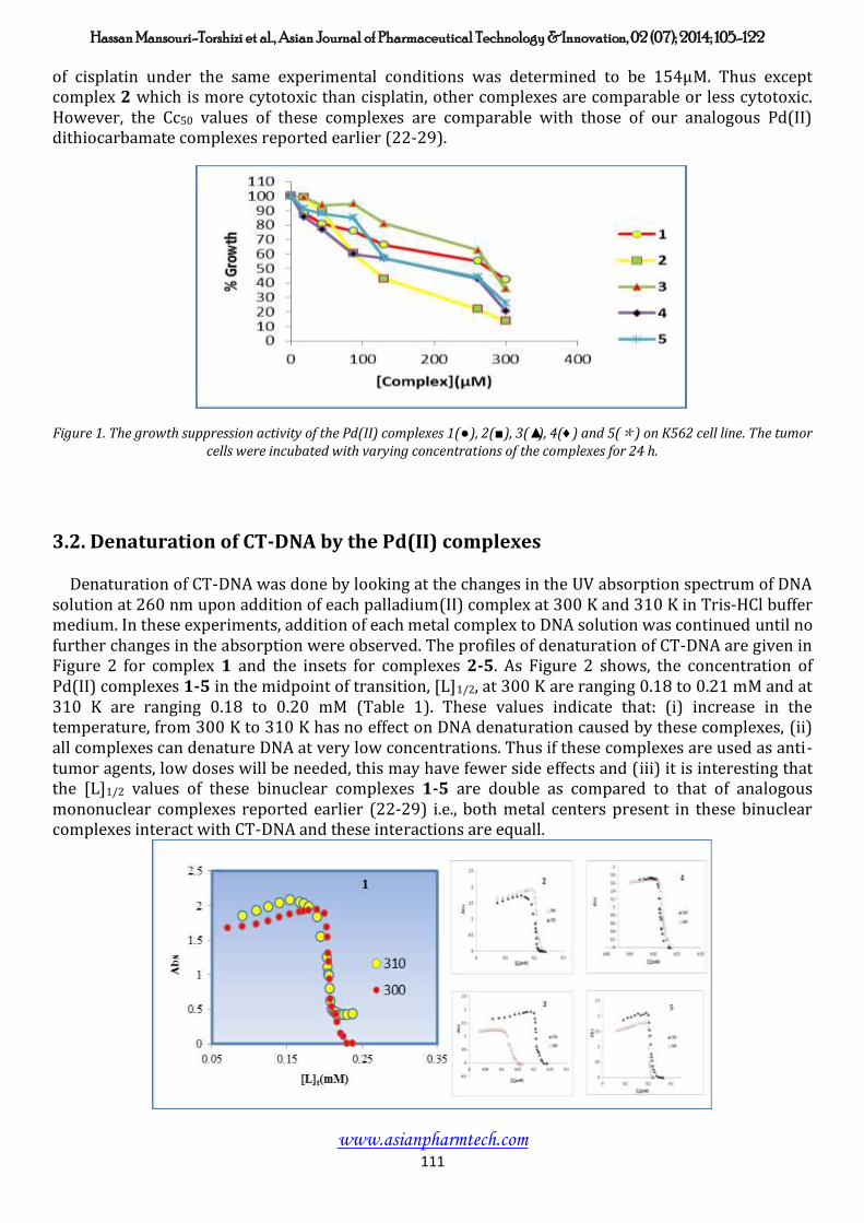
Figure 10. Florescence emission spectra of interacted CT-DNA-EB in the absence (1) and presence of different concentration of palladium(II) complexes: 30µM(2), 50µM(3), 70µM(4), 90µM(5), 110µM(6), 130µM(7) and EB alone(8).
Therefore, the binding of the complexes in title were evaluated using competitive binding studies involving ethidium bromide. Figure 10 showed the emission spectra of DNA-EB system with increasing amounts of the Pd(II) complexes. The addition of each complex to CT-DNA pretreated with ethidium bromide cause appreciable reduction in the emission intensity, indicating the replacement of ethidium bromide by the complexes (36).
Further studies to characterize and proof the mode of binding of Pd(II) complexes to CT-DNA were carried out. The number of ethidium bromide molecules intercalated to CT-DNA in presence of different concentrations of the Pd(II) complexes was calculated using Scatchard analysis (49). In this experiment, the wavelengths of excitation and emission were set at 540 nm and 700 nm respectively. Both have 0.5 nm slit widths. Solutions of CT-DNA, ethidium bromide and metal complexes were prepared in Tris-HCl buffer of pH 7.0. In this medium solutions of Pd(II) complexes were interacted with CT-DNA by incubating them at 300 K and 310 K for 5 h, appropriate amount of ethidium bromide was then added to them and further incubated at room temperature for 2 h and finally processed for fluorescence spectral measurement. Saturation curves of fluorescence intensity for the complexes 1-5 with DNA at different values (0.1, 0.23 and 0.40) were obtained in the presence of increased concentrations of ethidium bromide (51, 102, 153, 204, 255,306, 357, 408, 459 and 510 µM ). The fluorescence Scatchard plots obtained for binding of ethidium bromide to CT-DNA in absence (■) and presence (□, ○, ▲) of various concentrations of Pd(II) complex 1 and insets of complexes 2, 3, 4 and 5 are shown in Figure 11. Consequently, it might be concluded that the Pd(II) complexes inhibit competitively the ethidium bromide binding to DNA (type-A behavior) (49,50), where number of intercalated molecules per 1000 nucleotide, (intercept on the abscissa) remain constant and the slope of the graphs that is Kapp (apparent association constant) decrease in presence of increasing amount of complexes. The Kapp and n (number of binding sites per nucleotides) values for the Pd(II) complexes are listed in Table 3. These data suggested that the intercalation affinities of the binuclear Pd(II) complexes with CT-DNA is stronger than that of analogous mononuclear complexes reported earlier (22-29).

Hassan Mansouri-Torshizi et al., Asian Journal of Pharmaceutical Technology &Innovation, 02 (07); 2014; 105-122
www.asianpharmtech.com 119
Figure 11. Competition between complexes 1-5 with ethidium bromide for the binding sites of CT- DNA (Scatchard plot). In curve no. 1, Scatchard's plot was obtained with calf thymus DNA alone that its concentration was 60 µM. In curves nos. 2, 3 and 4 respectively, 30, 70 and 120 µM of complexes 1-5 were added, corresponding to molar ratio [complex]/[DNA] of 0.1, 0.23 and 0.40. Solution were made in 20 mM NaCl, 20 mM Tris-HCl (pH 7.0). Experiments were done at room temperature.
4. Conclusion
A series of five binuclear Pd(II) complexes formed by bridging two "2,2'-bipyridine palladium(II)" via alkylenebisdithiocarbamates were prepared and characterized by analytical and spectral techniques. In vitro cytotoxic results suggested that complexes 1-5 show growth inhibitory activity against K562. Detailed interaction studies of all complexes with calf thymus DNA were carried out. They cooperatively and intercalatively bind to CT-DNA at very low concentrations. However, at higher concentration, they denature CT-DNA. Determination of several binding and thermodynamic–parameters has also been attempted. Comparison of these data revealed that the CT-DNA binding affinity of binuclear complexes 1-5 almost doubls relative to mononuclear analogs reported earlier by us. The capability of cleavage of CT-DNA by these complexes was investigated by gel filtration, it indicates that only complexes 1 and 4 can break the CT-DNA into two fragments and their binding with both is strong enough not to break readily.
Acknowledgments Financial assistance from the Research Council of University of Sistan and Baluchestan and of University of Tehran is gratefully acknowledged.

Hassan Mansouri-Torshizi et al., Asian Journal of Pharmaceutical Technology &Innovation, 02 (07); 2014; 105-122
www.asianpharmtech.com 120
References
1. Metcalfe C, Thomas JA. Kinetically inert transition metal complexes that reversibly bind to DNA. Chem. Soc. Rev. 2003; 32: 215-24. 2. Liu XW, Li J, Li H, Zheng KC, Chao H, Ji LN. Synthesis, characterization, DNA-binding and photocleavage of [Ru (phen)2 (6-OH-dppz)]+2 and [Ru (phen)2 (6-NO2 dppz)]+2. J. Inorg. Biochem. 2005; 99: 2372- 80. 3. Gay M, Montana AM, Moreno V, Prieto MJ, Perez JM, Alonso C. Studies of interaction of tri chloro 2-cis-N,N dimethyl-ammonium methyl-cyclohexene-1-yl methylammonium platinum(II) chloride with DNA: Effects on secondary and tertiary structures of DNA cytotoxicassays on human ovarian cancer cell lines, resistant and non- resistant to cisplatin. BioorgMedic. Chem. 2006; 14: 1565-72. 4. Gao E, Zhu M, Yin H, Liu L, Wu Q, Sun Y. Synthesis, characterization, interaction with DNA and cytotoxicity in vitro of dinuclear Pd(II) and Pt(II) complexes dibridged by 2,2 azanediyldibenzoic acid. J. Inorg. Biochem. 2008; 102: 1958-64. 5. Liu HK, Sadler J. Metal complexes as DNA intercalators. Accounts. Chem. Res. 2011; 44: 349 -59. 6. Gao E, Wang L, Zhu M, Liu L, Zhang W. Synthesis, characterization, interaction with DNA and cytotoxicity in vitro of the complexes [M(dmphen)(CO3)]H2O [M=Pt(II) and Pd(II)]. Eur. J. Med. Chem. 2010; 45: 311-16. 7. Mota AJ, Dedieu A, Kuhn P, Matt D, Welter R, Neuburger M. Can weak interactions modify the binding properties of a strong nitrogen donor? Unusual N-coordination of a phosphoranylidene-substituted pyrazolone unit towardspalladium(II) centres: an experimental and theoretical study. Dalton. Trans. 2005; 3155-60. 8. Blight BA, Van Noortwyk KA, Wisner JA, Jennings MC. Pseudorotaxanes through Second-Sphere Coordination. Angew. Chem. 2005; 117: 1523-28. 9. Qu Y, Harris A, Hegmans A, Petz A, Kabolizadeh P, Penazova H, et al. Synthesis and DNA conformational changes of non-covalent polynuclear platinum complexes. J. Inorg. Biochem. 2004; 98: 1591. 10. Grant Collins J, Weate NJ. Potential adenine and minor groove binding platinum complexes. J. Inorg. Biochem. 2004; 98: 1578. 11. Trevisan A, Marzano C, Cristofori P, Borella Venturini M, Giovagnini L, Fregona D. Synthesis of a palladium(II)-dithiocarbamate-complex. biological assay and nephrotoxicity in rats. Arch. Toxicol. 2002; 76: 262-68. 12. Schuhmann E, Altman J, Karaghiosoff K, Beck W. Bis[platinum(II)] and Bis[palladium(II)] Complexes of α, -Dicarboxylic Acid Bis(1,2,4 triaminobutane- N4 Amides. Inorg. Chem. 1995; 34: 2316. 13. Krause-Heuer M, Grant MP, Orkey N, Aldrich-Wright JR. Drug delivery devices and targeting agents for platinum(II) anticancer complexes. Aust. J. Chem. 2008; 61: 675-81. 14. Billecke C, Finniss S, Tahash L, Miller C, Mikkelsen T, Farrell NP, Bogler O. Polynuclear platinum anticancer drugs are more potent than cisplatin and inducecell cycle arrest in glioma. Neuro-oncol. 2006; 8: 215–26. 15. Reedijk J. Why Does Cisplatin Reach Guanine-N7 with Competing S-Donor Ligands Available in the Cell?. Chem. Rev. 1999; 99: 2499. 16. Galbraith JA, Menzel KA, Ratilla MA, Kostic NM. Study of Stereodynamics by Variable-Temperature 195 Pt NMR Spectroscopy. Diastereomerism in Platinum(II) Thioether Complexes and Solvent Effects. Inorg. Chem. 1987; 26: 2073. 17. Van der Vijgh WJF. Clinical pharmacology of carboplatin. Clin. Pharmacokin. 1991; 21: 242-61. 18. Mital R, Jain N, Srivastava TS. Synthesis, characterization and cytotoxic studies of diamine and diimine palladium(II) complexes of diethyldithiocarbamate and binding of these and analogous platinum(II) complexes with DNA. Inorg. Chim. Acta. 1989; 166: 135-40. 19. Quiroga AG, Perez JM, Lopez-Solera I, Montero EI, Masagure JR, Alonso C, et al. Binuclear chloro-bridged palladated and platinated complexes derived from p isoprpylbenzaldehyde thiosemicarbazone with cytotoxicity against cisplatin resistant tumor cell lines. J. Inorg. Biochem. 1998; 69: 275-81. 20. Quiroga AG, Perez JM, Montero EI, West DX, Alonso C, Navarro- Ranninger C. Synthesis and characterization of Pd(II) and Pt(II) complexes of p isopropylbenzaldehyde N-protected thiosemicarbazones. Cytotoxic activity against ras-transformed cells. J. Inorg. Biochem. 1999; 75: 293-301. 21. Nikolis N, Methenitis C, Pneumatikakis G. Studies on the intraction of aritromycin B and its platinum(II) and palladium(II) metal complexes with calf thymus DNA and nucleotides. J. Inorg. Biochem. 2003; 95: 177-93. 22. Mansouri-Torshizi H, Moghaddam ME, Divsalar A, Saboury AA. 2,2' Bipyridinebutyldithiocarbamato platinum(II) and palladium(II) complexes: Synthesis, Characterization, Cytotoxicity and DNA-binding studies. Bioorg. Med. Chem. 2008; 16: 9616-25. 23. Mansouri-Torshizi H, Moghaddam ME, Divsalar A, Saboury AA. Diimine platinum(II) and palladium(II) complexes of dithiocarbamate derivative as potential antitumor agents: Synthesis, Characterization, Cytotoxicity and detail DNA-binding studies. J. Biomol. Struct. Dyn. 2009; 26: 575-86. 24. Mansouri-Torshizi H, Saeidifar M, Divsalar A, Saboury AA. Interaction studies between a 1,10-phenanthroline adduct of palladium(II) dithiocarbamate anti-tumor complex and calf thymus DNA: A synthesis, spectral and in vitro study. Spectrochim. Acta A. 2010; 77: 312-18. 25. Mansouri-Torshizi H, Saeidifar A, Saboury AA, Shahraki S. Interaction Studies of a Novel, Water-Soluble and Anti-Cancer Palladium(II) Complex with Calf Thymus DNA. B. Kor. Chem. Soc. 2010; 31: 435-41. 26. Mansouri-Torshizi H, Saeidifar M, Khosravi F, Divsalar A, Saboury AA, Hassani F. DNA Binding and Antitumor Activity of α-Diimineplatinum(II) and Palladium(II) Dithiocarbamate Complexes. Bioinorg. Chem. App. 2011; 1-11.
Hassan Mansouri-Torshizi et al., Asian Journal of Pharmaceutical Technology &Innovation, 02 (07); 2014; 105-122
www.asianpharmtech.com 121
27. Mansouri-Torshizi H, Saeidifar M, Divsalar A, Saboury A.A. Study on Interaction of DNA from Calf Thymus with 1,10- phenanthrolinehexyldithiocarbamato palladium(II) Nitrate as potential Antitumor Agent. J. Biomol. Struct. Dyn. 2011; 28: 805-14. 28. Eslami-Moghaddam M, Mansouri-Torshizi H, Divsalar A, Saboury AA. Synthesis, Characterization, Cytotoxic and DNA binding studies of diimine platinum(II) and palladium(II) complexes of short hydrocarbon chain ethyldithiocarbamate ligand, J. Iran. Chem. Soc., 2009, 6, 552-569. 29. Saeidifar M, Mansouri-Torshizi H, Behbehani GR, Saboury AA, Divsalar A. Thermodynamic study of new designed complex of ethlendiamine 8- hydroxyquinolinato palladium(II) chloride with calf thymus DNA. B. Kor. Chem. Soc. 2009; 30: 1951-55. 30. Palocsay A, Rund JV. Reaction between 1,10 phenanthroline and platinum(II) compounds: Reaction in aqueose solution. Inorg. Chem. 1969; 8: 524-28. 31. Manav N, Mishra AK, Kaushik NK. In Vitro antitumor and antibacterial studies of some Pt(IV) dithiocarbamate complexes. Spectrochim. Acta part A. 2006; 65: 32-35. 32. Grzegorz R, Edmund K, Waldmar N, Marek K, Tadeusz L. Chiral dioxovanadium(V) Schiff base complexes of 1,2-diphenyl-1,2-diaminoethane and aromatic o hydroxyaldehydes: Synthesis, Characterization, Catalytic properties and structure. Polyhedron. 2008; 27: 1601-09. 33. Goran NK, Vesna MD, Sreko RT. Synthesis and Characterization of tris(butyl-1-methyl-3-phenyl-propyl)-dithiocarbamato cobalt(III) seskvitoluene. J. Serb. Chem. Soc. 2002; 67: 123-26. 34. Kumar L, Kandasamy NR, Srivastava TS, Amonkar AJ, Adwankar M.K, Chitnis M.P. Synthesis and spectroscopic studies of potential anti-cancer [platinum(II)(2,2' bipyridine) (amino acid)+n (n=1 or 2)]complexes. J. Inorg. Biochem. 1985; 23: 1-11. 35. Kyoko N, Sumitaka I. Piezochromism and Related Phenomena Exhibited by Palladium Complexes. Platin. Met. Rev. 2004; 48: 117-24. 36. Jain N, Paul AK, Srivastava TS. Synthesis, characterization, cytotoxicity and DNA binding studies of diammineethyldithiocarbamato-platinum(II) nitrate. J. Inorg. Biochem. 1992; 45: 123-27. 37. Divsalar A, Saboury AA, Moosavi-Movahedi AA. Binding properties of a new anti-tumor component (2,2'-bipyridin octylglycinato Pd(II) nitrate) with bovine beta lactoglobulin-A and –B. J. Biomol. Struct. Dyn. 2007; 25: 173-82. 38. King AMQ, Nicholson H. The intraction of alflatoxin B1 with poly nucleotides and its effect on ribonucleic acid polymerase. Biochem. J. 1969; 114: 679-87. 39. Saboury AA. Areview on the ligand binding studies by isothermal titration calorimetry. J. Iran. Chem. Soc. 2006; 3: 1-21. 40. Bathaie SZ, Bolhasani A, Hoshyar R, Ranjbar B, Sabouni F, Moosavi- Movahedi AA. Interaction of saffron carotenoids as anticancer compounds with CT-DNA, oligo(dG.dC)15, and oligo (dA.dT)15. DNA Cell Biol. 2007; 26: 533-40. 41. Saboury AA, Shamsaei AA, Moosavi-Movahedi AA, Mansouri-Torshizi H. Thermodynamics of binding 2,2 bipyridineglycinato palladium(II) chloride on human serum albumin. J. Chin. Chem. Soc. 1999; 46: 917-22. 42. Scatchard G. The attractions of proteins for small molecules and ions. Ann N Y Acad Sci. 1949; 51: 660-72. 43. Peng B, Du X, Chen K, Ji H., Chao L. Synthesis, characterization and DNA binding studies of ruthenium(II) mixed-ligand complexes containing dipyrido[1,2,5]oxadiazolo [3,4-b]quinoxaline. Spectrochim. Acta A. 2009; 74: 896-901. 44. Hill AV. The possible effects of the aggregation of the molecules of haemoglobin on its dissociation curves. J. Physil (Lond). 1910; 40: 4-7. 45. Barrow GM. Physical Chemistry: Chap.7, 5rd ed. M.C Graw-Hill: New York. 1998. 46. Olmsted DR, Kearns. Mechanism of ethidium bromide fluorescence enhancement on binding to nucleic acids. Biochemistry-us. 1977; 16: 3647-54. 47. Baguley BC, Lebret M. Quenching of DNA-ethidium fluorescence by amsacrine and other antitumor agents: a possible electron-transfer effect. Biochemistry-us. 1984; 23: 937-43. 48. Divsalar A, Saboury AA, Mansoori-Torshizi H, Eslami-Moghaddam M, Ahmad F, Hakimelahi GH. Comparative Studies on the Interaction Between Bovine ß-lacto-globulin Type A and B and a New Designed Pd(II) Complex with Anti-tumor Activity at Different Temperatures. J. Biomol. Struct. Dyn. 2009; 26: 587-97. 49. Patel MN, Parmar PA, Gandhi DS. Squarepyramidal copper(II) complexes with fourth generation fluoroquinolone and neutral bidentate ligand: structure, antibacterial, SOD mimic and DNA-interaction studies. Bioorg. Med. Chem. 2010; 18: 1227-35. 50. Howe-Grant M, Wu KC, Bauer WR, Lippard SJ. Binding of platinum and palladium metallointercalation reagents and antitumor drugs to closed and open DNAs. Biochemistry-us. 1976; 15: 4339-46.
Hassan Mansouri-Torshizi et al., Asian Journal of Pharmaceutical Technology &Innovation, 02 (07); 2014; 105-122
www.asianpharmtech.com 122
Abbreviations
Nuclear magnetic resonance NMR
Fourier transform infrared FT-IR
Ultra Violet-visible UV-vis
Deoxyribonucleic acid DNA
Calf thymus DNA CT-DNA
50% cytotoxic concentration of compound Cc50
Ethidium bromide EB
Molecular Weight M.W.
2,2'-bipyridine bpy
Tris-HCl buffer containing 10 mM sodium chloride (pH 7.0) Tris-buffer
Ethylenebisdithiocarbamate en-bis-dtc
Propylenebisdithiocarbamate pn-bis-dtc
Butylenebisdithiocarbamate bu-bis-dtc
Hexylenebisdithiocarbamate he-bis-dtc
Octylenebisdithiocarbamate oc-bis-dtc