The Cytotoxicity, DNA Fragmentation, and Decreasing ...
27
Page 1/27 The Cytotoxicity, DNA Fragmentation, and Decreasing Velocity Induced by Chromium (III) Oxide on Rainbow Trout Spermatozoa Mustafa Erkan Özgür ( [email protected] ) Malatya Turgut Ozal University, Department of Aquaculture, Faculty of Fishery https://orcid.org/0000- 0002-2966-9627 Ahmet Ulu İnönü Üniversitesi: Inonu Universitesi Canbolat Gürses Inonu University: Inonu Universitesi İ mren Özcan Inonu University: Inonu Universitesi Samir Abbas Ali Noma Inonu University: Inonu Universitesi Süleyman Köytepe Inonu University: Inonu Universitesi Burhan Ate ş Inonu University: Inonu Universitesi Research Article Keywords: Cr2O3 , Cytotoxicity , Oncorhynchus mykiss , Spermatozoa , DNA damages Posted Date: May 24th, 2021 DOI: https://doi.org/10.21203/rs.3.rs-497860/v1 License: This work is licensed under a Creative Commons Attribution 4.0 International License. Read Full License Version of Record: A version of this preprint was published at Biological Trace Element Research on April 4th, 2022. See the published version at https://doi.org/10.1007/s12011-022-03211-9.
-
Upload
khangminh22 -
Category
Documents
-
view
4 -
download
0
Transcript of The Cytotoxicity, DNA Fragmentation, and Decreasing ...

Page 1/27
The Cytotoxicity, DNA Fragmentation, andDecreasing Velocity Induced by Chromium (III)Oxide on Rainbow Trout SpermatozoaMustafa Erkan Özgür ( [email protected] )
Malatya Turgut Ozal University, Department of Aquaculture, Faculty of Fishery https://orcid.org/0000-0002-2966-9627Ahmet Ulu İnönü Üniversitesi: Inonu Universitesi
Canbolat Gürses Inonu University: Inonu Universitesi
İmren Özcan Inonu University: Inonu Universitesi
Samir Abbas Ali Noma Inonu University: Inonu Universitesi
Süleyman Köytepe Inonu University: Inonu Universitesi
Burhan Ateş Inonu University: Inonu Universitesi
Research Article
Keywords: Cr2O3 , Cytotoxicity , Oncorhynchus mykiss , Spermatozoa , DNA damages
Posted Date: May 24th, 2021
DOI: https://doi.org/10.21203/rs.3.rs-497860/v1
License: This work is licensed under a Creative Commons Attribution 4.0 International License. Read Full License
Version of Record: A version of this preprint was published at Biological Trace Element Research on April4th, 2022. See the published version at https://doi.org/10.1007/s12011-022-03211-9.

Page 2/27
AbstractThe present study aimed to determine the cytotoxicity of Chromium (III) oxide micro particles (Cr2O3-Ps)in rainbow trout (Oncorhynchus mykiss) spermatozoa. Firstly, Cr2O3-Ps were synthesized and structurallycharacterized the surface, morphological for particle size and thermal properties. In addition, its structuraland elemental purity was determined using EDX spectrum and elemental maps. Structural purity, thermalproperties, and stability of Cr2O3-Ps were also examined in detail by performing thermal analysistechniques. The cytotoxicity of structurally de�ned Cr2O3-Ps was measured by the observation ofvelocities, antioxidant activities, and DNA damages in spermatozoa after exposure in vitro. The straightline velocity (VSL), the curvilinear velocity (VCL), and the angular path velocity (VAP) of spermatozoadecreased after exposure to Cr2O3-Ps. While the superoxide dismutase (SOD) and the catalase (CAT)decreased, the lipid peroxidation increased in a dose-dependent manner. However, the total glutathione(tGSH) did not affect in this period. DNA damages was also determined in spermatozoa using Cometassay. According to DNA in tail (%) data, DNA damages have been detected with gradually increasingconcentrations of Cr2O3-Ps. Furthermore, all of class types related to DNA fragmentation has beenobserved between 50 µg/L and 500 µg/L concentrations of Cr2O3-Ps exposed to rainbow troutspermatozoa. At the end of this study, we determined that the effective concentrations (EC50) were 76.67µg/L for VSL and 87.77 µg/L for VCL. Finally, these results about Cr2O3-Ps may say to be major riskconcentrations over 70 µg/L for �sh reproduction in aquatic environments.
IntroductionIn recently, although the growth rate in using metal oxide particles have been increased due to theircharacteristics such as huge speci�c surface areas, micro interface characteristics, and remediationability, they can have many potential environmental risks (Du et al. 2019; Zhu et al. 2019). Metal oxideshave been the focus of attention in many studies with their superior electrical properties, thermal stability,and extraordinary morphology (Chavali and Nikolova 2019). They also have an increasing use in manyindustries such as semiconductors, catalysts, solar panels, and UV protectors. Cr2O3 has a very importantplace among metal oxides, which have a very wide variety. Besides being used in many electronicapplications with approximately 3eV band spacing, it is used in industrial applications such as paint,pigment, catalyst, solar panels, wear resistant materials, thermal protection, electrochromic material, andhydrogen storage. This widespread use of Cr2O3 brings some environmental concerns.
Anthropogenic sources have many effects that can contaminate aquatic life and these have also toxicagents. However, many toxic contaminants pollute the water system. One of them, Chromium (Cr) is amajor metallic element posing a maximum threat to all animals and plants according to US-Environmental Protection Agency (US-EPA). The main sources of chromium entering the aquaticenvironment can reach from dyes, mining, electroplating, automobile, and textile manufacturing, metalprocessing and leather tanning (Canadian Water Quality Guidelines 1999; EPA 2015; Maitlo et al. 2019).However, while it is estimated that the production amounts of manufactured metal oxide nano or micro

Page 3/27
materials in 2020 are 1.663.168 tons (He et al. 2015), the production of Chromium in the world is 44.000gross weight in 2019 (U.S Geological Survey 2020).
In the modern industry, Chromium (III) oxide particles (Cr2O3-Ps) also started to use in pigments forceramics, dyes, paints, and cosmetics (Puerari et al. 2016). But unfortunately, there is no moreinformation about the toxicological characteristics of this micro material on reproduction system of �sh.The US Environmental Protection Agency (EPA) has set a maximum concentration level (MCL) of 100µg/L for chromium discharged into waters, because it is very toxic (EPA 2015). Due to the data that evenvery small amounts of chromium can be toxic, it was become a hot topic to investigate the relationshipswith water environments for this study.
In a broader context, there are not more studies that addressed the toxicity of Cr2O3-Ps in the literature. Inthe light of the literature, if we give some examples, researchers studied on the lung cells (BEAS-2B) andbronchoalveolar carcinoma-derived cells (A549) of human (Cho et al. 2013; Chusuei et al. 2013), bacteriain in activated sludge system (Wang et al. 2017), green alga (Costa et al. 2015), freshwater microcrustacean Daphnia magna and marine bacterium Aliivibrio �scheri (Puerari et al. 2016), Daphnia smilis(Tavares et al. 2014), Escherichia coli (Kaweeteerawat et al. 2015), Wistar rats (Lebedev et al. 2018) andsoybean (Glycine max) (Li et al. 2018). In light of these literatures, we couldn’t also �nd any study aboutCr2O3-Ps toxicity on �sh or �sh reproduction. For both no many studies and there are more productiveand using of chromium, this study has been designed since it has become a need to conduct scienti�cresearch on Cr2O3-Ps. Thus, this study was carried out to determine the cytotoxicity of Cr2O3-Ps onreproduction of rainbow trout (Oncorhynchus mykiss) with in vitro assay. Because it is known that in vitromethods have helped to determine the toxicity levels of environmental pollutants, the mechanisms,exposure times, concentrations (Özgür et al. 2019). For this aim, target particles were exposed on thespermatozoa of rainbow trout and it was investigated the velocities, movement styles, oxidative stressindices, effective concentrations, and DNA damages of spermatozoa.
Materials And Methods
ChemicalsIn this study, the decomposition reaction of ammonium dichromate at high temperatures was used in thesynthesis of Cr2O3-Ps. Accordingly, all chemicals used during synthesis and puri�cation were obtainedfrom Sigma-Aldrich. These chemicals used were preferred in analytical purity and were used without anypuri�cation process.
InstrumentationThe chemical structures of the obtained Cr2O3-Ps were measured in the range of 400–4000 cm− 1 usingPerkin Elmer Spectrum Two model FTIR device. Crystal property and purity of Cr2O3-Ps were carried outusing Rigaku Rad B-Dmax II powder brand X-ray device and were measured in the range of 20–80 2-theta.

Page 4/27
Structural and morphological properties of Cr2O3-Ps were measured with the LEO EVO-40xVP model SEMdevice after coating with Au-Pd conductive coating using a Bal-tec brand spatter. Bruker brand RönteckX�ash EDX detector connected to SEM device was used for elemental veri�cation of Cr2O3-Ps structure.Particle size analyzes of the obtained Cr2O3 particles were carried out by using Malvern Zetasizer Nano-ZS model device by dispersing it in water. Shimadzu DTA-50 and Shimadzu TGA-50 devices were used todetermine the thermal stability of the synthesized Cr2O3-Ps, and the analyses were carried out between30-1000°C using 10°C/min heating rate in the air atmosphere and used about 10 mg sample in Pt samplepan.
Preparation and characterization of Cr2O3-PsCr2O3-Ps were prepared by thermal decomposition of ammonium dichromate in accordance with theliterature (Mahieu et al. 1971; Galwey et al. 1983). First, ammonium dichromate was mixed with ethanol.It was burned at a high temperature in a controlled manner. When the entire mixture turned green, theresulting Cr2O3 was washed with ethanol. It was dried in an oven at 110 ° C for 3 hours. It was groundand its chemical structure, morphology, and purity were checked.
Experimental designSemen samples were collected from rainbow trout (Oncorhynchus mykiss, Walbaum, 1792) males (1900 ± 150 g) which were supplied from a commercial farm in Malatya, Turkey in January 2020. Semensamples were obtained by massage method from the front to the back of the male �sh abdomen withoutanesthesia. The semen pool was taken from 7 individuals of �sh. Fresh semen samples in semen poolwere diluted with inhibition solution of spermatozoa motility or inactivation solution (IS) (NaCl, 103mmol/L; KCl, 40 mmol/L; CaCl2, 1 mmol/L; MgSO4, 0.8 mmol/L; Hepes, 20 mmol/L (Özgür et al., 2018);pH 7.8, adjusted with 1 N-NaOH) as a stock solution. The pooled sample was diluted with IS at ratio 1:10(Semen:IS) to obtain a spermatozoa density of about 12×108 cells/mL.
In vitro exposure of Cr2O3-Ps was carried out on a semen pool. The semen sub-samples were exposedwith nominal concentrations such as 10, 50, 100, 500, and 1000 µ/L of Cr2O3-Ps at 4°C for 3 h inEppendorf tubes with the lid closed. The exposure the concentrations of Cr2O3-Ps were determinedaccording to maximum concentration level of Chromium that is 100 µ/L in EPA (EPA 2015). Semen sub-samples exposed with Cr2O3-Ps at the ratio of 1:20 (Cr2O3-Ps: Semen with IS) in Eppendorf tubes.
The velocities and movement style of spermatozoaThe velocities and movement style parameters of spermatozoa were analyzed by the computer assistedsemen analysis system (CASA). The values of velocity parameters such as VSL (straight line velocity,µm/s), VCL (curvilinear velocity, µm/s), VAP (angular path velocity, µm/s) and the movement styleparameters of spermatozoa such as LIN (linearity, %, (VSL/VCL)*100), BCF (beat cross frequency, Hz),and ALH (amplitude of lateral displacement of the spermatozoa head, µm) (Özgür et. al., 2019) areexamined in the study. For CASA system, it was used model of BASA-Sperm Aqua which has an Olympus

Page 5/27
BX53 microscope (200x magni�cation) and a CCD camera (30 fbs) by Merk Biotechnology Ltd. Co. inTurkey. In the analyzing under the microscope, the spermatozoa in semen were activated to investigatekinematic parameters with activation solution (AS) (CaCl2 1mM; Tris 20 mM, Glycine 30 mM, NaCl 125mM; pH 9) (Özgür et al., 2018) at ratio 1:20 (Semen:AS) under a microscope. The �nal dilution rate was2000 times.
Oxidative stress indices of spermatozoaThe sperm samples were collected in a plastic tube to prepare homogenates and 500 uL PBS (50 mM, pH7.4) was added to each tube. The homogenization process was occurred in cold media, each sample wassoni�ed 6 cycles between each cycle 15 seconds with an ultrasoni�er. Then, the homogenates werecentrifuged at 10.000 rpm for 10 min at 4°C, supernatants were separated from pellets, and the pelletswere stored for further analysis. In the beginning, to estimate the protein level in the supernatants, theBradford method was take-place and bovine serum albumin (BSA) was used as a standard (Bradford1976). After that, antioxidant enzymes were assayed. For instance, CAT activity was measured byfollowing the decrease in the absorbance of hydrogen peroxide (H2O2) at 240 nm at room temperature(Aebi 1984). One unit of CAT represents the amount of enzyme that hydrolysis 1 µmol of H2O2 perminute. SOD activity was measured by using the xanthine oxidase/cytochrome C method reported byMcCord and Fridovich (McCord and Fridovich 1969). One unit of SOD activity is the amount of theenzyme required to cause a half-maximal inhibition of cytochrome C reduction. Besides, tGSH wasobtained spectrophotometrically by using the reported previous method (Akerboom and Sies 1981). Thecolorimetric assay was followed the increase in the formation of 5-thio-2-nitrobenzoate (TNB) which ismeasured spectrophotometrically at 412 nm. Malondialdehyde (MDA) is a biomarker for lipidperoxidation. The MDA level of samples was assayed via the method described by Buege and Aust(Buege and Aust 1978). CAT and SOD levels were expressed as U/mg protein, while the MDA and tGSHresults were presented as nmol/mg protein.
The analysis of DNA fragmentation in spermatozoa usingComet assayThe comet assay was performed under alkaline condition. During the experiment, a version adapted tospermatozoa in study of Singh et al. (1988) was utilized (Singh et al. 1988). Previously chilledmicroscope slides were dipped into 0.7% extremely hot (approximately 70°C) normal melting agaroseprepared in phosphate buffered saline (PBS) solution. Afterwards, 10 µL (~ 100,000 cells) of previouslydiluted spermatozoa was mixed with 90 µL low melting point agarose (LMA, prepared as 0.5% in PBS)and then, 70 µL of the mixture was added onto the prepared normal melting agarose covered microscopeslides as a bottom layer. The slides including negative control (just spermatozoa) and positive control(spermatozoa treated with 35% of hydrogen peroxide) were put on a frozen �at tray for 10 minutes tosolidify. Before using ice-cold lysis solution (2.5 M NaCl, 100 mM EDTA, 10 mM Tris base, pH 10), 1%Triton X-100 and 10% DMSO were added into the solution. Prepared slides were then cautiouslyimmersed in lysis solution for at least 1 hour at 4°C. Slides were then removed from the lysis buffer and

Page 6/27
placed in an electrophoresis tank. The electrophoresis tank was �lled with freshly preparedelectrophoresis buffer (0.3 M NaOH, 1 mM EDTA, pH 13). Before electrophoresis, the slides were left in theelectrophoresis solution for 30 minutes to allow unwinding of DNA. After this procedure, electrophoresissystem was turned on for 30 min at 300 mA and 15 V. After electrophoresis, slides were taken from thehorizontal tank and washed three times with 0.4 M Tris buffer, pH 7.5 for 5 minutes to neutralize the alkalicondition. For each slide, 60 µL of ethidium bromide (EtBr concentration: 20 mg/mL) was pipetted intothe sample. Then, slides were covered with a coverslip and taken images using the inverted microscope(Olympus CKX41) with a combined �uorescence system at 40X magni�cation with Olympus cellSensEntry 2.1 software. Comets are formed upon the principle of releasing damaged DNA from the core of thenucleus during electrophoresis. In terms of tail length, tail & head intensities, tail & head DNA percentages,100 cells per sample (two duplicate sample slides, 50 randomly selected cells scored per slide) werescored using image analysis software CometScore 2.0. Totally, 100 cells were scored per slide induplicate. Analysis was performed blindly by one slide reader. DNA fragmentation/damage scoring isclassi�ed based on the size of the comet tail: class 0, undamaged (intact) DNA; 1, 2, 3, and 4 classes ofdamages meaning from the less to the most fragmented cells.
Statistics analysisNormality test and descriptive analysis (Means ± SE, p < 0.05) were performed between the data in theSPSS 15 program. The differences between groups were done by Variance Analysis (one-way ANOVA)with the Tukey test after the Test of Homogeneity of Variance in each group. Graph Pad Prism 5 fordrawing graphics was used.
For DNA damages/fragmentation analysis in spermatozoa using Comet assay, the compliance of data tonormal distribution was examined with Kolmogorov-Smirnov test. The data were summarized withmedian, minimum, and maximum values. Since all groups did not conform to normal distribution,Kruskal-Wallis test and then Conover pairwise comparison method was used for comparisons. Thesigni�cance level was accepted as 0.05 and 0.001 tails and head of spermatozoa. Moreover, abiostatistics web application was also used for the boxplot graphs (Arslan et. al., 2018).
Results
Characterization of Cr2O3-PsThe Cr2O3-Ps used in the study were primarily characterized by using FTIR and X-ray spectrum. Basicmorphological features and basic Cr2O3 surface analysis were examined by SEM analysis. Its structuralpurity and elemental map were measured by EDX analysis. Thermal properties of the synthesized Cr2O3-Ps were determined by TGA and DTA thermograms.
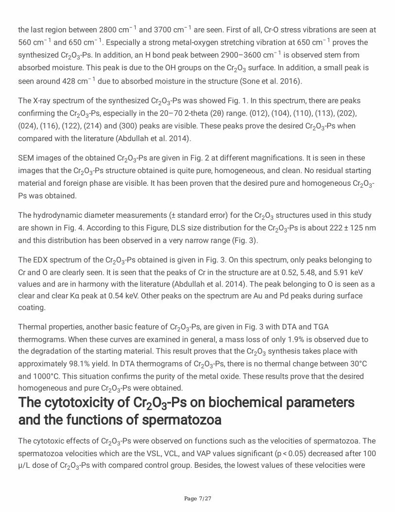
FTIR spectrum of Cr2O3-Ps is given in Fig. 1. Three main regions in the FTIR spectrum of Cr2O3-Ps are
clearly seen. The �rst region between 400–800 cm− 1, the second region between 2000–2800 cm− 1, and
Page 7/27
the last region between 2800 cm− 1 and 3700 cm− 1 are seen. First of all, Cr-O stress vibrations are seen at560 cm− 1 and 650 cm− 1. Especially a strong metal-oxygen stretching vibration at 650 cm− 1 proves thesynthesized Cr2O3-Ps. In addition, an H bond peak between 2900–3600 cm− 1 is observed stem fromabsorbed moisture. This peak is due to the OH groups on the Cr2O3 surface. In addition, a small peak is
seen around 428 cm− 1 due to absorbed moisture in the structure (Sone et al. 2016).
The X-ray spectrum of the synthesized Cr2O3-Ps was showed Fig. 1. In this spectrum, there are peakscon�rming the Cr2O3-Ps, especially in the 20–70 2-theta (2θ) range. (012), (104), (110), (113), (202),(024), (116), (122), (214) and (300) peaks are visible. These peaks prove the desired Cr2O3-Ps whencompared with the literature (Abdullah et al. 2014).
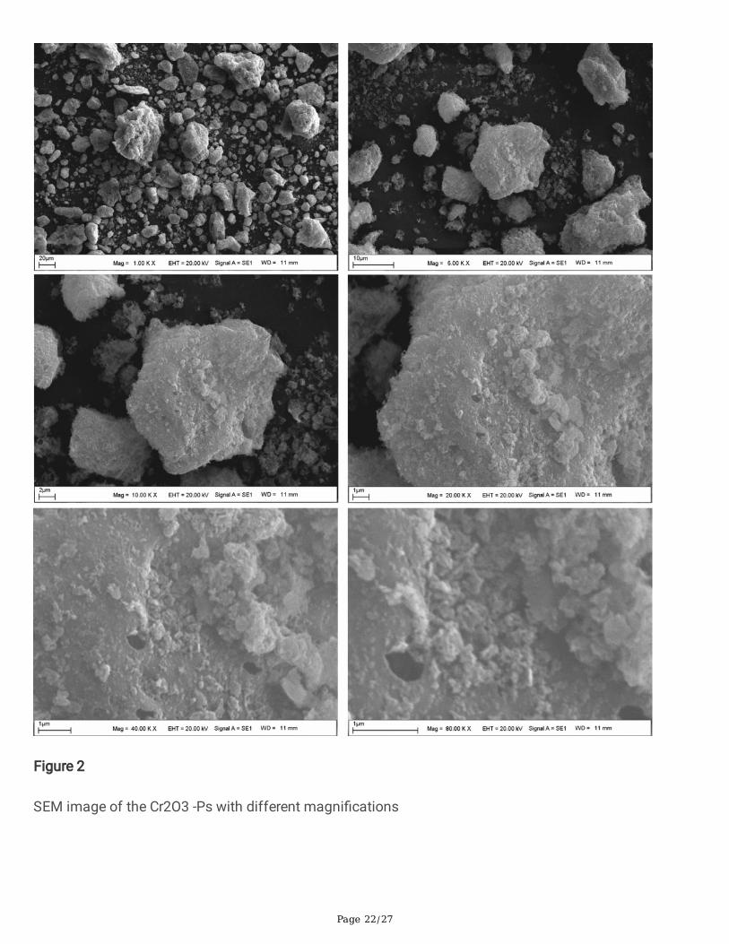
SEM images of the obtained Cr2O3-Ps are given in Fig. 2 at different magni�cations. It is seen in theseimages that the Cr2O3-Ps structure obtained is quite pure, homogeneous, and clean. No residual startingmaterial and foreign phase are visible. It has been proven that the desired pure and homogeneous Cr2O3-Ps was obtained.
The hydrodynamic diameter measurements (± standard error) for the Cr2O3 structures used in this studyare shown in Fig. 4. According to this Figure, DLS size distribution for the Cr2O3-Ps is about 222 ± 125 nmand this distribution has been observed in a very narrow range (Fig. 3).
The EDX spectrum of the Cr2O3-Ps obtained is given in Fig. 3. On this spectrum, only peaks belonging toCr and O are clearly seen. It is seen that the peaks of Cr in the structure are at 0.52, 5.48, and 5.91 keVvalues and are in harmony with the literature (Abdullah et al. 2014). The peak belonging to O is seen as aclear and clear Kα peak at 0.54 keV. Other peaks on the spectrum are Au and Pd peaks during surfacecoating.
Thermal properties, another basic feature of Cr2O3-Ps, are given in Fig. 3 with DTA and TGAthermograms. When these curves are examined in general, a mass loss of only 1.9% is observed due tothe degradation of the starting material. This result proves that the Cr2O3 synthesis takes place withapproximately 98.1% yield. In DTA thermograms of Cr2O3-Ps, there is no thermal change between 30°Cand 1000°C. This situation con�rms the purity of the metal oxide. These results prove that the desiredhomogeneous and pure Cr2O3-Ps were obtained.
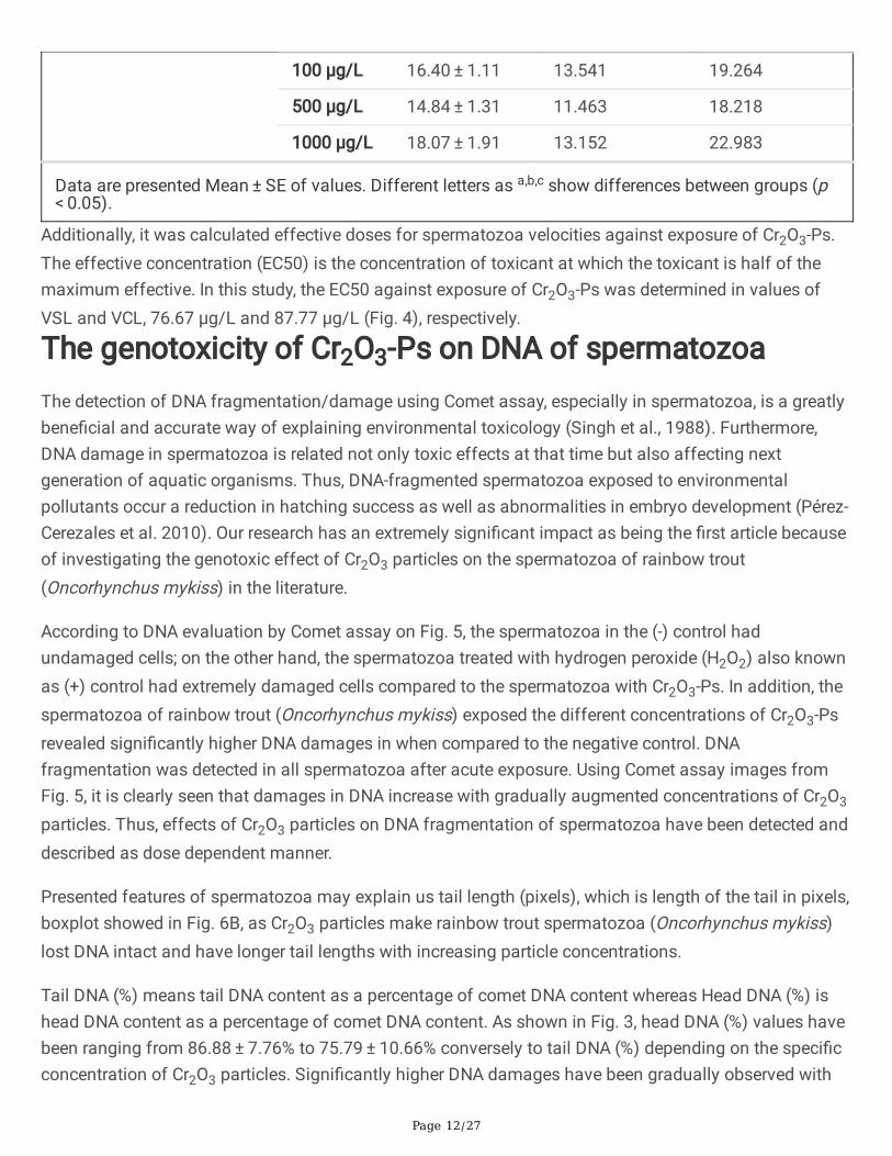
The cytotoxicity of Cr2O3-Ps on biochemical parametersand the functions of spermatozoaThe cytotoxic effects of Cr2O3-Ps were observed on functions such as the velocities of spermatozoa. Thespermatozoa velocities which are the VSL, VCL, and VAP values signi�cant (p < 0.05) decreased after 100µ/L dose of Cr2O3-Ps with compared control group. Besides, the lowest values of these velocities were

Page 8/27
observed at the dose of 1000 µg/L. however, after Cr2O3-Ps exposure, it was determined that there was nosigni�cant difference (p > 0.05) between 500 and 1000 µg/L in the velocities of spermatozoa.
According to our results, the cytotoxic effects as the functional deformations of spermatozoa were alsoinvestigated on the movement style of spermatozoa. For example, although the value of LIN decreased independent on 100 µ/L dose of Cr2O3-Ps, it increased at 500 or 1000 doses of Cr2O3-Ps. While there wassigni�cant (p < 0.05) difference between these doses, it was not determined statistically difference (p > 0.05) with compared control group. The value of ALH showed decreased with increasing doses of Cr2O3-Ps and its decreasing was signi�cant (p < 0.05) difference after 100 µ/L dose with compare controlgroup. The value of BCF and MAD values were statistically insigni�cant (p > 0.05) with up to Cr2O3-Psdoses according to control groups (Table 1, Fig. 4).

Page 9/27
Table 1The kinematic parameters of rainbow trout spermatozoa after exposed Cr2O3-Ps
Parameters N = 6 Mean ± S.E. 95% Con�dence Interval for Mean
Lower Bound Upper Bound
VSL (µm/s) Control 31.45 ± 1.17c 28.446 34.458
10 µg/L 28.73 ± 0.61c 27.170 30.292
50 µg/L 28.75 ± 0.69c 26.991 30.515
100 µg/L 16.57 ± 0.44b 15.439 17.705
500 µg/L 15.14 ± 0.81ab 13.051 17.231
1000 µg/L 12.02 ± 0.52ab 10.697 13.343
VCL (µm/s) Control 120.86 ± 4.11c 110.299 131.428
10 µg/L 106.41 ± 2.62c 99.683 113.142
50 µg/L 105.65 ± 2.26c 99.849 111.449
100 µg/L 71.28 ± 6.32b 55.028 87.529
500 µg/L 50.90 ± 2.05a 45.634 56.160
1000 µg/L 38.67 ± 2.22a 32.967 44.381
VAP (µm/s) Control 54.12 ± 7.02c 36.067 72.168
10 µg/L 48.56 ± 4.75bc 36.343 60.778
50 µg/L 46.23 ± 5.20bc 32.878 59.585
100 µg/L 32.10 ± 2.62ab 25.355 38.838
500 µg/L 30.74 ± 3.05ab 22.896 38.581
1000 µg/L 19.50 ± 1.73a 15.047 23.948
LIN (%) Control 26.06 ± 0.69ab 24.290 27.826
10 µg/L 24.86 ± 1.66ab 20.591 29.128
50 µg/L 26.02 ± 1.87ab 21.206 30.839
Data are presented Mean ± SE of values. Different letters as a,b,c show differences between groups (p < 0.05).

Page 10/27
100 µg/L 21.11 ± 2.06a 15.819 26.402
500 µg/L 29.97 ± 1.93b 25.006 34.942
1000 µg/L 31.48 ± 1.85b 26.733 36.217
BCF (Hz) Control 5.46 ± 0.75 3.545 7.377
10 µg/L 8.61 ± 1.43 4.938 12.279
50 µg/L 5.04 ± 1.20 1.952 8.135
100 µg/L 5.73 ± 1.21 2.614 8.853
500 µg/L 6.52 ± 1.38 2.980 10.050
1000 µg/L 5.63 ± 0.81 3.561 7.699
ALH (µm) Control 30.31 ± 3.85b 20.417 40.198
10 µg/L 21.61 ± 1.51ab 17.730 25.485
50 µg/L 22.42 ± 1.75ab 17.915 26.923
100 µg/L 19.45 ± 1.67a 15.172 23.730
500 µg/L 17.93 ± 1.16a 14.953 20.902
1000 µg/L 16.07 ± 1.24a 12.876 19.270
MAD ( 0 ) Control 0.04 ± 0.006 0.021 0.054
10 µg/L 0.03 ± 0.004 0.018 0.040
50 µg/L 0.04 ± 0.008 0.018 0.060
100 µg/L 0.03 ± 0.004 0.023 0.043
500 µg/L 0.03 ± 0.004 0.020 0.041
1000 µg/L 0.04 ± 0.005 0.022 0.048
Data are presented Mean ± SE of values. Different letters as a,b,c show differences between groups (p < 0.05).
According to our results, there was signi�cant (p < 0.05) decrease in the levels of CAT and SOD withincreasing concentrations of Cr2O3-Ps. However, the CAT activity decreased after 500 µg/L dose of Cr2O3-Ps, while the levels of SOD activity decreased after 100 µg/L dose of Cr2O3-Ps compare control group.Meanwhile, although the dose of 100 mg/L Cr2O3-Ps signi�cantly increased the MDA level compared tothe control group (p < 0.05), it was observed insigni�cant (p > 0.05) difference at tGSH levels according toall groups (Table 2, Fig. 4).

Page 11/27
Table 2Biochemical stress indices of rainbow trout spermatozoa after exposed Cr2O3-Ps
Parameters N = 6 Mean ± S.E. 95% Con�dence Interval for Mean
Lower Bound Upper Bound
CAT (U/mg protein) Control 20.44 ± 1.63b 16.248 24.634
10 µg/L 20.17 ± 1.74b 15.703 24.634
50 µg/L 18.26 ± 0.97ab 15.765 20.746
100 µg/L 17.94 ± 1.14ab 15.009 20.865
500 µg/L 13.48 ± 1.57a 9.441 17.519
1000 µg/L 12.51 ± 1.13a 9.606 15.422
SOD (U/mg protein) Control 6.99 ± 0.62b 5.393 8.583
10 µg/L 5.52 ± 0.18ab 5.051 5.985
50 µg/L 5.81 ± 0.33ab 4.957 6.654
100 µg/L 4.76 ± 0.25a 4.110 5.407
500 µg/L 4.43 ± 0.33a 3.575 5.275
1000 µg/L 4.30 ± 0.33a 3.445 5.162
MDA (nmol/mg protein) Control 1.43 ± 0.04a 1.318 1.535
10 µg/L 1.58 ± 0.10ab 1.328 1.835
50 µg/L 1.52 ± 0.04ab 1.415 1.631
100 µg/L 1.83 ± 0.11b 1.554 2.102
500 µg/L 1.60 ± 0.09ab 1.375 1.828
1000 µg/L 1.60 ± 0.12ab 1.306 1.897
TGSH (nmol/mg protein) Control 15.92 ± 1.14 12.994 18.846
10 µg/L 14.68 ± 0.98 12.157 17.207
50 µg/L 16.53 ± 1.88 11.694 21.369
Data are presented Mean ± SE of values. Different letters as a,b,c show differences between groups (p < 0.05).
Page 12/27
100 µg/L 16.40 ± 1.11 13.541 19.264
500 µg/L 14.84 ± 1.31 11.463 18.218
1000 µg/L 18.07 ± 1.91 13.152 22.983
Data are presented Mean ± SE of values. Different letters as a,b,c show differences between groups (p < 0.05).
Additionally, it was calculated effective doses for spermatozoa velocities against exposure of Cr2O3-Ps.The effective concentration (EC50) is the concentration of toxicant at which the toxicant is half of themaximum effective. In this study, the EC50 against exposure of Cr2O3-Ps was determined in values ofVSL and VCL, 76.67 µg/L and 87.77 µg/L (Fig. 4), respectively.
The genotoxicity of Cr2O3-Ps on DNA of spermatozoaThe detection of DNA fragmentation/damage using Comet assay, especially in spermatozoa, is a greatlybene�cial and accurate way of explaining environmental toxicology (Singh et al., 1988). Furthermore,DNA damage in spermatozoa is related not only toxic effects at that time but also affecting nextgeneration of aquatic organisms. Thus, DNA-fragmented spermatozoa exposed to environmentalpollutants occur a reduction in hatching success as well as abnormalities in embryo development (Pérez-Cerezales et al. 2010). Our research has an extremely signi�cant impact as being the �rst article becauseof investigating the genotoxic effect of Cr2O3 particles on the spermatozoa of rainbow trout(Oncorhynchus mykiss) in the literature.
According to DNA evaluation by Comet assay on Fig. 5, the spermatozoa in the (-) control hadundamaged cells; on the other hand, the spermatozoa treated with hydrogen peroxide (H2O2) also knownas (+) control had extremely damaged cells compared to the spermatozoa with Cr2O3-Ps. In addition, thespermatozoa of rainbow trout (Oncorhynchus mykiss) exposed the different concentrations of Cr2O3-Psrevealed signi�cantly higher DNA damages in when compared to the negative control. DNAfragmentation was detected in all spermatozoa after acute exposure. Using Comet assay images fromFig. 5, it is clearly seen that damages in DNA increase with gradually augmented concentrations of Cr2O3
particles. Thus, effects of Cr2O3 particles on DNA fragmentation of spermatozoa have been detected anddescribed as dose dependent manner.
Presented features of spermatozoa may explain us tail length (pixels), which is length of the tail in pixels,boxplot showed in Fig. 6B, as Cr2O3 particles make rainbow trout spermatozoa (Oncorhynchus mykiss)lost DNA intact and have longer tail lengths with increasing particle concentrations.
Tail DNA (%) means tail DNA content as a percentage of comet DNA content whereas Head DNA (%) ishead DNA content as a percentage of comet DNA content. As shown in Fig. 3, head DNA (%) values havebeen ranging from 86.88 ± 7.76% to 75.79 ± 10.66% conversely to tail DNA (%) depending on the speci�cconcentration of Cr2O3 particles. Signi�cantly higher DNA damages have been gradually observed with

Page 13/27
increasing concentrations of Cr2O3 particles from the data of tail DNA (%). In contrast to this data, thespermatozoa exposed to Cr2O3 particles exhibited lower DNA damages compared to cells treated withH2O2, (+) Control (Fig. 7).
The spermatozoa in the negative control had undamaged cells approximately 60% according to DNAevaluation by Comet assay from Fig. 8. The spermatozoa exposed to Cr2O3 particles showed signi�cantlyhigher DNA damages compared to the negative control. DNA damage was detected in all spermatozoa in50 µg/L concentration of Cr2O3 particles. Moreover, spermatozoa in Class 0 were not observed for Cr2O3
particles having 1000 µg/L concentration.
Values related to Tail Intensity (Pixels), Head Intensity (Pixels), Tail DNA (%), Head DNA (%), Tail length(Pixels), and Tail Moment, which is de�ned as Tail length (Pixels) x Tail DNA (%) in Table 3 and Table 4.
Table 3The comparisons between the percentages of DNA in Head (%), DNA in Tail (%), and Tail Moment using
Comet Assay of rainbow trout spermatozoa. The different concentrations of Cr2O3 -Ps, (-) and (+) controlsexpressed as Median (Min – Max)
Groups DNA in Head (%)
Median (Min – Max)
DNA in Tail (%)
Median (Min – Max)
Tail Moment
Median (Min – Max)
(-) Controla 82.99 (70.99–93.4)bcfg 17.01 (6.6–29.01)bcfg 0.3 (0.07–2.03)bg
(+) Control (H2O2)b 79.57 (7.69–97.22)acde 20.43 (0.28–92.31)acde 0.55 (0.06–23.68)acdf
10 µg/Lc 88.79 (67.44–99.52)abefg 11.21 (0.48–32.56)abefg 0.23 (0–2.3)befg
50 µg/Ld 85.57 (67.53–97.29)bfg 14.43 (2.71–32.47)bfg 0.22 (0.03–2.98)befg
100 µg/Le 83.71 (54.85–94.53)bcg 16.29 (5.47–45.15)bcg 0.4 (0.06–6.32)cdg
500 µg/Lf 79.18 (57.79–91.63)acd 20.82 (8.37–42.21)acd 0.38 (0.09–8.02)bcdg
1000 µg/Lg 78.73 (37.82–87.13)acde 21.27 (12.87–62.18)acde 0.6 (0.14–6.84)acdef
p < 0.001 < 0.001 < 0.001
Min, minimum; Max, maximum.
a: signi�cant from the negative control (a); b: signi�cant from the positive control (b); c: signi�cantfrom (c); d: signi�cant from (d); e: signi�cant from (e); f: signi�cant from (f); g: signi�cant from (g). Thevalues (n = 100) are Median (Min – Max) with common superscripts in the same line are signi�cantlydifferent.

Page 14/27
Table 4
The dose-dependent effects of Cr2O3-Ps on genotoxicity parameters of rainbow trout spermatozoa afteracute exposure
Parameters Head Intensity
(Pixels)
DNA inHead
(%)
Tail Intensity
(Pixels)
TailLength
(Pixels)
DNA inTail
(%)
TailMoment
(-) Controla 47183.44 ± 10921.84bcfg
83.72 ± 5.3bcfg
8814.06 ± 2519.2
2.34 ± 1.51bg
16.28 ± 5.3bcfg
0.42 ± 0.39bg
(+) Control(H2O2)b
28358.78 ± 18114.05acdefg
70.73 ± 21.89acde
11170.98 ± 14060.22cg
6.94 ± 8.6acdf
29.27 ± 22.06acde
3.12 ± 5.52acdf
10 µg/Lc 83807.06 ± 55963.65abdefg
86.88 ± 7.76abefg
10941.36 ± 5579.83bd
2.64 ± 1.95bg
13.12 ± 7.76abefg
0.37 ± 0.45befg
50 µg/Ld 44825.24 ± 16163.64bcfg
84.71 ± 7.72bfg
8038.12 ± 5027.89cg
2.68 ± 2.24bg
15.29 ± 7.72bfg
0.49 ± 0.62befg
100 µg/Le 47042.28 ± 20393.44bcfg
81.76 ± 8.28bcg
10035.4 ± 6086.71
3.68 ± 3.35
18.24 ± 8.28bcg
0.83 ± 1.25cdg
500 µg/Lf 38465.52 ± 18634.98abcde
78.59 ± 8.82acd
10449.36 ± 8936.16
3.48 ± 3.96bg
21.41 ± 8.82acd
0.84 ± 1.31bcdg
1000 µg/Lg 36698.08 ± 14178.36abcde
75.79 ± 10.66acde
11496.38 ± 7660.65bd
4.04 ± 2.89acdf
24.21 ± 10.66acde
1.13 ± 1.24acdef
The values (n = 100) are mean ± standard deviation with common superscripts in the same line aresigni�cantly different.
a: signi�cant from the negative control (a); b: signi�cant from the positive control (b); c: signi�cantfrom (c); d: signi�cant from (d); e: signi�cant from (e); f: signi�cant from (f); g: signi�cant from (g).
DiscussionThis study investigated the cytotoxic effects of different doses (10, 50, 100, 500, and 1000 µ/L) Cr2O3-Pson spermatozoa of rainbow trout (Oncorhynchus mykiss) in vitro conditions. For this target, the cytotoxiceffects were analyzed the functional deformations in the velocities, movement styles, enzymaticactivities, and DNA damages of spermatozoa, the end of the study was determined effectiveconcentrations of Cr2O3-Ps.
In this study, Cr2O3-Ps caused changes in the physical and biochemical functions in spermatozoa ofrainbow trout. In light of the literature for functional deformations, there are same results which areparallel with our data. For example, lower doses than 100 µg/mL dose of Cr2O3-Ps showed minimumtoxicity on viability or apoptosis of two human lung cell lines, BEAS-2B and A549 (Chusuei et al. 2013).

Page 15/27
Another hand, high doses such as 300 cm2/mL of Cr2O3-Ps had a negative on the function andmechanism of epithelial cells, monocytic/macrophage cells, human erythrocytes, and combined culture(Cho et al. 2013). However, it was observed morphological damages and partially disintegration in bodyof Daphnia similis (Tavares et al. 2014). However, especially the functional negative effects were shownon the enzyme activities and DNA fragmentations of spermatozoa with dose-dependent manner of Cr2O3-Ps in our study. Similarly, it was determined the negative effects such as signi�cant growth inhibitory andcell membrane damage and oxidative stress responses in ROS on Escherichia coli after exposure ofCr2O3-Ps (Kaweeteerawat et al. 2015). The exposure to Cr2O3-Ps on green alga showed cytotoxic effectssuch as inhabitation growth, lowered chlorophyll content, increasing ROS levels (Costa et al. 2015). Otherhand, it was found the negative biological functions such as longevity, reproduction, and growthparameters of Daphnia magna after Cr2O3-Ps exposure (Puerari et al. 2016) and the diversity of bacterialcommunities and inhibited the enzyme activity analysis (Wang et al. 2017).
Nano or micro particles have also harmful effects on the functional structures of cells such as reducedcell viability and inhibited proliferation (Agrawal and Kango 2019), reduction of spermatozoa velocities(Özgür et al. 2018a) with DNA damage and enzyme activities (Préaubert et al. 2018; Agrawal and Kango2019; Barkhade et al. 2019; Nikolovski et al. 2019; Santonastaso et al. 2019). In our results, the velocities(VSL, VCL, and VAP) and the movement styles (ALH and LIN) of spermatozoa were negative affected bydose-dependent manner of Cr2O3-Ps, and these results were parallel with data of above literatures.
The cytotoxic effects of Cr2O3-Ps were observed changes in enzymatic activities in spermatozoa.Biochemical parameters of spermatozoa such as CAT, SOD, MDA and tGSH as oxidative stress indiceswere measured after Cr2O3-Ps exposure. SOD catalyzes the conversion of superoxide (O2
−) radical toH2O2, while CAT carries out the conversion of H2O2 to water and oxygen. Therefore, the SOD-CAT systemprovides the �rst defense against oxygen toxicity (A�� et al. 2016). According to our results, there was asigni�cant decrease in the levels of CAT and SOD with increasing concentrations of Cr2O3-Ps. Thisdecrease in both SOD and CAT levels may have been caused by the excessive ROS production induced byCr2O3-Ps. The same results were reported by A�� et al. (A�� et al. 2016). They investigated the toxicityeffect of Ag-NPs on Oreochromis niloticus and Tilapia zillii. While 2 mg/L Ag-NPs did not lead to anysigni�cant change in the SOD and CAT levels, dose of 4 mg/L showed a signi�cant reduction in thelevels. Similarly, our previous study reported that SOD and CAT levels were signi�cantly decreased afterexposure to 100 mg/L of Fe3O4 NPs (Özgür et al. 2018b). In our study, While MDA level signi�cantlyincreased compared control group, tGSH levels did not change. MDA is an important biomarker todetermine lipid peroxidation. This increase in MDA level after exposure to the Cr2O3-Ps could be due tothe depletion of the antioxidant system, which is consistent with the aforementioned results. Also, Cr ionsproduced from Cr2O3-Ps are incriminated from increased lipid peroxidation (Sinha et al. 2005).Additionally, our outcomes were enforced by the results of Adebayo et al. (Adebayo et al. 2018). Otherhand, it is well known that GSH protects the biological systems from oxidative stress. Mechanisms ofchange in the tGSH level may be different. For instance, excessive ROS production may have affected

Page 16/27
tGSH levels. Alternatively, the Cr ions may have shown inhibition on GSH-synthesizing enzymes. Maybeboth are involved.
Investigating the effects of UV radiation and hydrogen peroxide (used as oxidative agents) on rainbowtrout, Oncorhynchus mykiss, spermatozoa, Dietrich et al. (2005) found a decrease in sperm motility andDNA integration after a long duration of UV. From our research, H2O2 effect on a spermatozoa cell hasbeen observed as (+) Control from Fig. 2A. Dose-dependent reductions in sperm motility and fertilizingfeatures were signi�cantly changed after spermatozoa were exposure to H2O2. Exposed of rainbow troutspermatozoa to some elements such as mercury and cadmium occurs an increase in DNA damagemeasured via Comet assay with brutal results in terms of sperm motility and hatching rate (Dietrich et al.2005). Unlike oocytes, �sh spermatozoa have been known not to have e�cient evolutional defensemechanisms like DNA repairing against environmental pollutants even though having extremelycondensed genetic material, DNA (Aitken et al. 2004). Furthermore, after exposure to physical/chemicalstresses, spermatozoa have exhibited extreme sensitivity to oxidative stress, which is responsible for DNAfragmentation, because of not having enough antioxidant defense mechanism and extreme content inunsaturated fatty acids (Labbe et al. 1995; Cabrita et al. 2010; Li et al. 2010a, b; Linhartova et al. 2013).
ConclusionAccording to literatures, it is clear that chromium and its components are highly toxic for living organismsin the introduction section. However, it has known that the cytotoxicity results can be very variablebecause there are many reasons such as cell type, the physiological functions of cell and physicalstructures of particles. This study clari�ed the lack of information on the effects of Cr2O3-Ps on thereproductive functions of �sh, especially sperm quality.
Finally, we concluded with all these results that may be useful data to determine the safety of Cr2O3-Ps inaquatic environment and ecotoxicology.
Declarations
Author contributionsMustafa Erkan Özgür: Supervision, Review, Investigation, Methodology, Conceptualization, Formalanalysis, Writing, Review & Editing. Ahmet Ulu: Investigation, Methodology, Formal analysis, Writing &editing. Canbolat Gürses: Investigation, Methodology, Formal analysis, Review & Editing. İmren Özcan:Formal analysis. Samir Abbas Ali Noma: Formal analysis. Süleyman Köytepe: Supervision, Methodology,Validation, Writing, Review & Editing. Burhan Ateş: Supervision, Review & Editing, Conceptualization.
Acknowledgment

Page 17/27
No funding from a project but analyzes for study were supported by physicochemical and biochemicallaboratories in Inönü University and Malatya Turgut Özal University. Therefore, the authors are grateful tothe universities for providing the resources to develop this study.
Data availabilityThe datasets used and/or analyzed during the current research are available from the correspondingauthor on reasonable request.
Ethical approval Not applicable.
Consent to participate Not applicable.
Consent to publish Not applicable.
Competing interestsThe authors declare no competing interests.
References1. Abdullah MM, Rajab FM, Al-Abbas SM (2014) Structural and optical characterization of Cr2O3
nanostructures: Evaluation of its dielectric properties. AIP Adv 4:. doi: 10.1063/1.4867012
2. Adebayo OA, Akinloye O, Adaramoye OA (2018) Cerium oxide nanoparticle elicits oxidative stress,endocrine imbalance and lowers sperm characteristics in testes of balb/c mice. Andrologia50:e12920. doi: 10.1111/and.12920
3. Aebi H (1984) Catalase in vitro. In: Methods in Enzymology. pp 121–126
4. A�� M, Saddick S, Abu Zinada OA (2016) Toxicity of silver nanoparticles on the brain of Oreochromisniloticus and Tilapia zillii. Saudi J Biol Sci 23:754–760. doi: 10.1016/j.sjbs.2016.06.008
5. Agrawal S, Kango N (2019) Development and catalytic characterization of L-asparaginase nano-bioconjugates. Int J Biol Macromol 135:1142–1150. doi: 10.1016/j.ijbiomac.2019.05.154
�. Aitken RJ, Koopman P, Lewis SEM (2004) Seeds of concern. Nature 432:48–52

Page 18/27
7. Akerboom TPM, Sies H (1981) Assay of Glutathione, Glutathione Disul�de, and Glutathione MixedDisul�des in Biological Samples. Methods Enzymol. doi: 10.1016/S0076-6879(81)77050-2
�. Barkhade T, Mahapatra SK, Banerjee I (2019) Study of mitochondrial swelling, membrane �uidity andROS production induced by nano-TiO2 and prevented by Fe incorporation. Toxicol Res (Camb)8:711–722. doi: 10.1039/c9tx00143c
9. Bradford MM (1976) A rapid and sensitive method for the quantitation of microgram quantities ofprotein utilizing the principle of protein-dye binding. Anal Biochem 72:248–254. doi: 10.1016/0003-2697(76)90527-3
10. Buege JA, Aust SD (1978) Microsomal Lipid Peroxidation. Methods Enzymol 52:. doi:10.1016/S0076-6879(78)52032-6
11. Cabrita E, Sarasquete C, Martínez-Páramo S, et al (2010) Cryopreservation of �sh sperm:Applications and perspectives. J. Appl. Ichthyol. 26:623–635
12. Canadian Water Quality Guidelines (1999) Canadian Water Quality Guidelines for the Protection ofAquatic Life. Can Counc Minist Environ 1–5
13. Chavali MS, Nikolova MP (2019) Metal oxide nanoparticles and their applications in nanotechnology.SN Appl Sci 1:. doi: 10.1007/s42452-019-0592-3
14. Cho WS, Du�n R, Bradley M, et al (2013) Predictive value of in vitro assays depends on themechanism of toxicity of metal oxide nanoparticles. Part Fibre Toxicol 10:. doi: 10.1186/1743-8977-10-55
15. Chusuei CC, Wu CH, Mallavarapu S, et al (2013) Cytotoxicity in the age of nano: The role of fourthperiod transition metal oxide nanoparticle physicochemical properties. Chem Biol Interact 206:319–326. doi: 10.1016/j.cbi.2013.09.020
1�. Costa CH da, Perreault F, Oukarroum A, et al (2015) Effect of chromium oxide (III) nanoparticles onthe production of reactive oxygen species and photosystem II activity in the green algaChlamydomonas reinhardtii. Sci Total Environ 565:951–960. doi: 10.1016/j.scitotenv.2016.01.028
17. Dietrich GJ, Szpyrka A, Wojtczak M, et al (2005) Effects of UV irradiation and hydrogen peroxide onDNA fragmentation, motility and fertilizing ability of rainbow trout (Oncorhynchus mykiss)spermatozoa. Theriogenology 64:1809–1822. doi: 10.1016/j.theriogenology.2005.04.010
1�. Du J, Xu S, Zhou Q, et al (2019) The ecotoxicology of titanium dioxide nanoparticles, an importantengineering nanomaterial. Toxicol. Environ. Chem. 101:165–189
19. EPA (2015) Water: Chromium In Drinking Water. In: United States Environmwntal Prot. Agency.water.epa.gov/drink/info/chromium/
20. Galwey AK, Pöppl L, Rajam S (1983) A melt mechanism for the thermal decomposition ofammonium dichromate. J Chem Soc Faraday Trans 1 Phys Chem Condens Phases 79:2143–2151.doi: 10.1039/F19837902143
21. He X, Aker WG, Fu PP, Hwang HM (2015) Toxicity of engineered metal oxide nanomaterials mediatedby nano-bio-eco-interactions: A review and perspective. Environ. Sci. Nano 2:564–582

Page 19/27
22. Kaweeteerawat C, Ivask A, Liu R, et al (2015) Toxicity of metal oxide nanoparticles in Escherichia colicorrelates with conduction band and hydration energies. Environ Sci Technol 49:1105–1112. doi:10.1021/es504259s
23. Labbe C, Maisse G, Müller K, et al (1995) Thermal acclimation and dietary lipids alter thecomposition, but not �uidity, of trout sperm plasma membrane. Lipids 30:23–33. doi:10.1007/BF02537038
24. Lebedev S, Gavrish I, Rusakova E, et al (2018) In�uence of various chromium compounds onphysiological, morpho-biochemical parameters, and digestive enzymes activity in Wistar rats. TraceElem Electrolytes 35:242–245. doi: 10.5414/tex0155419
25. Li J, Song Y, Wu K, et al (2018) Effects of Cr2O3 nanoparticles on the chlorophyll �uorescence andchloroplast ultrastructure of soybean (Glycine max). Environ Sci Pollut Res 25:19446–19457. doi:10.1007/s11356-018-2132-x
2�. Li P, Li ZH, Dzyuba B, et al (2010a) Evaluating the impacts of osmotic and oxidative stress oncommon carp (Cyprinus carpio, L.) sperm caused by cryopreservation techniques. Biol Reprod83:852–858. doi: 10.1095/biolreprod.110.085852
27. Li ZH, Li P, Dzyuba B, Randak T (2010b) In�uence of environmental related concentrations of heavymetals on motility parameters and antioxidant responses in sturgeon sperm. Chem Biol Interact188:473–477. doi: 10.1016/j.cbi.2010.09.005
2�. Linhartova P, Gazo I, Shaliutina A, Hulak M (2013) The in vitro effect of duroquinone on functionalcompetence, genomic integrity, and oxidative stress indices of sterlet (Acipenser ruthenus)spermatozoa. Toxicol Vitr 27:1612–1619. doi: 10.1016/j.tiv.2013.04.002
29. Mahieu B, Apers DJ, Capron PC (1971) Thermal decomposition of ammonium dichromate. J InorgNucl Chem 33:2857–2866. doi: 10.1016/0022-1902(71)80047-7
30. Maitlo HA, Kim KH, Kumar V, et al (2019) Nanomaterials-based treatment options for chromium inaqueous environments. Environ. Int. 130
31. McCord JM, Fridovich I (1969) Superoxide dismutase. An enzymic function for erythrocuprein(hemocuprein). J Biol Chem 244:6049–6055. doi: 10.1016/0003-2697(69)90079-7
32. Nikolovski D, Cumic J, Pantic I (2019) Application of gray level co-occurrence matrix algorithm fordetection of discrete structural changes in cell nuclei after exposure to iron oxide nanoparticles and6-hydroxydopamine. Microsc Microanal 25:982–988. doi: 10.1017/S1431927619014594
33. Özgür, Mustafa Erkan; Okumuş F. K (2019) A Novel Computer Assisted Sperm Analyzer forAssessment of Spermatozoa Motility in Fish; BASA-Sperm Aqua. El-Cezeri Fen ve Mühendislik Derg.doi: 10.31202/ecjse.486342
34. Özgür ME, Balcıoğlu S, Ulu A, et al (2018a) The in vitro toxicity analysis of titanium dioxide (TiO2)nanoparticles on kinematics and biochemical quality of rainbow trout sperm cells. Environ ToxicolPharmacol 62:11–19. doi: 10.1016/j.etap.2018.06.002
35. Özgür ME, Ulu A, Balcioğlu S, et al (2018b) The toxicity assessment of iron oxide (Fe 3 O 4 )nanoparticles on physical and biochemical quality of rainbow trout spermatozoon. Toxics 6:62. doi:

Page 20/27
10.3390/toxics6040062
3�. Özgür ME, Ulu A, Özcan İ, et al (2019) Investigation of toxic effects of amorphous SiO2 nanoparticleson motility and oxidative stress markers in rainbow trout sperm cells. Environ Sci Pollut Res26:15641–15652. doi: 10.1007/s11356-019-04941-5
37. Pérez-Cerezales S, Martínez-Páramo S, Beirão J, Herráez MP (2010) Fertilization capacity withrainbow trout DNA-damaged sperm and embryo developmental success. Reproduction 139:989–997. doi: 10.1530/REP-10-0037
3�. Préaubert L, Tassistro V, Auffan M, et al (2018) Very low concentration of cerium dioxidenanoparticles induce DNA damage, but no loss of vitality, in human spermatozoa. Toxicol Vitr50:236–241. doi: 10.1016/j.tiv.2018.03.013
39. Puerari RC, da Costa CH, Vicentini DS, et al (2016) Synthesis, characterization and toxicologicalevaluation of Cr2O3 nanoparticles using Daphnia magna and Aliivibrio �scheri. Ecotoxicol EnvironSaf 128:36–43. doi: 10.1016/j.ecoenv.2016.02.011
40. Santonastaso M, Mottola F, Colacurci N, et al (2019) In vitro genotoxic effects of titanium dioxidenanoparticles (n-TiO2) in human sperm cells. Mol Reprod Dev 86:1369–1377. doi:10.1002/mrd.23134
41. Singh NP, McCoy MT, Tice RR, Schneider EL (1988) A simple technique for quantitation of low levelsof DNA damage in individual cells. Exp Cell Res 175:184–191. doi: 10.1016/0014-4827(88)90265-0
42. Sinha S, Saxena R, Singh S (2005) Chromium induced lipid peroxidation in the plants of Pistiastratiotes L.: Role of antioxidants and antioxidant enzymes. Chemosphere 58:595–604. doi:10.1016/j.chemosphere.2004.08.071
43. Sone BT, Manikandan E, Gurib-Fakim A, Maaza M (2016) Single-phase α-Cr2O3nanoparticles’ greensynthesis using Callistemon viminalis’ red �ower extract. Green Chem Lett Rev 9:85–90. doi:10.1080/17518253.2016.1151083
44. Tavares KP, Caloto-Oliveira Á, Vicentini DS, et al (2014) Acute toxicity of copper and chromium oxidenanoparticles to Daphnia similis. Ecotoxicol Environ Contam 9:43–50. doi: 10.5132/eec.2014.01.006
45. U.S Geological Survey (2020) Mineral Commodity Summaries 2020
4�. Wang X, Zheng Q, Yuan Y, et al (2017) Bacterial community and molecular ecological network inresponse to Cr2O3 nanoparticles in activated sludge system. Chemosphere 188:10–17. doi:10.1016/j.chemosphere.2017.08.072
47. Zhu Y, Liu X, Hu Y, et al (2019) Behavior, remediation effect and toxicity of nanomaterials in waterenvironments. Environ. Res. 174:54–60
Figures

Page 21/27
Figure 1
FTIR and X-ray spectrum of the Cr2O3 –Ps
Page 22/27
Figure 2
SEM image of the Cr2O3 -Ps with different magni�cations

Page 23/27
Figure 3
Hydrodynamic size distribution, EDX spectrum, TGA and DTA thermograms of Cr2O3 –Ps

Page 24/27
Figure 4
The kinematics, biochemical stress indices and EC50 values of spermatozoa after Cr2O3-Ps exposure

Page 25/27
Figure 5
The different types of DNA damages in rainbow trout spermatozoa against different concentrations ofCr2O3-Ps. Each comet represents DNA fragmentations except (-) Control
Figure 6
A) The different representative images of DNA fragmentations using Comet assay. B) The comparison oftail length (pixels) values between the negative and positive controls as well as the differentconcentrations of Cr2O3-Ps. The box-plot graph shows the distribution of tail length (pixels) values ofspermatozoa, and the horizontal lines indicate the median values. Each experiment was done asduplicated with having 100 spermatozoa per condition analyzed. The lowercase letters above the boxplotare signi�cantly different. a: signi�cant from the negative control (a); b: signi�cant from the positive

Page 26/27
control (b); c: signi�cant from (c); d: signi�cant from (d); e: signi�cant from (e); f: signi�cant from (f); g:signi�cant from (g). NS, not signi�cant
Figure 7
Genotoxicity values in terms of Tail DNA (%) and Head DNA (%) measured using Comet assay in rainbowtrout spermatozoa after acute exposure of Cr2O3-Ps

Page 27/27
Figure 8
Effects of Cr2O3-Ps at different concentrations on DNA integrity of spermatozoa of rainbow trout
Supplementary Files
This is a list of supplementary �les associated with this preprint. Click to download.
GraphicalAbstract.png