Balancing positive and negative plant interactions: how mosses structure vascular plant communities
Upload
khangminh22Category
view
7download
0
RESEARCH ARTICLE
Cytotoxicity, antimicrobial and antioxidant
activities of mosses obtained from open
habitats
Grzegorz J. WolskiID1*, Beata Sadowska2, Marek Fol2, Anna PodsędekID
3,
Dominika Kajszczak3, Agnieszka KobylińskaID4*
1 Department of Geobotany and Plant Ecology, Faculty of Biology and Environmental Protection, University
of Lodz, Łodź, Poland, 2 Department of Immunology and Infectious Biology, Institute of Microbiology,
Biotechnology and Immunology, Faculty of Biology and Environmental Protection, University of Lodz, Łodź,Poland, 3 Institute of Molecular and Industrial Biotechnology, Faculty of Biotechnology and Food Sciences,
Lodz University of Technology, Łodź, Poland, 4 Department of Plant Ecophysiology, Faculty of Biology and
Environmental Protection, University of Lodz, Łodź, Poland
* [email protected] (AK); [email protected] (GJW)
Abstract
Mosses are mainly the object of ecological and taxonomic research. This group of plants are
still underestimated by scientists in other aspects of research. Recent research has shown
that these plants contain remarkable and unique substances with high biological activity.
Five species of mosses from a large urban ecosystem were identified for present study. In
order to determine their biological potential, multifaceted studies were carried out, including:
total phenolics content, antioxidant activity, antimicrobial and antifungal study, cytotoxicity
evaluation, and scratch assay to assess pro-regenerative effect in the context of their possi-
ble use as the ingredients of biologically active cosmetics. Additionally, determination of indi-
vidual phenolic compounds in selected extracts of the tested mosses was made. Research
showed that Ceratodon purpureus and Dryptodon pulvinatus extracts had the greatest
potential as antioxidants and antimicrobial activity. The cytotoxicity assessment indicated
that the extracts from Dryptodon pulvinatus and Rhytidiadelphus squarossus exerted the
strongest negative effect on mouse fibroblast line L929 viability at higher concentrations.
While, the extract from Tortulla muralis best stimulated human foreskin fibroblast line HFF-1
proliferation and wound healing. The research on individual phenolic compounds content in
the extracts tested indicated over 20 peaks on UPLC chromatograms. The conducted study
has shown that mosses, especially so far unexplored species of open ecosystems, and e.g.
epilytic habitats, may be a valuable source of biologically active substances and thus may
constitute important medical and cosmetic possibilities.
Introduction
Mosses are a group of plants widely distributed worldwide but still poorly researched. Cur-
rently, it is estimated that there are approximately 22,000–25,000 of these organisms all over
PLOS ONE
PLOS ONE | https://doi.org/10.1371/journal.pone.0257479 September 20, 2021 1 / 24
a1111111111
a1111111111
a1111111111
a1111111111
a1111111111
OPEN ACCESS
Citation: Wolski GJ, Sadowska B, Fol M, Podsędek
A, Kajszczak D, Kobylińska A (2021) Cytotoxicity,
antimicrobial and antioxidant activities of mosses
obtained from open habitats. PLoS ONE 16(9):
e0257479. https://doi.org/10.1371/journal.
pone.0257479
Editor: Umakanta Sarker, Bangabandhu Sheikh
Mujibur Rahman Agricultural University,
BANGLADESH
Received: May 27, 2021
Accepted: September 1, 2021
Published: September 20, 2021
Copyright: © 2021 Wolski et al. This is an open
access article distributed under the terms of the
Creative Commons Attribution License, which
permits unrestricted use, distribution, and
reproduction in any medium, provided the original
author and source are credited.
Data Availability Statement: All relevant data are
within the paper and its Supporting Information
files.
Funding: The authors received no specific funding
for this work.
Competing interests: The authors have declared
that no competing interests exist.
the World [1, 2]. Bryophytes are also a very diverse and non-homogeneous group of plants,
which in the latest taxonomic studies has been divided into three separate phyla: mosses (Bryo-
phyta Schimp.), liverworts (Marchantiophyta Stotler & Stotl.-Crand.), and hornworts (Antho-
cerotophyta Stotler & Stotl.-Crand.) [3, 4].
Bryophytes have rarely been the object of human interest. This is mainly due to their low
calorific value and poor organoleptic properties, which makes them unusable as a food source
[5]. However, bryophytes are used as animal feed in the circumpolar regions [3]. Since these
little plants require quite good optical equipment for proper identification, their common rec-
ognition, and thus learning about them seem to be also hindered. The above-presented facts
have led to a situation where bryophytes are now considered as useless to humans, and even
their ecological role is underestimated [3, 5]. However, some scientists appreciate their impor-
tance. Bryophytes are most often used in ecological research due to their high bioindicative
value relative to the plant communities in which they develop [6–9] or the substrates on which
they grow [10–13]. In addition, some species are also considered to be outstanding indicators
of the naturalness of plant ecosystems, mainly forest ecosystems [14–16].
Although the interest in the use of bryophytes in medicine goes back only the last few
decades [17, 18], these plants have been used as natural medication for centuries (mainly in
Asian countries) [19]. In the latest decade, the interest in these plants has been significantly
growing due to the discovery that bryophytes, similarly to vascular plants, produce many
chemical compounds showing high biological activity [2, 17, 20, 21]. Numerous studies con-
cerning tissues of bryophytes have documented, among others, the presence of hydrocarbons
and aromatic compounds (e.g., bibenzyls) along with polycyclic aromatic. Varied groups of
organic compounds such as: flavonoids, including flavones (e.g., apigenin, luteolin), flavonols
(e.g., kaempferol), isoflavones and other hydroxy flavonoids; terpenoids, including lipophilic
mono-, sesqui-, and diterpenoids; glycosides: glycosides of three- and tetraoxygenated couma-
rins, orobol glycosides; lipids and fatty acids and other volatile constituents have been identi-
fied [22–29]. Therefore, well-expressed antibacterial, antifungal, antiviral activities have been
demonstrated for a number of bryophytes. Their cytotoxicity with respect to cancer cells, anti-
platelet, antithrombin, vasopressin antagonist, cardiotonic, allergic, tumor effecting, insecti-
cidal, molluscicidal, plant growth regulatory and neuroprotective activities, as well as the
ability to inhibit a number of biochemically important enzymes have been also confirmed in
many studies. Bryophytes exhibit antioxidant and UV photoprotective properties too. Due to
the phenolic content, including flavonoids and hydroxycinnamic acids, they can reduce direct
and indirect effects of damaging UV radiation penetrating their tissues [1, 5, 17, 20, 21, 26, 27,
29–31, 32–40]. Mosses and their extracts so far have found application, among others, in the
treatment of: skin diseases and skin-associated problems; respiratory system diseases; and car-
dio vascular system diseases. Various types of bryophytes have also antipyretic and diuretic
properties, therefore they have been used to treat: hepatitis, fractures, tonsillitis, neurasthenia,
viral diseases, and other illnesses [1, 2, 33, 34, 40–45].
Recent studies have demonstrated that many compounds isolated from bryophytes reveal
high biological activity [32, 33, 46]. However, although 3,000 bryophytes are reported to have
medicinal value, only few have been developed for medical use [29, 34, 37]. Conducted for sev-
eral decades studies on the use of bryophytes as a potential source of biologically active sub-
stances have focused mainly on liverworts, much less often on mosses or hornworts [2].
Reported research usually concentrates on moss species common mostly in the epigeic forest
[5, 21, 29, 31, 34–38, 47–51]. The species from other ecological groups, and also from open
habitats are often overlooked. Due to the lack to our knowledge of this sort of data the authors
have undertaken research aimed at the characterization and determination of the medicinal
potential of widespread mosses collected from open habitat areas of large urban ecosystems:
PLOS ONE Medical potential of open habitat mosses
PLOS ONE | https://doi.org/10.1371/journal.pone.0257479 September 20, 2021 2 / 24

Ceratodon purpureus (Hedw.) Brid., Dryptodon pulvinatus (Hedw.) Brid., Hypnum cupressi-forme Hedw., Rhytidiadelphus squarossus (Hedw.) Warnst., Tortulla muralis Hedw. as possible
ingredients of biologically active cosmetics / pharmaceutics to support the treatment of skin
disorders and wound healing.
Material and methods
Plant material
Plants were collected from the area of the Scientific and Didactic Garden, Faculty of Biology and
Environmental Protection, University of Lodz, and its immediate surroundings, in the spring of
2018. The species common for this area, covering all available habitats, were selected for this
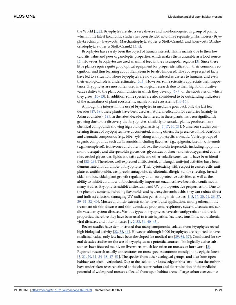
research: epilithic species (Tortula muralis and Dryptodon pulvinatus, Fig 1A and 1B), epiphytic
species (Hypnum cupressiforme from Salix alba bark, Fig 1C), and epigeic species (growing on soil
Fig 1. Studied moss species, their stem leaf and habitat. A–T. muralis, B–D. pulvinatus, C–H. cupressiforme, D–C. purpureus, E–R. squarrosus; scale in μm (foto. G. J.
Wolski, Scientific and Didactic Garden, Faculty of Biology and Environmental Protection, University of Lodz, 08.03.2019).
https://doi.org/10.1371/journal.pone.0257479.g001
PLOS ONE Medical potential of open habitat mosses
PLOS ONE | https://doi.org/10.1371/journal.pone.0257479 September 20, 2021 3 / 24

Ceratodon purpureus and lawn Rhytidiadelphus squarrosus, Fig 1D and 1E), and a detailed
description of the selected species along with taxonomic features is presented in S1 Appendix.
Extraction procedure
The weighed fresh plant material was air-dried at 40˚C for 24 h, ground were ground in a mor-
tar with a pestle to the powder and extracted with pure ethanol at a solid to liquid ratio 1:10
(w/v) for 30 min, at room temperature on the rocker. The supernatant was decanted and the
sediment was boiled 2 min. with pure ethanol. The combined ethanol extracts were centri-
fuged for 10 min at 4500 × g, 4˚C (3K30, Sigma). After removal of ethanol at� 40˚C with a
vacuum rotary evaporator (RV06-ML IKA- WERKE, Germany) the extracts were reconsti-
tuted at 1 mg/mL of 5% ethanol.
Total phenolics content
Total phenolic content was estimated by the Folin-Ciocalteu method using gallic acid as a stan-
dard [52]. 500 μL of the extract were combined with 3.65 mL of distilled water, 100 μL of
Folin-Ciocalteu’s reagent and 1000 μL of 10% Na2CO3. The mixture was vortexed thoroughly
and, after incubation at room temperature in darkness for 60 min, the absorbance was mea-
sured at 765 nm against a ‘blank’ without the sample extract (UV-Vis spectrophotometer U-
2001, Hitachi). Quantification was done on the basis of the standard curve of gallic acid (solu-
tion 0.25–5 μg/mL). The results were expressed as mg of gallic acid equivalents (GAE) per g of
fresh weight (FW). All raw data is added as S1 Raw data.
2,20-azinobis (3-ethylbenzthiazoline-6-sulphonic acid) radical cation
decolouration assay
A modified method of Re et al. [53, 54] was used. Briefly, a fresh solution of ABTS�+ was pre-
pared by dissolving 19.5 mg 2,20-azinobis(3-ethylbenzthiazoline-6-sulphonic acid) (ABTS;
Sigma, Deisenhofen, Germany) and 3.3 mg potassium persulphate (dipotassium peroxodisul-
phate; Sigma) in 7 mL of 0.1 mol/L phosphate buffer, pH 7.4. This solution was stored in the
dark for 12 h for completion of the reaction. A 50 μL of ethanolic extract of bryophytes was
added to 2 ml ABTS�+ solution in 0.1 mol/L phosphate buffer, pH 7.4, diluted (usually approxi-
mately 1:80) to give an absorbance about 0.9 and read at 734 nm. The extent of ABTS�+ bleach-
ing (decrease in absorbance, corrected for a small decrease in absorbance of ABTS�+ solution
alone) is proportional to the activity of anti-oxidants in a given sample. Calculations were
made on the basis of standard curves obtained for a Trolox solution. Antioxidant capacity of
plant extracts were expressed as Trolox equivalent [μM]. All raw data is added as S1 Raw data.
Ferric ion reducing anti-oxidant power assay
For the ferric ion reducing anti-oxidant power (FRAP) assay, a modification of the method of
Benzie and Strain was used [55, 56]. Working solution was prepared immediately before mea-
surements by mixing 10 volumes of acetate buffer, pH 3.6, with 1 volume of 10 mM/L 2,4,6–
tris-2-pyridyl-s-triazine (TPTZ; Sigma) and 1 volume of 20 mM/L FeCl3 (Sigma). A 50 μL of
extracts were mixed with 2 mL of the working solution and incubated at 37˚C. After 5 min, the
absorbance of the samples was read at 593 nm in a spectrophotometer against a reagent blank.
The increase in absorbance is proportional to the activity of antioxidants in the sample. Calcu-
lations were made on the basis of standard curves obtained for FeSO4 solution. The ferric
reducing ability of extracts measured as antioxidant power were expressed as FeSO4 equivalent
[μM]. All raw data is added as S1 Raw data.
PLOS ONE Medical potential of open habitat mosses
PLOS ONE | https://doi.org/10.1371/journal.pone.0257479 September 20, 2021 4 / 24

Microorganisms, culture conditions and suspensions for antimicrobial
study
The reference strains of Gram-positive bacteria: Staphylococcus aureus ATCC 29213 (MSSA,
methicillin-susceptible S. aureus), Staphylococcus aureus ATCC 43300 (MRSA, methicillin-
resistant S. aureus), Staphylococcus epidermidis ATCC 12228, Enterococcus faecalis ATCC
29212, Gram-negative bacteria: Escherichia coli ATCC 25922, Pseudomonas aeruginosa ATCC
25619, and fungi: Candida albicans ATCC 10231, Candida glabrata ATCC 90030 were used to
assess biostatic/biocidal activity of the extracts from selected mosses. The group of microor-
ganisms tested (bacteria and fungi) contain standard reference strains used for screening
method of antimicrobial activity testing. Microorganisms were grown for 24 h at 37˚C in tryp-
tic-soy agar (TSA; BTL, Poland) or Sabouraud dextrose agar (SDA; BTL, Poland) according to
nutritional requirements. Microbial suspensions at a density of about 5 × 10⁵ CFU/mL were
prepared in Mueller-Hinton broth (MHB; BTL, Poland) or in RPMI-1640 medium with L-glu-
tamine (Sigma-Aldrich, Merck, Germany) containing 2% glucose (RPMI/Glu) for bacteria or
fungi, respectively.
Minimum inhibitory and bactericidal/fungicidal concentration (MIC,
MBC/MFC)
Broth microdilution method was used according to the EUCAST guidelines [EUCAST] to
determine MIC of moss extracts. Tested extracts were diluted in MHB or RPMI/Glu (for bac-
teria and fungi, respectively) to the final concentration range of 0.015–1.0 mg/mL in 96-well
culture plate (Falcon, USA) in volume of 50 μL/well. Then microbial suspensions (5 × 10⁵
CFU/mL, 50 μL) were added and the plates were incubated at 37˚C for 24 h (bacteria) or 48 h
(fungi). Since primary D. pulvinatus and R. squarrosus extracts were prepared in 100% Et-OH
at a concentration of 40 mg/mL and the highest concentration of the solvent in tested wells
with 1 mg/mL of these two extracts reached 2.5%, two types of microbial growth positive con-
trol were simultaneously prepared: microbial suspensions in medium alone (K + 1) and micro-
bial suspensions in medium containing 2.5% Et-OH (K + 2). MIC was defined as the lowest
concentration of the extract inhibiting visible bacterial/fungal growth during of co-incubation
time compared to the appropriate positive control. MBC/MFC of the extracts tested referred
to the lowest concentration that killed 99.9% of microbial inoculum added to the wells, thus
there was no bacteria or yeast growth after subculturing 10 μL from the wells marked as MIC
and these containing higher concentrations on TSA/SDA (incubation 24–48 h at 37˚C). Exper-
iments were carried out in duplicate for each extract.
Cytotoxicity evaluation of the extracts for mouse fibroblasts line L929 by
MTT method
Mouse fibroblasts L929 (ATTC, CCL-1) were cultured as monolayers in 25 cm2 tissue culture
flasks (BD Falcon) in RPMI 1640 medium (Sigma/Merck, Germany) supplemented with 10%
fetal bovine serum (Biological Industries) and 100 U/mL penicillin, 100 μg/mL streptomycin,
0,292 mg/mL L-glutamine (Gibco/Intvitrogen). Every three days the monolayer was treated
with trypsin/ethylene diamine tetraacetic acid (BioWest) and sub-cultured. Cells were main-
tained at 37˚C under a humidified atmosphere containing 5% CO2. Cytotoxicity was assessed
using the colorimetric MTT assay [57]. Briefly, confluent monolayer was trypsinized, the sus-
pension of 1 × 10⁵ cells/mL prepared, 100 μL per well of 96-well tissue culture plate (Greiner
bio-one) seeded, and incubated at 37˚C in a humidified 5% CO2 atmosphere. For background
absorption, some wells were remained cell-free (blank control). After 24h the growth medium
PLOS ONE Medical potential of open habitat mosses
PLOS ONE | https://doi.org/10.1371/journal.pone.0257479 September 20, 2021 5 / 24

was discarded, and 100 μL of extract diluted in RPMI 1640 medium (supplemented as earlier)
was added at the following concentrations: 1000, 500, 250, 125, 62.5, 31.2, 15.6, and 7.8 μg/mL
(n = 3). In each case the concentration of extracts’ solvent (Et-OH) did not exceed 2,5% and
did not disturb fibroblast growth. Three wells were remained untreated as negative control.
Following 24 h incubation under the conditions as earlier, the treatment medium were
removed and 50 μL/well of MTT ((3-(4,5-dimethylthiazol-2-yl)-2,5-diphenyltetrazolium bro-
mide; Sigma/Merck) in phosphate-buffered saline (1 mg/mL) after filtration through a 0.2 μm
syringe filter was added and incubated for 2 h as above. The MTT solution was removed and
replaced with 150 μL/well of DMSO (dimethyl sulfoxide; Sigma/Merck) to dissolve the blue
formazan product formed by living cells. Additionally, 25 μL/well of Sorensen’s glycine buffer
(glycine 0.1 M, NaCl 0.1 M, pH:10.5 with 0.1 NaOH) was added. The absorbance (A) was
detected at 570 nm with the use of the microplate reader Multiskan EX (Thermo Labsystem-
Fischer, USA). The viability of cells cultured in full RPMI 1640 alone (negative control) was
considered as 100%, and the percentage of cell viability was calculated using the formula
below:
Survival rate (%) = [(As–Ab)/(Ac–Ab)] × 100
As–absorbance of test sample (the cells exposed to the extract)
Ac–absorbance of negative control (the cells in medium alone)
Ab–absorbance of blank control
All raw data is added as S2 Raw data.
Determination of individual phenolic compounds using UPLC/Q-TOF-MS
analysis
Caffeic acid, (+)-catechin, apigenin, naringin, formic acid, methanol and acetonitrile were
obtained from Sigma Aldrich (Steinheim, Germany). Chlorogenic acid, quercetin 3-glucoside,
quercetin 3-rutinoside, kaempferol 3-glucoside and p-coumaric acid were obtained from
Extrasynthese (Lyon, France). Procyanidin B1 and 3,5-dicaffeoylquinic acid was purchased
from PhytoLab (Vestenbergsgreuth, Germany).
Phenolic compounds were identified using the Acquity ultra-performance liquid chroma-
tography (UPLC) system coupled with a quadruple-time of flight mass spectrometry
(Q-TOF-MS) instrument (Waters Corp., Milford, MA, USA) equipped with an electrospray
ionization (ESI) source. Separations of individual phenolics were carried out using a Acquity
UPLCR HSS T3 C18 column (150 × 2.1 mm, 1.8 μm; Corp., Milford, MA, USA) at 30˚C, as
previously described [58]. The mobile phase was a mixture of 0.1% formic acid (A) and aceto-
nitrile (B). The gradient program was as follows: initial conditions 99% (A), 12 min 65% (A),
12.5 min 0% (A), 13.5 min 99% (A). The flow rate was 0.45 mL/min and the injection volume
was 5 μL. The mass spectrometer was operating in the negative mode for a mass range of 150–
1500 Da, fixed source temperature at 100˚C, desolvation temperature 250˚C, desolvation gas
flow of 600 L/h, cone voltage of 45 V, capillary voltage of 2.0 kV, collision energy 50 V. Leucine
enkephalin was used as a lock mass. The instrument was controlled by Mass-LynxTM V 4.1
software. Photodiode detector spectra were measured over the wavelength range 200–600 nm.
Compounds were identified using their UV-Vis characteristic, MS and MS2 properties using
data gathered in house and from literature. Calibration curves were run for the external stan-
dards: (+)-catechin, procyanidin B1, caffeic acid, chlorogenic acid, 3,5-dicaffeoylquinic acid,
p-coumaric acid, quercetin 3-glucoside, quercetin 3-rutinoside, kaempferol 3-glucoside, api-
genin, and naringin.
PLOS ONE Medical potential of open habitat mosses
PLOS ONE | https://doi.org/10.1371/journal.pone.0257479 September 20, 2021 6 / 24

The crude extracts prior to injection into UPLC-MS system were extracted twice with chlo-
roform to remove lipophilic compounds. The organic phase was discarded and the aqueous
phase was concentrated to 3 mL, and applied to a C18 solid phase extraction cartridge (360 mg
capacity, Waters Corp., Milford, MA) to separate phenolic compounds fraction. Interfering
water-soluble components were removed with water (10 mL) while absorbed phenolics were
recovered with methanol (10 mL). The solvent allowed to evaporate completely under reduced
pressure and solid residue was dissolved in 2 mL of methanol.
Scratch assay
Human foreskin fibroblasts (HFF-1 line; LGC Standards, UK) were culture in 24-well plates
(Nunc, Denmark) with previously marked reference lines on wells’ bottom (2.5 × 10⁵ cells/
well) in Dulbecco’s Modified Eagle Medium (DMEM) supplemented with 15% fetal bovine
serum (FBS; Biological Industries, USA) and penicillin-streptomycin solution (P/S; Biowest,
France) at 37˚C, 5% CO2. The 24 h-old cell monolayers were gently scratched across the refer-
ence lines, forming the wounds. The medium containing detached cells was removed, the cells
were washed twice using culture medium and finally fresh culture medium alone (control
cells) or the medium with moss extracts (15 μg/mL) were added. The cells were grown for the
next 24 h. The images of the wounds immediately after scratching (T0) and 24 h later (T24)
were done using Motic Microscope model AE-2000T with inverted field of view and integrated
camera (Conbest Sp. Z o.o., Poland). The changes of total wound areas were analyzed using
ImageJ software. The percentage of wound closure was calculated according to the following
formula: [(wound area T0—wound area T24) / wound area T0] × 100. Experiment was carried
out twice.
Results
Total phenolic content
The results of the colorimetric analysis of total phenolic content (TPC) based on the absor-
bance values of the extract solutions reacted with Folin Ciocalteu’s reagent, expressed as mg
gallic acid equivalent (GAE) per g FW of sample, are given in Fig 2. The highest values–
1.71 ± 0.03 was noted for C. purpureus. Significantly lower (by about 40%)– 1,04 ± 0.03 and
1.00 ± 0.02 mg GAE/g FW of sample–were determined for T. muralis and H. cupressiforme,respectively. The next species D. pulvinatus and R. squarossus had comparable concentration
of phenolics, approximately by 60% lower than that obtained for C. purpureus.
Antioxidant capacity
Five plant species used in our experiments were screened for their potential as antioxidants. Due
to the fact that different antioxidants had varying reactivity and substrate specificity we applied
two various methods: the ferric reducing antioxidant power (FRAP) and the 2,20-azinobis-3-
ethylbenzotiazoline-6-sulfonic acid (ABTS�+) assay for the measurement of the total antioxidant
capacity. Our results indicates that both radical scavenging activity and reducing properties in
analyzed extracts decreased in the following order: C. purpureus> D. pulvinatus> T. muralis>H. cupressiforme> R. squarrosus. Trolox equivalent for C. purpureus was 40% higher than for
D. pulvinatus and over 60 and 70% higher in comparison to T. muralis, H. cupressiforme and
R. squarrosus, respectively (Fig 3). Similar tendency was observed using FRAP method. The fer-
ric ion reducing anti-oxidant power determined for C. purpureus compared to D. pulvinatus,T. muralis, H. cupressiforme and R. squarrosus was better about 30%, 40% and 50%, respectively
(Fig 4).
PLOS ONE Medical potential of open habitat mosses
PLOS ONE | https://doi.org/10.1371/journal.pone.0257479 September 20, 2021 7 / 24

Antimicrobial activity
The antimicrobial activity of moss extracts against selected bacterial and fungal strains are pre-
sented in detail in Table 1 as minimum inhibitory concentration (MIC) and minimum bacteri-
cidal/fungicidal concentration (MBC/MFC). The moss extracts showed no biostatic and
biocidal activity in whole concentration range tested (up to 1 mg/mL). The only exception was
the D. pulvinatus origin extract, which inhibited the growth of C. glabrata at a concentration
of 0.5 mg/mL. However, the lack of similar effect against C. albicans starts a discussion about
antifungal properties of this extract, which need further investigations.
Cytotoxicity evaluation
The cytotoxicity effect of five moss extracts was analyzed after 24 h exposure of L929 cells to
tested preparations using MTT assay. The results showed that all tested extracts did not affect
the viability of L929 cells at the concentration up to 125 μg/mL, and up to 500 μg/mL for T.
muralis and H. cupressiforme extracts. The most unfavorable effect was noted for R. squarrosusextract with cell viability at the level of 68.6 ± 9.2% when used at 125 μg/mL. Culturing of L929
at higher concentrations of both R. squarrosus and D. pulvinatus extracts resulted in a sharp
decrease in the cells viability (Fig 5) and caused reduced confluence of the monolayer (Fig 6).
The extracts from T. muralis, H. cupressiforme and C. purpureus used at the highest concentra-
tion tested (1000 μg/mL) were also cytotoxic, however nearly 40% of cells were still viable,
whereas the cell viability did not exceed 10% in the case of D. pulvinatus and R. squarrosusused at the same concentration.
Fig 2. The total phenolic content of selected mosses expressed as mg gallic acid equivalent per g of fresh sample. The results are
expressed as mean values of 3 independent experiments ± SD.
https://doi.org/10.1371/journal.pone.0257479.g002
PLOS ONE Medical potential of open habitat mosses
PLOS ONE | https://doi.org/10.1371/journal.pone.0257479 September 20, 2021 8 / 24

Fig 3. The ABTS cation scavenging activity of selected mosses extracts calculated as Trolox equivalent [μM]. The results are
expressed as mean values of 3 independent experiments ± SD.
https://doi.org/10.1371/journal.pone.0257479.g003
Fig 4. Reducing antioxidant power of mosses extracts calculated as FeSO4 equivalent [μM]. The results are expressed as mean
values of 3 independent experiments ± SD.
https://doi.org/10.1371/journal.pone.0257479.g004
PLOS ONE Medical potential of open habitat mosses
PLOS ONE | https://doi.org/10.1371/journal.pone.0257479 September 20, 2021 9 / 24

Table 1. The antimicrobial activity of mosses extracts.
Microorganism MIC [mg/mL]
MBC/MFC [mg/mL]
T. muralis H. cupressiforme C. purpureus D. pulvinatus R. squarrosusBacteria
Staphylococcus aureus ATCC 29213 > 1 > 1 > 1 > 1 > 1
> 1 > 1 > 1 > 1 > 1
Staphylococcus aureus ATCC 43300 > 1 > 1 > 1 > 1 > 1
> 1 > 1 > 1 > 1 > 1
Staphylococcus epidermidis ATCC 12228 > 1 > 1 > 1 > 1 > 1
> 1 > 1 > 1 > 1 > 1
Enterococcus faecalis ATCC 29212 > 1 > 1 > 1 > 1 > 1
> 1 > 1 > 1 > 1 > 1
Escherichia coli ATCC 25922 > 1 > 1 > 1 > 1 > 1
> 1 > 1 > 1 > 1 > 1
Pseudomonas aeruginosa ATCC 25619 > 1 > 1 > 1 > 1 > 1
> 1 > 1 > 1 > 1 > 1
Fungi
Candida albicans ATCC 10231 > 1 > 1 > 1 > 1 > 1
> 1 > 1 > 1 > 1 > 1
Candida glabrata ATCC 90030 > 1 > 1 > 1 0.5 > 1
> 1 > 1 > 1 > 1 > 1
MIC–minimum inhibitory concentration measured by broth microdilution method; MBC/MFC—minimum bactericidal/fungicidal concentration determined based on
microbial culture on solid media.
https://doi.org/10.1371/journal.pone.0257479.t001
Fig 5. Dose dependent effects of ethanolic moss extracts on L929 cells after 24 h of exposure. Cell viability was quantified using MTT colorimetric method.
The results are expressed as mean ± SD. Statistical significance (p< 0.05) was calculated with Kruskal-Wallis test.
https://doi.org/10.1371/journal.pone.0257479.g005
PLOS ONE Medical potential of open habitat mosses
PLOS ONE | https://doi.org/10.1371/journal.pone.0257479 September 20, 2021 10 / 24

Identification and content of phenolic compounds with UPLC–
PDA-Q/TOF-MS method
Due to the strongest effect of D. pulvinatus and R. squarrosus extracts on eukaryotic cells,
including also potential antifungal activity, a detailed chemical composition in relation to indi-
vidual phenolic compounds was tested for these two extracts.
The results of qualitative analysis of extracts determined by UPLC/MS method are pre-
sented in Fig 7 and Tables 2 and 3.
Chromatogram of D. pulvinatus showed twenty three peaks, five of which were identified
based on retention times (Rt), Uv-Vis spectra, deprotonated molecules ([M-H]-), diagnostic
fragments (MS/MS), comparison with the standard reference compounds, and the tentative
identification of 3 compounds was based on the literature data. In contrast, chromatogram of
R. squarrosus showed twenty four peaks, but only half of them could be identified, while the
tentative identification of as many as ten compounds was based on literature data [58, 59].
Both extracts showed the presence of different groups of phenolic compounds, such as hydro-
xycinnamic acids, flavanols, flavonols, flavones, and flavanones. The sum of the identified phe-
nolic components in D. pulvinatus and R. squarrosus was 25.49 and 6.42 mg/100 g FW of plant
material, respectively (Table 4). Quantitatively in the group of identified phenolic compounds,
kaempferol derivatives and sinapoyl hexoside were the dominant compounds in D. pulvinatuswhile apigenin hexoside and chlorogenic acid in R. squarrosus.
Pro-regenerative activity of moss extracts
To test pro-regenerative activity of moss extracts in vitro scratch assay on human fibroblasts
HFF-1 line was used. The moss extracts were applied at very low concentration (15 μg/mL) to
avoid any cytotoxic activity.
Fig 6. The effect of moss extracts used at the concentration 250 μg/mL on L929 cell monolayer. The images were
taken by inverted light microscopy after 24 h exposure of the cells to the extracts.
https://doi.org/10.1371/journal.pone.0257479.g006
PLOS ONE Medical potential of open habitat mosses
PLOS ONE | https://doi.org/10.1371/journal.pone.0257479 September 20, 2021 11 / 24

Fig 7. UPLC chromatograms of D. pulvinatus (A) and R. squarrosus (B) at 280 nm.
https://doi.org/10.1371/journal.pone.0257479.g007
PLOS ONE Medical potential of open habitat mosses
PLOS ONE | https://doi.org/10.1371/journal.pone.0257479 September 20, 2021 12 / 24

Two groups of moss extracts were used for the study, one group (T. muralis, C. purpureus)of preparations with low cytotoxicity (demonstrated only at a concentration above 500 μg/mL)
and for comparison, the extract with the highest cytotoxic potential appearing already at a con-
centration above 125 μg/mL (D. pulvinatus). The results obtained for three selected extracts
representing different models of phytochemical composition and biological activity are pre-
sented in Table 5 and Fig 8.
It was demonstrated, that the extracts with middle and low total phenolic content (from T.
muralis and D. pulvinatus, respectively) stimulated fibroblasts proliferation and wound heal-
ing. The increase in wound closure achieved of 13.1% and 8.8%, respectively, in comparison to
control cells. However, pro-regenerative activity cannot be attributed general to moss extracts,
since average wound closure was 3.5% lower in the presence of the extract from C. purpureus,which is characterized, what’s interesting, by the highest total phenolic content.
Discussion
In the search for new biologically active compounds, mosses can be considered as a largely
neglected material, since there have not been many studies dedicated to the composition of
secondary metabolites and their biological activity in mosses in comparison to, for example,
similar studies of higher plants. Although over 400 years ago, the Chinese used some Fissidensand Polytrichum species as diuretics and hair growth stimulation tonics. Traditional cultures
Table 2. Profile of phenolic compounds in D. pulvinatus determined by UPLC-ESI-MS/MS analysis.
Peak Rt λmax [M−H]− MS/MS Compound
(min) (nm) (m/z)(m/z)
1 4.95 243, 283 519 164,192,221,136,182 Unidentified
2 5.59 243, 288 371 119,124 Unidentified
3 5.72 243, 292 499 174,145,129,187 Unidentified
4 6.03 243, 284 499 174,145,129 Unidentified
5 6.25 243, 340 345 153,137,181,137 Unidentified
6 6.48 243, 284 329 123,153 Unidentified
7 6.64 243, 288 355 148,165,132,227,121,152 Unidentified
8 6.82 243, 314 507 153,181,182,137 Unidentified
9 8.67 243, 284 345 153,181,139,111 Unidentified
10 9.20 280, 335 337 174,175,163,129 Coumaroylquinic acida
11 9.69 243, 345 447 284,133,297,175 Kaempferol 3-galactosidea
12 10.11 243, 283 539 298,299,327,297 Unidentified
13 10.43 243, 283 449 135,151,107 Eriodyctiol hexosidea
14 10.53 243, 326 431 268,117,283,161,163 Apigenin hexosidea
15 10.75 243, 345 609 271,300,255,243,151 Quercetin 3-rutinosdie
16 10.88 243, 347 593 285 Kaempferol 3-rutinosidea
17 11.15 243, 347 447 284,227,133,256,199 Kaempferol 3-glucoside
18 11.28 243, 310 505 138,165,214,137 Unidentified
19 11.78 243, 283 449 135,151 Eriodictyol hexosidea
20 12.07 243, 280 577 289,151,269,327,343 Procyanidin dimera
21 12.29 279, 338 447 133,285,151,199,179 Kaempferol derivative Ia
22 12.50 266, 336 447 133,285,151,175,199 Kaempferol derivative II a
23 12.66 267, 324 431 269,239,211,151 Sinapoyl hexosidea
a–Tentatively identified [58, 59].
https://doi.org/10.1371/journal.pone.0257479.t002
PLOS ONE Medical potential of open habitat mosses
PLOS ONE | https://doi.org/10.1371/journal.pone.0257479 September 20, 2021 13 / 24

in India and North America used Bryum, Mnium, Philonotis spp. and Polytrichum juniperi-num for healing burns, bruises and wounds. Marchantia polymorpha was used as a diuretic in
France. In many of these cases, a scientific basis has been recently identified to justify use of
these plants. Several liverwort and moss extracts have antibacterial, antifungal and antiviral
activity [17].
In the recent years bryophytes as a group of plants containing a broad range of biologically
active substances, such as phenolics, terpenoids, fatty acids, have been widely tested for pro-
health activity. Among beneficial effects of bryophytes -origin products (mainly the extracts)
anti-oxidative, anti-inflammatory, anti-platelet, anti-leukemic, also antibacterial and antifun-
gal activity are mentioned [2, 36, 37, 60, 61].
In modern cosmetology or phytomedicine the most popular are herbs with the high con-
centration of phenolics, which is usually reflected in their good antioxidant activity [62, 63].
Antioxidants were also identified in bryophytes tissues, however, so far an attention was
focused mainly on liverworts [34, 50], or on epigeic forest mosses species. Mosses harvested
from open habitats areas of large urban ecosystem have been omitted, although potentially
they can be a rich source of antioxidants [31, 47–49, 51]. Thus in our research five species of
widespread mosses harvested from open habitats areas of large urban ecosystem were screened
for their medical potential.
Table 3. Profile of phenolic compounds in R. squarrosus determined by UPLC-ESI-MS/MS analysis.
Peak Rt λmax [M−H]− MS/MS Compound
(min) (nm) (m/z) (m/z)
1 5.66 243, 286 371 119 Unidentified
2 6.20 243, 281 289 109,123,108,159,155 (+)-Catechin
3 6.43 243, 281 329 153,119,162 Unidentified
4 6.77 325 353 153,181,137 Chlorogenic acid
5 7.11 282, 322 179 134,108 Caffeic acid
6 7.23 243 447 145,173,211 Unidentified
7 8.28 243 431 138,153,189,205,163,122 Unidentified
8 8.62 243, 284 345 153,137,181 Unidentified
9 8.89 243 387 182,139,153 Unidentified
10 9.10 243, 308 439 131,101,113,175,147 Unidentified
11 9.30 243, 276 433 138,153 Unidentified
12 9.53 243, 283 539 142,172,114,185,317,203 Unidentified
13 10.05 243, 285 167 Unidentified
14 10.18 243, 283 297 101,159,113 Unidentified
15 10.47 243 433 283,117,161 Unidentified
16 10.72 243, 345 609 271,255,300,243,151 Quercetin 3-rutinoside
17 10.83 243, 347 593 285,199,151 Kaempferol 3-rutinosidea
18 10.89 243, 338 431 117,281,161,253 Apigenin hexosidea
19 11.18 243 505 138,165,211,269 Unidentified
20 11.44 243, 280 431 245,187,202,159,145 Unidentified
21 11.77 283, 320 515 191,135,251,115 3,5-Dicaffeoylquinic acid
22 12.02 243, 281 521 281,254,159,299 Unidentified
23 12.23 243, 279 415 145,241 Unidentified
24 12.74 243, 345 607 284,299,256 Kaempferol derivative IIa
a–Tentatively identified [58, 59].
https://doi.org/10.1371/journal.pone.0257479.t003
PLOS ONE Medical potential of open habitat mosses
PLOS ONE | https://doi.org/10.1371/journal.pone.0257479 September 20, 2021 14 / 24

Analyses of TPC indicated that phenolics concentration decreased in the following order:
C. purpureus> T. muralis>H. cupressiforme> D. pulvinatus> R. squarrosus. Generally it is
accepted that most plants containing a large number of polyphenols exhibit antioxidant activ-
ity [64–66]. Therefore, we checked our moss extracts toward their antioxidant potential. In
order to determine total antioxidant capacity, ABTS and FRAP assays were used. Interestingly,
among the samples tested, D. pulvinatus extract despite quite low TPC expressed the highest
antioxidant potential. Moreover, comparison of the results obtained for mosses with these
Table 4. Contents of identified phenolic compounds in the extracts of D. pulvinatus and R. squarrosus.
Compound D. pulvinatus R. squarrosusContent
(mg/100 g of FW)
Caffeic acid Not detected 0.30 ± 0.00
Coumaroylquinic acida 0.32 ± 0.00 Not detected
Chlorogenic acid Not detected 1.26 ± 0.02
3,5-Dicaffeoylquinic acid Not detected 0.12 ± 0.00
Sinapoyl hexosideb 4.42 ± 0.10 Not detected
Kaempferol 3-galactosidec 0.22 ± 0.00 Not detected
Kaempferol 3-rutinosidec 1.70 ± 0.02 0.69 ± 0.00
Kaempferol 3-glucoside 4.99 ± 0.05 Not detected
Kaempferol derivative Ic 1.73 ± 0.02 Not detected
Kaempferol derivative IIc 6.11 ± 0.09 Not detected
Kaempferol derivative IIIc Not detected 0.28 ± 0.01
Quercetin 3-rutinoside 0.98 ± 0.03 0.70 ± 0.02
Eriodyctiol hexoside Id 0.22 ± 0.00 Not detected
Eriodyctiol hexoside IId 0.47 ± 0.04 Not detected
Apigenin hexosidee 1.93 ± 0.03 2.14 ± 0.17
(+)-Catechin Not detected 0.93 ± 0.03
Procyanidin dimerf 2.40 ± 0.09 Not detected
TOTAL 25.49 ± 0.47 6.42 ± 0.27
Results are expressed as a mean ± standard deviation (n = 3).a–quantified as chlorogenic acid equivalentsb–quantified as p-coumaric acid equivalentsc–quantified as kaempferol 3-glucoside equivalentsd–quantified as naringin equivalentse–quantified as apigenin equivalentsf–quantified as procyanidin B1 equivalents.
https://doi.org/10.1371/journal.pone.0257479.t004
Table 5. The pro-regenerative potential of the moss extracts on human foreskin fibroblasts (HFF-1 line) tested in vitro by scratch assay.
Preparation Average wound area at T0 [pixels2] Average wound area at T24 [pixels2] Average wound closure
[% ± S.D.]
T. muralis 2484847 514316 79.3 ± 14.5
C. purpureus 2461775 919097 62.7 ± 0.5
D. pulvinatus 2594336 647935 75.0 ± 0.6
Control cells 2320792 793976 66.2 ± 8.6
T0, T24 –time [h] after wound scratch
https://doi.org/10.1371/journal.pone.0257479.t005
PLOS ONE Medical potential of open habitat mosses
PLOS ONE | https://doi.org/10.1371/journal.pone.0257479 September 20, 2021 15 / 24

demonstrated by accepted medical plants possessing very high antioxidant potential indicated
that antioxidant activity of mosses was possible to be considered high. However, antioxidant
activity was not always correlated with the highest phenolics content (Table 6).
From group of phenolics the most numerous are flavonoids which play multiple functions
in photoprotection and oxidative stress regulation, as well as in plant defense against herbivo-
rous insects, fungi and other pathogens [67]. Flavonoids occur in many various compartments
in mesophyll cells, including the nucleus, the chloroplasts and vacuoles, as well as in the epi-
dermis [68]. It is wealthy to note that high light irradiance up-regulates the biosynthesis of
dihydroxy B-ring-substituted flavonoids (such as luteolin-7-O-glycosides and quercetin-3-
O-glycosides) whereas does not affect the biosynthesis of monohydroxy B-ring-substituted
flavonoids (such as apigenin-7-O-glycosides and kaempferol-3-O-glycosides). Monohydroxy
flavonoids, which have very similar UV-spectral features of their dihydroxy counterparts, pre-
dominate in plants growing under deep or partial shading [69]. Light-responsive dihydroxy
flavonoids have much greater ability than their monohydroxy counterparts to inhibit the
Fig 8. The effect of moss extracts on human foreskin fibroblasts (HFF-1) migration tested in vitro by scratch assay. Representative images of the scars taken just after
scratching (T0) and 24 h later (T24).
https://doi.org/10.1371/journal.pone.0257479.g008
Table 6. TPC content and antioxidant activity of mosses extracts in comparison to accepted medical plants.
Species Total phenolics mg GAE per g of FW Antioxidant Activity
ABTS FRAP
Trolox equivalent [μM] FeSO4equivalent [μM]
Ceratodon purpureus 1.71 ± 0.03 20.98 ± 0.02 771.33 ± 16.8
Dryptodon pulvinatus 0.70 ± 0.06 12.40 ± 0.62 549.4 ± 14.6
Hypnum cupressiforme 1.00 ± 0.02 7.59 ± 0.63 476.67 ± 6.62
Rhytidiadelphus squarrosus 0.65 ± 0.01 6.4 ± 0.72 412 ± 7.73
Tortulla muralis 1.04 ± 0.03 7.99 ± 0.14 469 ± 16.1
Thymus vulgaris 4.80 ± 0.26 13.10 ± 0.19 1092 ± 37
Rosmarinus officinalis 5.45 ± 0.06 13.62 ± 0.39 2076 ± 11
Salvia officinalis 2.23 ± 0.16 14.75 ± 0.11 1005 ± 10
Mentha piperita 1.38 ± 0.15 9.22 ± 0.69 788 ± 15
https://doi.org/10.1371/journal.pone.0257479.t006
PLOS ONE Medical potential of open habitat mosses
PLOS ONE | https://doi.org/10.1371/journal.pone.0257479 September 20, 2021 16 / 24

generation of ROS, and then quench ROS once they are formed. Thus the ratios of the ‘effec-
tive antioxidants’ to the ‘poor antioxidants’ significantly increase upon high light irradiance,
irrespective of the relative proportions of different solar wavelengths reaching the leaf surface
[70, 71]. Besides, the biosynthesis of effective antioxidant flavonoids is enhanced when plants
growing under strong light are concomitantly faced with other stress agents [72, 73]. Lillo et al.
[72] demonstrated that the content of flavonoids increases also in response to nitrogen and
phosphorus depletion in plants. Obtained results confirmed these data because the highest
antioxidant power was reported for species exposed to strong sunlight–D. pulvinatus.Flavonoids as well as other phenolics have been reported as responsible also for antimicro-
bial effect of these extracts [36, 37, 61]. Observed activity is usually species-, extract type- and
method-dependent, thus presented results are extremely different. Erturk et al. [35] tested cho-
sen mosses: Ctenidiummolluscum, Eurhynchium striatulum, Homalothecium lutescens¸ H. seri-ceum, Hypnum cupressiforme, Leucodon sciuroides, Thiudium delicatulum, Tomentypnumnitens against 13 different microorganisms: four Gram-positive bacteria (Bacillus cereus, Lis-teria monocytogenes, Clostridium perfringens, and Staphylococcus aureus), six Gram-negative
bacteria (Escherichia coli, Salmonella Typhimurium, Klebsiella pneumoniae, Shigella sonnei,Yersinia enterocolitica, and Pseudomonas aeruginosa) and three yeast strains (Aspergillus niger,Candida albicans, Saccharomyces cerevisiae) and showed that mosses may be used as possible
natural antioxidant and antimicrobial agents to control various human, animal and plant dis-
ease. In this study two different methods: disc diffusion method and broth microdilution
method were used to assess antimicrobial activity of moss samples collected from different
locations of Turkey, including being also a subject of our study. Based on disc diffusion
method and observed zones of microbial growth inhibition, antibacterial and antifungal activ-
ity of H. cupressiforme extract were stated. However, it is necessary to notice that the extract
was used at a concentration of 20 mg/mL and its MIC against S. aureus, E. coli and P. aerugi-nosa tested by microdilution method reached above 25 mg/mL [35]. Thus, it’s no wonder that
in our study the direct biostatic/biocidal activity of that and other extracts has not been dem-
onstrated, when the highest concentration tested was 1.0 mg/mL. Furthermore, based on the
results and well known limited bioavailability of plant-origin preparations in the tissues we
proposed a completely different conclusion, that moss extracts tested did not possess antimi-
crobial activity at all. The more that our cytotoxic tests on mouse fibroblasts line L929 demon-
strated cytotoxicity of the T. muralis, H. cupressiforme and C. purpureus extracts at a
concentration above 0.5 mg/mL and the extracts from D. pulvinatus and R. squarrosus above
0.125 mg/mL, so their higher concentrations could not even be used in vivo. On the other
hand, Manoj and Murugan [74] demonstrated, that methanol extract of Plagiochila beddomeiSteph.–a liverwort expressed antimicrobial activity against a wide group of bacteria and fungi
at much more lower concentrations. Its MICs reached since 0.0625 to 1 mg/mL, including
MIC = 0.5 mg/mL against S. aureus and E. coli, MIC = 1 mg/mL against P. aeruginosa or
MIC = 0.75 mg/mL against C. albicans. Aqua-acetonic and aqua-ethanolic extracts from other
liverworts Reboulia hemisphaerica and Marchantia palmata, and from moss Hydrogonium gra-cilantum also possessed antibacterial activity at very low concentrations with MIC/MBC rang-
ing from 0.00194 to 1 mg/mL [37]. In our study the extract from D. pulvinatus was an only
one with similar activity, but just against C. glabrata. Probably it results from their different
composition and a lower concentration of active components. Perhaps other method of extrac-
tion and the final product purification or enrichment could improve antimicrobial effect of
our moss extracts.
In vitro cytotoxicity assays allows to evaluate the potential toxic effect of newly identified/
obtained compounds or nature-derived products, which can affect basic functions of cells and
through the interference of certain biological processes lead to dysfunctions and even death of
PLOS ONE Medical potential of open habitat mosses
PLOS ONE | https://doi.org/10.1371/journal.pone.0257479 September 20, 2021 17 / 24

cell. Continuous cell lines, as a simplified system, is a useful tool, which allows to minimize the
influence of confounding factors. The choice of available types of cell lines for cytotoxicity
tests is very large and rich [75–80]. One of the most commonly used cell lines in cytotoxicity
testing is the fibroblast cell line L929 [81–85]. L929 fibroblasts are frequently used for testing
multiple plant extract obtained for their potential to be profitable for a wide range of treatment
[86, 87].
Literature directly relating to the cytotoxic properties of the moss extracts is rather poor,
hence the direct comparison of the results obtained to the results of the work of other teams is
quite difficult. Klavina et al. [5] noticed that from among several tested moss extracts these
from Sphagnum magellanicum, Dicranum polysetum and Pleurozium schreberi showed the
highest inhibitory activity (0.9–5 μg/mL), however it was depended on the kind of used cell
line they were tested on. The rat glioma cells were markedly more sensitive to those extract
than the other cell line: human lung carcinoma, mouse melanoma and human breast adeno-
carcinoma cell lines. Our extract tested on L929 cells showed generally lower cytotoxicity in
comparison to those indicated above because even at 125 μg/mL the cells were mostly still
alive. Interesting observation was made by Yağlıoğlu et al. [88]. They tested several extracts (a.
o. hexane, CHCl3, EtOAc, MeOH) prepared from two species of mosses: the acrocarpous moss
Tortella tortuosa (Hedw.) Limpr. and the pleurocarpous moss Rhytidiadelphus triquetrus(Hedw.) Warnst. and tested them using human cervix carcinoma (HeLa) and rat brain tumor
(C6) cells. It has been found that the antiproliferative effect depended on the type of solvent,
which was used to prepared the extracts. In the case of hexane and ethyl acetate extracts the
level of proliferation inhibition was the highest (70–100%) starting already from 20 μg/mL up
to 100 μg/mL (R. triquetrus), and in comparison to our extracts they were several time more
cytotoxic. In contrast to that, the extracts of T. tortuosa caused only app. 20% decrease of cell
proliferation even at the highest concentration thus they appear to be far less cytotoxic than
our extracts, especially from this one prepared from R. squarrosus.Phenolic compounds, widely distributed secondary metabolites in plants, form a group of
molecules with highly diversified chemical structures. These phytocompounds are classified
according to their carbon skeleton into the following main classes: simple phenols, phenolic
acids, stilbenes, flavonoids, tannins [89]. The antioxidant activity of phenolic compounds
depends on their structure. Generally, the antioxidant capacity is related to the number and
the position of hydroxyl groups in the phenolic compound [62]. Moreover, the glycosylation
or replacing of the hydroxyl group, or replacing this group by a methoxy group has a strongly
suppressive influence on the antioxidant activity. Therefore, phenolic composition may have
significant importance for the observed biological activity of plants. In the present study, a
wide range of phenolic compounds, including hydroxycinnamic acids and different flavonoids
sub-groups, were determined in two tested species of mosses such as D. pulvinatus and R.
squarrosus. Both these mosses showed the presence of kaempferol-3-rutinoside, quercetin-
3-rutinoside, and apigenin hexoside. Coumaroylquinic acid, sinapoyl hexoside, kaempferol-
3-galactoside, procyanidin dimer and two eriodycitol hexoside were identified only in D. pulvi-natus. In comparison, (+)-catechin, caffeic acid, 3,5-dicaffeoylquinic acid, and unknown
kaempferol derivative were found in R. squarrosus. The presence of flavones in mosses is con-
firmed by other authors [5, 26, 88]. Phenolic acids such as coumaric, ferulic, gallic, caffeic, pro-
tocatechuic, cinnamic, sinapic, salicylic, chlorogenic and hydroxybenzoic acid were also found
[74, 88]. Furthermore, the mosses can contain a series of polyhydroxylated coumarins [5, 25]
as well as biflavonoids and hydroxybenzoic acid derivatives which havenot been studied in the
present work. In addition, Klavina et al. [5] postulated that due to the high complexity of the
chemical composition of mosses, there are no single substances that are specific for moss spe-
cies or genus and are not influenced by environmental impacts.
PLOS ONE Medical potential of open habitat mosses
PLOS ONE | https://doi.org/10.1371/journal.pone.0257479 September 20, 2021 18 / 24

Not only the phenolic compounds profiles but also the quantities of the identified phenolics
were evidently different. Among identified compounds, kaempferol derivatives and sinapoyl
hexoside were the dominant compounds in D. pulvinatus while apigenin hexoside and chloro-
genic acid in R. squarrosus. The differences in the quantitative composition of phenolic com-
ponents has been also confirmed by Klavina et al. [5] study conducted for 13 different species
of mosses. For example, p-hydroxyacetophenone was the dominant phenolic compounds in
Sphagnum fallax and 7,8-dixydroxy-5-metoxycoumarin-7-β-sophoroside in Rhytidiadelphustriquetrus.
A lot of so called medicinal plants, such as Aloe vera, Calendula officinalis, Centella asiatica,
Echinacea purpurea, Ginkgo biloba, Hypericum perforatum, Matricaria chamomilla, Lavandulaangustifolia or Thymus vulgaris and their products were described as able to support wound
healing in both in vitro and in vivo studies. It was demonstrated, that they increased prolifera-
tion, migration, differentiation and secretory activity of fibroblast and keratinocytes, which are
key cells during wound closure. The promotion of inflammatory cell infiltration, angiogenesis,
extracellular matrix deposition and epithelialization in the presence of plant-derived products
was also shown [89–93]. Therefore, we could also expected such pro-regenerative activity of
the moss extracts tested. As it was shown in scratch assay, which simulated simplified process
of wound healing, the extracts from T. muralis and D. pulvinatus intensified fibroblasts prolif-
eration and thus wound closure. Manoj and Murugan [94] on animal model of skin wound
also demonstrated, that methanolic and aqueous extracts from Plagiochila beddomei supported
formation of granulation tissue, collagen production and angiogenesis, and observed changes
were better than that in control group receiving Madecassol as reference drug. However, it is
worth noting that only selected moss extracts possess a supportive effect for wound healing,
which probably depends on their unique phytochemical composition.
In conclusion, the use of crude moss extracts as antimicrobial components of for instance
ointments, dressings or disinfectants, is pointless. However, their species-specific stimulatory
effect on fibroblasts migration at very low concentrations as well as antioxidant properties,
entitles to consider further study on their regenerative activity and possible application as the
preparations supporting wound healing.
Supporting information
S1 Appendix. Description of the studied moss species.
(DOCX)
S1 Raw data. ABTS, FRAP, TPC.
(XLSX)
S2 Raw data. L929.
(XLS)
Author Contributions
Data curation: Grzegorz J. Wolski, Agnieszka Kobylińska.
Formal analysis: Grzegorz J. Wolski, Beata Sadowska, Marek Fol, Anna Podsędek, Dominika
Kajszczak, Agnieszka Kobylińska.
Investigation: Grzegorz J. Wolski, Beata Sadowska, Marek Fol, Anna Podsędek, Dominika
Kajszczak, Agnieszka Kobylińska.
PLOS ONE Medical potential of open habitat mosses
PLOS ONE | https://doi.org/10.1371/journal.pone.0257479 September 20, 2021 19 / 24

Methodology: Beata Sadowska, Marek Fol, Anna Podsędek, Dominika Kajszczak, Agnieszka
Kobylińska.
Project administration: Grzegorz J. Wolski.
Supervision: Grzegorz J. Wolski.
Writing – original draft: Grzegorz J. Wolski, Beata Sadowska, Marek Fol, Anna Podsędek,
Dominika Kajszczak, Agnieszka Kobylińska.
Writing – review & editing: Grzegorz J. Wolski, Beata Sadowska, Marek Fol, Anna Podsędek,
Dominika Kajszczak, Agnieszka Kobylińska.
References1. Frahm JP. A new contribution to the moss flora of Baltic and Saxon amber. Rev Palaeobot Palynol.
2004; 129: 81–101. https://doi.org/10.1016/j.revpalbo.2003.11.004
2. Asakawa Y, Ludwiczuk A. Bryophytes: Liverworts, mosses, and hornworts: Extraction and isolation pro-
cedures. Methods Mol Biol. 2013. https://doi.org/10.1007/978-1-62703-577-4_1 PMID: 23963899
3. Glime JM. Photosynthesis: the Process. Chapter 11–1, Embryol Ecol Vol 1 Physiol Ecol. 2007.
4. Shaw JA, Szovenyi P, Aguero B. Bryophyte diversity and evolution: Windows into the early evolution of
land plants. Am J Bot. 2011; 98: 352–369. https://doi.org/10.3732/ajb.1000316 PMID: 21613131
5. Klavina L, Springe G, Nikolajeva V, Martsinkevich I, Nakurte I, Dzabijeva D, et al. Chemical composition
analysis, antimicrobial activity and cytotoxicity screening of moss extracts (Moss phytochemistry). Mol-
ecules. 2015; 20: 1721–17243.
6. Matuszkiewicz JM. Zespoły leśne Polski. Wydawnictwo Naukowe, editor. PWN; 2001.
7. Krestov PV. Forest Vegetation of Easternmost Russia (Russian Far East). Forest Vegetation of North-
east Asia. 2003. https://doi.org/10.1007/978-94-017-0143-3_5
8. Chytry M, Horsak M, Danihelka J, Ermakov N, German DA, Hajek M, et al. A modern analogue of the
Pleistocene steppe-tundra ecosystem in southern Siberia. Boreas. 2019; 48: 36–56. https://doi.org/10.
1111/bor.12338
9. Wolski GJ, Kruk A. Determination of plant communities based on bryophytes: The combined use of
Kohonen artificial neural network and indicator species analysis. Ecol Indic. 2020. https://doi.org/10.
1016/j.ecolind.2020.106160
10. Cieśliński S, Czyżewska K, Faliński JB, Klama H, Mułenko W, Żarnowiec J. Relikty lasu puszczańs-
kiego. In: Faliński JB, editor. Białowieski Park Narodowy (1921–1996) w badaniach geobotanicznych.
Seminarium Geobotanicum; 1996.
11. Vanderpoorten A, Engels P, Sotiaux A. Trends in diversity and abundance of obligate epiphytic bryo-
phytes in a highly managed landscape. Ecography (Cop). 2004. https://doi.org/10.1111/j.0906-7590.
2004.03890.x
12. Friedel A, Oheimb GV, Dengler J, Hardtle W. Species diversity and species composition of epiphytic
bryophytes and lichens—A comparison of managed and unmanaged beech forests in NE Germany.
Feddes Repert. 2006. https://doi.org/10.1002/fedr.200511084
13. Fudali E, Wolski GJ. Ecological Diversity of Bryophytes on Tree Trunks in Protected Forests (A Case
Study from Central Poland). Herzogia. 2015; 28: 87–103. https://doi.org/10.13158/heia.28.1.2015.87
14. Klama H, Żarnowiec J, Jędrzejko K. Mszaki naziemne w strukturze zbiorowiskroślinnych rezerwatow
przyrody Makroregionu Południowego Polski. Bielsko-Biała: Politechnika Łodzka Filia w Bielsku-Białej;
1999. https://doi.org/10.1046/j.1537-2995.1999.39399219278.x PMID: 10204584
15. Cieśliński S, Czyżewska K, Faliński JB, Klama H, Mułenko W, Żarnowice J. Epiphytes and epiphytism,
Cryptogamous plants in the forest communities of Białowieża National Park. Functional groups analysis
and general synthesis (Project CRYPTO 3). Faliński JB, Mułenko W, editor. 1996.
16. Klama H. Distribution patterns of liverworts (Marchantiopsida) in natural forest communities (BiałowieżaPrimeval Forest, NE Poland). 2002.
17. Asakawa Y, Tori M, Masuya T, Frahm JP. Ent-sesquiterpenoids and cyclic bis(bibenzyls) from the ger-
man liverwort Marchantia polymorpha. Phytochemistry. 1990. https://doi.org/10.1016/0031-9422(90)
80125-Z
18. Zinsmeister HD, Becker H, Eicher T. Bryophytes, a Source of Biologically Active, Naturally Occurring
Material? Angewandte Chemie International Edition in English. 1991. https://doi.org/10.1002/anie.
199101301
PLOS ONE Medical potential of open habitat mosses
PLOS ONE | https://doi.org/10.1371/journal.pone.0257479 September 20, 2021 20 / 24

19. Bodade RG, Borkar PS, Arfeen S, Khobragade CN. In vitro screening of bryophytes for antimicrobial
activity. J Med Plants. 2008; 7: 23–28.
20. Hanif U, Ali HA, Shahwar D, Farid S, Ishtiaq S. Evaluation of two bryophytes (Funaria hygrometrica and
Polytrichum commune) as a source of natural antioxidant. Asian J Chem. 2014. https://doi.org/10.
14233/ajchem.2014.16431
21. Kadam PS, College PRM, Akurdi P. Antimicrobial activity and polyphenol content of some bryophytes
from Lonavala. Asian J Multidiscip Stud. 2016; 4: 103–105.
22. Ichikawa T, Namikawa M, Yamada K, Sakai K, Kondo K. Novel cyclopentenonyl fatty acids from
mosses, Dicranum scoparium and Dicranum japonicum. Tetrahedron Lett. 1983. https://doi.org/10.
1016/S0040-4039(00)87961-7 PMID: 31595093
23. Sievers H, Burkhardt G, Becker H. Zinsmeister HD. Hypnogenols and other dihydroflavonols from the
moss Hypnum cupressiforme. Phytochemistry. 1992; 31: 3233–3237.
24. Sievers H, Burkhardt G, Becker H, Zinsmeister HD. Further biflavonoids and 3’-phenylflavonoids from
Hypnum cupressiforme. Phytochemistry. 1994; 35: 795–798.
25. Jung M, Zinsmeister HD, Geiger H. New Three- and Tetraoxygenated Coumarin Glucosides from the
Mosses Atrichum undulatum and Polytrichum formosum. Zeitschrift fur Naturforsch—Sect C J Biosci.
1994. https://doi.org/10.1515/znc-1994-11-1201
26. Basile A, Giordono S, Lopez-Saez JA. Cobianchi RC. Antibacterial activity of pure flavonoids isolated
from mosses. Phytochemistry. 1999; 52: 1479–1482. https://doi.org/10.1016/s0031-9422(99)00286-1
PMID: 10647220
27. Asakawa Y. Biologically active compounds from bryophytes. Pure Appl Chem. 2007; 79: 557–580.
https://doi.org/10.1351/pac200779040557
28. VeljićM, Tarbuk M, Marin PD, Cirić A, SokovićM, Marin M. Antimicrobial activity of methanol extracts of
mosses from Serbia. Pharm Biol. 2008; 1744–5116. https://doi.org/10.1080/13880200802367502
29. Aslanbaba B, Yilmaz S, Tonguc Yayintaş O, Ozyurt D, et al. Total phenol content and antioxidant activ-
ity of mosses from yenice forest (ida mountain). J Sci Perspect. 2017; 1: 1–12. https://doi.org/10.26900/
jsp.2017.0
30. Opelt K, Chobot V, Hadacek F, Schonmann S, Eberl L, Berg G. Investigations of the structure and func-
tion of bacterial communities associated with Sphagnum mosses. Environ Microbiol. 2007; 9: 2795–
2809. https://doi.org/10.1111/j.1462-2920.2007.01391.x PMID: 17922763
31. Chobot V, Kubicova L, Nabbout S, Jahodař L, Hadacek F. Evaluation of Antioxidant Activity of Some
Common Mosses. Zeitschrift fur Naturforsch C. 2008; 63: 476–482. https://doi.org/10.1515/znc-2008-
7-802 PMID: 18810988
32. Krzaczkowski L, Wright M, Reberioux D, Massiot G, Etievant C, Gairin JE. Pharmacological screening
of bryophyte extracts that inhibit growth and induce abnormal phenotypes in human HeLa cancer cells.
Fundam Clin Pharmacol. 2009; 23: 473–482. https://doi.org/10.1111/j.1472-8206.2009.00698.x PMID:
19709324
33. Cheng X, Xiao Y, Wang X, Wang P, Li H, Yan H, et al. Anti-tumor and pro-apoptotic activity of ethanolic
extract and its various fractions from Polytrichum commune L.ex Hedw in L1210 cells. J Ethnopharma-
col. 2012; 143: 49–56. https://doi.org/10.1016/j.jep.2012.05.054 PMID: 22687253
34. Gokbulut A, Satilmiş B, Batcioglu K, Cetin B, Şarer E. Antioxidant activity and luteolin content of March-
antia polymorpha L. Turkish J Biol. 2012; 36: 381–385. https://doi.org/10.3906/biy-1106-15
35. Erturk O, Sahin H, Erturk EY, Hotaman HE, Koz B, Ozdemir O. The antimicrobial and antioxidant activi-
ties of extracts obtained from some moss species in Turkey. Herba Pol From Bot to Med Res. 2015; 61:
52–65.
36. Kadam Pratima S. Phytochemical Screening and Antibacterial Activity of Bryophytes. Int J Life Sci.
2017; 5: 405–408.
37. Kandpal V, Chaturvedi P, Negi K, Gupta S, Sharma A. Evaluation of antibiotic and biochemical potential
of bryophytes from kumaun hills and tarai belt of himalayas. Int J Pharm Pharm Sci. 2016; 8: 65–69.
38. Aydoğan S, Erdağ B, Yildiz Aktaş L. Bioaccumulation and oxidative stress impact of Pb, Ni, Cu, and Cr
heavy metals in two bryophyte species, Pleurochaete squarrosa and Timmiella barbuloides. Turk J Bot-
any. 2017; 41: 464–475. https://doi.org/10.3906/bot-1608-33
39. Wang X, Yu W, Lou H. Antifungal Constituents from the Chinese Moss Homalia trichomanoides. Chem
Biodivers. 2005; 2: 139–145. https://doi.org/10.1002/cbdv.200490165 PMID: 17191927
40. Singh M, Govindarajan R, Nath V, Rawat AKS, Mehrotra S. Antimicrobial, wound healing and antioxi-
dant activity of Plagiochasma appendiculatum Lehm. et Lind. J Ethnopharmacol. 2006; 107(1): 67–:
67–72. https://doi.org/10.1016/j.jep.2006.02.007 PMID: 16600543
PLOS ONE Medical potential of open habitat mosses
PLOS ONE | https://doi.org/10.1371/journal.pone.0257479 September 20, 2021 21 / 24

41. Friederich S, Maier UH, Deus-Neumann B, Asakawa Y, Zenk MH. Biosynthesis of cyclic bis(bibenzyls)
in Marchantia polymorpha. Phytochemistry. 1999; 50: 589–598. https://doi.org/10.1016/S0031-9422
(98)00557-3
42. Qu J-B, Xie C-F, Ji M, Shi Y-Q, Lou H-X. Water-Soluble Constituents from the Liverwort Marchantia
polymorpha. Helv Chim Acta. 2007; 90: 2109–2115. https://doi.org/10.1002/hlca.200790218
43. Mewari N, Kumar P. Antimicrobial activity of extracts of Marchantia polymorpha. Pharm Biol. 2008; 46:
819–822. https://doi.org/10.1080/13880200802315725
44. Saroya AS. Herbalism, phytochemistry and ethnopharmacology. Herbalism, Phytochemistry and Eth-
nopharmacology. 2011. https://doi.org/10.1201/b10878
45. Yayintas OT, Alpaslan D, Karagul Yuceer Y, Yilmaz S, Sahiner N. Chemical composition, antimicrobial,
antioxidant and anthocyanin activities of mosses Cinclidotus fontinaloides (Hedw.) P.Beauv. and Palus-
triella commutata (Hedw.) Ochyra gathered from Turkey. Nat Prod Res. 2017; 31: 2169–2173. https://
doi.org/10.1080/14786419.2016.1277355 PMID: 28067067
46. Ucuncu O, Cansu TB, Ozdemir T, Karaoğlu Alpay Ş, Yayli N. Chemical composition and antimicrobial
activity of the essential oils of mosses Tortula muralis Hedw., Homalothecium lutescens (Hedw) H.
Rob., Hypnum cupressiforme Hedw., and Pohlia nutans (Hedw.) Lindb. from Turkey. Turkish J Chem.
2010; 34: 1–10. https://doi.org/10.3906/kim-1002-62
47. Sun SQ, He M, Cao T, Zhang YC, Han W. Response mechanisms of antioxidants in bryophyte (Hyp-
num plumeaforme) under the stress of single or combined Pb and/or Ni. itle. Environ Monit Assess.
2009; 149: 291–302. https://doi.org/10.1007/s10661-008-0203-z PMID: 18274872
48. Pejin B, Bogdanovic-Pristov J. Abts cation scavenging activity and total phenolic content of three moss
species. Hem Ind Ind. 2012; 66: 723–726. https://doi.org/10.2298/hemind120131022p
49. Pejin B, Bogdanovic-Pristov J, Pejin I, Sabovljevic M. Potential antioxidant activity of the moss Bryum
moravicum. Nat Prod Res. 2013; 27: 900–902. https://doi.org/10.1080/14786419.2012.665915 PMID:
22394152
50. Dey A, Mukherjee A. Therapeutic potential of bryophytes and derived compounds against cancer. J
Acute Dis. 2015; 4: 236–248. https://doi.org/10.1016/j.joad.2015.04.011
51. Bhattarai HD, Paudel B, Lee HK, Oh H, Yim JH. In vitro Antioxidant Capacities of Two Benzonaphthox-
anthenones: Ohioensins F and G, Isolated from the Antarctic Moss Polytrichastrum alpinum. Zeitschrift
fur Naturforsch C. 2009; 64: 197–200. https://doi.org/10.1515/znc-2009-3-408 PMID: 19526712
52. Singleton VL, Rossi JA. Colorimetry of Total Phenolics with Phosphomolybdic-Phosphotungstic Acid
Reagents. Am J Enol Vitic. 1965; 16: 144–158.
53. Re R, Pellegrini N, Proteggente A, Pannala A, Yang M, Rice-Evans C. Antioxidant activity applying an
improved ABTS radical cation decolorization assay. Free Radic Biol Med. 1999; 26: 1231–7. https://doi.
org/10.1016/s0891-5849(98)00315-3 PMID: 10381194
54. Lewinska A, Wnuk M, Slota E, Bartosz G. Total anti-oxidant capacity of cell culture media. Clin Exp
Pharmacol Physiol. 2007. https://doi.org/10.1111/j.1440-1681.2007.04637.x PMID: 17600557
55. Benzie IFF, Strain JJ. The Ferric Reducing Ability of Plasma (FRAP) as a Measure of “Antioxidant
Power”: The FRAP Assay. Anal Biochem. 1996; 239: 70–76. https://doi.org/10.1006/abio.1996.0292
PMID: 8660627
56. Bartosz G. Total antioxidant capacity. Adv Clin Chem. 2003. https://doi.org/10.1016/s0065-2423(03)
37010-6
57. Ahmadian S, Barar J, Saei AA, Fakhree MAA, Omidi Y. Cellular Toxicity of Nanogenomedicine in MCF-
7 Cell Line: MTT assay. J Vis Exp. 2009. https://doi.org/10.3791/1191 PMID: 19352311
58. Podsędek A, Zakłos-Szyda M, Polka D, Sosnowska D. Effects of Viburnum opulus fruit extracts on adi-
pogenesis of 3T3-L1 cells and lipase activity. J Funct Foods. 2020; 73: 104111. https://doi.org/10.1016/
j.jff.2020.104111
59. Senica M, Stampar F, Veberic R, Mikulic-Petkovsek M. Fruit Seeds of the Rosaceae Family: A Waste,
New Life, or a Danger to Human Health? J Agric Food Chem. 2017; 65: 10621–10629. https://doi.org/
10.1021/acs.jafc.7b03408 PMID: 29125745
60. Nowicka P, Wojdyło A. Anti-Hyperglycemic and Anticholinergic Effects of Natural Antioxidant Contents
in Edible Flowers. Antioxidants. 2019; 8: 308. https://doi.org/10.3390/antiox8080308
61. Rashmi M, Vijay KP, Ramesh C. Potential of Bryophytes As Therapeutics. Int J Pharm Sci Res. 2014.
PMID: 25750914
62. Rice-Evans CA, Miller NJ, Paganga G. Structure-antioxidant activity relationships of flavonoids and
phenolic acids. Free Radic Biol Med. 1996; 20: 933–956. https://doi.org/10.1016/0891-5849(95)02227-
9 PMID: 8743980
PLOS ONE Medical potential of open habitat mosses
PLOS ONE | https://doi.org/10.1371/journal.pone.0257479 September 20, 2021 22 / 24

63. Zheng W, Wang SY. Antioxidant Activity and Phenolic Compounds in Selected Herbs. J Agric Food
Chem. 2001; 49: 5165–5170. https://doi.org/10.1021/jf010697n PMID: 11714298
64. Ahn M, Kumazawa S, Usui Y, Nakamura J, Matsuka M, Zhu F, et al. Antioxidant activity and constitu-
ents of propolis collected in various areas of China. Food Chem. 2007; 101: 1383–1392. https://doi.org/
10.1016/j.foodchem.2006.03.045
65. Kucuk M, Kolayli S, Karaoglu Ş, Ulusoy E, Baltaci C, Candan F. Biological activities and chemical com-
position of three honeys of different types from Anatolia. Food Chem. 2007. https://doi.org/10.1016/j.
foodchem.2005.10.010
66. Wilczyńska A. Phenolic content and antioxidant activity of different types of polish honey—A short
report. Polish J Food Nutr Sci. 2010.
67. Pagare S, Bhatia M, Tripathi N, Pagare S, Bansal YK. Secondary Metabolites of Plants and their Role:
Overview. Curr Trends Biotechnol Pharm. 2015; 9: 293–304.
68. Donaldson L. Autofluorescence in Plants. Molecules. 2020; 25: 2393. https://doi.org/10.3390/
molecules25102393
69. Agati G, Brunetti C, Ferdinando DM, Ferrini F, Pollastri S, Tattini M. Functional roles of flavonoids in
photoprotection: New evidence, lessons from the past. Plant Physiol Biochem. 2013; 72: 35–45. https://
doi.org/10.1016/j.plaphy.2013.03.014 PMID: 23583204
70. Agati G, Azzarello E, Pollastri S, Tattini M. Flavonoids as antioxidants in plants: Location and functional
significance. Plant Sci. 2012; 196: 67–76. https://doi.org/10.1016/j.plantsci.2012.07.014 PMID:
23017900
71. Brunetti C, Ferdinando DM, Fini A, Pollastri S, Tattini M. Flavonoids as Antioxidants and Developmental
Regulators: Relative Significance in Plants and Humans. Int J Mol Sci. 2013; 14: 3540–3555. https://
doi.org/10.3390/ijms14023540
72. Lillo C, Lea US, Ruoff P. Nutrient depletion as a key factor for manipulating gene expression and prod-
uct formation in different branches of the flavonoid pathway. Plant, Cell and Environment. 2008. pp.
31:587–601. https://doi.org/10.1111/j.1365-3040.2006.01601.x PMID: 17177874
73. Agati G, Biricolti S, Guidi L, Ferrini F, Fini A, Tattini M. The biosynthesis of flavonoids is enhanced simi-
larly by UV radiation and root zone salinity in L. vulgare leaves. J Plant Physiol. 2011; 168: 204–212.
https://doi.org/10.1016/j.jplph.2010.07.016 PMID: 20850892
74. Manoj GS, Murugan K. Phenolic profiles, antimicrobial and antioxidant potentiality of methanolic extract
of a liverwort, Plagiochila beddomei Steph. Indian J Nat Prod Resour. 2012.
75. Yamamoto A, Honma R, Tanaka A, Sumita M. Generic tendency of metal salt cytotoxicity for six cell
lines. J Biomed Mater Res. 1999; 47: 396–403. https://doi.org/10.1002/(sici)1097-4636(19991205)
47:3<396::aid-jbm15>3.0.co;2-r PMID: 10487892
76. Moodley D, Grobler SR, Olivler A. Cytotoxicity of a dentine bonding agent on four different cell-lines.
SADJ. 2005.
77. Puerto M, Pichardo S, Jos A, Camean AM. Comparison of the toxicity induced by microcystin-RR and
microcystin-YR in differentiated and undifferentiated Caco-2 cells. Toxicon. 2009; 54: 161–169. https://
doi.org/10.1016/j.toxicon.2009.03.030 PMID: 19374914
78. Vijayarathna S, Sasidharan S. Cytotoxicity of methanol extracts of Elaeis guineensis on MCF-7 and
Vero cell lines. Asian Pac J Trop Biomed. 2012; 2: 826–829. https://doi.org/10.1016/S2221-1691(12)
60237-8 PMID: 23569855
79. Calhelha RC, Falcão S, Queiroz MJ, Vilas-Boas M, Ferreira IC. Cytotoxicity of Portuguese Propolis:
The Proximity of the In Vitro Doses for Tumor and Normal Cell Lines. Biomed Res Int. 2014; 1–7.
https://doi.org/10.1155/2014/897361 PMID: 24982911
80. Bittkau KS, Dorschmann P, Blumel M, Tasdemir D, Roider J, Klettner A, et al. Comparison of the Effects
of Fucoidans on the Cell Viability of Tumor and Non-Tumor Cell Lines. Mar Drugs. 2019; 17: 441.
https://doi.org/10.3390/md17080441
81. de Barros M, Mota da Silva L, Boeing T, Somensi LB, Cury BJ, de Moura Burci L, et al. Pharmacological
reports about gastroprotective effects of methanolic extract from leaves of Solidago chilensis (Brazilian
arnica) and its components quercitrin and afzelin in rodents. Naunyn Schmiedebergs Arch Pharmacol.
2016; 389: 403–417. https://doi.org/10.1007/s00210-015-1208-0 PMID: 26758066
82. Amparo TR, Rodrigues IV, Seibert JB, Souza RHZ, Oliveira AR de, Cabral VAR, et al. Antibacterial
activity of extract and fractions from branches of Protium spruceanum and cytotoxicity on fibroblasts.
Nat Prod Res. 2018; 32: 1951–1954. https://doi.org/10.1080/14786419.2017.1354182
83. Atalay H, Celik A, Ayaz F. Investigation of genotoxic and apoptotic effects of zirconium oxide nanoparti-
cles (20 nm) on L929 mouse fibroblast cell line. Chem Biol Interact. 2018; 296: 98–104. https://doi.org/
10.1016/j.cbi.2018.09.017 PMID: 30273565
PLOS ONE Medical potential of open habitat mosses
PLOS ONE | https://doi.org/10.1371/journal.pone.0257479 September 20, 2021 23 / 24

84. Nair AV., Nayak M, Prasada LK, Shetty V, Kumar CNV, Nair RR. Comparative evaluation of cytotoxicity
and genotoxicity of two bioceramic sealers on fibroblast cell line: An in vitro study. J Contemp Dent
Pract. 2018. https://doi.org/10.5005/jp-journals-10024-2315
85. Cannella V, Altomare R, Chiaramonte G, Bella BS, Mira F, Russotto L, et al. Cytotoxicity Evaluation of
Endodontic Pins on L929 Cell Line. Biomed Res Int. 2019; 2019: 1–5. https://doi.org/10.1155/2019/
3469525 PMID: 31815131
86. Dandannavar V, Balakrishna J, Dhawale L, Joseph J, Gollapalli S, Pillai A, et al. Wound-healing poten-
tial of methanolic extract of Rhaphidophora korthalsii leaves possibly mediated by collagen and fibro-
nectin expression in L929 cell line. Natl J Physiol Pharm Pharmacol. 2019. https://doi.org/10.5455/
njppp.2019.9.0623404072019
87. Graidist P, Martla M, Sukpondma Y. Cytotoxic Activity of Piper cubeba Extract in Breast Cancer Cell
Lines. Nutrients. 2015; 7: 2707–2718. https://doi.org/10.3390/nu7042707 PMID: 25867951
88. Yağlıoğlu MŞ, Abay G, Demirtas IYA. Phytochemical screening, antiproliferative and cytotoxic activities
of the mosses Rhytidiadelphus triquetrus (Hedw.) Warnst. and Tortella tortuosa (Hedw.) Limpr. Anatol
Bryol. 2017; 3: 31–42.
89. Ozcan T, Akpinar-Bayizit A, Yilmaz-Ersan L, Delikanli B. Phenolics in Human Health. Int J Chem Eng
Appl. 2014; 5: 393–396. https://doi.org/10.7763/IJCEA.2014.V5.416
90. Habbu PV, Joshi H. PBS. Potential wound healers from plant origin. Pharmacogn Rev. 2007; 1: 271–
282.
91. Gupta N, Jain U. Investigation of wound healing activity of methanolic extract of stem bark of Mimusops
elengi linn. African J Tradit Complement Altern Med. 2011; 8: 98–103. https://doi.org/10.4314/ajtcam.
v8i2.63197 PMID: 22238490
92. Pereira RF, Bartolo PJ. Traditional Therapies for Skin Wound Healing. Adv Wound Care. 2016; 5: 208–
229. https://doi.org/10.1089/wound.2013.0506
93. Moghadam SE, Moridi Farimani M, Soroury S, Ebrahimi SN, Jabbarzadeh E. Hypermongone C Accel-
erates Wound Healing through the Modulation of Inflammatory Factors and Promotion of Fibroblast
Migration. Molecules. 2019; 24: 2022. https://doi.org/10.3390/molecules24102022
94. Manoj GS, Murugan K. Wound healing potential of aqueous and methanolic extracts of Plagiochila bed-
domei steph.—A bryophyte. Int J Pharm Pharm Sci. 2012; 4: 222–227.
PLOS ONE Medical potential of open habitat mosses
PLOS ONE | https://doi.org/10.1371/journal.pone.0257479 September 20, 2021 24 / 24
Copyright © 2022 FDOKUMEN