Environmental Shaping of Sponge Associated Archaeal Communities
Upload
khangminh22Category
view
0download
0
marine drugs
Review
Bioprospecting Sponge-Associated Microbes forAntimicrobial Compounds
Anak Agung Gede Indraningrat 1,2, Hauke Smidt 1 and Detmer Sipkema 1,*1 Laboratory of Microbiology, Wageningen University, Dreijenplein 10, Wageningen 6703 HB,
The Netherlands; [email protected] (A.A.G.I.); [email protected] (H.S.)2 Department of Biology, Faculty of Mathematics and Science Education, Institut Keguruan dan Ilmu
Pendidikan Persatuan Guru Republik Indonesia (IKIP PGRI) Bali, Jl. Seroja Tonja, Denpasar 80238, Indonesia* Correspondence: [email protected]; Tel.: +31-0317-483113
Academic Editor: Kirsten BenkendorffReceived: 29 February 2016; Accepted: 26 April 2016; Published: 2 May 2016
Abstract: Sponges are the most prolific marine organisms with respect to their arsenal ofbioactive compounds including antimicrobials. However, the majority of these substancesare probably not produced by the sponge itself, but rather by bacteria or fungi that areassociated with their host. This review for the first time provides a comprehensive overviewof antimicrobial compounds that are known to be produced by sponge-associated microbes.We discuss the current state-of-the-art by grouping the bioactive compounds produced bysponge-associated microorganisms in four categories: antiviral, antibacterial, antifungal andantiprotozoal compounds. Based on in vitro activity tests, identified targets of potent antimicrobialsubstances derived from sponge-associated microbes include: human immunodeficiency virus 1(HIV-1) (2-undecyl-4-quinolone, sorbicillactone A and chartarutine B); influenza A (H1N1)virus (truncateol M); nosocomial Gram positive bacteria (thiopeptide YM-266183, YM-266184,mayamycin and kocurin); Escherichia coli (sydonic acid), Chlamydia trachomatis (naphthacene glycosideSF2446A2); Plasmodium spp. (manzamine A and quinolone 1); Leishmania donovani (manzamineA and valinomycin); Trypanosoma brucei (valinomycin and staurosporine); Candida albicans anddermatophytic fungi (saadamycin, 5,7-dimethoxy-4-p-methoxylphenylcoumarin and YM-202204).Thirty-five bacterial and 12 fungal genera associated with sponges that produce antimicrobials wereidentified, with Streptomyces, Pseudovibrio, Bacillus, Aspergillus and Penicillium as the prominentproducers of antimicrobial compounds. Furthemore culture-independent approaches to morecomprehensively exploit the genetic richness of antimicrobial compound-producing pathways fromsponge-associated bacteria are addressed.
Keywords: antimicrobial compounds; sponges; sponge-associated microbes
1. Introduction
Antimicrobial resistance (AMR) is an emerging global threat, decreasing the possibilitiesfor prevention and treatment of infectious diseases caused by viruses, bacteria, parasites andfungi [1,2]. A global surveillance report by the World Health Organization (WHO) [2] indicatedan increase of morbidity and mortality of infectious diseases due to AMR, which could lead to aworld-wide economic loss of up to 100 trillion US dollars (USD) in 2050 as the result of a 2%–3%reduction in the gross domestic product (GDP) [1]. A conservative estimation is that AMR nowannually attributes to 700,000 deaths globally, with a potential leap to 10 million in 2050 [1]. AMRis a response of microorganisms against antimicrobial compounds, which can arise via severalmechanisms such as chromosomal mutations [1], binding site modifications [2] or horizontal transferof genes conferring resistance [3]. For several pathogenic bacteria such as Staphylococcus aureus [4],
Mar. Drugs 2016, 14, 87; doi:10.3390/md14050087 www.mdpi.com/journal/marinedrugs
Mar. Drugs 2016, 14, 87 2 of 66
Pseudomonas aeruginosa [1,5], and Mycobacterium tuberculosis [6], the emergence of multi drug resistant(MDR) strains has been reported, which make infections with these strains increasingly difficult totreat with currently available antibiotics [3].
In the context of the arms race between humans and infectious agents, the discovery anddevelopment of new types of antimicrobial compounds with pronounced bioactivity and clinicalsignificance are urgent [4,5]. The efforts to modify existing drugs are often not effective to overcomethe mutation rate of pathogens and do not lead to the introduction of new classes of antimicrobialcompounds [6]. The terrestrial environment has been the main focus of microbial-derived drugdiscovery since the first report on Penicillin in 1929 [7], followed by the booming of new classes ofantibiotics in 1960s [8]. Although novel antimicrobials are still being discovered from the soil niche, e.g.,turbomycin A and B [9] and teixobactin [10], there are issues with de-replication, which significantlyreduces the discovery rate of new compounds from heavily screened environments [11].
In comparison with soils, the marine environment has been largely neglected for discovery ofantibiotics until recently, mainly because of accessibility issues, but yet hold a huge biodiversityand potential novelty of antimicrobial compounds [12]. Of many marine organisms, sponges(phylum Porifera) are considered as the most prolific source of therapeutic compounds as theseanimals harbour a large variety of secondary metabolites, many of which are beneficial for humanhealth purposes [13–17]. The “Supply Issue” is the main obstacle to exploit the biological activity ofsponges’ metabolites since a large quantity of biomaterial is required for experimental purposes [13].Interestingly, in recent years an increasing number of studies highlighted that many active substancesfrom sponges are of bacterial origin due to similarity to chemical structures found in terrestrialmicroorganisms [13–15]. Furthermore, several studies have reported a wide diversity of antimicrobialactivities from sponge-associated microbes, which make these microbial communities a valuable sourcefor novel antimicrobials [14,16–20].
This review highlights the current knowledge of antimicrobial compounds produced bysponge-associated microbes. Our definition of “antimicrobial” is not limited to antibacterial agents,but also includes compounds active against viruses, fungi and infectious protozoa. For each of thefour biological activities, a few substances are highlighted because of their high activity, along withthe most complete overview to date of other known compounds with antimicrobial activity fromsponge-associated microorganisms. To compare different bioactive compounds and crude extracts,inhibitory concentrations of substances reviewed have been as much as possible expressed in thesame unit (µg/mL). Original articles use minimum inhibitory concentrations (MIC), half maximuminhibitory concentrations (IC50) and the concentration of a drug that give the half-maximal response(EC50). As they are not easily converted, we sticked to the original measures.
Moreover, we analyzed the distribution of bacterial and fungal genera associated with spongesthat have been reported to produce antimicrobial compounds to identify the most prolific genera.In addition, the potential for application of metagenomics to complement culture-dependentantimicrobial screening strategies is also discussed.
2. Antiviral Compounds
New antiviral compounds are needed due to the increased occurrence of diseases caused by viralinfections and because of antiviral escape strategies [21]. Marine organisms, and sponges in particular,have been shown to be a valuable source for antivirals. For example, the discovery of the nucleosidesspongothymidine and spongouridine from the sponge Tethya crypta was the basis for the compoundAra-A (vidarabine) that is active against the herpes simplex virus [21–24].
Screening of sponge-associated microbes yielded several prospective anti-HIV-1 (humanimmunodeficiency virus-1) compounds (Table 1 and Figure 1). Bultel-Poncé et al. [25] isolatedPseudomonas sp. 1531-E7 from the marine sponge Homophymia sp. resulting in the discovery of theantiviral compound 2-undecyl-4-quinolone (1) (Figure 1). The compound had an IC50 concentrationas low as 10´3 µg/mL in vitro against HIV-1. Bringmann et al. [26] elucidated the chemical structure

Mar. Drugs 2016, 14, 87 3 of 66
of sorbicillactone A (2) which was isolated from Penicillium chrysogenum, a fungus associated withthe sponge Ircinia fasciculata. Sorbicillactone A displayed cytoprotective effects on HIV-1-infectedcells of the human cell line H9 at concentrations of 0.1–1 µg/mL. In addition, in vitro testing usingH9 cells indicated that sorbicillactone A reduced the appearance of the HIV-1 protein up to 70% ata concentration of 0.3 µg/mL [26]. The sponge-associated fungus Stachybotrys chartarum MXH-X73produces the compound stachybotrin D (3), which exhibited anti-HIV-1 activity by targeting reversetranscriptase [27]. At EC50 concentrations from 2.73 µg/mL to 10.51 µg/mL, stachybotrin D was activenot only against the wild type HIV-1 but also against several non-nucleoside reverse transcriptaseinhibitor (NNRTI) resistant HIV-1 strains. Li et al. [28] reported identification of three other anti-HIV-1compounds from Stachybotrys chartarum: chartarutine B, G, and H. Of these three chartarutinecompounds, chartarutine B (4) showed the lowest concentration that resulted in 50% inhibitionof HIV-1 (IC50 of 1.81 µg/mL), followed by chartarutine G (IC50 of 2.05 µg/mL) and chartarutine H(IC50 of 2.05 µg/mL), respectively.
Mar. Drugs 2016, 14, 87 3 of 79
sorbicillactone A (2) which was isolated from Penicillium chrysogenum, a fungus associated with the sponge Ircinia fasciculata. Sorbicillactone A displayed cytoprotective effects on HIV-1-infected cells of the human cell line H9 at concentrations of 0.1–1 µg/mL. In addition, in vitro testing using H9 cells indicated that sorbicillactone A reduced the appearance of the HIV-1 protein up to 70% at a concentration of 0.3 µg/mL [26]. The sponge-associated fungus Stachybotrys chartarum MXH-X73 produces the compound stachybotrin D (3), which exhibited anti-HIV-1 activity by targeting reverse transcriptase [27]. At EC50 concentrations from 2.73 µg/mL to 10.51 µg/mL, stachybotrin D was active not only against the wild type HIV-1 but also against several non-nucleoside reverse transcriptase inhibitor (NNRTI) resistant HIV-1 strains. Li et al. [28] reported identification of three other anti-HIV-1 compounds from Stachybotrys chartarum: chartarutine B, G, and H. Of these three chartarutine compounds, chartarutine B (4) showed the lowest concentration that resulted in 50% inhibition of HIV-1 (IC50 of 1.81 µg/mL), followed by chartarutine G (IC50 of 2.05 µg/mL) and chartarutine H (IC50 of 2.05 µg/mL), respectively.
Sponge-associated microbes have also been found to produce anti-influenza compounds (Table 1). Zhao et al. [29] elucidated 14 new isoprenylated cyclohexanols coined as truncateols A-N from the sponge-associated fungus Truncatella angustata, and these compounds were tested in vitro against the influenza A (H1N1) virus. Truncateols C, E and M displayed bioactivity against H1N1, with truncateol M (5) being the most potent inhibitor, as shown by its IC50 value of 2.91 µg/mL. This inhibitory concentration was almost six fold lower than that of the positive control oseltamivir at 14.52 µg/mL. Truncateol M was predicted to be active at the late stage of the virus infection, likely during the assembly or release step of the virion [29] due to resemblance of the inhibition patterns observed for neuraminidase-inhibitor drugs, e.g., zanamivir and oseltamivir [30]. In addition, the presence of a chlorine atom in the chemical structure of trucanteol M is of particular interest since halogenation often enhances bioactivity of a given compound [31,32].
Figure 1. Chemical structures of the antiviral compounds 2-undecyl-4-quinolone (1), sorbicillactone A (2), stachybotrin D (3), chartarutine B (4), and truncateol M (5).
Figure 1. Chemical structures of the antiviral compounds 2-undecyl-4-quinolone (1), sorbicillactone A(2), stachybotrin D (3), chartarutine B (4), and truncateol M (5).
Sponge-associated microbes have also been found to produce anti-influenza compounds (Table 1).Zhao et al. [29] elucidated 14 new isoprenylated cyclohexanols coined as truncateols A-N from thesponge-associated fungus Truncatella angustata, and these compounds were tested in vitro against theinfluenza A (H1N1) virus. Truncateols C, E and M displayed bioactivity against H1N1, with truncateolM (5) being the most potent inhibitor, as shown by its IC50 value of 2.91 µg/mL. This inhibitoryconcentration was almost six fold lower than that of the positive control oseltamivir at 14.52 µg/mL.Truncateol M was predicted to be active at the late stage of the virus infection, likely during theassembly or release step of the virion [29] due to resemblance of the inhibition patterns observed forneuraminidase-inhibitor drugs, e.g., zanamivir and oseltamivir [30]. In addition, the presence of achlorine atom in the chemical structure of trucanteol M is of particular interest since halogenationoften enhances bioactivity of a given compound [31,32].

Mar. Drugs 2016, 14, 87 4 of 66
Table 1. Bioactive compounds with antiviral activity from sponge-associated microbes.
Sponge Origin (Depth) Microorganism Phylum Compound Property Target Reference
Homophymia sp. Touho, New Caledonia (ND) Pseudomonas sp. 1531-E7 Proteobacteria 2-undecyl-4-quinolone IC50 (10´3 µg/mL) HIV-1 [25]
Ircinia fasciculata Bight of Fetovaia,Italy (17.5 m) Penicillium chrysogenum Ascomycota Sorbicillactone A
Reducing protein expression andactivity of reverse
transcriptase (0.3–1 µg/mL)HIV-1 [26]
Xestospongia testudinaria Paracel Islands (ND) Stachybotrys chartarumMXH-X73 Ascomycota Stachybotrin D EC50 (3.71 µg/mL) HIV-1 [27]
Xestospongia testudinaria Paracel Islands (ND) Stachybotrys chartarumMXH-X73 Ascomycota Stachybotrin D EC50 (3.09 µg/mL)
Non-nucleoside reversetranscriptase inhibitor
(NNRTI) resistantHIV-1 strain 1RT-K103N
[27]
Xestospongia testudinaria Paracel Islands (ND) Stachybotrys chartarumMXH-X73 Ascomycota Stachybotrin D EC50 (10.51 µg/mL) NNRTI resistant
HIV-1RT-L100I, K103N[27]
Xestospongia testudinaria Paracel Islands (ND) Stachybotrys chartarumMXH-X73 Ascomycota Stachybotrin D EC50 (5.87 µg/mL) NNRTI resistant
HIV-1RT-K103N, V108I[27]
Xestospongia testudinaria Paracel Islands (ND) Stachybotrys chartarumMXH-X73 Ascomycota Stachybotrin D EC50 (6.27 µg/mL) NNRTI resistant
HIV-1RT-K103N, G190A[27]
Xestospongia testudinaria Paracel Islands (ND) Stachybotrys chartarumMXH-X73 Ascomycota Stachybotrin D EC50 (2.73 µg/mL) NNRTI resistant
HIV-1RT-K103N, P225H[27]
Niphates sp. Beibuwan Bay, China (10 m) Stachybotrys chartarum Ascomycota Chartarutine B IC50 (1.81 µg/mL) HIV-1 [28]
Niphates sp. Beibuwan Bay, China (10 m) Stachybotrys chartarum Ascomycota Chartarutine G IC50 (2.05 µg/mL) HIV-1 [28]
Niphates sp. Beibuwan Bay, China (10 m) Stachybotrys chartarum Ascomycota Chartarutine H IC50 (2.05 µg/mL) HIV-1 [28]
Amphimedon sp. Yongxin island, China (10 m) Truncatella angustata Ascomycota Truncateol M IC50 (2.91 µg/mL) H1N1 [29]
Callyspongia sp. Sanya, China (ND) Epicoccum sp. JJY40 Ascomycota Pyronepolyene C-glucosideiso-D8646-2-6 IC50 (56.06 µg/mL) H1N1 [33]
Callyspongia sp. Sanya, China (ND) Epicoccum sp. JJY40 Ascomycota Pyronepolyene C-glucoside,8646-2-6 IC50 (62.07 µg/mL) H1N1 [33]
Unidentified Naozhou Sea, China (ND) Aspergillus terreus MXH-23 Ascomycota Butyrolactone III Percentage of inhibition(53.9%˘ 0.53% at 50 µg/L) H1N1 [34]
Unidentified Naozhou Sea, China (ND) Aspergillus terreus MXH-23 Ascomycota
5-[(3,4-dihydro-2,2-dimethyl-2H-1-benzopyran-6-yl)-methyl]-3-hydroxy-4-(4-hydroxyphenyl)-2(5H)-furanone
Percentage of inhibition(57.8%˘ 1.99% at 50 µg/L) H1N1 [34]
Unidentified Paracel Islands (ND) Aspergillus sydowii ZSDS1-F6 Ascomycota(Z)-5-(Hydroxymethyl)-2-(60)-methylhept-20-en-
20-yl)-phenolIC50 (14.30 µg/mL) H3N2 [35]
Unidentified Paracel Islands (ND) Aspergillus sydowii ZSDS1-F6 Ascomycota Diorcinol IC50 (15.31 µg/mL) H3N2 [35]

Mar. Drugs 2016, 14, 87 5 of 66
Table 1. Cont.
Unidentified Paracel slands (ND) Aspergillus sydowii ZSDS1-F6 Ascomycota Cordyol C IC50 (19.33 µg/mL) H3N2 [35]
Unidentified Paracel Islands (ND) Stachybotrys sp. HH1ZSDS1F1-2 Ascomycota Stachybogrisephenone B IC50 (10.2 µg/mL) Enterovirus 71 (EV71) [36]
Unidentified Paracel Islands (ND) Stachybotrys sp. HH1ZSDS1F1-2 Ascomycota Grisephenone A IC50 (16.94 µg/mL Enterovirus 71 (EV71) [36]
Unidentified Paracel Islands (ND) Stachybotrys sp. HH1ZSDS1F1-2 Ascomycota 3,6,8-Trihydroxy-1-methylxanthone IC50 (10.4 µg/mL) Enterovirus 71 (EV71) [36]
Petromica citrina Saco do Poço, Brazil (5–15 m) Bacillus sp. B555 Firmicutes Unidentified IC50 (27.35 µg/mL)EC50 (>500 µg/mL)
Bovine viral diarrheavirus [37]
Petromica citrina Saco do Poço, Brazil (5–15 m) Bacillus sp. B584 Firmcutes Unidentified IC50 (10.24 µg/mL)EC50 (277 µg/mL)
Bovine viral diarrheavirus [37]
Petromica citrina Saco do Poço, Brazil (5–15 m) Bacillus sp. B616 Firmicutes Unidentified IC50 (47 µg/mL)EC50 (1500 µg/mL)
Bovine viral diarrheavirus [37]
Table 1 is organised according to the target virusses. IC50: half maximum inhibitory concentration; EC50: the concentration of a drug that give the half-maximal response; ND: notdetermined; HIV: human immunodeficiency virus; H1N1 and H3N2 are influenza A virus subtypes.
Mar. Drugs 2016, 14, 87 6 of 66
3. Antibacterial Compounds
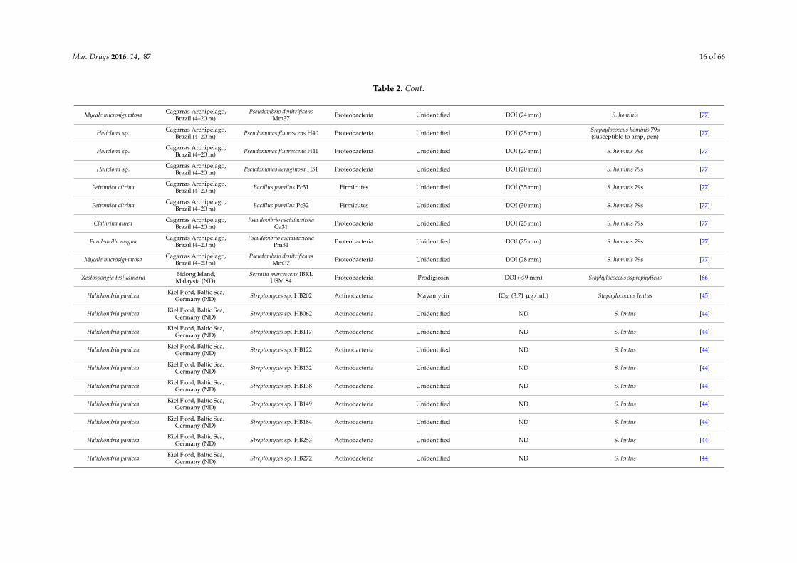
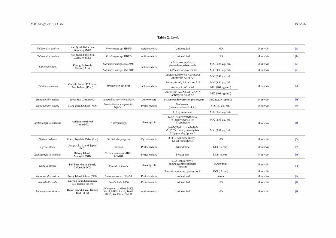
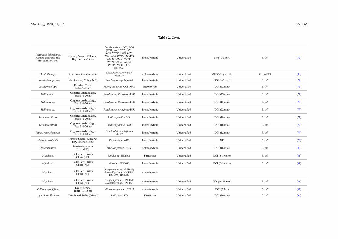
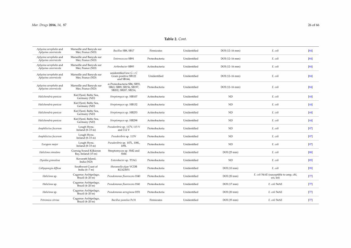
The screening procedure for antibacterial activity often includes both Gram positive andGram negative target strains, including, e.g., Staphylococcus spp., Streptococcus spp., Bacillus spp.,Clostridium spp., Escherichia spp., and Pseudomonas spp. From a medical point of view, these generareceive attention because they are well represented among the causative agents for human infectiousdiseases, such as pneumonia, urinary tract and blood stream infections [38,39]. Microbial isolates frommarine sponges have been shown to exhibit bioactivity against a wide spectrum of pathogenic bacteria(Table 2). The novel thiopeptide antibiotics YM-266183 (6) and YM-266184 (7) (Figure 2), which wereisolated from the sponge-associated bacterium Bacillus cereus QN03323, showed antibacterial activityagainst nosocomial infectious Gram positive bacteria in vitro [40,41]. Both YM-266183 and YM-266184effectively inhibited Staphylococcus aureus and vancomycin-resistant Enterococcus faecium as indicatedby minimal inhibition concentration (MIC) values as low as 0.025 µg/mL. In addition, compoundYM-266184 was found particularly active against methicillin resistant Staphylococcus aureus (MRSA)with a MIC of 0.39 µg/mL. Compound YM-266183 also inhibited MRSA but required a two-fold higherconcentration of the pure compound. Bioactivity of these thiopeptides was also observed againstStreptococcus epidermidis and Enterococcus spp. (Table 2). The compound kocurin (8) was identifiedfrom three sponge-associated actinobacteria: Kocuria marina F-276,310, Kocuria palustris F-276,345,and Micrococcus yunnanensis F-256,446 [42,43]. Kocurin is a new member of the thiazolyl peptidefamily and exhibited anti-MRSA activity with an MIC of 0.25 µg/mL, which to date is the most potentanti-MRSA compound reported from sponge-associated microbes. Scheenemaan et al. [44] isolatedStreptomyces sp. HB202 from the sponge Haliclona simulans, which lead to discovery of the polyketidemayamycin. In vitro assays with mayamycin (9) showed bioactivity against S. aureus and MRSA withIC50 values of 1.16 µg/mL and 0.58 µg/mL respectively, along with an IC50 of 0.14 µg/mL againstStaphylococcus epidermidis [45].
Although many studies on antibacterial activity from sponge-associated microbes includedGram negative strains (Table 2), reports on pronounced antibacterial compounds active against Gramnegative bacteria are limited in comparison to those that inhibit Gram positive strains. One of theexamples of an inhibitor of a Gram negative bacterium is the compound naphthacene glycosideSF2446A2 (10) isolated from Streptomyces sp. RV15 that was originally obtained from the marinesponge Dysidea tupha [46]. Naphthacene glycoside SF2446A2 (10) inhibited the Gram-negativebacterium Chlamydia trachomatis at an IC50 value of 2.81 ˘ 0.24 µg/mL. Reimer et al. [46] underlinedthat compound 10 not only effectively inhibited the formation of chlamydial inclusion bodiesduring the primary infection but also affected the ability of C. trachomatis in producing viableprogeny during the developmental cycle. Chlamydia trachomatis is an obligate intracellular Gramnegative bacterium which is a leading cause of sexually transmitted diseases, and currentlyno methods are available to treat this infectious microorganism [46,47]. Li et al. [48] isolatedfour new bisabolane-typesesquiterpenoids: aspergiterpenoid A, (´)-sydonol, (´)-sydonic acid,(´)-5-(hydroxymethyl)-2-(21,61,61-trimethyltetrahydro-2H-pyran-2-yl)phenol and a known compound(Z)-5-(Hydroxymethyl)-2-(61-methylhept-21-en-21-yl)phenol from a sponge-associated Aspergillus sp.(Table 2). Of these five substances, the compound sydonic acid (11) exhibited the lowest MIC valueagainst Escherichia coli at 1.33 µg/mL. This is the lowest inhibition concentration against E.coli reportedfrom a compound produced by sponge-associated microbes although the inhibition concentration isstill higher than the positive control ciprofloxacin (0.21 µg/mL) (Table 2).
Pruksakorn et al. [49] reported three prospective anti-tuberculosis compounds: trichoderin A(12), A1 and B from the sponge-associated fungus Trichoderma sp. 05FI48. Both under standardaerobic growth and dormancy-inducing hypoxic conditions, these three compounds inhibitedMycobacterium smegmatis, M. bovis BCG, and M. tuberculosis H37Rv with MIC values in the rangeof 0.02–2.0 µg/mL. Of these three compounds, trichoderin A was the most potent compound indicatedby the lowest MIC values against those Mycobacterium strains. Additional analysis revealed thatbioactivity of trichoderin A is based on its ability to inhibit adenosine triphosphate (ATP) synthesis

Mar. Drugs 2016, 14, 87 7 of 66
of mycobacteria [50]. Compounds such as trichoderin A are particularly important because in manycases, pathogens such as Campylobacter spp., Helicobacter pylori, and Legionella pneumophila are difficultto treat due to the fact that they are present in a dormant state [51]. Such physiologically inactive cellshighly contribute to the need for prolongued antibiotic treatments, which may lead to the emergenceof resistant strains [52,53].
Mar. Drugs 2016, 14, 87 7 of 79
difficult to treat due to the fact that they are present in a dormant state [51]. Such physiologically inactive cells highly contribute to the need for prolongued antibiotic treatments, which may lead to the emergence of resistant strains [52,53].
Figure 2. Chemical structures of the antibacterial compounds YM-266183 (6), YM-266184 (7), kocurin (8), mayamycin (9), naphthacene glycoside SF2446A2 (10), sydonic acid (11) and trichoderin A (12).
Figure 2. Chemical structures of the antibacterial compounds YM-266183 (6), YM-266184 (7), kocurin (8),mayamycin (9), naphthacene glycoside SF2446A2 (10), sydonic acid (11) and trichoderin A (12).

Mar. Drugs 2016, 14, 87 8 of 66
Table 2. Bioactive compounds with antibacterial activity from sponge-associated microbes.
Sponge Origin (Depth) Microorganism Phylum Compound Property Target References
Halichondria japonica Iriomote island, Japan (ND) Bacillus cereus QNO3323 Firmicutes Thiopeptide YM-266183 MIC (0.025 µg/mL) Staphylococcus aureus [40,41]
Halichondria japonica Iriomote island, Japan (ND) Bacillus cereus QNO3323 Firmicutes Thiopeptide YM-266184 MIC (0.025 µg/mL) S. aureus [40,41]
Halichondria panicea Kiel Fjord, Baltic Sea,Germany (ND) Streptomyces sp. HB202 Actinobacteria Mayamycin IC50 (1.16 µg/mL) S. aureus [45]
Spheciospongia vagabunda Red Sea (ND) Micrococcus sp. EG45 Actinobacteria Microluside A MIC (12.42 µg/mL) S. aureus NCTC 8325 [54]
Isodictya setifera Ross island, Antartica(30–40 m) Pseudomonas aeruginosa Proteobacteria Phenazine-1-carboxylic acid MIC (>4.99 µg/mL) S. aureus [55]
Isodictya setifera Ross island, Antartica(30–40 m) Pseudomonas aeruginosa Proteobacteria Phenazine-1-carboxamide MIC (>4.99 µg/mL) S. aureus [55]
Hymeniacidon perleve Bohai Sea, China (ND) Aspergillus versicolor MF359 Ascomycota 5-Methoxydihydrosterigmatocystin MIC (12.5 µg/mL) S. aureus [56]
Melophus sp. Lau group,Fiji islands (10 m) Penicillium sp. FF001 Ascomycota Citrinin MIC (1.95 µg/mL) S. aureus [57]
Petrosia sp. Jeju island, Korea (20 m) Aspergillus versicolor Ascomycota Averantin MIC (3.13 µg/mL) S. aureus SG511 [58]
Petrosia sp. Jeju island, Korea (20 m) Aspergillus versicolor Ascomycota Nidurufin MIC (6.25 µg/mL) S. aureus SG511 [58]
Petrosia sp. Jeju island, Korea (20 m) Aspergillus versicolor Ascomycota Averantin and nidurufin MIC (3.13 µg/mL) S. aureus 285 [58]
Petrosia sp. Jeju island, Korea (20 m) Aspergillus versicolor Ascomycota Averantin MIC (1.56 µg/mL) S. aureus 503 [58]
Petrosia sp. Jeju island, Korea (20 m) Aspergillus versicolor Ascomycota Nidurufin MIC (3.13 µg/mL) S aureus 503 [58]
Hymeniacidon perleve Nanji island, China (ND) Pseudoalteromonas piscicidaNJ6-3-1 Ascomycota Norharman
(beta-carboline alkaloid) MIC (50 µg/mL) S. aureus [59]
Halichondria panicea Bogil island, Korea (ND) Exophiala sp. Ascomycota Chlorohydroaspyrones A MIC (62.5 µg/mL) S. aureus [60]
Halichondria panicea Bogil island, Korea (ND) Exophiala sp. Ascomycota Chlorohydroaspyrones B MIC (62.5 µg/mL) S. aureus [60]
Axinella sp. South China Sea,China (ND) Eupenicillium sp. Ascomycota αβ-Dehydrocurvularin MIC (375 µg/mL) S. aureus [61]
Haliclona sp. Cagarras Archipelago,Brazil (4–20 m)
Pseudomonas fluorescensH40, H41 and Pseudomonas
aeruginosa H51Proteobacteria Diketopiperazine MIC (512 µg/mL) S. aureus [62]
Spongia officinalis Southeast Coast India(10–15 m) Streptomyces sp. MAPS15 Actinobacteria 2-pyrrolidone MIC (500 µg/mL) S. aureus PC6 [63]
Dysidea herbacea Koror, Republic Palau (1 m) Oscillatoria spongeliae Cyanobacteria 2-(21,41-dibromophenyl)-4,6-dibromophenol ND S. aureus [64]
Hyrtios altum Aragusuku island,Japan (ND) Vibrio sp. Proteobacteria Trisindoline DOI (10 mm) S. aureus [65]
Xestospongia testudinaria Bidong Island,Malaysia (ND)
Serratia marcescens IBRLUSM 84 Proteobacteria Prodigiosin DOI (ď9 mm) S. aureus [66]

Mar. Drugs 2016, 14, 87 9 of 66
Table 2. Cont.
unidentified South China Sea (10 m) Nocardiopsis sp. 13-33-15and 13-12-13 Actinobacteria
1,6-Dihydroxyphenazine DOI (25˘ 0.6 mm) S. aureus SJ51 [67]1,6-Dimethoxyphenazine DOI (21˘ 0.1 mm) S. aureus SJ51
Aplysina aerophoba Banyuls-sur-Mer, France(5–15 m) Bacillus subtilis A184 Firmicutes Surfactin Iturin Fengycin ND S. aureus [68]
Aplysina aerophoba Banyuls-sur-Mer, France(5–15 m) Bacillus subtilis A190 Firmicutes Surfactin ND S. aureus [68]
Halichondria sp. West Coast of India (10 m) Bacillus licheniformis SAB1 FirmicutesIndole DOI (7–10 mm) S. aureus [69]
3-Phenylpropionic DOI (4–6 mm) S. aureus
Niphates olemda Bali Bata National Park,Indonesia (ND) Curvularia lunata Ascomycota
1,3,8-Trihydroxy-6-methoxyanthraquinone
(lunatin) DOI (10 mm) S. aureus [70]
Bisanthraquinone cytoskyrin A
Haliclona simulans Gurraig Sound KilkieranBay, Ireland (15 m) Bacillus subtilis MMA7 Firmicutes Subtilomycin ND S. aureus [71]
Polymastia boletiformis,Axinella dissimilis and
Haliclona simulans
Gurraig Sound, KilkieranBay, Ireland (15 m)
Pseudovibrio sp. W64, W69,W89, W74 Proteobacteria Tropodithietic acid DOI (ě2 mm) S. aureus [72]
Polymastia boletiformis,Axinella dissimilis and
Haliclona simulans
Gurraig Sound, KilkieranBay, Ireland (15 m)
Pseudovibrio sp. JIC17, W10,W71, W74, W78, W96,WM33, WC15, WC30,
HMMA3
Actinobacteria Unidentified DOI (ě1 mm) S. aureus [72]
Polymastia boletiformis,Axinella dissimilis and
Haliclona simulans
Gurraig Sound, KilkieranBay, Ireland (15 m)
Pseudovibrio sp. JIC5, JIC6,W62, W63, W65, W99,
WC43, W85, W94, WM31,WM34, WM40, WC13,WC21, WC22, WC32,
WC41, HC6,
Proteobacteria Unidentified DOI (ě4 mm) S. aureus [72]
Dendrilla nigra Vizhinjam coast, India(10–15 m) Streptomyces sp. MSI051 Ascomycota Unidentified MIC (68˘ 2.8 µg
protein/mL) S. aureus [73]
Hymeniacidon perleve Nanji Island, China (ND) Pseudomonas sp. NJ6-3-1 Proteobacteria Unidentified DOI (3–5 mm) S. aureus [74]
Callyspongia spp Kovalam Coast,India (5–10 m) Aspergillus flavus GU815344 Proteobacteria Unidentified DOI (27 mm) S. aureus [75]
Haliclona sp. Cagarras Archipelago,Brazil (4–20 m) Pseudomonas fluorescens H41 Proteobacteria Unidentified DOI (20 mm) S. aureus [76]
Haliclona sp. Cagarras Archipelago,Brazil (4–20 m) Pseudomonas fluorescens H40 Proteobacteria Unidentified DOI (23 mm) S. aureus [77]
Haliclona sp. Cagarras Archipelago,Brazil (4–20 m) Pseudomonas fluorescens H41 Firmicutes Unidentified DOI (20 mm) S. aureus [77]
Haliclona sp. Cagarras Archipelago,Brazil (4–20 m) Pseudomonas aeruginosa H51 Proteobacteria Unidentified DOI (30 mm) S. aureus [77]

Mar. Drugs 2016, 14, 87 10 of 66
Table 2. Cont.
Dragmacidon reticulatus Cagarras Archipelago,Brazil (4–20 m) Bacillus pumilus Dr31 Actinobacteria Unidentified DOI (19 mm) S. aureus [77]
Petromica citrina Cagarras Archipelago,Brazil (4–20 m) Bacillus pumilus Pc31 Firmicutes Unidentified DOI (40 mm) S. aureus [77]
Petromica citrina Cagarras Archipelago,Brazil (4–20 m) Bacillus pumilus Pc32 Firmicutes Unidentified DOI (40 mm) S. aureus [77]
Clathrina aurea Cagarras Archipelago,Brazil (4–20 m)
Pseudovibrio ascidiaceicolaCa31 Proteobacteria Unidentified DOI (28 mm) S. aureus [77]
Paraleucilla magna Cagarras Archipelago,Brazil (4–20 m)
Pseudovibrio ascidiaceicolaPm31 Proteobacteria Unidentified DOI (27 mm) S. aureus [77]
Mycale microsigmatosa Cagarras Archipelago,Brazil (4–20 m)
Pseudovibrio denitrificansMm37 Proteobacteria Unidentified DOI (20 mm) S. aureus [77]
Axinella dissimilis Gurraig Sound, KilkieranBay, Ireland (15 m) Pseudovibrio Ad30 Proteobacteria Unidentified ND S. aureus [78]
Pseudoceratina clavata Heron Island, Great BarrierReef (14 m)
Salinispora sp. M102, M403,M412, M413, M414, SW10,
SW15 and SW17Actinobacteria Unidentified DOI (>5 mm) S. aureus [79]
Pseudoceratina clavata Heron Island,Australia (14 m) Salinispora sp. SW02 Actinobacteria Unidentified DOI (<5 mm) S. aureus [79]
Dendrilla nigra Southeast coast ofIndia (ND) Streptomyces sp. BTL7 Actinobacteria Unidentified DOI (16 mm) S. aureus [80]
Mycale sp. Gulei Port, Fujian,China (ND)
Bacillus sp. HNS004,HNS010 Firmicutes Unidentified DOI (15–30 mm) S. aureus [81]
Mycale sp. Gulei Port, Fujian,China (ND) Vibrio sp. HNS022, HNS029 Proteobacteria Unidentified DOI (15–30 mm) S. aureus [81]
Mycale sp. Gulei Port, Fujian,China (ND) Streptomyces sp. HNS054 Actinobacteria Unidentified DOI (15–30 mm) S. aureus [81]
Mycale sp. Gulei Port, Fujian,China (ND) Bacillus sp. HNS005 Firmicutes Unidentified DOI (10–15 mm) S. aureus [81]
Mycale sp. Gulei Port, Fujian,China (ND)
Cobetia sp. HNS027;Streptomyces sp. HNS047,HNS056; Nocardiopsis sp.
HNS048, HNS051, HNS055;Nocardia sp. HNS052
Actinobacteria Unidentified DOI (10–15 mm) S. aureus [81]
Mycale sp. Gulei Port, Fujian,China (ND) Bacillus sp. HNS015 Firmicutes Unidentified DOI (8–10 mm) S. aureus [81]
Mycale sp. Gulei Port, Fujian,China (ND) Pseudomonas sp. HNS021 Proteobacteria Unidentified DOI (8–10 mm) S. aureus [81]
Mycale sp. Gulei Port, Fujian,China (ND)
Cobetia sp. HNS023; Vibriosp. HNS038; Labrenzia sp.HNS063; Streptomyces sp.HNS049; Nocardiopsis sp.
HNS058
Actinobacteria Unidentified DOI (8–10 mm) S. aureus [81]

Mar. Drugs 2016, 14, 87 11 of 66
Table 2. Cont.
unidentified Rovinj, Croatia (3–20 m) Streptomyces sp. RV15 Actinobacteria Unidentified DOI (17 mm) S. aureus [82]
unidentified Rovinj, Croatia (3–20 m) Dietzia sp. EG67 Actinobacteria Unidentified DOI (13 mm) S. aureus [82]
unidentified Rovinj, Croatia (3–20 m) Microbacterium sp. EG69 Actinobacteria Unidentified DOI (13 mm) S. aureus [82]
unidentified Rovinj, Croatia (3–20 m) Micromonospora sp. RV115 Actinobacteria Unidentified DOI (12 mm) S. aureus [82]
unidentified Rovinj, Croatia (3–20 m) Rhodococcus sp. EG33 Actinobacteria Unidentified DOI (12 mm) S. aureus [82]
unidentified Rovinj, Croatia (3–20 m) Rubrobacter sp. RV113 Actinobacteria Unidentified DOI (9 mm) S. aureus [82]
Suberites carnosus Lough Hyne, Co. Cork,Ireland (15 m) Arthrobacter sp. W13C11 Actinobacteria Unidentified ND S. aureus [83]
Suberites carnosus Lough Hyne, Co. Cork,Ireland (15 m)
Pseudovibrio sp. W13S4,W13S21, W13S23, W13S26,
W13S31Proteobacteria Unidentified ND S. aureus [83]
Aplysina aerophoba andAplysina cavernicola
Marseille and Banyuls surMer, France (ND)
Bacillus SB8, SB17,Enterococcus SB91 Firmicutes Unidentified DOI (12–16 mm) S. aureus [84]
Aplysina aerophoba andAplysina cavernicola
Marseille and Banyuls surMer, France (ND) Arthrobacter SB95 Actinobacteria Unidentified DOI (12–16 mm) S. aureus [84]
Aplysina aerophoba andAplysina cavernicola
Marseille and Banyuls surMer, France (ND)
unidentified low G + CGram positive SB122 and
SB144Unidentified Unidentified DOI (12–16 mm) S. aureus [84]
Aplysina aerophoba andAplysina cavernicola
Marseille and Banyuls surMer, France (ND)
α-Proteobacteria SB6, SB55,SB63, SB89, SB156, SB197,
SB202, SB207, SB214Proteobacteria Unidentified DOI (12–16 mm) S. aureus [84]
Dysidea granulosa Kavaratti Island,India (ND) Enterobacter sp. TTAG Proteobacteria Unidentified DOI (22 mm) S. aureus [85]
Petrosia ficiformis Paraggi, Ligurian Sea,Italy (8 m) Rhodococcus sp. E1 Actinobacteria Unidentified ND S. aureus [86]
Unidentified Atlantic coast, USA (ND)
Kocuria palustris F-276,310;Kocuria marina F-276,345Micrococcus yunnanensis
F-256,446
Actinobacteria Kocurin MIC (0.25 µg/mL) methicillin-resistant Staphylococcus aureus(MRSA) [42,43]
Halichondria japonica Iriomote island, Japan (ND) Bacillus cereus QNO3323 Firmicutes Thiopeptide YM-266183 MIC (0.78 µg/mL) MRSA [40,41]
Halichondria japonica Iriomote island, Japan (ND) Bacillus cereus QNO3323 Firmicutes Thiopeptide YM-266184 MIC (0.39 µg/mL) MRSA [40,41]
Halichondria panicea Kiel Fjord, Baltic Sea,Germany (ND) Streptomyces sp. HB202 Actinobacteria Mayamycin IC50 (0.58 µg/mL) MRSA [45]
Melophus sp. Lau group,Fiji islands (10 m) Penicillium sp. FF001 Ascomycota Citrinin MIC (3.90 µg/mL) MRSA [57]
Halichondria panicea Bogil island, Korea (ND) Exophiala sp. AscomycotaChlorohydroaspyrones A MIC (125 µg/mL) MRSA
[60]Chlorohydroaspyrones B MIC (62.5 µg/mL) MRSA
Callyspongia spp. Gulf of Mannar, India (ND) Pseudomonas spp. RHLB 12 Proteobacteria Chromophore compound DOI (4 mm) at 50 µM MRSA [87]

Mar. Drugs 2016, 14, 87 12 of 66
Table 2. Cont.
Xestospongia testudinaria Bidong Island,Malaysia (ND)
Serratia marcescens IBRLUSM 84 Proteobacteria Prodigiosin DOI (22.5 mm) MRSA [66]
Halichondria sp. West Coast of India (10 m) Bacillus licheniformis SAB1 Firmicutes Indole 3-phenylpropionic DOI (4–6 mm) MRSA [69]
Haliclona simulans Gurraig Sound KilkieranBay, Ireland (15 m) Bacillus subtilis MMA7 Firmicutes Subtilomycin ND MRSA [71]
Haliclona sp. Cagarras Archipelago,Brazil (4–20 m) Pseudomonas fluorescens H40 Proteobacteria Unidentified DOI (23 mm) MRSA [77]
Haliclona sp. Cagarras Archipelago,Brazil (4–20 m) Pseudomonas fluorescens H41 Proteobacteria Unidentified DOI (27 mm) MRSA [77]
Haliclona sp. Cagarras Archipelago,Brazil (4–20 m) Pseudomonas aeruginosa H51 Proteobacteria Unidentified DOI (17 mm) MRSA [77]
Axinella dissimilis Gurraig Sound, KilkieranBay, Ireland (15 m) Pseudovibrio Ad30 Proteobacteria Unidentified ND MRSA [78]
Haliclona simulans Gurraig Sound, KilkieranBay, Ireland (15 m)
Streptomyces sp. SM2 andSM4 Actinobacteria Unidentified ND MRSA [88]
Haliclona sp. Cagarras Archipelago,Brazil (4–20 m) Pseudomonas fluorescens H40 Proteobacteria Unidentified DOI (20 mm) community-associated MRSA [77]
Haliclona sp. Cagarras Archipelago,Brazil (4–20 m) Pseudomonas fluorescens H41 Proteobacteria Unidentified DOI (22 mm) community-associated MRSA [77]
Haliclona sp. Cagarras Archipelago,Brazil (4–20 m) Pseudomonas aeruginosa H51 Proteobacteria Unidentified DOI (43 mm) community-associated MRSA [77]
Petromica citrina Cagarras Archipelago,Brazil (4–20 m) Bacillus pumilus Pc31 Firmicutes Unidentified DOI (40 mm) community-associated MRSA [77]
Petromica citrina Cagarras Archipelago,Brazil (4–20 m) Bacillus pumilus Pc32 Firmicutes Unidentified DOI (40 mm) community-associated MRSA [77]
Clathrina aurea Cagarras Archipelago,Brazil (4–20 m)
Pseudovibrio ascidiaceicolaCa31 Proteobacteria Unidentified DOI (17 mm) community-associated MRSA [77]
Paraleucilla magna Cagarras Archipelago,Brazil (4–20 m)
Pseudovibrio ascidiaceicolaPm31 Proteobacteria Unidentified DOI (25 mm) community-associated MRSA [77]
Mycale microsigmatosa Cagarras Archipelago,Brazil (4–20 m)
Pseudovibrio denitrificansMm37 Proteobacteria Unidentified DOI (20 mm) community-associated MRSA [77]
Aplysina aerophoba Banyuls-sur-Mer,France (15 m) Bacillus subtilis A202 Firmicutes Iturin ND multi drug-resistant S. aureus [68]
Halichondria panicea Bogil island, Korea (ND) Exophiala sp. Ascomycota Chlorohydroaspyrones A MIC (125 µg/mL) multi drug-resistant S. aureus [60]
Chlorohydroaspyrones B MIC (125 µg/mL) multi drug-resistant S. aureus [60]
Haliclona simulans Gurraig Sound KilkieranBay, Ireland (15 m) Bacillus subtilis MMA7 Firmicutes Subtilomycin ND heterogeneous vancomycin intermediate
Staphylococcus aureus (hVISA) [71]
Axinella dissimilis Gurraig Sound, KilkieranBay, Ireland (15 m) Pseudovibrio Ad30 Proteobacteria Unidentified ND hVISA [78]

Mar. Drugs 2016, 14, 87 13 of 66
Table 2. Cont.
Haliclona simulans Gurraig Sound KilkieranBay, Ireland (15 m)
Streptomyces sp. SM2 andSM4 Proteobacteria Unidentified ND hVISA [88]
Haliclona simulans Gurraig Sound KilkieranBay, Ireland (15 m)
Streptomyces sp. SM2 andSM4 Proteobacteria Unidentified ND vancomycin intermediate Staphylococcus
aureus (VISA) [88]
Melophus sp. Lau group,Fiji islands (10 m) Penicillium sp. FF001 Ascomycota Citrinin MIC (0.97 µg/mL) rifampicin-resistant S.aureus [57]
Halichondria panicea Baltic Sea (ND) Streptomyces sp. HB202 Actinobacteria Mayamycin IC50 (0.14 µg/mL) Staphylococcus epidermidis [45]
Halichondria panicea Kiel Fjord, Baltic Sea,Germany (ND) Streptomyces sp. HB202 Actinobacteria Streptophenazines G IC50 (3.57˘ 0.21 µg/mL) S. epidermidis [89]
Halichondria panicea Kiel Fjord, Baltic Sea,Germany (ND) Streptomyces sp. HB202 Actinobacteria Streptophenazines K IC50 (6.16˘ 0.85 µg/mL) S. epidermidis [89]
Axinella corrugata Arvoredo Biological MarineReserve, Brazil (ND) Penicillium sp. Ascomycota Dipeptide
cis-cyclo(leucyl-tyrosyl)reducing 85% of biofilm
formation at 1000 µg/mL S. epidermidis [90]
unidentified sponge Vizhijam coast (10–12 m) Aspergillus clavatus MFD15 Ascomycota 1H-1,2,4-Triazole-3-carboxaldehyde5-methyl MIC (800˘ 10 µg/mL) S. epidermidis [91]
Spongia officinalis Southeast Coast India(10–15 m) Streptomyces sp. MAPS15 Actinobacteria 2-Pyrrolidone MIC (500 µg/mL) S. epidermidis PC5 [63]
Xestospongia testudinaria Bidong Island,Malaysia (ND)
Serratia marcescens IBRLUSM 84 Proteobacteria Prodigiosin DOI (<9 mm) S. epidermidis [66]
Aplysina aerophoba Banyuls-sur-Mer, France(5–15 m) Bacillus subtilis A184 Firmicutes Surfactin Iturin Fengycin ND S. epidermidis [68]
Aplysina aerophoba Banyuls-sur-Mer, France(5–15 m) Bacillus subtilis A190 Firmicutes Surfactin ND S. epidermidis [68]
Haliclona sp. Cagarras Archipelago,Brazil (4–20 m) Pseudomonas fluorescens H40 Proteobacteria Unidentified DOI (35 mm) S. epidermidis [77]
Haliclona sp. Cagarras Archipelago,Brazil (4–20 m) Pseudomonas fluorescens H41 Proteobacteria Unidentified DOI (30 mm) S. epidermidis [77]
Haliclona sp. Cagarras Archipelago,Brazil (4–20 m) Pseudomonas aeruginosa H51 Proteobacteria Unidentified DOI (28 mm) S. epidermidis [77]
Dragmacidonreticulatus Cagarras Archipelago,Brazil (4–20 m) Bacillus pumilus Dr31 Firmicutes Unidentified DOI (20 mm) S. epidermidis [77]
Petromica citrina Cagarras Archipelago,Brazil (4–20 m) Bacillus pumilus Pc31 Firmicutes Unidentified DOI (45 mm) S. epidermidis [77]
Petromica citrina Cagarras Archipelago,Brazil (4–20 m) Bacillus pumilus Pc32 Firmicutes Unidentified DOI (38 mm) S. epidermidis [77]
Clathrina aurea Cagarras Archipelago,Brazil (4–20 m)
Pseudovibrio ascidiaceicolaCa31 Proteobacteria Unidentified DOI (25 mm) S. epidermidis [77]
Paraleucilla magna Cagarras Archipelago,Brazil (4–20 m)
Pseudovibrio ascidiaceicolaPm31 Proteobacteria Unidentified DOI (35 mm) S. epidermidis [77]

Mar. Drugs 2016, 14, 87 14 of 66
Table 2. Cont.
Mycale microsigmatosa Cagarras Archipelago,Brazil (4–20 m)
Pseudovibrio denitrificansMm37 Proteobacteria Unidentified DOI (30 mm) S. epidermidis [77]
Pseudoceratina clavata Heron Island,Australia (14 m)
Salinispora sp. M102, M403,M412, M413, M414, SW10,
SW15, SW17Actinobacteria Unidentified DOI (<5 mm) S. epidermidis [79]
Pseudoceratina clavata Heron Island,Australia (14 m) Salinispora sp. SW02 Actinobacteria Unidentified DOI (>5 mm) S. epidermidis [79]
Callyspongia diffusaBay of Bengal,
India (10–15 m)
Streptomyces sp. CPI 13 Actinobacteria Unidentified DOI (6.6 mm) S. epidermidis [92]
Micromonospora sp. CPI 12 Actinobacteria Unidentified DOI (6.6 mm) S. epidermidis [92]
Saccharomonospora sp. CPI 3 Actinobacteria Unidentified DOI (6.3 mm) S. epidermidis [92]
Haliclona sp. Cagarras Archipelago,Brazil (4–20 m) Pseudomonas fluorescens H40 Proteobacteria Unidentified DOI (25 mm) S. epidermidis 57s (susceptibile to amp,
cip, pen, tet) [77]
Haliclona sp. Cagarras Archipelago,Brazil (4–20 m) Pseudomonas fluorescens H41 Proteobacteria Unidentified DOI (25 mm) S. epidermidis 57s [77]
Haliclona sp. Cagarras Archipelago,Brazil (4–20 m) Pseudomonas aeruginosa H51 Proteobacteria Unidentified DOI (33 mm) S. epidermidis 57s [77]
Petromica citrina Cagarras Archipelago,Brazil (4–20 m) Bacillus pumilus Pc31 Firmicutes Unidentified DOI (30 mm) S. epidermidis 57s [77]
Petromica citrina Cagarras Archipelago,Brazil (4–20 m) Bacillus pumilus Pc32 Firmicutes Unidentified DOI (30 mm) S. epidermidis 57s [77]
Clathrina aurea Cagarras Archipelago,Brazil (4–20 m)
Pseudovibrio ascidiaceicolaCa31 Proteobacteria Unidentified DOI (15 mm) S. epidermidis 57s [77]
Paraleucilla magna Cagarras Archipelago,Brazil (4–20 m)
Pseudovibrio ascidiaceicolaPm31 Proteobacteria Unidentified DOI (17 mm) S. epidermidis 57s [77]
Mycale microsigmatosa Cagarras Archipelago,Brazil (4–20 m)
Pseudovibrio denitrificansMm37 Proteobacteria Unidentified DOI (16 mm) S. epidermidis 57s [77]
Xestospongia testudinaria Weizhou coral reef,China (ND)
Aspergillus sp. Ascomycota
(Z)-5-(Hydroxymethyl)-2-(61-methylhept-21-
en-21-yl)phenolMIC (4.66 µg/mL)
Staphylococcus albus [48]Aspergiterpenoid A MIC (1.24 µg/mL)
(´)-5-(Hydroxymethyl)-2-(21,61,61-trimethyltetrahydro-
2H-pyran-2-yl)phenolMIC (1.26 µg/mL)
Haliclona sp. Cagarras Archipelago,Brazil (4–20 m) Pseudomonas fluorescens H40 Proteobacteria Unidentified DOI (27 mm) Staphylococcus haemolyticus [77]
Haliclona sp. Cagarras Archipelago,Brazil (4–20 m) Pseudomonas fluorescens H41 Proteobacteria Unidentified DOI (27 mm) S. haemolyticus [77]
Haliclona sp. Cagarras Archipelago,Brazil (4–20 m) Pseudomonas aeruginosa H51 Proteobacteria Unidentified DOI (35 mm) S. haemolyticus [77]

Mar. Drugs 2016, 14, 87 15 of 66
Table 2. Cont.
Petromica citrina Cagarras Archipelago,Brazil (4–20 m) Bacillus pumilus Pc31 Firmicutes Unidentified DOI (40 mm) S. haemolyticus [77]
Petromica citrina Cagarras Archipelago,Brazil (4–20 m) Bacillus pumilus Pc32 Firmicutes Unidentified DOI (40 mm) S. haemolyticus [77]
Clathrina aurea Cagarras Archipelago,Brazil (4–20 m)
Pseudovibrio ascidiaceicolaCa31 Proteobacteria Unidentified DOI (38 mm) S. haemolyticus [77]
Paraleucilla magna Cagarras Archipelago,Brazil (4–20 m)
Pseudovibrio ascidiaceicolaPm31 Proteobacteria Unidentified DOI (40 mm) S. haemolyticus [77]
Mycale microsigmatosa Cagarras Archipelago,Brazil (4–20 m)
Pseudovibrio denitrificansMm37 Proteobacteria Unidentified DOI (43 mm) S. haemolyticus [77]
Haliclona sp. Cagarras Archipelago,Brazil (4–20 m) Pseudomonas fluorescens H40 Proteobacteria Unidentified DOI (19 mm) S. haemolyticus 109s
(susceptible to amp, gen, oxa, pen) [77]
Haliclona sp. Cagarras Archipelago,Brazil (4–20 m) Pseudomonas fluorescens H41 Proteobacteria Unidentified DOI (15 mm) S. haemolyticus 109s [77]
Haliclona sp. Cagarras Archipelago,Brazil (4–20 m) Pseudomonas aeruginosa H51 Proteobacteria Unidentified DOI (35 mm) S. haemolyticus 109s [77]
Petromica citrina Cagarras Archipelago,Brazil (4–20 m) Bacillus pumilus Pc31 Firmicutes Unidentified DOI (31 mm) S. haemolyticus 109s [77]
Petromica citrina Cagarras Archipelago,Brazil (4–20 m) Bacillus pumilus Pc32 Firmicutes Unidentified DOI (36 mm) S. haemolyticus 109s [77]
Clathrina aurea Cagarras Archipelago,Brazil (4–20 m)
Pseudovibrio ascidiaceicolaCa31 Proteobacteria Unidentified DOI (23 mm) S. haemolyticus 109s [77]
Paraleucilla magna Cagarras Archipelago,Brazil (4–20 m)
Pseudovibrio ascidiaceicolaPm31 Proteobacteria Unidentified DOI (30 mm) S. haemolyticus 109s [77]
Mycale microsigmatosa Cagarras Archipelago,Brazil (4–20 m)
Pseudovibrio denitrificansMm37 Proteobacteria Unidentified DOI (20 mm) S. haemolyticus 109s [77]
Haliclona sp. Cagarras Archipelago,Brazil (4–20 m) Pseudomonas fluorescens H40 Proteobacteria Unidentified DOI (31mm) Staphylococcus hominis [77]
Haliclona sp. Cagarras Archipelago,Brazil (4–20 m) Pseudomonas fluorescens H41 Proteobacteria Unidentified DOI (28 mm) S. hominis [77]
Haliclona sp. Cagarras Archipelago,Brazil (4–20 m) Pseudomonas aeruginosa H51 Proteobacteria Unidentified DOI (37 mm) S. hominis [77]
Petromica citrina Cagarras Archipelago,Brazil (4–20 m) Bacillus pumilus Pc31 Firmicutes Unidentified DOI (41 mm) S. hominis [77]
Petromica citrina Cagarras Archipelago,Brazil (4–20 m) Bacillus pumilus Pc32 Firmicutes Unidentified DOI (43 mm) S. hominis [77]
Clathrina aurea Cagarras Archipelago,Brazil (4–20 m)
Pseudovibrio ascidiaceicolaCa31 Proteobacteria Unidentified DOI (23 mm) S. hominis [77]
Paraleucilla magna Cagarras Archipelago,Brazil (4–20 m)
Pseudovibrio ascidiaceicolaPm31 Proteobacteria Unidentified DOI (25 mm) S. hominis [77]
Mar. Drugs 2016, 14, 87 16 of 66
Table 2. Cont.
Mycale microsigmatosa Cagarras Archipelago,Brazil (4–20 m)
Pseudovibrio denitrificansMm37 Proteobacteria Unidentified DOI (24 mm) S. hominis [77]
Haliclona sp. Cagarras Archipelago,Brazil (4–20 m) Pseudomonas fluorescens H40 Proteobacteria Unidentified DOI (25 mm) Staphylococcus hominis 79s
(susceptible to amp, pen) [77]
Haliclona sp. Cagarras Archipelago,Brazil (4–20 m) Pseudomonas fluorescens H41 Proteobacteria Unidentified DOI (27 mm) S. hominis 79s [77]
Haliclona sp. Cagarras Archipelago,Brazil (4–20 m) Pseudomonas aeruginosa H51 Proteobacteria Unidentified DOI (20 mm) S. hominis 79s [77]
Petromica citrina Cagarras Archipelago,Brazil (4–20 m) Bacillus pumilus Pc31 Firmicutes Unidentified DOI (35 mm) S. hominis 79s [77]
Petromica citrina Cagarras Archipelago,Brazil (4–20 m) Bacillus pumilus Pc32 Firmicutes Unidentified DOI (30 mm) S. hominis 79s [77]
Clathrina aurea Cagarras Archipelago,Brazil (4–20 m)
Pseudovibrio ascidiaceicolaCa31 Proteobacteria Unidentified DOI (25 mm) S. hominis 79s [77]
Paraleucilla magna Cagarras Archipelago,Brazil (4–20 m)
Pseudovibrio ascidiaceicolaPm31 Proteobacteria Unidentified DOI (25 mm) S. hominis 79s [77]
Mycale microsigmatosa Cagarras Archipelago,Brazil (4–20 m)
Pseudovibrio denitrificansMm37 Proteobacteria Unidentified DOI (28 mm) S. hominis 79s [77]
Xestospongia testudinaria Bidong Island,Malaysia (ND)
Serratia marcescens IBRLUSM 84 Proteobacteria Prodigiosin DOI (ď9 mm) Staphylococcus saprophyticus [66]
Halichondria panicea Kiel Fjord, Baltic Sea,Germany (ND) Streptomyces sp. HB202 Actinobacteria Mayamycin IC50 (3.71 µg/mL) Staphylococcus lentus [45]
Halichondria panicea Kiel Fjord, Baltic Sea,Germany (ND) Streptomyces sp. HB062 Actinobacteria Unidentified ND S. lentus [44]
Halichondria panicea Kiel Fjord, Baltic Sea,Germany (ND) Streptomyces sp. HB117 Actinobacteria Unidentified ND S. lentus [44]
Halichondria panicea Kiel Fjord, Baltic Sea,Germany (ND) Streptomyces sp. HB122 Actinobacteria Unidentified ND S. lentus [44]
Halichondria panicea Kiel Fjord, Baltic Sea,Germany (ND) Streptomyces sp. HB132 Actinobacteria Unidentified ND S. lentus [44]
Halichondria panicea Kiel Fjord, Baltic Sea,Germany (ND) Streptomyces sp. HB138 Actinobacteria Unidentified ND S. lentus [44]
Halichondria panicea Kiel Fjord, Baltic Sea,Germany (ND) Streptomyces sp. HB149 Actinobacteria Unidentified ND S. lentus [44]
Halichondria panicea Kiel Fjord, Baltic Sea,Germany (ND) Streptomyces sp. HB184 Actinobacteria Unidentified ND S. lentus [44]
Halichondria panicea Kiel Fjord, Baltic Sea,Germany (ND) Streptomyces sp. HB253 Actinobacteria Unidentified ND S. lentus [44]
Halichondria panicea Kiel Fjord, Baltic Sea,Germany (ND) Streptomyces sp. HB272 Actinobacteria Unidentified ND S. lentus [44]

Mar. Drugs 2016, 14, 87 17 of 66
Table 2. Cont.
Halichondria panicea Kiel Fjord, Baltic Sea,Germany (ND) Streptomyces sp. HB288 Actinobacteria Unidentified ND S. lentus [44]
Halichondria panicea Kiel Fjord, Baltic Sea,Germany (ND) Streptomyces sp. HB298 Actinobacteria Unidentified ND S. lentus [44]
Halichondria panicea Kiel Fjord, Baltic Sea,Germany (ND) Streptomyces sp. HB328 Actinobacteria Unidentified ND S. lentus [44]
Kiel Fjord, Baltic Sea,Germany (ND) Streptomyces sp. HB375 Actinobacteria Unidentified ND S. lentus [44]
Halichondria panicea Kiel Fjord, Baltic Sea,Germany (ND) Streptomyces sp. HB383 Actinobacteria Unidentified ND S. lentus [44]
Dendrilla nigra Southwest Coast of India(10-12 m)
Nocardiopsis dassonvilleiMAD08 Actinobacteria Unidentified MIC (600 µg/mL) Staphylococcus sp. PC8 [93]
Halichondria japonica Iriomote island, Japan (ND) Bacillus cereus QNO3323 Firmicutes Thiopeptide YM-266183 MIC (1.56 µg/mL) Methicillin-Resistant Streptococcusepidermidis (MRSE) [40,41]
Halichondria japonica Iriomote island, Japan (ND) Bacillus cereus QNO3323 Firmicutes Thiopeptide YM-266184 MIC (0.2 µg/mL) MRSE [40,41]
Aplysina aerophoba Banyuls-sur-Mer, France(5–15 m) Bacillus subtilis A202 Firmicutes Iturin ND Multi drug-resistant S. epidermidis [68]
Dysidea granulosa Kavaratti Island,India (ND) Enterobacter sp. TTAG Proteobacteria Unidentified DOI (23 mm), MIC crude
extract (5 mg/mL) Streptococcus sp. [85]
Petrosia sp. Jeju island, Korea (20 m) Aspergillus versicolor Ascomycota Averantin MIC (0.78 µg/mL) Streptococcus pyogenes 308A [58]
Petrosia sp. Jeju island, Korea (20 m) Aspergillus versicolor Ascomycota Nidurufin MIC (3.13 µg/mL) Streptococcus pyogenes 308A [58]
Petrosia sp. Jeju island, Korea (20 m) Aspergillus versicolor Ascomycota Averantin MIC (3.13 µg/mL) Streptococcus pyogenes 77A [58]
Petrosia sp. Jeju island, Korea (20 m) Aspergillus versicolor Ascomycota Nidurufin MIC (6.25 µg/mL) Streptococcus pyogenes 77A [58]
Halichondria sp. West Coast of India (10 m) Bacillus licheniformis SAB1 Firmicutes Indole DOI (1–3 mm) Streptococcus pyogenes [69]
Halichondria sp. West Coast of India (10 m) Bacillus licheniformis SAB1 Firmicutes 3-Phenylpropionic DOI (4–6 mm) Streptococcus pyogenes [69]
Haliclona simulans Gurraig Sound KilkieranBay, Ireland (15 m)
Streptomyces sp. SM2 andSM4 Actinobacteria Unidentified ND Streptococcus pneumoniae [88]
Callyspongia diffusa Bay of Bengal,India (10–15 m) Saccharomonospora sp. CPI 9 Actinobacteria Unidentified ND haemolytic Streptococcus sp (6.3) [92]
Halichondria japonica Iriomote island, Japan (ND) Bacillus cereus QNO3323 Firmicutes Thiopeptide YM-266183 MIC (1.56 µg/mL) Bacillus subtilis ATCC 633 [40,41]
Halichondria japonica Iriomote island, Japan (ND) Bacillus cereus QNO3323 Firmicutes Thiopeptide YM-266184 MIC (1.56 µg/mL) B. subtilis ATCC 633 [40,41]
Halichondria panicea Kiel Fjord, Baltic Sea,Germany (ND) Streptomyces sp. HB202 Actinobacteria Mayamycin IC50 (3.71 µg/mL) B. subtilis [45]
Halichondria panicea Kiel Fjord, Baltic Sea,Germany (ND) Streptomyces sp. HB202 Actinobacteria Streptophenazines G IC50 (3.49˘ 0.38 µg/mL) B. subtilis [89]

Mar. Drugs 2016, 14, 87 18 of 66
Table 2. Cont.
Halichondria panacea Kiel Fjord, Baltic Sea,Germany (ND) Streptomyces sp. HB202 Actinobacteria Streptophenazines K IC50 (9.18˘ 2.89 µg/mL) B. subtilis [89]
Halichondria panicea Kiel Fjord, Baltic Sea,Germany (ND) Streptomyces sp. HB084 Actinobacteria Unidentified ND B. subtilis [44]
Halichondria panicea Kiel Fjord, Baltic Sea,Germany (ND) Streptomyces sp. HB095 Actinobacteria Unidentified ND B. subtilis [44]
Halichondria panicea Kiel Fjord, Baltic Sea,Germany (ND) Streptomyces sp. HB096 Actinobacteria Unidentified ND B. subtilis [44]
Halichondria panicea Kiel Fjord, Baltic Sea,Germany (ND) Streptomyces sp. HB105 Actinobacteria Unidentified ND B. subtilis [44]
Halichondria panicea Kiel Fjord, Baltic Sea,Germany (ND) Streptomyces sp. HB107 Actinobacteria Unidentified ND B. subtilis [44]
Halichondria panicea Kiel Fjord, Baltic Sea,Germany (ND) Streptomyces sp. HB116 Actinobacteria Unidentified ND B. subtilis [44]
Halichondria panicea Kiel Fjord, Baltic Sea,Germany (ND) Streptomyces sp. HB117 Actinobacteria Unidentified ND B. subtilis [44]
Halichondria panicea Kiel Fjord, Baltic Sea,Germany (ND) Streptomyces sp. HB118 Actinobacteria Unidentified ND B. subtilis [44]
Halichondria panicea Kiel Fjord, Baltic Sea,Germany (ND) Streptomyces sp. HB122 Actinobacteria Unidentified ND B. subtilis [44]
Halichondria panicea Kiel Fjord, Baltic Sea,Germany (ND) Streptomyces sp. HB132 Actinobacteria Unidentified ND B. subtilis [44]
Halichondria panicea Kiel Fjord, Baltic Sea,Germany (ND) Streptomyces sp. HB138 Actinobacteria Unidentified ND B. subtilis [44]
Halichondria panicea Kiel Fjord, Baltic Sea,Germany (ND) Streptomyces sp. HB181 Actinobacteria Unidentified ND B. subtilis [44]
Halichondria panicea Kiel Fjord, Baltic Sea,Germany (ND) Streptomyces sp. HB184 Actinobacteria Unidentified ND B. subtilis [44]
Halichondria panicea Kiel Fjord, Baltic Sea,Germany (ND) Streptomyces sp. HB202 Actinobacteria Unidentified ND B. subtilis [44]
Halichondria panicea Kiel Fjord, Baltic Sea,Germany (ND) Streptomyces sp. HB253 Actinobacteria Unidentified ND B. subtilis [44]
Halichondria panicea Kiel Fjord, Baltic Sea,Germany (ND) Streptomyces sp. HB272 Actinobacteria Unidentified ND B. subtilis [44]
Halichondria panicea Kiel Fjord, Baltic Sea,Germany (ND) Streptomyces sp. HB298 Actinobacteria Unidentified ND B. subtilis [44]
Halichondria panicea Kiel Fjord, Baltic Sea,Germany (ND) Streptomyces sp. HB328 Actinobacteria Unidentified ND B. subtilis [44]
Mar. Drugs 2016, 14, 87 19 of 66
Table 2. Cont.
Halichondria panicea Kiel Fjord, Baltic Sea,Germany (ND) Streptomyces sp. HB375 Actinobacteria Unidentified ND B. subtilis [44]
Halichondria panicea Kiel Fjord, Baltic Sea,Germany (ND) Streptomyces sp. HB383 Actinobacteria Unidentified ND B. subtilis [44]
Callyspongia sp. Kyung-Po beach,Korea (12 m)
Brevibacterium sp. KMD 003Actinobacteria
6-Hydroxymethyl-1-phenazine-carboxamide MIC (5.06 µg/mL) B. subtilis [94]
Brevibacterium sp. KMD 003 1,6-Phenazinedimethanol MIC (4.80 µg/mL) B. subtilis [94]
Haliclona simulansGurraig Sound Kilkieran
Bay, Ireland (15 m) Streptomyces sp. SM8 Actinobacteria
Mixture Kitamycin A or B andAntimycin A3 or A7 MIC (7.42 µg/mL)
B. subtilis [95]Antimycin A2, A8, A11 or A17 MIC (9.40 µg/mL)
Antimycin A3 or A7 MIC (400 µg/mL)
Antimycin A2, A8, A11 or A17,antimycin A3 or A7 MIC (400 µg/mL)
Hymeniacidon perleve Bohai Sea, China (ND) Aspergillus versicolor MF359 Ascomycota 5-Methoxydihydrosterigmatocystin MIC (3.125 µg/mL) B. subtilis [56]
Hymeniacidon perleve Nanji island, China (ND) Pseudoalteromonas piscicidaNJ6-3-1 Proteobacteria Norharman
(beta-carboline alkaloid) MIC (50 µg/mL) B. subtilis [59]
Xestospongia testudinaria Weizhou coral reef,China (ND)
Aspergillus sp. Ascomycota
(´)-Sydonic acid MIC (0.66 µg/mL)
B. subtilis [48]
(Z)-5-(Hydroxymethyl)-2-(61-methylhept-21-en-
21-yl)phenolMIC (2.33 µg/mL)
(´)-5-(Hydroxymethyl)-2-(21,61,61-trimethyltetrahydro-
2H-pyran-2-yl)phenolMIC (0.62 µg/mL)
Dysidea herbacea Koror, Republic Palau (1 m) Oscillatoria spongeliae Cyanobacteria 2-(21,41-Dibromophenyl)-4,6-dibromophenol ND B. subtilis [64]
Hyrtios altum Aragusuku island, Japan(ND) Vibrio sp. Proteobacteria Trisindoline DOI (17 mm) B. subtilis [65]
Xestospongia testudinaria Bidong Island,Malaysia (ND)
Serratia marcescens IBRLUSM 84 Proteobacteria Prodigiosin DOI (<9 mm) B. subtilis [66]
Niphates olemda Bali Bata National Park,Indonesia (ND) Curvularia lunata Ascomycota
1,3,8-Trihydroxy-6-methoxyanthraquinone
(lunatin)DOI (9 mm) B. subtilis [70]
Bisanthraquinone cytoskyrin A DOI (12 mm) B. subtilis
Hymeniacidon perleve Nanji Island, China (ND) Pseudomonas sp. NJ6-3-1 Proteobacteria Unidentified 5 mm B. subtilis [74]
Axinella dissimilis Gurraig Sound, KilkieranBay, Ireland (15 m) Pseudovibrio Ad30 Proteobacteria Unidentified ND B. subtilis [78]
Pseudoceratina clavata Heron Island, Great BarrierReef (14 m)
Salinispora sp. M102, M403,M412, M413, M414, SW02,SW10, SW 15 and SW 17
Actinobacteria Unidentified ND B. subtilis [79]

Mar. Drugs 2016, 14, 87 20 of 66
Table 2. Cont.
Dendrilla nigra Southeast coast of India(ND) Streptomyces sp. BTL7 Actinobacteria Unidentified DOI (15 mm) B. subtilis [80]
Mycale sp. Gulei Port, Fujian,China (ND)
Bacillus sp. HNS004HNS015; Firmicutes Unidentified DOI (8–10 mm) B. subtilis [81]
Mycale sp. Gulei Port, Fujian,China (ND)
Pseudomonas sp. HNS021;HNS027; Vibrio sp. HNS038 Proteobacteria Unidentified DOI (8–10 mm) B. subtilis [81]
Mycale sp. Gulei Port, Fujian,China (ND)
Labrenzia sp. HNS063;Streptomyces sp. HNS047;Nocardiopsis sp. HNS048,
HNS055, HNS058; Cobetiasp. HNS023,
Actinobacteria Unidentified DOI (8–10 mm) B. subtilis [81]
Mycale sp. Gulei Port, Fujian,China (ND)
Bacillus sp. HNS005,HNS010, Firmicutes Unidentified DOI (10–15 mm) B. subtilis [81]
Mycale sp. Gulei Port, Fujian,China (ND)
Streptomyces sp. HNS049,HNS056 Actinobacteria Unidentified DOI (10–15 mm) B. subtilis [81]
Mycale sp. Gulei Port, Fujian,China (ND)
Vibrio sp. HNS022,HNS029; Firmicutes Unidentified DOI (15–30 mm) B. subtilis [81]
Mycale sp. Gulei Port, Fujian,China (ND) Streptomyces sp. HNS054 Actinobacteria Unidentified DOI (15–30 mm) B. subtilis [81]
Sigmadocia fibulatus Hare Island, India (5-10 m) Bacillus sp. SC3 Firmicutes Unidentified ND B. subtilis [96]
Amphilectus fucorum Lough Hyne,Ireland (8–15 m)
Pseudovibrio sp. 113VPseudovibrio 83V1 Proteobacteria Unidentified ND B. subtilis [97]
Eurypon major Lough Hyne,Ireland (8–15 m)
Pseudovibrio sp. 107L, 108L,109L Proteobacteria Unidentified ND B. subtilis [97]
Suberites carnosus Lough Hyne, Co. Cork,Ireland (15 m) Arthrobacter sp. W13C11 Actinobacteria Unidentified ND B. subtilis [83]
Suberites carnosus Lough Hyne, Co. Cork,Ireland (15 m)
Pseudovibrio sp. W13S4,W13S21, W13S23, W13S26,
W13S31Proteobacteria Unidentified ND B. subtilis [83]
Haliclona simulans Gurraig Sound, KilkieranBay, Ireland (15 m)
Streptomyces sp. SM2 andSM4 Actinobacteria Unidentified ND B. subtilis [88]
Isodictya setifera Ross island, Antartica(30–40 m) Pseudomonasaeruginosa Proteobacteria Phenazine-1-carboxylic acid and
phenazine-1-carboxamide MIC (<0.49 µg/mL) Bacillus cereus [55]
Xestospongia testudinaria Weizhou coral reef,China (ND) Aspergillus sp. Ascomycota
(Z)-5-(Hydroxymethyl)-2-(61-methylhept-21-
en-21-yl)phenolMIC (2.33 µg/mL) B. cereus [48]
Xestospongia testudinaria Bidong Island,Malaysia (ND)
Serratia marcescens IBRLUSM 84 Proteobacteria Prodigiosin DOI (10–14 mm) B. cereus [66]
Dendrilla nigra Vizhinjam coast, India(10–15 m) Streptomyces sp. MSI051 Actinobacteria Unidentified MIC (46˘ 1.62 µg
protein/mL) B. cereus [73]
Axinella dissimilis Gurraig Sound, KilkieranBay, Ireland (15 m) Pseudovibrio Ad30 Proteobacteria Unidentified ND B. cereus [78]

Mar. Drugs 2016, 14, 87 21 of 66
Table 2. Cont.
Dendrilla nigra Southeast coast ofIndia (ND) Streptomyces sp. BTL7 Actinobacteria Unidentified DOI (16 mm) B. cereus [80]
Haliclona simulans Gurraig Sound KilkieranBay, Ireland (15 m)
Streptomyces sp. SM2 andSM4 Actinobacteria Unidentified ND B. cereus [88]
Xestospongia testudinaria Bidong Island,Malaysia (ND)
Serratia marcescens IBRLUSM 84 Proteobacteria Prodigiosin DOI (10–14 mm) Bacillus licheniformis [66]
Xestospongia testudinaria Bidong Island,Malaysia (ND)
Serratia marcescens IBRLUSM 84 Proteobacteria Prodigiosin DOI (<9 mm) Bacillus thuringiensis [66]
unidentified South China Sea (10 m) Nocardiopsis sp. 13-33-15and 13-12-13
Actinobacteria1,6-Dihydroxyphenazine DOI (16˘ 0.5 mm)
Bacillus mycoides SJ14 [67]1,6-Dimethoxyphenazine DOI (20˘ 0.4 mm)
Aplysina aerophoba Banyuls-sur-Mer, France(5–15 m) Bacillus subtilis A184 Firmicutes Surfactin iturin fengycin ND Bacillus megaterium [68]
Aplysina aerophoba Banyuls-sur-Mer, France(5–15 m) Bacillus subtilis A190 Firmicutes Surfactin ND B. megaterium [68]
Aplysina aerophoba Banyuls-sur-Mer, France(5–15 m) Bacillus subtilis A202 Firmicutes Iturin ND B. megaterium [68]
Haliclona simulans Gurraig Sound KilkieranBay, Ireland (15 m) Bacillus subtilis MMA7 Firmicutes Subtilomycin ND B. megaterium [71]
Dysidea avara Mediterranean sea (ND) Actinokinespora sp. EG49Actinobacteria
1,6-Dihydroxyphenazine (resultof the co-culture) DOI (11 mm) Bacillus sp. P25 [98]
Spheciospongia vagabunda Red Sea (ND) Nocardiopsis sp. RV163
Callyspongia diffusa Bay of Bengal,India (10–15 m) Streptomyces sp. CPI 13 Actinobacteria Unidentified DOI (6.6 mm) Bacillus sp. [92]
Callyspongia diffusa Bay of Bengal,India (10–15 m) Micromonospora sp. CPI 12 Actinobacteria Unidentified DOI (8 mm) Bacillus sp. [92]
Haliclona sp. Cagarras Archipelago,Brazil (4–20 m) Pseudomonas fluorescens H40 Proteobacteria Unidentified DOI (19 mm) Enterococcus faecalis [77]
Haliclona sp. Cagarras Archipelago,Brazil (4–20 m) Pseudomonas fluorescens H41 Proteobacteria Unidentified DOI (17 mm) E. faecalis [77]
Haliclona sp. Cagarras Archipelago,Brazil (4–20 m) Pseudomonas aeruginosa H51 Proteobacteria Unidentified DOI (32 mm) E. faecalis [77]
Clathrina aurea Cagarras Archipelago,Brazil (4–20 m)
Pseudovibrio ascidiaceicolaCa31 Proteobacteria Unidentified DOI (11 mm), E. faecalis [77]
Paraleucilla magna Cagarras Archipelago,Brazil (4–20 m)
Pseudovibrio ascidiaceicolaPm31 Proteobacteria Unidentified DOI (12 mm), E. faecalis [77]
Mycale microsigmatosa Cagarras Archipelago,Brazil (4–20 m)
Pseudovibrio denitrificansMm37 Proteobacteria Unidentified DOI (14 mm) E. faecalis [77]

Mar. Drugs 2016, 14, 87 22 of 66
Table 2. Cont.
unidentified Rovinj, Croatia (3–20 m) Streptomyces sp. RV15 Actinobacteria Unidentified DOI (11 mm) E. faecalis [82]
unidentified Rovinj, Croatia (3–20 m) Microbacterium sp. EG69 Actinobacteria Unidentified DOI (9 mm) E. faecalis [82]
unidentified Rovinj, Croatia (3–20 m) Micromonospora sp. RV115 Actinobacteria Unidentified DOI (10 mm) E. faecalis [82]
unidentified Rovinj, Croatia (3–20 m) Rhodococcus sp. EG33 Actinobacteria Unidentified DOI (8 mm) E. faecalis [82]
Halocondria japonica Iriomote island, Japan (ND) Bacillus cereus QNO3323 Firmicutes Thiopeptide YM-266183 MIC (0.1 µg/mL) E. faecalis CAY 04_1 [40,41]
Halocondria japonica Iriomote island, Japan (ND) Bacillus cereus QNO3323 Firmicutes Thiopeptide YM-266184 MIC (0.025 µg/mL) E. faecalis CAY 04_1 [40,41]
Spheciospongia vagabunda Red Sea (ND) Micrococcus sp. EG45 Actinobacteria Microluside A MIC (9.55 µg/mL) E. faecalis JH212 [54]
Haliclona sp. Cagarras Archipelago,Brazil (4–20 m) Pseudomonas fluorescens H41 Proteobacteria Unidentified DOI (20 mm) E. faecalis 5AE (susceptible to van) [77]
Clathrina aurea Cagarras Archipelago,Brazil (4–20 m)
Pseudovibrio ascidiaceicolaCa31 Proteobacteria Unidentified DOI (12 mm) E. faecalis 5AE [77]
Mycale microsigmatosa Cagarras Archipelago,Brazil (4–20 m)
Pseudovibrio denitrificansMm37 Proteobacteria Unidentified DOI (15 mm) E. faecalis 5AE [77]
Halocondria japonica Iriomote island, Japan (ND) Bacillus cereus QNO3323 Firmicutes Thiopeptide YM-266183 MIC 0.2 µg/mL Enterococcus faecium CAY 09_1 [40,41]
Halocondria japonica Iriomote island, Japan (ND) Bacillus cereus QNO3323 Firmicutes Thiopeptide YM-266184 MIC (0.05 µg/mL) E. faecium CAY 09_1 [40,41]
Halocondria japonica Iriomote island, Japan (ND) Bacillus cereus QNO3323 Firmicutes Thiopeptide YM-266183 MIC (0.025 µg/mL) Vancomycin-Resistant E. faeciumCAY 09_2 [40,41]
Halocondria japonica Iriomote island, Japan (ND) Bacillus cereus QNO3323 Firmicutes Thiopeptide YM-266184 MIC (0.025 µg/mL) Vancomycin-Resistant E. faeciumCAY 09_2 [40,41]
Melophus sp. Lau group,Fiji islands (10 m) Penicillium sp. FF001 Ascomycota Citrinin MIC (1.95 µg/mL) Vancomycin-resistant E. faecium [57]
Haliclona simulans Gurraig Sound KilkieranBay, Ireland (15 m) Bacillus subtilis MMA7 Firmicutes Subtilomycin ND E. faecium [71]
Haliclona sp. Cagarras Archipelago,Brazil (4–20 m) Pseudomonas fluorescens H40 Proteobacteria Unidentified DOI (18 mm) E. faecium [77]
Haliclona sp. Cagarras Archipelago,Brazil (4–20 m) Pseudomonas fluorescens H41 Proteobacteria Unidentified DOI (21 mm) E. faecium [77]
Haliclona sp. Cagarras Archipelago,Brazil (4–20 m) Pseudomonas aeruginosa H51 Proteobacteria Unidentified DOI (30 mm) E. faecium [77]
Dragmacidonreticulatus Cagarras Archipelago,Brazil (4–20 m) Bacillus pumilus Dr31 Firmicutes Unidentified DOI (20 mm) E. faecium [77]
Petromica citrina Cagarras Archipelago,Brazil (4–20 m) Bacillus pumilus Pc31 Firmicutes Unidentified DOI (23 mm) E. faecium [77]
Petromica citrina Cagarras Archipelago,Brazil (4–20 m) Bacillus pumilus Pc32 Firmicutes Unidentified DOI (20 mm) E. faecium [77]
Clathrina aurea Cagarras Archipelago,Brazil (4–20 m)
Pseudovibrio ascidiaceicolaCa31 Proteobacteria Unidentified DOI (22 mm) E. faecium [77]

Mar. Drugs 2016, 14, 87 23 of 66
Table 2. Cont.
Paraleucilla magna Cagarras Archipelago,Brazil (4–20 m)
Pseudovibrio ascidiaceicolaPm31 Proteobacteria Unidentified DOI (20 mm) E. faecium [77]
Mycale microsigmatosa Cagarras Archipelago,Brazil (4–20 m)
Pseudovibrio denitrificansMm37 Proteobacteria Unidentified DOI (15 mm) E. faecium [77]
Axinella dissimilis Gurraig Sound, KilkieranBay, Ireland (15 m) Pseudovibrio Ad30 Proteobacteria Unidentified ND E. faecium [78]
Axinella dissimilis Gurraig Sound, KilkieranBay, Ireland (15 m) Pseudovibrio Ad30 Proteobacteria Unidentified ND Vancomycin-resistant Enterococcus sp. [78]
Callyspongia sp. Kyung-Po beach,Korea (12 m)
Brevibacterium sp. KMD 003 Actinobacteria
6-Hydroxymethyl-1-phenazine-carboxamide MIC (1.26 µg/mL) Enterococcus hirae [94]
1,6-Phenazinedimethanol MIC (1.20 µg/mL) E. hirae [94]
Haliclona sp. Cagarras Archipelago,Brazil (4–20 m) Pseudomonas fluorescens H40 Proteobacteria Unidentified DOI (22 mm) Enterobacter cloacae [77]
Haliclona sp. Cagarras Archipelago,Brazil (4–20 m) Pseudomonas fluorescens H41 Proteobacteria Unidentified DOI (25 mm) E. cloacae [77]
Haliclona sp. Cagarras Archipelago,Brazil (4–20 m) Pseudomonas aeruginosa H51 Proteobacteria Unidentified DOI (18 mm) E. cloacae [77]
Callyspongia diffusa Southwest Coast of India(6–7 m)
Shewanella algae VCDBKC623651 Proteobacteria Unidentified DOI (11mm) E. cloacae [99]
Haliclona sp. Cagarras Archipelago,Brazil (4–20 m) Pseudomonas fluorescens H40 Proteobacteria Unidentified DOI (19 mm) E. cloacae AE (susceptible to amp, cef,
fox, tet) [77]
Haliclona sp. Cagarras Archipelago,Brazil (4–20 m) Pseudomonas fluorescens H41 Proteobacteria Unidentified DOI (12 mm) E. cloacae AE [77]
Haliclona sp. Cagarras Archipelago,Brazil (4–20 m) Pseudomonas aeruginosa H51 Proteobacteria Unidentified DOI (23 mm) E. cloacae AE [77]
Petromica citrina Cagarras Archipelago,Brazil (4–20 m) Bacillus pumilus Pc31 Firmicutes Unidentified DOI (20 mm) E. cloacae AE [77]
Petromica citrina Cagarras Archipelago,Brazil (4–20 m) Bacillus pumilus Pc32 Firmicutes Unidentified DOI (20 mm) E. cloacae AE [77]
Haliclona sp. Cagarras Archipelago,Brazil (4–20 m) Pseudomonas fluorescens H40 Proteobacteria Unidentified DOI (28 mm) Enterobacter hafniae
Haliclona sp. Cagarras Archipelago,Brazil (4–20 m) Pseudomonas fluorescens H41 Proteobacteria Unidentified DOI (21 mm) E. hafniae [77]
Haliclona sp. Cagarras Archipelago,Brazil (4–20 m) Pseudomonas aeruginosa H51 Proteobacteria Unidentified DOI (23 mm) E. hafniae [77]
Petromica citrina Cagarras Archipelago,Brazil (4–20 m) Bacillus pumilus Pc31 Firmicutes Unidentified DOI (18 mm) E. hafniae [77]
Axinella dissimilis Gurraig Sound, KilkieranBay, Ireland (15 m) Pseudovibrio Ad30 Proteobacteria Unidentified ND Enterobacter aerogenes [78]

Mar. Drugs 2016, 14, 87 24 of 66
Table 2. Cont.
Xestospongia testudinaria Weizhou coral reef,China (ND)
Aspergillus sp. Ascomycota
(´)-Sydonic acid MIC (1.33 µg/mL
Escherichia coli [48]
(Z)-5-(Hydroxymethyl)-2-(61-methylhept-21-
en-21-yl)phenolMIC (2.33 µg/mL)
Aspergiterpenoid A MIC (4.72 µg/mL
(´)-Sydonol MIC (5.04 µg/mL)
Halocondria japonica Iriomote island, Japan (ND) Bacillus cereus QNO3323 Firmicutes Thiopeptide YM-266183 MIC (>100 µg/mL) E. coli JCM 5491 [40,41]
Halocondria japonica Iriomote island, Japan (ND) Bacillus cereus QNO3323 Firmicutes Thiopeptide YM-266184 MIC (>100 µg/mL) E. coli JCM 5491 [40,41]
unidentified sponge Vizhijam coast (10–12 m) Aspergillus clavatus MFD15 Ascomycota 1H-1,2,4-Triazole-3-carboxaldehyde5-methyl MIC (800˘ 10 µg/mL) E. coli [91]
Spongia officinalis Southeast Coast India(10–15 m) Streptomyces sp. MAPS15 Actinobacteria 2-Pyrrolidone MIC (400 µg/mL) E. coli PC1 [63]
Dysidea herbacea Koror, Republic Palau (1 m) Oscillatoria spongeliae Cyanobacteria 2-(21,41-Dibromophenyl)-4,6-dibromophenol ND E. coli [64]
Hyrtios altum Aragusuku island,Japan (ND) Vibrio sp Proteobacteria Trisindoline DOI (16 mm) E. coli [65]
Xestospongia testudinaria Bidong Island,Malaysia (ND)
Serratia marcescens IBRLUSM 84 Proteobacteria Prodigiosin DOI (ď9 mm) E. coli [66]
unidentified South China Sea (10 m) Nocardiopsis sp. 13-33-15and 13-12-13 Actinobacteria 1,6-Dihydroxyphenazine DOI (8˘ 0.4 mm) E. coli SJ42 [67]
unidentified South China Sea (10 m) Nocardiopsis sp. 13-33-15and 13-12-13 Actinobacteria 1,6-Dimethoxyphenazine DOI (10˘ 0.6mm) E. coli SJ42 [67]
Aplysina aerophoba Banyuls-sur-Mer, France(5–15 m) Bacillus subtilis A184 Firmicutes Surfactin Iturin Fengycin ND E. coli [68]
Aplysina aerophoba Banyuls-sur-Mer, France(5–15 m) Bacillus subtilis A190 Firmicutes Surfactin ND E. coli [68]
Aplysina aerophoba Banyuls-sur-Mer, France(5–15 m) Bacillus subtilis A202 Firmicutes Iturin ND E. coli [68]
Niphates olemda Bali Bata National Park,Indonesia (ND) Curvularia lunata Ascomycota
1,3,8-Trihydroxy-6-methoxyanthraquinone
(lunatin)DOI (11 mm) E. coli [70]
Niphates olemda Bali Bata National Park,Indonesia (ND) Curvularia lunata Ascomycota Bisanthraquinone cytoskyrin A DOI (11 mm) E. coli [70]
Niphates olemda Bali Bata National Park,Indonesia (ND) Curvularia lunata Ascomycota
1,3,8-Trihydroxy-6-methoxyanthraquinone
(lunatin)DOI (10.5 mm) E.coli HBI-101 [70]
Niphates olemda Bali Bata National Park,Indonesia (ND) Curvularia lunata Ascomycota Bisanthraquinone cytoskyrin A DOI (9 mm) E.coli HBI-101 [70]
Polymastia boletiformis,Axinella dissimilis and
Haliclona simulans
Gurraig Sound, KilkieranBay, Ireland (15 m)
Pseudovibrio sp. W64, W69,W89, W74 Proteobacteria Tropodithietic acid DOI (ě 2 mm) E. coli [72]
Mar. Drugs 2016, 14, 87 25 of 66
Table 2. Cont.
Polymastia boletiformis,Axinella dissimilis and
Haliclona simulans
Gurraig Sound, KilkieranBay, Ireland (15 m)
Pseudovibrio sp. JIC5, JIC6,JIC17, W62, W65, W71,W99, WC43, W85, W78,
W94, W96, WM31, WM33,WM34, WM40, WC13,WC21, WC22, WC30,WC32, WC41, HC6,
HMMA3
Proteobacteria Unidentified DOI (ě2 mm) E. coli [72]
Dendrilla nigra Southwest Coast of India Nocardiopsis dassonvilleiMAD08 Actinobacteria Unidentified MIC (300 µg/mL) E. coli PC1 [93]
Hymeniacidon perleve Nanji Island, China (ND) Pseudomonas sp. NJ6-3-1 Proteobacteria Unidentified DOI (1–3 mm) E. coli [74]
Callyspongia spp Kovalam Coast,India (5–10 m) Aspergillus flavus GU815344 Ascomycota Unidentified DOI (42 mm) E. coli [75]
Haliclona sp. Cagarras Archipelago,Brazil (4–20 m) Pseudomonas fluorescens H40 Proteobacteria Unidentified DOI (25 mm) E. coli [77]
Haliclona sp. Cagarras Archipelago,Brazil (4–20 m) Pseudomonas fluorescens H41 Proteobacteria Unidentified DOI (15 mm) E. coli [77]
Haliclona sp. Cagarras Archipelago,Brazil (4–20 m) Pseudomonas aeruginosa H51 Proteobacteria Unidentified DOI (22 mm) E. coli [77]
Petromica citrina Cagarras Archipelago,Brazil (4–20 m) Bacillus pumilus Pc31 Proteobacteria Unidentified DOI (18 mm) E. coli [77]
Petromica citrina Cagarras Archipelago,Brazil (4–20 m) Bacillus pumilus Pc32 Proteobacteria Unidentified DOI (16 mm) E. coli [77]
Mycale microsigmatosa Cagarras Archipelago,Brazil (4–20 m)
Pseudovibrio denitrificansMm37 Proteobacteria Unidentified DOI (12 mm) E. coli [77]
Axinella dissimilis Gurraig Sound, KilkieranBay, Ireland (15 m) Pseudovibrio Ad30 Proteobacteria Unidentified ND E. coli [78]
Dendrilla nigra Southeast coast ofIndia (ND) Streptomyces sp. BTL7 Actinobacteria Unidentified DOI (16 mm) E. coli [80]
Mycale sp. Gulei Port, Fujian,China (ND) Bacillus sp. HNS005 Firmicutes Unidentified DOI (8–10 mm) E. coli [81]
Mycale sp. Gulei Port, Fujian,China (ND) Vibrio sp. HNS038; Proteobacteria Unidentified DOI (8–10 mm) E. coli [81]
Mycale sp. Gulei Port, Fujian,China (ND)
Streptomyces sp. HNS047;Nocardiopsis sp. HNS051,
HNS055, HNS056Actinobacteria
Mycale sp. Gulei Port, Fujian,China (ND)
Streptomyces sp. HNS054;Nocardiopsis sp. HNS058 Actinobacteria Unidentified DOI (10–15 mm) E. coli [81]
Callyspongia diffusa Bay of Bengal,India (10–15 m) Micromonospora sp. CPI 12 Actinobacteria Unidentified DOI (7.5m ) E. coli [92]
Sigmadocia fibulatus Hare Island, India (5-10 m) Bacillus sp. SC3 Firmicutes Unidentified DOI (26 mm) E. coli [96]
Mar. Drugs 2016, 14, 87 26 of 66
Table 2. Cont.
Aplysina aerophoba andAplysina cavernicola
Marseille and Banyuls surMer, France (ND) Bacillus SB8, SB17 Firmicutes Unidentified DOI (12–16 mm) E. coli [84]
Aplysina aerophoba andAplysina cavernicola
Marseille and Banyuls surMer, France (ND) Enterococcus SB91 Proteobacteria Unidentified DOI (12–16 mm) E. coli [84]
Aplysina aerophoba andAplysina cavernicola
Marseille and Banyuls surMer, France (ND) Arthrobacter SB95 Actinobacteria Unidentified DOI (12–16 mm) E. coli [84]
Aplysina aerophoba andAplysina cavernicola
Marseille and Banyuls surMer, France (ND)
unidentified low G + CGram positive SB122
and SB144,Unidentified Unidentified DOI (12–16 mm) E. coli [84]
Aplysina aerophoba andAplysina cavernicola
Marseille and Banyuls surMer, France (ND)
α-Proteobacteria SB6, SB55,SB63, SB89, SB156, SB197,
SB202, SB207, SB214,Proteobacteria Unidentified DOI (12–16 mm) E. coli [84]
Halichondria panicea Kiel Fjord, Baltic Sea,Germany (ND) Streptomyces sp. HB107 Actinobacteria Unidentified ND E. coli [44]
Halichondria panicea Kiel Fjord, Baltic Sea,Germany (ND) Streptomyces sp. HB132 Actinobacteria Unidentified ND E. coli [44]
Halichondria panicea Kiel Fjord, Baltic Sea,Germany (ND) Streptomyces sp. HB253 Actinobacteria Unidentified ND E. coli [44]
Halichondria panicea Kiel Fjord, Baltic Sea,Germany (ND) Streptomyces sp. HB298 Actinobacteria Unidentified ND E. coli [44]
Amphilectus fucorum Lough Hyne,Ireland (8–15 m)
Pseudovibrio sp. 117V, 115 Vand 112 V Proteobacteria Unidentified ND E. coli [97]
Amphilectus fucorum Lough Hyne,Ireland (8–15 m) Pseudovibrio sp. 113V Proteobacteria Unidentified ND E. coli [97]
Eurypon major Lough Hyne,Ireland (8–15 m)
Pseudovibrio sp. 107L, 108L,109L Proteobacteria Unidentified ND E. coli [97]
Haliclona simulans Gurraig Sound KilkieranBay, Ireland (15 m)
Streptomyces sp. SM2 andSM4 Actinobacteria Unidentified DOI (25 mm) E. coli [88]
Dysidea granulosa Kavaratti Island,India (ND) Enterobacter sp. TTAG Proteobacteria Unidentified ND E. coli [85]
Callyspongia diffusa Southwest Coast ofIndia (6–7 m)
Shewanella algae VCDBKC623651 Proteobacteria Unidentified DOI (10 mm) E. coli [99]
Haliclona sp. Cagarras Archipelago,Brazil (4–20 m) Pseudomonas fluorescens H40 Proteobacteria Unidentified DOI (20 mm) E. coli 54AE (susceptible to amp, chl,
sxt, tet) [77]
Haliclona sp. Cagarras Archipelago,Brazil (4–20 m) Pseudomonas fluorescens H41 Proteobacteria Unidentified DOI (17 mm) E. coli 54AE [77]
Haliclona sp. Cagarras Archipelago,Brazil (4–20 m) Pseudomonas aeruginosa H51 Proteobacteria Unidentified DOI (20 mm) E. coli 54AE [77]
Petromica citrina Cagarras Archipelago,Brazil (4–20 m) Bacillus pumilus Pc31 Firmicutes Unidentified DOI (35 mm) E. coli 54AE [77]

Mar. Drugs 2016, 14, 87 27 of 66
Table 2. Cont.
Petromica citrina Cagarras Archipelago,Brazil (4–20 m) Bacillus pumilus Pc32 Firmicutes Unidentified DOI (39 mm) E. coli 54AE [77]
Clathrina aurea Cagarras Archipelago,Brazil (4–20 m)
Pseudovibrio ascidiaceicolaCa31 Proteobacteria Unidentified DOI (15 mm) E. coli 54AE [77]
Paraleucilla magna Cagarras Archipelago,Brazil (4–20 m)
Pseudovibrio ascidiaceicolaPm31 Proteobacteria Unidentified DOI (21 mm) E. coli 54AE [77]
Mycale microsigmatosa Cagarras Archipelago,Brazil (4–20 m)
Pseudovibrio denitrificansMm37 Proteobacteria Unidentified DOI (25 mm) E. coli 54AE [77]
Petrosia ficiformis Paraggi, Ligurian Sea,Italy (8 m) Pseudoalteromonas sp. F6 Proteobacteria Unidentified ND Escherichia faecalis [86]
Halichondria panicea Kiel Fjord, Baltic Sea,Germany (ND) Streptomyces sp. HB202 Actinobacteria Mayamycin IC50 (1.16 µg/mL) Klebsiella pneumoniae [45]
Spongia officinalis Southeast CoastIndia (10–15 m) Streptomyces sp. MAPS15 Actinobacteria 2-Pyrrolidone MIC (700 µg/mL) K. pneumonia PC7 [63]
Haliclona sp. Cagarras Archipelago,Brazil (4–20 m) Pseudomonas fluorescens H40 Proteobacteria Unidentified DOI (25 mm) K. pneumoniae [77]
Haliclona sp. Cagarras Archipelago,Brazil (4–20 m) Pseudomonas fluorescens H41 Actinobacteria Unidentified DOI (24 mm) K. pneumoniae [77]
Dendrilla nigra Southeast coast of India(ND) Streptomyces sp. BTL7 Actinobacteria Unidentified DOI (<10 mm) K. pneumoniae [80]
Callyspongia diffusa Bay of Bengal,India (10–15 m) Streptomyces sp. CPI 13 Actinobacteria Unidentified DOI (9.2 mm) K. pneumoniae [92]
Callyspongia diffusa Bay of Bengal,India (10–15 m) Saccharomonospora sp. CPI 3 Actinobacteria Unidentified DOI (6.3 mm) K. pneumoniae [92]
Dysidea granulosa Cagarras Archipelago,Brazil (4–20 m) Enterobacter sp. TTAG Proteobacteria Unidentified DOI (22 mm) K. pneumoniae [85]
Haliclona sp. Cagarras Archipelago,Brazil (4–20 m) Pseudomonas fluorescens H40 Proteobacteria Unidentified DOI (18 mm) K. pneumoniae 52 AE [77]
Haliclona sp. Cagarras Archipelago,Brazil (4–20 m) Pseudomonas fluorescens H41 Proteobacteria Unidentified DOI (15 mm) K. pneumoniae 52 AE [77]
Haliclona sp. Cagarras Archipelago,Brazil (4–20 m) Pseudomonas aeruginosa H51 Proteobacteria Unidentified DOI (21 mm) K. pneumoniae 52 AE [77]
Haliclona sp. Cagarras Archipelago,Brazil (4–20 m) Pseudomonas fluorescens H40 Proteobacteria Unidentified DOI (16 mm) K. pneumoniae 19AE (susceptible to amp,
atm, caz, cpd, fox) [77]
Haliclona sp. Cagarras Archipelago,Brazil (4–20 m) Pseudomonas fluorescens H41 Proteobacteria Unidentified DOI (20 mm) K. pneumoniae 19AE [77]
Haliclona sp. Cagarras Archipelago,Brazil (4–20 m) Pseudomonas aeruginosa H51 Proteobacteria Unidentified DOI (32 mm) K. pneumoniae 19AE [77]
Haliclona sp. Cagarras Archipelago,Brazil (4–20 m) Pseudomonas fluorescens H40 Proteobacteria Unidentified DOI (20 mm) Neisseria gonorrhoeae [77]

Mar. Drugs 2016, 14, 87 28 of 66
Table 2. Cont.
Haliclona sp. Cagarras Archipelago,Brazil (4–20 m) Pseudomonas fluorescens H41 Proteobacteria Unidentified DOI (27 mm) N. gonorrhoeae [77]
Haliclona sp. Cagarras Archipelago,Brazil (4–20 m) Pseudomonas aeruginosa H51 Proteobacteria Unidentified DOI (52 mm) N. gonorrhoeae [77]
Petromica citrina Cagarras Archipelago,Brazil (4–20 m) Bacillus pumilus Pc31 Firmicutes Unidentified DOI (28 mm) N. gonorrhoeae [77]
Petromica citrina Cagarras Archipelago,Brazil (4–20 m) Bacillus pumilus Pc32 Firmicutes Unidentified DOI (29 mm) N. gonorrhoeae [77]
Haliclona sp. Cagarras Archipelago,Brazil (4–20 m) Pseudomonas fluorescens H40 Proteobacteria Unidentified DOI (24 mm) N. gonorrhoeae 4277 (susceptible to pen) [77]
Haliclona sp. Cagarras Archipelago,Brazil (4–20 m) Pseudomonas fluorescens H41 Proteobacteria Unidentified DOI (24 mm) N. gonorrhoeae 4277 [77]
Haliclona sp. Cagarras Archipelago,Brazil (4–20 m) Pseudomonas aeruginosa H51 Proteobacteria Unidentified DOI (32 mm) N. gonorrhoeae 4277 [77]
Petromica citrina Cagarras Archipelago,Brazil (4–20 m) Bacillus pumilus Pc31 Firmicutes Unidentified DOI (32 mm) N. gonorrhoeae 4277 [77]
Petromica citrina Cagarras Archipelago,Brazil (4–20 m) Bacillus pumilus Pc32 Firmicutes Unidentified DOI (32 mm) N. gonorrhoeae 4277 [77]
Haliclona sp. Cagarras Archipelago,Brazil (4–20 m) Pseudomonas fluorescens H40 Proteobacteria Unidentified DOI (24 mm) N. gonorrhoeae 4957 (susceptible to cip) [77]
Haliclona sp. Cagarras Archipelago,Brazil (4–20 m) Pseudomonas fluorescens H41 Proteobacteria Unidentified DOI (29 mm) N. gonorrhoeae 4957 [77]
Haliclona sp. Cagarras Archipelago,Brazil (4–20 m) Pseudomonas aeruginosa H51 Proteobacteria Unidentified DOI (36 mm) N. gonorrhoeae 4957 [77]
Petromica citrina Cagarras Archipelago,Brazil (4–20 m) Bacillus pumilus Pc31 Firmicutes Unidentified DOI (23 mm) N. gonorrhoeae 4957 [77]
Petromica citrina Cagarras Archipelago,Brazil (4–20 m) Bacillus pumilus Pc32 Firmicutes Unidentified DOI (23 mm) N. gonorrhoeae 4957 [77]
Haliclona sp. Cagarras Archipelago,Brazil (4–20 m) Pseudomonas fluorescens H40 Proteobacteria Unidentified DOI (20 mm) N. gonorrhoeae 5728 (cip, pen, tet) [77]
Haliclona sp. Cagarras Archipelago,Brazil (4–20 m) Pseudomonas fluorescens H41 Proteobacteria Unidentified DOI (19 mm) N. gonorrhoeae 5728 [77]
Haliclona sp. Cagarras Archipelago,Brazil (4–20 m) Pseudomonas aeruginosa H51 Proteobacteria Unidentified DOI (46 mm) N. gonorrhoeae 5728 [77]
Petromica citrina Cagarras Archipelago,Brazil (4–20 m) Bacillus pumilus Pc31 Firmicutes Unidentified DOI (29 mm) N. gonorrhoeae 5728 [77]
Petromica citrina Cagarras Archipelago,Brazil (4–20 m) Bacillus pumilus Pc32 Firmicutes Unidentified DOI (32 mm) N. gonorrhoeae 5728 [77]
Haliclona sp. Cagarras Archipelago,Brazil (4–20 m) Pseudomonas fluorescens H40 Proteobacteria Unidentified DOI (18 mm) N. gonorrhoeae 5729 (susceptible to azm,
pen, tet) [77]

Mar. Drugs 2016, 14, 87 29 of 66
Table 2. Cont.
Haliclona sp. Cagarras Archipelago,Brazil (4–20 m) Pseudomonas fluorescens H41 Proteobacteria Unidentified DOI (16 mm) N. gonorrhoeae 5729 [77]
Haliclona sp. Cagarras Archipelago,Brazil (4–20 m) Pseudomonas aeruginosa H51 Proteobacteria Unidentified DOI (35 mm) N. gonorrhoeae 5729 [77]
Petromica citrina Cagarras Archipelago,Brazil (4–20 m) Bacillus pumilus Pc31 Firmicutes Unidentified DOI (25 mm) N. gonorrhoeae 5729 [77]
Petromica citrina Cagarras Archipelago,Brazil (4–20 m) Bacillus pumilus Pc32 Firmicutes Unidentified DOI (32 mm) N. gonorrhoeae 5729 [77]
Haliclona sp. Cagarras Archipelago,Brazil (4–20 m) Pseudomonas fluorescens H40 Proteobacteria Unidentified DOI (20 mm) N. gonorrhoeae 6002 (susceptible to tet) [77]
Haliclona sp. Cagarras Archipelago,Brazil (4–20 m) Pseudomonas fluorescens H41 Proteobacteria Unidentified DOI (26 mm) N. gonorrhoeae 6002 [77]
Haliclona sp. Cagarras Archipelago,Brazil (4–20 m) Pseudomonas aeruginosa H51 Proteobacteria Unidentified DOI (28 mm) N. gonorrhoeae 6002 [77]
Petromica citrina Cagarras Archipelago,Brazil (4–20 m) Bacillus pumilus Pc31 Firmicutes Unidentified DOI (28 mm) N. gonorrhoeae 6002 [77]
Petromica citrina Cagarras Archipelago,Brazil (4–20 m) Bacillus pumilus Pc32 Firmicutes Unidentified DOI (26 mm) N. gonorrhoeae 6002 [77]
Haliclonaocculata Gulf of Mannar, India (ND) Bacillus licheniformis T6-1 Firmicutes Fluorophore compound DOI (6 mm) at 50 µM Salmonella typhi [87]
Dysidea granulosa Kavaratti Island,India (ND) Enterobacter sp. TTAG Proteobacteria Unidentified DOI (19 mm) S. typhi [85]
Callyspongia diffusa Southwest Coast of India(6–7 m)
Shewanella algae VCDBKC623651 Proteobacteria Unidentified DOI (11 mm) S. typhi [99]
Dendrilla nigra Southeast coast ofIndia (ND) Streptomyces sp. BTL7 Actinobacteria Unidentified DOI (16 mm) S. typhi [80]
Axinella dissimilis Gurraig Sound, KilkieranBay, Ireland (15 m) Pseudovibrio Ad30 Proteobacteria Unidentified ND Salmonella typhimurium [78]
Polymastia boletiformis,Axinella dissimilis and
Haliclona simulans
Gurraig Sound, KilkieranBay, Ireland (15 m)
Pseudovibrio sp. W64, W69,W89, W74 Proteobacteria Tropodithietic acid DOI (ě2 mm) S. typhimurium [72]
Polymastia boletiformis,Axinella dissimilis and
Haliclona simulans
Gurraig Sound, KilkieranBay, Ireland (15 m)
Pseudovibrio sp. JIC5, W63,W65, W71, W99, W96,
WM40, WC32, WC41, HC6Proteobacteria Unidentified DOI (ě2 mm) S. typhimurium [72]
Polymastia boletiformis,Axinella dissimilis and
Haliclona simulans
Gurraig Sound, KilkieranBay, Ireland (15 m)
Pseudovibrio sp. W10, W62,WC43, W85, W78, W94,WM31, WM34, WC13,
WC21, WC30
Proteobacteria Unidentified DOI (ě1 mm) S. typhimurium [72]
Haliclona sp. Cagarras Archipelago,Brazil (4–20 m) Pseudomonas fluorescens H40 Proteobacteria Unidentified DOI (21 mm) Salmonella enterica [77]
Haliclona sp. Cagarras Archipelago,Brazil (4–20 m) Pseudomonas fluorescens H41 Proteobacteria Unidentified DOI (17 mm) S. enterica [77]
Haliclona sp. Cagarras Archipelago,Brazil (4–20 m) Pseudomonas aeruginosa H51 Proteobacteria Unidentified DOI (25 mm) S. enterica [77]

Mar. Drugs 2016, 14, 87 30 of 66
Table 2. Cont.
Clathrina aurea Cagarras Archipelago,Brazil (4–20 m)
Pseudovibrio ascidiaceicolaCa31 Proteobacteria Unidentified DOI (14 mm) S. enterica [77]
Paraleucilla magna Cagarras Archipelago,Brazil (4–20 m)
Pseudovibrio ascidiaceicolaPm31 Proteobacteria Unidentified DOI (12 mm) S. enterica [77]
Mycale microsigmatosa Cagarras Archipelago,Brazil (4–20 m)
Pseudovibrio denitrificansMm37 Proteobacteria Unidentified DOI (14 mm) S. enterica [77]
Halichondria panicea Kiel Fjord, Baltic Sea,Germany (ND) Streptomyces sp. HB202 Actinobacteria Mayamycin IC50 (1.16 µg/mL) Pseudomonas aeruginosa [45]
Haliclona sp. Cagarras Archipelago,Brazil (4–20 m)
Pseudomonas fluorescensH40, H41 and Pseudomonas
aeruginosa H51Proteobacteria Diketopiperazine
cyclo-(L-Leu-L-Pro) MIC (512 µg/mL ) P. aeruginosa [62]
Halichondria sp. West Coast of India (10 m) Bacillus licheniformis SAB1 Firmicutes Indole DOI (4–6 mm) P. aeruginosa [69]
Halichondria sp. West Coast of India (10 m) Bacillus licheniformis SAB1 Firmicutes 3-Phenylpropionic DOI (4–6 mm) P. aeruginosa [69]
Haliclona sp. Cagarras Archipelago,Brazil (4–20 m) Pseudomonas fluorescens H40 Proteobacteria Unidentified DOI (23 mm) P. aeruginosa [77]
Haliclona sp. Cagarras Archipelago,Brazil (4–20 m) Pseudomonas fluorescens H41 Proteobacteria Unidentified DOI (20 mm) P. aeruginosa [77]
Petromica citrina Cagarras Archipelago,Brazil (4–20 m) Bacillus pumilus Pc31 Firmicutes Unidentified DOI (35 mm) P. aeruginosa [77]
Petromica citrina Cagarras Archipelago,Brazil (4–20 m) Bacillus pumilus Pc32 Firmicutes Unidentified DOI (35 mm) P. aeruginosa [77]
Haliclona sp. Cagarras Archipelago,Brazil (4–20 m) Pseudomonas aeruginosa H51 Proteobacteria Unidentified DOI (30 mm) P. aeruginosa [77]
Clathrina aurea Cagarras Archipelago,Brazil (4–20 m)
Pseudovibrio ascidiaceicolaCa31 Proteobacteria Unidentified DOI (30 mm) P. aeruginosa [77]
Paraleucilla magna Cagarras Archipelago,Brazil (4–20 m)
Pseudovibrio ascidiaceicolaPm31 Proteobacteria Unidentified DOI (22 mm) P. aeruginosa [77]
Mycale microsigmatosa Cagarras Archipelago,Brazil (4–20 m)
Pseudovibrio denitrificansMm37 Proteobacteria Unidentified DOI (30 mm) P. aeruginosa [77]
Axinella dissimilis Gurraig Sound, KilkieranBay, Ireland (15 m) Pseudovibrio Ad30 Proteobacteria Unidentified ND P. aeruginosa [78]
Dendrilla nigra Southeast coast ofIndia (ND) Streptomyces sp. BTL7 Actinobacteria Unidentified DOI (21 mm) P. aeruginosa [80]
Callyspongia diffusa Bay of Bengal,India (10–15 m) Streptomyces sp. CPI 13 Actinobacteria Unidentified DOI (7.7 mm) P. aeruginosa [92]
Callyspongia diffusa Bay of Bengal,India (10–15 m) Micromonospora sp. CPI 12 Actinobacteria Unidentified DOI (6.9 mm) P. aeruginosa [92]
Callyspongia diffusa Bay of Bengal,India (10–15 m) Saccharomonospora sp. CPI 9 Actinobacteria Unidentified DOI (6.3 mm) P. aeruginosa [92]
Callyspongia diffusa Bay of Bengal,India (10–15 m) Saccharomonospora sp. CPI 3 Actinobacteria Unidentified DOI (6.3 mm) P. aeruginosa [92]

Mar. Drugs 2016, 14, 87 31 of 66
Table 2. Cont.
Haliclona sp. Cagarras Archipelago,Brazil (4–20 m) Pseudomonas aeruginosa H51 Proteobacteria Unidentified DOI (32 mm) P. aeruginosa 3AE (susceptible to
atm, tzp) [77]
Haliclona sp. Cagarras Archipelago,Brazil (4–20 m) Pseudomonas fluorescens H40 Proteobacteria Unidentified DOI (20 mm) P. aeruginosa 3AE [77]
Clathrina aurea Cagarras Archipelago,Brazil (4–20 m)
Pseudovibrio ascidiaceicolaCa31 Proteobacteria Unidentified DOI (14 mm) P. aeruginosa 3AE [77]
Paraleucilla magna Cagarras Archipelago,Brazil (4–20 m)
Pseudovibrio ascidiaceicolaPm31 Proteobacteria Unidentified DOI (12 mm) P. aeruginosa 3AE [77]
Mycale microsigmatosa Cagarras Archipelago,Brazil (4–20 m)
Pseudovibrio denitrificansMm37 Proteobacteria Unidentified DOI (15 mm) P. aeruginosa 3AE [77]
Halichondria panicea Kiel Fjord, Baltic Sea,Germany (ND) Streptomyces sp. HB107 Actinobacteria Unidentified ND Pseudomonas fluorescens [44]
Halichondria panicea Kiel Fjord, Baltic Sea,Germany (ND) Streptomyces sp. HB132 Actinobacteria Unidentified ND P. fluorescens [44]
Halichondria panicea Kiel Fjord, Baltic Sea,Germany (ND) Streptomyces sp. HB202 Actinobacteria Unidentified ND P. fluorescens [44]
Halichondria panicea Kiel Fjord, Baltic Sea,Germany (ND) Streptomyces sp. HB107 Actinobacteria Unidentified ND Pseudomonas syringae [44]
Halichondria panicea Kiel Fjord, Baltic Sea,Germany (ND) Streptomyces sp. HB138 Actinobacteria Unidentified ND P. syringae [44]
Halichondria panicea Kiel Fjord, Baltic Sea,Germany (ND) Streptomyces sp. HB272 Actinobacteria Unidentified ND P. syringae [44]
Halichondria panicea Kiel Fjord, Baltic Sea,Germany (ND) Streptomyces sp. HB298 Actinobacteria Unidentified ND P. syringae [44]
Callyspongia sp. Kyung-Po beach,Korea (12 m)
Brevibacterium sp. KMD 003 Actinobacteria
6-Hydroxymethyl-1-phenazine- carboxamide MIC (1.26 µg/mL) Micrococcus luteus [94]
1,6-Phenazinedimethanol MIC (1.20 µg/mL) M. luteus [94]
Isodictya setifera Ross island, Antartica(30–40 m) Pseudomonas aeruginosa Proteobacteria Phenazine-1-carboxylic acid and
phenazine-1-carboxamide MIC (>4.99 µg/mL) M. luteus [55]
Dendrilla nigra Southeast coast ofIndia (ND) Streptomyces sp. BTL7 Actinobacteria Unidentified DOI (19 mm), MIC (44 g
protein/mL) M. luteus [80]
Callyspongia diffusa Bay of Bengal,India (10–15 m) Saccharomonospora sp. CPI 9 Actinobacteria Unidentified DOI (6.6 mm) M. luteus [92]
Callyspongia diffusa Bay of Bengal,India (10–15 m) Saccharomonospora sp. CPI 3 Actinobacteria Unidentified DOI (6.6 mm ) M. luteus [92]
unidentified South China Sea (10 m) Nocardiopsis sp. 13-33-15and 13-12-13 Actinobacteria 1,6-Dihydroxyphenazine DOI (18˘ 0.9 mm) M. luteus SJ47 [67]
unidentified South China Sea (10 m) Nocardiopsis sp. 13-33-15and 13-12-13 Actinobacteria 1,6-Dimethoxyphenazine DOI (23˘ 0.5 mm) M. luteus SJ47 [67]
Xestospongia testudinaria Weizhou coral reef,China (ND) Aspergillus sp. Ascomycota (´)-Sydonic acid MIC (5.33 µg/mL) Micrococcus tetragenus [48]

Mar. Drugs 2016, 14, 87 32 of 66
Table 2. Cont.
Xestospongia testudinaria Weizhou coral reef,China (ND) Aspergillus sp. Ascomycota
(Z)-5-(Hydroxymethyl)-2-(61-methylhept-21-en-
21-yl)phenolMIC (2.33 µg/mL) M. tetragenus [48]
Xestospongia testudinaria Weizhou coral reef,China (ND) Aspergillus sp. Ascomycota Aspergiterpenoid A MIC (2.36 µg/mL) M. tetragenus [48]
Xestospongia testudinaria Weizhou coral reef,China (ND) Aspergillus sp. Ascomycota (´)-Sydonol MIC (0.32 µg/mL), M. tetragenus [48]
Xestospongia testudinaria Bidong Island,Malaysia (ND)
Serratia marcescens IBRLUSM 84 Proteobacteria Prodigiosin DOI (ď9 mm) Micrococcus sp. [66]
Petrosia ficiformis Paraggi, Ligurian Sea, Italy(8 m) Rhodococcus sp. E1 Actinobacteria Unidentified ND Micrococcus sp. [86]
Halichondria panicea Kiel Fjord, Baltic Sea,Germany (ND) Streptomyces sp. HB202 Actinobacteria Mayamycin IC50 (3.45 µg/mL) Brevibacterium epidermidis [45]
Halichondria panicea Kiel Fjord, Baltic Sea,Germany (ND) Streptomyces sp. HB202 Actinobacteria Mayamycin IC50 (3.89 µg/mL) Dermabacter hominis [45]
Halichondria panicea Kiel Fjord, Baltic Sea,Germany (ND) Streptomyces sp. HB202 Actinobacteria Mayamycin IC50 (14.48 µg/mL) Propionibacterium acnes [45]
Halichondria panicea Kiel Fjord, Baltic Sea,Germany (ND) Streptomyces sp. HB202 Actinobacteria Mayamycin IC50 (13.92 µg/mL) Xanthomonas campestris [45]
Dysidea tupha Rovinj, Croatia (ND) Streptomyces sp. RV15 Actinobacteria Naphthacene glycosideSF2446A2 IC50 (2.81˘ 0.24 µg/mL) Chlamydia trachomatis [46]
unidentified ND Trichoderma sp. 05FI48 Ascomycota Trichoderin A MIC (0.1 µg/mL) Mycobacterium smegmatis [49]
unidentified ND Trichoderma sp. 05FI48 Ascomycota Trichoderin A1 MIC (1.56 µg/mL) M. smegmatis [49]
unidentified ND Trichoderma sp. 05FI48 Ascomycota Trichoderin B MIC (0.63 µg/mL) M. smegmatis [49]
unidentified ND Trichoderma sp. 05FI48 Ascomycota Trichoderin A MIC (0.02 µg/mL) Mycobacterium bovis BCG [49]
unidentified ND Trichoderma sp. 05FI48 Ascomycota Trichoderin A1 MIC (0.16 µg/mL) M. bovis BCG [49]
unidentified ND Trichoderma sp. 05FI48 Ascomycota Trichoderin B MIC (0.02 µg/mL) M. bovis BCG [49]
unidentified ND Trichoderma sp. 05FI48 Ascomycota Trichoderin A MIC (0.12 µg /mL) Mycobacterium tuberculosis H37rv [49]
unidentified ND Trichoderma sp. 05FI48 Ascomycota Trichoderin A1 MIC (2.0 µg/mL) M. tuberculosis H37rv [49]
unidentified ND Trichoderma sp. 05FI48 Ascomycota Trichoderin B MIC (0.13 µg/mL) M. tuberculosis H37rv [49]
Xestospongia testudinaria Weizhou coral reef,China (ND) Aspergillus sp. Ascomycota (´)-Sydonic acid MIC (2.66 µg/mL) Vibrio parahaemolyticus [48]
Asbestopluma hypogea La Ciotat, France (17 m) Streptomyces sp. S1CA Actinobacteria Unidentified ND V. parahaemolyticus [100]
Mycale sp. Gulei Port, Fujian,China (ND) Bacillus sp. HNS010 Firmicutes Unidentified DOI (8–10 mm) V. parahaemolyticus [81]

Mar. Drugs 2016, 14, 87 33 of 66
Table 2. Cont.
Mycale sp. Gulei Port, Fujian,China (ND)
Cobetia sp. HNS023;Nocardiopsis HNS055;
HNS058Actinobacteria Unidentified DOI (8–10 mm) V. parahaemolyticus [81]
Mycale sp. Gulei Port, Fujian,China (ND) Streptomyces sp. HNS054 Actinobacteria Unidentified DOI (10–15 mm) V. parahaemolyticus [81]
Phorbas tenacior Mediterranean Sea,Marseille, France (15 m) Citricoccus sp.P1S7 Actinobacteria Unidentified DOI (3–6 mm) V. parahaemolyticus [101]
Phorbas tenacior Mediterranean Sea,Marseille, France (15 m)
Pseudovibrio sp. P1Ma4 andVibrio sp. P1MaNal1 Proteobacteria Unidentified DOI (2–3 mm) V. parahaemolyticus [101]
Xestospongia testudinaria Weizhou coral reef,China (ND) Aspergillus sp. Ascomycota (´)-Sydonic acid MIC (1.33 µg/mL) Vibrio anguillarum [48]
Haliclona simulans Gurraig Sound KilkieranBay, Ireland (15 m) Bacillus subtilis MMA7 Firmicutes Subtilomycin ND V. anguillarum [71]
Polymastia boletiformis,Axinella dissimilis and
Haliclona simulans
Gurraig Sound, KilkieranBay, Ireland (15 m) Pseudovibrio sp. W64, Proteobacteria Tropodithietic acid DOI (ě4 mm) V. anguillarum [72]
Polymastia boletiformis,Axinella dissimilis and
Haliclona simulans
Gurraig Sound, KilkieranBay, Ireland (15 m) Pseudovibrio sp. W69, W89, Proteobacteria Tropodithietic acid DOI (ě2 mm) V. anguillarum [72]
Polymastia boletiformis,Axinella dissimilis and
Haliclona simulans
Gurraig Sound, KilkieranBay, Ireland (15 m) Pseudovibrio sp. W74 Proteobacteria Tropodithietic acid DOI (ě1 mm) V. anguillarum [72]
Polymastia boletiformis,Axinella dissimilis and
Haliclona simulans
Gurraig Sound, KilkieranBay, Ireland (15 m)
Pseudovibrio sp. JIC5, W65,W99, W85, WM31, WM34,
HC6Proteobacteria Unidentified DOI (ě4 mm) V. anguillarum [72]
Polymastia boletiformis,Axinella dissimilis and
Haliclona simulans
Gurraig Sound, KilkieranBay, Ireland (15 m)
Pseudovibrio sp. JIC6, JIC17,WM33, WC15, WC22 Proteobacteria Unidentified DOI (ě1 mm) V. anguillarum [72]
Polymastia boletiformis,Axinella dissimilis and
Haliclona simulans
Gurraig Sound, KilkieranBay, Ireland (15 m)
Pseudovibrio sp. W62, W71,WC43, W78, W94,W96,WM40, WC13, WC21,WC30, WC32, WC41,
HMMA3
Proteobacteria Unidentified DOI (ě2 mm) V. anguillarum [72]
Phorbas tenacior Mediterranean Sea,Marseille, France (15 m) Citricoccus sp.P1S7 Actinobacteria Unidentified DOI (3–6 mm) V. anguillarum [101]
Callyspongia diffusa Southwest Coast of India(6–7 m)
Shewanella algae VCDBKC623651 Proteobacteria Unidentified DOI (10 mm) Vibrio anguillarum [99]
Dendrilla nigra Southeast coast ofIndia (ND) Streptomyces sp. BTL7 Actinobacteria Unidentified DOI (15 mm), MIC (176 g
protein/mL) Vibrio fisheri [80]
Phorbas tenacior Mediterranean Sea,Marseille, France (15 m) Citricoccus sp.P1S7 Actinobacteria Unidentified DOI (3–6 mm) Vibrio algynoliticus [101]

Mar. Drugs 2016, 14, 87 34 of 66
Table 2. Cont.
Dysidea herbacea Koror, Republic Palau (1 m) Oscillatoria spongeliae Cyanobacteria 2-(21,41-Dibromophenyl)-4,6-dibromophenol ND Vibrio harveyi, [64]
Halichondria sp. West Coast of India (10 m) Bacillus licheniformis SAB1 Firmicutes 4,41-Oxybis(3-phenylpropionicacid) DOI (4–6 mm) Vibrio cholerae [69]
Mycale sp. Gulei Port, Fujian,China (ND)
Vibrio sp. HNS022, HNS029;Streptomyces sp. HNS049,
HNS054, HNS056;Nocardiopsis sp. HNS055
Proteobacteria Unidentified DOI (8–10 mm) Vibrio diabolicus [81]
Callyspongia diffusa Southwest Coast of India(6–7 m)
Shewanella algae VCDBKC623651 Proteobacteria Unidentified DOI (14 mm) Vibrio fluvialis [99]
Asbestopluma hypogea La Ciotat, France (17 m) Streptomyces sp. S1CA Actinobacteria Unidentified ND Vibrio sp. S2SW [100]
Asbestopluma hypogea La Ciotat, France (17 m) Streptomyces sp. S1CA Actinobacteria Unidentified ND Vibrio sp. S3SW [100]
Xestospongia testudinaria Bidong Island,Malaysia (ND)
Serratia marcescens IBRLUSM 84 Proteobacteria Prodigiosin DOI (10-14 mm) Agrobacterium tumefaciens [66]
Aplysina aerophoba Banyuls-sur-Mer, France(5–15 m) Bacillus subtilis A184 Firmicutes Surfactin iturin fengycin ND tumefaciens [68]
Hymeniacidon perleve Nanji Island, China (ND) Pseudomonas sp. NJ6-3-1 Proteobacteria Unidentified DOI (3–5 mm) tumefaciens [74]
Xestospongia testudinaria Bidong Island,Malaysia (ND)
Serratia marcescens IBRLUSM 84 Proteobacteria Prodigiosin DOI (ď9 mm) Acinetobacter anitratus [66]
Haliclona sp. Cagarras Archipelago,Brazil (4–20 m) Pseudomonas fluorescens H40 Proteobacteria Unidentified DOI (20 mm) baumanii [77]
Haliclona sp. Cagarras Archipelago,Brazil (4–20 m) Pseudomonas fluorescens H41 Proteobacteria Unidentified DOI (20 mm) baumanii [77]
Haliclona sp. Cagarras Archipelago,Brazil (4–20 m) Pseudomonas fluorescens H40 Proteobacteria Unidentified DOI (19 mm) Acinetobacter calcoaceticus [77]
Haliclona sp. Cagarras Archipelago,Brazil (4–20 m) Pseudomonas fluorescens H41 Proteobacteria Unidentified DOI (18 mm) calcoaceticus [77]
Haliclona sp. Cagarras Archipelago,Brazil (4–20 m) Pseudomonas aeruginosa H51 Proteobacteria Unidentified DOI (30 mm) calcoaceticus [77]
Petromica citrina Cagarras Archipelago,Brazil (4–20 m) Bacillus pumilus Pc31 Firmicutes Unidentified DOI (35 mm) calcoaceticus [77]
Petromica citrina Cagarras Archipelago,Brazil (4–20 m) Bacillus pumilus Pc32 Firmicutes Unidentified DOI (30 mm) calcoaceticus [77]
Clathrina aurea Cagarras Archipelago,Brazil (4–20 m)
Pseudovibrio ascidiaceicolaCa31 Proteobacteria Unidentified DOI (18 mm) calcoaceticus [77]
Paraleucilla magna Cagarras Archipelago,Brazil (4–20 m)
Pseudovibrio ascidiaceicolaPm31 Proteobacteria Unidentified DOI (23 mm) calcoaceticus [77]
Mycale microsigmatosa Cagarras Archipelago,Brazil (4–20 m)
Pseudovibrio denitrificansMm37 Firmicutes Unidentified DOI (23 mm) calcoaceticus [77]

Mar. Drugs 2016, 14, 87 35 of 66
Table 2. Cont.
Petromica citrina Cagarras Archipelago,Brazil (4–20 m) Bacillus pumilus Pc31 Firmicutes Unidentified DOI (45 mm) Acinetobacter sp [77]
Petromica citrina Cagarras Archipelago,Brazil (4–20 m) Bacillus pumilus Pc32 Firmicutes Unidentified DOI (45 mm) Acinetobacter sp [77]
Halichondria sp. West Coast of India (10 m) Bacillus licheniformis SAB1 Firmicutes Indole DOI (1–3 mm) Acinetobacter sp. [69]
Halichondria sp. West Coast of India (10 m) Bacillus licheniformis SAB1 Firmicutes 3-Phenylpropionic DOI (4–6 mm) Acinetobacter sp [69]
Xestospongia testudinaria Bidong Island,Malaysia (ND)
Serratia marcescens IBRLUSM 84 Proteobacteria Prodigiosin DOI (ď9 mm) Erwinia sp [66]
Aplysina aerophoba Banyuls-sur-Mer, France(5–15 m) Bacillus subtilis A184 Firmicutes Surfactin Iturin Fengycin ND Clavibacter michiganensis [68]
Aplysina aerophoba Banyuls-sur-Mer, France(5–15 m) Bacillus subtilis A190 Firmicutes Surfactin ND Clavibacter michiganensis [68]
Aplysina aerophoba Banyuls-sur-Mer, France(5–15 m) Bacillus subtilis A202 Firmicutes Iturin ND Clavibacter michiganensis [68]
Aplysina aerophoba Banyuls-sur-Mer, France(5–15 m) Bacillus subtilis A184 Firmicutes Surfactin Iturin Fengycin ND Proteus vulgaris [68]
Aplysina aerophoba Banyuls-sur-Mer, France(5–15 m) Bacillus subtilis A190 Firmicutes Surfactin ND Proteus vulgaris [68]
Aplysina aerophoba Banyuls-sur-Mer, France(5–15 m) Bacillus subtilis A202 Firmicutes Iturin ND Proteus vulgaris [68]
Callyspongia diffusa Southwest Coast of India(6–7 m)
Shewanella algae VCDBKC623651 Proteobacteria Unidentified DOI (10 mm) Proteus vulgaris [99]
Callyspongia diffusa Bay of Bengal,India (10–15 m) Micromonospora sp. CPI 12 Actinobacteria Unidentified DOI (8 mm) Proteus mirabilis [92]
Callyspongia diffusa Bay of Bengal,India (10–15 m) Saccharomonospora sp. CPI 9 Actinobacteria Unidentified DOI (6 mm Proteus mirabilis [92]
Callyspongia diffusa Bay of Bengal,India (10–15 m) Saccharomonospora sp. CPI 3 Actinobacteria Unidentified DOI (6 mm) Proteus mirabilis [92]
Dysidea avara Mediterranean sea (ND) Actinokinespora sp. EG49 Actinobacteria 1,6-Dihydroxyphenazine (resultof co-culture) DOI (15 mm) Actinokinespora sp.
EG49[98]
Spheciospongia vagabunda Red Sea (ND) Nocardiopsis sp. RV163 Actinobacteria
Haliclona simulans Gurraig Sound KilkieranBay, Ireland (15 m) Bacillus subtilis MMA7 Firmicutes Subtilomycin ND Listeria monocytogenes [71]
Axinella dissimilis Gurraig Sound, KilkieranBay, Ireland (15 m) Pseudovibrio Ad30 Proteobacteria Unidentified ND Listeria monocytogenes [78]
Haliclona simulans Gurraig Sound, KilkieranBay, Ireland (15 m)
Streptomyces sp. SM2 andSM4 Actinobacteria Unidentified ND Listeria monocytogenes [88]
Haliclona simulans Gurraig Sound KilkieranBay, Ireland (15 m) Bacillus subtilis MMA7 Firmicutes Subtilomycin ND Listeria innocua [71]
Haliclona simulans Gurraig Sound KilkieranBay, Ireland (15 m) Bacillus subtilis MMA7 Firmicutes Subtilomycin ND Clostridium sporogenes [71]

Mar. Drugs 2016, 14, 87 36 of 66
Table 2. Cont.
Axinella dissimilis Gurraig Sound, KilkieranBay, Ireland (15 m) Pseudovibrio Ad30 Proteobacteria Unidentified ND Clostridium perfringens [78]
Axinella dissimilis Gurraig Sound, KilkieranBay, Ireland (15 m) Pseudovibrio Ad30 Proteobacteria Unidentified ND Clostridium difficile [78]
Dendrilla nigra Southeast coast ofIndia (15 m) Streptomyces sp. BTL7 Actinobacteria Unidentified DOI (10 mm) Clostridium botulinum [80]
Haliclona simulans Gurraig Sound, KilkieranBay, Ireland (15 m)
Streptomyces sp. SM2 andSM4 Actinobacteria Unidentified ND Clostridium difficile [88]
Haliclona simulans Gurraig Sound KilkieranBay, Ireland (15 m) Bacillus subtilis MMA7 Firmicutes Subtilomycin ND Lactobacillus lactis [71]
Callyspongia diffusa Southwest Coast of India(6–7 m)
Shewanella algae VCDBKC623651 Firmicutes Unidentified DOI (10 mm) L. lactis [99]
Haliclona simulans Gurraig Sound KilkieranBay, Ireland (15 m) Bacillus subtilis MMA7 Firmicutes Subtilomycin ND Aeromonas hydrophila [71]
Haliclona simulans Gurraig Sound KilkieranBay, Ireland (15 m) Bacillus subtilis MMA7 Firmicutes Subtilomycin ND Alteromonas sp. [71]
Polymastia boletiformis,Axinella dissimilis and
Haliclona simulans
Gurraig Sound, KilkieranBay, Ireland (15 m)
Pseudovibrio sp. W64, W69,W89, W74 Proteobacteria Tropodithietic acid DOI (ě4 mm) Yersinia ruckerri [72]
Polymastia boletiformis,Axinella dissimilis and
Haliclona simulans
Gurraig Sound, KilkieranBay, Ireland (15 m)
Pseudovibrio sp. JIC5, JIC17,W10, W62, W63, W65, W71,
W99, W85, W96, WM31,WM34, WM40, WC13,WC22, WC30, WC32,
WC41, HC6
Proteobacteria Unidentified DOI (ě4 mm) Y. ruckerri [72]
Polymastia boletiformis,Axinella dissimilis and
Haliclona simulans
Gurraig Sound, KilkieranBay, Ireland (15 m)
Pseudovibrio sp. WC43,W78, W94,WM33, WC21,
HMMA3Proteobacteria Unidentified DOI (ě4 mm) Y. ruckerri [72]
Polymastia boletiformis,Axinella dissimilis and
Haliclona simulans
Gurraig Sound, KilkieranBay, Ireland (15 m) Pseudovibrio sp. JIC6, WC15 Proteobacteria Unidentified DOI (ě1 mm) Y. ruckerri [72]
Polymastia boletiformis,Axinella dissimilis and
Haliclona simulans
Gurraig Sound, KilkieranBay, Ireland (15 m)
Pseudovibrio sp. W64, W69,W89 Proteobacteria Tropodithietic acid DOI (ě4 mm) Edwardsialla tarda [72]
Polymastia boletiformis,Axinella dissimilis and
Haliclona simulans
Gurraig Sound, KilkieranBay, Ireland (15 m) Pseudovibrio sp. W74 Proteobacteria Tropodithietic acid DOI (ě2 mm) E. tarda [72]
Polymastia boletiformis,Axinella dissimilis and
Haliclona simulans
Gurraig Sound, KilkieranBay, Ireland (15 m)
Pseudovibrio sp. JIC5, W63,W99 Proteobacteria Unidentified DOI (ě4 mm) E. tarda [72]
Polymastia boletiformis,Axinella dissimilis and
Haliclona simulans
Gurraig Sound, KilkieranBay, Ireland (15 m)
Pseudovibrio sp. JIC6, JIC17,W10, W62, W65, W71, W85,W96, WM31,WM34, WM40,WC13, WC32, WC41, HC6
Proteobacteria Unidentified DOI (ě2 mm) E. tarda [72]

Mar. Drugs 2016, 14, 87 37 of 66
Table 2. Cont.
Polymastia boletiformis,Axinella dissimilis and
Haliclona simulans
Gurraig Sound, KilkieranBay, Ireland (15 m)
Pseudovibrio sp. W78, W94,WM33, WC21, WC22,
WC30, HMMA3Proteobacteria Unidentified DOI (ě1 mm) E. tarda [72]
Polymastia boletiformis,Axinella dissimilis and
Haliclona simulans
Gurraig Sound, KilkieranBay, Ireland (15 m) Pseudovibrio sp. W64, W74 Proteobacteria Tropodithietic acid DOI (ě4 mm) Morganella morganii [72]
Polymastia boletiformis,Axinella dissimilis and
Haliclona simulans
Gurraig Sound, KilkieranBay, Ireland (15 m) Pseudovibrio sp. W69, W89, Proteobacteria Tropodithietic acid DOI (ě 2 mm) M. morganii [72]
Polymastia boletiformis,Axinella dissimilis and
Haliclona simulans
Gurraig Sound, KilkieranBay, Ireland (15 m)
Pseudovibrio sp. JIC5, W62,W65, W71, W99, W78,
WM34, HC6Proteobacteria Unidentified DOI (ě4 mm) M. morganii [72]
Polymastia boletiformis,Axinella dissimilis and
Haliclona simulans
Gurraig Sound, KilkieranBay, Ireland (15 m)
Pseudovibrio sp. JIC17, W94,W96, WM40, WC13, WC21,
WC32, WC41Proteobacteria Unidentified DOI (ě2 mm) M. morganii [72]
Polymastia boletiformis,Axinella dissimilis and
Haliclona simulans
Gurraig Sound, KilkieranBay, Ireland (15 m)
Pseudovibrio sp. JIC6, W10,WC43, W85, WM31, WC15,
WC22,WC30, HMMA3Proteobacteria Unidentified DOI (ě1 mm) M. morganii [72]
Polymastia boletiformis,Axinella dissimilis and
Haliclona simulans
Gurraig Sound, KilkieranBay, Ireland (15 m)
Pseudovibrio sp. W64, W69,W89 Proteobacteria Tropodithietic acid DOI (ě2 mm) Pandoraea sputorum [72]
Polymastia boletiformis,Axinella dissimilis and
Haliclona simulans
Gurraig Sound, KilkieranBay, Ireland (15 m) Pseudovibrio sp. W74 Proteobacteria Tropodithietic acid DOI (ě1 mm) P. sputorum [72]
Polymastia boletiformis,Axinella dissimilis and
Haliclona simulans
Gurraig Sound, KilkieranBay, Ireland (15 m) Pseudovibrio sp. W63 Proteobacteria Unidentified DOI (4 mm) P. sputorum [72]
Polymastia boletiformis,Axinella dissimilis and
Haliclona simulans
Gurraig Sound, KilkieranBay, Ireland (15 m)
Pseudovibrio sp. W62,WM40 Proteobacteria Unidentified DOI (ě2 mm) P. sputorum [72]
Polymastia boletiformis,Axinella dissimilis and
Haliclona simulans
Gurraig Sound, KilkieranBay, Ireland (15 m)
Pseudovibrio sp. JIC5, JIC6,JIC17, W10, W65, W71,W99, WC43, W85, W78,
W96, WM34, WC32, HC6
Proteobacteria Unidentified DOI (ě1 mm) P. sputorum [72]
Haliclona sp. Cagarras Archipelago,Brazil (4–20 m) Pseudomonas fluorescens H40 Proteobacteria Unidentified DOI (23 mm) Corynebacterium fimi [77]
Haliclona sp. Cagarras Archipelago,Brazil (4–20 m) Pseudomonas fluorescens H41 Proteobacteria Unidentified DOI (26 mm) Corynebacterium fimi [77]
Haliclona sp. Cagarras Archipelago,Brazil (4–20 m) Pseudomonas aeruginosa H51 Proteobacteria Unidentified DOI (43 mm) Corynebacterium fimi [77]
Haliclona sp. Cagarras Archipelago,Brazil (4–20 m)
Virgibacillus pantothenticusH31 Firmicutes Unidentified DOI (17 mm) Corynebacterium fimi [77]

Mar. Drugs 2016, 14, 87 38 of 66
Table 2. Cont.
Haliclona sp. Cagarras Archipelago,Brazil (4–20 m) Bacillus flexus H42 Firmicutes Unidentified DOI (21 mm) Corynebacterium fimi [77]
Dragmacidon reticulatus Cagarras Archipelago,Brazil (4–20 m) Bacillus pumilus Dr31 Firmicutes Unidentified DOI (20 mm) Corynebacterium fimi [77]
Petromica citrina Cagarras Archipelago,Brazil (4–20 m) Bacillus pumilus Pc31 Firmicutes Unidentified DOI (46 mm) Corynebacterium fimi [77]
Petromica citrina Cagarras Archipelago,Brazil (4–20 m) Bacillus pumilus Pc32 Firmicutes Unidentified DOI (42 mm) Corynebacterium fimi [77]
Clathrina aurea Cagarras Archipelago,Brazil (4–20 m)
Pseudovibrio ascidiaceicolaCa31 Proteobacteria Unidentified DOI (31 mm) Corynebacterium fimi [77]
Paraleucilla magna Cagarras Archipelago,Brazil (4–20 m)
Pseudovibrio ascidiaceicolaPm31 Proteobacteria Unidentified DOI (24 mm) Corynebacterium fimi [77]
Paraleucilla magna Cagarras Archipelago,Brazil (4–20 m)
Pseudovibrio ascidiaceicolaPm52 Proteobacteria Unidentified DOI (15 mm) Corynebacterium fimi [77]
Mycale microsigmatosa Cagarras Archipelago,Brazil (4–20 m)
Pseudovibrio denitrificansMm37 Proteobacteria Unidentified DOI (34 mm) Corynebacterium fimi [77]
Haliclona sp. Cagarras Archipelago,Brazil (4–20 m) Pseudomonas fluorescens H40 Proteobacteria Unidentified DOI (18 mm) Serratia marcescens [77]
Haliclona sp. Cagarras Archipelago,Brazil (4–20 m) Pseudomonas fluorescens H41 Proteobacteria Unidentified DOI (17 mm) S. marcescens [77]
Haliclona sp. Cagarras Archipelago,Brazil (4–20 m) Pseudomonas aeruginosa H51 Proteobacteria Unidentified DOI (20 mm) S. marcescens [77]
Clathrina aurea Cagarras Archipelago,Brazil (4–20 m)
Pseudovibrio ascidiaceicolaCa31 Proteobacteria Unidentified DOI (13 mm) Stenotrophomonas maltophilia [77]
Paraleucilla magna Cagarras Archipelago,Brazil (4–20 m)
Pseudovibrio ascidiaceicolaPm31 Proteobacteria Unidentified DOI (13 mm) S. maltophilia [77]
Mycale microsigmatosa Cagarras Archipelago,Brazil (4–20 m)
Pseudovibrio denitrificansMm37 Proteobacteria Unidentified DOI (15 mm) S. maltophilia [77]
Haliclona sp. Cagarras Archipelago,Brazil (4–20 m) Pseudomonas fluorescens H40 Proteobacteria Unidentified DOI (19 mm) Citrobacter freundii [77]
Haliclona sp. Cagarras Archipelago,Brazil (4–20 m) Pseudomonas fluorescens H41 Proteobacteria Unidentified DOI (16 mm) C. freundii [77]
Haliclona sp. Cagarras Archipelago,Brazil (4–20 m) Pseudomonas aeruginosa H51 Proteobacteria Unidentified DOI (26 mm) C. freundii [77]
Paraleucilla magna Cagarras Archipelago,Brazil (4–20 m)
Pseudovibrio ascidiaceicolaPm31 Proteobacteria Unidentified DOI (10 mm) C. freundii [77]
Xestospongia testudinaria Weizhou coral reef,China (ND) Aspergillus sp. Ascomycota (´)-Sydonic acid MIC (0.66 µg/mL) Sarcina lutea [48]
Dysidea herbacea Koror, Republic Palau (1 m) Oscillatoria spongeliae Cyanobacteria 2-(21,41-Dibromophenyl)-4,6-dibromophenol ND Synechococcus sp. [64]

Mar. Drugs 2016, 14, 87 39 of 66
Table 2. Cont.
Asbestopluma hypogea La Ciotat, France (17 m) Streptomyces sp. S1CA Actinobacteria Unidentified ND Ruegeria sp. S13SW [100]
Asbestopluma hypogea La Ciotat, France (17 m) Streptomyces sp. S1CA Actinobacteria Unidentified ND Sulfitobacter sp. S16SW [100]
Asbestopluma hypogea La Ciotat, France (17 m) Streptomyces sp. S1CA Actinobacteria Unidentified ND Pseudoalteromonas distincta [100]
Phorbas tenacior Mediterranean Sea,Marseille, France (15 m) Citricoccus sp.P1S7 Actinobacteria Unidentified 3–6 mm P. distincta [101]
Phorbas tenacior Mediterranean Sea,Marseille, France (15 m)
Pseudovibrio sp. P1Ma4 andVibrio sp. P1MaNal1 Proteobacteria Unidentified 2–3 mm P. distincta [101]
Dendrilla nigra Vizhinjam coast, India(10–15 m) Streptomyces sp. MSI051 Actinobacteria Unidentified MIC (32˘ 0.61 µg
protein/mL) unidentified biofilm bacterium EB1 [73]
Dendrilla nigra Vizhinjam coast, India(10–15 m) Streptomyces sp. MSI051 Actinobacteria Unidentified MIC (34˘ 2.18 µg
protein/mL) unidentified biofilm bacterium EB4 [73]
Halichondria panicea Kiel Fjord, Baltic Sea,Germany (ND) Streptomyces sp. HB107 Actinobacteria Unidentified ND Xanthomonas campestris [44]
Halichondria panicea Kiel Fjord, Baltic Sea,Germany (ND) Streptomyces sp. HB132 Actinobacteria Unidentified ND X. campestris [44]
Halichondria panicea Kiel Fjord, Baltic Sea,Germany (ND) Streptomyces sp. HB138 Actinobacteria Unidentified ND X. campestris [44]
Halichondria panicea Kiel Fjord, Baltic Sea,Germany (ND) Streptomyces sp. HB202 Actinobacteria Unidentified ND X. campestris [44]
Halichondria panicea Kiel Fjord, Baltic Sea,Germany (ND) Streptomyces sp. HB253 Actinobacteria Unidentified ND X. campestris [44]
Halichondria panicea Kiel Fjord, Baltic Sea,Germany (ND) Streptomyces sp. HB291 Actinobacteria Unidentified ND X. campestris [44]
Halichondria panicea Kiel Fjord, Baltic Sea,Germany (ND) Streptomyces sp. HB298 Actinobacteria Unidentified ND X. campestris [44]
Halichondria panicea Kiel Fjord, Baltic Sea,Germany (ND) Streptomyces sp. HB132 Actinobacteria Unidentified ND Erwinia amylovora [44]
Halichondria panicea Kiel Fjord, Baltic Sea,Germany (ND) Streptomyces sp. HB202 Actinobacteria Unidentified ND E. amylovora [44]
Halichondria panicea Kiel Fjord, Baltic Sea,Germany (ND) Streptomyces sp. HB320 Actinobacteria Unidentified ND E. amylovora [44]
Halichondria panicea Kiel Fjord, Baltic Sea,Germany (ND) Streptomyces sp. HB328 Actinobacteria Unidentified ND E. amylovora [44]
Halichondria panicea Kiel Fjord, Baltic Sea,Germany (ND) Streptomyces sp. HB100 Actinobacteria Unidentified ND Ralstonia solanacearum [44]
Halichondria panicea Kiel Fjord, Baltic Sea,Germany (ND) Streptomyces sp. HB107 Actinobacteria Unidentified ND R. solanacearum [44]
Halichondria panicea Kiel Fjord, Baltic Sea,Germany (ND) Streptomyces sp. HB117 Actinobacteria Unidentified ND R. solanacearum [44]

Mar. Drugs 2016, 14, 87 40 of 66
Table 2. Cont.
Halichondria panicea Kiel Fjord, Baltic Sea,Germany (ND) Streptomyces sp. HB142 Actinobacteria Unidentified ND R. solanacearum [44]
Halichondria panicea Kiel Fjord, Baltic Sea,Germany (ND) Streptomyces sp. HB156 Actinobacteria Unidentified ND R. solanacearum [44]
Halichondria panicea Kiel Fjord, Baltic Sea,Germany (ND) Streptomyces sp. HB238 Actinobacteria Unidentified ND R. solanacearum [44]
Halichondria panicea Kiel Fjord, Baltic Sea,Germany (ND) Streptomyces sp. HB253 Actinobacteria Unidentified ND R. solanacearum [44]
Halichondria panicea Kiel Fjord, Baltic Sea,Germany (ND) Streptomyces sp. HB254 Actinobacteria Unidentified ND R. solanacearum [44]
Halichondria panicea Kiel Fjord, Baltic Sea,Germany (ND) Streptomyces sp. HB272 Actinobacteria Unidentified ND R. solanacearum [44]
Halichondria panicea Kiel Fjord, Baltic Sea,Germany (ND) Streptomyces sp. HB274 Actinobacteria Unidentified ND R. solanacearum [44]
Halichondria panicea Kiel Fjord, Baltic Sea,Germany (ND) Streptomyces sp. HB375 Actinobacteria Unidentified ND R. solanacearum [44]
Pseudoceratina clavata Heron Island, Great BarrierReef (14 m) Salinispora sp. M101 Actinobacteria Unidentified DOI (>5 mm)
unidentified marine bacterial isolateSW09 from sponge P. clavata (high G +
C Gram-positive)[79]
Pseudoceratina clavata Heron Island, Great BarrierReef (14 m)
Salinispora sp. M102, M403,M413 Actinobacteria Unidentified DOI (>5 mm) unidentified marine bacterial
isolate SW09 [79]
Pseudoceratina clavata Heron Island, Great BarrierReef (14 m) Salinispora sp. M412 Actinobacteria Unidentified DOI (>5 mm) unidentified marine bacterial
isolate SW09 [79]
Pseudoceratina clavata Heron Island, Great BarrierReef (14 m)
Salinispora sp. M414, SW10,SW 15 and SW 17 Actinobacteria Unidentified DOI (>5 mm) unidentified marine bacterial
isolate SW09 [79]
Pseudoceratina clavata Heron Island, Great BarrierReef (14 m) Salinispora sp. SW02 Actinobacteria Unidentified DOI (>5 mm) unidentified marine bacterial
isolate SW09 [79]
Pseudoceratina clavata Heron Island, Great BarrierReef (14 m) Salinispora sp. M101 Actinobacteria Unidentified DOI (>5 mm)
unidentified marine bacterial isolateDE06 from sponge P. clavata: (low G +
C Gram-positive)[79]
Pseudoceratina clavata Heron Island, Great BarrierReef (14 m)
Salinispora sp. M102, M403,M413 Actinobacteria Unidentified DOI (>5 mm) unidentified marine bacterial
isolate DE06 [79]
Pseudoceratina clavata Heron Island, Great BarrierReef (14 m) Salinispora sp. M412 Actinobacteria Unidentified DOI (>5 mm) unidentified marine bacterial
isolate DE06 [79]
Pseudoceratina clavata Heron Island, Great BarrierReef (14 m) Salinispora sp. SW02 Actinobacteria Unidentified DOI (>5 mm) unidentified marine bacterial isolate
DE06 [79]
Pseudoceratina clavata Heron Island, Great BarrierReef (14 m)
Salinispora sp. M414, SW10,SW 15 and SW 17 Actinobacteria DOI (>5 mm) unidentified marine bacterial isolate
DE06 [79]
Pseudoceratina clavata Heron Island, Great BarrierReef (14 m) Salinispora sp. M101 Actinobacteria Unidentified DOI (>5 mm) unidentified bacterial isolate DE05 from
sponge P. clavata (γ-proteobacteria) [79]

Mar. Drugs 2016, 14, 87 41 of 66
Table 2. Cont.
Pseudoceratina clavata Heron Island, Great BarrierReef (14 m)
Salinispora sp. M102, M403,M413 Actinobacteria Unidentified DOI (>5 mm) unidentified bacterial isolate DE05
(γ-proteobacteria) [79]
Pseudoceratina clavata Heron Island, Great BarrierReef (14 m) Salinispora sp. M412 Actinobacteria Unidentified DOI (>5 mm) unidentified bacterial isolate DE05
(γ-proteobacteria) [79]
Pseudoceratina clavata Heron Island, Great BarrierReef (14 m)
Salinispora sp. M414, SW10,SW 15 and SW 17 Actinobacteria Unidentified DOI (>5 mm) Unidentified bacterial isolate DE05
(γ-proteobacteria) [79]
Pseudoceratina clavata Heron Island, Great BarrierReef (14 m) Salinispora sp. SW02 Actinobacteria Unidentified DOI (>5 mm) Unidentified bacterial isolate DE05
(γ-proteobacteria) [79]
Table 2 is organised according to the target bacteria. IC50: half maximum inhibitory concentration; MIC: minimum inhibitory concentration; DOI: diameter of inhibition; ND:not determined. Susceptible to [77]: amp = ampicillin; atm = aztreonam; azm = azithromycin; caz = ceftazidimine; cef = cefalotin; chl = chloramphenicol; cip = ciprofloxacin;cpd = cefpodoxime; fox = cefoxitin; gen = gentamicin; oxa = oxacillin; pen = penicillin; sxt = trimethoprim/sulfamethoxazole; tet = tetracycline; tzp = piperacillin/tazobactam;van = vancomycin.

Mar. Drugs 2016, 14, 87 42 of 66
4. Antifungal Activity
The incidence rate of fungal infections has increased significantly over the past decades. Thisis mainly caused by clinical use of antibacterial drugs and immunosuppressive agents after organtransplantation, cancer chemotherapy, and advances in surgery [102,103]. Several fungal species thatoften cause human infections include Candida albicans, Candida glabrata, Cryptococcus neoformans andAspergillus fumigatus [102,104,105]. The story becomes more complex as many of these pathogenic fungidevelop resistance against available antifungal drugs, which will prolong duration of treatments [106].
Screening for antifungals is often focused on finding compounds active against Candida albicans,the prominent agent for candidiasis (Table 3). Invasive candidiasis is accounted as the mostcommon nosocomial fungal infection resulting in an average mortality rate between 25%–38% [103].El-Gendy et al. [107] isolated Streptomyces sp. Hedaya 48 from the sponge Aplysina fistularis andidentified two compounds: the novel compound saadamycin (13) and the known compound5,7-dimethoxy-4-p-methoxylphenylcoumarin (14) (Figure 3). Bioassays indicated that both saadamycinand 5,7-dimethoxy-4-p-methoxylphenylcoumarin displayed pronounced antifungal activity againstCandida albicans with MIC values of 2.22 µg/mL and 15 µg/mL, respectively. In addition, bothcompounds displayed bioactivity against some pathogenic dermatophytes (skin-infecting fungi), suchas Epidermophyton floccosum, Trichophyton rubrum, Trichophyton mentagrophytes, Microsporum gypseum,Aspergillus niger, Aspergillus fumigatus, Fusarium oxysporum, and Cryptococcus humicolus (Table 3).Further analysis showed that saadamycin displayed a more potent bioactivity indicatedby a 3875 fold lower MIC than that of the reference compound, miconazole, whereas5,7-dimethoxy-4-p-methoxylphenylcoumarin was around a 200 fold more potent than miconazole.
Mar. Drugs 2016, 14, x 46 of 79
4. Antifungal Activity
The incidence rate of fungal infections has increased significantly over the past decades. This is mainly caused by clinical use of antibacterial drugs and immunosuppressive agents after organ transplantation, cancer chemotherapy, and advances in surgery [102,103]. Several fungal species that often cause human infections include Candida albicans, Candida glabrata, Cryptococcus neoformans and Aspergillus fumigatus [102,104,105]. The story becomes more complex as many of these pathogenic fungi develop resistance against available antifungal drugs, which will prolong duration of treatments [106].
Screening for antifungals is often focused on finding compounds active against Candida albicans, the prominent agent for candidiasis (Table 3). Invasive candidiasis is accounted as the most common nosocomial fungal infection resulting in an average mortality rate between 25%–38% [103]. El-Gendy et al. [107] isolated Streptomyces sp. Hedaya 48 from the sponge Aplysina fistularis and identified two compounds: the novel compound saadamycin (13) and the known compound 5,7-dimethoxy-4-p-methoxylphenylcoumarin (14) (Figure 3). Bioassays indicated that both saadamycin and 5,7-dimethoxy-4-p-methoxylphenylcoumarin displayed pronounced antifungal activity against Candida albicans with MIC values of 2.22 µg/mL and 15 µg/mL, respectively. In addition, both compounds displayed bioactivity against some pathogenic dermatophytes (skin-infecting fungi), such as Epidermophyton floccosum, Trichophyton rubrum, Trichophyton mentagrophytes, Microsporum gypseum, Aspergillus niger, Aspergillus fumigatus, Fusarium oxysporum, and Cryptococcus humicolus (Table 3). Further analysis showed that saadamycin displayed a more potent bioactivity indicated by a 3875 fold lower MIC than that of the reference compound, miconazole, whereas 5,7-dimethoxy-4-p-methoxylphenylcoumarin was around a 200 fold more potent than miconazole.
Antifungal activity was also detected from the sponge-associated fungus Phoma sp. Q60596. The sponge-derived fungus produced a new lactone compound, YM-202204 (15) [108], which was effective against C. albicans (IC80 of 6.25 µg/mL), along with Cryptococcus neoformans (IC80 of 1.56 µg/mL), Saccharomyces cerevisiae (IC80 of 1.56 µg/mL) and Aspergillus fumigatus (IC80 of 12.5 µg/mL). Furthermore, Nagai et al. [108] showed that YM-202204 was able to block the glycophosphatidylinositol (GPI) anchor, an important structure for protein attachment in the membrane of eukaryotic cells and one of the targets in developing antifungal drugs [109,110].
Figure 3. Chemical structures of the antifungal compounds saadamycin (13), 5,7-dimethoxy-4-p-methoxylphenylcoumarin (14) and YM-202204 (15).
Figure 3. Chemical structures of the antifungal compounds saadamycin (13), 5,7-dimethoxy-4-p-methoxylphenylcoumarin (14) and YM-202204 (15).
Antifungal activity was also detected from the sponge-associated fungus Phoma sp. Q60596.The sponge-derived fungus produced a new lactone compound, YM-202204 (15) [108], whichwas effective against C. albicans (IC80 of 6.25 µg/mL), along with Cryptococcus neoformans (IC80
of 1.56 µg/mL), Saccharomyces cerevisiae (IC80 of 1.56 µg/mL) and Aspergillus fumigatus (IC80 of12.5 µg/mL). Furthermore, Nagai et al. [108] showed that YM-202204 was able to block theglycophosphatidylinositol (GPI) anchor, an important structure for protein attachment in the membraneof eukaryotic cells and one of the targets in developing antifungal drugs [109,110].
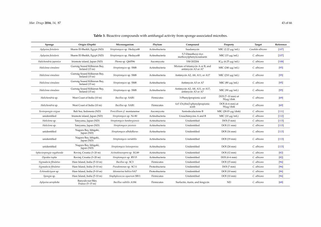
Mar. Drugs 2016, 14, 87 43 of 66
Table 3. Bioactive compounds with antifungal activity from sponge-associated microbes.
Sponge Origin (Depth) Microorganism Phylum Compound Property Target Reference
Aplysina fistularis Sharm El-Sheikh, Egypt (ND) Streptomyces sp. Hedaya48 Actinobacteria Saadamycin MIC (2.22 µg/mL) Candida albicans [107]
Aplysina fistularis Sharm El-Sheikh, Egypt (ND) Streptomyces sp. Hedaya48 Actinobacteria 5,7-Dimethoxy-4-p-methoxylphenylcoumarin MIC (15 µg/mL) C. albicans [107]
Halichondria japonica Iriomote island, Japan (ND) Phoma sp. Q60596 Ascomycota YM-202204 IC80 (6.25 µg/mL) C. albicans [108]
Haliclona simulans Gurraig Sound Kilkieran Bay,Ireland (15 m) Streptomyces sp. SM8 Actinobacteria Mixture of kitamycin A or B, and
antimycin A3 or A7 MIC (240 µg/mL) C. albicans [95]
Haliclona simulans Gurraig Sound Kilkieran Bay,Ireland (15 m) Streptomyces sp. SM8 Actinobacteria Antimycin A2, A8, A11, or A17 MIC (210 µg/mL) C. albicans [95]
Haliclona simulans Gurraig Sound Kilkieran Bay,Ireland (15 m) Streptomyces sp. SM8 Actinobacteria Antimycin A3 or A7 MIC (80 µg/mL) C. albicans [95]
Haliclona simulans Gurraig Sound Kilkieran Bay,Ireland (15 m) Streptomyces sp. SM8 Actinobacteria Antimycin A2, A8, A11, or A17,
antimycin A3 or A7 MIC (90 µg/mL) C. albicans [95]
Halichondria sp. West Coast of India (10 m) Bacillus sp. SAB1 Firmicutes 3-Phenylpropionic acid DOI (7–10 mm) at50µg/disk C. albicans [69]
Halichondria sp. West Coast of India (10 m) Bacillus sp. SAB1 Firmicutes 4,41-Oxybis(3-phenylpropionicacid)
DOI (4–6 mm) at50µg/disk C. albicans [69]
Xestospongia exigua Bali Sea, Indonesia (ND) Penicillium cf. montanense Ascomycota Xestodecalactone B MIC (28.03 µg/disk) C. albicans [111]
unidentified Iriomote island, Japan (ND) Streptomyces sp. Ni-80 Actinobacteria Urauchimycins A and B MIC (10 µg/mL) C. albicans [112]
Haliclona sp. Tateyama, Japan (ND) Streptomyces bambergiensis Actinobacteria Unidentified DOI (5 mm) C. albicans [113]
Haliclona sp. Tateyama, Japan (ND) Streptomyces javensis Actinobacteria Unidentified DOI (11 mm) C. albicans [113]
unidentified Nagura Bay, Ishigaki,Japan (ND) Streptomyces albidoflavus Actinobacteria Unidentified DOI (16 mm) C. albicans [113]
unidentified Nagura Bay, Ishigaki,Japan (ND) Streptomyces variabilis Actinobacteria Unidentified DOI (19 mm) C. albicans [113]
unidentified Nagura Bay, Ishigaki,Japan (ND) Streptomyces luteosporeus Actinobacteria Unidentified DOI (24 mm) C. albicans [113]
Spheciospongia vagabunda Rovinj, Croatia (3–20 m) Actinokineospora sp. EG49 Actinobacteria Unidentified DOI (12 mm) C. albicans [82]
Dysidea tupha Rovinj, Croatia (3–20 m) Streptomyces sp. RV15 Actinobacteria Unidentified DOI (4–6 mm) C. albicans [82]
Sigmadocia fibulatus Hare Island, India (5-10 m) Bacillus sp. SC3 Firmicutes Unidentified DOI (15 mm) C. albicans [96]
Sigmadocia fibulatus Hare Island, India (5-10 m) Pseudomonas sp. SC11 Proteobacteria Unidentified DOI (7 mm) C. albicans [96]
Echinodictyum sp. Hare Island, India (5-10 m) Idiomarina baltica SA7 Proteobacteria Unidentified DOI (10 mm) C. albicans [96]
Spongia sp. Hare Island, India (5-10 m) Staphylococcus equorum SB11 Firmicutes Unidentified DOI (10 mm) C. albicans [96]
Aplysina aerophoba Banyuls-sur-Mer,France (5–15 m) Bacillus subtilis A184 Firmicutes Surfactin, iturin, and fengycin ND C. albicans [68]

Mar. Drugs 2016, 14, 87 44 of 66
Table 3. Cont.
Aplysina aerophoba Banyuls-sur-Mer,France (5–15 m) Bacillus subtilis A190 Firmicutes Surfactin ND C. albicans [68]
Aplysina aerophoba Banyuls-sur-Mer,France (5–15 m) Bacillus subtilis A202 Firmicutes Iturin ND C. albicans [68]
Leucosolenia sp. Lough Hyne, Co. Cork,Ireland (15 m) Vibrio sp. SC-C1-5 Proteobacteria Unidentified ND C. albicans [83]
Leucosolenia sp. Lough Hyne, Co. Cork,Ireland (15 m) Vibrio sp. BSw21697 Proteobacteria Unidentified ND C. albicans [83]
Leucosolenia sp. Lough Hyne, Co. Cork,Ireland (15 m) Vibrio splendidus LGP32 Proteobacteria Unidentified ND C. albicans [83]
Leucosolenia sp. Lough Hyne, Co. Cork,Ireland (15 m) Bacillus amyloliquefaciens Proteobacteria Unidentified ND C. albicans [83]
Leucosolenia sp. Lough Hyne, Co. Cork,Ireland (15 m) Vibrio sp. SC-C1-5 Proteobacteria Unidentified ND Candida glabrata [83]
Leucosolenia sp. Lough Hyne, Co. Cork,Ireland (15 m) Vibrio sp. BSw21697 Proteobacteria Unidentified ND C. glabrata [83]
Leucosolenia sp. Lough Hyne, Co. Cork,Ireland (15 m) Vibrio splendidus LGP32 Proteobacteria Unidentified ND C. glabrata [83]
Leucosolenia sp. Lough Hyne, Co. Cork,Ireland (15 m) Bacillus amyloliquefaciens Firmicutes Unidentified ND C. glabrata [83]
Leucosolenia sp. Lough Hyne, Co. Cork,Ireland (15 m) Pseudoalteromonas sp. A2B10 Proteobacteria Unidentified ND C. glabrata [83]
Leucosolenia sp. Lough Hyne, Co. Cork,Ireland (15 m) Pseudoalteromonas sp. K2B-2 Proteobacteria Unidentified ND C. glabrata [83]
Leucosolenia sp. Lough Hyne, Co. Cork,Ireland (15 m) Pseudoalteromonas sp. LJ1 Proteobacteria Unidentified ND C. glabrata [83]
Leucosolenia sp. Lough Hyne, Co. Cork,Ireland (15 m) Pseudoalteromonas sp. S3178 Proteobacteria Unidentified ND C. glabrata [83]
Aplysina fistularis Sharm El-Sheikh, Egypt (ND) Streptomyces sp. Hedaya48 Actinobacteria Saadamycin MIC (5 µg/mL) Trichophyton rubrum [107]
Aplysina fistularis Sharm El-Sheikh, Egypt (ND) Streptomyces sp. Hedaya48 Actinobacteria 5,7-Dimethoxy-4-p-methoxylphenylcoumarin MIC (7.5 µg/mL) T. rubrum [107]
Aplysina fistularis Sharm El-Sheikh, Egypt (ND) Streptomyces sp. Hedaya48 Actinobacteria Saadamycin MIC (1.5 µg/mL) Trichophyton mentagrophytes [107]
Aplysina fistularis Sharm El-Sheikh, Egypt (ND) Streptomyces sp. Hedaya48 Actinobacteria 5,7-Dimethoxy-4-p-methoxylphenylcoumarin MIC (90 µg/mL), T. mentagrophytes [107]
Aplysina fistularis Sharm El-Sheikh, Egypt (ND) Streptomyces sp. Hedaya48 Actinobacteria Saadamycin MIC (1.25 µg/mL) Microsporum gypseum [107]
Aplysina fistularis Sharm El-Sheikh, Egypt (ND) Streptomyces sp. Hedaya48 Actinobacteria 5,7-Dimethoxy-4-p-methoxylphenylcoumarin MIC (100 µg/mL) M. gypseum [107]

Mar. Drugs 2016, 14, 87 45 of 66
Table 3. Cont.
Aplysina fistularis Sharm El-Sheikh, Egypt (ND) Streptomyces sp. Hedaya48 Actinobacteria Saadamycin MIC (1.0 µg/mL) Epidermophyton floccosum [107]
Aplysina fistularis Sharm El-Sheikh, Egypt (ND) Streptomyces sp. Hedaya48 Actinobacteria 5,7-Dimethoxy-4-p-methoxylphenylcoumarin MIC (50 µg/mL) E. floccosum [107]
Aplysina fistularis Sharm El-Sheikh, Egypt (ND) Streptomyces sp. Hedaya48 Actinobacteria Saadamycin MIC (1.2 µg/mL) Fusarium oxysporum [107]
Aplysina fistularis Sharm El-Sheikh, Egypt (ND) Streptomyces sp. Hedaya48 Actinobacteria 5,7-Dimethoxy-4-p-methoxylphenylcoumarin MIC (22 µg/mL) F. oxysporum [107]
Aplysina fistularis Sharm El-Sheikh, Egypt (ND) Streptomyces sp. Hedaya48 Actinobacteria Saadamycin MIC (5.16 µg/mL) Cryptococcus humicolus [107]
Aplysina fistularis Sharm El-Sheikh, Egypt (ND) Streptomyces sp. Hedaya48 Actinobacteria 5,7-Dimethoxy-4-p-methoxylphenylcoumarin MIC (10 µg/mL) C. humicolus [107]
Halichondria japonica Iriomote island, Japan (ND) Phoma sp. Q60596 Ascomycota YM-202204 IC80 (1.56 µg/mL) Cryptococcus neoformans [108]
Aplysina fistularis Sharm El-Sheikh, Egypt (ND) Streptomyces sp. Hedaya48 Actinobacteria Saadamycin MIC (1.6 µg/mL) Aspergillus fumigatus [107]
Aplysina fistularis Sharm El-Sheikh, Egypt (ND) Streptomyces sp. Hedaya48 Actinobacteria 5,7-Dimethoxy-4-p-methoxylphenyl-coumarin MIC (10 µg/mL) A. fumigatus [107]
Halichondria japonica Iriomote island, Japan (ND) Phoma sp. Q60596 Ascomycota YM-202204 IC80 (12.5 µg/mL) A. fumigatus [108]
Leucosolenia sp. Lough Hyne, Co. Cork,Ireland (15 m) Staphylococcus saprophyticus Firmicutes Unidentified ND A. fumigatus [83]
Leucosolenia sp. Lough Hyne, Co. Cork,Ireland (15 m) Staphylococcus sp. HJB003 Firmicutes Unidentified ND A. fumigatus [83]
Leucosolenia sp. Lough Hyne, Co. Cork,Ireland (15 m) Vibrio litoralis MANO22P Proteobacteria Unidentified ND A. fumigatus [83]
Leucosolenia sp. Lough Hyne, Co. Cork,Ireland (15 m) Vibrio sp. SC-C1-5 Proteobacteria Unidentified ND A. fumigatus [83]
Leucosolenia sp. Lough Hyne, Co. Cork,Ireland (15 m) Vibrio sp. BSw21697 Proteobacteria Unidentified ND A. fumigatus [83]
Leucosolenia sp. Lough Hyne, Co. Cork,Ireland (15 m) Vibrio splendidus LGP32 Proteobacteria Unidentified ND A. fumigatus [83]
Leucosolenia sp. Lough Hyne, Co. Cork,Ireland (15 m) Bacillus amyloliquefaciens Firmicutes Unidentified ND A. fumigatus [83]
Aplysina fistularis Sharm El-Sheikh, Egypt (ND) Streptomyces sp. Hedaya48 Actinobacteria Saadamycin MIC (1.0 µg/mL) Aspergillus niger [107]
Aplysina fistularis Sharm El-Sheikh, Egypt (ND) Streptomyces sp. Hedaya48 Actinobacteria 5,7-Dimethoxy-4-p-methoxylphenylcoumarin MIC (20 µg/mL) A. niger [107]
Halichondria sp. West Coast of India (10 m) Bacillus sp. SAB1 Firmicutes 3-Phenylpropionic acid DOI (1–3 mm) at 50µg/disc A. niger [69]
Halichondria sp. West Coast of India (10 m) Bacillus sp. SAB1 Firmicutes 4,41-Oxybis(3-phenylpropionicacid)
DOI (4–6 mm) at 50µg/disc A. niger [69]
Halichondria sp. West Coast of India (10 m) Bacillus sp. SAB1 Firmicutes 3-Phenylpropionic acid DOI (4–6 mm) at 50µg/disc Rhodotorula sp. [69]

Mar. Drugs 2016, 14, 87 46 of 66
Table 3. Cont.
Halichondria sp. West Coast of India (10 m) Bacillus sp. SAB1 Firmicutes 4,41-Oxybis(3-phenylpropionicacid)
DOI (7–10 mm) at 50µg/disc Rhodotorula sp. [69]
Halichondria japonica Iriomote island, Japan (ND) Phoma sp. Q60596 Ascomycota YM-202204 IC80 (1.56 µg/mL) Saccharomyces cerevisiae [108]
Hymeniacidon perleve Nanji island, China (ND) Pseudoalteromonas piscicidaNJ6-3-1 Proteobacteria Norharman
(a beta-carboline alkaloid) DOI (3–5 mm) S. cerevisiae [59]
Hymeniacidon perleve Nanji island, China (ND) Bacillus megaterium NJ6-3-2 Firmicutes Unidentified DOI (3–5 mm) S. cerevisiae [59]
Leucosolenia sp. Lough Hyne, Co. Cork,Ireland (15 m) Vibrio litoralis MANO22P Proteobacteria Unidentified ND S. cerevisiae [83]
Leucosolenia sp. Lough Hyne, Co. Cork,Ireland (15 m) Vibrio sp. SC-C1-5 Proteobacteria Unidentified ND S. cerevisiae [83]
Leucosolenia sp. Lough Hyne, Co. Cork,Ireland (15 m) Vibrio sp. BSw21697 Proteobacteria Unidentified ND S. cerevisiae [83]
Leucosolenia sp. Lough Hyne, Co. Cork,Ireland (15 m) Vibrio splendidus LGP32 Proteobacteria Unidentified ND S. cerevisiae [83]
Leucosolenia sp. Lough Hyne, Co. Cork,Ireland (15 m) Bacillus amyloliquefaciens Firmicutes Unidentified ND S. cerevisiae [83]
Psammocinia sp. Sdot-Yam, Israel (ND) Aspergillus insuetus Ascomycota Insuetolides A MIC (60.09 µg/mL) Neurospora crassa [114]
Psammocinia sp. Sdot-Yam, Israel (ND) Aspergillus insuetus Ascomycota Strobilactone A MIC (69.97 µg/mL) N. crassa [114]
Psammocinia sp. Sdot-Yam, Israel (ND) Aspergillus insuetus Ascomycota (E,E)-6-(60,70-Dihydroxy-20,40-octadienoyl)-strobilactone A MIC (71.79 µg/mL) N. crassa [114]
Myxilla incrustans The Caribbean Island ofDominica (ND) Microsphaeropsis sp. Ascomycota Microsphaeropsisin ND Eurotium repens [115]
Myxilla incrustans The Caribbean Island ofDominica (ND) Microsphaeropsis sp. Ascomycota (R)-Mellein ND E. repens [115]
Myxilla incrustans The Caribbean Island ofDominica (ND) Microsphaeropsis sp. Ascomycota (3R,4R)-
Hydroxymellein ND E. repens [115]
Myxilla incrustans The Caribbean Island ofDominica (ND) Microsphaeropsis sp. Ascomycota
4,8-Dihydroxy-3,4-dihydro-2H-
naphthalen-1-oneND E. repens [115]
Ectyoplasia ferox The Caribbean Island ofDominica (ND) Coniothyrium sp. Ascomycota (3R)-6-Methoxymellein ND E. repens [115]
Ectyoplasia ferox The Caribbean Island ofDominica (ND) Coniothyrium sp. Ascomycota (3R)-6-Methoxy-7-chloromellein ND E. repens [115]
Ectyoplasia ferox The Caribbean Island ofDominica (ND) Coniothyrium sp. Ascomycota (p-Hydroxyphenyl) ethanol ND E. repens [115]
Ectyoplasia ferox The Caribbean Island ofDominica (ND) Coniothyrium sp. Ascomycota Phenylethanol ND E. repens [115]

Mar. Drugs 2016, 14, 87 47 of 66
Table 3. Cont.
Myxilla incrustans The Caribbean Island ofDominica (ND) Microsphaeropsis sp. Ascomycota Microsphaeropsisin ND Ustilago violacea [115]
Myxilla incrustans The Caribbean Island ofDominica (ND) Microsphaeropsis sp. Ascomycota (R)-Mellein ND U. violacea [115]
Myxilla incrustans The Caribbean Island ofDominica (ND) Microsphaeropsis sp. Ascomycota (3R,4R)-Hydroxymellein ND U. violacea [115]
Myxilla incrustans The Caribbean Island ofDominica (ND) Microsphaeropsis sp. Ascomycota 4,8-Dihydroxy-3,4-dihydro-
2H-naphthalen-1-one ND U. violacea [115]
Ectyoplasia ferox The Caribbean Island ofDominica (ND) Coniothyrium sp. Ascomycota (3R)-6-Methoxymellein ND U. violacea [115]
Ectyoplasia ferox The Caribbean Island ofDominica (ND) Coniothyrium sp. Ascomycota (3R)-6-Methoxy-7-chloromellein ND U. violacea [115]
Ectyoplasia ferox The Caribbean Island ofDominica (ND) Coniothyrium sp. Ascomycota (p-Hydroxyphenyl) ethanol ND U. violacea [115]
Ectyoplasia ferox The Caribbean Island ofDominica (ND) Coniothyrium sp. Ascomycota Phenylethanol ND U. violacea [115]
Ectyoplasia ferox The Caribbean Island ofDominica (ND) Coniothyrium sp. Ascomycota (3S)-(3’,5’-
Dihydroxyphenyl)butan-2-one ND U. violacea [115]
Ectyoplasia ferox The Caribbean Island ofDominica (ND) Coniothyrium sp. Ascomycota (3S)-(3’,5’-
Dihydroxyphenyl)butan-2-one ND Mycotypha microspora [115]
Table 3 is organised according to the target fungi. IC50: half maximum inhibitory concentration; IC80: 80% inhibitory concentration; MIC: minimum inhibitory concentration; DOI:diameter of inhibition; ND: not determined.

Mar. Drugs 2016, 14, 87 48 of 66
5. Antiprotozoal Activity
Malaria, caused by Plasmodium spp. infections, represents the most devastating protozoal diseaseworldwide, and results in both mortality and economic loss, mainly in developing countries [116].Developing drugs with a better therapeutic profile against the parasite is one of the key aimsof current malaria research, which includes screening for antimalarial substances from marineorganisms [117,118].
Manzamine A (16) (Figure 4), first reported by Sakai and co-workers [119] from the spongeHaliclona sp., is a promising substance against Plasmodium spp. Initially, its antitumor property wasof main interest, but subsequently diverse antimicrobial activities such as: anti-HIV, antibacterial,and antifungal were identified from the compound [120]. Currently the antimalaria properties ofmanzamine A are considered its most promising bioactivity. Manzamine A was shown to inhibitP. falciparum D6 and W3 clonal cell lines that are sensitive and resistant against the antimalarialchloroquine [121], with IC50 values of 0.0045 and 0.008 µg/mL, respectively [122]. Furthermore, in vivoscreening by Ang et al. [116] showed that manzamine A at concentration of 0.008 µg/mL inhibited90% growth of the parasite Plasmodium berghei that causes malaria in rodents. In addition, Rao et al.reported [122] that manzamine A displayed anti-Leishmania activity, indicated by IC50 and IC90 valuesof 0.9 µg/mL and 1.8 µg/mL, respectively, against Leishmania donovani.
Isolation of manzamine A from several other sponge species [120] raised the hypothesis that it wasof microbial origin [123,124]. Hill et al. [125] confirmed this hypothesis by isolating Micromonospora sp.M42 as the microbial producer of manzamine A from the Indonesian sponge Acanthostrongylophoraingens. A series of analyses using molecular-microbial community analysis, and Matrix AssistedLaser Desorption Ionization-Mass Spectrometry (MALDI-MS) corroborated that indeed the strainMicromonospora sp. M42 synthesizes manzamine A [126,127]. Considering the therapeutic potential ofmanzamine A for treating malaria and leishmaniasis, Micromonospora sp. M42 could be a sustainableprovider of the substance, because the “Sponge Supply Problem” has been overcome [127]. Moreover,identification of several manzamine-derivatives e.g. manzamine E, F, J, and 8-hydroxymanzamine A,from marine sponges which displayed antibacterial, antifungal and antiprotozoal activity [122,124],could also lead to isolation of associated microbial producers in the future.
Pimentel-Elardo et al. [128] identified three compounds with anti-Leishmania andanti-Trypanosoma activity from a sponge-associated Streptomyces sp, namely the cyclic depsipeptidevalinomycin (17), the indolocarbazole alkaloid staurosporine (18) and butenolide (19) (Table 4).Valinomycin and staurosporine inhibited the growth of L. major with IC50 values of 0.12 µg/mLand 1.24 µg/mL, respectively. In addition, the three compounds displayed bioactivity againstTrypanosoma brucei with IC50 values of 0.0036 µg/mL for valinomycin, 0.0051 µg/mL for staurosporineand 7.92 µg/mL for butenolide.
Scopel et al. [129] isolated two sponge-associated fungi, namely Hypocrea lixii F02 andPenicillium citrinum F40 (Table 4) that were active against the protozoal parasite Trichomonas vaginalis,which causes trichomoniasis, a sexually transmitted disease [130]. Culture filtrates of both isolatesinhibited T. vaginalis ATCC 30236 and fresh clinical isolates, including the metronidazole-resistantTV-LACM2, with MIC values of 2.5 mg/mL. Further observation indicated that culture filtrates ofthese two fungi had no haemolytic effect against mammalian cells, which is one of the importantcriteria to further develop anti-protozoal drugs [129].

Mar. Drugs 2016, 14, 87 49 of 66
Table 4. Bioactive compounds with antiprotozoal activity from sponge-associated microbes.
Sponge Origin (Depth) Microorganism Phylum Compound Property Target References
Homophymia sp. Touho, New Caledonia(ND)
Pseudomonas sp.1531-E7 Proteobacteria 2-Undecyl-4-quinolone IC50 (1 µg/mL) Plasmodium falciparum [25]
Acanthostrongylophora ingens Manado, Indonesia(ND) Micromonospora sp. M42 Actinobacteria Manzamine A IC50 (0.0045 µg/mL) P. falciparum [124–127]
Hyattella intestinalis Palk strait, Tamil Nadu,India (ND)
unidentified bacterialisolate THB20 Unidentified Unidentified IC50 (41.88 µg/mL) P. falciparum [131]
Stylissa carteri Palk strait, Tamil Nadu,India (ND)
unidentified bacterialisolate THB17 Unidentified Unidentified IC50 (20.56 µg/mL) P. falciparum [132]
Clathria indica Palk strait, Tamil Nadu,India (ND)
unidentified bacterialisolate THB23 Unidentified Unidentified IC50 (28.80 µg/mL) P. falciparum [133]
Clathria vulpina Palk strait, Tamil Nadu,India (ND)
unidentified bacterialisolate THB15 Unidentified Unidentified IC50 (20.73 µg/mL) P. faciparum [134]
Haliclona grant Palk strait, Tamil Nadu,India (ND)
unidentified bacterialisolate THB14 Unidentified Unidentified IC50 (11.98 µg/mL) P. faciparum [135]
Acanthostrongylophora ingens Manado, Indonesia(ND) Micromonospora sp. M42 Actinobacteria Manzamine A In vivo inhibition (90%) at
concentration of 0.008 µg/mL Plasmodium berghei [116,125–127]
Aplysina aerophoba Rovinj, Croatia (3–20 m) Micromonospora sp.RV115 Actinobacteria Diazepinomicin IC50 (6.29 µg/mL) Trypanosoma brucei [136]
Spheciospongia vagabunda Rovinj, Croatia (3–20 m) Actinokineospora sp.EG49 Actinobacteria Unidentified Percentage of growth inhibition
(48%) T. brucei [82]
unidentified Rovinj, Croatia (3–20 m) Brevibacterium sp. EG10 Actinobacteria Unidentified Percentage of growth inhibitiongrowth inhibition (30%) T. brucei [82]
unidentified Rovinj, Croatia (3–20 m) Gordonia sp. EG50 Actinobacteria Unidentified Percentage of growth inhibitiongrowth inhibition (28%) T. brucei [82]
Dysidea tupha Rovinj, Croatia (3–20 m) Kocuria sp. RV89 Actinobacteria Unidentified Percentage of growth inhibitiongrowth inhibition (19%) T. brucei [82]
Dysidea avara Mediterranean sea(ND) Nocardiopsis sp. RV163 Actinobacteria 1,6-Dihydroxyphenazine
(produced from co-culture) IC50 (4.03 µg/mL) T. brucei [98]
Spheciospongia vagabunda Red Sea (ND) Actinokinespora sp.EG49 Actinobacteria Actinosporin A IC50 (19.19 µg/mL) T. brucei brucei [137]
Aplysinapolypoides Rovinj, Croatia (3–20 m) Streptomyces sp. 34 Actinobacteria Valinomycin IC50 (0.0036 µg/mL) T. brucei brucei [128]
Axinella aerophoba Rovinj, Croatia (3–20 m) Streptomyces sp. 22 Actinobacteria Valinomycin IC50 (0.0036 µg/mL) T. brucei brucei [128]
Tedania sp. Rovinj, Croatia (3–20 m) Streptomyces sp. 11 Actinobacteria Staurosporine IC50 (0.0051 µg/mL) T. brucei brucei [128]
Tethya sp. Rovinj, Croatia (3–20 m) Streptomyces sp. T03 Actinobacteria Butenolide IC50 (7.92 µg/mL) T. brucei brucei [128]
Petrosia ficiformis Milos, Greece (ND) Streptomyces sp. SBT344 Actinobacteria Unidentified IC50 (<10 µg/mL) T. brucei brucei [138]
Sarcotragus foetidus Milos, Greece (ND) Modestobacter sp.SBT363 Actinobacteria Unidentified IC50 (<10 µg/mL) T. brucei brucei [138]
Sarcotragus foetidus Milos, Greece (ND) Nonomuraea sp. SBT364 Actinobacteria Unidentified IC50 (<10 µg/mL) T. brucei brucei [138]
Phorbas tenacior Crete, Greece (ND) Micromonospora sp.SBT687 Actinobacteria Unidentified IC50 (14.87 µg/mL) T. brucei brucei [138]

Mar. Drugs 2016, 14, 87 50 of 66
Table 4. Cont.
Petrosia ficiformis Milos, Greece (ND) Streptomyces sp. SBT348 Actinobacteria Unidentified IC50 (16.52 µg/mL) T. brucei brucei [138]
Ircinia variabilis Milos, Greece (ND) Geodermatophilus sp.SBT381 Actinobacteria Unidentified IC50 (18.60 µg/mL) T. brucei brucei [138]
Spirastrella cunctatrix Milos, Greece (ND) Modestobacter sp.SBT362 Actinobacteria Unidentified IC50 (19.34 µg/mL) T. brucei brucei [138]
Spirastrella cunctatrix Milos, Greece (ND) Rhodococcus sp. SBT367 Actinobacteria Unidentified IC50 (19.97 µg/mL) T. brucei brucei [138]
Axinella polypoides Banyuls-sur-Mer,France (ND)
Streptomyces axinellaePol001T Actinobacteria Tetromycin 1 IC50 (26.02 µg/mL) T. brucei brucei [139]
Axinella polypoides Banyuls-sur-Mer,France (ND)
Streptomyces axinellaePol001T Actinobacteria Tetromycin 2 IC50 (40.35 µg/mL) T. brucei brucei [139]
Axinella polypoides Banyuls-sur-Mer,France (ND)
Streptomyces axinellaePol001T Actinobacteria Tetromycin 3 IC50 (23.18 µg/mL) T. brucei brucei [139]
Axinella polypoides Banyuls-sur-Mer,France (ND)
Streptomyces axinellaePol001T Actinobacteria Tetromycin 4 IC50 (32.17 µg/mL) T. brucei brucei [139]
Axinella polypoides Banyuls-sur-Mer,France (ND)
Streptomyces axinellaePol001T Actinobacteria Tetromycin B IC50 (17.20 µg/mL) T. brucei brucei [139]
Aplysina polypoides Rovinj, Croatia (3–20 m) Streptomyces sp. 34 Actinobacteria Valinomycin IC50 (<0.12 µg/mL) Leishmania major [128]
Axinella aerophoba Rovinj, Croatia (3–20 m) Streptomyces sp. 22 Actinobacteria Valinomycin IC50 (<0.12 µg/mL) L. major [128]
Tedania sp. Rovinj, Croatia (3–20 m) Streptomyces sp. 11 Actinobacteria Staurosporine IC50 (1.24 µg/mL) L. major [128]
Axinella polypoides Banyuls-sur-Mer,France (ND)
Streptomyces axinellaePol001T Actinobacteria Tetromycin 3 IC50 (31.72 µg/mL) L. major [139]
Spheciospongia vagabunda Rovinj, Croatia (3–20 m) Actinokineospora sp.EG49 Actinobacteria Unidentified growth inhibition (48%) L. major [82]
unidentified Rovinj, Croatia (3–20 m) Gordonia sp. EG50 Actinobacteria Unidentified growth inhibition (28%) L. major [82]
Axinella corrugataThe Arvoredo
Biological MarineReserve, Brazil (ND)
Hypocrea lixii F02 Ascomycota Unidentified MIC (250 µg/mL) Trichomonas vaginalisATCC 30236 [129]
Axinella corrugataThe Arvoredo
Biological MarineReserve, Brazil (ND)
Hypocrea lixii F02 Ascomycota Unidentified MIC (250 µg/mL) T. vaginalis fresh isolate [129]
Axinella corrugataThe Arvoredo
Biological MarineReserve, Brazil (ND)
Hypocrea lixii F02 Ascomycota Unidentified MIC (250 µg/mL)T. vaginalis
metronidazole-resistantLACM2
[129]
Stoeba sp.The Arvoredo
Biological MarineReserve, Brazil (ND)
Penicillium citrinum F40 Ascomycota Unidentified MIC (250 µg/mL) T. vaginalis ATCC 30236 [129]
Stoeba sp.The Arvoredo
Biological MarineReserve, Brazil (ND)
Penicillium citrinum F40 Ascomycota Unidentified MIC (250 µg/mL) T. vaginalis fresh isolate [129]
Stoeba sp.The Arvoredo
Biological MarineReserve, Brazil (ND)
Penicillium citrinum F40 Ascomycota Unidentified MIC (250 µg/mL)T.vaginalis
metronidazole-resistantLACM2
[129]
Table 4 is organised according to the target protozoa. IC50: half maximum inhibitory concentrations; MIC: minimum inhibitory concentration; ND: not determined.

Mar. Drugs 2016, 14, 87 51 of 66Mar. Drugs 2016, 14, x 56 of 79
Figure 4. Chemical structures of the antiprotozoal compounds manzamine A (16), valinomycin (17), staurosporine (18) and butenolide (19).
6. Dicussion
6.1. Antimicrobial Compounds from Sponge-Associated Microbes: What We Learned So Far
Bioprospecting is the effort to discover natural compounds with therapeutic and biological applications [140]. In line with this definition, sponge-associated microbes offer a huge potential as the source of antimicrobial substances as shown by many microbial isolates being reported to inhibit pathogenic reference strains in vitro and to synthesize active substances against one or several groups of infectious agents. Based on our review, antimicrobial compounds produced by sponge-associated microbes with the most pronounced bioactivity include: 2-undecyl-4-quinolone, sorbicillactone A, stachybotrin D and chartarutine B against HIV-1; truncateol M against H1N1 M; YM-266183, YM-266184, kocurin, mayamycin, sydonic acid, naphthacene glycoside SF2446A2 and trichoderin A against a variety of bacterial strains; saadamycin and YM-202204 against fungi; manzamine-A against malaria; and valinomycin against Trypanosoma. In this case the most pronounced activity is solely based on reported inhibition data and does not yet take potential side effects into account. Therefore the most promising compounds may be ones that have higher IC50 values, but cause less side effects. As these data are not available for the majority of the reported compounds, we have focused on the most potent compounds.
Sponge-associated bacteria and fungi are the two groups of microorganisms that have been found to produce antimicrobial compounds (Figure 5). The large majority of the antimicrobial compounds found in sponge-associated microbiota is produced by bacteria (90%), while fungi account for approximately 10% of the compounds reported. Sponge-associated bacteria derived antimicrobial compounds were found from 35 genera (Figure 5B). At a higher taxonomic level, these 35 bacterial genera can be classified into the four phyla Actinobacteria, Proteobacteria, Firmicutes and Cyanobacteria with percentages of 48.8%, 36.6%, 11.4% and 0.4% respectively. In contrast, sponge-associated fungi that have been found to produce antimicrobials are affiliated solely to the phylum Ascomycota.
Figure 4. Chemical structures of the antiprotozoal compounds manzamine A (16), valinomycin (17),staurosporine (18) and butenolide (19).
6. Dicussion
6.1. Antimicrobial Compounds from Sponge-Associated Microbes: What We Learned So Far
Bioprospecting is the effort to discover natural compounds with therapeutic and biologicalapplications [140]. In line with this definition, sponge-associated microbes offer a huge potential asthe source of antimicrobial substances as shown by many microbial isolates being reported to inhibitpathogenic reference strains in vitro and to synthesize active substances against one or several groupsof infectious agents. Based on our review, antimicrobial compounds produced by sponge-associatedmicrobes with the most pronounced bioactivity include: 2-undecyl-4-quinolone, sorbicillactoneA, stachybotrin D and chartarutine B against HIV-1; truncateol M against H1N1 M; YM-266183,YM-266184, kocurin, mayamycin, sydonic acid, naphthacene glycoside SF2446A2 and trichoderin Aagainst a variety of bacterial strains; saadamycin and YM-202204 against fungi; manzamine-A againstmalaria; and valinomycin against Trypanosoma. In this case the most pronounced activity is solelybased on reported inhibition data and does not yet take potential side effects into account. Thereforethe most promising compounds may be ones that have higher IC50 values, but cause less side effects.As these data are not available for the majority of the reported compounds, we have focused on themost potent compounds.
Sponge-associated bacteria and fungi are the two groups of microorganisms that have beenfound to produce antimicrobial compounds (Figure 5). The large majority of the antimicrobialcompounds found in sponge-associated microbiota is produced by bacteria (90%), while fungiaccount for approximately 10% of the compounds reported. Sponge-associated bacteria derivedantimicrobial compounds were found from 35 genera (Figure 5B). At a higher taxonomic level, these35 bacterial genera can be classified into the four phyla Actinobacteria, Proteobacteria, Firmicutesand Cyanobacteria with percentages of 48.8%, 36.6%, 11.4% and 0.4% respectively. In contrast,

Mar. Drugs 2016, 14, 87 52 of 66
sponge-associated fungi that have been found to produce antimicrobials are affiliated solely to thephylum Ascomycota.
Mar. Drugs 2016, 14, x 58 of 71
nitrogen‐containing heterocyclic compound with a wide range of biological activities [67,144], and
several studies from terrestrial environments and chemically synthesized phenazines have been
reported as antiviral [145], antibacterial [146], and antimalaria [147]. Moreover, this group of
compounds is attractive for therapeutic application since their structures are relatively small and
hence can easily reach tissues and organs [67,148].
Figure 5. Distribution of sponge‐associated microorganisms found to produce antimicrobial
compounds: (A) Bacteria and Fungi; (B) Bacterial genera; and (C) Fungal genera. Figure 5 was made
based on the summary of the taxonomic affiliations of sponge‐associated microbes (N = 272) that
were found to produce antimicrobials.
Figure 5. Distribution of sponge-associated microorganisms found to produce antimicrobialcompounds: (A) Bacteria and Fungi; (B) Bacterial genera; and (C) Fungal genera. Figure 5 wasmade based on the summary of the taxonomic affiliations of sponge-associated microbes (N = 272) thatwere found to produce antimicrobials.

Mar. Drugs 2016, 14, 87 53 of 66
Streptomyces is the most prominent genus as indicated by 30% of sponge bacteria-derivedcompounds. Streptomyces has become a main target for screening for bioactive compounds bothfrom terrestrial and marine environments due to the high diversity of secondary metabolites theyproduce [141,142]. Of the many sponge-associated Streptomyces isolates reported, Streptomyces sp.HB202 and Streptomyces sp. RV15 are of particular interest in term of producing antibacterialcompounds. Streptomyces sp. HB202, isolated from the sponge Halichondria panicea has beendocumented to produce three antibacterial substances: mayamycin, streptophenazine G and K,which are mainly active against Gram positive pathogenic bacteria (Table 2). Streptomyces sp. RV15,on the other hand, produces the compound naphthacene glycoside which up to now is the onlyanti-Chlamydia reported from sponge-associated microbes [46]. In addition, the report on crudeextract inhibition of Streptomyces sp. RV15 against S. aureus and E. faecalis [82] may give a hint todiscover other antibacterial substances from this strain. Streptomyces sp. Hedaya48 is currently the mostpotent sponge-associated bacterial isolate for antifungal activities with the production of saadamycinand 5,7-dimethoxy-4-p-methoxylphenylcoumarin [107]. In addition, isolation of the anti-Trypanosomaand anti-Leishmania compounds valinomycin, staurosporine and butenolide from Streptomyces sp.43, 21 and 11 [128], affirms Streptomyces as the currently most prominent producer of antimicrobialsubstances from sponges.
Pseudovibrio follows as the second most prolific bacterial genus isolated from sponges (20%) withrespect to antimicrobial activities. Reports on Pseudovibrio spp. are concentrated on antibacterialactivity and are mainly based on screening of crude extracts. Up to now, tropodithietic acid is the onlyantibacterial compound that has been identified from Pseudovibrio [72]. Although representing a lowerpercentage of the sponge-associated bacteria found to produce antimicrobials than Streptomyces andPseudovibrio, 9% of the currently known bioactives was found to be produced by sponge-associatedBacillus spp., with activities against viruses, bacteria and fungi. Bacillus cereus QNO3323 is currently themost prominent antimicrobial producer from this genus with the very potent thiopeptides YM-266183and YM-266184 that are active against Gram positive bacteria.
Sponge-associated Ascomycota found to produce antimicrobials can be further classified into12 genera. Of these 12 fungal genera, Aspergillus (30%) and Penicillium (23%) are currently the twomost prominent groups of sponge-associated fungi reported as antimicrobial producers. This findingis not suprising since both Aspergillus and Penicillium are known prolific producers of secondarymetabolites from other sources [143]. Aspergillus versicolor [58] and an unidentified Aspergillus sp.isolated from the sponge Xestospongia testudinaria [48] showed a strong antibacterial activity asindicated by potent inhibition of pathogenic bacteria. The antimicrobial activities found fromsponge-associated Penicillium spp. are particular remarkable as it is the only fungal genus that isfound to produce antivirals, antibacterials antifungals and antiprotozoals. Penicillium chrysogenum [26]and Penicillium sp. FF01 [57] are to date the most promising sponge-associated Penicillium isolates forwhich anti-HIV activity (sorbicillactone) and antibacterial activity (citrinin) were reported, respectively.Sponge-derived Stachybotrys spp. are only known for antiviral activity, particularly against HIV andenterovirus 71 (EV71), and there are no reports of other antimicrobial activities. Generally, althoughthe number of produced antimicrobials is outnumbered by those of sponge-associated bacteria,sponge-associated fungi should be considered as an important reservoir of antimicrobial compounds.
When the chemical structures of sponge-microbe-derived compounds are considered, a ratherdiverse array of structures is observed, including peptides, terpenoids, phenazines, indoles, phenolesand polyketides. Sixty percent of the antivirals from sponge-associated microbes are ketone derivatives(quinolone, sorbicillactone, isoindolinone, butyrolactone, furanone, xanthone, methanone, phenone).Peptide derivatives constitute 19% of the total identified antibacterial substances and roughly 12.5%from the total antimicrobial compounds reviewed here. Phenazine derivatives are the second mostfrequently isolated class of antibacterial compounds from sponge-associated microbes (15%) asexemplified in this review by the antibacterial compounds streptophenazine [89], phenazine alkaloidantibiotics [55], 6-hydroxymethyl-1-phenazine-carboxamide and 1,6-phenazinedimethanol [94].

Mar. Drugs 2016, 14, 87 54 of 66
Phenazine is a nitrogen-containing heterocyclic compound with a wide range of biologicalactivities [67,144], and several studies from terrestrial environments and chemically synthesizedphenazines have been reported as antiviral [145], antibacterial [146], and antimalaria [147]. Moreover,this group of compounds is attractive for therapeutic application since their structures are relativelysmall and hence can easily reach tissues and organs [67,148].
6.2. Discovering Antimicrobial Compounds from Sponge-Associated Microbes: From Culture-Dependent toCulture-Independent Methods
Isolation of antimicrobial producers provides a valuable basis for assessing the biotechnologicalpotential of sponge-associated microbes. In a wider perspective, however, only a small fractionof this sponge-microbial community has been isolated under laboratory conditions leaving themajority resistant to in vitro growth with current cultivation approaches [15,149,150]. Several studieshave focused on improving cultivability of sponge-associated microbes. Some of the approachesinclude using low nutrient media [151], floating filter cultures [152], employing different carbonsources, e.g., lectin [153], sponge extracts [152], and in situ cultivation using a diffusion growthchamber [154]. Furthermore, flow-cytometry and density gradient centrifugation have been appliedto separate sponge cells from their associated bacteria to enrich the inoculum [155,156]. Additionally,co-cultivation through mixing of two or more microbial isolates in vitro [157] is an approach proposedto discover more natural compounds from sponge-associated microbes. The idea behind co-culture liesin the fact that many biosynthetic gene clusters found in microorganisms remain cryptic understandard laboratory conditions, and co-cultivation might provide a possibility to activate thesesilent genes [158,159]. As an example, the co-culture by Dashti et al. [98] of the sponge-associatedActinobacteria, Actinokinespora sp. EG49 and Nocardiopsis sp. RV163, resulted in isolation of theantibacterial compound 1,6-dihydroxyphenazine, which was not found from the individual isolates.However, even if the cultivability of sponge-associated microbes is improved, there is a long wayahead to reach a point that we will be able to isolate and routinely cultivate 50% of the microbes thatare found in sponges. At the same time, the advance of genetic and molecular studies has resulted inthe development of tools to study genes, transcripts and proteins by directly analyzing environmentalDNA, RNA and proteins, thus bypassing cultivation procedures [157]. In relation to screening forantimicrobial activity, metagenomics has been applied to identify antimicrobials of uncultivatedmicroorganisms from terrestrial environments, such as the antimycobacterial nocardamine, the putativeantibacterial activity of terragines A–E [160], violacein that is active against S. aureus, Bacillus sp. andStreptococcus sp. [161] and a polyketide with activity against the yeast Saccharomyces cerevisiae [162].
Two main metagenomic approaches, functional screening and sequence homology-basedmethods, are generally distinguished [163]. Functional screening relies on detection of the metabolicactivities of metagenomic library clones without requiring any prior sequence information [163–165].Gillespie et al. [9] applied function-based metagenomics with E. coli as expression host, to identify theantibiotics turbomycin A and B from a soil sample. MacNeil et al. [166] identified the antimicrobialindirubin by constructing a BAC (bacterial artificial chromosome) library in E.coli. Yung et al. [167]reported two hydrolytic enzymes from fosmid clones CcAb1 and CcAb2, which were derived from ametagenome of the sponge Cymbastela concentrica using E. coli as the host. Both fosmid clones inhibitedthe growth of Bacillus sp. with an inhibition diameter of 20 mm, and clone CcAb1 showed additionalinhibition of S. aureus and an Alteromonas sp. with diameters of inhibition of 50 mm and 60 mm,respectively. Further phylogenetic analysis showed that active genes encoding for these enzymes wereof microbial origin [167]. He et al. [168] constructed a fosmid library of the sponge Discodermia calyxusing E. coli as the host and identified antimicrobial activity of the enzyme 3-hydroxypalmitic acidagainst B. cereus and C. albicans. In addition, using the same approach He et al. [169] observedan active clone, pDC113, that displayed a clear inhibition zone against B. cereus. Subsequently,11 cyclodipeptides were identified from this clone. Generally, it can be stated that although a numberof antimicrobials have been discovered through functional screening of metagenomic libraries from

Mar. Drugs 2016, 14, 87 55 of 66
sponges, the expression of large gene clusters such as those encoding (polyketide synthase( PKS) and(non-ribosomal peptide synthetase (NRPS) is still a difficult hurdle to take. Several key elements needto be considered to achieve successful expression of biosynthetic gene clusters; namely mobilizingthe biosynthetic pathway into a suitable vector, selecting an appropriate heterologous host and stablymaintaining the gene clusters in the host [170]. The size of many of these gene clusters requiresthe use of cloning vectors that can accept large inserts, such as fosmids, or BACs if the requiredinsert size is over 100 kb [171]. Selection of heterologous expression systems in particular is a crucialfactor before applying functional metagenomics to identify antimicrobials, because expression hostsare microbes as well and especially clones that express genes encoding for enzymes involved inproduction of antimicrobials may therefore be non-viable. Ongley et al. [170] pointed out someconsiderations in selecting an expression host such as relatedness to the native producer, availabilityof genetic tools and precursors, a high growth rate, and suitability for fermentation at a largescale. E. coli, the most commonly used expression host, has limitations for expressing parts ofmetagenomes because, e.g., of the sheer size of some gene clusters, genes with deviating codon usage,incompatible regulatory elements, lack of biosynthesis precursors or unavailability of posttranslationalmodifications [165,172]. Therefore, in order to make screening for antimicrobials through metagenomiclibraries more efficient, it is of utmost importance to diversify the suite of expression hosts used.Several non-E.coli hosts, such as Agrobacterium tumefaciens, Bacillus subtilis, Burkholderia graminis,Caulobacter vibrioides, Pseudoalteromonas haloplanktis, Pseudomonas putida, Ralstonia metallidurans,Rhizobium leguminosarum, Streptomyces avermitilis, S. albus, Pseudomonas putida, Sulfolobus solfataricus,Thermus thermophilus, Thiocapsa roseopersicina and Saccharopolyspora sp. have been developed andshould be more seriously considered as expression hosts when performing metagenomic screeningsfor antimicrobials [165,172,173].
Sequence-based screening, on the other hand, requires information on the sequence of genesinvolved in the production of a natural product as guidance to search for similar sequences in asequenced metagenomic library or scaffolds reconstructed from direct metagenomic sequencing [165].Homology-based screening is suitable to identify a compound with highly conserved biosynthesispathways, e.g., those mediated by PKS and NRPS [174]. Piel and colleagues [175–179] appliedthis method, and identified the antitumor polyketide onnamide from uncultivated bacteria of thesponge T. swinhoei. Sequence-based screening was applied by Fisch [180] to unravel the completepathway of the polyketide psymberin that was found to possess a potent antitumor activity, fromuncultivated sponge-associated microbes. By sequence-based screening of metagenomic libraries,Schirmer et al. [181] reported diverse polyketide gene clusters in microorganisms from the spongeDiscodermia dissoluta. The development of techniques that yield longer read lengths, such as PacificBiosciences (PacBio) RS II SMRT (Single Molecule Real-Time) sequencing technology, in which asingle read can be extended over 10 kbp [182], can be instrumental in increasing the accuracy inassembling large gene clusters. Application of PacBio for secondary metabolite gene clusters has beenreported by Alt and Wilkinson [183], who identified the 53,253 bp genomic fragment encoding thetransacyltransferase (trans-AT) polyketide synthase (PKS) from a marine Streptomyces sp responsiblefor the production of the antibiotic anthracimycin (atc). Furthermore, using Streptomyces coelicolor asheterologous expression host, the authors confirmed production of anthracimycin [183]. Furthermore,single cell analysis by combining cell separation and fluorescence-assisted cell sorting (FACS) could bea strategy to overcome the complexity of the microbial community in sponges since this method can beused to select for genomes from microbes that are present in low abundance in the sponge leading to asimplified reconstruction of secondary metabolite gene clusters present in these bacteria [184]. Thisstrategy has been applied by Wilson et al. [185] for resolving the gene clusters encoding the machineryneeded for the production of the polytheonamides produced by the candidate genus Entotheonellafrom the sponge Theonella swinhoei.
Inspired by these examples, homology-based screening could be further exploited to identifybiosynthesis gene sequences that could lead to the identification of novel antimicrobial substances from

Mar. Drugs 2016, 14, 87 56 of 66
Nature’s excessive diversity. Moreover, application of homology-based screening can benefit frompublicly available metagenomic sequencing data and prediction tools for analyzing biosynthesis geneclusters, e.g., AntiSMASH (Antibiotics and Secondary Metabolite Analysis Shell) [186,187]. Applicationof sequence-based screening, however, is limited by the fact that the found sequences need to be relatedto known compounds, inherently limiting the potential for novelty. Furthermore, information on genesequences is no guarantee that the acquisition of a complete gene pathway has been obtained [188].Therefore, sequence-based methagenomics should ideally be complemented by chemical analysis toconfirm whether the predicted compound exists and is fully functional (Figure 6).Mar. Drugs 2016, 14, x 61 of 71
Figure 6. General overview of the strategies used to discover antimicrobial compounds from sponge-associated microorganisms.
7. Conclusion and Outlook
Sponge-associated microbes already offer a rich source of potent antimicrobial compounds against viruses, bacteria, protozoa and fungi, and currently available compounds are predominantly active against HIV-1, H1N1, nosocomial Gram positive bacteria, Escherichia coli, Plasmodium spp, Leishmania donovani, Trypanosoma brucei, Candida albicans and dermatophytic fungi. Streptomyces, Pseudovibrio, Bacillus, Aspergillus and Penicillium are the microbial genera associated with sponges from which potent antimicrobial compounds are most frequently isolated. However, none of the antimicrobial compounds highlighted in this review have been succcesfully marketed as pharmaceuticals. To clearly translate bioactivity of these important compounds it is crucial to further unravel their mode of actions and measure their level of toxicity, since the majority of these studies has been focused on in vitro bioassays and elucidation of the chemical structures only.
The known versatility of antimicrobial activities found in sponge-associated microorganims could easily be expanded even without considering additional sponge sampling campaigns. Bioactivity screens of identified compounds or undefined sponge extracts is often restricted to a specific antimicrobial activity. The selection, for instance, relies on the specific research activities of the groups involved in isolating the microbes [117]. Consequently, it is probably safe to assume that other potent antimicrobial properties from many sponge isolates and their bioactive compounds remain undetected. Therefore, known antimicrobial compounds and producer strains are a valuable source for additional antimicrobial activities screenings using different target types (viruses, bacteria, fungi, protozoa and beyond). In addition, sponge-derived strain collections that comprise isolates that tested negative for antimicrobial activity at first may have done so, because the compound of interest is not produced under standard laboratory conditions. Exposure of these strains to potential microbial targets may lead to recovery of bioactivity that would otherwise go unnoticed.
Ideally, researchers who isolate microbes from sponges will deposit them to publicly available culture collections so that laboratories with complementary expertise and interests could benefit and screen the deposited isolates for different antimicrobial activities. This will make exchange of
Figure 6. General overview of the strategies used to discover antimicrobial compounds fromsponge-associated microorganisms.
7. Conclusions and Outlook
Sponge-associated microbes already offer a rich source of potent antimicrobial compoundsagainst viruses, bacteria, protozoa and fungi, and currently available compounds are predominantlyactive against HIV-1, H1N1, nosocomial Gram positive bacteria, Escherichia coli, Plasmodium spp,Leishmania donovani, Trypanosoma brucei, Candida albicans and dermatophytic fungi. Streptomyces,Pseudovibrio, Bacillus, Aspergillus and Penicillium are the microbial genera associated with spongesfrom which potent antimicrobial compounds are most frequently isolated. However, none ofthe antimicrobial compounds highlighted in this review have been succcesfully marketed aspharmaceuticals. To clearly translate bioactivity of these important compounds it is crucial to furtherunravel their mode of actions and measure their level of toxicity, since the majority of these studies hasbeen focused on in vitro bioassays and elucidation of the chemical structures only.
The known versatility of antimicrobial activities found in sponge-associated microorganims couldeasily be expanded even without considering additional sponge sampling campaigns. Bioactivityscreens of identified compounds or undefined sponge extracts is often restricted to a specificantimicrobial activity. The selection, for instance, relies on the specific research activities of the

Mar. Drugs 2016, 14, 87 57 of 66
groups involved in isolating the microbes [117]. Consequently, it is probably safe to assume that otherpotent antimicrobial properties from many sponge isolates and their bioactive compounds remainundetected. Therefore, known antimicrobial compounds and producer strains are a valuable sourcefor additional antimicrobial activities screenings using different target types (viruses, bacteria, fungi,protozoa and beyond). In addition, sponge-derived strain collections that comprise isolates that testednegative for antimicrobial activity at first may have done so, because the compound of interest isnot produced under standard laboratory conditions. Exposure of these strains to potential microbialtargets may lead to recovery of bioactivity that would otherwise go unnoticed.
Ideally, researchers who isolate microbes from sponges will deposit them to publicly availableculture collections so that laboratories with complementary expertise and interests could benefit andscreen the deposited isolates for different antimicrobial activities. This will make exchange of materialsand knowledge that can be obtained much more efficient. Importantly, a fair agreement on intellectualproperty rights needs to be established for translating this into reality. Lastly, the revolutionary advanceof next generation sequencing technologies combined with more diversified heterologous expressionsystems (Figure 6) are expected to open up the large unexplored reservoir of antimicrobials producedby yet uncultivated sponge-associated microbes.
Acknowledgments: Anak Agung Gede Indraningrat receives a PhD fellowship from the Indonesia EndowmentFund for Education (LPDP), grant number 20140812021557. This work was also supported by the EC grant“BluePharmTrain” (grant agreement no. 607786).
Author Contributions: Anak Agung Gede Indraningrat and Detmer Sipkema conceived the idea for the review,Anak Agung Gede Indraningrat compiled literatures, drew figures, and wrote the manuscript. Detmer Sipkemaand Hauke Smidt checked and improved the manuscript.
Conflicts of Interest: The authors declare no conflict of interest.
References
1. Antimicrobial Resistance: Tackling a Crisis for the Health and Wealth of Nations. Review on AntimicrobialResistance: Chaired by Jim O’Neill. 2014. Available online: http://amr-review.org/Publications (accessed on1 September 2015).
2. Antimicrobial Resistance Global Report on Surveillance. World Health Organization, Geneva,Switzerland, 2014. Available online: http://www.who.int/drugresistance/documents/surveillancereport/en/ (accessed on 1 September 2015).
3. Davies, J.; Davies, D. Origins and Evolution of Antibiotic Resistance. Microbiol. Mol. Biol. Rev. 2010, 74,417–433. [CrossRef] [PubMed]
4. Aminov, R.I. A brief history of the antibiotic era: Lessons learned and challenges for the future.Front. Microbiol. 2010, 1, 1–7. [CrossRef] [PubMed]
5. Moellering, R.C., Jr. Discovering new antimicrobial agents. Int. J. Antimicrob. Agents 2011, 37, 2–9. [CrossRef][PubMed]
6. Projan, S.J. Why is big Pharma getting out of antibacterial drug discovery? Curr. Opin. Microbiol. 2003, 6,427–430. [CrossRef] [PubMed]
7. Fleming, A. On the antibacterial action of cultures of a Penicillium, with special reference to their use in theisolation of B. influenzæ. Br. J. Exp. Pathol. 1929, 10, 226–236. [CrossRef]
8. White, R.J. The Early History of Antibiotic Discovery: Empiricism Ruled. In Antibiotic Discovery andDevelopment; Dougherty, T.J., Pucci, M.J., Eds.; Springer: New York, NY, USA, 2012.
9. Gillespie, D.E.; Brady, S.F.; Bettermann, A.D.; Cianciotto, N.P.; Liles, M.R.; Rondon, M.R.; Clardy, J.;Goodman, R.M.; Handelsman, J. Isolation of Antibiotics Turbomycin A and B from a Metagenomic Libraryof Soil Microbial DNA. Appl. Environ. Microbiol. 2002, 68, 4301–4306. [CrossRef] [PubMed]
10. Ling, L.L.; Schneider, T.; Peoples, A.J.; Spoering, A.L.; Engels, I.; Conlon, B.P.; Mueller, A.; Schaberle, T.F.;Hughes, D.E.; Epstein, S.; et al. A new antibiotic kills pathogens without detectable resistance. Br. Dent. J.2015, 517, 455–459. [CrossRef] [PubMed]
11. Taylor, P.L.; Wright, G.D. Novel approaches to discovery of antibacterial agents. Anim. Health Res. Rev. 2008,9, 237–246. [CrossRef] [PubMed]
Mar. Drugs 2016, 14, 87 58 of 66
12. Hughes, C.C.; Fenical, W. Antibacterials from the Sea. Chem. Eur. J. 2010, 16, 12512–12525. [CrossRef][PubMed]
13. Thoms, C.; Schupp, P. Biotechnological potential of marine sponges and their associated bacteria as producersof new pharmaceuticals (part II). J. Int. Biotechnol. Law 2005, 2, 257–264. [CrossRef]
14. Fuerst, J.A. Diversity and biotechnological potential of microorganisms associated with marine sponges.Appl. Microbiol. Biot. 2014, 98, 7331–7347. [CrossRef] [PubMed]
15. Taylor, M.W.; Radax, R.; Steger, D.; Wagner, M. Sponge-associated microorganisms: Evolution, ecology, andbiotechnological potential. Microbiol. Mol. Biol. R. 2007, 71, 295–347. [CrossRef] [PubMed]
16. Laport, M.S.; Santos, O.C.S.; Muricy, G. Marine Sponges: Potential Sources of New Antimicrobial Drugs.Curr. Pharm. Biotechnol. 2009, 10, 86–105. [CrossRef] [PubMed]
17. Thomas, T.R.A.; Kavlekar, D.P.; LokaBharathi, P.A. Marine drugs from sponge-microbe association—Areview. Mar. Drugs 2010, 8, 1417–1468. [CrossRef] [PubMed]
18. Santos-Gandelman, J.F.; Giambiagi-deMarval, M.; Oelemann, W.M.R.; Laport, M.S. Biotechnological Potentialof Sponge-Associated Bacteria. Curr. Pharm. Biotechnol. 2014, 15, 143–155. [CrossRef] [PubMed]
19. Graça, A.P.; Viana, F.; Bondoso, J.; Correia, M.I.; Gomes, L.A.G.R.; Humanes, M.; Reis, A.; Xavier, J.;Gaspar, H.; Lage, O. The antimicrobial activity of heterotrophic bacteria isolated from the marine spongeErylus deficiens (Astrophorida, Geodiidae). Front. Microbiol. 2015, 6. [CrossRef]
20. Hoppers, A.; Stoudenmire, J.; Wu, S.; Lopanik, N.B. Antibiotic activity and microbial community of thetemperate sponge, Haliclona sp. J. Appl. Microbiol. 2015, 118, 419–430. [CrossRef] [PubMed]
21. Sagar, S.; Kaur, M.; Minneman, K.P. Antiviral Lead Compounds from Marine Sponges. Mar. Drugs 2010, 8,2619–2638. [CrossRef] [PubMed]
22. Bergmann, W.; Feeney, R.J. The isolation of a new thymine pentoside from sponges. J. Am. Chem. Soc. 1950,72, 2809–2810. [CrossRef]
23. Bergmann, W.; Feeney, R.J. Contributions to the Study of Marine Products. XXXII. The nucleosides ofsponges. I. J. Organ. Chem. 1951, 16, 981–987. [CrossRef]
24. Yasuhara-Bell, J.; Lu, Y. Marine compounds and their antiviral activities. Antiviral Res. 2010, 86, 231–240.[CrossRef] [PubMed]
25. Bultel-Poncé, V.; Berge, J.-P.; Debitus, C.; Nicolas, J.-L.; Guyot, M. Metabolites from the sponge-associatedbacterium Pseudomonas species. Mar. Biotechnol. 1999, 1, 384–390. [CrossRef] [PubMed]
26. Bringmann, G.; Lang, G.; Muhlbacher, J.; Schaumann, K.; Steffens, S.; Rytik, P.G.; Hentschel, U.;Morschhauser, J.; Müller, W.E.G. Sorbicillactone A: A structurally unprecedented bioactive novel-typealkaloid from a sponge-derived fungus. Prog. Mol. Subcell. Biol. 2003, 37, 231–253. [PubMed]
27. Ma, X.H.; Lo, L.T.; Zhu, T.J.; Ba, M.Y.; Li, G.Q.; Gu, Q.Q.; Guo, Y.; Li, D.H. Phenylspirodrimanes withAnti-HIV activity from the sponge-derived fungus Stachybotrys chartarum MXH-X73. J. Nat. Prod. 2013, 76,2298–2306. [CrossRef] [PubMed]
28. Li, Y.; Liu, D.; Cen, S.; Proksch, P.; Lin, W. Isoindolinone-type alkaloids from the sponge-derived fungusStachybotrys chartarum. Tetrahedron 2014, 70, 7010–7015. [CrossRef]
29. Zhao, Y.; Si, L.; Liu, D.; Proksch, P.; Zhou, D.; Lin, W. Truncateols A-N, new isoprenylated cyclohexanolsfrom the sponge-associated fungus Truncatella angustata with anti-H1N1 virus activities. Tetrahedron 2015, 71,2708–2718. [CrossRef]
30. Krol, E.; Rychowska, M.; Szewczyk, B. Antivirals—Current trends in fighting influenza. Acta Biochim. Pol.2014, 61, 495–504. [PubMed]
31. Pauletti, P.M.; Cintra, L.S.; Braguine, C.G.; da Silva Filho, A.A.; Silva, M.L.A.E.; Cunha, W.R.; Januário, A.H.Halogenated Indole Alkaloids from Marine Invertebrates. Mar. Drugs 2010, 8, 1526–1549. [CrossRef][PubMed]
32. Neumann, C.S.; Fujimori, D.G.; Walsh, C.T. Halogenation Strategies In Natural Product Biosynthesis.Chem. Biol. 2008, 15, 99–109. [CrossRef] [PubMed]
33. Peng, J.X.; Jiao, J.Y.; Li, J.; Wang, W.; Gu, Q.Q.; Zhu, T.J.; Li, D.H. Pyronepolyene C-glucosides with NF-kappaB inhibitory and anti-influenza A viral (H1N1) activities from the sponge-associated fungus Epicoccum sp.JJY40. Bioorg. Med. Chem. Lett. 2012, 22, 3188–3190. [CrossRef] [PubMed]
34. Ma, X.H.; Zhu, T.J.; Gu, Q.Q.; Xi, R.; Wang, W.; Li, D.H. Structures and antiviral activities of butyrolactonederivatives isolated from Aspergillus terreus MXH-23. J. Ocean Univ. China 2014, 13, 1067–1070. [CrossRef]

Mar. Drugs 2016, 14, 87 59 of 66
35. Wang, J.F.; Lin, X.P.; Qin, C.; Liao, S.R.; Wan, J.T.; Zhang, T.Y.; Liu, J.; Fredimoses, M.; Chen, H.; Yang, B.; et al.Antimicrobial and antiviral sesquiterpenoids from sponge-associated fungus, Aspergillus sydowii ZSDS1-F6.J. Antibiot. 2014, 67, 581–583. [CrossRef] [PubMed]
36. Qin, C.; Lin, X.P.; Lu, X.; Wan, J.T.; Zhou, X.F.; Liao, S.R.; Tu, Z.C.; Xu, S.H.; Liu, Y.H. Sesquiterpenoids andxanthones derivatives produced by sponge-derived fungus Stachybotry sp. HH1 ZSDS1F1–2. J. Antibiot.2015, 68, 121–125. [CrossRef] [PubMed]
37. Bastos, J.C.S.; Kohn, L.K.; Fantinatti-Garboggini, F.; Padilla, M.A.; Flores, E.F.; da Silva, B.P.; deMenezes, C.B.A.; Arns, C.W. Antiviral Activity of Bacillus sp. Isolated from the Marine SpongePetromica citrina against Bovine Viral Diarrhea Virus, a Surrogate Model of the Hepatitis C Virus. Viruses2013, 5, 1219–1230. [CrossRef] [PubMed]
38. Inweregbu, K.; Dave, J.; Pittard, A. Nosocomial infections. Cont. Educ. Anaesth. Crit. Care Pain 2005, 5, 14–17.[CrossRef]
39. Weinstein, R.A.; Gaynes, R.; Edwards, J.R.; System, N.N.I.S. Overview of Nosocomial Infections Caused byGram-Negative Bacilli. Clin. Infect. Dis. 2005, 41, 848–854. [CrossRef] [PubMed]
40. Nagai, K.; Kamigiri, K.; Arao, N.; Suzumura, K.; Kawano, Y.; Yamaoka, M.; Zhang, H.P.; Watanabe, M.;Suzuki, K. YM-266183 and YM-266184, novel thiopeptide antibiotics produced by Bacillus cereus isolatedfrom a marine sponge—I. Taxonomy, fermentation, isolation, physico-chemical properties and biologicalproperties. J. Antibiot. 2003, 56, 123–128. [CrossRef] [PubMed]
41. Suzumura, K.; Yokoi, T.; Funatsu, M.; Nagai, K.; Tanaka, K.; Zhang, H.P.; Suzuki, K. YM-266183 andYM-266184, novel thiopeptide antibiotics produced by Bacillus cereus isolated from a marine sponge—II.Structure elucidation. J. Antibiot. 2003, 56, 129–134. [CrossRef] [PubMed]
42. Palomo, S.; Gonzalez, I.; de la Cruz, M.; Martin, J.; Tormo, J.R.; Anderson, M.; Hill, R.T.; Vicente, F.; Reyes, F.;Genilloud, O. Sponge-derived Kocuria and Micrococcus spp. as sources of the new thiazolyl peptide antibiotickocurin. Mar. Drugs 2013, 11, 1071–1086. [CrossRef] [PubMed]
43. Martín, J.; da S. Sousa, T.; Crespo, G.; Palomo, S.; González, I.; Tormo, J.R.; de la Cruz, M.; Anderson, M.;Hill, R.T.; Vicente, F.; et al. Kocurin, the true structure of PM181104, an Anti-Methicillin-ResistantStaphylococcus aureus (MRSA) thiazolyl peptide from the marine-derived bacterium Kocuria palustris.Mar. Drugs 2013, 11, 387–398. [CrossRef] [PubMed]
44. Schneemann, I.; Nagel, K.; Kajahn, I.; Labes, A.; Wiese, J.; Imhoff, J.F. Comprehensive investigation of marineActinobacteria associated with the sponge Halichondria panicea. Appl. Environ. Microbiol. 2010, 76, 3702–3714.[CrossRef] [PubMed]
45. Schneemann, I.; Kajahn, I.; Ohlendorf, B.; Zinecker, H.; Erhard, A.; Nagel, K.; Wiese, J.; Imhoff, J.F.Mayamycin, a cytotoxic polyketide from a Streptomyces strain isolated from the marine spongeHalichondria panicea. J. Natl. Prod. 2010, 73, 1309–1312. [CrossRef] [PubMed]
46. Reimer, A.; Blohm, A.; Quack, T.; Grevelding, C.G.; Kozjak-Pavlovic, V.; Rudel, T.; Hentschel, U.;Abdelmohsen, U.R. Inhibitory activities of the marine streptomycete-derived compound SF2446A2 againstChlamydia trachomatis and Schistosoma mansoni. J. Antibiot. 2015. [CrossRef] [PubMed]
47. Vasilevsky, S.; Greub, G.; Nardelli-Haefliger, D.; Baud, D. Genital Chlamydia trachomatis: Understandingthe Roles of Innate and Adaptive Immunity in Vaccine Research. Clin. Microbiol. Rev. 2014, 27, 346–370.[CrossRef] [PubMed]
48. Li, D.; Xu, Y.; Shao, C.-L.; Yang, R.-Y.; Zheng, C.-J.; Chen, Y.-Y.; Fu, X.-M.; Qian, P.-Y.; She, Z.-G.;de Voogd, N.J.; et al. Antibacterial Bisabolane-Type Sesquiterpenoids from the Sponge-Derived FungusAspergillus sp. Mar. Drugs 2012, 10, 234–241. [CrossRef] [PubMed]
49. Pruksakorn, P.; Arai, M.; Kotoku, N.; Vilchèze, C.; Baughn, A.D.; Moodley, P.; Jacobs, W.R., Jr.; Kobayashi, M.Trichoderins, novel aminolipopeptides from a marine sponge-derived Trichoderma sp., are active againstdormant mycobacteria. Bioorg. Med. Chem. Lett. 2010, 20, 3658–3663. [CrossRef] [PubMed]
50. Pruksakorn, P.; Arai, M.; Liu, L.; Moodley, P.; Jacobs, W.R., Jr.; Kobayashi, M. Action-Mechanismof Trichoderin A, an Anti-dormant Mycobacterial Aminolipopeptide from Marine Sponge-DerivedTrichoderma sp. Biol. Pharm. Bull. 2011, 34, 1287–1290. [CrossRef] [PubMed]
51. Oliver, J.D. Recent findings on the viable but nonculturable state in pathogenic bacteria. FEMS Microbiol.Rev. 2010, 34, 415–425. [CrossRef] [PubMed]
52. Coates, A.R.M.; Hu, Y. Novel approaches to developing new antibiotics for bacterial infections.Br. J. Pharmacol. 2007, 152, 1147–1154. [CrossRef] [PubMed]

Mar. Drugs 2016, 14, 87 60 of 66
53. Coates, A.R.M.; Hu, Y. Targeting non-multiplying organisms as a way to develop novel antimicrobials.Trends Pharmacol. Sci. 2008, 29, 143–150. [CrossRef] [PubMed]
54. Eltamany, E.E.; Abdelmohsen, U.R.; Ibrahim, A.K.; Hassanean, H.A.; Hentschel, U.; Ahmed, S.A. Newantibacterial xanthone from the marine sponge-derived Micrococcus sp. EG45. Bioorg. Med. Chem. Lett. 2014,24, 4939–4942. [CrossRef] [PubMed]
55. Jayatilake, G.S.; Thornton, M.P.; Leonard, A.C.; Grimwade, J.E.; Baker, B.J. Metabolites from an Antarcticsponge-associated bacterium, Pseudomonas aeruginosa. J. Natl. Prod. 1996, 59, 293–296. [CrossRef] [PubMed]
56. Song, F.H.; Ren, B.; Chen, C.X.; Yu, K.; Liu, X.R.; Zhang, Y.H.; Yang, N.; He, H.T.; Liu, X.T.;Dai, H.Q.; et al. Three new sterigmatocystin analogues from marine-derived fungus Aspergillus versicolorMF359. Appl. Microbiol. Biot. 2014, 98, 3753–3758. [CrossRef] [PubMed]
57. Subramani, R.; Kumar, R.; Prasad, P.; Aalbersberg, W. Cytotoxic and antibacterial substances againstmulti-drug resistant pathogens from marine sponge symbiont: Citrinin, a secondary metabolite ofPenicillium sp. Asian Pac. J. Trop. Biomed. 2013, 3, 291–296. [CrossRef]
58. Lee, Y.; Li, H.; Hong, J.; Cho, H.; Bae, K.; Kim, M.; Kim, D.-K.; Jung, J. Bioactive metabolites from thesponge-derived fungus Aspergillus versicolor. Arch. Pharm. Res. 2010, 33, 231–235. [CrossRef] [PubMed]
59. Zheng, L.; Chen, H.; Han, X.; Lin, W.; Yan, X. Antimicrobial screening and active compound isolation frommarine bacterium NJ6-3-1 associated with the sponge Hymeniacidon perleve. World J. Microbiol. Biotechnol.2005, 21, 201–206. [CrossRef]
60. Zhang, D.; Yang, X.; Kang, J.S.; Choi, H.D.; Son, B.W. Chlorohydroaspyrones A and B, Antibacterial AspyroneDerivatives from the Marine-Derived Fungus Exophiala sp. J. Nat. Prod. 2008, 71, 1458–1460. [CrossRef][PubMed]
61. Xie, L.; Ouyang, Y.; Zou, K.; Wang, G.; Chen, M.; Sun, H.; Dai, S.; Li, X. Isolation and Difference inAnti-Staphylococcus aureus Bioactivity of Curvularin Derivates from Fungus Eupenicillium sp. Appl. Biochem.Biotechnol. 2009, 159, 284–293. [CrossRef] [PubMed]
62. Santos, O.C.S.; Soares, A.R.; Machado, F.L.S.; Romanos, M.T.V.; Muricy, G.; Giambiagi-deMarval, M.;Laport, M.S. Investigation of biotechnological potential of sponge-associated bacteria collected in Braziliancoast. Lett. Appl. Microbiol. 2015, 60, 140–147. [CrossRef] [PubMed]
63. Sathiyanarayanan, G.; Gandhimathi, R.; Sabarathnam, B.; Seghal Kiran, G.; Selvin, J. Optimization andproduction of pyrrolidone antimicrobial agent from marine sponge-associated Streptomyces sp. MAPS15.Bioprocess. Biosyst. Eng. 2014, 37, 561–573. [CrossRef] [PubMed]
64. Unson, M.D.; Holland, N.D.; Faulkner, D.J. A brominated secondary metabolite synthesized by thecyanobacterial symbiont of a marine sponge and accumulation of the crystalline metabolite in the spongetissue. Mar. Biol. 1994, 119, 1–11. [CrossRef]
65. Kobayashi, M.; Aoki, S.; Gato, K.; Matsunami, K.; Kurosu, M.; Kitagawa, I. Marine Natural-Products. XXXIV.Trisindoline, a New Antibiotic Indole Trimer, Produced by a Bacterium of Vibrio sp. Separated from theMarine Sponge Hyrtios-Altum. Chem. Pharm. Bull. 1994, 42, 2449–2451. [CrossRef] [PubMed]
66. Ibrahim, D.; Nazari, T.F.; Kassim, J.; Lim, S.-H. Prodigiosin—An antibacterial red pigment produced bySerratia marcescens IBRL USM 84 associated with a marine sponge Xestospongia testudinaria. J. Appl.Pharm. Sci. 2014, 4, 1–6.
67. Karuppiah, V.; Li, Y.; Sun, W.; Feng, G.; Li, Z. Functional gene-based discovery of phenazines from theactinobacteria associated with marine sponges in the South China Sea. Appl. Microbiol. Biotechnol. 2015.[CrossRef] [PubMed]
68. Pabel, C.T.; Vater, J.; Wilde, C.; Franke, P.; Hofemeister, J.; Adler, B.; Bringmann, G.; Hacker, J.; Hentschel, U.Antimicrobial activities and matrix-assisted laser desorption/ionization mass spectrometry of Bacillusisolates from the marine sponge Aplysina aerophoba. Mar. Biotechnol. 2003, 5, 424–434. [CrossRef] [PubMed]
69. Devi, P.; Wahidullah, S.; Rodrigues, C.; Souza, L.D. The Sponge-associated Bacterium Bacillus licheniformisSAB1: A Source of Antimicrobial Compounds. Mar. Drugs 2010, 8, 1203–1212. [CrossRef] [PubMed]
70. Jadulco, R.; Brauers, G.; Edrada, R.A.; Ebel, R.; Wray, V.; Proksch, P. New Metabolites from Sponge-DerivedFungi Curvularia lunata and Cladosporium herbarum II. J. Natl. Prod. 2002, 65, 730–733. [CrossRef]
71. Phelan, R.W.; Barret, M.; Cotter, P.D.; O’Connor, P.M.; Chen, R.; Morrissey, J.P.; Dobson, A.D.W.; O’Gara, F.;Barbosa, T.M. Subtilomycin: A New Lantibiotic from Bacillus subtilis Strain MMA7 Isolated from the MarineSponge Haliclona simulans. Mar. Drugs 2013, 11, 1878–1898. [CrossRef] [PubMed]

Mar. Drugs 2016, 14, 87 61 of 66
72. Harrington, C.; Reen, F.; Mooij, M.; Stewart, F.; Chabot, J.-B.; Guerra, A.; Glöckner, F.; Nielsen, K.; Gram, L.;Dobson, A.; et al. Characterisation of Non-Autoinducing Tropodithietic Acid (TDA) Production from MarineSponge Pseudovibrio Species. Mar. Drugs 2014, 12, 5960–5978. [CrossRef] [PubMed]
73. Selvin, J. Exploring the Antagonistic Producer Streptomyces MSI051: Implications of Polyketide SynthaseGene Type II and a Ubiquitous Defense Enzyme Phospholipase A2 in the Host Sponge Dendrilla nigra.Curr. Microbiol. 2009, 58, 459–463. [CrossRef] [PubMed]
74. Zheng, L.; Yan, X.; Xu, J.; Chen, H.; Lin, W. Hymeniacidon perleve associated bioactive bacteriumPseudomonas sp. NJ6–3-1. Appl. Biochem. Microbiol. 2005, 41, 29–33. [CrossRef]
75. Meenupriya, J.; Thangaraj, M. Isolation and molecular characterization of bioactive secondary metabolitesfrom Callyspongia spp. associated fungi. Asian Pac. J. Trop. Med. 2010, 3, 738–740. [CrossRef]
76. Ye, L.; Santos-Gandelman, J.; Hardoim, C.P.; George, I.; Cornelis, P.; Laport, M. Antibacterial activity andmutagenesis of sponge-associated Pseudomonas fluorescens H41. Antonie Leeuwenhoek 2015, 108, 117–126.[CrossRef] [PubMed]
77. Santos, O.C.S.; Pontes, P.V.M.L.; Santos, J.F.M.; Muricy, G.; Giambiagi-deMarval, M.; Laport, M.S. Isolation,characterization and phylogeny of sponge-associated bacteria with antimicrobial activities from Brazil.Res. Microbiol. 2010, 161, 604–612. [CrossRef] [PubMed]
78. O’ Halloran, J.A.; Barbosa, T.M.; Morrissey, J.P.; Kennedy, J.; O’ Gara, F.; Dobson, A.D.W. Diversity andantimicrobial activity of Pseudovibrio spp. from Irish marine sponges. J. Appl. Microbiol. 2011, 110, 1495–1508.[CrossRef] [PubMed]
79. Kim, T.K.; Garson, M.J.; Fuerst, J.A. Marine actinomycetes related to the “Salinospora“ group from the GreatBarrier Reef sponge Pseudoceratina clavata. Environ. Microbiol. 2005, 7, 509–518. [CrossRef] [PubMed]
80. Selvin, J.; Joseph, S.; Asha, K.R.T.; Manjusha, W.A.; Sangeetha, V.S.; Jayaseema, D.M.; Antony, M.C.;Vinitha, A.J.D. Antibacterial potential of antagonistic Streptomyces sp. isolated from marine spongeDendrilla nigra. FEMS Microbiol. Ecol. 2004, 50, 117–122. [PubMed]
81. Su, P.; Wang, D.-X.; Ding, S.-X.; Zhao, J. Isolation and diversity of natural product biosynthetic genes ofcultivable bacteria associated with marine sponge Mycale sp. from the coast of Fujian, China. Can. J. Microbiol.2014, 60, 217–225. [CrossRef] [PubMed]
82. Abdelmohsen, U.R.; Pimentel-Elardo, S.M.; Hanora, A.; Radwan, M.; Abou-El-Ela, S.H.; Ahmed, S.;Hentschel, U. Isolation, Phylogenetic Analysis and Anti-infective Activity Screening of MarineSponge-Associated Actinomycetes. Mar. Drugs 2010, 8, 399–412. [CrossRef] [PubMed]
83. Flemer, B.; Kennedy, J.; Margassery, L.M.; Morrissey, J.P.; O’Gara, F.; Dobson, A.D.W. Diversity andantimicrobial activities of microbes from two Irish marine sponges, Suberites carnosus and Leucosolenia sp.J. Appl. Microbiol. 2012, 112, 289–301. [CrossRef] [PubMed]
84. Hentschel, U.; Schmid, M.; Wagner, M.; Fieseler, L.; Gernert, C.; Hacker, J. Isolation and phylogeneticanalysis of bacteria with antimicrobial activities from the Mediterranean sponges Aplysina aerophoba andAplysina cavernicola. FEMS Microbiol. Ecol. 2001, 35, 305–312. [CrossRef] [PubMed]
85. Gopi, M.; Kumaran, S.; Kumar, T.T.A.; Deivasigamani, B.; Alagappan, K.; Prasad, S.G. Antibacterial potentialof sponge endosymbiont marine Enterobacter sp. at Kavaratti Island, Lakshadweep archipelago. Asian Pac. J.Trop. Med. 2012, 5, 142–146. [CrossRef]
86. Chelossi, E.; Milanese, M.; Milano, A.; Pronzato, R.; Riccardi, G. Characterisation and antimicrobial activityof epibiotic bacteria from Petrosia ficiformis (Porifera, Demospongiae). J. Exp. Mar. Biol. Ecol. 2004, 309, 21–33.[CrossRef]
87. Skariyachan, S.; Rao, A.G.; Patil, M.R.; Saikia, B.; Bharadwaj Kn, V.; Rao Gs, J. Antimicrobial potential ofmetabolites extracted from bacterial symbionts associated with marine sponges in coastal area of Gulf ofMannar Biosphere, India. Lett. Appl. Microbiol. 2014, 58, 231–241. [CrossRef] [PubMed]
88. Kennedy, J.; Baker, P.; Piper, C.; Cotter, P.; Walsh, M.; Mooij, M.; Bourke, M.; Rea, M.; O’Connor, P.;Ross, R.P.; et al. Isolation and Analysis of Bacteria with Antimicrobial Activities from the Marine SpongeHaliclona simulans Collected from Irish Waters. Mar. Biotechnol. 2009, 11, 384–396. [CrossRef] [PubMed]
89. Kunz, A.L.; Labes, A.; Wiese, J.; Bruhn, T.; Bringmann, G.; Imhoff, J.F. Nature’s Lab for Derivatization: Newand Revised Structures of a Variety of Streptophenazines Produced by a Sponge-Derived Streptomyces Strain.Mar. Drugs 2014, 12, 1699–1714. [CrossRef] [PubMed]

Mar. Drugs 2016, 14, 87 62 of 66
90. Scopel, M.; Abraham, W.-R.; Henriques, A.T.; Macedo, A.J. Dipeptide cis-cyclo(Leucyl-Tyrosyl) produced bysponge associated Penicillium sp. F37 inhibits biofilm formation of the pathogenic Staphylococcus epidermidis.Bioorg. Med. Chem. Lett. 2013, 23, 624–626. [CrossRef] [PubMed]
91. Manilal, A.; Sabarathnam, B.; Kiran, G.S.; Sujith, S.; Shakir, C.; Selvin, J. Antagonistic Potentials of MarineSponge Associated Fungi Aspergillus clavatus MFD15. Asian J. Med. Sci. 2010, 2, 195–200.
92. Gandhimathi, R.; Arunkumar, M.; Selvin, J.; Thangavelu, T.; Sivaramakrishnan, S.; Kiran, G.S.;Shanmughapriya, S.; Natarajaseenivasan, K. Antimicrobial potential of sponge associated marineactinomycetes. J. Med. Mycol. 2008, 18, 16–22. [CrossRef]
93. Selvin, J.; Shanmughapriya, S.; Gandhimathi, R.; Seghal Kiran, G.; Rajeetha Ravji, T.; Natarajaseenivasan, K.;Hema, T.A. Optimization and production of novel antimicrobial agents from sponge associated marineactinomycetes Nocardiopsis dassonvillei MAD08. Appl. Microbiol. Biotechnol. 2009, 83, 435–445. [CrossRef][PubMed]
94. Choi, E.J.; Kwon, H.C.; Ham, J.; Yang, H.O. 6-Hydroxymethyl-1-phenazine-carboxamide and1,6-phenazinedimethanol from a marine bacterium, Brevibacterium sp. KMD 003, associated with marinepurple vase sponge. J. Antibiot. 2009, 62, 621–624. [CrossRef] [PubMed]
95. Viegelmann, C.; Margassery, L.M.; Kennedy, J.; Zhang, T.; O’Brien, C.; O’Gara, F.; Morrissey, J.P.;Dobson, A.D.W.; Edrada-Ebel, R. Metabolomic Profiling and Genomic Study of a Marine Sponge-AssociatedStreptomyces sp. Mar. Drugs 2014, 12, 3323–3351. [CrossRef] [PubMed]
96. Anand, T.P.; Bhat, A.W.; Shouche, Y.S.; Roy, U.; Siddharth, J.; Sarma, S.P. Antimicrobial activity of marinebacteria associated with sponges from the waters off the coast of South East India. Microbiol. Res. 2006, 161,252–262. [CrossRef] [PubMed]
97. Margassery, L.M.; Kennedy, J.; O’Gara, F.; Dobson, A.D.; Morrissey, J.P. Diversity and antibacterial activityof bacteria isolated from the coastal marine sponges Amphilectus fucorum and Eurypon major. Lett. Appl.Microbiol. 2012, 55, 2–8. [CrossRef] [PubMed]
98. Dashti, Y.; Grkovic, T.; Abdelmohsen, U.R.; Hentschel, U.; Quinn, R.J. Production of Induced SecondaryMetabolites by a Co-Culture of Sponge-Associated Actinomycetes, Actinokineospora sp. EG49 and Nocardiopsis sp.RV163. Mar. Drugs 2014, 12, 3046–3059. [CrossRef] [PubMed]
99. Rachanamol, R.S.; Lipton, A.P.; Thankamani, V.; Sarika, A.R.; Selvin, J. Molecular characterization andbioactivity profile of the tropical sponge-associated bacterium Shewanella algae VCDB. Helgol. Mar. Res. 2014,68, 263–269. [CrossRef]
100. Dupont, S.; Carre-Mlouka, A.; Domart-Coulon, I.; Vacelet, J.; Bourguet-Kondracki, M.-L. Exploring cultivableBacteria from the prokaryotic community associated with the carnivorous sponge Asbestopluma hypogea.FEMS Microbiol. Ecol. 2014, 88, 160–174. [CrossRef] [PubMed]
101. Dupont, S.; Carré-Mlouka, A.; Descarrega, F.; Ereskovsky, A.; Longeon, A.; Mouray, E.; Florent, I.;Bourguet-Kondracki, M.L. Diversity and biological activities of the bacterial community associated with themarine sponge Phorbas tenacior (Porifera, Demospongiae). Lett. Appl. Microbiol. 2014, 58, 42–52. [CrossRef][PubMed]
102. Karkowska-Kuleta, J.; Rapala-Kozik, M.; Kozik, A. Fungi pathogenic to humans: Molecular bases of virulenceof Candida albicans, Cryptococcus neoformans and Aspergillus fumigatus. Acta Biochim. Pol. 2009, 56, 211–224.[PubMed]
103. Van Thiel, D.H.; George, M.; Moore, C.M. Fungal Infections: Their Diagnosis and Treatment in TransplantRecipients. Int. J. Hepatol. 2012, 2012, 106923. [CrossRef] [PubMed]
104. Richardson, M.D. Changing patterns and trends in systemic fungal infections. J. Antimicrob. Chemoth. 2005,56, 5–11. [CrossRef] [PubMed]
105. Fidel, P.L.; Vazquez, J.A.; Sobel, J.D. Candida glabrata: Review of Epidemiology, Pathogenesis, and ClinicalDisease with Comparison to C. albicans. Clin. Microbiol. Rev. 1999, 12, 80–96. [PubMed]
106. Galimberti, R.; Torre, A.C.; Baztán, M.C.; Rodriguez-Chiappetta, F. Emerging systemic fungal infections.Clin. Dermatol. 2012, 30, 633–650. [CrossRef] [PubMed]
107. El-Gendy, M.A.; El-Bondkly, A.A. Production and genetic improvement of a novel antimycotic agent,Saadamycin, against Dermatophytes and other clinical fungi from Endophytic Streptomyces sp. Hedaya48.J. Ind. Microbiol. Biotechnol. 2010, 37, 831–841. [CrossRef] [PubMed]

Mar. Drugs 2016, 14, 87 63 of 66
108. Nagai, K.; Kamigiri, K.; Matsumoto, H.; Kawano, Y.; Yamaoka, M.; Shimoi, H.; Watanabe, M.; Suzuki, K.YM-202204, a new antifungal antibiotic produced by marine fungus Phoma sp. J. Antibiot. 2002, 55, 1036–1041.[CrossRef] [PubMed]
109. McLellan, C.A.; Whitesell, L.; King, O.D.; Lancaster, A.K.; Mazitschek, R.; Lindquist, S. Inhibiting GPI AnchorBiosynthesis in Fungi Stresses the Endoplasmic Reticulum and Enhances Immunogenicity. ACS Chem. Biol.2012, 7, 1520–1528. [CrossRef] [PubMed]
110. Butts, A.; Krysan, D.J. Antifungal Drug Discovery: Something Old and Something New. PLoS Pathog. 2012,8. [CrossRef] [PubMed]
111. Edrada, R.A.; Heubes, M.; Brauers, G.; Wray, V.; Berg, A.; Grafe, U.; Wohlfarth, M.; Muhlbacher, J.;Schaumann, K.; Sudarsono, S.; et al. Online analysis of xestodecalactones A–C, novel bioactive metabolitesfrom the fungus Penicillium cf. montanense and their subsequent isolation from the sponge Xestospongia exigua.J. Natl. Prod. 2002, 65, 1598–1604. [CrossRef]
112. Imamura, N.; Nishijima, M.; Adachi, K.; Sano, H. Novel Antimycin Antibiotics, Urauchimycin-a andUrauchimycin-B, Produced by Marine Actinomycete. J. Antibiot. 1993, 46, 241–246. [CrossRef] [PubMed]
113. Khan, S.T.; Takagi, M.; Shin-ya, K. Diversity, Salt Requirement, and Antibiotic Production of ActinobacteriaIsolated from Marine Sponges. Actinomycetologica 2010, 24, 18–23. [CrossRef]
114. Cohen, E.; Koch, L.; Thu, K.M.; Rahamim, Y.; Aluma, Y.; Ilan, M.; Yarden, O.; Carmeli, S. Novel terpenoids ofthe fungus Aspergillus insuetus isolated from the Mediterranean sponge Psammocinia sp. collected along thecoast of Israel. Bioorg. Med. Chem. 2011, 19, 6587–6593. [CrossRef] [PubMed]
115. Holler, U.; Konig, G.M.; Wright, A.D. Three new metabolites from marine-derived fungi of the generaConiothyrium and Microsphaeropsis. J. Natl. Prod. 1999, 62, 114–118. [CrossRef] [PubMed]
116. Ang, K.K.H.; Holmes, M.J.; Higa, T.; Hamann, M.T.; Kara, U.A.K. In vivo antimalarial activity of thebeta-carboline alkaloid manzamine A. Antimicrob. Agents Chim. 2000, 44, 1645–1649. [CrossRef]
117. Sipkema, D.; Franssen, M.C.R.; Osinga, R.; Tramper, J.; Wijffels, R.H. Marine sponges as pharmacy.Mar. Biotechnol. 2005, 7, 142–162. [CrossRef] [PubMed]
118. Fattorusso, E.; Taglialatela-Scafati, O. Marine Antimalarials. Mar. Drugs 2009, 7, 130–152. [CrossRef][PubMed]
119. Sakai, R.; Higa, T.; Jefford, C.W.; Bernardinelli, G. Manzamine A, a novel antitumor alkaloid from a sponge.J. Am. Chem. Soc. 1986, 108, 6404–6405. [CrossRef]
120. Radwan, M.; Hanora, A.; Khalifa, S.; Abou-El-Ela, S.H. Manzamines. Cell Cycle 2012, 11, 1765–1772.[CrossRef] [PubMed]
121. Eyase, F.L.; Akala, H.M.; Johnson, J.D.; Walsh, D.S. Inhibitory Activity of Ferroquine, versus Chloroquine,against Western Kenya Plasmodium falciparum Field Isolates Determined by a SYBR Green I in Vitro Assay.Am. J. Trop. Med. Hyg. 2011, 85, 984–988. [CrossRef] [PubMed]
122. Rao, K.V.; Santarsiero, B.D.; Mesecar, A.D.; Schinazi, R.F.; Tekwani, B.L.; Hamann, M.T. New manzaminealkaloids with activity against infectious and tropical parasitic diseases from an Indonesian sponge. J. Natl.Prod. 2003, 66, 823–828. [CrossRef] [PubMed]
123. Kobayashi, M.; Chen, Y.-J.; Aoki, S.; In, Y.; Ishida, T.; Kitagawa, I. Four new β-carboline alkaloids isolatedfrom two Okinawan marine sponges of Xestospongia sp. and Haliclona sp. Tetrahedron 1995, 51, 3727–3736.[CrossRef]
124. Rao, K.V.; Kasanah, N.; Wahyuono, S.U.; Tekwani, B.L.; Schinazi, R.F.; Hamann, M.T. Three new manzaminealkaloids from a common Indonesian sponge and their activity against infectious and tropical parasiticdiseases. J. Natl. Prod. 2004, 67, 1314–1318. [CrossRef] [PubMed]
125. Hill, R.T.; Hamann, M.; Peraud, O.T.; Kasanah, N. Manzamine Producing Actinomycetes. U.S. Patent20050244938 A1, 3 November 2005.
126. Peraud, O. Isolation and Characterization of a Sponge-Associated Actinomycete that Produces Manzamines.University of Maryland, 2006. Available online: http://drum.lib.umd.edu/handle/1903/4114 (accessed on1 September 2015).
127. Waters, A.L.; Peraud, O.; Kasanah, N.; Sims, J.; Kothalawala, N.; Anderson, M.A.; Abbas, S.H.; Rao, K.V.;Jupally, V.R.; Kelly, M.; et al. An analysis of the sponge Acanthostrongylophora igens’ microbiome yields anactinomycete that produces the natural product manzamine A. Front. Mar. Sci. 2014, 1. [CrossRef]

Mar. Drugs 2016, 14, 87 64 of 66
128. Pimentel-Elardo, S.M.; Kozytska, S.; Bugni, T.S.; Ireland, C.M.; Moll, H.; Hentschel, U. Anti-ParasiticCompounds from Streptomyces sp. Strains Isolated from Mediterranean Sponges. Mar. Drugs 2010, 8,373–380. [CrossRef] [PubMed]
129. Scopel, M.; dos Santos, O.; Frasson, A.P.; Abraham, W.-R.; Tasca, T.; Henriques, A.T.; Macedo, A.J.Anti-Trichomonas vaginalis activity of marine-associated fungi from the South Brazilian Coast. Exp. Parasitol.2013, 133, 211–216. [CrossRef] [PubMed]
130. Petrin, D.; Delgaty, K.; Bhatt, R.; Garber, G. Clinical and microbiological aspects of Trichomonas vaginalis.Clin. Microbiol Rev. 1998, 11, 300. [PubMed]
131. Inbaneson, S.J.; Ravikumar, S. In vitro antiplasmodial activity of marine sponge Hyattella intestinalis associatedbacteria against Plasmodium falciparum. Asian Pac. J. Trop. Biomed. 2011, 1, S100–S104. [CrossRef]
132. Inbaneson, S.J.; Ravikumar, S. In vitro antiplasmodial activity of marine sponge Stylissa carteri associatedbacteria against Plasmodium falciparum. Asian Pac. J. Trop. Dis. 2012, 2, 370–374. [CrossRef]
133. Inbaneson, S.J.; Ravikumar, S. In vitro antiplasmodial activity of marine sponge Clathria indica associatedbacteria against Plasmodium falciparum. Asian Pac. J. Trop. Biomed. 2012, 2, S1090–S1095. [CrossRef]
134. Inbaneson, S.J.; Ravikumar, S. In vitro antiplasmodial activity of Clathria vulpina sponge associated bacteriaagainst Plasmodium falciparum. Asian Pac. J. Trop. Dis. 2012, 2, 319–323. [CrossRef]
135. Inbaneson, S.J.; Ravikumar, S. In vitro antiplasmodial activity of bacterium RJAUTHB 14 associated withmarine sponge Haliclona Grant against Plasmodium falciparum. Parasitol. Res. 2012, 110, 2255–2262. [CrossRef][PubMed]
136. Abdelmohsen, U.R.; Szesny, M.; Othman, E.M.; Schirmeister, T.; Grond, S.; Stopper, H.; Hentschel, U.Antioxidant and Anti-Protease Activities of Diazepinomicin from the Sponge-Associated MicromonosporaStrain RV115. Mar. Drugs 2012, 10, 2208–2221. [CrossRef] [PubMed]
137. Abdelmohsen, U.R.; Cheng, C.; Viegelmann, C.; Zhang, T.; Grkovic, T.; Ahmed, S.; Quinn, R.J.; Hentschel, U.;Edrada-Ebel, R. Dereplication Strategies for Targeted Isolation of New Antitrypanosomal ActinosporinsA and B from a Marine Sponge Associated-Actinokineospora sp. EG49. Mar. Drugs 2014, 12, 1220–1244.[CrossRef] [PubMed]
138. Cheng, C.; MacIntyre, L.; Abdelmohsen, U.R.; Horn, H.; Polymenakou, P.N.; Edrada-Ebel, R.; Hentschel, U.Biodiversity, Anti-Trypanosomal Activity Screening, and Metabolomic Profiling of Actinomycetes Isolatedfrom Mediterranean Sponges. PLoS ONE 2015, 10, e0138528. [CrossRef] [PubMed]
139. Pimentel-Elardo, S.M.; Buback, V.; Gulder, T.A.M.; Bugni, T.S.; Reppart, J.; Bringmann, G.; Ireland, C.M.;Schirmeister, T.; Hentschel, U. New Tetromycin Derivatives with Anti-Trypanosomal and Protease InhibitoryActivities. Mar. Drugs 2011, 9, 1682–1697. [CrossRef] [PubMed]
140. Ashforth, E.J.; Fu, C.Z.; Liu, X.Y.; Dai, H.Q.; Song, F.H.; Guo, H.; Zhang, L.X. Bioprospecting forantituberculosis leads from microbial metabolites. Natl. Prod. Rep. 2010, 27, 1709–1719. [CrossRef] [PubMed]
141. Seipke, R.F.; Kaltenpoth, M.; Hutchings, M.I. Streptomyces as symbionts: An emerging and widespreadtheme? FEMS Microbiol. Rev. 2012, 36, 862–876. [CrossRef] [PubMed]
142. Traxler, M.F.; Kolter, R. Natural products in soil microbe interactions and evolution. Natl. Prod. Rep. 2015, 32,956–970. [CrossRef] [PubMed]
143. Keller, N.P.; Turner, G.; Bennett, J.W. Fungal secondary metabolism—From biochemistry to genomics.Nat. Rev. Microb. 2005, 3, 937–947. [CrossRef] [PubMed]
144. Laursen, J.B.; Nielsen, J. Phenazine Natural Products: Biosynthesis, Synthetic Analogues, and BiologicalActivity. Chem. Rev. 2004, 104, 1663–1686. [CrossRef] [PubMed]
145. Wang, W.; Préville, P.; Morin, N.; Mounir, S.; Cai, W.; Siddiqui, M.A. Hepatitis C viral IRES inhibition byphenazine and phenazine-like molecules. Bioorg. Med.Chem. Lett. 2000, 10, 1151–1154. [CrossRef]
146. Mavrodi, D.V.; Mavrodi, O.V.; Parejko, J.A.; Bonsall, R.F.; Kwak, Y.S.; Paulitz, T.C.; Thomashow, L.S.;Weller, D.M. Accumulation of the Antibiotic Phenazine-1-Carboxylic Acid in the Rhizosphere of DrylandCereals. Appl. Environ. Microb. 2012, 78, 804–812. [CrossRef] [PubMed]
147. Makgatho, M.E.; Anderson, R.; O’Sullivan, J.F.; Egan, T.J.; Freese, J.A.; Cornelius, N.; van Rensburg, C.E.J.Tetramethylpiperidine-substituted phenazines as novel anti-plasmodial agents. Drug Dev. Res. 2000, 50,195–202. [CrossRef]
148. Gao, X.; Lu, Y.; Xing, Y.; Ma, Y.; Lu, J.; Bao, W.; Wang, Y.; Xi, T. A novel anticancer and antifungus phenazinederivative from a marine actinomycete BM-17. Microbiol. Res. 2012, 167, 616–622. [CrossRef] [PubMed]

Mar. Drugs 2016, 14, 87 65 of 66
149. Hentschel, U.; Piel, J.; Degnan, S.M.; Taylor, M.W. Genomic insights into the marine sponge microbiome.Nat. Rev. Microbiol. 2012, 10, 641–675. [CrossRef] [PubMed]
150. Schippers, K.J.; Sipkema, D.; Osinga, R.; Smidt, H.; Pomponi, S.A.; Martens, D.E.; Wijffels, R.H. Cultivation ofsponges, sponge cells and symbionts: Achievements and future prospects. Adv. Mar. Biol. 2012, 62, 273–337.[PubMed]
151. Ozturk, B.; de Jaeger, L.; Smidt, H.; Sipkema, D. Culture-dependent and independent approaches foridentifying novel halogenases encoded by Crambe crambe (marine sponge) microbiota. Sci. Rep. UK 2013, 3.[CrossRef]
152. Sipkema, D.; Schippers, K.; Maalcke, W.J.; Yang, Y.; Salim, S.; Blanch, H.W. Multiple Approaches To Enhancethe Cultivability of Bacteria Associated with the Marine Sponge Haliclona (gellius) sp. Appl. Environ. Microb.2011, 77, 2130–2140. [CrossRef] [PubMed]
153. Muller, W.E.G.; Zahn, R.K.; Kurelec, B.; Lucu, C.; Muller, I.; Uhlenbruck, G. Lectin, a Possible Basis forSymbiosis between Bacteria and Sponges. J. Bacteriol. 1981, 145, 548–558. [PubMed]
154. Steinert, G.; Whitfield, S.; Taylor, M.; Thoms, C.; Schupp, P. Application of Diffusion Growth Chambersfor the Cultivation of Marine Sponge-Associated Bacteria. Mar. Biotechnol. 2014, 16, 594–603. [CrossRef][PubMed]
155. Zhang, F.; Blasiak, L.C.; Karolin, J.O.; Powell, R.J.; Geddes, C.D.; Hill, R.T. Phosphorus sequestration in theform of polyphosphate by microbial symbionts in marine sponges. Proc. Natl. Acad.Sci. USA 2015, 112,4381–4386. [CrossRef] [PubMed]
156. Unson, M.D.; Faulkner, D.J. Cyanobacterial Symbiont Biosynthesis of Chlorinated Metabolites fromDysidea-Herbacea (Porifera). Experientia 1993, 49, 349–353. [CrossRef]
157. Milshteyn, A.; Schneider, J.S.; Brady, S.F. Mining the Metabiome: Identifying Novel Natural Products fromMicrobial Communities. Chem. Biol. 2014, 21, 1211–1223. [CrossRef] [PubMed]
158. Marmann, A.; Aly, A.; Lin, W.; Wang, B.; Proksch, P. Co-Cultivation—A Powerful Emerging Tool forEnhancing the Chemical Diversity of Microorganisms. Mar. Drugs 2014, 12, 1043. [CrossRef] [PubMed]
159. Bertrand, S.; Bohni, N.; Schnee, S.; Schumpp, O.; Gindro, K.; Wolfender, J.-L. Metabolite induction viamicroorganism co-culture: A potential way to enhance chemical diversity for drug discovery. Biotechnol. Adv.2014, 32, 1180–1204. [CrossRef] [PubMed]
160. Wang, G.-Y.-S.; Graziani, E.; Waters, B.; Pan, W.; Li, X.; McDermott, J.; Meurer, G.; Saxena, G.; Andersen, R.J.;Davies, J. Novel Natural Products from Soil DNA Libraries in a Streptomycete Host. Organ. Lett. 2000, 2,2401–2404. [CrossRef]
161. Brady, S.F.; Chao, C.J.; Handelsman, J.; Clardy, J. Cloning and heterologous expression of a natural productbiosynthetic gene cluster from eDNA. Organ. Lett. 2001, 3, 1981–1984. [CrossRef]
162. Chung, E.J.; Lim, H.K.; Kim, J.C.; Choi, G.J.; Park, E.J.; Lee, M.H.; Chung, Y.R.; Lee, S.W. Forest soilmetagenome gene cluster involved in antifungal activity expression in Escherichia coli. Appl. Environ. Microb.2008, 74, 723–730. [CrossRef] [PubMed]
163. Simon, C.; Daniel, R. Metagenomic Analyses: Past and Future Trends. Appl. Environ. Microb. 2011, 77,1153–1161. [CrossRef] [PubMed]
164. Wilson, M.C.; Piel, J. Metagenomic Approaches for Exploiting Uncultivated Bacteria as a Resource for NovelBiosynthetic Enzymology. Chem. Biol. 2013, 20, 636–647. [CrossRef] [PubMed]
165. Piel, J. Approaches to Capturing and Designing Biologically Active Small Molecules Produced by UnculturedMicrobes. Annu. Rev. Microbiol. 2011, 65, 431–453. [CrossRef] [PubMed]
166. MacNeil, I.A.; Tiong, C.L.; Minor, C.; August, P.R.; Grossman, T.H.; Loiacono, K.A.; Lynch, B.A.; Phillips, T.;Narula, S.; Sundaramoorthi, R.; et al. Expression and isolation of antimicrobial small molecules from soilDNA libraries. J. Mol. Microb. Biotech. 2001, 3, 301–308.
167. Yung, P.Y.; Burke, C.; Lewis, M.; Kjelleberg, S.; Thomas, T. Novel Antibacterial Proteins from the MicrobialCommunities Associated with the Sponge Cymbastela concentrica and the Green Alga Ulva australis.Appl. Environ. Microb. 2011, 77, 1512–1515. [CrossRef] [PubMed]
168. He, R.; Wakimoto, T.; Egami, Y.; Kenmoku, H.; Ito, T.; Asakawa, Y.; Abe, I. Heterologously expressedβ-hydroxyl fatty acids from a metagenomic library of a marine sponge. Bioorg. Med. Chem. Lett. 2012, 22,7322–7325. [CrossRef] [PubMed]
169. He, R.; Wang, B.C.; Wakimoto, T.; Wang, M.Y.; Zhu, L.C.; Abe, I. Cyclodipeptides from Metagenomic Libraryof a Japanese Marine Sponge. J. Brazil. Chem. Soc. 2013, 24, 1926. [CrossRef]

Mar. Drugs 2016, 14, 87 66 of 66
170. Ongley, S.E.; Bian, X.Y.; Neilan, B.A.; Muller, R. Recent advances in the heterologous expression of microbialnatural product biosynthetic pathways. Natl. Prod. Rep. 2013, 30, 1121–1138. [CrossRef] [PubMed]
171. Baltz, R.H. Molecular engineering approaches to peptide, polyketide and other antibiotics. Nat. Biotechnol.2006, 24, 1533–1540. [CrossRef] [PubMed]
172. Dobson, A.W.; Jackson, S.; Kennedy, J.; Margassery, L.; Flemer, B.; O’Leary, N.; Morrissey, J.; O’Gara, F. MarineSponges—Molecular Biology and Biotechnology. In Springer Handbook of Marine Biotechnology; Kim, S.-K.,Ed.; Springer Berlin Heidelberg: Heidelberg, Germany, 2015; pp. 219–254.
173. Baltz, R.H. Streptomyces and Saccharopolyspora hosts for heterologous expression of secondary metabolitegene clusters. J. Ind. Microbiol. Biot. 2010, 37, 759–772. [CrossRef] [PubMed]
174. Kennedy, J.; Marchesi, J.R.; Dobson, A.D.W. Metagenomic approaches to exploit the biotechnologicalpotential of the microbial consortia of marine sponges. Appl. Microbiol. Biotechnol. 2007, 75, 11–20. [CrossRef][PubMed]
175. Piel, J. A polyketide synthase-peptide synthetase gene cluster from an uncultured bacterial symbiont ofPaederus beetles. Proc. Natl. Acad. Sci. USA 2002, 99, 14002–14007. [CrossRef] [PubMed]
176. Piel, J.; Hui, D.; Wen, G.; Butzke, D.; Platzer, M.; Fusetani, N.; Matsunaga, S. Antitumor polyketidebiosynthesis by an uncultivated bacterial symbiont of the marine sponge Theonella swinhoei. Proc. Natl. Acad.Sci. USA 2004, 101, 16222–16227. [CrossRef] [PubMed]
177. Piel, J.; Hui, D.Q.; Fusetani, N.; Matsunaga, S. Targeting modular polyketide synthases with iteratively actingacyltransferases from metagenomes of uncultured bacterial consortia. Environ. Microbiol. 2004, 6, 921–927.[CrossRef] [PubMed]
178. Piel, J.; Wen, G.; Platzer, M.; Hui, D. Unprecedented Diversity of Catalytic Domains in the First Four Modulesof the Putative Pederin Polyketide Synthase. ChemBioChem. 2004, 5, 93–98. [CrossRef] [PubMed]
179. Piel, J.; Butzke, D.; Fusetani, N.; Hui, D.; Platzer, M.; Wen, G.; Matsunaga, S. Exploring the Chemistry ofUncultivated Bacterial Symbionts: Antitumor Polyketides of the Pederin Family. J. Natl. Prod. 2005, 68,472–479. [CrossRef] [PubMed]
180. Fisch, K.M.; Gurgui, C.; Heycke, N.; van der Sar, S.A.; Anderson, S.A.; Webb, V.L.; Taudien, S.; Platzer, M.;Rubio, B.K.; Robinson, S.J.; et al. Polyketide assembly lines of uncultivated sponge symbionts fromstructure-based gene targeting. Nat. Chem. Biol. 2009, 5, 494–501. [CrossRef] [PubMed]
181. Schirmer, A.; Gadkari, R.; Reeves, C.D.; Ibrahim, F.; DeLong, E.F.; Hutchinson, C.R. Metagenomic analysisreveals diverse polyketide synthase gene clusters in microorganisms associated with the marine spongeDiscodermia dissoluta. Appl. Environ. Microb. 2005, 71, 4840–4849. [CrossRef] [PubMed]
182. Rhoads, A.; Au, K.F. PacBio Sequencing and Its Applications. Genomics Proteom. Bioinform. 2015, 13, 278–289.[CrossRef] [PubMed]
183. Alt, S.; Wilkinson, B. Biosynthesis of the Novel Macrolide Antibiotic Anthracimycin. Acs Chem. Biol. 2015, 10,2468–2479. [CrossRef] [PubMed]
184. Podar, M.; Abulencia, C.B.; Walcher, M.; Hutchison, D.; Zengler, K.; Garcia, J.A.; Holland, T.; Cotton, D.;Hauser, L.; Keller, M. Targeted access to the genomes of low-abundance organisms in complex microbialcommunities. Appl. Environ. Microb. 2007, 73, 3205–3214. [CrossRef] [PubMed]
185. Wilson, M.C.; Mori, T.; Ruckert, C.; Uria, A.R.; Helf, M.J.; Takada, K.; Gernert, C.; Steffens, U.A.E.; Heycke, N.;Schmitt, S.; et al. An environmental bacterial taxon with a large and distinct metabolic repertoire. Nature2014, 506, 58. [CrossRef] [PubMed]
186. Banik, J.J.; Brady, S.F. Recent application of metagenomic approaches toward the discovery of antimicrobialsand other bioactive small molecules. Curr. Opin. Microbiol. 2010, 13, 603–609. [CrossRef] [PubMed]
187. Medema, M.H.; Blin, K.; Cimermancic, P.; de Jager, V.; Zakrzewski, P.; Fischbach, M.A.; Weber, T.; Takano, E.;Breitling, R. antiSMASH: Rapid identification, annotation and analysis of secondary metabolite biosynthesisgene clusters in bacterial and fungal genome sequences. Nucleic Acids Res. 2011, 39, W339–W346. [CrossRef][PubMed]
188. Yun, J.; Ryu, S. Screening for novel enzymes from metagenome and SIGEX, as a way to improve it. Microb. CellFact. 2005, 4, 8. [CrossRef] [PubMed]
© 2016 by the authors; licensee MDPI, Basel, Switzerland. This article is an open accessarticle distributed under the terms and conditions of the Creative Commons Attribution(CC-BY) license (http://creativecommons.org/licenses/by/4.0/).
Copyright © 2022 FDOKUMEN


![Descriptors for Sponge Gourd [Luffa cylindrica (L.) Roem.]](https://static.fdokumen.com/doc/165x107/63187e763394f2252e02b92e/descriptors-for-sponge-gourd-luffa-cylindrica-l-roem.jpg)