
Brazilian fruit pulps as functional foods and additives: evaluation of bioactive compounds
Upload
khangminh22Category
view
0download
0
BIOACTIVE COMPOUNDS FROM MICROBES
EDITED BY : Roberto Mazzoli, Katharina Riedel and Enrica PessionePUBLISHED IN : Frontiers in Microbiology

1 May 2017 | Bioactive Compounds from MicrobesFrontiers in Microbiology
Frontiers Copyright Statement
© Copyright 2007-2017 Frontiers Media SA. All rights reserved.
All content included on this site, such as text, graphics, logos, button
icons, images, video/audio clips, downloads, data compilations and
software, is the property of or is licensed to Frontiers Media SA
(“Frontiers”) or its licensees and/or subcontractors. The copyright in the
text of individual articles is the property of their respective authors, subject to
a license granted to Frontiers.
The compilation of articles constituting this e-book, wherever published,
as well as the compilation of all other content on this site, is the exclusive
property of Frontiers. For the conditions for downloading and
copying of e-books from Frontiers’ website, please see the Terms for
Website Use. If purchasing Frontiers e-books from other websites
or sources, the conditions of the website concerned apply.
Images and graphics not forming part of user-contributed materials may
not be downloaded or copied without permission.
Individual articles may be downloaded and reproduced in accordance
with the principles of the CC-BY licence subject to any copyright or
other notices. They may not be re-sold as an e-book.
As author or other contributor you grant a CC-BY licence to others to
reproduce your articles, including any graphics and third-party materials
supplied by you, in accordance with the Conditions for Website Use and
subject to any copyright notices which you include in connection with your
articles and materials.
All copyright, and all rights therein, are protected by national and
international copyright laws.
The above represents a summary only. For the full conditions see the
Conditions for Authors and the Conditions for Website Use.
ISSN 1664-8714 ISBN 978-2-88945-185-2
DOI 10.3389/978-2-88945-185-2
About Frontiers
Frontiers is more than just an open-access publisher of scholarly articles: it is a pioneering approach to the world of academia, radically improving the way scholarly research is managed. The grand vision of Frontiers is a world where all people have an equal opportunity to seek, share and generate knowledge. Frontiers provides immediate and permanent online open access to all its publications, but this alone is not enough to realize our grand goals.
Frontiers Journal Series
The Frontiers Journal Series is a multi-tier and interdisciplinary set of open-access, online journals, promising a paradigm shift from the current review, selection and dissemination processes in academic publishing. All Frontiers journals are driven by researchers for researchers; therefore, they constitute a service to the scholarly community. At the same time, the Frontiers Journal Series operates on a revolutionary invention, the tiered publishing system, initially addressing specific communities of scholars, and gradually climbing up to broader public understanding, thus serving the interests of the lay society, too.
Dedication to Quality
Each Frontiers article is a landmark of the highest quality, thanks to genuinely collaborative interactions between authors and review editors, who include some of the world’s best academicians. Research must be certified by peers before entering a stream of knowledge that may eventually reach the public - and shape society; therefore, Frontiers only applies the most rigorous and unbiased reviews. Frontiers revolutionizes research publishing by freely delivering the most outstanding research, evaluated with no bias from both the academic and social point of view.By applying the most advanced information technologies, Frontiers is catapulting scholarly publishing into a new generation.
What are Frontiers Research Topics?
Frontiers Research Topics are very popular trademarks of the Frontiers Journals Series: they are collections of at least ten articles, all centered on a particular subject. With their unique mix of varied contributions from Original Research to Review Articles, Frontiers Research Topics unify the most influential researchers, the latest key findings and historical advances in a hot research area! Find out more on how to host your own Frontiers Research Topic or contribute to one as an author by contacting the Frontiers Editorial Office: [email protected]
2 May 2017 | Bioactive Compounds from MicrobesFrontiers in Microbiology
BIOACTIVE COMPOUNDS FROM MICROBES
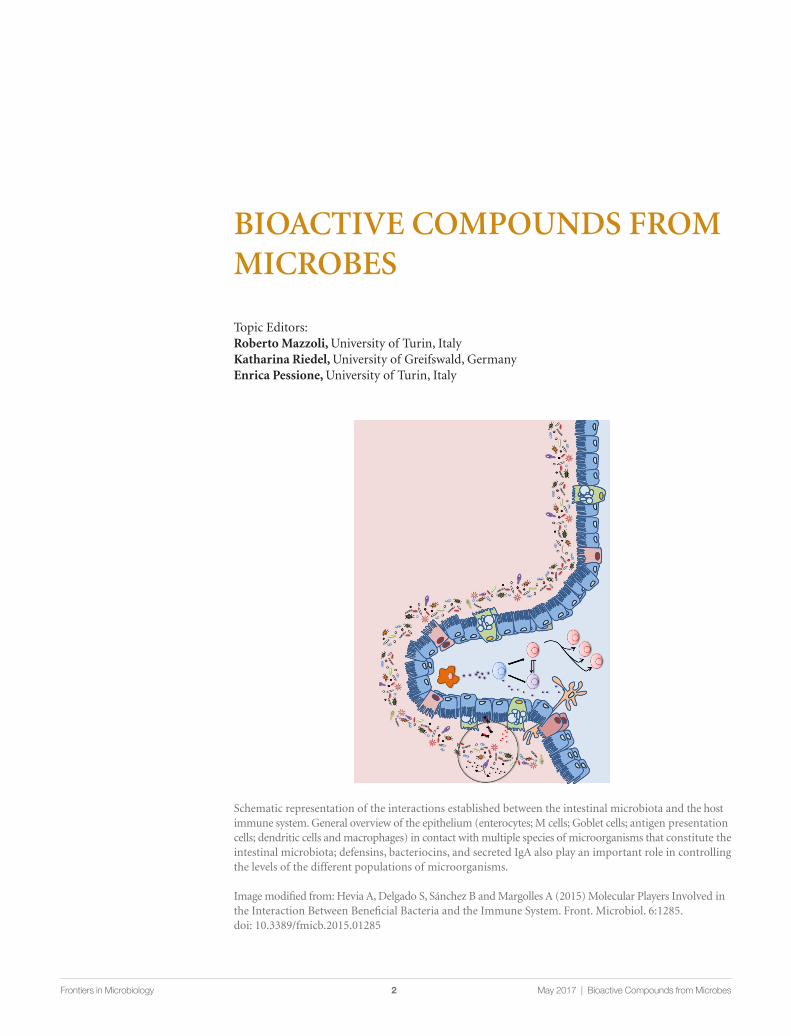
Schematic representation of the interactions established between the intestinal microbiota and the host immune system. General overview of the epithelium (enterocytes; M cells; Goblet cells; antigen presentation cells; dendritic cells and macrophages) in contact with multiple species of microorganisms that constitute the intestinal microbiota; defensins, bacteriocins, and secreted IgA also play an important role in controlling the levels of the different populations of microorganisms.
Image modified from: Hevia A, Delgado S, Sánchez B and Margolles A (2015) Molecular Players Involved in the Interaction Between Beneficial Bacteria and the Immune System. Front. Microbiol. 6:1285. doi: 10.3389/fmicb.2015.01285
Topic Editors: Roberto Mazzoli, University of Turin, ItalyKatharina Riedel, University of Greifswald, GermanyEnrica Pessione, University of Turin, Italy

3 May 2017 | Bioactive Compounds from MicrobesFrontiers in Microbiology
Microorganisms have had a long and surprising history. They were “invisible” until invention of microscope in the 17th century. Until that date, although they were extensively (but incon-sciously) employed in food preservation, beer and wine fermentation, cheese, vinegar, yogurt and bread making, as well as being the causative agents of infectious diseases, they were considered as “not-existing”. The work of Pasteur in the middle of the 19th century revealed several biological activities performed by microorganisms including fermentations and pathogenicity. Due to the urgent issue to treat infectious diseases (the main cause of death at those times) the “positive potential” of the microbial world has been neglected for about one century. Once the fight against the “evil” strains was fulfilled also thanks to the antibiotics, industry began to appreciate bacte-ria’s beneficial characteristics and exploit selected strains as starters for both food fermentations and aroma, enzyme and texturing agent production. However, it was only at the end of the 20th century that the probiotic potential of some bacteria such as lactic acid bacteria and bifidobac-teria was fully recognized. Very recently, apart from the probiotic activity of in toto bacteria, attention has begun to be directed to the chemical mediators of the probiotic effect. Thanks also to the improvement of techniques such as transcriptomics, proteomics and metabolomics, several bioactive compounds are continuously being discovered. Bioactive molecules produced by bacteria, yeasts and virus-infected cells proved to be important for improving or impairing human health. The most important result of last years’ research concerns the discovery that a very complex network of signals allows communication between organisms (from intra-species interactions to inter-kingdom signaling). Based on these findings a completely new approach has arisen: the system biology standpoind. Actually, the different organisms colonizing a certain environmental niche are not merely interacting with each other as individuals but should be considered as a whole complex ecosystem continuously exchanging information at the molecular level. In this context, this topic issue explores both antagonistic compounds (i.e. antibiotics) and “multiple function” cooperative molecules improving the physiological status of both stimulators and targets of this network. From the applicative viewpoint, these molecules could be hopefully exploited to develop new pharmaceuticals and/or nutraceuticals for improving human health.
Citation: Mazzoli, R., Riedel, K., Pessione, E., eds. (2017). Bioactive Compounds from Microbes. Lausanne: Frontiers Media. doi: 10.3389/978-2-88945-185-2

4 May 2017 | Bioactive Compounds from MicrobesFrontiers in Microbiology
Table of Contents
06 Editorial: Bioactive Compounds from MicrobesRoberto Mazzoli, Katharina Riedel and Enrica Pessione
Chapter 1: Diversity of bioactive molecules from microbes08 Bioactive Molecules Released in Food by Lactic Acid Bacteria: Encrypted
Peptides and Biogenic AminesEnrica Pessione and Simona Cirrincione
27 Identification of novel esterase-active enzymes from hot environments by use of the host bacterium Thermus thermophilusBenedikt Leis, Angel Angelov, Markus Mientus, Haijuan Li, Vu T. T. Pham, Benjamin Lauinger, Patrick Bongen, Jörg Pietruszka, Luís G. Gonçalves, Helena Santos and Wolfgang Liebl
39 Bioactive compounds produced by gut microbial tannase: implications for colorectal cancer developmentFélix López de Felipe, Blanca de las Rivas and Rosario Muñoz
43 Gut Microbiota Profiling: Metabolomics Based Approach to Unravel Compounds Affecting Human HealthPamela Vernocchi, Federica Del Chierico and Lorenza Putignani
Chapter 2: Immuno-modulating compounds64 Molecular Players Involved in the Interaction Between Beneficial Bacteria and
the Immune SystemArancha Hevia, Susana Delgado, Borja Sánchez and Abelardo Margolles
72 Bioactive Molecules Released From Cells Infected with the Human CytomegalovirusAnna Luganini, Maria E. Terlizzi and Giorgio Gribaudo
Chapter 3: Neuroactive compounds88 The Neuro-endocrinological Role of Microbial Glutamate and GABA Signaling
Roberto Mazzoli and Enrica Pessione105 Melatonin and Other Tryptophan Metabolites Produced by Yeasts: Implications
in Cardiovascular and Neurodegenerative DiseasesRuth Hornedo-Ortega, Ana B. Cerezo, Ana M. Troncoso, M. Carmen Garcia-Parrilla and Albert Mas

5 May 2017 | Bioactive Compounds from MicrobesFrontiers in Microbiology
Chapter 4: Antibiotic compounds112 The broad-spectrum antibiotic, zeamine, kills the nematode worm Caenorhabditis
elegansJosephine E. E. U. Hellberg, Miguel A. Matilla and George P. C. Salmond
125 Prodigiosin Induces Autolysins in Actively Grown Bacillus subtilis CellsTjaša Danevc ic , Maja Boric Vezjak, Maja Tabor, Maša Zorec and David Stopar
135 Bioengineering Lantibiotics for Therapeutic SuccessDes Field, Paul D. Cotter, Colin Hill and R. P. Ross

EDITORIALpublished: 08 March 2017
doi: 10.3389/fmicb.2017.00392
Frontiers in Microbiology | www.frontiersin.org March 2017 | Volume 8 | Article 392 |
Edited by:
George Tsiamis,
University of Patras, Greece
Reviewed by:
George Tsiamis,
University of Patras, Greece
Spyridon Ntougias,
Democritus University of Thrace,
Greece
*Correspondence:
Enrica Pessione
Specialty section:
This article was submitted to
Systems Microbiology,
a section of the journal
Frontiers in Microbiology
Received: 20 December 2016
Accepted: 24 February 2017
Published: 08 March 2017
Citation:
Mazzoli R, Riedel K and Pessione E
(2017) Editorial: Bioactive Compounds
from Microbes.
Front. Microbiol. 8:392.
doi: 10.3389/fmicb.2017.00392
Editorial: Bioactive Compounds fromMicrobes
Roberto Mazzoli 1, Katharina Riedel 2 and Enrica Pessione 1*
1 Laboratory of Microbial and Applied Biochemistry and Proteomics, Department of Life Sciences and Systems Biology,
University of Turin, Turin, Italy, 2Department for Microbial Physiology and Molecular Biology, Institute of Microbiology,
University of Greifswald, Greifswald, Germany
Keywords: gut-brain axis, antitumor activity, viral immune-escape, Bacteriocins, human-microbes cross talk
Editorial on the Research Topic
Bioactive Compounds fromMicrobes
Microorganisms are ubiquitous and essentially interact with all the other organisms present in thebiosphere, sometimes creating a network of signals that constitutes the basis for life on the Earth.Clarifying the nature of these molecular signals, their targets and the pathways underlying theirproduction, constitutes the essential pre-requisite for deciphering inter-kingdom communications,adaptive responses and systems biology (Nicholson et al., 2012; O’Mahony et al., 2015; Caniand Knauf, 2016). The insights acquired in the last two decades about human microbiota, andits fundamental role in maintaining a healthy physiological status, have opened the way tounderstanding the complex reciprocal talk between bacteria and humans (Hughes and Sperandio,2008; Lyte and Freestone, 2010; Mayer, 2011; Cryan and Dinan, 2012; Mazzoli, 2014; Halang et al.,2015; Kelly et al., 2015). In parallel with these aspects, new bioactive molecules from the microbialworld that interact with different cellular models continue to be discovered.
The times and the technological advances start now to be suitable for intercellular/interorganismcommunication to be studied at the molecular level. The aim of the present topic issue is to try todescribing the mediator molecules of a network of signals which is still largely underexplored andunderexploited.
As an example, some soil bacteria (such as Serratia spp.) can have antagonistic actions towardworms and the molecule involved, zeamine, is effective against yeasts and other biological systems,as well. Prodigiosin, the well-known pigment produced by the marine bacterium Vibrio ruber, has abroad antimicrobial spectrum and induces autolytic activity in the target cells (i.e., Bacillus subtilis).Lantibiotics are class I bacteriocins produced by Gram-positive bacteria that can be bioengineeredto both enhance their effectiveness against a larger number of bacterial strains and to improve theirstability during the gastric transit that is by rendering them protease-resistant. These overall dataopen new possibilities for antibiotic therapy in a period in which the phenomenon of antibioticresistance is considered as a major threat to public health (World Health Organization, 2014)since it is widespread in pathogenic, commensal, and food bacteria (Laxminarayan et al., 2013).Furthermore, the appearance of multiresistant bacterial strains (the so-called superbugs), oftencausing death, clearly constitutes a serious problem to be solved. Exploiting the microbial worldand its huge potential in finding new antimicrobial drugs is an urgent concern and some chaptersof this topic issue deal with these aspects.
Other interesting molecules are produced by cytomegalovirus-infected cells: these compoundsof viral origin (essentially proteins) can promote virus dissemination, persistence, and pathogenesisby counteracting host innate and adaptive immune responses. Conversely, some beneficialmicrobes, like Lactic Acid Bacteria (LAB) and Bifidobacteria can modulate the immune systemcontrolling inflammation by means of proteins and non-proteinaceous compounds (Pessione,2012).
6

Mazzoli et al. Editorial: Bioactive Compounds from Microbes
Several microbiota-derived compounds can contribute tocontrol host physiological and pathological states: Metabolomicprofiling of gut bacteria can allow to decipher several molecules(among which short chain fatty acids, for instance those producedby bacteria belonging to the clostridial cluster IV and XIV,vitamins and aromatic compounds) controlling cholesterolsynthesis, obesity, cardiovascular diseases, metabolic syndrome.Curiously, some bioactive peptides or amino acids can bedelivered by food in which LAB are present as fermentationstarters. These bacteria can decrypt peptides from foodproteins, which have anti-oxidant or metal chelating function,immunomodulatory properties as well as peptides controllinghyperglycemia, hypercholesterolemia, hypertension, cell cycle,apoptosis, and even having a refolding action on damagedproteins. An interesting tannin-degrading activity of bacteria(e.g., Streptococcus spp. and Fusobacterium spp.) can generategallic acid and pyrogallol, both having an anti-carcinogenicrole. Finally, microbial-derived amines can modulate a series ofpatho/physiological functions such as allergy, smooth musclerelaxation, anxiety, appetite, depression (Pessione et al., 2005).LAB are good producers of gamma-amino butyric acid (GABA)
(Laroute et al., 2016) that contributes to gut-to-brain signalingthrough different pathways involving enteric neurons, entero-endocrine cells and immune cells. Yeasts, mainly Saccharomycescerevisiae, can convert tryptophan into melatonin and serotoninthat are informational molecules related to circadian rhythm butalso promising agents for the prevention of neurodegenerativediseases.
This intense network of molecules, allowing communicationamong bacteria, viruses, and eukaryotic cells has evolved toguarantee optimal life in different ecological niches to eachcomponent of the ecosystem and is based upon effector-receptormodel. In-depth knowledge of all these biochemical signals,as well as the underlying finalism are still far to be fullyelucidated. Nevertheless, some mechanistic aspects highlightedin the present topic issue can open new perspectives in medicinebut can also shed light on evolution strategies.
AUTHOR CONTRIBUTIONS
EP wrote the manuscript which was then reviewed by RM andKR.
REFERENCES
Cani, P. D., and Knauf, C. (2016). How gut microbes talk to organs:
the role of endocrine and nervous routes. Mol. Metab. 5, 743–752.
doi: 10.1016/j.molmet.2016.05.011
Cryan, J. F., and Dinan, T. G. (2012). Mind-altering microorganisms: the impact
of the gut microbiota on brain and behaviour. Nat. Rev. Neurosci. 13, 701–712.
doi: 10.1038/nrn3346
Halang, P., Toulouse, C., Geißel, B., Michel, B., Flauger, B., Müller, M., et al. (2015).
Response of Vibrio cholerae to the catecholamine hormones Epinephrine and
Norepinephrine. J. Bacteriol. 197, 3769–3778. doi: 10.1128/JB.00345-15
Hughes, D. T., and Sperandio, V. (2008). Inter-kingdom signalling:
communication between bacteria and their hosts. Nat. Rev. Microbiol. 6,
111–120. doi: 10.1128/JB.00345-15
Kelly, J. R., Kennedy, P. J., Cryan, J. F., Dinan, T. G., Clarke, G., and Hyland,
N. P. (2015). Breaking down the barriers: the gut microbiome, intestinal
permeability and stress-related psychiatric disorders. Front. Cell. Neurosci.
9:392. doi: 10.3389/fncel.2015.00392
Laroute, V., Yasaro, C., Narin, W., Mazzoli, R., Pessione, E., Cocaign-
Bousquet, M., et al. (2016). GABA production in Lactococcus lactis is
enhanced by arginine and co-addition of malate. Front. Microbiol. 7:1050.
doi: 10.3389/fmicb.2016.01050
Laxminarayan, R., Duse, A., Wattal, C., Zaidi, A. K. M., Wertheim, H. F. L.,
Sumpradit, N., et al. (2013). Antibiotic resistance-the need for global solutions.
Lancet Infect. Dis. 13, 1057–1098. doi: 10.1016/S1473-3099(13)70318-9
Lyte, M., and Freestone, P. P. E. (2010). Microbial Endocrinology: Interkingdom
Signaling in Infectious Disease and Health. New York, NY: Springer.
Mayer, E. A. (2011). Gut feelings: the emerging biology of gut-brain
communication. Nat. Rev. Neurosci. 12, 453–466. doi: 10.1038/nrn3071
Mazzoli, R. (2014). “Neuro-active compounds produced by probiotics. Towards a
microbiota-(gut-) brain axis control?” in Interactive Probiotics, ed E. Pessione
(Boca Raton, FL: CRC Press; Taylor & Francis Group), 148–176.
Nicholson, J. K., Holmes, E., Kinross, J., Burcelin, R., Gibson, G., Jia, W., et al.
(2012). Host-gut microbiota metabolic interactions. Science 336, 1262–1267.
doi: 10.1126/science.1223813
O’Mahony, S. M., Clarke, G., Borre, Y. E., Dinan, T. G., and Cryan, J. F. (2015).
Serotonin, tryptophan metabolism and the brain-gut-microbiome axis. Behav.
Brain Res. 277, 32–48. doi: 10.1016/j.bbr.2014.07.027
Pessione, E. (2012). Lactic acid bacteria contribution to gut microbiota
complexity: lights and shadows. Front. Cell. Infect. Microbiol. 2:86.
doi: 10.3389/fcimb.2012.00086
Pessione, E., Mazzoli, R., Giuffrida, M. G., Lamberti, C., Garcia-Moruno,
E., Barello, C., et al. (2005). A proteomic approach to studying
biogenic amine producing lactic acid bacteria. Proteomics 5, 687–698.
doi: 10.1002/pmic.200401116
World Health Organization (2014). Antimicrobial Resistance: 2014 Global Report
on Surveillance. Geneva: World Health Organization.
Conflict of Interest Statement: The authors declare that the research was
conducted in the absence of any commercial or financial relationships that could
be construed as a potential conflict of interest.
Copyright © 2017 Mazzoli, Riedel and Pessione. This is an open-access article
distributed under the terms of the Creative Commons Attribution License (CC BY).
The use, distribution or reproduction in other forums is permitted, provided the
original author(s) or licensor are credited and that the original publication in this
journal is cited, in accordance with accepted academic practice. No use, distribution
or reproduction is permitted which does not comply with these terms.
Frontiers in Microbiology | www.frontiersin.org March 2017 | Volume 8 | Article 392 | 7
fmicb-07-00876 June 7, 2016 Time: 13:41 # 1
REVIEWpublished: 09 June 2016
doi: 10.3389/fmicb.2016.00876
Edited by:Steve Lindemann,
Pacific Northwest NationalLaboratory, USA
Reviewed by:Giulia Tabanelli,
CIRI Agroalimentare, ItalyGiovanna Suzzi,
Università degli Studi di Teramo, Italy
*Correspondence:Enrica Pessione
Specialty section:This article was submitted to
Systems Microbiology,a section of the journal
Frontiers in Microbiology
Received: 09 February 2016Accepted: 24 May 2016
Published: 09 June 2016
Citation:Pessione E and Cirrincione S (2016)Bioactive Molecules Released in Food
by Lactic Acid Bacteria: EncryptedPeptides and Biogenic Amines.
Front. Microbiol. 7:876.doi: 10.3389/fmicb.2016.00876
Bioactive Molecules Released inFood by Lactic Acid Bacteria:Encrypted Peptides and BiogenicAminesEnrica Pessione* and Simona Cirrincione
Laboratory of Biochemistry, Proteomics and Metabolic Engineering of Prokaryotes, Department of Life Sciences andSystems Biology, University of Torino, Torino, Italy
Lactic acid bacteria (LAB) can produce a huge amount of bioactive compounds. Sincetheir elective habitat is food, especially dairy but also vegetal food, it is frequent to findbioactive molecules in fermented products. Sometimes these compounds can haveadverse effects on human health such as biogenic amines (tyramine and histamine),causing allergies, hypertensive crises, and headache. However, some LAB productsalso display benefits for the consumers. In the present review article, the main nitrogencompounds produced by LAB are considered. Besides biogenic amines derived fromthe amino acids tyrosine, histidine, phenylalanine, lysine, ornithine, and glutamate bydecarboxylation, interesting peptides can be decrypted by the proteolytic activity ofLAB. LAB proteolytic system is very efficient in releasing encrypted molecules fromseveral proteins present in different food matrices. Alpha and beta-caseins, albumin andglobulin from milk and dairy products, rubisco from spinach, beta-conglycinin from soyand gluten from cereals constitute a good source of important bioactive compounds.These encrypted peptides are able to control nutrition (mineral absorption and oxidativestress protection), metabolism (blood glucose and cholesterol lowering) cardiovascularfunction (antithrombotic and hypotensive action), infection (microbial inhibition andimmunomodulation) and gut-brain axis (opioids and anti-opioids controlling mood andfood intake). Very recent results underline the role of food-encrypted peptides inprotein folding (chaperone-like molecules) as well as in cell cycle and apoptosis control,suggesting new and positive aspects of fermented food, still unexplored. In this context,the detailed (transcriptomic, proteomic, and metabolomic) characterization of LAB offood interest (as starters, biocontrol agents, nutraceuticals, and probiotics) can supply asolid evidence-based science to support beneficial effects and it is a promising approachas well to obtain functional food. The detailed knowledge of the modulation of humanphysiology, exploiting the health-promoting properties of fermented food, is an openfield of investigation that will constitute the next challenge.
Keywords: opioid, antioxidant, chaperon-like, cell-cycle control, antimicrobial peptides, beta-phenylethylamine,tyramine
Frontiers in Microbiology | www.frontiersin.org June 2016 | Volume 7 | Article 876 | 8

fmicb-07-00876 June 7, 2016 Time: 13:41 # 2
Pessione and Cirrincione LAB Bioactive Compounds in Food
INTRODUCTION
Human health is the result of a correct physiological statusoften resulting from the reciprocal interaction of gene-derivedsignals (genetics) and environment–generated information(epigenetics). Recognizing gene signals is relatively easywhereas environmental stimuli are often multiple, complex,and reciprocally interacting. Temperature, pH, redox balance,sleep, diet, drugs, and psychological status can deeply affectgene expression, metabolic pathways, and homeostasis (Carey,2012). However, human genes and environment are not the onlyplayers: microorganisms can take part to the complex molecularcross-talk existing between external world and “self.”
Firstly, endogenous symbiont microorganisms, the so-calledmicrobiota, can modulate gene expression, induce preferentialfood intake (Aydin, 2007), influence pH, redox balance andthe ratio between pro-inflammatory and anti-inflammatorycytokines (Belkaid and Hand, 2014). Briefly, control brain,metabolism, immune system, and several homeostatic routes.
Secondarily, food-derived bacteria and yeasts can exogenouslyaffect nutritional parameters, metabolism, oxidative status,immunity, blood pressure, appetite, behavior, also controlling theendogenous microbiota (Arora et al., 2013) by altering the ratioamong saccharolytic and proteolytic species, by modulatingsymbionts gene expression and several other functions(O’Flaerty, 2014). Hence, all fermented food, containingliving organisms, can contribute to modulation of the hostphysiological balance and it constitutes an opportunity toenrich the diet with new bioactive molecules finally resultingin phenotypic effects (appetite, cholesterol and blood pressurelowering, improvement of mood, antioxidant, and immunedefenses) on humans (Pessione, 2010). Actually, all types ofmodulations occur via a complex network of signals, amongwhich proteinaceous compounds play a crucial role.
Microorganisms are able to synthesize a large number ofmetabolites with assessed beneficial or detrimental properties forhuman health. Among these, nitrogen-bearing molecules suchas amino acids, amino acid derivatives and oligopeptides havereceived great attention since they can affect human physiologyin multiple ways.
As an example, amino acid derivatives such as selenocysteinesand selenomethionines, have recently been reported to bebiosynthesized in both Lactobacilli (Lamberti et al., 2011) andyeasts (Porto et al., 2015). Although selenoaminoacids are nottrue “bioactive compounds,” directly stimulating receptors onhuman cells, they can trigger effects deeply affecting humanhealth. The bioactive role of seleno-fixing microorganisms liesin the fact that diet-derived inorganic selenium is toxic (selenateand selenite) or poorly active (elemental selenium) whereasfixed selenium forms (selenomethionines and selenocysteines)are the only bioavailable for humans. On the other hand,only bacteria and yeasts can produce seleno-amino acids frominorganic selenium. Once properly inserted into selenoproteins(i.e., glutathione peroxidase), they can counteract oxidative stress.Besides this well-known antioxidant function, there are dataindicating that selenoproteins can modulate immune system(Huang et al., 2012) and activating anabolic circuits such as
thyroid hormone biosynthesis (Mullur et al., 2014). Furthermore,epidemiological studies show an inverse correlation betweenselenium level in blood and cancer mortality, and laboratoryexperiments have shown a selenium protective effect againstcancer initiation and development (Gromadzinska et al., 2008).In Lactobacillus reuteri exoproteome studies have demonstratedthat two secreted proteins (GAPDH and Phosphoketolase)contain selenocysteines opening the way to employ this strainto supply organic bioavailable forms of selenium (Galano et al.,2013; Mangiapane et al., 2014a,b).
Among amino acid derivatives found in food, biogenic aminesare worth of a special mention. Such compounds, althoughsometimes naturally present (especially in vegetal food) areoften the result of the bacterial decarboxylative activity onfree amino acids in food. Biogenic amines can be present innon-fermented food, like fish, due to spoilage bacteria thatduring protein putrefaction release free amino acids undergoingdecarboxylation. E. coli can produce cadaverine from lysine andputrescine from ornithine (Applebaum et al., 1975). Proteuscan produce putrescine from ornithine as a communicationsignal (Visick and Fuqua, 2005). However, also not-spoiledfood, such as fermented food, can present the risk of biogenicamine accumulation. Although starters, exogenously added toperform controlled fermentations, are accurately typed to avoidany risk, autochthonous or contaminant lactic acid bacteria(LAB) can contribute to amine release. LAB are strong amineproducers since they use this metabolic pathway (at the place ofrespiration) to both create a proton gradient and hence energy(for exhaustive review, see Pessione et al., 2010) and to alkalinizethe environment, very acidic since their main fermentationproducts are acids (lactic acid for homofermenter LAB andlactic+ formic+ acetic acid in heterofermenters).
Many experimental evidences demonstrate that someLAB strains also produce anti-hypertensive, anti-thrombotic,cholesterol-lowering, metal-chelating, antimicrobial, anti-oxidant, immune-modulating, chaperone-like and opioid/opioidantagonist peptides from food proteins (Pessione, 2012), andthey can modulate the concentration of opioid and cannabinoidreceptors in the gut epithelium (Hayes et al., 2007b). The nextsections will focus on some of the referred compounds and willillustrate the main effects exerted on human health.
ENCRYPTED BIOACTIVE PEPTIDES
Several bioactive peptides, lacking activity when protein-encrypted but acquiring their biological effects whenproteolytically released, have been shown to have health-promoting properties as anti-microbials, hypocholesterolemics,opioid and opioid antagonists, angiotensin-convertingenzyme inhibitors, anti-thrombotics, immunomodulators,cytomodulators, and anti-oxidants (Hayes et al., 2007b).
The human pool of digestive proteases and peptidases canliberate food-encrypted bioactive peptides that can be absorbedby the gut and then reach peripheral organs. However, theenzymatic activity of LAB largely contribute to their release,either into the food matrix (starter or autochthonous LAB) or in
Frontiers in Microbiology | www.frontiersin.org June 2016 | Volume 7 | Article 876 | 9

fmicb-07-00876 June 7, 2016 Time: 13:41 # 3
Pessione and Cirrincione LAB Bioactive Compounds in Food
the gut (endogenous microbiota or probiotics). LAB are ancientorganisms adapted to an anoxic environment that never evolvedthe capability to biosynthesize heme and hence to have functionalcytochromes, catalases, and peroxidases. They are very sensitiveto oxygen and devote most of their genes to oxygen stresscounteracting. Due to the limited length of the overall genome,the biosynthetic abilities of LAB are very limited especially inamino acid synthesis (Pessione, 2012). Therefore, LAB evolveda complex and sophisticated proteolytic system allowing themto get amino acids from the proteins present in the externalenvironment. A schematic representation of this system, whichincludes proteases, peptidases and membrane transporters, isreferred in Figure 1.
LAB Proteolytic SystemExtracellular protein hydrolysis into various long oligopeptidesis initiated by a cell-envelope proteinase (CEP). Oligopeptidesgenerated by this first cleavage are subsequently taken upby cells via specific transport systems and they undergofurther degradation into shorter peptides (bioactive or possibleprecursors of bioactive compounds) and amino acids in thecytoplasm (Savijoki et al., 2006). In LAB the oligopeptidetransporter system (Opp) is the main transporter, belongingto a superfamily of highly conserved ATP-binding cassettetransporters (Doeven et al., 2005). The Opp system of L. lactistransports peptides up to at least 18 residues (Savijoki et al., 2006).It should be underlined that most bioactive peptides are releasedinto the external environment only when cells undergo autolysis,because only occasionally the longer peptides (originated bythe first hydrolytic step) possess biological activity (Meisel andBockelmann, 1999). Nevertheless, some authors reported thatCEP from Lactobacillus delbrueckii subsp. lactis are suitableto liberate both metal-chelating and anti-hypertensive peptidesdirectly from caseins and without necessity of prior cell autolysis.Maximum CEP activity was a little bit decreased by the additionof NaCl (1%) and glycerol (5%; Hébert et al., 2008). However,proteolytic enzymes released by LAB proved to be very differentin the different LAB species and strains, giving rise to a differentpool of bioactive peptides (Hébert et al., 1999).
Food Matrices Containing EncryptedPeptidesLactic acid bacteria proteolytic system is suitable to producebioactive peptides from several food proteins, especiallycaseins that constitute the main nitrogen substrate presentin their habitat (milk and milk derivatives; Clare andSwaisgood, 2000). Casein consists of four main proteins(whose ratio is about 38:11:38:13): alpha s1 casein, alphas2 casein, beta-casein and k-casein, differing in aminoacid sequence, hydropathicity index, glycosylation andphosphorylation degree (Pessione, 2012). Usually, CEPshave a strong preference for hydrophobic caseins andLactococcus CEPs have been classified into several typesand subtypes depending on their substrate specificity forαS1-, β-, and κ-caseins (Kunji et al., 1996). Casein hydrolysis cangive rise mainly to opioid/anti-opioid peptides, antithromboticand antihypertensive, immunomodulatory, mineral-binding and
antimicrobial peptides (Thakur et al., 2012) easily detectableby means of chromatographic techniques after microbial fooddigestion (Saraswat et al., 2012).
Hydrolytic cleavage of milk whey proteins (alpha-lacto-albumine, beta-lacto-globulin, lactoferrin, andimmunoglobulins) can also generate bioactive molecules, suchas hypocholesterolemic peptides, which can decrease absorptionand enhance fecal extrusion of cholesterol. Actually, milk wheyproteins display a greater cholesterol-lowering activity thancasein, in particular beta-lactoglobulin (Nagaoka et al., 2001).Beta-lactoglobulin-released peptides also exhibit antioxidant(Hernández-Ledesma et al., 2005) and immune-modulating(Prioult et al., 2004) activity.
Bio-active molecules, especially ACE-inhibiting peptides,can also be found encrypted in bovine meat proteins, suchas hemoglobin (i.e., hemorphins) and serum albumin (i.e.,serorphin), collagen, elastine, fibrinogen (Minkiewicz et al., 2011;Lafarga et al., 2015).
However, also food of vegetal origin can be a source ofbioactive peptides. In soy β-conglycinin β-subunit (Kanekoet al., 2010) and in the large subunit of spinach rubisco, bothantihypertensive (Yang Y. et al., 2003) and opioid peptides (i.e.,soymorphins-5,-6, and -7, and rubiscolin-5 and -6, respectively),displaying anxiolytic effects, food intake controlling action andenhancement of memory, have been described (Yang et al., 2001;Yang S. et al., 2003; Hirata et al., 2007).
A special mention needs gluten: opioid peptides (i.e., glutenexorphins A4, A5, B4, B5, and C), have been detected andcharacterized in wheat gluten since 1992 (Fukudome andYoshikawa, 1992). Some of these peptides have analgesic actionon the CNS (Takahashi et al., 2000) but some can also acton opioid receptors located outside the blood–brain barrier,inducing prolactin secretion (Fanciulli et al., 2005). Bothantihypertensive (Rizzello et al., 2008) and antioxidant (Codaet al., 2012) peptides are the result of LAB activity on wheat andother cereals. Generally, the high proline content of gluten alphagliadins prevent hydrolysis by enzymes of the gastrointestinaltract, whereas LAB possess proline-specific peptidase systems.Alternatively, peptide degradation is achieved by the combinedaction of peptidase from different LAB strains simultaneouslypresent in fermented food (Gerez et al., 2008).
To date, LAB strains able to release bioactive peptidesfrom food proteins, with particular reference to milk caseins,include L. helveticus CP790, L. rhamnosus GG, L. bulgaricus SS1,and L. lactis subsp. cremoris FT4 (Gobbetti et al., 2002). Thehydrolytic ability is related to both the protein substrate, i.e.,its amino acid sequence and the proteolytic enzyme panel ofeach microbial strain (Griffiths and Tellez, 2013). It is possible toenhance (100-fold) the proteolytic potential of LAB by growingthem in milk (Wakai and Yamamoto, 2012). Actually it hasbeen demonstrated that peptide-rich media like MRS supplyinga ready-to-eat nitrogen source are unfavorable to the inductionof most proteolytic enzymes for instance those encoded by PepN and PepX (Smeianov et al., 2007). On the other hand, CEP areable to hydrolyze caseins only after growth on skim milk but notafter MRS pre-culture (Jensen et al., 2009). Generally, lactobacilligenome encodes a larger number of proteases, peptidases, amino
Frontiers in Microbiology | www.frontiersin.org June 2016 | Volume 7 | Article 876 | 10

fmicb-07-00876 June 7, 2016 Time: 13:41 # 4
Pessione and Cirrincione LAB Bioactive Compounds in Food
FIGURE 1 | Schematic representation of the proteolytic system of LAB.
acid permeases, and Opp transport systems than lactococci(Klaenhammer et al., 2005). However, more studies on a largernumber of species and strains are necessary to establish the truepotential of each strain.
Finally, it is worth considering that most information onLAB proteolytic system has been referred to food matrix isolatedmicroflora and to its action on bovine milk casein (Korhonenand Pihlanto, 2003). However, endogenous or probiotic or food –derived LAB can also produce bioactive peptides from thedifferent protein pool present in the host gut, opening interestingperspectives for improving human health (Sharon et al., 2014).In the following paragraphs, the best known bioactive moleculesgenerated from LAB proteases and peptidases are described.
Antimicrobial PeptidesLactic acid bacteria proteolytic activity on caseins gives rise tosmall peptides displaying antimicrobial (both bactericidal andbacteriostatic) activity. Alpha s1 casein and K-casein hydrolysisgives rise to isracidin and k-casecidin, respectively (McCannet al., 2006). Both peptides display inhibitory action on S. aureusgrowth (Meisel and Bockelmann, 1999; Atanasova et al., 2014).K-casein can also originate kappacin, a long bioactive peptideactive against Streptococcus mutans. This peptide, althoughderiving from K-casein, is phosphorylated but not glycosylated,and displays the ability to prevent bacterial adhesion to gingivalmucosa and to bind enterotoxins (Malkoski et al., 2001). Thealpha-s-2 casein fragment 165-203, called casocidin-I can inhibitboth S. carnosus and E. coli (Zucht et al., 1995). Lactobacillushelveticus can hydrolyze beta casein, by means of a PR4
proteinase, decrypting an antimicrobial peptide active towardseveral strains of Gram-positive bacteria (including S. aureusand Listeria innocua) but also against Gram-negative pathogenssuch as E. coli, Salmonella, Yersinia, (Minervini et al., 2003).Homology studies revealed that this peptide has a similar lengthto isracidin, but a stronger hydrophobic nature (lacking positivecharges) allowing a better activity on Gram-negative bacteria.Furthermore, it bears some proline residues near the C-terminal,which can prolong its half-life rendering this molecule moreresistant to the peptidolytic action (Gobbetti et al., 2004). Thecritical question concerning this peptide is that it has beendecrypted from human milk but not from other caseins presentin food (cow, buffalo, goat, ship).
Different peptides produced from caseins by differentLactobacillus species (L. acidophilus, L. helveticus, L. plantarum,and L. rhamnosus) and by Lactococcus lactis are active againstGram-negative rods such as Enterobacter sakazakii (Hayes et al.,2006). An interesting antimicrobial peptide, named LactoferricinB, having broad spectrum of action including Gram-negativeand Gram-positive bacteria, yeasts and filamentous fungi, hasbeen described as the result of the hydrolytic action on bovinelactoferrin (Dionysius and Milne, 1997; Kitts and Weiler,2003). This cationic peptide alters membrane permeabilitywith consequent dissipation of the proton gradient and hastherefore bactericidal action (FitzGerald and Murray, 2006). Itwas cloned and expressed in E. coli, showing activity againstKlebsiella pneumoniae, Streptococcus Mutans, and S. aureus(Luo et al., 2007). Lactoferricin B is especially active towardthe enterohemorrhagic strain of E. coli 0157h:7 (Muro-Urista
Frontiers in Microbiology | www.frontiersin.org June 2016 | Volume 7 | Article 876 | 11

fmicb-07-00876 June 7, 2016 Time: 13:41 # 5
Pessione and Cirrincione LAB Bioactive Compounds in Food
et al., 2011) and proved to be active also against wine-spoilage LAB such as Oenococcus oeni, Pediococcus damnosus,and Lactobacillus brevis (Enrique et al., 2009). It is difficultto establish the real LAB potential in hydrolyzing lactoferrin.Some experiments were performed by using both microbial-derived enzymes and fermentation with proteolytic startercultures (Sharma et al., 2011), however, the true potential ofeach strain has to be experimentally proved since a greatvariability was observed among strains (Korhonen, 2009). Morerecently, the proteolytic activity of different LAB strains on goatmilk beta casein and beta-lactoglobulin were tested. Lactococcuslactis l598, Lactobacillus lactis 1043, Streptococcus thermophilust3D1, Dt1and Lb. delbrueckii subsp. bulgaricus b38, b122 andb24 revealed a significant potential in producing antimicrobialpeptides inhibiting S. aureus, L. monocytogenes, Listeria innocua,Enterobacter aerogenes, and Salmonella enteritidis (Atanasovaet al., 2014).
Concerning the mechanism of action of such antibacterialcompounds, several models have been proposed. Beingamphipathic molecules, bearing a hydrophobic moiety and astrongly positively charged domain, this peptides can firstlybind to teichoic acids in Gram-positive-, or to LPS in Gram-negative bacteria (Vorland, 1999) and then interact with thenegatively charged bacterial membranes. Most of them act asproton gradient perturbing molecules, (causing membranedepolarization like polymyxin B and colistin; Lohner andBlondelle, 2005), however, some authors suggest that alsoremoval/chelating of membrane-bound iron can affect bacterialviability (Sharma et al., 2011). For what concern lactoferrin-derived peptides, however, it has long been established (Bellamyet al., 1992) that the domains of lactoferrin involved in theantibacterial activity are different from those involved in ironbinding. Complex mechanism, including inhibition of thesynthesis of macromolecules (Ulvatne et al., 2004) as well assynergic action with host innate immunity compounds were alsoreported (Brogden, 2005).
These antimicrobial compounds are appreciated in the foodindustry (especially dairy industry) as natural preservativescounteracting undesired contamination. This allows to reducethe amount of sugar and salt with excellent benefits for diabetics,obese, and hypertensive subjects. Furthermore, their stability inblood and serum render them promising infection control agents.Some of them are also good candidates to be tested for their anti-viral potential. Bojsen et al. (2007) reported the antiviral effect ofbovine whey proteins, whereas Civra et al. (2015) very recentlyidentified from donkey milk lactadherin a peptide displayinganti-Rotavirus activity. The actual role of LAB proteolysis in thedecrypting of the antiviral potential is far to be fully elucidated.An overview of antibacterial peptides is proposed in Table 1.
Metal-Binding PeptidesSome LAB strains digest casein, releasing casein phosphopeptides(CPP) phosphorylated on Ser residues. CPP have a strong anioniccharacter and hence are very resistant to further proteolyticdegradation (Sharma et al., 2011). They have been identifiedduring cheese ripening due to microbial protease activity (Singhet al., 1997). The typical cluster is Ser(P)-Ser(P)Ser(P)-Glu-Glu,
but they may contain also phosphorylated cysteines. Even ifthis motif is crucial for metal-binding, various phosphopeptidefractions revealed significant differences in this capability, whichmay be due to variant amino acid composition around thephosphorylated region. Hence, other factors can affect CPP-metalinteraction, namely the total number of amino acids and the totalnegative charge (Cross et al., 2005).
It has long been established (Gagnaire et al., 1996) that betaand k-caseins (being poor in hydroxylated amino acids andtherefore less suitable for phosphorylation) generate peptidesless active in metal binding, whereas the Ser/Thr rich alpha s1and alpha s2 caseins are the most suitable for chelating cationsby means of CPP. However, alpha s1 casein-derived peptide(59–79), although bearing five phosphate groups, is less activein allowing mineral uptake by the human tumor cells HT29than beta casein-derived peptides (1–25). Actually, besides thetypical cluster sequence, specific secondary structure motifs inthe bioactive peptide (such as beta-turn and loop), as well as thedegree of aggregation of CPP in presence of divalent cations, arerequired for a correct mineral absorption (Ferraretto et al., 2003).
Casein phosphopeptides can chelate up to 250 mgCa/g witha dissociation constant in the mM range, with an affinityeven higher for trace elements like zinc, iron, and copper(FitzGerald, 1998). Their mechanism of action rely on the factthat they can form soluble complexes with calcium even atalkaline pH (Berrocal et al., 1989). This results in increasedabsorption of calcium in the intestine, useful in the treatmentof osteoporosis. Re-calcification of dental enamel and preventionof dental caries were also reported (Reynolds et al., 2008).Enhanced gut bioavailability and potential higher absorptionof enzyme co-factors such as iron, zinc, copper, selenium,magnesium, and manganese, due to CPP was also described(Meisel and Olieman, 1998). On the other hand, controversialclinical results concerning both calcium (Korhonen and Pihlanto,2006) zinc (Miquel and Farré, 2007) and calcium and ironabsorption obtained on patients receiving CPP supplementationand controls, underline the difficulty to standardize thedifferent supplements (Teucher et al., 2006). Finally, calciumavailability is strongly dependent on meal composition (i.e.,phytates) and complex interactions between different foodsingested.
In spite of all these considerations, all described peptidesmay constitute interesting tools for microelements nutritionalimprovement. In our aging western society bone disease isthe third cause of nursery care and osteoporosis is not fullycontrolled by a healthy lifestyle (active movement and diet;Wei et al., 2003). Therefore, calcium-enriching supplementssuch as CPP open new nutraceutical perspectives. However, theneed to use purified and well-characterized CPP (for havinga reproducible nutritional impact) highly affects the costs.Hence, the use of LAB as probiotics or food supplements toobtain functional food, especially milk based drinks and yogurtswill be a promising strategy. Lactobacillus helveticus LA, isa LAB adapted to the cheese environment displaying goodproteolytic activity. It can decrypt from alpha casein a peptideshowing calcium-binding activity (Dimitrov, 2009). Lactobacillushelveticus LBK16H can decrypt the tripeptides Ile-Pro-Pro and
Frontiers in Microbiology | www.frontiersin.org June 2016 | Volume 7 | Article 876 | 12
fmicb-07-00876 June 7, 2016 Time: 13:41 # 6
Pessione and Cirrincione LAB Bioactive Compounds in Food
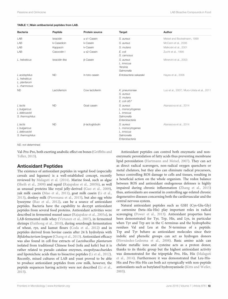
TABLE 1 | Main antibacterial peptides from LAB.
Bacteria Peptide Protein source Target Author
LAB Isracidin α s1-Casein S. aureus Meisel and Bockelmann, 1999
LAB k-Casedicin k-Casein S. aureus McCann et al., 2006
LAB Kappacin k-Casein S. mutans Malkoski et al., 2001
LAB Casocidin I α s2-Casein E. coliS. carnosus
Zucht et al., 1995
L. helveticus Isracidin-like β-Casein S. aureusL. innocuaYersiniaSalmonella
Minervini et al., 2003
L acidophilusL. helveticusL. plantarumL. rhamnosus
ND In toto casein Entobacteria sakazakii Hayes et al., 2006
ND Lactoferricin Cow lactoferrin K. pneumoniaeS. aureusS. mutansE. coli ol57
Luo et al., 2007; Muro-Urista et al., 2011
L lactisL bulgaricusL delbrueckiiS. thermophilus
ND Goat casein S. aureusL. monocytogenesL. innocuaSalmonellaEnterobacteria
Atanasova et al., 2014
L lactisL bulgaricusL delbrueckiiS. thermophilus
ND β-lactoglobulin S. aureusL. monocytogenesL. innocuaSalmonellaEnterobacteria
Atanasova et al., 2014
ND, not determined.
Val-Pro-Pro, both exerting anabolic effect on bones (Griffiths andTellez, 2013).
Antioxidant PeptidesThe existence of antioxidant peptides in vegetal food (especiallycereals and legumes) is a well-established concept, recentlyreviewed by Malaguti et al. (2014). Marine food, such as algae(Sheih et al., 2009) and squid (Rajapakse et al., 2005b), as wellas unusual proteins like royal jelly-derived (Guo et al., 2009),yak milk casein (Mao et al., 2011), goat milk casein (Li et al.,2013), donkey milk (Piovesana et al., 2015), but also egg whitelysozyme (Rao et al., 2012), can be a source of antioxidantpeptides. Bacteria have the capability to decrypt antioxidantpeptides from several food proteins. Antioxidant activities weredescribed in fermented mussel sauce (Rajapakse et al., 2005a), inLAB-fermented milk whey (Virtanen et al., 2007), in fermentedshrimps (Faithong et al., 2010), during sourdough fermentationof wheat, rye, and kamut flours (Coda et al., 2012) and inpeptides derived from bovine casein after 24 h hydrolysis withBifidobacterium longum (Chang et al., 2013). Antioxidant efficacywas also found in cell-free extracts of Lactobacillus plantarumisolated from traditional Chinese food (tofu and kefir) but it israther related to pseudo catalase enzymes, exopolysaccharidesand lipoteichoic acids than to bioactive peptides (Li et al., 2012).Recently, mixed cultures of LAB and yeast proved to be ableto produce antioxidant peptides from cow milk, however, thepeptide sequences having activity were not described (Li et al.,2015).
Antioxidant peptides can control both enzymatic and non-enzymatic peroxidation of fatty acids thus preventing membranelipid peroxidation (Hartmann and Meisel, 2007). They can actas direct radical scavengers, non-radical oxygen quenchers ormetal chelators, but they also can eliminate radical precursors,hence controlling ROS damage to cells and tissues, resulting ina beneficial action on the whole organism. The redox balancebetween ROS and antioxidant endogenous defenses is highlyimpaired during chronic inflammation (Zhang et al., 2015)thus, antioxidants are essential in controlling age-related chronicdegenerative diseases concerning both the cardiovascular and thecentral nervous system.
Natural antioxidant peptides such as GSH (Cys-Glu-Gly)or carnosine (beta-Ala-His) play important roles in radicalscavenging (Power et al., 2013). Antioxidant properties havebeen demonstrated for Tyr, Trp, His, and Lys, in particularwhen Tyr and Trp are in the C-terminus and the hydrophobicresidues Val and Leu at the N-terminus of a peptide.Trp and Tyr behave as antioxidant molecules since theirindolic and phenolic groups can act as hydrogen donors(Hernández-Ledesma et al., 2008). Basic amino acids canchelate metallic ions and cysteine acts as a proton donor,thanks to its thiolic group but the highest antioxidant activitywas demonstrated for the tripeptide Pro, His, His (Malagutiet al., 2014). Furthermore it was demonstrated that Leu-His-His and Pro-His-His can both act synergically with non-peptideantioxidants such as butylated hydroxyanisole (Kitts and Weiler,2003).
Frontiers in Microbiology | www.frontiersin.org June 2016 | Volume 7 | Article 876 | 13

fmicb-07-00876 June 7, 2016 Time: 13:41 # 7
Pessione and Cirrincione LAB Bioactive Compounds in Food
Immunomodulatory PeptidesIt is well established that some milk proteins (caseins,whey proteins, lactoferrin, lactoperoxidase) are able to controllymphocyte proliferation (Möller et al., 2008). Milk-derivedpeptides such as the C-terminus sequence of beta casein (193–209) can increase proliferation of rat lymphocytes (Meiseland Bockelmann, 1999), whereas alpha casein C-terminalhexapeptide (194–199) and beta casein fragments (63–68and 91–93) induce macrophage maturation and phagocytosisenhancement in vitro (Fiat and Jollès, 1989). In agreementwith these data, studies in vivo show that alpha casein-derivedpeptides can stimulate macrophages phagocytosis exerting aprotective effect against Klebsiella pneumoniae infection inmice (Migliore-Samour et al., 1989). Short-chain peptidesfrom milk whey proteins also stimulate the proliferation ofmurine spleen lymphocytes in vitro (Mercier et al., 2004).More detailed studies report that proliferation of humanperipheral blood lymphocytes is induced by the k-casein andalpha lactalbumin fragment Tyr-Gly-Gly (Kayser and Meisel,1996; Sharma et al., 2011). Natural killer (NK) cell activityand antibody synthesis can also be enhanced by food–derivedbioactive peptides (Hartmann and Meisel, 2007). However, milk-derived peptides do not act only as simply stimulators ofthe immune system: immune-modulating activities includingcytokines regulation and attenuation of allergic reactions has alsobeen described (Korhonen and Pihlanto, 2003; FitzGerald andMurray, 2006).
Transferring clinical data to food is not so simple.Actually, in this scenario, LAB (especially probiotic LAB)play a very central role: beta-casein medium fermented withLAB gives rise to bioactive peptides acting on monocytes,macrophages and T helper cells, particularly with Th1-like cells (Laffineur et al., 1996). Lactobacillus paracaseihydrolyzes beta-lactoglobulin originating peptides stimulatingInterleukin 10 (IL-10) production and downregulation ofIL-4 and gamma interferon secretion (Prioult et al., 2004).Casein hydrolysated by Lactobacillus rhamnosus GG displaysmodulating effects (both stimulation and suppression) onlymphocyte proliferation (Möller et al., 2008). Furthermore,casein hydrolysates from Lactobacillus rhamnosus GG caninduce enhancement of anti-inflammatory cytokines fromTh1 lymphocytes and a parallel decrease of pro-inflammatorycytokines and immunoglobulins produced by Th2 lymphocytes,thus controlling allergic reactions (Delcenserie et al., 2008).Lactobacillus helveticus proteolysis products down-regulatecytokines production and stimulate macrophage phagocytosis(Matar et al., 2001) and also show protective effects againstE. coli O157 (LeBlanc et al., 2004) and Salmonella typhimurium(Tellez et al., 2011) infection. All the non-proteolytic strainsof Lactobacillus helveticus failed to trigger immune systemmodulation. Three immune-active peptides derived both frombeta casein and from alpha lactalbumin after milk fermentationwith proteolytic strains of Lactobacillus helveticus were analyzed:they contain high percentage of proline histidine and lysine(Tellez et al., 2010). Establishing how, when and in what extentproteolysis occurs in food (and in the gut) is still an openquestion.
Cell Cycle and Apoptosis ModulatingPeptidesMilk-derived peptides, sometimes resulting from microbialproteolytic activity (Roy et al., 1999), can regulate cell growth,differentiation, and apoptosis (Roy et al., 2002). De Simoneet al. (2008) demonstrated that waste milk whey is able toinduce apoptosis in CaCo2 cancer cells, but also interfere withthe cell cycle (reducing the number of cells in S-phase andenhancing the number of cells in G1 phase), thus inhibitingproliferation. Cooked milk whey loose this property probably dueto irreversible denaturation of proteases decrypting the peptidesof interest. The endogenous microflora of milk (both starters andcontaminating bacteria) seems to be the main responsible of thephenomenon observed, since fresh milk does not exhibit thisbehavior (De Simone et al., 2008). Although the involved peptideswere not characterized, it is very well known that some sequencessuch as Arg-Gly-Asp-Asp-Asp-Asp-Asp-Asp-Asp-Asp-Asp canhave anti-proliferative effects on cancer cells models: the tripletArg-Gly-Asp can account for adhesion to the extracellular matrixwhereas the eight Asp residues can act intracellularly by bindingchromatin (Galvez and de Lumen, 1999). Meisel and FitzGerald(2003) also demonstrated that normal cells are less sensitive to theapoptotic induction than malignant cells. Tumor cells apoptosiswas also detected in an in vivo model in which mice were treatedwith milk that underwent fermentation by a highly proteolyticstrain of Lactobacillus helveticus. On the contrary, the use of low-proteolytic strains did not gave satisfactory results in terms ofbreast cancer control in the same mice (De LeBlanc et al., 2005).On the other hand, not all anticancer effects of milk are relatedto proteolysis. It has been demonstrated that alpha lactalbumincan form a complex with oleic acid in acid conditions. Thismodified protein can induce apoptosis in cancer cells in vitro(Svensson et al., 2000). Therefore, is it tempting to hypothesizethat acidification caused by LAB metabolism can also contributeto these effects, and further experiments proving this will becrucial to confirm such hypothesis and opening new perspectivesfor cancer control. Taken together all these results suggest that itis possible to increase host defenses against tumor degenerationby using food fermented with LAB.
Opioid and Anti-opioid PeptidesThese molecules constitute a promising frontier for treatingboth stress-related behaviors such as anxiety and depression andseason-related mood disorders. They act on the appetite/satietyas well, as demonstrated by Pfluger et al. (2012) who namedthese molecules “nutropioids.” In general, these peptides are ableto control the gut–brain axis at several levels, including gut–brain communication, brain cognitive function, and behavior(Cryan and Dinan, 2012). Their mechanism of action includesstimulation of receptors (K, delta, and mu) present inboth the central and the peripheral nervous system andsubsequent inhibition on adenylate cyclase activity. Some of thesecompounds are summarized in Table 2.
The best studied myorelaxant peptides are beta-casomorphins(which act on the mu receptors) and alpha s1 casein-derivedpeptide (which acts on the delta receptor). Their primary
Frontiers in Microbiology | www.frontiersin.org June 2016 | Volume 7 | Article 876 | 14

fmicb-07-00876 June 7, 2016 Time: 13:41 # 8
Pessione and Cirrincione LAB Bioactive Compounds in Food
TABLE 2 | Overview of the opioid and anti-opioid peptides.
Peptide Proteinsource
Effect Receptor Author
β-Casomorphin β-Casein Opioid µ Loukas et al., 1983
ND α S1-Casein Opioid δ Loukas et al., 1983
Casoxin k-Casein Anti-opioid µk Chiba andYoshikawa, 1986
Exorphin B5I Gluten Opioid Out of theBBB
Fukudome andYoshikawa, 1992
Soymorphine Soy Opioid µ Kaneko et al.,2010
Rubiscolin Rubisco Opioid δ Yang et al., 2001
sequences consist of 4–10 amino acids whose N-terminal residue,(essential to trigger biological activity), is Arg in alpha s1casein-derived peptide and Tyr in beta-casomorphins (Loukaset al., 1983). This difference, together with the presence ofsix proline residues in beta-casomorphins, can account forthe higher resistance to enzymatic digestion of the latter,resulting in enhanced half-life and absorption in human digestivetract. Once in blood, beta-casomorphins can reach receptorsin the brain and in the peripheral tissues thus exerting arelaxing action inducing calmness and sleeping (Chabanceet al., 1998). Another interesting peptide derived from alphas1casein is alpha-casozepine, displaying anxiolytic action butnot directly interacting with the mu and delta receptors. Ithas been demonstrated that the biological effect is mediated byactivation of serotonin and GABAA receptors, causing release ofendogenous serotonin, dopamine, and GABA (Mizushige et al.,2013). Conversely, casoxins peptides (originated from k-caseinhydrolysis) behave as opioid-antagonists over both the mu andk-type receptors. Due to this different physiological role, casoxinscan be employed to counteract depression (Chiba and Yoshikawa,1986).
Peptides produced with the contribution of LAB proteasesystem on dairy proteins are named exorphins and casoxins,the former having opioid–like and the latter opioid-antagonistfunction (Chabance et al., 1998). However, other foods maycontain opioid/anti-opioid peptides. It has long been establishedthat wheat gluten is a good source of opioid peptides (Fukudomeand Yoshikawa, 1992), and more recently, it has been shownthat gluten exorphin B5 can enhance prolactine secretion byacting on receptors present outside the blood brain barrier.In this case, the biological effect is linked to a reduceddopaminergic tone (Fanciulli et al., 2005). Soymorphins arevery interesting opioid peptides acting on mu-receptors presentin the gut and connected with the serotonin, dopamine, andGABA receptors systems. It was shown that soy-derived opioidpeptides act as anorexigenics and also reduce gastrointestinalmotility (Kaneko et al., 2010). A delta-opioid peptide calledrubiscolin was decrypted from the large subunit of spinachrubisco (Yang et al., 2001). The rubiscolin amino acid sequenceis Tyr-Pro-Leu-Asp-Leu-Phe and it displays an analgesic effectbesides being involved in memory consolidation (Yang S.et al., 2003). Hirata et al. (2007) also report anxiolytic actionof this natural peptide by activation of the sigma1 and D1
dopamine receptors, when delivered orally (100 mg/Kg). Thereplacement of Leu3 with Ile or Met enhances by a factor fourthe opioid activity, whereas the replacement of Phe with Valpotentiates the opioid activity more than 10-fold. The overallactivity of the sequence Tyr-Pro-Met-Asp-Leu-Val is 20 timeshigher than the natural rubisco derivative (Yang S. et al.,2003).
Antithrombotic PeptidesBlood coagulation occurs through a complex system of proteinsundergoing a sequential proteolytic cascade. These proteinsinclude pro-enzymes such as fibrinogen that is proteolyticallyactivated to fibrin by means of thrombin. Bovine k-casein derivedoligopeptides (fragment 106–116, 11 amino acids long) namedcasoplatelins (or thrombin inhibitory peptides), can inhibit thebinding of human fibrinogen (gamma chain) on the plateletsurface receptor, due to sequence homology between fibrinogengamma chain and K-casein. Actually, the mechanism involved inmilk clotting, defined by interaction of k-casein with chymosinbear a remarkable similarity to the process involved in bloodclotting, defined by interaction of fibrinogen with thrombin(Qian et al., 1995). As a result, they prevent aggregation ofADP-activated platelets. Also the glycopeptide present on theN-terminus of k-casein, a smaller molecule called casopiastrin,displays inhibiting activity on fibrinogen binding on the plateletmembrane (Ricci-Cabello et al., 2012). Although these moleculesare mainly the result of human proteolysis on milk, LABproteolytic activity on casein can contribute to their release bothin food and in vivo. Casoplatelins are present in high amounts inthe gut but, after absorption, significant concentrations are stillbio-available in blood thus contributing to thrombosis control inhumans.
Antihypertensive PeptidesLactobacillus helveticus, Lactobacillus delbrueckii subsp.Bulgaricus SS1, Lactobacillus delbrueckii subsp. lactis, andLactococcus lactis subsp. cremoris FT4 can modulate bloodpressure by producing angiotensin 1-converting enzymeinhibitory peptides (ACE inhibitors) from milk proteins.ACE is a carboxypeptidase converting angiotensin I (adecapeptide) into angiotensin II (an octapeptide) having astrong vasoconstrictor action (Hartmann and Meisel, 2007).Lactobacillus delbrueckii subsp. lactis hydrolyzes both alpha s1and beta casein (but not k-casein) by means of a cell envelopeproteinase releasing antihypertensive peptides: the proteolyticactivity was maximal during the logarithmic growth andaddition of NaCl and glycerol prevents correct proteolysis(Hébert et al., 2008). Casein-derived anti-hypertensivepeptides are 2–6 amino acids long oligopeptides. Generally,LAB peptidases, by shortening the poly/oligopeptide chain,contribute to enhance the anti ACE potential. Actually the6 (alpha s1 casokinin-6 = Thre-Thre-Met-Pro-Leu-Trp)and the 5 (alpha s1 casokinin-5 = Phe-Phe-Val-Ala-Proand betacasokinin = Lys-Val-Leu-Pro-Val) amino acids longoligopeptides are less active than the tripeptides made up ofVal-Pro-Pro and Ile-Pro-Pro. A very small dipeptide Tyr-Pro, proved to be effective in blood pressure control as well
Frontiers in Microbiology | www.frontiersin.org June 2016 | Volume 7 | Article 876 | 15

fmicb-07-00876 June 7, 2016 Time: 13:41 # 9
Pessione and Cirrincione LAB Bioactive Compounds in Food
(Yamamoto et al., 1999). The final active short peptides areresistant to both pH variations and human digestive enzymesthus opening interesting applicative perspectives (Gobbetti et al.,2004).
Cholesterol-Lowering PeptidesHypocholesterolemic peptides can be originated by beta-lactoglobulin, casein and soy proteins proteolysis (Hori et al.,2001). They can lower the total cholesterol of rats in vivo.Their mechanism of action probably consists in the reductionof the micellar cholesterol solubility or also to an enhancedability to bind taurocholate (Nagaoka et al., 2002). Theyoverall action prevents cholesterol absorption by CaCo2 cellsin vitro and enhances fecal steroid excretion in vivo. Thesequences displaying such activities are Ile-Ile-Ala-Glu-Lys, Ala-Leu-Pro-Met-His and Gly-Leu-Asp-Ile-Gln-Lys (Nagaoka et al.,2001).
Glucose-Uptake Stimulating PeptidesIt has been demonstrated that dipeptides containing branchedchain amino acids (Ile-Ile, Leu Leu, Ile-Leu, Leu-Ile, Ile-Val, Leu-Val, Val-Leu) can enhance glucose uptake in skeletalmuscle cells, favoring glycogen synthesis and thus controllinghyperglycemia (Morifuji et al., 2010). Generally, glucose uptakeby skeletal muscle cells is induced by exercize-training bymeans of increased expression or activity of key-signalingproteins (Goodyear and Kahn, 1998). Branched-chain aminoacids are the main nitrogen source for skeletal muscles andmilk whey proteins are particularly rich in branched chainamino acids (22.3%) as compared to caseins (20.3%), soyproteins (17.5%), or wheat gluten (14.1%; Morifuji et al.,2010). The dipeptide Ile-Leu is the most abundant aftermilk whey hydrolysis. Therefore, assessing the proteolyticactivity of LAB toward milk whey proteins (that constitutenatural nitrogen substrates for LAB), is a promising strategyto get nutritional supplements improving health. Proteomicapproaches using LC-MS-MS can be a valuable help foridentifying small nutritionally relevant peptides and a newdiscipline, called food peptidomics, is now expanding (Lahrichiet al., 2013).
Chaperone-Like PeptidesA very interesting class of protein-encrypted peptides are thosedisplaying chaperone-mimetic action (Bumagina et al., 2009;Artemova et al., 2010). Synthetic peptides were constructedon the model of bovine hemorphin-6, wheat gluten exorphinC, and spinach rubiscolin-5 (all opioid peptides), and theirability in refolding model proteins was investigated. Thetarget proteins to be refolded were heat-treated or DTT-aggregated carbonic anhydrase, ADH and bovine milk alphalactoglobulin. The results clearly indicate that the peptidesare able to refold damaged proteins thus opening the wayto the interesting hypothesis that food proteins, besidestheir nutritional role, can act as systems involved in qualitycontrol of proteins especially during stress (Artemova et al.,2010).
Mixed Function PeptidesSome food encrypted peptides, in case decrypted by LABproteolytic action, possess mixed function. In Table 3 arereported the peptides displaying two or more biological activities.
As an example the tripeptides Val-Pro-Pro and Ile-Pro-Pro,released from beta and K-casein by Lactobacillus helveticus areimmunomodulatory and hypotensive (Hartmann and Meisel,2007). This effect can be explained by the fact that ACE inhibitionfavors bradykinin production, and bradykinin plays a crucial rolein the inflammation process by stimulating macrophages andincreasing lymphokines secretion by lymphocytes (Sharma et al.,2011).
Some immunostimulatory peptides also exhibit antimicrobialfeatures: beta-lactoglobulin-derived peptides can improvephagocytosis but also stimulate the microbial autolytic system.This is not only restricted to bacteria but also to fungi and bothnaturally autolyzing and non-autolyzing strains are sensitive tothis effect (Hernández-Ledesma et al., 2008).
It has long been established that opioid peptides such ascasomorphin agonists also decrease cell proliferation by actingon somatostatin receptors (Hatzoglou et al., 1996a,b). Sinceopioid and somatostatin receptors are present on different cellsincluding central nervous, endocrine and immune system (DeSimone et al., 2008) the action of such peptides is more complexthan expected.
Several peptides are opioid and hypotensive, especiallythose derived from bovine milk proteins (casomorphins,lactorphins) and beta-casomorphin 7 is opioid, hypotensive, andimmunomodulatory: this last function is targeted on lymphocyteproliferation that is sometimes stimulated and sometimesinhibited depending on the peptide concentration (Meisel andBockelmann, 1999).
Lactoferricin, the lactoferrin-derived antimicrobial peptide,besides being an antibacterial, antifungal and antiviral molecule,can also control carcinogenesis by means of its anti-inflammatoryand immune-modulating properties. This effect seems to be dueto a positively charged region of the peptide that is very rich intryptophan and arginine residues (Sharma et al., 2011)
The reason of these mixed functions lies in the fact thatsome regions in the primary structure of caseins containoverlapping peptide sequences, exerting different physiologicalfunctions (Meisel and Bockelmann, 1999). These domains, highlyhydrophobic and rich in proline residues, have been defined as“strategic zones” and are resistant to proteolytic attack (Fiat andJollès, 1989).
In conclusion, of this paragraph it is important to underlinethat not all bioactive peptides released by the combinedaction of LAB proteolytic system and digestive proteases infood and in the gut can be available for humans. Actually,bioactive peptides may be degraded during digestion, maybe poorly absorbed and hence reach the target tissues at aconcentration lower than the one necessary to exert theirbiological function. As far as degradation is concerned, supplyingbioactive peptides by means of GRAS bacteria (like LABare) plus protein complements is simpler than encapsulatethem and this constitutes therefore a promising strategy totransport the molecules to the intestine, preventing degradation
Frontiers in Microbiology | www.frontiersin.org June 2016 | Volume 7 | Article 876 | 16

fmicb-07-00876 June 7, 2016 Time: 13:41 # 10
Pessione and Cirrincione LAB Bioactive Compounds in Food
TABLE 3 | Overview of peptides with mixed functions.
Activity
Peptide Protein source Immuno-modulator Cell cycle Anti-ACE Opioid Anti-microbial Author
β-Casomorphin-7 (YPFPGPI) β-CN + + + Hatzoglou et al., 1996a,b
β-Casokinins-10 (YQQPVLGPVR) β-CN + + Hartmann and Meisel, 2007
α-Lactorphin (YGLF) α-LA + + Meisel and Bockelmann, 1999
β-Lactorphin (YLLF) β-LG + + Meisel and Bockelmann, 1999
Antimicrobial pept ide (AGTWY) β-LG + + Hernández-Ledesma et al., 2008
Immunopeptide (YGG) α-LA + + + Sharma et al., 2011
in the upper digestive tract. Efforts have been made alsoto assess the real in vivo absorption of bioactive peptides:blood levels of antihypertensive peptides have been quantified(by LC ESI triple) after ingestion by human volunteers (vanPlaterink et al., 2006) but also in vitro studies measuringepithelial translocation across CaCo2 monolayers by meansof ESI-LC-MS-MS can supply reasonable information aboutabsorption. One challenging aspect of these approaches is theneed of improved “omics” methods aimed to identify low-molecular weight bioactive peptides. MALDI (matrix-assistedlaser desorption ionization) TOF-MS has proved to be inadequatebecause of matrix interference (Saavedra et al., 2013) andmatrix-free methods such as NALDI (nanostructure-assisted-laser desorption ionization; Kütt et al., 2011) have been re-proposed.
BIOACTIVE AMINES
A number of microorganisms synthesize, as decarboxylationproducts of precursor amino acids, biogenic amines. In LAB,expression (by transcriptional induction) and/or activation (bycatalytic modulation) of amino acid decarboxylation systems arenon-essential adaptive responses to energy depletion but alsostrategies to counteract acid stress (Pessione, 2012).
However, not all biogenic amines are bioactive moleculessince most of them only act as spoilage compounds (putrescine,cadaverine) sometimes enhancing the toxicity of truebioactive molecules (Medina et al., 2003). On the contrary,the decarboxylation of certain amino acids, as tyrosine andhistidine, can give rise to bioactive molecules (tyramineand histamine) involved in several pathogenic syndromes,more extensively described in the following paragraphs.On the other hand, not all bioactive compounds negativelyaffect human health: as an example, beta-phenylethylaminederived from phenylalanine or tryptamine originated fromtryptophan can exert beneficial actions (such as mood controland appetite/satiety balance regulation) when administered tohumans (Shimazu and Miklya, 2004). Similarly, it has beenreported that several LAB (L. bulgaricus, L. acidophilus, L. casei,L. plantarum) can synthesize melatonin, a bioactive molecule,deriving from tryptophan decarboxylation and serotoninmetabolism, acting on sleep and reproductive behavior butalso controlling immunity, inflammation and carcinogenesis(Tan et al., 2014). A particular case is the glutamate product
gamma amino butyrate (GABA): this molecule can be naturallypresent in food but also artificially added to enhance thenutraceutical value of a certain food (GABA-tea, GABA-rice;Abe et al., 1995; Oh, 2003) due to its relaxing action onmuscles and to its overall beneficial effects on the nervoussystem.
Bioamines in FoodCheese is the food most frequently associated with a too highbiogenic amine content. It is very rich in free amino acids dueto the desired proteolytic action performed by both bacteria andfungi during “maturation,” when colonized by decarboxylase-positive LAB (either autochthonous or contaminant bacteria)it can be a source of tyramine, histamine, putrescine, andbeta-phenylethylamine (Pessione et al., 2009). Tyramine andbeta phenylethylamine are particularly abundant in foodfrom animal origin (including fermented sausages) sincethese matrices are very rich in the precursor amino acids,tyrosine, and phenylalanine, used by animals to synthesizecatecholamines. Both tyramine and beta-phenylethylamine arebioactive compounds whose action will be better described in thefollowing paragraphs.
As far as fermented meat products are concerned, tyramine,cadaverine, putrescine, and histamine can be found (Singhet al., 2012). Poor quality processing favoring contaminationis the main cause of a too high bio-amine content of meat,however starter strains possessing the capability to form biogenicamines, like Lactobacillus curvatus, have been described aswell (Singh et al., 2012). To prevent such a risk, selectionof particular starter cultures possessing either amino oxidaseactivity (Alvarez and Moreno-Arribas, 2014) or bacteriocinsynthesis capability (Somda et al., 2011) should be taken intoconcern to contain the undesired consequences of spontaneousfermentations.
Other fermented foods are alcoholic beverages such as beer,ciders, and wine, that being of vegetal origin, are generallypoor of aromatic amino acid precursors (Lehtonen, 1996).At the end of alcoholic fermentation the only amino acidpresent in wine in significant amount is arginine (2,4 g/L):this can be converted by the arginine decarboxylase intoagmatine and then putrescine, or rather be directed tothe arginine deiminase pathway (ADI) generating ammonia,ornithine (generating putrescine) and carbamoyl phosphate(Figure 2) (Costantini et al., 2013). Both putrescine andornithine are not true bioactive compounds even if they can
Frontiers in Microbiology | www.frontiersin.org June 2016 | Volume 7 | Article 876 | 17

fmicb-07-00876 June 7, 2016 Time: 13:41 # 11
Pessione and Cirrincione LAB Bioactive Compounds in Food
FIGURE 2 | Schematic representation of the metabolic pathwaysinvolved in putrescine formation. ADI, arginine deiminase; ADC, argininedecarboxylase; AgDI, agmatine deiminase; ODC, ornithine decarboxylase.
alter the organoleptic properties of wine and enhance thetoxicity of other biogenic amines (Costantini et al., 2013).Carbamoyl phosphate can combine with ethanol in winegenerating ethyl carbamate a carcinogenic molecule (Jiao et al.,2014).
However, if the oenological procedures are not correctlyperformed, other bioactive amines can be generated. Actually,at the end of the alcoholic fermentation when all the grapesugars are exhausted, yeast cells die and undergo autolysis,releasing their own proteins and proteases (from lysosomes).Hence, if dead yeasts cells are not immediately separatedfrom wine, free amino acids (from yeast proteins) becomeavailable for autochthonous LAB able to perform malolacticfermentation (MLF; Coton et al., 1999). This secondaryfermentation is widely appreciated for its improvement ofwine flavor, and it is usually performed by the predominantspecies Oenococcus oeni or, more occasionally, by other LABspecies belonging to the genera Lactobacillus and Pediococcus,which may develop during the winemaking process (Lonvaud-Funel, 1999). Unfortunately, some LAB, besides TPP-dependent-acid decarboxylases, also bear PLP-or pyruvoil-dependentamino acid decarboxylases that can generate undesired amines,depending on the precursor amino acid present at thisfermentation stage (Recsei and Snell, 1985). Therefore, histamine,cadaverine, tyramine, and beta-phenylethylamine (whose actionwill be better described in the following paragraphs) can beproduced.
The total content of biogenic amines in food varies from1 to 50 mg/Kg (alcoholic and no-alcoholic beverages, soyproducts) to 20–1,500 mg/Kg [sausages, cheeses; EFSA Panelon Biological Hazards (BIOHAZ), 2011] At high concentrations,BA are risk factors for food intoxication, while moderate levelsmay lead to food intolerance (Ladero et al., 2010) It is verydifficult to establish a uniform maximum limit for ingestedBA since their toxic effect depends on the type of BA inquestion. The unique BA for which the European Food SafetyAuthority (EFSA) has legally set maximum limits is histamineand only related to fish and fish products (Linares et al., 2016).Furthermore, the toxic effects exerted by amines possessingbiological activity in humans, strongly depend on individualsensitivity to them (Santos, 1996). As an example, tyramine
tolerance is lower in patients treated with antidepressant mono-amino-oxidase inhibitory drugs (IMAO), which interact withamino-oxidase enzymatic systems (MAO, DAO), since thesepatients have an impaired ability to oxidize amines. For thesame reason, the toxic effects of bioactive amines in wineconsumers is enhanced by the presence of ethanol, which is oneof the most effective inhibitors of amine oxidases (Sessa et al.,1984).
Factors Affecting Amines Production inLABWhen considering the risk of bioactive amine release intofood, it is important to underline that maximum energydepletion and acidification occurs during the stationary growthphase. If (theoretically) LAB can be removed from food beforeresting, no amine accumulation takes place. This statementis supported by proteomic investigations that revealed that,in L. hilgardii, histamine production is induced only duringstationary phase (Pessione et al., 2005). On the contrary, northernblotting experiments showed activation of the HDC (histidinedecarboxylase) gene already during exponential growth phasein a different L. hilgardii strain (Landete et al., 2008). However,the same authors clearly demonstrated an inhibitory effectof both glucose and fructose (still high during exponentialgrowth phase) over the genes coding for HDC in L. hilgardii,O. oeni, and P. parvulus (Landete et al., 2008). Furthermore,in both L. hilgardii and L. brevis, it has been demonstratedthat tyramine accumulation is maximal in presence of eitherfructose or fructose plus L-malic acid (Moreno-Arribas et al.,2003) thus supporting the idea that it is not necessary toreach the stationary phase to trigger amine accumulation.Actually, also in a different bacterial model (E. faecalis) studiesinvolving sub-proteomes profiling demonstrated a high tyraminecontent already in the exponential phase when sugars are notyet consumed (experiments performed in chemically definedmedium having glucose as sugar carbon source; Pessione et al.,2009). Therefore, the evidences are still controversial and in partrelated to specific amino acid/amine reaction, in part linked to astrain difference.
A further strong evidence is the fact that amine productionis related to the availability of the precursor amino acid.Supplements of tyrosine in the medium can increase about10-fold tyramine accumulation and about twofold tyrosinedecarboxylase (TDC) activity in L. brevis (Moreno-Arribaset al., 2000). Similarly, in Enterococcus faecalis, TDC wasfound to be more abundant in the membrane extractswhen the strain was exposed to free tyrosine (Pessioneet al., 2009). Histamine is biosynthesized only in presenceof histidine: the cytosolic enzyme HDC was found moreabundant in cells grown in histidine medium (stimulatedcondition) in Lactobacillus hilgardii, and Lactobacillus 30a(Pessione et al., 2005; Mazzoli et al., 2009) as compared to controlconditions.
The only exception to this rule seems to be the GAD enzymethat can allow GABA accumulation even in the absence ofglutamate. However, the chemically defined-medium medium(CDM) used for growing L. lactis contains glutamine, so it is
Frontiers in Microbiology | www.frontiersin.org June 2016 | Volume 7 | Article 876 | 18

fmicb-07-00876 June 7, 2016 Time: 13:41 # 12
Pessione and Cirrincione LAB Bioactive Compounds in Food
possible that part of it is converted to glutamate that can undergodecarboxylation to GABA. On the other hand, a transcriptionalcontrol exists since addition of glutamate can trigger a lightenhance of GABA biosynthesis (Mazzoli et al., 2010).
A critical concern connected with the necessity of free aminoacid availability is how bacteria can get them. As far as LABare concerned, often in the same ecological niche where theylive proteins are available (cheese) or microbial autolysis (wine)generates free proteins that can undergo proteolytic breakdownby LAB (Lamberti et al., 2011).
Interestingly, other factors can affect bioactive amineproduction and, among these, acids (that can undergo or not asimilar decarboxylative process). It has been demonstrated thatL-lactic acid does not influence both histamine accumulationand HDC biosynthesis (Landete et al., 2008). Similarly, malatehas no effect on histamine accumulation (Mangani et al.,2005) or histidine consumption (Mazzoli et al., 2009). Inagreement with these data, Moreno-Arribas et al. (2000)showed that tyramine production was not affected by eithermalate or citrate, whereas Lonvaud-Funel (2001) observedthat both tyramine and histamine accumulation was lowerduring MLF and higher after it. The utilization of theamino acids tyrosine and histidine only after malate doesnot necessarily indicate malate inhibition, but simply thatbacteria prefer utilize carbon sources before consuming nitrogencompounds.
Other factors affecting amine accumulation in food includetemperature, spices and salt content, generally controllingmicrobial growth. The optimum growth for most amine-producing bacteria is 20–37◦C. Below this temperature, therisk to find biogenic amines in food decreases (Karovicováand Kohajdová, 2005). The sausage diameter as wellas the addition of spices can control a too high aminecontent in meat: the larger the size, the higher the risk ofamines due to higher water activity and lower salt content.However, NaCl not only negatively affects bacterial growth,proteolytic activity and histidine decarboxylase activity butalso stimulates both tyrosine and glutamate decarboxylase(GAD) synthesis acting as operon inducer (Singh et al.,2012).
HistamineThe ingestion of histamine-rich food may cause heterogeneoussymptoms such as diarrhea, arrhythmia, headache, and allergicsyndromes such as respiratory distress (asthma), rhino-conjunctivitis, urticaria, pruritus, and flushing (Manzottiet al., 2016). Histamine is a strong hypotensive compoundalso acting on many receptors present in the gastro intestinaltract (Santos, 1996). Hence, high content of histamine isresponsible of the toxicity of spoiled or not properly storedfish. In black carp fillets, for example, the contents of BAsremained invariable during the early stages of storage andbegan to increase at the later stages of storage occurs duringfood storage. In particular, a significantly higher concentrationof histamine (132.05 mg/kg on the third day) was detectedin the black carp filets stored at 20◦C, indicating that thistemperature favors the formation of histamine (Hongbing
et al., 2016). In wines, especially red wines undergoingspontaneous MLF by LAB, are frequently responsible ofthese syndromes. Amino acids generating bioactive amines(histidine, tryptophan, glutamate) can be released during the,post-alcoholic fermentation, by yeasts autolysis as referredabove (Alexandre et al., 2001). The concentration of freeamino acids in wine is also dependent on the proteolyticactivity of the strain concerned with MLF (Alexandre et al.,2001).
TyramineTyramine is a bioactive molecule acting both at vascular level(hypertension, vasal-constriction) and on the central nervoussystem (headache). Despite it is generally assumed that histamineis the most toxic BA, a recent work reveals that unexpectedly,tyramine is more cytotoxic than histamine on an in vitromodel of the human intestinal epithelium. Furthermore, theconcentrations found to be toxic are commonly reached in BA-rich foods (Linares et al., 2016). Severe syndromes have beendescribed in patients lacking a correct amino-oxidase activitybecause in treatment with mono-amino-oxidase inhibitorydrugs (MAOI). MAOI are used as antidepressant because theyprevent the oxidative catabolism of brain-active amines, suchas dopamine, serotonin, tryptamine, and β-phenylethylamine.Unfortunately, MAO-inhibitors cannot discriminate amongthe different MAOs, and their overall effect results in highconcentrations of tyramine that can cause hypertensive crises,brain hemorrhage and sometimes death (Pessione et al., 2009).Since tyramine is the biogenic amine most frequently foundin cheese (due to the referred high tyrosine content of dairyproducts) this syndrome is also called “cheese reaction.” Forthe reasons explained above, MAOI development has led tocompounds with improved tolerability profiles compared to theinitial irreversible MAOIs, particularly in regard to tyramine-induced hypertension and dietary restrictions (Yanez et al.,2012). The huge number of non-controlled autochthonousbacteria involved in cheese manufacturing and the accidentalcontamination by unwanted strains during storage rendercheese a risky food for tyramine ingestion, although startersare accurately typed. TDC activity has been described inseveral LAB strains of the genera Lactobacillus, Carnobacterium,and Leuconostoc. The enzyme from Lactobacillus brevis andEnterococcus faecalis has been fully characterized. Proteomicstudies reveal the membrane location of such a protein inEnterococcus faecalis (Pessione et al., 2009), thus confirming thehigh hydrophobic nature of this enzyme (previously predictedon the basis of gene analyses suggesting large hydrophobicdomains but also because of the harsh procedures needed topurify it). Interestingly, Enterococcus faecalis TDC proved to beable to produce not only tyramine from tyrosine but also beta-phenylethylamine from phenylalanine (Pessione et al., 2009).
Beta-Phenylethylamineβ-Phenylethylamine, the decarboxylation product ofphenylalanine, behaves like an endogenous amphetamine.It can control appetite/satiety thus being useful to treat obesityand related syndromes as well as in weight-control diets. Like
Frontiers in Microbiology | www.frontiersin.org June 2016 | Volume 7 | Article 876 | 19

fmicb-07-00876 June 7, 2016 Time: 13:41 # 13
Pessione and Cirrincione LAB Bioactive Compounds in Food
amphetamine it can also positively affect mood. However,controversial effects including insomnia, anxiety, and a MAO-blocking activity that may extend the half-life of tyramine thuspotentiating its toxicity (Millichap and Yee, 2003) have beendescribed. Evidence of a specific phenylalanine decarboxylaseenzyme in bacteria, has been reported only for Bacillus cohnii,a species taxonomically distant from LAB. Nevertheless, bothLactobacilli and Enterococci, frequently found in fermented food,display a clear β-phenylethylamine accumulation. Originally,some authors (Millichap and Yee, 2003) suggested that theβ-phenylethylamine found in fermented food is the result ofthe activity of tyrosine decarboxylating bacteria. It is interestingto underline that in mammals and in several eukaryotic cellsmodels a low-selective aromatic L-amino acid decarboxylase (EC4.1.1.28) exists. In bacteria, enzymatic studies proved that TDCfrom E. faecalis is active also toward DOPA, but not toward otheramino acids like phenylalanine while Lactobacillus bears a highlytyrosine selective TDC. More recently, the presence in E. faecalisof phenylalanine-decarboxylating TDC (EC 4.1.1.25) has beenclearly demonstrated by a bi-phasic kinetic of tyramine andβ-phenylethylamine production (Pessione et al., 2009). Tyrosineis consumed first and it is fully converted into tyramine (duringlogarithmic growth). Then phenylalanine is decarboxylatedto β-phenylethylamine, although with lower conversion rateand yield. These results are consistent with a double catalyticactivity of the same protein (TDC) having higher specificityfor tyrosine and only low affinity for phenylalanine. On theother hand, Marcobal et al. (2006) observed β-phenylethylamineaccumulation in a recombinant E. coli expressing the TDC geneof E. faecium.
GABAγ-Aminobutyric acid is one of the bioactive amines having truepositive effects on human health. It acts as a neurotransmitterin the central nervous system of most vertebrates and playsadditional roles on the overall human physiology such aslowering the blood pressure in mild hypertensive patients andacting as smooth muscles relaxation system (Mazzoli et al.,2010). Bacteria can produce GABA by the decarboxylation ofglutamate through GAD. Glutamate and GABA act as oppositemolecules having excitatory and inhibitory roles, respectively(Hyland and Cryan, 2010). Lactococcus lactis NCDO 2118can obtain GABA from glutamine opportunely converted intoglutamate (Mazzoli et al., 2010). It has long been establishedthat L. lactis possess only one gene encoding GAD and thatthis enzymes is highly specific for glutamate (apparent Km0.51 mM). Its molecular weight is about 54 kD and it has maximalactivity at pH 4.7, whereas no activity is present at neutral pH(Nomura et al., 1999). Differences have been reported betweenLactococcus lactis subsp. lactis and L. lactis subsp. cremoris:both possess gadCB genes encoding for GAD, however, one-base deletion of adenine and one-base insertion of thymine weredetected in the coding region of the latter rendering it unableto synthesize a functional GAD enzyme (Nomura et al., 2000).Lactobacilli are the best GABA-producers, however, Lactococci,Streptococci, and Bifidobacteria can synthesize GABA as well(Lyte, 2011). Several strategies have been proposed to obtain high
GABA amounts from cheap substrates through LAB sourdoughfermentation (Coda et al., 2012), batch fermentation usingimmobilized (Huang et al., 2007) or entrapped (Choi et al.,2006) Lactobacillus brevis cells for the construction of functionalfood. To achieve maximum GABA production, temperatureand pH are crucial (Dhakal et al., 2012). Nowadays, severalnutraceutical preparations, such as GABA tea (Abe et al., 1995),GABA rice (Oh, 2003), GABA soymilk (Tsai et al., 2006),and GABA chocolate (Nakamura et al., 2009) are available,especially in Asia. GABA natural content of cacao is about0.009%, however, this value is lower when considering themarket-available chocolate also containing milk, sugar, and otheringredients. Functionalized chocolate, containing 0.28% GABA,proved to be able to lower the level of salivary chromogranin Aand cortisol, thus reducing psychological stress (Nakamura et al.,2009).
CONCLUSION
The knowledge on how, when and where bioactive amines areproduced by LAB present in fermented food will be helpful byone side to prevent food borne diseases but on the other side alsoto use food as a nutraceutical delivery system to control mood,appetite and smooth muscle relaxation.
The knowledge about food-encrypted peptides and theirpotential can offer, in the coming decades, new tools forvaluable therapies for the treatment of infectious diseasesas well as metabolic, nutritional and psychiatric disorders,cardiovascular diseases, allergies and cancer, as suggestedby Lien and Lowman (2003). The increasing availability ofmass spectrometry facilities together with homology-basedidentification of potential bioactive peptide domains onprotein sequences allows in silico prediction of encryptedpeptides and will in the future be essential for targeting theright proteins. Several data banks are available for the bio-informatics analysis of potential bioactive peptides (Iwaniaket al., 2005; Lahrichi et al., 2013), especially antibacterialpeptides, due to the urgent issue to face antibiotic resistance(Khaldi, 2012; Mooney et al., 2013). APD2 and CAMPare databases set up to classify not only antibacterial butalso antifungal and antiviral peptides (Wang et al., 2009;Thomas et al., 2010). An even larger target is supplied byBIOPEP, Pep Bank, and Peptide DB, also classifying peptidehormones, growth factors and cytokines (Shtatland et al.,2007).
A new term, “nutritional peptidomics” has been proposedby Panchaud et al. (2012) to underline the importance ofcharacterizing (mainly by mass spectrometry) food-encryptedpeptides displaying health effects. These approaches allowto set up procedures to liberate the hidden bioactivemolecules by means of different proteolytic enzymes(pepsin, trypsin, chymotrypsin) but also to synthesizeartificial peptides displaying bioactivity (Zambrowicz et al.,2013). Microbial fermentation is one of the most promisingstrategies to generate bioactive peptides, hence genomicand proteomic characterization of new strains to predict
Frontiers in Microbiology | www.frontiersin.org June 2016 | Volume 7 | Article 876 | 20
fmicb-07-00876 June 7, 2016 Time: 13:41 # 14
Pessione and Cirrincione LAB Bioactive Compounds in Food
their proteolytic profile is a challenging approach in viewof obtaining functional food. As far as LAB are concerned,combining the substrate specificity and the cleavage pattern ofLAB proteases can highlight the strain potential to release health-promoting molecules (or possible dangerous compounds) in food(Hayes et al., 2007a,b). This is a valuable strategy for selectingthe right starters and to enhance the number of LAB strains tobe used as food additives or probiotics. Moreover, the use ofindustrial by-products, such as milk whey, as growth substratescan lower the cost of functional food production and furtherlyimprove nutraceutical food availability.
AUTHOR CONTRIBUTIONS
EP: Overall organization of the paper and content. SC: Figure andtable management and critical reading.
ACKNOWLEDGMENTS
SC benefits from a grant from CRT Foundation; Turin, Italy.The research on bioactive peptides has been supported by RicercaLocale (ex 60%).
REFERENCESAbe, Y., Umemura, S., Sugimoto, K., Hirawa, N., Kato, Y., Yokoyama, N., et al.
(1995). Effect of green tea rich in γ-aminobutyric acid on blood pressureof Dahl salt-sensitive rats. Am. J. Hypertens. 8, 74–79. doi: 10.1016/0895-7061(94)00141-W
Alexandre, H., Heintz, D., Chassagne, D., Guilloux-Benatier, M., Charpentier, C.,and Feuillat, M. (2001). Protease A activity and nitrogen fractionsreleased during alcoholic fermentation and autolysis in enologicalconditions. J. Ind. Microbiol. Biotechnol. 26, 235–240. doi: 10.1038/sj.jim.7000119
Alvarez, M. A., and Moreno-Arribas, M. V. (2014). The problem of biogenicamines in fermented foods and the use of potential biogenic amine-degradingmicroorganisms as a solution. Trends Food Sci. Technol. 39, 146–155. doi:10.1016/j.tifs.2014.07.007
Applebaum, D., Sabo, D. L., Fischer, E. H., and Morris, D. R. (1975).Biodegradative ornithine decarboxylase of Escherichia coli. Purification,properties, and pyridoxal 5’-phosphate binding site. Biochem 14, 3675–3681.doi: 10.1021/bi00687a025
Arora, T., Singh, S., and Sharma, R. K. (2013). Probiotics: interaction withgut microbiome and antiobesity potential. Nutrition 29, 591–596. doi:10.1016/j.nut.2012.07.017
Artemova, N. V., Bumagina, Z. M., Kasakov, A. S., Shubin, V. V., and Gurvits, B. Y.(2010). Opioid peptides derived from food proteins suppress aggregation andpromote reactivation of partly unfolded stressed proteins. Peptides 31, 332–338.doi: 10.1016/j.peptides.2009.11.025
Bumagina, Z. M., Kasakov, A. S., Shubin, V. V., and Gurvits, B. Y. (2009).Opioid peptides derived from food proteins suppress aggregation and promotereactivation of partly unfolded stressed proteins. Peptides 31, 332–338. doi:10.1016/j.peptides.2009.11.025
Atanasova, J., Moncheva, P., and Ivanova, I. (2014). Proteolytic and antimicrobialactivity of lactic acid bacteria grown in goat milk. Biotechnol. Biotechnol. Equip.28, 1073–1078. doi: 10.1080/13102818.2014.971487
Aydin, S. (2007). Ghrelin may be a universal peptide in all living organisms. Turk.J. Med. Sci. 37, 123–124.
Bellamy, W., Takase, M., Yamauchi, K., Wakabayashi, H., Kawase, K.,and Tomita, M. (1992). Identification of the bactericidal domain oflactoferrin. Biochim. Biophys. Acta 1121, 130–136. doi: 10.1016/0167-4838(92)90346-F
Belkaid, Y., and Hand, T. W. (2014). Role of the microbiota inimmunity and inflammation. Cell 157, 121–139. doi: 10.1016/j.cell.2014.03.011
Berrocal, R., Chanton, S., Juillerat, M. A., Favillare, B., Scherz, J. C., andJost, R. (1989). Tryptic phosphopeptides from whole casein. II. Physicochemicalproperties related to the solubilization of calcium. J. Dairy Res. 56, 335–341. doi:10.1017/S0022029900028776
Bojsen, A., Buesa, J., Montava, R., Kvistgaard, A. S., Kongsbak, M. B., Petersen,T. E., et al. (2007). Inhibitory activities of bovine macromolecular whey proteinson rotavirus infections in vitro and in vivo. J. Dairy Sci. 90, 66–74. doi:10.3168/jds.S0022-0302(07)72609-7
Brogden, K. A. (2005). Antimicrobial peptides: pore formers ormetabolic inhibitors in bacteria? Nat. Rev. Microbiol. 3, 238–250. doi:10.1038/nrmicro1098
Carey, N. (2012). The Epigenetics Revolution: How Modern Biology is rewritingOur Understanding of Genetics, Disease, and Inheritance. New York City, NY:Columbia University Press.
Chabance, B., Marteau, P., Rambaud, J. C., Migliore-Samour, D., Boynard, M.,Perrotin, P., et al. (1998). Casein peptide release and passage to the bloodin humans during digestion of milk or yogurt. Biochimie 80, 155–165. doi:10.1016/S0300-9084(98)80022-9
Chang, O. K., Seol, K.-H., Jeong, S.-G., Oh, M.-H., Park, B.-Y., Perrin, C.,et al. (2013). Casein hydrolysis by Bifidobacterium longum KACC91563 andantioxidant activities of peptides derived therefrom. J. Dairy Sci. 96, 5544–5555.doi: 10.3168/jds.2013-6687
Chiba H., and Yoshikawa M. (1986). “Biologically functional peptides from foodproteins: new opioid peptides from milk proteins,” in Protein Tailoring for Foodand Medical Uses, eds R. E. Feeney and J. R. Whitaker (New York: MarcelDekker Inc.), 123–153.
Choi, S. I., Lee, J. W., Park, S. M., Lee, M. Y., Ge, J. Y., Park, M. S., et al.(2006). Improvement of gamma-aminobutyric acid (GABA) production usingcell entrapment of Lactobacillus brevis GABA 057. J. Microbiol. Biotechnol. 16,562–568.
Civra, A., Giuffrida, M. G., Donalisio, M., Napolitano, L., Takada, Y., Coulson,B. S., et al. (2015). Identification of Equine Lactadherin-derived peptides thatinhibit rotavirus infection via integrin receptor competition. J. Biol. Chem. 290,12403–12414. doi: 10.1074/jbc.M114.620500
Clare, D. A., and Swaisgood, H. E. (2000). Bioactive milk peptides: a prospectus.J. Dairy Sci. 83, 1187–1195. doi: 10.3168/jds.S0022-0302(00)74983-6
Coda, R., Rizzello, C. G., Pinto, D., and Gobbetti, M. (2012). Selectedlactic acid bacteria synthesize antioxidant peptides during sourdoughfermentation of cereal flours. Appl. Environ. Microbiol. 78, 1087–1096. doi:10.1128/AEM.06837-11
Costantini, A., Pietroniro, R., Doria, F., Pessione, E., and Garcia-Moruno, E.(2013). Putrescine production from different amino acid precursors by lacticacid bacteria from wine and cider. Int. J. Food Microbiol. 165, 11–17. doi:10.1016/j.ijfoodmicro.2013.04.011
Coton, E., Torlois, S., Bertrand, A., and Lonvaud-Funel, A. (1999). Biogenic aminesand wine lactic acid bacteria. Bullettin 816, 22–34.
Cross, K. J., Huq, N. L., Palamara, J. E., Perich, J. W., and Reynolds,E. C. (2005). Physicochemical characterization of casein phosphopeptide-amorphous calcium phosphate nanocomplexes. J. Biol. Chem. 280, 15362–15369. doi: 10.1074/jbc.M413504200
Cryan, J. F., and Dinan, T. G. (2012). Mind-altering microorganisms: the impact ofthe gut microbiota on brain and behavior. Nat. Rev. Neurosci. 13, 701–712. doi:10.1038/nrn3346
De LeBlanc, A. M., Matar, C., Thériault, C., and Perdigón, G. (2005). Effects ofmilk fermented by Lactobacillus helveticus R389 on immune cells associated tomammary glands in normal and a breast cancer model. Immunobiology 210,349–358. doi: 10.1016/j.imbio.2005.05.024
De Simone, C., Picariello, G., Mamone, G., Stiuso, P., Dicitore, A., Vanacore, D.,et al. (2008). Characterisation and cytomodulatory properties of peptides fromMozzarella di Bufala Campana cheese whey. J. Pept. Sci. 15, 251–258. doi:10.1002/psc.1093
Delcenserie, V., Martel, D., Lamoureux, M., Amiot, J., Boutin, Y., and Roy, D.(2008). Immunomodulatory effects of probiotics in the intestinal tract. Curr.Issues Mol. Biol. 10, 37–54.
Frontiers in Microbiology | www.frontiersin.org June 2016 | Volume 7 | Article 876 | 21

fmicb-07-00876 June 7, 2016 Time: 13:41 # 15
Pessione and Cirrincione LAB Bioactive Compounds in Food
Dhakal, R., Bajpai, V. K., and Baek, K.-H. (2012). Production of gaba (γ -aminobutyric acid) by microorganisms: a review. Braz. J. Microbiol. 43, 1230–1241. doi: 10.1590/S1517-83822012000400001
Dimitrov, Z. (2009). Characterization of bioactive peptides with calcium-bindingactivity released by specially designed cheese starter. Biotechnol. Biotechnol.Equip. 23, 927–930. doi: 10.1080/13102818.2009.10818574
Dionysius, D. A., and Milne, J. M. (1997). Antibacterial peptides of bovinelactoferrin: purification and characterization. J. Dairy Sci. 80, 667–674. doi:10.3168/jds.S0022-0302(97)75985-X
Doeven, M. K., Kok, J., and Poolman, B. (2005). Specificity andselectivity determinants of peptide transport in Lactococcus lactisand other microorganisms. Mol. Microbiol. 57, 640–649. doi:10.1111/j.1365-2958.2005.04698.x
EFSA Panel on Biological Hazards (BIOHAZ) (2011). Scientific Opinion on riskbased control of biogenic amine formation in fermented foods. EFSA J. 9:2393.doi: 10.2903/j.efsa.2011.2393
Enrique, M., Manzanares, P., Yuste, M., Martínez, M., Vallés, S., and Marcos,J. F. (2009). Selectivity and antimicrobial action of bovine lactoferrin derivedpeptides against wine lactic acid bacteria. Food Microbiol. 26, 340–346. doi:10.1016/j.fm.2008.11.003
Faithong, N., Benjakul, S., Phatcharat, S., and Binsan, W. (2010).Chemical composition and antioxidative activity of Thai traditionalfermented shrimp and krill products. Food Chem. 119, 133–140. doi:10.1016/j.foodchem.2009.06.056
Fanciulli, G., Dettori, A., Demontis, M. P., Tomasi, P. A., Anania, V., andDelitala, G. (2005). Gluten exorphin B5 stimulates prolactin secretion throughopioid receptors located outside the blood-brain barrier. Life Sci. 76, 1713–1719.doi: 10.1016/j.lfs.2004.09.023
Ferraretto, A., Gravaghi, C., Fiorilli, A., and Tettamanti, G. (2003). Casein-derivedbioactive phosphopeptides: role of phosphorylation and primary structure inpromoting calcium uptake by HT-29 tumor cells. FEBS Lett. 551, 92–98. doi:10.1016/S0014-5793(03)00741-5
Fiat, A.-M., and Jollès, P. (1989). Caseins of various origins and biologically activecasein peptides and oligosaccharides: structural and physiological aspects. Mol.Cell. Biochem. 87, 5–30. doi: 10.1007/BF00421079
FitzGerald, R. J. (1998). Potential uses of caseinophosphopeptides. Int. Dairy J. 8,451–457. doi: 10.1016/s0958-6946(98)00068-5
FitzGerald, R. J., and Murray, B. A. (2006). Bioactive peptides and lacticfermentations. Int. J. Dairy Technol 59, 118–125. doi: 10.1111/j.1471-0307.2006.00250.x
Fukudome, S., and Yoshikawa, M. (1992). Opioid peptides derived from wheatgluten: their isolation and characterization. FEBS Lett. 296, 107–111. doi:10.1016/0014-5793(92)80414-C
Gagnaire, V., Pierre, A., Molle, D., and Leonil, J. (1996). Phosphopeptidesinteracting with colloidal calcium phosphate isolated by tryptichydrolysis of bovine casein micelles. J. Dairy Res. 63, 405–422. doi:10.1017/S0022029900031927
Galano, E., Mangiapane, E., Bianga, J., Palmese, A., Pessione, E., Szpunar, J.,et al. (2013). Privileged incorporation of selenium as selenocysteine inLactobacillus reuteri proteins demonstrated by selenium-specific imaging andproteomics. Mol. Cell. Proteomics 12, 2196–2204. doi: 10.1074/mcp.M113.027607
Galvez, A. F., and de Lumen, B. O. (1999). A soybean cDNA encoding a chromatin-binding peptide inhibits mitosis of mammalian cells. Nat. Biotechnol. 17,495–500. doi: 10.1038/8676
Gerez, C. L., Cuezzo, S., Rollán, G., and Font de Valdez, G. (2008).Lactobacillus reuteri CRL 1100 as starter culture for wheat doughfermentation. Food Microbiol. 25, 253–259. doi: 10.1016/j.fm.2007.10.011
Gobbetti, M., Minervini, F., and Rizzello, C. G. (2004). Angiotensin I-converting-enzyme-inhibitory and antimicrobial bioactive peptides. Int. J. Dairy Technol.69, 5297–5305. doi: 10.1111/j.1471-0307.2004.00139.x
Gobbetti, M., Stepaniak, L., De Angelis, M., Corsetti, A., and Di Cagno, R.(2002). Latent Bioactive peptides in milk proteins: proteolytic activationand significance in dairy processing. Food Sci. Nutr. 42, 223–239. doi:10.1080/10408690290825538
Goodyear, L. J., and Kahn, B. B. (1998). Exercise, glucose transport, and insulinsensitivity. Annu. Rev. Med. 49, 235–261. doi: 10.1146/annurev.med.49.1.235
Griffiths, M. W., and Tellez, A. M. (2013). Lactobacillus helvictus: the proteolyticsystem. Front. Microbiol. 4:30. doi: 10.3389/fmicb.2013.00030
Gromadzinska, J., Reszka, E., Bruzelius, K., Wasowicz, W., and Akesson, B. (2008).Selenium and cancer: biomarkers of selenium status and molecular action ofselenium supplements. Eur. J. Nutr. 47, 29–50. doi: 10.1007/s00394-008-2005-z
Guo, H., Kouzuma, Y., and Yonekura, M. (2009). Structures and propertiesof antioxidative peptides derived from royal jelly protein. Food Chem. 113,238–245. doi: 10.1016/j.foodchem.2008.06.081
Hartmann, R., and Meisel, H. (2007). Food-derived peptides with biologicalactivity: from research to food applications. Curr. Opin. Biotechnol. 18, 163–169.doi: 10.1016/j.copbio.2007.01.013
Hatzoglou, A., Bakogeorgou, E., and Castanas, E. (1996a). The antiproliferativeeffect of opioid receptor agonists on the T47D human breast cancer cell line,is partially mediated through opioid receptors. Eur. J. Pharmacol. 296, 199–207.doi: 10.1016/0014-2999(95)00703-2
Hatzoglou, A., Bakogeorgou, E., Hatzoglou, C., Martin, P. M., andCastanas, E. (1996b). Antiproliferative and receptor binding propertiesof α- and β-casomorphins in the T47D human breast cancer cellline. Eur. J. Pharmacol. 310, 217–223. doi: 10.1016/0014-2999(96)00339-1
Hayes, M., Ross, R. P., Fitzgerald, G. F., Hill, C., and Stanton, C. (2006).Casein-derived antimicrobial peptides generated by Lactobacillusacidophilus DPC6026. Appl. Environ. Microbiol. 72, 2260–2264. doi:10.1128/AEM.72.3.2260-2264.2006
Hayes, M., Stanton, C., Fitzgerald, G. F., and Ross, R. P. (2007a). Putting microbesto work: dairy fermentation, cell factories and bioactive peptides. Part I:overview. Biotechnol. J. 2, 426–434.
Hayes, M., Stanton, C., Fitzgerald, G. F., and Ross, R. P. (2007b). Puttingmicrobes to work: dairy fermentation, cell factories and bioactivepeptides. Part II: bioactive peptide functions. Biotechnol. J. 2, 435–449.doi: 10.1002/biot.200600246
Hébert, E. M., Mamone, G., Picariello, G., Raya, R. R., Savoy, G., Ferranti, P., et al.(2008). Characterization of the pattern of αs1- and β-casein breakdown andrelease of a bioactive peptide by a cell envelope proteinase from Lactobacillusdelbrueckii subsp. lactis CRL 581. Appl. Environ. Microbiol. 74, 3682–3689. doi:10.1128/AEM.00247-08
Hébert, E. M., Raya, R. R., and de Giori, G. S. (1999). Characterisation of a cell-envelope proteinase from Lactobacillus helveticus. Biotechnol. Lett. 21, 831–834.
Hernández-Ledesma, B., Dávalos, A., Bartolomé, B., and Amigo, L. (2005).Preparation of antioxidant enzymatic hydrolysates from α-Lactalbumin andβ-Lactoglobulin. Identification of Active Peptides by HPLC-MS/MS. J. Agric.Food Chem. 53, 588–593. doi: 10.1021/jf048626m
Hernández-Ledesma, B., Recio, I., and Amigo, L. (2008). β-Lactoglobulin as sourceof bioactive peptides. Amino Acids 35, 257–265. doi: 10.1007/s00726-007-0585-1
Hirata, H., Sonoda, S., Agui, S., Yoshida, M., Ohinata, K., and Yoshikawa, M.(2007). Rubiscolin-6, a δ opioid peptide derived from spinach Rubisco, hasanxiolytic effect via activating σ1 and dopamine D1 receptors. Peptides 28,1998–2003. doi: 10.1016/j.peptides.2007.07.024
Hongbing, F., Xiaochang, L., Hui, H., Song, S., Qian, X., Ligeng, F., et al.(2016). Quality changes and biogenic amines accumulation of black carp(Mylopharyngodon piceus) fillets stored at different temperatures. J. Food Prot.79, 524–586. doi: 10.4315/0362-028X.JFP-15-373
Hori, G., Wang, M.-F., Chan, Y.-C., Komtatsu, T., Wong, Y., Chenc, T.-H.,et al. (2001). Soy Protein hydrolyzate with bound phospholipids reducesserum cholesterol levels in hypercholesterolemic adult male volunteers. Biosci.Biotechnol. Biochem. 65, 72–78. doi: 10.1271/bbb.65.72
Huang, J., Mei, L., Wu, H., and Lin, D. (2007). Biosynthesis of γ-aminobutyricacid (GABA) using immobilized whole cells of Lactobacillus brevis. J. Microbiol.Biotechnol. 23, 865–871. doi: 10.1007/s11274-006-9311-5
Huang, Z., Rose, A. H., and Hoffmann, P. R. (2012). The role of seleniumin inflammation and immunity: from molecular mechanisms totherapeutic opportunities. Antioxid. Redox. Signal. 1 16, 705–743. doi:10.1089/ars.2011.4145
Hyland, N. P., and Cryan, J. F. (2010). A gut feeling about GABA: focus on GABABreceptors. Front. Pharmacol. 1:124. doi: 10.3389/fphar.2010.00124
Iwaniak, A., Dziuba, J., and Niklewicz, M. (2005). The BIOPEP database - atool for the in silico method of classification of food proteins as the source
Frontiers in Microbiology | www.frontiersin.org June 2016 | Volume 7 | Article 876 | 22

fmicb-07-00876 June 7, 2016 Time: 13:41 # 16
Pessione and Cirrincione LAB Bioactive Compounds in Food
of peptides with antihypertensive activity. Acta Aliment. 34, 417–425. doi:10.1556/AAlim.34.2005.4.9
Jensen, M. P., Vogensen, F. K., and Ardö, Y. (2009). Variation in caseinolyticproperties of six cheese related Lactobacillus helveticus strains. Int. Dairy J. 19,661–668. doi: 10.1016/j.idairyj.2009.04.001
Jiao, Z., Dong, Y., and Chen, Q. (2014). Ethyl carbamate in fermentedbeverages: presence, analytical chemistry, formation mechanism, andmitigation proposals. Compr. Rev. Food Sci. Food Saf. 13, 611–626. doi:10.1111/1541-4337.12084
Kaneko, K., Iwasaki, M., Yoshikawa, M., and Ohinata, K. (2010). Orallyadministered soymorphins, soy-derived opioid peptides, suppress feedingand intestinal transit via gut µ1-receptor coupled to 5-HT1A, D2, andGABAB systems. Am. J. Physiol. 299, G799–G805. doi: 10.1152/ajpgi.00081.2010
Karovicová, J., and Kohajdová, Z. (2005). Biogenic amines in food. Chem. Pap. 59,70–79.
Kayser, H., and Meisel, H. (1996). Stimulation of human peripheralblood lymphocytes by bioactive peptides derived from bovinemilk proteins. FEBS Lett. 383, 18–20. doi: 10.1016/0014-5793(96)00207-4
Khaldi, N. (2012). Bioinformatics approaches for identifying new therapeuticbioactive peptides in food. Funct. Foods Health Dis. 2:10.
Kitts, D. D., and Weiler, K. (2003). Bioactive proteins and peptidesfrom food sources. applications of bioprocesses used in isolation andrecovery. Curr. Pharm. Des. 9, 1309–1323. doi: 10.2174/1381612033454883
Klaenhammer, T. R., Barrangou, R., Logan Buck, B., Azcarate-Peril, M. A.,and Altermann, E. (2005). Genomic features of lactic acid bacteriaeffecting bioprocessing and health. FEMS Microbiol. Rev. 29, 393–409. doi:10.1016/j.fmrre.2005.04.007
Korhonen, H. (2009). Milk-derived bioactive peptides: from science toapplications. J. Funct. Foods 1, 177–187. doi: 10.1016/j.jff.2009.01.007
Korhonen, H., and Pihlanto, A. (2003). A. food-derived bioactive peptides -opportunities for designing future foods. Curr. Pharm. Des. 9, 1297–1308. doi:10.2174/1381612033454892
Korhonen, H., and Pihlanto, A. (2006). A. Bioactive peptides: production andfunctionality. Int. Dairy J. 16, 945–960. doi: 10.1016/j.idairyj.2005.10.012
Kunji, E. R. S., Mierau, I., Hagting, A., Poolman, B., and Konings, W. N. (1996).The proteotytic systems of lactic acid bacteria. Antonie Van Leeuwenhoek 70,187–221. doi: 10.1007/BF00395933
Kütt, M.-L., Malbe, M., and Stagsted, J. (2011). Nanostructure-assisted laserdesorption/ionization (NALDI) for analysis of peptides in milk and colostrum.Agron. Res. 9, 415–420.
Ladero, V., Calles-Enriquez, M., Fernandez, M., and Alvarez, M. A. (2010).Toxicological effects of dietary biogenic amines. Curr. Nutr. Food Sci. 6, 145–156. doi: 10.2174/157340110791233256
Lafarga, T., Rai, D. K., O’Connor, P., and Hayes, M. (2015). A bovine fibrinogen-enriched fraction as a source of peptides with in vitro renin and angiotensin-i-converting enzyme inhibitory activities. J. Agric. Food Chem. 63, 8676–8684.doi: 10.1021/acs.jafc.5b03167
Laffineur, E., Genetet, N., and Leonil, J. (1996). Immunomodulatory activity ofβ-Casein permeate medium fermented by lactic acid bacteria. J. Dairy Sci. 79,2112–2120. doi: 10.3168/jds.S0022-0302(96)76585-2
Lahrichi, S. L., Affolter, M., Zolezzi, I. S., and Panchaud, A. (2013).Food peptidomics: large scale analysis of small bioactive peptides —A pilot study. J. Proteomics 88, 83–91. doi: 10.1016/j.jprot.2013.02.018
Lamberti, C., Purrotti, M., Mazzoli, R., Fattori, P., Barello, C., Coïsson, J. D., et al.(2011). ADI pathway and histidine decarboxylation are reciprocally regulated inLactobacillus hilgardii ISE 5211: proteomic evidence. Amino Acids 41, 517–527.doi: 10.1007/s00726-010-0781-2
Landete, J. M., Pardo, I., and Ferrer, S. (2008). Regulation of hdcexpression and HDC activity by enological factors in lactic acid bacteria.J. Appl. Microbial. 105, 1544–1551. doi: 10.1111/j.1365-2672.2008.03865.x
LeBlanc, J., Fliss, I., and Matar, C. (2004). Induction of a humoral immune responsefollowing an Escherichia coli O157:H7 Infection with an immunomodulatorypeptidic fraction derived from lactobacillus helveticus-fermented milk.
Clin. Vaccine Immunol. 11, 1171–1181. doi: 10.1128/CDLI.11.6.1171-1181.2004
Lehtonen, P. (1996). Determination of amines and amino acids in wine - A review.Am. J. Enol. Vitic. 47, 127–133.
Li, S., Zhao, Y., Zhang, L., Zhang, X., Huang, L., Li, D., et al. (2012).Antioxidant activity of Lactobacillus plantarum strains isolated fromtraditional Chinese fermented foods. Food Chem. 135, 1914–1919. doi:10.1016/j.foodchem.2012.06.048
Li, Y., Liu, T.-J., and He, G.-Q. (2015). Antioxidant activity of peptides fromfermented milk with mix culture of lactic acid bacteria and yeast. Adv. J. FoodSci. Technol. 7, 422–427.
Li, Z., Jiang, A., Yue, T., Wang, J., Wang, Y., and Su, J. (2013). Purificationand identification of five novel antioxidant peptides from goat milk caseinhydrolysates. J. Dairy Sci. 96, 4242–4251. doi: 10.3168/jds.2012-6511
Lien, S., and Lowman, H. B. (2003). Therapeutic peptides. Trends Biotechnol. 21,556–562. doi: 10.1016/j.tibtech.2003.10.005
Linares, D. M., del Rio, B., Redruello, B., Ladero, V., Martin, M. C., Fernandez, M.,et al. (2016). Comparative analysis of the in vitro cytotoxicity of the dietarybiogenic amines tyramine and histamine. Food Chem. 197, 658–663. doi:10.1016/j.foodchem.2015.11.013
Lohner, K., and Blondelle, S. E. (2005). Molecular mechanisms of membraneperturbation by antimicrobial peptides and the use of biophysical studies in thedesign of novel peptide antibiotics. Comb. Chem. High Throughput Screen. 8,241–256. doi: 10.2174/1386207053764576
Lonvaud-Funel, A. (1999). Lactic acid bacteria in the quality improvement anddepreciation of wine. Antonie Van Leeuwenhoek 76, 317–331.
Lonvaud-Funel, A. (2001). Biogenic amines in wines: role of lactic acid bacteria.FEMS Microbiol. Lett. 199, 9–13. doi: 10.1111/j.1574-6968.2001.tb10643.x
Loukas, S., Varoucha, D., Zioudrou, C., Streaty, R. A., and Klee, W. A. (1983).Opioid activities and structures of alpha-casein-derived exorphins. Biochemistry22, 4567–4573. doi: 10.1021/bi00288a034
Luo, H., Chen, S., Ren, F., Guo, H., Lin, S., and Xu, W. (2007). In vitroreconstitution of antimicrobial pathogen activity by expressed recombinantbovine lactoferrin N-terminal peptide in Escherichia coli. J. Dairy Res. 74,233–238. doi: 10.1017/S0022029907002531
Lyte, M. (2011). Probiotics function mechanistically as delivery vehicles forneuroactive compounds: microbial endocrinology in the design, and use ofprobiotics. Bioessays 33, 574–581. doi: 10.1002/bies.201100024
Malaguti, M., Dinelli, G., Leoncini, E., Bregola, V., Bosi, S., Cicero, A. F. G.,et al. (2014). Bioactive peptides in cereals and legumes: agronomical,biochemical and clinical aspects. Int. J. Mol. Sci. 15, 21120–21135. doi:10.3390/ijms151121120
Malkoski, M., Dashper, S. G., O’Brien-Simpson, N. M., Talbo, G. H.,Macris, M., Cross, K. J., et al. (2001). Kappacin, a novel antibacterialpeptide from bovine milk. Antimicrob. Agents Chemother. 45, 2309–2315. doi:10.1128/AAC.45.8.2309-2315.2001
Mangani, S., Guerrini, S., Granchi, L., and Vincenzini, M. (2005). Putrescineaccumulation in wine: role of Oenococcus oeni. Curr. Microbiol. 51, 6–10. doi:10.1007/s00284-004-4425-1
Mangiapane, E., Lamberti, C., Pessione, A., Galano, E., Amoresano, A.,and Pessione, E. (2014a). Selenium effects on the metabolism of aSe-metabolizing Lactobacillus reuteri: analysis of envelope-enriched andextracellular proteomes. Mol. Biosyst. 10, 1272–1280. doi: 10.1039/c3mb70557a
Mangiapane, E., Pessione, A., and Pessione, E. (2014b). Selenium andselenoproteins: an overview on different biological systems. Curr. Protein Pept.Sci. 15, 598–607. doi: 10.2174/1389203715666140608151134
Manzotti, G., Breda, D., Di Gioacchino, M., and Burastero, S. E. (2016).Serum diamine oxidase activity in patients with histamine intolerance. Int. J.Immunopathol. Pharmacol. 29, 105–111. doi: 10.1177/0394632015617170
Mao, X.-Y., Cheng, X., Wang, X., and Wu, S.-J. (2011). Free-radical-scavengingand anti-inflammatory effect of yak milk casein before and after enzymatichydrolysis. Food Chem. 126, 484–490. doi: 10.1016/j.foodchem.2010.11.025
Marcobal, A., De las Rivas, B., and Muñoz, R. (2006). First genetic characterizationof a bacterial β-phenylethylamine biosynthetic enzyme in Enterococcusfaecium RM58. FEMS Microbiol. Lett. 258, 144–149. doi: 10.1111/j.1574-6968.2006.00206.x
Matar, C., Carlos Valdez, J., Medina, M., Rachid, M., and Perdigon, G.(2001). Immunomodulating effects of milks fermented by Lactobacillus
Frontiers in Microbiology | www.frontiersin.org June 2016 | Volume 7 | Article 876 | 23

fmicb-07-00876 June 7, 2016 Time: 13:41 # 17
Pessione and Cirrincione LAB Bioactive Compounds in Food
helveticus and its non-proteolytic variant. J. Dairy Res. 68, 601–609. doi:10.1017/S0022029901005143
Mazzoli, R., Lamberti, C., Coisson, J. D., Purrotti, M., Arlorio, M., Giuffrida, M. G.,et al. (2009). Influence of ethanol, malate and arginine on histamine productionof Lactobacillus hilgardii isolated from an Italian red wine. Amino Acids 36,81–89. doi: 10.1007/s00726-008-0035-8
Mazzoli, R., Pessione, E., Dufour, M., Laroute, V., Giuffrida, M. G., Giunta, C.,et al. (2010). Glutamate-induced metabolic changes in Lactococcus lactis NCDO2118 during GABA production: combined transcriptomic and proteomicanalysis. Amino Acids 39, 727–737. doi: 10.1007/s00726-010-0507-5
McCann, K. B., Shiellb, B. J., Michalskib, W. P., Leec, A., Wanc, J., Roginskia, H.,et al. (2006). Isolation and characterization of a novel antibacterial peptidefrom bovine αS1-casein. Int. Dairy J. 16, 316–323. doi: 10.1016/j.idairyj.2005.05.005
Medina, M. A., Urdiales, J. L., Rodríguez-Caso, C., Ramírez, F. J., and Sánchez-Jiménez, F. (2003). Biogenic amines and polyamines: similar biochemistryfor different physiological missions and biomedical applications. Crit. Rev.Biochem. Mol. Biol. 38, 23–59. doi: 10.1080/713609209
Meisel, H., and Bockelmann, W. (1999). Bioactive peptides encrypted in milkproteins: proteolytic activation and thropho-functional properties. Antonie VanLeeuwenhoek 76, 207–215. doi: 10.1023/A:1002063805780
Meisel, H., and FitzGerald, R. J. (2003). Biofunctional peptides from milk proteins:mineral binding and cytomodulatory effects. Curr. Pharm. Des. 9, 1289–1295.doi: 10.2174/1381612033454847
Meisel, H., and Olieman, C. (1998). Estimation of calcium-binding constants ofcasein phosphopeptides by capillary zone electrophoresis. Anal. Chim. Acta 372,291–297. doi: 10.1016/S0003-2670(98)00335-3
Mercier, A., Gauthier, S. F., and Fliss, I. (2004). Immunomodulating effects ofwhey proteins and their enzymatic digests. Int. Dairy J. 14, 175–183. doi:10.1016/j.idairyj.2003.08.003
Migliore-Samour, D., Floc’ha, F., and Jollès, P. (1989). Biologically active caseinpeptides implicated in immunomodulation. J. Dairy Res. 56, 357–362. doi:10.1017/S0022029900028806
Millichap, J. G., and Yee, M. M. (2003). The diet factor in pediatric and adolescentmigraine. Pediatr. Neurol. 28, 9–15. doi: 10.1016/S0887-8994(02)00466-6
Minervini, F., Algaron, F., Rizzello, C. G., Fox, P. F., Monnet, V., and Gobbetti, M.(2003). Angiotensin I-converting-enzyme-inhibitory and antibacterialpeptides from Lactobacillus helveticus PR4 proteinase-hydrolyzed caseinsof milk from six species. Appl. Environ. Microbiol. 69, 5297–5305. doi:10.1128/AEM.69.9.5297-5305.2003
Minkiewicz, P., Dziuba, J., and Michalska, J. (2011). Bovine meat proteinsas potential precursors of biologically active peptides - a computationalstudy based on the BIOPEP database. Food Sci. Technol. Res. 1, 39–45. doi:10.1177/1082013210368461
Miquel, E., and Farré, R. (2007). Effects and future trends of caseinphosphopeptides on zinc bioavailability. Trends Food Sci. Technol. 18, 139–143.doi: 10.1016/j.tifs.2006.11.004
Mizushige, T., Sawashi, Y., Yamada, A., Kanamoto, R., and Ohinata, K. (2013).Characterization of Tyr-Leu-Gly, a novel anxiolytic-like peptide released frombovine αS-casein. FASEB J. 27, 2911–2917. doi: 10.1096/fj.12-225474
Möller, N. P., Scholz-Ahrens, K. E., Roos, N., and Schrezenmeir, J. (2008). Bioactivepeptides and proteins from foods: indication for health effects. Eur. J. Nutr. 47,171–182. doi: 10.1007/s00394-008-0710-2
Mooney, C., Haslam, N. J., Holton, T. A., Pollastri, G., and Shields, D. C.(2013). Peptide Locator: prediction of bioactive peptides in protein sequences.Bioinformatics 29, 1120–1126. doi: 10.1093/bioinformatics/btt103
Moreno-Arribas, M. V., Polo, M. C., Jourganes, F., and Munoz, R. (2003).Screening of biogenic amine production by lactic acid bacteria isolated fromgrape must and wine. Int. J. Food Microbiol. 84, 117–123. doi: 10.1016/S0168-1605(02)00391-4
Moreno-Arribas, V., Torlois, S., Joyeux, A., Bertrand, A., and Lonvaud Funel, A.(2000). Isolation, properties and behaviour of tyramine-producing lactic acidbacteria from wine. J. Appl. Microbiol. 88, 584–593. doi: 10.1046/j.1365-2672.2000.00997.x
Morifuji, M., Kanda, A., Koga, J., Kawanaka, K., and Higuchi, M. (2010).Post-exercise carbohydrate plus whey protein hydrolysates supplementationincreases skeletal muscle glycogen level in rats. Amino Acids 38, 1109–1115.doi: 10.1007/s00726-009-0321-0
Mullur, R., Liu, Y.-Y., and Brent, G. A. (2014). Thyroid hormone regulation ofmetabolism. Physiol. Rev. 94, 355–382. doi: 10.1152/physrev.00030.2013
Muro-Urista, C., Alvarez-Fernandez, R., Riera-Rodriguez, F., Arana Cuenca, A.,and Tellez Jurado, A. (2011). Production and functionality of active peptidesfrom milk. Food Sci. Technol. Int. 17, 293–317. doi: 10.1177/1082013211398801
Nagaoka, S., Futamura, Y., Miwa, K., Awano, T., Yamauchi, K., Kanamaru, Y.,et al. (2001). Identification of novel hypocholesterolemic peptides derived frombovine milk β-lactoglobulin. Biochem. Biophys. Res. Commun. 281, 11–17. doi:10.1006/bbrc.2001.4298
Nagaoka, S., Masaoka, M., Zhang, Q., Hasegawa, M., and Watanabe, K. (2002).Egg ovomucin attenuates hypercholesterolemia in rats and inhibits cholesterolabsorption in Caco-2 cells. Lipids 37, 267–272. doi: 10.1007/s11745-002-0890-6
Nakamura, H., Takishima, T., Kometani, T., and Yokogoshi, H. (2009).Psychological stress-reducing effect of chocolate enriched with γ-aminobutyricacid (GABA) in humans: assessment of stress using heart rate variabilityand salivary chromogranin A. Int. J. Food Sci. Nutr. 60, 106–113. doi:10.1080/09637480802558508
Nomura, M., Kobayashi, M., and Ohmomo, T. (2000). Inactivation ofthe glutamate decarboxylase gene in Lactococcus lactis subsp. Cremoris.Appl. Environ. Microbiol. 66, 2235–2237. doi: 10.1128/AEM.66.5.2235-2237.2000
Nomura, M., Nakajima, I., Fujita, Y., Kobayashi, M., Kimoto, H., Suzuki, I., et al.(1999). Lactococcus lactis contains only one glutamate decarboxylase gene.Microbiology 145, 1375–1380. doi: 10.1099/13500872-145-6-1375
O’Flaerty, S. (2014). “Probiotic-mediated modulation of host gene expression,” inInteractive Probiotics, ed. E. Pessione (Boca Raton, FL: CRC Press).
Oh, S. H. (2003). Stimulation of γ-aminobutyric acid synthesis activity in brownrice by a chitosan/glutamic acid germination solution and calcium/calmodulin.BMB Rep. 36, 319–325. doi: 10.5483/BMBRep.2003.36.3.319
Panchaud, A., Affolter, M., and Kussman, M. (2012). Mass spectrometry fornutritional peptidomics: how to analyze food bioactives and their health effects.J. Proteomics 75, 3546–3559. doi: 10.1016/j.jprot.2011.12.022
Pessione, A., Lamberti, C., and Pessione, E. (2010). Proteomics as a tool forstudying energy metabolism in lactic acid bacteria. Mol. Biosyst. 8, 1419–1430.doi: 10.1039/C001948H
Pessione, E. (2010). “Biochemical aspects of nutrition: probiotics,” in BiochemicalAspects in Human Nutrition, eds L. Avigliano and L. Rossi (Kerala: Transworldresearch network).
Pessione, E. (2012). Lactic acid bacteria contribution to gut microbiotacomplexity: lights and shadows. Front. Cell Infect. Microbiol. 2:86. doi:10.3389/fcimb.2012.00086
Pessione, E., Mazzoli, R., Giuffrida, M. G., Lamberti, C., Garcia-Moruno, E.,Barello, C., et al. (2005). A proteomic approach to studying biogenicamine producing lactic acid bacteria. Proteomics 5, 687–689. doi:10.1002/pmic.200401116
Pessione, E., Pessione, A., Lamberti, C., Coïsson, J. D., Riedel, K., Mazzoli, R.,et al. (2009). First evidence of a membrane-bound, tyramine and beta-phenylethylamine producing, tyrosine decarboxylase in Enterococcus faecalis:a two-dimensional electrophoresis proteomic study. Proteomics 9, 2695–2710.doi: 10.1002/pmic.200800780
Pfluger, P. T., Schriever, S. C., and Tschöp, M. H. (2012). Nutropioids, hedonism inthe gut? Cell Metab. 16, 137–139. doi: 10.1016/j.cmet.2012.07.011
Piovesana, S., Capriotti, A. L., Cavaliere, C., La Barbera, G., Samperi, R.,Zenezini Chiozzi, R., et al. (2015). Peptidome characterization and bioactivityanalysis of donkey milk. J. Proteomics 119, 21–29. doi: 10.1016/j.jprot.2015.01.020
Porto, B., Mangiapane, E., Pessione, A., Neves, M. J., and Pessione, E. (2015).Martins FS The effects of sodium selenite on the probiotic Saccharomycescerevisiae UFMG A-905: a physiological and proteomic analysis. J. Funct. Food17, 828–836. doi: 10.1016/j.jff.2015.06.048
Power, O., Jakeman, P., and FitzGerald, R. J. (2013). Antioxidative peptides:enzymatic production, in vitro and in vivo antioxidant activity and potentialapplications of milk-derived antioxidative peptides. Amino Acids 44, 797–820.doi: 10.1007/s00726-012-1393-9
Prioult, G., Pecquet, S., and Fliss, I. (2004). Allergenicity of acidic peptidesfrom bovine β-lactoglobulin is reduced by hydrolysis with Bifidobacteriumlactis NCC362 enzymes. Int. Dairy J. 15, 439–448. doi: 10.1016/j.idairyj.2004.09.001
Frontiers in Microbiology | www.frontiersin.org June 2016 | Volume 7 | Article 876 | 24

fmicb-07-00876 June 7, 2016 Time: 13:41 # 18
Pessione and Cirrincione LAB Bioactive Compounds in Food
Qian, Z.-Y., Jollès, P., Migliore-Samour, D., Schoentgen, F., and Fiat, A.-M. (1995).Sheep κ-casein peptides inhibit platelet aggregation. Biochim. Biophys. Acta1244, 411–417. doi: 10.1016/0304-4165(95)00047-F
Rajapakse, N., Mendis, E., Jung, W.-K., Je, J.-Y., and Kim, S.-K. (2005a).Purification of a radical scavenging peptide from fermented musselsauce and its antioxidant properties. Food Res. Int. 38, 175–182. doi:10.1016/j.foodres.2004.10.002
Rajapakse, N., Mendisa, E., Byunb, H.-G., and Kima, S.-K. (2005b). Purificationand in vitro antioxidative effects of giant squid muscle peptides on freeradical-mediated oxidative systems. J. Nutri. Biochem. 16, 562–569. doi:10.1016/j.jnutbio.2005.02.005
Rao, S., Sun, J., Liu, Y., Zeng, H., Su, Y., and Yang, Y. (2012). ACEinhibitory peptides and antioxidant peptides derived from in vitro digestionhydrolysate of hen egg white lysozyme. Food Chem. 135, 1245–1252. doi:10.1016/j.foodchem.2012.05.059
Recsei, P. A., and Snell, E. E. (1985). Pyruvoyl-dependent histidine decarboxylases.Mechanism of cleavage of the proenzyme from Lactobacillus buchneri. J. Biol.Chem. 260, 2804–2806.
Reynolds, E. C., Cai, N. J. F., Cochrane, P., Shen, G. D., Walker, M. V.,and Morgan, C. (2008). Fluoride and casein phosphopeptide-amorphouscalcium phosphate. J. Dent. Res. 87, 344–348. doi: 10.1177/154405910808700420
Ricci-Cabello, I., Herrera, M. O., and Artacho, R. (2012). Possible roleof milk-derived bioactive peptides in the treatment and prevention ofmetabolic syndrome. Nutr. Rev. 70, 241–255. doi: 10.1111/j.1753-4887.2011.00448.x
Rizzello, C. G., Cassone, A., Di Cagno, R., and Gobbetti, M. (2008).Synthesis of angiotensin i-converting enzyme (ACE)-inhibitory peptides andγ-aminobutyric acid (GABA) during sourdough fermentation by selected lacticacid bacteria. J. Agric. Food Chem. 56, 6936–6943. doi: 10.1021/jf800512u
Roy, M. K., Kuwabara, Y., Hara, K., Watanabe, Y., and Tamai, Y. (2002).Peptides From the N-terminal End of Bovine Lactoferrin Induce Apoptosisin Human Leukemic (HL-60) Cells. J. Dairy Sci. 85, 852065–852074. doi:10.3168/jds.S0022-0302(02)74284-7
Roy, M. K., Watanabe, Y., and Tamai, Y. (1999). Induction of apoptosis in HL-60 cells by skimmed milk digested with a proteolytic enzyme from the yeastSaccharomyces cerevisiae. J. Biosci. Bioeng. 88, 426–432. doi: 10.1016/S1389-1723(99)80221-7
Saavedra, L., Hébert, E. M., Minahk, C., and Ferranti, P. (2013). An overview of“omic” analytical methods applied in bioactive peptide studies. Food Res. Int.54, 925–934. doi: 10.1016/j.foodres.2013.02.034
Santos, S. M. H. (1996). Biogenic amines: their importance in foods.Int. J. Food Microbiol. 29, 213–231. doi: 10.1016/0168-1605(95)00032-1
Saraswat, S., Snyder, B., and Isailovic, D. (2012). Quantification of HPLC-separated peptides and proteins by spectrofluorimetric detection of nativefluorescence and mass spectrometry. J. Chromatogr. B 902, 70–77. doi:10.1016/j.jchromb.2012.06.018
Savijoki, K., Ingmer, H., and Varmanen, P. (2006). Proteolytic systems of lacticacid bacteria. Appl. Microbiol. Biotechnol. 71, 394–406. doi: 10.1007/s00253-006-0427-1
Sessa, A., Desiderio, M. A., and Perin, A. (1984). Effect of acute ethanoladministration on diamine oxidase activity on the upper gastrointestinal tractof rat. Alcohol. Clin. Exp. Res. 8, 185–190. doi: 10.1111/j.1530-0277.1984.tb05835.x
Sharma, S., Singh, R., and Rana, S. (2011). Bioactive peptides: a review. Intc J.BIOautomation 15, 223–250.
Sharon, G., Garg, N., Debelius, J., Knight, R., Dorrestein, P. C., and Mazmanian,S. K. (2014). Specialized metabolites from the microbiome in health and disease.Cell Metab. 20, 719–730. doi: 10.1016/j.cmet.2014.10.016
Sheih, I.-C., Wub, T.-K., and Fang, T. J. (2009). Antioxidant propertiesof a new antioxidative peptide from algae protein waste hydrolysatein different oxidation systems. Bioresour. Technol. 100, 3419–3425. doi:10.1016/j.biortech.2009.02.014
Shimazu, S., and Miklya, I. (2004). Pharmacological studies with endogenousenhancer substances: beta phenylethylamine, tryptamine and their syntheticderivatives. Prog. Neuropsychopharmacol. Biol. Psichiatry 28, 421–427. doi:10.1016/j.pnpbp.2003.11.016
Shtatland, T., Guettler, D., Kossodo, M., Pivovarov, M., and Weissleder, R. (2007).PepBank - a database of peptides based on sequence text mining and publicpeptide data sources. BMC Bioinf. 8:280. doi: 10.1186/1471-2105-8-280
Singh, T. K., Fox, P. F., and Healy, A. (1997). Isolation and identification offurther peptides in the diafiltration retentate of the water-soluble fractionof Cheddar cheese. J. Dairy Res. 64, 433–443. doi: 10.1017/S0022029997002227
Singh, V. P., Pathak, V., and Verma, A. K. (2012). Fermented meat products:organoleptic qualities and biogenic amines–a review. Am. J. Food Technol. 7,278–288. doi: 10.3923/ajft.2012.278.288
Smeianov, V. V., Wechter, P., Broadbent, J. R., Hughes, J. E., Rodríguez,B. T., Christensen, T. K., et al. (2007). Comparative high-density microarrayanalysis of gene expression during growth of Lactobacillus helveticus in milkversus rich culture medium. Appl. Environ. Microbiol. 73, 2661–2672. doi:10.1128/AEM.00005-07
Somda, M. K., Savadogo, A., Barro, N., Thonart, P., and Traore, A. S. (2011).Effect of mineral salts in fermentation process using mango residues ascarbon source for bioethanol production. Asian J. Ind. Eng. 3, 29–38. doi:10.3923/ajie.2011.29.38
Svensson, M., Håkansson, A., Mossberg, A.-K., Linse, S., and Svanborg, C. (2000).Conversion of α-lactalbumin to a protein inducing apoptosis. Proc. Natl. Acad.Sci. U.S.A. 97, 4221–4226. doi: 10.1073/pnas.97.8.4221
Takahashi, M., Fukunaga, H., Kaneto, H., Fukudome, S., and Yoshikawa, M. (2000).Behavioral and pharmacological studies on gluten exorphin a5, a newly isolatedbioactive food protein fragment, in mice. Jpn. J. Pharmacol. 84, 259–265. doi:10.1254/jjp.84.259
Tan, D. X., Zheng, X., Kong, J., Manchester, L. C., Hardeland, R., Kim, S. J., et al.(2014). Fundamental issues related to the origin of melatonin and melatoninisomers during evolution: relation to their biological functions. Int. J. Mol. Sci.15, 15858–15890. doi: 10.3390/ijms150915858
Tellez, A., Corrediga, M., Brovkoa, L. Y., and Griffith, M. W. (2010).Characterization of immune-active peptides obtained from milkfermented by Lactobacillus helveticus. J. Dairy Res. 77, 129–136. doi:10.1017/S002202990999046X
Tellez, A., Corredig, M., Turner, P. V., Morales, R., and Griffiths, M. (2011).A peptidic fraction from milk fermented with Lactobacillus helveticusprotects mice against Salmonella infection. Int. Dairy J. 21, 607–614. doi:10.1016/j.idairyj.2011.03.011
Teucher, B., Majsak-Newman, G., Dainty, J. R., McDonagh, D., FitzGerald, R.,and Fairweather-Tait, S. J. (2006). Calcium absorption is not increased bycaseinophosphopeptides1,2,3. Am. J. Clin. Nutr. 84, 162–166.
Thakur, N., Qureshi, A., and Kumar, M. (2012). AVPpred: collection and predictionof highly effective antiviral peptides. Nucleic Acids Res. 40, W199–W204. doi:10.1093/nar/gks450
Thomas, S., Karnik, S., Barai, R. S., Jayaraman, V. K., and Idicula-Thomas, S.(2010). CAMP: a useful resource for research on antimicrobial peptides. NucleicAcids Res. 38, D774–D780. doi: 10.1093/nar/gkp1021
Tsai, J. S., Lin, Y. S., Pan, B. S., and Chen, T. J. (2006). Antihypertensivepeptides and γ-aminobutyric acid from prozyme 6 facilitated lactic acidbacteria fermentation of soymilk. Process Biochem. 41, 1282–1288. doi:10.1016/j.procbio.2005.12.026
Ulvatne, H., Samuelsen, Ø., Haukland, H. H., Krämer, M., and Vorland,L. H. (2004). Lactoferricin B inhibits bacterial macromolecular synthesis inEscherichia coli and Bacillus subtilis. FEMS Microbiol. 237, 377–384. doi:10.1111/j.1574-6968.2004.tb09720.x
van Platerink, C. J., Janssen, H.-G. M., Horsten, R., and Haverkamp, J. (2006).Quantification of ACE inhibiting peptides in human plasma using highperformance liquid chromatography–mass spectrometry. J. Chromatogr. B 830,151–157. doi: 10.1016/j.jchromb.2005.10.036
Virtanen, T., Pihlanto, A., Akkanen, S., and Korhonen, H. (2007). Developmentof antioxidant activity in milk whey during fermentation with lactic acidbacteria. J. Appl. Microbiol. 102, 106–115. doi: 10.1111/j.1365-2672.2006.03072.x
Visick, K. L., and Fuqua, C. (2005). Decoding microbial chatter: cell-cell communication in bacteria. J. Bacteriol. 187, 5507–5519. doi:10.1128/JB.187.16.5507-5519.2005
Vorland, L. H. (1999). Lactoferrin: a multifunctional glycoprotein. APMIS 107,971–981. doi: 10.1111/j.1699-0463.1999.tb01499.x
Frontiers in Microbiology | www.frontiersin.org June 2016 | Volume 7 | Article 876 | 25

fmicb-07-00876 June 7, 2016 Time: 13:41 # 19
Pessione and Cirrincione LAB Bioactive Compounds in Food
Wakai, T., and Yamamoto, N. (2012). “Antihypertensive peptides specific toLactobacillus helveticus fermented milk,” in Biotechnology – Molecular Studiesand Novel Applications for Improved Quality of Human Life, Biochemistry,Genetics and Molecular Biology, ed. R. H. Sammour (Rijeka: InTech), 159–172.
Wang, G., Li, X., and Wang, Z. (2009). APD2: the updated antimicrobial peptidedatabase and its application in peptide design. Nucleic. Acids Res. 37(Suppl. 1),D933–D937. doi: 10.1093/nar/gkn823
Wei, G. S., Jackson, J. L., Hatzigeorgiou, C., and Tofferi, J. K. (2003).Osteoporosis management in the new millennium. Prim. Care 30, 711–741. doi:10.1016/S0095-4543(03)00090-3
Yamamoto, N., Maeno, M., and Takano, T. (1999). Purification andcharacterization of an antihypertensive peptide from a yogurt-like productfermented by Lactobacillus helveticus CPN4. J. Dairy Sci. 82, 1388–1393. doi:10.3168/jds.S0022-0302(99)75364-6
Yanez, M., Padin, J. F., Arranz-Tagarro, J. A., Camina, M., and Laguna, R. (2012).History and therapeutic use of MAO-A inhibitors: a historical perspective ofmao-a inhibitors as antidepressant drug. Curr. Top. Med. Chem. 12, 2275–2282.doi: 10.2174/156802612805220011
Yang, S., Yunden, J., Sonoda, S., Doyama, N., Lipkowski, A. W., Kawamura, Y.,et al. (2001). Rubiscolin, a δ selective opioid peptide derived fromplant Rubisco. FEBS Lett. 509, 213–217. doi: 10.1016/S0014-5793(01)03042-3
Yang, S., Kawamura, Y., and Yoshikawa, M. (2003). Effect of rubiscolin, a δ opioidpeptide derived from Rubisco, on memory consolidation. Peptides 24, 325–328.doi: 10.1016/S0196-9781(03)00044-5
Yang, Y., Marczak, E. D., Yokoo, M., Usui, H., and Yoshikawa, M. (2003). Isolationand antihypertensive effect of angiotensin i-converting enzyme (ace) inhibitorypeptides from spinach rubisco. J. Agric. Food Chem. 51, 4897–4902. doi:10.1021/jf026186y
Zambrowicz, A., Timmer, M., Polanowski, A., Lubec, G., and Trziszka, T. (2013).Manufacturing of peptides exhibiting biological activity. Amino Acids 44, 315–320. doi: 10.1007/s00726-012-1379-7
Zhang, H., Hu, C. A., Kovacs-Nolan, J., and Mine, Y. (2015). Bioactive dietarypeptides and amino acids in inflammatory bowel disease. Amino Acids 47,2127–2141. doi: 10.1007/s00726-014-1886-9
Zucht, H.-D., Raida, M., Adermann, K., Mägert, H.-J., and Forssmann, W. G.(1995). Casocidin-I: a casein-αs2 derived peptide exhibits antibacterial activity.FEBS Lett. 372, 185–188. doi: 10.1016/0014-5793(95)00974-E
Conflict of Interest Statement: The authors declare that the research wasconducted in the absence of any commercial or financial relationships that couldbe construed as a potential conflict of interest.
Copyright © 2016 Pessione and Cirrincione. This is an open-access article distributedunder the terms of the Creative Commons Attribution License (CC BY). Theuse, distribution or reproduction in other forums is permitted, provided theoriginal author(s) or licensor are credited and that the original publication inthis journal is cited, in accordance with accepted academic practice. No use,distribution or reproduction is permitted which does not comply with theseterms.
Frontiers in Microbiology | www.frontiersin.org June 2016 | Volume 7 | Article 876 | 26

ORIGINAL RESEARCHpublished: 08 April 2015
doi: 10.3389/fmicb.2015.00275
Frontiers in Microbiology | www.frontiersin.org April 2015 | Volume 6 | Article 275 |
Edited by:
Katharina Riedel,
University of Greifswald, Germany
Reviewed by:
Rene Sutherland,
University of Pretoria, South Africa
Justin Joseph Donato,
University of St. Thomas, USA
*Correspondence:
Wolfgang Liebl,
Department of Microbiology, School of
Life Sciences Weihenstephan,
Technische Universität München,
Emil-Ramann-Str. 4, 85354 Freising,
Germany
Specialty section:
This article was submitted to Systems
Microbiology, a section of the journal
Frontiers in Microbiology
Received: 18 December 2014
Accepted: 19 March 2015
Published: 08 April 2015
Citation:
Leis B, Angelov A, Mientus M, Li H,
Pham VTT, Lauinger B, Bongen P,
Pietruszka J, Gonçalves LG, Santos H
and Liebl W (2015) Identification of
novel esterase-active enzymes from
hot environments by use of the host
bacterium Thermus thermophilus.
Front. Microbiol. 6:275.
doi: 10.3389/fmicb.2015.00275
Identification of novelesterase-active enzymes from hotenvironments by use of the hostbacterium Thermus thermophilus
Benedikt Leis 1, Angel Angelov 1, Markus Mientus 1, Haijuan Li 1, Vu T. T. Pham 1,
Benjamin Lauinger 2, Patrick Bongen 2, Jörg Pietruszka 2, Luís G. Gonçalves 3,
Helena Santos 3 and Wolfgang Liebl 1*
1Department of Microbiology, Technische Universität München, Freising, Germany, 2 Research Center Juelich, Institute of
Bioorganic Chemistry, Heinrich-Heine-Universität Düsseldorf, Juelich, Germany, 3 Instituto de Tecnologia Química e Biológica,
Universidade Nova de Lisboa, Oeiras, Portugal
Functional metagenomic screening strategies, which are independent of known
sequence information, can lead to the identification of truly novel genes and enzymes.
Since E. coli has been used exhaustively for this purpose as a host, it is important
to establish alternative expression hosts and to use them for functional metagenomic
screening for new enzymes. In this study we show that Thermus thermophilus HB27
is an excellent screening host and can be used as an alternative provider of truly
novel biocatalysts. In a previous study we constructed mutant strain BL03 with multiple
markerless deletions in genes for major extra- and intracellular lipolytic activities. This
esterase-diminished strain was no longer able to grow on defined minimal medium
supplemented with tributyrin as the sole carbon source and could be used as a
host to screen for metagenomic DNA fragments that could complement growth on
tributyrin. Several thousand single fosmid clones from thermophilic metagenomic libraries
from heated compost and hot spring water samples were subjected to a comparative
screening for esterase activity in both T. thermophilus strain BL03 and E. coli EPI300.
We scored a greater number of active esterase clones in the thermophilic bacterium
than in the mesophilic E. coli. From several thousand functionally screened clones only
two thermostable α/β-fold hydrolase enzymes with high amino acid sequence similarity to
already characterized enzymes were identifiable in E. coli. In contrast, five further fosmids
were found that conferred lipolytic activities in T. thermophilus only. Four open reading
frames (ORFs) were found which did not share significant similarity to known esterase
enzymes but contained the conserved GXSXG motif regularly found in lipolytic enzymes.
Two of the genes were expressed in both hosts and the novel thermophilic esterases,
which based on their primary structures could not be assigned to known esterase or
lipase families, were purified and preliminarily characterized. Our work underscores the
benefit of using additional screening hosts other than E. coli for the identification of novel
biocatalysts with industrial relevance.
Keywords: Thermus thermophiles, novel screening host, functional metagenomics, comparative screening, novel
metagenomic esterases
27

Leis et al. Metagenomic screening host Thermus thermophilus
Introduction
As only a small fraction of all microorganisms from environmen-tal samples can be isolated and studied by classic microbiologicalmethods, culture-independent strategies, such as metagenomics,have been employed to overcome these restrictions (Torsvik et al.,1990; Amann et al., 1995; Handelsman et al., 1998). The cloningand analysis of environmental DNA has led to the identificationof various new genes for valuable enzymes (for a recent reviewsee Leis et al., 2013). Sequence-based metagenomics strategieshave been successful in many cases, but sequences without sim-ilarity to known genes cannot be identified using this approach(Steele et al., 2009; Liebl, 2011; Liebl et al., 2014). Function-basedstrategies on the other hand rely on the direct detection of func-tional gene products via diverse screening setups. Until today,E. coli is the most commonly used bacterial host in functionalmetagenomics. Since only a fraction of the genes from random(meta)genomic gene libraries are expressed (Gabor et al., 2004;Sorek et al., 2007), the identification of truly novel genes frommetagenomes is hampered when a single host is used (Jenney andAdams, 2008). Other hosts than E. coli (e.g., diverse Proteobacte-ria, Actinobacteria, and eukaryotic fungi) have been shown to begood alternatives and new broad-range expression vectors havebeen developed for use in functional metagenomics (Courtoiset al., 2003; Martinez et al., 2004; Aakvik et al., 2009; Craig et al.,2010; Damon et al., 2011; Kakirde et al., 2011; Kellner et al., 2011;Parachin and Gorwa-Grauslund, 2011; Liebl et al., 2014).
In previous work we have shown that Thermus thermophilusHB27 can be used for screening of genomic libraries using theshuttle fosmid pCT3FK. These studies resulted in the identifica-tion of several genes that would not have been found by usingthe mesophilic E. coli host alone (Angelov et al., 2009; Leis et al.,2013).
In this study we present the generation of large-insert metage-nomic libraries and their subsequent screening for lipolytic activ-ities in E. coli and T. thermophilusHB27. For the screenings in T.thermophilus, a multiple clean deletion mutant was used (termedBL03) which lacks several characterized extracellular and puta-tive esterase encoding genes (Leis et al., 2014). The knockoutstrain had a substantially lower esterase activity than the wild
type and the deletions abolished its ability to grow on SHminimalmedium supplemented with tributyrin. Therefore, we decided touse this esterase-deficient strain in a high-throughput screeningand selection setup where complementation of growth on trib-utyrin minimal medium by cloned heterologous metagenomicgenes served to select for new environmental genes encodingtributyrin-cleaving esterases.
Materials and Methods
Bacterial Strains and Growth ConditionsTransformation and propagation of recombinant plasmids wereperformed using Escherichia coli XL1-Blue, XL10 GOLD R© ultra-competent cells (Stratagene, La Jolla, USA) and DH10B (Invit-rogen, Carlsbad, USA). E. coli EPI300-T1 R© (Epicentre, Madison,USA) was used for the generation and screening of metagenomiclibraries cloned in pCT3FK large-insert fosmids (Angelov et al.,
2009) based on pCC1FOS) upon addition of fosmid autoinduc-tion solution (Epicentre). E. coli strain BL21(DE3) was used forthe expression of target proteins in pET21a(+) (Novagen, MerckKGaA, Darmstadt, Germany). Bacterial cultures were grown inlysogeny broth (LB) or on LB agar plates supplemented withampicillin (100µg/ml), kanamycin (20µg/ml) and chloram-phenicol (12.5µg/ml) as necessary. For T. thermophilus HB27(DSMZ 7039) and T. thermophilus BL03 (genotype 1TT_P0042,1TT_C0340-1,1TT_C0904,1TT_C1787), culturing and trans-formations were done as described in Angelov et al. (2009), usingTB complex medium (8 g/l trypticase peptone, 4 g/l yeast extract,and 3 g/l NaCl) (Ramírez-Arcos et al., 1998) and SH minimalmedium (Leis et al., 2014) with high-carbonate mineral waterAqua Purania (TSI, Zeven, Germany) at pH 7.5. Cells were cul-tured at 70◦C or at 60◦C when using kanamycin (final con-centration of 20µg/ml) for the propagation of transformants.For the growth selection and complementation assay, bacterialsuspensions from overnight-grown liquid cultures were washedthree times in 50mM phosphate buffer (pH 7.5) and adjustedto equal optical densities before spotting 5 or 10µl on complexand minimal medium. For screening and selection purposes onsolid medium, tributyrin with a final concentration of 1.0% (v/v)was emulsified with an Ultra-Turrax homogenizer (IKA, Staufen,Germany) immediately before autoclaving of the medium.
Generation of a Large-Insert Fosmid Library andFunctional ScreeningsWater, sediment and biofilms were sampled from hot springsin the town of Furnas Azores, Portugal (37◦46′21.78′′N25◦18′14.673′′W). Compost heap samples were collected from acomposting plant (Bioenergiezentrum GmbH, Göttingen, Ger-many). After separation of microorganisms from the samples’matrices (filtration and scraping off cells from organic matter),the metagenomic DNA was prepared using the PowerSoil, Pow-erMax Soil and UltraClean Fecal DNA Isolation Kit (MO BIO,Carlsbad, USA). The generation of a large-insert DNA library wasdone using the pCC1FOS-derived pCT3FK T. thermophilus/E.coli shuttle fosmid (Angelov et al., 2009) according to the instruc-tions of the pCC1FOS Fosmid Library Production Kit (Epicen-tre, Madison, USA). Arrayed clones from E. coli were culturedin 96-well microtiter plate format. To overcome labor-intensivetransfer of single fosmid clones from E. coli to T. thermophilus, ahigh-throughput screening strategy was chosen based on growthcomplementation (Leis et al., 2014). Therefore, each pool of 96single fosmids from E. coli was transferred in the screening strainT. thermophilus BL03 by a single transformation reaction. Thetransformants of each reaction were grown on TB plates con-taining kanamycin as antibiotic. After 2 days of incubation at60◦C, colonies were suspended from the plates and washed threetimes using 50mM phosphate buffer (pH 7.5). The washed cellsuspensions (each comprising a pool of 96 T. thermophilus trans-formants obtained by transformation of strain BL03 with pooledfosmids from 96 E. coli clones) were spotted onto SH minimalmedium containing tributyrin (1% v/v) and kanamycin. Candi-date clone pools were able to grow on tributyrin due to heterol-ogous growth complementation after 2–3 days of incubation at60◦C. In order to determine single clone(s) whose fosmid-derived
Frontiers in Microbiology | www.frontiersin.org April 2015 | Volume 6 | Article 275 | 28

Leis et al. Metagenomic screening host Thermus thermophilus
inserts conferred tributyrase activity within each positive pool,T. thermophilus BL03 was transformed individually with eachof the 96 E. coli fosmids in deep well plate format as describedbefore (Angelov et al., 2009). Suspensions of the transformantswere washed several times and spotted on minimal medium con-taining tributyrin. T. thermophilus clones with candidate fosmidinserts grew after several days of incubation at 60◦C. The stabilityof each phenotype was assessed by repeated streaking and growthof the clones on SH with tributyrin.
Each candidate clone was analyzed for its ability to hydrolyzethe substrate in comparison to the control containing only theempty fosmid DNA. The corresponding halo formation was cal-culated as the area of the clearing zone without the area of thecorresponding colony size according to the following equation:
Halo area formed =
[diameter(halo)2 − diameter(colony)2]× 0.25× π
Sequencing and in silico Analysis of SequenceDataFosmids conferring significant halo formation were sequencedusing Roche’s GS-FLX series 454 pyrosequencer at the Göttin-gen Genomics laboratory (G2L) and LGC Genomics (Berlin,Germany). For phylogenetic analysis, bacterial and archaeal 16SrDNA sequences were amplified by PCR using degenerate primerpairs 616Valt, 100K, AC165, and AC1601, respectively (Supple-mentary Table S1). Phylogenetic analysis was done using ARB(Ludwig et al., 2004) and the Ribosomal Database Project II(RDP-II) server (Cole et al., 2009). Assembled contig sequenceswere analyzed in silico using SEED (Overbeek et al., 2005).BlastP (Altschul et al., 1990), conserved domain database (CDD,Marchler-Bauer et al., 2011), and the Pfam database (Puntaet al., 2012) were used for similarity searches and annotation.The presence of predicted signal peptides was analyzed with theSignalP server (Petersen et al., 2011). Regular expression algo-rithm was used to unravel potential esterase candidate genes byGXSXG conserved motif searches. For the generation of mul-tiple sequence alignments and similarity tree reconstruction,representative protein sequences from classified esterases/lipases(Chow et al., 2012) were used. Tree calculations based onmultiplesequence alignments were performed with the T-REX webserver(Boc et al., 2012) and the resulting Newick-formatted data wasvisualized with MEGA Version 5.2 (Tamura et al., 2011).
Cloning, Expression, and Purification ofMetagenomic EsterasesCandidate genes were PCR amplified with Phusion DNA poly-merase (ThermoFisher Scientific, Waltham, USA) using primerslisted in Supplementary Table S1 (oligonucleotides EstA2_for andEstA2_rev for cloning EstA2, EstB1_for and EstB1_rev for EstB1,respectively). The PCR products were cloned using a Clone-JET vector (Fermentas) backbone. Insertion of the polyhisti-dine (6xHis) sequence was performed by the Change-ITMultipleMutation Site DirectedMutagenesis Kit (Affymetrix, Santa Clara,USA) with 5′-phosphorylated oligonucleotides (EstA2_PHO_Fand EstB1_PHO_F) according to themanufacturer’s instructions.Metagenomic esterase-encoding genes (EstA2 and EstB1) were
cloned in pET21a(+) (Novagen) using NdeI and NcoI, and thesame strategy was used for cloning in the T. thermophilus vec-tor pMK18 (de Grado et al., 1999) by isothermal DNA assemblyusing the Gibson Assembly Master Mix (New England Bio-labs, Ipswich, USA) with vector primers pMK18_RBS_for andpMK18_RBS_rev. The insert was amplified with Est_rev and cor-responding forward primers of the esterases. For expression, E.coli BL21(DE3) Star cultures (250ml) were grown in Erlenmeyerflasks to mid-log phase (absorbance at 600 nm ranging from 0.6to 0.7) and induced with isopropyl-β-D-thiogalactopyranoside(IPTG) at a final concentration of 1.0mM. After 4 h at 37◦C,the cultures were harvested by centrifugation and disrupted bysonication (Hielscher Ultrasonics GmbH, Teltow, Germany).After removal of the cell debris, the lysate was subjected toheat treatment (at least 60◦C for 20min). The supernatant con-taining the thermostable and soluble protein of interest waspurified using Protino Ni-IDA 2000 protein purification system(Macherey-Nagel) according to the manufacturer’s instructions.Protein separation and purity was determined with sodium dode-cyl sulfate polyacrylamide gel electrophoresis (SDS-PAGE) basedon Laemmli (1970).
Enzyme AssaysE. coli and T. thermophilus cells from 5ml overnight growncultures were harvested by centrifugation and washed twice in50mM Tris-HCl (pH 7.5). All steps were performed on ice.Cells were resuspended in half volume of Tris buffer and dis-rupted by sonication until the turbid suspensions became clear.Cell debris was removed by centrifugation (21,000 × g, 10minat 4◦C) and supernatants were used for protein quantificationand measurement of intracellular lipolytic activities. The para-nitrophenyl (pNP) assays were performed according to Leis et al.(2014) by using 0.1ml of crude lysates incubated in 50mMTris-HCl (pH 7.5) buffer with 1.25mM pNP-caprylate, -laurate,and palmitate at 60◦C. After stopping the reaction with 0.25mlof 2M sodium carbonate and incubation on ice for 15min,the mixture was centrifuged at 21,000 × g for 5min at 4◦C.Esterase activity measurements for purified enzyme preparationswere performed using 1.25mM final concentrations of differ-ent para-nitrophenyl-substrates (pNP-propionate (C3), -butyrate(C4), -valerate (C5), -caproate (C6), -caprylate (C8), caprate(C10), -laurate (C12), -myristate (C14), and -palmitate (C16), allobtained from Sigma-Aldrich). The influence of the additivesNaCl, KCl, CaCl2, MgCl2 (salts), and inhibitor EDTA (chelat-ing agent) and phenylmethylsulfonylfluorid (PMSF), an agentknown to inhibit serine/cysteine proteases as well as acetyl-cholinesterases, was tested at final concentrations of 1mM.Other pNP-substrates were derived by esterification with 2-methyldecanoate (Reetz et al., 1997; Franken et al., 2010; Wuet al., 2013), Ibuprofen, Naproxen (Reetz et al., 2010; Sandströmet al., 2012), 6-methyl-2-(ortho-tolyl)hept-5-enoate (Gaich andMulzer, 2009), and indancarboxylic acid (Pietruszka et al., 2009)and the activity assays were performed at 60, 70, and 80◦C underthe conditions reported by López et al. (2014) and Chow et al.(2012).
One unit of specific enzymatic activity was defined as 1.0µmolof para-nitrophenol per minute andmg protein released from the
Frontiers in Microbiology | www.frontiersin.org April 2015 | Volume 6 | Article 275 | 29

Leis et al. Metagenomic screening host Thermus thermophilus
substrate. All measurements were performed at least in duplicates(n = 2).
Synthesis of Assay Substrates—RepresentativeProcedureSynthesis of 4-nitrophenyl 6-methyl-2-(ortho-tolyl)hept-5-enoate: 4-Nitrophenol (538 mg, 3.87mmol),4-dimethylaminopyridine (47.3 mg, 387µmol), anddicyclohexylcarbodiimide (798mg, 3.87mmol) were addedto a solution of 6-methyl-2-(ortho-tolyl)hept-5-enoic acid(900mg, 3.87mmol) in 25mL of dichloromethane undernitrogen. The mixture was stirred at room temperature.After 24 h the precipitate was filtered off and the solventwas removed under reduced pressure. The residue was puri-fied by flash chromatography (petroleum ether/ethyl acetate,99/1) to give the product as a yellow oil (1.07 g, 3.03mmol,78%). 1H-NMR (600 MHz, CDCl3,): δH [ppm] = 1.55 (s,3 H, 1-H), 1.71 (s, 3 H, 2-H), 1.88 [dddd, 2J(6a,6b)=13.7Hz,3J(6a,5b) =7.2Hz, 3J(6a,5a) = 7.2Hz, 3J(6a,7) = 6.9Hz, 1 H,6-Ha], 2.07 [dd,
3J(5,6) = 7.2Hz, 3J(5,4) = 7.2Hz, 2 H, 5-H], 2.26[dddd, 3J(6b,7) = 6.7Hz, 3J(6b,5a) = 7.2Hz, 3J(6b,5b) = 7.2Hz,2J(6b,6a) = 13.6Hz, 1 H, 6-Hb], 2.44 (s, 3 H, arom.-CH3), 4.12[dd, 3J(7,6a) = 6.8Hz, 3J(7,6b) = 6.7Hz, 1 H, 7-H], 5.14 [t,3J(4,5) = 7.2Hz, 1 H, 4-H], 7.15- (mc, 2 H, arom.-CH), 7.18–7.25(m, 3 H, arom.-CH), 7.35 (mc, 1 H, arom.-CH), 8.22 (mc, 2H, arom.-CH). 13C-NMR (151 MHz, CDCl3): δC [ppm] =
17.7 (C-1), 19.8 (arom.-CCH3), 25.7 (C-5), 25.8 (C-2), 32.8(C-6), 46.3 (C-7), 122.3 (arom.-CH), 122.3 (arom.-CH), 123.1(C-4), 125.1 (arom.-CH), 125.1 (arom.-CH), 126.6 (arom.-CH), 127.5 (arom.-CH), 130.8 (arom.-CH), 130.8 (arom.-CH),133.2 (C-3), 136.6 (arom.-Cipso), 136.6 (arom.-Cipso), 145.3(COOpNP), 155.6 (arom.-Cipso), 171.9 (arom.-CNO2). HR-MS(ESI, cation): calculated [C21H23NO4+Na
+]: m/z = 376.15193;found [C21H23NO4+Na
+]:m/z = 376.15211.
Results
Generation of Fosmid Libraries and PhylogeneticAnalysis of Hot Spring and Compost SamplesEnvironmental samples were obtained from strong acidic andneutral hot spring water sediments and biofilms (pH rangingbetween 2.0 and 7.0, temperature from 60 to 62◦C) from theSão Miguel island, Azores, Portugal. In addition, a naturallyheated lumber waste compost heap (63.3◦C after 4 days) sam-ple was obtained from a compost facility in Göttingen (Ger-many) (Table 1). Highmolecular weight DNA from both samplescould be obtained and enabled the generation of a large-insertmetagenomic library in the pCT3FK shuttle fosmid. The librarycomprised around 6048 single fosmid clones for the Azoressamples and 1920 clones for the compost sample with eachclone carrying approximately 35–40 kbp metagenomic inserts(Table 1). Phylogenetic analysis of the metagenomic DNA inserts
TABLE 1 | Overview of the constructed metagenomic libraries and the esterase screening results in E. coli EPI300 and T. thermophilus BL03.
Origin of metagenomic DNA Sample information Number of fosmid clones Number of lipolytic clones (60◦C)
E. coli EPI300 T. thermophilus BL03
Hot springs (Azores Islands, Portugal) AZ2 sediment, 60◦C pH 6.0 384 1 1
AZ3 sediment, 62◦C, pH 2.0 5472 0 4
AZ4 biofilms, 60◦C, pH 7.0 192 0 0
Compost from lumber waste (Göttingen, Germany) M12, heap II, 4th day, 63.3◦C 1920 1 1
Summary 7968 2 6
TABLE 2 | Frequency of operational taxonomic units (OTUs) identified by 16S rDNA-analysis of the metagenomic samples.
Metagenomic sample Kingdom OTUs with highest similarity to phylum (e.g., most predominant
families)
Number of sequences
(frequency)
Hot springs (water/sediments) Archaea Crenarchaeota ( Sulfolobaceae, Stygiolobus azoricus DSM 6296 only) 5 (100.0%)
Bacteria Proteobacteria (Enterobacteriaceae, Methylobacteriaceae,
Acetobacteraceae, Sphingomonadaceae, Burkholderiaceae,
Acidithiobacillaceae)
19 (82.6%)
Bacteriodetes (Flavobacteriaceae) 2 (8.7%)
Others (Cyanobacteria) 2 (8.7%)
Compost (lumber waste) Bacteria Firmicutes (Bacillaceae, Thermoanaerobacteriaceae,
Alicyclobacillaceae, Paenibacillaceae, Clostridiaceae)
208 (88.1%)
Actinobacteria (Streptosporangiaceae) 17 (7.2%)
Proteobacteria (Enterobacteriaceae, Rhodobacteraceae
Pseudomonadaceae, Myxococcaceae)
8 (3.4%)
Others (Chloroflexi, Actinobacteria) 3 (1.3%)
Frontiers in Microbiology | www.frontiersin.org April 2015 | Volume 6 | Article 275 | 30

Leis et al. Metagenomic screening host Thermus thermophilus
was done by sequencing of PCR products obtained with degen-erate primers which amplify bacterial and archaeal 16S rDNAsequences (Supplementary Table S1). This analysis showed a verylow archaeal diversity in the hot spring water, with only rep-resentatives of the genera Stygiolobus azoricus DSM 6296 (99%sequence identity) and Sulfolobus species (e.g., S. acidocaldariusDSM 639 with 95% sequence identity) found. In the compostsample a more diverse representation of bacteria was observedwith Firmicutes being the most highly represented phylum inwhich the majority of sequences were assigned to the ordersBacillales and Clostridiales (Table 2).
Functional Screening of Fosmid Libraries in E.
coli and T. thermophilusAfter screening of the generated libraries in E. coli using a plateassay at 60◦C, only two halo-forming colonies were identifiedon tributyrin agar plates, one from the Azores sample (termedAZ2-4-B6) and one from the compost sample (M12-4-D9). Inorder to perform the screening in T. thermophilus, the BL03strain was transformed with 83 fosmid pools, each prepared from96 fosmid clones. The transformant pools obtained in this waywere examined for their ability to grow on minimal mediumplates supplemented with tributyrin. A total of 13 pools showed
FIGURE 1 | (A) Growth complementation screening results of
T. thermophilus BL03 transformed with metagenomic fosmid DNA (upper
scan picture) and corresponding tributyrase halo formation capabilities on
substrate plates (lower scan picture). (B) Tributyrase activity of 8 single
fosmids in E. coli EPI300 (upper part of the table) and T. thermophilus BL03
(below). In this representation, the increase of halo formation (black bars,
halo area was calculated as described in the Materials and Methods section)
on 1% (v/v) tributyrin substrate plates after 3 days of incubation at 60◦C is
shown (from triplicate measurements, n = 3; error bars indicate the standard
deviation). In T. thermophilus, heterologous growth complementation results
after 3 days of growth at 60◦C on minimal medium are depicted.
Comparative pNP-activity data from crude extracts (specific activity mU ×
mg−1) is shown for each single fosmid clone as average values from
duplicate measurements (± standard deviation).
Frontiers in Microbiology | www.frontiersin.org April 2015 | Volume 6 | Article 275 | 31

Leis et al. Metagenomic screening host Thermus thermophilus
substantial growth on these plates (namely fosmid pools AZ2-4; AZ3-14; AZ3-25; AZ3-30; AZ3-32; AZ3-33; AZ3-37; AZ3-38;AZ3-47; AZ4-2; M12-3; M12-4; and M12-6). From these can-didate pools, single fosmid transformations were performed in96 well plate format. Again, growth on minimal medium wasmonitored, and positive clones were selected. After re-streakingthem two more times to confirm the stability of their phenotype,six fosmid-containing T. thermophilus clones could be isolatedfrom five different fosmid pools. They stably grew on minimalmedium agar supplemented with tributyrin, and hydrolysis haloson tributyrin substrate plates weremonitored in comparison withthe BL03 strain carrying an empty fosmid (Figure 1A). In addi-tion, the esterase activity of crude cell extracts obtained fromthe respective strains was measured with various pNP-substrates(Figure 1B). Almost all fosmid clones showed preferences for
medium to short acyl chain fatty acid pNP-esters. Crude extractfrom the strain AZ3-14-D2 exhibited the highest activities of allcrude extracts tested. This extract was most active over a broadrange of pNP-substrates tested and its long-chain palmitate fattyacid hydrolysis was approximately three-fold higher than in theBL03 empty fosmid control. The two fosmids that conferred trib-utyrase activity in E. coli, AZ2-4-B6 and M12-4-D9, were alsofound to confer activity in T. thermophilus when tested in pNP-assays. Fosmid M12-4-D9 could not be identified by heterol-ogous growth complementation screenings in T. thermophilusBL03, but showed higher activity on pNP-C8 compared to E. coli.In general, the pNP-substrate preference of the fosmid-encodedesterases for short-chain acyl esters was in agreement withthe observed capabilities of halo formation on tributyrin agarplates.
TABLE 3 | Overview of esterase-positive fosmids.
Fosmid clone Functional screening
positive in
Assembled
sequence (kb) (no. of
contigs)
Candidate ORF
(contig)
Similarity search by blastp (closest
species), additional information,
characterized proteins
Predicted protein
families and
domains (Pfam)
AZ2-4-B6† E. coli and
T. thermophilus
10.46 (2) 8c (contig 1) Meta-fission product hydrolase (Dyella
ginsengisoli LA-4) accession ACH87186.1 with
71% amino acid identity, a
2-hydroxy-6-oxo-6-phenylhexa-2,4-dienoate
hydrolase (Li et al., 2009), α/β-hydrolase
(Alicycliphilus denitrificans BC)
α/β-hydrolase 6
AZ3-14-D2 T. thermophilus 34.71 (4) 2 (contig 2) Acetyl-CoA acetyltransferase (Metallosphaera
sedula) WP_012020361.1 with 84% identity
Thiolase, N- and
C-terminus
AZ3-14-D11 T. thermophilus 32.68 (2) n.d. Sulfolobus species n.d.
AZ3-32-E5 T. thermophilus 28.97 (3) 12 (contig 1) Dipeptidyl
aminopeptidase/acylaminoacyl-peptidase
(Sulfolobus islandicus LAL14/1),
YP_007865031.1. with 60% identity; shares
similarities to α/β hydrolases, COesterases and
putative esterases, predicted active sites:
Ser412, Asp491 and His523
Peptidase_S9
AZ3-33-C12 T. thermophilus 39.46 (1) 2 Hypothetical protein (Acidianus hospitalis W1)
YP_004458132.1 with 66% similarity,
phospholipase A2/esterase (Desulfurococcus
kamchatkensis 1221n) YP_002428819.1 with
40% sequence identity, APE_2325 (Aeropyrum
pernix K1) NP_148539.1 with 48% identity
Archaeal
PaREP1/PaREP8
family, not significant
M12-4-D9‡ E. coli shotgun clone 1 Hypothetical protein (Caldibacillus debilis) and
carboxylesterase Est30 (Geobacillus
stearothermophilus), crystal structure available
at PDB: 1TQH, accession Q06174.2 with 71%
similarity (Liu et al., 2004)
α/β-hydrolase 6
M12-4-G10 T. thermophilus 50.87 (1) 18c Hypothetical protein (Sphingobium
quisquiliarum P25), EQB08083.1 with 40%
sequence identity
Metallo-β-lactamase B
superfamily, alkyl
sulfatase, N- and
C-terminus
†Sequenced separately at Göttingen Genomics Laboratory (G2L), no assembly statistics available, fosmid backbone pCT3FK sequence not included.‡ Identified from shotgun library in E. coli only.
n.d., no detectable candidate ORF.
Frontiers in Microbiology | www.frontiersin.org April 2015 | Volume 6 | Article 275 | 32

Leis et al. Metagenomic screening host Thermus thermophilus
Sequencing and in silico Analysis of the EsteraseActivity-Conferring Fosmid InsertsThe candidate fosmids were subjected to 454-sequencing andplasmids containing shotgun libraries of positive fosmids weresequenced with Sanger technology. The bioinformatic analysis ofthe fosmids is summarized in Table 3. Annotated α/β hydrolase 6family proteins sharing high similarity to already characterizedproteins were predicted from the sequence of two fosmids,namely M12-4-D9 and AZ2-4-B6. Not all positive fosmids fromhigh-throughput T. thermophilus screenings could be completelyassembled and sequenced (in average, 32.86 kb sequence infor-mation was obtained from each fosmid). The assembly data issummarized in Supplementary Table S2. Only in the case of twofosmids, AZ3-33-C12 (GenBank accession number KP892656)and M12-4-G10 (GenBank accession number KP892657) com-plete sequence assemblies where available for bioinformaticanalysis. By motif search, the conserved GXSXG pentapep-tide signature was identified in two of the predicted protein-encoding ORFs (EstA2 from fosmid AZ3-33-C12 and EstB1from M12-4-G10). Both candidates showed no or only weaksequence similarity to lipolytic enzymes. Pfam prediction did notreveal any esterase/lipase-encoding function. For fosmid clonesAZ3-32-E5, AZ3-14-D2, and AZ3-14-D11, sequence informa-tion was incomplete, therefore hampering further in silicoanalysis.
Heterologous Expression and PreliminaryCharacterization of Two Novel MetagenomicEsterasesFrom all candidate esterase-encoding ORFs, we selected EstA2and EstB1 for expression, purification and characterization usingT. thermophilus as an expression host. Expression of theseORFs in pMK18 (de Grado et al., 1999) led to significant pNP-butyrate-cleaving activity (measured at 60◦C). Further investi-gation revealed that short-chain acyl esters were the preferredsubstrates, such as pNP-butyrate, that was hydrolyzed most
FIGURE 2 | SDS-PAGE analysis of the purification steps of his-tagged
EstA2 (A) and EstB1 (B). Lanes M, protein ladder; Lanes 1, empty vector
control; lanes 2, crude lysate; lanes 3, flow-through; Lanes 4–5, eluate
fractions of target proteins. Predicted sizes of the proteins with C-terminal
his-tag fusions are 18.7 kDa for EstA2 and 64.6 kDa for EstB1, respectively.
optimally around 80–85◦C and pH 8.0. EstA2 (encoded on fos-mid AZ3-33-C12) resulted in a significant activity increase inthe extracellular fraction, resulting in a 1.54-fold induction ofactivity, while EstB1 showed a 1.53-fold increased activity levelcompared to 338.4± 4.8 mU/mg protein in BL03 with the emptypMK18 vector. However, no recombinant protein band could bedetected by SDS-PAGE analysis. We then attempted to produceC-terminally tagged versions of both proteins in E. coli by usingthe pET21a(+) expression vector. For both proteins, purification
FIGURE 3 | Characterization of the purified esterase-active proteins
EstA2 and EstB1. (A) Substrate specificity of various acyl chain length
pNP-esters. (B) Temperature and (C) pH optimum. The assays were
performed in 50mM phosphate buffer (pH 6–7) or 50mM Tris-HCl buffer (pH
7.2–8.8) under optimal activity parameters (80◦C for EstA2 and 75◦C for
EstB1, respectively) against 1.25mM pNP-butyrate. 0.065–1.3µg of purified
enzyme was used for the assays. Data represents average values and
standard deviations (error bars).
Frontiers in Microbiology | www.frontiersin.org April 2015 | Volume 6 | Article 275 | 33

Leis et al. Metagenomic screening host Thermus thermophilus
using heat treatment followed by IMAC gravity flow columnsresulted in functional protein preparations (Figure 2).
The characterization of the recombinant enzymes revealedsimilar substrate preferences for short acyl chain substrates (pNP-C3 > C4 > C5 > C6 > C8) as the ones observed in T. ther-mophilus, while activities on long-chain substrates (over C10)were comparably low (Figure 3). Under the assay conditions usedhere, optimum activity of EstA2 was observed around 80◦C andpH 8.0, while EstB1 was most active at 75◦C and pH 8.0. Thespecific activities of both enzymes on para-nitrophenyl butyratewere 44.05± 2.06 U/mg protein for EstA2 and 4.13± 0.71 U/mgfor EstB1. The influence of several additives, NaCl, KCl, CaCl2,MgCl2, EDTA, and PMSF at 1–10mM final concentration wasnegligible. Our data suggests both proteins to be esterases ratherthan lipases due to their characteristic substrate specificities.
EstA2 and EstB1 were tested toward various racemic mixturesof a selection of pNP-esters (Table 4). Activity was measuredover a wide range of temperatures (60–80◦C) to overcome con-straints by autohydrolysis of the substrates at elevated tem-peratures (Chow et al., 2012; López et al., 2014). In general,the activities measured were comparably low (mU range). Thehighest activities under the conditions tested were found withthe 4-nitrophenolates of-Naproxen and Ibuprofen. No activitycould be detected with the pNP-esters of indancarboxylic acidwhile 2-methyldecanoate and 6-methyl-2-(ortho-tolyl)hept-5-enoate were only hydrolyzed weakly in the presence of EstB1 atthe highest temperature tested so far.
Classification of Lipolytic EnzymesIn order to classify the gene products identified in our metage-nomic screenings, we performed multiple sequence alignmentswith known esterases and lipases from families I to VIII (Arpignyand Jaeger, 1999). Recently, the number of known lipolyticenzyme families has been extended by two new families, termedLipS and LipT (Chow et al., 2012). From the positive fosmidsidentified in E. coli (AZ2-4-B6 and M12-4-D9), both encodingα/β-hydrolase ORFs were identified as esterase class V and LipSfamily protein, respectively. The lipolytic enzymes discovered byusing T. thermophilus for functional library screening, EstA2 andEstB1, could not be assigned to any known esterase/lipase family(Figure 4).
Discussion
In the past, E. coli has been shown to be a good provider of inter-esting biocatalysts and enzymes from metagenomic screenings.Although it is clear that E. coli has restricted expression capabili-ties, it is the best developed and most commonly used host untiltoday. Here, we show that using an additional host in functionalscreenings of metagenomic libraries can lead to the discovery ofenzymes that would have remained hidden if only E. coli had beenused.
How heterologous sequences are recognized and expressedinto functional proteins by the transcriptional and translationalmachinery of an expression host is poorly understood. Factorsinfluencing these processes, amongst other unknown factors, are:(i) toxicity of the foreign DNA and encoded proteins (Sorek et al.,2007), (ii) recognition of transcriptional signals (promoters)(Warren et al., 2008), (iii) ribosomal binding and translation ini-tiation (Sørensen and Mortensen, 2005; Villegas and Kropinski,2008), (iv) mRNA stability (Kudla et al., 2009), (v) codon usage(Sørensen andMortensen, 2005), (vi) (post-translational) proteinmodification and secretion (Mergulhão et al., 2005). To overcomethese restrictions, other screening hosts have been implementedin function-based metagenomic screening approaches in order toincrease the detection frequency of active clones. First attemptsusing additional screening hosts involved Streptomyces and Pseu-domonas species (Courtois et al., 2003; Martinez et al., 2004;Lussier et al., 2011). By the use of broad-host range vectors, theexpression could recently be extended to other members of theProteobacteria, i.e., the same taxon as E. coli (Aakvik et al., 2009;Craig et al., 2010; Kakirde et al., 2011), and to eukaryotic expres-sion hosts (Damon et al., 2011; Kellner et al., 2011; Parachin andGorwa-Grauslund, 2011). Our approach is distinguished fromthese “broad-host vectors” strategies by performing the libraryconstruction steps in the well-established E. coli system, fol-lowed by transfer of the metagenomic fosmids in the alternativehost (Angelov et al., 2009). In order to facilitate the detectionof esterase-encoding fosmid inserts, we used a selection-basedstrategy which relies on the complementation of growth of acustom, esterase-deficient strain of T. thermophilus (strain BL03,Leis et al., 2014). The BL03 strain is a multiple esterase deletion
mutant which exhibits significantly reduced extracellular lipolyticactivities compared with the parent strain HB27 and was shown
TABLE 4 | Specific activity (mU/mg) of EstA2 and EstB1 on pNP-esters at various temperatures.
Tested substrates (pNP-derivatives) 60◦C 70◦C 80◦C
EstA2 EstB1 EstA2 EstB1 EstA2 EstB1
2-Methyldecanoate – – – – – 0.15 ± 0.04
Ibuprofen – 1.11 ± 0.31 0.68 ± 0.42 0.63 ± 0.02 2.55 ± 1.89 0.76 ± 0.02
Naproxen 1.01 ± 0.16 1.54 ± 0.03 2.94 ± 0.34 1.29 ± 0.72 3.09 ± 0.07 0.27 ± 0.06
4-Nitrophenyl 6-methyl-2-(ortho-tolyl)hept-5-enoate – – – – – 0.63 ± 0.00
Indancarboxylic acid – – – – – –
Activity tests were performed in 850µL of 50mM Sørensen buffer containing 0.1% (w/v) gum arabic and 5mM sodium deoxycholate (pH 8.0 at 60, 70, and 80◦C, respectively), mixed
with 120µL of DMSO and 20µL of 10mM of the pNP-ester substrates. Within 10µL residual volume, 0.4µg of the enzymes were added to the assay, which was performed for 30min.
Absorbance at 400 nm was measured in cuvettes, enzyme-free samples were measured as reference (the molar extinction coefficient was determined as ε = 14,000 M-1 × cm-1 ).
Frontiers in Microbiology | www.frontiersin.org April 2015 | Volume 6 | Article 275 | 34

Leis et al. Metagenomic screening host Thermus thermophilus
FIGURE 4 | Topologic view of a similarity tree of all lipolytic esterase
and lipase protein families I to VIII (according to Arpigny and Jaeger,
1999), including the recently defined families LipS and LipT as well as
classified, but still unknown protein families (Chow et al., 2012) and
the new esterases from our functional metagenomic screenings
(AZ2-4-B6, M12-4-D9, EstA2, EstB1). The data was generated from
multiple sequence alignments of selected representative protein sequences
(according to Chow et al., 2012) using the ClustalW algorithm (Boc et al.,
2012). Tree reconstruction and visualization was performed with MEGA
Version 5.2 (Tamura et al., 2011). The metagenomic proteins identified in this
study are marked with black arrows. The length of the tree branches does
not represent the phylogenetic distance between the protein sequences.
to be severely impaired in growth on minimal medium supple-mented with tributyrin (Leis et al., 2014). The growth could beregained via heterologous complementation when metagenomicDNA was expressed by the host. Low detection limits of classicalphenotypic plate screenings (clearing zones around the colonies)could thus be overcome, as even weak gene expression levels yieldenough metabolites to reconstitute growth. By implementing ahigh-throughput pooling strategy, approximately 8000 single fos-mid clones yielded six single candidate fosmid clones, whichrepresents a detection frequency that is comparable with screen-ing results obtained from other metagenomics studies aimedat lipolytic activities from hot environments (Rhee et al., 2005;Chow et al., 2012). To our knowledge, this is the first report ofidentifying novel metagenome-borne esterases by the use of ascreening organism other than traditional E. coli.
Metagenomic esterases having similarity to β-lactamase fam-ily proteins have been reported from various environmental
or anthropogenic sources, e.g., leachate samples (Rashamuseet al., 2009), arctic soil (Yu et al., 2011), alkaline compost(Kim et al., 2010), drinking water and soil metagenomes (Elendet al., 2006). They share the conserved sequence motif com-mon for class C β-lactamases (Ser-Xaa-Xaa-Lys, whereas Xaais any amino acid) that is important for catalytic activity ofesterases with similarity to penicillin-binding proteins (Petersenet al., 2001), and have been assigned to the less character-ized family VIII carboxylesterases (Arpigny and Jaeger, 1999).EstB1 lacks this conserved sequence motif and does not belongto the class C β-lactamase protein family. Instead, it could beassigned to class B metallo-β-lactamase proteins (PF00753). Thisgroup comprises hydrolases acting on thioester (thioesterase)and sulfate ester (sulfatase) bonds. Furthermore, thioesteraseshave also been shown to convert para-nitrophenol esters (Shahiet al., 2006; Kotowska et al., 2009). EstB1 shared 30% sequenceidentity with SdsA1 from Pseudomonas aeruginosa, which is a
Frontiers in Microbiology | www.frontiersin.org April 2015 | Volume 6 | Article 275 | 35
Leis et al. Metagenomic screening host Thermus thermophilus
characterized alkyl-sulfatase (Hagelueken et al., 2006). The otheresterase-acting enzyme identified in our metagenomic studieswas EstA2. It is most similar to hypothetical proteins from diversearchaeal origins. One characterized enzyme, to which EstA2shows very weak similarity, is a phospholipase from the hyper-thermophilic archaeon Aeropyrum pernix K1 encoded by ORFAPE2325 (Wang et al., 2004). Also being a small protein with18 kDa in size, its reported substrate spectrum and activity pat-tern is comparable to EstA2, as it also preferred short acyl chainpNP-substrates and was most active at elevated temperatures(above 80◦C).
In this study we have demonstrated that T. thermophilus is anexcellent host bacterium for the detection of novel metagenome-borne enzymes that could not readily have been detected by theuse of E. coli or by in silico analysis. To our knowledge, this is thefirst report thatmetagenome-derived esterases could be identifiedin an expression host other than E. coli. The functional screen-ing implementing T. thermophilus BL03 could uncover several
lipolytic enzymes from underrepresented species and archaealorigin, and most strikingly, some of them could not be assignedto classical esterase and lipase enzyme families and thereforerepresent truly novel esterase-active proteins. The confirmationof predicted ORFs responsible for the lipolytic activity of theremaining fosmids will be addressed in the future. The use of
complementation screens in T. thermophilus with shotgun sub-clones will help to identify more active genes that do not shareany known sequence signatures at all.
Acknowledgments
This work was supported by the German Research Founda-tion (DFG) and the Technische Universität München within thefunding programme Open Access Publishing. The authors grate-fully acknowledge the support by the Faculty Graduate CenterWeihenstephan of TUM Graduate School at Technische Uni-versität München, Germany. This work was funded by the Ger-man Federal Ministry of Education and Research (BMBF) inthe collaborative project ExpresSys within the funding measureGenoMik-Transfer. JP thanks T. Pham, C. Lorenz, M. Schölzel,R. C. Simon, D. Schumacher, and K. Morka for supportingexperiments.
Supplementary Material
The Supplementary Material for this article can be foundonline at: http://www.frontiersin.org/journal/10.3389/fmicb.2015.00275/abstract
References
Aakvik, T., Degnes, K. F., Dahlsrud, R., Schmidt, F., Dam, R., Yu, L., et al. (2009).
A plasmid RK2-based broad-host-range cloning vector useful for transfer of
metagenomic libraries to a variety of bacterial species. FEMS Microbiol. Lett.
296, 149–158. doi: 10.1111/j.1574-6968.2009.01639.x
Altschul, S. F., Gish, W., Miller, W., Myers, E. W., and Lipman, D. J. (1990). Basic
local alignment search tool. J. Mol. Biol. 215, 403–410. doi: 10.1016/S0022-
2836(05)80360-2
Amann, R. I., Ludwig, W., and Schleifer, K. H. (1995). Phylogenetic identification
and in situ detection of individual microbial cells without cultivation.Microbiol.
Rev. 59, 143–169.
Angelov, A., Mientus, M., Liebl, S., and Liebl, W. (2009). A two-host fosmid sys-
tem for functional screening of (meta)genomic libraries from extreme ther-
mophiles. Syst. Appl. Microbiol. 32, 177–185. doi: 10.1016/j.syapm.2008.01.003
Arpigny, J. L., and Jaeger, K. E. (1999). Bacterial lipolytic enzymes: classification
and properties. Biochem. J. 343, 177–183. doi: 10.1042/0264-6021:3430177
Boc, A., Diallo, A. B., and Makarenkov, V. (2012). T-REX: a web server for infer-
ring, validating and visualizing phylogenetic trees and networks. Nucleic Acid
Res. 40, W573–W579. doi: 10.1093/nar/gks485
Chow, J., Kovacic, F., Dall Antonia, Y., Krauss, U., Fersini, F., Schmeisser, C.,
et al. (2012). The metagenome-derived enzymes LipS and LipT increase the
diversity of known lipases. PLoS ONE 7:e47665. doi: 10.1371/journal.pone.
0047665
Cole, J. R., Wang, Q., Cardenas, E., Fish, J., Chai, B., Farris, R. J., et al. (2009).
The Ribosomal Database Project: improved alignments and new tools for rRNA
analysis. Nucleic Acid. Res. 37, D141–D145. doi: 10.1093/nar/gkn879
Courtois, S., Cappellano, C.M., Ball, M., Francou, F.-X., Normand, P., Helynck, G.,
et al. (2003). Recombinant environmental libraries provide access to microbial
diversity for drug discovery from natural products. Appl. Environ. Microbiol.
69, 49–55. doi: 10.1128/AEM.69.1.49-55.2003
Craig, J. W., Chang, F.-Y., Kim, J. H., Obiajulu, S. C., and Brady, S. F. (2010).
Expanding small-molecule functional metagenomics through parallel screen-
ing of broad-host-range cosmid environmental DNA libraries in diverse
proteobacteria. Appl. Environ. Microbiol. 76, 1633–1641. doi: 10.1128/AEM.
02169-09
Damon, C., Vallon, L., Zimmermann, S., Haider, M. Z., Galeote, V., Dequin, S.,
et al. (2011). A novel fungal family of oligopeptide transporters identified by
functional metatranscriptomics of soil eukaryotes. ISME J. 5, 1871–1880. doi:
10.1038/ismej.2011.67
de Grado, M., Castán, P., and Berenguer, J. (1999). A high-transformation-
efficiency cloning vector for Thermus thermophilus. Plasmid 42, 241–245. doi:
10.1006/plas.1999.1427
Elend, C., Schmeisser, C., Leggewie, C., Babiak, P., Carballeira, J. D., Steele,
H. L., et al. (2006). Isolation and biochemical characterization of two novel
metagenome-derived esterases. Appl. Environ. Microbiol. 72, 3637–3645. doi:
10.1128/AEM.72.5.3637-3645.2006
Franken, B., Jaeger, K.-E., and Pietruszka, J. (2010). “Protocols to screen for enan-
tioselective lipases,” inHandbook of Hydrocarbon and Lipid Microbiology, ed K.
N. Timmes (Berlin-Heidelberg: Springer), 4581–4586.
Gabor, E. M., Alkema, W. B. L., and Janssen, D. B. (2004). Quantifying the acces-
sibility of the metagenome by random expression cloning techniques. Environ.
Microbiol. 6, 879–886. doi: 10.1111/j.1462-2920.2004.00640.x
Gaich, T., and Mulzer, J. (2009). Total synthesis of (-)-Penifulvin A, an insec-
ticide with a dioxafenestrane skeleton. J. Am. Chem. Soc. 131, 452–453. doi:
10.1021/ja8083048
Hagelueken, G., Adams, T. M., Wiehlmann, L., Widow, U., Kolmar, H., Tümmler,
B., et al. (2006). The crystal structure of SdsA1, an alkylsulfatase from Pseu-
domonas aeruginosa, defines a third class of sulfatases. Proc. Natl. Acad. Sci.
U.S.A. 103, 7631–7636. doi: 10.1073/pnas.0510501103
Handelsman, J., Rondon, M. R., Brady, S. F., Clardy, J., and Goodman, R.
M. (1998). Molecular biological access to the chemistry of unknown soil
microbes: a new frontier for natural products. Chem. Biol. 5, R245–R249. doi:
10.1016/S1074-5521(98)90108-9
Jenney, F. E., and Adams, M.W.W. (2008). The impact of extremophiles on struc-
tural genomics (and vice versa). Extremophiles 12, 39–50. doi: 10.1007/s00792-
007-0087-9
Kakirde, K. S., Wild, J., Godiska, R., Mead, D. A., Wiggins, A. G., Goodman, R. M.,
et al. (2011). Gram negative shuttle BAC vector for heterologous expression of
metagenomic libraries. Gene 475, 57–62. doi: 10.1016/j.gene.2010.11.004
Kellner, H., Luis, P., Portetelle, D., and Vandenbol, M. (2011). Screen-
ing of a soil metatranscriptomic library by functional complementation
Frontiers in Microbiology | www.frontiersin.org April 2015 | Volume 6 | Article 275 | 36

Leis et al. Metagenomic screening host Thermus thermophilus
of Saccharomyces cerevisiae mutants. Microbiol. Res. 166, 360–368. doi:
10.1016/j.micres.2010.07.006
Kim, Y. H., Kwon, E. J., Kim, S. K., Jeong, Y. S., Kim, J., Yun, H. D., et al.
(2010). Molecular cloning and characterization of a novel family VIII alkaline
esterase from a compost metagenomic library. Biochem. Biophys. Res. Comm.
393, 45–49. doi: 10.1016/j.bbrc.2010.01.070
Kotowska, M., Pawlik, K., Smulczyk-Krawczyszyn, A., Bartosz-Bechowski, H., and
Kuczek, K. (2009). Type II Thioesterase ScoT, Associated with Streptomyces
coelicolor A3(2) modular polyketide synthase Cpk, hydrolyzes acyl residues and
has a preference for propionate. Appl. Environ. Microbiol. 75, 887–896. doi:
10.1128/AEM.01371-08
Kudla, G., Murray, A.W., Tollervey, D., and Plotkin, J. B. (2009). Coding-sequence
determinants of gene expression in Escherichia coli. Science 324, 255–258. doi:
10.1126/science.1170160
Laemmli, U. K. (1970). Cleavage of structural proteins during the assembly of the
head of bacteriophage T4. Nature 227, 680–685. doi: 10.1038/227680a0
Lane, D. J. (1991). “16S/23S rRNA sequencing,” in Nucleic Acid Techniques in Bac-
terial Systematics, eds E. Stackebrandt and M. Goodfellow (Chichester, NY:
Wiley), 115–175.
Leis, B., Angelov, A., and Liebl, W. (2013). Screening and expression of genes from
metagenomes. Adv. Appl. Microbiol. 83, 1–68. doi: 10.1016/B978-0-12-407678-
5.00001-5
Leis, B., Angelov, A., Li, H., and Liebl, W. (2014). Genetic analysis of lipoly-
tic activities in Thermus thermophilus HB27. J. Biotechnol. 191, 150–157. doi:
10.1016/j.jbiotec.2014.07.448
Li, A., Qu, Y., Zhou, J., and Ma, F. (2009). Enzyme-substrate interaction and
characterization of a 2,3-dihydroxybiphenyl 1,2-dioxygenase from Dyella gin-
sengisoli LA-4. FEMS Microbiol. Lett. 292, 231–239. doi: 10.1111/j.1574-
6968.2009.01487.x
Liebl, W. (2011). “Metagenomics,” in Encyclopedia of Geobiology, Encyclopedia of
Earth Sciences Series, eds J. Reitner and V. Thiel (Dordrecht, NL: Springer),
553–558.
Liebl, W., Angelov, A., Juergensen, J., Chow, J., Loeschcke, A., Drepper, T., et al.
(2014). Alternative hosts for functional (meta)genome analysis.Appl. Microbiol.
Biotechnol. 98, 8099–8109. doi: 10.1007/s00253-014-5961-7
Liu, P., Wang, Y.-F., Ewis, H. E., Abdelal, A. T., Lu, C.-D., Harrison, R. W.,
et al. (2004). Covalent reaction intermediate revealed in crystal structure of
the Geobacillus stearothermophilus carboxylesterase Est30. J. Mol. Biol. 342,
551–561. doi: 10.1016/j.jmb.2004.06.069
López, G., Chow, J., Bongen, P., Lauinger, B., Pietruszka, J., Streit, W. R., et al.
(2014). A novel thermostable esterase from Acidicaldus sp. strain USBA-GBX-
499 with enantioselectivity isolated from an acidic hot spring of Colombian
Andes. Appl. Microbiol. Biotechnol. 98, 8603–8616. doi: 10.1007/s00253-014-
5775-7
Ludwig, W., Strunk, O., Westram, R., Richter, L., andMeier, H., Yadhukumar, et al.
(2004). ARB: a software environment for sequence data. Nucleic Acids Res. 32,
1363–1371. doi: 10.1093/nar/gkh293
Lussier, F.-X., Chambenoit, O., Côté, A., Hupé, J.-F., Denis, F., Juteau,
P., et al. (2011). Construction and functional screening of a metage-
nomic library using a T7 RNA polymerase-based expression cosmid vec-
tor. J. Ind. Microbiol. Biotechnol. 38, 1321–1328. doi: 10.1007/s10295-010-
0915-2
Marchler-Bauer, A., Lu, S., Anderson, J. B., Chitsaz, F., Derbyshire, M. K.,
DeWeese-Scott, C., et al. (2011). CDD: a Conserved Domain Database for
the functional annotation of proteins. Nucleic Acids Res. 39, D225–D229. doi:
10.1093/nar/gkq1189
Martinez, A., Kolvek, S. J., Yip, C. L. T., Hopke, J., Brown, K. A., MacNeil, I. A.,
et al. (2004). Genetically modified bacterial strains and novel bacterial artifi-
cial chromosome shuttle vectors for constructing environmental libraries and
detecting heterologous natural products in multiple expression hosts. Appl.
Environ. Microbiol. 70, 2452–2463. doi: 10.1128/AEM.70.4.2452-2463.2004
Mergulhão, F. J. M., Summers, D. K., and Monteiro, G. A. (2005). Recombi-
nant protein secretion in Escherichia coli. Biotechnol. Adv. 23, 177–202. doi:
10.1016/j.biotechadv.2004.11.003
Overbeek, R., Begley, T., Butler, R. M., Choudhuri, J. V., Chuang, H.-Y., and
Cohoon, M. (2005). The subsystems approach to genome annotation and its
use in the project to annotate 1000 genomes. Nucleic Acids Res. 33, 5691–5702.
doi: 10.1093/nar/gki866
Parachin, N. S., and Gorwa-Grauslund, M. F. (2011). Isolation of xylose isomerases
by sequence- and function-based screening from a soil metagenomic library.
Biotechnol. Biofuels 4:9. doi: 10.1186/1754-6834-4-9
Petersen, E. I., Valinger, G., Sölkner, B., Stubenrauch, G., and Schwab, H. (2001).
A novel esterase from Burkholderia gladioli which shows high deacetylation
activity on cephalosporins is related to beta-lactamases and DD-peptidases.
J. Biotechnol. 89, 11–25. doi: 10.1016/S0168-1656(01)00284-X
Petersen, T. N., Brunak, S., Heijne, G., von and Nielsen, H. (2011). SignalP 4.0:
discriminating signal peptides from transmembrane regions. Nat. Methods 8,
785–786. doi: 10.1038/nmeth.1701
Pietruszka, J., Simon, R. C., Kruska, F., and Braun, M. (2009). Dynamic enzymatic
kinetic resolution of methyl 2,3-dihydro-1H-indene-1-carboxylate. Eur. J. Org.
Chem. 2009, 6217–6224. doi: 10.1002/ejoc.200901025
Punta, M., Coggill, P. C., Eberhardt, R. Y., Mistry, J., Tate, J., Boursnell, C., et al.
(2012). The Pfam protein families database. Nucleic Acids Res. 40, D290–D301.
doi: 10.1093/nar/gkr1065
Ramírez-Arcos, S., Fernández-Herrero, L. A., Marín, I., and Berenguer, J. (1998).
Anaerobic growth, a property horizontally transferred by an Hfr-like mecha-
nism among extreme thermophiles. J. Bacteriol. 180, 3137–3143.
Rashamuse, K., Magomani, V., Ronneburg, T., and Brady, D. (2009). A novel fam-
ily VIII carboxylesterase derived from a leachate metagenome library exhibits
promiscuous beta-lactamase activity on nitrocefin. Appl. Microbiol. Biotechnol.
83, 491–500. doi: 10.1007/s00253-009-1895-x
Reetz, M. T., Prasad, S., Carballeira, J. D., Gumulya, Y., and Bocola, M. (2010).
Iterative saturation mutagenesis accelerates laboratory evolution of enzyme
stereoselectivity: rigorous comparasion with traditional methods. J. Am. Chem.
Soc. 132, 9144–9152. doi: 10.1021/ja1030479
Reetz, M. T., Zonta, A., Schimossek, K., Jaeger, K.-E., and Liebeton, K. (1997).
Creation of enantioselective biocatalysts for organic chemistry by in vitro
evolution. Angew. Chem. Int. Ed. Engl. 36, 2830–2832. doi: 10.1002/anie.199
728301
Rhee, J. K., Ahn, D. G., Kim, Y. G., and Oh, J. W. (2005). New thermophilic
and thermostable esterase with sequence similarity to the hormone-sensitive
lipase family, cloned from a metagenomic library. Appl. Environ. Microbiol. 71,
817–825. doi: 10.1128/AEM.71.2.817-825.2005
Sandström, A. G.,Wikmark, Y., Engström, K., Nyhlén, J., and Bäckvall, J.-E. (2012).
Combinatorial reshaping of the Candida Antarctica lipase A substrate pocket
for enantioselectivity using an extremely condensed library. Proc. Natl. Acad.
Sci. U.S.A. 109, 78–83. doi: 10.1073/pnas.1111537108
Satokari, R. M., Vaughan, E. E., Akkermans, A. D., Saarela, M., and de Vos, W.
M. (2001). Bifidobacterial diversity in human feces detected by genus-specific
PCR and denaturing gradient gel electrophoresis. Appl. Environ. Microbiol. 67,
504–513. doi: 10.1128/AEM.67.2.504-513.2001
Shahi, P., Kumar, I., Sharma, R., Sanger, S., and Jolly, R. S. (2006). Characterization
of a novel long-chain acyl-CoA thioesterase from Alcaligenes faecalis. FEBS J.
273, 2374–2387. doi: 10.1111/j.1742-4658.2006.05244.x
Sorek, R., Zhu, Y., Creevey, C. J., Francino, M. P., Bork, P., and Rubin, E. M.
(2007). Genome-wide experimental determination of barriers to horizontal
gene transfer. Science 318, 1449–1452. doi: 10.1126/science.1147112
Sørensen, H. P., and Mortensen, K. K. (2005). Advanced genetic strategies for
recombinant protein expression in Escherichia coli. J. Biotechnol. 115, 113–128.
doi: 10.1016/j.jbiotec.2004.08.004
Steele, H. L., Jaeger, K.-E., Daniel, R., and Streit, W. R. (2009). Advances in recov-
ery of novel biocatalysts from metagenomes. J. Mol. Microbiol. Biotechnol. 16,
25–37. doi: 10.1159/000142892
Tamura, K., Peterson, D., Peterson, N., Stecher, G., Nei, M., and Kumar, S. (2011).
MEGA5: molecular evolutionary genetics analysis using maximum likelihood,
evolutionary distance, and maximum parsimony methods. Mol. Biol. Evol. 28,
2731–2739. doi: 10.1093/molbev/msr121
Torsvik, V., Goksøyr, J., and Daae, F. L. (1990). High diversity in DNA of soil
bacteria. Appl. Environ. Microbiol. 56, 782–787.
Villegas, A., and Kropinski, A. M. (2008). An analysis of initiation codon uti-
lization in the Domain Bacteria—concerns about the quality of bacterial
genome annotation. Microbiology 154, 2559–2661. doi: 10.1099/mic.0.2008/
021360-0
Wang, B., Lu, D., Gao, R., Yang, Z., Cao, S., and Feng, Y. (2004). A novel phos-
pholipase A2/esterase from hyperthermophilic archaeon Aeropyrum pernix K1.
Protein Expr. Purif. 35, 199–205. doi: 10.1016/j.pep.2004.01.010
Frontiers in Microbiology | www.frontiersin.org April 2015 | Volume 6 | Article 275 | 37

Leis et al. Metagenomic screening host Thermus thermophilus
Warren, R. L., Freeman, J. D., Levesque, R. C., Smailus, D. E., Flibotte, S., and Holt,
R. A. (2008). Transcription of foreign DNA in Escherichia coli. Genome Res. 18,
1798–1805. doi: 10.1101/gr.080358.108
Wu, Q., Soni, P., and Reetz, M. T. (2013). Laboratory evolution of enan-
tiocomplementary Candida antarctica lipase B mutants with broad
substrate scope. J. Am. Chem. Soc. 135, 1872–1881. doi: 10.1021/ja3
10455t
Yu, E. Y., Kwon, M.-A., Lee, M., Oh, J. Y., Choi, J.-E., Lee, J. Y., et al. (2011). Isola-
tion and characterization of cold-active family VIII esterases from an arctic soil
metagenome. Appl. Microbiol. Biotechnol. 90, 573–581. doi: 10.1007/s00253-
011-3132-7
Conflict of Interest Statement: The authors declare that the research was con-
ducted in the absence of any commercial or financial relationships that could be
construed as a potential conflict of interest.
Copyright © 2015 Leis, Angelov, Mientus, Li, Pham, Lauinger, Bongen, Pietruszka,
Gonçalves, Santos and Liebl. This is an open-access article distributed under the
terms of the Creative Commons Attribution License (CC BY). The use, distribution or
reproduction in other forums is permitted, provided the original author(s) or licensor
are credited and that the original publication in this journal is cited, in accordance
with accepted academic practice. No use, distribution or reproduction is permitted
which does not comply with these terms.
Frontiers in Microbiology | www.frontiersin.org April 2015 | Volume 6 | Article 275 | 38

PERSPECTIVE ARTICLEpublished: 05 December 2014
doi: 10.3389/fmicb.2014.00684
Bioactive compounds produced by gut microbial tannase:implications for colorectal cancer developmentFélix López de Felipe*, Blanca de las Rivas and Rosario Muñoz
Laboratory of Bacterial Biotechnology, Institute of Food Science, Technology and Nutrition – Spanish National Research Council (ICTAN-CSIC), Madrid, Spain
Edited by:
Enrica Pessione, University of Torino,Italy
Reviewed by:
Rene Sutherland, University ofPretoria, South AfricaRobert Kourist, Ruhr-UniversityBochum, Germany
*Correspondence:
Félix López de Felipe, Laboratory ofBacterial Biotechnology, Institute ofFood Science, Technology andNutrition – Spanish National ResearchCouncil (ICTAN-CSIC), CiudadUniversitaria s/n, Madrid 28040, Spaine-mail: [email protected]
The microorganisms in the human gastrointestinal tract have a profound influence on thetransformation of food into metabolites which can impact human health. Gallic acid (GA) andpyrogallol (PG) are bioactive compounds displaying diverse biological properties, includingcarcinogenic inhibiting activities. However, its concentration in fruits and vegetablesis generally low. These metabolites can be also generated as final products of tanninmetabolism by microbes endowed with tannase, which opens up the possibility of theiranti-cancer potential being increased. Patients with colorectal cancer (CRC) display animbalanced gut microbiota respect to healthy population.The recent use of next generationsequencing technologies has greatly improved knowledge of the identity of bacterialspecies that colonize non-tumorous and tumorous tissues of CRC patients.This informationprovides a unique opportunity to shed light on the role played by gut microorganisms inthe different stages of this disease. We here review the recently published gut microbiomeassociated to CRC patients and highlight tannase as an underlying gene function of bacterialspecies that selectively colonize tumorous tissues, but not adjacent non-malignant tissues.Given the anti-carcinogenic roles of GA and PG produced by gut tannin-degrading bacteria,we provide an overview of the possible consequences of this intriguing coincidence forCRC development.
Keywords: microbial tannase, bioactive compounds, colorectal cancer, gallic acid, pyrogallol
INTRODUCTIONAn imbalance in intestinal bacteria potentially leading to diseasestermed colonic dysbiosis is associated to CRC, however, the possi-ble role of intestinal microorganisms in the different CRC-stagesis currently under debate.
Host diet is a crucial variable to shape the gut microbiota anda risk factor for CRC. Increased fruit and vegetable consumptionis a dietary habit modification aimed to prevent gastrointestinalcancer (WHO Technical Report Series, No. 916 [TRS 916], 2003).Plant foods are main sources of tannins, dietary phytonutrientsthat display pro-apoptotic and antimetastasic properties in animaland in vitro studies (Serrano et al., 2009). Despite its promisingchemopreventive potential, results from human studies to eval-uate the association between the intake of foods rich in tanninsand cancer risk are more dissuading. For example, inverse associa-tion (Theodoratou et al., 2007) but also no association (Kyle et al.,2010) between dietary proanthocyanidin intake and CRC risk havebeen reported. These apparently conflicting results may reflectinterindividual differences in gut microbiota and reveal the diffi-culty to infer biological effects from dietary intake records withoutconsidering tannin-microbiota interactions. The microorganismsin the human gastrointestinal tract have a profound influence onthe transformation of food into metabolites which can impacthuman health (Nicholson et al., 2012). Thus, the highly individ-ual gut microbial activity, in particular the tannin-metabolizing
Abbreviations: CRC, colorectal cancer; GA, gallic acid; PG, pyrogallol; ROS, reactiveoxygen species.
activity of CRC-associated microbiota, will probably determinethe bioavailability and physiological effects of tannins and theirdegradation products on tumorous tissues.
TANNASE: A COMMON FUNCTION IN THE MICROBIOTACOLONIZING COLORECTAL TUMOROUS TISSUESGut microbial tannase activity is likely crucial to determine thetannin-metabolizing phenotype of CRC-associated microbiota.Tannase (tannin acylhydrolase) transforms the gallate esters oftannins and other phenolic compounds, such as epigallocate-chin gallate, into GA. GA can, subsequently, be decarboxylatedby gallate decarboxylase to yield PG as final product of tanninmetabolism. The capacity for tannin degradation is a survivaladvantage over other gut microorganisms because tannins dis-play antimicrobial activity (Chung et al., 1998), reduce the speciesrichness in the gut (Tan et al., 2013) and probably improvethe adaptation of tannin-degrading bacteria to the intestinalenvironment (Reverón et al., 2013).
Recently the CRC microbiome encompassing all DNA withinthe microbiota of late stage CRC patients has been disclosed(Tjalsma et al., 2012). This information enabled these authorsto flesh out the microbiota that colonizes tumorous or adjacentnon-malignant sites. If one focuses on these intestinal microor-ganisms and perform a search for homologous genes to theknown Lactobacillus plantarum tannase gene (KEGG databasehttp://www.genome.jp/kegg/), only bacterial spp. specificallyoverrepresented in tumorous tissues, but not in adjacent non-malignant tissues, display tannase-homologous genes (Table 1).
www.frontiersin.org December 2014 | Volume 5 | Article 684 | 39
López de Felipe et al. Microbial tannase in colorectal cancer
Table 1 | Gut microbial tannase in late stage CRC-patients.
On tumora Organism Tannase Geneb
Roseburia R. intestinalis XB6B4 + rix:RO1_26850
Streptococcaceae Streptococcus gallolyticus ATCC 3143 + sgt:SGGB_1624
S. gallolyticus ATCC BAA-2069 + sgg:SGGBAA2069_c16370
S. gallolyticus UCN34 + sga:GALLO_1609
S. gallolyticus UCN34 + sga:GALLO_0933
S. gallolyticus ATCC 3143 + sgt:SGGB_0917
S. gallolyticus ATCC BAA-2069 + sgg:SGGBAA2069_c09070
S. gallolyticus ATCC BAA-2069 + sgg:SGGBAA2069_c09080
Fusobacterium F. nucleatum subsp. vincentii + fnc:HMPREF0946_00654
F. nucleatum subsp. nucleatum ATCC 25586 + fnu:FN0616
F. nucleatum subsp. animalis + fus:HMPREF0409_00517
Aggregatibacter A. actinomycetemcomitans D7S-1 + aan:D7S_02361
A. actinomycetemcomitans D11S-1 + aat:D11S_1302
A. aphrophilus + aap:NT05HA_0741
A. actinomycetemcomitans ANH9381 + aao:ANH9381_1645
Coriobacteriaceae Slackia heliotrinireducens + shi:Shel_22280
Collinsella NA
Coriobacterium glomerans −Peptostreptococcaceae NA
Off tumora
Enterobacteriaceae Citrobacter −Shigella −Cronobacter −Kluyveria NA
Serratia −Salmonella −
Ruminococcus −Clostridiumc +Anaerovorax NA
Eubacterium −On/Off tumora
Bacteroides −Faecalibacterium −
aOn tumor: bacteria inhabiting tumorous tissues; Off tumor: bacteria inhabiting adjacent healthy tissues; On/Off tumor: bacteria inhabiting indistinctly tumorous andadjacent healthy tissues.bHomologous genes to L. plantarum tannase lp_2956.cTwo Clostridium spp. (C. botulinum E3 and C. strain SY8519) displayed genes with homology to L. plantarum tannase. However, this spp. are not reportedly associatedto colorectal malignancies.NA. Sequence not available.
This intriguing coincidence could be consistent with the selec-tivity of plant phenolic compounds to target cancer cells (Azmiet al., 2013) and suggest that the presence of gut microbial tannaseactivity may have potential implications on the colonic epithelialhealth.
IMPORTANCE OF TANNASE-GENERATED METABOLITES INCRC DEVELOPMENTRecent findings have highlighted the potent DNA-damagingactivities of tannins and tannase-derived metabolites, includingPG, GA, and epigallocatechin gallate (Hossain et al., 2013). The
Frontiers in Microbiology | Systems Microbiology December 2014 | Volume 5 | Article 684 | 40

López de Felipe et al. Microbial tannase in colorectal cancer
genotoxic behavior of these metabolites is determined by its pro-oxidant activity which is in line with the pro-oxidant activitydisplayed by a number of further anti-cancer phenolic compoundsin cellular/biological systems (Azmi et al., 2013).
Although the antioxidant capacity of tannins, PG and GA, isgenerally accepted, its pro-oxidant behavior should not be surpris-ing. Thus, plants elaborate these natural phenolics to have, amongother important functions, antimicrobial activity and antimicro-bials share a common mechanism to kill bacteria: through theoverproduction of ROS (Kohanski et al., 2007).
It is to be noted that the pro-oxidant behavior of tannase-derived metabolites in CRC-tissues can be boosted in the coloncompared to the small intestine due to a more pronouncedpro-oxidant environment in the former (Sanders et al., 2004).The presence of cooper, a transition metal participating inthe Fenton-like chemistry, is another factor for inducing thechemopreventive, pro-oxidant behavior of certain plant pheno-lic compounds (Azmi et al., 2013), including GA (Hossain et al.,2013), especially when it has been found in particularly aug-mented concentrations in a number of malignancies (Linder,2012). Nevertheless some phenolics, such as PG, do not necessarilyrequire a metal ion catalyst to produce OH radicals (Hossain et al.,2013).
Given the pro-oxidant activity of plant phenolics has been pro-posed as one of its anti-cancer properties (Azmi et al., 2013), thequestion arises as to whether the production of GA and PG bytannase-producing CRC-associated bacteria affects CRC develop-ment. There are several hints that the answer is yes. The potent ROSgeneration by GA and particularly PG strongly activates the tumorsuppressor p53 in RKO CRC cells (Hossain et al., 2013). In addi-tion GA and PG are known glutathione depletors (Park et al., 2007;You and Park, 2010) and could directly exert a cancer cell killingaction via potent ROS generation. Hence, the selective produc-tion of GA/PG by the CRC-associated microbiota could promotepro-oxidative stress in the tumorous tissues to increase the director apoptotic killing (via p53 activation) of cancerous cells. Con-sistent with this hypothesis recent findings have suggested thattannase-mediated biotransformation of green tea extracts or epi-gallocatechin gallate in vitro modulates the expression of severalpro and anti-apoptotic genes to enhance the pro-apoptotic behav-ior of these natural compounds (Macedo et al., 2012). Althoughdetails on these regulatory effects were not reported, an increasedbioavailability of GA produced by tannase could account for theobserved differences. Besides influencing apoptotic processes, GAmodulates the expression of genes involved in cell cycle, angio-genesis, and metastasis inducing death in various cancer cell lines(Verma et al., 2013). Among other processes GA significantly mod-ulates NF-κB, Akt, and ATM kinase signaling pathways to preventthe processes of carcinogenesis and it is a competitive inhibitor ofthe pro-inflammatory mediator COX-2.
ENHANCING THE ANTI-CARCINOGENIC POTENTIAL OFGALLIC ACID AND PYROGALLOL BY THE GUTTANNIN-DEGRADING BACTERIA COLONIZING COLORECTALTUMOROUS TISSUESGallic acid and PG display anticancer properties, however, itmust be noted that its concentration in fruits and vegetables is
generally low (Tomas-Barberán and Clifford, 2000). Hence thetannic acid transformation performance by gut tannin-degradingbacteria will likely play a key role to increase the concentrationand bioactivity of GA and PG in the colon. The bioaccumu-lation of GA and PG in microbial biofilms may be anotherfactor contributing to increase the anti-carcinogenic effects ofthese bioactive compounds. In this respect, biofilm formationon the collagen-rich surfaces accessible in the displaced mucosalepithelium of carcinomas is the most outstanding characteris-tic of Streptococcus gallolyticus, a tannin-degrading bacteriumunquestionably associated to CRC (Tjalsma et al., 2012). Thisbacterium has been proposed to increase, via adhesion, col-onization of tumorous tissues by members of Fusobacteriumspp. [another bacterium with capacity to produce tannase(Table 1)] which in turn own capacity to adhere to otherbacterial ssp. and attract them to the tumor (Tjalsma et al.,2012). According to this colonization pattern and given bac-terial taxa of the microbiome colonizing colon tumor tissuesgenerally own the capacity to produce tannase (Table 1), itis conceivable to speculate on the formation of multispeciesbiofilms on cancerous tissues settled by bacteria able to pro-cess dietary tannins. Microbe-generated GA and PG accumulatedin these biofilms may thus selectively increase its bioavailabil-ity and bioactivity in the tumorous tissues. While this scenariois probably correct for tumorous tissues of late stage CRCpatients, it is doubtful to anticipate the same in early andadenomatous stages as the occurrence of tannase-producingopportunistic pathogens in the healthy human gut, such as S.gallolyticus or Fusobacterium spp., is very rare (Tjalsma et al.,2012). In this regard, unraveling the microbiomes associatedto the early and adenomatous stages of CRC will be crucialto define the specific role of tannase-producing bacteria inCRC development. In addition, the identification of the gutmicrobiota in CRC patients in epidemiological studies address-ing the association between dietary tannin intake records andtumor recurrence or regression, may be critical in understand-ing the role of gut bacteria on the anti-cancer effects of dietarypolyphenols.
CONCLUSIONTannase is an underlying gene function of gut microorganisms thatselectively colonize tumorous tissues of late stage CRC patients.Based on the anti-carcinogenic inducing roles reported for GAand PG, the observed colonization pattern suggests that these guttannin-degrading bacteria are involved in the prevention ratherthan promotion of tumor progression. Tannase then could besubmitted as a link in the yet elusive relationship between dietaryfactors, CRC-associated gut microbiota and CRC progression. Adrawback to this hypothesis is that the ROS-mediated anti-cancereffects of the GA and PG produced by the tannase-producing bac-teria naturally colonizing tumorous tissues could be blocked bythe high levels of ROS-destroying antioxidants usually possessedby the cancerous cells. Nevertheless, therapeutic microbiologystrategies considering the affinity of tannase-producing bacteriafor colonic tumorous tissues could be a powerful weapon to effi-ciently produce ROS-generating drugs that selectively kill CRCcells.
www.frontiersin.org December 2014 | Volume 5 | Article 684 | 41

López de Felipe et al. Microbial tannase in colorectal cancer
ACKNOWLEDGMENTThis work was supported by grant AGL2011-22745.
REFERENCESAzmi, A. S., Sarkar, F. H., and Hadi, S. M. (2013). Pro-oxidant activity of dietary
chemopreventive agents: an under-appreciated anti-cancer property. F1000Res.2:135. doi: 10.12688/f1000research.2-135.v1
Chung, K. T., Lu, Z., and Chou, M. W. (1998). Mechanism of inhibition of tannicacid and related compounds on the growth of intestinal bacteria. Food Chem.Toxicol. 36, 1053–1060. doi: 10.1016/S0278-6915(98)00086-6
Hossain, M. Z., Gilbert, S. F., Patel, K., Ghosh, S., Bhunia, A. K., andKern, S. E. (2013). Biological clues to potent DNA-damaging activities infood and flavoring. Food Chem. Toxicol. 55, 557–567. doi: 10.1016/j.fct.2013.01.058
Kohanski, M. A., Dwyer, D. J., Hayete, B., Lawrence, C. A., and Collins, J. J. (2007).A common mechanism of cellular death induced by bactericidal antibiotics. Cell130, 781–783. doi: 10.1016/j.cell.2007.06.049
Kyle, J. A., Sharp, L., Little, J., Duthie, G. G., and McNeill, G. (2010). Dietaryflavonoid intake and colorectal cancer: a case-control study. Br. J. Nutr. 103,429–436. doi: 10.1017/S0007114509991784
Linder, M. C. (2012). The relationship of copper to DNA damage and damageprevention in humans. Mutat. Res. 733, 83–91. doi: 10.1016/j.mrfmmm.2012.03.010
Macedo, J. A., Ferreira, L. R., Camara, L. E., Santos, J. C., Gambero, A.,Macedo, G. A., et al. (2012). Chemopreventive potential of the tannase-mediated biotransformation of green tea. Food Chem. 133, 358–365. doi:10.1016/j.foodchem.2012.01.041
Nicholson, J. K., Holmes, E., Kinross, J., Burcelin, R., Gibson, G., Jia, W., et al.(2012). Host-gut microbiota metabolic interactions. Science 336, 1262–1267. doi:10.1126/science.1223813
Park, W. H., Han, Y. W., Kim, S. H., and Kim, S. Z. (2007). A super-oxide anion generator, pyrogallol induces apoptosis in As4.1 cells throughthe depletion of intracellular GSH content. Mutat. Res. 619, 81–92. doi:10.1016/j.mrfmmm.2007.02.004
Reverón, I., Rodríguez, H., Campos, H., Curiel, J. A., Ascaso, C., Carrascosa,A. V., et al. (2013). Tannic acid-dependent modulation of selected Lactobacil-lus plantarum traits linked to gastrointestinal survival. PLoS ONE 8:e66473. doi:10.1371/journal.pone.0066473
Sanders, L. M., Henderson, C. E., Hong, M. Y., Barhoumi, R., Burghardt, R. C.,Carrol, R. J., et al. (2004). Pro-oxidant environment of the colon compared to thesmall intestine may contribute to greater cancer susceptibility. Cancer Lett. 208,155–161. doi: 10.1016/j.canlet.2003.12.007
Serrano, J., Puupponen-Pimia, R., Dauer, A., Aura, A. M., and Saura-Calixto, F.(2009). Tannins: current knowledge of food sources, intake, bioavailability andbiological effects. Mol. Nutr. Food Res. 53, 310–329. doi: 10.1002/mnfr.200900039
Tan, H. Y., Sieo, C. C., Abdullah, N., Liang, J. B., Huang, X. D., and Ho, Y. W. (2013).Effect of condensed tannins on bovine rumen protist diversity based on 18S rRNAgene sequences. J. Eukaryot. Microbiol. 60, 98–100. doi: 10.1111/jeu.12011
Theodoratou, E., Kyle, J., Cetnarskyj, R., Farrington, S. M., Tenesa, A., Barnetson,R., et al. (2007). Dietary flavonoids and the risk of colorectal cancer. CancerEpidemiol. Biomarkers Prev. 16, 684–693. doi: 10.1158/1055-9965.EPI-06-0785
Tjalsma, H., Boleij, A., Marchesi, J. R., and Dutilh, B. E. (2012). A bacterialdriver-passenger model for colorectal cancer: beyond the usual suspects. Nat.Rev. Microbiol. 10, 575–582. doi: 10.1038/nrmicro2819
Tomas-Barberán, F. A., and Clifford, M. N. (2000). Dietary hydroxyben-zoic acid derivatives – nature, occurrence and dietary burden. J. Sci. FoodAgric. 80, 1024–1032. doi: 10.1002/(SICI)1097-0010(20000515)80:7<1024::AID-JSFA567>3.0.CO;2-S
Verma, S., Singh, A., and Mishra, A. (2013). Gallic acid: molecular rival of cancer.Environ. Toxicol. Pharmacol. 35, 473–485. doi: 10.1016/j.etap.2013.02.011
WHO Technical Report Series, No. 916 [TRS 916]. (2003). Diet, Nutrition and thePrevention of Chronic Diseases. Report of the joint FAO/WHO expert consultation.Geneva: WHO.
You, B. R., and Park, W. H. (2010). Gallic acid-induced lung cancer cell deathis related to glutathione depletion as well as reactive oxygen species increase.Toxicol. In Vitro 24, 1356–1362. doi: 10.1016/j.tiv.2010.04.009
Conflict of Interest Statement: The authors declare that the research was conductedin the absence of any commercial or financial relationships that could be construedas a potential conflict of interest.
Received: 06 October 2014; paper pending published: 03 November 2014; accepted:20 November 2014; published online: 05 December 2014.Citation: López de Felipe F, de las Rivas B and Muñoz R (2014) Bioactive compoundsproduced by gut microbial tannase: implications for colorectal cancer development.Front. Microbiol. 5:684. doi: 10.3389/fmicb.2014.00684This article was submitted to Systems Microbiology, a section of the journal Frontiersin Microbiology.Copyright © 2014 López de Felipe, de las Rivas and Muñoz. This is an open-accessarticle distributed under the terms of the Creative Commons Attribution License(CC BY). The use, distribution or reproduction in other forums is permitted, pro-vided the original author(s) or licensor are credited and that the original publication inthis journal is cited, in accordance with accepted academic practice. No use, distributionor reproduction is permitted which does not comply with these terms.
Frontiers in Microbiology | Systems Microbiology December 2014 | Volume 5 | Article 684 | 42

REVIEWpublished: 26 July 2016
doi: 10.3389/fmicb.2016.01144
Frontiers in Microbiology | www.frontiersin.org July 2016 | Volume 7 | Article 1144 |
Edited by:
Enrica Pessione,
University of Turin, Italy
Reviewed by:
Carl James Yeoman,
Montana State University, USA
Miguel Gueimonde,
Spanish National Research Council,
Spain
Justin Joseph Donato,
University of St. Thomas, USA
*Correspondence:
Pamela Vernocchi
Specialty section:
This article was submitted to
Systems Microbiology,
a section of the journal
Frontiers in Microbiology
Received: 03 February 2016
Accepted: 08 July 2016
Published: 26 July 2016
Citation:
Vernocchi P, Del Chierico F and
Putignani L (2016) Gut Microbiota
Profiling: Metabolomics Based
Approach to Unravel Compounds
Affecting Human Health.
Front. Microbiol. 7:1144.
doi: 10.3389/fmicb.2016.01144
Gut Microbiota Profiling:Metabolomics Based Approach toUnravel Compounds AffectingHuman HealthPamela Vernocchi 1*, Federica Del Chierico 1 and Lorenza Putignani 1, 2
1Unit of Human Microbiome, Genetic and Rare Diseases Area, Bambino Gesù Children’s Hospital, IRCCS, Rome, Italy, 2Unit
of Parasitology, Bambino Gesù Children’s Hospital, IRCCS, Rome, Italy
The gut microbiota is composed of a huge number of different bacteria, that produce
a large amount of compounds playing a key role in microbe selection and in the
construction of a metabolic signaling network. The microbial activities are affected
by environmental stimuli leading to the generation of a wide number of compounds,
that influence the host metabolome and human health. Indeed, metabolite profiles
related to the gut microbiota can offer deep insights on the impact of lifestyle and
dietary factors on chronic and acute diseases. Metagenomics, metaproteomics and
metabolomics are some of the meta-omics approaches to study the modulation of
the gut microbiota. Metabolomic research applied to biofluids allows to: define the
metabolic profile; identify and quantify classes and compounds of interest; characterize
small molecules produced by intestinal microbes; and define the biochemical pathways
of metabolites. Mass spectrometry and nuclear magnetic resonance spectroscopy are
the principal technologies applied to metabolomics in terms of coverage, sensitivity
and quantification. Moreover, the use of biostatistics and mathematical approaches
coupled with metabolomics play a key role in the extraction of biologically meaningful
information from wide datasets. Metabolomic studies in gut microbiota-related research
have increased, focusing on the generation of novel biomarkers, which could lead to
the development of mechanistic hypotheses potentially applicable to the development
of nutritional and personalized therapies.
Keywords: gut microbiota, metabolome, state of health, diseases, dietary habits, omic approach
INTRODUCTION
The gut microbiota is exclusively responsible for several metabolic important functions, includingvitamin and short chain fatty acid (SCFAs) production, amino acid (AAs) synthesis, bileacid biotransformation, hydrolysis and fermentation of non-digestible substrates (Putignaniet al., 2015). The beneficial effects of gut microbiota include: (i) immune-cell homeostasis anddevelopment (Th1 vs. Th2 and Th17), (ii) epithelial homeostasis, (iii) enteric nerve regulation, (iv)support of angiogenesis, food digestion, and fat metabolism (Holmes et al., 2011).
The gut microbiota, through metabolite production/fermentation, modulates signalingpathways involved in the homeostasis of intestinal mucosa. When a balanced interaction betweenthe gastrointestinal (GI) tract and the resident microbiota is disrupted, intestinal and extraintestinal
43

Vernocchi et al. Gut Microbiota Metabolome
diseases may develop (Putignani et al., 2015), such as allergy,inflammatory bowel disease (IBD), obesity, cancer anddiabetes, metabolic disorders, cardiovascular dyslipidemia,and neuropathology (Holmes et al., 2011).
The advent of the omics-based systems biology era has openeda new scenario in the comprehension of the gut ecosystemby shedding light on its shape, modulation and interplay withmicroorganisms, food functionality, and the role of nutrients inhealth (Moco et al., 2013; Putignani et al., 2015).
The “omics” technologies are presently applied to: (i)determine specific disease markers and novel diagnostic targets;(ii) discover functional alterations in the physiopathology ofseveral diseases; (iii) discover the relationship between the gutmicrobiota and the host metabolisms (De Preter and Verbeke,2013). In particular, the use of metabolomics, being a well-established and powerful top–down systems biology approach,is crucial to unravel the genetics–environment–health relation,as well as the typical clinical biomarkers of the different diseases(Moco et al., 2013). In fact, metabolomics changed the conceptthat the cellular metabolism profile is complete (Dettmer et al.,2007). Therefore, metabolomics is useful to elucidate the complexinteractions of components, to understand the whole system, andto discover new metabolites in order to provide both a differentperspective on cellular homeostasis (Liu et al., 2010), and new,unexpected pathways which may have a key physiological role(Zamboni et al., 2015). The metabolomics experiment (sampling,sample preparation, instrumental analysis, data processing, anddata interpretation) fulfill the goal of improving the currentstatus of biological information associated to the metabolomeand, more generally, to functional genomics (Harrigan andGoodacre, 2003). Nowadays, metabolomics is used to: (i) identifybiomarkers that could indicate the presence of a diseases, ora response to drug intervention (Dunn and Ellis, 2005); (ii)determine biochemical or environmental stresses (Le Gall et al.,2003); (iii) characterize microbial metabolism (Vaidyanathanet al., 2002); and (iv) characterize human health or disease(Holmes et al., 2011).
Indeed, the metabolomics approach has been applied toseveral studies on the gut microbiota, mostly focused on theexploration of disease-related metabolites in order to obtaindetailed information on the gut metabolic pathways. In fact,the gut microbiota is involved in several biochemical functionsdirectly associated to the perturbation of specific gut microbialpopulations, which may lead to the development of diseases(De Preter, 2015; De Preter et al., 2015). In other words, as thegut microbiota interacts with the host metabolism and affectsphysiological or pathological conditions, (Figure 1; Table 1;Del Chierico et al., 2012) the study of its composition helpsdiscriminate between unhealthy and healthy subjects.
Moreover, the identification of metabolites may highlighthow lifestyle and dietary habits affect specific disease conditions(Vernocchi et al., 2012).
Finally, metabolomics represents an unprecedented approachto collect the complex metabolic interactions between the hostand its commensal microbial partners, offering the opportunityto define individual and population phenotypes (Moco et al.,2013). In fact, several cellular metabolites are associated with
the phenotypes of living organisms (i.e., human, mice, bacteria),and they represent the substrates and products of differentbiochemical pathways reflecting genetic and environmentalfactors (Kim et al., 2016).
Furthermore, these data will serve as a basis to comprehend,at the cellular and molecular levels, the relationships betweennutritional status and disease risk predisposition, thus allowingto formulate nutritional recommendations.
This review is focused on the application of MS- and NMR-based metabolomic techniques to describe the gut microbiotametabolome and human physiology in relation to nutritionalprograms and therapies.
VOLATILE AND NON-VOLATILECOMPOUNDS: DETECTION METHODSAND DATA ANALYSIS
Metabolomics uses high throughput techniques to characterizeand quantify small molecules in several biofluids (urine, serum,plasma, feces, saliva), revealing a unique metabolic signature(Nicholson and Lindon, 2008).
However, due to the chemical diversity, the differentproperties of metabolites, and the large dynamic range ofmetabolite concentrations in samples, it is almost impossible tomeasure the complete metabolome with only one technique (DePreter and Verbeke, 2013; Smirnov et al., 2016). Consideringthat the amount of predictable metabolites and derivatives inmammals, plants and bacteria is unknown (Weckwerth andMorgenthal, 2005), there is the need of different analyticalplatforms and complex integrated computational pipelines,adjusted by analytical and chemical parameters, to covercomplete metabolome pathways in the different biofluids(Savorani et al., 2013). Moreover, the collection and preparationof samples, and the selection of the appropriate analyticalplatforms are fundamental requisites for reproducibility ofsample manipulation (Dunn and Ellis, 2005). Besides, thestorage and the continuous sample freeze/thawing may alter thecomposition and stability of the samples and consequently theprecision and accuracy of results (Roessner et al., 2000).
Finally, the fundamental requirements of metabolomicsstudies are: accurate study design; sample treatment and platformset up, corroborated by data analysis; integration of results; andbiological interpretation (Smirnov et al., 2016).
Metabolomics can be divided into two different groups:targeted analysis and non-targeted discovery analysis (Dettmeret al., 2007). In particular, the targeted approach is relatedto the analysis of the different classes of molecules (i.e.,carbohydrates, lipids, aminoacids), while non-targeted analysisgives a rapid snapshot of the metabolic profile of samples byusing technologies able to detect a wide number of metabolites(Smirnov et al., 2016).
Detection MethodsAt present, we are able to separate, detect, characterize,and quantify metabolites and their relevant metabolicpathways thanks to the rapid development of a range of
Frontiers in Microbiology | www.frontiersin.org July 2016 | Volume 7 | Article 1144 | 44

Vernocchi et al. Gut Microbiota Metabolome
FIGURE 1 | Effect of gut microbiota metabolome on organs and tissues.
TABLE 1 | Role of gut microbiota metabolites on health and disease.
Beneficial microbial activities Benefits References Harmful microbial
activities
Drawbacks References
SCFAs and vitamin production,
recovery of energy
Nutrients and energy
providing
Putignani et al.,
2015
Lipopolysaccharide
supply, inflammation
Obesity and metabolic
syndrome
Krajmalnik-Brown
et al., 2012
Butyrate production, fermentation
of non-digestible fibers
Cancer prevention Louis et al., 2014 Toxins production,
inflammation
Cancer promotion Louis et al., 2014
Antimicrobials production(e.g.,
bacteriocins, H2O2, acids etc.),
intestinal pH regulation,
competition for ecological niche
Inhibition of pathogens Kamada et al.,
2013
Tissue invasion,
inflammation,
disruption of the gut
barrier/homeostasis
Infectious diseases, leaky
gut
Kamada et al., 2013;
Michielan and D’Incà,
2015
Anti-inflammatory vs.
pro-inflammatory signals
development
Normal
gastrointestinal
immune function
Belkaid and Hand,
2014
Pro-inflammatory vs.
anti-inflammatory
signals development
IBD, immune disorders Putignani et al., 2015
Non-digestible carbohydrates
metabolism
Normal gut motility Flint et al., 2012 Metabolism imbalance IBS, metabolic disease
aggravation
Putignani et al., 2015
Propionate production Gluconeogenesis,
cholesterol synthesis
inhibition
De Vadder et al.,
2014
Acetate production Cholesterol synthesis,
cardiovascular diseases
Krajmalnik-Brown
et al., 2012
Frontiers in Microbiology | www.frontiersin.org July 2016 | Volume 7 | Article 1144 | 45

Vernocchi et al. Gut Microbiota Metabolome
analytical platforms, including gas chromatography (GC),liquid chromatography (LC), high pressure LC (HPLC), ultrapressure LC (UPLC), Fourier transform infrared spectroscopy(FTIR), ion cyclotrone resonance-FT (ICR-FT), capillaryelectrophoresis (CE) coupled to mass spectrometry (MS), andnuclear and proton nuclear magnetic resonance spectroscopy(NMR-1H-NMR) (Zheng et al., 2011; Vernocchi et al., 2012).
MS-based metabolomics allows targeted and untargetedmetabolome analysis and it has become an indispensable toolin metabolome analysis (Milne et al., 2013); moreover, MShas a broader dynamic range, exhibits a high sensitivity andselectivity (Zhao and Hartung, 2015), and allows determiningmetabolite fingerprints for establishing metabolome libraries,which facilitate the identification of metabolites (Martins-de-Souza, 2014).
Gas Chromatography Mass SpectrometryGC-MS is a combined system, with which thermally stableand volatile compounds are separated by GC and theneluting metabolites are detected by electron-impact (EI) massspectrometers. GC-MS is considered as the gold standard inmetabolomics (Harrigan and Goodacre, 2003). Even if GC hasseveral advantages, such as high efficiency, reproducibility andsensitivity, it shows also some drawbacks. In fact, it can only beperformed for volatile compounds, or those that can be madevolatile, or made stable by derivatization (Roessner et al., 2000;Vernocchi et al., 2012).
Volatile organic compounds (VOCs) are importantcomponents of the metabolome (i.e., alcohols, esters, aldehydes,ketones, SCFAs) and are found in biological samples (Millsand Walker, 2001). The sample preparation methods consistof liquid or solid phase extraction (SPE) (Dettmer et al., 2007).Another rapid and solvent-free sample preparation technique isheadspace-solid phase microextraction (HS-SPME) (Pawliszyn,1997), for which the different types of stationary phases (polarand non-polar) used as fiber coatings are commercially available.On the other side, to stabilize the metabolites, two stagesof derivatization with different kinds of reagents need to beperformed (Roessner et al., 2000). During these processes,small aliquots of samples are analyzed by split or splitlessmode on GC columns of various polarities, thus obtaininghigh chromatographic compound resolution and sensitivity,even if the resulted chromatograms are complex (i.e., multiplederivatization products), contain many metabolite peaks, andneed a long run time (longer than 60 min) (Roessner et al., 2000).Therefore, coupling GC with time-of-flight (TOF)-MS, whichhas high scan rates and produces accurate peak deconvolution ofcomplex samples in faster times, allows improving conventionalGC–MS techniques in the analysis of ultra-complex samples(Dunn and Ellis, 2005; Dettmer et al., 2007).
Finally, metabolite quantification is obtained by externalcalibration or response ratio (peak area of metabolite/peak areaof internal standard), while metabolite identification is obtainedby matching retention time and mass spectrum of the samplepeak with a pure compound previously analyzed, under identicalinstrumental conditions, with the same or different instruments
(Fiehn et al., 2000), or by matching the metabolite againstcommercial databases (i.e., NIST, WILEY, EPA, NIH).
GC-MS can be used in several fields, such as plantmetabolomics, as reported by Stashenko et al. (2004), who usedSPME-GC-MS for sampling the volatile plant metabolites, orfor example, by Akhatou et al. (2016), who combined GC-MS with multivariate statistical techniques to characterize theprimary metabolome of different strawberry cultivars, and tostudy the influence of multiple agronomic conditions. Moreover,as reported by Currie et al. (2016), GC-MS has proved usefulin microbial metabolomics related to pharmaceutical studiesto analyze the endogenous metabolite levels produced byPseudomonas putida in response to six pharmaceuticals; or infood studies, when used to characterize the microbial metaboliteproduction in: cheese (Vannini et al., 2008; Pisano et al., 2016),probiotic food (Patrignani et al., 2009; Tabanelli et al., 2015b),sourdough (Guerzoni et al., 2007), wine (Vernocchi et al., 2011;Patrignani et al., 2016), sausages (Tabanelli et al., 2015a), andready to eat products (Siroli et al., 2015). Moreover, GC-MSis used in clinical applications, for example to analyze volatilecompounds (SPME-GC-MS) in urine, blood, feces, hair, breathand saliva (Mills and Walker, 2001), or to evaluate biomarkersin several diseases, such as asthma (Gahleitner et al., 2013;Chang et al., 2015), schizophrenia (Liu et al., 2010), depressivedisorders (Ding et al., 2014), ulcerative colitis (Kohashi et al.,2014), and neonatal sepsis (Fanos et al., 2014). In the lastyears, GC-MS has become one of the most used techniques tostudy the modulation of gut microbiota as a result of nutrition(i.e., diet, nutraceutical food consumption), diseases, drug, andprobiotic administration. Garner et al. (2007) qualitatively andquantitatively analyzed the fecal metabolome to identify potentialbiomarkers in GI diseases; Di Cagno et al. (2011) characterizedthe fecal metabolome of celiac children subjected to gluten-free diet, compared to healthy children; Francavilla et al. (2012)evaluated the gut metabolome of allergic children; Vitali et al.analyzed the effects of symbiotic or prebiotic foods and probioticfoods on the human gut metabolome profile (Vitali et al., 2010,2012); De Filippis et al. evaluated the effects of theMediterraneandiet on the gut microbiota metabolome (De Filippis et al., 2015).
Moreover, De Preter (2015) and De Preter et al. (2015)applied this technique to make a clinical diagnosis of IBD, andto determine the impact of prebiotics on Chron’s disease. DelChierico et al. characterized the gut microbiota of non-alcoholicfatty liver disease (NAFLD), and obese pediatric patients tounravel disease signatures (Del Chierico et al., 2016). Finally, DeAngelis et al. analyzed the fecal metabolome of children withautism and pervasive developmental disorders (De Angelis et al.,2013).
Liquid Chromatography MassSpectrometryHPLC separation may cover a wide range of compounds’determination, even though its resolution is low. LC isprobably the most flexible separation method, as it allowsto separate compounds with little effort in few pre-analyticalsteps (compared to GC-MS) (Moco et al., 2007). Usually,
Frontiers in Microbiology | www.frontiersin.org July 2016 | Volume 7 | Article 1144 | 46

Vernocchi et al. Gut Microbiota Metabolome
the metabolite separation obtained with LC is followed byelectrospray ionization (ESI) or, to a lesser extent, by atmosphericpressure chemical ionization (APCI) (Bakhtiar et al., 2004). Thecombination of LC with MS allows to analyze polar, non-polarand neutral compounds separately in a complex matrix (Smirnovet al., 2016). This technique diverges from GC-MS for the lowertemperatures of analysis, and it does not require sample volatility,thus entailing an easier sample preparation (Dunn and Ellis,2005).
LC/MS is an excellent technique showing sensitivity,specificity, resolving power, and capability to extract additionalinformation about metabolites from their retention time (RT)domain (Forcisi et al., 2015).
Sample derivatization is commonly not necessary, although itcan be helpful to improve the chromatographic sensitivity andresolution (Leavens et al., 2002), or to produce ionisable groupsof metabolites otherwise not detectable by electrospray ionization(ESI) MS. Metabolite quantification is obtained by externalcalibration or response ratio, and metabolite identification ismore time intensive. Moreover, ESI does not produce molecularion fragmentation as it occurs by electron impact MS, so it doesnot provide direct metabolite identification by ESI mass spectracomparison, as ESI mass spectral libraries are not generallyavailable. Nevertheless, accurate mass measurements can beobtained by coupling MS/MS using metabolite identification(Lenz et al., 2004). The advent of HPLC and UPLC allowedto shorten the analyzing time, provided higher resolution,sensitivity and efficiency, and permitted to reduce the quantityof samples and solvent necessary for the analysis (Smirnov et al.,2016).
The application of LC/MS allows the identification of targetmetabolites within a complex sample, not only with theinformation about monoisotopic mass, but also providing adviceon the metabolite structure (Villas-Bôas et al., 2005).
LC-MS applications mainly concern the clinical andpharmaceutical fields (Bakhtiar et al., 2004). Nardotto et al.(2016) used LC/MS/MS systems to investigate patients withtype 2 diabetes mellitus treated with an oral dose of racemiccarvedilol, who showed accumulation in plasma. Mueller et al.applied this technique to measure plasma concentrations oftrimethylamine-N-oxide, betaine and choline in the evaluationof patients with suspected coronary artery disease (Mueller et al.,2015).
An example of LC and GC technologies’ combination, is alsogiven by Chow et al. who studied the fecal metabolome, includingthe application of non-targeted metabolomics to separate breast-fed from formula-fed infants by using GC/MS and LC/MS/MSanalysis to identify the various metabolites undergoing change(Chow et al., 2014).
Capillary Electrophoresis MassSpectrometryCEmay offer high-analyte resolution and detect a wider spectrumof (polar) compounds compared to HPLC, but it is properlyapplicable only to charged analytes (Ramautar et al., 2013).However, only a few studies on this subject have been publishedto date, such as Soga et al. (2003), who separated cationic, anionicnucleotidis, and CoA metabolites to describe the coverage of
the metabolome. These authors analyzed 1692 metabolites inbacterial extract (Chow et al., 2014).
Fourier Transform Infrared SpectroscopyFT-IR spectroscopy allows rapid, non-destructive and high-throughput determination of different sample types. Inparticular, it can simultaneously detect different molecules, suchas lipids and fatty acids (FAs), proteins, peptides, carbohydrates,polysaccharides, nucleic acids, (Harrigan and Goodacre, 2003;Dole et al., 2011), but sensitivity and selectivity of this techniqueare not high. On the contrary, ICR-FT/MS offers an ultrahighmass resolution able to distinguish slight variations in a widenumber of mass signals (Rosselló-Mora et al., 2008), and allowingto obtain the structural identification of new biomarkers (Janssonet al., 2009). In fact, Jansson et al. (2009) used ICR-FT/MS todistinguish between the masses of fecal metabolites in Chron’sdisease patients and healthy subjects.
Furthermore, FT-IR is principally useful for the identificationof functional groups (Vernocchi et al., 2012). In fact, FT-IR hasbeen used to assist infrared imaging in the diagnosis of manydiseases, such as Parkinson, cancer, Alzheimer, kidney stone,arthritis (Dole et al., 2011), diabetes, and early stage insulinresistance (Chen et al., 2008).
Moreover, it is possible to combine the LC and FT-IRtechniques, as performed by Walker et al. (2014), who identifiedtaurine and sulfate conjugated fatty acids in feces of diabetic miceby coupling ICR-FT/MS and UPLC-MS.
Finally, MS-based metabolomic techniques offer highselectivity and sensitivity for metabolites’ identification andquantification. In fact, they are considered as the mostappropriate techniques for the detection of large numbers ofmetabolites, and, in combination with advanced and high-throughput platforms, they may help decrease the complexityof metabolite separation (Zheng et al., 2011; Zhao and Hartung,2015). In particular, for the total screening of the small moleculesin a biological system, MS non-targeted metabolomics isa powerful tool for the identification of metabolite signalspresent in spectra (Naz et al., 2014). The identification can bepartly completed by matching against metabolite and spectraldatabases, such as METLIN (Smith et al., 2005), HMDB (Wishartet al., 2013), or ChemSpider (Pence and Williams, 2010). Todescribe the metabolic pathway of biological systems, it is alsopossible to refer to databases such as KEGG (Kanehisa et al.,2007), or Meta-Cyc (Caspi et al., 2007).
Nuclear Magnetic ResonanceSpectroscopyNMR spectroscopy, instead, uses the intramolecular magneticfield around atoms in molecules to change the resonancefrequency, thus allowing access to details of molecules’ electronicstructure and obtaining information about their dynamics,reaction state, and chemical environment. Moreover, a minimalsample preparation is necessary for biofluids, except for feces orgut luminal content, which require the removal of undigestedmaterial, dead microbes and other particles (Smirnov et al.,2016). For feces in particular, samples are prepared usingmethanol (for lipophilic compounds as lipids, cholate, small
Frontiers in Microbiology | www.frontiersin.org July 2016 | Volume 7 | Article 1144 | 47

Vernocchi et al. Gut Microbiota Metabolome
phenolic acids) or water (for aminoacids, glucose, glycerol)(Jacobs et al., 2008; Lamichhane et al., 2015).
NMR spectroscopy is mainly useful to determine metabolicfingerprints leading to the identification and quantification ofcompounds in a non-targeted large-scale, in a non-destructiveway, and with a high reproducibility (Lenz et al., 2004; Smolinskaet al., 2012).
However, it is a relatively insensitive technique, and can onlydetect metabolites in high concentrations. The annotation isrestricted to a limited number of lowmolecular weightmolecules,and this is the major pitfall of this application (Jansson et al.,2009; Zhao and Hartung, 2015). Indeed, sensitivity depends onthe natural concentration of the atoms in the matrix and, toimprove sensitivity, long times of analysis, higher magnetic fields,and cryogenic probes are needed (Keun et al., 2002).
Another type of NMR spectroscopy is 1H (proton) NMR,which is unbiased to particular metabolites (Dunn and Ellis,2005), unlike the other techniques discussed above. The NMRspectrum (chemical shift) depends on the shielding fromelectrons orbiting the nucleus, whereas for 1H-NMR the chemicalshift is arranged as the difference between the resonancefrequency of the observed proton and that of a reference protonin a reference metabolite (tetramethylsilane in solution, set at 0ppm) (Dunn and Ellis, 2005).
The obtained spectra are complex and contain a widenumber of signals, and frequently pure metabolites can beadded to give a more in-depth clarification. This technique isfrequently employed in clinical and pharmaceutical research andapplications, in particular in the analysis of biofluids and tissues,where 1H-NMR is used to detect the modulation of metabolitesin response to cellular stresses (Lindon et al., 2003). This has alsobeen reported by Bro et al. (2015), who used plasma to determinebreast cancer biomarkers, or by Villaseñor et al. (2014), whodescribed the global metabolic phenotyping of acute pancreatitis,and by Dumas et al. (2016), who used this technique to studymetabolic syndrome and fatty liver disease.
Moreover, when there is the need to understand how thediet or other external stimuli or diseases affect the microbiomecomposition (i.e., gut, urine, salive), metabolite detection isperformed using this functional technique. In fact, severalresearch studies have been conducted using NMR and 1H-NMR,for instance: Ndagijimana et al. (2009) described the effects ofsymbiotic food on human gut metabolic profile; Martin et al.(2012) studied the influence of gut metabolome on health anddiseases; Bjerrum et al. (2015) investigated the gut metabolicbiomarkers characterizing Chron’s disease, ulcerative colitis andhealthy subjects; Zhang et al. (2015) studied how the gutmicrobiota metabolome could alleviate obesity in children; Laghiet al. (2014) studied the antibiotic effect on vaginal microbiome;and Holmes et al. (2008) analyzed urine samples to discriminatemetabolites across populations in order to identify major riskfactors for coronary heart disease and stroke.
Hence, MS and 1H-NMR are by far the most frequentlyapplied and the most powerful techniques in metabolomics(Collino et al., 2012). Recent advances in NMR and MShave allowed to evaluate at the same time thousands ofmetabolites related to the “metabolome,” and to define
the end-points of metabolic processes in living systems(Nicholson et al., 2005).
In particular, 1H-NMR is currently the most used analyticaltechnique for metabolite profiling compared to MS, while thecombination of 1H-NMR and MS technologies would result in abetter coverage of the completemetabolome (Bjerrum et al., 2015;De Preter, 2015; Wissenbach et al., 2016). In fact, by couplingmetabolite separation technologies with spectrometry andspectroscopy, it is possible to reach a multidimensional approachleading to the structural identification of new metabolites (Chenet al., 2008).
However, the challenge for metabolomics is not only todiscover unknown chemical structures, but also to generatemeta-information, (i.e., sample origin, tissue, experimentalconditions) in an accessible format (Weckwerth andMorgenthal,2005). Thus, the structural identification of metabolites aspotential biomarkers associated with diseases will be a majortask of biological interpretation (Nassar and Talaat, 2004).In fact, small molecule metabolites are able to provide newmechanistic information on novel disease biomarkers, whichis extremely important, given the paucity of existing markers.Moreover, metabolomics can induce significant progress inthe identification of metabolomic fingerprints (which couldbe used as crucial diagnostic biomarkers) by producing acomprehensive map of metabolic pathway regulations, whichrepresent the downstream expression of genome, transcriptome,and proteome. This comprehensive map may help define thephenotype of an organism at a specific time (Zhang et al.,2012). Therefore, the analysis of metabolic differences betweenunperturbed and perturbed pathways could provide insights onthe underlying disease prognosis and diagnosis (Zhang et al.,2012; Figure 2).
Data AnalysisHence, statistic and bioinformatic techniques are used fordata mining complex metabolic profiles containing informationrelated to genetics, environmental factors, gut microbiotaactivity, lifestyle, and eating habits. These strategies supportthe complicated process of identifying new biomarkers, whichcould indicate the individual response to specific physiologicalfactors and/or nutritional interventions, and manage the relevantbiological outcomes (Moco et al., 2013).
The application of biostatistics and mathematical approachhas a key role in the extraction of biologically meaningfulinformation from wide datasets. In computational analysis, theproblems derive from a small batch of samples in contrastwith the high number of detected metabolites, and in theconsequent high dimensionality of the data matrix (Weckwerthand Morgenthal, 2005). Therefore, different statistical tools canbe employed to discriminate among the samples and within thesample set (Worley and Powers, 2012).
In particular, diverse univariate and multivariate methods canbe used as parametric (i.e., Student t-test, multivariate linearregression) or non-parametric (i.e., Mann-Whitney, Kruskal-Wallis) tests. Moreover, these methods can be divided intounsupervised techniques (i.e., principal component analysis(PCA), hierarchical cluster analysis), and supervised techniques
Frontiers in Microbiology | www.frontiersin.org July 2016 | Volume 7 | Article 1144 | 48

Vernocchi et al. Gut Microbiota Metabolome
FIGURE 2 | Metabolomic application in human health to improve clinical and nutritional settings.
(i.e., linear discriminant analysis, k-nearest neighbor). Amongthe supervised multivariate techniques, the partial least squaresdiscriminant analysis (PLS-DA) has proved to be a particularlyuseful tool, in the presence of an irresolute sample separationobtained from PCA as it offers the possibility to use a prioriinformation based on replicates (Raamsdonk et al., 2001;Gromski et al., 2015). PLS-DA is also used for its ability to identifypotential biomarkers.
Moreover, statistical analyses of multivariate datasets allowto visualize the biological and molecular consistency. Thisconsistency is based on the correlated functioning of metabolitesin response to external conditions. Finally, correlation networksrepresent fingerprints of biochemical interactions, like theregulation of enzyme activity, and the interplay of anabolism andcatabolism between the host and gut microbes (Weckwerth andMorgenthal, 2005).
METABOLITES ASSOCIATED WITHMICROBIAL METABOLISM ORMICROBIAL–HOST COMETABOLISM
The gut microbiota operates in a combined way with thehost through the metabolic communication enacted by the
different bacterial genera and species responsible for metaboliteproduction (Table 2).
Bacterial MetabolismSCFAsSeveral substances present in the large intestine, includingindigestible oligosaccharides, dietary plant polysaccharides orfibers, non-digested proteins and intestinal mucin are fermentedby the microbiota populations to produce SCFAs (Arora andSharma, 2011). In particular, clostridial clusters IV, XIVa (e.g.,Eubacterium, Roseburia, Faecalibacterium, andCoprococcus spp.)and Lactobacillus belonging to the phylum Firmicutes and thegroups of Actinobacteria (Bifidobacterium spp.) are the mainbacteria playing a central role in SCFAs metabolism (Nicholsonet al., 2012; van Zanten et al., 2014).
Acetate is an important SCFA present in the colon, whichcould have a trophic effect on the colonic epithelium not onlyby local action, but also by raising the mucosal blood flux.Moreover, after transport to the portal circulation across thecolonic mucosa, acetate passes through the liver and is regainedin peripheral blood. Acetate’s effect at the ileal level exceeds thatof mixed SCFA (Scheppach, 1994). However, acetate is adsorbedby tissues involved in the rise of cholesterol synthesis (Scheppachet al., 1991). On the other side, propionate inhibits cholesterol
Frontiers in Microbiology | www.frontiersin.org July 2016 | Volume 7 | Article 1144 | 49

Vernocchi et al. Gut Microbiota Metabolome
TABLE 2 | Metabolites associated with microbial metabolism or microbial–host cometabolism.
Metabolites Bacteria Biological functions References
BACTERIAL METABOLISM
SCFAs: acetate, propionate, butyrate;
branched CFAs: iso-butyrate, valerate
and iso-valerate
Clostridial clusters IV and XIVa
Lactobacillus, Eubacterium,
Roseburia, Faecalibacterium,
Coprococcus
Increasing cholesterol synthesis
(acetate); gluconeogenesis
(propionate); energy source for
colonocytes (butyrate); cholesterol
synthesis inhibition; linked to:
cardiovascular disease, ulcerative
colitis, Crohn’s disease,
antibiotic-associated diarrhea,
obesity, metabolic syndrome, bowel
disorders and cancer
Harig et al., 1989; Scheppach et al.,
1991; Scheppach, 1994; Sabatino
et al., 2005; Binder, 2010; Donohoe
et al., 2011; Fukuda et al., 2011;
Nicholson et al., 2012; Chambers
et al., 2015
Organic acids: benzoate, hippurate,
phenylacetate, phenylpropionate,
hydroxybenzoate,
hydroxyphenylacetate,
hydroxyphenylpropionate
3,4-dihydroxyphenylpropionat and
D-lactate
Clostridium difficile, Faecalibacterium
prausnitzii, Bifidobacterium,
Subdoligranulum, Lactobacillus
Related to hypertension and obesity,
colorectal cancer, autism in children in
humans and diabetes in a rat model
Lord and Bralley, 2008; Calvani et al.,
2010; Qiu et al., 2010; Zhao et al.,
2010; Zheng et al., 2011; Nicholson
et al., 2012
Vitamins: vitamin B9, vitamin B2,
vitamin B12, niacin, pyridoxine,
vitamin K, vitamin B1, vitamin B5,
vitamin B8
Bifidobacterium bifidum,
Bifidobacterium longum subsp.
infantis, Bifidobacterium breve, B.
longum subsp. longum
Bifidobacterium adolescentis,
commensal Lactobacilli, Bacillus
subtilis Escherichia coli and
anaerobes, Bacteroidetes,
Fusobacteria, Proteobacteria,
Actinobacteria
Cellular metabolism Deguchi et al., 1985; Noda et al.,
1994; Roth et al., 1996; Bacher et al.,
2000; Perkins and Pero, 2002;
Stanton et al., 2005; Pompei et al.,
2007; Smith et al., 2007; Rossi and
Amaretti, 2010; Magnúúsdóóttir,
et al., 2015
BACTERIAL TRANSFORMED COMPOUNDS
Bile salts: cholate, hyocholate,
deoxycholate, chenodeoxycholate,
α-muricholate, β-muricholate,
ω-muricholate, taurocholate,
glycocholate, taurochenoxycholate,
glycochenodeoxycholate,
taurocholate, tauro–α–muricholate,
tauro–β–muricholate, lithocholate,
ursodeoxycholate, hyodeoxycholate,
glycodeoxylcholate, taurohyocholate,
taurodeoxylcholate
Bacteroides, Clostridium,
Lactobacillus, Bifidobacterium,
Enterobacter, Eubacterium,
Escherichia
Absorption of dietary fats and
lipid-soluble vitamins, facilitate lipid
assimilation, maintain gut barrier
function, regulate triglycerides,
cholesterol and glucose by endocrine
functions and energy homeostasis.
Secondary bile salts linked to colon
cancer.
Lis et al., 1976; Russell and Setchell,
1992; Groh et al., 1993; Ridlon et al.,
2006; Dawson et al., 2009; Suhre
et al., 2010; Nicholson et al., 2012
Polyphenol: Hydroxycinnamic acids
and flavonoids
Lactobacillus, Bifidobacterium Secondary metabolites production Couteau et al., 2001; Clifford, 2004;
Manach et al., 2004; Taverniti and
Guglielmetti, 2012; Amaretti et al.,
2015; Marín et al., 2015; Raimondi
et al., 2015
Lipids: glycerol Bifidobacterium, Roseburia,
Lactobacillus, Klebsiella,
Enterobacter, Citrobacter, Clostridium
Intestinal permeability, glucose
homeostasis, promotion of chronic
systemic inflammation by LPS;
hyperinsulinemia improvement by
conjugated FAs, immune system
enhancement and lipoprotein profiles
alteration.
Holmes et al., 2011; Nicholson et al.,
2012
Amino Acids Colonic bacteria, Clostridium,
Peptostreptococcus anaerobius
ammonia production by deamination,
amines production by
decarboxylation
Moss et al., 1970; Clinton et al., 1988;
Macfarlane and Macfarlane, 1995
Frontiers in Microbiology | www.frontiersin.org July 2016 | Volume 7 | Article 1144 | 50

Vernocchi et al. Gut Microbiota Metabolome
synthesis (Scheppach, 1994; Wong et al., 2006). In fact, substratesthat can decrease the acetate/propionate ratio may diminishserum lipids and consequently decrease the risk of cardiovasculardisease (Wong et al., 2006). Butyrate represents the major energysource for colonocytes and has been studied for its role innourishing the colonic mucosa and preventing colon cancer bypromoting cell differentiation, cell-cycle arrest and apoptosisof transformed colonocytes (Scheppach, 1994; Velázquez et al.,1997; Walton et al., 2013).
Furthermore, butyrate improves insulin sensitivity and raisesenergy consumption in obese mice submitted to dietary regimen(Gao et al., 2009), and butyrate irrigation (enema) improvesinflammation in diversion colitis (Scheppach et al., 1992; Wonget al., 2006). Butyrate and propionate, but not acetate, inducethe production of gut hormones and reduce food intake (Linet al., 2012). The treatment with acetate induces a markedreduction in lipid accumulation in the adipose tissue, protectsagainst accumulation of fat in the liver, and improves glucosetolerance (Yamashita et al., 2007). In obese subjects, propionatesignificantly increases the release of post-prandial plasma peptideYY and glucagon-like peptide-1 from colonic cells, and reducesenergy intake (Chambers et al., 2015). Chambers et al. (2015)found that inulin-propionate ester administrated to overweightadults significantly reduced weight gain, intra-abdominal adiposetissue distribution, and intrahepatocellular lipid content, andimproved insulin resistance in the inulin control group.
Furthermore, other clinical studies demonstrated that theadministration of SCFAs has a positive effect on the treatmentof ulcerative colitis, Crohn’s disease, and antibiotic-associateddiarrhea and obesity, metabolic syndrome, bowel disorders, andcancer (Harig et al., 1989; Sabatino et al., 2005; Binder, 2010;Donohoe et al., 2011; Fukuda et al., 2011; Chambers et al.,2015). The degradation of proteins and amino acids by gutmicrobes also forms small amounts of branched chain FAs (iso-butyrate, valerate and iso-valerate) (Macfarlane and Gibson,2004). SCFAs can be detected by using both the GC-MS and1H-NMR spectroscopy techniques.
Organic AcidsSeveral organic acids result from bacterial metabolism of dietarypolyphenols or unassimilated AAs or carbohydrates (Lordand Bralley, 2008). High levels of organic acids in urines areassociated with microbial overgrowth (Lord and Bralley, 2008).In particular, the hyperproduction of organic acids is associatedwith the overgrowth of Clostridium difficile, Faecalibacteriumprausnitzii, Bifidobacterium spp., Subdoligranulum spp.,Lactobacillus (Lord and Bralley, 2008; Nicholson et al., 2012).
Amongst organic acids, urinary hippuric acid may be abiomarker of hypertension and obesity in humans, whileurinary 4-hydroxyphenylacetate and phenylacetate are potentialbiomarkers of colorectal cancer (Nicholson et al., 2012).
Lactic acid is the main product in the lactic acid bacteria(LAB) fermentation process. LAB are Gram+ and constitute aheterogeneous group of microorganisms that can also produceproteinaceous antimicrobial molecules, known as bacteriocins,that can help the producer microorganism to outcompete otherbacterial species (Alvarez-Sieiro et al., 2016). Moreover, lactic
acid represents a secondary metabolite that can be converted byclostridial cluster XIVa species into butyrate, or by clostridialcluster IX into propionate (Louis et al., 2007), thus inducingbenefits by inhibiting both the propagation of harmful bacteria,and the production of putrefactive intestinal products. Lactic acidalso participates in the intestinal peristalsis regulation (Sugawaraet al., 2016). Furthermore, lactic acid is correlated to healthyvaginal microbiota, in fact it decreases in bacterial vaginosis, andit’s produced by microbial species such as Lactobacillus crispatusand Lactobacillus jensenii (Vitali et al., 2007, 2015; Crucianiet al., 2015; Srinivasan et al., 2015). The detection of organicacids is most commonly obtained using LC-MS and 1H-NMRspectroscopy platforms.
VitaminsVitamins are indispensable micronutrients, essential forbiochemical reactions in all organisms. Humans are unableto synthesize most vitamins, hence, most of them need tobe obtained exogenously, and some are produced by the gutmicrobiota (Stanton et al., 2005; Rossi and Amaretti, 2010).
Recently, Magnúsdóttir et al. (2015), using the PubSEEDplatform, assessed the genomes of 256 human gut bacteriainvolved in the biosynthesis of eight B-vitamins: biotin, folate,cobalamin, niacin, pantothenate, riboflavin, pyridoxine andthiamin. In particular, the authors demonstrated that each of thereported vitamins was produced by 40–65% of the 256 human gutmicrobes (Magnúsdóttir et al., 2015). Moreover, the absorption ofsome vitamins occurs in the small intestine after conjugation ofvitamins with molecules (intrinsic factors) which are produced inthe stomach. Since some vitamins are synthetized by the colonicmicrobiota, they are not adsorbed by the colon but are excretedin feces (Wilson, 2005).
Bifidobacteria strains have been recognized to be the strongestvitamin producers (Deguchi et al., 1985; Noda et al., 1994;Pompei et al., 2007), and in particular Bifidobacteria andLactobacilli have been proposed as possible folate producers(Pompei et al., 2007; Kleerebezem and Vaughan, 2009). Folate(vitamin B9) is involved in various essential metabolic functions,such as DNA replication, repair and methylation, and synthesisof nucleotides, vitamins and certain AAs (LeBlanc et al., 2013).Folate is contained in leaf vegetables, cereals and liver.
The biosynthesis of thiamin (vitamin B1) consists of twopathways that unite in the final step of thiamin monophosphateproduction. Although thiamin diphosphate is the functionalversion of thiamin, all phyla (in particular Bacteroidetes andFusobacteria), except Firmicutes are producers of thiaminmonophosphate (Magnúsdóttir et al., 2015). Vitamin B1 iscontained in pork meat, oatmeal, brown rice, vegetables,potatoes, liver, and eggs.
Biotin (vitamin B8) can be synthesized de novo fromtwo pimeloyl precursors, namely malonyl-ACP and pimelate.Fusobacteria, Bacteroidetes and Proteobacteria synthetize biotinby different biochemical pathways, while Actinobacteria genomeslack the essential role of biotin biosynthesis (Magnúsdóttir et al.,2015). Vitamin B8 is contained in raw egg yolk, liver, peanuts andgreen leafy vegetables.
Frontiers in Microbiology | www.frontiersin.org July 2016 | Volume 7 | Article 1144 | 51

Vernocchi et al. Gut Microbiota Metabolome
Riboflavin (vitamin B2) plays an essential role in cellularmetabolism, being the precursor of the coenzymes flavinmononucleotide (FMN) and flavin adenine dinucleotide (FAD)(Li et al., 2014). Microbial riboflavin biosynthesis has beenextensively described in Bacillus subtilis (Perkins and Pero, 2002)and Escherichia coli (Bacher et al., 1996, 2000). Vitamin B2 iscontained in dairy products, bananas, popcorn, green beans andasparagus.
Cobalamin (vitamin B12) is the only vitamin that is exclusivelyproduced by microorganisms, particularly by anaerobes (Rothet al., 1996; Martens et al., 2002; Smith et al., 2007).
Besides, the production of niacin and pyridoxine appears to begenerated by Lactobacilli used in yogurt, cheese, and fermentedfoods (Shahani and Chandan, 1979; Alm, 1982).
Pantothenate (vitamin B5) is a coenzyme A (CoA) precursorand it can be synthesized de novo from 2-dihydropantoate and b-alanine. Bacteroidetes and several species of Proteobacteria andActinobacteria have been demonstrated to be CoA producers(Magnúsdóttir et al., 2015). This vitamin is also contained inmeat, broccoli, avocado. Detection of this vitamin is mostlyobtained using LC-MS and 1H-NMR spectroscopy platforms.
Furthermore, Vitamin K operates as a co-factor for theenzymatic conversion of specific protein glutamyl to γ-carboxyglutamyl residues. The daily requirement of vitaminK is satisfied by dietary intake of phylloquinone and by theproduction of polyisoprenyl-containing compounds synthesizedby the human gut microbiota (Suttie, 1995; Davidson et al., 1998;Martens et al., 2002). Green leafy vegetables such as spinach, eggyolks, and liver contain vitamin K.
Bacterial-Transformed CompoundsBile SaltsThe metabolism of bile salts is a well-known and basic skill of thegut microbiota metabolism, particularly associated to the generaof Bacteroides, Clostridium, Lactobacillus, Bifidobacterium,Enterobacter, Eubacterium, and Escherichia (Ridlon et al., 2006;Nicholson et al., 2012). Bacteria also contribute to the recoveryof bile salts escaping from active transport in the distal ileum(Begley et al., 2006). The gut microbiota chemically modifiesbile acids through a wide range of reactions, resulting in theformation of secondary and tertiary bile acids (Bortolini et al.,1997). Bile salts contribute to the absorption of dietary fats andlipid-soluble vitamins, facilitate lipid assimilation, maintaingut barrier function and regulate triglycerides, cholesterol andglucose by endocrine functions and energy homeostasis (Grohet al., 1993; Ridlon et al., 2006; Dawson et al., 2009).
However, bacterial bile salt hydrolysis has recently beenconsidered as a risk factor for the development of colon cancerbecause it causes the formation of harmful secondary bile saltsafter an initial deconjugation step (De Boever et al., 2000). Thesecondary free bile acids can diffuse through the lipid bilayerof the membrane, thus being much more inhibitory for thecells than the conjugated forms (Mayo and van Sinderen, 2010).De Boever et al. (2000) have speculated a plausible mechanismfor the protective properties of probiotic Lactobacillus reuteri,which could precipitate the deconjugated bile salts by a physicalbinding, making the harmful bile salts less bioavailable. Bile
salts have antimicrobial activity on gut microbes with inhibitoryeffects on Bacteroidetes and Actinobacteria microbial population(Islam et al., 2011), but high levels of these biomarkers in serumand urine are correlated with liver diseases (Bathena et al., 2015).The LC-MS and 1H-NMR spectroscopy platforms are the maintechniques used to detect bile acids.
PolyphenolsPolyphenols are considerably bioactive components in the diet(Manach et al., 2004). Hydroxycinnamic acids and flavonoids arethe two major classes of polyphenols. Fruits commonly containcaffeic acid, representing the most abundant hydroxycinnamicacid (Clifford, 2004). In particular, the chemically derivedchlorogenic acid is commonly present in apples, berries andkiwifruit, in vegetables such as potatoes (Manach et al., 2004) and,in high concentrations, in coffee (Clifford, 2004).
Recent studies have demonstrated that gut bacteria, includingstrains of Lactobacillus and Bifidobacterium, can metabolizechlorogenic acid to form caffeic acid and quinic acid (Couteauet al., 2001; Taverniti and Guglielmetti, 2012; Amaretti et al.,2015; Marín et al., 2015; Raimondi et al., 2015), while caffeicacid is further metabolized to form the µ-coumaric acid(3-hydroxycinnamic acid), 3-hydroxylphenylacetic acid anddihydroxyphenylpropionic acid (Konishi and Kobayashi, 2004).The 3,4- dihydroxyphenylacetic acid also derives from thecolonic catabolism of rutin (Jaganath et al., 2009). Conversely,phenolic acid metabolites of rutin are not produced in germ-freemice, implying that ring-fission products are generated only byintestinal bacteria (Selma et al., 2009; Parkar et al., 2013). Thepolyphenol detection is performed using LC-MS, GC-MS, and1H-NMR spectroscopy platforms.
LipidsSignificant amounts of glycerol derive from daily dietaryintake and/or from in situ microbial production, or fromenterocyte desquamation. Some gut bacteria may anaerobicallyreduce glycerol to 1,3-propanediol, with the production ofthe intermediate 3-hydroxypropanal. The accumulation of thismetabolite leads to the formation of reuterin, which is knownfor its antimicrobial properties (De Weirdt et al., 2010). Lipidsare also involved in intestinal permeability, in the regulationof glucose homeostasis via intestine-brain-liver-neural axis, inthe promotion of chronic systemic inflammation by LPS, inthe improvement of hyperinsulinemia by conjugated fatty acids(FAs), in the enhancement of the immune system, and in thealteration of lipoprotein profiles. Bifidobacterium, Roseburia,Lactobacillus, Klebsiella, Enterobacter, Citrobacter, Clostridiumgenera have been recognized as the main actors in lipidmetabolism (Nicholson et al., 2012). The platform constituted ofLC-MS, GC-MS, and 1H-NMR spectroscopy is mainly used toperform lipid detection.
Amino AcidsThe bacterial fermentation of proteins, occurring in the distalcolon, leads to AAs fermentation products having some relevancefor health (Macfarlane and Macfarlane, 1995). For instance,AAs deamination produces ammonia, whereas decarboxylation
Frontiers in Microbiology | www.frontiersin.org July 2016 | Volume 7 | Article 1144 | 52

Vernocchi et al. Gut Microbiota Metabolome
produces amines, which may have toxicological effects (SillaSantos, 1996). High ammonia concentrations have been foundto act as tumor promoters (Clinton et al., 1988). Bacterialdegradation of AAs cysteine and methionine leads to theformation of H2S, which is toxic and has also been reported tobe responsible for inhibition of butyrate oxidation in colonocytes(Roediger et al., 1993). Furthermore, the anaerobic fermentationof the aromatic AAs tyrosine and tryptophan by colonicbacteria produces phenols and indoles respectively, which areeventually excreted in the urine (Macfarlane and Macfarlane,1997). Phenols, such as p-cresol, have been proposed to act asprocarcinogens in colon cancer (Bone et al., 1976). Interestingly,gut bacterial production of p-cresol is significantly related toautism (Clayton, 2012; De Angelis et al., 2013), and C. difficileappears to be a significant p-cresol producer (Sivsammye andSims, 1990).
Finally, certain species of Clostridium (Moss et al., 1970) andPeptostreptococcus anaerobius (Lambert and Moss, 1980) canconvert phenylalanine to benzoic acid in a multistep processwith phenylpropionic acid (toxic metabolic product) actingas intermediate (Macfarlane and Macfarlane, 1997; Smith andMacfarlane, 1997). The AAs are mainly detected using LC-MS and 1H-NMR spectroscopy techniques, and also usingFT-ICR-MS.
BIOLOGICAL ACTION OF THE GUTMICROBIOTA IN HEALTHY AND DISEASEDSUBJECTS
In recent years, growing attention has been targeted to the roleof the gut microbiota in the pathogenesis of gastrointestinal(GI) diseases (Lozupone et al., 2012). The alteration of theinterplay between host and microbes at the gut level stimulatesperturbation of the homeostasis and leads to the development ofdisorders.
The microbial ecosystem undergoes changes when theequilibrium is broken, which leads to the modification of thebacterial metabolic activity, and/or to transfers in the distributionof local bacterial communities. In fact, the phylotype complexityregulates the equilibrium between pathogenic and commensaltaxa at the GI interface (Prakash et al., 2011).
The intestinal gut dysbiosis is associated with a plethoraof children and adult diseases, including genetic (i.e., cysticfibrosis [CF]), inflammatory (i.e., inflammatory boweldiseases and syndrome [IBDs, IBS], Chron’s [CD], ulcerativecolitis [UC], and celiac disease), metabolic (i.e., diabetes,obesity and non-alcoholic fatty liver disease [NAFLD]), andallergic (i.e., atopic dermatitis, food allergies) disorders (DelChierico et al., 2012), and neuropathologies (i.e., autism)(Figure 2).
Indeed, metabolomics is an approach allowing to perform acareful diagnosis of diseases, since metabolite profiles have a highresolution power, which enable to separate the groups based onmicrobial community profiles (Dicksved et al., 2008). Moreover,metabolites represent the terminal enzymatic process signatureoccurring in the gut, and the molecules within the pathways
range allow to distinguish healthy from diseased subjects, as wellas among disease phenotypes (Jansson et al., 2009; Table 3).
Inflammatory Bowel Disease/InflammatoryBowel Syndrome (IBD/IBS)As concerns inflammatory diseases at GI tract, it is well knownthat the microbiota results to be abnormal both in IBD andIBS, showing decreased levels of Actinobacteria and Firmicutes,and high levels of Proteobacteria compared to healthy subjects(Kinross et al., 2011; Carroll et al., 2012; Mukhopadhya et al.,2012; De Preter et al., 2013).
Irregular microbial fermentation leads to a high productionof hydrogen (in IBS), indole, phenols and others (Kumar et al.,2010). In fact, bacteria release volatile organic compounds(VOCs), determined by SPME-GC-MS, as by-products ofmetabolism. Hence, the rising acceptance of the gut microbiotainvolvement in the pathogenesis of IBD has led to the use of fecalmatrix as a sample to determine metabolite profiling (Waltonet al., 2013). Indeed, specific microbial VOCs profiles can providespecific biomarker candidates for diagnostic purposes (Schölleret al., 1997; Lechner and Rieder, 2007; Bunge et al., 2008).
Walton et al. (2013) observed differences among patientcategories (IBD, UC, and CD) based on compounds detected infecal samples, such as SCFAs and their corresponding alcohols,esters, and molecules, such as indoles and phenols, acetone andsulfur compounds.
The concentrations of propanoic and butanoic acids, revealedby using GC-MS, represent a source of energy affecting colonicmucosal growth, and these concentrations were found to behigher in CD subjects, compared to healthy controls (Best andLaposata, 2003), while acids such as oleic, stearic, palmitic,linoleic and arachidonic were higher in the ileum of CD patients(Jansson et al., 2009). Ahmed et al. observed an increase ofesters in diarrhea predominant IBS patients using the SPME-GC-MS technique (Ahmed et al., 2013). On the other side, Waltonet al. (2013) detected high levels of indole, phenol and p-cresol,generally considered to be toxic for the gut, in CD andUC groupscompared to controls.
Moreover, Jansson et al. (2009), using FT-ICR-MS, detectedseveral masses related to metabolites within the tyrosinemetabolic pathway, which differentiated CD from healthycontrols. In particular, dopaquinone (a dopa oxidation productand intermediate in the melanin formation from tyrosine)was significantly elevated in CD patients compared to healthysubjects. The authors also indicated that tryptophan andphenyalanine were related to the ileum CD phenotype. It wasalso observed, using 1H-NMR spectroscopy, that AAs in CDpatients with active disease showed a different profile (i.e.,alanine, isoleucine, leucine, and lysine) compared to CD patientsin remission (Marchesi et al., 2007). Furthermore, metabolitesrelated to bile acids pathways (i.e., glycocholate) were found inCD patients in remission (Jansson et al., 2009).
Other studies (Marchesi et al., 2007; Bjerrum et al., 2015;De Preter, 2015; De Preter et al., 2015) using 1H-NMR andGC-MS showed a depletion of bacterial products, such asSCFAs, branched chain FAs, dimethylamine and trimethylamine,
Frontiers in Microbiology | www.frontiersin.org July 2016 | Volume 7 | Article 1144 | 53

Vernocchi et al. Gut Microbiota Metabolome
TABLE3|Correlatedmicrobesandmetabolitesto
diseasesandtherelativemetabolomic
platform
s.
Disease
Biofluids
Correlatedmicrobes
Correlatedmetabolites
Platform
sReferences
IBD/IBS
FecesUrin
eActin
obacteria
,Firm
icutes(Faecalibacterium
prausnitzii,Clostridium
clusters
XIVaandIV),
Proteobacteria
(Escherichiacoli)
IBS:hyd
rogenandesters;IBD:alcohols,
esters,indoles,
phenols,acetone,su
lfur
compounds,
propanoicandbutanoic
acids,
phenolandp-creso
l,hippurate;CD:
tyrosine,dopamine,tryp
tophan,
phenylalanine,isoleucine,leucine,lysisn
e,
bile
acid
(i.e.,glycocholate);UC:
cadaverin
eandtaurin
e
SPME-G
C-M
S;Breath
gas
analyzer;
1H-N
MR;
FT-ICR-M
S
Best
andLaposata,2003;Marchesi
etal.,
2007;Ja
nssonetal.,
2009;
Kumaretal.,
2010;Ahmedetal.,
2013;
Stephensetal.,
2013;Walto
netal.,
2013;Bjerrum
etal.,
2015;DePreter,
2015;DePreteretal.,
2015
Obesity
Urin
eSerum
Firm
icutes(Clostridium
spp),Proteobacteria
,
Bacteroidetes,Bifidobacterium
spp.
hippurate,4-hyd
roxylphenylacetic
acid,
phenylacetylglycine,FFA
,BCAA,prim
ary
bile
acids(i.e.,cholic,chenodeoxycholic
acid),secondary
bile
acids(i.e.,lithocholic
acid)
1H-N
MR;LC-ESI-Q-TOF
Veselkovetal.,
2009;Resp
ondeketal.,
2013;Zhangetal.,
2015;Pauletal.,
2016
Cystic
fibrosis(CF)
Breath
condensate
Pseudomonasaeruginosa,Clostridium
clusters
XIVaandIV,Clostridiumacetobutylicum,F.
prausnitzii,Eubacteriumlim
nosum,
Eubacteriumbiforme,Ecoli,Bifodobacterium
spp.
C5–C
16hyd
rocarbonsand
N-m
ethyl-2-m
ethylpropylamineethanol,
methanol,acetate,2-propanol,lactate,
dim
ethylsu
lfideandacetone
1H-N
MR;GC-TOF-M
SWangetal.,
2006;Robroeks
etal.,
2010;Montuschietal.,
2012;Scalan
etal.,
2012;Schippaetal.,
2013
Non-alcoholic
FattyLiverDisease
(NAFLD)
Feces
Oscillospira,Ricke
nellaceae,Parabacteroides,
Bacteroidesfragilis,Sutterella,
Lachanosp
iraceae
ethanol,esters
(i.e.,ethylpropionate,
methylpentanoate,methylacetate),
4-M
ethyl-2-pentanone,1-butanoland
2-butanoate
SPME-G
C-M
SRamanetal.,
2013;DelC
hieric
oetal.,
2016
CeliacDisease
FecesSerum
Urin
e
Lactobacillus,Enterococcus,Bifidobacteria
,
Bacteroides,Staphylococcus,Salmonella,
Shighella,Klebsiella
acetoacetate,glucose
and
3-hyd
roxybutyric
acid,indoxylsulfate,
meta-[hyd
roxyphenyl]propionicacid,and
phenylacetylglycine,1-octen-3-ol,ethanol
and1-propanol,AAs(i.e.,proline,
methionine,histid
ine,andtryp
tophan,
isoleucine,asp
aragine,valine,creatin
ine),
choline,lactate,methylamine,non-anal,
4-M
ethyl-2-hexanone,ethylacetate
and
pyruvate
Breath
gasanalyzer;
GC-TOF-M
S;NMR
Canietal.,
2008;DiC
agnoetal.,
2011;
Calabrò
etal.,
2014;Francavilla
etal.,
2014
Foodallergies
Feces
Bifidobacteria
,Lactic
Acid
Bacteria
(LAB),
Bacteroides,
Clostrid
ia
SCFA
s(i.e.,butyric
andacetic
acid),lactic
acid
andthreonine
GC-M
S;NMR
Francavilla
etal.,
2012
Neuropathology
FecesSerum
Faecalibacterium,Ruminococcus,Clostridium,
Lachnosp
iraceae,Eubacteria
ceae,
Bacteroidetes,Alistipes,Akkermansia,
Sutterellaceae,Enterobacteria
ceae,
Bifidobacterium
tryp
tophan–n
icotin
icacid,su
lfurmetabolic
pathways,indolepyruvate,p-creso
land
ethylsu
lfate
LC-M
S;GC-M
S;
SPME-G
C-M
S
Kidd,2002;Oresicetal.,
2008;De
Angelis
etal.,
2013;Hsiaoetal.,
2013
Frontiers in Microbiology | www.frontiersin.org July 2016 | Volume 7 | Article 1144 | 54

Vernocchi et al. Gut Microbiota Metabolome
and high levels of AAs, suggesting a breakdown of thenormal bacterial ecology that induces dysbiosis, as a causeor a consequence of the disease in IBD. Le Gall et al.(2011), using Denaturing Gradient Gel Electrophoresis (DGGE),discriminated UC patients from controls, and also found acorrelation between the gut microbiota composition and themetabolite composition (high levels of cadaverine and taurine).Besides, UC patients with high levels of AAs showed low fecalconcentrations of Faecalibacterium prausnitzii and therefore asmall amount of butyric acid, since this bacteria is an SCFAproducer (De Preter et al., 2015).
The correction of somemicrobial fermentations by antibioticsor diet could improve symptoms and abnormal fermentation,which, in some IBS cases, is believed to underlie food intolerances(Nanda et al., 1989). Furthermore, using NMR spectroscopyanalysis of urine, it is possible to discriminate IBD patients fromcontrols based on metabolite content of hippurate (Stephenset al., 2013). The levels of hippurate were found to belower in IBD patients compared to controls, suggesting thathippurate is a biomarker of IBD. In particular, hippurate or N-benzoylglycine is a mammalian-microbial co-metabolite derivingfrom the microbial fermentation of dietary aromatic compounds(polyphenols, purines, or aromatic AAs) to benzoic acid, furtherconjugated to glycine in the liver (Williams et al., 2010).
ObesityThere is evidence that obese and healthy children (HC) showa different gut microbiota profile. Several mechanisms areinvolved in the energy metabolism regulation and representthe link between the gut microbiota and the metabolic diseasepathophysiology, such as energy harvesting from the diet, fatstorage regulation, and energy homeostasis of peptide synthesis(Cani and Delzenne, 2009; Krajmalnik-Brown et al., 2012).
In a study on mouse models, Zhang et al. (2011), usingLC-ESI-Q-TOF/NMR, found in urine increased excretion ofhippurate, 4-hydroxylphenylacetic acid and phenylacetylglycine,and decreased excretion of acetate and lactate, related to bodyweight gain and to an alteration of gut microbial changes(Veselkov et al., 2009). Moreover, Paul et al. (2016) identified,with the support of 1H-NMR metabolomics, a maternalmetabolic signature that may be related to programmingoffspring obesity risk in rats. In particular, pregnant rats showedhigh levels of circulating ketone bodies and free FA (FFA),especially associated with gestational diabetes (Catalano, 2010).The branched chains AA (BCAA) have also found to be related toincreased insulin resistance (Scholtens et al., 2014). In humans,FFA are transferred through the placenta from mother to childand are used for lipogenesis. FFA, together with the circulatingketons, may play an important role in the early depositionof excess of body fats in offspring. The production of thesemolecules in serum metabolome is normalized when it isassociated to diet enriched with oligrofructosaccharides (FOS)and to gut microbiota modulation (Paul et al., 2016). Respondeket al. (2013) studied the effects of FOS on the composition ofthe fecal microbiota and the metabolic parameters in animalmodels of diet-induced obesity (Respondek et al., 2013). Theauthors firstly found that the strains particularly stimulated
by FOS were Clostridium coccoides, Ruminococcus torques, andDorea longicatena, and the fecal metabolites modulated bythe supplementation, analyzed by using LC-ESI-TOF-MS, wereprimary bile acids (i.e., cholic and chenodeoxycholic acid) andsecondary bile acids (i.e., lithocholic acid).
Cystic Fibrosis (CF)Regarding CF disease, the majority of studies on the microbiotametabolome concern lung and the upper airways. It was observedthat in CF the production of isoprene is associated to eitherGram+ and Gram- species (Kuzma et al., 1995), while theproduction of hydrogen cyanide is prevalently associated toPseudomonas aeruginosa, (Carterson et al., 2004; Cody et al.,2009). Robroeks et al. (2010) analyzed exhaled breath samplesby GC-TOF-MS, and they managed to discriminate betweenCF and healthy controls, mainly based on the presence of C5–C16 hydrocarbons and N-methyl-2-methylpropylamine. Barkeret al. (2006) reported a significantly lower level of dimethylsulfide (probably associated to microbial metabolism), detectedby GC-MS in CF patients compared to controls. On the otherside, Montuschi et al. (2012), using 1H-NMR forexhaled breathcondensate (EBC) analysis, detected significantly higher valuesof ethanol, acetate, 2-propanol and acetone in CF patients, whichdifferentiated them from controls, whereas acetate, ethanol, 2-propanol and methanol were found to be relevant metabolitesfor distinguishing between patients with stable CF and patientswith unstable CF. Moreover, 2-Propanol, which represents anenzyme-mediated product of acetone reduction, was detected ina breath sample of CF colonized by P. aeruginosa (Wang et al.,2006). The high level of ethanol in EBC samples of CF, detectedbyMS basedmetabolomics, could also be related to the decreasedcapacity of P. aeruginosa to oxidize ethanol to acetate (Wanget al., 2006). On the contrary, elevated acetate concentrations inhealthy subjects may reflect resident bacteria in the oral cavity,such as Streptococcus mutans, debasing pyruvate into metabolicend products, such as acetate and lactate (Korithoski et al., 2008).
Non-alcoholic Fatty Liver Disease (NAFLD)Several VOCs, including ethanol, seem to be produced by colonicbacteria and may have toxic effects on the host after intestinalabsorption and delivery to the liver via the portal vein (Ramanet al., 2013). Moreover, Raman et al. (2013), using SPME-GC-MS,identified esters (i.e., ethyl propanoate, butyl butanoate, methylpentanoate, methyl acetate) in fecal samples of obese NAFLDpatients more frequently than in healthy controls.
The bacterial production of SCFAs and ethanol by severalgut microbes is well known, but very little is known aboutbacteria and biochemical pathways that may be involved inester production in the intestinal microbiota, even though mostof the esters linked to NAFLD are derivatives of short chainaliphatic alcohols and carboxylic acids (Raman et al., 2013).Also Del Chierico et al. (2016) evaluated the gut microbiotaprofiling of NAFLD and obese patients. The authors evidenced,with the support of multivariate analysis, that OTUs such asOscillospira, Ricknellaceae, Parabacteroides, Bacteroides fragilis,Sutterella, and Lachnospiraceae, and metabolites such as 4-Methyl-2-pentanone, 1-butanol and 2-butanone (detected with
Frontiers in Microbiology | www.frontiersin.org July 2016 | Volume 7 | Article 1144 | 55
Vernocchi et al. Gut Microbiota Metabolome
SPME-GC-MS), discriminated NAFLD from healthy subjects(Del Chierico et al., 2016).
Celiac DiseaseAs regards studies on celiac disease, it was clearly shown thatmetabolic differences between controls and celiac patients exist(Calabrò et al., 2014).
The main differences detected coupling MS-and NMR- basedmetabolomics approaches in celiac patients compared to controlswere lower levels of several AAs, as asparagine, isoleucine,methionine, proline, and valine, and also methylamine, pyruvate,creatinine, choline, methyl glutarate, lactate, lipids, andglycoproteins, and higher levels of glucose and 3-hydroxybutyricacid in serum and acetoacetate in the urine of celiac patients(Calabrò et al., 2014). The same authors also found higher levelsof some metabolites related to the gut microbiota in the urine,such as: indoxyl sulfate, meta-[hydroxyphenyl] propionic acid(m-HPPA), and phenylacetylglycine. In fact, M-HPPA in urinemostly originates from the gut microbiota, being one of themany products of the microbial mediated breakdown of plantphenolic compounds, such as caffeic acid and its conjugatechlorogenic acids (Phipps et al., 1998). Besides, Di Cagno et al.(2011) analyzed, using SPME-GC-MS and 1H-NMR, VOCs,and AAs of fecal and urine samples of treated (gluten free diet)celiac children. The samples showed higher levels of free AAs(proline, methionine, histidine, and tryptophan) and lower levelsof SCFAs, as butyric, isocaproic, and propanoic acids comparedto controls. In this study, it was also found that the levels of somealcohols, such as 1-octen-3-ol, ethanol and 1-propanol werehigher in treated celiac children compared to controls and it washypothesized that when alcohol production is correlated withintestinal bacteria synthesis this may also induce endotoxemia(Cani et al., 2008).
In another study, saliva samples have been analyzed, usingSPME-GC-MS, revealing high levels of non-anal, 4-methyl-2-hexanone, and ethyl-acetate in treated celiac children (Francavillaet al., 2014). These findings suggest the presence of microbialmetabolic activities at the oral cavity level (by Firmicutes,Actinobacteria and Bacteroidetes) that may also affect thesynthesis of VOCs (Kusano et al., 2013).
Food AllergiesFinally, the gut microbiota is believed to be associated withfood allergies. In particular, the prevalence of atopic diseases,including eczema and asthma, suggests that the modulation ofthe immune response mechanisms in the gut can directly affectthe development of allergic diseases and the development oftolerance (Watanabe et al., 2003; Penders et al., 2007). Moreover,the advent of dysbiosis during the early post-natal period mayfurther pre-dispose individuals to later inflammatory, immune,and allergic disorders (Francavilla et al., 2012).
However, there is still little scientific evidence on the relationbetween the gut microbiota metabolome and food allergy. Astudy by Francavilla et al. (2012) describing the metabolomeof infants with cow’s milk allergy, was conducted using thecombinate of SPME-GC-MS and 1H-NMR techniques on agroup of children fed with hydrolyzed formula with no lactose
(CMA-NL), and a group of children fed with lactose-containing(CMA-L) formula compared with controls. The authors foundthat the addition of lactose to the formula resulted in a significantincrease of Bifidobacteria and LAB counts, and a decreaseof Bacteroides/Clostridia. Consequently, the levels of SCFAsincreased, especially for acetic and butyric acids, in controls andCMA-L compared to CMA-NL infants. The same trend wasfound for lactic acid and threonine.
NeuropathologyThe composition of the intestinal microbiota plays a key rolein neuro-gastroenterology, which deals with the interactionsbetween the central nervous system and the gut (gut–brain axis).Numerous neuropathological diseases, such as autism spectrumdisorder (ASD), are probably associated with the gut microbiotaand thus the possibility to influence this connection is alluring(Holmes et al., 2011), even if to date there are still few studiesinvestigating this field. De Angelis et al. (2013) used SPME-GC-MS and 1H-NMR to study the fecal microbiota and themetabolome of children with Pervasive Developmental DisorderNot Otherwise Specified (PDD-NOS), and children with ASDcompared to healthy controls. The authors found an alteredcomposition of the microbiota and VOCs, which were partiallydifferent between children with PDD-NOS and ASD. The mainbiological significance of this work was related to the increasedlevels of Clostridum in PDD-NOS and ASD and the decreasedlevels of some health promoting bacteria (i.e., Bifidobacterium)and metabolites, such as free AA and SCFAs in PDD-NOS,in ASD children compared to controls (De Angelis et al.,2013). Furthermore, Kidd (2002) found that subjects with ASDtogether with their non-ASD siblings, presented with a deepalteration in the tryptophan–nicotinic acid and sulfur metabolicpathways (Kidd, 2002; Oresic et al., 2008). Important metabolicphenotype differences were observed between ASD and controlswith perturbations in the relative patterns of urinary metabolitesrelated to the gut microbiota (Kidd, 2002).
Hsiao et al. (2013) used GC- and LC-MS platforms tostudy the oral treatment with human commensal Bacteroidesfragilis (that corrects gut permeability) to modulate the microbialcomposition and the related defects in communicative behaviorsin mouse models with maternal immune activation (MIA),showing GI barrier defects and microbiota alterations indisplaying features of ASD. The authors detected the presence ofindolepyruvate 4-ethylphenylsulfate and p-cresol in mice serummetabolome, presumably deriving from microbial metabolism.Furthermore, since B. fragilis improves intestinal health, itcould also have a role in regulating intestinal permeability andmetabolic homeostasis (Nicholson et al., 2012).
CONCLUSIONS AND PERSPECTIVE
The challenge of systems medicine is to interpret the bodystructure as a whole system and not as a sum of singleparts (Moco et al., 2013). To pursue this aim, the widerange of top-down systems biology analyses should be used tointerpret the metabolic interactions between the host and its gutmicrobiota, and to comprehend how these interactions affect
Frontiers in Microbiology | www.frontiersin.org July 2016 | Volume 7 | Article 1144 | 56

Vernocchi et al. Gut Microbiota Metabolome
the physiological and pathological conditions. Furthermore, thecombination of these techniques with genome analysis may leadto a holistic view of the metabolic pathways, which can also bebacked up using mathematical models and statistical assessmentof data. In fact, by managing data it is possible to achieve a higherlevel of biological understanding. Therefore, novel algorithmsand statistical analysis need to be improved to integrate the“omics” data, and a stochastic model of metabolic networks needsto be introduced to lead to a novel knowledge of co-regulation inbiochemical networks.
The metabolomics approach may identify physiological andclinical biomarkers that are not obtainable using targetedmethods (Weckwerth and Morgenthal, 2005).
In conclusion, the generation of new gut microbiotabiomarkers will offer the chance to associate complex metabolicpathways with the etiology of different diseases, in orderto evaluate the causal relationship between metabolites andpathogenesis. Moreover, these novel biomarkers could lead to the
development of mechanistic hypotheses that could be targeted tothe development of nutritional and personalized therapy tools inearly disease prediction in asymptomatic conditions, and enablea more accurate prognosis of the disease progress.
AUTHOR CONTRIBUTIONS
PV, conceived and wrote the manuscript. FD, participated inthe writing and produced tables and figures. LP, supervised andreviewed the manuscript.
ACKNOWLEDGMENTS
This work was supported by the Ministry of Health, RicercaCorrente RC201402G003251 and 201502P003534 assigned to LP,Children’s Hospital Bambino Gesù, IRCCS. The authors thankPaola Giovanna Volpi for her careful English revision of themanuscript.
REFERENCES
Ahmed, I., Greenwood, R., Costello Bde, L., Ratcliffe, N. M., and Probert,
C. S. (2013). An investigation of fecal volatile organic metabolites in
irritable bowel syndrome. PLoS ONE 8:e58204. doi: 10.1371/journal.pone.00
58204
Akhatou, I., González-Domínguez, R., and Fernández-Recamales, Á. (2016).
Investigation of the effect of genotype and agronomic conditions on
metabolomic profiles of selected strawberry cultivars with different
sensitivity to environmental stress. Plant Physiol. Biochem. 101, 14–22.
doi: 10.1016/j.plaphy.2016.01.016
Alm, L. (1982). Effect of fermentation on B-vitamin content of milk in Sweden. J.
Dairy Sci. 65, 353–359. doi: 10.3168/jds.S0022-0302(82)82199-1
Alvarez-Sieiro, P., Montalbán-López, M., Mu, D., and Kuipers, O. P. (2016).
Bacteriocins of lactic acid bacteria: extending the family. Appl. Microbiol.
Biotechnol. 100, 2939–2951. doi: 10.1007/s00253-016-7343-9
Amaretti, A., Raimondi, S., Leonardi, A., Quartieri, A., and Rossi, M. (2015).
Hydrolysis of the rutinose-conjugates flavonoids rutin and hesperidin
by the gut microbiota and bifidobacteria. Nutrients 7, 2788–2800. doi:
10.3390/nu7042788
Arora, T., and Sharma, R. (2011). Fermentation potential of the gut microbiome:
implications for energy homeostasis and weight management. Nutr. Rev. 69,
99–106. doi: 10.1111/j.1753-4887.2010.00365.x
Bacher, A., Eberhardt, S., Fischer, M., Kis, K., and Richter, G. (2000).
Biosynthesis of vitamin b2 (riboflavin). Annu. Rev. Nutr. 20, 153–167. doi:
10.1146/annurev.nutr.20.1.153
Bacher, A., Eberhardt, S., and Richter, G. (1996). “Biosynthesis of riboflavin,” in
Escherichia coli and Salmonella typhimurium: Cellular and Molecular Biology,
ed F. C. Neidhardt (Washington, DC: ASM Press), 657–664.
Bakhtiar, R., Ramos, L., and Tse, F. L. S. (2004). Toxicokinetic assessment of
methylphenidate (Ritalin) in a 13-week oral toxicity study in dogs. Biomed.
Chromatogr. 18, 45–50. doi: 10.1002/bmc.290
Barker, M., Hengst, M., Schmid, J., Buers, H.-J., Mittermaier, B., Klemp,
D., et al. (2006). Volatile organic compounds in the exhaled breath
of young patients with cystic fibrosis. Eur. Respir. J. 27, 929–936. doi:
10.1183/09031936.06.00085105
Bathena, S. P., Thakare, R., Gautam, N., Mukherjee, S., Olivera, M., Meza, J.,
et al. (2015). Urinary bile acids as biomarkers for liver diseases I. Stability
of the baseline profile in healthy subjects. Toxicol. Sci. 143, 296–307. doi:
10.1093/toxsci/kfu227
Begley, M., Hill, C., and Gahan, C. G. M. (2006). Bile salt hydrolase
activity in probiotics. Appl. Environ. Microbiol. 72, 1729–1738. doi:
10.1128/AEM.72.3.1729-1738.2006
Belkaid, Y., and Hand, T. W. (2014). Role of the microbiota in immunity and
inflammation. Cell 157, 121–141. doi: 10.1016/j.cell.2014.03.011
Best, C. A., and Laposata, M. (2003). Fatty acid ethyl esters: toxic non-oxidative
metabolites of ethanol and markers of ethanol intake. Front. Biosci. 8,
e202–e217. doi: 10.2741/931
Binder, H. J. (2010). Role of colonic short-chain fatty acid transport in diarrhea.
Annu. Rev. Physiol. 72, 297–313. doi: 10.1146/annurev-physiol-021909-135817
Bjerrum, J. T., Wang, Y., Hao, F., Coskun, M., Ludwig, C., Günther, U., et al.
(2015). Metabonomics of human fecal extracts characterize ulcerative colitis,
Crohn’s disease and healthy individuals. Metabolomics 11, 122–133. doi:
10.1007/s11306-014-0677-3
Bone, E., Tamm, A., and Hill, M. (1976). The production of urinary phenols by gut
bacteria and their possible role in the causation of large bowel cancer. Am. J.
Clin. Nutr. 29, 1448–1454.
Bortolini, O., Medici, A., and Poli, S. (1997). Biotransformations on steroid nucleus
of bile acids. Steroids 62, 564–577. doi: 10.1016/S0039-128X(97)00043-3
Bro, R., Kamstrup-Nielsen, M. H., Engelsen, S. B., Savorani, F., Rasmussen, M. A.,
Hansen, L., et al. (2015). Forecasting individual breast cancer risk using plasma
metabolomics and biocontours. Metabolomics 11, 1376–1380. doi: 10.1007/
s11306-015-0793-8
Bunge, M., Araghipour, N., Mikoviny, T., Dunkl, J., Schnitzhofer, R., Hansel, A.,
et al. (2008). On-line monitoring of microbial volatile metabolites by proton
transfer reaction-mass spectrometry. Appl. Environ. Microbiol. 74, 2179–2186.
doi: 10.1128/AEM.02069-07
Calabrò, A., Gralka, E., Luchinat, C., Saccenti, E., and Tenori, L. (2014). A
metabolomic perspective on coeliac disease. Autoimmune Dis. 2014:756138.
doi: 10.1155/2014/756138
Calvani, R., Miccheli, A., Capuani, G., Tomassini Miccheli, A., Puccetti, C., Delfini,
M., et al. (2010). Gut microbiome-derived metabolites characterize a peculiar
obese urinary metabotype. Int. J. Obes. 34, 1095–1098. doi: 10.1038/ijo.2010.44
Cani, P. D., Bibiloni, R., Knauf, C., Waget, A., Neyrinck, A. M., Delzenne, N.
M., et al. (2008). Changes in gut microbiota control metabolic endotoxemia-
induced inflammation in high-fat diet-induced obesity and diabetes in mice.
Diabetes 57, 1470–1481. doi: 10.2337/db07-1403
Cani, P. D., and Delzenne, N. M. (2009). The role of the gut microbiota in
energy metabolism and metabolic disease. Curr. Pharm. Des. 15, 1546–1558.
doi: 10.2174/138161209788168164
Carroll, I. M., Ringel-Kulka, T., Siddle, J. P., and Ringel, Y. (2012). Alterations
in composition and diversity of the intestinal microbiota in patients with
diarrhea-predominant irritable bowel syndrome.Neurogastroenterol. Motil. 24,
521–530, e248. doi: 10.1111/j.1365-2982.2012.01891.x
Carterson, A. J., Morici, L. A., Jackson, D. W., Frisk, A., Lizewski, S. E.,
Jupiter, R., et al. (2004). The transcriptional regulator AlgR controls cyanide
Frontiers in Microbiology | www.frontiersin.org July 2016 | Volume 7 | Article 1144 | 57

Vernocchi et al. Gut Microbiota Metabolome
production in Pseudomonas aeruginosa. J. Bacteriol. 186, 6837–6844. doi:
10.1128/JB.186.20.6837-6844.2004
Caspi, R., Foerster, H., Fulcher, C. A., Kaipa, P., Krummenacker, M., Latendresse,
M., et al. (2007). The MetaCyc database of metabolic pathways and enzymes
and the BioCyc collection of pathway/genome databases. Nucleic Acids Res. 36,
D623–D631. doi: 10.1093/nar/gkm900
Catalano, P. M. (2010). Obesity, insulin resistance, and pregnancy outcome.
Reproduction 140, 365–371. doi: 10.1530/REP-10-0088
Chambers, E. S., Viardot, A., Psichas, A., Morrison, D. J., Murphy, K. G., Zac-
Varghese, S. E., et al. (2015). Effects of targeted delivery of propionate to the
human colon on appetite regulation, body weight maintenance and adiposity
in overweight adults. Gut 64, 1744–1754. doi: 10.1136/gutjnl-2014-307913
Chang, C., Guo, Z., He, B., and Yao, W. (2015). Metabolic alterations in the sera of
Chinese patients with mild persistent asthma: a GC-MS-based metabolomics
analysis. Acta Pharmacol. Sin. 36, 1356–1366. doi: 10.1038/aps.2015.102
Chen, J., Zhao, X., Fritsche, J., Yin, P., Schmitt-Kopplin, P., Wang, W., et al.
(2008). Practical approach for the identification and isomer elucidation of
biomarkers detected in a metabonomic study for the discovery of individuals
at risk for diabetes by integrating the chromatographic and mass spectrometric
information. Anal. Chem. 80, 1280–1289. doi: 10.1021/ac702089h
Chow, J., Panasevich, M. R., Alexander, D., Vester Boler, B. M., Rossoni Serao, M.
C., Faber, T. A., et al. (2014). Fecal metabolomics of healthy breast-fed versus
formula-fed infants before and during in vitro batch culture fermentation. J.
Proteome Res. 13, 2534–2542. doi: 10.1021/pr500011w
Clayton, T. A. (2012). Metabolic differences underlying two distinct rat urinary
phenotypes, a suggested role for gut microbial metabolism of phenylalanine
and a possible connection to autism. FEBS Lett. 586, 956–961. doi:
10.1016/j.febslet.2012.01.049
Clifford, M. N. (2004). Diet-derived phenols in plasma and tissues and their
implications for health. Planta Med. 70, 1103–1114. doi: 10.1055/s-2004-
835835
Clinton, S. K., Bostwick, D. G., Olson, L. M., Mangian, H. J., and Visek, W. J.
(1988). Effects of ammonium acetate and sodium cholate on N-methyl-N’-
nitro-N-nitrosoguanidine-induced colon carcinogenesis of rats. Cancer Res. 48,
3035–3039.
Cody, W. L., Pritchett, C. L., Jones, A. K., Carterson, A. J., Jackson, D., Frisk,
A., et al. (2009). Pseudomonas aeruginosa AlgR controls cyanide production
in an AlgZ-dependent manner. J. Bacteriol. 191, 2993–3002. doi: 10.1128/
JB.01156-08
Collino, S., Martin, F. P. J., and Rezzi, S. (2012). Clinical Metabolomics paves the
way towards future healthcare strategies. Br. J. Clin. Pharmacol. 75, 619–629.
doi: 10.1111/j.1365-2125.2012.04216.x
Couteau, D., McCartney, A. L., Gibson, G. R., Williamson, G., and Faulds, C.
B. (2001). Isolation and characterization of human colonic bacteria able to
hydrolyse chlorogenic acid. J. Appl. Microbiol. 90, 873–881. doi: 10.1046/j.1365-
2672.2001.01316.x
Cruciani, F., Biagi, E., Severgnini, M., Consolandi, C., Calanni, F., Donders,
G., et al. (2015). Development of a microarray-based tool to characterize
vaginal bacterial fluctuations and application to a novel antibiotic treatment
for bacterial vaginosis. Antimicrob. Agents Chemother. 59, 2825–2834. doi:
10.1128/AAC.00225-15
Currie, F., Broadhurst, D. I., Dunn, W. B., Sellick, C. A., and Goodacre, R.
(2016). Metabolomics reveals the physiological response of Pseudomonas
putida KT2440 (UWC1) after pharmaceutical exposure. Mol. BioSyst. 12,
1367–1377. doi: 10.1039/C5MB00889A
Davidson, R. T., Foley, A. L., Engelke, J. A., and Suttie, J. W. (1998). Conversion of
dietary phylloquinone to tissue menaquinone-4 in rats is not dependent on gut
bacteria. J. Nutr. 128, 220–223.
Dawson, P. A., Lan, T., and Rao, A. (2009). Bile acid transporters. J. Lipid Res. 50,
2340–2357. doi: 10.1194/jlr.R900012-JLR200
De Angelis, M., Piccolo, M., Vannini, L., Siragusa, S., De Giacomo, A.,
Serrazzanetti, D. I., et al. (2013). Fecal microbiota and metabolome of children
with autism and pervasive developmental disorder not otherwise specified.
PLoS ONE 8:e76993. doi: 10.1371/journal.pone.0076993
De Boever, P., Wouters, R., Verschaeve, L., Berckmans, P., Schoeters, G., and
Verstraete, W. (2000). Protective effect of the bile salt hydrolase-active
Lactobacillus reuteri against bile salt cytotoxicity. Appl. Microbiol. Biotechnol.
53, 709–714. doi: 10.1007/s002530000330
De Filippis, F., Pellegrini, N., Vannini, L., Jeffery, I. B., La Storia, A., Laghi, L.,
et al. (2015). High-level adherence to a Mediterranean diet beneficially impacts
the gut microbiota and associated metabolome. Gut. doi: 10.1136/gutjnl-2015-
309957. [Epub ahead of print].
Deguchi, Y., Morishita, T., andMutai, M. (1985). comparative studies on synthesis
of water-soluble vitamins among human species of bifidobacteria. Agric. Biol.
Chem. 49, 13–19.
Del Chierico, F., Nobili, V., Vernocchi, P., Russo, A., De Stefanis, C., Gnani,
D., et al. (2016). Gut microbiota profiling of pediatric NAFLD and obese
patients unveiled by an integratedmeta-omics based approach.Hepatology. doi:
10.1002/hep.28572. [Epub ahead of print].
Del Chierico, F., Vernocchi, P., Bonizzi, L., Carsetti, R., Castellazzi, A. M.,
Dallapiccola, B., et al. (2012). Early-life gut microbiota under physiological
and pathological conditions: the central role of combined meta-omics-
based approaches. J. Proteomics 75, 4580–4587. doi: 10.1016/j.jprot.2012.
02.018
De Preter, V. (2015). Metabolomics in the clinical diagnosis of inflammatory bowel
disease. Dig. Dis. Basel Switz. 33(Suppl. 1), 2–10. doi: 10.1159/000437033
De Preter, V., Joossens, M., Ballet, V., Shkedy, Z., Rutgeerts, P., Vermeire, S., et al.
(2013). Metabolic profiling of the impact of oligofructose-enriched inulin in
Crohn’s disease patients: a double-blinded randomized controlled trial. Clin.
Transl. Gastroenterol. 4, e30. doi: 10.1038/ctg.2012.24
De Preter, V., Machiels, K., Joossens, M., Arijs, I., Matthys, C., Vermeire, S.,
et al. (2015). Faecal metabolite profiling identifies medium-chain fatty acids as
discriminating compounds in IBD. Gut 64, 447–458. doi: 10.1136/gutjnl-2013-
306423
De Preter, V., and Verbeke, K. (2013). Metabolomics as a diagnostic tool in
gastroenterology. World J. Gastrointest. Pharmacol. Ther. 4, 97–107. doi:
10.4292/wjgpt.v4.i4.97
Dettmer, K., Aronov, P. A., and Hammock, B. D. (2007). Mass spectrometry-based
metabolomics.Mass Spectrom. Rev. 26, 51–78. doi: 10.1002/mas.20108
De Vadder, F., Kovatcheva-Datchary, P., Goncalves, D., Vinera, J., Zitoun,
C., Duchampt, A., et al. (2014). Microbiota-generated metabolites promote
metabolic benefits via gut-brain neural circuits. Cell 156, 84–96. doi:
10.1016/j.cell.2013.12.016
De Weirdt, R., Possemiers, S., Vermeulen, G., Moerdijk-Poortvliet, T. C. W.,
Boschker, H. T. S., Verstraete, W., et al. (2010). Human faecal microbiota
display variable patterns of glycerol metabolism. FEMS Microbiol. Ecol. 74,
601–611. doi: 10.1111/j.1574-6941.2010.00974.x
Di Cagno, R., De Angelis, M., De Pasquale, I., Ndagijimana, M., Vernocchi, P.,
Ricciuti, P., et al. (2011). Duodenal and faecal microbiota of celiac children:
molecular, phenotype and metabolome characterization. BMC Microbiol.
11:219. doi: 10.1186/1471-2180-11-219
Dicksved, J., Halfvarson, J., Rosenquist, M., Järnerot, G., Tysk, C., Apajalahti, J.,
et al. (2008). Molecular analysis of the gut microbiota of identical twins with
Crohn’s disease. ISME J. 2, 716–727. doi: 10.1038/ismej.2008.37
Ding, X., Yang, S., Li, W., Liu, Y., Li, Z., Zhang, Y., et al. (2014). The potential
biomarker panels for identification of major depressive disorder (MDD)
patients with and without early life stress (ELS) by metabonomic analysis. PLoS
ONE 9:e97479. doi: 10.1371/journal.pone.0097479
Dole, M. N., Patel, P. A., Sawant, S. D., and Shedpure, P. S. (2011). Advance
applications of fourier transform infrared spectroscopy. Int. J. Pharm. Sci. Rev.
Res. 7, 159–166.
Donohoe, D. R., Garge, N., Zhang, X., Sun, W., O’Connell, T. M., Bunger, M.
K., et al. (2011). The Microbiome and Butyrate Regulate Energy Metabolism
and Autophagy in the Mammalian Colon. Cell Metab. 13, 517–526. doi:
10.1016/j.cmet.2011.02.018
Dumas, M. E., Kinross, J., and Nicholson, J. K. (2016). Metabolic phenotyping
and systems biology approaches to understanding metabolic syndrome and
fatty liver disease. Gastroenterology 146, 46–62. doi: 10.1053/j.gastro.2013.
11.001
Dunn, W. B., and Ellis, D. I. (2005). Metabolomics: current analytical
platforms and methodologies. TrAC Trends Anal. Chem. 24, 285–294. doi:
10.1016/j.trac.2004.11.021
Fanos, V., Caboni, P., Corsello, G., Stronati, M., Gazzolo, D., Noto, A., et al.
(2014). Urinary 1H-NMR and GC-MS metabolomics predicts early and late
onset neonatal sepsis. Early Hum. Dev. 90, S78–S83. doi: 10.1016/S0378-
3782(14)70024-6
Frontiers in Microbiology | www.frontiersin.org July 2016 | Volume 7 | Article 1144 | 58

Vernocchi et al. Gut Microbiota Metabolome
Fiehn, O., Kopka, J., Dörmann, P., Altmann, T., Trethewey, R. N., and Willmitzer,
L. (2000). Metabolite profiling for plant functional genomics. Nat. Biotechnol.
18, 1157–1161. doi: 10.1038/81137
Flint, H. J., Scott, K. P., Duncan, S. H., Louis, P., and Forano, E. (2012). Microbial
degradation of complex carbohydrates in the gut.Gut Microbes 3, 289–306. doi:
10.4161/gmic.19897
Forcisi, S., Moritz, F., Lucio, M., Lehmann, R., Stefan, N., and Schmitt-Kopplin, P.
(2015). Solutions for low and high accuracy mass spectrometric data matching:
a data-driven annotation strategy in nontargeted metabolomics. Anal. Chem.
87, 8917–8924. doi: 10.1021/acs.analchem.5b02049
Francavilla, R., Calasso, M., Calace, L., Siragusa, S., Ndagijimana, M., Vernocchi,
P., et al. (2012). Effect of lactose on gut microbiota and metabolome of
infants with cow’s milk allergy. Pediatr. Allergy Immunol. 23, 420–427. doi:
10.1111/j.1399-3038.2012.01286.x
Francavilla, R., Ercolini, D., Piccolo, M., Vannini, L., Siragusa, S., De Filippis, F.,
et al. (2014). Salivarymicrobiota andmetabolome associated with celiac disease.
Appl. Environ. Microbiol. 80, 3416–3425. doi: 10.1128/AEM.00362-14
Fukuda, S., Toh, H., Hase, K., Oshima, K., Nakanishi, Y., Yoshimura, K., et al.
(2011). Bifidobacteria can protect from enteropathogenic infection through
production of acetate. Nature 469, 543–547. doi: 10.1038/nature09646
Gahleitner, F., Guallar-Hoyas, C., Beardsmore, C. S., Pandya, H. C., and Thomas,
C. P. (2013). Metabolomics pilot study to identify volatile organic compound
markers of childhood asthma in exhaled breath. Bioanalysis 5, 2239–2247. doi:
10.4155/bio.13.184
Gao, Z., Yin, J., Zhang, J., Ward, R. E., Martin, R. J., Lefevre, M., et al. (2009).
Butyrate improves insulin sensitivity and increases energy expenditure in mice.
Diabetes 58, 1509–1517. doi: 10.2337/db08-1637
Garner, C. E., Smith, S., de Lacy Costello, B., White, P., Spencer, R., Probert, C. S.
J., et al. (2007). Volatile organic compounds from feces and their potential for
diagnosis of gastrointestinal disease. FASEB J. 21, 1675–1688. doi: 10.1096/fj.06-
6927com
Groh, H., Schade, K., and Hörhold-Schubert, C. (1993). Steroid metabolism
with intestinal microorganisms. J. Basic Microbiol. 33, 59–72. doi:
10.1002/jobm.3620330115
Gromski, P. S., Muhamadali, H., Ellis, D. I., Xu, Y., Correa, E., Turner, M. L., et al.
(2015). A tutorial review: metabolomics and partial least squares-discriminant
analysis – a marriage of convenience or a shotgun wedding. Anal. Chim. Acta
879, 10–23. doi: 10.1016/j.aca.2015.02.012
Guerzoni, M. E., Vernocchi, P., Ndagijimana, M., Gianotti, A., and Lanciotti,
R. (2007). Generation of aroma compounds in sourdough: effects of stress
exposure and lactobacilli–yeasts interactions. Food Microbiol. 24, 139–148. doi:
10.1016/j.fm.2006.07.007
Harig, J. M., Soergel, K. H., Komorowski, R. A., and Wood, C. M. (1989).
Treatment of diversion colitis with short-chain-fatty acid irrigation. N. Engl.
J. Med. 320, 23–28. doi: 10.1056/NEJM198901053200105
Harrigan, G. G., and Goodacre, R. (2003).Metabolic Profiling: Its Role in Biomarker
Discovery and Gene Function Analysis. Boston, MA: Springer US; Imprint;
Springer.
Holmes, E., Li, J. V., Athanasiou, T., Ashrafian, H., and Nicholson, J. K.
(2011). Understanding the role of gut microbiome-host metabolic signal
disruption in health and disease. Trends Microbiol. 19, 349–359. doi:
10.1016/j.tim.2011.05.006
Holmes, E., Loo, R. L., Stamler, J., Bictash, M., Yap, I. K. S., Chan, Q., et al. (2008).
Human metabolic phenotype diversity and its association with diet and blood
pressure. Nature 453, 396–400. doi: 10.1038/nature06882
Hsiao, E. Y., McBride, S. W., Hsien, S., Sharon, G., Hyde, E. R., McCue, T.,
et al. (2013). Microbiota modulate behavioral and physiological abnormalities
associated with neurodevelopmental disorders. Cell 155, 1451–1463. doi:
10.1016/j.cell.2013.11.024
Islam, K. B., Fukiya, S., Hagio, M., Fujii, N., Ishizuka, S., Ooka, T., et al. (2011). Bile
acid is a host factor that regulates the composition of the cecal microbiota in
rats. Gastroenterology 141, 1773–1781. doi: 10.1053/j.gastro.2011.07.046
Jacobs, D. M., Deltimple, N., van Velzen, E., van Dorsten, F. A., Bingham, M.,
Vaughan, E. E., et al. (2008). (1)H NMR metabolite profiling of feces as a tool
to assess the impact of nutrition on the human microbiome. NMR Biomed. 21,
615–626. doi: 10.1002/nbm.1233
Jaganath, I. B., Mullen, W., Lean, M. E. J., Edwards, C. A., and Crozier,
A. (2009). In vitro catabolism of rutin by human fecal bacteria and the
antioxidant capacity of its catabolites. Free Radic. Biol. Med. 47, 1180–1189.
doi: 10.1016/j.freeradbiomed.2009.07.031
Jansson, J., Willing, B., Lucio, M., Fekete, A., Dicksved, J., Halfvarson, J., et al.
(2009). Metabolomics reveals metabolic biomarkers of Crohn’s disease. PLoS
ONE 4:e6386. doi: 10.1371/journal.pone.0006386
Kamada, N., Chen, G. Y., Inohara, N., and Núñez, G. (2013). Control of pathogens
and pathobionts by the gut microbiota. Nat. Immunol. 14, 685–690. doi:
10.1038/ni.2608
Kanehisa, M., Araki, M., Goto, S., Hattori, M., Hirakawa, M., Itoh, M., et al. (2007).
KEGG for linking genomes to life and the environment. Nucleic Acids Res. 36,
D480–D484. doi: 10.1093/nar/gkm882
Keun, H. C., Beckonert, O., Griffin, J. L., Richter, C., Moskau, D., Lindon, J. C., et al.
(2002). Cryogenic probe 13C NMR spectroscopy of urine for metabonomic
studies. Anal. Chem. 74, 4588–4593. doi: 10.1021/ac025691r
Kidd, P. (2002). Autism, an extreme challenge to integrative medicine. Part: 1: The
knowledge base. Altern. Med. Rev. 7, 292–316.
Kim, S. J., Kim, S. H., Kim, J. H., Hwang, S., and Yoo, H. J. (2016). Understanding
metabolomics in biomedical research. Endocrinol. Metab. Seoul Korea 31, 7–16.
doi: 10.3803/EnM.2016.31.1.7
Kinross, J. M., Darzi, A. W., and Nicholson, J. K. (2011). Gut microbiome-host
interactions in health and disease. Genome Med. 3, 14. doi: 10.1186/gm228
Kleerebezem, M., and Vaughan, E. E. (2009). Probiotic and gut lactobacilli
and bifidobacteria: molecular approaches to study diversity and activity.
Annu. Rev. Microbiol. 63, 269–290. doi: 10.1146/annurev.micro.091208.
073341
Kohashi, M., Nishiumi, S., Ooi, M., Yoshie, T., Matsubara, A., Suzuki, M.,
et al. (2014). A novel gas chromatography mass spectrometry-based serum
diagnostic and assessment approach to ulcerative colitis. J. Crohns Colitis 8,
1010–1021. doi: 10.1016/j.crohns.2014.01.024
Konishi, Y., and Kobayashi, S. (2004). Transepithelial transport of chlorogenic
acid, caffeic acid, and their colonic metabolites in intestinal caco-2 cell
monolayers. J. Agric. Food Chem. 52, 2518–2526. doi: 10.1021/jf035407c
Korithoski, B., Lévesque, C. M., and Cvitkovitch, D. G. (2008). The involvement
of the pyruvate dehydrogenase E1alpha subunit, in Streptococcus mutans
acid tolerance. FEMS Microbiol. Lett. 289, 13–19. doi: 10.1111/j.1574-
6968.2008.01351.x
Krajmalnik-Brown, R., Ilhan, Z.-E., Kang, D.-W., and DiBaise, J. K. (2012). Effects
of gut microbes on nutrient absorption and energy regulation.Nutr. Clin. Pract.
27, 201–214. doi: 10.1177/0884533611436116
Kumar, S., Misra, A., and Ghoshal, U. C. (2010). Patients with irritable bowel
syndrome exhale more hydrogen than healthy subjects in fasting state. J.
Neurogastroenterol. Motil. 16, 299–305. doi: 10.5056/jnm.2010.16.3.299
Kusano, M., Mendez, E., and Furton, K. G. (2013). Comparison of the volatile
organic compounds from different biological specimens for profiling potential.
J. Forensic Sci. 58, 29–39. doi: 10.1111/j.1556-4029.2012.02215.x
Kuzma, J., Nemecek-Marshall, M., Pollock, W. H., and Fall, R. (1995). Bacteria
produce the volatile hydrocarbon isoprene. Curr. Microbiol. 30, 97–103. doi:
10.1007/BF00294190
Laghi, L., Picone, G., Cruciani, F., Brigidi, P., Calanni, F., Donders, G., et al. (2014).
Rifaximin modulates the vaginal microbiome and metabolome in women
affected by bacterial vaginosis. Antimicrob. Agents Chemother. 58, 3411–3420.
doi: 10.1128/AAC.02469-14
Lambert, M. A., and Moss, C. W. (1980). Production of p-hydroxyhydrocinnamic
acid from tyrosine by Peptostreptococcus anaerobius. J. Clin. Microbiol. 12,
291–293.
Lamichhane, S., Yde, C. C., Schmedes, M. S., Jensen, H. M., Meier, S., and Bertram,
H. C. (2015). Strategy for nuclear-magnetic-resonance-based metabolomics
of human feces. Anal. Chem. 87, 5930–5937. doi: 10.1021/acs.analchem.
5b00977
Leavens, W. J., Lane, S. J., Carr, R. M., Lockie, A. M., and Waterhouse, I. (2002).
Derivatization for liquid chromatography/electrospray mass spectrometry:
synthesis of tris(trimethoxyphenyl)phosphonium compounds and their
derivatives of amine and carboxylic acids. Rapid Commun. Mass Spectrom. 16,
433–441. doi: 10.1002/rcm.559
LeBlanc, J. G., Milani, C., de Giori, G. S., Sesma, F., van Sinderen, D.,
and Ventura, M. (2013). Bacteria as vitamin suppliers to their host:
a gut microbiota perspective. Curr. Opin. Biotechnol. 24, 160–168. doi:
10.1016/j.copbio.2012.08.005
Frontiers in Microbiology | www.frontiersin.org July 2016 | Volume 7 | Article 1144 | 59
Vernocchi et al. Gut Microbiota Metabolome
Lechner, M., and Rieder, J. (2007). Mass spectrometric profiling of low-molecular-
weight volatile compounds–diagnostic potential and latest applications. Curr.
Med. Chem. 14, 987–995. doi: 10.2174/092986707780362916
Le Gall, G., Colquhoun, I. J., Davis, A. L., Collins, G. J., and Verhoeyen, M.
E. (2003). Metabolite profiling of tomato (Lycopersicon esculentum) using
1H NMR spectroscopy as a tool to detect potential unintended effects
following a genetic modification. J. Agric. Food Chem. 51, 2447–2456. doi:
10.1021/jf0259967
Le Gall, G., Noor, S. O., Ridgway, K., Scovell, L., Jamieson, C., Johnson, I. T., et al.
(2011). Metabolomics of fecal extracts detects altered metabolic activity of gut
microbiota in ulcerative colitis and irritable bowel syndrome. J. Proteome Res.
10, 4208–4218. doi: 10.1021/pr2003598
Lenz, E. M., Bright, J., Knight, R., Wilson, I. D., and Major, H. (2004).
Cyclosporin A-induced changes in endogenous metabolites in rat urine: a
metabonomic investigation using high field 1H NMR spectroscopy, HPLC-
TOF/MS and chemometrics. J. Pharm. Biomed. Anal. 35, 599–608. doi:
10.1016/j.jpba.2004.02.013
Li, L., Hu, L., Han, L.-P., Ji, H., Zhu, Y., Wang, X., et al. (2014). Expression of turtle
riboflavin-binding protein represses mitochondrial electron transport gene
expression and promotes flowering in Arabidopsis. BMCPlant Biol. 14:381. doi:
10.1186/s12870-014-0381-5
Lin, H. V., Frassetto, A., Kowalik, E. J. Jr., Nawrocki, A. R., Lu, M. M., Kosinski,
J. R., et al. (2012). Butyrate and propionate protect against diet-induced
obesity and regulate gut hormones via free fatty acid receptor 3-independent
mechanisms. PLoS ONE 7:e35240. doi: 10.1371/journal.pone.0035240
Lindon, J. C., Holmes, E., and Nicholson, J. K. (2003). So what’s the deal with
metabonomics? Anal. Chem. 75, 384A–391A. doi: 10.1021/ac031386+
Lis, A. W., McLaughlin, I., Mpclaughlin, R. K., Lis, E. W., and Stubbs, E. G. (1976).
Profiles of ultraviolet-absorbing components of urine from autistic children,
as obtained by high-resolution ion-exchange chromatography. Clin. Chem. 22,
1528–1532.
Liu, J.-Y., Li, N., Yang, J., Li, N., Qiu, H., Ai, D., et al. (2010). Metabolic profiling
of murine plasma reveals an unexpected biomarker in rofecoxib-mediated
cardiovascular events. Proc. Natl. Acad. Sci. U.S.A. 107, 17017–17022. doi:
10.1073/pnas.1011278107
Lord, R. S., and Bralley, J. A. (2008). Clinical applications of urinary organic acids.
Part 2. Dysbiosis markers. Altern. Med. Rev. J. Clin. Ther. 13, 292–306.
Louis, P., Hold, G. L., and Flint, H. J. (2014). The gut microbiota, bacterial
metabolites and colorectal cancer. Nat. Rev. Microbiol. 12, 661–672. doi:
10.1038/nrmicro3344
Louis, P., Scott, K. P., Duncan, S. H., and Flint, H. J. (2007). Understanding the
effects of diet on bacterial metabolism in the large intestine. J. Appl. Microbiol.
102, 1197–1208. doi: 10.1111/j.1365-2672.2007.03322.x
Lozupone, C. A., Stombaugh, J. I., Gordon, J. I., Jansson, J. K., and Knight, R.
(2012). Diversity, stability and resilience of the human gut microbiota. Nature
489, 220–230. doi: 10.1038/nature11550
Macfarlane, G. T., and Gibson, G. R. (2004). “Microbiological aspects of SCFA
production in the large bowel,” in Physiological and Clinical Aspects of Short-
Chain Fatty Acids, eds J. H. Cummings, J. L. Rombeau, and T. Sakata
(Cambridge; New York, NY: Cambridge University Press), 87–105.
Macfarlane, G. T., and Macfarlane, S. (1997). Human colonic microbiota:
ecology, physiology and metabolic potential of intestinal bacteria. Scand. J.
Gastroenterol. Suppl. 222, 3–9. doi: 10.1080/00365521.1997.11720708
Macfarlane, S., and Macfarlane, G. T. (1995). “Proteolysis and amino acid
fermentation.,” in Human Colonic Bacteria: Role in Nutrition, Physiology, and
Pathology, eds G. R. Gibson and G. T. Macfarlane (Boca Raton, FL: CRC press),
75–100.
Magnúsdóttir, S., Ravcheev, D., de Crécy-Lagard, V., and Thiele, I. (2015).
Systematic genome assessment of B vitamin biosynthesis suggests co-operation
among gut microbes. Front Genet. 20:148. doi: 10.3389/fgene.2015.00148
Manach, C., Scalbert, A., Morand, C., Rémésy, C., and Jiménez, L. (2004).
Polyphenols: food sources and bioavailability. Am. J. Clin. Nutr. 79, 727–747.
Marchesi, J. R., Holmes, E., Khan, F., Kochhar, S., Scanlan, P., Shanahan, F., et al.
(2007). Rapid and noninvasive metabonomic characterization of inflammatory
bowel disease. J. Proteome Res. 6, 546–551. doi: 10.1021/pr060470d
Marín, L., Miguélez, E. M., Villar, C. J., and Lombó, F. (2015). Bioavailability of
dietary polyphenols and gut microbiota metabolism: antimicrobial properties.
Biomed Res. Int. 2015:905215. doi: 10.1155/2015/905215
Martens, J. H., Barg, H., Warren, M. J., and Jahn, D. (2002). Microbial production
of vitamin B12. Appl. Microbiol. Biotechnol. 58, 275–285. doi: 10.1007/s00253-
001-0902-7
Martin, F.-P. J., Collino, S., Rezzi, S., and Kochhar, S. (2012). Metabolomic
applications to decipher gut microbial metabolic influence in health and
disease. Front. Physiol. 3:113. doi: 10.3389/fphys.2012.00113
Martins-de-Souza, D. (2014). Proteomics, metabolomics, and protein
interactomics in the characterization of the molecular features of major
depressive disorder. Dialogues Clin. Neurosci. 16, 63–73.
Mayo, B., and van Sinderen, D. (eds.). (2010). Bifidobacteria: Genomics and
Molecular Aspects. Norfolk: Caister Academic.
Michielan, A., and D’Incà, R. (2015). Intestinal permeability in inflammatory
bowel disease: pathogenesis, clinical evaluation, and therapy of leaky gut.Med.
Inflamm. 2015:628157. doi: 10.1155/2015/628157
Mills, G. A., and Walker, V. (2001). Headspace solid-phase microextraction
profiling of volatile compounds in urine: application to metabolic
investigations. J. Chromatogr. B. Biomed. Sci. App. 753, 259–268. doi:
10.1016/S0378-4347(00)00554-5
Milne, S. B., Mathews, T. P., Myers, D. S., Ivanova, P. T., and Brown, H. A. (2013).
Sum of the parts: mass spectrometry-basedmetabolomics. Biochemistry (Mosc.)
52, 3829–3840. doi: 10.1021/bi400060e
Moco, S., Collino, S., Rezzi, S., and Martin, F.-P. J. (2013). Metabolomics
perspectives in pediatric research. Pediatr. Res. 73, 570–576. doi:
10.1038/pr.2013.1
Moco, S., Vervoort, J., Moco, S., Bino, R. J., De Vos, R. C. H., and Bino, R. (2007).
Metabolomics technologies and metabolite identification. TrAC Trends Anal.
Chem. 26, 855–866. doi: 10.1016/j.trac.2007.08.003
Montuschi, P., Paris, D., Melck, D., Lucidi, V., Ciabattoni, G., Raia, V., et al.
(2012). NMR spectroscopymetabolomic profiling of exhaled breath condensate
in patients with stable and unstable cystic fibrosis. Thorax 67, 222–228. doi:
10.1136/thoraxjnl-2011-200072
Moss, C. W., Lambert, M. A., and Goldsmith, D. J. (1970). Production of
hydrocinnamic acid by clostridia. Appl. Microbiol. 19, 375–378.
Mueller, D.M., Allenspach, M., Othman, A., Saely, C. H., Muendlein, A., Vonbank,
A., et al. (2015). Plasma levels of trimethylamine-N-oxide are confounded
by impaired kidney function and poor metabolic control. Atherosclerosis 243,
638–644. doi: 10.1016/j.atherosclerosis.2015.10.091
Mukhopadhya, I., Hansen, R., El-Omar, E. M., and Hold, G. L. (2012). IBD-what
role do Proteobacteria play? Nat. Rev. Gastroenterol. Hepatol. 9, 219–230. doi:
10.1038/nrgastro.2012.14
Nanda, R., James, R., Smith, H., Dudley, C. R., and Jewell, D. P. (1989).
Food intolerance and the irritable bowel syndrome. Gut 30, 1099–1104. doi:
10.1136/gut.30.8.1099
Nardotto, G. H. B., Coelho, E. B., Marques, M. P., and Lanchote, V. L. (2016).
Chiral analysis of carvedilol and its metabolites hydroxyphenyl carvedilol
and O-desmethyl carvedilol in human plasma by liquid chromatography-
tandem mass spectrometry: application to a clinical pharmacokinetic study. J.
Chromatogr. B 1015–1016, 173–180. doi: 10.1016/j.jchromb.2016.02.028
Nassar, A.-E. F., and Talaat, R. E. (2004). Strategies for dealing with metabolite
elucidation in drug discovery and development.DrugDiscov. Today 9, 317–327.
doi: 10.1016/S1359-6446(03)03018-6
Naz, S., Vallejo, M., García, A., and Barbas, C. (2014). Method validation strategies
involved in non-targeted metabolomics. J. Chromatogr. A 1353, 99–105. doi:
10.1016/j.chroma.2014.04.071
Ndagijimana, M., Laghi, L., Vitali, B., Placucci, G., Brigidi, P., and Guerzoni, M.
E. (2009). Effect of a synbiotic food consumption on human gut metabolic
profiles evaluated by (1)H Nuclear Magnetic Resonance spectroscopy. Int. J.
Food Microbiol. 134, 147–153. doi: 10.1016/j.ijfoodmicro.2009.04.016
Nicholson, J. K., Holmes, E., Kinross, J., Burcelin, R., Gibson, G., Jia, W., et al.
(2012). Host-gut microbiota metabolic interactions. Science 336, 1262–1267.
doi: 10.1126/science.1223813
Nicholson, J. K., Holmes, E., and Wilson, I. D. (2005). Opinion: gut
microorganisms, mammalian metabolism and personalized health care. Nat.
Rev. Microbiol. 3, 431–438. doi: 10.1038/nrmicro1152
Nicholson, J. K., and Lindon, J. C. (2008). Systems biology: metabonomics. Nature
455, 1054–1056. doi: 10.1038/4551054a
Noda, H., Akasaka, N., andOhsugi, M. (1994). Biotin production by bifidobacteria.
J. Nutr. Sci. Vitaminol. (Tokyo) 40, 181–188. doi: 10.3177/jnsv.40.181
Frontiers in Microbiology | www.frontiersin.org July 2016 | Volume 7 | Article 1144 | 60

Vernocchi et al. Gut Microbiota Metabolome
Oresic, M., Simell, S., Sysi-Aho, M., Näntö-Salonen, K., Seppänen-Laakso, T.,
Parikka, V., et al. (2008). Dysregulation of lipid and amino acid metabolism
precedes islet autoimmunity in children who later progress to type 1 diabetes.
J. Exp. Med. 205, 2975–2984. doi: 10.1084/jem.20081800
Parkar, S. G., Trower, T. M., and Stevenson, D. E. (2013). Fecal microbial
metabolism of polyphenols and its effects on human gut microbiota. Anaerobe
23, 12–19. doi: 10.1016/j.anaerobe.2013.07.009
Patrignani, F., Burns, P., Serrazanetti, D., Vinderola, G., Reinheimer, J., Lanciotti,
R., et al. (2009). Suitability of high pressure-homogenized milk for the
production of probiotic fermented milk containing Lactobacillus paracasei and
Lactobacillus acidophilus. J. Dairy Res. 76, 74. doi: 10.1017/S0022029908003828
Patrignani, F., Chinnici, F., Serrazanetti, D. I., Vernocchi, P., Ndagijimana, M.,
Riponi, C., et al. (2016). Production of volatile and sulfur compounds by
10 saccharomyces cerevisiae strains inoculated in trebbiano must. Front.
Microbiol. 7:243. doi: 10.3389/fmicb.2016.00243
Paul, H. A., Bomhof, M. R., Vogel, H. J., and Reimer, R. A. (2016). Diet-
induced changes in maternal gut microbiota and metabolomic profiles
influence programming of offspring obesity risk in rats. Sci. Rep. 6:20683. doi:
10.1038/srep20683
Pawliszyn, J. (1997). Solid Phase Microextraction: Theory and Practice. New York,
NY: Wiley-VCH.
Pence, H. E., and Williams, A. (2010). ChemSpider: an online chemical
information resource. J. Chem. Educ. 87, 1123–1124. doi: 10.1021/ed100697w
Penders, J., Thijs, C., van den Brandt, P. A., Kummeling, I., Snijders, B., Stelma,
F., et al. (2007). Gut microbiota composition and development of atopic
manifestations in infancy: the KOALA Birth Cohort Study. Gut 56, 661–667.
doi: 10.1136/gut.2006.100164
Perkins, N. J., and Pero, J. (2002). “Vitamin biosynthesis,” in Bacillus subtilis and
Its Closest Relatives from Genes to Cells, eds A. L. Sonenshein, R. Losick, and J.
A. Hoch (Washington, DC: ASM Press), 271–286.
Phipps, A. N., Stewart, J., Wright, B., and Wilson, I. D. (1998). Effect of diet on
the urinary excretion of hippuric acid and other dietary-derived aromatics
in rat. A complex interaction between diet, gut microflora and substrate
specificity. Xenobiotica Fate Foreign Compd. Biol. Syst. 28, 527–537. doi:
10.1080/004982598239443
Pisano, M. B., Scano, P., Murgia, A., Cosentino, S., and Caboni, P. (2016).
Metabolomics and microbiological profile of Italian mozzarella cheese
produced with buffalo and cow milk. Food Chem. 192, 618–624. doi:
10.1016/j.foodchem.2015.07.061
Pompei, A., Cordisco, L., Amaretti, A., Zanoni, S., Matteuzzi, D., and Rossi, M.
(2007). Folate production by bifidobacteria as a potential probiotic property.
Appl. Environ. Microbiol. 73, 179–185. doi: 10.1128/AEM.01763-06
Prakash, S., Rodes, L., Coussa-Charley, M., and Tomaro-Duchesneau, C. (2011).
Gutmicrobiota: next frontier in understanding human health and development
of biotherapeutics. Biol. Targets Ther. 5, 71–86. doi: 10.2147/BTT.S19099
Putignani, L., Del Chierico, F., Vernocchi, P., Cicala, M., Cucchiara, S.,
Dallapiccola, B., et al. (2015). Gut microbiota dysbiosis as risk and premorbid
factors of IBD and IBS along the childhood-adulthood transition. Inflamm.
Bowel Dis. 22, 487–504. doi: 10.1097/MIB.0000000000000602
Qiu, Y., Cai, G., Su, M., Chen, T., Liu, Y., Xu, Y., et al. (2010). Urinary
metabonomic study on colorectal cancer. J. Proteome Res. 9, 1627–1634. doi:
10.1021/pr901081y
Raamsdonk, L. M., Teusink, B., Broadhurst, D., Zhang, N., Hayes, A., Walsh, M.
C., et al. (2001). A functional genomics strategy that uses metabolome data
to reveal the phenotype of silent mutations. Nat. Biotechnol. 19, 45–50. doi:
10.1038/83496
Raimondi, S., Anighoro, A., Quartieri, A., Amaretti, A., Tomás-Barberán, F. A.,
Rastelli, G., et al. (2015). Role of bifidobacteria in the hydrolysis of chlorogenic
acid.Microbiologyopen 4, 41–52. doi: 10.1002/mbo3.219
Raman, M., Ahmed, I., Gillevet, P. M., Probert, C. S., Ratcliffe, N. M., Smith, S.,
et al. (2013). Fecal microbiome and volatile organic compound metabolome
in obese humans with nonalcoholic fatty liver disease. Clin. Gastroenterol.
Hepatol. 11, 868–875.e1–3. doi: 10.1016/j.cgh.2013.02.015
Ramautar, R., Somsen, G. W., and de Jong, G. J. (2013). CE-MS for metabolomics:
developments and applications in the period 2010-2012. Electrophoresis 34,
86–98. doi: 10.1002/elps.201200390
Respondek, F., Gerard, P., Bossis, M., Boschat, L., Bruneau, A., Rabot, S., et al.
(2013). Short-chain fructo-oligosaccharidesmodulate intestinal microbiota and
metabolic parameters of humanized gnotobiotic diet induced obesity mice.
PLoS ONE 8:e71026. doi: 10.1371/journal.pone.0071026
Ridlon, J. M., Kang, D.-J., and Hylemon, P. B. (2006). Bile salt
biotransformations by human intestinal bacteria. J. Lipid Res. 47, 241–259. doi:
10.1194/jlr.R500013-JLR200
Robroeks, C. M. H. H. T., van Berkel, J. J. B. N., Dallinga, J. W., Jöbsis, Q.,
Zimmermann, L. J. I., Hendriks, H. J. E., et al. (2010). Metabolomics of volatile
organic compounds in cystic fibrosis patients and controls. Pediatr. Res. 68,
75–80. doi: 10.1203/PDR.0b013e3181df4ea0
Roediger, W. E., Duncan, A., Kapaniris, O., and Millard, S. (1993). Reducing
sulfur compounds of the colon impair colonocyte nutrition: implications
for ulcerative colitis. Gastroenterology 104, 802–809. doi: 10.1016/0016-
5085(93)91016-B
Roessner, U., Wagner, C., Kopka, J., Trethewey, R. N., and Willmitzer, L. (2000).
Technical advance: simultaneous analysis of metabolites in potato tuber by gas
chromatography-mass spectrometry. Plant J. Cell Mol. Biol. 23, 131–142. doi:
10.1046/j.1365-313x.2000.00774.x
Rosselló-Mora, R., Lucio, M., Peña, A., Brito-Echeverría, J., López-López, A.,
Valens-Vadell, M., et al. (2008). Metabolic evidence for biogeographic isolation
of the extremophilic bacterium Salinibacter ruber. ISME J. 2, 242–253. doi:
10.1038/ismej.2007.93
Rossi, M., and Amaretti, A. (2010). “Probiotic properties of Bifidobacteria,” in
Bifidobacteria: Genomics and Molecular Aspects, eds M. Baltasar and D. van
Sinderen (Norfolk: Caister Academic), 97–123.
Roth, J. R., Lawrence, J. G., and Bobik, T. A. (1996). Cobalamin (coenzyme B12):
synthesis and biological significance. Annu. Rev. Microbiol. 50, 137–181. doi:
10.1146/annurev.micro.50.1.137
Russell, D. W., and Setchell, K. D. (1992). Bile acid biosynthesis. Biochemistry
(Mosc.) 31, 4737–4749. doi: 10.1021/bi00135a001
Sabatino, A. D., Morera, R., Ciccocioppo, R., Cazzola, P., Gotti, S., Tinozzi,
F. P., et al. (2005). Oral butyrate for mildly to moderately active Crohn’s
disease. Aliment. Pharmacol. Ther. 22, 789–794. doi: 10.1111/j.1365-2036.2005.
02639.x
Savorani, F., Rasmussen, M. A., Mikkelsen, M. S., and Engelsen, S. B. (2013). A
primer to nutritional metabolomics by NMR spectroscopy and chemometrics.
Food Res. Int. 54, 1131–1145. doi: 10.1016/j.foodres.2012.12.025
Scalan, P. D., Buckling, A., Kong, W., Wild, Y., Lynch, S. W., and Harrison,
F. (2012). Gut dysbiosis in cystic fibrosis. J. Cyst. Fibros. 11, 454–455. doi:
10.1016/j.jcf.2012.03.007
Scheppach, W. (1994). Effects of short chain fatty acids on gut morphology and
function. Gut 35, S35–S38. doi: 10.1136/gut.35.1_suppl.s35
Scheppach, W., Pomare, E. W., Elia, M., and Cummings, J. H. (1991). The
contribution of the large intestine to blood acetate in man. Clin. Sci. 80,
177–182. doi: 10.1042/cs0800177
Scheppach, W., Sommer, H., Kirchner, T., Paganelli, G. M., Bartram, P., Christl, S.,
et al. (1992). Effect of butyrate enemas on the colonicmucosa in distal ulcerative
colitis. Gastroenterology 103, 51–56. doi: 10.1016/0016-5085(92)91094-K
Schippa, S., Iebba, V., Santangelo, F., Gagliardi, A., De Biase, R. V., Stamato, A.,
et al. (2013). Cystic fibrosis transmembrane conductance regulator (CFTR)
allelic variants relate to shifts in faecal microbiota of cystic fibrosis patients.
PLoS ONE 8:e61176. doi: 10.1371/journal.pone.0061176
Schöller, C., Molin, S., and Wilkins, K. (1997). Volatile metabolites from some
gram-negative bacteria. Chemosphere 35, 1487–1495. doi: 10.1016/S0045-
6535(97)00209-9
Scholtens, D. M., Muehlbauer, M. J., Daya, N. R., Stevens, R. D., Dyer, A. R., Lowe,
L. P., et al. (2014). Metabolomics reveals broad-scale metabolic perturbations
in hyperglycemic mothers during pregnancy. Diabetes Care 37, 158–166. doi:
10.2337/dc13-0989
Selma, M. V., Espín, J. C., and Tomás-Barberán, F. A. (2009). Interaction between
phenolics and gut microbiota: role in human health. J. Agric. Food Chem. 57,
6485–6501. doi: 10.1021/jf902107d
Shahani, K. M., and Chandan, R. C. (1979). Nutritional and healthful aspects of
cultured and culture-containing dairy foods. J. Dairy Sci. 62, 1685–1694. doi:
10.3168/jds.S0022-0302(79)83481-5
Silla Santos, M. H. (1996). Biogenic amines: their importance in foods. Int. J. Food
Microbiol. 29, 213–231. doi: 10.1016/0168-1605(95)00032-1
Siroli, L., Patrignani, F., Serrazanetti, D. I., Tabanelli, G., Montanari, C., Gardini,
F., et al. (2015). Lactic acid bacteria and natural antimicrobials to improve the
Frontiers in Microbiology | www.frontiersin.org July 2016 | Volume 7 | Article 1144 | 61

Vernocchi et al. Gut Microbiota Metabolome
safety and shelf-life of minimally processed sliced apples and lamb’s lettuce.
Food Microbiol. 47, 74–84. doi: 10.1016/j.fm.2014.11.008
Sivsammye, G., and Sims, H. V. (1990). Presumptive identification of Clostridium
difficile by detection of p-cresol in prepared peptone yeast glucose broth
supplemented with p-hydroxyphenylacetic acid. J. Clin. Microbiol. 28,
1851–1853.
Smirnov, K. S., Maier, T. V., Walker, A., Heinzmann, S. S., Forcisi, S., Martinez, I.,
et al. (2016). Challenges of metabolomics in human gut microbiota research.
Int. J. Med. Microbiol. doi: 10.1016/j.ijmm.2016.03.006. [Epub ahead of print].
Smith, A. G., Croft, M. T., Moulin, M., and Webb, M. E. (2007). Plants need their
vitamins too.Curr. Opin. Plant Biol. 10, 266–275. doi: 10.1016/j.pbi.2007.04.009
Smith, C. A., O’Maille, G., Want, E. J., Qin, C., Trauger, S. A., Brandon, T. R.,
et al. (2005). METLIN: a metabolite mass spectral database. Ther. Drug Monit.
27,747–751. doi: 10.1097/01.ftd.0000179845.53213.39
Smith, E. A., and Macfarlane, G. T. (1997). Dissimilatory amino Acid metabolism
in human colonic bacteria. Anaerobe 3, 327–337. doi: 10.1006/anae.1997.0121
Smolinska, A., Blanchet, L., Buydens, L. M. C., and Wijmenga, S. S. (2012).
NMR and pattern recognition methods in metabolomics: from data acquisition
to biomarker discovery: a review. Anal. Chim. Acta 750, 82–97. doi:
10.1016/j.aca.2012.05.049
Soga, T., Ohashi, Y., Ueno, Y., Naraoka, H., Tomita, M., and Nishioka, T.
(2003). Quantitative metabolome analysis using capillary electrophoresis mass
spectrometry. J. Proteome Res. 2, 488–494. doi: 10.1021/pr034020m
Srinivasan, S., Morgan, M. T., Fiedler, T. L., Djukovic, D., Hoffman, N. G.,
Raftery, D., et al. (2015). Metabolic signatures of bacterial vaginosis. MBio 6,
e00204–e00215. doi: 10.1128/mBio.00204-15
Stanton, C., Ross, R. P., Fitzgerald, G. F., and Van Sinderen, D. (2005). Fermented
functional foods based on probiotics and their biogenicmetabolites.Curr. Opin.
Biotechnol. 16, 198–203. doi: 10.1016/j.copbio.2005.02.008
Stashenko, E. E., Jaramillo, B. E., and Martínez, J. R. (2004). Analysis
of volatile secondary metabolites from Colombian Xylopia aromatica
(Lamarck) by different extraction and headspace methods and gas
chromatography. J. Chromatogr. A 1025, 105–113. doi: 10.1016/j.chroma.2003.
10.059
Stephens, N. S., Siffledeen, J., Su, X., Murdoch, T. B., Fedorak, R. N., and
Slupsky, C. M. (2013). Urinary NMR metabolomic profiles discriminate
inflammatory bowel disease from healthy. J. Crohns Colitis 7, e42–e48. doi:
10.1016/j.crohns.2012.04.019
Sugawara, T., Sawada, D., Ishida, Y., Aihara, K., Aoki, Y., Takehara, I., et al.
(2016). Regulatory effect of paraprobiotic Lactobacillus gasseri CP2305 on gut
environment and function. Microb. Ecol. Health Dis. 27:30259. doi: 10.3402/
mehd.v27.30259
Suhre, K., Meisinger, C., Döring, A., Altmaier, E., Belcredi, P., Gieger,
C., et al. (2010). Metabolic footprint of diabetes: a multiplatform
metabolomics study in an epidemiological setting. PLoS ONE 5:e13953.
doi: 10.1371/journal.pone.0013953
Suttie, J. W. (1995). The importance of menaquinones in human nutrition. Annu.
Rev. Nutr. 15, 399–417. doi: 10.1146/annurev.nu.15.070195.002151
Tabanelli, G., Bargossi, E., Gardini, A., Lanciotti, R., Magnani, R., Gardini,
F., et al. (2015a). Physico-chemical and microbiological characterisation of
Italian fermented sausages in relation to their size: Italian fermented sausages
characterisation. J. Sci. Food Agric. 96, 2773–2781. doi: 10.1002/jsfa.7442
Tabanelli, G., Vernocchi, P., Patrignani, F., Del Chierico, F., Putignani, L.,
Vinderola, G., et al. (2015b). Effects of sub-lethal high-pressure homogenization
treatment on the outermost cellular structures and the volatile-molecule
profiles of two strains of probiotic lactobacilli. Front. Microbiol. 6:1006. doi:
10.3389/fmicb.2015.01006
Taverniti, V., and Guglielmetti, S. (2012). Health-promoting properties of
Lactobacillus helveticus. Front. Microbiol. 3:392. doi: 10.3389/fmicb.2012.00392
Vaidyanathan, S., Kell, D. B., and Goodacre, R. (2002). Flow-injection electrospray
ionization mass spectrometry of crude cell extracts for high-throughput
bacterial identification. J. Am. Soc. Mass Spectrom. 13, 118–128. doi:
10.1016/S1044-0305(01)00339-7
Vannini, L., Patrignani, F., Iucci, L., Ndagijimana, M., Vallicelli, M., Lanciotti,
R., et al. (2008). Effect of a pre-treatment of milk with high pressure
homogenization on yield as well as on microbiological, lipolytic and proteolytic
patterns of “Pecorino” cheese. Int. J. Food Microbiol. 128, 329–335. doi:
10.1016/j.ijfoodmicro.2008.09.018
van Zanten, G. C., Krych, L., Röytiö, H., Forssten, S., Lahtinen, S. J., Al-
Soud, W. A., et al. (2014). Synbiotic Lactobacillus acidophilus NCFM and
cellobiose does not affect human gut bacterial diversity but increases abundance
of lactobacilli, bifidobacteria and branched-chain fatty acids: a randomized,
double-blinded cross-over trial. FEMS Microbiol. Ecol. 90, 225–236. doi:
10.1111/1574-6941.12397
Velázquez, O. C., Lederer, H. M., and Rombeau, J. L. (1997). Butyrate and the
colonocyte. Production, absorption, metabolism, and therapeutic implications.
Adv. Exp. Med. Biol. 427, 123–134. doi: 10.1007/978-1-4615-5967-2_14
Vernocchi, P., Ndagijimana, M., Serrazanetti, D. I., López, C. C., Fabiani, A.,
Gardini, F., et al. (2011). Use of Saccharomyces cerevisiae strains endowed
with β-glucosidase activity for the production of Sangiovese wine. World J.
Microbiol. Biotechnol. 27, 1423–1433. doi: 10.1007/s11274-010-0594-1
Vernocchi, P., Vannini, L., Gottardi, D., Del Chierico, F., Serrazanetti, D. I.,
Ndagijimana, M., et al. (2012). Integration of datasets from different analytical
techniques to assess the impact of nutrition on humanmetabolome. Front. Cell.
Infect. Microbiol. 2:156. doi: 10.3389/fcimb.2012.00156
Veselkov, K. A., Lindon, J. C., Ebbels, T. M. D., Crockford, D., Volynkin, V. V.,
Holmes, E., et al. (2009). Recursive segment-wise peak alignment of biological
(1)h NMR spectra for improved metabolic biomarker recovery. Anal. Chem.
81, 56–66. doi: 10.1021/ac8011544
Villas-Bôas, S. G., Mas, S., Akesson,M., Smedsgaard, J., andNielsen, J. (2005).Mass
spectrometry in metabolome analysis. Mass Spectrom. Rev. 24, 613–646. doi:
10.1002/mas.20032
Villaseñor, A., Kinross, J. M., Li, J. V., Penney, N., Barton, R. H., Nicholson, J.
K., et al. (2014). 1H NMR global metabolic phenotyping of acute pancreatitis
in the emergency unit. J. Proteome Res. 5, 362–375. doi: 10.1021/pr5
00161w
Vitali, B., Cruciani, F., Picone, G., Parolin, C., Donders, G., and Laghi, L.
(2015). Vaginal microbiome and metabolome highlight specific signatures of
bacterial vaginosis. Eur. J. Clin. Microbiol. Infect. Dis. 34, 2367–2376. doi:
10.1007/s10096-015-2490-y
Vitali, B., Ndagijimana, M., Cruciani, F., Carnevali, P., Candela, M., Guerzoni,
M. E., et al. (2010). Impact of a synbiotic food on the gut microbial
ecology and metabolic profiles. BMC Microbiol. 10:4. doi: 10.1186/1471-
2180-10-4
Vitali, B., Ndagijimana, M., Maccaferri, S., Biagi, E., Guerzoni, M. E., and Brigidi,
P. (2012). An in vitro evaluation of the effect of probiotics and prebiotics
on the metabolic profile of human microbiota. Anaerobe 18, 386–391. doi:
10.1016/j.anaerobe.2012.04.014
Vitali, B., Pugliese, C., Biagi, E., Candela, M., Turroni, S., Bellen, G., et al.
(2007).Dynamics of vaginal bacterial communities in women developing
bacterial vaginosis, candidiasis, or no infection, analyzed by PCR-denaturing
gradient gel electrophoresis and real-time PCR. Appl. Environ. Microbiol. 73,
5731–5741. doi: 10.1128/AEM.01251-07
Walker, A., Lucio, M., Pfitzner, B., Scheerer, M. F., Neschen, S., de Angelis, M.
H., et al. (2014). Importance of sulfur-containing metabolites in discriminating
fecal extracts between normal and type-2 diabetic mice. J. Proteome Res. 13,
4220–4231. doi: 10.1021/pr500046b
Walton, C., Fowler, D. P., Turner, C., Jia, W., Whitehead, R. N., Griffiths, L.,
et al. (2013). Analysis of volatile organic compounds of bacterial origin in
chronic gastrointestinal diseases. Inflamm. Bowel Dis. 19, 2069–2078. doi:
10.1097/MIB.0b013e31829a91f6
Wang, T., Carroll, W., Lenny, W., Boit, P., and Smith, D. (2006). The analysis
of 1-propanol and 2-propanol in humid air samples using selected ion flow
tube mass spectrometry. Rapid Commun. Mass Spectrom. 20, 125–130. doi:
10.1002/rcm.2285
Watanabe, S., Narisawa, Y., Arase, S., Okamatsu, H., Ikenaga, T., Tajiri, Y., et al.
(2003). Differences in fecal microflora between patients with atopic dermatitis
and healthy control subjects. J. Allergy Clin. Immunol. 111, 587–591. doi:
10.1067/mai.2003.105
Weckwerth, W., and Morgenthal, K. (2005). Metabolomics: from pattern
recognition to biological interpretation.DrugDiscov. Today 10, 1551–1558. doi:
10.1016/S1359-6446(05)03609-3
Williams, H. R. T., Cox, I. J., Walker, D. G., Cobbold, J. F. L., Taylor-Robinson,
S. D., Marshall, S. E., et al. (2010). Differences in gut microbial metabolism
are responsible for reduced hippurate synthesis in Crohn’s disease. BMC
Gastroenterol. 10:108. doi: 10.1186/1471-230X-10-108
Frontiers in Microbiology | www.frontiersin.org July 2016 | Volume 7 | Article 1144 | 62

Vernocchi et al. Gut Microbiota Metabolome
Wilson, M. (2005). Microbial Inhabitants of Human. Cambridge; New York, NY:
Cambridge University Press.
Wishart, D. S., Jewison, T., Guo, A. C., Wilson, M., Knox, C., Liu, Y., et al. (2013).
HMDB 3.0–the human metabolome database in 2013. Nucleic Acids Res. 41,
D801–D807. doi: 10.1093/nar/gks1065
Wissenbach, D. K., Oliphant, K., Rolle-Kampczyk, U., Yen, S., Höke, H., Baumann,
S., et al. (2016). Optimization of metabolomics of defined in vitro gut microbial
ecosystems. Int. J. Med. Microbiol. doi: 10.1016/j.ijmm.2016.03.007. [Epub
ahead of print].
Wong, J. M. W., de Souza, R., Kendall, C. W. C., Emam, A., and Jenkins,
D. J. A. (2006). Colonic health: fermentation and short chain fatty
acids. J. Clin. Gastroenterol. 40, 235–243. doi: 10.1097/00004836-200603000-
00015
Worley, B., and Powers, R. (2012). Multivariate analysis in metabolomics. Curr.
Metabolomics 1, 92–107. doi: 10.2174/2213235x11301010092
Yamashita, H., Fujisawa, K., Ito, E., Idei, S., Kawaguchi, N., Kimoto, M., et al.
(2007). Improvement of obesity and glucose tolerance by acetate in type 2
diabetic otsuka long-evans tokushima fatty (OLETF) rats. Biosci. Biotechnol.
Biochem. 71, 1236–1243. doi: 10.1271/bbb.60668
Zamboni, N., Saghatelian, A., and Patti, G. J. (2015). Defining the
metabolome: size, flux, and regulation. Mol. Cell 58, 699–706. doi:
10.1016/j.molcel.2015.04.021
Zhang, G., Bai, H., Zhang, H., Dean, C., Wu, Q., Li, J., et al. (2011). Neuropeptide
exocytosis involving synaptotagmin-4 and oxytocin in hypothalamic
programming of body weight and energy balance. Neuron 69, 523–535.
doi: 10.1016/j.neuron.2010.12.036
Zhang, A., Sun, H., and Wang, X. (2012). Saliva metabolomics opens door
to biomarker discovery, disease diagnosis, and treatment. Appl. Biochem.
Biotechnol. 168, 1718–1727. doi: 10.1007/s12010-012-9891-5
Zhang, C., Yin, A., Li, H., Wang, R., Wu, G., Shen, J., et al. (2015).
Dietary modulation of gut microbiota contributes to alleviation of both
genetic and simple obesity in children. EBioMedicine 2, 966–982. doi:
10.1016/j.ebiom.2015.07.007
Zhao, L., and Hartung, T. (2015). Metabonomics and toxicology. Methods Mol.
Biol. Clifton NJ 1277, 209–231. doi: 10.1007/978-1-4939-2377-9_15
Zhao, L., Liu, X., Xie, L., Gao, H., and Lin, D. (2010). 1HNMR-basedmetabonomic
analysis of metabolic changes in streptozotocin-induced diabetic rats. Anal. Sci.
Int. J. Jpn. Soc. Anal. Chem. 26, 1277–1282. doi: 10.2116/analsci.26.1277
Zheng, X., Xie, G., Zhao, A., Zhao, L., Yao, C., Chiu, N. H. L., et al. (2011). The
footprints of gut microbial-mammalian co-metabolism. J. Proteome Res. 10,
5512–5522. doi: 10.1021/pr2007945
Conflict of Interest Statement: The authors declare that the research was
conducted in the absence of any commercial or financial relationships that could
be construed as a potential conflict of interest.
Copyright © 2016 Vernocchi, Del Chierico and Putignani. This is an open-access
article distributed under the terms of the Creative Commons Attribution License (CC
BY). The use, distribution or reproduction in other forums is permitted, provided the
original author(s) or licensor are credited and that the original publication in this
journal is cited, in accordance with accepted academic practice. No use, distribution
or reproduction is permitted which does not comply with these terms.
Frontiers in Microbiology | www.frontiersin.org July 2016 | Volume 7 | Article 1144 | 63

MINI REVIEWpublished: 18 November 2015
doi: 10.3389/fmicb.2015.01285
Frontiers in Microbiology | www.frontiersin.org November 2015 | Volume 6 | Article 1285 |
Edited by:
Enrica Pessione,
University of Turin, Italy
Reviewed by:
Hailan Piao,
Washington State University in
Tri-Cities, USA
Edoardo Puglisi,
Università Cattolica del Sacro Cuore,
Italy
*Correspondence:
Borja Sánchez
Specialty section:
This article was submitted to
Systems Microbiology,
a section of the journal
Frontiers in Microbiology
Received: 10 August 2015
Accepted: 31 October 2015
Published: 18 November 2015
Citation:
Hevia A, Delgado S, Sánchez B and
Margolles A (2015) Molecular Players
Involved in the Interaction Between
Beneficial Bacteria and the Immune
System. Front. Microbiol. 6:1285.
doi: 10.3389/fmicb.2015.01285
Molecular Players Involved in theInteraction Between BeneficialBacteria and the Immune SystemArancha Hevia, Susana Delgado, Borja Sánchez* and Abelardo Margolles
Instituto de Productos Lácteos de Asturias, Consejo Superior de Investigaciones Científicas, Villaviciosa, Spain
The human gastrointestinal tract is a very complex ecosystem, in which there is a
continuous interaction between nutrients, host cells, and microorganisms. The gut
microbiota comprises trillions of microbes that have been selected during evolution on
the basis of their functionality and capacity to survive in, and adapt to, the intestinal
environment. Host bacteria and our immune system constantly sense and react to one
another. In this regard, commensal microbes contribute to gut homeostasis, whereas the
necessary responses are triggered against enteropathogens. Some representatives of
our gut microbiota have beneficial effects on human health. Some of the most important
roles of these microbes are to help to maintain the integrity of the mucosal barrier,
to provide nutrients such as vitamins, or to protect against pathogens. In addition,
the interaction between commensal microbiota and the mucosal immune system is
crucial for proper immune function. This process is mainly performed via the pattern
recognition receptors of epithelial cells, such as Toll-like or Nod-like receptors, which are
able to recognize the molecular effectors that are produced by intestinal microbes. These
effectors mediate processes that can ameliorate certain inflammatory gut disorders,
discriminate between beneficial and pathogenic bacteria, or increase the number of
immune cells or their pattern recognition receptors (PRRs). This review intends to
summarize the molecular players produced by probiotic bacteria, notably Lactobacillus
and Bifidobacterium strains, but also other very promising potential probiotics, which
affect the human immune system.
Keywords: Bifidobacterium, immunomodulation, Lactobacillus, molecular players, probiotic
THE HUMAN GUT MICROBIOTA AND THE IMMUNE SYSTEM
From the early stages of life, one of the most important roles of the gut microbiota is to contributeto the development of a proper immune system. Normally, humans live in a homeostatic symbiosiswith their gastrointestinal microbes, providing them with nutrients and a friendly environment,whereas the microbiota aids in the appropriate development and maintenance of the host’s gutmucosa. Epithelial function is influenced by direct host/microbiota interactions and microbialmetabolism. The large intestine acts as an anaerobic bioreactor for the enteric bacterial community,which is fueled by host diet components that cannot be processed in the small intestine, as well asby endogenous nutrients, such as host glycans from mucus and cell debris released from epithelialcells. Additionally, the microbiota synthesizes essential amino acids, vitamins, and short chain fattyacids (SCFAs) by degrading a variety of proteins and otherwise non-digestible polysaccharides(Sekirov et al., 2010).
64

Hevia et al. Immunomodulatory Effectors of Probiotics
The commensal microbiota ensures the mechanical integrityof the mucosal barrier, thereby offering protection againstharmful pathogenic microbes (Figure 1). Commensal bacteriacan adhere to the intestinal mucus and competitively inhibit theadhesion of enteropathogens; they also produce bacteriocins andSCFAs, compounds that are able to inhibit the growth of othermicroorganisms. Additionally, some antimicrobial metabolites,such as the defensins secreted in the intestine, contribute to thehost’s control of these microorganisms (Salzman et al., 2007).Further protection of the host is provided by inducing themucosal immune system to produce immunoglobulin A, whichis released in the intestinal lumen in large amounts and limitslocal bacterial colonization, thereby preventing bacteria frompenetrating the epithelium (Salzman et al., 2007).
A major issue is how the intestine distinguishes between theabundant, normal microbiota and rare pathogens. The immunesystem fights pathogenic bacteria, but tolerates the presence ofcommensal species, even though their cellular structures are quitesimilar and they have common mechanisms of interacting withhost immune cell receptors; this phenomenon is called immunetolerance. In this way, our immune cells differentiate betweencommensals and pathogens. This is carried out by our innate
FIGURE 1 | Schematic representation of the interactions established between the intestinal microbiota and the host immune system. (A) General
overview of the epithelium in contact with multiple species of microorganisms that constitute the intestinal microbiota: (1) enterocytes; (2)M cells; (3) Goblet cells;
antigen presentation cells (APC): (4) dendritic cells and (5) macrophages; (6) defensins, bacteriocins, and secreted IgA (sIgA) also play an important role in controlling
the levels of the different populations of microorganisms. A fine-tuned balance of Tcell maturation toward Treg or Thelper cells must be established to assure the
tolerogenic response of the host immune system. (B) Examples of molecular interactions between microbial antigens and host cells through Pattern Recognition
Receptors (PRRs). LPS, lipopolysaccharide; PGN, peptidoglycan; dsRNA, double-strand RNA; TLR, toll-like receptor; NODLR, nucleotide-binding oligomerization
domain-like receptors; RIG-like helicases, retinoic acid-inducible gene 1 like helicases.
immune system through pattern recognition receptors (PRRs)(Figure 1), including Toll-like receptors (TLRs), transmembranereceptors that scan the external milieu of the intestinal lumen,and Nod-like intracellular receptors (NODLR), which guardthe cytoplasmic space (Claes et al., 2015; Sellge and Kufer,2015). Other PRRs have also been described, such as C-typelectin receptors, formylated peptide receptors, retinoic acid-inducible (RIG)-like helicases, and intracellular interleukin-1 (IL-1)-converting enzyme protease-activating factor (Deneset al., 2012; Bufe et al., 2015; Dambuza and Brown, 2015;Yao et al., 2015). PRRs are able to specifically recognize andbind different microbial macromolecular ligands, which aredesignated as microbial-associatedmolecular patterns (MAMPs),such as lipopolysaccharide, flagellin and other proteins, bacterialpeptidoglycan, viral RNAs, and fungal carbohydrates. As aresult, the T cell subset involved in regulating the immunebalance is finely tuned by the host and the microbes withwhich it interacts, and disequilibrium between effector Thelper (Th) and regulatory T cells (Treg) leads to impairedimmune responses (Noack and Miossec, 2014; Nyirenda et al.,2015; Yousefi et al., 2015). Effector Th cells are derived fromprogenitor naïve CD4+ T cells via maturational processes
Frontiers in Microbiology | www.frontiersin.org November 2015 | Volume 6 | Article 1285 | 65

Hevia et al. Immunomodulatory Effectors of Probiotics
that are induced by antigenic stimulation. Their functiondepends on complex interactions with antigen-presenting cells(APCs) in a permissive environment, which is characterizedby the antigen type and load, costimulatory molecules, andcytokine signaling. CD4+ T cells may differentiate into differentTh phenotypes (mainly Th1, Th2, and Th17) that producedistinct cytokines with different biological functions, or theymay evolve into the inducible Treg lineage, which performsimmunomodulatory functions (Sakaguchi et al., 2010; Wing andSakaguchi, 2014). The Th1 subgroup recognizes intracellularpathogens and mainly produces IL-2, interferon (IFN), andtumor necrosis factor alpha (TNFα), thereby supporting typicalcellular immunity. Th2 cells, which are essential for eliminatingextracellular pathogens such as helminths, express IL-4, IL-5, IL-10, and IL-13, which aid humoral immunity. The Th17subset, which is involved in fighting Gram-negative bacteria,fungi, and some protozoa, secretes IL-17, which has strongpro-inflammatory effects. Overall, Th responses are accuratelybalanced to avoid both self-antigen reactivity and excessivereactions to antigens. In fact, dysregulated Th1 responsesdrive cell-mediated autoimmune disorders, and enhanced Th2activity is involved in atopy, whereas Th17 cells are probablyresponsible for chronic tissue inflammation. In contrast, skewingthe response away from Treg cells may lead to the onset and/orprogression of autoimmune diseases in humans (Eisenstein andWilliams, 2009).
PROBIOTICS AND THE IMMUNE SYSTEM
During the last few years, it has been proposed that the intestinalmicrobiota can be positively modulated by the administrationof bacteria or bacterial substrates, and it is likely that, to someextent, this might lead to a significant modulation of the immunesystem (Dongarrà et al., 2013; Sánchez et al., 2015; Scott et al.,2015). To this end, substantial research efforts are concentratedon using probiotics as potential modulators of gut microbialcommunity. Probiotics are commensal microorganisms that arepresent in the intestinal tract and in many fermented foods, andthey are defined as “live microorganisms that, when administeredin adequate amounts, confer a health benefit on the host”(Hill et al., 2014). The vast majority of probiotic bacteria areGram-positive strains, mainly species of the Lactobacillus andBifidobacterium genera, although some non-pathogenic strainsof Escherichia coli and certain yeasts are also considered tobe probiotics. Currently, there is an increasing interest inconsidering some common colonizers of the human gut to benovel probiotics, because of their potential health properties;they are called emerging probiotics (Hill et al., 2014; Rodríguezet al., 2015). Some examples are Faecalibacterium prausnitzii andAkkermansia muciniphila.
Probiotics can exert their beneficial properties in a widerange of ways, including direct cell-to-cell contact in thehuman gut, by secreting diverse molecules that act as thefinal mediators of probiotic crosstalk, or through cross-feedingmechanisms. The chemical composition of the moleculareffectors is very diverse and consists of proteins that are secretedinto the extracellular milieu or localized on the surface of
the bacteria, low molecular weight peptides, amino acids, cell-wall polysaccharides or components, bacterial DNA, or SCFAs(Macpherson and Harris, 2004; Turroni et al., 2013). Given thedifferent molecular natures of these molecular effectors, theirmechanisms of action are very diverse. Therefore, this reviewincludes only a summary of the molecular bases underlying theimmunomodulatory properties of probiotic bacteria (Figure 2).In addition, we must consider that genetic differences in theexpression of host receptors, the variable composition of theautochthonous microbiota in different individuals, and otherhost factors that contribute to the response to bacterial signalsare likely to explain the variability in responses to probioticsin responding and non-responding individuals (Salonen et al.,2014).
IMMUNOMODULATORY EFFECTORS
A significant number of relevant studies have highlightedthe immunomodulatory effects that Lactobacillus andBifidobacterium strains exert on the host immune system.For instance, there is evidence that Bifidobacterium bifidumLMG13195 and Bifidobacterium breve IPLA 20004 enhanceintestinal barrier function and preferentially elicit Tregcell differentiation, which induces the expression of anti-inflammatory cytokines, when co-cultured with the humancolorectal adenocarcinoma cell line HT29 (López et al., 2012).Lactobacillus rhamnosus GG interacts with macrophages in sucha way that activated macrophages can discriminate betweenprobiotic and pathogenic bacteria by INF-mediated TLR generegulation (Miettinen et al., 2008), and the interaction betweenLactobacillus casei CRL 431 and gut-associated immune cellsinduces an increase in the number of CD-206 and TLR2receptors (Aragón et al., 2014).
The mediators of these interactions are largely unknown,although surface and cell-envelope molecules have beenidentified as some of the main players. Among them, we candistinguish between proteins and other components, such aspeptidoglycan (PG), exopolysaccharides (EPS), teichoic acids(TA), and lipoteichoic acids (LTA). Known molecular effectorsthat mediate immunomodulatory mechanisms are listed inTable 1.
Surface ProteinsCell surface proteins include the S-layer proteins (Slp), whichconstitute the major surface proteins of some lactobacilli. InLactobacillus helveticus fb213, Lactobacillus acidophilus fb116,and L. acidophilus fb214, Slp are well studied, and it is likely thatthey are necessary for lactobacilli survival in the gastrointestinaltract, as they can bind to components of the extracellularmatrix, such as collagen and fibronectin, of intestinal cells(Meng et al., 2014; Yadav et al., 2015). Konstantinov andcolleagues used an slpA knockout mutant of L. acidophilusto show that the interaction occurs via the recognition ofSlpA by a specific receptor of dendritic cells, denominatedDC-SIGN (Konstantinov et al., 2008). Additionally, proteinsfrom structures that are included in the PG layer, such aspili, fimbria, and flagella, are recognized by the host immune
Frontiers in Microbiology | www.frontiersin.org November 2015 | Volume 6 | Article 1285 | 66
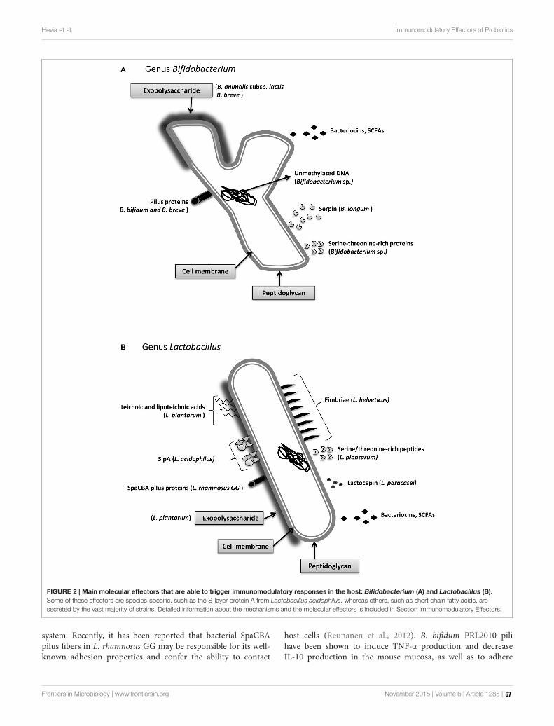
Hevia et al. Immunomodulatory Effectors of Probiotics
FIGURE 2 | Main molecular effectors that are able to trigger immunomodulatory responses in the host: Bifidobacterium (A) and Lactobacillus (B).
Some of these effectors are species-specific, such as the S-layer protein A from Lactobacillus acidophilus, whereas others, such as short chain fatty acids, are
secreted by the vast majority of strains. Detailed information about the mechanisms and the molecular effectors is included in Section Immunomodulatory Effectors.
system. Recently, it has been reported that bacterial SpaCBApilus fibers in L. rhamnosus GG may be responsible for its well-known adhesion properties and confer the ability to contact
host cells (Reunanen et al., 2012). B. bifidum PRL2010 pilihave been shown to induce TNF-α production and decreaseIL-10 production in the mouse mucosa, as well as to adhere
Frontiers in Microbiology | www.frontiersin.org November 2015 | Volume 6 | Article 1285 | 67

Hevia et al. Immunomodulatory Effectors of Probiotics
TABLE 1 | Examples of immunomodulatory effectors produced by classic/emerging probiotics.
Immunomodulatory effector Species Probiotic type Effect on host immune
system
References
Surface Layer Protein A (SlpA) L. acidophilus Classic Immunomodulation of intestinal
dendritic cells
Konstantinov et al., 2008
Pili proteins (SpaCBA) L. rhamnosus Classic Contact with mucosal cells Reunanen et al., 2012
Pili B. bifidum Classic Increase TNF-α and decrease
IL-10 production
Turroni et al., 2013
B. breve Classic Host colonization O’Connell Motherway et al., 2011
Fimbriae E. coli Emerging Host-colonization Kleta et al., 2014
L. plantarum Classic Immunomodulation Murofushi et al., 2015
Serpin B. longum Classic Human neutrophil and
pancreatic elastase inhibitor
Ivanov et al., 2006
Serine-threonine rich proteins Bifidobacterium sp. Classic Intestinal homeostasis Nezametdinova et al., 2014
Lactobacillus sp. Classic Intestinal homeostasis Zakharevich et al., 2012
Serine-threonine rich peptide (STp) L. plantarum Classic Anti-inflammatory; modulates
intestinal dendritic cell function
Bernardo et al., 2012; Al-Hassi et al., 2014
Lactocepin L. paracasei Classic Hydrolyzes IP-10 von Schillde et al., 2012
Secreted 15 kDa protein F. prausnitzii Emerging Anti-inflammatory Quévrain et al., 2015
Exopolysaccharides B. breve Classic Immunomodulation Fanning et al., 2012
B. lactis* Classic Immunomodulation Hidalgo-Cantabrana et al., 2014
Unmethylated CpG DNA Bifidobacterium sp. Classic Induces Th1 response Ménard et al., 2010
Teichoic/Lipoteichoic acids L. plantarum Classic Anti-inflammatory Grangette et al., 2005
Butyrate R. hominis
F. prausnitzii
A. muciniphila
Emerging Anti-inflammatory Maslowski et al., 2009
*Synonym of B. animalis subsp. lactis.
to diverse extracellular matrix proteins (Turroni et al., 2013),while B. breve UCC2003 pili are essential for host colonization(O’Connell Motherway et al., 2011). In another recent work, genecomplementation studies were used to show that the fimbriaeof the probiotic strain E. coli Nissle 1917 were involved in theadhesion to porcine intestinal cells, thereby helping to preventinfection with enteropathogenic E. coli (EPEC) (Kleta et al.,2014).
Cell Wall Non-proteinaceous ComponentsNon-proteinaceous cell wall components have different roles inmicrobe-host crosstalk. It has been shown that the EPS fromLactobacillus and Bifidobacterium strains can have a modulatorrole in preventing pathogen invasion, even though the EPSof pathogenic bacteria have been classically viewed as possiblevirulent factors. Examples of immunomodulatory EPS are thosefrom B. breve and Bifidobacterium animalis subsp. lactis (Fanninget al., 2012; Hidalgo-Cantabrana et al., 2014) or Lactobacillusplantarum strains (Murofushi et al., 2015). TAs are linearpolymers of ribitol phosphate or glycerol phosphate that arecovalently bound to D-alanine, monosaccharides, or aminosugars, and they are attached either to PG (wall TAs) or to thecytoplasmic membrane (membrane TAs or lipoteichoic acids;LTAs). TAs from L. plantarum were shown to display anti-inflammatory properties, as shown by the different cytokineproduction profiles of peripheral blood mononuclear cells(PBMCs) and monocytes exposed to this molecule (Grangetteet al., 2005). In addition, mice fed a diet supplemented with L.
plantarum LTAs or with an LTA-producing strain showed betterscores in a colitis model compared with the control group andmice that were fed a L. plantarum LTA-deficient strain (Grangetteet al., 2005). Although there have been a few promising results,this topic requires further research to clarify the mechanisms ofaction of the cell wall components of probiotics on the human gutmicrobiota.
Soluble CompoundsSoluble components that are produced by probiotic bacteriacan also affect the bacterial-host interplay. In Bifidobacteriumlongum the secretion of serpin, a serine protease inhibitor,which specifically binds and inactivates human neutrophil andpancreatic elastase, was shown to contribute to gut homeostasis(Ivanov et al., 2006). Additionally, it has been observedthat some proteins with characteristic biochemical motifs thatare produced by both commensal and pathogenic bacteriacan elicit specific functions and affect immune cells of theintestinal lumen. This is the case for a family of serine-threonine rich proteins, which was described in species ofLactobacillus and Bifidobacterium, with a recently describedkinase function (Zakharevich et al., 2012; Nezametdinova et al.,2014). In lactobacilli, a serine-threonine peptide, STp, which iscontained in a protein secreted by L. plantarum, was shownto be involved in bacterial aggregation (Hevia et al., 2013).Additionally, this peptide can modulate the dendritic cellphenotype of ulcerative colitis (UC) patients (Bernardo et al.,2012; Al-Hassi et al., 2014). It was also demonstrated that the
Frontiers in Microbiology | www.frontiersin.org November 2015 | Volume 6 | Article 1285 | 68

Hevia et al. Immunomodulatory Effectors of Probiotics
immunomodulatory effect of Lactobacillus paracasei is mediated,at least in part, by the secreted protease lactocepin, whichselectively degrades the chemokine IFN-γ-inducible protein 10(IP-10) that functions in lymphocyte recruitment (von Schilldeet al., 2012). There are other examples of non-proteinaceouscompounds that can exert certain effects on the host. Somespecies of Bifidobacterium possess unmethylated CpG motifsin their DNA that were able to induce TLR9 activation,which is known to trigger a Th1 orientation of the immunesystem (Ménard et al., 2010). In contrast, in other studies, itwas shown that intragastric and subcutaneous administrationof DNA from a probiotic mix ameliorated the severity ofcolitis in a murine experimental colitis model, whereas amethylated probiotic DNA had no effect (Rachmilewitz et al.,2004).
EMERGING PROBIOTICS, A NOVELSOURCE OF IMMUNOMODULATORYEFFECTORS
In addition to Lactobacillus and Bifidobacterium, othermicroorganisms have received substantial interest amongresearchers as potentially new, beneficial gut bacteria. Most ofthem are common colonizers of the human gut under normalconditions. Some of these microbial types are considered tobe markers of dysbiosis in intestinal inflammatory diseases,such as UC and Crohn’s disease (Manichanh et al., 2006; ElAidy et al., 2013). In these conditions, a loss of microbialdiversity and a significant reduction of members of Clostridiumclusters IV and XIVa have been reported, particularly in bacteriainvolved in butyrate and propionate metabolism, such asRuminococcus, Eubacterium, Roseburia, and Faecalibacterium. Inthis section, we will highlight current research on F. prausnitziiand A. muciniphila, two bacteria that have received muchattention during the last few years because of their potentialimmunomodulatory properties.
F. prausnitzii is a “novel” intestinal bacterium whoseimmunomodulatory properties have been well characterizedin vitro and in vivo. This anaerobic, Gram-positive bacteriumseems to play a role in the maintenance of gut homeostasis, andits population is normally reduced in intestinal inflammatorydiseases (Sokol et al., 2008; Cao et al., 2014; Machiels et al.,2014). In 2008, Sokol and colleagues studied the effects of wholebacteria, a cell culture supernatant, bacterial DNA, ormembrane-derived fractions in vitro using the Caco-2 epithelial colorectaladenocarcinoma cell line and PBMCs, as well as in vivo ina mouse model of 2,4,6-trinitrobenzene sulfonic acid (TNBS)-induced colitis (Sokol et al., 2008). The results showed that F.prausnitzii cells exerted anti-inflammatory effects in PBMCs.Furthermore, its culture supernatant reduced IL-8 secretion andabolished the activation of nuclear factor kappa-light-chain-enhancer of activated B cells (NF-κB) in Caco-2 cells. Moreover,although no significant improvement of the disease was detectedin mice, partial disease scores significantly improved in coliticmice receiving the cell culture supernatants, compared with thenon-receiving group. Accordingly, the authors hypothesized that
the beneficial effects of F. prausnitzii must be executed by asoluble compound that is secreted by the bacteria. In relationto this, recent work showed that F. prausnitzii secreted a 15-kDa protein with anti-inflammatory properties. This proteinwas able to inhibit the NF-κB pathway in intestinal epithelialcells, and it prevented colitis in an animal model (Quévrainet al., 2015). Additional research showed that this bacteriumrestored physiological parameters and downregulated cytokineprofiles in mice with colitis, as well as increased the Tregpopulation to a greater degree than other commensals suchas B. longum (Qiu et al., 2013; Martín et al., 2015). UCpatients have fewer butyrate-producing Roseburia hominis andF. prausnitzii (Machiels et al., 2014). It is likely that a significantpart of their anti-inflammatory action results from the effectof SCFAs in colonocytes, as acetate, propionate, and butyratemodulate the inflammatory responses of immune cells throughreceptors such as Gpr43 and Gpr41 (Maslowski et al., 2009).However, despite all the information that has recently beendiscovered about these bacterial groups in healthy and diseasedstates, and besides butyrate seeming to be the key homeostasispromoter, additional work is required to elucidate the molecularmechanisms through which F. prausnitzii interacts with the hostgut environment.
A. muciniphila is another common member of the healthy gutmicrobiota in humans at all stages of age (Collado et al., 2007;Belzer and de Vos, 2012). A. muciniphila is a Gram-negative,strictly anaerobic, mucin-degrading microorganism member ofthe Verrucomicrobia phylum, and it was one of the first bacteriashown to utilize mucin, the glycosylated protein layer that coversthe gut epithelium, as its sole carbon, nitrogen, and sulfur source.The products derived frommucin degradation are mainly SCFAsthat feed colonocyte metabolism and confer health propertiesto the host. By degrading the mucin of the external mucuslayer, A. muciniphila helps with the continuous renovation ofthe protective cover of the mucosae, and it maintains a healthyprotective barrier that prevents the entrance of enteropathogensinto the epithelium (Lukovac et al., 2014). In addition, whenA. muciniphila was administered to mice, there were increasedintestinal levels of endocannabinoids that control inflammation,the gut barrier, and gut peptide secretion, suggesting animmunomodulatory role for this bacterium (Everard et al., 2013).
CONCLUDING REMARKS
In conclusion, even though much effort has been put intoprobiotic research during recent decades, the mechanismsunderlying the immunomodulatory effects of beneficialintestinal bacteria have scarcely been elucidated. There iscompelling evidence that novel bacterial players, other thanLactobacillus and Bifidobacterium, could play a role in theseprocesses and are much more important than previouslythought; however, difficulties in growing some of thesebacteria on laboratory- and industrial-scales, and the lack ofmolecular tools needed to perform functional genomic analyses,seriously hamper the characterization of novel strains. Furtherresearch is needed to overcome these culturing and functionalcharacterization difficulties to perform well-designed pre-clinical
Frontiers in Microbiology | www.frontiersin.org November 2015 | Volume 6 | Article 1285 | 69
Hevia et al. Immunomodulatory Effectors of Probiotics
and intervention studies that shed new light on the mechanismsresponsible for the beneficial effects attributed to these bacteria.
AUTHOR CONTRIBUTIONS
AH, SD, BS, and AM contributed to the conception and designof the work, and to the acquisition, analysis, and interpretation ofthe data. All authors contributed to the drafting of themanuscriptand approved the final version to be published.
ACKNOWLEDGMENTS
This research was funded by grants AGL2013-44039-Rand AGL2013-44761-P from the Spanish “Plan Estatalde I+D+I.” BS and AH were recipients of a Ramón yCajal postdoctoral contract and a Formación de PersonalInvestigador—Research Personnel Training (FPI) grant,respectively, from the Spanish Ministry of Economy andCompetitiveness.
REFERENCES
Al-Hassi, H. O., Mann, E. R., Sanchez, B., English, N. R., Peake, S. T., Landy, J.,
et al. (2014). Altered human gut dendritic cell properties in ulcerative colitis
are reversed by Lactobacillus plantarum extracellular encrypted peptide STp.
Mol. Nutr. Food Res. 58, 1132–1143. doi: 10.1002/mnfr.201300596
Aragón, F., Carino, S., Perdigón, G., and de Moreno de LeBlanc, A. (2014). The
administration of milk fermented by the probiotic Lactobacillus casei CRL 431
exerts an immunomodulatory effect against a breast tumour in a mouse model.
Immunobiology 219, 457–464. doi: 10.1016/j.imbio.2014.02.005
Belzer, C., and de Vos, W. M. (2012). Microbes inside-from diversity to function:
the case of Akkermansia. ISME J. 6, 1449–1458. doi: 10.1038/ismej.2012.6
Bernardo, D., Sánchez, B., Al-Hassi, H. O., Mann, E. R., Urdaci, M. C., Knight,
S. C., et al. (2012). Microbiota/host crosstalk biomarkers: regulatory response
of human intestinal dendritic cells exposed to Lactobacillus extracellular
encrypted peptide. PLoS ONE 7:e36262. doi: 10.1371/journal.pone.0036262
Bufe, B., Schumann, T., Kappl, R., Bogeski, I., Kummerow, C., Podgórska, M.,
et al. (2015). Recognition of bacterial signal peptides by mammalian formyl
peptide receptors: a new mechanism for sensing pathogens. J. Biol. Chem. 290,
7369–787. doi: 10.1074/jbc.M114.626747
Cao, Y., Shen, J., and Ran, Z. H. (2014). Association between Faecalibacterium
prausnitzii reduction and inflammatory bowel disease: a meta-analysis and
systematic review of the literature. Gastroenterol. Res. Pract. 2014, 872725. doi:
10.1155/2014/872725
Claes, A. K., Zhou, J. Y., and Philpott, D. J. (2015). NOD-like receptors:
guardians of intestinal mucosal barriers. Physiology (Bethesda) 30, 241–250. doi:
10.1152/physiol.00025.2014
Collado, M. C., Derrien, M., Isolauri, E., de Vos, W. M., and Salminen, S. (2007).
Intestinal integrity and Akkermansia muciniphila, a mucin-degrading member
of the intestinal microbiota present in infants, adults, and the elderly. Appl.
Environ. Microbiol. 73, 7767–7770. doi: 10.1128/AEM.01477-07
Dambuza, I. M., and Brown, G. D. (2015). C-type lectins in immunity: recent
developments. Curr. Opin. Immunol. 32, 21–27. doi: 10.1016/j.coi.2014.12.002
Denes, A., Lopez-Castejon, G., and Brough, D. (2012). Caspase-1: is IL-1 just the
tip of the ICEberg? Cell Death Dis. 3, e338. doi: 10.1038/cddis.2012.86
Dongarrà, M. L., Rizzello, V., Muccio, L., Fries, W., Cascio, A., Bonaccorsi, I., et al.
(2013). Mucosal immunology and probiotics. Curr. Allergy Asthma Rep. 13,
19–26. doi: 10.1007/s11882-012-0313-0
Eisenstein, E. M., and Williams, C. B. (2009). The T(reg)/Th17 cell balance:
a new paradigm for autoimmunity. Pediatr. Res. 65, 26R–31R. doi:
10.1203/PDR.0b013e31819e76c7
El Aidy, S., Van den Abbeele, P., Van de Wiele, T., Louis, P., and Kleerebezem,
M. (2013). Intestinal colonization: how key microbial players become
established in this dynamic process: microbial metabolic activities and
the interplay between the host and microbes. Bioessays 35, 913–923. doi:
10.1002/bies.201300073
Everard, A., Belzer, C., Geurts, L., Ouwerkerk, J. P., Druart, C., Bindels, L. B., et al.
(2013). Cross-talk between Akkermansia muciniphila and intestinal epithelium
controls diet-induced obesity. Proc. Natl. Acad. Sci. U.S.A. 110, 9066–9071. doi:
10.1073/pnas.1219451110
Fanning, S., Hall, L. J., Cronin, M., Zomer, A., MacSharry, J., Goulding, D., et al.
(2012). Bifidobacterial surface-exopolysaccharide facilitates commensal-host
interaction through immune modulation and pathogen protection. Proc. Natl.
Acad. Sci. U.S.A. 109, 2108–2113. doi: 10.1073/pnas.1115621109
Grangette, C., Nutten, S., Palumbo, E., Morath, S., Hermann, C., Dewulf, J.,
et al. (2005). Enhanced antiinflammatory capacity of a Lactobacillus plantarum
mutant synthesizing modified teichoic acids. Proc. Natl. Acad. Sci. U.S.A. 102,
10321–10326. doi: 10.1073/pnas.0504084102
Hevia, A., Martínez, N., Ladero, V., Alvarez, M. A., Margolles, A., and Sánchez,
B. (2013). An extracellular Serine/Threonine-rich protein from Lactobacillus
plantarum NCIMB 8826 is a novel aggregation-promoting factor with affinity
tomucin.Appl. Environ.Microbiol. 79, 6059–6066. doi: 10.1128/AEM.01657-13
Hidalgo-Cantabrana, C., Sánchez, B., Milani, C., Ventura, M., Margolles, A.,
and Ruas-Madiedo, P. (2014). Genomic overview and biological functions
of exopolysaccharide biosynthesis in Bifidobacterium spp. Appl. Environ.
Microbiol. 80, 9–18. doi: 10.1128/AEM.02977-13
Hill, C., Guarner, F., Reid, G., Gibson, G. R., Merenstein, D. J., Pot, B., et al.
(2014). Expert consensus document. The international scientific association for
probiotics and prebiotics consensus statement on the scope and appropriate
use of the term probiotic. Nat. Rev. Gastroenterol. Hepatol. 11, 506–514. doi:
10.1038/nrgastro.2014.66
Ivanov, D., Emonet, C., Foata, F., Affolter, M., Delley, M., Fisseha, M., et al.
(2006). A serpin from the gut bacterium Bifidobacterium longum inhibits
eukaryotic elastase-like serine proteases. J. Biol. Chem. 281, 17246–17252. doi:
10.1074/jbc.M601678200
Kleta, S., Nordhoff, M., Tedin, K., Wieler, L. H., Kolenda, R., Oswald, S., et al.
(2014). Role of F1C fimbriae, flagella, and secreted bacterial components in
the inhibitory effect of probiotic Escherichia coli Nissle 1917 on a typical
enteropathogenic E. coli infection. Infect Immunol. 82, 1801–1812. doi:
10.1128/IAI.01431-13
Konstantinov, S. R., Smidt, H., de Vos, W. M., Bruijns, S. C., Singh, S. K., Valence,
F., et al. (2008). S layer protein A of Lactobacillus acidophilus NCFM regulates
immature dendritic cell and T cell functions. Proc. Natl. Acad. Sci. U.S.A. 105,
19474–19479. doi: 10.1073/pnas.0810305105
López, P., González-Rodríguez, I., Sánchez, B., Ruas-Madiedo, P., Suárez, A.,
Margolles, A., et al. (2012). Interaction of Bifidobacterium bifidum LMG13195
with HT29 cells influences regulatory-T-cell-associated chemokine receptor
expression. Appl. Environ. Microbiol. 78, 2850–2857. doi: 10.1128/AEM.
07581-11
Lukovac, S., Belzer, C., Pellis, L., Keijser, B. J., de Vos, W. M., Montijn,
R. C., et al. (2014). Differential modulation by Akkermansia muciniphila
and Faecalibacterium prausnitzii of host peripheral lipid metabolism and
histone acetylation in mouse gut organoids. mBio 5, e01438–e01414. doi:
10.1128/mBio.01438-14
Machiels, K., Joossens, M., Sabino, J., De Preter, V., Arijs, I., Eeckhaut, V., et al.
(2014). A decrease of the butyrate-producing species Roseburia hominis and
Faecalibacterium prausnitzii defines dysbiosis in patients with ulcerative colitis.
Gut 63, 1275–1283. doi: 10.1136/gutjnl-2013-304833
Macpherson, A. J., and Harris, N. L. (2004). Interactions between commensal
intestinal bacteria and the immune system.Nat. Rev. Immunol. 4, 478–485. doi:
10.1038/nri1373
Manichanh, C., Rigottier-Gois, L., Bonnaud, E., Gloux, K., Pelletier, E.,
Frangeul, L., et al. (2006). Reduced diversity of faecal microbiota in
Crohn’s disease revealed by a metagenomic approach. Gut 55, 205–211. doi:
10.1136/gut.2005.073817
Martín, R., Miquel, S., Chain, F., Natividad, J. M., Jury, J., Lu, J., et al. (2015).
Faecalibacterium prausnitzii prevents physiological damages in a chronic low-
grade inflammationmurinemodel. BMCMicrobiol. 15:67. doi: 10.1186/s12866-
015-0400-1
Maslowski, K. M., Vieira, A. T., Ng, A., Kranich, J., Sierro, F., Yu, D., et al. (2009).
Regulation of inflammatory responses by gut microbiota and chemoattractant
receptor GPR43. Nature 461, 1282–1286. doi: 10.1038/nature08530
Frontiers in Microbiology | www.frontiersin.org November 2015 | Volume 6 | Article 1285 | 70
Hevia et al. Immunomodulatory Effectors of Probiotics
Ménard, O., Gafa, V., Kapel, N., Rodriguez, B., Butel, M. J., andWaligora-Dupriet,
A. J. (2010). Characterization of immunostimulatory CpG-rich sequences from
different Bifidobacterium species. Appl. Environ. Microbiol. 76, 2846–255. doi:
10.1128/AEM.01714-09
Meng, J., Zhu, X., Gao, S. M., Zhang, Q. X., Sun, Z., and Lu, R. R. (2014).
Characterization of surface layer proteins and its role in probiotic properties
of three Lactobacillus strains. Int. J. Biol. Macromol. 65, 110–114. doi:
10.1016/j.ijbiomac.2014.01.024
Miettinen, M., Veckman, V., Latvala, S., Sareneva, T., Matikainen, S., and
Julkunen, I. (2008). Live Lactobacillus rhamnosus and Streptococcus pyogenes
differentially regulate Toll-like receptor (TLR) gene expression in human
primary macrophages. J. Leukoc. Biol. 84, 1092–1100. doi: 10.1189/jlb.
1206737
Murofushi, Y., Villena, J., Morie, K., Kanmani, P., Tohno, M., Shimazu, T., et al.
(2015). The toll-like receptor family protein RP105/MD1 complex is involved
in the immunoregulatory effect of exopolysaccharides from Lactobacillus
plantarum N14. Mol. Immunol. 64, 63–75. doi: 10.1016/j.molimm.2014.
10.027
Nezametdinova, V. Z., Zakharevich, N. V., Alekseeva, M. G., Averina, O.
V., Mavletova, D. A., and Danilenko, V. N. (2014). Identification and
characterization of the serine/threonine protein kinases in Bifidobacterium.
Arch. Microbiol. 196, 125–136. doi: 10.1007/s00203-013-0949-8
Noack, M., and Miossec, P. (2014). Th17 and regulatory T cell balance in
autoimmune and inflammatory diseases. Autoimmun. Rev. 13, 668–677. doi:
10.1016/j.autrev.2013.12.004
Nyirenda, M. H., Morandi, E., Vinkemeier, U., Constantin-Teodosiu, D.,
Drinkwater, S., Mee, M., et al. (2015). TLR2 stimulation regulates the balance
between regulatory T Cell and Th17 function: a novel mechanism of reduced
regulatory T Cell function in multiple sclerosis. J. Immunol. 194, 5761–5774.
doi: 10.4049/jimmunol.1400472
O’Connell Motherway, M., Zomer, A., Leahy, S. C., Reunanen, J., Bottacini, F.,
Claesson, M. J., et al. (2011). Functional genome analysis of Bifidobacterium
breve UCC2003 reveals type IVb tight adherence (Tad) pili as an essential
and conserved host-colonization factor. Proc. Natl. Acad. Sci. U.S.A. 108,
11217–11222. doi: 10.1073/pnas.1105380108
Qiu, X., Zhang, M., Yang, X., Hong, N., and Yu, C. (2013). Faecalibacterium
prausnitzii upregulates regulatory T cells and anti-inflammatory cytokines
in treating TNBS-induced colitis. J. Crohns. Colitis. 7, e558–e568. doi:
10.1016/j.crohns.2013.04.002
Quévrain, E., Maubert, M. A., Michon, C., Chain, F., Marquant, R., Tailhades,
J., et al. (2015). Identification of an anti-inflammatory protein from
Faecalibacterium prausnitzii, a commensal bacterium deficient in Crohn’s
disease. Gut. doi: 10.1136/gutjnl-2014-307649. [Epub ahead of print].
Rachmilewitz, D., Katakura, K., Karmeli, F., Hayashi, T., Reinus, C., Rudensky,
B., et al. (2004). Toll-like receptor 9 signaling mediates the anti-inflammatory
effects of probiotics in murine experimental colitis. Gastroenterology 126,
520–528. doi: 10.1053/j.gastro.2003.11.019
Reunanen, J., von Ossowski, I., Hendrickx, A. P., Palva, A., and de Vos, W.
M. (2012). Characterization of the SpaCBA pilus fibers in the probiotic
Lactobacillus rhamnosus GG. Appl. Environ. Microbiol. 78, 2337–2344. doi:
10.1128/AEM.07047-11
Rodríguez, J. M., Murphy, K., Stanton, C., Ross, R. P., Kober, O. I., Juge,
N., et al. (2015). The composition of the gut microbiota throughout life,
with an emphasis on early life. Microb. Ecol. Health Dis. 26:26050. doi:
10.3402/mehd.v26.26050
Sakaguchi, S., Miyara, M., Costantino, C. M., and Hafler, D. A. (2010). FOXP3+
regulatory T cells in the human immune system. Nat. Rev. Immunol. 10,
490–500. doi: 10.1038/nri2785
Salonen, A., Lahti, L., Salojärvi, J., Holtrop, G., Korpela, K., Duncan, S. H.,
et al. (2014). Impact of diet and individual variation on intestinal microbiota
composition and fermentation products in obese men. ISME J. 8, 2218–2230.
doi: 10.1038/ismej.2014.63
Salzman, N. H., Underwood, M. A., and Bevins, C. L. (2007). Paneth
cells, defensins, and the commensal microbiota: a hypothesis on intimate
interplay at the intestinal mucosa. Semin. Immunol. 19, 70–83. doi:
10.1016/j.smim.2007.04.002
Sánchez, B., Gueimonde, M., Peña, A. S., and Bernardo, D. (2015). Intestinal
microbiota as modulators of the immune system. J. Immunol. Res. 2015:159094.
doi: 10.1155/2015/159094
Scott, K. P., Antoine, J. M., Midtvedt, T., and van Hemert, S. (2015). Manipulating
the gut microbiota to maintain health and treat disease. Microb. Ecol. Health
Dis. 26:25877. doi: 10.3402/mehd.v26.25877
Sekirov, I., Russell, S. L., Antunes, L. C., and Finlay, B. B. (2010). Gut microbiota in
health and disease. Physiol. Rev. 90, 859–904. doi: 10.1152/physrev.00045.2009
Sellge, G., and Kufer, T. A. (2015). PRR-signaling pathways-Learning from
microbial tactics. Semin. Immunol. 27, 75–84. doi: 10.1016/j.smim.2015.03.009
Sokol, H., Pigneur, B., Watterlot, L., Lakhdari, O., Bermúdez-Humarán, L.
G., Gratadoux, J. J., et al. (2008). Faecalibacterium prausnitzii is an anti-
inflammatory commensal bacterium identified by gut microbiota analysis of
Crohn disease patients. Proc. Natl. Acad. Sci. U.S.A. 105, 16731–16736. doi:
10.1073/pnas.0804812105
Turroni, F., Serafini, F., Foroni, E., Duranti, S., O’Connell Motherway, M.,
Taverniti, V., et al. (2013). Role of sortase-dependent pili of Bifidobacterium
bifidum PRL2010 in modulating bacterium–host interactions. Proc. Natl. Acad.
Sci. U.S.A. 110, 11151–11156. doi: 10.1073/pnas.1303897110
von Schillde, M. A., Hörmannsperger, G., Weiher, M., Alpert, C. A., Hahne,
H., Bäuerl, C., et al. (2012). Lactocepin secreted by Lactobacillus exerts anti-
inflammatory effects by selectively degrading proinflammatory chemokines.
Cell Host Microbe 11, 387–396. doi: 10.1016/j.chom.2012.02.006
Wing, J. B., and Sakaguchi, S. (2014). Foxp3+ T(reg) cells in humoral immunity.
Int. Immunol. 26, 61–69. doi: 10.1093/intimm/dxt060
Yadav, A. K., Tyagi, A., Kumar, A., Panwar, S., Grover, S., Saklani, A. C., et al.
(2015). Adhesion of lactobacilli and their anti-infectivity Potential. Crit. Rev.
Food Sci. Nutr. doi: 10.1080/10408398.2014.918533. [Epub ahead of print].
Yao, H., Dittmann, M., Peisley, A., Hoffmann, H. H., Gilmore, R. H., Schmidt, T.,
et al. (2015). ATP-Dependent Effector-like Functions of RIG-I-like Receptors.
Mol. Cell. 58, 541–548. doi: 10.1016/j.molcel.2015.03.014
Yousefi, M., Movassaghpour, A. A., Shamsasenjan, K., Ghalamfarsa, G.,
Sadreddini, S., Jadidi-Niaragh, F., et al. (2015). The skewed balance between
Tregs and Th17 in chronic lymphocytic leukemia. Future Oncol. 11, 1567–182.
doi: 10.2217/fon.14.298
Zakharevich, N. V., Osolodkin, D. I., Artamonova, I. I., Palyulin, V. A., Zefirov,
N. S., and Danilenko, V. N. (2012). Signatures of the ATP-binding pocket as
a basis for structural classification of the serine/threonine protein kinases of
gram-positive bacteria. Proteins 80, 1363–1376. doi: 10.1002/prot.24032
Conflict of Interest Statement: The authors declare that the research was
conducted in the absence of any commercial or financial relationships that could
be construed as a potential conflict of interest.
Copyright © 2015 Hevia, Delgado, Sánchez and Margolles. This is an open-access
article distributed under the terms of the Creative Commons Attribution License (CC
BY). The use, distribution or reproduction in other forums is permitted, provided the
original author(s) or licensor are credited and that the original publication in this
journal is cited, in accordance with accepted academic practice. No use, distribution
or reproduction is permitted which does not comply with these terms.
Frontiers in Microbiology | www.frontiersin.org November 2015 | Volume 6 | Article 1285 | 71

fmicb-07-00715 May 12, 2016 Time: 17:53 # 1
REVIEWpublished: 13 May 2016
doi: 10.3389/fmicb.2016.00715
Edited by:Katharina Riedel,
University of Greifswald, Germany
Reviewed by:Paul Richard Himes,
University of Louisville, USADonatella De Pascale,The Institute of Protein
Biochemistry-CNR, Italy
*Correspondence:Giorgio Gribaudo
Specialty section:This article was submitted to
Systems Microbiology,a section of the journal
Frontiers in Microbiology
Received: 18 January 2016Accepted: 29 April 2016Published: 13 May 2016
Citation:Luganini A, Terlizzi ME
and Gribaudo G (2016) BioactiveMolecules Released From Cells
Infected with the HumanCytomegalovirus.
Front. Microbiol. 7:715.doi: 10.3389/fmicb.2016.00715
Bioactive Molecules Released FromCells Infected with the HumanCytomegalovirusAnna Luganini, Maria E. Terlizzi and Giorgio Gribaudo*
Laboratory of Microbiology and Virology, Department of Life Sciences and Systems Biology, University of Turin, Turin, Italy
Following primary infection in humans, the human cytomegalovirus (HCMV) persistsin a latent state throughout the host’s lifetime despite a strong and efficient immuneresponse. If the host experiences some form of immune dysregulation, such asimmunosuppression or immunodeficiency, HCMV reactivates, thereby emerging fromlatency. Thus, in the absence of effective functional immune responses, as occurs inimmunocompromised or immunoimmature individuals, both HCMV primary infectionsand reactivations from latency can cause significant morbidity and mortality. However,even in immunocompetent hosts, HCMV represents a relevant risk factor for thedevelopment of several chronic inflammatory diseases and certain forms of neoplasia.HCMV infection may shift between the lytic and latent state, regulated by a delicateand intricate balance between virus-mediated immunomodulation and host immunedefenses. Indeed, HCMV is a master in manipulating innate and adaptive host defensepathways, and a large portion of its genome is devoted to encoding immunomodulatoryproteins; such proteins may thus represent important virulence determinants. However,the pathogenesis of HCMV-related diseases is strengthened by the activities of bioactivemolecules, of both viral and cellular origin, that are secreted from infected cells andcollectively named as the secretome. Here, we review the state of knowledge on thecomposition and functions of HCMV-derived secretomes. In lytic infections of fibroblastsand different types of endothelial cells, the majority of HCMV-induced secretedproteins act in a paracrine fashion to stimulate the generation of an inflammatorymicroenvironment around infected cells; this may lead to vascular inflammation andangiogenesis that, in turn, foster HCMV replication and its dissemination through hosttissues. Conversely, the HCMV secretome derived from latently infected hematopoieticprogenitor cells induces an immunosuppressive extracellular environment that interfereswith immune recognition and elimination of latently infected cells, thereby promotingviral persistence. Characterization of the composition and biological activities of HCMVsecretomes from different types of infected cells will lay the foundation for futureadvances in our knowledge about the pathogenesis HCMV diseases and may providetargets for the development of novel antiviral intervention strategies.
Keywords: human cytomegalovirus, lytic infection, latency, bioactive molecules, secretome, chronic disease,angiogenesis, immunoevasion
Frontiers in Microbiology | www.frontiersin.org May 2016 | Volume 7 | Article 715 | 72

fmicb-07-00715 May 12, 2016 Time: 17:53 # 2
Luganini et al. HCMV Secretome Regulates Viral Infections
THE HUMAN CYTOMEGALOVIRUS
Human cytomegalovirus (HCMV) is an opportunistic beta-Herpesvirus that infects more than 90% of people worldwidewith an infection rate that increases with age (Landolfo et al.,2003; Britt, 2008; Mocarski et al., 2014). HCMV infection, asfor the other human Herpesviruses, is characterized by twophases: lytic and latent. Following primary infection, in whichthe virus productively replicates in a broad range of differentcell types (Sinzger et al., 2008), HCMV establishes latencyin cells of myeloid lineage through a still poorly understoodmechanism (Britt, 2008; Crough and Khanna, 2009; Mocarskiet al., 2014). In the normal immunocompentent host, primaryinfection is usually asymptomatic or mildly symptomatic andpersistent (Britt, 2008; Mocarski et al., 2014). Nevertheless,HCMV infection is considered, even in immunocompetenthosts, a risk factor for the development of various vasculardiseases, immunosenescence, and tumor development (Britt,2008; Mocarski et al., 2014; Nogalski et al., 2014). In contrast, inindividuals in which the ability to develop an appropriate cellularimmune response is compromised, such as immunosuppressedpatients and the immunoimmature fetus during pregnancy,HCMV infection is one of the major causes of morbidityand mortality. Indeed, primary infection or reactivation fromlatency causes overt diseases in immunosuppressed hosts, suchas transplant recipients taking immunosuppressive drugs andAIDS patients. In these settings, HCMV can be responsiblefor a wide range of clinical conditions, for example retinitis,pneumonia, colitis, hepatitis, and several chronic inflammatorydiseases, such as atherosclerosis, transplant vascular sclerosis(TSV), and chronic allograft rejection (CR; Britt, 2008; Croughand Khanna, 2009; Mocarski et al., 2014). Congenital HCMVinfections, on the other hand, represent the most prominent viralcause of birth defects such as malformations, hearing loss andlearning disabilities (Kenneson and Cannon, 2007; Britt, 2008;Mocarski et al., 2014), and the incidence of HCMV transmissionto the fetus is strictly related to maternal seroprevalence. Primarymaternal infection carries a risk of transmission between 14.2and 52.4% (Kenneson and Cannon, 2007), while a transmissionrate of 1.4% has been reported in relation to maternal reactivatedinfections. However, the severity of congenital HCMV disease issimilar for both primary and non-primary infections (Ahlforset al., 2001; Rahav et al., 2007). Globally, the incidence ofHCMV congenital infection is between 0.3 and 2.3% of all livebirths in developing countries. In congenitally infected fetuses,about 10–15% develops evident symptoms after birth, with anincidence of perinatal mortality of about 3–20 newborns per100,000 live births, and of 24–160 cases of neurologic diseases(hearing loss and mental retardation) per 100,000 neonates.In the remaining 85–90% of HCMV-infected newborns, nosymptoms are displayed at birth, but late signs of infection,such as hearing defects, are evident in 30–200 newborns per100,000 live births. The impact of congenital HCMV infectionon public health is thus significant, given that hearing loss, themost common long-term sequelae of infants with congenitalHCMV, it is the leading cause of non-genetic deafness inchildren.
The broad range of clinical manifestations of HCMV diseasesreflects the capacity of the virus to productively infect anextremely wide range of cell types in the host, such as skinand lung fibroblasts, epithelial and endothelial cells (ECs),vascular smooth muscle cells, hepatocytes, monocyte-derivedmacrophages, neuronal, and glial cells, thus determining itspotential to spread to all areas of the body (Sinzger, 2008; Sinzgeret al., 2008).
The HCMV genome is the largest among the Herpesviruses;its 235 kb double-stranded DNA is structured into a unique long(UL) and a unique short (US) region, both of which are flankedby terminal and internal inverted repeats (TRL/S and IRL/S,respectively; Landolfo et al., 2003; Mocarski et al., 2014). Eventhough its annotation remains provisional and its coding capacityhas recently been proposed to be much greater than originallythought (Stern-Ginossar et al., 2012), it is generally acceptedthat the HCMV genome encodes at least 170 canonical proteins(Mocarski et al., 2014). Whole-genome functional profiling oftwo HCMV laboratory strains revealed that a set of just 50herpesvirus-common proteins, encoded by genes mainly locatedin the central region of the UL domain, is required for productiveviral replication in primary fibroblasts (Murphy and Shenk, 2008;Mocarski et al., 2014). The remaining two-thirds of canonicalHCMV protein-coding genes, mostly betaherpesvirus- or CMV-specific, are confined within the terminal regions of the genomeand are not essential in cultured fibroblasts. Although, specificfunctions have yet to be assigned to many of these non-essentialgenes, they are mainly thought to be involved in regulatingvirus cell tropism, dissemination, and viral persistence andlatency within the host, as well as the modulation of intrinsic,innate, and acquired host immune responses, thus contributingto viral pathogenesis in a variety of ways (Mocarski et al.,2014).
During lytic infection, viral gene expression occurs in threephases, named immediate early (IE or α), early (E or β), andlate (L or γ) in relation to their temporal kinetic profiles, andleads to genome replication, assembly and release of matureinfectious viral particles. In brief, HCMV gene expression beginswith de novo expression of IE genes, predominantly IE1-72 andIE2-86, that activate the expression of E genes, required forreplication of the viral genome and the subsequent transcriptionof L (primarily structural) genes. IE2-86 protein autoregulates itsown expression by negatively acting on the Major IE Promoter(MIEP) of HCMV (Figure 1; Stinski and Petrik, 2008); it binds tothe cis-repressive sequence (CRS) of MIEP, resulting in a decreasein MIEP transcription (Stinski and Petrik, 2008). The activities ofIE proteins, in turn, determine the expression of E genes, whichare divided in two subgroups, β1 (E) and β2 (E-L), accordingto their time of expression; their functions are mainly relatedto HCMV replication machinery, viral DNA replication factors,repair enzymes, and immune evasion (Landolfo et al., 2003;Mocarski et al., 2014). L proteins are then expressed accordingto two different kinetics profiles (γ1 and γ2), shaped by distincttimes of expression and their sensitivity to inhibitors of viral DNAreplication. L proteins functions are associated to the assembly,maturation and egress of newly formed viral particles from thehost cell.
Frontiers in Microbiology | www.frontiersin.org May 2016 | Volume 7 | Article 715 | 73

fmicb-07-00715 May 12, 2016 Time: 17:53 # 3
Luganini et al. HCMV Secretome Regulates Viral Infections
FIGURE 1 | Regulation of HCMV MIEP activity during lytic and latent infections. (A) In cells permissive to lytic infection, the MIEP is associated withacetylated histones (Ac; H4 and H3) and many constitutive and inducible cellular transcription factors (NF-κB, CREB, AP1, SRF, and Elk-1) bind to cognate bindingsites to activate IE genes transcription. (B) In latently infected cells, cellular transcriptional repressors (YY1, ERF, and Gfi-1) bind and recruit histone deacetylases(HDAC1) and methyltransferases (HMT) to the MIEP. The reduced content of acetylated H4 and the increase of dimethylated H3 histones (Me), promotes recruitmentof the silencing protein (heterochromatin protein 1 -HP1β-), thus determining repression of the MIEP transcriptional activity.
In contrast to lytic replication, only a small subset ofHCMV genes are expressed during latency (e.g., transcriptsfor the latency-associated viral IL-10/UL111A, LUNA [LatencyUnique Natural Antigen], US28, UL138, and UL144) withoutany detectable production of infectious virus (Avdic et al., 2011;Reeves, 2011; Poole et al., 2014). HCMV latency is restricted toprimary myeloid progenitors, such as granulocyte macrophageprogenitors (GMPs), CD34+ hematopoietic progenitor cells, andCD14+ monocytes (Hahn et al., 1998; Scrivano et al., 2011;Rossetto et al., 2013). In these infected cell-types the carriage ofviral DNA occurs in the absence of the expression of lytic genes(Mocarski et al., 2014). Only upon the terminal differentiation ofthese cells into macrophages or dendritic cells (DCs), HCMV lyticgenes are expressed: IE gene products activate the expression ofother genes, leading to viral DNA replication and de novo virusproduction (Taylor-Wiedeman et al., 1994; Hahn et al., 1998;Soderberg-Nauclér et al., 2001; Reeves et al., 2005; Britt, 2008;Huang et al., 2012; Mocarski et al., 2014). Since the number ofcells carrying the latent viral genome is extremely low in vivo(about 0.001–0.01% of monocytes), it has been necessary todevelop complex experimental latency models in order to studythe related mechanisms. Thus, experimental HCMV latency andreactivation analyses have been performed using in vitro modelsthat exploit primary myeloid progenitors cells: namely GMPcells, CD34+ hematopoietic cells, and CD14+monocytes (Reeveset al., 2005; Cheung et al., 2006; Reeves and Sinclair, 2010; Liu
et al., 2013). All of these studies have highlighted that a crucialaspect of HCMV latency is the repression of MIEP followingits association with repressive chromatin markers (Bain et al.,2003; Wright et al., 2005; Sinclair and Sissons, 2006), involvingthe recruitment of histones and transcriptional silencing factorsto the MIEP (Reeves, 2011). In addition to several binding sitesfor cellular transcription factors (e.g., NF-κB, CREB/ATF, AP1,SRF, Elk-1) that stimulate its transcriptional activity (Figure 1;Caposio et al., 2007a, 2010; Lashmit et al., 2009; Isern et al.,2011; Mocarski et al., 2014), the MIEP contains a series ofmultiple binding sites for transcription factors that may lead toits repression (YY1, ERF and Gfi-1; Figure 1; Reeves, 2011). Infact, during HCMV latency, YY1 and ERF bind to MIEP, andrecruit histone deacetylases and methyltransferases than thentarget histones associated with MIEP (Wright et al., 2005). Themethylated histones then become targets for the recruitmentof heterochromatin protein 1 (HP-1), which augments MIEPrepression and contributes to the establishment of latency (Liuet al., 2010). Therefore, permissive or latent infection may bedetermined by the balance between activating and repressivetranscription factors that control MIEP activity (Figure 1; Reevesand Sinclair, 2013). However, this balance is thought to bemainly under the control of host inflammatory responses, eitherin immunocompetent or immunosuppressed individuals (Liuet al., 2013). Various studies have indeed shown that HCMVreactivation, during the allogeneic response to a transplanted
Frontiers in Microbiology | www.frontiersin.org May 2016 | Volume 7 | Article 715 | 74

fmicb-07-00715 May 12, 2016 Time: 17:53 # 4
Luganini et al. HCMV Secretome Regulates Viral Infections
organ, is mediated by the expression of inflammatory cytokines(i.e., TNF, LPS, and IL-6) that may, in turn, activate MIEP-interacting cellular transcription factors (e.g., NF-κB and AP-1), thus promoting MIEP activation and lytic HCMV geneexpression (Hummel and Abecassis, 2002). However, in otherexperimental systems, the induction of IE gene expression hasbeen associated with the state of cellular differentiation, asobserved in non-permissive monocytes upon their differentiationinto permissive macrophages in response to tumor necrosis factor(TNF) and IFN-γ (Soderberg-Nauclér et al., 1997).
MODULATION OF HOST IMMUNERESPONSES BY HCMV
Among the HCMV genes not essential for growth in culturedfibroblasts, about 40 have been shown to be involved in themodulation of host immune responses to viral infection byinterfering with innate immune mechanisms and counteractingadaptive antibody and T-cell mediated immunity (Mocarski et al.,2014). The ability of HCMV to establish persistent infection,in spite of a robust T-cell and neutralizing antiviral antibodyresponse, is mainly ensured by the activity of many virus-encodedimmunomodulatory proteins. HCMV immunomodulation istherefore of pathogenetic importance in the establishment ofvirus persistence in the host. In this regard, HCMV is considereda master of immune evasion strategies, as highlighted bythe results of several functional genomic, transcriptomic, andproteomic analyses that have defined many immunomodulatoryfunctions of several HCMV proteins. In particular, these viralproteins have been observed to: (1) interfere with antigenpresentation by major histocompatibility complex-I (MHC-I) molecules (e.g., the viral gene products US2, US3, US6,US10, US11, and UL82); (2) permit immune evasion throughinterference with and T and NK cells functions (e.g., the proteinsencoded by genes UL16, UL18, UL40, UL83, UL141, UL142,US18, and US20); and (3) mimic the activities of cellularcytokines/chemokines (e.g., proteins UL111A, UL128, UL146,and UL147) or act as fake host cytokine/chemokine receptors(as observed for proteins UL144, UL21.5, US27, US28, UL33,and UL78; McSharry et al., 2012; Van Damme and Van Loock,2014).
The best-characterized mechanisms of HCMVimmunoevasion can be summarized as follows:
(1) Interference with antigen presentation by MHC-I molecules.A remarkable example is provided by glycoproteinsUS2, US3, US6, and US11, characterized by a singletransmembrane (TM)-spanning and an immunoglobulin(Ig) domain, which most likely arose by duplication ofa single viral ancestor gene (Gewurz et al., 2001). TheseHCMV glycoproteins, when expressed in fibroblasts,endothelial/epithelial cells, or DCs, induce, throughdifferent mechanisms, a decrease in the cell-surfaceexpression of MHC-proteins required for the activationof CD8+ T lymphocytes (Mocarski, 2002). In particular,both US2 and US11 proteins are able to redirect nascent
MHC class I proteins from the ER into the cytosol untilubiquitin-dependent proteasome-mediated degradationoccurs. The US2 glycoprotein has also been observed toinhibit MHC class II protein translocation, thus inducing asubstantial block of CD4+ T cell expansion. On the otherhand, the US3 gene product prevents the egress of MHCclass I proteins from the endoplasmic reticulum (ER) to theGolgi apparatus, whereas the US6 protein is able to bindthe transporter of antigen processing (TAP) in the lumenof the ER, preventing ATP-dependent peptide loading onthe cell membrane (Mocarski, 2002). In conclusion, thistype of immunoevasion prevents the immune recognition ofHCMV-infected cells.
(2) Immune evasion through interference with T and NKcells functions. Lytically HCMV-infected cells exhibit anoteworthy resistance to NK cell-mediated cytolysis in vitro(Wilkinson et al., 2008). HCMV is, in fact, able to alter andmodify NK cell activity through different ‘evasion’ strategies,such as those exerted by: (i) UL16, a viral glycoprotein ableto directly bind and sequester in the ER MICB, ULBP1,and ULBP2 -ligands for the activating NKG2D receptorpresent on NK cells- thus preventing their expression onthe surface of HCMV-infected cells (Wilkinson et al., 2008);(ii) UL18, a viral-MHC-I homolog, which was found tobind the NK cell inhibitory receptor LIR1/ILT2 with a1000-fold higher affinity than HLA-I molecules (Wilkinsonet al., 2008); (iii) UL40, a viral protein able to promotethe HLA-E upregulation on the surface of HCMV-infectedcells. Since HLA-E acts as a NK cell-mediated cytotoxicitysuppressor through the NK cell inhibitory receptor complexCD94/NKG2A (Prod’homme et al., 2012), its upregulationcontributes to the NK cell immunoevasion of HCMV-infected cells; (iv) UL83, that encodes for the major HCMVtegument protein pp65 that suppresses the induction ofseveral interferons (IFNs) and proinflammatory chemokinetranscripts; pp65 may also directly bind to the activatorreceptor NKp30, present on the surface of NK cells, thussuppressing NK cell activation (Wilkinson et al., 2008);(v) UL141, a viral glycoprotein able to sequester CD155molecule in the ER. CD155 expression leads to NK cellactivation by binding to NK receptors CD226 and CD96(Nemcovicová et al., 2013). Therefore, sequestration ofCD155 prevents NK cell activation and survival of infectedcells; (vi) US18 and US20 proteins recently observed topromote lysosomal MICA degradation (Fielding et al., 2014).MICA, the MHC class I polypeptide-related sequence A, isa natural ligand of the NKG2D receptor. NKG2D-MICAbinding induces NK cell activation, leading to a cytolyticresponse against HCMV-infected cells. Therefore, MICAdegradation reduces the capability of NK cells to recognizeinfected cells (Fielding et al., 2014).
(3) Mimic physiological activities of cellular cytokines/chemokines or host cytokine/chemokine receptors. HCMVencodes several homologs of chemokine/cytokine and/ortheir receptors that may interfere with the corresponding
Frontiers in Microbiology | www.frontiersin.org May 2016 | Volume 7 | Article 715 | 75

fmicb-07-00715 May 12, 2016 Time: 17:53 # 5
Luganini et al. HCMV Secretome Regulates Viral Infections
host counterparts. In this regard, an important exampleof a HCMV-encoded chemokine receptor homolog isrepresented by the multifunctional US28 protein. US28is considered a putative immunoevasion molecule due toits capability to bind and internalize a range of cellularchemokines, including a broad spectrum of CC and CX3Cchemokines, thus limiting the ability of host chemotacticmolecules to elicit their effects in immune cells stimulation(Mocarski et al., 2014). However, US28 makes use ofboth the extracellular chemokine milieu and differentintracellular G-protein pathways to produce a wide varietyof pathophysiological cell responses (Beisser et al., 2008).In fact, upon stimulation with some chemokines, US28specifically promotes smooth muscle cells and macrophagemigration, thus contributing to both virus disseminationand pathogenesis of HCMV-associated vascular diseases(Vomaske et al., 2009). On the other hand, constitutiveexpression of US28 induces, in a ligand-independentmanner, cyclooxygenase-2 (COX-2) expression via NF-κBactivation, leading to the production of VEGF, one of themost abundant angiogenic factors (Maussang et al., 2009).
Moreover, the release of virus-encoded cytokines/chemokineshomologs (see HCMV-Induced Secreted Cellular Proteins)permits the virus to modulate specific host immune defensemechanisms in infected tissues, since these bioactive solublemolecules act in a paracrine fashion.
BIOACTIVE MOLECULES RELEASEDFROM HCMV-INFECTED CELLS
The pathogenesis of acute HCMV diseases is related to end-organ damage that results from both lytic virus replication andhost immune responses, whereas diseases associated to persistentinfections in both immunocompetent and immunocompromisedpatients (transplant recipients and AIDS patients) are relatedto chronic inflammation (Britt, 2008). However, besides thedirect cytopathic effect of virus replication on host tissues,the pathogenesis of HCMV diseases may be influenced by theactivity of virus-induced molecules secreted from virus-infectedcells. These secreted factors, of both viral and cellular origin,by acting in a paracrine fashion alter and modify the localmicroenvironment, thus contributing to the development ofHCMV-related diseases (Streblow et al., 2008).
HCMV-Induced Secreted CellularProteinsEarly in vitro studies on the ability of HCMV to stimulate thesecretion of bioactive cellular factors identified several cytokinesand growth factors released from different types of infectedcells. First, Almeida et al. (1994) observed that HCMV infectionof HUVECs (Human Umbilical Vein Endothelial Cells) greatlyincreased the expression of IL-6 mRNA and the secretion of thiscytokine into the supernatant of infected cells. Dengler et al.(2000) went on to demonstrate the paracrine activity of IL-1β released from HCMV-infected cells, which resulted in an
upregulation of pro-inflammatory adhesion molecules on non-infected neighboring cells. In fact, by analyzing the expressionof plasma membrane proteins on the surface of both HCMV-infected HUVECs and human vascular smooth muscle cells(hvSMC), they observed a 200-fold overexpression of the InterCellular Adhesion Molecule-1 (ICAM-1) and de novo inductionof both Vascular Cell Adhesion Molecule 1 (VCAM-1) andE-selectin (Dengler et al., 2000). In regard to the EC model,we observed an upregulation of ICAM-1, IL-8, CCL5/RANTES,CXCL10/IP-10, CXCL11/I-TAC, and COX-2 gene expression inHUVECs cells infected with a low-passage HCMV strain, thusindicating a direct involvement of HCMV in the modificationof the extracellular vascular environment through the release ofinflammatory mediators (Caposio et al., 2007b).
Using astrocyte and microglia cell models, Maxim et al. (2001)observed that HCMV stimulated the release of CCL2/MCP-1and IL-8 in the supernatants of infected astrocytes, whereasinfection of microglia cells led to an increased secretion of TNF-α, IL-6, CCL2/MCP-1, IL-8, CCL5/RANTES, and CCL3/MIP-1α.These findings thus suggest the ability of infected astrocytes torecruit microglia cells, through the release of the chemoattractantCCL2/MCP-1 (Maxim et al., 2001).
However, a different picture emerged from the investigation ofthe HCMV-mediated modulation of chemokine gene expressionin the context of HCMV retinitis – a virus-induced inflammationof the retina characterized by vasculitis and retina degenerationthat leads to retinal detachment in immunocompromised hosts.In the in vitro cell model provided by human retinal pigmentepithelial cells, Momma et al. (2003) observed, by means ofRT-PCR and ELISA assays, that HCMV infection caused anupregulation of IL-8, whereas CCL2/MCP-1 and CCL7/MCP-3 levels were downregulated. The authors thus suggested thatthe altered secretion of IL-8, CCL2/MCP-1 and CCL7/MCP-3 byretinal cells may be involved in the initiation and developmentof the inflammatory process responsible for the pathogenesis ofHCMV retinitis. In this scenario, the increased secretion of IL-8 may stimulate the recruitment and trafficking of leukocytesto the site of infection within the retina; on the other hand,the decrease in CCL2/MCP-1 and CCL7/MCP-3 levels in theextracellular environment may limit the migration of leukocytes,thus contributing to the virus escape of the host innate immuneresponse (Momma et al., 2003).
Together, these earlier studies demonstrated how HCMV isable to alter in a cell-type specific manner the extracellularmicroenvironment surrounding infected cells by stimulating thesecretion of cellular bioactive factors.
However, only after the introduction of high-throughputlarge-scale proteomic approaches including antibody-basedarrays and liquid chromatography-mass spectrometry (MS/LC),was it possible to drawn up an in-depth depiction of thecomplexity of the bioactive molecules released upon HCMVinfection (Dumortier et al., 2008; Botto et al., 2011; Caposioet al., 2011, 2013; Fiorentini et al., 2011; Mason et al., 2012;MacManiman et al., 2014; Noriega et al., 2014; Gustafsson et al.,2015). These global biochemical characterizations confirmedthe cell-type specific patterns of secreted cellular factors fromHCMV-infected cells and most of the alterations of types and
Frontiers in Microbiology | www.frontiersin.org May 2016 | Volume 7 | Article 715 | 76

fmicb-07-00715 May 12, 2016 Time: 17:53 # 6
Luganini et al. HCMV Secretome Regulates Viral Infections
quantities of bioactive molecules already observed in earlierstudies.
Thanks to proteomics studies, the entire array of bioactiveproteins of present in supernatants from different types ofHCMV-infected cells, has now been defined, qualitatively andquantitatively, and designated the HCMV secretome (Streblowet al., 2008).
In the first of these studies, Dumortier et al. (2008) analyzedvirus-free supernatants from lytically infected fibroblasts(NHDF) to investigate the complexity and heterogeneityof the HCMV-induced secretome. Using gel-free liquidchromatography (LCQ)-MS-MS, they identified more than1,200 proteins in the secretome of HCMV-infected cells.Among them, several factors involved in angiogenesis (AG)and wound healing (WH) were further confirmed using awide range human cytokine antibody array (RayBio G Series2000 arrays). A conspicuous number of cytokines (IL-5, IL-6,IL-1α, IL-1β, GM-CSF, osteoprotegerin, TNF-α, TNF-RI and-RII), growth factors (angiopoietin, angiotensinogen, FGF,GDNF, HGF, IGF-BP, osteopontin, PDGF, PIGF, SPARC, andVEGF), extracellular matrix proteins, chemokines (IL-8/CXCL8and CXCL1/GRO-α), enzymes, and adhesion molecules wereidentified (Table 1). The biological effects of HCMV secretome-derived proteins on AG and WH were then assessed usinga modified matrigel in vitro tubule formation assay and anelectric WH assay, respectively (Dumortier et al., 2008).However, many of the identified factors in the NHDF-derivedsecretome are also involved in the inflammatory responseand EC activation, thus leading to cell proliferation, adhesion,and inflammatory response, all of which are indeed linked toTVS pathogenesis (Figure 2; Dumortier et al., 2008; Streblowet al., 2008). Thus, it was suggested that HCMV infection altersthe local microenvironment through the secretion of cellularfactors from infected cells that, in turn, by acting in a paracrinefashion, may stimulate AG and WH, both associated to thedevelopment of vascular diseases, such as TVS (Streblow et al.,2008).
Subsequent work by Botto et al. (2011) analyzed thesecretome of infected HUVECs and identified by means ofa cytokine/growth factors antibody array, 29 soluble factors,including IL-6, GM-CSF, and IL-8/CXCL8. A comparison ofthe HCMV secretomes derived from NHDF and HUVEC,highlighted that the presence in both secretomes of abundantcytokines/chemokines involved in AG, such as IL-6, GM-CSF,IL-8/CXCL8, CCL4/MIP-1β, CCL7/MCP-3, CCL20/MIP-3α, andCXCL11/I-TAC (Dumortier et al., 2008; Botto et al., 2011;Caposio et al., 2011, 2013). Among the EC-derived factors,IL-6 was identified as the major bioactive molecule involvedin neovessel formation since its selective depletion severelydecreased tubule formation (Botto et al., 2011). IL-6 is aninflammatory cytokine involved in different pathophysiologicalcontexts, such as inflammation, lymphocytes differentiation, cellproliferation, and the inhibition of apoptotic signals (Schelleret al., 2011). IL-6 binding to its receptor (gp130) activates threedifferent intracellular transduction pathways: (i) the JAK/STATcascade, involved in growth regulation, survival, differentiation,and long-term inflammation-promoting effects (Horvath, 2004);
(ii) the ERK1/2-MAPK pathway, and (iii) the phosphoinositol3-kinase (PI3K)/Akt pathway, both of which are implicated inthe regulation of cell growth and differentiation (Wegiel et al.,2008; Rose-John, 2012). In particular, IL-6 binding to its receptorleads to the phosphorylation of STAT3, which subsequentlyprevents apoptosis by blocking the activation of caspase 3and 7, and most importantly, by increasing the expression ofan apoptosis inhibitor, such as survivin (Hodge et al., 2005).Addition of the secretome derived from HCMV-infected ECs touninfected ECs was found to stimulate expression of survivin,while antibody neutralization of IL-6 in the same secretome,abolished its capability to induce survivin or activate caspase 3or 7 (Botto et al., 2011). Therefore, it was concluded that EC-derived HCMV secretome promotes EC survival through theexpression of survivin via activation of the IL-6 pathway (Bottoet al., 2011).
At the same time, in a study aimed at investigating thesusceptibility of lymphatic endothelial cells (LECs) to HCMVinfection, we characterized in detail the secretome of thisparticular type of EC (Fiorentini et al., 2011). Although, earlyreports have shown the presence of HCMV antigens andDNA in lymphoid tissues, the ability of HCMV to infectLECs remained unaddressed due to the lack of a suitablein vitro system. In this study, we observed that a clinicalisolate of HCMV productively infected purified lymph node-derived LECs and dysregulated the expression of several LECgenes involved in the inflammatory response to viral infection(Fiorentini et al., 2011). Qualitative and quantitative cytokineantibody array analysis of virus-free supernatants from HCMV-infected LECs revealed the virus-induced secretion of severalcytokines, chemokines, and growth factors that may be involvedin the regulation of EC physiological properties. Among thereleased bioactive cellular proteins, the 20 most abundantincluded several cytokines (IL-1α, IL-3, IL-5, IL-6, IL-13, IL-15, GM-CSF, and TNF-β), several chemokines (CCL1/I-309,CCL7/MCP-3, CCL20/MIP-3α, CXCL1/GROα, CXCL5/ENA-78,CXCL9/MIG, and CXCL11/I-TAC), receptors (ICAM-1 andTNF-R1), and growth factors (TGF-β1 and bFGF). Functionalassays then allowed us to establish that the secretome producedby HCMV-infected LECs indeed stimulated AG in both LECsand blood vessel ECs. Furthermore, neutralization of eitherIL-6 or GM-CSF in the secretome brought about the lossof its angiogenic properties. Involvement of IL-6 and GM-CSF in the HCMV-mediated lymphoangiogenesis was furthersupported by the finding that recombinant IL-6 and GM-CSFreproduce the same angiogenic effects as seen for the whole LEC-derived secretome. Thus, these results suggest that HCMV canstimulate both hemangiogenesis and lymphangiogenesis throughan indirect mechanism that relies on the stimulation of IL-6 andGM-CSF secretion from virus-infected cells (Fiorentini et al.,2011).
A comparison of the HCMV-secretomes from different typesof ECs (LECs and HUVEC) and fibroblasts (NHDF) highlightsthe presence of several common factors involved in bothAG and WH processes, such as IL-6, GM-CSF, CCL7/MCP-3,CCL20/MIP-3α, CCL4/MIP-1β, and CXCL11/I-TAC (Table 1;Caposio et al., 2013).
Frontiers in Microbiology | www.frontiersin.org May 2016 | Volume 7 | Article 715 | 77

fmicb-07-00715 May 12, 2016 Time: 17:53 # 7
Luganini et al. HCMV Secretome Regulates Viral Infections
TABLE 1 | The most abundant bioactive factors determined by Ray Biotech protein assay analysis in the secretome of lytic HCMV-infected cells.
Cell types
Classification Symbol Gene annotation Biological function NHDF1 HUVEC2 LEC3
Cytochine/chemochine CXCL1; GROα Chemokine (C-X-C motif) ligand 1 Chemoattractant for neutrophils • • •
CXCL5; ENA-78 Chemokine (C-X-C motif) ligand 5 Recruitment and activation of leukocytes • •
CXCL6; GCP-2 Chemokine (C-X-C motif) ligand 6 Chemoattractant for neutrophilic granulocytes •
CXCL9; MIG Chemokine (C-X-C motif) ligand 9 Leukocyte trafficking •
CXCL10; IP-10 Chemokine (C-X-C motif) ligand 10 Stimulation of monocytes, natural killer, andT-cell migration
• •
CXCL11; I-TAC Chemokine (C-X-C motif) ligand 11 Chemoattractant • • •
CXCL16 Chemokine (C-X-C motif) ligand 16 Recruitment of leukocytes • •
CCL1; I-309 Chemokine (C-C motif) ligand 1 Leukocyte trafficking •
CCL3; MlP-1α Chemokine (C-C motif) ligand 3 Inflammatory responses • •
CCL4; MlP-1β Chemokine (C-C motif) ligand 4 Chemokinetic and inflammatory functions • • •
CCL5; RANTES Chemokine (C-C motif) ligand 5 Chemoattractant for blood monocytes,memory T helper cells and eosinophils
• •
CCL7; MCP-3 Chemokine (C-C motif) ligand 7 Chemoattractant for macrophages • • •
CCL8; MCP-2 Chemokine (C-C motif) ligand 8 Leukocyte trafficking • •
CCL15; MIP-1δ Chemokine (C-C motif) ligand 15 T-cells and monocyte chemoattractant •
CCL20; MIP-3α Chemokine (C-C motif) ligand 20 Leukocyte trafficking • • •
CCL23; MPIF-1 Chemokine (C-C motif) ligand 23 Chemotactic activity on resting Tlymphocytes and monocytes
•
IL-1α Interleukin-1α Role in immune responses and inflammatoryprocesses
•
IL-3 Interleukin-3 Role in cell growth, differentiation, andapoptosis
•
IL-5 Interleukin-5 Growth and differentiation factor for B cellsand eosinophils
• •
IL-6 Interleukin-6 Anti-inflammatory cytokine • • •
IL-8 Interleukin-8 Leukocyte trafficking • •
IL-10 Interleukin-10 Anti-inflammatory cytokine •
IL-13 Interleukin-13 Anti-inflammatory cytokine •
IL-15 Interleukin-15 T and natural killer cells activation andproliferation
•
LTA; TNF-β Lymphotoxin A Proinflammatory cytokine •
TNF-α Tumor necrosis factor alpha Proinflammatory cytokine • •
Osteoprotegerin PG/TNFRSF11B Lymph-node organogenesis and vascularcalcification
• •
Growth factor GM-CSF Granulocyte-MacrophageColony-Stimulating-Factor
Bone and cartilagine development • • •
bFGF Fibroblast Growth Factor 2 (basic) Mitogenic and angiogenic activities •
TGF-β1 Transforming growth factor beta 1 Cell growth, proliferation, differentiation,adhesion, and migration
• •
HGF Hepatocyte growth factor Angiogenesis •
Receptor ICAM-1 Intercellular adhesion molecule 1 Cell adhesion for endothelial and immunesystem cells
• • •
TNF-RI Tumor necrosis factor receptor 1 Regulator of inflammation • •
TNF-RII Tumor necrosis factor receptor 2 Regulator of inflammation •
ECM MMP-1 Matrix metallopeptidase 1 Disruption of extracellular matrix • •
TIMP-1 TIMP metallopeptidase inhibitor 1 Inhibitor of the matrix metalloproteinases •
TIMP-2 TIMP metallopeptidase inhibitor 2 Inhibitor of the matrix metalloproteinases •
TIMP-4 TIMP metallopeptidase inhibitor 4 Inhibitor of the matrix metalloproteinases •
Enzyme ANG Angiogenin, ribonuclease, RNase Afamily, 5
Angiogenesis •
Frontiers in Microbiology | www.frontiersin.org May 2016 | Volume 7 | Article 715 | 78

fmicb-07-00715 May 12, 2016 Time: 17:53 # 8
Luganini et al. HCMV Secretome Regulates Viral Infections
FIGURE 2 | The role of endothelial cell-derived HCMV secretome in accelerating vascular diseases. Productive HCMV infection of endothelial cells leads tothe release of many bioactive proteins that can modify the microenvironment around host cells. The ability of these secreted factors to promote neo-vessel formation,wound healing, and the inflammatory response may help explain the role of HCMV infection in the development of HCMV-associated vascular diseases.
Taking into consideration the results of the aforementionedstudies, the overall effect of HCMV-derived secretomes on AGand WH in ECs is very likely related to the combinationof experimental conditions and different bioactive factors,which may cooperate in a synergetic manner. In this regard,Gustafsson et al. (2015) recently observed the influence ofdifferent experimental settings, such as the cell type from whichthe HCMV-secretome is derived, the culture conditions, thetime points of supernatant collection, and the concentrationof relevant bioactive molecules, in promoting AG and WH incultured ECs.
Whilst a number of studies have provided evidence forfunctional roles of HCMV-derived secretomes produced bylytically infected fibroblasts and ECs (Dumortier et al., 2008;Botto et al., 2011; Fiorentini et al., 2011; MacManiman et al.,2014), little is known about the activities of secretomes releasedfrom latently HCMV-infected cells. However, exploitation ofa new experimental in vitro model of HCMV latency allowedMason et al. (2012) to obtain some first insights into howlatent infection is able to stimulate the release of bioactivemolecules. This experimental model involves the infectionof CD34+ hematopoietic progenitor cells cultivated in theabsence of stimuli that may promote their differentiationinto mature DCs or macrophages, that are fully permissive tolytic HCMV infection. These experimentally latently infectedCD34+ cells showed long-term carriage of viral genomes inwhich the MIEP remained associated with transcriptionallyrepressive chromatin, thus hampering further HCMV lytic geneexpression. These observations correlate with those made exvivo of natural HCMV latent infections (Reeves et al., 2005),and provide evidence in support of the use of experimentally
latently infected CD34+ cells as an effective model for thestudy of HCMV latent infection and reactivation (Reeveset al., 2005; Reeves and Sinclair, 2010). Indeed, analysis ofsupernatants from HCMV-infected CD34+ cells by large-scalecytokine antibody array has revealed the presence of manybioactive cellular proteins known to be involved in bothregulation of the immune response and chemoattraction ofT cells (Table 2). The most highly abundant cellular factorsidentified in this latent HCMV-secretome included: severalcytokines/chemokines (XCL1/LYMPHOTACTIN, CXCL9/MIG,CXCL12/SDF-1, CXCL13/BLC, CCL1/I-309, CCL2/MCP-1,CCL8/MCP-2, CCL13/MCP-4, CCL15/MIP-1δ, CCL17/TARC,CCL20/MIP-3α, CCL27/CTACK, IL-8, IL-10/CSIF, IL-13, IL-15,LEPTIN, LTA/TNF-β, and TNF-α), growth factors (BMP-4,BMP-6, CSF-1/M-CSF, IGF-1/MGF, IGFBP3/IBP3; NT-3/NTF3,TGF-β1, and TGF-β3), a receptor (ICAM-1), and the angiogenicRNase angiogenin. However, an increase in secreted CCL2was also observed from latently infected GMPs, and relatedto the stimulation of CD14+ monocytes migration toward thesite of latency (Stern and Slobedman, 2008); whereas secretionof both CCL2 and CCL8 was measured during a short-termexperimental latent infection of CD14+ monocytes, and linkedto the recruitment of CD14+ monocytes and CD4+ Th1 cells tolatently infected cells (Noriega et al., 2014). Functional analysisof the HCMV-secretome derived from latently infected CD34+cells, however, highlighted that chemoattraction of CD4+ T cellsexclusively depended on the binding of CCL8 to CC chemokinereceptors CCR3 and CCR5 expressed on CD4+ T cells. In fact,the depletion of CD4+ T cells bearing CCR3 or CCR5 receptors,as well as treatment of the secretome with a neutralizing anti-CCL8 antibody, resulted in a substantially blockade of CD4+
Frontiers in Microbiology | www.frontiersin.org May 2016 | Volume 7 | Article 715 | 79

fmicb-07-00715 May 12, 2016 Time: 17:53 # 9
Luganini et al. HCMV Secretome Regulates Viral Infections
TABLE 2 | Main bioactive proteins assessed in the HCMV secretome of latently infected CD34+ cells.
Classification Symbol Gene annotation Biological function
Cytochine/chemochine XCL1; LTN Chemokine (C motif) ligand 1 Leukocyte trafficking
CXCL9; MIG Chemokine (C-X-C motif) ligand 9 Leukocyte trafficking
CXCL12; SDF1 Chemokine (C-X-C motif) ligand 12 Leukocyte trafficking
CXCL13; BLC Chemokine (C-X-C motif) ligand 13 B lymphocyte chemoattractant
CCL1; I-309 Chemokine (C-C motif) ligand 1 Leukocyte trafficking
CCL2; MCP-1 Chemokine (C-C motif) ligand 2 Leukocyte trafficking
CCL8; MCP-2 Chemokine (C-C motif) ligand 8 Leukocyte trafficking
CCL13; MCP-4 Chemokine (C-C motif) ligand 13 Leukocyte trafficking
CCL15; MIP-1δ Chemokine (C-C motif) ligand 15 T-cells and monocyte chemoattractant
CCL17; TARC Chemokine (C-C motif) ligand 17 T cells chemotaxis and trafficking
CCL20; MIP-3α Chemokine (C-C motif) ligand 20 Leukocyte trafficking
CCL27; CTACK Chemokine (C-C motif) ligand 27 T-cell-mediated inflammation, T lymphocyte chemoattractant
IL-8 Interleukin-8 Leukocyte trafficking
IL-10; CSIF Interleukin-10 Anti-inflammatory cytokine
IL-13 Interleukin-13 Anty-inflammatory cytokine
IL-15 Interleukin-15 T and natural killer cells activation and proliferation
LEP Leptin Angiogenesis and wound healing
LTA; TNF-β Lymphotoxin A Proinflammatory cytokine
TNF-α Tumor necrosis factor alpha Proinflammatory cytokine
Growth factor BMP-4 Bone morphogenetic protein 4 Bone and cartilagine development
BMP-6 Bone morphogenetic protein 6 Bone and cartilagine growth
CSF-1; M-CSF Colony stimulating factor 1 Monocytes proliferation, differentiation, and survival
IGF-1; MGF Insulin-like growth factor 1 Cell growth and development
IGFBP3; IBP3 Insulin-like growth factor binding protein 3 Cell growth and development
NT-3; NTF3 Neurotrophin-3 Neurogenesis
TGF-β1 Transforming growth factor beta 1 Cell growth, proliferation, differentiation, adhesion and migration
TGF-β3 Transforming growth factor beta 3 Cell adhesion, proliferation, differentiation
Receptor ICAM-1 Intercellular adhesion molecule 1 Cell adhesion for endothelial and immune system cells
Enzyme ANG Angiogenin, ribonuclease, RNase A family Angiogenesis
T-cell migration toward latently infected cells (Mason et al.,2012). Moreover, in the same HCMV-secretome, high levels ofTGF-β and cIL-10, both involved in the induction of immunetolerance by suppressing proliferation and T-cells functions weredetected (Bettini and Vignali, 2009). This observation, led tothe hypothesis that the secretome of latently infected CD34+cells may create an immunosuppressive microenvironment, thusinterfering with the immune recognition of latently infectedCD34+ and promoting viral persistence. This hypothesis wasindeed supported by the finding that TGF-β and cIL-10 inthe latent secretome inhibited both the CD4+ Th1-mediatedcytotoxicity and the production of antiviral Th1-type cytokines,such as IFN-γ, TNF-α, and TNF-β. Furthermore, treatmentof latent secretome with neutralizing antibodies specific forcIL-10 and TGF-β significantly restored IFN-γ production andthe cytotoxic functions of CD4+ cells, thus confirming that theobserved inhibition of CD4+ T cells functions strictly dependedon the secretion of TGF-β and cIL-10 by latently infected CD34+cells (Mason et al., 2012). In addition, cIL-10 may prevent celldeath and promote survival of latently infected cells (Weber-Nordt et al., 1996; Poole et al., 2011), since it was observedthat cIL-10 is required for upregulation of the anti-apoptoticprotein PEA-15 – which abolishes FAS-induced apoptosis
(Poole et al., 2015). As described in Section “HCMV-InducedSecreted Cellular Proteins” above, the viral homolog LAvIL10also stimulates the release of cIL-10 in latently infected CD34+cells. Subsequently, cIL-10 may downregulate the expression ofcellular microRNA hsa-miR-92a, thus inducing the secretion ofthe cellular CCL8 (Poole et al., 2014). Together these findingssuggest that TGF-β and cIL-10 in the latent HCMV secretome byacting in a paracrine manner on uninfected bystander CD34+cells, create and further expand a microenvironment conduciveto the latent carriage of HCMV genomes. In fact, it was observedthat uninfected CD34+ cells synthetized TGF-β and cIL-10,when cultured in the presence of the latent secretome (Masonet al., 2012). Overall, these findings support the existence of acomplex virus strategy during latency aimed at the generationof a heavily immunosuppressive microenvironment aroundlatently infected CD34+ that allows them to evade host immunerecognition (Figure 3). In fact, even if HCMV infections leadsto the recruitment of CD4+ T cells to site of latency throughthe release of CCL8, it then inhibits their antiviral functions bygenerating the strong immunosuppressive microenvironment,thereby avoiding the elimination of latently infected cells(Sinclair and Reeves, 2013). Furthermore, the upregulation ofcIL-10 protects latently infected CD34+ from apoptosis, thus
Frontiers in Microbiology | www.frontiersin.org May 2016 | Volume 7 | Article 715 | 80

fmicb-07-00715 May 12, 2016 Time: 17:53 # 10
Luganini et al. HCMV Secretome Regulates Viral Infections
FIGURE 3 | Human cytomegalovirus (HCMV)-derived secretome and virus latency. HCMV latency is established in bone marrow cells and is carried out inCD34+ hematopoietic progenitor cells and in CD14+ monocytes. (A) Latently infected CD34+ cells secrete CCL2/MCP-1 and CCL8/MCP-2 cytokines that recruitCD14+ monocytes and CD4+ Th1 cells to latently infected cells. However, the secretion of highly immunosuppressive cellular IL-10 and TGF-β inhibit proliferationand the antiviral functions of CD4+ Th1 cells. (B) Upon differentiation of CD14+ monocytes into mature macrophages or dendritic cells, reactivation of HCMV IEgene expression is promoted and the viral lytic gene program is initiated.
promoting their survival in order to maintain latent viral genomecarriage (Poole et al., 2015).
Although, HCMV lytic and latent host-cell interactionsinvolve different target cell types and viral genetic programs,and lead to different outcomes, many bioactive factors arecommon between the HCMV-induced secretome derivedfrom lytically and latently infected cells. In particular, variouscytokines/chemokines, such as CXCL9/MIG, CCL1/I-309,CCL8/MCP-2, CCL15/MIP-1δ, CCL20/MIP-3α, IL-8, IL-10,IL-13, IL-15, LTA/TNF-β, and TNF-α, the growth factor TGF-β,the receptor ICAM-1, and the enzyme ANG are present in bothlytically- and latently-infected cells secretomes. However, thepresence of a set of common bioactive factors between the twofunctionally different secretomes is of great interest consideringthe opposite outcomes of both forms of infections; indeed,we might hypothesize that some of the pathophysiologicalalterations of the microenvironment surrounding HCMV-infected cells that are induced by the bioactive molecules that thetwo secretomes have in common, may occur during both lyticand latent infection.
Secreted HCMV ProteinsMany viral-encoded secreted bioactive proteins have beenidentified in the supernatants of herpesvirus-infected cells.Among them, cytokine-like proteins, such as the viralcounterparts of cellular IL-10, IL-17, and IL-6 were foundin the supernatants from cells infected with Epstein–Barr virus
(EBV; Ryon et al., 1993; Swaminathan et al., 1993), herpesvirussaimiri (Yao et al., 1995), or human herpesvirus-8 (HHV-8;Neipel et al., 1997), respectively. In this section, we review themain biochemical and functional features of the HCMV-encodedproteins that have identified to date within the secretome ofinfected cells.
UL21.5This was the first protein of viral origin identified as a secretedproduct in the supernatant of HCMV-infected cells (Müllberget al., 1999). UL21.5 mRNA is abundantly expressed at thelate stage of HCMV infection (Rawlinson and Barrell, 1993)and is transcribed by two exons; the first encodes a signalpeptide of 20 amino acids followed by an additional sequence ofeight amino acids, while the second exon codes the remaining75 residues. Intriguingly, UL21.5 mRNA is also packagedinto virions, thus suggesting a role of the encoded proteineven before the infecting viral genome reaches the nucleusand becomes transcriptionally active (Bresnahan and Shenk,2000).
Later, it was demonstrated that pUL21.5 is a soluble CCchemokine receptor able to selectively bind CCL5/RANTES withhigh affinity and thus preventing its interaction with cellularreceptors (Wang et al., 2004). pUL21.5 by acting as a secretedreceptor decoy even at a distance from infected cells, maytherefore counteract chemokine-mediated host antiviral response(Wang et al., 2004).
Frontiers in Microbiology | www.frontiersin.org May 2016 | Volume 7 | Article 715 | 81

fmicb-07-00715 May 12, 2016 Time: 17:53 # 11
Luganini et al. HCMV Secretome Regulates Viral Infections
UL146 and UL147UL146 and UL147 proteins are considered as potential viralencoded CXC-chemokines due to the presence of putative signalsequences, cysteine spacing, and (in pUL146 only) an ELR-CXCsequence (glutamic acid -E-, leucine -L-, arginine -R-, nominatedthe ELR sequence, upstream of the CXC motif). Such featuresare common to cellular CXC cytokines, such as IL-8 and Gro-α (Penfold et al., 1999; McSharry et al., 2012). Sequence analysisof UL146 and UL147 proteins from the Toledo strain of HCMVreveals 24 and 16% amino acid identity to IL-8 (McSharry et al.,2012), respectively; accordingly, UL146 protein is also known asvCXCL-1, and UL147 protein as vCXCL-2.
While the functions of UL147 have not yet been characterized,light has been shed on those of UL146 (Miller-Kittrell et al.,2007; Heo et al., 2015). vCXCL-1 (UL146) is a 117 aminoacids glycoprotein secreted into the culture medium with latekinetics. The UL146 gene product is highly polymorphic amonglow-passage strains (Heo et al., 2008) and absent in laboratorystrains of the virus, such as AD169 and Towne (Cha et al.,1996). Recently, it was observed that vCXCL-1 is able to:(i) induce calcium flux via CXCR2 (Heo et al., 2015); (ii)selectively stimulate the expression of beta2 integrin (CD11band CD11c), responsible for the adherence of leukocytes tothe vascular endothelium through an interaction with ICAM-1and for modulating the life span of neutrophils (Mayadas andCullere, 2005; Heo et al., 2015); (iii) induce the migration ofneutrophils in vitro (Heo et al., 2015); and (iv) upregulate CCL22,a chemokine involved in chemotaxis in monocytes, dendritic andNK cells, and chronically activated T lymphocytes (Heo et al.,2015).
Together, the capability of vCXCL-1 to alter the trafficking ofHCMV-infected neutrophils, as potential HCMV carriers, mayfacilitate viral dissemination and promote the spread of HCMVthrough the host (McSharry et al., 2012).
UL128The UL128 protein is part of the pentameric complex composedof gH (pUL115), gL (pUL75), pUL130, and pUL131A that isrequired for HCMV entry into epithelial, endothelial and DCs(Revello and Gerna, 2010). However, pUL128 contains fourconserved cysteine amino acids at its N-terminus, similar tothose found in the CC-chemokines (Akter et al., 2003). Tothis regard, it was observed that UL128 is able to induce themigration of human PBMCs (peripheral blood mononuclearcells) to levels comparable to those induced by the humanCCL3/MIP-1α chemokine (Zheng et al., 2012). In addition,treatment with soluble pUL128 increases the expression of bothIL-6 and TNF-α in PBMCs, and stimulates PBMCs proliferationthrough activation of the MAPK pathway (Zheng et al.,2012). Alteration in PBMCs trafficking and cytokines secretionby soluble pUL128 may thus facilitate HCMV disseminationthrough the recruitment of infected carrier cells.
IL-10Among the HCMV-encoded cytokine-like molecules, thelatency-associated viral cellular IL-10 homologu (LAcmvIL-10),encoded by HCMV UL111A gene is worthy of note (Ouyang
et al., 2014). This HCMV gene encodes two IL-10 homologsgenerated by alternative splicing (Kotenko et al., 2000; Jenkinset al., 2004): cmvIL-10 of 175 amino acids, expressed duringproductive infection with late kinetics (Spencer et al., 2002;Chang et al., 2004), and LAcmvIL-10, of 139 amino acids, theresult of a C-terminal truncation, reported to be expressedwith early kinetics during both productive and latent infection(Jenkins et al., 2004).
As the cellular IL-10 (cIL10), cmvIL-10, and LAcmvIL-10downregulate the expression of MHC-II molecules in latentlyinfected GMPs (Spencer et al., 2008). However, cmvIL-10 sharesmore functions in common with cIL-10 than LAcmvIL-10(Jenkins et al., 2008b). In fact, cmvIL-10, but not LAcmvIL-10, increases the expression of the IgG (FCγ) receptors CD32and CD64, and increases FCγ receptor-mediated phagocytosis(Jaworowski et al., 2009). Similar to cIL-10, cmvIL-10 theninhibits the expression of proinflammatory cytokines in LPS-stimulated MDDCs (Monocyte-Derived Dendritic Cells; Jenkinset al., 2008a,b). Concerning the role of LAcmvIL-10, it wasrecently observed that its expression in latently infected CD34+cells resulted in the downregulation of cellular microRNA hsa-miR-92a, which upregulates the myeloid transcription factorGATA2 (Poole et al., 2013, 2014). GATA2, in turn, increasesthe transcription of the latency-associated HCMV genes LUNAand UL144 (Reeves, 2011; Poole et al., 2013), as well as thetranscription of the cellular IL-10 (Poole et al., 2011). Since cIL-10is one of the most abundant cytokines in the secretome producedby cells latently infected with HCMV, and believed to createan immune suppressive environment and to suppress apoptosis(Mason et al., 2012; Poole and Sinclair, 2015), LAcmvIL-10 isthought to reduce the ability of CD4+ cells to recognize HCMV-infected cells during latent infection (Mason et al., 2012; Pooleand Sinclair, 2015).
UL7More recently, a novel HCMV-encoded secreted molecule,the protein encoded by UL7 gene, has been identified asa critical component of the HCMV-secretome responsiblefor vascular dysregulation associated with persistent HCMVinfection (MacManiman et al., 2014). The UL7 gene belongsto the RL11 family, located in the left end of the viralgenome, and characterized by early-late-phase kinetics duringlytic HCMV infection (Engel et al., 2011). Structure predictionof UL7 protein identified a 222-amino-acid type I glycoproteincharacterized by a putative leader peptide (35 amino acids),an extracellular immunoglobulin superfamily domain (Ig-likedomain; 102 amino acids), a mucine-like stalk region (55 aminoacids), a hydrophobic transmembrane sequence (22 amino acids)and a short cytoplasmic tail (eight amino acids; Engel et al.,2011). Engel et al. (2011) observed that the UL7 protein isproteolytically cleaved in correspondence with the stalk region,resulting in a heavily glycosylated ectodomain that is releasedfrom infected cells. Interestingly, the UL7 Ig-like domain sharessignificant amino acid identity with both CD229 (member ofSLAM family), which is involved in T-cell signaling (Engel et al.,2011), and the carcinoembryonic antigen-related cell adhesionmolecule (CEACAM) protein, that is highly expressed during
Frontiers in Microbiology | www.frontiersin.org May 2016 | Volume 7 | Article 715 | 82
fmicb-07-00715 May 12, 2016 Time: 17:53 # 12
Luganini et al. HCMV Secretome Regulates Viral Infections
vasculogenesis (MacManiman et al., 2014). However, UL7 isnot able to bind directly CD229 or any other member of theSLAM family, but it can contact a putative UL7 ligand onthe surface of monocyte-derived DCs, thus interfering withpro-inflammatory responses (Engel et al., 2011). In particular,UL7 has been observed to downregulate the production ofpro-inflammatory cytokines, such as TNF, IL-6, and IL-8 inprimary human monocyte-derived DCs and in the PMA-induced myeloid cell lines, U937 and THP-1 (Engel et al.,2011). These findings have thus suggested UL7 as a novelHCMV-encoded product able to contribute to the inhibitionof the host antiviral defense in favor of the establishment ofpersistent infection (Engel et al., 2011). Moreover, the activityof CEACAM-1 in promoting vasculogenesis led MacManimanet al. (2014) to hypothesize a potential role of UL7 in thisprocess. In support of this, using in vitro AG assays, theyfound that the secretome generated by an UL7-deficient HCMVproduced a 50% reduction in tubulogenesis compared with thatfor the secretome from wild-type virus. Furthermore, followingadenovirus-mediated transduction of human aortic endothelialcells (HEACs) with either the full length UL7 or the UL7ectodomain, they observed the appearance of a robust networkof interconnecting tubules, thus sustaining the involvement ofUL7, and in particular its ectodomain, in the promotion ofECs differentiation (MacManiman et al., 2014). Moreover, theobservation of an increased secretion of IL-6 and the activation ofSTATs and MAPK pathways in ECs overexpressing UL7 furthersubstantiates the role of UL7 in angiogenesis (MacManiman et al.,2014). Taken together, these findings support a role of secretedUL7 protein in regulating some physiological properties of ECsand in the stimulation of angiogenesis.
The Importance of HCMV-InducedBioactive Molecules in VirusPathogenesisHuman cytomegalovirus infections are associated with theacceleration of various long-term vascular diseases, especiallyin immunocompromised hosts (Griffiths et al., 2015). In thesesettings, HCMV has been correlated to the acceleration of anumber of vascular diseases, such as atherosclerosis, restenosis,and TVS, which are all determinants of chronic rejection (CR),one of the most important long-term conditions leading to graftfailure and re-transplantation (Grahame-Clarke, 2005; Streblowet al., 2007). TVS is a complex phenomenon in which theleading event is a diffuse and concentric intimal proliferation,which, in turn, determines vessel occlusion due to perivascularinflammation, ECs dysfunction, and hvSMC proliferation withextracellular matrix deposition. In addition, the progression ofTVS is promoted by the same mechanisms that lead to AGand WH (Streblow et al., 2007, 2008). In early TVS lesions,macrophages, T cells, B cells, and NK cells are present, whileprogression to late lesions is associated with a thinning intimacontaining SMC and macrophages (Streblow et al., 2005). Strongcorrelations between HCMV reactivation and the acceleration ofvascular diseases development have been made on the basis of: (i)the high efficiency rate of infection for those cell types involved
in TVS, including EC, SMC, and monocyte-derived macrophages(Lemström et al., 1993); (ii) the delay in the TSV progression anda prolonged graft survival in transplant recipients after treatmentwith ganciclovir, an approved anti-HCMV drug (Lemström et al.,1997); and (iii) in vivo studies on CMV animal models (Lemströmet al., 1997; Streblow et al., 2003). In particular, in a rat modelof aortic allografts, it was observed that infection with ratcytomegalovirus (RCMV) accelerates atherosclerosis, leading toan overall rearrangement of both the structural and functionalarchitecture of vessel cells (Lemström et al., 1993; Streblow et al.,2003), the reversal of these effects through the use of an anti-CMV agent, such as ganciclovir, further support a role of the virusin the pathogenesis of experimental atherosclerosis (Lemströmet al., 1994, 1997). RCMV infection has also been associated withan altered profile of released cytokines, in which higher levels ofIL-2 and IL-4 were evident compared with controls (Zhou et al.,1999).
Furthermore, the observation that the HCMV-secretomesfrom both fibroblasts and ECs stimulate AG and WH inin vitro models suggests that HCMV infection of the allograft intransplant recipients and the subsequent release of a virus-alteredsecretome may promote AG and WH in the host, thus resulting inthe acceleration of TVS (Streblow et al., 2008; Caposio et al., 2011,2013). On the other hand, the stimulation of “de novo” lymphaticand blood vessels sprouting may represent a strategy adopted bythe virus to enhance its replication and dissemination from theinitial infection site to other host tissues.
In contrast, the available data about the activities of theHCMV-derived secretome from latently infected cells, indicatethat the generation of a secretome-induced immunosuppressivemicroenvironment around latently infected cells may helpthe virus escape recognition by the hosts’s immune system,thus favoring cell survival, viral persistence and potentialreactivations.
Clearly, in both lytic and latent infections, the activities ofthe wide range of bioactive molecules in HCMV-secretomes mayhave severe pathogenetic consequences since they facilitate bothvirus dissemination and persistence in the host in spite of a robustantiviral response.
CONCLUSION
One of the main intriguing features of HCMV biology is theexceptionally wide arsenal of virus-encoded proteins that havebeen observed to be capable of counteracting host innate andadaptive immune defenses. Persistence of the virus, even inthe immunocompetent host, and its ability to avoid immuneclearance may be due in part to the activities of the manyviral immunomodulatory proteins (Mocarski et al., 2014). Inthis review, we have examined what is known about thebioactive molecules, of both viral and cellular origin, secretedfrom HCMV-infected cells. These proteins are able to modifythe extracellular environment of host infected tissues, thuspromoting both virus dissemination and persistence, as well assome pathophysiological processes, such as AG and WH, thatmay drive the development of associated vascular diseases.
Frontiers in Microbiology | www.frontiersin.org May 2016 | Volume 7 | Article 715 | 83

fmicb-07-00715 May 12, 2016 Time: 17:53 # 13
Luganini et al. HCMV Secretome Regulates Viral Infections
However, more work is needed to fully appreciate the importanceof HCMV-derived secretomes in the pathogenesis of HCMVdiseases.
To date, ECs, fibroblasts, and hematopoietic precursor cellshave been mainly used to investigate the composition andfunctions of HCMV-derived secretomes; but, in the future, adeeper understanding of the importance of the HCMV secretomemay come from the analysis of secretomes derived from other celltypes. For example, little is known about the impact of HCMVinfection on the secretion of bioactive molecules from epithelialcells, which represent one of the main cell targets for productiveinfection in the natural host. Does the HCMV-induced secretomeof epithelial cells able to alter the host’s immune recognition ofinfected cells or to catalyze organ damage in relation to lyticinfection as occurs in the retina (Momma et al., 2003)?
The findings that we have reviewed here have led to abetter comprehension of both the viral and cellular bioactivemolecules involved in the host-HCMV relationship. Little isknown, however, about the molecular mechanisms underlyingthe modulation and secretion of such cellular bioactive proteins.For example, the HCMV mechanisms that interfere directly orindirectly with the cellular secretion pathways remain to beidentified. Knowledge of these molecular dynamics would be
useful for the development of strategies aimed at attenuating orblocking the ability of HCMV to interfere with the host immuneresponses that contributes to the pathological outcome. Finally, itwill also be necessary to confirm the in vitro findings in in vivomodels, such as a humanized mouse model. The confirmation ofexperimental data and hypotheses would support the rationaleto deepen our understanding of HCMV immunomodulation inorder to identify targets for novel therapeutic strategies to preventor control HCMV diseases.
AUTHOR CONTRIBUTIONS
AL wrote the manuscript; MT wrote the manuscript; and GGconceived of the review and wrote the manuscript.
ACKNOWLEDGMENTS
The authors would like to acknowledge support from the ItalianMinistry for Universities and Scientific Research (ResearchPrograms of Significant National Interest, PRIN 2010-11, grantno. 2010PHT9NF) to GG, and Ex-60% to AL, MT, and GG.
REFERENCESAhlfors, K., Ivarsson, S. A., and Harris, S. (2001). Secondary maternal
cytomegalovirus infection-A significant cause of congenital disease. Pediatrics107, 1227–1228. doi: 10.1542/peds.107.5.1227
Akter, P., Cunningham, C., McSharry, B. P., Dolan, A., Addison, C., Dargan, D. J.,et al. (2003). Two novel spliced genes in human cytomegalovirus. J. Gen. Virol.84, 1117–1122. doi: 10.1099/vir.0.18952-0
Almeida, G. D., Porada, C. D., St Jeor, S., and Ascensao, J. L. (1994). Humancytomegalovirus alters interleukin-6 production by endothelial cells. Blood 83,370–376.
Avdic, S., Cao, J. Z., Cheung, A. K., Abendroth, A., and Slobedman, B. (2011). Viralinterleukin-10 expressed by human cytomegalovirus during the latent phase ofinfection modulates latently infected myeloid cell differentiation. J. Virol. 85,7465–7471. doi: 10.1128/JVI.00088-11
Bain, M., Mendelson, M., and Sinclair, J. (2003). Ets-2 Repressor Factor (ERF)mediates repression of the human cytomegalovirus major immediate-earlypromoter in undifferentiated non-permissive cells. J. Gen. Virol. 84, 41–49. doi:10.1099/vir.0.18633-0
Beisser, P. S., Lavreysen, H., Bruggeman, C. A., and Vink, C. (2008). Chemokinesand chemokines receptors encoded by cytomegaloviruses. Curr. Top. Microbiol.Immunol. 325, 221–242. doi: 10.1007/978-3-540-77349-8_23
Bettini, M., and Vignali, D. A. (2009). Regulatory T cells and inhibitorycytokines in autoimmunity. Curr. Opin. Immunol. 21, 612–618. doi:10.1016/j.coi.2009.09.011
Botto, S., Streblow, D. N., DeFilippis, V., White, L., Kreklywich, C. N., Smith, P. P.,et al. (2011). IL-6 in human cytomegalovirus secretome promotes angiogenesisand survival of endothelial cells through the stimulation of survivin. Blood 117,352–361. doi: 10.1182/blood-2010-06-291245
Bresnahan, W. A., and Shenk, T. (2000). A subset of viral transcriptspackaged within human cytomegalovirus particles. Science 288, 2373–2376. doi:10.1126/science.288.5475.2373
Britt, W. (2008). Manifestations of human cytomegalovirus infection: proposedmechanisms of acute and chronic disease. Curr. Top. Microbiol. Immunol. 325,417–470. doi: 10.1007/978-3-540-77349-8_23
Caposio, P., Luganini, A., Bronzini, M., Landolfo, S., and Gribaudo, G. (2010). TheElk-1 and serum response factor binding sites in the major immediate-earlypromoter of human cytomegalovirus are required for efficient viral replication
in quiescent cells and compensate for inactivation of the NF-κB sites inproliferating cells. J. Virol. 84, 4481–4493. doi: 10.1128/JVI.02141-09
Caposio, P., Luganini, A., Hahn, G., Landolfo, S., and Gribaudo, G. (2007a).Activation of the virus-induced IKK/NF-κB signalling axis is critical for thereplication of human cytomegalovirus in quiescent cells. Cell. Microbiol. 9,2040–2054. doi: 10.1111/j.1462-5822.2007.00936.x
Caposio, P., Musso, T., Luganini, A., Inoue, H., Gariglio, M., Landolfo, S., et al.(2007b). Targeting the NF-κB pathway through pharmacological inhibitionof IKK2 prevents human cytomegalovirus replication and virus-inducedinflammatory response in infected endothelial cells. Antiviral Res. 73, 175–184.doi: 10.1016/j.antiviral.2006.10.001
Caposio, P., Orloff, S. L., and Streblow, D. N. (2011). The role of cytomegalovirusin angiogenesis. Virus Res. 157, 204–211. doi: 10.1016/j.virusres.2010.09.011
Caposio, P., Streblow, D. N., and Nelson, J. A. (2013). “CytomegalovirusProteomics,” in Cytomegaloviruses: From Molecular Pathogenesis toIntervention, Vol. I, ed. M. J. Reddehase (Norfolk: Caister Academic Press),86–108.
Cha, T. A., Tom, E., Kemble, G. W., Duke, G. M., Mocarski, E. S., and Spaete,R. R. (1996). Human cytomegalovirus clinical isolates carry at least 19 genesnot found in laboratory strains. J. Virol. 70, 78–83.
Chang, W. L., Baumgarth, N., Yu, D., and Barry, P. A. (2004). Humancytomegalovirus-encoded interleukin-10 homolog inhibits maturation ofdendritic cells and alters their functionality. J. Virol. 78, 8720–8731. doi:10.1128/JVI.78.16.8720-8731.2004
Cheung, A. K., Abendroth, A., Cunningham, A. L., and Slobedman, B. (2006).Viral gene expression during the establishment of human cytomegaloviruslatent infection in myeloid progenitor cells. Blood 108, 3691–3699. doi:10.1182/blood-2005-12-026682
Crough, T., and Khanna, R. (2009). Immunobiology of human cytomegalovirus:from bench to bedside. Clin. Microbiol. Rev. 22, 76–98. doi:10.1128/CMR.00034-08
Dengler, T. J., Raftery, M. J., Werle, M., Zimmermann, R., and Schönrich, G.(2000). Cytomegalovirus infection of vascular cells induces expression of pro-inflammatory adhesion molecules by paracrine action of secreted interleukin-1beta. Transplantation 69, 1160–1168. doi: 10.1097/00007890-200003270-00022
Dumortier, J., Streblow, D. N., Moses, A. V., Jacobs, J. M., Kreklywich, C. N.,Camp, D., et al. (2008). Human cytomegalovirus secretome contains factors
Frontiers in Microbiology | www.frontiersin.org May 2016 | Volume 7 | Article 715 | 84

fmicb-07-00715 May 12, 2016 Time: 17:53 # 14
Luganini et al. HCMV Secretome Regulates Viral Infections
that induce angiogenesis and wound healing. J. Virol. 82, 6524–6535. doi:10.1128/JVI.00502-08
Engel, P., Pérez-Carmona, N., Mar Albà, M., Robertson, K., Ghazal, P., andAngulo, A. (2011). Human cytomegalovirus UL7, a homologue of theSLAM-family receptor CD229, impairs cytokine production. Immunol. Cell.Biol. 89, 753–766. doi: 10.1038/icb.2011.55
Fielding, C. A., Aicheler, R., Stanton, R. J., Wang, E. C. Y., Han, S., Seirafian, S.,et al. (2014). Two novel human cytomegalovirus NK cell evasion functionstarget mica for lysosomal degradation. PLoS Pathog. 10:e1004058. doi:10.1371/journal.ppat.1004058
Fiorentini, S., Luganini, A., Dell’Oste, V., Lorusso, B., Cervi, E., Caccuri, F.,et al. (2011). Human cytomegalovirus productively infects lymphaticendothelial cells and induces a secretome that promotes angiogenesisand lymphangiogenesis through interleukin-6 and granulocyte-macrophagecolony-stimulating factor. J. Gen. Virol. 92, 650–660. doi: 10.1099/vir.0.025395-0
Gewurz, B. E., Gaudet, R., Tortorella, D., Wang, E. W., and Ploegh, H. L. (2001).Virus subversion of immunity: a structural perspective. Curr. Opin. Immunol.13, 442–450. doi: 10.1016/S0952-7915(00)00239-9
Grahame-Clarke, C. (2005). Human cytomegalovirus, endothelial function andatherosclerosis. Herpes 12, 42–45.
Griffiths, P., Baraniak, I., and Reeves, M. (2015). The pathogenesis of humancytomegalovirus. J. Pathol. 235, 288–297. doi: 10.1002/path.4437
Gustafsson, R. K. L., Jeffery, H. C., Yaiw, K. C., Wilhelmi, V., Kostopoulou, O. N.,Davoudi, B., et al. (2015). Direct infection of primary endothelial cells withhuman cytomegalovirus prevents angiogenesis and migration. J. Gen. Virol. 96,3598–3612. doi: 10.1099/jgv.0.000301
Hahn, G., Jores, R., and Mocarski, E. S. (1998). Cytomegalovirus remains latent ina common precursor of dendritic and myeloid cells. Proc. Natl. Acad. Sci. U.S.A.95, 3937–3942. doi: 10.1073/pnas.95.7.3937
Heo, J., Dogra, P., Masi, T. J., Pitt, E. A., de Kruijf, P., Smit, M. J., et al. (2015).Novel human cytomegalovirus viral chemokines, vCXCL-1s, display functionalselectivity for neutrophil signaling and function. J. Immunol. 195, 227–236. doi:10.4049/jimmunol.1400291
Heo, J., Petheram, S., Demmler, G., Murph, J. R., Adler, S. P., Bale, J.,et al. (2008). Polymorphisms within human cytomegalovirus chemokine(UL146/UL147) and cytokine receptor genes (UL144) are not predictiveof sequelae in congenitally infected children. Virology 378, 86–96. doi:10.1016/j.virol.2008.05.002
Hodge, D. R., Hurt, E. M., and Farrar, W. L. (2005). The role of IL-6 andSTAT3 in inflammation and cancer. Eur. J. Cancer 41, 2502–2512. doi:10.4049/jimmunol.1400291
Horvath, C. M. (2004). The Jak-STAT pathway stimulated by interleukin 6. Sci.STKE 260:tr9. doi: 10.1126/stke.2602004tr9
Huang, M. M., Kew, V. G., Jestice, K., Wills, M. R., and Reeves, M. B. (2012).Efficient human cytomegalovirus reactivation is maturation dependent inthe Langerhans dendritic cell lineage and can be studied using a CD14+experimental latency model. J. Virol. 86, 8507–8515. doi: 10.1128/JVI.00598-12
Hummel, M., and Abecassis, M. M. (2002). A model for reactivation of CMV fromlatency. J. Clin. Virol. 25, S123–S136. doi: 10.1016/S1386-6532(02)00088-4
Isern, E., Gustems, M., Messerle, M., Borst, E., Ghazal, P., and Angulo, A. (2011).The activator protein 1 binding motifs within the human cytomegalovirusmajor immediate-early enhancer are functionally redundant and act in acooperative manner with the nf-κb sites during acute infection. J. Virol. 85,1732–1746. doi: 10.1128/JVI.01713-10
Jaworowski, A., Cheng, W. J., Westhorpe, C. L., Abendroth, A., Crowe, S. M., andSlobedman, B. (2009). Enhanced monocyte Fc phagocytosis by a homologue ofinterleukin-10 encoded by human cytomegalovirus. Virology 391, 20–24. doi:10.1016/j.virol.2009.06.003
Jenkins, C., Abendroth, A., and Slobedman, B. (2004). A novel viral transcriptwith homology to human interleukin-10 is expressed during latent humancytomegalovirus infection. J. Virol. 78, 1440–1447. doi: 10.1128/JVI.78.3.1440-1447.2004
Jenkins, C., Garcia, W., Abendroth, A., and Slobedman, B. (2008a). Expressionof a human cytomegalovirus latency-associated homolog of interleukin-10 during the productive phase of infection. Virology 370, 285–294. doi:10.1016/j.virol.2007.09.002
Jenkins, C., Garcia, W., Godwin, M. J., Spencer, J. V., Stern, J. L., Abendroth,et al. (2008b). Immunomodulatory properties of a viral homolog ofhuman interleukin-10 expressed by human cytomegalovirus during thelatent phase of infection. J. Virol. 82, 3736–3750. doi: 10.1128/JVI.02173-07
Kenneson, A., and Cannon, M. J. (2007). Review and meta- analysis of theepidemiology of congenital cytomegalovirus (CMV) infection. Rev. Med. Virol.17, 253–276. doi: 10.1002/rmv.535
Kotenko, S. V., Saccani, S., Izotova, L. S., Mirochnitchenko, O. V., andPestka, S. (2000). Human cytomegalovirus harbors its own unique IL-10 homolog (cmvIL-10). Proc. Natl. Acad. Sci. U.S.A. 97, 1695–1700. doi:10.1073/pnas.97.4.1695
Landolfo, S., Gariglio, M., Gribaudo, G., and Lembo, D. (2003). Thehuman cytomegalovirus. Pharmacol. Ther. 98, 269–297. doi: 10.1016/S0163-7258(03)00034-2
Lashmit, P., Shuhui, W., Hongmei, L., Hiroki, I., and Stinski, M. F. (2009). TheCREB site in the proximal enhancer is critical for cooperative interactionwith the other transcription factor binding sites to enhance transcription ofthe major intermediate-early genes in Human Cytomegalovirus-infected cells.J. Virol. 83, 8893–8904. doi: 10.1128/JVI.02239-08
Lemström, K., Sihvola, R., Bruggeman, C., Häyry, P., and Koskinen, P.(1997). Cytomegalovirus infection-enhanced cardiac allograft vasculopathy isabolished by DHPG prophylaxis in the rat. Circulation 95, 2614–2616. doi:10.1161/01.CIR.95.12.2614
Lemström, K. B., Bruning, J. H., Bruggeman, C. A., Lautenschlager, I. T., andHäyry, P. J. (1993). Cytomegalovirus infection enhances smooth muscle cellproliferation and intimal thickening of rat aortic allografts. J. Clin. Investig. 92,549–558. doi: 10.1172/JCI116622
Lemström, K. B., Bruning, J. H., Bruggeman, C. A., Lautenschlager, I. T., and Häyry,P. J. (1994). Triple drug immunosuppression significantly reduces immuneactivation and allograft arteriosclerosis in cytomegalovirus-infected rat aorticallografts and induces early latency of viral infection. Am. J. Pathol. 144,1334–1347.
Liu, X. F., Xueqiong, W., Shixian, Y., Zheng, Z., Michael, A., and Hummel, M.(2013). Epigenetic control of cytomegalovirus latency and reactivation. Viruses5, 1325–1345. doi: 10.3390/v5051325
Liu, X. F., Yan, S., Abecassis, M., and Hummel, M. (2010). Biphasic recruitmentof transcriptional repressors to the murine cytomegalovirus major immediate-early promoter during the course of infection in vivo. J. Virol. 84, 3631–3643.doi: 10.1128/JVI.02380-09
MacManiman, J. D., Meuser, A., Botto, S., Smith, P. P., Liu, F., Jarvis, M. A.,et al. (2014). Human cytomegalovirus-encoded pUL7 is a Novel CEACAM1-like molecule responsible for promotion of angiogenesis. MBio 5:e02035. doi:10.1128/mBio.02035-14
Mason, G. M., Poole, E., Sissons, J. G. P., Wills, M. R., and Sinclair, J. H.(2012). Human cytomegalovirus latency alters the cellular secretome, inducingcluster of differentiation (CD)4+ T-cell migration and suppression of effectorfunction. Proc. Natl. Acad. Sci. U.S.A. 109, 14538–14543. doi: 10.1073/pnas.1204836109
Maussang, D., Langemeijer, E., Fitzsimons, C. P., Stigter-van Walsum, M.,Dijkman, R., Borg, M. K., et al. (2009). The human cytomegalovirus-encodedchemokine receptor US28 promotes angiogenesis and tumor formation viacyclooxygenase-2.Cancer Res. 69, 2861–2869. doi: 10.1158/0008-5472.CAN-08-2487
Maxim, C. J. C., Shuxian, H., Yager, S. L., Gekker, G., Peterson, P. K., andLokensgard, J. R. (2001). Cytomegalovirus induces cytokine and chemokineproduction differentially in microglia and astrocytes: antiviral implications.J. Neuro Virol. 7, 135–147. doi: 10.1080/13550280152058799
Mayadas, T. N., and Cullere, X. (2005). Neutrophil beta2 integrins:moderators of life or death decisions. Trends Immunol. 26, 388–395. doi:10.1016/j.it.2005.05.002
McSharry, B. P., Avdic, S., and Slobedman, B. (2012). Human cytomegalovirusencoded homologs of cytokines, chemokines and their receptors:roles in immunomodulation. Viruses 4, 2448–2470. doi: 10.3390/v4112448
Miller-Kittrell, M., Sai, J., Penfold, M., Richmond, A., and Sparer, T. E.(2007). Functional characterization of chimpanzee cytomegalovirus chemokine,vCXCL-1 (CCMV). Virology 364, 454–465. doi: 10.1016/j.virol.2007.03.002
Frontiers in Microbiology | www.frontiersin.org May 2016 | Volume 7 | Article 715 | 85

fmicb-07-00715 May 12, 2016 Time: 17:53 # 15
Luganini et al. HCMV Secretome Regulates Viral Infections
Mocarski, E. S. (2002). Immunomodulation by cytomegaloviruses: manipulativestrategies beyond evasion. Trends Microbiol. 10, 332–339. doi: 10.1016/S0966-842X(02)02393-4
Mocarski, E. S., Shenk, T., Griffith, P. D., and Pass, R. F. (2014).“Cytomegaloviruses,” in Fields Virology, 6th Edn, ed. D. M. Knipe (Philadelphia,PA: Lippincott Williams and Wilkins), 1960–2014.
Momma, Y., Nagineni, C. N., Chin, M. S., Srinivasan, K., Detrick, B., and Hooks,J. J. (2003). Differential expression of chemokines by human retinal pigmentepithelial cells infected with cytomegalovirus. Invest. Ophthalmol. Vis. Sci. 44,2026–2033. doi: 10.1167/iovs.02-0980
Müllberg, J., Hsu, M. L., Rauch, C. T., Gerhart, M. J., Kaykas, A., andCosman, D. (1999). The R27080 glycoprotein is abundantly secreted fromhuman cytomegalovirus-infected fibroblasts. J. Gen. Virol. 80, 437–440. doi:10.1099/0022-1317-80-2-437
Murphy, E., and Shenk, T. (2008). Human cytomegalovirus genomes. Curr. Top.Microbiol. Immunol. 325, 1–19. doi: 10.1007/978-3-540-77349-8_1
Neipel, F., Albrecht, J. C., Ensser, A., Huang, Y. Q., Li, J. J., Friedman-Kien, A. E.,et al. (1997). Human herpesvirus 8 encodes a homolog of interleukin-6. J. Virol.71, 839–842.
Nemcovicová, I., Benedict, C. A., and Zajonc, D. M. (2013). Structureof human cytomegalovirus UL141 binding to TRAIL-R2 reveals novel,non-canonical death receptor interactions. PLoS Pathog. 9:e1003224. doi:10.1371/journal.ppat.1003224
Nogalski, M. T., Collins-McMillen, D., and Yurochko, A. D. (2014). Overviewof human cytomegalovirus pathogenesis. Methods Mol. Biol. 1119, 15–28. doi:10.1007/978-1-62703-788-4_2
Noriega, V. M., Haye, K. K., Kraus, T. A., Kowalsky, S. R., Ge, Y., Moran, T. M.,et al. (2014). Human cytomegalovirus modulates monocyte-mediated innateimmune responses during short-term experimental latency in vitro. J. Virol. 88,9391–9405. doi: 10.1128/JVI.00934-14
Ouyang, P., Rakus, K., van Beurden, S. J., Westphal, A. H., Davison,A. J., Gatherer, D., et al. (2014). IL-10 encoded by viruses: a remarkableexample of independent acquisition of a cellular gene by viruses and itssubsequent evolution in the viral genome. J. Gen. Virol. 95, 245–262. doi:10.1099/vir.0.058966-0
Penfold, M. E., Dairaghi, D. J., Duke, G. M., Saederup, N., Mocarski, E. S., Kemble,G. W., et al. (1999). Cytomegalovirus encodes a potent alpha chemokine. Proc.Natl. Acad. Sci. U.S.A. 96, 9839–9844. doi: 10.1073/pnas.96.17.9839
Poole, E., Avdic, S., Hodkinson, J., Jackson, S., Wills, M., Slobedman, B., et al.(2014). Latency-associated viral interleukin-10 (IL-10) encoded by humancytomegalovirus modulates cellular IL-10 and CCL8 Secretion during latentinfection through changes in the cellular microRNA hsa-miR-92a. J. Virol. 88,13947–13955. doi: 10.1128/JVI.02424-14
Poole, E., Lau, J. C. H., and Sinclair, J. (2015). Latent infection of myeloidprogenitors by human cytomegalovirus protects cells from FAS-mediatedapoptosis through the cellular IL-10/PEA-15 pathway. J. Gen. Virol. 96, 2355–2359. doi: 10.1099/vir.0.000180
Poole, E., McGregor Dallas, S. R., Colston, J., Joseph, R. S., and Sinclair, J. (2011).Virally induced changes in cellular microRNAs maintain latency of humancytomegalovirus in CD34+ progenitors. J. Gen. Virol. 92, 1539–1549. doi:10.1099/vir.0.031377-0
Poole, E., and Sinclair, J. (2015). Sleepless latency of human cytomegalovirus. Med.Microbiol. Immunol. 204, 421–429. doi: 10.1007/s00430-015-0401-6
Poole, E., Walther, A., Raven, K., Benedict, C. A., Mason, G. M., and Sinclair, J.(2013). The myeloid transcription factor GATA-2 regulates the viral UL144 geneduring human cytomegalovirus latency in an isolate-specific manner. J. Virol.87, 4261–4271. doi: 10.1128/JVI.03497-12
Prod’homme, V., Tomasec, P., Cunningham, C., Lemberg, M. K., Stanton,R. J., McSharry, B. P., et al. (2012). Human cytomegalovirus UL40 signalpeptide regulates cell surface expression of the NK cell ligands HLA-E and gpUL18. J. Immunol. 188, 2794–2804. doi: 10.4049/jimmunol.1102068
Rahav, G., Gabbay, R., Ornoy, A., Shechtman, S., Arnon, J., and Diav-Citrin, O. (2007). Primary versus non primary cytomegalovirus infectionduring pregnancy. Israel. Emerg. Infect. Dis. 13, 1791–1793. doi:10.3201/eid1311.061289
Rawlinson, W. D., and Barrell, B. G. (1993). Spliced transcripts of humancytomegalovirus. J. Virol. 67, 5502–5513.
Reeves, M., and Sinclair, J. H. (2013). Regulation of human cytomegalovirustranscription in latency: beyond the major immediate-early promoter. Viruses5, 1395–1413. doi: 10.3390/v5061395
Reeves, M. B. (2011). Chromatin-mediated regulation of cytomegalovirus geneexpression. Virus Res. 157, 134–143. doi: 10.1016/j.virusres.2010.09.019
Reeves, M. B., MacAry, P. A., Lehner, P. J., Sissons, J. G., and Sinclair, J. H. (2005).Latency, chromatin remodeling, and reactivation of human cytomegalovirusin the dendritic cells of healthy carriers. Proc. Natl. Acad. Sci. U.S.A. 102,4140–4145. doi: 10.1073/pnas.0408994102
Reeves, M. B., and Sinclair, J. H. (2010). Analysis of latent viral gene expressionin natural and experimental latency models of human cytomegalovirus and itscorrelation with histone modifications at a latent promoter. J. Gen. Virol. 91,599–604. doi: 10.1099/vir.0.015602-0
Revello, M. G., and Gerna, G. (2010). Human cytomegalovirus tropism forendothelial/epithelial cells: scientific background and clinical implications. Rev.Med. Virol. 20, 136–155. doi: 10.1002/rmv.645
Rose-John, S. (2012). IL-6 trans-signaling via the soluble IL-6 receptor: importancefor the pro-inflammatory activities of IL-6. Int. J. Biol. Sci. 8, 1237–1247. doi:10.7150/ijbs.4989
Rossetto, C. C., Tarrant-Elorza, M., and Pari, G. S. (2013). Cis and trans actingfactors involved in human cytomegalovirus experimental and natural latentinfection of CD14 (+) monocytes and CD34 (+) cells. PLoS Pathog. 9:e1003366.doi: 10.1371/journal.ppat.1003366
Ryon, J. J., Hayward, S. D., MacMahon, E. M., Mann, R. B., Ling, Y., Charache, P.,et al. (1993). In situ detection of lytic Epstein-Barr virus infection: expressionof the NotI early gene and viral interleukin-10 late gene in clinical specimens.J. Infect. Dis. 168, 345–351. doi: 10.1093/infdis/168.2.345
Scheller, J., Chalaris, A., Schmidt-Arras, D., and Rose-John, S. (2011). The pro- andanti-inflammatory properties of the cytokine interleukin-6. Biochim. Biophys.Acta 1813, 878–888. doi: 10.1016/j.bbamcr.2011.01.034
Scrivano, L., Sinzger, C., Nitschko, H., Koszinowski, U. H., and Adler, B.(2011). HCMV spread and cell tropism are determined by distinctvirus populations. PLoS Pathog. 7:e1001256. doi: 10.1371/journal.ppat.1001256
Sinclair, J., and Sissons, P. (2006). Latency and reactivation of humancytomegalovirus. J. Gen. Virol. 87, 1763–1779. doi: 10.1371/journal.ppat.1001256
Sinclair, J. H., and Reeves, M. B. (2013). Human cytomegalovirus manipulation oflatently infected cells. Viruses 5, 2803–2824. doi: 10.3390/v5112803
Sinzger, C. (2008). Entry route of HCMV into endothelial cells. J. Clin. Virol. 41,174–179. doi: 10.1016/j.jcv.2007.12.002
Sinzger, C., Digel, M., and Jahn, G. (2008). Cytomegalovirus cell tropism. Curr.Top. Microbiol. Immunol. 325, 63–83. doi: 10.1007/978-3-540-77349-8_4
Soderberg-Nauclér, C., Fish, K. N., and Nelson, J. A. (1997). Interferon-gamma and tumor necrosis factor-alpha specifically induce formation ofcytomegalovirus-permissive monocyte-derived macrophages that are refractoryto the antiviral activity of these cytokines. J. Clin. Invest. 100, 3154–3163. doi:10.1172/JCI119871
Soderberg-Nauclér, C., Streblow, D. N., Fish, K. N., Allan-Yorke, J., Smith, P. P.,and Nelson, J. A. (2001). Reactivation of latent human cytomegalovirus inCD141 monocytes is differentiation dependent. J. Virol. 75, 7543–7554. doi:10.1128/JVI.75.16.7543-7554.2001
Spencer, J. V., Cadaoas, J., Castillo, P. R., Saini, V., and Slobedman, B. (2008).Stimulation of B lymphocytes by cmvIL-10 but not LAcmvIL-10. Virology 374,164–169. doi: 10.1016/j.virol.2007.11.031
Spencer, J. V., Lockridge, K. M., Barry, P. A., Lin, G., Tsang, M., Penfold, M. E.,et al. (2002). Potent immunosuppressive activities of cytomegalovirus-encodedinterleukin-10. J. Virol. 76, 1285–1292. doi: 10.1128/JVI.76.3.1285-1292.2002
Stern, J. L., and Slobedman, B. (2008). Human cytomegalovirus latentinfection of myeloid cells directs monocyte migration by up-regulatingmonocyte chemotactic protein-1. J. Immunol. 180, 6577–6585. doi:10.4049/jimmunol.180.10.6577
Stern-Ginossar, N., Weisburd, B., Michalski, A., Le, V. T., Hein, M. Y., Huang, S. X.,et al. (2012). Decoding human cytomegalovirus. Science 338, 1088–1093. doi:10.1126/science.1227919
Stinski, M. F., and Petrik, D. T. (2008). Functional roles of the humancytomegalovirus essential IE86 protein. Curr. Top. Microbiol. Immunol. 325,133–152. doi: 10.1007/978-3-540-77349-8_8
Frontiers in Microbiology | www.frontiersin.org May 2016 | Volume 7 | Article 715 | 86

fmicb-07-00715 May 12, 2016 Time: 17:53 # 16
Luganini et al. HCMV Secretome Regulates Viral Infections
Streblow, D. N., Dumortier, J., Moses, A. V., Orloff, S. L., and Nelson, J. A.(2008). Mechanisms of cytomegalovirus-accelerated vascular disease: inductionof paracrine factors that promote angiogenesis and wound healing. Curr. Top.Microbiol. Immunol. 325, 397–416. doi: 10.1007/978-3-540-77349-8_23
Streblow, D. N., Kreklywich, C., Yin, Q., De La Melena, V. T., Corless,C. L., Smith, P. A., et al. (2003). Cytomegalovirus-mediated upregulation ofchemokine expression correlates with the acceleration of chronic rejectionin rat heart transplants. J. Virol. 77, 2182–2194. doi: 10.1128/JVI.77.3.2182-2194.2003
Streblow, D. N., Kreklywich, C. N., Smith, P., Soule, J. L., Meyer, C., Yin, M., et al.(2005). Rat cytomegalovirus-accelerated transplant vascular sclerosis is reducedwith mutation of the chemokine-receptor R33. Am. J. Transplant. 5, 436–442.doi: 10.1111/j.1600-6143.2004.00711.x
Streblow, D. N., Orloff, S. L., and Nelson, J. A. (2007). Acceleration ofallograft failure by cytomegalovirus. Curr. Opin. Immunol. 19, 577–582. doi:10.1016/j.coi.2007.07.012
Swaminathan, S., Hesselton, R., Sullivan, J., and Kieff, E. (1993). Epstein-Barr virusrecombinants with specifically mutated BCRF1 genes. J. Virol. 67, 7406–7413.
Taylor-Wiedeman, J., Sissons, P., and Sinclair, J. (1994). Induction of endogenoushuman cytomegalovirus gene expression after differentiation of monocytesfrom healthy carriers. J. Virol. 68, 1597–1604.
Van Damme, E., and Van Loock, M. (2014). Functional annotation of humancytomegalovirus gene products: an update. Front. Microbiol. 19:218. doi:10.3389/fmicb.2014.00218
Vomaske, J., Nelson, J. A., and Streblow, D. N. (2009). Human cytomegalovirusUS28: a functionally selective chemokine binding receptor. Infect. Disord. DrugsTargets 9, 548–556. doi: 10.2174/187152609789105696
Wang, D., Bresnahan, W. A., and Shenk, T. (2004). Human cytomegalovirusencodes a highly specific RANTES decoy receptor. Proc. Natl. Acad. Sci. U.S.A.101, 16642–16647. doi: 10.1073/pnas.0407233101
Weber-Nordt, R. M., Henschler, R., Schott, E., Wehinger, J., Behringer, D.,Mertelsmann, R., et al. (1996). Interleukin-10 increases Bcl-2 expression andsurvival in primary human CD34+ hematopoietic progenitor cells. Blood 88,2549–2558.
Wegiel, B., Bjartell, A., Culig, Z., and Persson, J. L. (2008). Interleukin-6 activatesPI3K/Akt pathway and regulates cyclin A1 to promote prostate cancer cellsurvival. Int. J. Cancer 122, 1521–1529. doi: 10.1002/ijc.23261
Wilkinson, G. W., Tomasec, P., Stanton, R. J., Armstrong, M., Prod’homme, V.,Aicheler, R., et al. (2008). Modulation of natural killer cells by humancytomegalovirus. J. Clin. Virol. 41, 206–212. doi: 10.1016/j.jcv.2007.10.027
Wright, E., Bain, M., Teague, L., Murphy, J., and Sinclair, J. (2005). Ets-2repressor factor recruits histone deacetylase to silence human cytomegalovirusimmediate-early gene expression in non-permissive cells. J. Gen. Virol. 86,535–544. doi: 10.1099/vir.0.80352-0
Yao, Z., Fanslow, W. C., Seldin, M. F., Rousseau, A. M., Painter, S. L., Comeau,M. R., et al. (1995). Herpesvirus Saimiri encodes a new cytokine, IL-17, whichbinds to a novel cytokine receptor. Immunity 3, 811–821. doi: 10.1016/1074-7613(95)90070-5
Zheng, Q., Tao, R., Gao, H., Xu, J., Shang, S., and Zhao, N. (2012). HCMV-encoded UL128 enhances TNF–alpha and IL–6 expression and promotesPBMC proliferation through the MAPK/ERK pathway in vitro. Viral Immunol.25, 98–105. doi: 10.1089/vim.2011.0064
Zhou, Y. F., Shou, M., Guetta, E., Guzman, R., Unger, E. F., Yu, Z. X., et al.(1999). Cytomegalovirus infection of rats increases the neointimal response tovascular injury without consistent evidence of direct infection of the vascularwall. Circulation 100, 1569–1575. doi: 10.1161/01.CIR.100.14.1569
Conflict of Interest Statement: The authors declare that the research wasconducted in the absence of any commercial or financial relationships that couldbe construed as a potential conflict of interest.
Copyright © 2016 Luganini, Terlizzi and Gribaudo. This is an open-access articledistributed under the terms of the Creative Commons Attribution License (CC BY).The use, distribution or reproduction in other forums is permitted, provided theoriginal author(s) or licensor are credited and that the original publication in thisjournal is cited, in accordance with accepted academic practice. No use, distributionor reproduction is permitted which does not comply with these terms.
Frontiers in Microbiology | www.frontiersin.org May 2016 | Volume 7 | Article 715 | 87

fmicb-07-01934 November 28, 2016 Time: 12:6 # 1
REVIEWpublished: 30 November 2016
doi: 10.3389/fmicb.2016.01934
Edited by:Janice Lorraine Strap,
University of Ontario Instituteof Technology, Canada
Reviewed by:Kevin Graeme Murphy,
Imperial College London, UKAttila Szabo,
University of Oslo, Norway
*Correspondence:Roberto Mazzoli
Specialty section:This article was submitted to
Systems Microbiology,a section of the journal
Frontiers in Microbiology
Received: 20 May 2016Accepted: 17 November 2016Published: 30 November 2016
Citation:Mazzoli R and Pessione E (2016)The Neuro-endocrinological Role
of Microbial Glutamateand GABA Signaling.
Front. Microbiol. 7:1934.doi: 10.3389/fmicb.2016.01934
The Neuro-endocrinological Roleof Microbial Glutamate and GABASignalingRoberto Mazzoli* and Enrica Pessione
Laboratory of Biochemistry, Proteomics and Metabolic Engineering of Prokaryotes, Department of Life Sciences andSystems Biology, University of Torino, Torino, Italy
Gut microbiota provides the host with multiple functions (e.g., by contributing tofood digestion, vitamin supplementation, and defense against pathogenic strains) andinteracts with the host organism through both direct contact (e.g., through surfaceantigens) and soluble molecules, which are produced by the microbial metabolism.The existence of the so-called gut–brain axis of bi-directional communication betweenthe gastrointestinal tract and the central nervous system (CNS) also supports acommunication pathway between the gut microbiota and neural circuits of the host,including the CNS. An increasing body of evidence has shown that gut microbiota isable to modulate gut and brain functions, including the mood, cognitive functions, andbehavior of humans. Nonetheless, given the extreme complexity of this communicationnetwork, its comprehension is still at its early stage. The present contribution will attemptto provide a state-of-the art description of the mechanisms by which gut microbiotacan affect the gut–brain axis and the multiple cellular and molecular communicationcircuits (i.e., neural, immune, and humoral). In this context, special attention will be paidto the microbial strains that produce bioactive compounds and display ascertainedor potential probiotic activity. Several neuroactive molecules (e.g., catecholamines,histamine, serotonin, and trace amines) will be considered, with special focus onGlu and GABA circuits, receptors, and signaling. From the basic science viewpoint,“microbial endocrinology” deals with those theories in which neurochemicals, producedby both multicellular organisms and prokaryotes (e.g., serotonin, GABA, glutamate), areconsidered as a common shared language that enables interkingdom communication.With regards to its application, research in this area opens the way toward the possibilityof the future use of neuroactive molecule-producing probiotics as therapeutic agents forthe treatment of neurogastroenteric and/or psychiatric disorders.
Keywords: inter-kingdom communication, gut–brain axis, gut microbiota, enteric nervous system,enteroendocrine cells, neuroactive molecules, mood, anxiety
COMMUNICATION SIGNALS THROUGH DIFFERENTBIOLOGICAL KINGDOMS
Living organisms exchange information through systems that are based upon signal-receptorinteractions. The specific sense organs of higher animals have evolved to perceive signals, butthe whole living world, including protozoans, plants, fungi and bacteria, efficiently communicatesby exchanging information at a molecular level. Some universally sensed signals and widespreadreceptors can be found in different kingdoms.
Frontiers in Microbiology | www.frontiersin.org November 2016 | Volume 7 | Article 1934 | 88

fmicb-07-01934 November 28, 2016 Time: 12:6 # 2
Mazzoli and Pessione Microbial Glutamate and GABA Signaling
It has been suggested that ghrelin-like molecules, i.e.,those structurally related to the appetite-stimulating lipopeptidehormone which was first isolated from the gastrointestinal tractof rats (Kojima et al., 1999), are ubiquitously present in livingorganisms (Aydin et al., 2006). These molecules have beenfound in microorganisms (i.e., viruses, archaea, and phototrophicbacteria), animals and plants, where they fulfill different functions(e.g., inhibition of apoptosis), especially those connected withfood intake and cell proliferation (Aydin, 2007). For instance, astructural homolog (N-octanoyl homoserine lactone) of ghrelinis produced by Gram-negative bacteria as a quorum sensing(QS) autoinducer and cell-to-cell communication molecule,which is also involved in food-searching (Aydin et al.,2006).
Bacteria produce a huge range of compounds that are involvedin inter-microbial and host–microbe relationships. It is well-known that QS peptides, once interpreted as specific bacteria–bacteria communication signals (and regulating phenotypes suchas competence, sporulation, bioluminescence, biofilm formation,bacteriocin/toxin production) also have metal (e.g., iron) bindingand antibiotic properties, that is, additional functions whichcan promote the improvement of human health (Schertzeret al., 2009). It has been established that antimicrobial pigments,such as Serratia spp. prodigionines (active against fungal andprotozoan infections), can also control cancer and immunity(Williamson et al., 2006). So, a single signal can be detectedby different cellular systems. “Dark side” examples also exist,as in the case of farnesol, a QS-like molecule synthesized bythe yeast Candida albicans. Farnesol controls yeast/myceliumtransition, but also triggers the disruption of human erythrocytes,and interferes with cytokine expression (Hornby et al., 2001;Navarathna et al., 2007). The extremely long coevolution of hostand colonizing microbes can account for these inter-kingdomeffects.
The neurochemicals and their related receptors that arefound in mammals are far from being an exclusive to theseorganisms, and are widely dispersed throughout nature, even inmicroorganisms and plants that do not have nervous systems.For instance, the stress-related neuro-endocrine hormonefamily of catecholamines has also been demonstrated in fish,insects, plants and bacteria (Lyte, 2011). Microorganismsare able to produce a wide variety of neurochemicals. Thisfeature has been studied extensively by scientists engaged infood safety, since the presence of neuroactive molecules infood has been found to be responsible for cases of poisoning,such as the “cheese reaction” and the “scombroid syndrome”(related to food containing large amounts of tyramine andhistamine, respectively) (Pessione et al., 2005, 2009). It hasbeen suggested that at least some molecular neurotransmittersand/or neuromodulators and/or neurohormones have beenconserved or shared during co-evolution as the “words” of acommon language, thus allowing communication betweenphylogenetically very distant organisms (Lyte, 2011). Ithas been hypothesized that such a common languagecontributes to the homeostatic regulation of gut microbiotaand possibly to the functioning of the brain and behavior (Lyte,2011).
MICROBIALNEURO-IMMUNO-ENDOCRINOLOGY:HOW GUT MICROBIOTA AND THE HOSTCROSS-TALK
Microbial neuro-endocrinology is a branch of microbiologywhich has recently arisen, and it refers to the bidirectionalcommunication that exists between commensal or parasitemicrobiota and the host. Although, some reports refer to theability of bacteria to control the level of neurotransmittersthrough Toll-like receptors (TLRs) and heat-shock proteins(Sivamaruthi et al., 2015), this form of communication may occurdirectly by means of neurochemicals.
The first evidence that bacteria could respond to mammalianneuro-endocrine hormones dates back to 1929. These studiesreported the effect of neuro-hormones, and in particularthose belonging to the catecholamine family, on enhancingthe pathogenic phenotype of several bacterial strains, such asClostridium perfringens, during in vivo infections (Lyte, 2011).At that time, these observations were interpreted to mean thatneurochemicals suppressed local immunity and thus favored therapid unimpeded growth of infectious microbes (Lyte, 2011).However, evidence that prokaryotes can directly sense andrespond to neurochemicals (e.g., altering their growth and/orvirulence potential) has started to accumulate since the early1990s. As an example, it is known that some intestinal molecules,such as serotonin [5-hydroxy tryptamin (5-HT)], can modulatethe pathogenic potential of Pseudomonas fluorescens by affectingits motility and pyoverdin production, but without affectingits growth (Biaggini et al., 2015). Currently, this feature is notconsidered rare, but is widespread over a high number of diversemicroorganisms (Lyte, 2011).
These findings have gained a great deal of attention in light ofthe parallel emerging evidence of the so-called microbiota-gut-brain axis, which allows bidirectional communication betweenthe gut microbiota and the central nervous system (CNS)of animals, including humans (Cryan and Dinan, 2012).It has been reported that gut microbiota can control thetryptophan metabolism of the host by enhancing the fraction oftryptophan available for the kynurenine route and decreasing theamount available for 5-HT synthesis (O’Mahony et al., 2015).These authors suggested that the deficiencies observed in theserotonergic system of the elderly could be due to quantitativeand qualitative modifications of gut microbiota during aging(O’Mahony et al., 2015). The cognitive and behavioral functionsof germ-free mice can be altered following colonization withmicrobiota from a different mouse strain, and this can lead to asimilar increased exploratory behavior to that of the donor mousestrain (Bercik et al., 2011b).
The observation of psychiatric co-morbidities in variouschronic inflammatory intestinal disorders has been interpretedby some authors as further evidence of the influence ofgut microbiota on the CNS (Bercik et al., 2012; Cryan andDinan, 2012). Altered gut microbiota has been observed inindividuals affected by severe psychiatric disorders, such asautism (Adams et al., 2011; Williams et al., 2011). Studies on
Frontiers in Microbiology | www.frontiersin.org November 2016 | Volume 7 | Article 1934 | 89

fmicb-07-01934 November 28, 2016 Time: 12:6 # 3
Mazzoli and Pessione Microbial Glutamate and GABA Signaling
animals infected by pathogenic bacteria, or treated with probioticor antibiotic agents have suggested that gut microbiota mayplay a role in the genesis of multiple sclerosis, anxiety anddepression (Berer et al., 2011; Cryan and Dinan, 2012). However,many psychiatric co-morbidities are thought to be the resultof perinatal infections, and should be interpreted within thecontext of altered predisposition rather than microbiota-CNScommunication (Buka et al., 2008; Eßlinger et al., 2016; Gilmanet al., 2016). Since clear evidence of the role of gut microbiota inthe development of these syndromes is still lacking, this subject isstill the topic of debate.
However, all these reports have contributed to the conceptof the microbiota-gut-brain axis emerging as a “circular”interactive network consisting of multiple (immune, neural, andendocrine) interaction mechanisms. On one hand, the role ofthe psychological status in the pathogenesis of/recovery frominfectious disease is recognized within the medical community(Lyte, 2011). On the other hand, these studies have revolutionizedthe traditional conception of human–microbiota relationships,and hopefully opened fascinating perspectives as regards thebalancing of microbiota through the application of probiotics asadjuvant agents in both neuro-enteric and psychiatric disorders(Mazzoli, 2014). However, it should be pointed out that, due totheir extreme complexity and several confounding factors, theunderstanding of the molecular mechanisms by which intestinalmicrobiota can influence gut–brain communication or respondto human-derived signals is still at its very beginning. And it isalso for this reason that the efficacy of probiotics as therapeuticagents in psychiatric disorders is still a subject of massive debatewithin the circle of experts (Bron et al., 2011; Kelly et al., 2015;Santocchi et al., 2016).
The Gut–Brain AxisThe gut–brain axis defines a bidirectional communication systemthat connects the GI tract with the CNS through multiplepathways involving neural, endocrine, and immune cells. The gutbrain axis allows the CNS to regulate GI functions, includingmotility and secretion, and the GI tract to signal sensationssuch as hunger, pain, or discomfort to the CNS (Julio-Pieperet al., 2013). However, new complex emotional (affective mood),cognitive (memory formation) and behavioral functions (foodintake) are beginning to be discovered (Berntson et al., 2003),and bacteria could play a role in this scenario (Cani and Knauf,2016). Direct evidence that gut stimuli can affect emotional statesincludes the fact that the intragastric infusion of fatty acidsin humans has been shown to reduce the brain response toexperimentally induced sad emotions (Van Oudenhove et al.,2011).
The reciprocal influence between the GI tract and the CNS issustained by the enteric nervous system (ENS) (Furness, 2006;Cryan and Dinan, 2012), which is considered as the third branchof the autonomic nervous system and consists of about 200–600 million neurons. Owing to its high number of neurons,complexity, and similarity in signaling molecules with the brain,ENS has also been referred to as the “second brain” (Gershon,1998). ENS interfaces with the gut-associated lymphoid tissue(GALT, which contains more than two-thirds of the body’s
immune cells) and with 1000s of entero-endocrine cells (EECs,which contain more than 20 identified hormones). Furthermore,the GI tract is directly innervated by the CNS through spinaland vagal afferents (Mayer, 2011). For this reason, gut-to-braincommunications can occur through three main routes: (i) directperception of stimuli by primary afferent neurons (belonging tothe ENS or the CNS); (ii) immune system mediated connections;(iii) EEC mediated connections.
Direct Neuronal Perception of Gut Chemical StimuliIt is generally believed that the afferent terminals that innervatethe gut cannot sense luminal chemosignals directly, but canthrough intermediate cells (e.g., EECs, immune cells) (Mayer,2011). Under physiological conditions, protein-sized moleculescannot pass the intestinal epithelium through the paracellularpathway. However, lipophilic and small hydrophilic compoundsof up to 600 Da can cross the intestinal barrier throughmultiple routes, e.g., through transcellular (namely, passivediffusion into the lipid bilayer and/or small aqueous pores) andparacellular routes (Keita and Söderholm, 2010). It can thereforebe hypothesized that small neuroactive compounds may alsouse the latter pathways to diffuse in the lamina propria, thatis, in contact with intrinsic and/or extrinsic neural afferents, orinto the portal circulation, and therefore possibly exert extra-intestinal effects. Some experimental evidence can in fact supportthis hypothesis (Psichas et al., 2015). For instance, FFAR3receptors for short chain fatty acids (SCFAs) have been detectedin submucosal and myenteric ganglia (Nøhr et al., 2013), andthe responsiveness of enteric neurons to glucose, amino acidsand fatty acids has been demonstrated (Liu et al., 1999; Furnesset al., 2013; Neunlist and Schemann, 2014). Furthermore, patternrecognition receptors that are able to bind a variety of microbialantigens have been identified in neural cells, including entericneurons, which could possibly enable a rapid activation withoutthe intermediation of immune cells (Lim et al., 2016) (for moredetails, see Bacteria-Immune-Endocrine-Vagal Connections).
Neuroimmune ConnectionsAbout 70% of the body’s immune cells are present within theGALT (Mayer, 2011). The existence of close connections betweenafferent nerve terminals and immune cells (e.g., plasma cells,eosinophils, and mast cells) within the gut mucosa has beenwell-established (Keita and Söderholm, 2010; Mayer, 2011). Forexample, the terminals of some vagal afferents can respond toa variety of neuroactive compounds secreted by lymphocytesand mast cells, such as histamine, 5-HT, prostaglandins andvarious cytokines (Keita and Söderholm, 2010). Furthermore,neuropeptide receptors have been identified in mast cells, thussuggesting bi-directional communication between the nervousand immune systems (Keita and Söderholm, 2010).
Neuro-endocrine ConnectionsEntero-endocrine cells account for less than 1% of epithelial cellsin the gut; nonetheless, they constitute the largest endocrineorgan of the body (Mayer, 2011). More than 20 different typesof EECs have been identified, and these differ according tothe type(s) of regulatory peptides (e.g., glucagon-like peptides,
Frontiers in Microbiology | www.frontiersin.org November 2016 | Volume 7 | Article 1934 | 90

fmicb-07-01934 November 28, 2016 Time: 12:6 # 4
Mazzoli and Pessione Microbial Glutamate and GABA Signaling
GLPs, pancreatic peptide YY, PYY, cholecystokinin, CCK,secretin) or bioactive molecules that they secrete. EECs regulatedigestive functions through ENS circuits, and communicatewith the CNS (e.g., with the hypothalamus), either directly, i.e.,through endocrine pathways, or through paracrine signaling tovagal afferents (Raybould, 2010). Although, EECs and afferentneurons have long been thought to communicate indirectly,i.e., through neuropeptides released by EECs, a recent studyhas also demonstrated direct synaptic contacts between EECsand neurons (Bohórquez et al., 2015). Experimental evidencehas reported that suggests EECs could act as both pre-synaptic(to communicate gut feelings to the nervous system) and post-synaptic (e.g., for the possible modulation of the responsivenessof EECs by efferent neurons) elements.
Immune-Endocrine ConnectionsImmune and enteroendocrine pathways are not distinct routes ofthe gut–brain axis, but can influence each other to some extent.Indeed, the release of interleukin-4 and 13 from CD4+ T cells, ina mouse gut inflammation model has been reported to increasecholecystokinin secretion in EECs (McDermott et al., 2006).Enterochromaffin cells (ECs) have been described as being able tomodulate gut inflammation through 5-HT signaling (Mawe et al.,2009).
Bacteria-Immune-Endocrine-VagalConnectionsStudies in germ-free animals have underlined the importanceof gut microbiota in the early-life development of the gut–brain axis, with particular reference to the hypothalamic-pituitary-adrenal axis (Sudo et al., 2004). Pattern recognitionreceptors such as TLRs, which are transmembrane proteinsthat are able to recognize bacterial envelope components, suchas lipopolysaccharide (LPS) and lipoteichoic acids (Medzhitov,2007), have been detected in a number of neuronal cellswhich could potentially mediate the direct neuronal sensingof microbial antigens (Lim et al., 2016). TLR4, which can beactivated by bacterial LPS, has been detected in the nodoseganglion of the vagus nerve of rats (Hosoi et al., 2005). TLRs3, 7, and 4 are expressed in the myenteric and submucosalplexus of the murine intestine, the human ileum and the lowerdorsal root ganglia (Barajon et al., 2009). Similar results werealso obtained by Qi et al. (2011), who detected the expressionof TLRs 3, 7, and 9 in both human and murine dorsal rootganglion neurons. High mRNA levels of TLRs 1, 4, 5, and 6have been found in the colonic dorsal root ganglion neuronsof mice (Ochoa-Cortes et al., 2010). Furthermore, a number ofstudies have reported the direct excitation of neurons by LPS(Lim et al., 2016). However, in yet other studies the stimulation ofpattern recognition receptors of neurons did not cause any directexcitation, but instead sensitized them and potentiated theirexcitability (Lim et al., 2016). Nonetheless, these researches havehighlighted the existence of other possible pathways for bacteriasensing and communication pathways between the nervous andthe immune systems.
Microbial exocellular polysaccharides (EPSs) are essential forprotecting bacteria from the host immune response, but also for
interacting with intestinal mucosal cells, including epithelial cellsand EECs, thus inducing the release of molecular messengers thatare able to modulate neural signaling or directly act on primaryafferent axons (Forsythe and Kunze, 2013).
Mucosal immune cells are able to distinguish commensal frompathogenic bacteria (Artis, 2008) through TLRs, which are ableto detect microbial antigens. Similarly, dendritic immune cells(extending their dendrites between epithelial cell tight junctionsso as to directly sense the luminal environment) and someEECs can respond to the presence and activity of intraluminalmicrobial organisms through TLRs (Mayer, 2011). The intestinalinfusion of Escherichia coli proteins has been reported to increasethe secretion of anorexigenic GLP-1 and PYY, thus providingdirect evidence of the ability of the gut microbiota to controlappetite (Tennoune et al., 2014; Breton et al., 2016). In addition,EECs possess G-protein-coupled receptors that are able tosense microbial metabolites, such as amino acids (for exampleglutamate) (Bezençon et al., 2007), protein hydrolysates (Choiet al., 2007) and both long and short fatty acids (Tanaka et al.,2008; Nøhr et al., 2013). The expression of GPR41 receptorof SCFAs has been detected in several types of EECs (e.g.,those secreting CCK, ghrelin, gastrin, GLP-1, PYY, neurotensin,secretin) throughout the GI tract of mice, from the stomach tothe colon (Nøhr et al., 2013). Both the GPR41 and 43 receptorsof SCFAs are expressed by enteroendocrine L-cells (Nøhr et al.,2013). Their stimulation promotes the secretion of GLP-1 andPYY (Samuel et al., 2008; Tolhurst et al., 2012; Nøhr et al., 2013).Furthermore, gut microbiota indirectly regulates the release ofGLP-1, GLP-2 and PYY by modulating the differentiation ofstem cells into EECs, and thus modifying the number of GLPsand PYY secreting L-cells (Everard et al., 2011). Another groupof gut microbiota metabolites, namely indole and tryptaminederived from tryptophan, are known to modulate gut hormonerelease. Indole triggers GLP-1 secretion by enteroendocrineL-cells (Chimerel et al., 2014). Tryptamine induces ECs to secrete5-HT (Takaki et al., 1985). Gut microbiota also contributes toregulating sensitivity to leptin, the so-called “satiety hormone”(Schéle et al., 2013).
Several microbial metabolites that display neuroactiveproperties have been described: (i) gaseous molecules, such ascarbon monoxide, hydrogen sulfide and nitric oxide (Bienenstockand Collins, 2010); (ii) SCFAs such as n-butyrate, propionate,and acetate (Engelstoft et al., 2008; Nicholson et al., 2012);(iii) amines, such as putrescine, spermidine, spermine andcadaverine, which have been shown to be involved in CNSresponses to stress (Bienenstock and Collins, 2010).
Curiously, bacteria can produce a wide range of moleculeswhich mimic human hormones. For instance, muramyl dipeptide(similar to serotonin) (Masek and Kadlec, 1983) and indole(similar to melatonin) (Norris et al., 2013) can both cause sleepand drowsiness, while LPS from Gram-negative bacteria candirectly act on thyroid cells, via type 4 TLRs, and up-regulatethyroglobulin gene expression (Nicola et al., 2009).
On the other hand, the direct response of bacteria tosome of the regulatory peptides/neuroactive molecules thatare secreted by EECs and/or the human nervous systemhas been demonstrated, which indicates the presence of
Frontiers in Microbiology | www.frontiersin.org November 2016 | Volume 7 | Article 1934 | 91

fmicb-07-01934 November 28, 2016 Time: 12:6 # 5
Mazzoli and Pessione Microbial Glutamate and GABA Signaling
neuromodulator/neurotrasmitter receptors in the bacterialenvelope (Lyte, 2011). For example, the stress hormonesepinephrine and norepinephrine increase the in vitro growthof E. coli by more than four orders of magnitude (Freestoneet al., 2002) and the Clostridium/Bacteroides ratio in the humangut (Bailey et al., 2011). A recent report has demonstrated thatVibrio cholerae can respond to epinephrine and norepinephrine(enhancing the growth rate, swimming motility, and productionof virulence factors such as iron sequestrating phenotype) bymeans of specific sensor proteins (Halang et al., 2015).
Actually, the most forefront hypotheses consider that brainand gut commensal bacteria communicate with each otherthrough shared chemical mediators, and this is part of thehomeostasis mechanisms that help to maintain gut microbiotastability and possibly brain functions and behavior (Bienenstockand Collins, 2010; Lyte, 2011). This neurochemical-mediated“two-way street” (Figure 1) is one of the principles that supportsthe microbial endocrinology construct (Lyte, 2011).
BACTERIAL PRODUCTION OFNEUROACTIVE MOLECULES
The molecules that have assessed neuroactive propertiesproduced by bacteria are reported in Table 1. This list is farfrom being exhaustive, and will likely include a progressivelyhigher number of strains and molecules as the results of agreater number of studies become available. The bacteria that areable to produce catecholamines include Bacillus spp. (producingdopamine), Escherichia spp., and Bacillus spp. (producingnoradrenalin) (Lyte, 2011). The presence of the completebiosynthetic pathway for catecholamines in these organisms hasraised the hypothesis that cell-to-cell signaling in vertebrates maybe the result of a late horizontal gene transfer from bacteria (Lyte,2011).
Strains belonging to Streptococcus spp., Escherichia spp. andlactic acid bacteria (LAB) are known to synthesize 5-HT (Lyte,2011; Matur and Eraslan, 2012). It should be underlined that,unlike what is generally believed, the gut (and not the brain) is themain producer of 5-HT in mammals. The ECs of the gut mucosaare the predominant site for the synthesis and storage of 5-HT inhumans (Gershon and Tack, 2007). The release of 5-HT occursin response to chemical stimuli, e.g., the presence of nutrients(e.g., glutamate, glucose), or food-related/bacterial toxins in theintestinal lumen (Mayer, 2011; Kitamura et al., 2012). However,5-HT produced by gut microbiota could possibly contribute tothe overall 5-HT pool in the gut. It is known that 5-HT is involvedin the regulation of gut peristalsis, in vagal circuits associated withnausea and vomiting and in the perception of visceral discomfortand pain through spinal afferents (Costedio et al., 2007). Forthese reasons, 5-HT has been the target of several treatmentsof gastrointestinal and gut–brain associated disorders such asinflammatory bowel disease, irritable bowel syndrome, post-infectious irritable bowel syndrome, and idiopathic constipation(Costedio et al., 2007).
Several LAB strains are able to synthesize β-phenylethylamineand/or tyramine and/or tryptamine that belong to a group of
structurally related neuroactive compounds which are commonlyknown as trace amines (Mazzoli, 2014). Trace amines (whichalso include octopamine) are physiologically present in thehuman nervous system, although in low amounts comparedto 5-HT and catecholamines, with which they display closemetabolic and neurophysiological relationships (Burchett andHicks, 2006). Tryptamine production has been detected inLactobacillus bulgaricus (Pessione et al., 2009), while Leuconostocand Enterococcus species have been reported to produce bothtyramine and β-phenylethylamine (Pessione et al., 2009). It isworth noting that tryptophan decarboxylases have been foundin at least 10% of the samples tested from the NIH HumanMicrobiome Project (Williams et al., 2014). Despite their lowamounts, trace amines, such as β-phenylethylamine, could playa significant role as the co-transmitters and neuromodulators(Burchett and Hicks, 2006) that are involved in moodcontrol, appetite/satiety circuits, and attention deficit/hyperactivedisorders (Shimazu and Miklya, 2004). Alterations in the traceamine network also seem to be involved in psychiatric syndromessuch as bipolar disorder (BD), parkinsonism, and hepaticencephalopathy (Burchett and Hicks, 2006).
Lactic acid bacteria and several other bacteria, such as thosethat can contaminate fish or shellfish products, produce largeamounts of histamine, which is widely known as a mediator ofallergy and anaphylaxis but also plays a role as a neurotransmitterin both the CNS and the ENS (Lyte, 2011).
Molecules that control QS communication between bacteria(mainly peptides or acyl-homoserine lactones) have also beenshown to be involved in neuronal functioning (Hughes andSperandio, 2008).
Bacterial Production of Glutamate andγ-Aminobutyric Acid (GABA)L-Glutamate (Glu) and γ-aminobutyric acid (GABA) are mainlyknown for their role as the main neuro-transmitters inthe mammalian CNS, with excitatory and inhibitory roles,respectively (Hyland and Cryan, 2010; Julio-Pieper et al., 2013).However, these molecules are widespread in nature and havemultiple functions, including plant signaling and communicationbetween bacteria (Dagorn et al., 2013).
Several bacterial strains are able to produce Glu. Coryneformbacteria, such as Corynebacterium glutamicum, Brevibacteriumlactofermentum and Brevibacterium flavum, have been usedextensively for the industrial fermentative production of Glu(Sano, 2009). LAB strains belonging to Lactobacillus plantarum,Lactobacillus paracasei, and Lactococcus lactis are also able tosynthesize Glu (Tanous et al., 2005). A recent study has revealedthat about 15% of the LAB strains isolated from Asian fermentedfoods are Glu producers (Zareian et al., 2012).
Both prokaryotes and eukarya synthesize GABA throughthe decarboxylation of Glu by glutamate decarboxylase (GAD).GAD has been found in both Gram-positive and Gram-negativebacteria, where it is associated with systems that are involvedin pH homeostasis and the generation of metabolic energy (i.e.,proton motive force) (Pessione, 2012; Tsai et al., 2013). Marinemicroorganisms (Morse et al., 1980), E. coli (Richard and Foster,2003) and Pseudomonas (Chou et al., 2008) can synthesize GABA.
Frontiers in Microbiology | www.frontiersin.org November 2016 | Volume 7 | Article 1934 | 92
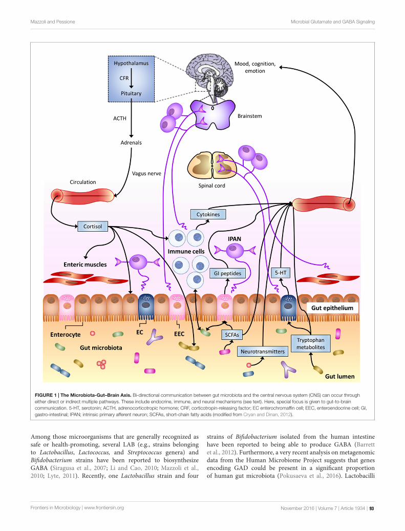
fmicb-07-01934 November 28, 2016 Time: 12:6 # 6
Mazzoli and Pessione Microbial Glutamate and GABA Signaling
FIGURE 1 | The Microbiota-Gut–Brain Axis. Bi-directional communication between gut microbiota and the central nervous system (CNS) can occur througheither direct or indirect multiple pathways. These include endocrine, immune, and neural mechanisms (see text). Here, special focus is given to gut-to-braincommunication. 5-HT, serotonin; ACTH, adrenocorticotropic hormone; CRF, corticotropin-releasing factor; EC enterochromaffin cell; EEC, enteroendocrine cell; GI,gastro-intestinal; IPAN; intrinsic primary afferent neuron; SCFAs, short-chain fatty acids (modified from Cryan and Dinan, 2012).
Among those microorganisms that are generally recognized assafe or health-promoting, several LAB (e.g., strains belongingto Lactobacillus, Lactococcus, and Streptococcus genera) andBifidobacterium strains have been reported to biosynthesizeGABA (Siragusa et al., 2007; Li and Cao, 2010; Mazzoli et al.,2010; Lyte, 2011). Recently, one Lactobacillus strain and four
strains of Bifidobacterium isolated from the human intestinehave been reported to being able to produce GABA (Barrettet al., 2012). Furthermore, a very recent analysis on metagenomicdata from the Human Microbiome Project suggests that genesencoding GAD could be present in a significant proportionof human gut microbiota (Pokusaeva et al., 2016). Lactobacilli
Frontiers in Microbiology | www.frontiersin.org November 2016 | Volume 7 | Article 1934 | 93

fmicb-07-01934 November 28, 2016 Time: 12:6 # 7
Mazzoli and Pessione Microbial Glutamate and GABA Signaling
TABLE 1 | Main neuroactive amines and amino acids released by bacteria.
Genus/species Bioactive molecule and(precursor)
Physiological effects Reference
Lactobacillus spp., Enterococcus Histamine (His) Hypotension, allergies Pessione et al., 2005
Enterococcus faecalis Tyramine (Tyr) Hypertension, headaches Pessione et al., 2009
Enterococcus faecalis β-phenylethylamine (Phe) Appetite/satiety control, moodcontrol
Shimazu and Miklya, 2004
Bacillus Dopamine (Tyr) Multiple Lyte, 2011
Bacillus, Escherichia coli Nor-adrenaline (Tyr) Multiple Lyte, 2011
Bifidobacteria, LAB Melatonin (Trp) Smooth muscle relaxation,Sleep/wake cycle regulator
Wong et al., 2015
Lactobacillus bulgaricus, Streptococcus, Escherichiacoli
Serotonin/triptamin (Trp) Vagal circuit regulation, peristalsis Matur and Eraslan, 2012
Corynebacterium glutamycum, Lactobacillusplantarum, Lactobacillus paracasei, Lactococcus lactis
Glu Multiple Tanous et al., 2005; Sano, 2009;Zareian et al., 2012
LAB, Escherichia coli, Pseudomonas GABA (Glu) Anxiolytic, miorelaxant Richard and Foster, 2003; Chouet al., 2008; Mazzoli et al., 2010
include the strains with the highest GABA production, althoughthis metabolic ability is more likely strain- rather than genus-related (Li and Cao, 2010).
Glu/GABA: RECEPTORS, SIGNALING,AND TRANSPORT
Bacterial Targets: Glu and GABAReceptors in ProkaryotesThe role of GABA in the communication between bacteria is inline with the identification of GABA-binding proteins in differentprokaryotes. These proteins consist not only of transporters,such as GabP in E. coli or Bacillus subtilis) (Brechtel and King,1998; Hu and King, 1998) and Bra in Agrobacterium tumefaciens(Chevrot et al., 2006), but also of possible specific receptors(Guthrie and Nicholson-Guthrie, 1989). It has been reportedthat metabotropic Glu and GABAB receptors, as well as bacterialperiplasmic amino acid binding proteins, may have evolved froma common ancestor (Cao et al., 2009). In addition, as reported inthe previous section, many bacteria (including those colonizingthe human gut) can synthesize GABA, thus suggesting that, asin the case of eukaryotes, GABA might be a conserved andubiquitous communication molecule.
The fact that GABA has an effect on the physiologyof Pseudomonas can be supported by several observations.A periplasmic protein showing high affinity for GABA anddisplaying similar biochemical features to a subunit of themammalian GABAA ionotropic receptor was identified in anenvironmental strain of P. fluorescens (Guthrie and Nicholson-Guthrie, 1989; Guthrie et al., 2000). Specific receptors for sensingGABA have been found in other Pseudomonads including:the PctC of P. aeruginosa and the McpG of P. putida, whichare involved in GABA chemoreception (Reyes-Darias et al.,2015). Another study has demonstrated that GABA increasesthe cytotoxicity and virulence of P. aeruginosa through asophisticated modulation of the protein expression (Dagorn et al.,2013). A functional proteomic analysis revealed that six proteins
were differentially expressed in P. aeruginosa when exposed toGABA (Lyte, 2011). Hence, some bacteria are able to respond toGABA, of both prokaryote and mammalian origin.
A structurally similar ion channel to eukaryotic ionotropicGABAA receptors was identified in the plant pathogen Erwiniachrysanthemi (Zimmermann and Dutzler, 2011). The prokaryoticprotein is activated by different amines, including GABA, andis modulated by benzodiazepines in a similar manner to itseukaryotic homolog (Zimmermann and Dutzler, 2011; Spurnyet al., 2012).
Less information is available on prokaryotic Glu receptors. Asfar as we know, only a single case of a Glu-activated potassiumchannel in a bacterium (i.e., GluR0 from Synechocystis PCC 6803)has been reported to date (Chen et al., 1999). However, 100prokaryotic channel proteins with putative Glu binding domainshave recently been identified through a bioinformatic study (Geret al., 2010). Among them, 22 proteins have been found to behomologs of vertebrate ionotropic Glu receptors (Ger et al.,2010).
Based on these findings, it is possible to hypothesize thatmembers of the human gut microbiota can also sense andrespond to GABA/Glu, although this has yet to be confirmedthrough dedicated studies. Furthermore, human-derived ordietary Glu can affect the balance of gut microbial populations bystimulating the growth of certain bacterial species at the expenseof others. It is known, for instance, that LAB decarboxylate Gluto GABA in order to create a protonic gradient and enhancemetabolic energy. Hence, Glu can constitute a privileged energysource that can be used to expand LAB populations.
Host-Targets: theGlutamatergic/GABAergic System in theCNSGlu and GABA are the major excitatory and inhibitoryneurotransmitters found in the human CNS, respectively (Julio-Pieper et al., 2013). These compounds are the molecular effectorsof a homeostatic neuronal circuit that is characterized by extremecomplexity and sophistication at both a physiological and a
Frontiers in Microbiology | www.frontiersin.org November 2016 | Volume 7 | Article 1934 | 94

fmicb-07-01934 November 28, 2016 Time: 12:6 # 8
Mazzoli and Pessione Microbial Glutamate and GABA Signaling
FIGURE 2 | (A) Representation of a Glutamatergic/GABAergic neural network, including an excitatory (Glutamatergic) pyramidal neuron (red), inhibitory (GABAergic)neurons (blue) and the related connections. The inhibitory neuron on the left is integrated into a feedback (FB) circuit, while the inhibitory neuron on the right isintegrated into a feed-forward (FF) loop. Inhibitory synapses targeted to the soma or dendrites of the pyramidal cell are surrounded by light blue staining, whichindicates a background GABA concentration that contributes to the overall inhibition (adapted from Roth and Draguhn, 2012). (B) Scheme of theGlutamatergic/GABAergic neurotransmission in the CNS. α-KG, α-keto-glutarate; EEATs, excitatory amino acid transporters; GABA, γ-aminobutyric acid; GABAA-R,GABAA receptor; GABAB-R, GABAB receptor; GABAC-R, GABAC receptor; GABA-T, GABA transaminase; GAD, Glu decarboxylase; GAT-1, GABA transporter 1;GlnT, glutamine transporter; Glu, glutamate; iGluR, ionotropic Glu receptor; mGluR, metabotropic Glu receptor; SSAl, succinic semialdehyde; SSAlDH, succinicsemialdehyde dehydrogenase; TCA cycle, the Krebs cycle; vGAT, vesicular GABA transporter; vGluT, vesicular Glu transporter. Symbols: , GABA; N, Glu.
biochemical level. This system also includes glutamine (Gln),which plays a key role as a non-neuroactive intermediate in therecycling of neurotrasmitters in the brain, mainly Glu and GABA(Soeiro-de-Souza et al., 2015).
At a physiological level, this network consists of feed forward(FF) and feedback (FB) neuronal connections in which themain role of inhibitory (i.e., GABAergic) neurons is currentlyconsidered as that of balancing the activity of excitatory (i.e.,Glutamatergic) neurons (Roth and Draguhn, 2012; Figure 2A).Both Glutamatergic and GABAergic neurotransmissions arecomplex systems in which glial cells (mainly astrocytes), pre- andpost-synaptic neurons and a large spectrum of different receptors,transporters and enzymes are involved (Cherlyn et al., 2010;Femenía et al., 2012; Figure 2B).
The biochemical relationship between Glu and GABA goesfar beyond this reciprocally modulating activity, since Gluand GABA are easily interconverted. Actually, the biochemicalreactions involved in GABA-to-Glu-to-Gln conversion areintegral features of such a regulatory network, which also involves
the central energy metabolism of neurons (Soeiro-de-Souza et al.,2015; Figure 2B).
Glu is synthesized in situ in the CNS since it cannot passthe blood–brain barrier (BBB) (Hawkins, 2009). One of themain Glu biosynthetic pathways consists of the conversionof glucose (through the Embden-Meyerhof-Parnas pathwayand the Krebs cycle) to α-ketoglutarate (α-KG), which issubsequently transaminated (Cherlyn et al., 2010). Glu isaccumulated in vesicles and, after suitable stimulation, releasedto the synaptic space, where it can bind to a panel of Glureceptors (GluRs) that may be found in post-synaptic neurons.GluRs differ on the basis of their mechanism of action (e.g.,ionotropic, iGluR, or metabotropic, mGluR, Glu receptors)and pharmacological responsiveness [e.g., N-methyl D-aspartate,NMDA, or α-amino-3-hydroxy-5-methylisoxazole-4-proprinoicacid (AMPA) or kainate sensitive receptors]. Ligand-gatedionotropic receptors mediate fast synaptic responses by directlyregulating the ion influx. Metabotropic receptors are coupledto G-proteins and modulate slower signal transduction cascades
Frontiers in Microbiology | www.frontiersin.org November 2016 | Volume 7 | Article 1934 | 95

fmicb-07-01934 November 28, 2016 Time: 12:6 # 9
Mazzoli and Pessione Microbial Glutamate and GABA Signaling
(Hyland and Cryan, 2010; Julio-Pieper et al., 2013). Any excessGlu present in the synaptic cleft is taken up by glial cells orneurons to prevent the prolonged excitation of post-synapticneurons, which can lead to cell death (Cherlyn et al., 2010). Gluin the glia is either oxidatively deaminated to α-KG [by Gludehydrogenase (GluDH)], or converted to Gln by Gln synthetase.Gln is released from glial cells, uptaken by pre-synaptic neurons[via Gln transporters (GlnT)], and is then converted back toGlu by glutaminase. Apart from its role in neurotransmission,Glu is also involved in other neural processes, such as neuronaldevelopment and synaptic plasticity (Cherlyn et al., 2010).
Inhibitory pre-synaptic neurons convert Glu to GABAthrough GAD and pack it into vesicles via vesicular GABAtransporters (vGATs). Again in this case, Gln is an alternativeGlu source to α-KG. The multiple GABA receptors that arepresent in post-synaptic neurons include ionotropic (GABAA,GABAC) and metabotropic (GABAB) receptors. Apart fromGABA, these receptors are possibly modulated by a number ofother compounds, e.g., barbiturates, baclofen, benzodiazepinesand steroids (Simeone et al., 2003; Goetz et al., 2007).Any excess unbound GABA is cleared by glial cells throughGABA transporters (GAT-1). GABA is then converted byGABA transaminase (GABA-T) into succinic semialdehyde, withconcomitant Glu biosynthesis (Sivilotti and Nistri, 1991; Michelsand Moss, 2007).
Glu/GABA: From the Gut to the BrainAs described above, several bacteria, including probiotic bacteriaand bacteria that colonize the human GI tract, are able to produceGlu or GABA (Barrett et al., 2012). As far as we know, only onestudy concerning the modulation of host Glutamate/GABAergicsystems by GABA/Glu producing gut-colonizing bacteria isavailable in the literature (Bravo et al., 2011). A recent study hasdemonstrated that a GABA-producing Bifidobacterium dentiumis able to attenuate sensitivity of dorsal root ganglia neuronsin a rat model of visceral pain (Pokusaeva et al., 2016). Itis worth noting that these few examples refer to the limitednumber of studies on interaction between gut microbiota and thehost nervous system which is currently available. Experimentalevidence is likely to grow as a higher number of reports becomeavailable. To date, most studies on the Glu/GABA effects onhumans concern dietary Glu/GABA.
Glu and GABA can be found in food as natural componentsor as food supplements. Glu is among the most abundantamino acids (8–10%) found in dietary proteins. Furthermore,monosodium glutamate is employed extensively as a flavoradditive in food (Brosnan et al., 2014). In Germany, Glu averageintake (comprinsing both free Glu and Glu as protein constituentin foods) of about 10 g/day has been estimated (Beyreuther et al.,2007). In asian consumers this value is likely higher since freeGlu intake can reach up to 4 g/day in these countries (Beyreutheret al., 2007). GABA is relatively abundant in plant food, such asbrown rice germs and sprouts, spinach, barley and bean sprouts,where GABA concentrations of between 300 and 720 nmol/gdry weight have been detected (Oh et al., 2003), while higherlevels are found in fermented foods (Abdou et al., 2006). Inrecent years, the utilization of GABA as a food supplement has
progressively increased in both Asian and Western (USA andEurope) countries (Boonstra et al., 2015). GABA concentrationof few micrograms per gram of cecal content has been detected inmice (Pokusaeva et al., 2016).
Hundreds of people have reported the benefits of using GABA-supplemented food, for instance in alleviating anxiety and/orimproving sleep quality (Boonstra et al., 2015). Moreover, at leastfive different studies have reported that the oral administration ofGABA or GABA-supplemented food/beverages (correspondingGABA amounts of about 50–100 mg) has had positive effectson human health. These effects include: (i) the reduction ofpsychological stress in people who performed arithmetic tasks(Nakamura et al., 2009; Kanehira et al., 2011; Yoto et al., 2012);(ii) the reduction of stress in acrophobic subjects exposed toheights (Abdou et al., 2006); (iii) an increased ability to performprioritized planned actions (Steenbergen et al., 2015).
Multiple pathways by which gut luminal Glu/GABA mayaffect the CNS can be hypothesized, including their transportacross intestinal and BBB and their sensing through afferentterminals that innervate the GI tract (Figure 3). In the nextsections, evidence possibly supporting these mechanisms will beillustrated.
Direct Influence of the Luminal Glu/GABA in the Guton the CNSMultiple transporters that mediate Glu absorption have beenfound in the apical membrane of GI epithelial cells, mainly inthe small intestine, but also in the stomach (Burrin and Stoll,2009), while little or no transport of amino acids from thelumen to portal blood occurs in the colon (Julio-Pieper et al.,2013). Studies on pigs and rodents have indicated the excitatoryamino acid carrier 1 is the most abundant Glu transporter in themucosa of the small intestine (Burrin and Stoll, 2009). A studyperformed in infant pigs has indicated that rate of Glu absorptionand transport to the portal circulation linearly increases withincreasing intraduodenal Glu intake (Janeczko et al., 2007).Portal absorption rate increased from 3.80 µmol kg−1 h−1fornormal Glu intake (i.e., 510 µmol kg−1 h−1) to 20.463.80 µmol kg−1 h−1 for piglets infused with 3.5-fold increasedGlu intake (Janeczko et al., 2007). Glu is one of the mainnutrients for enterocytes. Several studies on different animalmodels, including pre-term infants and human adults, haveagreed on that most Glu present in the GI lumen is oxidizedto CO2 or, secondarily, converted to other amino acids by thegut mucosa (Burrin and Stoll, 2009). Only a small percentage(between 5 and 17%, depending on the studies) of the ingestedGlu is transported into the portal circulation but this does notgenerally affect the Glu concentration in the plasma to any greatextent (Hays et al., 2007; Brosnan et al., 2014). Furthermore,an increased plasma Glu concentration (following Glu intake)does not necessarily affect the Glu concentration in brain tissues,since it is generally recognized that Glu cannot pass the BBB(Janeczko et al., 2007). A 20-fold (or more) increase in plasmaGlu concentration was found necessary to access the brain tissuesin rodents (Brosnan et al., 2014). The reaching of such a high Gluconcentration in plasma after dietary intake (or Glu biosynthesisby gut microbiota) seems rather unlikely.
Frontiers in Microbiology | www.frontiersin.org November 2016 | Volume 7 | Article 1934 | 96

fmicb-07-01934 November 28, 2016 Time: 12:6 # 10
Mazzoli and Pessione Microbial Glutamate and GABA Signaling
FIGURE 3 | Potential pathways for luminal Glu/GABA in the gut (of either dietary or gut–microbiota origin) to influence the nervous system (see text).5-HT, serotonin; EC enterochromaffin cell; EEC, enteroendocrine cell; GI, gastro-intestinal; IPAN; intrinsic primary afferent neuron (from Cryan andDinan, 2012).
As far as GABA transporters are concerned, they aremostly found in the CNS (Gadea and López-Colomé, 2001).However, H+/GABA symport across the apical membrane ofhuman intestinal epithelial (Caco-2) cells has been demonstrated(Thwaites et al., 2000). In fact, the hPAT1 H+/amino acid
symporter, which is thought to mediate the uptake of luminalGABA, is present throughout the human GI tract, from thestomach to the discending colon, with maximal expression inthe small intestine (Chen et al., 2003). Furthermore, GABAcarriers have been detected in the basolateral membrane of
Frontiers in Microbiology | www.frontiersin.org November 2016 | Volume 7 | Article 1934 | 97

fmicb-07-01934 November 28, 2016 Time: 12:6 # 11
Mazzoli and Pessione Microbial Glutamate and GABA Signaling
Caco-2 cells (Nielsen et al., 2012). These findings suggest thatluminal GABA should be able to cross the intestinal barrier andpossibly reach extra-intestinal targets. In fact, orally administeredGABA in rats was reported to increased GABA concentrationin their blood, with a peak after 30 min (Abdou et al., 2006).Nonetheless, GABA ability to cross the BBB is still controversial(Boonstra et al., 2015). Most early studies, performed between1950 and 1980, agreed on the impermeability of BBB to GABA,but more recent investigations have reported that GABA mayactually cross this barrier, although in small amounts (Boonstraet al., 2015). These discrepancies have been ascribed to thedifferent method of administration (e.g., oral versus injection),or animal model employed. Some studies have also suggestedthat BBB permeability to GABA could diminish with increasingage (Boonstra et al., 2015). Interestingly, GABA-transportershave been found in the BBB of mice (Kakee et al., 2001).Direct evidence of GABA facilitated transport across the BBBof mice has been reported, although the efflux rate from thebrain was 17 times higher than the influx rate (Kakee et al.,2001). Unfortunately, no information about the possible presenceof similar transporters in the BBB of humans is currentlyavailable.
All together, these findings suggest that a direct action ofluminal gut Glu and/or GABA on the CNS cannot be excluded,although further in vivo studies are required to confirm thishypothesis. These investigations should also take into accountthat gut and/or BBB permeability are affected by several factors,including stress, diet and gut microbiota (Braniste et al., 2014;Kelly et al., 2015).
Sensing of Luminal Glu/GABA in the GutMultiple Glu receptor types (including ionotropic, types 1 and 4metabotropic receptors and heterodimeric, TAS1R1 + TAS1R3,L-Glu taste receptors) have been detected in GI epithelial cellsand/or enteric neurons in the stomach, small intestine and colon(Kondoh et al., 2009; San Gabriel et al., 2009; Kitamura et al.,2012). More in detail, mGlu4 receptors have been detected inthe mucosa of both the gastric antrum and duodenum (Akibaet al., 2009), while both mGlu4 and mGlu7 receptors have beenidentified in the colon epithelium (Chang et al., 2005; Julio-Pieperet al., 2010). TAS1R3, a subunit of umami taste receptors, hasbeen found in enteroendocrine L-cells and X/A-like cells (Janget al., 2007; Hass et al., 2010). This evidence suggests a possiblerole in luminal Glu sensing. In fact, small intestine luminal L-Glu seems to play a role in the defense mechanisms of duodenalmucosa in rats, by increasing the intracellular pH and mucusgel thickness (Akiba et al., 2009). A role of mGluRs in thehuman colon in the control of colon peristalsis and electrolytetransport has been described (Julio-Pieper et al., 2013). Highlevels of mGlu7 and mGlu8 have been detected in myentericneurons, where they are possibly involved in the regulation ofgut motility (Julio-Pieper et al., 2013). However, results on theability of gut luminal Glu to directly excite enteric neurons stillappear contradictory (Kirchgessner, 2001; Wang et al., 2014).On the other hand, an indirect activation of vagal afferents(i.e., via the production and release of nitric oxide and 5-HT)through the administration of Glu to the gastric lumen has been
demonstrated (Uneyama et al., 2006). These data strongly suggesta role of ECs in the mediation of luminal Glu sensing. Thisluminal Glu sensing mechanism stimulates different areas in theCNS, including the cerebral cortex, basal ganglia, limbic system,and hypothalamus (for a comprehensive review, refer to Kondohet al., 2009), and induces flavor preference in rats (Kitamura et al.,2012).
GABAB receptors are abundantly expressed in the GI tract(Hyland and Cryan, 2010). GABA and its ionotropic andmetabotropic receptors are widely distributed throughout theENS, in both submucosal and myenteric neurons, from thestomach to the ileum (Auteri et al., 2015). In addition,Nakajima et al. (1996) reported the expression of GABABreceptors in cells morphologically similar to EECs, wherethey co-localized with cells containing somatostatin (in thestomach) or serotonin (in the duodenum). However, a laterstudy failed to detect any positive EEC for GABAB receptors(Casanova et al., 2009). Schwörer et al. (1989) showedthat the release of 5-HT by ECs in the small intestine ofguinea-pig is modulated by GABAA and GABAB receptors.Involvement of GABAB receptors in modulation of sensitivityof vagal and spinal afferents has been reported (Hyland andCryan, 2010). Actually, the GABAB metabotropic receptorsin the GI tract are thought to regulate several functions,including gut motility and gut-to-brain signaling (Hylandand Cryan, 2010). Experimental evidence of GABAB/µ-typeopiod receptor interaction, which mediates the synergisticpotentiation of the anti-nociceptive action of GABA/morphineco-administration, has been reported (Hara et al., 1999). A similarGABAB receptor-mediated potentiation effect has also beendescribed for drugs used to reduce visceral pain (Hara et al.,2004).
Since GABA receptors have been found in a wide rangeof immune cells, such as dendritic cells, mast cells andT-cells (Auteri et al., 2015), and are involved in regulatingimmunological processes, such as the down-regulation of pro-inflammatory cytokine release (Bjurstöm et al., 2008), their rolein neuro-immune dialog in the gut has been proposed (Auteriet al., 2015).
It is worth reminding that GABA is produced through Gludecarboxylation in both eukarya and prokaryotes. Hence, GABA-producing microorganisms that are present in the gut maysignificantly affect luminal Glu/GABA ratio and, therefore, gutsignaling.
PROBIOTICS FOR THE TREATMENT OFPSYCHIATRIC DISORDERS?
As far as its application to the field of probiotics is concerned,microbial neuro-endocrinology addresses the ability of probioticsto both synthesize and respond to neuroactive compounds, andthus to affect the host neurological processes and sometimes theoverall physiology of the host. A microbial endocrinology-basedapproach may be relevant to help understand how probiotics caninfluence the health of a host and how host can select commensalmicrobial populations.
Frontiers in Microbiology | www.frontiersin.org November 2016 | Volume 7 | Article 1934 | 98

fmicb-07-01934 November 28, 2016 Time: 12:6 # 12
Mazzoli and Pessione Microbial Glutamate and GABA Signaling
A number of studies have provided evidence on theability of probiotic strains to modulate the mood and stressresponses of humans, and to reduce anxiety and depression(Logan and Katzman, 2005; Messaoudi et al., 2011; Arseneault-Breard et al., 2012). As an example, the administration ofBifidobacteria and LAB as probiotic supplements to humanscan increase the levels of morning salivary melatonin, thusreducing the symptoms of irritable bowel syndrome (Wong et al.,2015). Actually, most of the currently recognized probiotic strainsbelong to Bifidobacteria and LAB, with lactobacilli being amongthe most abundant members of such health-promoting strains(Vaughan et al., 2011). A direct modulation of myenteric neuronactivity by unidentified Bifidobacterium longum fermentationproduct(s) displaying anxiolytic effects has also been reported(Bercik et al., 2011a).
Alterations in the Glu/ Gln/ GABA circuits in the CNShave been reported for generalized anxiety disorders, as well asin other psychiatric conditions, such as the major depressivedisorder (MDD), BD (also known as manic depressive disorder)and schizophrenia (SZ) (Cherlyn et al., 2010; Femenía et al.,2012; Soeiro-de-Souza et al., 2015). It is worth noting thatthese complex neurobehavioral disorders affect a significantportion of the overall population throughout the world (e.g.,individuals affected by BD or SZ account for about 2–3% ofthe global population) (Cherlyn et al., 2010). Different classesof drugs addressed to different steps of Glutamate/GABA-ergic neurotransmission (e.g., reducing Glu release, increasingGlu re-uptake, or increasing GABA levels by enhancing GADor inhibiting GABA transaminase and succinic semialdehydedehydrogenase) or serendipitously discovered (e.g., valproicacid) have been used in the treatment of such mood disorders(Terbach and Williams, 2009; Femenía et al., 2012). However,drugs frequently have negative secondary effects. For instance,most antidepressants (such as tricyclic antidepressants), whilehaving a positive effect on the mood of humans, do notalleviate and can even aggravate cognitive symptoms, orimpair vigilance (e.g., selective 5-HT reuptake inhibitors),and may cause dependence (e.g., benzodiazepines) (Femeníaet al., 2012). Finding alternative or adjuvant therapies istherefore a research priority and, in this respect, the useof neuromodulating bacteria or so-called psychobiotics is afascinating perspective.
Interestingly, the anxiolytic and anti-depressant-like effectsof Lactobacillus rhamnosus JB-1 ingestion in mice involve areduction in the plasma corticosterone levels and alterationsof both GABAA and GABAB receptor expression patternsin specific brain areas (Bravo et al., 2011). Furthermore,increases in the Glx (i.e., Glu + Gln) and GABA levelsin the CNS have been observed after 2 and 4 weeks ofL. rhamnosus JB-1 treatment, respectively (Janik et al., 2016).These effects were mediated by gut-to-brain communicationthrough the vagus nerve (Bravo et al., 2011). However, whetherL. rhamnosus stimulates the vagus nerve with its own GABAor induces an endogenous GABA production still has to beclarified.
Modulation of the glutamatergic system in the centralamygdala, cortex and hippocampus of mice by gut microbiota
has been described (Sudo et al., 2004; Neufeld et al.,2011).
Although, GABA production has been speculated as the keyfactor in the ability of a Lactobacillus helveticus–B. longummixture to reduce anxiety-like behavior(s) (Lyte, 2011), theobserved probiotic effects on the psychological health of a hostare generally likely the result of multiple interactions with thehost (ENS, EEC, GALT). On the other hand, anorexigenic andanxiolytic soymorphin peptides (produced by LAB proteolyticaction toward soy betaconglycinine) act via the GABAB receptorand, in this overall action, multiple circuits (including 5-HT anddopamine receptors) are involved (Kaneko et al., 2010). A recentinvestigation have suggested that a GABA-producing B. dentiumis able to reduce nociception in a rat model of visceral pain(Pokusaeva et al., 2016).
Because of the wide range of potential neuro-activecompounds (orexant, anorexant, opioid, opioid antagonist)produced by bacteria and the complex network that existsbetween them, a deep characterization of the proteome andmetabolome of probiotic strains is one key aspect that wouldhelp to understand the different effects exerted by each strainon human health. Since not only Glu/GABA receptors butalso other circuits can be potential targets for bacterial-derivedmolecules, it is tempting to hypothesize the use of probioticsthat produce bioactive compounds (e.g., GABA, 5-HT, opioidpeptides) as a novel frontline strategy for the adjuvant treatmentof gastrointestinal, neuroenteric, neurological and psychiatricconditions (Hyland and Cryan, 2010; Lyte, 2011; Reid, 2011).
Together with this optimistic viewpoint and the enthusiasmthat these findings have generated in the scientific domain,it is worth stating that a number of issues suggests cautionin application of psychobiotics. The comprehension of themechanisms by which gut microbiota and the nervous systemcommunicate is still at its beginning. Multiple levels ofcomplexity hinder the detailed understanding of this network andthese include (but are not limited to) the fact that: the gut–brainaxis comprises multiple (i.e., neural, immune, and endocrine)pathways with several reciprocal interactions; multiple molecularmediators, such as GABA and Glu, are involved, which play rolesin a number of different biological processes; gut microbiotaand even single commensal microorganisms interact withthe host through multiple pathways and chemical mediators(e.g., cell envelope components, production/metabolism ofneurotransmitters or neuroactive molecules). Therefore, it maybe difficult to specifically address health problems with suchmicrobial agents without the risk of unbalancing other pathways.As for other potential therapeutic agents, rigorous evaluation ofthe potential beneficial effects together with possible negativeside effects should be performed for each probiotic/psychobioticagent.
CONCLUSION
The increasing knowledge about gut bacteria–brain bi-directional communication has provided scientific evidencethat can explain the popular statement that somebody has
Frontiers in Microbiology | www.frontiersin.org November 2016 | Volume 7 | Article 1934 | 99
fmicb-07-01934 November 28, 2016 Time: 12:6 # 13
Mazzoli and Pessione Microbial Glutamate and GABA Signaling
made a decision based his/her on gut feeling, but also thesaying “we are what we eat,” as it is supposed that diet canaffect the microbiota composition of the gut. It is now evidentthat gut microbiota is able to interfere, through its cellularcomponents or by secreting bioactive compounds, with the gut–brain axis through immune, neural and humoral pathways.As the knowledge of this domain increases, new unexpectedand exciting perspectives for both basic and applied sciencewill likely emerge. At the same time, the observed effectsof human-derived neuro-active compounds on gut microbiotawould seem to suggest that some neurochemicals share acommon language that enables interkingdom communication,as proposed by Lyte and Freestone (2010). The ability ofa microbial strain to affect the overall patho-physiologyof a host thus appears to be the result of a long andsophisticated synergic evolution between gut bacteria and thehost.
AUTHOR CONTRIBUTIONS
Authors equally contributed to writing the manuscript. EPmainly contributed to describe bioactive molecules producedby bacteria and involved in interkingdom signaling, withparticular attention to microbiota-gut-brain axis. RM focusedhis contribution mainly on Glutamate- and GABA-mediatedcommunication, including Glu and GABA biosynthesis andreceptors in both bacteria and humans, gut-to-brain transportand sensing and Glu-ergic/GABA-ergic neural transmission inthe CNS.
ACKNOWLEDGMENT
EP benefited from a MIUR-University of Torino “Fondi ricercalocale (ex-60%)” grant.
REFERENCESAbdou, A. M., Higashiguchi, S., Horie, K., Kim, M., Hatta, H., and Yokogoshi, H.
(2006). Relaxation and immunity enhancement effects of γ-aminobutyric acid(GABA) administration in humans. Biofactors 26, 201–208. doi: 10.1002/biof.5520260305
Adams, J. B., Johansen, L. J., Powell, L. D., Quig, D., and Rubin, R. A. (2011).Gastrointestinal flora and gastrointestinal status in children with autism—comparisons to typical children and correlation with autism severity. BMCGastroenterol. 11:22. doi: 10.1186/1471-230X-11-22
Akiba, Y., Watanabe, C., Mizumori, M., and Kaunitz, J. D. (2009). LuminalL-glutamate enhances duodenal mucosal defense mechanisms via multipleglutamate receptors in rats. Am. J. Physiol. Gastrointest. Liver Physiol. 297,G781–G791. doi: 10.1152/ajpgi.90605.2008
Arseneault-Breard, J., Rondeau, I., Gilbert, K., Girard, S. A., Tompkins, T. A.,Godbout, R., et al. (2012). Combination of Lactobacillus helveticus R0052 andBifidobacterium longum R0175 reduces post-myocardial infarction depressionsymptoms and restores intestinal permeability in a rat model. Br. J. Nutr. 107,1793–1799. doi: 10.1017/S0007114511005137
Artis, D. (2008). Epithelial-cell recognition of commensal bacteria andmaintenance of immune homeostasis in the gut. Nat. Rev. Immunol. 8,411–420. doi: 10.1038/nri2316
Auteri, M., Zizzo, M. G., and Serio, R. (2015). GABA and GABA receptors in thegastrointestinal tract: from motility to inflammation. Pharmacol. Res. 93, 11–21.doi: 10.1016/j.phrs.2014.12.001
Aydin, S. (2007). Ghrelin may be a universal peptide in all living organisms. Turk.J. Med. Sci. 37, 123–124.
Aydin, S., Geckil, H., Zengin, F., Ibrahim Ozercan, H., Karatas, F., Aydin, S., et al.(2006). Ghrelin in plants: what is the function of an appetite hormone in plants?Peptides 27, 1597–1602. doi: 10.1016/j.peptides.2006.01.005
Bailey, M. T., Dowd, S. E., Galley, J. D., Hufnagle, A. R., Allen, R. G., andLyte, M. (2011). Exposure to a social stressor alters the structure of theintestinal microbiota: implications for stressor-induced immunomodulation.Brain Behav. Immun. 25, 397–407. doi: 10.1016/j.bbi.2010.10.023
Barajon, I., Serrao, G., Arnaboldi, F., Opizzi, E., Ripamonti, G., Balsari, A., et al.(2009). Toll-like receptors 3, 4, and 7 are expressed in the enteric nervoussystem and dorsal root ganglia. J. Histochem. Cytochem. 57, 1013–1023. doi:10.1369/jhc.2009.953539
Barrett, E., Ross, R. P., O’Toole, P. W., Fitzgerald, G. F., and Stanton, C.(2012). γ-Aminobutyric acid production by culturable bacteria from the humanintestine. J. Appl. Microbiol. 113, 411–417. doi: 10.1111/j.1365-2672.2012.05344.x
Bercik, P., Collins, S. M., and Verdu, E. F. (2012). Microbes and the gut-brain axis.Neurogastroenterol. Motil. 24, 405–413. doi: 10.1111/j.1365-2982.2012.01906.x
Bercik, P., Denou, E., Collins, J., Jackson, W., Lu, J., Jury, J., et al. (2011b). Theintestinal microbiota affect central levels of brain-derived neurotropic factorand behavior in mice. Gastroenterology 141, 599–609. doi: 10.1053/j.gastro.2011.04.052
Bercik, P., Park, A. J., Sinclair, D., Khoshdel, A., Lu, J., Huang, X., et al. (2011a). Theanxiolytic effect of Bifidobacterium longum NCC3001 involves vagal pathwaysfor gut– brain communication. Neurogastroenterol. Motil. 23, 1132–1139. doi:10.1111/j.1365-2982.2011.01796.x
Berer, K., Mues, M., Koutrolos, M., Rasbi, Z. A., Boziki, M., Johner, C., et al.(2011). Commensal microbiota and myelin autoantigen cooperate to triggerautoimmune demyelination. Nature 479, 538–541. doi: 10.1038/nature10554
Berntson, G. G., Sarter, M., and Cacioppo, J. T. (2003). Ascending visceralregulation of cortical affective information processing. Eur. J. Neurosci. 18,2103–2109. doi: 10.1046/j.1460-9568.2003.02967.x
Beyreuther, K., Biesalski, H. K., Fernstrom, J. D., Grimm, P., Hammes, W. P.,Heinemann, U., et al. (2007). Consensus meeting: monosodium glutamate - anupdate. Eur. J. Clin. Nutr. 61, 304–313. doi: 10.1038/sj.ejcn.1602526
Bezençon, C., le Coutre, J., and Damak, S. (2007). Taste-signaling proteins arecoexpressed in solitary intestinal epithelial cells. Chem. Senses 32, 41–49. doi:10.1093/chemse/bjl034
Biaggini, K., Barbey, C., Borrel, V., Feuilloley, M., Déchelotte, P., and Connil, N.(2015). The pathogenic potential of Pseudomonas fluorescens MFN1032 onenterocytes can be modulated by serotonin, substance P and epinephrine. Arch.Microbiol. 197, 983–990. doi: 10.1007/s00203-015-1135-y
Bienenstock, J., and Collins, S. (2010). 99th Dahlem conference on infection,inflammation and chronic inflammatory disorders: psycho-neuroimmunologyand the intestinal microbiota: clinical observations and basic mechanisms. Clin.Exp. Immunol. 160, 85–91. doi: 10.1111/j.1365-2249.2010.04124.x
Bjurstöm, H., Wang, J., Ericsson, I., Bengtssonm, M., Liu, Y., Kumar-Mendu, S.,et al. (2008). GABA, a natural immunomodulator of T lymphocytes.J. Neuroimmunol. 205, 44–50. doi: 10.1016/j.jneuroim.2008.08.017
Bohórquez, D. V., Shahid, R. A., Erdmann, A., Kreger, A. M., Wang, Y.,Calakos, N., et al. (2015). Neuroepithelial circuit formed by innervation ofsensory enteroendocrine cells. J. Clin. Invest. 125, 782–786. doi: 10.1172/JCI78361
Boonstra, E., de Kleijn, R., Colzato, L. S., Alkemade, A., Forstmann, B. U., andNieuwenhuis, S. (2015). Neurotransmitters as food supplements: the effects ofGABA on brain and behavior. Front. Psychol. 6:1520. doi: 10.3389/fpsyg.2015.01520
Braniste, V., Al-Asmakh, M., Kowal, C., Anuar, F., Abbaspour, A., Tóth, M., et al.(2014). The gut microbiota influences blood-brain barrier permeability in mice.Sci. Transl. Med. 6:263ra158. doi: 10.1126/scitranslmed.3009759
Bravo, J. A., Forsythe, P., Chew, M. V., Escaravage, E., Savignac, H. M., Dinan, T. G.,et al. (2011). Ingestion of Lactobacillus strain regulates emotional behavior and
Frontiers in Microbiology | www.frontiersin.org November 2016 | Volume 7 | Article 1934 | 100
fmicb-07-01934 November 28, 2016 Time: 12:6 # 14
Mazzoli and Pessione Microbial Glutamate and GABA Signaling
central GABA receptor expression in a mouse via the vagus nerve. Proc. Natl.Acad. Sci. U.S.A. 108, 16050–16055. doi: 10.1073/pnas.1102999108
Brechtel, C. E., and King, S. C. (1998). 4-Aminobutyrate (GABA) transportersfrom the amine-polyamine-choline superfamily: substrate specificity and ligandrecognition profile of the 4-aminobutyrate permease from Bacillus subtilis.Biochem. J. 333, 565–571. doi: 10.1042/bj3330565
Breton, J., Tennoune, N., Lucas, N., Francois, M., Legrand, R., Jacquemot, J.,et al. (2016). Gut commensal E. coli Proteins Activate Host Satiety Pathwaysfollowing nutrient-induced bacterial growth. Cell Metab. 23, 324–334. doi: 10.1016/j.cmet.2015.10.017
Bron, P. A., van Baarlen, P., and Kleerebezem, M. (2011). Emerging molecularinsights into the interaction between probiotics and the host intestinal mucosa.Nat. Rev. Microbiol. 10, 66–78.
Brosnan, J. T., Drewnowski, A., and Friedman, M. I. (2014). Is there a relationshipbetween dietary MSG and obesity in animals or humans ? Amino Acids 46,2075–2087. doi: 10.1007/s00726-014-1799-7
Buka, S. L., Cannon, T. D., Torrey, E. F., Yolken, R. H., and Collaborative StudyGroup on the Perinatal Origins of Severe Psychiatric Disorders (2008). Maternalexposure to herpes simplex virus and risk of psychosis among adult offspring.Biol. Psychiatry 63, 809–815. doi: 10.1016/j.biopsych.2007.09.022
Burchett, S. A., and Hicks, T. P. (2006). The mysterious trace amines:protean neuromodulators of synaptic transmission in mammalian brain. Prog.Neurobiol. 79, 223–246. doi: 10.1016/j.pneurobio.2006.07.003
Burrin, D. G., and Stoll, B. (2009). Metabolic fate and function of dietaryglutamate in the gut. Am. J. Clin. Nutr. 90, 850S–856S. doi: 10.3945/ajcn.2009.27462Y
Cani, P. D., and Knauf, C. (2016). How gut microbes talk to organs: the role ofendocrine and nervous routes. Mol. Metab. 5, 743–752. doi: 10.1016/j.molmet.2016.05.011
Cao, J. H., Huang, S. L., Qian, J., Huang, J. L., Jin, L. X., Su, Z., et al. (2009).Evolution of the class C GPCR Venus flytrap modules involved positive selectedfunctional divergence. BMC Evol. Biol. 9:67. doi: 10.1186/1471-2148-9-67
Casanova, E., Guetg, N., Vigot, R., Seddik, R., Julio-Pieper, M., Hyland, N. P., et al.(2009). A mouse model for visualization of GABA(B) receptors. Genesis 47,595–602. doi: 10.1002/dvg.20535
Chang, H. J., Yoo, B. C., Lim, S. B., Jeong, S. Y., Kim, W. H., and Park, J. G. (2005).Metabotropic glutamate receptor 4 expression in colorectal carcinoma and itsprognostic significance. Clin. Cancer Res. 11, 3288–3295. doi: 10.1158/1078-0432.CCR-04-1912
Chen, G. Q., Cui, C., Mayer, M. L., and Gouaux, E. (1999). Functionalcharacterization of a potassium-selective prokaryotic glutamate receptor.Nature 402, 817–821. doi: 10.1038/45568
Chen, Z., Fei, Y. J., Anderson, C. M., Wake, K. A., Miyauchi, S., Huang, W., et al.(2003). Structure, function and immunolocalization of a proton-coupled aminoacid transporter (hPAT1) in the human intestinal cell line Caco-2. J. Physiol. 546,349–361. doi: 10.1113/jphysiol.2002.026500
Cherlyn, S. Y., Woon, P. S., Liu, J. J., Ong, W. Y., Tsai, G. C., and Sim, K.(2010). Genetic association studies of glutamate, GABA and related genes inschizophrenia and bipolar disorder: a decade of advance. Neurosci. Biobehav.Rev. 34, 958–977. doi: 10.1016/j.neubiorev.2010.01.002
Chevrot, R., Rosen, R., Haudecoeur, E., Cirou, A., Shelp, B. J., Ron, E., et al.(2006). GABA controls the level of quorum-sensing signal in Agrobacteriumtumefaciens. Proc. Natl. Acad. Sci. U.S.A. 103, 7460–7464. doi: 10.1073/pnas.0600313103
Chimerel, C., Emery, E., Summers, D. K., Keyser, U., Gribble, F. M., andReimann, F. (2014). Bacterial metabolite indole modulates incretin secretionfrom intestinal enteroendocrine L cells. Cell Rep. 9, 1202–1208. doi: 10.1016/j.celrep.2014.10.032
Choi, S., Lee, M., Shiu, A. L., Yo, S. J., and Aponte, G. W. (2007). Identification of aprotein hydrolysate responsive G protein-coupled receptor in enterocytes. Am.J. Physiol. Gastrointest. Liver Physiol. 292, G98–G112. doi: 10.1152/ajpgi.00295.2006
Chou, H. T., Kwon, D. H., Hegazy, M., and Lu, C. D. (2008). Transcriptomeanalysis of agmatine and putrescine catabolism in Pseudomonas aeruginosaPAO1. J. Bacteriol. 190, 1966–1975. doi: 10.1128/JB.01804-07
Costedio, M. M., Hyman, N., and Mawe, G. M. (2007). Serotonin and its rolein colonic function and in gastrointestinal disorders. Dis. Colon Rectum 50,376–388. doi: 10.1007/s10350-006-0763-3
Cryan, J. F., and Dinan, T. G. (2012). Mind-altering microorganisms: the impactof the gut microbiota on brain and behaviour. Nat. Rev. Neurosci. 13, 701–712.doi: 10.1038/nrn3346
Dagorn, A., Hillion, M., Chapalain, A., Lesouhaitier, O., Duclairoir Poc, C.,Vieillard, J., et al. (2013). Gamma-aminobutyric acid acts as a specific virulenceregulator in Pseudomonas aeruginosa. Microbiology 159, 339–351. doi: 10.1099/mic.0.061267-0
Engelstoft, M. S., Egerod, K. L., Holst, B., and Schwartz, T. W. (2008). A gut feelingfor obesity: 7TM sensors on enteroendocrine cells. Cell Metab. 8, 447–449.doi: 10.1016/j.cmet.2008.11.004
Everard, A., Lazarevic, V., Derrien, M., Girard, M., Muccioli, G. G., Neyrinck,A. M., et al. (2011). Responses of gut microbiota and glucose and lipidmetabolism to prebiotics in genetic obese and diet-induced leptin-resistantmice. Diabetes Metab. Res. Rev. 60, 2775–2786.
Eßlinger, M., Wachholz, S., Manitz, M. P., Plümper, J., Sommer, R., Juckel, G.,et al. (2016). Schizophrenia associated sensory gating deficits develop afteradolescent microglia activation. Brain Behav. Immun. 58, 99–106. doi: 10.1016/j.bbi.2016.05.018
Femenía, T., Gómez-Galán, M., Lindskog, M., and Magara, S. (2012).Dysfunctional hippocampal activity affects emotion and cognition in mooddisorders. Brain Res. 1476, 58–70. doi: 10.1016/j.brainres.2012.03.053
Forsythe, P., and Kunze, W. A. (2013). Voices from within: gut microbes and theCNS. Cell. Mol. Life Sci. 70, 55–69. doi: 10.1007/s00018-012-1028-z
Freestone, P. P., Williams, P. H., Haigh, R. D., Maggs, A. F., Neal, C. P., andLyte, M. (2002). Growth stimulation of intestinal commensal Escherichia coli bycatecholamines: a possible contributory factor in trauma-induced sepsis. Shock18, 465–470. doi: 10.1097/00024382-200211000-00014
Furness, J., Rivera, L., Cho, H., Bravo, D., and Callaghan, B. (2013). The gut asa sensory organ. Nat. Rev. Gastroenterol. Hepatol. 10, 729–740. doi: 10.1038/nrgastro.2013.180
Furness, J. B. (2006). The Enteric Nervous System. Oxford: Blackwell.Gadea, A., and López-Colomé, A. M. (2001). Glial transporters for glutamate,
glycine, and GABA: II, GABA transporters. J. Neurosci. Res. 63, 461–468. doi:10.1002/jnr.1040
Ger, M. F., Rendon, G., Tilson, J. L., and Jakobsson, E. (2010). Domain-basedidentification and analysis of glutamate receptor ion channels and their relativesin prokaryotes. PLoS ONE 5:e12827. doi: 10.1371/journal.pone.0012827
Gershon, M. D. (1998). The Second Brain. New York, NY: Harper Collins.Gershon, M. D., and Tack, J. (2007). The serotonin signaling system: from
basic understanding to drug development for functional GI disorders.Gastroenterology 132, 397–414. doi: 10.1053/j.gastro.2006.11.002
Gilman, S. E., Cherkerzian, S., Buka, S. L., Hahn, J., Hornig, M., and Goldstein, J. M.(2016). Prenatal immune programming of the sex-dependent risk for majordepression. Transl. Psychiatry 6:e822. doi: 10.1038/tp.2016.91
Goetz, T., Arslan, A., Wisden, W., and Wulff, P. (2007). GABA(A) receptors:structure and function in the basal ganglia. Prog. Brain Res. 160, 21–41. doi:10.1016/S0079-6123(06)60003-4
Guthrie, G. D., and Nicholson-Guthrie, C. S. (1989). gamma- Aminobutyric aciduptake by a bacterial system with neurotransmitter binding characteristics.Proc. Natl. Acad. Sci. U.S.A. 86, 7378–7381. doi: 10.1073/pnas.86.19.7378
Guthrie, G. D., Nicholson-Guthrie, C. S., and Leary, H. L. Jr. (2000). A bacterialhigh-affinity GABA binding protein: isolation and characterization. Biochem.Biophys. Res. Commun. 268, 65–68. doi: 10.1006/bbrc.1999.1960
Halang, P., Toulouse, C., Geißel, B., Michel, B., Flauger, B., Müller, M., et al. (2015).Response of Vibrio cholerae to the catecholamine hormones Epinephrine andNorepinephrine. J. Bacteriol. 197, 3769–3778. doi: 10.1128/JB.00345-15
Hara, K., Saito, Y., Kirihara, Y., and Sakura, S. (2004). The interaction betweengamma-aminobutyric acid agonists and diltiazem in visceral antinociception inrats. Anesth. Analg. 98, 1380–1384. doi: 10.1213/01.ANE.0000107935.84035.48
Hara, K., Saito, Y., Kirihara, Y., Yamada, Y., Sakura, S., and Kosaka, Y. (1999). Theinteraction of antinociceptive effects of morphine and GABA receptor agonistswithin the rat spinal cord. Anesth. Analg. 89, 422–427. doi: 10.1213/00000539-199908000-00032
Hass, N., Schwarzenbacher, K., and Breer, H. (2010). T1R3 is expressed in brushcells and ghrelin-producing cells of murine stomach. Cell Tissue Res. 339,493–504. doi: 10.1007/s00441-009-0907-6
Hawkins, R. A. (2009). The blood–brain barrier and glutamate. Am. J. Clin. Nutr.90, 867S–874S. doi: 10.3945/ajcn.2009.27462BB
Frontiers in Microbiology | www.frontiersin.org November 2016 | Volume 7 | Article 1934 | 101

fmicb-07-01934 November 28, 2016 Time: 12:6 # 15
Mazzoli and Pessione Microbial Glutamate and GABA Signaling
Hays, S. P., Ordonez, J. M., Burrin, D. G., and Sunehag, A. L. (2007). Dietaryglutamate is almost entirely removed in its first pass through the splanchnicbed in premature infants. Pediatr. Res. 62, 353–356. doi: 10.1203/PDR.0b013e318123f719
Hornby, J. M., Jensen, E. C., Lisec, A. D., Tasto, J. J., Jahnke, B., Shoemaker, R., et al.(2001). Quorum sensing in the dimorphic fungus Candida albicans is mediatedby farnesol. Appl. Environ. Microbiol. 67, 2982–2992. doi: 10.1128/AEM.67.7.2982-2992.2001
Hosoi, T., Okuma, Y., Matsuda, T., and Nomura, Y. (2005). Novel pathway forLPS-induced afferent vagus nerve activation: possible role of nodose ganglion.Auton. Neurosci. 120, 104–107. doi: 10.1016/j.autneu.2004.11.012
Hu, L. A., and King, S. C. (1998). Membrane topology of the Escherichia coliγ-aminobutyrate transporter: implications on the topography and mechanismof prokaryotic and eukaryotic transporters from the APC superfamily. Biochem.J. 336, 69–76. doi: 10.1042/bj3360069
Hughes, D. T., and Sperandio, V. (2008). Inter-kingdom signalling:communication between bacteria and their hosts. Nat. Rev. Microbiol. 6,111–120. doi: 10.1038/nrmicro1836
Hyland, N. P., and Cryan, J. F. (2010). A gut feeling about GABA: focus onGABA(B) receptors. Front. Pharmacol. 1:124. doi: 10.3389/fphar.2010.00124
Janeczko, M. J., Stoll, B., Chang, X., Guan, X., and Burrin, D. G. (2007). Extensivegut metabolism limits the intestinal absorption of excessive supplementaldietary glutamate loads in infant pigs. J. Nutr. 137, 2384–2390.
Jang, H. J., Kokrashvili, Z., Theodorakis, M. J., Carlson, O. D., Kim, B. J., Zhou, J.,et al. (2007). Gut-expressed gustducin and taste receptors regulate secretionof glucagon-like peptide-1. Proc. Natl. Acad. Sci. U.S.A. 104, 15069–15074.doi: 10.1073/pnas.0706890104
Janik, R., Thomason, L. A., Stanisz, A. M., Forsythe, P., Bienenstock, J., andStanisz, G. J. (2016). Magnetic resonance spectroscopy reveals oral Lactobacilluspromotion of increases in brain GABA, N-acetyl aspartate and glutamate.Neuroimage 125, 988–995.
Julio-Pieper, M., Hyland, N. P., Bravo, J. A., Dinan, T. G., and Cryan, J. F. (2010).A novel role for the metabotropic glutamate receptor-7: modulation of faecalwater content and colonic electrolyte transport in the mouse. Br. J. Pharmacol.160, 367–375. doi: 10.1111/j.1476-5381.2010.00713.x
Julio-Pieper, M., O’Connor, R. M., Dinan, T. G., and Cryan, J. F. (2013). Regulationof the brain-gut axis by group III metabotropic glutamate receptors. Eur. J.Pharmacol. 698, 19–30. doi: 10.1016/j.ejphar.2012.10.027
Kakee, A., Takanaga, H., Terasaki, T., Naito, M., Tsuruo, T., and Sugiyama, Y.(2001). Efflux of a suppressive neurotransmitter, GABA, across the blood-brainbarrier. J. Neurochem. 79, 110–118. doi: 10.1046/j.1471-4159.2001.00540.x
Kanehira, T., Yoshiko, N., Nakamura, K., Horie, K., Horie, N., Furugori, K., et al.(2011). Relieving occupational fatigue by consumption of a beverage containingγ-amino butyric acid. J. Nutr. Sci. Vitaminol. 57, 9–15. doi: 10.3177/jnsv.57.9
Kaneko, K., Iwasaki, M., Yoshikawa, M., and Ohinata, K. (2010). Orallyadministered soymorphins, soy-derived opioid peptides, suppress feeding andintestinal transit via gut mu(1)-receptor coupled to 5-HT(1A), D(2), andGABA(B) systems. Am. J. Physiol. Gastrointest. Liver Physiol. 299, G799–G805.doi: 10.1152/ajpgi.00081.2010
Keita, A. V., and Söderholm, J. D. (2010). The intestinal barrier and its regulationby neuroimmune factors. Neurogastroenterol. Motil. 22, 718–733. doi: 10.1111/j.1365-2982.2010.01498.x
Kelly, J. R., Kennedy, P. J., Cryan, J. F., Dinan, T. G., Clarke, G., and Hyland, N. P.(2015). Breaking down the barriers: the gut microbiome, intestinal permeabilityand stress-related psychiatric disorders. Front. Cell. Neurosci. 9:392. doi: 10.3389/fncel.2015.00392
Kirchgessner, A. L. (2001). Glutamate in the enteric nervous system. Curr. Opin.Pharmacol. 1, 591–596. doi: 10.1016/S1471-4892(01)00101-1
Kitamura, A., Tsurugizawa, T., Uematsu, A., Torii, K., and Uneyama, H. (2012).New therapeutic strategy for amino acid medicine: effects of dietary glutamateon gut and brain function. J. Pharmacol. Sci. 118, 138–144. doi: 10.1254/jphs.11R06FM
Kojima, M., Hosoda, H., Date, Y., Nakazato, M., Matsuo, H., and Kangawa, K.(1999). Ghrelin is a growth-hormone-releasing acylated peptide from stomach.Nature 402, 656–660. doi: 10.1038/45230
Kondoh, T., Mallick, H. N., and Torii, K. (2009). Activation of the gut-brain axisby dietary glutamate and physiologic significance in energy homeostasis. Am. J.Clin. Nutr. 90, 832S–837S. doi: 10.3945/ajcn.2009.27462V
Li, H., and Cao, Y. (2010). Lactic acid bacterial cell factories for gamma-aminobutyric acid. Amino Acids 39, 1107–1116. doi: 10.1007/s00726-010-0582-7
Lim, J. Y., Choi, S. I., Choi, G., and Hwang, S. W. (2016). Atypical sensors fordirect and rapid neuronal detection of bacterial pathogens. Mol. Brain. 9:26.doi: 10.1186/s13041-016-0202-x
Liu, M., Seino, S., and Kirchgessner, A. (1999). Identification and characterizationof glucoresponsive neurons in the enteric nervous system. J. Neurosci. 19,10305–10317.
Logan, A. C., and Katzman, M. (2005). Major depressive disorder: probiotics maybe an adjuvant therapy. Med. Hypotheses 64, 533–538. doi: 10.1016/j.mehy.2004.08.019
Lyte, M. (2011). Probiotics function mechanistically as delivery vehicles forneuroactive compounds: microbial endocrinology in the design and use ofprobiotics. Bioessays 33, 574–581. doi: 10.1002/bies.201100024
Lyte, M., and Freestone, P. P. E. (2010). Microbial Endocrinology: InterkingdomSignaling in Infectious Disease and Health. New York, NY: Springer.
Masek, K., and Kadlec, O. (1983). Sleep factor, muramyl peptides, and theserotoninergic system. Lancet 1, 1277.
Matur, E., and Eraslan, E. (2012). “The impact of probiotics on the gastrointestinalphysiology,” in New Advances in the Basic and Clinical Gastroenterology, ed. T.Brzozowski (Rijeka: InTech), 51–74.
Mawe, G. M., Strong, D. S., and Sharkey, K. A. (2009). Plasticity of enteric nervefunctions in the inflamed and postinflamed gut. Neurogastroenterol. Motil. 21,481–491. doi: 10.1111/j.1365-2982.2009.01291.x
Mayer, E. A. (2011). Gut feelings: the emerging biology of gut-braincommunication. Nat. Rev. Neurosci. 12, 453–466. doi: 10.1038/nrn3071
Mazzoli, R. (2014). “Neuro-active compounds produced by probiotics. Towards amicrobiota-(gut-) brain axis control?,” in Interactive Probiotics, ed. E. Pessione(Boca Raton, FL: CRC Press), 148–176.
Mazzoli, R., Pessione, E., Dufour, M., Laroute, V., Giuffrida, M. G., Giunta, C., et al.(2010). Glutamate-induced metabolic changes in Lactococcus lactis NCDO 2118during GABA production: combined transcriptomic and proteomic analysis.Amino Acids 39, 727–737. doi: 10.1007/s00726-010-0507-5
McDermott, J. R., Leslie, F. C., D’Amato, M., Thompson, D. G., Grencis, R. K., andMcLaughlin, J. T. (2006). Immune control of food intake: enteroendocrine cellsare regulated by CD4+ T lymphocytes during small intestinal inflammation.Gut 55, 492–497. doi: 10.1136/gut.2005.081752
Medzhitov, R. (2007). Recognition of microorganisms and activation of theimmune response. Nature 449, 819–826. doi: 10.1038/nature06246
Messaoudi, M., Lalonde, R., Violle, N., Javelot, H., Desor, D., Nejdi, A., et al.(2011). Assessment of psychotropic-like properties of a probiotic formulation(Lactobacillus helveticus R0052 and Bifidobacterium longum R0175) in ratsand human subjects. Br. J. Nutr. 105, 755–764. doi: 10.1017/S0007114510004319
Michels, G., and Moss, S. J. (2007). GABAA receptors: properties and trafficking.Crit. Rev. Biochem. Mol. Biol. 42, 3–14. doi: 10.1080/10409230601146219
Morse, D. E., Duncan, H., Hooker, N., Baloun, A., and Young, G. (1980). GABAinduces behavioral and developmental metamorphosis in planktonic molluscanlarvae. Fed. Proc. 39, 3237–3241.
Nakajima, K., Tooyama, I., Kuriyama, K., and Kimura, H. (1996).Immunohistochemical demonstration of GABAB receptors in the ratgastrointestinal tract. Neurochem. Res. 21, 211–215. doi: 10.1007/BF02529137
Nakamura, H., Takishima, T., Kometani, T., and Yokogoshi, H. (2009).Psychological stress-reducing effect of chocolate enriched with gamma-aminobutyric acid (GABA) in humans: assessment of stress using heart ratevariability and salivary chromogranin A. Int. J. Food Sci. Nutr. 60, 106–113.doi: 10.1080/09637480802558508
Navarathna, D. H., Nickerson, K. W., Duhamel, G. E., Jerrels, T. R., and Petro, T. M.(2007). Exogenous farnesol interferes with the normal progression of cytokineexpression during candidiasis in a mouse model. Infect. Immun. 75, 4006–4011.doi: 10.1128/IAI.00397-07
Neufeld, K. M., Kang, N., Bienenstock, J., and Foster, J. A. (2011). Reducedanxiety-like behavior and central neurochemical change in germ-free mice.Neurogastroenterol. Motil. 23:e119. doi: 10.1111/j.1365-2982.2010.01620.x
Neunlist, M., and Schemann, M. (2014). Nutrient-induced changes in thephenotype and function of the enteric nervous system. J. Physiol. 592, 2959–2965. doi: 10.1113/jphysiol.2014.272948
Frontiers in Microbiology | www.frontiersin.org November 2016 | Volume 7 | Article 1934 | 102

fmicb-07-01934 November 28, 2016 Time: 12:6 # 16
Mazzoli and Pessione Microbial Glutamate and GABA Signaling
Nicholson, J. K., Holmes, E., Kinross, J., Burcelin, R., Gibson, G., Jia, W., et al.(2012). Host-gut microbiota metabolic interactions. Science 336, 1262–1267.doi: 10.1126/science.1223813
Nicola, J. P., Velez, M. L., Lucero, A. M., Fozzatti, L., Pellizas, C. G., andMasini-Repiso, A. M. (2009). Functional toll-like receptor 4 conferringlipopolysaccharide responsiveness is expressed in thyroid cells. Endocrinology150, 500–508. doi: 10.1210/en.2008-0345
Nielsen, C. U., Carstensen, M., and Brodin, B. (2012). Carrier-mediatedγ-aminobutyric acid transport across the basolateral membrane of humanintestinal Caco-2 cell monolayers. Eur. J. Pharm. Biopharm. 81, 458–462. doi:10.1016/j.ejpb.2012.03.007
Nøhr, M. K., Pedersen, M. H., Gille, A., Egerod, K. L., Engelstoft, M. S., Husted,A. S., et al. (2013). GPR41/FFAR3 and GPR43/FFAR2 as cosensors for short-chain fatty acids in enteroendocrine cells vs FFAR3 in enteric neurons andFFAR2 in enteric leukocytes. Endocrinology 154, 3552–3564. doi: 10.1210/en.2013-1142
Norris, V., Molina, F., and Gewirtz, A. T. (2013). Hypothesis: bacteria control hostappetites. J. Bacteriol. 195, 411–416. doi: 10.1128/JB.01384-12
Ochoa-Cortes, F., Ramos-Lomas, T., Miranda-Morales, M., Spreadbury, I.,Ibeakanma, C., Barajas-Lopez, C., et al. (2010). Bacterial cell products signalto mouse colonic nociceptive dorsal root ganglia neurons. Am. J. Physiol.Gastrointest. Liver Physiol. 299, G723–G732. doi: 10.1152/ajpgi.00494.2009
Oh, S. H., Moon, Y. J., and Oh, C. H. (2003). γ-Aminobutyric acid (GABA) contentof selected uncooked foods. Preven. Nutr. Food Sci.8, 75–78. doi: 10.3746/jfn.2003.8.1.075
O’Mahony, S. M., Clarke, G., Borre, Y. E., Dinan, T. G., and Cryan, J. F. (2015).Serotonin, tryptophan metabolism and the brain-gut-microbiome axis. Behav.Brain Res. 277, 32–48. doi: 10.1016/j.bbr.2014.07.027
Pessione, E. (2012). Lactic acid bacteria contribution to gut microbiota complexity:lights and shadows. Front. Cell. Infect. Microbiol. 2:86. doi: 10.3389/fcimb.2012.00086
Pessione, E., Mazzoli, R., Giuffrida, M. G., Lamberti, C., Garcia-Moruno, E.,Barello, C., et al. (2005). A proteomic approach to studying biogenic amineproducing lactic acid bacteria. Proteomics 5, 687–698. doi: 10.1002/pmic.200401116
Pessione, E., Pessione, A., Lamberti, C., Coïsson, D. J., Riedel, K., Mazzoli, R.,et al. (2009). First evidence of a membrane-bound, tyramine and beta-phenylethylamine producing, tyrosine decarboxylase in Enterococcus faecalis:a two-dimensional electrophoresis proteomic study. Proteomics 9, 2695–2710.doi: 10.1002/pmic.200800780
Pokusaeva, K., Johnson, C., Luk, B., Uribe, G., Fu, Y., Oezguen, N., et al. (2016).GABA-producing Bifidobacterium dentium modulates visceral sensitivity in theintestine. Neurogastroenterol. Motil. doi: 10.1111/nmo.12904 [Epub ahead ofprint].
Psichas, A., Reimann, F., and Gribble, F. M. (2015). Gut chemosensingmechanisms. J. Clin. Invest. 125, 908–917. doi: 10.1172/JCI76309
Qi, J., Buzas, K., Fan, H., Cohen, J. I., Wang, K., Mont, E., et al. (2011).Painful pathways induced by TLR stimulation of dorsal root ganglion neurons.J. Immunol. 186, 6417–6426. doi: 10.4049/jimmunol.1001241
Raybould, H. E. (2010). Gut chemosensing: interactions between gut endocrinecells and visceral afferents. Auton. Neurosci. 153, 41–46. doi: 10.1016/j.autneu.2009.07.007
Reid, G. (2011). Neuroactive probiotics. Bioessays 33:562. doi: 10.1002/bies.201100074
Reyes-Darias, J. A., García, V., Rico-Jiménez, M., Corral-Lugo, A., Lesouhaitier, O.,Juárez-Hernández, D., et al. (2015). Specific gamma-aminobutyrate chemotaxisin pseudomonads with different lifestyle. Mol. Microbiol. 97, 488–501. doi:10.1111/mmi.13045
Richard, H. T., and Foster, J. W. (2003). Acid resistance in Escherichia coli. Adv.Appl. Microbiol. 52, 167–186. doi: 10.1016/S0065-2164(03)01007-4
Roth, F. C., and Draguhn, A. (2012). GABA metabolism and transport: effects onsynaptic efficacy. Neural Plast. 2012:805830. doi: 10.1155/2012/805830
Samuel, B. S., Shaito, A., Motoike, T., Rey, F. E., Backhed, F., Manchester,J. K., et al. (2008). Effects of the gut microbiota on host adiposity aremodulated by the short-chain fatty-acid binding G protein-coupled receptor,Gpr41. Proc. Natl. Acad. Sci. U.S.A. 105, 16767–16772. doi: 10.1073/pnas.0808567105
San Gabriel, A., Nakamura, E., Uneyama, H., and Torii, K. (2009). Taste, visceralinformation and exocrine reflexes with glutamate through umami receptors.J. Med. Invest. 56(Suppl.), 209–217. doi: 10.2152/jmi.56.209
Sano, C. (2009). History of glutamate production. Am. J. Clin. Nutr. 90, 728S–732S.doi: 10.3945/ajcn.2009.27462F
Santocchi, E., Guiducci, L., Fulceri, F., Billeci, L., Buzzigoli, E., Apicella, F.,et al. (2016). Gut to brain interaction in autism spectrum disorders: arandomized controlled trial on the role of probiotics on clinical, biochemicaland neurophysiological parameters. BMC Psychiatry 16:183. doi: 10.1186/s12888-016-0887-5
Schéle, E., Grahnemo, L., Anesten, F., Hallén, A., Bäckhed, F., and Jansson, J. O.(2013). The gut microbiota reduces leptin sensitivity and the expression ofthe obesity-suppressing neuropeptides proglucagon (Gcg) and brain-derivedneurotrophic factor (Bdnf) in the central nervous system. Endocrinology 154,3643–3651. doi: 10.1210/en.2012-2151
Schertzer, J. W., Boulette, M. L., and Whiteley, M. (2009). More than a signal:non-signaling properties of quorum sensing molecules. Trends Microbiol. 17,189–195. doi: 10.1016/j.tim.2009.02.001
Schwörer, H., Racké, K., and Kilbinger, H. (1989). GABA receptors are involvedin the modulation of the release of 5-hydroxytryptamine from the vascularlyperfused small intestine of the guinea-pig. Eur. J. Pharmacol. 165, 29–37. doi:10.1016/0014-2999(89)90767-X
Shimazu, S., and Miklya, I. (2004). Pharmacological studies with endogenousenhancer substances: beta phenylethylamine, tryptamine and their syntheticderivatives. Prog. Neuropsychopharmacol. Biol. Psichiatry 28, 421–427. doi: 10.1016/j.pnpbp.2003.11.016
Simeone, T. A., Donevan, S. D., and Rho, J. M. (2003). Molecular biologyand ontogeny of gamma-aminobutyric acid (GABA) receptors in themammalian central nervous system. J. Child. Neurol. 18, 39–48. doi: 10.1177/08830738030180012101
Siragusa, S., De Angelis, M., Di Cagno, R., Rizzello, C. G., Coda, R., andGobbetti, M. (2007). Synthesis of gamma-aminobutyric acid by lactic acidbacteria isolated from a variety of Italian cheeses. Appl. Environ. Microbiol. 73,7283–7290. doi: 10.1128/AEM.01064-07
Sivamaruthi, B. S., Madhumita, R., Balamurugan, K., and Rajan, K. E. (2015).Cronobacter sakazakii infection alters serotonin transporter and improved fearmemory retention in the rat. Front. Pharmacol. 6:188. doi: 10.3389/fphar.2015.00188
Sivilotti, L., and Nistri, A. (1991). GABA receptor mechanisms in the centralnervous system. Prog. Neurobiol. 36, 35–92. doi: 10.1016/0301-0082(91)90036-Z
Soeiro-de-Souza, M. G., Henning, A., Machado-Vieira, R., Moreno, R. A.,Pastorello, B. F., da Costa, et al. (2015). Anterior cingulate glutamate-glutamine cycle metabolites are altered in euthymic bipolar I disorder. Eur.Neuropsychopharmacol. 25, 2221–2229. doi: 10.1016/j.euroneuro.2015.09.020
Spurny, R., Ramerstorfer, J., Price, K., Brams, M., Ernst, M., Nury, H., et al.(2012). Pentameric ligand-gated ion channel ELIC is activated by GABA andmodulated by benzodiazepines. Proc. Natl. Acad. Sci. U.S.A. 109, E3028–E3034.doi: 10.1073/pnas.1208208109
Steenbergen, L., Sellaro, R., Stock, A.-K., Beste, C., and Colzato, L. S. (2015).γ-Aminobutyric acid (GABA) administration improves action selectionprocesses: a randomised controlled trial. Sci. Rep. 5:12770. doi: 10.1038/srep12770
Sudo, N., Chida, Y., Aiba, Y., Sonoda, J., Oyama, N., Yu, X. N., et al. (2004).Postnatal microbial colonization programs the hypothalamic–pituitary–adrenal system for stress response in mice. J. Physiol. 558, 263–275. doi: 10.1113/jphysiol.2004.063388
Takaki, M., Mawe, G. M., Barasch, J. M., Gershon, M. D., and Gershon, M. D.(1985). Physiological responses of guinea-pig myenteric neurons secondary tothe release of endogenous serotonin by tryptamine. Neuroscience 16, 223–240.doi: 10.1016/0306-4522(85)90059-4
Tanaka, T., Katsuma, S., Adachi, T., Koshimizu, T. A., Hirasawa, A., andTsujimoto, G. (2008). Free fatty acids induce cholecystokinin secretion throughGPR120. Naunyn Schmiedebergs Arch. Pharmacol. 377, 523–527. doi: 10.1007/s00210-007-0200-8
Tanous, C., Chambellon, E., Sepulchre, A. M., and Yvon, M. (2005). The geneencoding the glutamate dehydrogenase in Lactococcus lactis is part of a remnant
Frontiers in Microbiology | www.frontiersin.org November 2016 | Volume 7 | Article 1934 | 103

fmicb-07-01934 November 28, 2016 Time: 12:6 # 17
Mazzoli and Pessione Microbial Glutamate and GABA Signaling
Tn3 transposon carried by a large plasmid. J. Bacteriol. 187, 5019–5022. doi:10.1128/JB.187.14.5019-5022.2005
Tennoune, N., Chan, P., Breton, J., Legrand, R., Chabane, Y. N., Akkermann, K.,et al. (2014). Bacterial ClpB heat-shock protein, an antigen-mimetic ofthe anorexigenic peptide α-MSH, at the origin of eating disorders. Transl.Psychiatry 4:e458. doi: 10.1038/tp.2014.98
Terbach, N., and Williams, R. S. (2009). Structure-function studies for the panacea,valproic acid. Biochem. Soc. Trans. 37, 1126–1132. doi: 10.1042/BST0371126
Thwaites, D. T., Basterfield, L., McCleave, P. M., Carter, S. M., and Simmons, N. L.(2000). Gamma-aminobutyric acid (GABA) transport across human intestinalepithelial (Caco-2) cell monolayers. Br. J. Pharmacol. 129, 457–464. doi: 10.1038/sj.bjp.0703069
Tolhurst, G., Heffron, H., Lam, Y. S., Parker, H. E., Habib, A. M., Diakogiannaki, E.,et al. (2012). Short-chain fatty acids stimulate glucagon-like peptide-1 secretionvia the G-protein-coupled receptor FFAR2. Diabetes Metab. Res. Rev. 61,364–371.
Tsai, M. F., McCarthy, P., and Miller, C. (2013). Substrate selectivity in glutamate-dependent acid resistance in enteric bacteria. Proc. Natl. Acad. Sci. U.S.A. 110,5898–5902. doi: 10.1073/pnas.1301442110
Uneyama, H., Niijima, A., San Gabriel, A., and Torii, K. (2006). Luminal aminoacid sensing in the rat gastric mucosa. Am. J. Physiol. Gastrointest. Liver Physiol.291, G1163–G1170. doi: 10.1152/ajpgi.00587.2005
Van Oudenhove, L., McKie, S., Lassman, D., Uddin, B., Paine, P., Coen, S., et al.(2011). Fatty acid-induced gut-brain signaling attenuates neural and behavioraleffects of sad emotion in humans. J. Clin. Invest. 121, 3094–3099. doi: 10.1172/JCI46380
Vaughan, E. E., Tannock, G. W., Salminen, S., and Morelli, L. (2011). “Healthyheroes: LABs and gut microbes,” in Thirthy Years of Research on Lactic AcidBacteria, eds A. Ledeboer, J. Hugenholtz, J. Kok, W. Konings, and J. Wouters(Rotterdam: 24 Media Labs), 151–169.
Wang, G. D., Wang, X. Y., Xia, Y., and Wood, J. D. (2014). Dietary glutamate:interactions with the enteric nervous system. J. Neurogastroenterol. Motil. 20,41–53. doi: 10.5056/jnm.2014.20.1.41
Williams, B. B., Van Benschoten, A. H., Cimermancic, P., Donia, M. S.,Zimmermann, M., Taketani, M., et al. (2014). Discovery and characterization
of gut microbiota decarboxylases that can produce the neurotransmittertryptamine. Cell Host Microbe 16, 495–503. doi: 10.1016/j.chom.2014.09.001
Williams, B. L., Hornig, M., Buie, T., Bauman, M. L., Cho Paik, M., Wick, I., et al.(2011). Impaired carbohydrate digestion and transport and mucosal dysbiosisin the intestines of children with autism and gastrointestinal disturbances. PLoSONE 6:e24585. doi: 10.1371/journal.pone.0024585
Williamson, N. R., Fineran, P. C., Leeper, F. J., and Salmond, G. P. (2006). Thebiosynthesis and regulation of bacterial prodiginines. Nat. Rev. Microbiol. 4,887–899. doi: 10.1038/nrmicro1531
Wong, R. K., Yang, C., Song, G. H., Wong, J., and Ho, K. Y. (2015). Melatoninregulation as a possible mechanism for probiotic (VSL#3) in irritable bowelsyndrome: a randomized double-blinded placebo study. Dig. Dis. Sci. 60,186–194. doi: 10.1007/s10620-014-3299-8
Yoto, A., Murao, S., Motoki, M., Yokoyama, Y., Horie, N., Takeshima, K., et al.(2012). Oral intake of γ-aminobutyric acid affects mood and activities of centralnervous system during stressed condition induced by mental tasks. Amino Acids43, 1331–1337. doi: 10.1007/s00726-011-1206-6
Zareian, M., Ebrahimpour, A., Bakar, F. A., Mohamed, A. K., Forghan, B., Ab-Kadir, M. S., et al. (2012). A glutamic acid-producing lactic acid bacteria isolatedfrom Malaysian fermented foods. Int. J. Mol. Sci. 13, 5482–5497. doi: 10.3390/ijms13055482
Zimmermann, I., and Dutzler, R. (2011). Ligand activation of the prokaryoticpentameric ligand-gated ion channel ELIC. PLoS Biol. 9:e1001101. doi: 10.1371/journal.pbio.1001101
Conflict of Interest Statement: The authors declare that the research wasconducted in the absence of any commercial or financial relationships that couldbe construed as a potential conflict of interest.
Copyright © 2016 Mazzoli and Pessione. This is an open-access article distributedunder the terms of the Creative Commons Attribution License (CC BY). The use,distribution or reproduction in other forums is permitted, provided the originalauthor(s) or licensor are credited and that the original publication in this journalis cited, in accordance with accepted academic practice. No use, distribution orreproduction is permitted which does not comply with these terms.
Frontiers in Microbiology | www.frontiersin.org November 2016 | Volume 7 | Article 1934 | 104

MINI REVIEWpublished: 19 January 2016
doi: 10.3389/fmicb.2015.01565
Edited by:Enrica Pessione,
University of Turin, Italy
Reviewed by:Robin Anderson,
United States Departmentof Agriculture/Agricultural Research
Service, USAPaul Richard Himes,
University of Louisville, USA
*Correspondence:Albert Mas
Specialty section:This article was submitted to
Systems Microbiology,a section of the journal
Frontiers in Microbiology
Received: 20 October 2015Accepted: 27 December 2015
Published: 19 January 2016
Citation:Hornedo-Ortega R, Cerezo AB,
Troncoso AM, Garcia-Parrilla MCand Mas A (2016) Melatonin
and Other Tryptophan MetabolitesProduced by Yeasts: Implications
in Cardiovascularand Neurodegenerative Diseases.
Front. Microbiol. 6:1565.doi: 10.3389/fmicb.2015.01565
Melatonin and Other TryptophanMetabolites Produced by Yeasts:Implications in Cardiovascular andNeurodegenerative DiseasesRuth Hornedo-Ortega1, Ana B. Cerezo1, Ana M. Troncoso1, M. Carmen Garcia-Parrilla1
and Albert Mas2*
1 Facultad de Farmacia, Universidad de Sevilla, Sevilla, Spain, 2 Facultad de Enología, Universitat Rovira i Virgili, Tarragona,Spain
Yeast metabolism produces compounds derived from tryptophan, which are foundin fermented beverages, such as wine and beer. In particular, melatonin andserotonin, may be relevant due to their bioactivity in humans. Indeed, the former isa neurohormone related to circadian rhythms, which also has a putative protectiveeffect against degenerative diseases. Moreover, serotonin is a neurotransmitter itself,in addition to being a precursor of melatonin synthesis. This paper summarizesdata reported on fermented beverages, to evaluate dietary intake. Additionally, thearticle reviews observed effects of yeast amino acid metabolites on the prevention ofneurodegenerative diseases (Alzheimer’s and Parkinson’s) and angiogenesis, focusingon evidence of the molecular mechanism involved and identification of molecular targets.
Keywords: wine, beer, VEGF, β-amyloid, α-synuclein, tryptophol, serotonin
ORIGIN, OCCURRENCE, AND DIETARY INTAKE
The presence of bioactive compounds in fermented beverages has long been observed and they havebeen studied with great interest. A large body of research has focused on polyphenols, in particular,since these bioactive compounds are already present in plants and released into fermentedproducts. Yeast also transforms certain other molecules into biologically active compounds.Among these, the case of amino acid tryptophan is of interest, since it is the precursor of at leastthree biologically active compounds: melatonin, serotonin, and tryptophol (Mas et al., 2014).
Tryptophol is an alcohol produced by the Ehrlich pathway and it has long been detected inappreciable concentrations in wines and beers in the mg/L range (Bartolomé et al., 2000; Monagaset al., 2007). Therefore, its occurrence in beverages is widely recognized. Moreover, tryptophol hasalso been indicated as a quorum sensing molecule for yeast (Sprague and Winans, 2006).
Just a few years ago, melatonin was detected in wines in much lower levels: within the ng/Lrange. Not only was it evidenced in wines, but also in other fermented foods, as summarized inTable 1. Furthermore, Rodriguez-Naranjo et al. (2011) highlighted that melatonin was producedafter alcoholic fermentation, pinpointing the role Saccharomyces plays. Indeed, different strainssynthetized melatonin at different levels (Rodriguez-Naranjo et al., 2012).
The synthetic pathway of melatonin in yeast is not completely elucidated, yet it seems theformation of serotonin might be an intermediate in the pathway (Mas et al., 2014). In addition,serotonin has been detected at mg/L levels in red wine following malolactic fermentation (Wanget al., 2014). Further research is required to explore the roles of yeast and bacteria in the occurrenceof these bioactive compounds in fermented products.
Frontiers in Microbiology | www.frontiersin.org January 2016 | Volume 6 | Article 1565 | 105
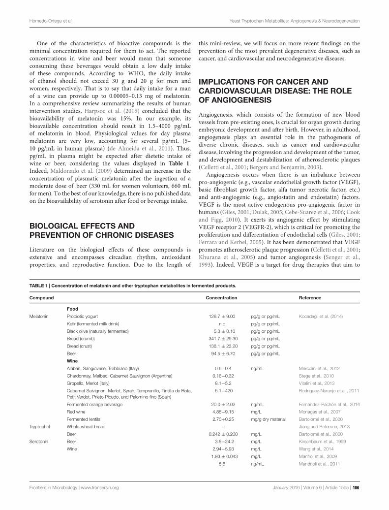
Hornedo-Ortega et al. Yeast Tryptophan Metabolites: Angiogenesis & Neurodegeneration
One of the characteristics of bioactive compounds is theminimal concentration required for them to act. The reportedconcentrations in wine and beer would mean that someoneconsuming these beverages would obtain a low daily intakeof these compounds. According to WHO, the daily intakeof ethanol should not exceed 30 g and 20 g for men andwomen, respectively. That is to say that daily intake for a manof a wine can provide up to 0.00005–0.13 mg of melatonin.In a comprehensive review summarizing the results of humanintervention studies, Harpsøe et al. (2015) concluded that thebioavailability of melatonin was 15%. In our example, itsbioavailable concentration should result in 1.5–4000 pg/mLof melatonin in blood. Physiological values for day plasmamelatonin are very low, accounting for several pg/mL (5–10 pg/mL in human plasma) (de Almeida et al., 2011). Thus,pg/mL in plasma might be expected after dietetic intake ofwine or beer, considering the values displayed in Table 1.Indeed, Maldonado et al. (2009) determined an increase in theconcentration of plasmatic melatonin after the ingestion of amoderate dose of beer (330 mL for women volunteers, 660 mLfor men). To the best of our knowledge, there is no published dataon the bioavailability of serotonin after food or beverage intake.
BIOLOGICAL EFFECTS ANDPREVENTION OF CHRONIC DISEASES
Literature on the biological effects of these compounds isextensive and encompasses circadian rhythm, antioxidantproperties, and reproductive function. Due to the length of
this mini-review, we will focus on more recent findings on theprevention of the most prevalent degenerative diseases, such ascancer, and cardiovascular and neurodegenerative diseases.
IMPLICATIONS FOR CANCER ANDCARDIOVASCULAR DISEASE: THE ROLEOF ANGIOGENESIS
Angiogenesis, which consists of the formation of new bloodvessels from pre-existing ones, is crucial for organ growth duringembryonic development and after birth. However, in adulthood,angiogenesis plays an essential role in the pathogenesis ofdiverse chronic diseases, such as cancer and cardiovasculardisease, involving the progression and development of the tumor,and development and destabilization of atherosclerotic plaques(Celletti et al., 2001; Bergers and Benjamin, 2003).
Angiogenesis occurs when there is an imbalance betweenpro-angiogenic (e.g., vascular endothelial growth factor (VEGF),basic fibroblast growth factor, alfa tumor necrotic factor, etc.)and anti-angiogenic (e.g., angiostatin and endostatin) factors.VEGF is the most active endogenous pro-angiogenic factor inhumans (Giles, 2001; Dulak, 2005; Cebe-Suarez et al., 2006; Cookand Figg, 2010). It exerts its angiogenic effect by stimulatingVEGF receptor 2 (VEGFR-2), which is critical for promoting theproliferation and differentiation of endothelial cells (Giles, 2001;Ferrara and Kerbel, 2005). It has been demonstrated that VEGFpromotes atherosclerotic plaque progression (Celletti et al., 2001;Khurana et al., 2005) and tumor angiogenesis (Senger et al.,1993). Indeed, VEGF is a target for drug therapies that aim to
TABLE 1 | Concentration of melatonin and other tryptophan metabolites in fermented products.
Compound Concentration Reference
Food
Melatonin Probiotic yogurt 126.7 ± 9.00 pg/g or pg/mL Kocadagli et al. (2014)
Kefir (fermented milk drink) n.d pg/g or pg/mL
Black olive (naturally fermented) 5.3 ± 0.10 pg/g or pg/mL
Bread (crumb) 341.7 ± 29.30 pg/g or pg/mL
Bread (crust) 138.1 ± 23.20 pg/g or pg/mL
Beer 94.5 ± 6.70 pg/g or pg/mL
Wine
Alaban, Sangiovese, Trebbiano (Italy) 0.6−0.4 ng/mL Mercolini et al., 2012
Chardonnay, Malbec, Cabernet Sauvignon (Argentina) 0.16−0.32 Stege et al., 2010
Gropello, Merlot (Italy) 8.1−5.2 Vitalini et al., 2013
Cabernet Saivignon, Merlot, Syrah, Tempranillo, Tintilla de Rota,Petit Verdot, Prieto Picudo, and Palomino fino (Spain)
5.1−420 Rodriguez-Naranjo et al., 2011
Fermented orange beverage 20.0 ± 2.02 ng/mL Fernández-Pachón et al., 2014
Red wine 4.88−9.15 mg/L Monagas et al., 2007
Fermented lentils 2.70+0.25 mg/g dry material Bartolomé et al., 2000
Tryptophol Whole-wheat bread − Jiang and Peterson, 2013
Beer 0.242 ± 0.200 mg/L Bartolomé et al., 2000
Serotonin Beer 3.5−24.2 mg/L Kirschbaum et al., 1999
Wine 2.94−5.93 mg/L Wang et al., 2014
1.93 ± 0.043 mg/L Manfroi et al., 2009
5.5 ng/mL Mandrioli et al., 2011
Frontiers in Microbiology | www.frontiersin.org January 2016 | Volume 6 | Article 1565 | 106

Hornedo-Ortega et al. Yeast Tryptophan Metabolites: Angiogenesis & Neurodegeneration
inhibit VEGF signaling (Ferrara and Kerbel, 2005). Anti-VEGFantibodies, aptamers and small molecule VEGFR tyrosine kinaseinhibitors have been developed and given regulatory approval forthe treatment of colon, lung, breast, kidney, and liver cancer, inaddition to neovascular age-related macular degeneration (Giles,2001; Ferrara and Kerbel, 2005). However, serious side effects,such as hypertension, have been reported with prolonged use ofanti-VEGF therapies (Zhu et al., 2007; Wu et al., 2008; Kapperset al., 2010). The use of natural products in reducing VEGF-induced angiogenesis may prove to be more beneficial than thecurrent anti-VEGF drugs available (Moyle et al., 2015).
Melatonin has been associated with a decline in VEGFsecretion levels in the serum of advanced cancer patients(Lissoni et al., 2001), in addition to markedly reducingthe expression of VEGF in HUVEC and culture cancercells at 1 μM and 1 mM (Dai et al., 2008; Cui et al.,2012; Álvarez-García et al., 2013; Gonçalves et al., 2014).Melatonin has also been proven to reduce endothelial cellproliferation, invasion, migration, and tube formation, throughdownregulation of VEGF at 1 mM (Álvarez-García et al.,2013). The possible cell signaling pathway when melatonininhibits HUVEC proliferation has been related to the followingpathway: melatonin receptors/ERK/PI3K/Akt/PKC/NF-kB (Cuiet al., 2008). Additionally, Sohn et al. (2015) have recentlydemonstrated that melatonin (1 mM) upregulates miRNA3195and miRNA374b, whose overexpression synergistically reducedVEGF production in hypoxic PC-3 prostate cancer cells,indicating the important role of miRNA3195 and miRNA374b inmelatonin induced antiangiogenic activity. Melatonin (40mg/kg)has also shown an antitumor effect on mammary tumor growthinmice after 21 days of treatment; the mice displayed significantlysmaller tumor volume and tumor regression (Jardim-Perassiet al., 2014). Additionally, in the same study, a lower expressionof VEGFR2 was observed in the melatonin-treated tumorscompared to the vehicle-treated tumors. More research isconsequently needed to focus on determining the molecularmechanism by which melatonin exerts its angiogenic effect andthe molecular target involved.
NEURODEGENERATIVE DISEASES
Alzheimer’s disease (AD) and Parkinson’s disease (PD) are themost common human neurodegenerative diseases. In both cases,their incidence increases with age. The aggregation of proteinsthat results in different fibrillar structures is responsible forthese disorders. Specifically, they are owing to the abnormalpathological assembly of amyloid- beta (Aβ), tau and α- synuclein(αS).
Indeed, several studies have demonstrated that protofibrilsand oligomers of αS and Aβ are more neurotoxic than fibrils(Pike et al., 1993; Lashuel et al., 2002; Volles and Lansbury, 2003;Outeiro et al., 2008).
This review focus on the evidence of certain bioactiveswhich can present in fermented products. However, to give afair balance, alcohol effects on neurodegeneration have to behighlighted as it is a major component formed by yeast in
alcoholic beverages. It is well-Known that alcohol intake crossesthe BBB (Blood–Brain Barrier) easily producing the excessiverelease of neurotransmitters, oxidative stress and inflammatoryresponse which turns out in neurotoxicity and finally cell death.(Persidsky and Potula, 2014).
ALZHEIMER’S DISEASE
Alzheimer’s disease is a progressive and irreversibleneurodegenerative disorder characterized by loss of memoryand cognition, abstract thinking, and personality alteration.The etiology of AD is unknown in more than 90% of cases.In the pathogenesis of AD there are two principal hallmarks:neurofibrillary tangles (NFTs) and amyloid plaques. NFT areformed by the intracellular accumulation of phosphorylatedtau protein and amyloid plaques, by extracellular accumulationof amyloid β peptides (Hardy and Selkoe, 2002). The amyloidbeta peptide is formed via cleavage of the amyloid precursorprotein (APP). In the non-amyloidogenic pathway (normalstate), APP is cleaved by α-secretase, to generate sAPP (solubleN- terminal fragment), which is neuroprotective as it is involvedin the enhancement of synaptogenesis, neurite outgrowth, andneuronal survival. Conversely, in the disease state, APP is cleavedby β and γ secretase, resulting in insoluble beta amyloid peptide,which has high potential for assembly and formation of toxicaggregates (Gandy, 2005).
Several mechanisms have been proposed to explain βAneurotoxicity, such as oxidative stress and loss of endogenousantioxidants (Behl et al., 1994; Abramov and Duchen, 2005;Hamel et al., 2008); mitochondrial damage, depolarization, andmitochondrial permeability transition pore opening (Moreiraet al., 2001, 2010; Abramov et al., 2004, 2007); destabilizationof intracellular calcium homeostasis in neurons (Bezprozvannyand Mattson, 2008); glial cells (Abramov et al., 2003, 2004), andneuroinflammation (McNaull et al., 2010).
Levels of melatonin and its precursors (serotonin andtryptophan) are significantly decreased in elderly AD individualsand are associated with the emergence of AD (Zhou et al., 2003;Greilberger et al., 2010). A growing body of evidence supportsthe protective role of melatonin in several molecular mechanismsimplicated in the development of AD.
Among these mechanisms, the most significant one is thatmelatonin prevents amyloid aggregation and overproduction.This neurohormone has a great affinity for Aβ peptide,preventing amyloid fibril formation (Masilamoni et al., 2008),as determined by circular dichroism (CD) spectroscopy,electron microscopy, nuclear magnetic resonance (NMR)and electrospray ionization-mass spectrometry (ESI-MS). Inparticular, a hydrophobic interaction has been observed betweenmelatonin and Aβ, specifically on the 29–40 residues of theAβ segment (Skribanek et al., 2001). Additionally, melatoninhas inhibitory effects on the formation of secondary β-sheetstructures through the disruption of the histidine (His+) andaspartate (Asp−) salt bridges in Aβ peptide that promote fibrildissolution (Fraser et al., 1991; Huang et al., 1997; Pappolla et al.,1998).
Frontiers in Microbiology | www.frontiersin.org January 2016 | Volume 6 | Article 1565 | 107

Hornedo-Ortega et al. Yeast Tryptophan Metabolites: Angiogenesis & Neurodegeneration
Melatonin presents a great capacity to regulate the synthesisand maturation of APP at different levels by: decreasing itsmRNA encoding β-APP (Song and Lahiri, 1997; Lahiri, 1999);blocking cAMP production, which is involved in activating theAPP gene promoter, (Husson et al., 2002); and inactivating GSK-3, which promotes α-secretase mediated cleavage of APP, favoringthe non-amyloidogenic pathway (McArthur et al., 1997; Zhuet al., 2001; Hoppe et al., 2010). In vivo studies with transgenicmice over-expressing APP (in 9–10 months they develop senileplaques) and fed with 0.5 mg/mL of melatonin in their drinkingwater (3 mL/day) found a reduction in important markers of thedisease, including Aβ levels in the brain, and that some animalssurvived (Matsubara et al., 2003). The amount given to rodentsare within the pharmacological dose and out of the range of thedose that can be achieved with moderate consumption of wine.Therefore it cannot be concluded that these effect will be observedin humans after wine intake. Further research is required toobtain the evidence at dietary doses.
Furthermore, melatonin exhibits a protective effect on thecholinergic system. In AD patients, a dramatic decrease ofacetylcholine has been observed (Francis et al., 1999), which wasrelated to a decrease in enzyme choline acetyltransferase (ChAT)activity and an increase in acetylcholinesterase (AChE) activity(Bieschke et al., 2005). Indeed, AChE inhibitors increase thesynaptic levels of acetylcholine, which is why they are used as atreatment for mild to moderate AD. In vivo administration ofmelatonin in rats (50 mg/kg body weight) has led to significantlyreduced AChE activity, with maintenance of calcium levelsunder conditions of oxidative stress (Masilamoni et al., 2008).Regarding ChAT, melatonin increased its activity, after 4 monthsof melatonin administration in rats (Feng et al., 2004).
Finally, melatonin reduces Aβ-induced oxidative stress relatedto reactive oxygen species (ROS) and proinflamatory cytokines,such as IL6 and IL1-β in in vivo studies (Masilamoni et al., 2008).As a result of these effects, melatonin protects brain neurons fromdamage and death by increasing viability in hippocampal neuronsand glial cells following treatment with Aβ1–40, Aβ25–40, andAβ1–28. Moreover, melatonin prevents the death of murine N2aneuroblastoma and PC12 cells by using Aβ25–35 (Pappolla et al.,1997; Ionov et al., 2011).
There is scarce literature available in relation to the activityof other tryptophan metabolites, with indole 3-acetic acid andtryptophol being the only bioactive molecules reported so far.Morshedi et al. (2007) proved the inhibitory effect of theseindole derivatives on the amyloid fibrillation of hen egg-white lysozyme, which is another model for exploring theamyloidogenic mechanism.
PARKINSON’S DISEASE
Parkinson’s disease is the second most commonneurodegenerative disorder. Its diagnosis is based on motorabnormalities, such as resting tremor, bradykinesia, and rigidity(Duvoisin, 1992). Indeed, patients present other non-motorsymptoms, such as depression, anxiety, and sleep disorders(Jenner et al., 2013). Only 10% of patients have a genetic basis,
with 90% being considered sporadic cases. PD is characterizedby the degeneration of the subcortical structure of the brain.Specifically, there are significant losses of dopaminergic neuronsin the substantia nigra pars compacta (SNpc; Forno, 1996),although other cell populations are also susceptible to theneurodegeneration process.
α-Synuclein (αS) is a 140 amino acid and a highly abundantneuronal protein. It is found as a soluble cytoplasmatic proteinassociated with synaptic vesicles (Iwai et al., 1995). It is thoughtthat it plays a role in neurotransmission and cognitive function.Although its physiological function is uncertain, the pathology isassociated with the accumulation of αS aggregates, which are themain component of Lewy bodies (LBs; Spillantini et al., 1997).LBs are spherical inclusions formed by αS aggregate (99%) andother proteins.
Despite the main risk factor being aging, other possible riskfactors include mutation in the SNCA (alpha-synuclein gene)and exposure to environmental toxins. The latter are also linkedto metabolic abnormalities involving neurotransmitter systems(dopamine, serotonin, GABA, and glutamate), fatty acids, suchas arachidonic acid-cascade, oxidative stress and mitochondrialfunction (Henchcliffe and Beal, 2008; Quinones and Kaddurah-Daouk, 2009; Kaidanovich-Beilin et al., 2012; Lei and Powers,2013).
Furthermore, several studies suggest that αS oligomers andprotofibrils are an important factor in neurotoxicity in PD.αS protofibrils cause membrane permeabilization, which alterscellular homeostasis and may activate an apoptotic process(Volles and Lansbury, 2003). Indeed, there is evidence to supportthe capacity of αS to inhibit proteosomal activity, which wouldprevent elimination of misfolded proteins (Giasson and Lee,2003).
Substantial evidence also suggests that a significant factor indopaminergic neuronal loss in the PD brain are ROS, which resultfrom dopamine metabolism, low glutathione concentration andhigh levels of iron and calcium in the SNpc (Jenner and Olanow,2006). Additionally, the brain contains high concentrations ofpolyunsaturated fatty acids, which, under oxidative stress, resultin lipid peroxidation and generation of toxic products (Liu et al.,2008).
No treatment is currently available for the prevention or cureof PD. However, a combination of L-DOPA and antioxidantshas been recommended to reduce the rate of progression of thedisease, due to the decrease in dopamine levels and significantincrease of oxidative stress commonly concomitant to this typeof disorder (Zhu et al., 2004).
Concerning the role of melatonin in PD, several workshave reported different mechanisms of action. Lin et al.(2007) demonstrated that melatonin attenuates arsenite-inducedapoptosis by reducing aggregated αS levels in rat brains, by meansof Western blot analysis. Additionally, Ishido (2007) showed thatmelatonin inhibits αS assembly, using immunostaining in ratpheochromocytoma cells.
It is also important to highlight that melatonin dose-dependently inhibits all steps of the αS assembly process.Ono et al. (2012) observed a reduction in the number offibrils and the corresponding increase of the number of
Frontiers in Microbiology | www.frontiersin.org January 2016 | Volume 6 | Article 1565 | 108

Hornedo-Ortega et al. Yeast Tryptophan Metabolites: Angiogenesis & Neurodegeneration
short fibrils and amorphous aggregates (25–250 μM) usingelectron microscopy and thioflavin S experiments. Indeed,melatonin presents a significant destabilization effect (also dose-dependently), suggesting a decrease in beta-sheet levels. In thesame study, the authors performed experiments with primarycultures of mesencephalon and neostriatum with MTT: (3-(4,5-Dimethylthiazol-2-yl)-2,5-Diphenyltetrazolium Bromide) acolorimetric assay for assessing cell metabolic activity. The resultsshowed that melatonin reduced the toxic effects of αS afterpretreatment (2–6 days) with an increase in cell viability ofbetween 56 and 97%.
In addition to this, it is well known that melatonin exhibitsantioxidant properties (Reiter et al., 1997; Kotler et al., 1998).Cellular injury cause by αS-mediated perturbation of cellularredox reactions is an important mechanism proposed for PD(George et al., 2009). Melatonin has been suggested as apotential therapeutic agent in diseases where oxidative stressis thought to be a major pathogenic factor. Mayo et al.(1998) observed that this hormone was an effective freeradical scavenger and that it prevented apoptosis in neuronalcells. Moreover, in vitro studies on MPTD-induced (1-methyl-4-phenyl-1,2,3,6-tetrahydropyridine) PD in mice have shownthat melatonin protects against excitotoxicity by reducing theautoxidation of dopamine. The administration of melatonin leads
to normalization of complex I activity and oxidative status inmitochondria (Escames et al., 2010).
In conclusion, and based on the preceding evidence,we shouldconsider that melatonin presents strong inhibitory effects onprotofibril formation and peptide oligomerization.
AUTHOR CONTRIBUTIONS
RH-O and AC Literature search and first draft. AT, MG-P, andAM thorough revision and discussion and final document.
FUNDING
This work was supported by the Ministry of Economy andCompetitiveness, Spain (grant no. AGL2013-47300-C1 andAGL2013-47300-C3).
ACKNOWLEDGMENTS
We acknowledge the Universidad de Sevilla for the contracts ofAC and RH-O.
REFERENCES
Abramov, A. Y., Canevar, L., and Duchen, M. R. (2003). Changes in intracellularcalcium and glutathione in astrocytes as the primary mechanism of amyloidneurotoxicity. J. Neurosci. 23, 5088–5095.
Abramov, A. Y., Canevari, L., and Duchen, M. R. (2004). Beta-amyloid peptidesinduce mitochondrial dysfunction and oxidative stress in astrocytes and deathof neurons through activation of NADPH oxidase. J. Neurosci. 24, 565–575. doi:10.1523/JNEUROSCI.4042-03.2004
Abramov, A. Y., and Duchen, M. R. (2005). The role of an astrocytic NADPHoxidase in the nurotoxicity of amyloid beta peptides. Philos. Trans. R. Soc. Lond.B Biol. Sci. 360, 2309–2314. doi: 10.1098/rstb.2005.1766
Abramov, A. Y., Fraley, C., Diao, C. T., Winkfein, R., Colicos, M. A., Duchen,M. R., et al. (2007). Targeted polyphosphatase expression alters mitochondrialmetabolism and inhibits calcium-dependent cell death. Proc. Natl. Acad. Sci.U.S.A. 104, 18091–18096. doi: 10.1073/pnas.0708959104
Álvarez-García, V., González, A., Alonso-González, C., Martínez-Campa, C., andCos, S. (2013). Regulation of vascular endothelial growth factor by melatonin inhuman breast cancer cells. J. Pineal Res. 54, 373–380. doi: 10.1111/jpi.12007
Bartolomé, B., Pena-Neira, A., and Gomez-Cordoves, C. (2000). Phenolics andrelated substances in alcohol-free beers. Eur. Food Res. Technol. 210, 419–423.doi: 10.1007/s002170050574
Behl, C., Davis, J. B., Lesley, R., and Schubert, D. (1994). Hydrogen peroxidemediates amyloid beta protein toxicity. Cell 77, 817–827. doi: 10.1016/0092-8674(94)90131-7
Bergers, G., and Benjamin, L. E. (2003). Tumorigenesis and the angiogenic switch.Nat. Rev. Cancer. 3, 401–410. doi: 10.1038/nrc1093
Bezprozvanny, I., and Mattson, M. P. (2008). Neuronal calcium mishandling andthe pathogenesis of Alzheimer’s disease. Trends Neurosci. 31, 454–463. doi:10.1016/j.tins.2008.06.005
Bieschke, J., Zhang, Q., Powers, E. T., Lerner, R. A., and Kelly, J. W. (2005).Oxidative metabolites accelerate Alzheimer’s amyloidogenesis by a two-stepmechanism, eliminating the requirement for nucleation. Biochemistry 44, 4977–4983. doi: 10.1021/bi0501030
Cebe-Suarez, S., Zehnder-Fjallman, A., and Ballmer-Hofer, K. (2006). The role ofVEGF receptors in angiogenesis; complex partnerships. Cell. Mol. Life Sci. 63,601–615. doi: 10.1007/s00018-005-5426-3
Celletti, F. L., Waugh, J. M., Amabile, P. G., Brendolan, A., Hilfiker, P. R., andDake,M.D. (2001). Vascular endothelial growth factor enhances atheroscleroticplaque progression.Nat. Med. 7, 425–429. doi: 10.1038/86490
Cook, K. M., and Figg, W. D. (2010). Angiogenesis inhibitors: current strategiesand future prospects. CA-Cancer J. Clin. 60, 222–243. doi: 10.3322/caac.20075
Cui, P., Yu, M., Luo, Z., Dai, M., Han, J., Xiu, R., et al. (2008). Intracellularsignaling pathways involved in cell growth inhibition of human umbilical veinendothelial cells by melatonin. J. Pineal Res. 44, 107–114. doi: 10.1111/j.1600-079X.2007.00496.x
Cui, P., Yu, M., Peng, X., Dong, L., and Yang, Z. (2012). Melatonin prevents humanpancreatic carcinoma cell PANC-1-induced human umbilical vein endothelialcell proliferation and migration by inhibiting vascular endothelial growthfactor expression. J. Pineal Res. 52, 236–243. doi: 10.1111/j.1600-079X.2011.00933.x
Dai, M., Cui, P., Yu, M., Han, J., Li, H., and Xiu, R. (2008). Melatonin modulatesthe expression of VEGF and HIF-1α induced by CoCl 2 in cultured cancer cells.J. Pineal Res. 44, 121–126. doi: 10.1111/j.1600-079X.2007.00498.x
de Almeida, E. A., Di Mascio, P., Harumi, T., Spence, D. W., Moscovitch, A.,Hardeland, R., et al. (2011). Measurement of melatonin in body fluids:standards, protocols and procedures. Childs Nerv. Syst. 27, 879–891. doi:10.1007/s00381-010-1278-8
Dulak, J. (2005). Nutraceuticals as anti-angiogenic agents: hopes and reality.J. Physiol. Pharmacol. 1, 51–67.
Duvoisin, R. C. (1992). Overview of Parkinsons disease Ann. N. Y. Acad. Sci. 648,187–193. doi: 10.1111/j.1749-6632.1992.tb24537.x
Escames, G., López, A., García, J. A., García, L., Acuña-Castroviejo, D., García, J. J.,et al. (2010). The role of mitocondria in brain aging and the effects of melatonin.Curr. Neuropharmacol. 8, 182–193. doi: 10.2174/157015910792246245
Feng, Z., Chang, Y., Cheng, Y., Zhang, B. L., Qu, Z. W., Qin, C., et al.(2004). Melatonin alleviates behavioral deficits associated with apoptosis andcholinergic system dysfunction in the APP 695 transgenic mouse modelof Alzheimers disease. J. Pineal Res. 37, 129–136. doi: 10.1111/j.1600-079X.2004.00144.x
Fernández-Pachón, M. S., Medina, S., Herrero-Martín, G., Cerrillo, I., Berná, G.,Escudero-López, B., et al. (2014). Alcoholic fermentation induces melatoninsynthesis in orange juice. J. Pineal Res. 56, 31–38. doi: 10.1111/jpi.12093
Frontiers in Microbiology | www.frontiersin.org January 2016 | Volume 6 | Article 1565 | 109

Hornedo-Ortega et al. Yeast Tryptophan Metabolites: Angiogenesis & Neurodegeneration
Ferrara, N., and Kerbel, R. S. (2005). Angiogenesis as a therapeutic target. Nature438, 967–974. doi: 10.1038/nature04483
Forno, L. S. (1996). Neuropathology of Parkinsons disease. J. Neurophatol. Exp.Neurol. 55, 259–272. doi: 10.1097/00005072-199603000-00001
Francis, P. T., Palmer, A. M., Snape, M., and Wilcock, G. K. (1999). The cholinergichypothesis of Alzheimers disease: a review of progress. J. Neurol. Neurosurg.Psychiatry 66, 137–147. doi: 10.1136/jnnp.66.2.137
Fraser, P. E., Nguyen, J. T., Surewicz, W. K., and Kirschner, D. A. (1991). pH-dependent structural transitions of Alzheimer’s amyloid peptides.Biophys. J. 60,1190–1201.
Gandy, S. (2005). The role of cerebral amyloid β accumulation in common formsof Alzheimer disesase. J. Clin. Invest. 115, 1121–1129. doi: 10.1172/JCI200525100
George, J. L., Mok, S., Moses, D., Wilkins, S., Bush, A. I., Cherny, R. A., et al.(2009). Targeting the progression of Parkinson’s disease.Curr. Neuropharmacol.7, 9–36. doi: 10.2174/157015909787602814
Giasson, B., and Lee, V. M. (2003). Are ubiquitination pathways central toParkinson’s disease? Cell 114, 1–8. doi: 10.1016/S0092-8674(03)00509-9
Giles, F. J. (2001). The vascular endothelial growth factor (VEGF) signalingpathway: a therapeutic target in patients with hematologic malignancies.Oncologist 6, 32–39. doi: 10.1634/theoncologist.6-suppl_5-32
Gonçalves, N. N., Rodrigues, R. V., Jardim-Perassi, B. V., Moschetta, M. G.,Lopes, J. R., Colombo, J., et al. (2014). Molecular markers of angiogenesisand metastasis in lines of oral carcinoma after treatment with melatonin.Anti Cancer Agents Med. Chem. 14, 1302–1311. doi: 10.2174/1871520614666140812110246
Greilberger, J., Fuchs, D., Leblhuber, F., Greilberger, M., Wintersteiger, R., andTafeit, E. (2010). Carbonyl proteins as a clinical marker in Alzheimers diseaseand its relation to tryptophan degradation and immune activation. Clin. Lab.56, 441–448.
Hamel, E., Nicolakakis, N., Aboulkassim, T., Ongali, B., and Tong, X. K.(2008). Oxidative stress and cerebrovascular dysfunction in mouse modelsof Alzheimers disease. Exp. Physiol. 93, 116–120. doi: 10.1113/expphysiol.2007.038729
Hardy, J., and Selkoe, D. J. (2002). The amyloid hypothesis of Alzheimersdisease:progress and problems on the road to therapeutics. Science 297, 353–356. doi:10.1126/science.1072994
Harpsøe, N. G., Andersen, L. P., Gögenurm, I., and Rosenberg, J. (2015). Clinicalpharmacokinetics of melatonin: a systematic review. Eur. J. Clin. Pharmacol. 71,901–909. doi: 10.1007/s00228-015-1873-4
Henchcliffe, C., and Beal, M. F. (2008). Mitochondrial biology and oxidative stressin Parkinson disease pathogenesis. Nat. Clin. Pract. Neurol. 4, 600–609. doi:10.1038/ncpneuro0924
Hoppe, J. B., Frozza, R. L., Horn, A. P., Comiran, R. A., Bernardi, A.,Campos, M. M., et al. (2010). Amyloid-beta neurotoxicity in organotypicculture is attenuated by melatonin: involvement of GSK-3beta, tau andneuroinflammation. J. Pineal Res. 48, 230–238. doi: 10.1111/j.1600-079X.2010.00747.x
Huang, T. H., Fraser, P. E., and Chakrabartty, A. (1997). Fibrillogenesis ofAlzheimer’s Abeta peptides studied by fluorescence energy transfer. J. Mol. Biol.269, 214–224. doi: 10.1006/jmbi.1997.1050
Husson, I., Mesples, B., Bac, P., Vamecq, J., Evrard, P., and Gressens, P.(2002). Melatoninergic neuroprotection of the murine periventricular whitematter against neonatal excitotoxic challenge. Ann. Neurol. 51, 82–92. doi:10.1002/ana.10072
Ionov, M., Burchell, V., Klajnert, B., Bryszewska, M., and Abramov, A. Y.(2011). Mechanism of neuroprotection of melatonin against beta-amyloid neurotoxicity. Neuroscience 180, 229–237. doi: 10.1016/j.neuroscience.2011.02.045
Ishido, M. (2007). Melatonin inhibits maneb-induced aggregation of alpha-synuclein in rat pheochromocytoma cells. J. Pineal Res. 42, 125–130. doi:10.1111/j.1600-079X.2006.00390.x
Iwai, A., Masliah, E., Yoshimoto, M., Ge, N., Flanagan, L., de Silva, H. A. R., et al.(1995). The precursor protein of non-Aβ component of Alzheimer’s diseaseamyloid is a presynaptic protein of the central nervous system. Neuron 14,467–475. doi: 10.1016/0896-6273(95)90302-X
Jardim-Perassi, B. V., Arbab, A. S., Ferreira, L. C., Borin, T. F., Varma, N. R. S.,Iskander, A. S. M., et al. (2014). Effect of melatonin on tumor growth and
angiogenesis in xenograft model of breast Cancer. PLoS ONE 9:e85311. doi:10.1371/journal.pone.0085311
Jenner, P., Morris, H. R., Robbins, T. W., Goedert, M., Hardy, J., Ben-Shlomo, Y.,et al. (2013). Parkinson’s disease–the debate on the clinical phenomenology,aetiology, pathology and pathogenesis. J. Parkinson’s Dis. 3, 1–11. doi:10.3233/JPD-130175
Jenner, P., and Olanow, W. (2006). The pathogenesis of cell death in Parkinson’sdisease. Neurology 66, S24–S36. doi: 10.1212/WNL.66.10_suppl_4.S24
Jiang, D., and Peterson, D. G. (2013). Identification of bitter compounds inwhole wheat bread. Food Chem. 141, 1345–1353. doi: 10.1016/j.foodchem.2013.03.021
Kaidanovich-Beilin, O., Cha, D. S., and McIntyre, R. S. (2012). Crosstalkbetween metabolic and neuropsychiatric disorders. F1000 Biol. Repl. 4:14. doi:10.3410/B4-14
Kappers, M. H. W., Smedts, F. M. M., Horn, T., van Esch, J. H. M., Sleijfer, S.,Leijten, F., et al. (2010). Hypertension induced by the tyrosine kinaseinhibitor sunitinib is associated with increased circulating endothelin-1 levels.Hypertension 56, 675–681. doi: 10.1161/HYPERTENSIONAHA.109.149690
Khurana, R., Simons, M., Martin, J. F., and Zachary, I. C. (2005). Role ofangiogenesis in cardiovascular disease - A critical appraisal. Circulation 112,1813–1824. doi: 10.1161/CIRCULATIONAHA.105.535294
Kirschbaum, J., Meier, A., and Brtickner, H. (1999). Determination of biogenicamines in fermented beverages and vinegars by pre-column derivatizationwith para-Nitrobenzyloxycarbonyl Chloride (PNZ-CI) and Reversed-Phase LC.Chromatographia 49, 117–124. doi: 10.1007/BF02575272
Kocadagli, T., Yilmaz, C., and Gökmen, V. (2014). Determination of melatonin andits isomer in foods by liquid chromatography tandem mass spectrometry. FoodChem. 153, 151–156. doi: 10.1016/j.foodchem.2013.12.036
Kotler, M., Rodríguez, C., Sáinz, R. M., Antolín, I., and Menéndez-Peláez, A.(1998). Melatonin increases gene expression for antioxidant enzymes in ratbrain cortex. J. Pineal Res. 24, 83–89. doi: 10.1111/j.1600-079X.1998.tb00371.x
Lahiri, D. K. (1999). Melatonin affects the metabolism of the beta-amyloidprecursor protein in different cell types. J. Pineal. Res. 26, 137–146. doi:10.1111/j.1600-079X.1999.tb00575.x
Lashuel, H. A., Petre, B. M., Wall, J., Simon, M., Nowak, R. J., Walz, T., et al.(2002). α-synuclein, especially the Parkinson’s disease- associated mutants,forms pore-like annular and tubular protofibrils. J. Mol. Biol. 322, 1089–1102.doi: 10.1016/S0022-2836(02)00735
Lei, S., and Powers, R. (2013). NMR metabolomics analysis of Parkinson’s Disease.Curr. Metabolomics. 1, 191–209. doi: 10.2174/2213235X113019990004
Lin, A. M., Fang, S. F., Chao, P. L., and Yang, C. H. (2007). Melatonin attenuatesarsenite-induced apoptosis in rat brain: involvement of mitochondrial andendoplasmic reticulum pathways and aggregation of alpha-synuclein. J. PinealRes. 43, 163–171. doi: 10.1111/j.1600-079X.2007.00456.x
Lissoni, P., Rovelli, F., Malugani, F., Bucovec, R., Conti, A., and Maestroni, G. J. M.(2001). Anti-angiogenic activity of melatonin in advanced cancer patients.Neurol. Endocrinol. Lett. 22, 45–47.
Liu, X., Yamada, N., Maruyama, W., and Osawa, T. (2008). Formationof dopamine adducts derived from brain polyunsaturated fatty acids:mechanism for Parkinson disease. J. Biol. Chem. 283, 34887–34895. doi:10.1074/jbc.M805682200
Maldonado, M. D., Moreno, H., and Calvo, J. R. (2009). Melatonin present in beercontributes to increase the levels of melatonin and antioxidant capacity of thehuman serum. Clin. Nutr. 28, 188–191. doi: 10.1016/j.clnu.2009.02.001
Mandrioli, R., Morganti, E., Mercolini, L., Kenndler, E., and Raggi, M. A.(2011). Fast analysis of amino acids in wine by capillary electrophoresiswith laser-induced fluorescence detection. Electrophoresis 32, 2809–2815. doi:10.1002/elps.201100112
Manfroi, L., Silva, P. H. A., Rizzonc, L. A., Sabainid, P. S., and Glória,M. B. A. (2009). Influence of alcoholic and malolactic starter cultureson bioactive amines in Merlot wines. Food Chem. 116, 208–213. doi:10.1016/j.foodchem.2009.02.034
Mas, A., Guillamon, J. M., Torija, M. J., Beltran, G., Cerezo, A. B. A. M., Troncoso,A. M., et al. (2014). Bioactive compounds derived from the yeast metabolismof aromatic amino acids during alcoholic fermentation. Biomed. Res. Int. 7. doi:10.1155/2014/898045
Masilamoni, J. G., Jesudason, E. P., Dhandayuthapani, S., Ashok, B. S., Vignesh, S.,Jebaraj, W. C., et al. (2008). The neuroprotective role of melatonin against
Frontiers in Microbiology | www.frontiersin.org January 2016 | Volume 6 | Article 1565 | 110
Hornedo-Ortega et al. Yeast Tryptophan Metabolites: Angiogenesis & Neurodegeneration
amyloid beta peptide injected mice. Free Radic. Res. 42, 661–673. doi:10.1080/10715760802277388
Matsubara, E., Bryant-Thomas, T., Pacheco Quinto, J., Henry, T. L., Poeggeler, B.,Herbert, D., et al. (2003). Melatonin increases survival and inhibits oxidativeand amyloid pathology in a transgenic model of Alzheimer’s disease.J. Neurochem. 85, 1101–1108. doi: 10.1046/j.1471-4159.2003.01654.x
Mayo, J. C., Sainz, R. M., Uria, H., Antolin, I., Esteban, M. M., andRodriguez, C. (1998). Melatonin induced by 6-hydroxydopamine in neuronalcells: implications for Parkinson’s disease. J. Pineal Res. 24, 179–192. doi:10.1111/j.1600-079X.1998.tb00531.x
McArthur, A. J., Hunt, A. E., and Gillette, M. U. (1997). Melatonin actionand signal transduction in the rat suprachiasmatic circadian clock: activationof protein kinase C at dusk and dawn. Endocrinology 138, 627–634. doi:10.1210/endo.138.2.4925
McNaull, B. B., Todd, S., McGuinness, B., and Passmore, A. P. (2010).Inflammation and anti-inflammatory strategies for Alzheimer’s disease—amini-review. Gerontology 56, 3–14. doi: 10.1159/000237873
Mercolini, L., Mandrioli, R., and Raggi, M. A. (2012). Content of melatoninand other antioxidants in grape-related foodstuffs: measurement usinga MEPS-HPLC-F method. J. Pineal Res. 53, 21–28. doi: 10.1111/j.1600-079X.2011.00967.x
Monagas, M., Gomez-Cordoves, C., and Bartolome, B. (2007). Evaluation ofdifferent Saccharomyces cerevisiae strains for red winemaking. Influenceon the anthocyanin, pyranoanthocyanin and non-anthocyanin phenoliccontent and colour characteristics of wines. Food Chem. 104, 814–823. doi:10.1016/j.foodchem.2006.12.043
Moreira, P. I., Carvalho, C., Zhu, X., Smith, M. A., and Perry, G. (2010).Mitochondrial dysfunction is a trigger of Alzheimer’s disease pathophysiology.Biochim. Biophys. Acta 1802, 2–10. doi: 10.1016/j.bbadis.2009.10.006
Moreira, P. I., Santos, M., Moreno, A. J. M., and Oliveira, C. (2001). Amyloid beta-peptide promotes permeability transition pore in brain mitochondria. Biosci.Rep. 21, 789–800. doi: 10.1023/A:1015536808304
Morshedi, D., Rezaei-Ghaleh, N., Ebrahim-Habibi, A., Ahmadian, S., and Nemat-Gorgani, M. (2007). Inhibition of amyloid fibrillation of lysozyme by indolederivatives - possible mechanism of action. FEBS J. 274, 6415–6425. doi:10.1111/j.1742-4658.2007.06158.x
Moyle, C. W. A., Cerezo, A. B., Winterbone, M. S., Hollands, W. J., Alexeev, Y.,Needs, P. W., et al. (2015). Potent inhibition of VEGFR-2 activation bytight binding of green tea epigallocatechin gallate and apple procyanidinsto VEGF: relevance to angiogenesis. Mol. Nutr. Food Res. 59, 401–412. doi:10.1002/mnfr.201400478
Ono, K., Mochizuki, H., Ikeda, T., Nihira, T., Takasaki, J., Teplow, D. B., et al.(2012). Effect of melatonin on α-synuclein self-assembly and cytotoxicity.Neurobiol. Aging 33, 2172–2185. doi: 10.1016/j.neurobiolaging.2011.10.015
Outeiro, T. F., Putcha, P., Tetzlaff, J. E., Spoelgen, R., Koker, M., Carvalho, F., et al.(2008). Formation of toxic oligomeric α-synuclein species in living cells. PLoSONE 3:e1867. doi: 10.1371/journal.pone.0001867
Pappolla, M., Bozner, P., Soto, C., Shao, H., Robakis, N. K., Zagorski, M., et al.(1998). Inhibition of Alzheimer beta-fibrillogenesis by melatonin. J. Biol. Chem.273, 7185–7188. doi: 10.1074/jbc.273.13.7185
Pappolla, M. A., Sos, M., Omar, R. A., Bick, R. J., Hickson-Bick, D. L., Reiter, R. J.,et al. (1997). Melatonin prevents death of neuroblastoma cells exposed to theAlzheimer amyloid peptide. J. Neurosci. 17, 1683–1690.
Persidsky, Y. G., and Potula, R. (2014). “Alcohol and neurodegeneration,” inNeuroinflammation and Neurodegeneration, ed. P.K. Peterson and M. Toborek(New York, NY: Springer), 511–526.
Pike, C. J., Burdick, D., Walencewicz, A. J., Glabe, C. G., and Cotman, C. W.(1993). Neurodegeneration induced by beta-amyloid peptides in vitro: the roleof peptide assembly state. J. Neurosci. 13, 1676–1687.
Quinones, M. P., and Kaddurah-Daouk, R. (2009). Metabolomics tools foridentifying biomarkers for neuropsychiatric diseases. Neurobiol. Dis. 35, 165–176. doi: 10.1016/j.nbd.2009.02.019
Reiter, R., Tang, L., Garcia, J. J., and Muñoz-Hoyos, A. (1997). Pharmacologicalactions of melatonin in oxygen radical pathophysiology. Life Sci. 60, 2255–2271.doi: 10.1016/S0024-3205(97)00030-1
Rodriguez-Naranjo, M. I., Gil-Izquierdo, A., Troncoso, A. M., Cantos-Villar, E.,and Garcia-Parrilla, M. C. (2011). Melatonin is synthesised by yeastduring alcoholic fermentation in wines. Food Chem. 126, 1608–1613. doi:10.1016/j.foodchem.2010.12.038
Rodriguez-Naranjo, M. I., Torija, M. J., Mas, A., Cantos-Villar, E., and Garcia-Parrilla, M. C. (2012). Production of melatonin by Saccharomyces strainsunder growth and fermentation conditions. J. Pineal Res. 53, 219–224. doi:10.1111/j.1600-079X.2012.00990.x
Senger, D. R., Van DeWater, L., Brown, L. F., Nagy, J. A., Yeo, K. T., Yeo, T. K., et al.(1993). Vascular permeability factor (VPF, VEGF) in tumor biology. CancerMetast. Rev. 12, 303–324. doi: 10.1007/BF00665960
Skribanek, Z., Balaspiri, L., and Mak, M. (2001). Interaction between syntheticamyloid-beta-peptide (1-40) and its aggregation inhibitors studied byelectrospray ionization mass spectrometry. J. Mass Spectrom. 36, 1226–1229.doi: 10.1002/jms.243
Sohn, E. J., Won, G., Lee, L., Lee, S., and Kim, S. H. (2015). Upregulationof miRNA3195 and miRNA374b mediates the anti-angiogenic properties ofmelatonin in hypoxic PC-3 prostate cancer cells. J. Cancer. 6, 19–28. doi:10.7150/jca.9591
Song, W., and Lahiri, D. K. (1997). Melatonin alters the metabolism of thebeta-amyloid precursor protein in the neuroendocrine cell line PC12. J. Mol.Neurosci. 9, 75–92. doi: 10.1007/BF02736852
Spillantini, M. G., Schmidt, M. L., Lee, V. M.-Y., Trojanowski, J. Q., Ross, J., andMichel, G. M. (1997). α-Synuclein in Lewy bodies. Nature 388, 839–840. doi:10.1038/42166
Sprague, G. F. Jr., andWinans, S. C. (2006). Eukaryotes learn how to count: quorumsensing by yeast. Genes Dev. 20, 1045–1049. doi: 10.1101/gad.1432906
Stege, P. W., Sombra, L. L., Messina, G., Martinez, L. D., and Silva,M. F. (2010). Determination of melatonin in wine and plant extracts bycapillary electrochromatography with immobilized carboxylic multi-walledcarbon nanotubes as stationary phase. Electrophoresis 31, 2242–2248. doi:10.1002/elps.200900782
Vitalini, S., Gardana, C., Simonetti, P., Fico, G., and Iriti, M. (2013). Melatonin,melatonin isomers and stilbenes in Italian traditional grape products and theirantiradical capacity. J. Pineal Res. 54, 322–333. doi: 10.1111/jpi.12028
Volles, M. J., and Lansbury, P. T. Jr. (2003). Zeroing in on the pathogenic formof alpha-synuclein and its mechanism of neurotoxicity in Parkinson’s Disease.Biochemistry 42, 7871–7878. doi: 10.1021/bi030086j
Wang, Y. Q., Ye, D. O., Zhu, B. Q., Wu, G. F., and Duan, C. Q. (2014).Rapid HPLC analysis of amino acids and biogenic amines in wines duringfermentation and evaluation of matrix effect. Food Chem. 163, 6–15. doi:10.1016/j.foodchem.2014.04.064
Wu, S., Chen, J. J., Kudelka, A., Lu, J., and Zhu, X. (2008). Incidence and riskof hypertension with sorafenib in patients with cancer: a systematic reviewand meta-analysis. Lancet Oncol. 9, 117–123. doi: 10.1016/S1470-2045(08)70003-2
Zhou, J. N., Liu, R. Y., Kamphorst, W., Hofman, M. A., and Swaab, D. F.(2003). Early neuropathological Alzheimers changes in aged individuals areaccompanied by decreased cerebrospinal fluid melatonin levels. J. Pineal Res.35, 125–130. doi: 10.1034/j.1600-079X.2003.00065.x
Zhu, G., Wang, D., Lin, Y. H., McMahon, T., Koo, E. H., and Messing, R. O.(2001). Protein kinase C epsilon suppresses Abeta production and promotesactivation of alphasecretase. Biochem. Biophys. Res. Commun. 285, 997–1006.doi: 10.1006/bbrc.2001.5273
Zhu, M., Rajamani, S., Kaylor, J., Han, S., Zhou, F., and Fink, A. L. (2004).The flavonoid baicalein inhibits fibrillation of alpha-synucleinand disaggregatesexisting fibrils. J Biol. Chem. 279, 26846–26857. doi: 10.1074/jbc.M403129200
Zhu, X., Wu, S., Dahut, W. L., and Parikh, C. R. (2007). Risks of Proteinuriaand hypertension with Bevacizumab, an antibody against vascular endothelialgrowth factor: systematic Review and meta-analysis. Am. J. Kidney Dis. 49,186–193. doi: 10.1053/j.ajkd.2006.11.039
Conflict of Interest Statement: The authors declare that the research wasconducted in the absence of any commercial or financial relationships that couldbe construed as a potential conflict of interest.
Copyright © 2016 Hornedo-Ortega, Cerezo, Troncoso, Garcia-Parrilla and Mas.This is an open-access article distributed under the terms of the Creative CommonsAttribution License (CC BY). The use, distribution or reproduction in other forumsis permitted, provided the original author(s) or licensor are credited and that theoriginal publication in this journal is cited, in accordance with accepted academicpractice. No use, distribution or reproduction is permitted which does not complywith these terms.
Frontiers in Microbiology | www.frontiersin.org January 2016 | Volume 6 | Article 1565 | 111
ORIGINAL RESEARCH ARTICLEpublished: 26 February 2015
doi: 10.3389/fmicb.2015.00137
The broad-spectrum antibiotic, zeamine, kills the nematodeworm Caenorhabditis elegansJosephine E. E. U. Hellberg1, Miguel A. Matilla1,2* and George P. C. Salmond 1*
1 Department of Biochemistry, University of Cambridge, Cambridge, UK2 Department of Environmental Protection, Estación Experimental del Zaidín – Consejo Superior de Investigaciones Científicas, Granada, Spain
Edited by:
Enrica Pessione, University of Torino,Italy
Reviewed by:
Linda Thomashow, United StatesDepartment of Agriculture –Agricultural Research Service, USAPaul Alan Hoskisson, University ofStrathclyde, UK
*Correspondence:
George P. C. Salmond, Department ofBiochemistry, University ofCambridge, Tennis Court Road,Cambridge CB2 1QW, UKe-mail: [email protected];Miguel A. Matilla, Department ofEnvironmental Protection, EstaciónExperimental del Zaidín – ConsejoSuperior de InvestigacionesCientíficas, Profesor Albareda 1,Granada 18008, Spaine-mail: [email protected]
Soil bacteria can be prolific producers of secondary metabolites and other biologicallyactive compounds of economic and clinical importance. These natural products are oftensynthesized by large multi-enzyme complexes such as polyketide synthases (PKSs) ornon-ribosomal peptide synthases (NRPSs).The plant-associated Gram-negative bacterium,Serratia plymuthica A153, produces several secondary metabolites and is capable of killingthe nematode worm Caenorhabditis elegans; a commonly used model for the study ofbacterial virulence. In this study, we show that disruption of the hybrid PKS/NRPS zeamine(zmn) gene cluster results in the attenuation of “fast-killing” of C. elegans, indicating thatzeamine has nematicidal activity. C. elegans also exhibits age-dependent susceptibility tozeamine, with younger worms being most sensitive to the bioactive molecule. The zmngene cluster is widely distributed within Serratia and phytopathogenic Dickeya species andinvestigation of strains harboring the zmn gene cluster showed that several of them arehighly virulent in C. elegans. Zeamine was described previously as a phytotoxin and broad-spectrum antibacterial compound. In addition to its nematicidal properties, we show herethat zeamine can also kill Saccharomyces cerevisiae and Schizosaccharomyces pombe.Theexpression of the zmn gene cluster and regulation of zeamine production were alsoinvestigated.Transcription of the cluster was growth phase-dependent, and was modulatedby the post-transcriptional RNA chaperone, Hfq.The results of this study show that zeamineis a highly toxic molecule with little, or no, apparent host specificity in very diverse biologicalsystems. In its current form, zeamine(s) may be useful as a lead compound suitable forchemical modification and structure-activity assays. However, because of widespread non-selective toxicity in multiple bioassays, unmodified zeamine(s) is unlikely to be suitable asa therapeutic antibiotic.
Keywords: Serratia plymuthica, Dickeya, secondary metabolite, PKS, NRPS, antibiotic, Caenorhabditis elegans,
zeamine
INTRODUCTIONBacteria belonging to the Gram-negative genus Serratia are mem-bers of the γ-proteobacterial family Enterobacteriaceae. The bestcharacterized member of the genus, Serratia marcescens, was firstdescribed in 1819 as a pigmented microbial isolate (Grimont andGrimont, 1978). Serratia species are ecologically diverse althoughstrains of Serratia plymuthica are commonly isolated from soil aswell as from the rhizosphere of wheat (Åström and Gerhardson,1988), oilseed rape (Neupane et al., 2012a,b,c), melon (Kamenskyet al., 2003), and pea (Gould et al., 2008), the anthosphere of oilpumpkin (Fürnkranz et al., 2012) and from rotting potato tissue(Czajkowski et al., 2012). Many strains of S. plymuthica are capa-ble of producing compounds with antibiotic activity, such as theantifungal and anti-oomycete haterumalide, oocydin A (Levenforset al., 2004), and the antifungal pyrrolnitrin (Liu et al., 2007). Sev-eral S. plymuthica strains were shown to be effective as biocontrolagents (De Vleesschauwer and Höfte, 2003), for example beingcapable of controlling the phytopathogenic gray mold Botrytiscinerea and white mold, Sclerotina sclerotiorum, on greenhouse-grown melon (Kamensky et al., 2003), as well as suppressing the
growth of Penicillium blue and green mold on citrus fruit (Mezianeet al., 2006). The strain used in this study, S. plymuthica A153,was isolated from the rhizosphere of wheat (Åström and Ger-hardson, 1988), despite producing phytopathogen-antagonisticcompounds (Thaning et al., 2001; Levenfors et al., 2004). Recently,the strain has also been shown to be virulent in the Caenorhabditiselegans animal infection model (Matilla et al., 2012).
Caenorhabditis elegans is a free-living nematode that reachesan adult length of 1–2 mm. Although first developed as a geneticmodel in the 1970s (Brenner, 1974), C. elegans has undergonea renaissance over the past decade as a model system for assay-ing and understanding bacterial pathogenesis (Sifri et al., 2005).The importance of this has been two-fold. Firstly, not only aresome soil-dwelling nematodes agricultural pests that attack eco-nomically important crops (reviewed by Jones et al., 2013) butthey are also hard to control with traditional pesticides, and thatmakes nematode-pathogenic bacteria attractive as potential bio-control agents (Chitwood, 2002). Secondly, although nematodesand mammals are separated by over 900 million years of evo-lution (Hedges et al., 2006), there are a surprising number of
www.frontiersin.org February 2015 | Volume 6 | Article 137 | 112

Hellberg et al. Zeamine is a potent nematicide
bacterial virulence factors affecting both taxa (Rahme et al., 2000).Altogether, C. elegans has become a useful model for the identi-fication of novel virulence factors and the characterization of therelationships between pathogens and genetically amenable hosts(Sifri et al., 2005).
Bacteria are capable of antagonizing C. elegans by severalmechanisms (Sifri et al., 2005). The most common of these isbacterial colonization of the nematode intestine, where bacte-ria accumulate in the intestinal lumen (causing it to expand)and interfere with its normal function (Sifri et al., 2005). Thisis the mechanism by which many human pathogens – suchas Salmonella typhimurium (Aballay et al., 2000), Pseudomonasaeruginosa (Mahajan-Miklos et al., 1999), and S. marcescens (Kurzet al., 2003) – infect C. elegans. This type of ‘worm killing,’also referred to as ‘slow killing,’ takes place over the span ofseveral days and correlates with bacterial proliferation in theintestine (Sifri et al., 2005; Portal-Celhay et al., 2012). On theother hand, some bacteria are capable of killing worms overmuch shorter time-spans. This ‘fast killing’ is usually mediatedby toxins that are produced by the bacteria (Sifri et al., 2005).Although toxic proteins have been described (Wei et al., 2003),the toxins are typically secondary metabolites. For example, P.aeruginosa PA14 is capable of killing C. elegans through oxida-tive stress by using the phenazine compound pyocyanin as avirulence factor (Mahajan-Miklos et al., 1999; Cezairliyan et al.,2013).
Secondary metabolites are traditionally considered to be non-essential organic molecules that are synthesized by cells during thelater stages of growth, without playing any direct role in growthor development (Price-Whelan et al., 2006). Although the trueroles of secondary metabolites remain elusive, some of them canconfer fitness advantages to producing bacteria. Thus, secondarymetabolites with antibiotic activity are thought to increase the fit-ness of bacteria in complex natural environments by antagonizingmicrobial competitors (Mazzola et al., 1992) or by deterring pre-dation (Pradel et al., 2007). Additionally, since soil is a stressfulenvironment where nutrients are limited (Challis and Hopwood,2003), it has been speculated that soil bacteria are enriched forthe production of secondary metabolites performing primaryfunctions acting, for example, as synergistic siderophores (Price-Whelan et al., 2006). Many secondary metabolites are synthesizedby large multidomain proteins such as non-ribosomal peptidesynthases (NRPSs) or polyketide synthases (PKSs) (Sattely et al.,2008). Frequently, the genes encoding these enzymes are car-ried on the biosynthetic gene clusters together with the genes fortailoring enzymes responsible for the modification of the finalstructure of the molecule (Osbourn, 2010). The modular andmobile nature of biosynthetic gene clusters can enable the mixedassembly of biosynthetic genes from different sources to formhybrid gene clusters containing both NRPS- and PKS-encodinggenes.
Preliminary work in this laboratory showed that S. plymuth-ica A153 is a nematode-pathogen capable of killing C. elegansrapidly (Matilla et al., 2012), suggesting that it produced anematicide toxin. In this study we investigated the relationshipbetween A153 and C. elegans to characterize the pathogen-worminteraction, with particular emphasis on identifying the A153
nematicide, genes involved in its biosynthesis and the regulationthereof.
MATERIALS AND METHODSSTRAINS, PLASMIDS, PHAGES, CULTURE MEDIA, AND GROWTHCONDITIONSBacterial strains, plasmids, and phages used in this studyare listed in Table 1. Serratia, Dickeya, and derived strainswere grown at 30◦C, unless otherwise indicated, in L broth(LB, per liter: 5 g yeast extract, 10 g tryptone, 5 g NaCl),potato dextrose (16 g of potato dextrose broth l−1), mini-mal medium [MM: 0.1% (w/v) (NH4)2SO4, 0.41 mM MgSO4,15 mM carbon source, 40 mM K2HPO4, 14.7 mM KH2PO4,pH 6.9–7.1], optimized minimal medium [OMM: 0.2% (w/v)(NH4)2SO4, 1.66 mM MgSO4, 0.2% (w/v) mannitol, 0.2%(w/v) glycerol, 60.3 mM K2HPO4, 33.1 mM KH2PO4, 15.9 μMMnCl2, 90.1 μM CaCl2, 32.9 μM FeSO4], 1-carbon OMM[1C-OMM: 0.2% (w/v) (NH4)2SO4, 1.66 mM MgSO4, 15 mMcarbon source, 60.3 mM K2HPO4, 33.1 mM KH2PO4, 15.9 μMMnCl2, 90.1 μM CaCl2, 32.9 μM FeSO4]. Escherichia colistrains were grown at 37◦C in LB. E. coli DH5α was usedfor gene cloning. Media for propagation of E. coli β2163 wassupplemented with 300 μM 2,6-diaminopimelic acid. Whereappropriate, antibiotics were used at the following final con-centrations (in μg mL−1): ampicillin, 100; kanamycin, 25 (E.coli), 75 (Serratia); streptomycin, 50; tetracycline, 10. Sucrosewas added to a final concentration of 10% (w/v) to select forderivatives of a second crossover event during marker exchangemutagenesis. C. elegans was maintained at 15◦C using stan-dard methods (Brenner, 1974). Saccharomyces cerevisiae andSchizosaccharomyces pombe were grown at 30◦C in yeast peptonedextrose (YPD, per liter: 10 g yeast extract, 20 g peptone, 20 gglucose).
DNA TECHNIQUES, DNA MANIPULATION, AND BIOINFORMATICSANALYSESPlasmid DNA was isolated using the Anachem Keyprepplasmid DNA kit. Manufacturer’s instructions were fol-lowed for DNA digestion (New England Biolabs). TheAnachem gel recovery kit was used to recover DNA fromagarose gels. Ligation reactions and total DNA extractionwere performed using standard protocols (Sambrook et al.,1989). Competent cells were prepared using calcium chlo-ride and transformations were performed using standard pro-tocols (Sambrook et al., 1989). PCR fragments for cloningwere amplified using Phusion® high fidelity DNA polymerase(New England Biolabs), and all sequences were confirmed.DNA sequencing was performed at the University of Cam-bridge DNA Sequencing Facility on an Applied Biosystems3730xl DNA analyzer. Genome comparison analyses wereperformed using the wgVISTA on-line tool (Frazer et al.,2004). Open reading frames (ORFs) in the zeamine (zmn)gene cluster were predicted using Glimmer 3.0 (Delcheret al., 1999). BLAST was used for functional gene assign-ment. Protein domain organization was identified usingthe NCBI conserved domains database (Marchler-Bauer et al.,2011).
Frontiers in Microbiology | Systems Microbiology February 2015 | Volume 6 | Article 137 | 113

Hellberg et al. Zeamine is a potent nematicide
Table 1 | Strains, phages, and plasmids used in this study.
Strain Genotype or relevant characteristica Reference or source
Serratia plymuthica A153 strains
A153 Wild-type, rhizosphere isolate (Zea+) Åström and Gerhardson (1988)
A153Lb lac- derivative of A153 Matilla and Salmond (unpublished results)
A153A lac-,�andR, made by marker exchange mutagenesis Matilla and Salmond (unpublished results)
A153C lac-,�csrB Matilla and Salmond (unpublished results)
A153H lac-,�hfq, made by marker exchange mutagenesis Matilla and Salmond (unpublished results)
A153AH lac-,�andR�hfq, made by marker exchange mutagenesis Matilla and Salmond (unpublished results)
A153P lac-,�pigP ::Km, Kmr Matilla and Salmond (unpublished results)
A153R lac-,�rpoS::Km, Kmr Matilla and Salmond (unpublished results)
A153T6 lac-; 1.259 bp deletion of the promoter region of the two predicted
transcriptional units of the T6SS of A153; made by marker exchange
mutagenesis
Matilla and Salmond (unpublished results)
A153Ce10 lac-, zmn16 ::mini-Tn5Sm/Sp, Zea−, Smr This study
A153Ce10A lac-,�andR, zmn16 ::mini-Tn5Sm/Sp, Zea−, Smr; derivative of A153A
following transduction using �MAM1 grown on strain A153Ce10
This study
A153JH1 lac-, �andR, zmn16 ::Tn-KRCPN1, Zea−; Kmr This study
A153JH5 lac-, �andR, zmn18/19::Tn-KRCPN1, Zea−; Kmr This study
A153JH6 lac-, �andR, zmn13::Tn-KRCPN1lacZ, Zea−; Kmr This study
A153JH6H lac-, �andR, �hfq, zmn13::Tn-KRCPN1lacZ, Zea−, Kmr; derivative of
A153AH following transduction using �MAM1 grown on strain A153JH6
This study
A153JH8 lac-, �andR, ydhI::Tn-KRCPN1, Zea−; Kmr This study
A153JH9c lac-, �andR mpg::Tn-KRCPN1, Zea−; Kmr This study
A153JH10 lac-, �andR, ydhJ ::Tn-KRCPN1, Zea−; Kmr This study
A153JH11c lac-, �andR, far ::Tn-KRCPN1, Zea−; Kmr This study
A153JH14 lac-, �andR ydhI::Tn-KRCPN1, Zea−; Kmr This study
A153JH23 lac-, �andR ydhI::Tn-KRCPN1, Zea−; Kmr This study
A153JH24 lac-, �andR zmn10::Tn-KRCPN1, Zea−; Kmr This study
A153JH27 lac-, �andR zmn9::Tn-KRCPN1, Zea−; Kmr This study
A153JH28 lac-, �andR zmn10::Tn-KRCPN1, Zea−; Kmr This study
Serratia strains
S. plymuthica AS9 Wild-type Neupane et al. (2012a)
S. plymuthica AS12 Wild-type Neupane et al. (2012b)
S. plymuthica AS13 Wild-type Neupane et al. (2012c)
Escherichia coli strains
OP50 uracil auxotroph Brenner (1974)
β2163 F− RP4-2-Tc::Mu �dapA::(erm-pir ), Kmr Demarre et al. (2005)
Bacillus subtilis strains
JH642 pheA1 trpC2 Perego et al. (1989)
Dickeya strains
Dickeya sp. MK7 Wild-type Pritchard et al. (2013b)
Dickeya solani MK10 Wild-type Pritchard et al. (2013a)
Dickeya solani MK16 Wild-type Pritchard et al. (2013a)
Dickeya solani IPO 2222 Wild-type Pritchard et al. (2013a)
(Continued)
www.frontiersin.org February 2015 | Volume 6 | Article 137 | 114

Hellberg et al. Zeamine is a potent nematicide
Table 1 | Continued
Strain Genotype or relevant characteristica Reference or source
Dickeya sp. NCPPB 3274 Wild-type Pritchard et al. (2013b)
Dickeya sp. CSL RW 240 Wild-type Pritchard et al. (2013a)
Fungal strains
Verticillium dahliae 5368 Wild-type, plant pathogen J. Cooper
Saccharomyces cerevisiae Wild-type S. Oliver
Schizosaccharomyces pombe Wild-type J. Mata
Caenorhabditis elegans
DH26 fer-15-(b26 ) Caenorhabditis genetics center
Bacteriophage
�MAM1 Generalized transducing phage for S. plymuthica A153 Matilla and Salmond (2014)
Plasmids
pKRCPN1 Kmr, Tcr; Derivative of pDS1028uidA with the uidA and cat genes replaced
with ´lacZ and km genes
Roberts (2010)
pUT-mini-Tn5 -Sm/Sp Delivery plasmid for mini-Tn5Sm/Sp, Apr, Smr de Lorenzo et al. (1990)
pTRB30 pQE-80L (Quiagen)-based expression vector, Apr replaced by Kmr, Kmr T. Blower
pJEEUH13 hfq expression vector, Kmr This study
aAp, ampicillin; Km, kanamycin; Sm, streptomycin; Tc, tetracycline; Zmn, zeamine. bStrain A153L is considered the A153 wild-type precursor of derivatives usedin this study and is referred to as such throughout. cTransposon insertions in these strains were in previously unlabelled genes, and have been named as follows:A153JH9, mannose-1-phosphate guanyltransferase (mpg); A153JH11, ferric aerobactin receptor (far).
IN VITRO GROWTH KINETICSThe kinetics of bacterial growth (OD600) were measured with aUnicam Heλios spectrophotometer at 600 nm, using bacterial cul-tures grown in LB or OMM at 215 r.p.m. at 25◦C, unless otherwiseindicated. Three replicates were used for each condition and strain.
RANDOM TRANSPOSON MUTAGENESIS, MUTANT SCREENING, ANDGENERALIZED TRANSDUCTIONRandom transposon mutagenesis of A153 was performed byconjugation with E. coli β2163 harboring plasmids containingtransposons Tn-KRCPN1 or mini-Tn5Sm/Sp, using a previouslypublished protocol (Matilla et al., 2012). Mutant libraries forphenotypic screening were collected on antibiotic-selective platesbefore screening. Screening for mutants with reduced nemati-cidal activity was performed as described in Kurz et al. (2003),and screening for mutants with reduced antibacterial activity wasperformed as described by Zhou et al. (2011). To confirm thatmutant phenotypes were associated with single insertions and toensure association between mutation and phenotype, the muta-tions were transduced into the parent genetic background usingthe generalized transducing bacteriophage �MAM1, as describedin Matilla and Salmond (2014). Random-primed PCR and ampli-con sequencing were used to identify transposon insertion points,as previously described Matilla et al. (2012). The genome of S. ply-muthica A153 has been recently sequenced (Matilla and Salmond,unpublished results).
PHENOTYPIC AND VIRULENCE ASSAYSAntibacterial and antifungal assays against Bacillus subtilis JH642and Verticillium dahliae 5368, respectively, were performed asdescribed in Matilla et al. (2012), at 25◦C unless stated otherwise.
Assays for fungicidal activity against yeast were performed in thesame manner as the antibacterial assays, but using overnight cul-tures S. cerevisiae and S. pombe grown in YPD. C. elegans virulenceassays were performed as previously described Matilla et al. (2012),with the exception that synchronized worms were obtained byegg-lay, as described by Gems et al. (1998), with synchronized pop-ulations allowed to develop at 25◦C to induce the fer-15 sterilityphenotype. Large-scale phenotypic screens for nematicidal activ-ity were performed using around 50 synchronized L1-stage wormsper well in 24-well plates (Kurz et al., 2003), where each well wasinoculated with a transposon mutant for testing. Plates were incu-bated at 25◦C and the survival of the worms was scored after 24and 48 h. A Mantel-Cox log-rank test was used for statistical anal-ysis of worm survival curves and was performed using Prism 5.0(GraphPad Software). P values of 0.05 and below were consideredstatistically significant.
MEASUREMENT OF β-GALACTOSIDASE ACTIVITYTranscriptional fusion assays to detect expression of the lacZreporter gene were performed as described previously (Ramsay,2013), using the fluorogenic substrate 4-methylumbelliferyl β-D-galactoside (MUG2). Gene transcription values were expressed asrelative fluorescence units (r.f.u.) s−1 OD600
−1.
QUANTIFICATION OF ZEAMINE ACTIVITYQuantification of A153 supernatant bioactivity was performedusing cultures of the andrimid-negative mutant, A153A. Bac-terial cells were grow at 25◦C, for 72 h, in MM supplementedwith one of the 15 different carbon sources tested. Cultures wereharvested after 72 h and culture supernatants were sterile-filtered(0.2 μm). Supernatant bioactivity (given as zeamine activity, ZA)
Frontiers in Microbiology | Systems Microbiology February 2015 | Volume 6 | Article 137 | 115

Hellberg et al. Zeamine is a potent nematicide
was determined using a B. subtilis antibacterial bioassay (Matillaet al., 2012), and given as the fraction of the diameter of the resul-tant halo (H) and the diameter of the well (W) over the maximumOD600 attained by the bacteria in the relevant media.
GENETIC COMPLEMENTATION OF �hfqFor single-copy complementation of the in-frame hfq dele-tion mutant A153H, an inducible plasmid construct wasfirst generated, using wild-type hfq. For this, the A153hfq ORF was amplified using primers hfq-BamHI (5′-TAATTCCGCTAAGGGGCAATCTTTGCA-3′) and hfq-PstI (5′-TAATCTGCAGCTCGCAACGCGCTTTATTC-3′). The PCR product wasdigested with PstI and BamHI and inserted at the same sitesin the vector pTRB30. The resulting plasmid, pJEEUH13, wasintroduced into A153H by electroporation. Gene expression wasinduced with 0.1 mM IPTG.
RESULTSCHARACTERIZATION OF A153 FAST-KILLING OF Caenorhabditis elegansIt has been reported previously that Serratia spp. such as S.marcescens (Kurz and Ewbank, 2000) and Serratia sp. ATCC 39006(Coulthurst et al., 2004) are capable of killing C. elegans overthe span of 3–5 days by establishing an infection in the nema-tode intestine. We observed that S. plymuthica A153 is capable ofkilling worms quickly, within hours (Figure 1). To better under-stand this interaction, the A153 killing dynamics of C. elegans weresubjected to an initial characterization, whereby C. elegans L4 lar-vae were transferred from the standard C. elegans food-sourceE. coli OP50 onto lawns of A153. After transfer, the worms suc-cumbed in stages. Initially, they were strongly repulsed by thebacteria and showed strong avoidance of the bacterial lawn. Theimmediacy of this effect suggests that the bacteria may produceodorants or surfactants that deter nematode grazing (Pradel et al.,2007; Burlinson et al., 2013). Worms that remained on the bacte-rial lawn quickly become immobilized, within hours of transfer,and remained alive for a latency period of about 6 h, beforethey started dying. While immobilized, but before death, wormsshowed little to no spontaneous movement, although the pharynxand body wall muscles could be induced to contract by the gen-tle touch of an instrument. The majority of worms were deadwithin 24 h of transfer. The speed of A153 killing of C. ele-gans suggested that A153 produces a fast-acting nematicide thatfunctions as a potent virulence factor in the nematode infectionmodel.
Caenorhabditis elegans SHOW AGE-DEPENDENT SENSITIVITY TO A153FAST-KILLINGThe age of individual C. elegans has been shown previously todetermine worm susceptibility to bacterial pathogens. Some fast-killing bacterial toxins such as pyocyanin have been shown to bemore toxic to younger worms (Mahajan-Miklos et al., 1999). Incontrast, under slow-killing infection models, older worms aregenerally more sensitive (Laws et al., 2004; Portal-Celhay et al.,2012). To determine if there were any age-dependent susceptibilityeffects to A153 fast-killing of C. elegans, worms from each of thefour C. elegans larval stages (L1–L4) and day-1 and day-2 adultswere transferred onto lawns of A153 and nematode survival was
FIGURE 1 | Serratia plymuthica A153 antagonizes Caenorhabditis
elegans. Survival of C. elegans when cultured on S. plymuthica A153. Theresults of a representative trial with at least 50 worms under each conditionis shown.
assayed hourly. The results revealed that C. elegans show significantage-dependent differential susceptibility to the A153 nematicide,with early larvae being more sensitive than late larvae (Figure 2; L1vs. L4: P << 0.05), and with larvae being more sensitive than adultworms (Figure 2; L4 vs. d1: P < < 0.05). This shows that A153fast-killing and C. elegans susceptibility to the A153 nematicide isinversely correlated with the developmental stage and age of theworm.
ISOLATION OF A153 MUTANTS WITH REDUCED VIRULENCE AGAINSTC. elegansTo identify the genes involved in A153 fast-killing of C. elegans,a mutant library was generated using random transposon muta-genesis. In an initial screen, the library was screened looking formutants with reduced nematicidal activity against L1-stage larvae.
FIGURE 2 | Caenorhabditis elegans shows age-dependent sensitivity
to the A153 nematicide. Survival of different larval stages and adult C.elegans worms when cultured on S. plymuthica A153. The results of arepresentative trial with at least 50 worms under each condition is shown.
www.frontiersin.org February 2015 | Volume 6 | Article 137 | 116

Hellberg et al. Zeamine is a potent nematicide
This approach yielded one mutant, A153Ce10, which showed sig-nificantly reduced ‘fast-killing’ of worms. This mutant still showedwild-type antibacterial (Figure 3A) and antifungal (Figure 3B)activities, suggesting that A153Ce10 is specifically deficient inworm-killing. Random-primed PCR confirmed that the trans-poson in A153Ce10 was in the gene zmn16, encoding a putativethioester reductase, and forming part of a hybrid FAS/PKS/NRPSgene cluster responsible for the biosynthesis of the broad-spectrumantibacterial antibiotic, zeamine (zmn), which was first describedby Masschelein et al. (2013).
Zeamine was first identified in the plant pathogen Dick-eya zeae EC1 as a phytotoxic virulence factor with antibacterialproperties (Wu et al., 2010; Zhou et al., 2011), and our resultssuggest that zeamine also has nematicidal activity. In A153, the
FIGURE 3 | Serratia plymuthica A153 mutants that show reduced
virulence in C. elegans are specifically attenuated for zeamine
production. Plate-based bioassays showing andrimid (A) oocydin A (B) andzeamine (C) production. (A,B) The upper culture spot shows the A153wild-type whereas the lower culture spot shows the derived mutantA153Ce10. (C) The upper culture spot shows the A153 �andR mutantA153A whereas the lower culture spot shows the derived mutantA153Ce10A, which is representative of all mutants characterized in thisstudy. In all cases, plates were incubated at 25◦C either overnight (A,C) orfor 5 days (B).
antibacterial activity of zeamine was masked by the productionof another potent antibiotic, andrimid (cf. Figures 3A,C; Matillaand Salmond, unpublished results). Using a mutant defective inthe production of this antibacterial compound, A153A, a ran-dom transposon mutant library was constructed and screened formutants lacking the small halo associated with the production ofzeamine by A153 (Figure 3C). Six mutants showing no, or reduced,antibacterial activity (A153JH1, A135JH5, A153JH6, A153JH24,A153JH27, and A153JH28) and with transposon insertions in thezmn gene cluster (Figure 4A) were isolated. These mutants showedsignificantly reduced nematicidal activity compared to that of theA153A parent strain (P < < 0.05; Figure 5A, Figure S1), confirm-ing that zeamine is indeed the A153 nematicide and responsible ofthe observed A153 fast-killing of C. elegans.
The A153 zmn gene cluster spans over 54 kb and con-tains 22 genes (zmn1-22), organized into three putative operons(Figure 4A; Table S1). As previously described in S. plymuth-ica RVH1, the A153 zmn gene cluster contains the genes forthree multidomain PKSs (zmn10, zmn11, zmn18) and two mul-tidomain NRPSs (zmn16, zmn17). In addition to these biosyn-thetic genes, the cluster also contains genes encoding modifyingenzymes (zmn3, zmn12, zmn14, zmn15, zmn22) and transport-related proteins (zmn7, zmn8, zmn9, zmn20, zmn21) – thelatter with a proposed role in conferring innate resistance tothe zeamine antibiotic (Masschelein et al., 2013). A putativeintegrase-encoding gene marks the downstream end of the clus-ter, suggesting that it could have been acquired by horizontal genetransfer.
THE zmn GENE CLUSTER IS WIDELY DISPERSED WITHIN Serratia ANDDickeya GENERAGenome comparison analyses revealed that the zmn gene clusteris present in S. plymuthica strains AS9, AS12, AS13, A30, S13,and V4 (Table S2). Additionally, we also identified the biosyn-thetic gene cluster in several phytopathogenic strains belonging tothe Dickeya genus, including D. solani MK10, MK16, IPO222; D.zeae DZ2Q and ZJU1202; and Dickeya spp. MK7 and NCPPB3274 (Table S2). The Serratia and Dickeya zmn clusters spanbetween 50.64- and 54.02-Kbp and are between 59.7 and 96.4%
FIGURE 4 |The zeamine gene cluster is present in Serratia and
Dickeya strains. Genetic organization of the zmn gene cluster sequencein S. plymuthica A153 (A). Location of the Tn-KRCPN1 transposoninsertions are indicated by red arrowheads. Inserts show mutantantibacterial phenotypes against B. subtilis after an overnight incubation
at 25◦C. Note the panel depicting the phenotype of the A153 �andRparent strain to the far right. DNA homology (%) between the zmngene cluster of A153 and those of S. plymuthica RVH1 (B) and D.solani MK10 (C) is presented. Sequence comparisons were performedusing wgVISTA and show regions of >50% homology.
Frontiers in Microbiology | Systems Microbiology February 2015 | Volume 6 | Article 137 | 117

Hellberg et al. Zeamine is a potent nematicide
FIGURE 5 | Continued
FIGURE 5 | Continued
Kinetics of the nematicidal properties of Serratia plymuthica A153
strains. (A) Mutation of the zmn gene cluster attenuates S. plymuthicaA153 fast killing of C. elegans. Compared with the A153A parent strain,mutants A153JH1, A153JH5, A153JH6 and A153Ce10A, show reducedvirulence in C. elegans (P < < 0.05). (B) Mutation of the chaperone Hfqeliminates A153 virulence in C. elegans. The virulence could be partiallyrestored by the in trans expression of hfq (using plasmid pJEEUH13). Geneexpression was induced using 0.1 mM IPTG. Inserts show antibacterialphenotypes of the parent and complemented strains after an overnightincubation at 25◦C. (C) Mutation of the regulators rpoS, pigP, and csrB hadno effect on A153 fast−killing of C. elegans, and neither did deletion of theA153 T6SS. For each experiment, worms were transferred at the L4 stageand the results of one representative trial with at least 50 worms undereach condition is shown.
FIGURE 6 | Virulence of enterobacterial strains harboring the zmn gene
cluster. The pigmented S. plymuthica strains AS9, AS12 and AS13 showfast killing of C. elegans (A). In contrast, D. solani strains MK10, MK16,IPO2222 and Dickeya spp. MK7 and NCPPB3274 showed no fast killing ofC. elegans (B). For each experiment, worms were transferred at the L4stage and the results of a representative trial with at least 50 worms undereach condition is shown.
identical at the DNA level with the A153 zmn gene cluster(Table S3).
In silico analyses showed that the A153 and RVH1 zmn geneclusters have the same gene and domain organization (Figure 4B).
www.frontiersin.org February 2015 | Volume 6 | Article 137 | 118

Hellberg et al. Zeamine is a potent nematicide
FIGURE 7 | Continued
However, we found that the A153 PKS Zmn10 contains a previ-ously unidentified dehydratase (DH) domain. Based on the highsequence identity (around 95%) between zmn gene clusters inA153 and related S. plymuthica strains, we hypothesized that allthese strains could possess high nematicidal activities. Thus, usingL4-stage larvae, we showed that S. plymuthica strains AS9, AS12,and AS13 are highly virulent and capable of killing C. elegans atsimilar levels to those of A153, with 50% of worms dying within18 h of transfer (Figure 6A).
FIGURE 7 | Continued
The expression of the S. plymuthica A153 zmn gene cluster is growth
phase-dependent and post-transcriptionally regulated by the RNA
chaperone Hfq. (A) Transcription of the zmn gene cluster throughoutgrowth in Serratia plymuthica A153. The dip in β−galactosidase activitypost−peak suggests the enzyme is subject to proteolytic turnover, whichhas been observed previously in A153 (Matilla and Salmond, unpublishedresults). (B) Different carbon sources have different effects on thecorrelation between zmn gene transcription and the bioactivity ofA153A−derived supernatants. β−galactosidase activity was measured instrain A153JH6 grown in 1C−OMM supplemented with 15 mM sorbitol,mannitol, sucrose, or gluconic acid. (C) Expression of the zmn gene clusteris regulated by the RNA chaperone Hfq. β−galactosidase activity wasmeasured in strain A153JH6 in the presence (red) or absence (blue) of achromosomal hfq gene deletion. (A,B) The strains were grown in LB at25◦C and β-galactosidase activities were measured in strains expressingchromosomal zmn13::lacZ fusions. In all panels solid symbols representgrowth of the corresponding strains whereas open symbols representβ−galactosidase activity. Data shown are the average values ±SD of at leastthree experiments.
FIGURE 8 | Zeamine is toxic to the ascomycete yeasts Saccharomyces
cerevisiae and Schizosaccharomyces pombe. (A) Bioactivity of cell-freesupernatants of A153A show bioactivity against Bacillus subtilis and theascomycete yeasts S. cerevisiae and S. pombe. A153 strains were grown inOMM at 25◦C for 48 h and the bioactivities were quantified from bioassayplates incubated overnight at 30◦C. Data shown are average values ±SDfrom at least three experiments. (B) Supernatants from stationary phasecultures of A153JH6 grown under the same conditions produce noantibiotic halos on bioassay plates, confirming that the halos are producedby zeamine.
Importantly, pairwise comparisons in silico indicate that geneszmn1-4 are not present in strains of Dickeya and that the zmn5homolog constitutes the first gene of these zmn gene clusters
Frontiers in Microbiology | Systems Microbiology February 2015 | Volume 6 | Article 137 | 119

Hellberg et al. Zeamine is a potent nematicide
(Figure 4C). In addition, whereas the zmn gene clusters in differ-ent strains of Dickeya show around 60% sequence identity with theA153 cluster, the putative permease Zmn9 is only about 50% con-served between the genera. The NCBI conserved domains databasepredicts that the Serratia Zmn9 contains a zinc-dependent phos-pholipase domain, whereas Zmn9 in Dickeya is predicted tocontain a CDP-alcohol phosphatidyltransferase domain. To inves-tigate if strains of Dickeya show similar nematicidal differencesto zmn-carrying strains of Serratia despite these differences, asubset of Dickeya strains that carry the zmn gene cluster (MK7,MK10, MK16, IPO2222, and NCPPB 3274) were chosen for C.elegans virulence assays. Interestingly, although the tested Dickeyastrains had variable pathogenic capacity against L4-stage C. ele-gans (Figure 6B), none of them was found to show the fast-killingphenotype observed in strains of S. plymuthica that carry the zmngene cluster.
THE zmn GENE CLUSTER IS TRANSCRIBED IN A GROWTHPHASE-DEPENDENT MANNERTo investigate the transcription of the zmn gene cluster, β-galactosidase activity was measured in a chromosomal zmn13::lacZfusion strain (strain A153JH6). Transcription of zmn biosyn-thetic genes started in mid-logarithmic phase of growth andreached an apparent maximum in early stationary phase ofgrowth (Figure 7A). The sharp decrease in β-galactosidase lev-els beyond this point may be explained by proteolytic turnover ofβ-galactosidase (Matilla and Salmond, unpublished results).
THE PRODUCTION OF ZEAMINE IS CARBON SOURCE-DEPENDENTZeamine production has been shown previously to differ betweengrowth media, being higher in D. zeae EC1 when grown in anOMM, compared to the standard LB culture medium (Zhouet al., 2011). Thus, we investigated zeamine production in strainsgrown in different carbon sources by determining the bioactiv-ity of cell-free supernatants against B. subtilis, which is sensitiveto zeamine (Figure 3C). Our results showed that the produc-tion of the bioactive molecule is carbon source-dependent, withsome carbon sources favoring high levels of zeamine biosynthe-sis (e.g., sorbitol and mannitol) whereas others do not supportthe production of zeamine at all (e.g., gluconic acid; Table S4).Expression of the zmn gene cluster, reported using β-galactosidaseassays, was examined in a modified OMM with different car-bon sources (1C-OMM). Unexpectedly, no correlation betweenzmn gene transcription and zeamine production was observed(Figure 7B; Table S4).
THE RNA CHAPERONE Hfq REGULATES THE PRODUCTION OF ZEAMINEAND THE EXPRESSION OF zmn BIOSYNTHETIC GENESThe RNA chaperone Hfq acts as a regulator of gene expres-sion by interacting with small regulatory RNAs to stabilize theinteraction between these and their target mRNAs through theformation of regulatory RNA-RNA complexes (Vogel and Luisi,2011). Mutants defective in hfq are highly pleiotropic and canbe attenuated in both virulence and the production of secondarymetabolites in Serratia sp. ATCC39006 (Wilf et al., 2012). To inves-tigate whether Hfq is involved in regulating the production ofzeamine in S. plymuthica A153, C. elegans virulence assays were
performed. The results showed that deletion of hfq in strain A153Hstrongly attenuated virulence to C. elegans L4 larvae (P < < 0.05;Figure 5B). The virulence of A153H could be partially restoredby expressing hfq in trans (Figure 5B). β-galactosidase assaysshowed that deletion of hfq abolished the transcription of the zmngene cluster (Figure 7C), confirming that Hfq positively regulatesthe production of zeamine. It is well known that Hfq regulatesthe translation of the stationary-phase sigma factor RpoS (Vogeland Luisi, 2011). To investigate whether Hfq regulation is depen-dent on RpoS, we phenotypically characterized an rpoS mutantin A153. However, this mutant showed the same antibacterialand nematicidal activities as the wild-type strain (Figure 5C).Mutants defective in the non-coding small RNA csrB (Babitzkeand Romeo, 2007) and the transcriptional regulator PigP (Fin-eran et al., 2005) were also unaffected in their virulence against C.elegans (Figure 5C).
ZEAMINE IS TOXIC TO ASCOMYCETE YEASTSZeamine shows some structural similarity to another family ofhybrid polyamino-polyketides: fabclavines. These natural prod-ucts have been shown to have broad-spectrum antibiotic activ-ity against a diverse set of organisms including bacteria andascomycete fungi (Fuchs et al., 2014). Using cell-free supernatantsof A153A and A153JH6, we showed that zeamine is bioactiveagainst the ascomycete yeasts S. cerevisiae and S. pombe (Figure 8).
THE TYPE VI SECRETION SYSTEM OF S. plymuthica A153 IS NOTINVOLVED IN VIRULENCEThe bacterial Type VI secretion system (T6SS) is the most recentlydescribed secretion system in Gram-negative bacteria and has beenfound to promote bacterial virulence against both prokaryoticcompetitors and eukaryote hosts (Coulthurst, 2013). In silico anal-yses revealed that a T6SS gene cluster highly homologous to thatpresent in Citrobacter rodentium ICC168 is present in the genomeof A153 (Matilla and Salmond, unpublished results). The role ofthe T6SS in virulence against nematodes remains largely unchar-acterized but Sana et al. (2012) showed that it plays a role in P.aeruginosa “slow killing” of C. elegans. To investigate if the S. ply-muthica A153 T6SS is involved in virulence, an A153 T6SS mutantwas constructed (A153T6) and characterized. However, no differ-ence in the virulence was observed between A153T6 and the A153wild-type (Figure 5C).
DISCUSSIONAlthough zeamine was first described as a phytotoxin with broad-spectrum antibacterial properties (Wu et al., 2010; Zhou et al.,2011), this study showed that zeamine is also a potent nematicide.Furthermore, it is a characteristic of some bacterial toxins thatyounger hosts are more susceptible than older ones (Mahajan-Miklos et al., 1999), and consistent with this, C. elegans showedage-dependent sensitivity to the A153 nematicide. In addition, wehave shown that zeamine is capable of killing S. cerevisiae and S.pombe, and therefore zeamine, by definition, is also a fungicide.This leads us to conclude that zeamine is a very broad-spectrumantibiotic that is capable of antagonizing a phylogenetically diverseset of organisms, making it unsuitable, in an unmodified form, forapplication as a therapeutic antibiotic.
www.frontiersin.org February 2015 | Volume 6 | Article 137 | 120
Hellberg et al. Zeamine is a potent nematicide
Like the fabclavines (Fuchs et al., 2014), the zeamine moleculehas a polyaminated fatty acid backbone that is derived from mod-ified secondary lipid metabolism (Wu et al., 2010; Masscheleinet al., 2013). Zeamine also shows some structural similarity withcompounds such as the phytotoxin syringomycin, which has beenshown to antagonize plant cells by forming ion channels in theplant cell membrane (Hutchison and Gross, 1997). Altogether,it is possible that zeamine has lipophilic properties and mightinteract with the lipids of cell membranes through a mechanismanalogous to that of cationic antimicrobial peptides (Hancock,2001). A membranal target would be entirely consistent withthe observed strong susceptibility of very diverse organisms tozeamine.
Fatty acids have been shown previously to function as nemati-cides against various phytopathogenic nematodes, and have beenhypothesized to disrupt plasma membranes to facilitate solu-bilization of the nematode cuticle or hypodermis (Anke et al.,1995; Davis et al., 1997). Similarly, cationic peptides are capa-ble of interacting with and disrupting cell membranes owing toa three-dimensional amphiphilic structure (Joondan et al., 2014),and various classes thereof have been shown effective against fungi,protozoa and mammalian cells (Hancock, 2001). Intriguingly,cationic peptides share many of these properties with zeamine,and have been shown previously to function as nematicides (Col-grave et al., 2008), and are capable of causing severe damage to theintestine of lepidopteran larvae (Barbeta et al., 2008). It is there-fore possible that the nematicidal cytotoxicity of zeamine involvesdamage to, and vacuolisation of, the cells that line the C. elegansintestine.
Our results raise questions about the role of zeamine in nature.The majority of sequenced S. plymuthica strains that carry thezmn gene cluster were isolated from agricultural contexts, such asthe plant antho- and rhizosphere (e.g., Åström and Gerhardson,1988; Fürnkranz et al., 2012; Neupane et al., 2012a,b,c). Plant rootexudates are rich in sugars and other organic nutrients that favorroot colonization by soil-borne bacteria (Bais et al., 2006), andthis study has shown that different sugars have different effectson zeamine production by A153 – with some repressing the pro-duction of the antibiotic whilst others favor it. In this regard, it isnotable that A153 was initially isolated on the basis that it antag-onized plant growth (Åström and Gerhardson, 1988), and thatzeamine has been found to be a potent phytotoxin, capable ofantagonizing both shoot and root development in rice seedlings(Zhou et al., 2011). Together with our results showing that zeamineis also a potent nematicide, these observations raise the question:are there any possible large-scale effects of zeamine production byrhizosphere-associated soil bacteria?
Various Dickeya spp. are pathogens of plants, and as someof these have been found to contain the zmn gene cluster, theirnematicidal properties were investigated. However, contrary toexpectation, none of the selected strains were found to show fast-killing of C. elegans. The absence of genes zmn1-4 in strainsof Dickeya suggests that the cryptic zmn gene clusters of theassayed Dickeya isolates may not be effectively or functionallyexpressed, correlating with a lack of rapid nematicidal activ-ity. Alternatively, and considering the low sequence conservationbetween Serratia and Dickeya zmn9, it is possible that the final
biosynthetic products of the zmn gene clusters of the Dickeyastrains analyzed in this study do not possess the same bio-logical properties as zeamine. For example, Masschelein et al.(2013) showed that S. plymuthica RVH1 is capable of produc-ing three different zeamine molecules (zeamine, zeamine I, andzeamine II), of which zeamine I is the predominant moleculeproduced by D. zeae EC1 (Wu et al., 2010). The individualcontributions of these molecules to the antibiotic activity ofzeamine, sensu lato, is unclear, although further work on thistopic might reveal differential toxic activities between differentzeamines and derived molecules. If different zeamine species arefound to show differential specific toxicities, this would raisethe possibility that these may be used as more-specific antibi-otics or pesticides. It is also possible that the zmn gene clustersof the Dickeya isolates tested in this study are simply cryp-tic under the conditions tested – as is known for gene clustersencoding other secondary metabolites (Osbourn, 2010). It is,however, clear that various Dickeya spp. are virulent in the C.elegans model, consistent with previous work that found thatthe plant pathogens D. dadantii 3937c, Agrobacterium tumefaciensCFBP2413, and Pectobacterium carotovorum CFBP 2141, are capa-ble of killing C. elegans through infection (Couillault and Ewbank,2002).
The expression pattern of the A153 zmn gene cluster is char-acteristic for secondary metabolite antibiotics, which are typicallyproduced during conditions of nutrient limitation and reducedgrowth (Bibb, 2005). As is common with secondary metabolites,transcription of zmn biosynthetic genes is sensitive to environ-mental conditions. In contrast to findings in other bacteria suchas S. plymuthica RVH1 (Masschelein et al., 2013), zmn gene tran-scription in A153 does not appear to be thermoregulated (FigureS2). Zeamine production does, however, show media-dependenteffects in A153, as has previously been reported for D. zeaeEC1 (Zhou et al., 2011). The basis of this effect appears to bepost-transcriptional, as we did not observe a correlation betweenzmn gene transcription and zeamine bioactivity between differentcarbon sources (cf. Figure 7B; Table S4).
We observed a correlation between zmn gene transcriptionand the activity of the RNA chaperone Hfq – with the A153zmn gene cluster being silent in a �hfq background – consis-tent with its role as a regulator of secondary metabolism inother species of Serratia. Mutation of hfq has been shown pre-viously to reduce the transcription of genes involved in ironuptake in E. coli (Vecerek et al., 2003), and intriguingly, as partof our mutagenesis program, we isolated a mutant (A153JH11)with a transposon insertion in the A153 homolog of the ferricaerobactin receptor (Figure S3A), which showed increased pro-duction of zeamine (Figure S3B), suggesting that production ofthe antibiotic is increased during conditions that might mimiciron limitation.
In summary, the work presented in this study has shown thatS. plymuthica A153 produces the very broad-spectrum antibiotic,zeamine. Although zeamine was initially described as a phyto-toxin with antibacterial activity, our results show that zeamine isalso a potent nematicidal compound and antifungal. That zeamineantagonizes such a phylogenetically diverse set of organisms sug-gests that it targets a highly conserved cellular process, which
Frontiers in Microbiology | Systems Microbiology February 2015 | Volume 6 | Article 137 | 121

Hellberg et al. Zeamine is a potent nematicide
would make it unsuitable as a specific antibiotic. That target islikely to be the cell membranes of diverse hosts. However, theresearch presented here may help toward the development ofzeamine analogs with enhanced host specificity in nematodes andfungi.
ACKNOWLEDGMENTSWe thank Ian Toth, Richard Cooper, Juan Mata, and Steve Oliverfor generous donations of bacterial and fungal strains. The DH26nematode strain was provided by the Caenorhabditis Genet-ics Center. We would also like to thank Eric Miska and PeterSarkies for advice on the C. elegans virulence assay and AlisonDrew for technical support. MAMV was supported by the EUMarie-Curie Intra-European Fellowship for Career Development(FP7-PEOPLE-2011-IEF), grant number 298003. The Salmondlaboratory is supported by funding through the Biotechnologyand Biological Sciences Research Council (BBSRC; UK). Workwith plant pathogens was carried out under DEFRA licence No.50864/197900/1.
SUPPLEMENTARY MATERIALThe Supplementary Material for this article can be found onlineat: http://www.frontiersin.org/journal/10.3389/fmicb.2015.00137/abstract
REFERENCESAballay, A., Yorgey, P., and Ausubel, F. M. (2000). Salmonella typhimurium pro-
liferates and establishes a persistent infection in the intestine of Caenorhabditiselegans. Curr. Biol. 10, 1539–1542. doi: 10.1016/S0960-9822(00)00830-7
Anke, H., Stadler, M., Mayer, A., and Sterner, O. (1995). Secondary metabo-lites with nematicidal and antimicrobial activity from nematophagous fungi andAscomycetes. Can. J. Bot. 73, S932–S939. doi: 10.1139/b95-341
Åström, B., and Gerhardson, B. (1988). Differential reactions of wheat and peagenotypes to root inoculation with growth-affecting rhizosphere bacteria. PlantSoil 109, 263–269. doi: 10.1007/BF02202093
Babitzke, P., and Romeo, T. (2007). CsrB sRNA family: sequestration ofRNA-binding regulatory proteins. Curr. Opin. Microbiol. 10, 156–163. doi:10.1016/j.mib.2007.03.007
Bais, H. P., Weir, T. L., Perry, L. G., Gilroy, S., and Vivanco, J. M. (2006). The role ofroot exudates in rhizosphere interactions with plants and other organisms. Annu.Rev. Plant Biol. 57, 233–266. doi: 10.1146/annurev.arplant.57.032905.105159
Barbeta, B. L., Marshall, A. T., Gillon, A. D., Craik, D. J., and Anderson, M. A. (2008).Plant cyclotides disrupt epithelial cells in the midgut of lepidopteran larvae. Proc.Natl. Acad. Sci. U.S.A. 105, 1221–1225. doi: 10.1073/pnas.0710338104
Bibb, M. I. (2005). Regulation of secondary metabolism in Streptomyces. Curr. Opin.Microbiol. 8, 208–215. doi: 10.1016/j.mib.2005.02.016
Brenner, S. (1974). The genetics of Caenorhabditis elegans. Genetics 77, 71–94.Burlinson, P., Studholme, D., Cambray-Young, J., Heavens, D., Rathjen, J., Hodgkin,
J., et al. (2013). Pseudomonas fluorescens NZI7 repels grazing by C. elegans, anatural predator. ISME J. 7, 1126–1138. doi: 10.1038/ismej.2013.9
Cezairliyan, B., Vinayavekhin, N., Grenfell-Lee, D., Yuen, G. J., Saghatelian,A., and Ausubel, F. M. (2013). Identification of Pseudomonas aeruginosaphenazines that kill Caenorhabditis elegans. PLOS Pathog. 9:e1003101. doi:10.1371/journal.ppat.1003101
Challis, G. L., and Hopwood, D. A. (2003). Synergy and contingency as driv-ing forces for the evolution of multiple secondary metabolite production byStreptomyces species. Proc. Natl. Acad. Sci. U.S.A. 100, 14555–14561. doi:10.1073/pnas.1934677100
Chitwood, D. J. (2002). Phytochemical based strategies for nematode control. Annu.Rev. Phytopathol. 40, 221–249. doi: 10.1146/annurev.phyto.40.032602.130045
Colgrave, M. L., Kotze, A. C., Huang, Y. H., O’Grady, J., Simonsen, S. M., and Craik,D. J. (2008). Cyclotides: natural, circular plant peptides that possess significantactivity against gastrointestinal nematode parasites of sheep. Biochemistry 47,5581–5589. doi: 10.1021/bi800223y
Couillault, C., and Ewbank, J. J. (2002). Diverse bacteria are pathogens of Caenorhab-ditis elegans. Infect. Immun. 70, 4705–4707. doi: 10.1128/IAI.70.8.4705-4707.2002
Coulthurst, S. J. (2013). The Type VI secretion system–a widespread and versatilecell targeting system. Res. Microbiol. 164, 640–654. doi: 10.1016/j.resmic.2013.03.017
Coulthurst, S. J., Kurz, C. L., and Salmond, G. P. C. (2004). luxS mutants ofSerratia defective in autoinducer-2-dependent ‘quorum sensing’ show strain-dependent impacts on virulence and production of carbapanem and prodigiosin.Microbiology 150, 1901–1910. doi: 10.1099/mic.0.26946-0
Czajkowski, R., De Boer, W. J., Van Veen, J. A., and Van der Wolf, J. M. (2012).Characterization of bacterial isolates from rotting potato tuber tissue showingantagonism to Dickeya sp. biovar 3 in vitro and in planta. Plant Pathol. 61,169–182. doi: 10.1111/j.1365-3059.2011.02486.x
Davis, E. L., Meyers, D. M., Dullum, C. J., and Feitelson, J. S. (1997). Nematicidalactivity of fatty acid esters on soybean cyst and root-knot nematodes. J. Nematol.29, 677–684.
Delcher, A. L., Harmon, D., Kasif, S., White, O., and Salzberg, S. L. (1999). Improvedmicrobial gene identification with GLIMMER. Nucleic Acids Res. 27, 4636–4641.doi: 10.1093/nar/27.23.4636
de Lorenzo, V., Herrero, M., Jakubzik, U., and Timmis, K. N. (1990). Mini-Tn5transposon derivatives for insertion mutagenesis, promoter probing, and chro-mosomal insertion of cloned DNA in Gram negative eubacteria. J. Bacteriol. 172,6568–6572.
Demarre, G., Guerout, A. M., Matsumoto-Mashimo, C., Rowe-Magnus, D. A., Mar-liere, P., and Mazel, D. (2005). A new family of mobilizable suicide plasmids basedon broad host range R388 plasmid (IncW) and RP4 plasmid (IncPα) conjugativemachineries and their conjugant Escherichia coli host strains. Res. Microbiol. 156,245–255. doi: 10.1016/j.resmic.2004.09.007
De Vleesschauwer, D., and Höfte, M. (2003). Using Serratia plymuthica to con-trol fungal pathogens of plants. CAB Rev. 2:046. doi: 10.1079/PAVSNNR20072046
Fineran, P. C., Slater, H., Everson, L., Hughes, K., and Salmond, G. P. (2005).Biosynthesis of tripyrrole and β-lactam secondary metabolites in Serratia: inte-gration of quorum sensing with multiple new regulatory components in thecontrol of prodigiosin and carbapenem antibiotic production. Mol. Microbiol.56, 1495–1517. doi: 10.1111/j.1365-2958.2005.04660.x
Frazer, K. A., Pachter, L., Poliakov, A., Rubin, E. M., and Dubchak, I. (2004).VISTA. Computational tools for comparative genomics. Nucleic Acids Res. 32,W273–W279. doi: 10.1093/nar/gkh458
Fuchs, S. W., Grundmann, F., Kurz, M., Kaiser, M., and Bode, H. B. (2014).Fabclavines: bioactive peptide–polyketide-polyamino hybrids from Xenorhabdus.ChemBioChem 15, 512–516. doi: 10.1002/cbic.201300802
Fürnkranz, M., Lukesch, B., Müller, H., Huss, H., Grube, M., and Berg, G. (2012).Microbial diversity inside pumpkins: microhabitat-specific communities displaya high antagonistic potential against phytopathogens. Microb. Ecol. 63, 418–428.doi: 10.1007/s00248-011-9942-4
Gems, D., Sutton, A. J., Sundermeyer, M. L., Albert, P. S., King, K. V., Edgley, M. L.,et al. (1998). Two pleiotropic classes of daf-2 mutation affect larval arrest, adultbehavior, reproduction and longevity in Caenorhabditis elegans. Genetics 150,129–155.
Gould, M., Nelson, L. M., Waterer, D., and Hynes, R. K. (2008). Biocontrol ofFusarium sambucinum, dry rot of potato, by Serratia plymuthica 5-6. BiocontrolSci. Technol. 18, 1005–1016. doi: 10.1080/09583150802478189
Grimont, P. A., and Grimont, F. (1978). The genus Serratia. Annu. Rev. Microbiol.32, 221–248. doi: 10.1146/annurev.mi.32.100178.001253
Hancock, R. E. W. (2001). Cationic peptides: effectors in innate immunityand novel antimicrobials. Lancet Infect. Dis. 1, 156–164. doi: 10.1016/S1473-3099(01)00092-5
Hedges, S. B., Dudley, J., and Kumar, S. (2006). TimeTree: a public knowledge-base of divergence times among organisms. Bioinformatics 22, 2971–2972. doi:10.1093/bioinformatics/btl505
Hutchison, M. L., and Gross, D. C. (1997). Lipopeptide phytotoxins producedby Pseudomonas syringae pv. syringae: comparison of the biosurfactant andion channel-forming activities of syringopeptin and syringomycin. Mol. PlantMicrobe Interact. 10, 347–354. doi: 10.1094/MPMI.1997.10.3.347
Jones, J. T., Haegeman, A., Danchin, E. G., Gaur, H. S., Helder, J., Jones, M. G., et al.(2013). Top 10 plant-parasitic nematodes in molecular plant pathology. Mol.Plant Pathol. 14, 946–961. doi: 10.1111/mpp.12057
www.frontiersin.org February 2015 | Volume 6 | Article 137 | 122
Hellberg et al. Zeamine is a potent nematicide
Joondan, N., Jhaumeer-Laulloo, S., and Caumul, P. (2014). A study ofthe antibacterial activity of L-Phenylalanine and L-Tyrosine esters in rela-tion to their CMCs and their interactions with 1,2-dipalmitoyl-sn-glycero-3-phosphocholine, DPPC as model membrane. Microbiol. Res. 169, 675–685. doi:10.1016/j.micres.2014.02.010
Kamensky, M., Ovadis, M., Chet, I., and Chernin, L. (2003). Soil-bornestrain IC14 of Serratia plymuthica with multiple mechanisms of antifungalactivity provides biocontrol of Botrytis cinerea and Sclerotinia sclerotiorumdiseases. Soil Biol. Biochem. 35, 323–331. doi: 10.1016/S0038-0717(02)00283-3
Kurz, C. L., Chauvet, S., Andrès, E., Aurouze, M., Vallet, I., and Michel, G. P.,et al. (2003). Virulence factors of the human opportunistic pathogen Serra-tia marcescens identified by in vivo screening. EMBO J. 22, 1451–1460. doi:10.1093/emboj/cdg159
Kurz, C. L., and Ewbank, J. J. (2000). Caenorhabditis elegans for the study ofhost-pathogen interactions. Trends Microbiol. 8, 142–144. doi: 10.1016/S0966-842X(99)01691-1
Laws, T. R., Harding, S. V., Smith, M. P., Atkins, T. P., and Titball, R. W. (2004).Age influences resistance of Caenorhabditis elegans to killing by pathogenic bac-teria. FEMS Microbiol. Lett. 234, 281–287. doi: 10.1111/j.1574-6968.2004.tb09545.x
Levenfors, J. J., Hedman, R., Thaning, C., Gerhardson, B., and Welch, C. J.(2004). Broad-spectrum antifungal metabolites produced by the soil bac-terium Serratia plymuthica A 153. Soil Biol. Biochem. 36, 677–685. doi:10.1016/j.soilbio.2003.12.008
Liu, X., Bimerew, M., Ma, Y., Müller, H., Ovadis, M., Eberl, L., et al. (2007). Quorum-sensing signaling is required for production of the antibiotic pyrrolnitrin in arhizospheric biocontrol strain of Serratia plymuthica. FEMS Microbiol. Lett. 270,299–305. doi: 10.1111/j.1574-6968.2007.00681.x
Mahajan-Miklos, S., Tan, M.-W., Rahme, L. G., and Ausubel, F. M. (1999). Molecularmechanisms of bacterial virulence elucidated using a Pseudomonas aeruginosa-Caenorhabditis elegans pathogensis model. Cell 96, 47–56. doi: 10.1016/S0092-8674(00)80958-7
Marchler-Bauer, A., Lu, S., Anderson, J. B., Chitsaz, F., Derbyshire, M. K.,DeWeese-Scott, C., et al. (2011). CDD: a conserved domain database for thefunctional annotation of proteins. Nucleic Acids Res. 39, D225–D229. doi:10.1093/nar/gkq1189
Masschelein, J., Mattheus, W., Gao, L. J., Moons, P., Van Houdt, R., Uyt-terhoeven, B., et al. (2013). A PKS/NRPS/FAS hybrid gene cluster fromSerratia plymuthica RVH1 encoding the biosynthesis of three broad spectrum,zeamine-related antibiotics. PLoS ONE 8:e54143. doi: 10.1371/journal.pone.0054143
Matilla, M. A., and Salmond, G. P. (2014). The viunalikevirus, bacteriophageφMAM1, is a broad host-range, high efficiency generalised transducing phagethat infects environmental and clinical isolates of the enterobacteria, Serratiaand Kluyvera. Appl. Environ. Microbiol. 80, 6446–6457. doi: 10.1128/AEM.01546-14
Matilla, M. A., Stöckmann, H., Leeper, F. J., and Salmond, G. P. (2012).Bacterial biosynthetic gene clusters encoding the anti-cancer haterumalideclass of molecules: biogenesis of the broad spectrum antifungal and anti-oomycete compound, oocydin A. J. Biol. Chem. 287, 39125–39138. doi:10.1074/jbc.M112.401026
Mazzola, M., Cook, R. J., Thomashow, L. S., Weller, D. M., and Pierson, L. S. (1992).Contribution of phenazine antibiotic biosynthesis to the ecological competenceof fluorescent pseudomonads in soil habitats. Appl. Environ. Microbiol. 58, 2616–2624.
Meziane, H., Gavriel, S., Ismailov, Z., Chet, I., Chernin, L., and Höfte, M. (2006).Control of green and blue mould on orange fruit by Serratia plymuthica strainsIC14 and IC1270 and putative modes of action. Postharvest Biol. Technol. 39,125–133. doi: 10.1016/j.postharvbio.2005.10.007
Neupane, S., Högberg, N., Ahlström, S., Lucas, S., Han, J., Lapidus, A., et al.(2012a). Complete genome sequence of the rapseed plant-growth promotingSerratia plymuthica strain AS9. Stand. Genomic Sci. 6, 54–62. doi: 10.4056/sigs.2595762
Neupane, S., Finlay, R. D., Alström, S., Goodwin, L., Kyrpides, N. C.,Lucas, S., et al. (2012b). Complete genome sequence of Serratia plymuth-ica strain AS12. Stand. Genomic Sci. 6, 165–173. doi: 10.4056/sigs.2705996
Neupane, S., Finlay, R. D., Kyrpides, N. C., Goodwin, L., Ahlström, S., Lucas,S., et al. (2012c). Complete genome sequence of the plast-associated Serra-tia plymuthica strain AS13. Stand. Genomic Sci. 7, 22–30. doi: 10.4056/sigs.2966299
Osbourn, A. (2010). Secondary metabolic gene clusters: evolutionary toolkitsfor chemical innovation. Trends Genet. 26, 449–457. doi: 10.1016/j.tig.2010.07.001
Perego, M., Cole, S. P., Burbulys, D., Trach, K., and Hoch, J. A. (1989).Characterization of the gene for a protein kinase which phosphorylates thesporulation-regulatory proteins Spo0A and Spo0F of Bacillus subtilis. J. Bacteriol.171, 6187–6196.
Portal-Celhay, C., Bradley, E. R., and Blaser, M. J. (2012). Control of bacterialproliferation in regulation of lifespan in Caenorahbditis elegans. BMC Microbiol.12:49. doi: 10.1186/1471-2180-12-49
Pradel, E., Zhang, Y., Pujol, N., Matsuyama, T., Bargmann, C. I., andEwbank, J. J. (2007). Detection and avoidance of a natural productfrom the pathogenic bacterium Serratia marscences by Caenorhabditis ele-gans. Proc. Natl. Acad. Sci. U.S.A. 104, 2295–2300. doi: 10.1073/pnas.0610281104
Price-Whelan, A., Dietrich, L. E., and Newman, D. K. (2006). Rethinking ‘secondary’metabolism: physiological roles for phenazine antibiotics. Nature Chem. Biol. 2,71–78. doi: 10.1038/nchembio764
Pritchard, L., Humphris, S., Baeyen, S., Maes, M., Van Vaerenbergh, J., Elphin-stone, J., et al. (2013a). Draft genome sequences of four Dickeya dianthicolaand four Dickeya solani strains. Genome Announc. 1, e00087–e00112. doi:10.1128/genomeA.00087-12
Pritchard, L., Humphris, S., Saddler, G. S., Elphinstone, J. G., Pirhonen, M.,and Toth, I. K. (2013b). Draft genome sequences of 17 isolates of the plantpathogenic bacterium Dickeya. Genome Announc. 1, e00978–e00913. doi:10.1128/genomeA.00978-13
Rahme, L. G., Ausubel, F. M., Cao, H., Drenkard, E., Goumnerov, B. C., Lau,G. W., et al. (2000). Plants and animals share functionally common bacterial vir-ulence factors. Proc. Natl. Acad. Sci. U.S.A. 97, 8815–8821. doi: 10.1073/pnas.97.16.8815
Ramsay, J. P. (2013). High-throughput β-galactosidase and β-glucuronidase assaysusing fluorogenic substrates. Bio-Protocol. 3:e827.
Roberts, K. J. (2010). Quorum Sensing in the Mouse Intestinal Pathogen Citrobacterrodentium. Ph.D. thesis, University of Cambridge, Cambridge.
Sambrook, J., Fritsch, E. F., and Maniatis, T. (1989). Molecular Cloning, Vol. 2.New York: Cold Spring Harbor Laboratory Press, 14–29.
Sana, T. G., Hachani, A., Bucior, I., Soscia, C., Garvis, S., Termine, E., et al.(2012). The second type VI secretion system of Pseudomonas aeruginosa strainPAO1 is regulated by quorum sensing and Fur and modulates internalizationin epithelial cells. J. Biol. Chem. 287, 27095–27105. doi: 10.1074/jbc.M112.376368
Sattely, E. S., Fischbach, M. A., and Walsh, C. T. (2008). Total biosynthesis: in vitroreconstitution of polyketide and nonribosomal peptide pathways. Nat. Prod. Rep.25, 757–793. doi: 10.1039/b801747f
Sifri, C. D., Begun, J., and Ausubel, F. M. (2005). The worm has turned – microbialvirulence modeled in Caenorhabditis elegans. Trends Microbiol. 13, 119–127. doi:10.1016/j.tim.2005.01.003
Thaning, C., Welch, C. J., Borowicz, J. J., Hedman, R., and Gerhardson, B. (2001).Suppression of Sclerotinia sclerotiorum apothecial formation by the soil bac-terium Serratia plymuthica: identification of a chlorinated macrolide as one of thecausal agents. Soil Biol. Biochem. 33, 1817–1826. doi: 10.1016/S0038-0717(01)00109-2
Vecerek, B., Moll, I., Afonyushkin, T., Kaberdin, V., and Bläsi, U. (2003). Interactionof the RNA chaperone Hfq with mRNAs: direct and indirect roles of Hfq in ironmetabolism of Escherichia coli. Mol. Microbiol. 50, 897–909. doi: 10.1046/j.1365-2958.2003.03727.x
Vogel, J., and Luisi, B. F. (2011). Hfq and its constellation of RNA. Nat. Rev. Microbiol.9, 578–589. doi: 10.1038/nrmicro2615
Wei, J. Z., Hale, K., Carta, L., Platzer, E., Wong, C., Fang, S. C., et al. (2003). Bacillusthuringiensis crystal proteins that target nematodes. Proc. Natl. Acad. Sci. U.S.A.100, 2760–2765. doi: 10.1073/pnas.0538072100
Wilf, N. M., Williamson, N. R., Ramsay, J. P., Poulter, S., Bandyra, K. J., andSalmond, G. P. C. (2012). The RNA chaperone, Hfq, controls two luxR-typeregulators and plays a key role in pathogenesis and production of antibiotics in
Frontiers in Microbiology | Systems Microbiology February 2015 | Volume 6 | Article 137 | 123

Hellberg et al. Zeamine is a potent nematicide
Serratia sp. ATCC39006. Environ. Microbiol. 13, 2649–2666. doi: 10.1111/j.1462-2920.2011.02532.x
Wu, J., Zhang, H. B., Xu, J. L., Cox, R. J., Simpson, T. J., and Zhang, L. H. (2010).13C labeling reveals multiple amination reactions in the biosynthesis of a novelpolyketide polyamine antibiotic zeamine from Dickeya zeae. Chem. Commun. 46,333–335. doi: 10.1039/b916307g
Zhou, J., Zhang, H., Wu, J., Liu, Q., Xi, P., Lee, J., et al. (2011). A novelmultidomain polyketide synthase is essential for zeamine production and thevirulence of Dickeya zeae. Mol. Plant Microbe Interact. 24, 1156–1164. doi:10.1094/MPMI-04-11-0087
Conflict of Interest Statement: The authors declare that the research was conductedin the absence of any commercial or financial relationships that could be construedas a potential conflict of interest.
Received: 13 January 2015; paper pending published: 26 January 2015; accepted: 05February 2015; published online: 26 February 2015.Citation: Hellberg JEEU, Matilla MA and Salmond GPC (2015) The broad-spectrumantibiotic, zeamine, kills the nematode worm Caenorhabditis elegans. Front. Microbiol.6:137. doi: 10.3389/fmicb.2015.00137This article was submitted to Systems Microbiology, a section of the journal Frontiersin Microbiology.Copyright © 2015 Hellberg, Matilla and Salmond. This is an open-access arti-cle distributed under the terms of the Creative Commons Attribution License(CC BY). The use, distribution or reproduction in other forums is permitted,provided the original author(s) or licensor are credited and that the original pub-lication in this journal is cited, in accordance with accepted academic practice. Nouse, distribution or reproduction is permitted which does not comply with theseterms.
www.frontiersin.org February 2015 | Volume 6 | Article 137 | 124
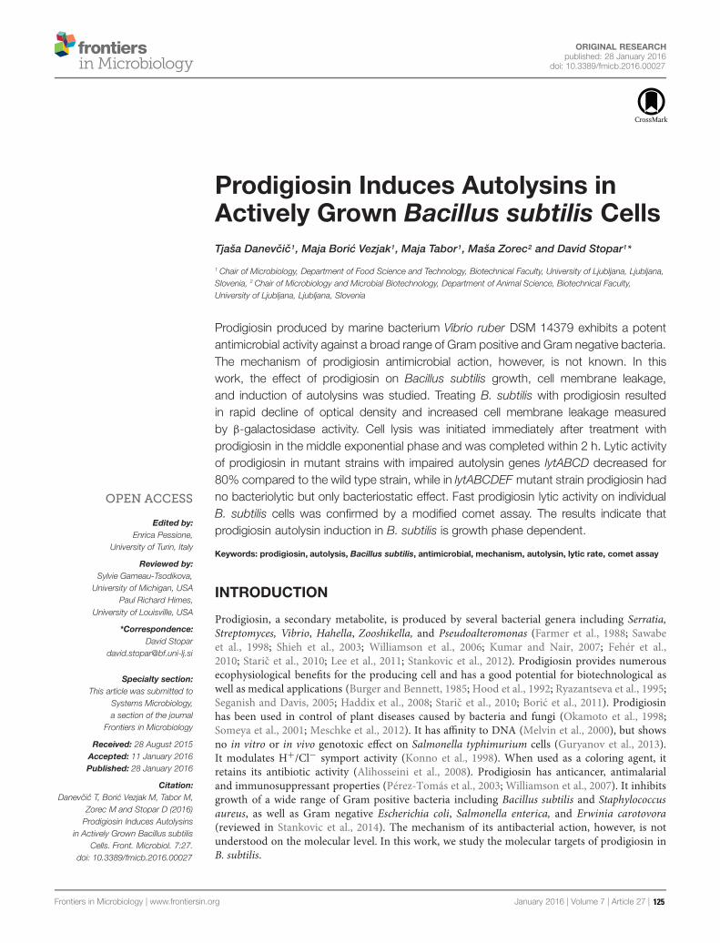
ORIGINAL RESEARCHpublished: 28 January 2016
doi: 10.3389/fmicb.2016.00027
Edited by:Enrica Pessione,
University of Turin, Italy
Reviewed by:Sylvie Garneau-Tsodikova,
University of Michigan, USAPaul Richard Himes,
University of Louisville, USA
*Correspondence:David Stopar
Specialty section:This article was submitted to
Systems Microbiology,a section of the journal
Frontiers in Microbiology
Received: 28 August 2015Accepted: 11 January 2016Published: 28 January 2016
Citation:Danevcic T, Boric Vezjak M, Tabor M,
Zorec M and Stopar D (2016)Prodigiosin Induces Autolysins
in Actively Grown Bacillus subtilisCells. Front. Microbiol. 7:27.
doi: 10.3389/fmicb.2016.00027
Prodigiosin Induces Autolysins inActively Grown Bacillus subtilis CellsTjaša Danevcic1, Maja Boric Vezjak1, Maja Tabor1, Maša Zorec2 and David Stopar1*
1 Chair of Microbiology, Department of Food Science and Technology, Biotechnical Faculty, University of Ljubljana, Ljubljana,Slovenia, 2 Chair of Microbiology and Microbial Biotechnology, Department of Animal Science, Biotechnical Faculty,University of Ljubljana, Ljubljana, Slovenia
Prodigiosin produced by marine bacterium Vibrio ruber DSM 14379 exhibits a potentantimicrobial activity against a broad range of Gram positive and Gram negative bacteria.The mechanism of prodigiosin antimicrobial action, however, is not known. In thiswork, the effect of prodigiosin on Bacillus subtilis growth, cell membrane leakage,and induction of autolysins was studied. Treating B. subtilis with prodigiosin resultedin rapid decline of optical density and increased cell membrane leakage measuredby β-galactosidase activity. Cell lysis was initiated immediately after treatment withprodigiosin in the middle exponential phase and was completed within 2 h. Lytic activityof prodigiosin in mutant strains with impaired autolysin genes lytABCD decreased for80% compared to the wild type strain, while in lytABCDEF mutant strain prodigiosin hadno bacteriolytic but only bacteriostatic effect. Fast prodigiosin lytic activity on individualB. subtilis cells was confirmed by a modified comet assay. The results indicate thatprodigiosin autolysin induction in B. subtilis is growth phase dependent.
Keywords: prodigiosin, autolysis, Bacillus subtilis, antimicrobial, mechanism, autolysin, lytic rate, comet assay
INTRODUCTION
Prodigiosin, a secondary metabolite, is produced by several bacterial genera including Serratia,Streptomyces, Vibrio, Hahella, Zooshikella, and Pseudoalteromonas (Farmer et al., 1988; Sawabeet al., 1998; Shieh et al., 2003; Williamson et al., 2006; Kumar and Nair, 2007; Fehér et al.,2010; Staric et al., 2010; Lee et al., 2011; Stankovic et al., 2012). Prodigiosin provides numerousecophysiological benefits for the producing cell and has a good potential for biotechnological aswell as medical applications (Burger and Bennett, 1985; Hood et al., 1992; Ryazantseva et al., 1995;Seganish and Davis, 2005; Haddix et al., 2008; Staric et al., 2010; Boric et al., 2011). Prodigiosinhas been used in control of plant diseases caused by bacteria and fungi (Okamoto et al., 1998;Someya et al., 2001; Meschke et al., 2012). It has affinity to DNA (Melvin et al., 2000), but showsno in vitro or in vivo genotoxic effect on Salmonella typhimurium cells (Guryanov et al., 2013).It modulates H+/Cl− symport activity (Konno et al., 1998). When used as a coloring agent, itretains its antibiotic activity (Alihosseini et al., 2008). Prodigiosin has anticancer, antimalarialand immunosuppressant properties (Pérez-Tomás et al., 2003; Williamson et al., 2007). It inhibitsgrowth of a wide range of Gram positive bacteria including Bacillus subtilis and Staphylococcusaureus, as well as Gram negative Escherichia coli, Salmonella enterica, and Erwinia carotovora(reviewed in Stankovic et al., 2014). The mechanism of its antibacterial action, however, is notunderstood on the molecular level. In this work, we study the molecular targets of prodigiosin inB. subtilis.
Frontiers in Microbiology | www.frontiersin.org January 2016 | Volume 7 | Article 27 | 125

Danevcic et al. Prodigiosin Induces Autolysins in Bacillus subtilis
Many antibacterial agents target cell membrane and cellwall (Falk et al., 2010; Lacriola et al., 2013). In B. subtilisinduction of autolysins and subsequent cell wall degradation isa major mechanism of antimicrobial action of several differentcompounds (Falk et al., 2010; Lacriola et al., 2013). Autolysinsare enzymes involved in hydrolysis and remodeling of thepeptidoglycan in the bacterial cell wall (Smith et al., 2000). Itis absolutely critical to maintain the regulation of potentiallysuicidal activity of autolysins. The regulation of autolysin activityis mainly due to the local pH in respiring cells. In respiringcells, protons are extruded across the cytoplasmic membraneand bind to cell wall constituents (Jolliffe et al., 1981; Kemperet al., 1993; Calamita and Doyle, 2002). The protonation of thecell wall constituents create a wall matrix of low pH. Protonmotive force sustains this local low pH and, in turn, autolyticactivity is inhibited because N-acetylmuramyl-L-alanine amidasecannot function below pH 5.5. There are 36 autolysins presentin the B. subtilis genome (Smith et al., 2000). Among differentautolysins in B. subtilis, two autolysins of vegetative growthnamed LytC and LytD are responsible for about 95% of thebacterial cell autolysis during active growth (reviewed in Smithet al., 2000). It has been shown previously that β-galactosidaserelease correlates with activation of pathways leading to autolysis(Falk et al., 2007). Apart from β-galactosidase release severalother methods (e.g., optical density, cell count) can be usedto determine bacterial cell lysis. The comet assay is a methodprimarily used for the detection of DNA damage and cell lysisin a single cell (Ostling and Johanson, 1984; Singh et al., 1988).Recently, comet assay was applied to detect bacterial lysis afterbacteriophage treatment (Khairnar et al., 2014). Using cometassay to detect single cell lysis provides an advantage over enzymeactivity or optical density measurements of cell lysis which probea population response.
In this work, the mechanism of antibacterial activity ofprodigiosin on B. subtilis was studied. The ability of prodigiosinto affect bacterial population structure was determined in simpleco-culture experiments. The effect of prodigiosin on the bacterialgrowth, cell wall integrity, autolysis, and DNA damage wasdetermined on the wild type strain and several autolysin mutants.This study is the first attempt to determine the mechanismof antibacterial activity of prodigiosin in B. subtilis in vivo.The results indicate that prodigiosin activates rapid autolysis inB. subtilis.
MATERIALS AND METHODS
Bacterial Strains, Mutant StrainConstruction and Growth ConditionsBacterial strains used in this study were B. amyloliquefaciensFZB42, B. licheniformis ATCC9445A, B. subtilis NCIB3610all obtained from BGSC, B. subtilis ATCC6051, B. mycoidesDSM2048 obtained from DSMZ, B. subtilis PS-216 wt (Stefanicand Mandic-Mulec, 2009), B. subtilis PS-216 amyE::Phyperspank-mKate2 cat (Stefanic et al., 2015), Escherichia coli MG1655 andVibrio ruber DSM14379 (Stopar et al., 2004; Boric et al., 2011).In addition mutant strains of B. subtilis PS-216 �lytABC::neo,
B. subtilis PS-216 �lytABC::neo �lytD::tet, B. subtilis PS-216 �lytABC::neo �lytD::tet �lytE::cam, B. subtilis PS-216�lytABC::neo �lytD::tet �lytE::cam �lytF::spc and B. subtilisPS-216 srfA-lacZ neo were prepared. The lyt mutants wereconstructed by transforming the B. subtilis PS-216 wt strain withthe DNA isolated from B. subtilis L16611 and B. subtilis L16628obtained from BGSC (Margot and Karamata, 1992). The srfA-lacZ construct was made by transforming B. subtilis PS-216 wtstrain with the DNA isolated from B. subtilis KTB308 (BaconSchneider et al., 2002). Bacillus strains were grown at 37◦C(except B. mycoides at 28◦C) and 200 rpm in LB medium or CMmedium (Albano et al., 1987) or at 28◦C and 200 rpm in PYEmedium with 3% (w/V) NaCl (Danevcic et al., 2005).
Prodigiosin ExtractionVibrio ruber DSM14379 was cultured overnight in PYE mediumwith 3% (w/V) NaCl at 28◦C and 200 rpm (Danevcic et al., 2005).Overnight culture was diluted 100 times in 400 mL of fresh M9medium supplied with 5 g/L glucose (Staric et al., 2010) andincubated for 16 h at the same growth conditions. Cells wereharvested with centrifugation at 10397 g for 10 min. Prodigiosinwas extracted from cells with an equal volume of acetone at 28◦Cand 200 rpm for 2 h. After centrifugation for 15 min at 10397 g toremove cell debris, the solvent was evaporated. Dry biomass wasresuspended in sterile 96% (V/V) ethanol and the pigment extractwas filtered through 0.20 μm filter. Prodigiosin concentrationin the extract was determined as described previously by Staricet al. (2010). The prodigiosin purity in the extract was confirmedby HPLC using Kinetex C18 column (250 mm × 4.6 mm,5 μm, Phenomenex, USA) according to the modified methoddescribed previously (Nakashima et al., 2006). The separation wasachieved using 0.1% trifluoroacetic acid (A) and methanol (B)mobile phases, and a gradient elution program at 1 mL−1 minwith the following parameters: 0–5 min 100% A, 5–25 min 0–100% B (linear gradient), 25–30 min 100% B (isocratic), and30–50 min 100% A (isocratic) to re-equilibrate the column,monitored by UV-VIS detection. The purity was determinedby HPLC at different wavelengths in the range from 400 to600 nm. The HPLC elugram at 535 nm is given in supplementarymaterials (Supplementary Figure S1). In the tested range theextract contained more than 98% prodigiosin of both isomersα and β.
Minimal Inhibitory ConcentrationDeterminationMinimal inhibitory concentration (MIC) values were determinedin 96-well microtiter plate assay according to CLSI standards(Clinical and Laboratory Standards Institute, 2009). Briefly,bacterial strains were grown overnight in LB medium at 28◦Cor 37◦C. Inoculum was prepared by diluting overnight culturesin 0.9% NaCl to 0.5 McFarland (∼107 cells per mL). Wellsof microtiter plates were filled with 0.1 mL of the appropriateconcentration of the prodigiosin, ethanol or water and 0.01 mLof the inoculum to have the approximate number of cells ineach inoculated well 105. The dilutions of prodigiosin, ethanoland water were made in LB medium. Prodigiosin was dilutedin the final concentration range from 0.6 mg L−1 to 367.1 mg
Frontiers in Microbiology | www.frontiersin.org January 2016 | Volume 7 | Article 27 | 126

Danevcic et al. Prodigiosin Induces Autolysins in Bacillus subtilis
L−1, while ethanol was diluted in the final concentration rangefrom 0.02 to 12.03% (V/V). For positive controls LB mediumwas diluted with the same amounts of sterile distilled wateras ethanol. Microtiter plates were incubated without shaking at37◦C for 20 h and the optical density at 650 nm (OD650) wasmeasured spectrophotometrically at the beginning and the endof incubation. The lowest prodigiosin concentration in the wellwith no growth after 20 h of incubation, was taken as the MICvalue.
Co-culture ExperimentsBacillus subtilis PS-216 wt was grown either in co-culturewith V. ruber DSM14379 or in pure culture. For co-cultureexperiments, strains were separately grown in PYE medium with3% (w/V) NaCl (V. ruber) or in LB medium (B. subtilis) until thestationary phase. Co-cultures were inoculated into PYE mediumwith 3% (w/V) NaCl with V. ruber DSM14379: B. subtilis PS-216in the ratio 1:2 (V:V), the overall inoculum being 1% (V/V). CFUwas determined at the beginning and 20 h after incubation at28◦C and 200 rpm. CFU values were determined on PYE plateswithout NaCl (for B. subtilis PS-216 wt) and PYE plates with3% (w/V) NaCl (for both strains). Due to the red pigmentationof V. ruber DSM14379 the colonies were clearly distinguished.Malthusian fitness was calculated based on CFU counts accordingto Ahn et al. (2006). Prodigiosin concentration in co-cultures wasdetermined as described previously in Boric et al. (2011).
Cell Treatment with Prodigiosin inDifferent Growth PhasesBacillus subtilis PS-216 wt cells were cultured in liquid LBmedium at 37◦C and 200 rpm overnight. Overnight culturewas diluted 100 times in 20 mL of fresh LB medium andincubated further at the same growth conditions. OD650 wasmeasured in regular intervals during the culture incubation.At appropriate OD650 in different growth phases [i.e., middleexponential (OD650 ∼ 0.7), late exponential (OD650 ∼ 1.1) andstationary phase OD650 ∼ 1.3], the strains were treated with5.9 mg L−1 of prodigiosin. As a control, an equivalent amountof sterile 96% (V/V) ethanol was added to the LB medium in thefinal concentration 0.19% (V/V).
Treatment of Different Bacterial Strainswith ProdigiosinBacillus mycoides DSM2048 cells were cultured in liquid LBmedium at 28◦C and 200 rpm overnight, while B. licheniformisATCC9445A cells were cultured in liquid LB medium at 37◦Cand 200 rpm overnight. Overnight cultures were diluted 100times in 20 mL of fresh LB medium and incubated further at thesame growth conditions. Cultures were treated with prodigiosin(1.2 mg L−1 B. mycoides or 6.9 mg L−1 B. licheniformis)or ethanol as a control [0.04% (V/V) B. mycoides or 0.23% (V/V) B. licheniformis] in the middle exponential phase atOD650 between 0.4 and 0.5. Treated cells were then incubatedfurther and OD650 was measured in regular intervals during6 h of incubation. To determine the influence of prodigiosin on
different strains, the rate of lysis was calculated from measureddecrease of OD650 after prodigiosin treatment.
Cell Morphology and ViabilityBacillus subtilis PS-216 wt and B. subtilis PS-216amyE::Phyperspank-mKate2 strains were grown in LB mediumas described above and treated with 5.9 mg L−1 of prodigiosinor 0.19% (V/V) of ethanol in early exponential phase at OD650between 0.4 and 0.5. Cell morphology and viability wereinspected by Differential Interference Contrast (DIC) andfluorescence microscopy using appropriate filter set under theinverted microscope Axio Observer Z1 (Carl Zeiss, Germany) at0, 1, and 5 h after the prodigiosin or ethanol treatment.
Effect of Prodigiosin on Inactive CellsBacillus subtilis PS-216 wt cells were grown in LB medium asdescribed above. When cells reached OD650 between 0.4 and0.5, bacterial culture was autoclaved at 121◦C and 1.03 bar for20 min to inactivate cells. After autoclaving, cell suspension wascooled down to room temperature and 5.9 mg L−1 of prodigiosinwas added. Cell suspension was incubated further at 37◦C and200 rpm and OD650 was measured every 30 min for 5 h.
Cytoplasmic Membrane LeakageCytoplasmic membrane leakage was determined asβ-galactosidase activity according to the modified methodby Mensa et al. (2011). Briefly, B. subtilis PS-216 srfA-lacZ (neo)cells were grown overnight in LB medium with appropriateantibiotic at 37◦C and 200 rpm and diluted 100 times in 20 mLof the fresh CM medium. Cells were incubated at 37◦C and200 rpm until they reach OD650 ∼ 0.6. At that point 70 μL ofcells were transferred in a microtiter plate and mixed with 20 μLof Z buffer (60 mM Na2HPO4, 40 mM NaH2PO4, 10 mM KCl,10 mM MgSO4, pH 7) supplemented with 4 mg mL−1 ONPG(o-nitrophenyl-β-galactoside) and 40 μM β-mercaptoethanol.Cells were then treated with 10 μL of prodigiosin to a finalconcentration of 2.9 mg L−1. Microtiter plates were incubatedin a spectrometer for 2 h at 37◦C and absorbance at 420 nm wasmeasured every 2 min to determine the rate of ONPG hydrolysis.The addition of 10 μL of 0.1% (w/V) sodium dodecyl sulfate(SDS) detergent [final concentration 0.01% (w/V)] representeda positive control, while the addition of 10 μL of 96% (V/V)ethanol [final concentration 9.6% (V/V)] represented a negativecontrol. To calculate specific enzyme activity, the rate of ONPGhydrolysis was normalized to the cell biomass.
Bacterial Response to Different AutolysinInducing AgentsBacillus subtilis PS-216 wt strain was grown in LB mediumas described above and treated with 0.0125% (V/V) Triton X-100, 0.01% (w/V) SDS , 0.01% (w/V) cetyl trimethylammoniumbromide (CTAB), 100 μg mL−1 ampicillin or 100 μg mL−1
kanamycin in the middle exponential phase at OD650 between0.4 and 0.5. Ampicillin was used as a positive control knownto induce cell lysis, whereas kanamycin was used as a negativecontrol (Chung et al., 2009; Falk et al., 2010; Lacriola et al., 2013).
Frontiers in Microbiology | www.frontiersin.org January 2016 | Volume 7 | Article 27 | 127

Danevcic et al. Prodigiosin Induces Autolysins in Bacillus subtilis
Cell suspension was incubated further at 37◦C and 200 rpm andOD650 was measured every 30 min for 5 h. To determine theimpact of different detergents on bacterial strain, the rate of lysiswas calculated from measured decrease of OD650 after treatmentwith antimicrobial agents.
Autolytic Response of Bacterial Strainsto ProdigiosinBacillus subtilis strains PS-216 wt, PS-216 �lytABC, PS-216�lytABCD, PS-216 �lytABCDE and PS-216 �lytABCDEF weregrown in LB medium as described above and treated with 5.9 mgL−1 of prodigiosin or 0.19 % (V/V) of ethanol as a negativecontrol at OD650 between 0.4 and 0.7. Cells were then incubatedfurther and OD650 was measured in regular intervals during 6 hof incubation at 37◦C and 200 rpm. To determine the influenceof prodigiosin on different mutant strains, the rate of lysis wascalculated from measured decrease of OD650 after prodigiosintreatment.
Modified Comet AssayBacillus subtilis strains PS-216 wt, PS-216 �lytABC, PS-216�lytABCD, PS-216 �lytABCDE and PS-216 �lytABCDEF weregrown in LB medium as described above. At OD650 between 0.4and 0.6 cells were treated with 5.9 mg L−1 of prodigiosin or 0.19%(V/V) of ethanol. Cells were then incubated further for 1 h at37◦C and 200 rpm to lyse. Minigels were prepared on frosted-end microscopic slides. Slides were first dipped into 1% (w/V)normal melting point agarose (NMP) and one side was wipedclean. After drying on a hot plate (1 min, 100◦C), the secondlayer of 300μL 1% (w/V) NMP agarose was applied and solidifiedat 4◦C for 5 min. For the third layer treated or non-treatedcells were resuspended in 1% (w/V) low melting agarose (ratio1:10) and left to solidify for 5 min. Minigels were immediatelyelectrophoresed in TBE (Tris/borate/EDTA buffer, pH 8.3) at 1V/cm and run for 30 min. Afterward minigels were neutralized in400 mMTris buffer (pH 7.4). Gels were stained with 30x solutionof GelRedTM (Biotium, USA) and observed under epifluorescentmicroscope (Olympus BX-50, Japan) equipped with EMCCDcamera (LucaEMr, Andor Technology Ltd., United Kingdom) andthe appropriate filter set. Images were captured by Komet 7.0software (Andor Technology Ltd., UK).
Data AnalysisAll the values presented are averages with standard deviations orstandard errors. Results were statistically evaluated using one-way ANOVA. Samples with p-values equal or lower than 0.05were taken as statistically significant.
RESULTS AND DISCUSSION
Prodigiosin isolated from V. ruber DSM14379 inhibits growthof different bacterial species. The MIC value for Escherichiacoli is 103.4 ± 6.3 mg L−1, while different Bacillus sp. strainstested have significantly lower MIC values. The MIC valuesfor different Bacillus sp. strains were in the range between 5and 7 mg L−1 (MIC values for B. amyloliquefaciens FZB42 was
FIGURE 1 | The effect of prodigiosin addition on Bacillus subtilisPS-216 wt growth in LB medium at 37◦C and 200 rpm at differentgrowth phases. The arrows represent the time of addition of 5.9 mg L−1
prodigiosin (opened squares) or 0.19% (V/V) ethanol in the control samples(filled squares). (A) cells treated in the middle of the exponential phase; (B)cells treated in the late exponential phase; (C) cells treated in the stationaryphase. Data are presented as averages and standard deviations (n = 3).
Frontiers in Microbiology | www.frontiersin.org January 2016 | Volume 7 | Article 27 | 128

Danevcic et al. Prodigiosin Induces Autolysins in Bacillus subtilis
FIGURE 2 | Bacillus subtilis PS-216 wt cells (A) and B. subtilis PS-216 amyE::Phyperspank -mKate2 (B) grown in LB medium at 37◦C and 200 rpmobserved by DIC and fluorescent microscopy at 0, 1, and 5 h after the addition of either 5.9 mg L−1 of prodigiosin or 0.19% (V/V) of ethanol in controlsamples in the middle of the exponential phase. The scale bar represents 10 μm.
6.1 ± 1.2 mg L−1, B. licheniformis ATTC9445A was 6.9 ± 2.2 mgL−1, B. mycoides was 1.2 mg L−1, B. subtilis NCIB3610 was5.2 ± 1.4 mg L−1, B. subtilis ATCC6051 was 5.9 mg L−1 andB. subtilis PS-216 was 5.9 mg L−1) and were lower as previouslyreported (Stankovic et al., 2012; Priya et al., 2013). Approximatelythe same MIC values for different Bacillus species may suggestthat prodigiosin has the same mode of action on all testedBacillus sp. Only undomesticated natural isolate B. subtilis PS-216 wt was used for further analysis with MIC value 5.9 mgL−1. To test the in vivo activity of prodigiosin on B. subtilis PS-216 wt simple co-cultures with prodigiosin producing V. ruberDSM14379 were made. There was a significant decrease in thenumber of B. subtilis cells after incubation in co-culture. The
Malthusian fitness of B. subtilis grown in a monoculture was5.7 ± 0.2, but was significantly lower −6.8 ± 1 in co-cultureexperiments. The concentration of prodigiosin in co-culture atthe end of the experiment was 16.3 ± 0.4 mg L−1 which is2.7 fold higher than the MIC required for growth inhibition ofB. subtilis PS-216 wt. The results indicate that prodigiosin hasin vivo antimicrobial activity.
The effect of prodigiosin on B. subtilis was dependent onthe growth phase as shown in Figure 1. Prodigiosin treatmentin the middle of the exponential phase (Figure 1A) causedinstantaneous termination of growth and immediate decrease ofoptical density. Cell lysis was completed in 2 h after prodigiosinaddition. As shown in Figure 2 cell number significantly
Frontiers in Microbiology | www.frontiersin.org January 2016 | Volume 7 | Article 27 | 129
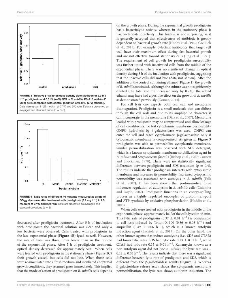
Danevcic et al. Prodigiosin Induces Autolysins in Bacillus subtilis
FIGURE 3 | Relative β-galactosidase activity upon addition of 5.9 mgL−1 prodigiosin and 0.01% (w/V) SDS in B. subtilis PS-216 srfA-lacZ(neo) cells compared with control [addition of 0.19% (V/V) ethanol].Cells were grown in LB medium at 37◦C and 200 rpm. Data are presented asaverages and standard errors (n = 3–6).
FIGURE 4 | Lytic rates of different lyt mutants measured as a rate ofOD650 decrease after treatment with prodigiosin (5.9 mg L−1) in LBmedium at 37◦C and 200 rpm. Data are presented as averages andstandard deviations (n = 3).
decreased after prodigiosin treatment. After 5 h of incubationwith prodigiosin the bacterial solution was clear and only afew bacteria were observed. Cells treated with prodigiosin inthe late exponential phase (Figure 1B) lysed as well. However,the rate of lysis was three times lower than in the middleof the exponential phase. After 5 h of prodigiosin treatment,optical density decreased for approximately 50%. When cellswere treated with prodigiosin in the stationary phase (Figure 1C)their growth ceased, but cells did not lyse. When those cellswere re-inoculated into a fresh medium and incubated at optimalgrowth conditions, they resumed grow immediately. This impliesthat the mode of action of prodigiosin on B. subtilis cells depends
on the growth phase. During the exponential growth prodigiosinhas a bacteriolytic activity, whereas in the stationary phase ithas bacteriostatic activity. This finding is not surprising, as itis generally accepted that effectiveness of antibiotic is greatlydependent on bacterial growth rate (Hobby et al., 1942; Greulichet al., 2015). For example, β-lactam antibiotics that target cellwall have their maximum effect during fast bacterial growthand are not effective toward stationary cells (Eng et al., 1991).The requirement of cell growth for prodigiosin susceptibiltywas further tested with inactivated cells from the middle of theexponential phase. There was no significant change in opticaldensity during 5 h of the incubation with prodigiosin, suggestingthat the inactive cells did not lyse (data not shown). After theaddition of the control containing ethanol (Figure 1), the growthofB. subtilis continued. Although the culture was not significantlydiluted (the total volume increased only by 0.2%), the addedethanol may have had a positive effect on the growth of B. subtilisas demonstrated previously (Gomaa, 2014).
For cell lysis one expects both cell wall and membranedisintegration. Prodigiosin is a small molecule that can diffusethrough the cell wall and due to its amphiphilic character itcan incorporate in the membrane (Diaz et al., 2007). Membraneloaded with prodigiosin may be compromised and allow leakageof cell constituents. To test cytoplasmic membrane permeabilityONPG hydrolysis by β-galactosidase was used. ONPG canenter the cell and reach cytoplasmatic β-galactosidase only ifcytoplasmic membrane is compromised. As given in Figure 3prodigiosin was able to permeabilize cytoplasmic membrane.Similar permeabilization was observed with SDS detergent,which is a known cytoplasmic membrane solubilization agent inB. subtilis and Streptococcus faecalis (Bishop et al., 1967; Cornettand Shockman, 1978). There were no statistically significantdifferences between prodigiosin and SDS treatment (p = 0.4).The results indicate that prodigiosin interacts with cytoplasmicmembrane and increases its permeability. Increased cytoplasmicpermeability was associated with autolysis in B. subtilis (Falket al., 2007). It has been shown that proton-motive forceinfluences regulation of autolysins in B. subtilis cells (Calamitaand Doyle, 2002). Prodigiosin functions in an energy-spillingprocess as a tightly regulated uncoupler of proton transportand ATP synthesis by oxidative phosphorylation (Haddix et al.,2008).
When cells were treated with prodigiosin in the middle of theexponential phase, approximately half of the cells lysed in 45min.This lytic rate of prodigiosin (0.37 ± 0.01 h−1) is comparableto cell lysis induced by Triton X-100 (0.36 ± 0.03 h−1) andampicillin (0.49 ± 0.06 h−1), which is a known autolysininduction agent (Lacriola et al., 2013). On the other hand, theother known agents that induce autolysins (i.e., SDS and CTAB)had lower lytic rates. SDS had lytic rate 0.13 ± 0.01 h−1, whileCTAB had lytic rate 0.15 ± 0.01 h−1. Kanamycin known as anon-autolysin agent did not lyse B. subtilis, the lytic rate was –0.12 ± 0.03 h−1. The results indicate that there was a significantdifference between lytic rate of prodigiosin and SDS, which isdifferent from the β-galactosidase results (Figure 3). Whereasβ-galactosidase release assay shows the cytopasmic membranepermeabilisation, the lytic rate shows autolysin induction. The
Frontiers in Microbiology | www.frontiersin.org January 2016 | Volume 7 | Article 27 | 130

Danevcic et al. Prodigiosin Induces Autolysins in Bacillus subtilis
FIGURE 5 | The modified comet assay for studying prodigiosin mediated cell lysis of B. subtilis PS-216 wt cells grown in LB medium at 37◦C and200 rpm. (A) non-treated bacterial cells; (B) control cells treated with 0.19% (V/V) of ethanol; (C) cells treated with 5.9 mg L−1 of prodigiosin. Cells were treated inthe middle exponential phase and were inspected 1 h after the treatment. The gels were stained with GelRedTM. The images were observed with epifluorescentmicroscopy.
two are not necessarily coupled. For example, SDS is a typicalsolubilizing agent of cytoplasmic membrane proteins in B. subtilis(Bishop et al., 1967), but is usually not used as an autolysininducer due to its low lytic effectiveness. It has been demonstratedin Enterobacteriacea that the SDS-grown cells underwent rapidlysis only when they ran out of energy (Kramer et al., 1980).The energy dependence reflects a requirement for ATP ratherthan for a proton gradient or membrane potential (Aspedonand Nickerson, 1994). Prodigiosin, on the other hand, actsas a potent lytic agent for B. subtilis that uncouples protontransport in the cytoplasmic membrane (Haddix et al., 2008),thereby activating autolysins. Prodigiosin lytic activity was notlimited to B. subtilis cells only. For example, treating B. mycoidesand B. licheniformis with prodigiosin at MIC concentrations
induces cell lysis and the cell suspension cleared after 20 h oftreatment. The corresponding lytic rates were 0.014 ± 0.002 h−1
for B. mycoides and 0.009 ± 0.001 h−1 for B. licheniformis, whichindicates that prodigiosin lytic activity could be a more generalmechanism in Bacillus species.
To prove that prodigiosin induces autolysis several mutantstrains of B. subtilis PS-216 deficient in major autolysis genes(lyt genes) were produced. The lytic rates after treatment withprodigiosin of mutant strains deficient to a different extent inautolytic response are given in Figure 4. In the lytABC deficientstrain the cell lysis was significantly reduced and the lytic ratewas approximately two fold lower compared to the wild typestrain. When additionally lytD gen was deleted a further decreasein the lytic rate was observed. The lytABCD deficient strain
Frontiers in Microbiology | www.frontiersin.org January 2016 | Volume 7 | Article 27 | 131

Danevcic et al. Prodigiosin Induces Autolysins in Bacillus subtilis
FIGURE 6 | The modified comet assay on different autolysin deficient strains of B. subtilis PS-216 cells grown in LB medium at 37◦C and 200 rpmtreated with 0.19% (V/V) of ethanol (control) or treated with 5.9 mg L−1 of prodigiosin. Cells were treated in the middle exponential phase and wereinspected 1 h after the treatment. The gels were stained with GelRedTM. The images were observed with epifluorescent microscopy.
showed approximately 80% reduction of lysis compared to thewild type strain. This is consistent with previous reports of akey role of LytC and LytD in autolysis (Smith et al., 2000). Inaddition, it has been shown previously that lytE mutant maysignificantly reduce autolysis (Margot et al., 1998). Consistentlyin lytABCDE mutant there was almost no lysis after prodigiosinaddition. Finally, in lytABCDEF mutant the lytic rate uponaddition of prodigiosin was 50 times lower compared to thewild type strain. Although no lytic response was observed inlytABCDEF mutant the addition of prodigiosin in the middle ofexponential phase had a bacteriostatic effect. The results obtainedby different lyt deficient strains strongly suggest that prodigiosininduces autolysin dependent bacteriolysis in B. subtilis. For acomplete lysis of B. subtilis by prodigiosin a complement ofdifferent autolysins is needed. Since autolysins can be inducedalso by other amphiphatic molecules such as short- andmedium–chain fatty acids (Tsuchido et al., 1985) this further supports thehypothesis that prodigiosin is a potent autolysin modulator.
The release of nucleic material during cell lysis was followedwith modified comet assay (Figure 5). Non-treated cellsretained cell integrity through the experiment, displaying clearlydefined shape without tail or halo (Figure 5A). Similary,in control cultures treated with 0.19% (V/V) ethanol, thesame concentration as used for prodigiosin solubilization, cellswere intact (Figure 5B). On the other hand, treatment withprodigiosin induced instant lysis in almost all examined cells(Figure 5C). Cells that lysed appeared blurred with rapid declineof fluorescence indicating DNA diffusing out of disrupted cells.The comet assay was also applied to B. subtilis mutant strainsdeficient in major autolysin genes. Lyt mutants treated withprodigiosin responded differently. The comet assay suggeststhat lysis was most intense in lytABC mutant. The degree ofcell lysis decreased from lytABC to lytABCDEF deficient strain(Figure 6). Negligible cell lysis was observed in lytABCDEFmutant strain after prodigiosin treatment. The results obtainedwith the modified comet assay confirm that prodigiosin inducesrapid autolysis in B. subtilis cells.
Finally, we note that upon entry into the stationary phaseprodigiosin acts as a bacteriostatic agent. During transition to thestationary phase B. subtilis cells begin to lose their ability to moveand start to produce biofilm. Biofilms and motility are mutuallyexclusive lifestyles of B. subtilis (Diethmaier et al., 2011) andare controlled by a common regulator, the transcription factorSinR. During biofilm formation SinR interacts with SinI and de-represses synthesis of extracellular matrix important in biofilmformation. One of the components of extracellular matrix EpsEinteracts with flagellar motor switch protein FliG and preventsrotation of the flagellum (Blair et al., 2008). In addition, uponthe entry into a stationary phase cells shut down synthesis offlagella components. The synthesis of flagella and autolysin areunder the same regulation control. The synthesis of flagellaproteins requires genes which are under the control of alternativeRNA polymerase sigma factor, σD (Shahbabian et al., 2009). σD
is a pleiotrophic transcription regulator during the exponentialgrowth and has a positive effect on autolysin synthesis (Fein,1979). Upon entry into the stationary phase σH decreases theexpression of σD (Britton et al., 2002), which in turn down-regulates autolysin production. In the absence of autolysins,as demonstrated in this work, the lytic activity of prodigiosinis abolished. However, as shown with lytABCDEF mutantprodigiosin may still act as a bacteriostatic agent preventingfurther growth of B. subtilis.
CONCLUSION
The application of prodigiosin as an antibacterial agent ishampered by the lack of knowledge of its molecular targets.The results of this study demonstrate that prodigiosin exhibitsa potent antimicrobial activity against B. subtilis. It acts asa bacteriolytic agent during the exponential growth and as abacteriostatic agent during the stationary growth. Prodigiosininterferes with cytoplasmic membrane function and increases itspermeability. The obtained results indicate that prodigiosin’s
Frontiers in Microbiology | www.frontiersin.org January 2016 | Volume 7 | Article 27 | 132

Danevcic et al. Prodigiosin Induces Autolysins in Bacillus subtilis
bacteriolytic activity is due to the induction of autolysins.Prodigiosin proves to be a strong autolysin inducer in B. subtiliscells comparable in its activity to Triton X-100. Since prodigiosinfunctions as an uncoupler of proton transport and ATP synthesis,it is ideally suited for autolysins induction and killing of differentBacillus species.
AUTHOR CONTRIBUTIONS
Conceived and designed the experiments: TD, MV, and DS.Performed the experiments: TD, MV, MT, and MZ. Analyzedthe data: TD, MV, MT, and MZ. Wrote the paper: TD, MVand DS.
ACKNOWLEDGMENTS
We acknowledge financial support from the Slovenian ResearchAgency (ARRS no. 1000-09-310208). This work was alsosupported by the Slovenian Research Agency grant P4-0116. Weare thankful to Polonca Stefanic for providing B. subtilis PS-216wt and PS-216 srfA-lacZ (neo) strains.
SUPPLEMENTARY MATERIAL
The Supplementary Material for this article can be foundonline at: http://journal.frontiersin.org/article/10.3389/fmicb.2016.00027
REFERENCES
Ahn, H. J., La, H. J., and Forney, L. J. (2006). System for determining the relativefitness of multiple bacterial populations without using selective markers. Appl.Environ. Microbiol. 72, 7383–7385. doi: 10.1128/AEM.01246-06
Albano, M., Hahn, J., and Dubnau, D. (1987). Expression of competence genes inBacillus subtilis. J. Bacteriol. 169, 3110–3117.
Alihosseini, F., Ju, K. S., Lango, J., Hammock, B. D., and Sun, G. (2008).Antibacterial colorants: characterization of prodiginines and their applicationson textile materials. Biotechnol. Prog. 24, 742–747. doi: 10.1021/bp070481r
Aspedon, A., and Nickerson, K. W. (1994). The energy dependence of detergentresistance in Enterobacter cloacae: a likely requirement for ATP rather than aproton gradient or a membrane potential. Can. J. Microbiol. 40, 184–191. doi:10.1139/m94-031
Bacon Schneider, K., Palmer, T. M., and Grossman, A. D. (2002). Characterizationof comQ and comX, two genes required for production of ComXpheromone in Bacillus subtilis. J. Bacteriol. 184, 410–419. doi: 10.1128/JB.184.2.410-419.2002
Bishop, D. G., Rutberg, L., and Samulsson, B. (1967). The solubilization of thecytoplasmic membrane of Bacillus subtilis by sodium dodecyl sulphate. Eur. J.Biochem. 2, 454–459. doi: 10.1111/j.1432-1033.1967.tb00159.x
Blair, K. M., Turner, L., Winkelman, J. T., Berg, H. C., and Kearns, D. B. (2008).A molecular clutch disables flagella in the Bacillus subtilis biofilm. Science 320,1636–1638. doi: 10.1126/science.1157877
Boric, M., Danevcic, T., and Stopar, D. (2011). Prodigiosin from Vibrio sp.DSM 14379; a new UV-protective pigment. Microb. Ecol. 62, 528–536. doi:10.1007/s00248-011-9857-0
Britton, R. A., Eichenberger, P., Gonzalez-Pastor, J. E., Fawcett, P., Monson, R.,Losick, R., et al. (2002). Genome-wide analysis of the stationary-phase sigmafactor (sigma-H) regulon of Bacillus subtilis. J. Bacteriol. 184, 4881–4890. doi:10.1128/JB.184.17.4881-4890
Burger, S. R., and Bennett, J. W. (1985). Droplet enrichment factors ofpigmented and nonpigmented Serratia marcescens: possible selective functionfor prodigiosin. Appl. Environ. Microbiol. 50, 487–490.
Calamita, H. G., and Doyle, R. J. (2002). Regulation of autolysins in teichuronicacid-containing Bacillus subtilis cells. Mol. Microbiol. 44, 601–606. doi:10.1046/j.1365-2958.2002.02872.x
Chung, J. K., Yoon, H. E., Shin, H. C., Choi, E. Y., and Byeon, W. H. (2009).Induction of growth phase-specific autolysis in Bacillus subtilis 168 by growthinhibitors. J. Microbiol. 47, 50–59. doi: 10.1007/s12275-008-0256-2
Clinical and Laboratory Standards Institute (2009). Methods for DilutionAntimicrobial Susceptibility Tests for Bacteria that Grow Aerobically; ApprovedStandard, 8th Edn. Wayne, PA: Clinical and Laboratory Standards Institute.
Cornett, J. B., and Shockman, G. D. (1978). Cellular lysis of Streptococcus faecalisinduced with Triton X-100. J. Bacteriol. 135, 153–160.
Danevcic, T., Rilfors, L., Štrancar, J., Lindblom, G., and Stopar, D. (2005). Effects oflipid composition on the membrane activity and lipid phase behaviour ofVibriosp. DSM14379 cells grown at various NaCl concentrations. Biochim. Biophys.Acta 1712, 1–8. doi: 10.1016/j.bbamem.2005.03.013
Diaz, R. I. S., Regourd, J., Santacroce, P. V., Davis, J. T., Jakeman, D. L., andThompson, A. (2007). Chloride anion transport and copper – mediated DNAcleavage by C-ring functionalized prodigiosenes. Chem. Commun. 26, 2701–2703. doi: 10.1039/B701919J
Diethmaier, C., Pietack, N., Gunka, K., Wrede, C., Lehnik-Habrink, M.,Herzberg, C., et al. (2011). A novel factor controlling bistability in Bacillussubtilis: the YmdB protein affects flagellin expression and biofilm formation.J. Bacteriol. 193, 5997–6007. doi: 10.1128/JB.05360-11
Eng, R. H., Padberg, F. T., Smith, S. M., Tan, E. N., and Cherubin, C. E.(1991). Bactericidal effects of antibiotics on slowly growing and nongrowingbacteria. Antimicrob. Agents Chemother. 35, 1824–1828. doi: 10.1128/AAC.35.9.1824
Falk, S. P., Noah, J. W., and Weisblum, B. (2010). Screen for inducers ofautolysis in Bacillus subtilis. Antimicrob. Agents Chemother. 54, 3723–3729. doi:10.1128/AAC.01597-09
Falk, S. P., Ulijasz, A. T., and Weisblum, B. (2007). Differential assay forhigh-throughput screening of antibacterial compounds. J. Biomol. Screen. 12,1102–1108. doi: 10.1177/1087057107308161
Farmer, J. J., Hickman-Brenner, F. W., Fanning, G. R., Gordon, C.M., and Brenner,D. J. (1988). Characterization of Vibrio metschnikovii and Vibrio gazogenes byDNA-DNA hybridization and phenotype. J. Clin. Microbiol. 26, 1993–2000.
Fehér, D., Barlow, R. S., Lorenzo, P. S., and Hemscheidt, T. K. (2010).A 2-Substituted Prodiginine, 2-(p-Hydroxybenzyl)prodigiosin, fromPseudoalteromonas rubra. J. Nat. Prod. 71, 1970–1972. doi: 10.1021/np800493p
Fein, J. E. (1979). Possible involvement of bacterial autolytic enzymes in flagellarmorphogenesis. J. Bacteriol. 137, 933–946.
Gomaa, E. Z. (2014). Production of polyhydroxyalkanoates (PHAs) by Bacillussubtilis and Escherichia coli grown on cane molasses fortified with ethanol. Braz.Arch. Biol. Technol. 57, 145–154. doi: 10.1590/S1516-89132014000100020
Greulich, P., Scott, M., Evans, M. R., and Allen, R. J. (2015). Growth – dependentbacterial susceptibility to ribosome – targeting antibiotics. Mol. Syst. Biol. 11,796. doi: 10.15252/msb.20145949
Guryanov, I. D., Karamova, N. S., Yusupova, D. V., Gnezdilov, O. I., andKoshkarova, L. A. (2013). Bacterial pigment prodigiosin and its genotoxic effect.Russ. J. Bioorg. Chem. 39, 106–111. doi: 10.1134/S1068162012060040
Haddix, P. L., Jones, S., Patel, P., Burnham, S., Knights, K., Powell, J. N., et al.(2008). Kinetic analysis of growth rate, ATP, and pigmentation suggests anenergy-spilling function for the pigment prodigiosin of Serratia marcescens.J. Bacteriol. 190, 7453–7463. doi: 10.1128/JB.00909-08
Hobby, G. L., Meyer, K., and Chaffee, E. (1942). Observation on the mechanismof penicillin. Proc. Soc. Exp. Biol. Med. 50, 281–285. doi: 10.3181/00379727-50-13773
Hood, D. W., Heidstra, R., Swoboda, U. K., and Hodgson, D. A. (1992). Moleculargenetic analysis of proline and tryptophan biosynthesis in Streptomycescoelicolor A3(2): interaction between primary and secondary metabolism – areview.Gene 115, 5–12. doi: 10.1016/0378-1119(92)90533-U
Jolliffe, L. K., Doyle, R. J., and Streips, U. N. (1981). The energized membraneand cellular autolysis in Bacillus subtilis. Cell 25, 753–763. doi: 10.1016/0092-8674(81)90183-5
Frontiers in Microbiology | www.frontiersin.org January 2016 | Volume 7 | Article 27 | 133

Danevcic et al. Prodigiosin Induces Autolysins in Bacillus subtilis
Kemper,M. A., Urrutia, M. M., Beveridge, T. J., Koch, A. L., and Doyle, R. J. (1993).Proton motive force may regulate cell wall-associated enzymes of Bacillussubtilis. J. Bacteriol. 175, 5690–5696.
Khairnar, K., Sanmukh, S., Chandekar, R., and Paunikar, W. (2014).A simple and novel modification of comet assay for determination ofbacteriophage mediated bacterial cell lysis. J. Virol. Methods 203, 33–38. doi:10.1016/j.jviromet.2014.02.028
Konno, H., Matsuya, H., Okamoto, M., Sato, T., Tanaka, Y., Yokoyama, K.,et al. (1998). Prodigiosins uncouple mitochondrial and bacterial F-ATPases:evidence for their H+/Cl- symport activity. J. Biochem. 124, 547–556. doi:10.1093/oxfordjournals.jbchem.a022147
Kramer, V. C., Calabrese, D. M., and Nickerson, K. W. (1980). Growth ofEnterobacter cloacae in the presence of 25% sodium dodecyl sulfate. Appl.Environ. Microbiol. 40, 973–976.
Kumar, N. R., and Nair, S. (2007). Vibrio rhizosphaerae sp. nov.; a red-pigmented bacterium that antagonizes phytopathogenic bacteria. Int. J. Syst.Evol. Microbiol. 57, 2241–2246. doi: 10.1099/ijs.0.65017-0
Lacriola, C. J., Falk, S. P., and Weisblum, B. (2013). Screen for agents that induceautolysis in Bacillus subtilis. Antimicrob. Agents Chemother. 57, 229–234. doi:10.1128/AAC.00741-12
Lee, J. S., Kim, Y. S., Park, S., Kim, J., Kang, S. J., Lee, M. H., et al. (2011).Exceptional production of both prodigiosin and cycloprodigiosin as majormetabolic constituents by a novel marine bacterium, Zooshikella rubidus S1-1.Appl. Environ. Microbiol. 77, 4967–4973. doi: 10.1128/AEM.01986-10
Margot, P., and Karamata, D. (1992). Identification of the structural genesfor N-acetylmuramoyl-L-alanine amidase and its modifier in Bacillus subtilis168: inactivation of these genes by insertional mutagenesis has no effect ongrowth or cell separation. Mol. Gen. Genet. 232, 359–366. doi: 10.1007/BF00266238
Margot, P., Whalen, M., Ghdamhoseinian, A., Piggot, P., and Karamata, D. (1998).The lytE gene of Bacillus subtilis 168 encodes a cell wall hydrolase. J. Bacteriol.180, 749–752.
Melvin, M. S., Tomlinson, J. T., Saluta, G. R., Kucera, G. L., Lindquist, N., andManderville, R. A. (2000). Double-strand DNA cleavage by copper-prodigiosin.J. Am. Chem. Soc. 122, 6333–6334. doi: 10.1021/ja0000798
Mensa, B., Kim, Y. H., Choi, S., Scott, R., Caputo, G. A., and DeGrado,W. F. (2011).Antibacterial mechanism of action of arylamide foldamers. Antimicrob. AgentsChemother. 55, 5043–5053. doi: 10.1128/AAC.05009-11
Meschke,H.,Walter, S., and Schrempf, H. (2012). Characterization and localizationof prodiginines from Streptomyces lividans suppressing Verticillium dahliaein the absence or presence of Arabidopsis thaliana. Environ. Microbiol. 14,940–952. doi: 10.1111/j.1462-2920.2011.02665.x
Nakashima, T., Miyazaki, Y., Matsuyama, Y., Muraoka, W., Yamaguchi, K., andOda, T. (2006). Producing mechanism of an algicidal compound against redtide phytoplankton in a marine bacterium γ-proteobacterium. Appl. Microbiol.Biotechnol. 73, 684–690. doi: 10.1007/s00253-006-0507-2
Okamoto, H., Sato, Z., Sato, M., Koiso, Y., Iwasaki, S., and Isaka, M. (1998).Identification of antibiotic red pigments of Serratia marcescens F-1-1, abiocontrol agent of damping-off of cucumber, and antimicrobial activityagainst other plant pathogens. Ann. Phytopathol. Soc. Jpn. 64, 294–298. doi:10.3186/jjphytopath.64.294
Ostling, O., and Johanson, K. J. (1984). Microelectrophoretic study of radiation-induced DNA damages in individual mammalian cells. Biochem. Biophys. Res.Commun. 123, 291–298. doi: 10.1016/0006-291X(84)90411-X
Pérez-Tomás, R., Montaner, B., Llagostera, E., and Soto-Cerrato, V. (2003).The prodigiosins, proapoptotic drugs with anticancer properties. Biochem.Pharmacol. 66, 1447–1452. doi: 10.1016/S0006-2952(03)00496-9
Priya, K. A., Satheesh, S., Ashokkumar, B., Varalakshmi, P., Selvakumar, G., andSivakumar, N. (2013). “Antifouling activity of prodigiosin from estuarine isolateof Serratia marcescens CMST 07,” in Microbiological Research in AgroecosystemManagement, Vol. XVI, ed. R. K. Velu (New Delhi: Springer), 11–21. doi:10.1007/978-81-322-1087-0_2
Ryazantseva, I. N., Andreyeva, I. N., Klementyeva, G. S., Ogorodnikova, T. I.,and Petrov, V. Y. (1995). Pigment-dependent light influence on the energeticsof Serratia marcescens. Thermochim. Acta 251, 63–67. doi: 10.1016/0040-6031(94)02148-H
Sawabe, T., Makino, H., Tatsumi, M., Nakano, K., Tajima, K., Iqbal, M. M., et al.(1998). Pseudoalteromonas bacteriolytica sp. nov.; a marine bacterium that is the
causative agent of red spot disease of Laminaria japonica. Int. J. Syst. Bacteriol.48, 769–774. doi: 10.1099/00207713-48-3-769
Seganish, J. L., and Davis, J. T. (2005). Prodigiosin is a chloride carrier thatcan function as an anion exchanger. Chem. Commun. 46, 5781–5783. doi:10.1039/B511847F
Shahbabian, K., Jamalli, A., Zig, L., and Putzer, H. (2009). Rnase Y, a novelendoribonuclease, initiates riboswitch turnover in Bacillus subtilis. EMBO J. 28,3523–3533. doi: 10.1038/emboj.2009.283
Shieh, W. Y., Chen, Y. W., Chaw, S. M., and Chiu, H. H. (2003). Vibrioruber sp. nov.; a red, facultatively anaerobic, marine bacterium isolatedfrom sea water. Int. J. Syst. Evol. Microbiol. 53, 479–484. doi: 10.1099/ijs.0.02307-0
Singh, N. P., McCoy, M. T., Tice, R. R., and Schneider, E. L. (1988).A simple technique for quantification of low levels of DNA damage inindividual cells. Exp. Cell Res. 175, 184–191. doi: 10.1016/0014-4827(88)90265-0
Smith, T. J., Blackman, S. A., and Foster, S. J. (2000). Autolysis of Bacillus subtilis:multiple enzymes with multiple functions. Microbiology 146, 249–262. doi:10.1099/00221287-146-2-249
Someya, N., Nakajima, M., Hirayae, K., Hibi, T., and Akutsu, K. (2001).Synergistic antifungal activity of chitinolytic enzymes and prodigiosinproduces by biocontrol bacterium, Serratia marcescens strain B2 against graymold pathogen, Botrytis cinerea. J. Gen. Plant Pathol. 67, 312–317. doi:10.1007/PL00013038
Stankovic, N., Radulovic, V., Petkovic, M., Vuckovic, I., Jadranin, M., Vasiljevic, B.,et al. (2012). Streptomyces sp. JS520 produces exceptionally high quantitiesof undecylprodigiosin with antibacterial, antioxidative, and UV-protectiveproperties. Appl. Microbiol. Biotechnol. 96, 1217–1231. doi: 10.1007/s00253-012-4237-3
Stankovic, N., Senerovic, L., Ilic-Tomic, T., Vasiljevic, B., and Nikodinovic-Runic, J.(2014). Properties and applications of undecylprodigiosin and other bacterialprodigiosins. Appl. Microbiol. Biotechnol. 98, 3841–3858. doi: 10.1007/s00253-014-5590-1
Staric, N., Danevcic, T., and Stopar, D. (2010). Vibrio sp. DSM14379 pigmentproduction – a competitive advantage in the environment? Microb. Ecol. 60,592–598. doi: 10.1007/s00248-010-9671-0
Stefanic, P., Kraigher, B., Lyons, N. A., Kolter, R., and Mandic-Mulec, I. (2015). Kindiscrimination between sympatric Bacillus subtilis isolates. Proc. Natl. Acad. Sci.U.S.A. 112, 14042–14047. doi: 10.1073/pnas.1512671112
Stefanic, P., and Mandic-Mulec, I. (2009). Social interactions and distribution ofBacillus subtilis pherotypes at microscale. J. Bacteriol. 191, 1756–1764. doi:10.1128/JB.01290-08
Stopar, D., Cerne, A., Zigman, M., Poljsak-Prijatelj, M., and Turk, V. (2004). Viralabundance and a high proportion of lysogens suggest that viruses are importantmembers of the microbial community in the Gulf of Trieste. Microb. Ecol. 47,1–8. doi: 10.1007/s00248-002-3009-5
Tsuchido, T., Hiraoka, T., Takano, M., and Shibasaki, I. (1985). Involvement ofautolysin in Bacillus subtilis induced by short– and medium-chain fatty acids.J. Bacteriol. 162, 42–46.
Williamson, N. R., Fineran, P. C., Gristwood, T., Chawrai, S. R., Leeper, F. J.,and Salmond, G. P. C. (2006). The biosynthesis and regulation of bacterialprodiginines.Nat. Rev. Microbiol. 4, 887–899. doi: 10.1038/nrmicro1531
Williamson, N. R., Fineran, P. C., Gristwood, T., Chawrai, S. R., Leeper,F. J., and Salmond, G. P. C. (2007). Anticancer and immunosuppressiveproperties of bacterial prodiginines. Future Microbiol. 2, 605–618. doi:10.2217/17460913.2.6.605
Conflict of Interest Statement: The authors declare that the research wasconducted in the absence of any commercial or financial relationships that couldbe construed as a potential conflict of interest.
Copyright © 2016 Danevcic, Boric Vezjak, Tabor, Zorec and Stopar. This is anopen-access article distributed under the terms of the Creative Commons AttributionLicense (CC BY). The use, distribution or reproduction in other forums is permitted,provided the original author(s) or licensor are credited and that the originalpublication in this journal is cited, in accordance with accepted academic practice.No use, distribution or reproduction is permitted which does not comply with theseterms.
Frontiers in Microbiology | www.frontiersin.org January 2016 | Volume 7 | Article 27 | 134

MINI REVIEWpublished: 27 November 2015
doi: 10.3389/fmicb.2015.01363
Edited by:Enrica Pessione,
University of Torino, Italy
Reviewed by:Dimitris Tsaltas,
Cyprus University of Technology,Cyprus
Shuji Tani,Osaka Prefecture University, Japan
*Correspondence:Des Field
Specialty section:This article was submitted to
Systems Microbiology,a section of the journal
Frontiers in Microbiology
Received: 29 September 2015Accepted: 17 November 2015Published: 27 November 2015
Citation:Field D, Cotter PD, Hill C and Ross
RP (2015) Bioengineering Lantibioticsfor Therapeutic Success.Front. Microbiol. 6:1363.
doi: 10.3389/fmicb.2015.01363
Bioengineering Lantibioticsfor Therapeutic SuccessDes Field 1*, Paul D. Cotter 2,3, Colin Hill 1,3 and R. P. Ross 2,3
1 School of Microbiology, University College Cork, Cork, Ireland, 2 Teagasc Food Research Centre, Fermoy, Ireland,3 APC Microbiome Institute, University College Cork, Cork, Ireland
Several examples of highly modified antimicrobial peptides have been described.While many such peptides are non-ribosomally synthesized, ribosomally synthesizedequivalents are being discovered with increased frequency. Of the latter group, thelantibiotics continue to attract most attention. In the present review, we discuss theimplementation of in vivo and in vitro engineering systems to alter, and even enhance,the antimicrobial activity, antibacterial spectrum and physico-chemical properties,including heat stability, solubility, diffusion and protease resistance, of these compounds.Additionally, we discuss the potential applications of these lantibiotics for use astherapeutics.
Keywords: antimicrobial peptide, nisin, mutagenesis, lantibiotic, post-translational modification, bacteriocin,bacterial resistance
INTRODUCTIONGiven that antibiotic resistance has now reached a crisis point, novel compounds and innovativemethods are urgently required to arrest the spread and development of drug-resistant infectionsin both the nosocomial and community environments. Ideally, such novel substances shouldexhibit distinctly different mechanisms of action to currently used chemotherapeutics in orderto decrease resistance development. Ribosomally synthesized antimicrobial peptides producedby bacteria (bacteriocins) constitute an emerging class of natural products that have attractedconsiderable interest as promising alternatives to existing antibiotics (Sahl and Bierbaum, 2008).Within this diverse group of peptides, the lantibiotics, i.e., class I bacteriocins which contain the post-translationally modified amino acids lanthionine and methyllanthionine, have become the focus ofmany biomedical and pharmaceutical research groups due to their demonstrable high potency invitro, multiple modes of action and ability to destroy target cells rapidly (Cotter et al., 2005; Caveraet al., 2015). In general, lantibiotics exhibit activity against Gram positive bacteria. Importantly, thisincludes many drug resistant targets including methicillin resistant Staphylococcus aureus (MRSA),vancomycin intermediate S. aureus (VISA), vancomycin resistant enterococci (VRE), Streptococcuspneumoniae and Clostridium difficile, amongst others (Cotter et al., 2013). Furthermore, severallantibiotic peptides have demonstrated excellent in vivo activities and have progressed toward clinicalevaluation for the treatment of life-threatening diseases (Dawson and Scott, 2012; Sandiford, 2015).Indeed, these and a range of other desirable features make them suitable for use in human andveterinary medicine and also in the pharmaceutical industry (Dischinger et al., 2014). However,despite these promising attributes, there are a number of limitations that has prevented theirmore widespread use, including instability and/or insolubility at physiological pH, low productionlevels and susceptibility to proteolytic digestion. The implementation of multiple technologies,including genome mining as well as high-throughput screening strategies in combination with invivo and in vitro expression systems has provided a wealth of information relating to the widespreadexistence, structural diversity and functionality of lantibiotics while facilitating the identification
Frontiers in Microbiology | www.frontiersin.org November 2015 | Volume 6 | Article 1363 | 135

Field et al. Bioengineering Lantibiotics
FIGURE 1 | Representative structures of various single and two-component lantibiotics. Post translational modifications are indicated as follows: Abu:2-aminobutyric acid, Ala-S-Ala: lanthionine, Abu-S-Ala: 3-methyllanthionine, Dha: dehydroalanine, Dhb: dehydrobutyrine. D-Ala residues (in lacticin 3147)are shaded gray.
of structural regions that can be targeted to enhance theirbiological and physicochemical properties. The presentreview will focus on recent developments with regard to theseachievements.
LANTIBIOTICS: THE CASE FORTHERAPEUTIC USE (IN VITROAND IN VIVO POTENCY)New antimicrobials that possess novel modes of action,particularly against drug resistant organisms so that theycan be specifically targeted for clinical applications, arerequired as a matter of urgency. In this regard lantibioticshold considerable potential as a consequence of their unusualstructure, unique mechanisms of action and their potencyagainst multi-drug resistant bacteria. Today, close to 100 ofthese bioactive peptides have been described, the majority ofwhich are produced by Gram-positive bacteria (Dischingeret al., 2014). The common feature that links all lantibiotics is thepresence of a number of distinctive amino acids which resultfrom enzymatically mediated post-translational modifications,including dehydration and cyclisation, leading to the formation
of the eponymous (methyl)lanthionine bridges. These bridgesconvert the linear peptide chain into a polycyclic form givingstructure and function to the peptide. It should be noted that onlythose peptides that display antimicrobial activity within the largerfamily of lanthionine-containing peptides or lanthipeptides aretermed lantibiotics.
Many lantibiotics exert their antimicrobial action throughcomplexation with lipid II, an essential precursor of the bacterialcell wall, either by inhibiting cell wall synthesis throughsequestration of lipid II and/or by disruption of membraneintegrity and pore formation (Breukink and de Kruijff, 2006).Indeed, the prototypical and best studied lantibiotic nisinperforms both of these functions as a consequence of two distinctstructural domains located at the N- and C-termini (Figure 1).It has been established that the A, B, and C rings form a“cage-like” enclosure that facilitates binding of the pyrophosphatemoiety of lipid II, thus inhibiting cell wall synthesis (Hsuet al., 2004) This binding enhances the ability of the C-terminalsegment, containing rings D and E, to form pores in the cellmembrane, resulting in the rapid efflux of ions and cytoplasmicsolutes (Wiedemann et al., 2001). This mechanism of actionis not common to all lantibiotics, and some of them lack the
Frontiers in Microbiology | www.frontiersin.org November 2015 | Volume 6 | Article 1363 | 136

Field et al. Bioengineering Lantibiotics
TABLE 1 | A Selection of Lantibiotics and their Potential Therapeutic Applications.
Lantibiotic Commercially relevanttargets
In vivotests
Potential applications Reference
Nisin Gram positive bacteria X Treatment of staphylococcal (including MRSA)and enterococcal infections. Treatment ofbacterial mastitis. Oral hygiene, deodorants.Anti-cancer
Mota-Meira et al. (2000); Brumfittet al. (2002); Cotter et al. (2005);Piper et al. (2009b); Joo et al.(2012); Kamarajan et al. (2015)
Mersacidin MRSA VRE, C. difficile X Treatment of staphylococcal (including MRSA)and enterococcal infections. Treatment of CDAD
Niu and Neu (1991); Hoffmannet al. (2002); Appleyard et al.(2009)
Actagardine MRSA, VRE, C. difficile X Treatment of staphylococcal (including MRSA)and enterococcal infections. Treatment of CDAD
Hoffmann et al. (2002)
Deoxyactagardine/NVB302 C. difficile X Treatment of C. difficile infections Dawson and Scott (2012)Gallidermin/Epidermin Propionibacteria,
Staphylococci, StreptococciX Skin disorders including acne, eczema, folliculitis
and impetigoBonelli et al. (2006)
Pinensins Yeast/fungi Antifungal/yeast Mohr et al. (2015)Planosporicin MRSA, VRE, Streptococci X Treatment of staphylococcal (including MRSA)
and enterococcal infections including VRECastiglione et al. (2007)
Microbisporicin MRSA, VISA, VRE, C. difficile X Treatment of staphylococcal (including MRSA andVISA) and enterococcal infections including VRE.Acne
Castiglione et al. (2008)
Mutacin B-Ny266 Multi-drug resistant bacteria X Treatment of multi-drug resistant bacteriaincluding MRSA and VRE
Mota-Meira et al. (2000)
Lacticin 3147 Gram positive bacteria X Treatment of bacterial mastitis. staphylococcaland enterococcal infections including VRE. Acne
Galvin et al. (1999); Lawton et al.(2007); Piper et al. (2009b)
Salivaricin B Streptococci including S.pyogenes and S. sobrinus
X Treatment of streptococcal infections withemphasis on the causative agents of sore throats(caused mainly by S. pyogenes) and dental caries(caused in part by S. sobrinus).
Tagg (2004); Wescombe et al.(2009)
Duramycin Increase chloride transportand fluid secretions
X Treatment of Cystic Fibrosis, ocular diseases anddisorders
Grasemann et al. (2007); Oliynyket al. (2010)
ability to elicit pores or to bind lipid II or both, but can stillexhibit antimicrobial activity (Pag and Sahl, 2002). The pooractivity of lantibiotics toward Gram negative bacteria is dueto the outer membrane (OM) of the Gram negative cell wallwhich acts as a barrier for the cell, restricting the access ofthe peptides to the cytoplasmic membrane (Nikaido and Vaara,1985).
Lantibiotics have been classified on the basis of theirbiosynthetic pathways (Willey and van der Donk, 2007).According to this scheme, class I lantibiotics are those modifiedby two separate enzymes, a LanB (dehydratase) and LanC(cyclase); class II are modified by a single LanM enzyme withboth dehydratase and cyclase activity. The third and fourthclasses of lanthipeptides are also modified by a single enzyme(general nomenclature LanKC for class III and LanL for class IV;van der Donk and Nair, 2014). Most of the class III lanthipeptidesreported thus far have no or weak antimicrobial activities, butsome have been shown to possess anti-allodynic/antinociceptiveactivity (Meindl et al., 2010; Iorio et al., 2014), antiviral activity(Férir et al., 2013) or morphogenetic activities (Willey andGaskell, 2011). The designation Lan is used generically to referto proteins associated with the biosynthesis of, or immunityto, lantibiotics. A typical lantibiotic operon will also containgenes encoding enzymes to carry out transport/processing(LanT), immunity (LanI and LanFEG), proteolytic processing(LanP) as well as the structural gene (LanA). Other enzymes,responsible for the formation of less common residues may
also be present. Importantly, individual components of thelantibiotic biosynthetic machinery show even greater flexibilityas demonstrated by their activity in vitro (Li et al., 2009).
ACTIVITY OF LANTIBIOTICS IN VITROAlthough lantibiotics such as nisin have been in use for decades assafe and natural food preservatives (Delves-Broughton, 2005), thecontinued escalation of multi-drug resistant bacterial infectionshas led to a re-appraisal of their capacity for use against life-threatening infections. Amultitude of studies have highlighted thein vitro potency of lantibiotics against nosocomial pathogens (thereader is directed to a comprehensive review: Piper et al., 2009a).Many lantibiotics, including lacticin 3147, mutacins B-Ny266and 1140, nisin, mersacidin, epidermin, Pep5, and planosporicinexhibit activity against clinically-relevant targets (Table 1) suchas MRSA, VRE, Propionibacterium acne, Streptococcus mutans,Streptococcus pyogenes, S. pneumoniae, C. difficile, Listeria, andBacillus species (Severina et al., 1998; Galvin et al., 1999; Mota-Meira et al., 2000; Brumfitt et al., 2002; Rea et al., 2007; Ghobrialet al., 2009; Piper et al., 2009b). Notably, both Pep 5 andepidermin successfully inhibit the adhesion of staphylococcal cellsto the surfaces of siliconized catheters (Fontana et al., 2006).Although it is the general view that lantibiotics exhibit lesspotential as chemotherapeutics to combat infections with Gram-negative organisms, lantibiotics including mutacin B-Ny266 areselectively active against a few strains ofNeisseria andHelicobacter
Frontiers in Microbiology | www.frontiersin.org November 2015 | Volume 6 | Article 1363 | 137

Field et al. Bioengineering Lantibiotics
(Mota-Meira et al., 2000), while purified nisin displays activityagainst Escherichia coli (Kuwano et al., 2005).
Nisin has also been shown to effectively inhibit spore outgrowthincluding spores ofBacillus anthracis (Gut et al., 2008) and those ofC. difficile (Nerandzic and Donskey, 2010). Additionally, studieshave revealed that the lantibiotic gallidermin efficiently preventsbiofilm formation in both S. aureus and S. epidermidis species(Saising et al., 2012).
Recently, an intriguing and novel (sub-)class of lantibioticstermed pinensins were found to be highly active against manyfilamentous fungi and yeasts but displayed only weak antibacterialactivity. Not only do pinensin A and pinensin B represent thefirst examples of a lantibiotic fungicide, they are also the firstlantibiotics to be isolated from a Gram-negative native producer(Mohr et al., 2015).
LANTIBIOTICS DEMONSTRATE IN VIVOPOTENCYWhile the in vitro success of a chemotherapeutic agent does notalways necessarily translate to in vivo efficacy, there have beena number of encouraging studies to suggest that this may notbe a major shortcoming of lantibiotics. For instance, mutacinB-Ny266 was shown to be as active as vancomycin againstMRSA in vivo (Mota-Meira et al., 2005), mersacidin was ableto effectively eradicate an MRSA infection in a mouse rhinitismodel (Kruszewska et al., 2004) and Nisin F, a natural nisinvariant, was also found to successfully control S. aureus infectionin rats (De Kwaadsteniet et al., 2009). Similarly, microbsporicin(Figure 1) (NAI-107) was evaluated for its therapeutic potentialin nosocomial infection and demonstrated efficacy against MRSAin a rat endocarditis model (Jabes et al., 2011). The efficacyof MU1140 (mutacin 1140) has also been investigated in vivo(Ghobrial et al., 2009) and is currently in pre-clinical developmentfor the treatment of Gram positive infections. NVB302, aderivative of deoxyactagardine B, is currently undergoing phaseI clinical trials as a therapeutic for the treatment of C. difficileinfections due to its selective targeting of this organism over thepredominantly Gram negative normal gut flora (Dawson andScott, 2012). Investigations into the use of lantibiotics to controlthe microorganisms responsible for dental plaque, halitosis and“sore throat” infections have also yielded promising results(Hillman, 2002; Burton et al., 2006; Dierksen et al., 2007).
Some lantibiotics possess additional bioactivities that holdpromise for therapeutic application. A smaller subcategory oflantibiotics, such as cinnamycin and duramycin, have beenfound to influence eukaryotic metabolic functions by bindingphosphatidylethanolamine in cell membranes and, in turn,inhibiting the enzyme phospholipase A2 (Marki et al., 1991). Inaddition to this activity, duramycin demonstrated efficacy in thetreatment of cystic fibrosis by inhalation (Grasemann et al., 2007)as a result of its ability to stimulate chloride secretion in bronchialepithelial cells (Oliynyk et al., 2010).
Remarkably, the first instance of a lantibiotic, or indeed anybacteriocin, to prevent the growth of cancer cells has beenconfirmed. In a study by Joo and coworkers, nisin Z was shownto be effective in the treatment of head and neck squamous
cell carcinoma (HNSCC; Joo et al., 2012). In subsequent mousetrials involving a highly purified form of nisin Z, reducedtumorigenesis in vivowas observed and long-term treatment withnisin Z extended survival. In addition, nisin treatedmice exhibitednormal organ histologywith no evidence of inflammation, fibrosisor necrosis (Kamarajan et al., 2015).
BIOENGINEERING AND SYNTHETICBIOLOGY- GENERATING MOREEFFECTIVE LANTIBIOTICSBioengineering (engineering within the cell) and the use ofsynthetic biology-based (in vitro engineering) approacheshave been important for advancing our understanding of thefundamentals of bacteriocin activity and structure–functionrelationships (these approaches are the subject of a numberof recent comprehensive reviews: Tabor, 2014; Escano andSmith, 2015). However, there is also a steadily growingnumber of engineered lantibiotic peptides that demonstrateenhanced functionalities (activity and/or stability) whichmake them more attractive from a clinical perspective (Cotteret al., 2013). The following provides some recent examples ofbioengineered lantibiotics exhibiting enhanced pharmacologicaland physicochemical properties as well as developments ingenetic systems to increase peptide yields.
Several bioengineered variants of the prototypical lantibioticnisin have been generated that provide excellent examples ofhow lantibiotic functionality can be modulated by as little asone residue change. The nisin Z derivatives N20K, M21K, N27K,H31K generated by protein engineering displayed improvedsolubility, particularly at alkaline pH values where the solubility ofthe parent nisin is particularly reduced (Rollema et al., 1995; Yuanet al., 2004). Furthermore, the consequences of effecting singleresidue alterations at distinct locations in nisin has generatedvariants that exhibit not only improved antimicrobial activityagainst strains of clinical relevance (MRSA, VRE, VISA, MRSP,and C. difficile) but has also brought about the widening of itsantimicrobial spectrum to include some Gram negative bacteria(Field et al., 2008, 2012, 2015; Molloy et al., 2013). More dramaticsubstitutions at the location of rings A andB at theN-terminal endof nisinA revealed that the various activities of nisin can be alteredby changing the amino acid arrangement in this region of thepeptide (Rink et al., 2007). The hinge-region of nisin has also beenthe subject of mutagenesis resulting in variants with enhancedantimicrobial activity (Field et al., 2008; Healy et al., 2013) aswell as derivatives with an enhanced ability to diffuse throughcomplex polymers (Rouse et al., 2012). In both mutacin 1140and nukacin ISK-1 peptides, single residue changes brought abouta significant increase in activity against several Gram positivestrains(Islam et al., 2009; Chen et al., 2013). Similarly, mutagenesisof the mersacidin gene was ultimately successful in that severalvariants were identified which exhibited enhanced activity againsta range of different targets including clinically relevant MRSA,VRE and S. pneumoniae (Appleyard et al., 2009).
Generating enhanced variants of two-component lantibioticspresents an even greater challenge given that two peptides arerequired to work jointly in synergy. However, a lacticin 3147
Frontiers in Microbiology | www.frontiersin.org November 2015 | Volume 6 | Article 1363 | 138

Field et al. Bioengineering Lantibiotics
derivative with enhanced activity against a pathogenic strain of S.aureuswas recently identified (Field et al., 2013), the first occasionsuch an increase in antibacterial properties has been observed forbioengineered two-component lantibiotics.
Synthetic biology approaches are another promising means toprovide insights into structure-stability relationships and generatenovel derivatives with improved function. Chemical synthesisenables the limitations of the modification machinery to bebypassed, extending the range of analogs that can be produced.For example, deoxyactagardine B is a single peptide lantibioticthat is rigid, compact and globular and differs from actagardine(Figure 1) by two amino acids and the absence of a sulfoxidebond (Boakes et al., 2010). A synthetically introduced C-terminalmodification (1, 7 diaminoheptane) produced a variant, NVB302,that displayed greater solubility and activity compared to theparent molecule. NVB302 is now in phase I clinical trials forthe treatment of C. difficile infections (Dawson and Scott, 2012).It has also been established that lantibiotics are susceptible tooxidation of the sulfur-containing lanthionine and this can leadto sharp decreases in antimicrobial activity. In the case of lactocinS, lanthionines were replaced with diaminopimelate to produceseveral analogs, one of which revealed greater stability whilst stillretaining 100% biological activity (Ross et al., 2012).
Chemical synthesis methods were employed to produceenhanced analogs of the lantibiotic epilancin 15X (Knerr and vander Donk, 2012). A novel approach termed in vitromutasynthesishas produced improved variants of the class II lantibiotic lacticin481. Here, non-standard amino acids were introduced intothe structural peptide by organic synthesis, and subsequentlymodified in vitro with purified LctM to generate derivativeswith superior specific activity against a target strain (Levengoodet al., 2009). Notably, synthetic chemistry approaches wereemployed to generate hybrids of nisin and vancomycin thatdemonstrated a 40-fold increase in potency compared to eachof the components separately (Arnusch et al., 2008). Similarly,the nisin N-terminus (1–12) was synthetically modified by thecoupling of simple membrane-active lipids to create biologicallyactive and proteolytically stable hybrids (Koopmans et al., 2015).
Regardless of these bioengineering successes, one concernthat remains to be tackled is that of production. Indeed,the discovery, study and application of lantibiotics is oftencompromised by limited, or the absence of, production of thesepeptides by the native producer, a problem which is furthercompounded when working with bioengineered derivatives.However, a number of instances have demonstrated that quitethe opposite effect can be achieved in terms of production. In thecase ofmutacin 1140 and nukacin ISK-1, single residue alterationsdid not increase specific activity but instead increased peptideproduction by up to fourfold (Islam et al., 2009; Chen et al.,2013). Importantly, a recent study involving synthetic biologyapproaches describes the development of a genetic system thatfacilitates significant overproduction of nisin (Kong andLu, 2014).Although heterologous expression of lantibiotic peptides (andtheir bioengineered derivatives) has been demonstrated in theGramnegative host E. coli on several occasions (Nagao et al., 2007;Caetano et al., 2011, 2014; Shi et al., 2012; Basi-Chipalu et al.,2015), a recent study describes a multigene assembly strategy for
the overexpression of the two-component lantibiotic lichenicidinin E. coli (Kuthning et al., 2015). Such systems may also helpin attaining higher yields to simplify isolation of and improvecost-efficiency of novel derivatives that are often compromised bylimited production.
A major drawback that has yet to be overcome with respectto therapeutic use is the sensitivity of lantibiotics to proteolyticcleavage by intestinal enzymes. For example, nisin, pep5 andepidermin have been shown to be susceptible to the proteasestrypsin and chymotrypsin (Jarvis and Mahoney, 1969; Bierbaumet al., 1996). Bioengineering strategies could be employed toreplace the residues that serve as recognition sites by these andother digestive enzymes and potentially overcome the issue ofvulnerability to proteolytic breakdown in the gastrointestinaltract. Indeed, the recent discovery of the class II lantibioticpseudomycoicidin (which was found to be resistant to trypsin)provides the perfect example for this approach. A trypsin cleavagesite which is located in the conserved lipid II binding motif, isprotected by the presence of at least one thioether ring structure.This was confirmed by experiments with site-directed mutantpeptides where the removal of thioether forming Cys residuesresulted in the establishment of protease sensitivity (Basi-Chipaluet al., 2015).
Lastly, it should be remarked that the efficacy of individuallantibiotics could be further boosted through combinationwith other antimicrobials or membrane-active substances. Forexample, nisin displayed synergistic activity with the antibioticscolistin and clarithromycin against Pseudomonas aeruginosa(Giacometti et al., 2000) and with ramoplanin and other non-β-lactam antibiotics against many strains of MRSA and VRE(Brumfitt et al., 2002). Similarly, nisin-ceftriaxone and nisin-cefotaxime were found to be highly synergistic against clinicalisolates of Salmonella enterica serovar Typhimurium as evident bycheckerboard test and time-kill assay (Rishi et al., 2014).
CONCLUSIONLantibiotics possess many of the attributes essential for thetreatment of infections caused by multi-drug resistant bacteriaand their potential for use as alternatives to traditional antibiotictherapies has been mooted for decades. While greater than 100lantibiotic peptides have been described, not all of these have beencharacterized in great depth and so many may possess traits ofcommercial value. Indeed, as the number of microbial genomesequences has increased dramatically, an even larger collection ofnew lantibiotic biosynthetic gene clusters has been revealed. Theseclusters can be applied directly or, the information gained fromtheir analysis, can be used indirectly to guide the bioengineeringof new and existing peptide structures.
Finally, although nisin remains the only lantibiotic thatis extensively exploited, its full use as a therapeutic entityhas not yet been fulfilled, in part due to its low solubilityand stability at physiological pH. It is thus notable that abroad range of technologies have been developed for theengineering of lantibiotics and the past decade has seenseveral bioengineering studies describe the generation of peptidederivatives including nisin with enhanced functionality in terms
Frontiers in Microbiology | www.frontiersin.org November 2015 | Volume 6 | Article 1363 | 139

Field et al. Bioengineering Lantibiotics
of specific activity, spectrum of activity, solubility and/ortemperature and pH stability. Critically, genetic systems are incontinuous development to increase yields of peptide that mayaid commercial viability. The further application of these systemsto enhance nisin and other lantibiotics has the potential to leadto the development of novel derivatives for therapeutic use.Additionally, bioengineering in combination with semi-synthesiswill expand structural diversity still further. It is thus likelythat these peptides will be only the first of many generationsof bioengineered lantibiotic and lantibiotic-like peptides. Giventhese recent developments and the fact that several lantibioticsare currently in clinical and preclinical trials reinforces our beliefthat bioengineered lantibiotics can contribute to a solution toantibiotic resistance across a broad range of bacterial pathogens.
AUTHOR CONTRIBUTIONS
DF drafted themanuscript. PC, CH, and RR revised and approvedthe final manuscript.
ACKNOWLEDGMENTS
DF, CH, PC, RR are supported by the Irish Government underthe National Development Plan, through a Science FoundationIreland (SFI) Technology and Innovation Development Award(TIDA 14/TIDA/2286) to DF, a SFI Investigator awards to CHand RR (10/IN.1/B3027), SFI-PI funding (11/PI/1137) to PDCand the Alimentary Pharmabiotic Centre under Grant NumberSFI/12/RC/2273.
REFERENCES
Appleyard, A. N., Choi, S., Read, D. M., Lightfoot, A., Boakes, S., Hoffmann,A., et al. (2009). Dissecting structural and functional diversity of thelantibiotic mersacidin. Chem. Biol. 16, 490–498. doi: 10.1016/j.chembiol.2009.03.011
Arnusch, C. J., Bonvin, A. M., Verel, A. M., Jansen, W. T., Liskamp, R. M., deKruijff, B., et al. (2008). The vancomycin-nisin(1–12) hybrid restores activityagainst vancomycin resistant Enterococci. Biochemistry 47, 12661–12663. doi:10.1021/bi801597b
Basi-Chipalu, S., Dischinger, J., Josten,M., Szekat, C., Zweynert, A., Sahl, H. G., et al.(2015). Pseudomycoicidin, a Class II Lantibiotic from Bacillus pseudomycoides.Appl. Environ. Microbiol. 81, 3419–3429. doi: 10.1128/AEM.00299-15
Bierbaum,G., Szekat, C., Josten,M.,Heidrich, C., Kempter, C., Jung,G., et al. (1996).Engineering of a novel thioether bridge and role of modified residues in thelantibiotic Pep5. Appl. Environ. Microbiol. 62, 385–392.
Boakes, S., Appleyard, A. N., Cortes, J., and Dawson, M. J. (2010). Organization ofthe biosynthetic genes encoding deoxyactagardine B (DAB), a new lantibioticproduced by Actinoplanes liguriae NCIMB41362. J. Antibiot (Tokyo) 63,351–358. doi: 10.1038/ja.2010.48
Bonelli, R. R., Schneider, T., Sahl, H. G., and Wiedemann, I. (2006). Insightsinto in vivo activities of lantibiotics from gallidermin and epiderminmode-of-action studies. Antimicrob. Agents Chemother. 50, 1449–1457. doi:10.1128/AAC.50.4.1449-1457.2006
Breukink, E., and de Kruijff, B. (2006). Lipid II as a target for antibiotics. Nat. Rev.Drug Discov. 5, 321–332. doi: 10.1038/nrd2004
Brumfitt, W., Salton, M. R., and Hamilton-Miller, J. M. (2002). Nisin, aloneand combined with peptidoglycan-modulating antibiotics: activity againstmethicillin-resistant Staphylococcus aureus and vancomycin-resistantenterococci. J. Antimicrob. Chemother. 50, 731–734. doi: 10.1093/jac/dkf190
Burton, J. P., Chilcott, C. N., Moore, C. J., Speiser, G., and Tagg, J. R. (2006).A preliminary study of the effect of probiotic Streptococcus salivarius K12 onoral malodour parameters. J. Appl. Microbiol. 100, 754–764. doi: 10.1111/j.1365-2672.2006.02837.x
Caetano, T., Barbosa, J., Moesker, E., Sussmuth, R. D., and Mendo, S. (2014).Bioengineering of lanthipeptides in Escherichia coli: assessing the specificityof lichenicidin and haloduracin biosynthetic machinery. Res. Microbiol. 165,600–604. doi: 10.1016/j.resmic.2014.07.006
Caetano, T., Krawczyk, J. M., Mosker, E., Sussmuth, R. D., and Mendo, S. (2011).Heterologous expression, biosynthesis, and mutagenesis of type II lantibioticsfrom Bacillus licheniformis in Escherichia coli. Chem. Biol. 18, 90–100. doi:10.1016/j.chembiol.2010.11.010
Castiglione, F., Cavaletti, L., Losi, D., Lazzarini, A., Carrano, L., Feroggio, M., et al.(2007). A novel lantibiotic acting on bacterial cell wall synthesis produced by theuncommon actinomycete Planomonospora sp. Biochemistry 46, 5884–5895. doi:10.1021/bi700131x
Castiglione, F., Lazzarini, A., Carrano, L., Corti, E., Ciciliato, I., Gastaldo, L., etal. (2008). Determining the structure and mode of action of microbisporicin, apotent lantibiotic active against multiresistant pathogens. Chem. Biol. 15, 22–31.doi: 10.1016/j.chembiol.2007.11.009
Cavera, V. L., Arthur, T. D., Kashtanov, D., and Chikindas, M. L. (2015).Bacteriocins and their position in the next wave of conventional antibiotics.Int. J. Antimicrob. Agents 46, 494–501. doi: 10.1016/j.ijantimicag.2015.07.011
Chen, S., Wilson-Stanford, S., Cromwell, W., Hillman, J. D., Guerrero, A., Allen, C.A., et al. (2013). Site-directedmutations in the lanthipeptidemutacin 1140.Appl.Environ. Microbiol. 79, 4015–4023. doi: 10.1128/AEM.00704-13
Cotter, P. D., Hill, C., and Ross, R. P. (2005). Bacterial lantibiotics: strategiesto improve therapeutic potential. Curr. Protein Pept. Sci. 6, 61–75. doi:10.2174/1389203053027584
Cotter, P. D., Ross, R. P., and Hill, C. (2013). Bacteriocins—a viable alternative toantibiotics? Nat. Rev. Microbiol. 11, 95–105. doi: 10.1038/nrmicro2937
Dawson, M. J., and Scott, R. W. (2012). New horizons for host defensepeptides and lantibiotics. Curr. Opin. Pharmacol. 12, 545–550. doi:10.1016/j.coph.2012.06.006
De Kwaadsteniet, M., Doeschate, K. T., and Dicks, L. M. (2009). Nisin F in thetreatment of respiratory tract infections caused by Staphylococcus aureus. Lett.Appl. Microbiol. 48, 65–70. doi: 10.1111/j.1472-765X.2008.02488.x
Delves-Broughton, J. (2005). Nisin as a food preservative. Food Australia 57,525–527.
Dierksen, K. P., Moore, C. J., Inglis, M., Wescombe, P. A., and Tagg, J. R.(2007). The effect of ingestion of milk supplemented with salivaricin A-producing Streptococcus salivarius on the bacteriocin-like inhibitory activity ofstreptococcal populations on the tongue. FEMSMicrobiol. Ecol. 59, 584–591. doi:10.1111/j.1574-6941.2006.00228.x
Dischinger, J., Basi Chipalu, S., and Bierbaum, G. (2014). Lantibiotics: promisingcandidates for future applications in health care. Int. J. Med. Microbiol. 304,51–62. doi: 10.1016/j.ijmm.2013.09.003
Escano, J., and Smith, L. (2015). Multipronged approach for engineering novelpeptide analogues of existing lantibiotics. Expert. Opin. Drug Discov. 10,857–870. doi: 10.1517/17460441.2015.1049527
Férir, G., Petrova, M. I., Andrei, G., Huskens, D., Hoorelbeke, B., Snoeck, R., et al.(2013). The Lantibiotic peptide labyrinthopeptin A1 demonstrates broad Anti-HIV and Anti-HSV activity with potential for microbicidal applications. PLoSONE 8:e64010. doi: 10.1371/journal.pone.0064010
Field, D., Begley, M., O’Connor, P. M., Daly, K. M., Hugenholtz, F., Cotter, P. D.,et al. (2012). Bioengineered nisin a derivatives with enhanced activity againstboth gram positive and gram negative pathogens. PLoS ONE 7:e46884. doi:10.1371/journal.pone.0046884
Field, D., Gaudin, N., Lyons, F., O’Connor, P. M., Cotter, P. D., Hill, C., et al.(2015). A bioengineered nisin derivative to control biofilms of Staphylococcuspseudintermedius. PLoS ONE 10:e0119684. doi: 10.1371/journal.pone.0119684
Field, D., Molloy, E. M., Iancu, C., Draper, L. A., PM, O. C., Cotter, P. D., et al.(2013). Saturation mutagenesis of selected residues of the alpha-peptide of thelantibiotic lacticin 3147 yields a derivative with enhanced antimicrobial activity.Microb. Biotechnol. 6, 564–575. doi: 10.1111/1751-7915.12041
Field, D., O’Connor, P. M., Cotter, P. D., Hill, C., and Ross, R. P. (2008).The generation of nisin variants with enhanced activity against specificgram-positive pathogens. Mol. Microbiol. 69, 218–230. doi: 10.1111/j.1365-2958.2008.06279.x
Frontiers in Microbiology | www.frontiersin.org November 2015 | Volume 6 | Article 1363 | 140

Field et al. Bioengineering Lantibiotics
Fontana, M. B., de Bastos Mdo, C., and Brandelli, A. (2006). Bacteriocins Pep5and epidermin inhibit Staphylococcus epidermidis adhesion to catheters. Curr.Microbiol. 52, 350–353. doi: 10.1007/s00284-005-0152-5
Galvin, M., Hill, C., and Ross, R. P. (1999). Lacticin 3147 displays activity inbuffer against gram-positive bacterial pathogens which appear insensitive instandard plate assays. Lett. Appl. Microbiol. 28, 355–358. doi: 10.1046/j.1365-2672.1999.00550.x
Ghobrial, O. G., Derendorf, H., and Hillman, J. D. (2009). Pharmacodynamicactivity of the lantibiotic MU1140. Int. J. Antimicrob. Agents 33, 70–74. doi:10.1016/j.ijantimicag.2008.07.028
Giacometti, A., Cirioni, O., Barchiesi, F., and Scalise, G. (2000). In vitro activityand killing effect of polycationic peptides onmethicillin-resistant Staphylococcusaureus and interactions with clinically used antibiotics. Diagn. Microbiol. Infect.Dis. 38, 115–118. doi: 10.1016/S0732-8893(00)00175-9
Grasemann, H., Stehling, F., Brunar, H., Widmann, R., Laliberte, T. W., Molina, L.,et al. (2007). Inhalation of Moli1901 in patients with cystic fibrosis. Chest 131,1461–1466. doi: 10.1378/chest.06-2085
Gut, I. M., Prouty, A. M., Ballard, J. D., van der Donk, W. A., and Blanke, S. R.(2008). Inhibition of Bacillus anthracis spore outgrowth by nisin. Antimicrob.Agents Chemother. 52, 4281–4288. doi: 10.1128/AAC.00625-08
Healy, B., Field, D., O’Connor, P. M., Hill, C., Cotter, P. D., and Ross, R. P. (2013).Intensive mutagenesis of the nisin hinge leads to the rational design of enhancedderivatives. PLoS ONE 8:e79563. doi: 10.1371/journal.pone.0079563
Hillman, J. D. (2002). Genetically modified Streptococcus mutans for theprevention of dental caries. Antonie Van Leeuwenhoek 82, 361–366. doi:10.1023/A:1020695902160
Hoffmann, A., Pag, U., Wiedemann, I., and Sahl, H. G. (2002). Combination ofantibioticmechanisms in lantibiotics. Farmaco 57, 685–691. doi: 10.1016/S0014-827X(02)01208-9
Hsu, S. T., Breukink, E., Tischenko, E., Lutters, M. A., de Kruijff, B., Kaptein,R., et al. (2004). The nisin-lipid II complex reveals a pyrophosphate cage thatprovides a blueprint for novel antibiotics. Nat. Struct. Mol. Biol. 11, 963–967.doi: 10.1038/nsmb830
Iorio, M., Sasso, O., Maffioli, S. I., Bertorelli, R., Monciardini, P., Sosio, M.,et al. (2014). A glycosylated, labionin-containing lanthipeptide with markedantinociceptive activity. ACS Chem. Biol. 9, 398–404. doi: 10.1021/cb400692w
Islam, M. R., Shioya, K., Nagao, J., Nishie, M., Jikuya, H., Zendo, T., et al. (2009).Evaluation of essential and variable residues of nukacin ISK-1 byNNK scanning.Mol. Microbiol. 72, 1438–1447. doi: 10.1111/j.1365-2958.2009.06733.x
Jabes, D., Brunati, C., Candiani, G., Riva, S., Romano’, G., and Donadio, S. (2011).Efficacy of the new lantibiotic NAI-107 in experimental infections induced byMDR Gram positive pathogens. Antimicrob. Agents Chemother. 55, 1671–1676.doi: 10.1128/AAC.01288-10
Jarvis, B., and Mahoney, R. R. (1969). Inactivation of nisin by alpha-chymotrypsin.J. Dairy Sci. 52, 1448–1449. doi: 10.3168/jds.S0022-0302(69)86771-8
Joo, N. E., Ritchie, K., Kamarajan, P., Miao, D., and Kapila, Y. L. (2012).Nisin, an apoptogenic bacteriocin and food preservative, attenuates HNSCCtumorigenesis via CHAC1. Cancer Med. 1, 295–305. doi: 10.1002/cam4.35
Kamarajan, P., Hayami, T., Matte, B., Liu, Y., Danciu, T., Ramamoorthy, A., etal. (2015). Nisin ZP, a bacteriocin and food preservative, inhibits head andneck cancer tumorigenesis and prolongs survival. PLoS ONE 10:e0131008. doi:10.1371/journal.pone.0131008
Knerr, P. J., and van der Donk, W. A. (2012). Chemical synthesis and biologicalactivity of analogues of the lantibiotic epilancin 15X. J. Am. Chem. Soc. 134,7648–7651. doi: 10.1021/ja302435y
Kong, W., and Lu, T. (2014). Cloning and optimization of a nisin biosynthesispathway for bacteriocin harvest. ACS Synth. Biol. 3, 439–445. doi:10.1021/sb500225r
Koopmans, T., Wood, T. M., t Hart, P., Kleijn, L. H., Hendrickx, A. P., Willems, R. J.,et al. (2015). Semisynthetic lipopeptides derived from nisin display antibacterialactivity and lipid II binding on par with that of the parent compound. J. Am.Chem. Soc. 137, 9382–9389. doi: 10.1021/jacs.5b04501
Kruszewska, D., Sahl, H. G., Bierbaum, G., Pag, U., Hynes, S. O., and Ljungh,A. (2004). Mersacidin eradicates methicillin-resistant Staphylococcus aureus(MRSA) in a mouse rhinitis model. J. Antimicrob. Chemother. 54, 648–653. doi:10.1093/jac/dkh387
Kuthning, A.,Mosker, E., and Sussmuth, R. D. (2015). Engineering the heterologousexpression of lanthipeptides in Escherichia coli by multigene assembly. Appl.Microbiol. Biotechnol. 99, 6351–6361. doi: 10.1007/s00253-015-6557-6
Kuwano, K., Tanaka, N., Shimizu, T., Nagatoshi, K., Nou, S., and Sonomoto,K. (2005). Dual antibacterial mechanisms of nisin Z against Gram-positiveand Gram-negative bacteria. Int. J. Antimicrob. Agents 26, 396–402. doi:10.1016/j.ijantimicag.2005.08.010
Lawton, E. M., Ross, R. P., Hill, C., and Cotter, P. D. (2007). Two-peptidelantibiotics: a medical perspective. Mini Rev. Med. Chem. 7, 1236–1247. doi:10.2174/138955707782795638
Levengood, M. R., Knerr, P. J., Oman, T. J., and van der Donk, W. A. (2009).In vitro mutasynthesis of lantibiotic analogues containing non-proteinogenicamino acids. J. Am. Chem. Soc. 131, 12024–12025. doi: 10.1021/ja903239s
Li, B., Cooper, L. E., and van der Donk, W. A. (2009). In vitro studies oflantibiotic biosynthesis. Methods Enzymol. 458, 533–558. doi: 10.1016/S0076-6879(09)04821-6
Marki, F., Hanni, E., Fredenhagen, A., and vanOostrum, J. (1991).Mode of action ofthe lanthionine-containing peptide antibiotics duramycin, duramycin B and C,and cinnamycin as indirect inhibitors of phospholipase A2.Biochem. Pharmacol.42, 2027–2035. doi: 10.1016/0006-2952(91)90604-4
Meindl, K., Schmiederer, T., Schneider, K., Reicke, A., Butz, D., Keller, S., et al.(2010). Labyrinthopeptins: a new class of carbacyclic lantibiotics. Angew. Chem.Int. Ed. Engl. 49, 1151–1154. doi: 10.1002/anie.200905773
Mohr, K. I., Volz, C., Jansen, R., Wray, V., Hoffmann, J., Bernecker, S., et al.(2015). Pinensins: the first antifungal lantibiotics. Angew. Chem. Int. Ed. 54,11254–11258. doi: 10.1002/anie.201500927
Molloy, E. M., Field, D., Connor, P. M. O., Cotter, P. D., Hill, C., and Ross, R.P. (2013). Saturation mutagenesis of lysine 12 leads to the identification ofderivatives of nisin a with enhanced antimicrobial activity. PLoS ONE 8:e58530.doi: 10.1371/journal.pone.0058530
Mota-Meira, M., LaPointe, G., Lacroix, C., and Lavoie, M. C. (2000). MICsof mutacin B-Ny266, nisin A, vancomycin, and oxacillin against bacterialpathogens.Antimicrob. Agents Chemother. 44, 24–29. doi: 10.1128/AAC.44.1.24-29.2000
Mota-Meira, M., Morency, H., and Lavoie, M. C. (2005). In vivo activity of mutacinB-Ny266. J. Antimicrob. Chemother. 56, 869–871. doi: 10.1093/jac/dki295
Nagao, J., Aso, Y., Shioya, K., Nakayama, J., and Sonomoto, K. (2007). Lantibioticengineering: molecular characterization and exploitation of lantibiotic-synthesizing enzymes for peptide engineering. J. Mol. Microbiol. Biotechnol. 13,235–242. doi: 10.1159/000104749
Nerandzic, M. M., and Donskey, C. J. (2010). Triggering germination representsa novel strategy to enhance killing of Clostridium difficile Spores. PLoS ONE5:e12285. doi: 10.1371/journal.pone.0012285
Nikaido, H., and Vaara, M. (1985). Molecular basis of bacterial outer membranepermeability. Microbiol. Rev. 49, 1–32.
Niu, W. W., and Neu, H. C. (1991). Activity of mersacidin, a novel peptide,compared with that of vancomycin, teicoplanin, and daptomycin. Antimicrob.Agents Chemother. 35, 998–1000. doi: 10.1128/AAC.35.5.998
Oliynyk, I., Varelogianni, G., Roomans, G. M., and Johannesson, M. (2010). Effectof duramycin on chloride transport and intracellular calcium concentrationin cystic fibrosis and non-cystic fibrosis epithelia. APMIS 118, 982–990. doi:10.1111/j.1600-0463.2010.02680.x
Pag, U., and Sahl, H. G. (2002). Multiple activities in lantibiotics–modelsfor the design of novel antibiotics? Curr. Pharm Des. 8, 815–833. doi:10.2174/1381612023395439
Piper, C., Cotter, P. D., Ross, R. P., and Hill, C. (2009a). Discovery ofmedically significant lantibiotics. Curr. Drug Discov. Technol. 6, 1–18. doi:10.2174/157016309787581075
Piper, C., Draper, L. A., Cotter, P. D., Ross, R. P., and Hill, C. (2009b). A comparisonof the activities of lacticin 3147 and nisin against drug-resistant Staphylococcusaureus and Enterococcus species. J. Antimicrob. Chemother. 64, 546–551. doi:10.1093/jac/dkp221
Rea, M. C., Clayton, E., O’Connor, P. M., Shanahan, F., Kiely, B., Ross, R. P., etal. (2007). Antimicrobial activity of lacticin 3,147 against clinical Clostridiumdifficile strains. J. Med. Microbiol. 56, 940–946. doi: 10.1099/jmm.0.47085-0
Rink, R., Wierenga, J., Kuipers, A., Kluskens, L. D., Driessen, A. J., Kuipers, O. P.,et al. (2007). Dissection and modulation of the four distinct activities of nisinby mutagenesis of rings A and B and by C-terminal truncation. Appl. Environ.Microbiol. 73, 5809–5816. doi: 10.1128/AEM.01104-07
Rishi, P., Preet Singh, A., Garg, N., and Rishi, M. (2014). Evaluation of nisin-beta-lactam antibiotics against clinical strains of Salmonella enterica serovar Typhi. JAntibiot (Tokyo) 67, 807–811. doi: 10.1038/ja.2014.75
Frontiers in Microbiology | www.frontiersin.org November 2015 | Volume 6 | Article 1363 | 141

Field et al. Bioengineering Lantibiotics
Rollema, H. S., Kuipers, O. P., Both, P., de Vos, W. M., and Siezen, R. J. (1995).Improvement of solubility and stability of the antimicrobial peptide nisin byprotein engineering. Appl. Environ. Microbiol. 61, 2873–2878.
Ross, A. C., McKinnie, S. M., and Vederas, J. C. (2012). The synthesis of active andstable diaminopimelate analogues of the lantibiotic peptide lactocin S. J. Am.Chem. Soc. 134, 2008–2011. doi: 10.1021/ja211088m
Rouse, S., Field, D., Daly, K. M., O’Connor, P. M., Cotter, P. D., Hill, C., et al. (2012).Bioengineered nisin derivatives with enhanced activity in complex matrices.Microb. Biotechnol. 5, 501–508. doi: 10.1111/j.1751-7915.2011.00324.x
Sahl, H., and Bierbaum, G. (2008). Multiple activities of natural agents such asdefensins and bacteriocins suggest a change in strategy when developing newantimicrobials. Microbe 3, 467–473.
Saising, J., Dube, L., Ziebandt, A.-K., Voravuthikunchai, S. P., Nega, M., and Götz,F. (2012). Activity of Gallidermin on Staphylococcus aureus and Staphylococcusepidermidis Biofilms. Antimicrob. Agents Chemother. 56, 5804–5810. doi:10.1128/AAC.01296-12
Sandiford, S. K. (2015). Perspectives on lantibiotic discovery—where have we failedand what improvements are required? Expert. Opin. Drug Discov. 10, 315–320.doi: 10.1517/17460441.2015.1016496
Severina, E., Severin, A., and Tomasz, A. (1998). Antibacterial efficacy of nisinagainst multidrug-resistant Gram-positive pathogens. J. Antimicrob. Chemother.41, 341–347. doi: 10.1093/jac/41.3.341
Shi, Y., Bueno, A., and van der Donk, W. A. (2012). Heterologous production ofthe lantibiotic Ala(0)actagardine in Escherichia coli. Chem. Commun. (Camb)48, 10966–10968. doi: 10.1039/c2cc36336d
Tabor, A. B. (2014). Recent advances in synthetic analogues of lantibiotics: what canwe learn from these? Bioorg. Chem. 55, 39–50. doi: 10.1016/j.bioorg.2014.04.004
Tagg, J. R. (2004). Prevention of streptococcal pharyngitis by anti-Streptococcuspyogenes bacteriocin-like inhibitory substances (BLIS) produced byStreptococcus salivarius. Indian J. Med. Res. 119(Suppl.), 13–16.
van der Donk, W. A., and Nair, S. K. (2014). Structure and mechanism oflanthipeptide biosynthetic enzymes. Curr. Opin. Struct. Biol. 29, 58–66. doi:10.1016/j.sbi.2014.09.006
Wescombe, P. A., Heng, N. C. K., Burton, J. P., Chilcott, C. N., and Tagg, J.R. (2009). Streptococcal bacteriocins and the case for Streptococcus salivariusas model oral probiotics. Future Microbiol. 4, 819–835. doi: 10.2217/fmb.09.61
Wiedemann, I., Breukink, E., van Kraaij, C., Kuipers, O. P., Bierbaum, G., de Kruijff,B., et al. (2001). Specific binding of nisin to the peptidoglycan precursor lipidII combines pore formation and inhibition of cell wall biosynthesis for potentantibiotic activity. J. Biol. Chem. 276, 1772–1779. doi: 10.1074/jbc.M006770200
Willey, J. M., and Gaskell, A. A. (2011). Morphogenetic signaling molecules of thestreptomycetes. Chem. Rev. 111, 174–187. doi: 10.1021/cr1000404
Willey, J. M., and van der Donk, W. A. (2007). Lantibiotics: peptides ofdiverse structure and function. Annu. Rev. Microbiol. 61, 477–501. doi:10.1146/annurev.micro.61.080706.093501
Yuan, J., Zhang, Z. Z., Chen, X. Z., Yang, W., and Huan, L. D. (2004). Site-directedmutagenesis of the hinge region of nisinZ andproperties of nisinZmutants.Appl.Microbiol. Biotechnol. 64, 806–815. doi: 10.1007/s00253-004-1599-1
Conflict of Interest Statement: The authors declare that the research wasconducted in the absence of any commercial or financial relationships that couldbe construed as a potential conflict of interest.
Copyright © 2015 Field, Cotter, Hill and Ross. This is an open-access article distributedunder the terms of the Creative Commons Attribution License (CC BY). The use,distribution or reproduction in other forums is permitted, provided the originalauthor(s) or licensor are credited and that the original publication in this journalis cited, in accordance with accepted academic practice. No use, distribution orreproduction is permitted which does not comply with these terms.
Frontiers in Microbiology | www.frontiersin.org November 2015 | Volume 6 | Article 1363 | 142
Copyright © 2022 FDOKUMEN