Spice-Derived Bioactive Ingredients: Potential Agents or Food ...
Upload
khangminh22Category
view
0download
0
applied sciences
Review
Extracts of Peanut Skins as a Source of BioactiveCompounds: Methodology and Applications
Lisa L. Dean
Food Science and Market Quality and Handling Research Unit, USDA, ARS, SEA, Raleigh, NC 27695-7624, USA;[email protected]; Tel.: +1-919-515-9110
Received: 30 October 2020; Accepted: 26 November 2020; Published: 29 November 2020 �����������������
Featured Application: Optimized extraction of polyphenols from peanut skins.
Abstract: Peanut skins are a waste product of the peanut processing industry with little commercialvalue. They are also significant sources of the polyphenolic compounds that are noted for theirbioactivity. The extraction procedures for these compounds range from simple single solvent extractsto sophisticated separation schemes to isolate and identify the large range of compounds present.To take advantage of the bioactivities attributed to the polyphenols present, a range of products bothedible and nonedible containing peanut skin extracts have been developed. This review presents therange of studies to date that are dedicated to extracting these compounds from peanut skins and theirvarious applications.
Keywords: peanut skins; peanut testa; peanut processing; polyphenols; procyanidins; agriculturalwaste; bioactivity; antioxidants
1. Introduction
The global production of peanuts is projected to be 47 metric tons for the 2020 crop year [1].In addition to the edible kernel, the peanut seed consists of the woody outer shell and a paper-likesubstance that surrounds the kernel itself known as the testa or skin. For most peanut products,the skin is removed and discarded [2]. The skin removal is done by a process known as blanching,which subjects the shelled raw peanut kernels to mild dry heat treatment and mechanical abrasion.The skin portion represents approximately 3% of the total kernel mass, resulting in thousands of tonsof this material being produced each year which has no real food value. It has some applications inanimal feed but is limited by the bitter flavor and high levels of protein binding components, whichhave been identified as polyphenols [3–5]. It is these polyphenolic compounds that have proven to givevalue to peanut skins. Information on peanut skins has briefly been included in a recent review [6].Readers are referred to another recent review for a more complete discussion of the types of phenoliccompounds that are found in nuts, including peanuts [7]. This article reviews the bioactive compoundspresent in peanut skins with an emphasis on the extraction methodology that has been developed torecover them.
2. Extraction of Peanut Skins
2.1. Compound Identification
The first reported attempts to chemically characterize peanut skins were concerned with theirpigmentation. Off colors in isolated peanut protein were attributed to high molecular weightpolyphenols or tannins leaching from the peanut skins during processing [8]. An early review of theprotein-bound anthocyanins identified peanut skins as a source of those compounds [9]. Tannins were
Appl. Sci. 2020, 10, 8546; doi:10.3390/app10238546 www.mdpi.com/journal/applsci
Appl. Sci. 2020, 10, 8546 2 of 26
known for their metal-chelating properties and peanut skins were reported as an inexpensive source ofthese compounds that could be complexed and used to remove heavy metals from wastewater [10].Removal of skins was also proposed as a method to reduce the aflatoxin content of peanuts [11].This fungitoxicity led to research on the relationship of the content of these compounds with peanutmaturity [12]. One of the first studies to try to elucidate the actual structure of the compoundsinvestigated the possibility that peanut skin extracts might be a substitute for pine bark in preparingphenolic resins [13].
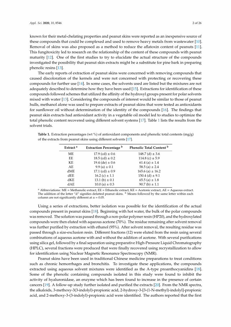
The early reports of extraction of peanut skins were concerned with removing compounds thatcaused discoloration of the kernels and were not concerned with protecting or recovering thesecompounds for further use [14]. In some cases, the solvents used are listed but the mixtures are notadequately described to determine how they have been used [15]. Extractions for identification of thesecompounds followed schemes that utilized the affinity of the hydroxyl groups present for polar solventsmixed with water [13]. Considering the compounds of interest would be similar to those of peanuthulls, methanol alone was used to prepare extracts of peanut skins that were tested as antioxidantsfor sunflower oil without determination of the identity of the compounds [16]. The findings thatpeanut skin extracts had antioxidant activity in a vegetable oil model led to studies to optimize thetotal phenolic content recovered using different solvent systems [17]. Table 1 lists the results from thesolvent trials.
Table 1. Extraction percentages (wt %) of antioxidant components and phenolic total contents (mg/g)of the extracts from peanut skins using different solvents [17].
Extract a Extraction Percentage b Phenolic Total Content b
ME 17.9 (cd) ± 0.6 148.7 (d) ± 3.6EE 18.5 (cd) ± 0.2 114.8 (c) ± 5.9KE 19.4 (de) ± 0.6 61.4 (a) ± 1.4AE 9.9 (a) ± 0.1 58.5 (a) ± 2.4
dME 17.1 (cd) ± 0.9 165.6 (a) ± 16.2dEE 16.2 (c) ± 1.1 150.4 (d) ± 9.1dKE 13.1 (b) ± 0.1 65.5 (a) ± 1.8dAE 10.0 (a) ± 0.3 90.7 (b) ± 1.1
a Abbreviations: ME = Methanolic extract, EE = Ethanolic extract, KE = Acetonic extract, AE = Aqueous extract.The addition of the letter “d” signifies defatted peanut skins. b Means followed by the same letter within eachcolumn are not significantly different at α = 0.05.
Using a series of extractions, better isolation was possible for the identification of the actualcompounds present in peanut skins [18]. Beginning with hot water, the bulk of the polar compoundswas removed. The solution was passed through a non-polar polymer resin (HP20), and the hydroxylatedcompounds were then eluted with aqueous acetone (70%). The residue remaining after solvent removalwas further purified by extraction with ethanol (95%). After solvent removal, the resulting residue waspassed through a size-exclusion resin. Different fractions (12) were eluted from the resin using severalcombinations of aqueous acetone with and without the addition of acetone. With several purificationsusing silica gel, followed by a final separation using preparative High-Pressure Liquid Chromatography(HPLC), several fractions were produced that were finally recovered using recrystallization to allowfor identification using Nuclear Magnetic Resonance Spectroscopy (NMR).
Peanut skins have been used in traditional Chinese medicine preparations to treat conditionssuch as chronic hemorrhages and bronchitis. To investigate these applications, the compoundsextracted using aqueous solvent mixtures were identified as the A-type proanthocyanidins [18].Some of the phenolic containing compounds isolated in this study were found to inhibit theactivity of hyaluronidase, an enzyme which has been found to increase in the presence of certaincancers [19]. A follow-up study further isolated and purified the extracts [20]. From the NMR spectra,the alkaloids, 3-methoxy-3(3-indolyl)-propionic acid, 2-hydroxy-3-[3-(1-N-methyl)-indolyl]-propionicacid, and 2-methoxy-3-(3-indolyl)-propionic acid were identified. The authors reported that the first

Appl. Sci. 2020, 10, 8546 3 of 26
two compounds had never been isolated from a natural source. Other compounds identified wereseveral flavonoid glycosides. Further research with this extraction technology allowed for the isolationand identification of several oligomeric proanthocyanidins [21]. The activity of the flavan-3-ols,catechin and epicatechin and their oligomers, and the proanthocyanidins has been the subject ofresearch in cocoa sources and tea [22]. As those found in peanut skins are composed of the A linkageform compared to the B or C forms in those other sources, comparison studied have investigatedthe comparative activity. The difference in the 3-dimensional structure of the compounds due to theextra 2β-O7 linkage in the A-type found in peanut skins could affect their interaction with membranephospholipids (Figure 1).
Appl. Sci. 2020, 10, x FOR PEER REVIEW 3 of 26
several flavonoid glycosides. Further research with this extraction technology allowed for the isolation and identification of several oligomeric proanthocyanidins [21]. The activity of the flavan-3-ols, catechin and epicatechin and their oligomers, and the proanthocyanidins has been the subject of research in cocoa sources and tea [22]. As those found in peanut skins are composed of the A linkage form compared to the B or C forms in those other sources, comparison studied have investigated the comparative activity. The difference in the 3-dimensional structure of the compounds due to the extra 2β-O7 linkage in the A-type found in peanut skins could affect their interaction with membrane phospholipids (Figure 1).
Figure 1. Structures of the procyanidin dimers. (a) The A-form found in peanut skins. (b) The B-form found in cocoa.
As this has proven to influence membrane fluidity and thus the ability of the compounds to interact with free radicals within the cell, the action of the different forms was examined [23]. A system using fluorescent probes that could be oxidized by peroxyl radicals that were induced by the assay on liposomes was used. The effectiveness was found to be dependent on the number of available flavanol monomers present, so no significant differences were seen between the types of dimers and trimers. The A-type dimer did have a different effect on the ordering of the core, with different rigidification indicating a different type of reaction with this form of the dimer. It has been proposed that the rigidity of the A-type due to the additional bond between the adjoining flavan-3-ols plays a role in their interaction with large biomolecules [24].
The color of peanut skins has been proven to have a relationship with the composition of the phenolic compound present [25–29]. In an examination of all the market types with skin colors from light brown to dark red, total flavonoids were not found to be as closely correlated to skin color as they were to growing location. The statistical relationship between the total flavonoids and the procyanidin content indicated that these compounds are the major flavonoids present. This indicates these compounds are more sensitive to stress conditions. The total phenols were more closely related to the hue of the skin color. Only the peanuts with the black seed coats were found to contain cyanidin-3-O-sambubioside both free and in glucosides [27]. Black seed coated peanuts were used to study the genetic control of peanut seed coat color [30]. In this report, the flavonoids present in white, red, and striped peanut skins in addition to those in the black peanut seed coats were extracted using 75% methanol in water acidified with 0.5% acetic acid. The extracts were filtered, and the flavonoids were identified and quantified using High-Pressure Liquid Chromatography-Mass Spectrometry-
Figure 1. Structures of the procyanidin dimers. (a) The A-form found in peanut skins. (b) The B-formfound in cocoa.
As this has proven to influence membrane fluidity and thus the ability of the compounds tointeract with free radicals within the cell, the action of the different forms was examined [23]. A systemusing fluorescent probes that could be oxidized by peroxyl radicals that were induced by the assay onliposomes was used. The effectiveness was found to be dependent on the number of available flavanolmonomers present, so no significant differences were seen between the types of dimers and trimers.The A-type dimer did have a different effect on the ordering of the core, with different rigidificationindicating a different type of reaction with this form of the dimer. It has been proposed that therigidity of the A-type due to the additional bond between the adjoining flavan-3-ols plays a role intheir interaction with large biomolecules [24].
The color of peanut skins has been proven to have a relationship with the composition of thephenolic compound present [25–29]. In an examination of all the market types with skin colorsfrom light brown to dark red, total flavonoids were not found to be as closely correlated to skincolor as they were to growing location. The statistical relationship between the total flavonoidsand the procyanidin content indicated that these compounds are the major flavonoids present.This indicates these compounds are more sensitive to stress conditions. The total phenols weremore closely related to the hue of the skin color. Only the peanuts with the black seed coatswere found to contain cyanidin-3-O-sambubioside both free and in glucosides [27]. Black seedcoated peanuts were used to study the genetic control of peanut seed coat color [30]. In this report,the flavonoids present in white, red, and striped peanut skins in addition to those in the blackpeanut seed coats were extracted using 75% methanol in water acidified with 0.5% acetic acid.

Appl. Sci. 2020, 10, 8546 4 of 26
The extracts were filtered, and the flavonoids were identified and quantified using High-PressureLiquid Chromatography-Mass Spectrometry-Time of Flight (HPLC-MS-TOF). Although differentflavonoids and their glycosides were found to be unique to the different colored peanut skins, they werefound to have the same biosynthetic pathways for anthocyanins but with different modifications.Individual flavonoids were isolated and identified from the skins of black seed-coated peanuts afterextraction with acidified water followed by a partition into ethyl acetate [29]. The extracts werefractionated using Amberlite XAD-7HP resin to remove the most polar compounds followed byYMC® Gel ODS-AQHG resin to separate the hydrophilic ones and identified using High-PressureLiquid Chromatography-Electrospray Ionization-Mass Spectrometry (HPLC-ESI-MS/MS) and NMRspectroscopy. Three unique flavonoids (quercetin-methylpentoside, quercetin-feruloyl-hexoside,quercetin-3-dihexoside) and four anthocyanins (cyanidin-3-o-sophoroside, cyanidin-3-o-sambubiside,cyandin-3-o-glucosylrutinoside, cyanidin-3-o-xylosylrutioside) were identified.
Using HPLC to determine the various compounds that compose peanut skins has advancedfrom initial reports of the oligomers [31]. Aqueous ethanol was the extractant for the analysis ofpeanut skins, and the method identified five phenolic acids (gallic acid, caffeic acid, p-coumaric acid,protocatechuic acid, ellagic acid), two stilbenes (piceid, resveratrol), and eight flavonoids (catechin,epicatechin, epigallocatechin, catechin gallate, epicatechin gallate, epigallocatechin gallate, procyanidinB2, quercetin). This report validated the methodology for the quantitation using the runner, Spanish,and Virginia market types. Concentrating specifically on peanut skins from the Virginia market type,a study compared the compounds extracted when either methanol, ethanol, acetone, or water atboiling temperature was used individually [32]. Using 100% acetone resulted in the highest amountof the smallest phenolic compounds. The study used HPLC-MSn to identify a range of compoundsthat were polymers of catechin and epicatechin with and without sugar moieties up to 9 catechins.These polymers were found mostly in the A form. An advanced study to determine the content andstructural formation of the trimer and tetramer of the procyanidins used extraction with 30% methanolin water followed by 70% acetone in water [24]. The extracts were combined, and the solvent wasevaporated to dryness. The dried material was dissolved in water and partitioned against ethyl acetatecontaining increasing amounts of aqueous ethanol (5%, 10%, 15%). The extracts were then loaded ontosilica gel and rinsed with a series of solvents to purify and isolate the components. Identification wasperformed using NMR and electron circular dichroism spectroscopy (CD). The different polymerswere tested for anti-inflammatory activity in a macrophage system and the tetramers were found to bethe most effective with the dimers having little or no effect. This showed that biological systems coulddifferentiate between the different forms of the procyanidins.
As studies became more sophisticated to examine the composition of peanut skins in order todetermine the source of bioactivity, multistep extraction schemes were used to selectively isolatecertain compounds. A series of methanol and water mixtures was used to remove specifically theA-type procyanidins [33]. Drawing on previous research, the extraction scheme was created [18,20,21].Peanut skins were extracted sequentially with 20% aqueous methanol, 70% aqueous methanol,and then finally 70% aqueous acetone. Each fraction was concentrated and then partitionedinto ethyl acetate to remove soluble saccharides. The fractions were then analyzed by HPLC-MS,which revealed the 20% methanol fraction contained mostly oligomers of the A-type procyanidinsand the 70% acetone fraction contained larger polymers. The use of peanut skins as a specific sourceof compounds for isolation has been described. Peanut skin has been a source of resveratrol andwas extracted using 20% ethanol in water [34]. Increasingly, previously unidentified compoundshave been reported from peanut skin extractions. Researchers have described an A-type procyanidin(epicatechin-(2β→O→7,4β→8)-[catechin-(6→4β)]-epicatechin) that had not been reported before afterextraction of peanut skins with 70% acetone in water, followed by fractionation on Sephadex LH-20and elution with ethanol [35].
Research to differentiate between the classes of procyanidins present in peanut skins has usedthe technique of hydrogen/deuterium exchange (HDX) to elucidate the structural differences between

Appl. Sci. 2020, 10, 8546 5 of 26
isomers [36]. The technique was able to differentiate between the A-type procyanidins in peanutskin extracts containing up to three linkages. The extracts were prepared using 70% acetone in watercontaining 0.1% formic acid after defatting of the peanut skins. The filtered extracts were dried undernitrogen gas and then reconstituted in the deuterated mobile phase.
As a follow-up to a previous study of the composition of whole peanuts, the authors used thesame extraction system of 80% methanol in water after defatting with hexane to compare peanut skinsfrom two different peanut market types [37,38]. The analysis using HPLC-MS of the extracts foundthat the Valencia market type peanut skin extracts had higher levels of flavonols, quercetin, and itsmethylated analog, isorhamnetin.
One of the most complete studies of the phenolic type compounds in peanut skins used theextraction procedure developed for the analysis of grains to prepare samples for instrumentalanalysis [39]. The peanut skins analyzed were obtained after commercial blanching, which requiresmild heat treatment. As heat treatment has been reported to liberate smaller phenolic compounds fromlarger polymers, this proved advantageous in allowing for a range of identifications [40]. Acidifiedwater was used for the initial extraction followed by a partition into diethyl ether to capture the freephenolic compounds. A solvent exchange of methanol for the ether was performed before the analysisby HPLC-MS. From this, 88 individual phenolic type compounds were found, although some couldonly be identified by their class (Table 2). In addition, 60 proanthocyanidins were found, with mostbeing of the A-type. The same group used the same extraction scheme with fractionation to determinethe bound phenolics [41]. After the extraction and portioning of the free phenolics into diethyl ether,the aqueous phase was then base-hydrolyzed and acid-hydrolyzed to convert the ester derivatives totheir carboxylic acid or flavonoid analogs. The analysis was then done with the same LC-MS system.An additional 78 compounds that existed as esters or glycosides were tentatively identified. The readeris referred to these publications for the listings of these compounds.
Table 2. Content of selected phenolics quantified in dry-blanched peanut skins (PS) by C18 ReversePhase High-Pressure Liquid Chromatography (RP-HPLC) [39].
Free Phenolic Compounds a Content b (mg/100 g)
Protocatechuic acid 3.43 ± 0.04
p-Hydroxybenzoic acid 1.03 ± 0.06
Caftaric acid 51 ± 0.12
cis-Coutaric acid 10.1 ± 0.52
trans-Coutaric acid 2.11 ± 0.08
p-Coumaroyl-O-pentoside 5.52 ± 0.23
p-Coumaric acid 0.53 ± 0.06
Chicoric acid 3.44 ± 0.12
p-Coumaroylcaffeoyltartaric acid 2.26 ± 0.13
Chicoric acid 3.12 ± 0.13
di-p-Coumaroyltartaric acid 13.8 ± 1.53
p-Coumaroylsinapoyltartaric acid 6.32 ± 0.94
p-Coumaroylferuloyltartaric acid 5.87 ± 0.71
trans-Resveratrol 0.36 ± 0.05
Quercetin 2.11 ± 0.27
Isorhamnetin 1.51 ± 0.02
Diosmetin 0.40 ± 0.01a Caftaric acid and chicoric acids were quantified as caffeic acid equivalents; coutaric acids and other p-coumaroylderivatives were quantified using p-coumaric acid equivalents; isorhamnetin and diosmetin were quantified usingcorresponding flavonoid aglycone equivalents. b Values are reported as means of triplicate analyses ± standarddeviation. Findings are reported as mg respective phenolic/100-g dry weight (d.w.) of dry-blanched PS.

Appl. Sci. 2020, 10, 8546 6 of 26
2.2. Extraction Optimization
The microwave-assisted extraction was another method used for peanut skins [42]. In this case,30% ethanol in water was chosen as this had previously been proven to be the optimum for totalphenolic extraction [43]. Surface response methodology was used to determine the power and time toextract the highest total phenols without decreasing the antioxidant effect. It was reported that 30 s at90% microwave power produces the optimum product.
As the interest in recovering phenolic compounds from peanut skins increased, studies to optimizetheir recovery were done. Most of these studies concentrated on achieving the highest values using theFolin-Coicalteu assay rather than any defined class of polyphenols [44]. Factors such as particle size ofthe skin material, the proportion of solvent to the mass of skins, contact time with the skin material,maceration or shaking, and the number of extractions using 70% ethanol in water were evaluated.The efficiency of the extraction was evaluated using the 2,2-diphenyl-1-picrylhydrazyl (DPPH) assayfor free radical scavenging power, which indicated that the method of agitation was not significant andthat 10 minutes of extraction at 40 mL per 5 grams of peanut skins using 4 extractions was optimum.An optimization study examining proanthocyanidins from peanut skins used aqueous ethanol andultrasonic extraction [45] (an aqueous solution of ethanol (55%) at an outlet power of 120 W at 35 ◦C).This resulted in the extraction of proanthocyanidins with a yield of 12.1%.
Water only was used to extract primarily the procyanidins that were then used to evaluatethe potency to suppress allergenic response [46]. Further purification of the extracts was donewith a reverse-phase resin to bind the polyphenols. Successive elutions were made with increasingconcentrations of acetone in water and finally with 80% ethanol in water. The final eluate was thenfurther purified using a size-exclusion resin, followed by a gel filtration resin and finally with silica gel toisolate specifically the procyanidin A-1 for further testing. When compared with the unpurified extractfrom the peanut skins, the isolated compounds were not as potent in suppressing immunoglobulinsynthesis and regulating systemic T helper cytokine production.
Studies dedicated to the optimization of the extraction of phenolic compounds from peanut skinsused response surface methodology and various chemical assays for comparisons [47,48]. Ethanoland methanol at concentrations of 30%, 60%, and 90% in water were compared with pure water andpure ethyl acetate. The total phenolics recovered (TPC) were optimized in ethanol at 30% (118 mgGallic Equivalents per gram skins) and in methanol at 60% (112 mg Gallic Equivalents per gram skins).Pure water recovery was lower at 81 mg and ethyl acetate recovery was an order of magnitude lower.Using the Oxygen Radical Absorbance Capacity (ORAC) activity as the measure of antioxidant activity,the optimized alcohol extracts were similar (2050 µmol Trolox equivalents per gram for ethanol and2149 µmol Trolox equivalents per gram for methanol). Pure water was less than half as effective.Varying the temperature of the extraction was investigated in this study. Increasing the temperaturefrom 30 ◦C to 60 ◦C using 30% ethanol in the water had no effect on the TPC recovery but when using 30%methanol, increasing the temperature to 60 ◦C resulted in a 20% increase in the TPC. This was attributedto methanol being better able to solubilize more polar compounds. The use of microwave-assistedextraction was compared with the solvent extractions using conventional heating and mechanicalshaking with the optimized solvent mixtures. The microwave procedure increased the ORAC activity to2789 µmol Trolox equivalents per gram when using the 30% ethanol in water mixture as the extractionsolvent. The main savings in this procedure is the time involved was less without compromising theactivity of the extracts. Optimization of extraction using responsive surface methodology was alsoused to find the most effective conditions to extend oil shelf life [48]. Using ethanol in water over arange of 20% to 100% with a variation of time from 5 to 150 min and temperature from 25◦ to 90 ◦C,the study used both the DPPH and the 2,2′-Azino-bis(3-ethylbenzothiazoline-6-sulfonic acid (ABTS+)assays to determine the optimal conditions. It was found that length of time was not of significance butthat temperatures above 60 ◦C with 75% ethanol in water were optimum. In addition, soybean oil withthe optimized extract added at 25 to 1000 mg/kg was subjected to accelerated conditions of agitationat 60 ◦C and compared to the addition of 200 mg/kg of butylated hydroxytoluene (BHT), a synthetic

Appl. Sci. 2020, 10, 8546 7 of 26
antioxidant compound. It required 750 mg/kg of the optimal extract to produce the same antioxidanteffect of 200 mg/kg of BHT, the synthetic chemical antioxidant.
Isolation of specific proanthocyanidins was performed using preparative HPLC [49]. This allowedfor more automated separation of peanut skin extracts. Using earlier work as a guide [21,32], acetone inwater (60%) that had been acidified was used as the extractant. An initial purification was performedusing Amberlite resin eluted with methanol. This was followed by fractionation on Sephadex LH-20.The fractions were evaluated using the DPPH assay and the most active fraction was further purifiedusing preparative HPLC. This allowed for the isolation of proanthocyanidins A1 and A2 with themajor portion of the isolate being the A1 form. The most recent study of the composition of peanutskin extracts compared them to grape skin extracts [50]. The skins were extracted using 80% ethanol inwater following a procedure previously published [44]. The extracts were purified using AB-8 resinto purify the procyanidins. The extracts were then evaluated using ORAC, DPPH, ABTS+, and theFerric Reducing Antioxidant Power (FRAP) assays as well as a cell assay using Human hepatocellularcarcinoma (HepG2 cells). Both the A- and the B-type of the procyanidins were identified by HPLC-MSwith the A-type predominating as found by others. In most of the assays, the peanut skin extracts hadslightly higher values than the grape skin extracts, with the exception of the cell-based assay wherethe grape skin extracts scored higher. To release the phenolics from peanut skins by the degradationof the actual cell walls for the skins, enzymatic digestion was used [51]. Cellulase was used at anincubation temperature of 55 ◦C followed by extraction with aqueous ethanol at 45%. The yield ofproanthocyanidins was 16.17%.
An exploration of the effectiveness of heat in increasing the solubility and thus the extractionefficiency of solvent in removing phenolic compounds from peanut skins was performed. As many ofthese compounds are found to be bound into sugars and other cellular structures, the authors theorizedthat heat treatment of the skins before extraction would increase the yield [52]. Using temperaturesfrom 90 ◦C to 180 ◦C, peanut skins from the market types, runner, Virginia, and Spanish were extractedusing 70% ethanol in water solutions. TPC and antioxidant activity using the Trolox equivalentantioxidant capacity (TEAC) and the Peroxy-radical trapping capacity assays were compared. Heatingto 135 ◦C resulted in the highest TPC in the runner peanuts (280 mg/g). Both the Virginia and Spanishpeanuts did not show any significant change in TPC with heating within the tested range and wereboth lower (149 mg/g for the Virginia type and 137 mg/g for the Spanish type) than the runner type.Correspondingly, the activity of the peanut skin extracts was not significantly different between themarket types (1.38 mg TEAC/g dry skin), except for the higher value of the runner type peanuts heatedat 135 ◦C (2.56 mg TEAC/g dry skin). Only the runner type peanut skins showed any significantPeroxy-radical trapping capacity. The increase in TPC in the runner type was attributed to the formationof Maillard products due to the heat treatment and/or the liberation of phenolics from larger complexes.The differences in market types were attributed to different types of phenolic compounds or complexesbeing present and some may have been more labile to heat.
2.3. “Green” Extractions
Supercritical extraction uses pressure to condense a solvent or solvent mixture with temperaturesabove the boiling point of the solvents so that the dielectric constant is decreased [53]. This results in achange in the polarity so that it becomes similar to solvents such as methanol, ethanol, and acetone atroom temperature. This allows for the use of more “green” or less toxic chemicals to be used withenhanced solubility of phenolic compounds in the case of peanut skins. A study using this technologyoptimized the total phenolic content of extracts from peanut skins to be 10 min at 220 ◦C using 60.5%ethanol in water [53]. This shows the potential of using a more environmentally friendly technique.Further study with this technique to extract peanut skins determined the effect of the size of theparticles of peanut skin extracted [54]. Peanut skins milled to particle sizes of 300, 355, 425, and 500 µMwere extracted using both traditional Soxhlet extraction and supercritical extraction using carbondioxide. Optimum yields per weight were obtained by both methods when the particle size was

Appl. Sci. 2020, 10, 8546 8 of 26
425 µM and pure ethanol was the solvent compared to either pure water or n-hexane. Although theyield from the Soxhlet extraction using ethanol was higher (36.38% by weight) than carbon dioxideusing the supercritical system (15.53%), the antioxidant activity using the DPPH assay was higherfor the supercritical system (93.43%) than for the Soxhlet extraction (62.11%). This was attributedto less temperature degradation of the polyphenols by the lower temperatures of operation for thesupercritical system. The catechin recovery from this system was higher (139.92 µg/g peanut skin)compared to Soxhlet extraction (31.79 µg/g peanut skin) when ethanol was used as the solvent [55].More recently, this system has been used to prepare extracts for investigations into the antioxidantprotection in cell systems [56]. The extracts were compared to the activity of quercetin, which wasthought to be the main compound present. After proving the additives were not cytotoxic up to250 µg/mL, O2
− scavenging activity was assayed. Quercetin was assayed to be seven times higher thanthe peanut skin extract, which was attributed to the pro-oxidant properties of the catechins present inthose extracts. The addition of the quercetin and the peanut skins along with an oxidative stressor wasproven to be protective against oxidative stress, whereas pretreatment was not.
A system that used ultrasound was applied to peanut skins with the specific goal of recovering thestilbenoid, resveratrol was reported [57]. Yeast CICC1912, Aspergillus oryzae 3.951, and Aspergillus niger3.3148 were immobilized onto cellulose beads. Peanut skins were extracted with 80% ethanol inwater and the extract was dried. Optimization of the recovery of resveratrol was performed usingseveral surfactants, liquid-solid ratios, ultrasonic powers, and culture temperatures and times. At theoptimized conditions, recovery of 96.58 µg/g of resveratrol was obtained, which was four-fold higherthan from an untreated sample.
3. Bioactive Compounds in Peanut Skins
3.1. Chemical Antioxidant Activity
The antioxidant activity of peanut skin extracts was first investigated as a side project fromresearch on peanut hulls [16]. The total phenolic content was reported using the Folin-Coicalteu assayand the antioxidant activity in sunflower oil was reported to be less than that of BHT, a commonly usedsynthetic antioxidant ingredient for food oils. Isolated compounds from peanut skins using a series ofextractions and chromatography were found to have free radical scavenging ability when evaluatedusing the DPPH assay. These included flavonoid glycosides [19] and proanthocyanidins [21].
A comparison study of several black seed-coated peanuts with red seeded ones evaluated thetotal phenolic content (TPC), content of several phenolic classes of compounds, and several antioxidantassays [28]. Extraction of the peanut skins was performed using 70% acetone in water acidified withacetic acid after defatting with hexane. The TPC was not found to vary widely between the varieties.The skins from red seed-coated peanuts were found to be higher in total flavonoid and total condensedtannin content than the black seed coated ones, although the black peanut skins had higher anthocyanincontents. Correspondingly, one of the red seed-coated peanut types had higher antioxidant powerwhen measured using the FRAP assay but all the peanut skins from the seeds tested had similar freeradical reducing power when measuring using the DPPH assay.
Phenolic compounds in peanut skins are increased by stress conditions. Fungal attack as a sourceof stress was investigated [58]. Peanuts with skins of red, reddish-brown, and black colors wereinoculated by the fungus, Rhizopus oligosporus. The seeds were then germinated for 3 days in the dark.The germinated seeds and ungerminated seeds used as the controls were extracted using a mixtureof acetone, ethanol, and water (2:2:1 v/v/v) that had been acidified. From the extracts, 45 differentcompounds including phytoalexins were identified using HPLC-MS. In addition, the antioxidantactivity was determined using total phenolic content (TPC), total flavonoid content (TFC), oxygen radicalabsorbance capacity (ORAC), hydroxy radical absorbance capacity (HORAC), superoxide radicalabsorbance capacity (SORAC), and DPPH. In general, the black skins had higher activity in most of the

Appl. Sci. 2020, 10, 8546 9 of 26
assays, with the exception of SORAC. The red and reddish-brown colored skins tended to be similar instrength. In most cases, the treated seeds had higher activity than the untreated controls.
Aqueous methanol continued to be used in studies after defatting with hexane [59]. The extractswere found to have little antioxidant activity when incorporated into vegetable oils and lard but werefound to test high in free radical activity testing using chemical tests. The extracts were found to havemetal-chelating activity. The same extraction scheme was used to compare the effectiveness of skinsfrom high oleic peanuts with those from peanuts with normal oleic acid levels [60]. No significantdifferences were found in the antioxidant power of the extracts from the two different types of peanuts.
The use of several solvents individually without the addition of water was studied to determinethe yield of soluble material by weight [61]. The highest recoveries were from ethanol, followed bymethanol and acetone. Although the amount of solids recovered from these solvents was equivalent(ca. 200 mg from 5 g peanut skin), the acetone extract had only 3% of the antioxidant activity comparedto the alcohol extracts (3% vs. 9.8% for ethanol and 93.5% for methanol) using a β-carotene-basedassay. Hexane and ethyl acetate yielded much less solid material (35 mg and 90 mg, respectively)and comparable activity to the acetone extract (3%). This proved the active components are polarcompounds. Differences between peanut skin extracts from raw and roasted peanuts were determinedusing a series of solvents and solvent mixtures [62]. Pure ethanol, 80% ethanol in water, pure ethylacetate, and 80% ethyl acetate in water were used. The highest yields by both weight and total phenoliccompounds were found with the ethanol in the water mixture followed by pure ethanol. Higher levelswere also found in the roasted peanut skins compared to the peanut skin extracts from the raw peanuts.The roasted peanut skin extract prepared using 80% ethanol in water was found to be effective asan antioxidant when added to sunflower oil, but even at 800 ppm, the effectiveness as monitoringusing the Peroxide Value assay was less than that of BHT added at 200 ppm. Using only methanol,raw peanut skins were extracted for determination of total phenolic content (TPC), antioxidant activityusing the DPPH assay, and inhibition of linoleic acid peroxidation [63]. Only 91.74 mg/g of TPC werefound in this study compared to 148.7 mg/g reported in another study [17]. This is an example of thevariability of the levels of extractable compound present in peanut skins.
The effectiveness of different solvent mixtures in terms of antioxidant activity was comparedwith ethanol and methanol [40,64]. Methanol or ethanol in water (80%) were used. The lipid in theextract was removed by partitioning into chloroform. The aqueous portion was then extracted withethyl acetate to remove the nonphenolic compounds remaining as described by the methodologydeveloped for purifying phenolic compounds from tea [65]. The aqueous portion was freeze-dried andthe resulting powder dissolved in methanol for testing. A combination of solvents was used to prepareextracts from different parts of the peanut, including the skins that contained the different classes ofpolyphenols [66]. The initial extraction of peanut skins from a local market was performed using equalparts of methanol and water. The skins were then extracted with 70% acetone in water and the 2 extractswere combined. This represented the extractable polyphenols in the authors’ description and was usedto determine the total phenolic content (TPC) using the Folin-Coicalteu assay. The non-hydrolyzablepolyphenols or condensed tannins were determined after treating the extracts with acidified butanol.The extracts were then evaluated using chemical assays for antioxidant activity, anticancer activity in acell-based assay, and as an antioxidant by the heating of oil spiked with the peanut skin extracts.
The solvent mixtures and solvent-assisted extraction were used in a study of various portions ofpeanuts including peanut skins [67]. A mixture of acetone and water (70%/30%) was acidified andused as the solvent. Acidification was used to free phenolics from glycoside complexes. Peanuts weresubjected to increasing time of roasting to produce Maillard browning products. Increased roastingwas found to increase the total phenolic content as well as corresponding ORAC assay values.A methanol/water mixture (50/50 v/v) followed by a mixture of acetone and water (70/30 v/v) wasused to extract peanut skins, hulls, and defatted peanut flour, both raw and roasted [66]. The extractswere evaluated using TPC, nonhydrolyzable tannins (NEPP), and oxidative stability test on flaxseed

Appl. Sci. 2020, 10, 8546 10 of 26
oil. The extracts from the skin tested much higher than the other parts of the peanut with the roastedsamples being lower than the raw ones.
The procyanidins have been found in a range of plant parts in several forms. Comparison of thosefound in peanut skins with those from other foods has been the subject of several studies [7,23,65,68,69].As previously discussed, peanut skins contain primarily the A-type. One report revealed both A-typeand B-type dimers showed high antioxidant potency in a dose-dependent manner in the DPPH andABTS+ assays [70]. However, the type of system used had an effect. In general, the B-type dimersshowed a higher radical scavenging potency when tested in aqueous systems, whereas in tissue orlipid systems, the A-type dimers were just as effective or had even higher antioxidant potency thanB-type ones.
Comparisons of the activities of extracts from peanut skins with other agricultural by-productsused extracts prepared using 80% ethanol in water [71]. Of the materials tested, mango and grapeskins and seeds and peanut skins, the highest amounts of flavonoids and total phenolics werefound in the peanut skins on a dry weight basis. The DPPH activity of the peanut skin extract(756.54 ± 65.45 mg TE/g) was not significantly different from the grape extract (728.46 ± 92.26 mg TE/g)but was higher than that of the mango extract (158.53 ± 4.95 mg TE/g). In another study, using 20%methanol in water, the types of procyanidins recovered were compared [69]. Peanut skin extracts werefound to contain mainly the A-type dimers and grape skins almost exclusively the B-type dimers.
3.2. Antimicrobial Activity
As phenolic compounds are known to have antimicrobial properties, it is logical to examine theantimicrobial properties of peanut skin extracts. The first report used 80% ethanol in water to extractpeanut skins [72]. The extract was used to determine the antioxidant power in meat when compared tosynthetic antioxidants. While levels of 0.06% to 0.10% peanut skin extract had the equivalent power of0.02% synthetic antioxidant in delaying the onset of rancidity in the meat samples, the inhibition of themicroorganic growth was much less than a chemical antibiotic.
Certain phenolic compounds including proanthocyanidins have proven antimicrobial properties.The study of extracts from peanut skins against certain bacterial strains has used thisjustification [68,73,74]. Peanut skins were initially extracted into boiling water, then portionedinto ethyl acetate to isolate the proanthocyanidin fraction [73]. After solvent removal, the extractswere incorporated into growth media containing apple juice to create a model food system.Saccharomyces cerevisiae, Zygosaccharomyces bailii, and Zygosaccharomyces bisporus were used as thetest organisms in individual cultures. Although peanut skin extract proved to inhibit the yeast growthdue to alteration of the cell membranes, the amount needed to be effective was thought to be toohigh for practical use in food. Using acetone in water acidified with acetic acid (70:28:2), hexanedefatted peanut skins were extracted to recover the polyphenols [68]. Plated media were dosed withincreasing amounts of the extracts in water from 250 to 1500 ppm. The plates were inoculated usingSalmonella typhimurium, E. coli OH157:H7, or Listeria monocytogenes as the test organisms. No inhibitionof Listeria monocytogenes was seen by the addition of peanut skin extract and less inhibition wasseen in the growth of the other organisms tested when compared to similar amounts of grape seedextracts. This was attributed to the B-type procyanidins present in grape seed extracts being moreeffective than the A-type procyanidins found in peanut skin extracts. Comparison of the antimicrobialeffects of extracts from peanut skins with those from dry blanched peanuts used 70% acetone inwater as the extractant [74]. The peanut skin extracts were found to be less effective in inhibiting thegrowth of a range of both gram-positive and gram-negative organisms by an order of magnitude.This was attributed to the phenolics from the blanched peanut extracts containing small phenolicsrather than polymers of phenolic compounds. Peanut skins were extracted with 80% ethanol in waterand then either freeze-dried or spray-dried with maltodextrin as a carrier [74]. The antimicrobialaction against Staphylococcus aureus, Listeria monocytogenes, Salmonella enterica subs. Enterica serovarEnteritidis, and Escherichia coli was evaluated. None of the powders showed any inhibition of

Appl. Sci. 2020, 10, 8546 11 of 26
Escherichia coli. The freeze-dried extract was the most effective in inhibiting the other pathogenicorganisms. Although the spray-dried extracts were inhibitory as well, the lesser degree was attributedto the dilution of the phenolic content by the addition of maltodextrin in the preparation of the powders.
Antiviral activity in peanut skin extracts has been demonstrated in cell culture trials when used incombination with antiviral drug compounds [75]. Pure ethanol was used to extract defatted peanutskins. The effect was most pronounced during the early stages of infection. The effect was attributedto the polyphenols present. The resveratrol content was also determined but not found to showconcentration dependence.
Fungal growth on grain producing mycotoxins has damaging health effects when consumed andreduce the acceptability of the crops. A study demonstrates the effect of peanuts skin extracts using70% ethanol in the water on the growth of Fusarium verticilliodes on maize [76]. The different fractionsseparated on Sephadex resin had different effects on the inhibition with only one having a large effect.The different factions were only distinguished by their visible colors. The lightest colored fraction wasthe most effective, which would indicate that the procyanidins were not involved.
3.3. Anticancer Activity
Being able to purify the individual polyphenol compounds, especially the procyanidins, allowedfor further study of their biological effects. Using a scheme worked out earlier [21], 11 individualprocyanidins of the A-type as monomers, dimers, trimers, and tetramers were identified [77]. In culturedhuman melanoma cells, inflammatory cytokinetic production was reduced after lipopolysaccharidechallenge. Dimers and trimers showed greater effects than the monomers or tetramers. This providedscientific insight into some of the effects attributed to peanut skins by traditional medicine.The effectiveness of peanut skin extract as an anticancer agent was demonstrated using humanprostate cancer DU145 cells [78]. Peanut skins were extracted using 70% ethanol in water andthen subsequently fractionated by size exclusion chromatography using ToyoPearl HW-40S resin.The procyanidin B3 was determined to be the most active against the proliferation of the cancer cells.
Resveratrol is one of the naturally occurring phytoalexins which plants produce in responseto stress such as a fungal attack. Peanut skins have been reported to contain this compound [79].This compound has been reported to have chemoprotective properties against cancer. The methodof action has been proposed to be interference with the pathways of signal transduction, exert somecontrol on cell-cycle regulating proteins, and has been shown to induce apoptosis in some cancercells [80]. Using peanut skins as a source of these compounds, a skin-lightening effect was found usingreptile cells [81].
In addition, cell systems using human tumor carcinoma cell lines (liver, colon, cervical, and breast)were treated with the extract from the roasted peanut skins. The effective dose for reducing cellsurvival to 50% (IC50) differed for the type of cell. The largest dose was needed for the liver tumorcells (19.3 µg/mL) and the lowest for the colon tumor cell line (10.9 µg/mL). There was no effect on thebreast tumor cell line. No specific compound was identified in the study as responsible for the effects.Using the human cervical adenocarcinoma (HeLa) cell line, the effectiveness of peanut skin extracts asan inhibitor of enzymes was tested against histone deacetylase [82]. The increased activity of histonedeacetylases has been associated with certain human cancers. Based on a previous publication [40],100% methanol was used to extract peanut skins. Treatment with the peanut skin extract resulted in theaccumulation of the acetylated forms of histone proteins, indicating inactivation of the deacetylases.As such, the extracts were proposed as a possible supplemental cancer treatment.
3.4. Enzyme Inhibition
The action of phenolic compounds as inhibitors of carboxylases has been reviewed [83]. This typeof activity has been determined in peanut skins [84]. Acetone (70% in water) was used to extractpeanut skins and the extracts were fractionated using microfiltration and Sephadex LH eluted withincreasing acetone levels in water solutions. The structures of the compounds in the fraction with the

Appl. Sci. 2020, 10, 8546 12 of 26
highest action against α-amylase was found to be oligomers of polyflavan-3-ols, up to 15-units in sizeusing high-resolution Matrix-Assisted Laser Desorption Ionization-Time of Flight Mass Spectroscopy(MALDI-TOF-MS). These compounds were determined to contain catechin/epicatechin units togetherwith several afzelechin/epiafzelechin units and gallocatechin/epigallocatechin units with both A-typeand B-type linkages. A recent review included information about the ability of methanol extracts ofpeanut skins to inhibit α-amylase and lipid peroxidase [85]. Alpha-glucosidase and lipase play a rolein the absorption of glucose and triacylglycerols from the diet. The inhibition of these enzymes couldplay a role in the management of diabetes and weight. Peanut skins were extracted using 70% acetonein water and partitioned into ethyl acetate as described in a previous study [58]. This was to ensurethe extraction of the soluble phenols. The residue was treated with acid and the free phenolics wererecovered in a mixture of ethyl acetate and ethyl ether [86]. Subsequent treatment at alkaline pH by theaddition of sodium hydroxide freed the phenolic compounds bound into esters. A final extraction intomethanol was done to recover those compounds. All the phenolic compound classes showed inhibitionof the enzymes with the greatest effect being from the free phenols and the least by the insolublebound compounds. Along with white grape pomace, peanut skins were investigated for their abilityto inhibit the enzymes, α-amylase, and pancreatic lipase to propose applications for agricultural wastematerials [87]. Peanut skins were extracted with ethanol using sonication, filtered, and lyophilizedfor analysis. Inhibition by peanut skins was found to be close to 100% for lipase and above 90% forα-amylase. These actions were higher than those of either red or white grape pomace extracts.
3.5. Effects in Animal Models
Animal models have been used to determine the effectiveness of the polyphenols in peanut skinsto influence blood chemistry. Studies on rats for lowering cholesterol used the water-soluble portionincorporated into a high-fat diet [88–90]. Filtration was used to partition the extracts into a highmolecular weight (MW) fraction and a low MW fraction. The findings were that the lower MW fractionwas more effective in slowing the transport of cholesterol from the diet into the intestinal transportsystem. Further study showed that the trimer was more effective than the dimer [90]. This wasattributed to the low MW fraction being better able to disrupt the micelles involved. In anotherstudy using the rat model, plasma, and liver triglyceride (TG) and cholesterol (TC) levels weresignificantly reduced, while fecal secretion of TG and TC was greatly increased upon peanut skinextract administration [91]. The effect of the extracts from peanut skins on the blood chemistry of the ratsubjects using a water-only extraction scheme is described by an earlier publication [92]. The plasma ofrats fed the peanut skin extracts as part of high fat or “Western” diet was monitored [90]. Total bloodlipids were reduced by the peanut skin fortified diet. The bioavailability of procyanidin A2 wasconsidered to be greater. Peanut skins extracted with 70% ethanol in water were used to evaluate theeffectiveness of peanut skin extract to mitigate the effects of an atherogenic diet in a mouse modelsystem [93]. The addition of 0.78% peanut skin extract to a high-fat diet produced some lowering ofhepatic cholesterol and glycogen levels in the test subjects. Extracts of peanut skins prepared using40% ethanol in water followed by elution from HP-20 resin with ethanol were used in a mouse modelstudy [94]. The dried extract was added to the animal diet at 4, 80, and 160 mg/kg of the body weight.At the two highest dosages, reduced body weights, food intake, adipose tissue, and expression ofleptin protein were reported. Extracts of peanut skins were made with either pure water or methanolto determine the protective effects of extracts against liver damage induced by chloroform (CCl4) inmice [95]. The animal subjects were fed either 50 or 100 mg/kg of extract. One group was fed priorto injection of CCl4 and another group was fed after injection. Ingestion of CCl4 is known to bothcause peroxidation of the liver membranes and to interfere with Ca2+ homeostasis, which leads to livercell damage. By monitoring levels of certain liver enzymes in the blood of the experimental animalsand examination of the liver tissues by microscopy, the study showed that the methanolic extract wasbetter able to decrease the levels of the plasma enzymes indicating less damage than the aqueousextracts. The action of the superoxide dismutase, a protective enzyme against oxidation, was increased.

Appl. Sci. 2020, 10, 8546 13 of 26
The physical examination of the liver cells also showed fewer fat cells and inflammatory damage whenthe methanol extracts were fed. This indicated that the antioxidant-rich methanolic extracts frompeanut skins had a protective effect on the liver of the test animals. Extraction of peanut skins hasbecome commercialized to provide material for further studies. Aqueous ethanol without furtherdefinition was described as the solvent for the extraction of peanut skins for a study of the effect ofthe material on the formation of blood platelets in an animal model [96]. The goal of the study wasto elucidate the mechanism of the observed action on hemorrhage described by traditional Chinesemedical practice.
The action of the anthocyanins from black peanut skins against UV-induced skin cell damagewas reported [97]. After extraction with acidified water followed by a partition into ethyl acetate aspreviously described [29], the anthocyanins were isolated and used to treat human keratinocyte cells(HaCaT cells) as well as the dorsal skin of live mice. After exposure to UV-B radiation, cell apoptoticdeath was investigated. The effectiveness was found to be dose-responsive, but high levels were foundto be toxic to the cells (over 40 µg/mL).
The abundance of tannins in peanut skins would indicate that feeding this material to animalswould be advantageous in that phenolics have been reported to have anthelmintic properties, that isthe ability to suppress internal parasites [98]. A study with lambs, a ruminant species, did not havepositive results [99]. Animals infected with gastrointestinal nematodes did not show any decrease infecal egg counts when fed at increasing levels up to 0.53% of their body weight per day with peanutskins incorporated into alfalfa pellets when compared to the control.
Current research in the action of components from food products has focused on the effectson the gut microbiota. Peanuts skin extracts have been investigated for this effect using a mousemodel [100]. Significant changes in the organisms present in the gut were observed. Most importantly,those involved in fatty acid biosynthesis and sugar metabolism were increased. This has positiveimplications in the compounds extracted from peanut skins to have possible antidiabetic effects.
4. Food Applications
In food, the use of peanut skin extracts as a functional food additive has been explored in severalways. Using water alone, peanut skins were extracted to create an infusion or “tea” [101]. Since theheat was required to produce the product, the chemical antioxidant strength was evaluated usingTPC, Trolox equivalent antioxidant capacity assay (TEAC), and the ORAC assay. The result showedthat the antioxidant potential was preserved and increased with the amount of peanut skins treated.The infusions were also found to be higher in response to the assays than green or black tea, indicatingthat peanut skins could have equivalent or better health properties. The preparation of an encapsulatedproduct from peanut skin extracts produced a free-flowing powder that could be used for incorporationinto foods was reported [102]. Antioxidant properties as defined by ORAC and total TPC weredescribed. Using the extraction scheme of 70% ethanol in water followed by partitioning into ethylacetate, a peanut skin extract powder was produced which was then incorporated at 0.02 and 0.1%by weight into raw meat that was then processed into a salami product [103]. After drying andstorage, the products were evaluated by testing to determine lipid oxidation and sensory descriptorsand compared to the synthetic antioxidant, butylated hydroxyanisole (BHA). Peroxide values werelower, but free fatty acid contents were not changed over untreated control. This showed the potentialof peanut skin extracts as an antioxidant from a natural source. Use of peanut skin extract as anantioxidant in fresh meat described ethanol (80% in water) extracts incorporated into chicken meatground and made into patties [104]. After cooking, the patties were stored under refrigeration.Using the thiobarbituric acid reactive substances assay (TBARS) as a measure of lipid oxidation,the addition of the peanut skin extract resulted in TBARS scores below 1.0 mg equivalents per kg.The control samples scored close to 20, proving the effectiveness when the peanut skin extract wasadded at 3.00%. A similar study used 80% ethanol in water as the extractant for peanut skins andthe extract produced was tested for effectiveness as an antioxidant in patties produced using ground

Appl. Sci. 2020, 10, 8546 14 of 26
sheep meat [105]. Using the TBARS assay, the extract was compared to a synthetic antioxidant (BHT).The effect on the production of rancidity indicators was similar for the peanut skin extracts and BHT.Neither additive was effective against microbial growth. A brief review of the use of peanut skinextracts to preserve meat products is available [106]. This work focused on the effectiveness of peanutskin extracts, in particular the polyphenols to serve as antioxidants and antimicrobials.
Recovery of the phenolic compounds on a scale to enable the use of the extract as a functionalfood ingredient has been studied. The skins from black peanuts were extracted with acidified waterand then adsorbed onto a variety of resins to determine the most effective one [107]. Seven differentcommercial resins were compared and an apolar, macro-porous styrene-divinyl benzene resin producedin China with the trade name of DM-1, was chosen as the most effective based on adsorption andease of desorption of the proanthocyanins with a recovery of over 18% of the dry weight of theoriginal material.
Using peanut skin extracts as an ingredient in food products causes problems due to the extremebitterness and astringency properties of the material. These properties are attributed to the high tannincontent. Using maltodextrin to encapsulate the extracts creates a free-flowing powder that is moreeasily handled and able to mitigate the negative flavor impacts [102]. Using the encapsulated materialallowed for an increase in the chemical bioactivity as measured using the DPPH assay without changingthe sensory properties when added to milk chocolate [108]. The antioxidant activity was also foundto be retained and the negative sensory properties of peanut skin extract were eliminated when theencapsulated peanut skin extracts were used in flavored coatings for peanuts [109]. The bioavailabilityof phenolic compounds from peanut skins has been found to be low in the human intestinal tract, thus astudy on producing edible films containing peanut skin extract which would allow for transfer throughthe oral mucosa was conducted [110]. Using a previously reported extraction scheme [44], peanut skinextract was incorporated into a gel consisting of gelatin and hydroxypropyl methyl cellulose and castinto films. Release of the polyphenols in the peanut skin extract after ingestion was found to be less thanoptimal due to binding with the proteins of the gelatin. Using the encapsulation procedure previouslydescribed [102], extracts of peanut skins were used to coat peanuts used in a clinical study of thehyperglycemic response [111]. Human subjects who ingested a glucose solution along with the peanutskin extracts were found to have lower peak blood glucose levels at 45 minutes. This demonstratedthe antidiabetic properties of the peanut skin extract prepared in this manner. The regulation ofbody fat and blood glucose by peanut skin extracts was evaluated in human subjects [112]. Aqueousextracts of peanut skins were dried and enclosed in gelatin capsules for consumption. Capsules wereprepared to contain 200 mg of peanut skin extract powder. Sixteen adults ingested 2 capsules twice aday for 6 weeks. Other studies have shown peanut skin extracts inhibit α-amylase [85,87], so it washypothesized that ingestion of peanut skin extracts would influence fasting blood glucose and bodyfat deposition. Although none of the subjects suffered from either type 1 or type 2 diabetes, some didhave abnormal fasting blood glucose levels. These individuals were the only subjects that showeda significant decline in their body fat and fasting blood glucose. None of the subjects showed anynegative effects on their kidney or liver function. This shows that peanut skin extract has the potentialfor use in blood glucose control. A more specific study used the isolated catechin, procyanidin A1
dimer, and epicatechin-(4β→6)-epicatechin-(2β→O→7, 4β→8)-catechin (EEC) trimer that had beenpurified from hot water extracts of peanut skins [113]. It was found that the trimer had a dose-responseeffect on the digestive enzymes, α-amylase and α-glucosidase, but the catechin monomer and the dimerseemed ineffective. In addition, the trimer was able to suppress glucose transport in a cell system.
As discussed above [29], extraction with acidified water followed by a partition into ethyl acetatewas used to evaluate the thermal stability of the anthocyanins at pH values similar to those of variousbeverages such as carbonated drinks, teas, and energy drinks [114]. This was done to assess theirvalue as functional food additives. Extracts from peanut skins using aqueous ethanol (17.7%) wereused as an antioxidant in the coating for flavored peanuts and found to be as effective as BHT [115].The effectiveness as an antioxidant in meat was evaluated using extracts prepared using only methanol

Appl. Sci. 2020, 10, 8546 15 of 26
as the extractant [116]. The addition of 400 ppm of phenolics from the extract extended the shelf lifeof the meat by 60% without affecting the color or cooking characteristics. There was no inhibition ofmicrobial growth, however.
5. Processing Effects
Processing effects on the activity of the bioactive compounds have been investigated. The removalof peanut skins from peanuts can involve mechanical abrasion, heat, and exposure to air.An investigation that compared hand peeling of the skins from peanuts in comparison to thosefrom the mild heat treatment of conventional blanching and the more intense heat of roasting wascompared in terms of the antioxidant properties of the peanut skin extracts [40]. Blanching wasfound to have a greater effect on the concentration of total phenols when compared to the roastingprocess. The use of ethanol was found to be a slightly more effective solvent in terms of the totalphenolic content compared to methanol. To determine the processing effect on the antioxidant activity,the ABTS+ assay was used. The activity of the extracts in terms of free radical scavenging activity wasfound to differ with the processing effect and solvent used. For the peeled skins with no heat treatment,the activity was higher when methanol was used. For the roasted samples, the skin extract had greateractivity when ethanol was used. These two systems were 10 times more effective than blanchedskins regardless of the solvent used. To evaluate how peanut roasting, the most common processingprocedure for peanuts, affects the bioactivity of peanut skin extracts, several procedures for removalwere evaluated [117]. After peeling, blanching (light heat treatment), and roasting, the recoveredskins were extracted using 80% ethanol in water solutions. Comparisons of TPC, total antioxidantactivity (TAA) as free radical scavenging ability, and the DPPH assay were made. The extracts wereassayed as both the crude extracts and those that had been further purified by isolating only theprocyanidins. Blanching before skin removal resulted in significant losses in TPC, while roasting didnot. Correspondingly, the antioxidant levels were decreased much more in the blanched over the rawpeeled and roasted skins. This same pattern was reflected in the concentrations of the procyanidinsrecovered from the skins after process treatment. Further investigations made use of the assays alreadydiscussed to evaluate the bioactivity of peanut skin extracts but added more focused testing. The freeradical and oxygen scavenging properties, along with in vitro testing using human erythrocytes wasexamined [118].
Treatment with ozone has been proposed as a method to reduce aflatoxin contamination inpeanuts. Ozonation, however, proved to be detrimental to the phenolic compounds present inpeanut skins [119]. The peanut skins were exposed to ozonation for an increasing amount of time.The skins were extracted with methanol (80% in water) using sonication as described in a previouspublication [40]. After the methanol was removed, the aqueous solution was defatted with chloroform.The extract was then partitioned into ethyl acetate to recover the phenolics. The ethyl acetate wasevaporated, and the extract was dissolved in methanol for analysis by HPLC-TOF-MS and total phenoliccontent using the Folin-Ciocalteu assay. Total flavonoids increased with ozonation time, which wasattributed to the disruption of cell walls releasing the compounds from glycosides. The flavonoids andproanthocyanidins were found to decrease rapidly up to 30 min of ozone exposure and then moreslowly decrease up to 60 min without being totally lost. Almost total reduction was seen after 60 h oftreatment. Correspondingly, the antioxidant activity as measured by the DPPH assay was found to bereduced as well.
Another process effect study subjected peanut skins to gamma irradiation before extraction withmethanol alone [120]. This study drew on older research with similar legumes to peanuts [121].The extracts were used to measure polyphenols using TPC, the amount of condensed tannins,total flavonoids as quercetin, and antioxidant activity using DPPH and ABTS. Only the total flavonoidswere slightly increased by the irradiation. The irradiation did not affect DPPH activity but did showdose response with ABTS+. In addition, the stability of soybean oil when the peanut skin extractsadded showed only a slight increase over the untreated control regardless of the strength of the

Appl. Sci. 2020, 10, 8546 16 of 26
dosage but was much less than the addition of the synthetic antioxidant, BHT. Peanut skins wereincluded in a study of phenolics and their bioactivity of all parts of the peanut seeds and using onlymethanol as the extracting solvent [63]. In addition to the chemical measurements seen in other studies,this study used the Linoleic Acid Peroxidation System to evaluate the antioxidant assay. The skinextract was found to be the highest in % inhibition of linoleic peroxidation (82.10%), compared tohulls (79.85%), roasted peanut kernel extracts (29.32%), raw peanut kernel extracts (26.57%), and thesynthetic antioxidant BHA (88.85%). Pure methanol was also used to prepare peanut skin extracts thatwere evaluated for their phytogenic properties when added to chicken feed [122]. Levels of 1.0, 2.0,and 3.0 g/kg were added to the diets. A positive dose response was seen in the blood chemistry of theanimals as well as a reduction in abdominal fat. Sensory characteristics were not changed.
6. Other Applications
Outside of the use of peanut skins for edible applications, peanut skins have been reportedto be carbonized to prepare nanospheres [123]. The skins were subjected to pyrolysis at 700 ◦C ina nitrogen atmosphere and then oxidized with acid and dried. The creation of nanospheres wasconfirmed by microscopy and the particles characterized. In the quest to find low-cost applications forthis available agricultural waste material, these types of studies are of interest. Use of peanut skinsincorporated into nanoparticles to detect a synthetic dye [124]. After sterilization, the peanut skins wereextracted with water. The extracts were combined with different precious metal salts under pressure toproduce nanoparticles, which were then incorporated into glassy electrodes. These electrodes wereable to sense Sudan IV, a synthetic dye that has been used as a food adulterant and is an industrialcontaminant in the environment. Extracts of peanut skins prepared using hot water were used tocreate iron nanoparticles to be used for heavy metal removal from aqueous solutions [125]. A similarapplication used peanut skin extracts to dye fibers, which were then embedded with silver ions tocreate nanoparticles [126]. These fibers were used to produce fabrics with antimicrobial propertiesfrom a natural source. After evaluating several organic solvents, water was used as the extractant forthe peanut skins as that gave the desired color and adequate antioxidant properties for the application.The advantage of these materials is that they are considered environmentally friendly to create and touse. Toxic reagents were not needed, and they were prepared from an agricultural waste product.
To take advantage of the antioxidant properties of peanut skin extracts, they were incorporatedinto films made from the polysaccharides that make up an Asian mushroom [127]. The gellingproperties of the white jelly mushroom (Tremella fuciformis) were used to create a biodegradable film.Peanut skins were extracted using 70% ethanol in water and then dried. The powder was incorporatedinto the gel upon casting. The films did show antioxidant capabilities with the addition of the peanutskin extracts but became more brittle and opaque as the amount of extract was increased over a rangeof 0 to 100 mg/100 mL of gel. Another study used chitosan as the source of the gel matrix to createa biodegradable packaging film [128]. Starch was chosen as the source of the polysaccharides withchitosan added to adjust the water solubility and flexibility. Extracts of peanut shells and skins wereused to add a natural source of antioxidants. The peanut skins were extracted with 80% methanol todetermine the antioxidant capacity using the DPPH assay and to characterize the polyphenols presentusing HPLC-MS. However, the peanut skins were ground to a fine powder and added to the gels toform the films. Extracts of the peanut skins were not used in this case.
7. Negative Aspects of the Usage of Peanut Skin Extracts
The allergenicity of peanuts is a negative factor in the use of any parts of the peanut as a foodingredient. There has been evidence that the main polyphenol in peanut skin extracts, procyanindin A1,has anti-allergenic effects [129]. After purification, this compound was shown to affect the degranulationof cells induced by degranulation downstream of protein kinase C activation or Ca2+ influx frominternal stores in a rat cell model. This would present the possibility of using extracts from peanut skinsas a possible therapeutic agent. A more extensive examination of the protein present in peanut skins

Appl. Sci. 2020, 10, 8546 17 of 26
was reported [130]. Of these, 123 different proteins were identified in peanut skins. In addition, 38 ofthese were unique to the skins and not found in the peanut seed itself. It is thought that many of theskin proteins are expressed in response to stress and serve some role in the defense of the plant seed toinsect and fungal attack. To extract these proteins, a lysis buffer was used, followed by precipitation ofthe protein by methanol to remove the interfering phenolic compounds. However, when the phenolicswere allowed to remain with the proteins, the allergenic response was eliminated. This indicatesthat when the proteins are bound into polyphenols, their ability to bind to IgE sites increases and itelicits the allergenic response. This concept was further studied by using the polyphenol compoundsextracted from peanut skin to bind to the protein-binding sites in peanut flour [131]. Water extracts ofpeanut skins were freeze-dried and then extracted again with 80% ethanol in water and the ethanol wasevaporated away. The extracts were mixed with peanut flour which was prepared from roasted peanutsthat have been defatted and milled to a powder. After freeze-drying the mixtures, stable aggregateswere formed. These aggregates added to rat basophil leukemia cells (RBL-2H3) that had been sensitizedwith human plasma from a peanut-sensitive individual. The peanut skin extract aggregate was seento inhibit the phosphorylation of p44/42 of mitogen-activated protein kinase (MAPK) and slightlyinduced the phosphorylation of p-38. This enzyme is involved in cell proliferation. Blockage of thebinding sites was thought to interfere with MAPK signaling by suppressing Ca2+ channeling.
Another issue with the extraction of peanut skins is the concentration of heavy metals in theextracts. Since most of the metals and their complexes are water-soluble, they are concentrated bywhen using aqueous solvent mixes to recover the phenolic compounds. Using 70% ethanol in waterto extract peanuts skins, arsenic and cadmium were found in measurable quantities in the extractafter concentration by removal of the ethanol [132]. The study showed that peanut shells can reducethe amount of cadmium by adsorption, but the arsenic levels remained unchanged. The high levelof concentration that occurs when peanut skins are extracted can lead to a buildup of heavy metalcontaminants in the extracts.
8. Conclusions
The chemical composition of peanut skins has been of interest for over fifty years. Prior to the 1990s,most of the studies have concentrated on the negative aspects of the compounds present. More recently,there has been an increased interest in sustainability in agricultural processes, which has led to morestudies for applications of agricultural materials previously considered to be waste, including peanutskins. The polyphenols present in this material are now considered of value. Extraction schemeshave become increasingly sophisticated and the studies of their bioactivity have increased to focusmore on food applications where peanut skin extracts serve as a nutraceutical ingredient. In addition,the methods and solvents used are becoming more environmentally friendly to allow for these usesand to make the extractions more appealing. The use of green solvent technologies will increase theappeal of making use of peanut skins as a food ingredient and as a nutraceutical additive. As thisbecomes more widely known, the value of peanut skins will greatly increase. Apart from these solvents,further eco-friendly aspects such as automation of solvent extraction of bioactive compounds frompeanut skins will be greatly beneficial [133,134]. This review has tried to cover the current literaturewith this focus in mind. Table 3 gathers most of the studies discussed in this review to allow the readerto locate those using extractions schemes of interest.
Table 3. Extraction systems used for peanut skins.
First Author (Year) Market Type Extraction System Determination *
Appeldoorn et al. (2009) [69] Commercial source Aqueous acetone,methanol HPLC-MS
Appeldoorn et al. (2009) [33] Commercial source Defatted, 20% methanol,column isolation HPLC-MS, NMR
Attree et al. (2015) [28] Not stated Defatted, 70% acetone,acidified TPC, TFC, CTC, TAC, DPPH, FRAP

Appl. Sci. 2020, 10, 8546 18 of 26
Table 3. Cont.
First Author (Year) Market Type Extraction System Determination *
Ballard (2008) [47] Commercial blancher Range of solvents,optimized to ethanol TPC, ORAC, HPLC
Ballard et al. (2009) [43] Virginia Methanol, ethanol, water TPC
Ballard et al. (2010) [42] Commercial blancher 30% ethanol TPC, ORAC, HPLC-MS
Bansode et al. (2018) [131] Not stated Water, 80% ethanol TPC
Bodoira et al. (2017) [53] Runner Defatted, water TPC, TFC, DPPH
Chang et al. (2020) [50] Not stated Defatted, 80% ethanol TPC, Procyanidins, HPLC-MS,antioxidant activity in cell culture
Chukwumah et al. (2012) [38] Runner, Valencia Defatted, 80% methanol,SPE HPLC-MS
Constanza et al. (2012) [102] Runner, Virginia 70% ethanol TPC, ORAC, HPLC-MS
Davis et al. (2010) [67] Runner 70% acetone, acidified HPLC
de Carmargo et al. (2012) [120] Runner Methanol TPC, TFC, CTC, DPPH, ABTS, oiloxidative stability
de Carmargo et al. (2017) [74] Runner 70% acetone, acidifiedTPC, ABTS+, DPPH, FRAP,
hydroxyl radical scavenging,enzyme inhibition, HPLC-MS
Dong et al. (2013) [70] Not statedDefatted, aqueous
ethanol, aqueous acetone,column isolation
DPPH, ABTS+, hydroxy radicalscavenging, lipid peroxidation in an
animal model, HPLC-MS
El-Hack et al. (2018) [119] Methanol DPPH, meat characterization
Elsorady et al. (2018) [62] Local market 70% ethanol TPC. TFC. TBA. DPPH, HPLC, oiloxidative stability
Francisco et al. (2009) [31] Runner, Virginia 70% ethanol HPLC
Fransciso et al. (2009) [52] Runner 70% ethanol TPC, ABTS+, peroxyl radicaltrapping
Franco et al. (2018) [48] Virginia 75% ethanol, optimized TPC, TFC, DPPH, ABTS, crocinbleaching, oil oxidative stability
Hoang et al. (2007) [59] VirginiaDefatted, range of
methanol concentrationsin water
TPC, CTC, DPPH, metal chelation,HPLC
Hoang et al. (2008) [60] Runner, Virginia Hexane, ethanol, ethylacetate
TPC, DPPH, FRAP, superoxideradical scavenging, oil oxidative
stability
Huang et al. (2003) [61] SpanishHexane, ethanol,
methanol, ethyl acetate,column isolation
Linoleic acid oxidation, β-carotene,IR, NMR, HPLC-MS
Huang et al. (2019) [30] Not stated, various seedcolors 75% methanol, acidified HPLC-MS
Jin et al. (2020) [57] Not stated Water with surfactantmodifiers TPC, HPLC
Karchesy et al. (1986) [13] Not stated, red skins 50% acetone, columnfractionation TLC, NMR
Khaopha et al. (2015) [82] Valencia Methanol HPLC, enzyme inhibition in cellculture
Liu et al. (2010) [45] Not stated Not defined DPPH, superoxide scavenging,hydroxy free radical scavenging
Longo et al. (2018) [36] Local retailer 70% acetone, acidified HPLC-MS (hydrogen/deuteriumexchange
Lou et al. (1999) [18] Not stated 70% acetone, columnfractionation UV, IR, NMR
Lou et al. (2004) [21] Not stated 70% acetone, columnfractionation TLC, IR, NMR, HPLC
Ma et al. (2014) [39] Not stated, red skins 80% acetone, columnfractionation HPLC-MS

Appl. Sci. 2020, 10, 8546 19 of 26
Table 3. Cont.
First Author (Year) Market Type Extraction System Determination *
Ma et al. (2014) [41] Commercial blancher 80% acetone HPLC-MS
Munekata et al. (2017) [104] Runner 80% ethanol TPC, ORAC, ABTS+, HPLC,antimicrobial activity
Nepote et al. (2000) [16] Runner Methanol TPC, oil oxidation stability
Nepote et al. (2002) [17] Runner Defatted, range ofsolvents TPC, DPPH, oil oxidation stability
Nepote et al. (2005) [44] Runner 70% ethanol TPC, DPPH
Oldini et al. (2016) [49] Runner 60% acetone, acidified,column isolation DPPH, ABTS+, FRAP, NMR
Pominski et al. (1951) [14] Spanish Sodium hydroxidesolution
Pigment decay, proteindetermination
Putra et al. (2018) [54] Not stated Supercritical carbondioxide DPPH, extraction yield
Putra et al. (2018) [55] Not stated Supercritical carbondioxide HPLC, extraction yield
Rossi et al. (2020) [56] Runner 60% ethanol Superoxide radical scavenging, celltoxicity
Sarnoski et al. (2012) [73] VirginiaAcetone, ethanol,
methanol, water, columnisolation
HPLC-MS
Sato et al. (2018) [96] Not stated Aqueous ethanol,column fractionation
Blood platelet formation in ananimal model
Stansbury et al. (1950) [8] Spanish Defatted, 95% ethanol UV
Taha et al. (2012) [66] Local market 50% methanol, 70%ethanol
TPC, CTC, oil oxidation stability,anticarcinogenic activity in a cell
model
Takano et al. (2007) [46] Not stated Water, column isolation Allergenic response in an animalmodel
Tomochika et al. (2011) [129] Not stated Water, column isolation NMR, MS, enzyme inhibition in cellculture
Tsujita et al. (2014) [84] Not stated 70% acetone MALDI-TOF-MS, amylase activity
White et al. (2013) [130] Runner Buffer system HPLC-MS, electrophoresis
Win et al. (2011) [63] Virginia Methanol TPC, DPPH, Linoleic acidperoxidation, HPLC
Yu et al. (2007) [118] Local market 80% ethanol, columnisolation TPC, HPLC-MS
Zhang et al. (2013) [35] Retail source 70% acetone, columnfractionation Enzyme assay
Zhang et al. (2014) [119] Local market 80% methanol TPC, TFC, TAC, DPPH, HPLC-MS
* Abbreviations used: ABTS+-2,2′-Azino-bis(3-ethylbenzothiazoline-6-sulfonic acid) assay; CTC = condensedtannin content; DPPH-2,2-diphenyl-1-picrylhydrazyl assay; FRAP-ferric reducing antioxidant power; HPLC-highpressure liquid chromatography; IR-infrared spectroscopy; MALDI-matrix assisted laser desorption/ionization;MS-mass spectroscopy; NMR-nuclear magnetic resonance spectroscopy; ORAC-oxygen radical absorbance capacity;TAC-total anthocyanidin content; TFC-total flavonoids content; TLC-thin layer chromatography; TOF-time of flight;TPC-total phenolic content; UV-ultraviolet spectroscopy.
Funding: This research received no external funding.
Conflicts of Interest: The author declares not conflict of interest.
References
1. United States Department of Agriculture (USDA). International Product Assessment Division. Availableonline: https://ipad.fas.usda.gov/cropexplorer/cropview/commodityView.aspx?cropid=2221000&sel_year=2020 (accessed on 20 October 2020).
2. Zhao, X.; Chen, J.; Du, F. Potential use of peanut by-products in food processing: A review. J. Food Sci. Technol.2012, 49, 521–529. [CrossRef] [PubMed]

Appl. Sci. 2020, 10, 8546 20 of 26
3. Hale, O.M.; McCormick, W.C. Value of peanut skin (testa) as a feed ingredient for growing-finishing swine.J. Anim. Sci. 1981, 53, 1006–1010. [CrossRef]
4. Hill, G.M.; Utley, P.R.; Newton, G.L. Influence of dietary crude protein on peanut skin digestibility andutilization by feedlot steers. J. Anim. Sci. 1986, 62, 887–894. [CrossRef]
5. Hill, G.M.; Utley, P.R.; Newton, G.L. Digestibility and utilization of ammonia-treated and urea-supplementedpeanut skin diets fed to cattle. J. Anim. Sci. 1986, 63, 705–714. [CrossRef] [PubMed]
6. Tas, A.G.; Gökmen, V. Phenolic compounds in natural and roasted nuts and their skins: A brief review.Curr. Opin. Food Sci. 2017, 14, 103–109. [CrossRef]
7. Bodoira, R.; Maestri, D. Phenolic compounds from nuts: Extraction, chemical and profiles and bioactivity.J. Agric. Food Chem. 2020, 68, 927–948. [CrossRef]
8. Stansbury, M.F.; Field, E.T.; Guthrie, J.D. The tannin and related pigments in the red skins (Testa) of peanutkernels. J. Amer. Oil Chem. Soc. 1950, 27, 317–321. [CrossRef]
9. Robinson, G.M.; Robinson, R., XXXI. A survey of anthocyanins. III. Notes on the distribution ofleuco-anthocyanins. Biochem. J. 1933, 27, 206–212.
10. Randall, J.M.; Hautala, E.; McDonald, G. Binding of heavy metal ions by formaldehyde-polymerized peanutskins. J. Appl. Polym. Sci. 1978, 22, 379–387. [CrossRef]
11. Paulsen, M.R.; Brusewitz, G.H.; Clary, B.L.; Odell, G.V.; Pominski, J. Aflatoxin content and skin removalof Spanish peanuts as affected by treatments with chemicals, water spray, heated air, and liquid nitrogen.J. Food Sci. 1976, 41, 667–671. [CrossRef]
12. Sanders, T.H. Changes in tannin-like compounds of peanut fruit parts during maturation. Peanut Sci.1977, 4, 51–53. [CrossRef]
13. Karchesy, J.J.; Hemingway, R.W. Condensed tannins: (4β→8; 2β→O-7)-linked procyanidins inArachis hypogaea L. J. Agric. Food Chem. 1986, 34, 966–973. [CrossRef]
14. Pominski, J.; McCourtney, E.J.; Stansbury, M.F.; D’Aquin, E.L.; Vix, H.L.E. Lye-dipping for the removalof objectionable skin color from various grades of shelled Spanish peanuts. J. Amer. Oil Chem. Soc.1951, 28, 513–516. [CrossRef]
15. Liu, Z.; Yang, Q.; Zhang, C.; Zhang, Y.; Wang, S.; Sun, J. Study on antioxidant activity of proanthocyanidinsfrom peanut skins. Adv. Mater. Res. 2011, 197, 1582–1586. [CrossRef]
16. Nepote, V.; Grosso, N.R.; Guzmán, C.A. Antioxidant activity of methanolic extracts from peanut skin.Molecules 2000, 5, 487–488. [CrossRef]
17. Nepote, V.; Grosso, N.R.; Guzmán, C.A. Extraction of antioxidant components from peanut skins.Grasas Aceites 2002, 53, 391–395. [CrossRef]
18. Lou, H.; Yamazaki, Y.; Sasaki, T.; Urchida, M.; Tanaka, H.; Oka, S. A-type proanthocyanidins from peanutskins. Phytochemistry 1999, 51, 297–308. [CrossRef]
19. Whatcott, C.J.; Han, H.; Posner, R.G.; Hostettler, G.; von Hoff, D.D. Targeting the tumor microenvironment incancer. Why hyaluronidase deserves a second look. Cancer Discov. 2011, 1, 291–296. [CrossRef]
20. Lou, H.; Yuan, H.; Yamazaki, Y.; Sasaki, T.; Oka, S. Alkaloids and flavonoids from peanut skins. Planta Med.2001, 67, 345–349. [CrossRef]
21. Lou, H.; Yuan, H.; Ma, B.; Dongmei, R.; Mei, J.; Oka, S. Polyphenolics from peanut skin and their freeradical-scavenging effects. Phytochemistry 2004, 65, 2391–2399. [CrossRef]
22. Santos-Buelga, C.; Scalbert, A. Proanthocyanidins and tannin-like compounds—Nature, occurrence, dietaryintake and effects on nutrition on health. J. Sci. Food Agric. 2000, 80, 1094–1117. [CrossRef]
23. Verstraeten, S.V.; Hammerstone, J.F.; Keen, C.L.; Fraga, C.G.; Oteiza, P.I. Antioxidant and membrane effectsof procyanidin dimers and trimers isolated from peanut and cocoa. J. Agric. Food Chem. 2005, 53, 5041–5048.[CrossRef] [PubMed]
24. Dudek, M.K.; Glinski, V.B.; Davey, M.H.; Silva, D.; Kazmierksi, S.; Glinski, J.A. Trimeric and tetramericA-type procyanidins from peanut skins. J. Nat. Prod. 2017, 80, 415–420. [CrossRef] [PubMed]
25. Chukwumah, Y.; Walker, L.T.; Verghese, M. Peanut skin color: A biomarker for total polyphenolic contentand antioxidative capacities of peanut cultivars. Int. J. Mol. Sci. 2009, 10, 4941–4952. [CrossRef] [PubMed]
26. Shem-Tov, Y.; Bedani, H.; Segev, A.; Hedvat, I.; Galili, S.; Hovav, R. Determination of total polyphenol,flavonoid, and anthocyanin contents and antioxidant capacities of skins from peanut (Arachis hypogaea) lineswith different skin color. J. Food Biochem. 2012, 36, 301–308. [CrossRef]

Appl. Sci. 2020, 10, 8546 21 of 26
27. Kuang, Q.; Yu, Y.; Attree, R.; Xu, B. A comparative study on anthocyanin, saponin, and oil profiles of blackand red seed coat peanut (Arachis hypogacea (sic)) grown in China. Int. J. Food Prop. 2017, 20, 131–140.[CrossRef]
28. Attree, R.; Du, B.; Xu, B. Distribution of phenolic compounds in seed coat and cotyledon, and theircontribution of antioxidant capacities of red and black seed coat peanuts (Arachis hypogaea L.). Ind. Crops Prod.2015, 67, 448–456. [CrossRef]
29. Zhao, Z.; Wu, M.; Zhan, Y.; Zhan, K.; Chang, X.; Yang, H.; Li, Z. Characterization and purificationof anthocyanins from black peanut (Arachis hypogaea L.) skin by combined column chromatography.J. Chromatogr. A 2017, 1519, 74–82. [CrossRef]
30. Huang, J.; Xing, M.; Li, Y.; Cheng, F.; Gu, H.; Yue, C.; Zhang, Y. Comparative transcriptome analysis ofthe skin-specific accumulation of anthocyanins in black peanut (Arachis hypogaea L.). J. Agric. Food Chem.2019, 67, 1312–1324. [CrossRef]
31. Francisco, M.L.L.D.; Resurreccion, A.V.A. Development of a reversed phase high performance liquidchromatography (RP-HPLC) procedure for the simultaneous determination of phenolic compounds inpeanut skin extracts. Food Chem. 2009, 117, 356–363. [CrossRef]
32. Sarnoski, P.J.; Johnson, J.V.; Reed, K.A.; Tanko, J.M.; O’Keefe, S.F. Separation and characterisation ofproanthocyanidins in Virginia type peanut skins. Food Chem. 2012, 131, 927–939. [CrossRef]
33. Appeldoorn, M.M.; Vincken, J.-P.; Sanders, M.; Hollman, P.C.H.; Gruppen, H. Combined normal-phase andreversed-phase liquid chromatography ESI-MS as a tool to determine the molecular diversity of A-typeprocyanidins in peanut skins. J. Agric. Food Chem. 2009, 57, 6007–6013. [CrossRef] [PubMed]
34. Xu, Q.; Ju, Y.; Ge, H. Research on resin separation technology of resveratrol and catechin from peanut skin.Adv. Mater. Res. 2012, 610–613, 3450–3460.
35. Zhang, H.; Yang, Y.Y.; Ma, C. Structures and antioxidant and intestinal disaccharidase inhibitory activities ofA-type proanthocyanidins from peanut skin. J. Agric. Food Chem. 2013, 61, 8814–8820. [CrossRef]
36. Longo, E.; Rossetti, F.; Merkyte, V.; Bosselli, E. Disambiguation of isomeric procyanidins with cyclic B-typeand non-cyclic A-type structures from wine and peanut skins with HPLC-HDX-HRMS/MS. J. Am. Soc.Mass Spectrom. 2018, 29, 2268–2277. [CrossRef]
37. Chukwumah, Y.; Walker, L.; Volger, A.; Verghese, M. Changes in the phytochemical composition of raw,boiled, and roasted peanuts. J. Agric. Food Chem. 2007, 55, 9266–9273. [CrossRef]
38. Chukwumah, Y.; Walker, L.; Volger, A.; Verghese, M. Profiling of bioactive compounds in cultivars of Runnerand Valencia peanut market-types using liquid chromatography/APCI mass spectrometry. Food Chem.2012, 132, 525–531. [CrossRef]
39. Ma, Y.; Kosinska-Cagnazzo, A.; Kerr, W.L.; Amarowicz, R.; Swanson, R.B.; Pegg, R.B.Separation and characterization of phenolic compounds from dry-blanched peanut skins by liquidchromatography-electrospray ionization mass spectrometry. J. Chromatogr. A 2014, 1356, 64–81. [CrossRef]
40. Yu, J.; Ahmedna, M.; Goktepe, I. Effects of processing methods and extraction solvents on concentration andantioxidant activity of peanut skin phenolics. Food Chem. 2005, 90, 199–206. [CrossRef]
41. Ma, Y.; Kosinska-Cagnazzo, A.; Kerr, W.L.; Amarowicz, R.; Swanson, R.B.; Pegg, R.B. Separationand characterization of soluble esterified and glycoside-bound phenolic compounds in dry-blanchedpeanut skins by liquid chromatography-electrospray ionization mass spectrometry. J. Agric. Food Chem.2014, 62, 11488–11504. [CrossRef]
42. Ballard, T.S.; Mallikarjunan, P.; Zho, K.; O’Keefe, S.F. Microwave-assisted extraction of phenolic antioxidantcompounds from peanut skins. Food Chem. 2010, 120, 1185–1192. [CrossRef]
43. Ballard, T.S.; Mallikarjunan, P.; Zho, K.; O’Keefe, S.F. Optimizing the extraction of phenolic antioxidantcompounds from peanut skins using response surface methodology. J. Agric. Food Chem. 2009, 57, 3064–3072.[CrossRef] [PubMed]
44. Nepote, V.; Grosso, N.R.; Guzmán, C.A. Optimization of phenol antioxidants from peanut skins. J. Sci.Food Agric. 2005, 85, 33–38. [CrossRef]
45. Liu, Z.Q.; Yang, Q.L.; Zhang, C.S.; Sun, J.; Zhang, Y.; Wang, S.Q. Optimization of ultrasonic extractiontechnique of proanthocyanidin from peanut skin. Adv. Mater. Res. 2010, 156–157, 778–784. [CrossRef]
46. Takano, F.; Takata, T.; Yoshihara, A.; Nakamura, Y.; Ohta, T. Aqueous extract of peanut skin and its mainconstituent procyanidin A1 suppress serum IgE and IgG1 levels in mice-immunized with ovalbumin.Biol. Pharm. Bull. 2007, 30, 922–927. [CrossRef]

Appl. Sci. 2020, 10, 8546 22 of 26
47. Ballard, T.S. Optimizing the Extraction of Phenolic Antioxidant Compounds from Peanut Skins. Ph.D. Thesis,Virginia Polytechnic Institute and State University, Blacksburg, VA, USA, 2008.
48. Franco, D.; Rodríguez-Amado, I.; Agredán, R.; Mundkata, P.E.S.; Vázquez, J.A.; Barba, F.J.; Lorenzo, J.M.Optimization of antioxidants extraction from peanut skin to prevent oxidative processes during soybean oilstorage. LWT Food Sci. Technol. 2018, 88, 1–8. [CrossRef]
49. Oldoni, T.L.C.; Melo, P.S.; Massarioli, A.P.; Moreno, I.A.M.; Bezerra, R.M.N.; Rosalen, P.L.; da Silva, G.V.J.;Nascimento, A.M.; Alencar, S.M. Bioassay-guided isolation of proanthocyanidins with antioxidantactivity from peanut (Arachis hypogaea) skin by combination of chromatography techniques. Food Chem.2016, 192, 306–312. [CrossRef]
50. Chang, M.; Sun, X.; Bai, H.; Liu, R.; Jin, Q.; Wang, X. Composition and antioxidant study of procyanidinsfrom peanut skins. J. Food Meas. Charact. 2020, 14, 2781–2789. [CrossRef]
51. Zang, C.S.; Yang, Q.L.; Jia, K.; Liu, Z.Q.; Yu, L.N.; Sun, J. Research on extraction of proanthocyanidin frompeanut skin by Aspergillus niger fermentation. Adv. Mater. Res. 2011, 236–238, 2499–2504. [CrossRef]
52. Francisco, M.L.L.D.; Resurreccion, A.V.A. Total phenolics and antioxidant capacity of heat-treated peanutskins. J. Food Comp. Anal. 2009, 22, 16–24. [CrossRef]
53. Bodoira, R.; Rossi, Y.; Monenegro, M.; Maestri, D.; Velez, A. Extraction of antioxidant polyphenolic compoundsfrom peanut skin using waste-ethanol at high pressure and temperature conditions. J. Supercrit. Fluids2017, 128, 57–65. [CrossRef]
54. Putra, N.R.; Rizkiyah, D.N.; Zaini, A.S.; Yunus, M.A.C.; Machmudah, S.; Idham, Z.b.; Ruslan, M.S.H. Effectof particle size on yield extract and antioxidant activity of peanut skin using modified supercritical carbondioxide. J. Food Process. Preserv. 2018, 42, e13689. [CrossRef]
55. Putra, N.R.; Yunus, M.A.C.; Ruslan, M.S.H.; Idham, Z.; Idrus, F.N. Comparison extraction of peanut skinbetween CO2 supercritical fluid extraction and Soxhlet extraction in terms of oil yield and catechin. Pertankia J.Sci. Technol. 2018, 26, 799–810.
56. Rossi, Y.E.; Bohl, L.P.; Vanden Braber, N.L.; Ballatore, M.B.; Escobar, F.M.; Bodoira, R.; Maestri, D.M.;Porporatto, C.; Cavaglieri, L.R.; Montenegro, M.A. Polyphenols of peanut (Arachis hypogaea L.) skin asbioprotectors of normal cells. Studies of cytotoxicity, cytoprotection and interactions with ROS. J. Funct. Foods2020, 67, 103862. [CrossRef]
57. Jin, S.; Gao, M.; Kong, W.; Yang, B.; Kuang, H.; Yang, B.; Fu, Y.; Cheng, Y.; Li, H. Enhanced and sustainablepretreatment for bioconversion and extraction of resveratrol from peanut skin using ultrasound-assistedsurfactant aqueous system with microbial consortia immobilized on cellulose. 3 Biotech 2020, 10, 293.[CrossRef]
58. Wu, Z.; Song, L.; Huang, D. Food grade fungal stress on germinating peanut seeds induced phytoalexinsand enhanced polyphenolic antioxidants. J. Agric. Food Chem. 2011, 59, 5993–6003. [CrossRef]
59. Hoang, V.H.; Pokorný, J.; Sakura, H. Peanut skin antioxidants. J. Food Lipids 2007, 14, 298–314.60. Hoang, V.H.; Apoštolová, P.; Dostálová, J.; Pudil, R.; Pokorný, J. Antioxidant activity of peanut skin extracts
from conventional and high oleic peanuts. Czech J. Food Sci. 2008, 26, 447–457. [CrossRef]61. Huang, S.C.; Yen, G.C.; Yen, L.-W.; Yen, W.-J.; Duh, P.-D. Identification of an antioxidant, Ethyl Proto
catechuate in peanut seed testa. J. Agric. Food Chem. 2003, 51, 2380–2383. [CrossRef]62. Elsorady, M.E.I.; Ali, S.E. Antioxidant activity of roasted and unroasted peanut skin extracts. Int. Food Res. J.
2018, 25, 43–50.63. Win, M.M.; Abdul-Hamid, A.; Baharin, B.S.; Anwar, F.; Sabu, M.C.; Pak-Dek, M.S. Phenolic compounds and
antioxidant activity of peanut’s skin, hull, raw kernel and roasted kernel flour. Pak. J. Bot. 2011, 43, 1635–1642.64. Yu, J.; Ahmedna, M.; Goktepe, P. Peanut skin phenolics: Extraction, identification, antioxidant activity and
potential applications. In Antioxidant Measurement and Applications; Shahidi, F., Ho, C.-T., Eds.; AmericanChemical Society: Washington, DC, USA, 2007; pp. 226–241.
65. Hara, Y. Methods of extracting polyphenolic constitutes of tea leaves. In Green Tea-Health Benefits andApplications; Marcel Dekker, Inc.: New York, NY, USA, 2001; pp. 22–25.
66. Taha, F.S.; Wagdy, S.M.; Singer, F.A. Comparison between antioxidant activities of phenol extracts fromdifferent parts of peanut. Life Sci. J. 2012, 9, 207–215.
67. Davis, J.P.; Dean, L.L.; Price, K.M.; Sanders, T.H. Roast effects on the hydrophilic and lipophilic antioxidantcapacities of peanut flours, blanched peanut seed and peanut skins. Food Chem. 2010, 119, 539–547. [CrossRef]

Appl. Sci. 2020, 10, 8546 23 of 26
68. Levy, J.; Boyer, R.R.; Neilson, A.P.; O’Keefe, S.F.; Chu, H.S.S.; Williams, R.C.; Dorenkott, M.R.; Goodrich, K.M.Evaluation of peanut skin and grape seed extracts to inhibit growth of foodborne pathogens. Food Sci. Nutr.2017, 5, 1130–1138. [CrossRef]
69. Appeldoorn, M.M.; Sanders, M.; Vincken, J.-P.; Cheynla, V.; le Guernevé, G.; Hollman, P.C.H.; Gruppen, H.Efficient isolation of major procyanidin A-type dimers from peanut skins and B-type dimers from grapeseeds. Food Chem. 2009, 117, 713–720. [CrossRef]
70. Dong, X.-q.; Zou, B.; Zang, Y.; Ge, Z.-z.; Li, C.-m. Preparation of A-type proanthocyanidin dimers frompeanut skins and persimmon pulp and comparison of the antioxidant activity of A-type and B-type dimers.Fitoterapia 2013, 91, 128–139. [CrossRef]
71. Braga, G.C.; Melo, P.S.; Bergamaschi, K.B.; Tiveron, H.P.; Massarioli, A.P.; de Alencar, S.M. Extraction yield,antioxidant activity and peanut agro-industrial by-products. Ciência Rural 2016, 46, 1498–1504. [CrossRef]
72. Yu, J.; Ahmedna, M.; Goktepe, I. Potential of peanut skin phenolic extract as antioxidative and antibacterialagent in cooked and raw ground beef. Int. J. Food Sci. Technol. 2010, 45, 1337–1344. [CrossRef]
73. Sarnoski, P.J.; Boyer, R.R.; O’Keefe, S.F. Applications of proanthocyanidins from peanut skins as a naturalyeast inhibitory agent. J. Food Sci. 2012, 77, 242–249. [CrossRef]
74. De Camargo, A.C.; Regitano-d’Arce, M.A.B.; Rasera, G.B.; Canniatti-Brazaca, S.G.; do Prado-Silva, L.;Alvarenga, V.O.; Sant’Ana, A.S.; Shahidi, F. Phenolic acids and flavonoids of peanut by-products: Antioxidantcapacity and antimicrobial effects. Food Chem. 2017, 237, 538–544. [CrossRef]
75. Makau, J.N.; Watanabe, K.; Mohammed, M.M.D.; Nishida, N. Antiviral activity of peanut (Arachis hypogaea L.)skin extract against human influenza viruses. J. Med. Food. 2018, 21, 777–784. [CrossRef] [PubMed]
76. Pizzolitto, R.R.; Dambolena, J.S.; Zunino, M.P.; Larrauri, M.; Grosso, N.; Nepote, V.; Dalcero, A.M.;Zygadio, J.A. Activity of natural compounds from peanut skins on Fusarium verticilliodes growth andfumonisin B1 production. Ind. Crops Prod. 2013, 47, 286–290. [CrossRef]
77. Tatsuno, T.; Juno, M.; Arima, P.; Kawabata, T.; Hasegawa, T.; Yahagi, N.; Takaro, F.; Ohta, T. Anti-inflammatoryand anti-melanogenic proanthocyanidin oligomers from peanut skin. Biol. Pharm. Bull. 2012, 35, 909–916.[CrossRef] [PubMed]
78. Chen, L.; Yan, F.; Chen, W.; Zhao, L.; Zhang, J.; Lu, Q.; Liu, R. Procyanidin from peanut skin inducesantiproliferation effect in human prostate carcinoma cells DU145. Chem. Biol. Interact. 2018, 288, 12–23.[CrossRef]
79. Ogaki, R.; Sagawa, I. trans-Resveratrol content in seeds and seed coats of Japanese peanut cultivars and thatin peanut products. J. Jpn. Soc. Food Sci. 2003, 50, 570–573. [CrossRef]
80. Ulrich, S.; Wolter, F.; Stein, J.M. Molecular mechanisms of the chemopreventive effects of resveratrol and itsanalogs in carcinogenesis. Mol. Nutr. Food Res. 2005, 49, 452–461. [CrossRef]
81. Galgut, J.M.; Ali, S.A. Effect of mechanism of action of resveratrol: A novel melanolytic compound from thepeanut skins of Arachis hypogaea. J. Recept. Signal Transduct. 2011, 31, 374–380. [CrossRef]
82. Khaopha, S.; Joglog, S.; Patanothai, A.; Senawong, T. Histone deacetylase inhibitory activity of peanut testaextracts against human cancer cell lines. J. Food Biochem. 2015, 39, 263–273. [CrossRef]
83. Xiao, J.; Ni, X.; Kai, G.; Chen, X. A review on structure-activity relationship of dietary phenols inhibitingα-amylase. Crit. Rev. Food Sci. Nutr. 2013, 53, 497–506. [CrossRef]
84. Tsujita, T.; Shintani, T.; Sato, H. Preparation and characterisation of peanut skin polyphenols. Food Chem.2014, 151, 15–20. [CrossRef]
85. Bansode, R.R.; Randolph, P.; Ahmedna, M.; Williams, L.L.; Yu, J. Bioavailability and hypolipidemic effects ofpeanut skin polyphenols. J. Med. Food. 2015, 18, 265–272. [CrossRef] [PubMed]
86. de Camargo, A.C.; Regitano-d’Arce, M.A.B.; Shahidi, F. Phenolic profile of peanut by-products; Antioxidantpotential and inhibition of alpha-glucosidase and lipase activites. J. Amer. Oil Chem. Soc. 2017, 94, 959–971.[CrossRef]
87. Fernandes, A.C.F.; Martins, I.M.; Moreira, D.K.T.; Macedo, G.A. Use of agro-industrial residues as potentantioxidant, antiglycation agents, and α-amylase and pancreatic lipase inhibitory activity. J. Food Proc. Pres.2020, 44, e14397. [CrossRef]
88. Tamura, T.; Inoue, N.; Shimizu-Inuka, A.; Tadaishi, M.; Takita, T.; Aria, S.; Mura, K. Serum cholesterolreduction by feeding high-cholesterol diet containing a lower molecular weight polyphenol fraction frompeanut skins. Biosci. Biotechnol. Biochem. 2012, 76, 834–837. [CrossRef] [PubMed]

Appl. Sci. 2020, 10, 8546 24 of 26
89. Tamura, T.; Inoue, N.; Ozawa, M.; Shimizu-Ibuka, H.; Azai, S.; Abe, N.; Koshino, H.; Mura, K. Peanut skinpolyphenols, procyanidin A1 and epicatechin-(4β→6)-epicatechin-(2β-O-7, 4β-8)-catechin, exert cholesterolmicelle-degrading activity in Vitro. Biosci. Biotechol. Biochem. 2013, 77, 1306–1309. [CrossRef] [PubMed]
90. Bansode, R.R.; Randolph, P.; Ahmedna, M.; Hurley, S.; Hanner, T.; Baxter, S.A.S.; Johnston, T.A.; Su, M.;Holmes, B.M.; Yu, J.; et al. Bioavailability of polyphenols from peanut skin extract associated with plasmalipid lowering function. Food Chem. 2014, 148, 24–29. [CrossRef] [PubMed]
91. Bansode, R.R.; Randolph, P.; Horley, S.; Ahmedna, M. Evaluation of hypolipidemic effects of peanutskin-derived polyphenols in rats on Western-diet. Food Chem. 2012, 135, 1659–1666. [CrossRef]
92. Shimizu-Ibuka, A.; Udagawa, H.; Kobayashi-Hattori, K.; Mura, K.; Tokue, C.; Takita, T.; Aria, S.Hypocholesterolemic effect of peanut skin and its fractions: A case record of rats fed on a high-cholesteroldiet. Biosci. Biotechnol. Biochem. 2009, 71, 205–208. [CrossRef]
93. Toomer, O.T.; Vu, T.; Pereira, M.; Williams, K. Dietary supplementation with peanut skin polyphenolicextracts (PSPE) reduces hepatic lipid and glycogen stores in mice fed an atherogenic diet. J. Funct. Foods2019, 55, 362–370. [CrossRef]
94. Xiang, L.; Wu, Q.; Cheng, L.; Sun, K.; Li, J.; Yoshida, M.; Qi, J. Leptin and adiponectin signaling pathway areinvolved in the antiobesity effects of peanut skin extracts. Oxid. Med. Cell Long. 2019, 2935315. [CrossRef]
95. Ibrahim, A.K.; AL-Azawi, A.H. Hepatoprotective effect of (Arachis hypogaea L.) peanut skin extracts on CCl4induced liver damage in mice. Biosci. Res. 2018, 15, 3415–3428.
96. Sato, T.; Akiyama, M.; Nakahama, K.-i.; Seo, S.; Watanbe, J.; Tatsuzaki, J.; Morita, I. A novel mode of stimulatingplatelet formation activity in megakaryocytes with peanut skin extract. J. Nat. Med. 2018, 72, 211–219.[CrossRef] [PubMed]
97. Li, K.; Zhang, M.; Chen, H.; Peng, J.; Jiang, F.; Shi, X.; Bai, Y.; Jian, M.; Jia, Y. Anthocyanins from black peanutskin protect against UV-B induced keratinocyte cell and skin oxidation damage through activating Nrf2signaling. Food Funct. 2019, 10, 6815–6828. [CrossRef]
98. Min, B.R.; Hart, S.P. Tannins for the suppression of internal parasites. J. Anim. Sci. 2003, 81, 102–109.99. MacKown, C.T.; Brown, M.A.; Walker, E.L. Tannin rich peanut skins lack anthelmintic properties.
Small Rumin. Res. 2011, 96, 195–200. [CrossRef]100. Xiang, L.; Wu, Q.; Osada, H.; Yoshida, M.; Pan, W.; Qi, J. Peanut skin extract ameliorates the symptoms of
type 2 diabetes mellitus in mice by alleviating inflammation and maintaining gut microbiota homeostatis.Aging 2020, 12, 13991–14018. [CrossRef] [PubMed]
101. Francisco, M.L.d.L.; Resurreccion, A.V.A. Antioxidant capacity and sensory profiles of peanut skin infusions.LWT Food Sci. Technol. 2012, 47, 189–198. [CrossRef]
102. Constanza, K.E.; White, B.L.; Davis, J.P.; Sanders, T.H.; Dean, L.L. Value-added processing of peanut skins:Antioxidant capacity, total phenolics, and procyanidin content of peanut skin extracts. J. Agric. Food Chem.2012, 60, 10776–10783. [CrossRef]
103. Larrauri, M.; Barrionuevo, M.G.; Riveros, C.; Mestrallet, M.G.; Zunino, M.P.; Zygadlo, A.; Grosso, N.R.;Nepote, V. Effect of peanut skin extract on chemical stability and sensory properties of salami during storage.J. Sci. Food Agric. 2013, 93, 1751–1757. [CrossRef]
104. Munekata, P.E.S.; Calomeni, A.V.; Rodrigues, C.E.C.; Fávaro-Trindade, C.S.; Alencar, S.M.; Trindade, M.A.Peanut skin extract reduces lipid oxidation in cooked chicken patties. Poult. Sci. 2015, 94, 442–446. [CrossRef]
105. Munekata, P.E.S.; Fernandes, R.d.P.P.; de Melo, M.P.; Trindade, M.A.; Lorenzo, J.M. Influences of peanut skinextract on shelf-life of sheep patties. Asian Pac. J. Trop. Biomed. 2016, 6, 586–596. [CrossRef]
106. Lorenzo, J.M.; Munekata, P.E.S.; Sant’ Ana, A.S.; Baptista, R.C.; Barba, F.J.; Toldrá, F.; Mora, L.; Trindade, M.A.Main characteristics of peanut skin and its role for the preservation of meat products. Trends Food Sci. Technol.2018, 77, 1–10. [CrossRef]
107. Zhao, Z.; Wu, M.; Jiang, Q.; Zhang, Y.; Change, X.; Zhan, K. Adsorption and desorption studies of anthocyaninsfrom black peanut skins on macroporous resins. Int. J. Food Eng. 2015, 11, 841–849. [CrossRef]
108. Dean, L.L.; Klevorn, C.M.; Hess, B.J. Minimizing the negative flavor attributes and evaluatin consumeracceptance of chocolate fortified with peanut skin extracts. J. Food Sci. 2016, 81, 2824–2830. [CrossRef][PubMed]
109. Christman, L.M.; Dean, L.L.; Almeida, C.B.; Weissburg, J.R. Acceptability of peanut skin as a naturalantioxidant in flavor coated peanuts. J. Food Sci. 2018, 83, 2571–2577. [CrossRef] [PubMed]

Appl. Sci. 2020, 10, 8546 25 of 26
110. Tedesco, M.P.; Monaco-Louenҫo, C.A.; Carvalho, R.A. Characterization of oral disintegrating film of peanutskin extract-Potential toute for buccal delivery of phenolic compounds. Int. J. Biol. Macromol. 2017, 97, 18–25.[CrossRef]
111. Christman, L.M.; Dean, L.L.; Allen, J.C.; Feng-Godinez, S.; Toomer, O.T. Peanut skin phenolic extractattenuates hyperglycemic response in vivo and in vitro. PLoS ONE 2019, 14, e0214591. [CrossRef]
112. Lin, Y.-H.; Lin, Y.-k.; Shen, Y.-C. Evaluation effects of peanut skin extract on blood glucose regulation andbody fat reduction. J. Biobased Mater. Bioenergy 2017, 11, 242–247. [CrossRef]
113. Tamara, T.; Ozawa, M.; Kobayashi, S.; Watanabe, H.; Arai, S.; Mura, K. Inhibitory effect of oligomericpolyphenols from peanut-skin on sugar digestion enzymes and glucose transport. Food Sci. Technol. Res.2015, 21, 111–115. [CrossRef]
114. Peng, J.; Jia, Y.; Wang, Y.; Yang, Z.; Li, K. Study of physicochemical stability of anthocyanin extracts fromblack peanut skin and their digestion enzyme and adipogenesis inhibitory activities. LWT Food Sci. Technol.2019, 107, 107–116. [CrossRef]
115. Nepote, V.; Mestrallet, M.G.; Grosso, N.R. Natural antioxidant effect from peanut skins in honey-roastedpeanuts. J. Food Sci. 2004, 69, 295–300. [CrossRef]
116. O’Keefe, S.F.; Wang, H. Effects of peanut skin extract on quality and storage stability of beef products.Meat Sci. 2006, 73, 278–286. [CrossRef] [PubMed]
117. Yu, J.; Ahmedna, M.; Goktepe, I.; Dai, J. Peanut skin procyanidins: Composition and antioxidant activities.J. Food Comp. Anal. 2006, 19, 364–371. [CrossRef]
118. Wang, J.; Yuan, X.; Jin, Z.; Tian, Y.; Song, H. Free radical and reactive oxygen species scavenging activities ofpeanut skin extracts. Food Chem. 2007, 104, 242–250. [CrossRef]
119. Zhang, Z.; Diao, E.; Shen, X.; Ma, W.; Ji, N.; Dong, H. Ozone-induced changes in phenols and antioxidantcapacities of peanut skin. J. Food Proc. Eng. 2014, 37, 506–514. [CrossRef]
120. De Camargo, A.; de Souza Vieira, T.M.F.; Regitaro-D’Arce, M.A.B.; Calori-Domingues, M.A.;Cannaiatti-Brazaca, S.G. Gamma radiation effects on peanut skin antioxidants. Int. J. Mol. Sci. 2012, 13, 3073–3084.[CrossRef]
121. Price, M.L.; Hagerman, A.E.; Butler, L.G. Tannin content of cowpeas, chickpeas, pigeon peas, and mungbean. J. Agric. Food Chem. 1980, 28, 459–461. [CrossRef]
122. El-Hack, M.E.; Ashour, E.A.; Elaraby, G.M.; Osman, A.O.; Arif, M. Influences of dietary supplementation ofpeanut skin powder (Arachis Hypogaea) on growth performance, carcass traits, blood chemistry, antioxidantactivity, and meat quality of broilers. Anim. Prod. Sci. 2018, 58, 965–972. [CrossRef]
123. Saxena, M.; Sarkar, S. Synthesis of carbongenic nanosphere from peanut skin. Diam. Relat. Mater.2012, 24, 11–18. [CrossRef]
124. Pani, H.; Thanh, T.D.; Kim, N.H.; Lee, J.H.; Yun, S.-I. Peanut skin extract mediated synthesis of goldnanoparticles, silver nanoparticles and gold-silver bionanocomposites for electrochemical Sudan IV sensing.IET Nanobiotechnol. 2016, 10, 431–437. [CrossRef]
125. Pan, Z.; Lin, Y.; Sarkar, B.; Owens, G.; Chen, Z. Green synthesis of iron nanoparticles using red peanut skinextract: Synthesis mechanism, characterization, and effect of conditions on chromium removal. J. ColloidInterface Sci. 2020, 558, 106–114. [CrossRef] [PubMed]
126. Rehan, M.; Elshemy, N.S.; Haggag, K.; Montaser, A.S.; Ibrahim, G.E. Phytochemicals and volatile compoundsof peanut red skin extract: Simultaneous coloration and in situ synthesis of silver nanoparticles formultifunctional viscose fibers. Celluose 2020. [CrossRef]
127. Ju, A.; Song, K.B. Active biodegradable films based on water soluble polysaccharides from white jellymushroom (Tremella fuciformis) containing roasted peanut skin extract. LWT Food Sci. Technol. 2020, 126, 109293.[CrossRef]
128. Meng, W.; Shi, J.; Lian, X.Z.H.; Wang, W.; Peng, Y. Effects of peanut shell and skin extracts on the antioxidantability, physical and structure properties of starch-chitosan active packing films. Int. J. Biol. Macromol.2020, 152, 127–146. [CrossRef]
129. Tomochika, K.; Shimizu-Ibuka, A.; Tamura, T.; Mura, K.; Abe, N.; Owase, J.-I.; Arai, S. Effects of peanutskins Procyanidin A1 on degranulation of RBL-2H3 cells. Biosci. Biotechnol. Biochem. 2011, 75, 1644–1648.[CrossRef]

Appl. Sci. 2020, 10, 8546 26 of 26
130. White, B.J.; Gokce, E.; Nepomuceno, A.; Muddiman, D.C.; Sanders, T.H.; Davis, J.P. Comparative proteomicanalysis and IgE binding properties of peanut seed and testa (skin). J. Agric. Food Chem. 2013, 61, 3957–3968.[CrossRef]
131. Bansode, R.R.; Plundrich, N.J.; Randolph, P.D.; Lila, M.A.; Williams, L.L. Peanut flour aggregation withpolyphenolic extracts derived from peanut skin inhibits IgE binding capacity and attenuates RBL-2H3 cellsdegranulation via MAPK signaling pathway. Food Chem. 2018, 263, 307–314. [CrossRef]
132. Massie, B.J.; Sanders, T.H.; Dean, L.L. Removal of heavy metal contamination from peanut skin extracts bywaste biomass adsorption. J. Food Proc. Eng. 2015, 38, 555–561. [CrossRef]
133. Alexovic, M.; Dotsikas, Y.; Bober, P.; Sabo, J. Achievements in robotic automation of solvent extraction andrelated approaches for bioanalysis of pharmaceuticals. J. Chromatogr. A 2018, 1092, 402–421. [CrossRef]
134. Silvestre, C.I.C.; Santos, J.L.M.; Lima, J.L.F.C.; Zagatto, E.A.G. Liquid-liquid extraction in flow analysis:A critical review. Anal. Chim. Acta 2009, 652, 54–65. [CrossRef]
Publisher’s Note: MDPI stays neutral with regard to jurisdictional claims in published maps and institutionalaffiliations.
© 2020 by the author. Licensee MDPI, Basel, Switzerland. This article is an open accessarticle distributed under the terms and conditions of the Creative Commons Attribution(CC BY) license (http://creativecommons.org/licenses/by/4.0/).
Copyright © 2022 FDOKUMEN