personal safety triggering system on android mobile platform
Upload
independentCategory
view
0download
0
263
Original Paper
Cell Physiol Biochem 2009;24:263-270 Accepted: June 18, 2009Cellular PhysiologyCellular PhysiologyCellular PhysiologyCellular PhysiologyCellular Physiologyand Biochemistrand Biochemistrand Biochemistrand Biochemistrand Biochemistryyyyy
Copyright © 2009 S. Karger AG, Basel
Fax +41 61 306 12 34E-Mail [email protected]
© 2009 S. Karger AG, Basel1015-8987/09/0244-0263$26.00/0
Accessible online at:www.karger.com/cpb
Triggering of Suicidal Erythrocyte Death byAmphotericin BHasan Mahmud1, Daniele Mauro1,2, Syed M. Qadri1, Michael Föller1
and Florian Lang1
1Department of Physiology, University of Tübingen, 2Department of Experimental and Clinical Medicine„G. Salvatore“ University „Magna Graecia“ Campus Biomedico, Catanzaro
Prof. Dr. Florian LangPhysiologisches Institut, der Universität TübingenGmelinstr. 5, D-72076 Tübingen (Germany)Tel. +49 7071 29 72194, Fax: +49 7071 29 5618E-Mail [email protected]
Key WordsPhosphatidylserine • Scrambling • Calcium • Cell vol-ume • Eryptosis • Apoptosis
AbstractAmphotericin B is widely used as antifungal drug. Sideeffects include anemia. A variety of drugs anddiseases associated with anemia has recently beenshown to trigger suicidal erythrocyte death oreryptosis, i.e. cell membrane scrambling and cellshrinkage. Eryptosis may be triggered by increasedcytosolic Ca2+ activity and by lack of ATP. The presentstudy explored whether amphotericin B stimulateseryptosis. Cell membrane scrambling was estimatedfrom annexin V-binding to phosphatidylserineexposed at the cell surface, cell shrinkage from forwardscatter in FACS analysis, cytosolic Ca2+ activity fromFluo3 fluorescence and the cytosolic ATPconcentration from a luciferase-based assay.Exposure to amphotericin B (0.1 - 1 µg/ml) within 48hours significantly increased annexin V-binding,decreased forward scatter, increased cytosolic Ca2+
activity and decreased cytosolic ATP content. Inconclusion, amphotericin B stimulates suicidal celldeath of erythrocytes, which may in turn contribute
to the clearance of circulating erythrocytes and thusto anemia.
Introduction
Amphotericin B is widely used against infection withfungi or parasites [1-9]. Side effects of amphotericin Bmay include anemia [10]. Amphotericin B is known togenerate cation channels in the plasma membrane [11].The channels mediate entry of Na+ and may, at least innucleated cells, eventually increase intracellular Ca2+
concentration [12]. Amphotericin B has further beenshown to stimulate a signaling cascade involving TLR-2,Btk, PLC, PKC, c-Src and NF-kappaB [13]. Theamphotericin B-induced increase in the cyotosolic Naconcentration activity may be further followed byenhanced insertion of the Na+/K+ ATPase into the cellmembrane [14]. In erythrocytes, Amphotericin B haspreviously been reported to trigger lysis, which couldcontribute or even account for the amphotericin B-inducedanemia [15]. On the other hand, in several clinicalconditions leading to hemolytic anemia the acceleratedloss of erythrocytes has been shown to be the result of

264
A
B
C
enhanced suicidal erythrocyte death or eryptosis ratherthan hemolysis [16].
Eryptosis may be triggered by Ca2+ entry intoerythrocytes through Ca2+-permeable cation channels[17-24]. The activation of those channels is a majormechanism stimulating eryptosis [16]. Specifically,enhanced cytosolic Ca2+ concentration activates Ca2+-sensitive K+ channels [25, 26], which in turn results inexit of KCl with osmotically obliged water and thus incell shrinkage [27]. Moreover, Ca2+ stimulates scramblingof the erythrocyte membrane with exposure ofphosphatidylserine at its surface, which is apparent fromannexin V-binding of the erythrocytes [24, 28-31].The Ca2+ sensitivity of phospholipid scrambling isincreased by ceramide [32]. Phosphatidylserine-exposingerythrocytes are rapidly cleared by phagocytosis fromcirculating blood [33-35].
The present study explored whether AmphotericinB stimulates Ca2+ entry into and subsequent suicidal deathof erythrocytes.
Materials and Methods
Erythrocytes, solutions and chemicalsLeukocyte-depleted erythrocytes were kindly provided
by the blood bank of the University of Tübingen. The study isapproved by the ethics committee of the University of Tübingen(184/2003V).
Erythrocytes were incubated in vitro at a hematocrit of0.4% in Ringer solution containing (in mM) 125 NaCl, 5 KCl, 1MgSO4, 32 N-2-hydroxyethylpiperazine-N-2-ethanesulfonic acid(HEPES), 5 glucose, 1 CaCl2; pH 7.4 at 37°C for 48 hours. Whereindicated, amphotericin B (Sigma, Schnelldorf, Germany) wasadded at the indicated concentrations. In Ca2+-free Ringer, 1mM CaCl2 was substituted for 1 mM ethylene glycol tetraaceticacid (EGTA).
FACS analysis of annexin V-binding and forward scatterAfter incubation under the respective experimental
condition, 50 µl cell suspension were washed in Ringer solutioncontaining 5 mM CaCl2 and then stained with Annexin-V-Fluos(1:500 dilution; Roche, Mannheim, Germany) in this solutionfor 20 min under protection from light. In the following, theforward scatter of the cells was determined, and annexin Vfluorescence intensity was measured in FL-1 with an excitationwavelength of 488 nm and an emission wavelength of 530 nmon a FACS calibur (BD, Heidelberg, Germany).
Measurement of intracellular Ca2+
After incubation 50 µl erythrocyte suspension werewashed in Ringer solution and then loaded with Fluo-3/AM(Calbiochem, Bad Soden, Germany) in Ringer solutioncontaining 5 mM CaCl2 and 2 µM Fluo-3/AM. The cells were
Fig. 1. Effect of amphotericin B on cytosolic Ca2+ concentrationin erythrocytes. A. Histogram of Fluo3 fluorescence in arepresentative experiment of erythrocytes exposed for 48 hoursto Ringer solution without (-, black line) and with (+, red line)0.2 µg/ml amphotericin B.B. Arithmetic means ± SEM (n = 6) ofthe geo means of Fluo3 fluorescence in erythrocytes exposedfor 48 hours to Ringer without (white bar) or with (black bars)amphotericin B. *** (p<0.001) indicates significant differencefrom the respective value in the absence of amphotericin B. C.Dependence of the effect of amphotericin B on the storageperiod of the erythrocytes. Arithmetic means ± SEM (n = 6) ofthe geo means of Fluo3 fluorescence of erythrocytes treatedfor 48 hours without (squares) or with (triangles) 1.0 µg/mlamphotericin B. The experiment was performed on day 0 andrepeated 2 days, 4 days, and 6 days later using erythrocytesfrom the same sample.
Mahmud/Mauro/Qadri/Föller/LangCell Physiol Biochem 2009;24:263-270

265
incubated at 37°C for 20 min and washed twice in Ringer solutioncontaining 5 mM CaCl2. The Fluo-3/AM-loaded erythrocyteswere resuspended in 200 µl Ringer. Then, Ca2+-dependentfluorescence intensity was measured in fluorescence channelFL-1 in FACS analysis.
Determination of ceramide formationTo determine ceramide, a monoclonal antibody-based
assay was used. After incubation, cells were stained for 1 hourat 37°C with 1 µg/ml anti-ceramide antibody (clone MID 15B4;Alexis, Grünberg, Germany) in PBS containing 0.1% bovineserum albumin (BSA) at a dilution of 1:5. After two washingsteps with PBS-BSA, cells were stained for 30 minutes withpolyclonal fluorescein-isothiocyanate (FITC)-conjugated goatanti-mouse IgG and IgM specific antibody (Pharmingen,Hamburg, Germany) diluted 1:50 in PBS-BSA. Unbound sec-ondary antibody was removed by repeated washing with PBS-
BSA. Samples were then analysed by flow cytometric analysisin FL-1.
Measurement of hemolysisAfter 48 hours of incubation at 37°C, the samples were
centrifuged (3 min at 400 g, RT), and the supernatants wereharvested. As a measure of hemolysis, the hemoglobin (Hb)concentration of the supernatants was determinedphotometrically at 405 nm. The absorption of the supernatantof erythrocytes lysed in distilled water was defined as 100%hemolysis.
Determination of intracellular ATP concentrationFor determination of erythrocyte ATP, 90 µl of erythro-
cyte pellets were incubated for 48 h at 37°C in Ringer solutionwith or without Amphotericin B (final hematocrit 5%). All ma-nipulations were then performed at 4°C to avoid ATP degrada-
Fig. 2. Effect of amphotericin B on phosphatidylserine exposure of erythrocytes. A. Histogram of erythrocyte annexin V-bindingin a representative experiment of erythrocytes exposed for 48 hours to Ringer solution without (-, black line) and with (+, red line)0.2 µg/ml amphotericin B. B. Arithmetic means ± SEM (n = 6) of the percentage of phosphatidylserine-exposing erythrocytesfollowing exposure for 48 hours to Ringer solution without (white bar) or with (black bars) amphotericin B. *** (p<0.001)indicates significant difference from the respective value without exposure to amphotericin B. C. Dependence of the effect ofamphotericin B on the storage period of the erythrocytes. Arithmetic means ± SEM (n = 6) of the percentage of phosphatidyl-serine-exposing erythrocytes treated for 48 hours without (squares) or with (triangles) 1.0 µg/ml amphotericin B. The experimentwas performed on day 0 and repeated 2 days, 4 days, and 6 days later using erythrocytes from the same sample. D. Arithmeticmeans ± SEM (n = 4) of the percentage of phosphatidylserine-exposing erythrocytes following exposure for 48 hours to Ringersolution in the presence (left bars) or absence (right bars) of extracellular Ca2+ without (white bars) or with (black bars) 0.1 µg/mlamphotericin B (ATB). ** (p<0.001) indicates significant difference from the respective value without exposure to amphotericinB. ## (p<0.001) indicates significant difference from the respective value in the presence of extracellular Ca2+.
Amphotericin B - Induced Eryptosis Cell Physiol Biochem 2009;24:263-270

266
Fig. 3. Analysis of the integrity of the erythrocyte membrane under the influence of amphotericin B. A. Transmissionmicrophotograph (left panel) and fluorescence microphotograph (right panel) of an erythrocyte stained with fluorescent annexinV. Prior to microscopy, the erythrocytes were exposed for 48 h to 0.5 µg/ml amphotericin B in Ringer solution. B. Arithmetic means± SEM (n= 4) of the percentage of hemolysed erythrocytes exposed for 48 hours to Ringer solution without (white bar) or with(black bars) amphotericin B at the indicated concentrations.
tion. Cells were lysed in distilled water, and proteins were pre-cipitated by addition of HClO4 (5%). After centrifugation, analiquot of the supernatant (400 µl) was adjusted to pH 7.7 byaddition of saturated KHCO3 solution. After dilution of thesupernatant, the ATP concentrations of the aliquots were de-termined utilizing the luciferin–luciferase assay kit (Roche Di-agnostics) on a luminometer (Berthold Biolumat LB9500, BadWildbad, Germany) according to the manufacturer’s protocol.ATP concentrations refer to the cytosol of erythrocytes.
StatisticsData are expressed as arithmetic means ± SEM, and
statistical analysis was made using paired ANOVA with Tukey’stest as post-test, as appropriate. n denotes the number ofdifferent erythrocyte specimens studied. Since differenterythrocyte specimens used in distinct experiments aredifferently susceptible to eryptotic effects, paired comparisonwas employed.
Results
Activation of cation channels is expected to increasecytosolic Ca2+ activity. Fluo3 fluorescence was employedto test, whether amphotericin B modifies the cytosolicCa2+ concentration. As illustrated in Fig. 1, a 48 hoursincubation of erythrocytes in the presence of 0.5 µg/mlamphotericin B was followed by a significant increase inFluo3 fluorescence. Accordingly, exposure toamphotericin B increases cytosolic Ca2+ activity of humanerythrocytes. To check whether the effect of amphotericinB on Fluo3 fluorescence of human erythrocytes isdependent on the storage period of the cells, erythrocyteswere incubated in the presence or absence of 1.0 µg/ml
amphotericin B on day 0. The same experiment wasrepeated 2 days, 4 days, and 6 days later using the sameerythrocyte specimens. As shown in Fig. 1C, the effectof amphotericin B was more pronounced followingprolonged storage of erythrocytes.
An increase in cytosolic Ca2+-activity is expected tostimulate cell membrane scrambling, which could bequantified by determination of annexin V-binding.As shown in Fig. 2A,B, the percentage ofphosphatidylserine-exposing erythrocytes was low in theabsence of amphotericin B. Exposure of erythrocytesfor 48 hours to Ringer solution containing amphotericin B(0.1 µg/ml to 1 µg/ml) was followed by a significantincrease in annexin V-binding (Fig. 2A,B). As shown inFig. 2C, the percentage of PS-exposing cells increasedfollowing prolonged storage of both, untreated andamphotericin B-treated cells.
To test whether amphotericin B-induced increase inthe cytosolic Ca2+ concentration indeed accounts forphosphatidylserine exposure, erythrocytes were exposedto amphotericin B in the absence of extracellular Ca2+.As shown in Fig. 2D, amphotericin B-inducedphosphatidylserine exposure was significantly blunted inthe absence of extracellular Ca2+.
At the applied concentrations amphotericin B didnot disrupt the integrity of the cell membrane (Fig. 3A)and did not induce significant hemolysis (Fig. 3B).
An increase in cytosolic Ca2+-activity is furtherexpected to induce cell shrinkage, which should bereflected by a decrease of forward scatter in FACSanalysis. Exposure of erythrocytes for 48 hours to Ringer
Mahmud/Mauro/Qadri/Föller/LangCell Physiol Biochem 2009;24:263-270

267
Fig. 4. Effect of amphotericin B on erythrocyte forward scatter.A. Original histogram of the forward scatter of erythrocytesfollowing exposure for 48 hours to Ringer solution without(-, black line) and with (+, red line) 0.1 µg/ml amphotericin B. B.Arithmetic means ± SEM (n = 6) of erythrocyte forward scatterfollowing exposure for 48 hours to Ringer solution without(white bar) or with (black bars) amphotericin B. ** (p<0.01)indicates significant difference from the respective valuewithout exposure to amphotericin B.
Fig. 5. Effect of amphotericin B on erythrocyte ATP content.Arithmetic means ± SEM (n = 8) of the ATP concentration aftera 48 hours incubation in Ringer solution without (white bar) orwith (black bars) amphotericin B at the indicated concentrations,or in energy-depleted Ringer solution (-glu, grey bar) as apositive control. **, *** indicate significant difference (p<0.01,p<0.001) from control (absence of amphotericin B and presenceof glucose).
solution with amphotericin B (0.1 µg/ml) was indeedfollowed by a significant decrease of erythrocyte forwardscatter, an effect reversed at higher concentrations ofamphotericin B (1 µg/ml; Fig. 4).
Cell membrane scrambling and cell shrinkage couldbe triggered by ceramide, which could thus, in theory,have contributed to the effects of amphotericin B.Immune fluorescence was employed to determineceramide formation. A 48 hours exposure of erythrocytesto 0.1 - 1 µg/ml amphotericin B did, however, not resultin a significant increase in the ceramide abundance atthe erythrocyte surface (data not shown).
Cellular depletion of ATP is further known to stimulateeryptosis [36]. The intracellular ATP concentration oferythrocytes was therefore determined upon incubationin the presence or absence of amphotericin B. As shownin Fig. 5, a 48 hours exposure of human erythrocytes to0.5 – 1 µg/ml amphotericin B resulted in a significantdecrease of the cytosolic ATP concentration. Incubationin glucose-free solution served as a positive control (Fig.5, grey bar).
Discussion
According to the present observations, amphoter-icin B increases cytosolic Ca2+-activity and decreasesthe intracellular ATP concentrations, effects, which in turn
trigger cell membrane scrambling and cell shrinkage.Accordingly, amphotericin B stimulates suicidal erythro-cyte death or eryptosis. At higher concentrations the am-photericin B induced cell shrinkage is reversed, whichmay be due to excessive entry of Na+ along with Cl- andosmotically obliged water.
The concentrations required to elicit eryptosis arewell within the range of those reached under treatmentwith amphotericin [37, 38]. Eryptosis clearly exceedshemolysis and thus, the observed phosphatidylserineexposure is not secondary to hemolysis. Higher levels ofhemolysis have been reported earlier following exposureof erythrocytes to amphotericin B formulations [15].
In nucleated cells amphotericin B has been demon-strated to enhance the cation coductance in the plasmamembrane [11] with subsequent Na+ entry and increasein the intracellular Ca2+ concentration [12]. It has furtherbeen shown to trigger a signaling cascade involving TLR-2, Btk, PLC, PKC, c-Src and NF-kappaB [13]. Activa-tion of PKC has in turn been shown to stimulate eryptosis[39].
Phosphatidylserine-exposing cells are recognized bymacrophages [40], which engulf and degrade the affectedcells [41]. Accordingly, phosphatidylserine-exposing eryth-rocytes are rapidly cleared from circulating blood[35],which may lead to anemia. As a matter of fact,eryptosis contributes to the anemia of several clinical dis-orders, including iron deficiency [35], phosphate deple-
Amphotericin B - Induced Eryptosis Cell Physiol Biochem 2009;24:263-270
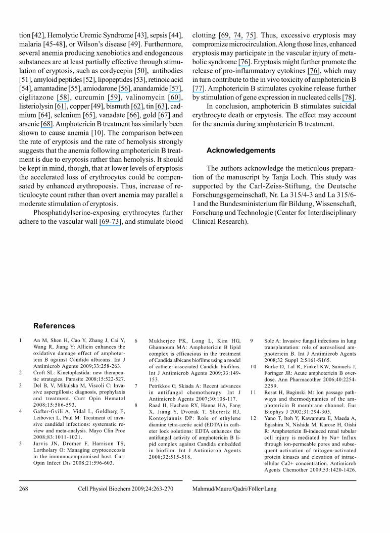
268
tion [42], Hemolytic Uremic Syndrome [43], sepsis [44],malaria [45-48], or Wilson’s disease [49]. Furthermore,several anemia producing xenobiotics and endogeneoussubstances are at least partially effective through stimu-lation of eryptosis, such as cordycepin [50], antibodies[51], amyloid peptides [52], lipopeptides [53], retinoic acid[54], amantadine [55], amiodarone [56], anandamide [57],ciglitazone [58], curcumin [59], valinomycin [60],listeriolysin [61], copper [49], bismuth [62], tin [63], cad-mium [64], selenium [65], vanadate [66], gold [67] andarsenic [68]. Amphotericin B treatment has similarly beenshown to cause anemia [10]. The comparison betweenthe rate of eryptosis and the rate of hemolysis stronglysuggests that the anemia following amphotericin B treat-ment is due to eryptosis rather than hemolysis. It shouldbe kept in mind, though, that at lower levels of eryptosisthe accelerated loss of erythrocytes could be compen-sated by enhanced erythropoesis. Thus, increase of re-ticulocyte count rather than overt anemia may parallel amoderate stimulation of eryptosis.
Phosphatidylserine-exposing erythrocytes furtheradhere to the vascular wall [69-73], and stimulate blood
clotting [69, 74, 75]. Thus, excessive eryptosis maycompromize microcirculation. Along those lines, enhancederyptosis may participate in the vascular injury of meta-bolic syndrome [76]. Eryptosis might further promote therelease of pro-inflammatory cytokines [76], which mayin turn contribute to the in vivo toxicity of amphotericin B[77]. Amphotericin B stimulates cyokine release furtherby stimulation of gene expression in nucleated cells [78].
In conclusion, amphotericin B stimulates suicidalerythrocyte death or erpytosis. The effect may accountfor the anemia during amphotericin B treatment.
Acknowledgements
The authors acknowledge the meticulous prepara-tion of the manuscript by Tanja Loch. This study wassupported by the Carl-Zeiss-Stiftung, the DeutscheForschungsgemeinschaft, Nr. La 315/4-3 and La 315/6-1 and the Bundesministerium für Bildung, Wissenschaft,Forschung und Technologie (Center for InterdisciplinaryClinical Research).
References
1 An M, Shen H, Cao Y, Zhang J, Cai Y,Wang R, Jiang Y: Allicin enhances theoxidative damage effect of amphoter-icin B against Candida albicans. Int JAntimicrob Agents 2009;33:258-263.
2 Croft SL: Kinetoplastida: new therapeu-tic strategies. Parasite 2008;15:522-527.
3 Del B, V, Mikulska M, Viscoli C: Inva-sive aspergillosis: diagnosis, prophylaxisand treatment. Curr Opin Hematol2008;15:586-593.
4 Gafter-Gvili A, Vidal L, Goldberg E,Leibovici L, Paul M: Treatment of inva-sive candidal infections: systematic re-view and meta-analysis. Mayo Clin Proc2008;83:1011-1021.
5 Jarvis JN, Dromer F, Harrison TS,Lortholary O: Managing cryptococcosisin the immunocompromised host. CurrOpin Infect Dis 2008;21:596-603.
6 Mukherjee PK, Long L, Kim HG,Ghannoum MA: Amphotericin B lipidcomplex is efficacious in the treatmentof Candida albicans biofilms using a modelof catheter-associated Candida biofilms.Int J Antimicrob Agents 2009;33:149-153.
7 Petrikkos G, Skiada A: Recent advancesin antifungal chemotherapy. Int JAntimicrob Agents 2007;30:108-117.
8 Raad II, Hachem RY, Hanna HA, FangX, Jiang Y, Dvorak T, Sherertz RJ,Kontoyiannis DP: Role of ethylenediamine tetra-acetic acid (EDTA) in cath-eter lock solutions: EDTA enhances theantifungal activity of amphotericin B li-pid complex against Candida embeddedin biofilm. Int J Antimicrob Agents2008;32:515-518.
9 Sole A: Invasive fungal infections in lungtransplantation: role of aerosolised am-photericin B. Int J Antimicrob Agents2008;32 Suppl 2:S161-S165.
10 Burke D, Lal R, Finkel KW, Samuels J,Foringer JR: Acute amphotericin B over-dose. Ann Pharmacother 2006;40:2254-2259.
11 Resat H, Baginski M: Ion passage path-ways and thermodynamics of the am-photericin B membrane channel. EurBiophys J 2002;31:294-305.
12 Yano T, Itoh Y, Kawamura E, Maeda A,Egashira N, Nishida M, Kurose H, OishiR: Amphotericin B-induced renal tubularcell injury is mediated by Na+ Influxthrough ion-permeable pores and subse-quent activation of mitogen-activatedprotein kinases and elevation of intrac-ellular Ca2+ concentration. AntimicrobAgents Chemother 2009;53:1420-1426.
Mahmud/Mauro/Qadri/Föller/LangCell Physiol Biochem 2009;24:263-270

269
13 Matsuo K, Hotokezaka H, Ohara N,Fujimura Y, Yoshimura A, Okada Y, HaraY, Yoshida N, Nakayama K: Analysis ofamphotericin B-induced cell signalingwith chemical inhibitors of signalingmolecules. Microbiol Immunol2006;50:337-347.
14 Vinciguerra M, Hasler U, Mordasini D,Roussel M, Capovilla M, Ogier-Denis E,Vandewalle A, Martin PY, Feraille E:Cytokines and sodium induce protein ki-nase A-dependent cell-surface Na,K-AT-Pase recruitment via dissociation of NF-kappaB/IkappaB/protein kinase A cata-lytic subunit complex in collecting ductprincipal cells. J Am Soc Nephrol2005;16:2576-2585.
15 Jensen GM, Skenes C.R, Bunch TH,Weissman CA, Amirghahari N, SatoriusA, Moynihan KL, Eley CGS: Determi-nation of the relative toxicity of am-photericin B formulations: a red bloodcell potassium release assay. Drug Deliv-ery 1999;3:81-88.
16 Lang F, Gulbins E, Lerche H, Huber SM,Kempe DS, Föller M: Eryptosis, a win-dow to systemic disease. Cell PhysiolBiochem 2008;22:373-380.
17 Bernhardt I, Weiss E, Robinson HC,Wilkins R, Bennekou P: Differential ef-fect of HOE642 on two separate mono-valent cation transporters in the humanred cell membrane. Cell Physiol Biochem2007;20:601-606.
18 Duranton C, Huber SM, Lang F: Oxida-tion induces a Cl(-)-dependent cationconductance in human red blood cells. JPhysiol 2002;539:847-855.
19 Duranton C, Huber S, Tanneur V, LangK, Brand V, Sandu C, Lang F:Electrophysiological properties of thePlasmodium Falciparum-induced cationconductance of human erythrocytes. CellPhysiol Biochem 2003;13:189-198.
20 Foller M, Kasinathan RS, Koka S, LangC, Shumilina E, Birnbaumer L, Lang F,Huber SM: TRPC6 contributes to theCa(2+) leak of human erythrocytes. CellPhysiol Biochem 2008;21:183-192.
21 Huber SM, Gamper N, Lang F: Chlorideconductance and volume-regulatory non-selective cation conductance in humanred blood cell ghosts. Pflugers Arch2001;441:551-558.
22 Kaestner L, Christophersen P, BernhardtI, Bennekou P: The non-selective volt-age-activated cation channel in the hu-man red blood cell membrane: reconcili-ation between two conflicting reports andfurther characterisation.Bioelectrochemistry 2000;52:117-125.
23 Kaestner L, Bernhardt I: Ion channels inthe human red blood cell membrane: theirfurther investigation and physiologicalrelevance. Bioelectrochemistry2002;55:71-74.
24 Lang KS, Duranton C, Poehlmann H,Myssina S, Bauer C, Lang F, Wieder T,Huber SM: Cation channels triggerapoptotic death of erythrocytes. CellDeath and Differentiation2003;10(2):249-256.
25 Bookchin RM, Ortiz OE, Lew VL: Acti-vation of calcium-dependent potassiumchannels in deoxygenated sickled redcells. Prog Clin Biol Res 1987;240:193-200.
26 Brugnara C, de Franceschi L, Alper SL:Inhibition of Ca(2+)-dependent K+ trans-port and cell dehydration in sickle eryth-rocytes by clotrimazole and other imi-dazole derivatives. J Clin Invest1993;92:520-526.
27 Lang PA, Kaiser S, Myssina S, Wieder T,Lang F, Huber SM: Role of Ca2+-acti-vated K+ channels in human erythro-cyte apoptosis. Am J Physiol Cell Physiol2003;285:C1553-C1560.
28 Berg CP, Engels IH, Rothbart A, LauberK, Renz A, Schlosser SF, Schulze-OsthoffK, Wesselborg S: Human mature red bloodcells express caspase-3 and caspase-8, butare devoid of mitochondrial regulatorsof apoptosis. Cell Death Differ2001;8:1197-1206.
29 Brand VB, Sandu CD, Duranton C,Tanneur V, Lang KS, Huber SM, Lang F:Dependence of Plasmodium falciparumin vitro growth on the cation permeabil-ity of the human host erythrocyte. CellPhysiol Biochem 2003;13:347-356.
30 Bratosin D, Estaquier J, Petit F, ArnoultD, Quatannens B, Tissier JP, SlomiannyC, Sartiaux C, Alonso C, Huart JJ,Montreuil J, Ameisen JC: Programmedcell death in mature erythrocytes: a modelfor investigating death effector pathwaysoperating in the absence of mitochon-dria. Cell Death Differ 2001;8:1143-1156.
31 Daugas E, Cande C, Kroemer G: Eryth-rocytes: death of a mummy. Cell DeathDiffer 2001;8:1131-1133.
32 Lang KS, Myssina S, Brand V, Sandu C,Lang PA, Berchtold S, Huber SM, LangF, Wieder T: Involvement of ceramidein hyperosmotic shock-induced death oferythrocytes. Cell Death Differ2004;11:231-243.
33 Foller M, Feil S, Ghoreschi K, Koka S,Gerling A, Thunemann M, Hofmann F,Schuler B, Vogel J, Pichler B, KasinathanRS, Nicolay JP, Huber SM, Lang F, FeilR: Anemia and splenomegaly in cGKI-deficient mice. Proc Natl Acad Sci U S A2008;105:6771-6776.
34 Foller M, Sopjani M, Koka S, Gu S,Mahmud H, Wang K, Floride E,Schleicher E, Schulz E, Munzel T, LangF: Regulation of erythrocyte survival byAMP-activated protein kinase. FASEB J2009;23:1072-1080.
35 Kempe DS, Lang PA, Duranton C, AkelA, Lang KS, Huber SM, Wieder T, LangF: Enhanced programmed cell death ofiron-deficient erythrocytes. FASEB J2006;20:368-370.
36 Floride E, Foller M, Ritter M, Lang F:Caffeine inhibits suicidal erythrocytedeath. Cell Physiol Biochem2008;22:253-260.
37 Nath CE, McLachlan AJ, Shaw PJ,Coakley JC, Earl JW: Amphotericin Bdose optimization in children with ma-lignant diseases. Chemotherapy2007;53:142-147.
38 Wiederhold NP, Tam VH, Chi J, PrinceRA, Kontoyiannis DP, Lewis RE: Phar-macodynamic activity of amphotericinB deoxycholate is associated with peakplasma concentrations in a neutropenicmurine model of invasive pulmonary as-pergillosis. Antimicrob AgentsChemother 2006;50:469-473.
39 Klarl BA, Lang PA, Kempe DS,Niemoeller OM, Akel A, Sobiesiak M,Eisele K, Podolski M, Huber SM, WiederT, Lang F: Protein kinase C mediateserythrocyte “programmed cell death”following glucose depletion. Am J PhysiolCell Physiol 2006;290:C244-C253.
40 Fadok VA, Bratton DL, Rose DM,Pearson A, Ezekewitz RA, Henson PM:A receptor for phosphatidylserine-spe-cific clearance of apoptotic cells. Nature2000;405:85-90.
41 Boas FE, Forman L, Beutler E:Phosphatidylserine exposure and red cellviability in red cell aging and in hemolyticanemia. Proc Natl Acad Sci U S A1998;95:3077-3081.
42 Birka C, Lang PA, Kempe DS, HoeflingL, Tanneur V, Duranton C, Nammi S,Henke G, Myssina S, Krikov M, HuberSM, Wieder T, Lang F: Enhanced sus-ceptibility to erythrocyte “apoptosis”following phosphate depletion. PflugersArch 2004;448:471-477.
43 Lang PA, Beringer O, Nicolay JP, AmonO, Kempe DS, Hermle T, Attanasio P,Akel A, Schafer R, Friedrich B, Risler T,Baur M, Olbricht CJ, Zimmerhackl LB,Zipfel PF, Wieder T, Lang F: Suicidal deathof erythrocytes in recurrent hemolyticuremic syndrome. J Mol Med2006;84:378-388.
44 Kempe DS, Akel A, Lang PA, Hermle T,Biswas R, Muresanu J, Friedrich B,Dreischer P, Wolz C, Schumacher U,Peschel A, Gotz F, Doring G, Wieder T,Gulbins E, Lang F: Suicidal erythrocytedeath in sepsis. J Mol Med 2007;85:269-277.
45 Brand V, Koka S, Lang C, Jendrossek V,Huber SM, Gulbins E, Lang F: Influenceof amitriptyline on eryptosis,parasitemia and survival of Plasmodiumberghei-infected mice. Cell PhysiolBiochem 2008;22:405-412.
Amphotericin B - Induced Eryptosis Cell Physiol Biochem 2009;24:263-270

270
4 6 Duranton C, Tanneur V, Lang C, BrandVB, Koka S, Kasinathan RS, Dorsch M,Hedrich HJ, Baumeister S, Lingelbach K,Lang F, Huber SM: A high specificity andaffinity interaction with serum albuminstimulates an anion conductance in ma-laria-infected erythrocytes. Cell PhysiolBiochem 2008;22:395-404.
47 Koka S, Lang C, Boini KM, Bobbala D,Huber SM, Lang F: Influence of chlo-rpromazine on eryptosis, parasitemia andsurvival of Plasmodium berghe infectedmice. Cell Physiol Biochem2008;22:261-268.
4 8 Koka S, Lang C, Niemoeller OM, BoiniKM, Nicolay JP, Huber SM, Lang F: In-fluence of NO synthase inhibitor L-NAME on parasitemia and survival ofPlasmodium berghei infected mice. CellPhysiol Biochem 2008;21:481-488.
49 Lang PA, Schenck M, Nicolay JP, BeckerJU, Kempe DS, Lupescu A, Koka S, EiseleK, Klarl BA, Rubben H, Schmid KW,Mann K, Hildenbrand S, Hefter H, HuberSM, Wieder T, Erhardt A, Haussinger D,Gulbins E, Lang F: Liver cell death andanemia in Wilson disease involve acidsphingomyelinase and ceramide. Nat Med2007;13:164-170.
5 0 Lui JC, Wong JW, Suen YK, Kwok TT,Fung KP, Kong SK: Cordycepin inducederyptosis in mouse erythrocytes througha Ca2+-dependent pathway withoutcaspase-3 activation. Arch Toxicol2007;81:859-865.
5 1 Attanasio P, Shumilina E, Hermle T,Kiedaisch V, Lang PA, Huber SM, WiederT, Lang F: Stimulation of eryptosis byanti-A IgG antibodies. Cell PhysiolBiochem 2007;20:591-600.
5 2 Nicolay JP, Gatz S, Liebig G, Gulbins E,Lang F: Amyloid induced suicidal eryth-rocyte death. Cell Physiol Biochem2007;19:175-184.
53 Wang K, Mahmud H, Föller M, Biswas R,Lang KS, Bohn E, Goetz F, Lang F:Lipopeptides in the Triggering of Eryth-rocyte Cell Membrane Scrambling. CellPhysiol Biochem 2008;22:381-386.
5 4 Niemoeller OM, Foller M, Lang C, HuberSM, Lang F: Retinoic acid induced sui-cidal erythrocyte death. Cell PhysiolBiochem 2008;21:193-202.
55 Föller M, Geiger C, Mahmud H, NicolayJP, Lang F: Stimulation of suicidal eryth-rocyte death by amantadine. Eur JPharmacol 2008;581:13-18.
56 Nicolay JP, Bentzen PJ, GhashghaeiniaM, Wieder T, Lang F: Stimulation oferythrocyte cell membrane scramblingby amiodarone. Cell Physiol Biochem2007;20:1043-1050.
57 Bentzen PJ, Lang F: Effect ofanandamide on erythrocyte survival. CellPhysiol Biochem 2007;20:1033-1042.
58 Niemoeller OM, Mahmud H, Foller M,Wieder T, Lang F: Ciglitazone and 15d-PGJ2 induced suicidal erythrocyte death.Cell Physiol Biochem 2008;22:237-244.
59 Bentzen PJ, Lang E, Lang F: Curcumininduced suicidal erythrocyte death. CellPhysiol Biochem 2007;19:153-164.
60 Schneider J, Nicolay JP, Foller M, WiederT, Lang F: Suicidal erythrocyte death fol-lowing cellular K+ loss. Cell PhysiolBiochem 2007;20:35-44.
61 Foller M, Shumilina E, Lam R, MohamedW, Kasinathan R, Huber S, ChakrabortyT, Lang F: Induction of suicidal erythro-cyte death by listeriolysin from Listeriamonocytogenes. Cell Physiol Biochem2007;20:1051-1060.
62 Braun M, Foller M, Gulbins E, Lang F:Eryptosis triggered by bismuth.Biometals 2009;22:453-460.
63 Nguyen TT, Foller M, Lang F: Tin trig-gers suicidal death of erythrocytes. J ApplToxicol 2008;29:79-83.
64 Sopjani M, Foller M, Dreischer P, LangF: Stimulation of eryptosis by cadmiumions. Cell Physiol Biochem 2008;22:245-252.
65 Sopjani M, Foller M, Gulbins E, Lang F:Suicidal death of erythrocytes due to se-lenium-compounds. Cell Physiol Biochem2008;22:387-394.
66 Foller M, Sopjani M, Mahmud H, LangF: Vanadate-induced suicidal erythrocytedeath. Kidney Blood Press Res2008;31:87-93.
67 Sopjani M, Foller M, Lang F: Gold stimu-lates Ca2+ entry into and subsequent sui-cidal death of erythrocytes. Toxicology2008;244:271-279.
68 Mahmud H, Foller M, Lang F: Arsenic-induced suicidal erythrocyte death. ArchToxicol 2009;83:107-113.
69 Andrews DA, Low PS: Role of red bloodcells in thrombosis. Curr Opin Hematol1999;6:76-82.
70 Closse C, Dachary-Prigent J, BoisseauMR: Phosphatidylserine-related adhesionof human erythrocytes to vascular en-dothelium. Br J Haematol 1999;107:300-302.
71 Gallagher PG, Chang SH, Rettig MP,Neely JE, Hillery CA, Smith BD, LowPS: Altered erythrocyte endothelial ad-herence and membrane phospholipidasymmetry in hereditary hydrocytosis.Blood 2003;101:4625-4627.
72 Pandolfi A, Di Pietro N, Sirolli V,Giardinelli A, Di Silvestre S, Amoroso L,Di Tomo P, Capani F, Consoli A,Bonomini M: Mechanisms of uremicerythrocyte-induced adhesion of humanmonocytes to cultured endothelial cells.J Cell Physiol 2007;213:699-709.
73 Wood BL, Gibson DF, Tait JF: Increasederythrocyte phosphatidylserine exposurein sickle cell disease: flow-cytometricmeasurement and clinical associations.Blood 1996;88:1873-1880.
74 Chung SM, Bae ON, Lim KM, Noh JY,Lee MY, Jung YS, Chung JH:Lysophosphatidic acid induces thrombo-genic activity throughphosphatidylserine exposure andprocoagulant microvesicle generation inhuman erythrocytes. ArteriosclerThromb Vasc Biol 2007;27:414-421.
75 Zwaal RF, Comfurius P, Bevers EM: Sur-face exposure of phosphatidylserine inpathological cells. Cell Mol Life Sci2005;62:971-988.
76 Zappulla D: Environmental stress, eryth-rocyte dysfunctions, inflammation, andthe metabolic syndrome: adaptations toCO2 increases? J Cardiometab Syndr2008;3:30-34.
77 Chapman SW, Sullivan DC, Cleary JD:In search of the holy grail of antifungaltherapy. Trans Am Clin Climatol Assoc2008;119:197-215.
78 Turtinen LW, Croswell A, Obr A:Microarray analysis of amphotericin B-treated THP-1 monocytic cells identi-fies unique gene expression profilesamong lipid and non-lipid drug formula-tions. J Chemother 2008;20:327-335.
Mahmud/Mauro/Qadri/Föller/LangCell Physiol Biochem 2009;24:263-270
Copyright © 2022 FDOKUMEN