Critical role of extracellular vesicles in modulating the cellular effects of cytokines
Upload
khangminh22Category
view
3download
0
�����������������
Citation: Brun, J.-F.; Varlet-Marie, E.;
Myzia, J.; Raynaud de Mauverger, E.;
Pretorius, E. Metabolic Influences
Modulating Erythrocyte
Deformability and Eryptosis.
Metabolites 2022, 12, 4. https://
doi.org/10.3390/metabo12010004
Academic Editor: Norbert Nemeth
Received: 29 November 2021
Accepted: 18 December 2021
Published: 21 December 2021
Publisher’s Note: MDPI stays neutral
with regard to jurisdictional claims in
published maps and institutional affil-
iations.
Copyright: © 2021 by the authors.
Licensee MDPI, Basel, Switzerland.
This article is an open access article
distributed under the terms and
conditions of the Creative Commons
Attribution (CC BY) license (https://
creativecommons.org/licenses/by/
4.0/).
metabolites
H
OH
OH
Review
Metabolic Influences Modulating Erythrocyte Deformabilityand Eryptosis
Jean-Frédéric Brun 1,* , Emmanuelle Varlet-Marie 2, Justine Myzia 1, Eric Raynaud de Mauverger 1
and Etheresia Pretorius 3
1 UMR CNRS 9214-Inserm U1046 Physiologie et Médecine Expérimentale du Cœur et desMuscles-PHYMEDEXP, Unité D’explorations Métaboliques (CERAMM), Département de PhysiologieClinique, Université de Montpellier, Hôpital Lapeyronie-CHRU de Montpellier, 34295 Montpellier, France;[email protected] (J.M.); [email protected] (E.R.d.M.)
2 UMR CNRS 5247-Institut des Biomolécules Max Mousseron (IBMM), Laboratoire du Département dePhysicochimie et Biophysique, UFR des Sciences Pharmaceutiques et Biologiques, Université de Montpellier,34090 Montpellier, France; [email protected]
3 Department of Physiological Sciences, Stellenbosch University, Stellenbosch, Private Bag X1 MATIELAND,Stellenbosch 7602, South Africa; [email protected]
* Correspondence: [email protected]; Tel.: +04-67-33-82-84
Abstract: Many factors in the surrounding environment have been reported to influence erythrocytedeformability. It is likely that some influences represent reversible changes in erythrocyte rigiditythat may be involved in physiological regulation, while others represent the early stages of eryptosis,i.e., the red cell self-programmed death. For example, erythrocyte rigidification during exerciseis probably a reversible physiological mechanism, while the alterations of red blood cells (RBCs)observed in pathological conditions (inflammation, type 2 diabetes, and sickle-cell disease) are morelikely to lead to eryptosis. The splenic clearance of rigid erythrocytes is the major regulator of RBCdeformability. The physicochemical characteristics of the surrounding environment (thermal injury,pH, osmolality, oxidative stress, and plasma protein profile) also play a major role. However, thereare many other factors that influence RBC deformability and eryptosis. In this comprehensive review,we discuss the various elements and circulating molecules that might influence RBCs and modifytheir deformability: purinergic signaling, gasotransmitters such as nitric oxide (NO), divalent cations(magnesium, zinc, and Fe2+), lactate, ketone bodies, blood lipids, and several circulating hormones.Meal composition (caloric and carbohydrate intake) also modifies RBC deformability. Therefore,RBC deformability appears to be under the influence of many factors. This suggests that severalhomeostatic regulatory loops adapt the red cell rigidity to the physiological conditions in order tocope with the need for oxygen or fuel delivery to tissues. Furthermore, many conditions appear toirreversibly damage red cells, resulting in their destruction and removal from the blood. These twocategories of modifications to erythrocyte deformability should thus be differentiated.
Keywords: erythrocyte deformability; metabolism; hormones; homeostasis; eryptosis; stress; COVID-19; sleep apnea
1. Introduction
Red blood cells (RBCs) are known to markedly modify their shape in order to transitinto small capillary vessels, whose radius is smaller than their own [1]. This ability todeform also results in RBC elongation in flow. This property plays an important role inthe blood viscosity at high shear rates, so that in this situation blood can be modeled as aNewtonian fluid [2,3], whose viscosity reflects RBC deformability.
In fact, the term ‘red cell deformability’ is not so easy to define, because it appears moreand more obvious that RBCs can undergo many varieties of deformation in narrow channelsor in flow according to the experimental or physiological situation. Classical studies usingmicroscopic flow visualization led to the idea that the deformation of RBCs resulted from
Metabolites 2022, 12, 4. https://doi.org/10.3390/metabo12010004 https://www.mdpi.com/journal/metabolites

Metabolites 2022, 12, 4 2 of 30
continuous viscous deformation, which was called “fluid drop-like adaptation” by H.Schmid-Schönbein [4]. This kind of deformation was determined by the cytoplasm fluidityand the surface-area-to-volume ratio of the red cells.
Over recent years, these classical concepts have been reassessed with new sophisticatedexperimental approaches, resulting in a more complex picture, mostly in the contextof a new emphasis given to the rheological behavior of RBCs in sickle-cell disease [5].Studying the stiffness of RBCs from individuals with sickle-cell trait, Zheng [6] developed amicrosystem able to measure the individual mechanical properties (i.e., shear modulus andviscosity) of a single red cell submitted to a shear stress. After the RBCs were deformedunder the influence of this shear stress, the dynamic RBC recovery was monitored andanalyzed according to the Kelvin–Voigt model, allowing the measurement of an elasticshear modulus of RBCs submitted to different shear rates. Even more recently, anothergroup [7–9] developed a microfluidic impedance red cell assay (MIRCA) in order to measureRBC transition through narrow openings and also challenged to some extent the concept of‘fluid drop-like’ RBCs whose deformation was assumed to be mostly related to viscositywith little or no elastic component. The authors defined new parameters such as an RBCocclusion index (ROI) and an RBC electrical impedance index (REI), which measure thecumulative percentage of vessel occlusion and the impedance change, respectively.
These recent experiments suggest that the process of RBC deformability was untilnow misinterpreted by previous microfluidic measurements. Furthermore, Lanotte andcoworkers [10] recently performed experiments on and simulations of microcirculatoryflow in various conditions of volume fractions and shear rates. They showed that RBCsundergo a large variety of morphological modifications during their deformation. With anincreasing shear rate, the RBCs were first shown to tumble, then they were shown to roll,then they adopted the form of a tumbling stomatocyte, and finally they exhibited variouspolylobed shapes that were only observed above a threshold value of the viscosity contrastbetween the plasma and cytosol. In another paper, the same team further described thecomplexity of the mechanisms involved in these transition processes from one shape toanother under the influence of an increase in shear stress [11].
All this recent literature emphasizes the complexity of red cell deformation, which isfar from a simple phenomenon. In light of this quite recent literature, it becomes clear thatthe experiments performed in recent decades on so-called ‘red cell deformability’ exploredonly a limited aspect of this physiological mechanism. Until now, our knowledge on theregulation of RBC deformability is mostly based on the measurement of the deformation ofRBCs entering a narrow channel and the deformation of RBCs submitted to a shear stress inflow. These two approaches are likely to rely on different cellular mechanisms. It is likely thatmost of this information needs to be investigated again with newer experimental approaches.
Some experiments have been conducted with artificially stiffened erythrocytes, show-ing that impaired deformability dramatically decreases perfusion, with a quite differenteffect in various tissues [12]. It is clear that such experiments do not reflect typical in vivosituations, but they are not meaningless. Examples of extremely rigid erythrocytes canbe found in situations such as sickle-cell disease [5,13]. In this case, consistent with theseexperiments, stiffened RBCs can clearly be responsible for vessel occlusion. In fact, inthe majority of cases, the modification of the erythrocyte rigidity is not so dramatic andthe RBCs remain able to deform and transit through the microcirculation. However, suchmoderately rigidified erythrocytes transit mostly in the largest microvessels, a situationthat has been termed capillary maldistribution [14,15].
Research has also focused on studying the physiological and pathological changesthat happen to RBCs during either disease or when RBCs are artificially rigidified. Over thelast 30 years of the 20th century, an impressive body of literature has been published on thefactors that modify erythrocyte deformability. This early research did not, however, clearlyseparate reversible and irreversible RBC rigidification, and most of this early research wascarried out before the emergence of the concept of eryptosis (or programmed RBC death,which is similar to apoptosis but specific to the anuclear RBC) [16–23].

Metabolites 2022, 12, 4 3 of 30
Conditions where eryptosis have been noted, have all been reported to impair ery-throcyte deformability. This is the case for hypoxia, iron deficiency, cancers, dehydration,metabolic syndrome, phosphate depletion, hemolytic anemia, heart failure, diabetes melli-tus, chronic kidney disease, mycoplasma infection, malaria, hemolytic uremic syndrome,sepsis, sickle-cell disease, etc. Additionally, factors known to inhibit eryptosis such ascatecholamines, erythropoietin, adenosine, resveratrol, urea, vitamin E, and caffeine havealso been reported to modify erythrocyte rheology [24]. It is clear that the most importantregulator of erythrocyte deformability is the clearance of rigid erythrocytes within thespleen. A mechanical checking of the deformability of circulating erythrocytes is regularlyperformed in the splenic microcirculation, so that the RBCs that are not able to correctlysqueeze through the narrow splenic slits are trapped and removed from circulation [25–27].
Therefore, we suggest that all the literature dealing with the factors that modifyerythrocyte deformability should be analyzed in light of the new concept of eryptosis. Forexample, the pathologic alterations of erythrocytes that occur in metabolic diseases suchas diabetes [28,29] should probably be considered as a completely different process to thephysiological reversible erythrocyte stiffening observed during muscular exercise [30]. Theexercise-induced decrease in red cell deformability is a very interesting example of thisdifference. In healthy athletes, exercise transiently modifies the blood rheology withoutevidence of increased eryptosis [31,32], while in sickle-cell disease patients, it induces along-lasting stiffening of the red cells that seems to be explained by irreversible damageand probable further eryptosis [5]. Sickle-cell disease is clearly a condition associated withincreased eryptosis [33].
Additionally, there are many influences that can modify RBC deformability. This paperis an attempt to summarize this large body of literature and to integrate our knowledgewith regards to the classical definitions of deformability and eryptosis.
2. The Main Classical Physicochemical Modifiers of RBC Deformability
Classically, the most important modifiers of RBC deformability were the physico-chemical characteristics of the surrounding environment [34]. A biphasic influence ofpH and osmolality on RBC deformability displaying a “u-shaped curve” has been de-scribed. The deformability of red cells appears to be optimal within the physiological rangeand is markedly impaired above and below these narrow physiological boundaries. Thisstiffening effect of pH and osmolality changes has been assumed to increase erythrocytetrapping in the spleen and thus decrease RBC lifespan [35]. It has also been shown thatan environment containing proteins such as albumin is mandatory for preventing alter-ations of the erythrocyte shape, since albumin has the ability to prevent and even reverseechinocytosis [36].
Recently, the physiological relevance of such osmotic changes to red cell water contenthas been emphasized by studies showing that aquaporin-1 (AQP1), which is expressedin red cell membranes, may drive rapid water exchange and that this exchange resultsin an important volume change (up to 39%) [37]. This effect is almost suppressed inAQP1-knockout (KO) erythrocytes [37]. Such alterations of the erythrocyte volume inmicrovessels result in an increase in the osmotic gradient between the plasma and interstitialfluid. Red cells thus appear to be “micropumps” that regulate in situ local osmolarity [37].Accordingly, red cells are likely to be major regulators of water exchange in the body, andthus, to contribute to body water homeostasis [37].
Some studies have also been devoted to divalent cations. For example, magnesiumhas been shown to protect RBCs from in vitro experimental rigidification by several proce-dures [38,39].
3. A Brief Overview of Eryptosis
The mechanisms of eryptosis are described in the classical publications by the Langgroup [16–19,21,24,40–46]. Eryptosis occurs in conditions such as heart failure, uremia,haemolytic uremic syndrome, sepsis, fever, dehydration, mycoplasma infection, anemia,

Metabolites 2022, 12, 4 4 of 30
metabolic syndrome, cancer, diabetes, hepatic failure, Wilson’s disease, malaria, sickle-cell anaemia, iron deficiency, thalassemia, glucose-6-phosphate dehydrogenase deficiency,Parkinson’s disease, type 2 diabetes, Alzheimer’s disease, and rheumatoid arthritis [20].Figure 1 shows a brief overview of eryptosis, which is triggered by various signalingpathways, including the presence of circulating inflammatory molecules that results inoxidative stress. Eryptosis is therefore the culminative term for the end-stage of the process,resulting in cell death; even so, as RBCs are known to be exceptionally resilient, they dohave the ability to recover if the stressor molecule or environment is changed. However,there is, as with all physiological and molecular pathways, a point of no return, after whichrecovery is not possible.
Metabolites 2022, 12, x FOR PEER REVIEW 4 of 31
3. A Brief Overview of Eryptosis The mechanisms of eryptosis are described in the classical publications by the Lang
group [16–19,21,24,40–46]. Eryptosis occurs in conditions such as heart failure, uremia, haemolytic uremic syndrome, sepsis, fever, dehydration, mycoplasma infection, anemia, metabolic syndrome, cancer, diabetes, hepatic failure, Wilson’s disease, malaria, sickle-cell anaemia, iron deficiency, thalassemia, glucose-6-phosphate dehydrogenase defi-ciency, Parkinson’s disease, type 2 diabetes, Alzheimer’s disease, and rheumatoid arthritis [20]. Figure 1 shows a brief overview of eryptosis, which is triggered by various signaling pathways, including the presence of circulating inflammatory molecules that results in oxidative stress. Eryptosis is therefore the culminative term for the end-stage of the pro-cess, resulting in cell death; even so, as RBCs are known to be exceptionally resilient, they do have the ability to recover if the stressor molecule or environment is changed. How-ever, there is, as with all physiological and molecular pathways, a point of no return, after which recovery is not possible.
Figure 1. A brief overview of eryptosis (adapted from [47]) showing the pathways that initiate it, under the influence of osmotic shock or oxidative stress, resulting in activation of intracellular path-ways, leading in turn to phospholipid membrane scrambling (1); cell shrinkage (2); and membrane blebbing (3). Figure created using www.biorender.com.
4. RBC Receptors The receptors expressed on the red cell membrane play an important role in its opti-
mal functioning. The following paragraphs will provide a brief summary of these recep-tors and their role in deformability and eryptosis. Several receptors for various ligands are present on the red cell membrane [1]. Some of these are mentioned later in this review. Among them, we should mention the N-methyl D-aspartate (NMDA) receptors, which are expressed on the red cell membrane. These receptors are major targets of divalent cat-ions and mediate most of their effects in various conditions. They contribute to the regu-lation of intracellular calcium in erythrocytes [48]. However, it has been shown that the experimental activation of NMDA receptors has no measurable effect on the rheological
Figure 1. A brief overview of eryptosis (adapted from [47]) showing the pathways that initiate it,under the influence of osmotic shock or oxidative stress, resulting in activation of intracellular path-ways, leading in turn to phospholipid membrane scrambling (1); cell shrinkage (2); and membraneblebbing (3). Figure created using www.biorender.com accessed on 17 December 2021.
4. RBC Receptors
The receptors expressed on the red cell membrane play an important role in its optimalfunctioning. The following paragraphs will provide a brief summary of these receptorsand their role in deformability and eryptosis. Several receptors for various ligands arepresent on the red cell membrane [1]. Some of these are mentioned later in this review.Among them, we should mention the N-methyl D-aspartate (NMDA) receptors, whichare expressed on the red cell membrane. These receptors are major targets of divalentcations and mediate most of their effects in various conditions. They contribute to theregulation of intracellular calcium in erythrocytes [48]. However, it has been shown thatthe experimental activation of NMDA receptors has no measurable effect on the rheologicalproperties of erythrocytes [49], suggesting that the effects of divalent cations on red celldeformability are not mediated by NMDA receptors. Notwithstanding, an abnormallyhigh abundance of N-methyl D-aspartate receptors on the erythrocyte membranes ofsickle-cell disease patients has been reported and is associated with an excessive calcium

Metabolites 2022, 12, 4 5 of 30
uptake. Presumably, this process can trigger the cascade of red-cell-damaging events thatinclude RBC rigidification [50]. Moreover, Unal and coworkers recently reported thatmemantine, an antagonist of NMDA receptors, impairs RBC deformability in rats [51]. Allthis suggests that, despite the lack of a measurable effect of NMDA receptor activation onthe deformability of normal erythrocytes, the inactivation of these receptors does have suchan effect, whose physiological significance needs to be more precisely established.
There is a large body of literature about the purinergic receptors in RBCs, and de-spite a relative paucity of hemorheological studies dealing with this issue, this literaturesuggests that they may be important modulators of red cell deformability. P1 and P2purinergic receptors are expressed on the red cell membrane and are able to bind extracel-lular nucleosides and nucleotides [52]. P1 receptors are stimulated by adenosine and P2receptors are stimulated by adenosine triphosphate (ATP). P2 receptors comprise P2X andP2Y receptors. Their activation triggers the intracellular signaling pathways in progenitorerythrocytes, resulting in reactive oxygen species formation, microparticle release, andapoptosis. In mature erythrocytes, P2 receptor stimulation is involved in cell volume regu-lation, phosphatidylserine exposure, eicosanoid release, hemolysis, impaired ATP release,and susceptibility or resistance to infection [52]. Furthermore, the P1 receptor agonistadenosine protects against eryptosis via the activation of a pathway which most proba-bly acts downstream of PKC. Purinergic signaling in erythrocytes is probably involvedin the maintenance of microcirculation in ischemic tissue [45]. Erythrocytes, in fact, arenot only targets of purinergic stimulation but are also able to release ATP and ADP [53].It has been shown that ATP and ADP are continuously released by RBCs and are laterconverted outside the cell into adenosine, which then re-enters the red cell. ATP releaseby erythrocytes is triggered by hypoxia, hypercapnia, mechanical deformation, reducedO2 tension, acidosis, cell swelling, prostacyclin analogues, and β-adrenoceptor agonists.According to Sprague and coworkers [54], erythrocyte transit through narrow capillaries,where the shear stress makes them deform, results in ATP release. This ATP then binds topurinergic P2 receptors expressed on endothelial cell membranes, resulting in a release ofNO and PGI2 [55]. Caffeine increases ATP release from RBCs, probably via its effect on theintracellular cAMP levels. Intracellular ATP is essential for maintaining the function andstructural integrity of erythrocytes. By contrast, ATP depletion has been shown to sensitizeRBCs to the eryptotic effects of Ca2+ [20].
The A3 adenosine receptor is also expressed on red cells. Its antagonist reversine (2-(4-morpholinoanilino)-6-cyclohexylaminopurine) has important effects in nucleated cells,since it is known to induce cell cycle arrest, inhibit cell proliferation, influence cellulardifferentiation, induce cell swelling, and trigger apoptosis. In fact, since erythrocyteslack mitochondria, they exhibit a different response to reversine. In this case, reversinepowerfully inhibits cell membrane scrambling after energy depletion, Ca2+ loading, andoxidative stress, and therefore prevents the occurrence of eryptosis [41].
Purinergic signaling is involved in the response to low blood O2 which triggers ATPrelease by erythrocytes, leading to the stimulation of P2 × 2/3 receptors in the aorticbody [56]. The breakdown of ATP into ADP inhibits ATP release via a negative feedback,which involves the P2Y13 receptors in human erythrocytes [57].
Undoubtedly, all these purinergic effects are likely to modulate RBC rheology, but,surprisingly, there is very little published information on this issue. I. Juhan-Vague reportedforty years ago that the ADP released by rigid RBCs impaired the deformability of normalerythrocytes [28]. More recently, it was reported that the ADP release from RBCs in healthyhuman volunteers was lower in middle-aged than in young healthy individuals, and thatfish oil intake improved the erythrocyte deformability, parallel to a 50% decrease in theADP release [58].
Thus, erythrocyte deformability is included in a regulatory loop involving purinergicsignaling. Erythrocyte stiffening inhibits the release of ATP, which is in turn increasedwhen red cells become more deformable, e.g., when treated by hydroxyurea or the HMG-CoA reductase inhibitor simvastatin [59]. Subsequently, the stiffened erythrocytes release
Metabolites 2022, 12, 4 6 of 30
ADP, which inhibits the release of ATP. This mechanism probably aims at maintainingRBC energy stores, but it is also likely to induce a self-potentiating loop resulting in RBCrigidification. Moreover, when erythrocytes are well-deformable and release ATP, theyinduce more NO production by the vessel wall, and this NO results in vasodilation andincreased RBC deformability [60].
Acetylcholine (Ach) can also bind to RBCs that express both muscarinic [61] andnicotinic cholinergic receptors [62]. The hemorheologic effects of Ach are both an increasein RBC deformability and a decrease in RBC aggregation [63]. Further studies by the teamof A. Muravyov have helped to describe the signaling pathways involved in the effects ofAch [64].
The RBC membrane also expresses receptors for the endogenous ligands of benzodi-azepines [65], corticotropin-releasing factor (CRF) [66], and prolactin [67]. Most of thesechemical messengers have been reported to modify in vitro or in vivo erythrocyte rheologicproperties, but the data on these effects remain to some extent conflicting.
It is logical to hypothesize that the receptor-mediated alterations in erythrocyte de-formability induced by chemical messengers that physiologically circulate in the bloodare reversible adaptative processes that do not involve eryptosis. However, physiologicalfactors such as NO, anandamide, iron, adenosine, retinoic acid, and zinc appear in thelist of eryptosis-inducing substances that can be found in the recent review on this topicpublished by Pretorius and coworkers [20]. Therefore, it can be assumed that eryptosis(which involves an irreversible modification of erythrocytes leading to their prematuredeath) can actually be triggered outside of any pathologic context, as an adaptation to afully physiological situation.
The following paragraphs will revisit our previous knowledge on the effect of variouscirculating molecules on RBC deformability and integrate our new knowledge regarding eryptosis.
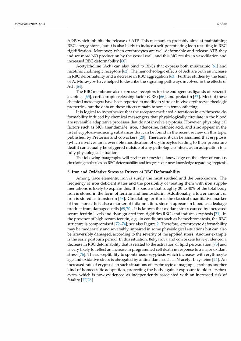
5. Iron and Oxidative Stress as Drivers of RBC Deformability
Among trace elements, iron is surely the most studied and the best-known. Thefrequency of iron deficient states and the possibility of treating them with iron supple-mentations is likely to explain this. It is known that roughly 30 to 40% of the total bodyiron is stored in the form of ferritin and hemosiderin. Additionally, a lower amount ofiron is stored as transferrin [68]. Circulating ferritin is the classical quantitative markerof iron stores. It is also a marker of inflammation, since it appears in blood as a leakageproduct from damaged cells [69,70]. It is known that oxidant stress caused by increasedserum ferritin levels and dysregulated iron rigidifies RBCs and induces eryptosis [71]. Inthe presence of high serum ferritin, e.g., in conditions such as hemochromatosis, the RBCstructure is compromised [72–74]; see also Figure 2. Therefore, erythrocyte deformabilitymay be moderately and reversibly impaired in some physiological situations but can alsobe irreversibly damaged, according to the severity of the applied stress. Another exampleis the early postburn period. In this situation, Bekyarova and coworkers have evidenced adecrease in RBC deformability that is related to the activation of lipid peroxidation [75] andis very likely to reflect an increase in programmed cell death in response to a major oxidantstress [76]. The susceptibility to spontaneous eryptosis which increases with erythrocyteage and oxidative stress is abrogated by antioxidants such as N-acetyl-L-cysteine [24]. Anincreased rate of eryptosis in such situations of erythrocyte damaging is perhaps anotherkind of homeostatic adaptation, protecting the body against exposure to older erythro-cytes, which is now evidenced as independently associated with an increased risk offatality [77,78].

Metabolites 2022, 12, 4 7 of 30Metabolites 2022, 12, x FOR PEER REVIEW 7 of 31
Figure 2. Morphology of RBCs in the presence of high circulating serum ferritin. This picture shows that in conditions like hemochromatosis, RBC structure is markedly compromised. (A) Individual with hereditary hemochromatosis (H63D/C2882Y), serum ferritin level 374 ng/mL−1; (B) individual with hereditary hemochromatosis (H63D/wild type), serum ferritin level 1500 ng/mL−1. Raw data from [72].
The high levels of serum ferritin found in those with conditions such as Alzheimer’s disease and Parkinson’s disease may also modify erythrocyte function and structure [47,79–83]. It is now well established that Fe++ triggers eryptosis [20]. This eryptotic effect of iron may explain why erythrocyte deformability is significantly impaired in hemochro-matosis and hyperferritinemia [74,84,85].
Although a high value of serum ferritin cannot rule out the existence of iron defi-ciency, a low ferritin value is well recognized as being highly specific to iron deficiency. Experimental studies in iron-deficient rats have evidenced a lower erythrocyte deforma-bility that appeared to be related at least in part to the lower hemoglobin content of the erythrocytes [86,87]. Athletes with low plasma ferritin also exhibit a higher value of blood viscosity, a higher plasma viscosity, and a higher RBC aggregation index when compared to athletes exhibiting normal values of plasma ferritin. By contrast, no difference in hem-atocrit or RBC deformability could be evidenced between these two subgroups [88]. In fact, iron is known to damage the structure of RBC membranes [89], resulting in more rouleaux networks.
6. Antioxidants Erythrocyte deformability is improved by the antioxidants vitamin E [90,91], alpha-
tocopherol [92], alpha-tocotrienol [93], fish oil, and dietary tea catechins [94]. Muscular activity is a situation known to be associated with an increase in oxidant stress, which in this case should of course be considered as a purely physiological event. However, this rise in oxidative stress can be very important and become harmful. Trained athletes ap-pear to be protected against the potential deleterious effects of this oxidative stress. This is evidenced by the fact that in both rats [95] and humans [96], exercise-induced oxidative stress decreases RBC deformability in sedentary individuals but not in exercise-trained ones.
7. Zinc Zinc is also known to increase the deformability of artificially hardened RBCs in vitro
[97] and is frequently low in the serum of athletes, reflecting some degree of deficiency. It is known that athletes who exhibit low serum zinc values have a higher blood viscosity and impaired erythrocyte deformability [98], and this hemorheologic profile is associated with a decrease in exercise performance. Experimentally, a double-blind randomized trial with oral zinc gluconate in healthy volunteers was found to decrease the blood viscosity
Figure 2. Morphology of RBCs in the presence of high circulating serum ferritin. This picture showsthat in conditions like hemochromatosis, RBC structure is markedly compromised. (A) Individualwith hereditary hemochromatosis (H63D/C2882Y), serum ferritin level 374 ng/mL−1; (B) individualwith hereditary hemochromatosis (H63D/wild type), serum ferritin level 1500 ng/mL−1. Raw datafrom [72].
The high levels of serum ferritin found in those with conditions such as Alzheimer’sdisease and Parkinson’s disease may also modify erythrocyte function and structure [47,79–83].It is now well established that Fe2+ triggers eryptosis [20]. This eryptotic effect of iron mayexplain why erythrocyte deformability is significantly impaired in hemochromatosis andhyperferritinemia [74,84,85].
Although a high value of serum ferritin cannot rule out the existence of iron deficiency,a low ferritin value is well recognized as being highly specific to iron deficiency. Exper-imental studies in iron-deficient rats have evidenced a lower erythrocyte deformabilitythat appeared to be related at least in part to the lower hemoglobin content of the erythro-cytes [86,87]. Athletes with low plasma ferritin also exhibit a higher value of blood viscosity,a higher plasma viscosity, and a higher RBC aggregation index when compared to athletesexhibiting normal values of plasma ferritin. By contrast, no difference in hematocrit or RBCdeformability could be evidenced between these two subgroups [88]. In fact, iron is knownto damage the structure of RBC membranes [89], resulting in more rouleaux networks.
6. Antioxidants
Erythrocyte deformability is improved by the antioxidants vitamin E [90,91], alpha-tocopherol [92], alpha-tocotrienol [93], fish oil, and dietary tea catechins [94]. Muscularactivity is a situation known to be associated with an increase in oxidant stress, which in thiscase should of course be considered as a purely physiological event. However, this rise inoxidative stress can be very important and become harmful. Trained athletes appear to beprotected against the potential deleterious effects of this oxidative stress. This is evidencedby the fact that in both rats [95] and humans [96], exercise-induced oxidative stress decreasesRBC deformability in sedentary individuals but not in exercise-trained ones.
7. Zinc
Zinc is also known to increase the deformability of artificially hardened RBCs in vitro [97]and is frequently low in the serum of athletes, reflecting some degree of deficiency. It isknown that athletes who exhibit low serum zinc values have a higher blood viscosity andimpaired erythrocyte deformability [98], and this hemorheologic profile is associated witha decrease in exercise performance. Experimentally, a double-blind randomized trial withoral zinc gluconate in healthy volunteers was found to decrease the blood viscosity [99],while no significant effect on performance could be evidenced. Zinc was also shown todecrease erythrocyte aggregation both in vitro and in vivo [100]. More recently, in contrastwith these findings, it was shown that zinc is also able to promote eryptosis [42]. It is

Metabolites 2022, 12, 4 8 of 30
likely that, as discussed above for iron, the impact of this mineral on RBCs can be differentaccording to the severity of the applied stress, which explains this apparent paradox.
8. RBCs and Their Energy Needs
Since erythrocytes need energy to undergo deformation, the depletion of their energeticstores has a marked effect on their deformability. This is regularly observed when RBCsare stored in vitro. In this situation, there is a gradual temperature- and time-dependentdecrease in the glucose and ATP levels, with a simultaneous rise in the intracellular levelsof lactate and LDH. Parallel to this process, there is a time- and temperature-dependentswelling and an echinocytic transformation of RBCs. At the same time, a gradual increasein the RBC rigidity can be evidenced with the measurement of blood viscosity at a highshear rate. This process of echinocytosis can be partially reversed if the erythrocytes areresuspended in a buffer containing 0.2% albumin [101]. The literature on eryptosis showsthat the glucose depletion of RBCs (and more generally, an energy crisis, see Figure 1 andTable 1) triggers the process of programmed cell death in RBCs [46].
Table 1. Factors influencing red cell deformability and eryptosis.
Increases RBCDeformability
Decreases RBCDeformability
Increases Eryptosis (After[24] and [20])
Decreases Eryptosis(After [24] and [20])
Biologicallyactive
molecules andmetabolites
ATPNOH2S
Carbon monoxideZn++
Lactate (in trainedathletes)
Ketone bodiesCholesterol
Glucose > 200 dg/mLLactate (in sedentary
subjects)
AluminiumArsenic
CadmiumCarbon monoxide
Ceramide(acylsphingosine)
ChromiumCopper
Fe2+,
Energy depletionGlucose (via glycation)
Osmotic shockZn++
NOErythropoietin
Catecholamines β and α
Hormones andchemical
messengers
AcetylcholineEpinephrineEndothelin 1
ApelinLeptin
ProgesteroneErythropoietinSomatostatin
Prostaglandin E1DHEA
GlucagonMelatonin
ADPPGE2
Norepinephrine (?)Leukotriene B4
ThyroxinIGF-I
Estradiol
AnandamideEstradiol
Leukotriene C(4)Lithium
Lysophosphatidic acidMercury
PAFPhosphate
ProgesteroneProstaglandin E2
Silver ionsSphingosine
AdenosineChloride
ErythropoietinNitroprusside (NO-donor)
Urea
Nutritionalfactors
Tea catechinsVitamin E
α-tocopherol, αtocoterol
Carbohydrate intake
CurcurminGossypolOxysterol
Phytic acidRetinoic acidRetinoic acid
Selenium (sodium selenite)Tannic acidVitamin K
CaffeineGlutathione
MonohydroxyethylrutosideN-acetylcysteine
NaringinVitamin E
On the other hand, hyperglycemia also has an effect on erythrocytes that has been ex-tensively investigated. In older studies, it was reported that short-term hyperglycemia doesnot markedly impair the blood rheology unless extremely high concentrations (hundreds of

Metabolites 2022, 12, 4 9 of 30
millimoles per liter) were reached. The relevance of these experiments is unclear, since suchconcentrations can never be found in human diseases [102]. However, the chronic exposure ofRBCs to high concentrations of glucose is known to increase intracellular sorbitol, and highvalues of sorbitol within RBCs were shown to be associated with impaired erythrocyte deforma-bility [103]. In fact, the concentrations of sorbitol used in the abovementioned experimentswere extremely high and probably irrelevant to what can be found in human diseases.
Obviously, it is important to study this issue of high glucose levels because it canbe relevant to the pathophysiology of the vascular complications of diabetes mellitus. Indiabetes, it is well known that the blood rheology is altered [104], but these alterationsare rather moderate when the disease is correctly equilibrated [105,106]. Thus, one cannotexpect in the case of diabetes the conditions of rheologic occlusions as have been observedin classical experiments with hardened RBCs [12] or sickle-cell disease [5,13]. In contrastwith these conditions of “overtly abnormal blood rheology”, diabetes represents an exampleof “covertly abnormal” blood rheology [107], which has a different pathophysiologicalrelevance but is also likely to induce some microcirculatory disturbances.
A recent study on 300 patients showed that there is a threshold value for the effect ofchronic hyperglycemia on red cell rigidity at a value of 9.05% glycated hemoglobin. Thismeans that the average blood glucose levels need to chronically remain above roughly200 mg/dL to result in a measurable decrease in red cell deformability [108]. This is likelyto reflect nonreversible alterations of red cell structure and properties that do not havethe same significance and may be involved in a pathologic process. Not surprisingly, ina recent prospective study on 247 diabetics, it was shown that RBCs’ characteristics arepredictors of the development of diabetic retinopathy [109]. In this context, eryptosishas been reported to occur [110]. In fact, in the metabolic syndrome which representsa disorder that combines hyperglycemia, dyslipidemia, hypertension, and obesity andleads to diabetes and atherosclerosis, increased eryptosis is already observed [111]. In vivo,an acute hyperglycemic “spike”, which is a situation associated with a rise in oxidantstress in diabetes, has been shown to impair blood rheology [112]. Recently, Babu andSingh [113] reported an effect of glucose added in vitro to a medium containing erythrocytefrom diabetic patients. Increasing the glucose concentrations resulted in an increase inRBC aggregation and a decrease in RBC deformability. This decrease in deformabilitywas associated with a change in the shape of the erythrocytes. The RBCs’ perimeter-to-area ratio was increased, and this effect likely explained, at least in part, the alterationin deformability. This effect was observed in the RBCs from diabetic patients but not inthe RBCs from healthy subjects. This question was also investigated by Shin [114], whoobserved significant hemorheological changes in red cells incubated with glucose. Both thedeformability and aggregation of the erythrocytes decreased in a dose- and time-dependentmanner. These authors interpreted these hemorheological modifications as a consequenceof the glucose-induced (auto)oxidation and glycation of the erythrocytes.
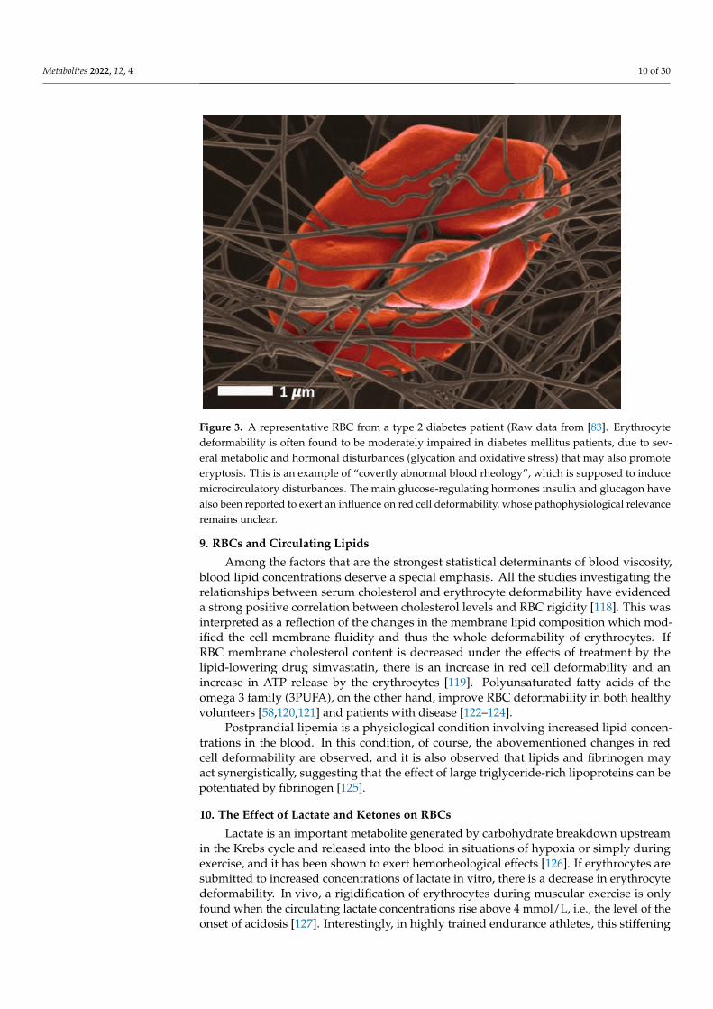
In fact, in normal red cells, glucose deprivation (energy crisis) [20,24], rather thanhyperglycemia, induces irreversible red cell damage and eryptosis [115,116]. Interestingly,in physiological conditions, RBC rigidity is positively correlated with carbohydrate intakein trained athletes [117]. Presumably, this physiological effect does not have the samesignificance as the red cell stiffening induced by chronic hyperglycemia. Physiologicalchanges in blood glucose concentration may be associated with some hemorheologicalalterations, but when these changes reach a pathological range, they may be associated withred cell damage due to free radicals or other factors and thus trigger irreversible alterationsand eryptosis. Figure 3 shows an RBC from an individual with diabetes.
Metabolites 2022, 12, 4 10 of 30
Metabolites 2022, 12, x FOR PEER REVIEW 10 of 31
In fact, in normal red cells, glucose deprivation (energy crisis) [20,24], rather than hyperglycemia, induces irreversible red cell damage and eryptosis [115,116]. Interest-ingly, in physiological conditions, RBC rigidity is positively correlated with carbohydrate intake in trained athletes [117]. Presumably, this physiological effect does not have the same significance as the red cell stiffening induced by chronic hyperglycemia. Physiolog-ical changes in blood glucose concentration may be associated with some hemorheological alterations, but when these changes reach a pathological range, they may be associated with red cell damage due to free radicals or other factors and thus trigger irreversible alterations and eryptosis. Figure 3 shows an RBC from an individual with diabetes.
Figure 3. A representative RBC from a type 2 diabetes patient (Raw data from [83]. Erythrocyte deformability is often found to be moderately impaired in diabetes mellitus patients, due to several metabolic and hormonal disturbances (glycation and oxidative stress) that may also promote eryp-tosis. This is an example of “covertly abnormal blood rheology”, which is supposed to induce mi-crocirculatory disturbances. The main glucose-regulating hormones insulin and glucagon have also been reported to exert an influence on red cell deformability, whose pathophysiological relevance remains unclear.
9. RBCs and Circulating Lipids Among the factors that are the strongest statistical determinants of blood viscosity,
blood lipid concentrations deserve a special emphasis. All the studies investigating the relationships between serum cholesterol and erythrocyte deformability have evidenced a strong positive correlation between cholesterol levels and RBC rigidity [118]. This was interpreted as a reflection of the changes in the membrane lipid composition which mod-ified the cell membrane fluidity and thus the whole deformability of erythrocytes. If RBC membrane cholesterol content is decreased under the effects of treatment by the lipid-lowering drug simvastatin, there is an increase in red cell deformability and an increase in ATP release by the erythrocytes [119]. Polyunsaturated fatty acids of the omega 3 family (3PUFA), on the other hand, improve RBC deformability in both healthy volunteers [58,120,121] and patients with disease [122–124].
Postprandial lipemia is a physiological condition involving increased lipid concen-trations in the blood. In this condition, of course, the abovementioned changes in red cell deformability are observed, and it is also observed that lipids and fibrinogen may act syn-ergistically, suggesting that the effect of large triglyceride-rich lipoproteins can be poten-tiated by fibrinogen [125].
Figure 3. A representative RBC from a type 2 diabetes patient (Raw data from [83]. Erythrocytedeformability is often found to be moderately impaired in diabetes mellitus patients, due to sev-eral metabolic and hormonal disturbances (glycation and oxidative stress) that may also promoteeryptosis. This is an example of “covertly abnormal blood rheology”, which is supposed to inducemicrocirculatory disturbances. The main glucose-regulating hormones insulin and glucagon havealso been reported to exert an influence on red cell deformability, whose pathophysiological relevanceremains unclear.
9. RBCs and Circulating Lipids
Among the factors that are the strongest statistical determinants of blood viscosity,blood lipid concentrations deserve a special emphasis. All the studies investigating therelationships between serum cholesterol and erythrocyte deformability have evidenceda strong positive correlation between cholesterol levels and RBC rigidity [118]. This wasinterpreted as a reflection of the changes in the membrane lipid composition which mod-ified the cell membrane fluidity and thus the whole deformability of erythrocytes. IfRBC membrane cholesterol content is decreased under the effects of treatment by thelipid-lowering drug simvastatin, there is an increase in red cell deformability and anincrease in ATP release by the erythrocytes [119]. Polyunsaturated fatty acids of theomega 3 family (3PUFA), on the other hand, improve RBC deformability in both healthyvolunteers [58,120,121] and patients with disease [122–124].
Postprandial lipemia is a physiological condition involving increased lipid concen-trations in the blood. In this condition, of course, the abovementioned changes in redcell deformability are observed, and it is also observed that lipids and fibrinogen mayact synergistically, suggesting that the effect of large triglyceride-rich lipoproteins can bepotentiated by fibrinogen [125].
10. The Effect of Lactate and Ketones on RBCs
Lactate is an important metabolite generated by carbohydrate breakdown upstreamin the Krebs cycle and released into the blood in situations of hypoxia or simply duringexercise, and it has been shown to exert hemorheological effects [126]. If erythrocytes aresubmitted to increased concentrations of lactate in vitro, there is a decrease in erythrocytedeformability. In vivo, a rigidification of erythrocytes during muscular exercise is onlyfound when the circulating lactate concentrations rise above 4 mmol/L, i.e., the level of theonset of acidosis [127]. Interestingly, in highly trained endurance athletes, this stiffening
Metabolites 2022, 12, 4 11 of 30
effect of lactate on RBCs is no longer found. Conversely, lactate appears in this case toimprove erythrocyte deformability [128]. This specific training-induced pattern of responseto lactate may be one of the explanations for the exercise-induced arterial hypoxemia thatoccurs in extreme athletes.
Ketone bodies are another metabolite that can be used by tissues as an alternativefuel in some physiological situations such as starvation. Situations such as a short-termketogenetic diet [129] have been experimentally shown to impair red cell flexibility.
11. Nitric Oxide and RBC Function
Nitric oxide (NO) is surely one of the most important substances known to interactwith erythrocytes, which are in turn able to release it [130,131]. The major source of nitricoxide is the endothelial cell, but nitric oxide can also be produced by the erythrocyte,which possesses functional NO-synthesizing mechanisms [132]. NO synthesis in RBCsvia nitric oxide synthase (NOS) and NO release into the blood stream can be inducedby mechanical stress, so that NO is released by the red cell in close proximity to thevessel wall [133]. One of the effects of NO on RBCs is to protect them from subhemolyticmechanical damage [134], but NO also increases RBC deformability, as demonstrated by M.Bor-Kuçukatay and coworkers [135]. These investigators also reported that, in contrast toNO donors which improved erythrocyte deformability, NOS inhibitors above a thresholdconcentration value decreased erythrocyte deformability. Nitric oxide donors, as well asthe NO precursor L-arginine and the potassium blocker TEA, have been shown to reversethe effects of NOS inhibitors [135]. Therefore, NO is not only a potent regulator of vasculartone but is also a major physiological regulator of blood rheology via its direct effect onRBC deformability. Furthermore, NO release by polymorphonuclear leukocytes increasesRBC deformability [136]. In fact, the effect of NO on erythrocyte rigidity depends on theNO concentration, as studied by Mesquita and coworkers [137], who reported a biphasiceffect. At a NO concentration of 10(−7) M, the erythrocyte deformability improved, while at10(−5) M, the membrane lipid fluidity decreased. At a NO concentration of 10(−3) M, therewas an increase in the methemoglobin concentration and the RBC deformability decreased,although the membrane fluidity and lipid peroxidation were not changed compared to thecontrol. We should also mention the experiments with spermine NONOate that resulted inan increase in the RBC deformability, due to an effect on the RBC membrane associatedwith an improvement in its oxygen carrying properties [63]. Older erythrocytes exhibit adecrease in both internal NO synthesis and sensitivity to external NO, which likely explains,at least in part, why older erythrocytes are less deformable [138]. It is important to pointout that nitric oxide is also a protector of the red cell against eryptosis [44]. Presumably,this antieryptotic effect is likely to prevent a further decrease in RBC deformability.
In human diseases, the effects of nitric oxide on RBC deformability have some po-tentially interesting applications. In Plasmodium falciparum malaria, hypoargininemia hasbeen reported to impair nitric oxide production and decrease erythrocyte deformability,even more so at febrile temperatures [139]. In sickle-cell anemia, oxidative stress impairsthe effectiveness of RBC NOS for producing NO, so that the stimulating effect of NO onerythrocyte deformability is blunted [140,141]. In experimental hypertension, the effect ofNO on RBC deformability is also impaired [142]. NO donors such as nitroglycerine help tomaintain red cell deformability in conditions such as cardiopulmonary bypass. High-dosenitroglycerin has been shown in this case to improve erythrocyte deformability throughactivating the phosphorylation of aquaporin 1 [143].
In fact, NO is not the only representative of the novel family of gasotransmitters,which are signaling molecules that easily diffuse across lipid membranes and exert theireffect only in the area of their biosynthesis. Another gasotransmitter that is generatingan increasing interest in hemorheology is hydrogen sulfide (H2S). Hydrogen sulfide isproduced from L-cysteine and D-cysteine under the influence of enzymes such as cystathio-nine β-synthase, cystathionine γ-lyase, 3-mercaptopyruvate sulfurtransferase, and cysteineaminotransferase [144]. This gasotransmitter has been shown to exert a cardioprotective

Metabolites 2022, 12, 4 12 of 30
effect and to regulate vascular tone via an effect on the contractility of vascular smoothmuscle cells. H2S has been reported to play a role in angiogenesis, the functional propertiesof platelets, thrombus stability, and erythrogenesis. Its involvement in the pathogenesisof atherosclerosis and arterial hypertension is a matter of current research. On the whole,all three known gaseous mediators, NO, CO, and H2S, improve RBC deformability anddecrease RBC aggregation. [145]. H2S, like the other gasotransmitters, is assumed to act asan oxygen sensor and to be in close synergistic interaction with NO and CO to performthis function.
12. Hormones and Circulating Chemical Messengers
As explained above, the number of chemical messengers and hormones exhibitingspecific receptors on RBCs is regularly increasing. We propose a tentative list of these inTable 1. Among the chemical messengers, we should mention immunoglobulins (IgG),complements [146], and lectins [147].
12.1. Insulin and IGF-I
The fuel-regulating hormone insulin deserves a special mention in regard to this.Insulin binds on the red cell membrane and activates intracellular pathways, with aneffect on erythrocyte deformability that was evidenced in several prior studies [28,148,149]and has been confirmed by more recent investigations [141]. The influence of insulin onerythrocyte rheology seems to be mediated by an effect on the cell membrane [148] thatincludes changes in the molecular composition of the lipid membrane bilayer and thusin its microviscosity, which is associated with alterations to the function of membraneNa/K ATPase [150]. Similarly to the other factors presented in this review, the effectsof insulin may be an improvement or a decrease in erythrocyte deformability. Whenvery high, supraphysiological levels of insulin are applied in vitro, there is a decreasein RBC deformability, as was recently reconfirmed during insulin clamp experiments inhypertensives [151]. Interestingly, the ATP concentrations in RBCs are closely correlatedwith the free insulin levels in plasma [152]. Since, as reported above, intracellular ATP is animportant determinant of RBC deformability, this positive relationship between the insulinand ATP content of the red cell may be involved in the regulation of red cell deformability.
C-peptide is a pancreatic hormone co-secreted with insulin. It is mostly a usefulindex of endogenous insulin secretion but is also supposed to exert some hormonal effects.Among these effects, C-peptide appears to increase eNOS in diabetics, resulting in afluidification of the RBC membranes [153]. This effect is associated with some blood flowredistributions, a reduction in the NaK ATPase pump function, and an improvement in therenal function [153].
Another important hormone closely related to insulin is insulin-like growth factorI (IGF-I). IGF-I can bind on insulin receptors and thus, via this binding, can exert someinsulinlike effects. It also has its own receptors that mediate important anabolic effectsthroughout the entire body. There are also IGF-I receptors on the red cell membrane [154].Some clinical reports suggest that IGF-I may be a regulator of blood viscosity. In trainedathletes, values of IGF-I within the upper quintile of distribution are associated with animpairment of blood fluidity. Since in this study IGF-I is correlated with a lower RBCdeformability, measured with viscometry at high shear rate [155], this observation raisesthe possibility of a direct effect of IGF-I on RBC deformability via the binding of thishormone on its receptors on the red cell membrane.
12.2. Glucagon and RBCs
Glucagon is another important hormone involved in the regulation of fuel metabolism,in which it exerts an action opposite to that of insulin. Glucagon also exerts cataboliceffects on the body’s carbohydrate and protein stores. It was reported to decrease RBCdeformability by P. Valensi and coworkers in 1986 [156]. However, more recently, theopposite effect has been reported by R. Komatsu and coworkers. These investigators

Metabolites 2022, 12, 4 13 of 30
showed that intravenously injected glucagon improved the erythrocyte deformability (asmeasured with a technique of filterability), resulting in a significant decrease in wholeblood viscosity, which was associated with an increase in the blood flow [157]. Both of thesereports are in disagreement with another publication by W. Reinhart, who reported thatneither C-peptide, insulin, or glucagon had any measurable influence on RBC deformability,as assessed by blood viscometry [158].
12.3. Thyroid Hormones
Erythrocytes also exhibit receptors for the thyroid hormone L-triodothyronine [159].Whether thyroid hormones are regulators of blood rheology remains unclear, but a decreasein RBC deformability has been reported to exist in hyperthyroidism and to be reversibleafter the successful treatment of the disease [160].
12.4. Leptin
In this context, leptin, a hormone released by adipocytes and thus belonging to thefamily of adipokines [161], is involved in a feedback loop linking the size of the body fatstores and the energy intake, but it has also been shown to improve red cell deformabilityvia a NO- and cGMP-dependent mechanism [162]. In a preliminary work, we reported thatleptin was correlated with plasma viscosity and erythrocyte disaggregation [163], and morerecently, we confirmed that it was closely associated with increased red cell deformabilityand aggregation [164]. This hormone also regulates the body water stores via a directeffect on the adrenal production of aldosterone [165,166]. Therefore, leptin, which has beenuntil now barely investigated in hemorheological research, may be one of the importantphysiological regulators of red cell rheology, involving it in regulatory loops that linkenergy stores and circulation.
12.5. Erythropoietin
Erythropoietin (EPO) is undoubtedly a major regulator of blood viscosity. This hor-mone released by the kidney is stimulated by hypoxia and inhibited by increases in plasmaviscosity at the level of the juxtaglomerular apparatus in the nephron. It stimulates ery-throcyte development in the bone marrow. The team of W. Reinhardt has elegantly demon-strated in a seminal paper that EPO mediates the homeostatic regulation of viscosity(“viscoregulation”) that follows a rise in plasma viscosity [167] and results in a decrease inRBC mass. In the early 1990s, several teams closely analyzed the evolution of blood viscos-ity factors during the natural history of chronic renal failure. The studies by our Portuguesecolleagues J. Martins e Silva and C. Saldanha, and by M. Delamaire, are more thoroughlyreviewed in our preceding review [168]. CKD-associated hemorheological disturbances(less deformable RBCs, increased plasma viscosity) were corrected by a treatment withrecombinant human erythropoietin (rhEPO). In cancer patients, rhEPO increases red celldeformability and decreases red cell aggregation [169]. It is important to notice that EPO,in addition to being a hormone that improves red cell deformability, also has antieryptoticproperties, although it remains unable to completely counteract the triggering of eryptosisinduced by an intracellular calcium influx in RBCs [170]. Young erythrocytes appear to beparticularly prone to eryptosis following a decline of EPO, a phenomenon which has beentermed neocytolysis [24].
12.6. Somatostatin
Somatostatin is another important hormone synthesized in the pancreas and in othertissues which circulates in blood [171]. Somatostatin can induce dramatic circulatorychanges and increase the peripheral blood flow in humans [172]. In addition, somatostatininterferes with platelet functions [173]. Consistent with this circulatory effect, somatostatinseems to improve erythrocyte deformability, as suggested by its beneficial effect whenassessed in vivo by our group using several techniques [174].

Metabolites 2022, 12, 4 14 of 30
12.7. Melatonin
Melatonin is a hormone released by the pineal gland, which plays an important role inthe circadian rhythms in the body. Additionally, melatonin has been reported to decreaseRBC deformability in experiments. However, pinealectomy by itself did not induce anystatistically significant change in erythrocyte deformability [175]. Erythrocyte deformability,measured by a method of filterability, was not modified by in vitro incubation of bloodsamples with melatonin [176]. Therefore, this issue remains controversial and requiresmore study.
12.8. Leukotrienes and Prostaglandins
Leukotrienes belong to the family of arachidonic acid derivatives, and some of themare known to exert powerful biological effects. Some leukotrienes, but not all [177,178], havebeen reported to impair erythrocyte deformability. Leukotriene B4 decreases the filterabilityof washed resuspended erythrocytes measured with the hemorheometre [178]. Consistentwith what is observed for the other arachidonic derivatives, some prostaglandins have anopposite effect. Prostaglandin E1 and the prostacyclin analogue iloprost improve red andwhite cell filterability in vitro and in vivo [179,180]. PGE2 decreases the deformability ofRBCs and increases their aggregability [181].
12.9. Sex Hormones
The literature about the hemorheological effects of sex hormones has primarily beendriven by the issue of the thrombogenic effects of oral contraceptives (OCs). We previouslyreviewed this classical literature more thoroughly [168]. Briefly, it was shown that OCusers in the late 1970s exhibited a lower erythrocyte deformability that was associatedwith a moderate increase in the whole blood viscosity, while the values of the plasmaviscosity remained in the normal range. Hematocrit was also found to be increased underOCs in some studies but not in others. It was assumed that the progestin componentof the OC was responsible for a rise in the circulating fibrinogen that explained most ofthis pattern. In fact, this picture observed in old OC pills (whose progestin compoundswere mostly 19-nortestosterone derivatives) is no longer observed. More recent low-dosecompounds appear to be almost devoid of hemorheological side-effects, in contrast tothe older compounds, although they still induce moderately higher RBC aggregation. Inaddition, more recent physiological investigations have been conducted in women withnormal menstrual cycles, showing that their estradiol levels were positively correlatedwith their whole blood viscosity, plasma viscosity, and fibrinogen, and that their RBCdeformability was impaired correlatively to their estradiol levels. Thus, in physiologicalconditions, estrogens appear to decrease blood fluidity. On the other hand, progesteronehad the opposite effect in physiological conditions, decreasing both the fibrinogen and theblood viscosity, and increasing the RBC deformability. Therefore, the RBC deformabilitywas lower in the follicular than in the luteal phase. Interestingly, the recent literature onestrogens and red cell rheology has pointed out the role of nitric oxide. Estrogens areknown to physiologically increase both NO synthesis and release by the endothelial cells.This effect is likely to be mediated by the estrogen-responsive elements located in thepromoting region of the gene coding for endothelial NOS.
Beside the classical mode of action of estrogens, which involves intracellular receptors,recent emphasis has been placed on the membrane receptors, on which estrogens canspecifically bind and thus act more rapidly, inducing an increase in cytosolic Ca++ in somecells. In addition, estrogens also exhibit antioxidant properties which can delay NO clearingfrom blood [168]. All these mechanisms are therefore likely to improve RBC deformabilityvia NO-mediated mechanisms. This seems to be in disagreement with the reports showingthat in vivo hormonal treatment by either transcutaneous or oral estrogens impairs RBCdeformability by increasing membrane rigidity [168].
In vitro, 17 β-estradiol at a concentration of 10−5 M also decreased the RBC aggrega-tion in the blood samples of postmenopausal women undergoing hormone therapy [182].
Metabolites 2022, 12, 4 15 of 30
A recent paper by M. Grau [183] suggests that the gender differences in hematologicalparameters in males compared to females (with higher RBC deformability in females)might be related to the higher estradiol levels that are associated with higher RBC NOlevels. However, Pretorius and coworkers also reported on the effects of estrogen andprogesterone on the RBC structure, and they noted increased eryptosis [184,185].
12.10. Dehydroepiandrosterone
The incorporation of dehydroepiandrosterone sulfate (DHEAS) into the human redcell membrane increases the acyl chain motion in the middle portion of the membrane andinduces the echinocytosis of red cells. It is suggested that the increase in the viscosity of ared cell suspension, the decreased deformability, and the decrease in the deoxygenation rateof hemoglobin in the presence of DHEAS probably reflect the presence of echinocytes. Inthe presence of plasma proteins, the incorporation of DHEAs into red cells was remarkablysuppressed [186].
12.11. Apelin
Apelin is a recently discovered hormone belonging to the family of adipokines, whichcan bind on the G-protein-coupled APJ receptor which is expressed over the surface ofmany cells in the body. It is widely expressed in various organs such as the heart, lungs,and kidney and exerts a hypotensive effect via the activation of the APJ receptors on thesurface of endothelial cells, thus inducing the release of NO. In addition, it decreases thehypothalamic secretion of vasopressin and increases water intake, thus exerting importanteffects on the homeostatic regulation of the body’s fluid stores. In rats that exhibited adecrease in erythrocyte deformability due to the experimental induction of diabetes andischemia-reperfusion injury of the heart, apelin-13 has been shown to reverse this loss ofdeformability [187].
12.12. Catecholamines
The catecholamines norepinephrine and epinephrine are the two circulation messen-gers of the sympathetic nervous system that can be released into the blood by the adrenalmedulla and act on RBCs via specific α- and β- receptor agonists. It is well known thatnorepinephrine is mostly an α-adrenergic agonist, while epinephrine is mostly a potentβ-adrenergic agonist. Over recent years, evidence has been accumulated that both of thesecatecholamines are important regulators of RBC rheology. The α1-adrenergic receptor canbe detected on human erythrocyte membranes [188]. There are also β-adrenergic receptorson the red cell membrane that have been shown in animal studies to mediate the regulatingeffects of catecholamines on cell volume and ion transport [189]. The first studies on cate-cholamines and red cell rheology were conducted in the late 1980s by Pfafferott and Volger,who reported that in vitro, both norepinephrine (α-adrenergic agonist) and isoprenaline(β-adrenergic agonist) decreased erythrocyte deformability [190]. More recently, however,Hilario demonstrated that epinephrine (β-adrenergic agonist) actually improves RBC de-formability when measured with a more accurate technique but is also able to induce thetransformation of human RBCs into echinocyte [191]. Catecholamines (both β-agonists andα-agonists) also have an antieryptotic effect [43], consistent with the short-term beneficialrole of these hormones regarding the body’s adaptation to unusual stresses. In fact, inexperiments on species whose RBCs are nucleated, catecholamines induced a dramaticincrease in the cell volume as a result of an accumulation of sodium and chloride due tothe activation of an amiloride-sensitive, cyclic, AMP-dependent Na+-H+ exchanger whichallowed Na+ to enter in exchange for internal H+. At the same time, the RBC deformabilitywas improved (despite the increase in the cell volume). Both the RBC fluidification andthe activation of this ionic exchange were likely to be an adaptive response to hypoxiawhich resulted in the increased oxygen-carrying capacity of the RBCs [192]. The literature,however, suggests that this epinephrine may improve RBC deformability [193–195], pre-sumably via β-adrenergic receptors, while there is apparently no effect of either α1- and

Metabolites 2022, 12, 4 16 of 30
α2-receptor agonists. It was also shown that RBCs incubated with epinephrine and isopro-terenol exhibited significant changes of deformability, by 10% and 30%, respectively. Thisis consistent with the other classical effects of catecholamines mediated by β-adrenergicreceptors (vasodilation, increased cardiac output, etc.) that all lead to an increased bloodflow. The team of A. Muravyov extensively studied the effect of catecholamines on therheological properties of the human RBC, showing that the effect of these hormones on RBCdeformability is mostly under the control of intracellular Ca2+-regulating pathways [196].In contrast to this positive effect of catecholamines on RBC deformability in physiologicalconditions, an increased viscosity and decreased RBC deformability were observed inuntreated pheochromocytoma [197].
12.13. Cortisol
Beside catecholamines, cortisol is a major hormone involved in the body’s adaptationto stress. Cortisol has been shown to bind to the erythrocyte membrane, impairing theepinephrine binding at this level and resulting in an increase in the microviscosity of themembranes and a rise in Na(+),K(+)-ATPase activity [198]. This is in line with the othereffects of corticosteroids that moderate the effects of stress hormones in order to cope withthem. Windberger has also described the hemorheological profile of Cushing syndrome indogs, i.e., hypercortisolism due to increased and sustained cortisol release by the adrenalcortex, showing that this situation is associated with increased plasma viscosity and RBCaggregation [199]. This area surely deserves more investigation.
12.14. Endocannabinoids
The Endocannabinoid System (ECS) is an important regulatory system aiming at main-taining homeostasis in a variety of body functions such as temperature, mood, the immunesystem, and energy input and output. It exerts a wide variety of effects on emotionalbehavior, feeding behavior, appetite, nervous functions, fertility, and pre-and postnatal de-velopment. This system involves endogenous lipid mediators called endocannabinoids, ofwhich the most well-known are 2-arachidonoylglycerol and anandamide, which are synthe-sized from the pool of arachidonic acid in cell membrane phospholipids. The modulationof this system with cannabinergic, cannabimimetic, and cannabinoid-based therapeuticdrugs that are currently under development offers some potential for treating a number ofdiseases [200]. The endocannabinoid system (ECS), via the activation of the type-1 cannabi-noid receptor (CB1), is involved in the process of exostasis, i.e., overeating and storage dueto the anticipation of future energy needs, and thus adds some adaptatory regulations tohomeostasis [201]. Endocannabinoids and their receptors are almost ubiquitous in the bodyand regulate intermediary metabolism and energy expenditure. They interfere with glucoseand lipid metabolism so as to promote energy storage and reduce energy expenditure.This system is overactive in obesity and appears to play a role in the maintenance of fatmass [202]. All these functions modulated by endocannabinoids are also known to be asso-ciated with hemorheological processes. However, little is known about endocannabinoidinvolvement in the regulation of blood rheology. It is thus interesting to point out that theendocannabinoid anandamide has been reported to induce apoptosis in several varietiesof nucleated cells, and to increase the activity of RBC cytosolic Ca2+, resulting in the cellshrinkage and cell membrane scrambling of mature RBCs and, subsequently, inducingeryptosis [40].
12.15. Other Hormones
Erythrocytes are also known to be able to release endothelin-1 (ET1), a potent vaso-constrictor which is also produced by endothelial and smooth muscle cells. ET1 has beenreported to improve the deformability (as assessed by filterability) of stiffened RBCs via anactivation of the protein kinase C [203]. However, another study was unable to evidencethis effect when the RBC deformability was evaluated with viscometry [204].

Metabolites 2022, 12, 4 17 of 30
Finally, we should mention the surprising lack of a hemorheologic effect of calciumregulating hormones (parathormone, calcitonin, and vitamin D), which contrasts to themajor importance of intracellular calcium in regard to erythrocyte rheology [205].
13. RBCs in Various Pathophysiological Situations
In the previous paragraphs, we referred to a pathological structure of the RBCs in condi-tions such as type 2 diabetes and hereditary hemochromatosis. We have published extensivelyon the RBC structural changes in other inflammatory conditions such as rheumatoid arthritis,Alzheimer’s disease, and Parkinson’s disease and also on the effect of selected dysregulatedinflammatory markers including cytokines on RBC structure [47,79,82,206–209]. In all inflam-matory conditions, the circulating dysregulated inflammatory markers have a profoundeffect on RBCs, platelets, and plasma protein structure. Both deformability patterns anderyptosis are visible in these conditions. Similar changes have been noted in septic shockand sleep apnea patients.
13.1. Stress
Stress is a neuroendocrine complex reaction (“general adaptation syndrome”) whichaims at maintaining homeostasis in the body when it is submitted to an unusual situation.The stress can be sufficient to cope with this unusual situation (eustress) or result in aprolonged disturbance of the body’s functions (distress) [210]. All stress hormones (asindicated above in this review) are likely to exert hemorheologic effects, although thesituation is not very clear concerning cortisol. Catecholamines exert complementary effects:epinephrine increases red cell deformability (thus favoring microcirculatory perfusion),while norepinephrine increases red cell aggregation and induces a fluid shift reductionof plasma volume with a parallel rise in hematocrit due to α1-receptor stimulation [211].Therefore, unsurprisingly, stress induces hemorheological modifications. A pioneeringstudy was presented by A. Ehrly [212], evidencing a rise in blood and plasma viscosityafter video-film-induced emotional stress. More recently, a hyperviscosity syndrome wasevidenced among evacuees who survived the earthquake and tsunami of Fukushima inJapan [213]. In this case, there was polycythemia.
A study on American veterans with Gulf War Illness (GWI) also deals with this issue.These patients experience chronic symptoms that include fatigue, pain, and cognitiveimpairment. Assuming that this symptom cluster may be related to impaired tissue oxygendelivery, the investigators measured the red cell deformability and found that it wasincreased in veterans suffering from GWI. [214]. Further studies on GWI showed that theincreased deformability of the red cells was not affected during maximal exercise [215].No clear explanation for this finding was given, but the effect of epinephrine on red celldeformability may be a logic explanation.
The issue of stress and hemorheology is more thoroughly developed in our previousreview paper [216] but requires, of course, further investigation. On the whole, we canassume that “eustress” induces reversible adaptative alterations in the blood viscosityfactors, while “distress” may induce more important damage that may involve eryptosis.
13.2. Chronic Fatigue Syndrome
By contrast, in people suffering from chronic fatigue syndrome, the red blood cellswere found to be significantly stiffer than those in healthy controls [217]. A previous reporton the same disease did not evidence this decrease in deformability [218], probably due tomethodological concerns. Since oxidant stress is one of the mechanisms underlying chronicfatigue syndrome [219] and this oxidant stress damages red cells and makes them stiffer,this finding is logical. In this situation, the increased apoptosis of various blood cell lineswas described [220], and, although we are not aware of a report on increased eryptosis, itis logical to assume that red cells are stiffened in chronic fatigue syndrome as a result ofoxidant stress, which damages the cells and may lead to their programmed death.

Metabolites 2022, 12, 4 18 of 30
13.3. Septic Shock
There are conditions that combine many disturbances to various functions, includingRBC properties. The most impressive example of this is perhaps septic shock. Sepsis hasbeen shown to be associated with impaired blood rheology [221–223]. Furthermore, in thiscontext, decreased red blood cell deformability has been reported to be associated with apoor outcome in septic patients [224,225]. The hemorheologic disturbances observed dur-ing sepsis are associated with an altered metabolism; decreased 2,3-bisphosphoglycerate;the redistribution of membrane phospholipids; changes in the RBC volume, the affinitybetween hemoglobin and oxygen, the morphology, the antioxidant status, the intracellularCa2+ homeostasis, and the membrane proteins; membrane phospholipid redistribution; andRBC O2–dependent adenosine triphosphate efflux. During septic shock, a phenomenonof RBC autoxidation is observed and has been supposed to worsen the disorder [226,227].Unsurprisingly, marked alterations in RBC rheology, including reduced deformabilityand increased aggregation, occur early on in septic patients, and reductions in RBC de-formability over time are associated with a poor prognosis [224,228]. The sepsis-associatedimpairment of erythrocyte deformability is associated with a decrease in erythrocyte NO-releasing activity, both abnormalities being apparently a consequence of the inflammatoryreaction [229]. In experimentally induced septic shock in rats, prostacyclin (iloprost) andnitric oxide prevented the sepsis-induced loss of red blood cell deformability via a complexmechanism [230].
13.4. Sleep Apnea
Another situation associated with a decrease in RBC deformability which is probablymultifactorial is obstructive sleep apnea syndrome (OSAS). This syndrome has emergedover recent decades as an important risk factor for atherosclerosis and cardiovascular disor-ders [231–233]. In OSAS, endothelial dysfunction is markedly disturbed due to recurrenthypoxemic events, and there is a decrease in erythrocyte deformability [231]. Additionally,a decrease in NO bioavailability is also observed and may be both a consequence and aworsening factor of the defect in RBC deformability [232]. In this disease, there is also anincrease in plasma viscosity and an increase in RBC aggregation [231]. OSAS is also knownto be associated with apoptosis in various tissues and thus, presumably, eryptosis [233]. Incontrast with hypoxemia, it is interesting to point out that the opposite condition, hyper-oxia, seems to have no measurable effect on blood rheology, so that its use for donor-organpreservation before graft is not known to induce any hemorheologic disturbance [234].
13.5. COVID-19
The recent COVID-19 pandemic has generated a host of studies in all areas of biomed-ical research [235], and, unsurprisingly, disturbances in microcirculation [236] and therheologic properties of blood [237] have been described. It is clear that endotheliopathiesare important clinical features of acute COVID-19 [238,239]. Various circulating and dysreg-ulated inflammatory coagulation biomarkers, including fibrin(ogen), D-dimer, P-selectin,the von Willebrand Factor (VWF), C-reactive protein (CRP), and various cytokines directlybind to endothelial receptors and are likely to be indicative of a poor prognosis [240–243].This poor prognosis is further worsened by a substantial deposition of microclots in thelungs [244–246]. The plasma of COVID-19 patients also carries a massive load of preformedamyloid clots and there are numerous reports of damage to erythrocytes [247–249] andplatelets and the dysregulation of inflammatory biomarkers [240–243,250]. Recently, wealso determined whether the spike protein may interfere with blood flow by comparingnaïve healthy plasma samples with and without added spike protein to plasma samplesfrom COVID-19-positive patients (before treatment). We concluded that the spike proteinmay have direct pathological effects on blood rheology [251]. Significant pathologicalchanges in microcirculation and the presence of persistent microclots have also been notedin Long COVID/PASC [250].

Metabolites 2022, 12, 4 19 of 30
14. Concluding Remarks
Since the review on this topic that we published 20 years ago [168], our knowledgeof the effects of various factors on erythrocyte deformability has expanded significantly.However, many issues remain incompletely understood at this time. It is clear, however,that this entire body of information we have put together suggests that there is an exquisitelyregulated system involved in the homeostasis of blood rheology and flow distribution.The viscoregulatory loops hypothesized in the middle of the 20th century are now welldescribed [167]. Others will probably be evidenced over the coming years.
The best known is probably the endothelium–leukocyte–liver axis, which involvespolymorphonuclear neutrophils and monocytes that are able to release many biologicallyactive substances that may in turn modify the blood viscosity factors. The release of cy-tokines such as IL-6 induces a rise in fibrinogen, which increases plasma viscosity and RBCaggregation. Circulating free radicals and arachidonic acid derivatives modify erythro-cyte membrane properties and thus RBC deformability. Free radicals increase the rigidityand aggregation of erythrocytes. These interactions between white cell activation anderythrocyte rheologic properties have been remarkably well-investigated by the team of O.Baskurt [252]. This study showed that most of the hemorheological profiles of inflammatorydiseases (and vascular diseases) are explained by these leukocyte–erythrocyte interactionsand are likely to play an important role in the body’s response to an inflammatory stimula-tion [253].
As indicated above, some other integrated responses of the body such as stress, theregulation of energy stores, and growth involve a hemorheologic response whose relevanceis not completely understood. The relationship between the size of energy stores and theblood rheology even in normal conditions is also an issue that deserves more research,since some reports suggest that the hormones released by the adipose tissue (leptin andadiponectin) have circulatory effects [254]. Our recent report showing that leptin is moreclosely correlated with red cell aggregation than other determinants quantifying adiposestores may suggest that this hormone is involved in a regulatory loop linking the body’senergy status and the hemorheological modulation of microcirculation [163]. Anotherinteresting recent finding is that red cell rigidity in obesity is associated with higher valuesof angiogenesis [255]. Such a relationship needs to be more closely investigated, but it maylead to speculation that obesity-associated RBC stiffness may result in an adaptation ofthe microcirculation network. Presumably, NO, which is known to modulate angiogenesisin vitro and in vivo [256,257], may be involved in this effect.
In addition, according to several teams, dietary habits appear to be associated withthe modification of blood rheology [258]. Very little is known about this issue, whichrequires further investigation. In order to propose a hypothesis, taking into accountthe abovementioned effects of circulating metabolites, exercise, and nutrition on bloodrheology, we recently proposed the “healthy primitive lifestyle paradigm”. This hypothesisassumes that evolution has selected in Homo sapiens genetic polymorphisms leading toinsulin resistance as an adaptative strategy to cope with the lifestyle of our Paleolithicancestors. According to the current scientific data, our Paleolithic ancestors performed a lotof prolonged low-intensity physical activity and their food was mostly based on lean meatand wild herbs (i.e., poor in saturated fat, rich in low-glycemic-index carbohydrates, andmoderately high in protein). According to this hypothesis, an individual whose exerciseand nutritional habits are close to this lifestyle represents the mainstream phenotype,and highly trained athletes represent a minority group. Sedentary subjects undergo ahost of metabolic (and hemorheologic) modifications that aim at coping with the lack ofphysiological activity, which represents a pathologic situation in these populations [259].We previously published data in agreement with this theory, thus integrating them into alogical picture of hemorheologic homeostasis [260].
Therefore, although this issue remains incompletely studied, we think that the bodyof information summarized in this review supports the concept that among the numeroussituations where RBC rigidification has been reported to occur, some involve reversible

Metabolites 2022, 12, 4 20 of 30
changes in a fully physiological context, while others reflect quite irreversible damage tothe red cells that will lead to eryptosis (Figure 4). In some situations, however, there may bea continuum between the two processes if the stimulus is too strong. Exercise, for example,generally involves reversible changes in RBC rheology but may also induce irreversibledamage and programmed erythrocyte death. Such a triggering of eryptosis in this casemay represent a protective mechanism against the deleterious effect of old RBCs, which isknown to be associated with an increased risk of death [77,78]. However, further research isrequired to more precisely delineate the respective importance of reversible (physiological)and almost-irreversible (eryptosis related) RBC stiffening in various situations.
Metabolites 2022, 12, x FOR PEER REVIEW 20 of 31
In addition, according to several teams, dietary habits appear to be associated with the modification of blood rheology [258]. Very little is known about this issue, which re-quires further investigation. In order to propose a hypothesis, taking into account the abovementioned effects of circulating metabolites, exercise, and nutrition on blood rheol-ogy, we recently proposed the “healthy primitive lifestyle paradigm”. This hypothesis assumes that evolution has selected in Homo sapiens genetic polymorphisms leading to insulin resistance as an adaptative strategy to cope with the lifestyle of our Paleolithic ancestors. According to the current scientific data, our Paleolithic ancestors performed a lot of prolonged low-intensity physical activity and their food was mostly based on lean meat and wild herbs (i.e., poor in saturated fat, rich in low-glycemic-index carbohydrates, and moderately high in protein). According to this hypothesis, an individual whose exer-cise and nutritional habits are close to this lifestyle represents the mainstream phenotype, and highly trained athletes represent a minority group. Sedentary subjects undergo a host of metabolic (and hemorheologic) modifications that aim at coping with the lack of phys-iological activity, which represents a pathologic situation in these populations [259]. We previously published data in agreement with this theory, thus integrating them into a log-ical picture of hemorheologic homeostasis [260].
Therefore, although this issue remains incompletely studied, we think that the body of information summarized in this review supports the concept that among the numerous situations where RBC rigidification has been reported to occur, some involve reversible changes in a fully physiological context, while others reflect quite irreversible damage to the red cells that will lead to eryptosis (Figure 4). In some situations, however, there may be a continuum between the two processes if the stimulus is too strong. Exercise, for ex-ample, generally involves reversible changes in RBC rheology but may also induce irre-versible damage and programmed erythrocyte death. Such a triggering of eryptosis in this case may represent a protective mechanism against the deleterious effect of old RBCs, which is known to be associated with an increased risk of death [77,78]. However, further research is required to more precisely delineate the respective importance of reversible (physiological) and almost-irreversible (eryptosis related) RBC stiffening in various situ-ations.
Figure 4. Regulatory loops involved in the modulation of red cell deformability. According to the physiological or pathological context, the factors thoroughly enumerated in this review increase or decrease red cell deformability, thus contributing to the adaptation of microcirculatory blood flow to this context. Erythrocyte stiffening may be a reversible event or one of the components of the cascade of events leading to programmed red cell death (eryptosis). Figure created using www.bi-orender.com.
Author Contributions: Conceptualization and writing, J.-F.B., E.P., E.V.-M., J.M. and E.R.d.M.; Re-view, editing and visualization, E.P. All authors have read and agreed to the published version of the manuscript.
Figure 4. Regulatory loops involved in the modulation of red cell deformability. According to thephysiological or pathological context, the factors thoroughly enumerated in this review increase ordecrease red cell deformability, thus contributing to the adaptation of microcirculatory blood flow tothis context. Erythrocyte stiffening may be a reversible event or one of the components of the cascadeof events leading to programmed red cell death (eryptosis). Figure created using www.biorender.comaccessed on 17 December 2021.
Author Contributions: Conceptualization and writing, J.-F.B., E.P., E.V.-M., J.M. and E.R.d.M.; Re-view, editing and visualization, E.P. All authors have read and agreed to the published version ofthe manuscript.
Funding: This research received no external funding.
Conflicts of Interest: The authors declare no conflict of interest.
References1. Huisjes, R.; Bogdanova, A.; Van Solinge, W.W.; Schiffelers, R.M.; Kaestner, L.; Van Wijk, R. Squeezing for Life—Properties of Red
Blood Cell Deformability. Front. Physiol. 2018, 9, 656. [CrossRef] [PubMed]2. Munter, W.A.; Stein, P.D. Newtonian behavior of blood at high rates of shear. Biorheology 1973, 10, 501–508. [CrossRef]3. Skalak, R.; Chien, S. Rheology of blood cells as soft tissues. Biorheology 1982, 19, 453–461. [CrossRef] [PubMed]4. Schmid-Schönbein, H.; Gaehtgens, P. What is red cell deformability? Scand. J. Clin. Lab. Investig. 1981, 156, 13–26. [CrossRef]5. Nader, E.; Skinner, S.; Romana, M.; Fort, R.; Lemonne, N.; Guillot, N.; Gauthier, A.; Antoine-Jonville, S.; Renoux, C.; Hardy-
Dessources, M.D.; et al. Blood Rheology: Key Parameters, Impact on Blood Flow, Role in Sickle Cell Disease and Effects ofExercise. Front. Physiol. 2019, 10, 1329. [CrossRef]
6. Zheng, Y.; Cachia, M.A.; Ge, J.; Xu, Z.; Wang, C.; Sun, Y. Mechanical differences of sickle cell trait (SCT) and normal red bloodcells. Lab Chip 2015, 15, 3138–3146. [CrossRef] [PubMed]
7. Man, Y.; Kucukal, E.; An, R.; Watson, Q.D.; Bosch, J.; Zimmerman, P.A.; Little, J.A.; Gurkan, U.A. Microfluidic assessment of redblood cell mediated microvascular occlusion. Lab Chip 2020, 20, 2086–2099. [CrossRef] [PubMed]
8. Man, Y.; Kucukal, E.; An, R.; Bode, A.; Little, J.A.; Gurkan, U.A. Standardized microfluidic assessment of red blood cell-mediated microcapillary oc-clusion: Association with clinical phenotype and hydroxyurea responsiveness in sickle cell disease.Microcirculation 2021, 28, e12662. [CrossRef]
9. Man, Y.; Maji, D.; An, R.; Ahuja, S.P.; Little, J.A.; Suster, M.A.; Mohseni, P.; Gurkan, U.A. Microfluidic electrical impedanceassessment of red blood cell-mediated microvascular occlusion. Lab Chip 2021, 21, 1036–1048. [CrossRef] [PubMed]

Metabolites 2022, 12, 4 21 of 30
10. Lanotte, L.; Mauer, J.; Mendez, S.; Fedosov, D.A.; Fromental, J.M.; Claveria, V.; Nicoud, F.; Gompper, G.; Abkarian, M. Red cells’dynamic morphologies govern blood shear thinning under microcirculatory flow conditions. Proc. Natl. Acad. Sci. USA 2016, 113,13289–13294. [CrossRef]
11. Mauer, J.; Mendez, S.; Lanotte, L.; Nicoud, F.; Abkarian, M.; Gompper, G.; Fedosov, D.A. Flow-Induced Transitions of Red BloodCell Shapes under Shear. Phys. Rev. Lett. 2018, 121, 118103. [CrossRef]
12. Chien, S. Red cell deformability and its relevance to blood flow. Annu. Rev. Physiol. 1987, 49, 177–192. [CrossRef]13. Connes, P.; Alexy, T.; Detterich, J.; Romana, M.; Hardy-Dessources, M.D.; Ballas, S.K. The role of blood rheology in sickle cell
disease. Blood Rev. 2016, 30, 111–118. [CrossRef]14. Barshtein, G.; Ben-Ami, R.; Yedgar, S. Role of red blood cell flow behavior in hemodynamics and hemostasis. Expert Rev.
Cardiovasc. Ther. 2007, 5, 743–752. [CrossRef]15. Le Devehat, C.; Khodabandehlou, T.; Vimeux, M. Relationship between hemorheological and microcirculatory abnormalities in
diabetes mellitus. Diabete Metab. 1994, 20, 401–404.16. Lang, F.; Abed, M.; Lang, E.; Föller, M. Oxidative stress and suicidal erythrocyte death. Antioxid. Redox Signal. 2014, 21, 138–153.
[CrossRef]17. Lang, F.; Gulbins, E.; Lang, P.A.; Zappulla, D.; Föller, M. Ceramide in suicidal death of erythrocytes. Cell. Physiol. Biochem. 2010,
26, 21–28. [CrossRef]18. Lang, F.; Lang, E.; Foller, M. Physiology and pathophysiology of eryptosis. Transfus. Med. Hemother. 2012, 39, 308–314. [CrossRef]19. Lang, E.; Qadri, S.M.; Lang, F. Killing me softly–suicidal erythrocyte death. Int. J. Biochem. Cell Biol. 2012, 44, 1236–1243.
[CrossRef]20. Pretorius, E.; Du Plooy, J.N.; Bester, J. A Comprehensive Review on Eryptosis. Cell. Physiol. Biochem. 2016, 39, 1977–2000.
[CrossRef] [PubMed]21. Qadri, S.M.; Bauer, J.; Zelenak, C.; Mahmud, H.; Kucherenko, Y.; Lee, S.H.; Ferlinz, K.; Lang, F. Sphingosine but not sphingosine-
1-phosphate stimulates suicidal erythrocyte death. Cell. Physiol. Biochem. 2011, 28, 339–346. [CrossRef] [PubMed]22. Qadri, S.M.; Bissinger, R.; Solh, Z.; Oldenborg, P.A. Eryptosis in health and disease: A paradigm shift towards understanding the
(patho)physiological implications of programmed cell death of erythrocytes. Blood Rev. 2017, 31, 349–361. [CrossRef] [PubMed]23. Qadri, S.M.; Donkor, D.A.; Bhakta, V.; Eltringham-Smith, L.J.; Dwivedi, D.J.; Moore, J.C.; Pepler, L.; Ivetic, N.; Nazi, I.; Fox-
Robichaud, A.E.; et al. Phosphatidylserine externalization and procoagulant activation of erythrocytes induced by Pseudomonasaeruginosa virulence factor pyocyanin. J. Cell. Mol. Med. 2016, 20, 710–720. [CrossRef]
24. Lang, E.; Lang, F. Triggers, inhibitors, mechanisms, and significance of eryptosis: The suicidal erythrocyte death. BioMed Res. Int.2015, 2015, 513518. [CrossRef] [PubMed]
25. Duez, J.; Holleran, J.P.; Ndour, P.A.; Pionneau, C.; Diakite, S.; Roussel, C.; Dussiot, M.; Amireault, P.; Avery, V.M.; Buffet, P.A.Mechanical clearance of red blood cells by the human spleen: Potential therapeutic applications of a biomimetic RBC filtrationmethod. Transfus. Clin. Biol. 2015, 22, 151–157. [CrossRef]
26. Klei, T.R.; Dalimot, J.; Nota, B.; Veldthuis, M.; Mul, F.P.; Rademakers, T.; Hoogenboezem, M.; Nagelkerke, S.Q.; van Ijcken, W.F.;Oole, E.; et al. Hemolysis in the spleen drives erythrocyte turnover. Blood 2020, 136, 1579–1589. [CrossRef] [PubMed]
27. Miko, I.; Nemeth, N.; Sogor, V.; Kiss, F.; Toth, E.; Peto, K.; Furka, A.; Vanyolos, E.; Toth, L.; Varga, J.; et al. Comparativeerythrocyte deformability investigations by filtrometry, slit-flow and rotational ektacytometry in a long-term follow-up animalstudy on splenectomy and different spleen preserving operative techniques: Partial or subtotal spleen resection and spleenautotransplantation. Clin. Hemorheol. Microcirc. 2017, 66, 83–96. [CrossRef]
28. Juhan, I.; Vague, P.; Buonocore, M.; Moulin, J.P.; Jouve, R.; Vialettes, B. Abnormalities of erythrocyte deformability and plateletaggregation in insulin-dependent diabetics corrected by insulin in vivo and in vitro. Lancet 1982, 1, 535–537. [CrossRef]
29. Vague, P.; Juhan, I. Red cell deformability, platelet aggregation, and insulin action. Diabetes 1983, 32 (Suppl. 2), 88–91. [CrossRef][PubMed]
30. Brun, J.F.; Varlet-Marie, E.; Connes, P.; Aloulou, I. Hemorheological alterations related to training and overtraining. Biorheology2010, 47, 95–115. [CrossRef] [PubMed]
31. Nader, E.; Guillot, N.; Lavorel, L.; Hancco, I.; Fort, R.; Stauffer, E.; Renoux, C.; Joly, P.; Germain, M.; Connes, P. Eryptosis andhemorheological responses to maximal exercise in athletes: Comparison between running and cycling. Scand. J. Med. Sci. Sports2018, 28, 1532–1540. [CrossRef] [PubMed]
32. Nader, E.; Monedero, D.; Robert, M.; Skinner, S.; Stauffer, E.; Cibiel, A.; Germain, M.; Hugonnet, J.; Scheer, A.; Joly, P.; et al. Impactof a 10 km running trial on eryptosis, red blood cell rheology, and electrophysiology in endurance trained athletes: A pilot study.Eur. J. Appl. Physiol. 2020, 120, 255–266. [CrossRef] [PubMed]
33. Nader, E.; Romana, M.; Guillot, N.; Fort, R.; Stauffer, E.; Lemonne, N.; Garnier, Y.; Skinner, S.C.; Etienne-Julan, M.; Robert, M.;et al. Association Between Nitric Oxide, Oxidative Stress, Eryptosis, Red Blood Cell Microparticles, and Vascular Function inSickle Cell Anemia. Front. Immunol. 2020, 11, 551441. [CrossRef]
34. Koutsouris, D.; Delatour-Hanss, E.; Hanss, M. Physico-chemical factors of erythrocyte deformability. Biorheology 1985, 22, 119–132.[CrossRef]
35. Engström, K.G.; Meiselman, H.J. Analysis of red blood cell membrane area and volume regulation using micropipette aspirationand perfusion. Biorheology 1995, 32, 115–116. [CrossRef]

Metabolites 2022, 12, 4 22 of 30
36. Reinhart, W.H.; Piety, N.Z.; Deuel, J.W.; Makhro, A.; Schulzki, T.; Bogdanov, N.; Goede, J.S.; Bogdanova, A.; Abidi, R.; Shevkoplyas,S.S. Washing stored red blood cells in an albumin solution improves their morphologic and hemorheologic properties. Transfusion2015, 55, 1872–1881. [CrossRef] [PubMed]
37. Sugie, J.; Intaglietta, M.; Sung, L.A. Water transport and homeostasis as a major function of erythrocytes. Am. J. Physiol. HeartCirc. Physiol. 2018, 314, H1098–H1107. [CrossRef]
38. Dupuy-Fons, C.; Brun, J.F.; Mallart, C.; Carvajal, J.; Fussellier, M.; Bardet, L.; Orsetti, A. In vitro influence of zinc and magnesiumon the deformability of red blood cells artificially hardened by heating. Biol. Trace Elem. Res. 1995, 47, 247–255. [CrossRef]
39. Semenov, A.N.; Shirshin, E.A.; Muravyov, A.V.; Priezzhev, A.V. The Effects of Different Signaling Pathways in Adenylyl CyclaseStimulation on Red Blood Cells Deformability. Front. Physiol. 2019, 10, 923. [CrossRef]
40. Bentzen, P.J.; Lang, F. Effect of anandamide on erythrocyte survival. Cell. Physiol. Biochem. 2007, 20, 1033–1042. [CrossRef]41. Jemaà, M.; Fezai, M.; Lang, F. Inhibition of Suicidal Erythrocyte Death by Reversine. Cell. Physiol. Biochem. 2017, 41, 2363–2373.
[CrossRef] [PubMed]42. Kiedaisch, V.; Akel, A.; Niemoeller, O.M.; Wieder, T.; Lang, F. Zinc-induced suicidal erythrocyte death. Am. J. Clin. Nutr. 2008, 87,
1530–1534. [CrossRef] [PubMed]43. Lang, P.A.; Kempe, D.S.; Akel, A.; Klarl, B.A.; Eisele, K.; Podolski, M.; Hermle, T.; Niemoeller, O.M.; Attanasio, P.; Huber, S.M.;
et al. Inhibition of erythrocyte “apoptosis” by catecholamines. Naunyn. Schmiedeberg’s Arch. Pharmacol. 2005, 372, 228–235.[CrossRef]
44. Nicolay, J.P.; Liebig, G.; Niemoeller, O.M.; Koka, S.; Ghashghaeinia, M.; Wieder, T.; Haendeler, J.; Busse, R.; Lang, F. Inhibition ofsuicidal erythrocyte death by nitric oxide. Pflugers Arch. 2008, 456, 293–305. [CrossRef] [PubMed]
45. Niemoeller, O.M.; Bentzen, P.J.; Lang, E.; Lang, F. Adenosine protects against suicidal erythrocyte death. Pflugers Arch. 2007, 454,427–439. [CrossRef]
46. Qadri, S.M.; Mahmud, H.; Lang, E.; Gu, S.; Bobbala, D.; Zelenak, C.; Jilani, K.; Siegfried, A.; Foller, M.; Lang, F. Enhanced suicidalerythrocyte death in mice carrying a loss-of-function mutation of the adenomatous polyposis coli gene. J. Cell. Mol. Med. 2012, 16,1085–1093. [CrossRef]
47. Pretorius, E.; Swanepoel, A.C.; Buys, A.V.; Vermeulen, N.; Duim, W.; Kell, D.B. Eryptosis as a marker of Parkinson’s disease.Aging 2014, 6, 788–819. [CrossRef]
48. Makhro, A.; Hänggi, P.; Goede, J.S.; Wang, J.; Brüggemann, A.; Gassmann, M.; Schmugge, M.; Kaestner, L.; Speer, O.; Bogdanova,A. N-methyl-D-aspartate receptors in human erythroid precursor cells and in circulating red blood cells contribute to theintracellular calcium regulation. Am. J. Physiol. Cell Physiol. 2013, 305, C1123–C1138. [CrossRef]
49. Reinhart, W.H.; Geissmann-Ott, C.; Bogdanova, A. Activation of N-methyl D-aspartate (NMDA) receptors has no influence onrheological properties of erythrocytes. Clin. Hemorheol. Microcirc. 2011, 49, 307–313. [CrossRef]
50. Hänggi, P.; Makhro, A.; Gassmann, M.; Schmugge, M.; Goede, J.S.; Speer, O.; Bogdanova, A. Red blood cells of sickle celldisease patients exhibit abnormally high abundance of N-methyl D-aspartate receptors mediating excessive calcium uptake. Br. J.Haematol. 2014, 167, 252–264. [CrossRef] [PubMed]
51. Unal, Y.; Comu, F.M.; Emik, U.; Alkan, M.; Pampal, H.K.; Arslan, M. The effects of propofol and memantine on erythrocytedeformability. Bratisl. Lek. Listy 2014, 115, 253–255. [CrossRef]
52. Sluyter, R. P2X and P2Y receptor signaling in red blood cells. Front. Mol. Biosci. 2015, 2, 60. [CrossRef]53. Aursnes, I.; Gjesdal, K.; Abildgaard, U. Platelet aggregation induced by ADP from unsheared erythrocytes at physiological
Ca++-concentration. Br. J. Haematol. 1981, 47, 149–152. [CrossRef]54. Sprague, R.S.; Ellsworth, M.L.; Stephenson, A.H.; Lonigro, A.J. ATP: The red blood cell link to NO and local control of the
pulmonary circulation. Am. J. Physiol. 1996, 271, H2717–H2722. [CrossRef]55. Vulpis, V. Endothelin, microcirculation and hemorheology. Clin. Hemorheol. Microcirc. 1999, 21, 273–276. [CrossRef] [PubMed]56. Piskuric, N.A.; Zhang, M.; Vollmer, C.; Nurse, C.A. Potential roles of ATP and local neurons in the monitoring of blood O2 content
by rat aortic bodies. Exp. Physiol. 2014, 99, 248–261. [CrossRef]57. Wang, L.; Olivecrona, G.; Gotberg, M.; Olsson, M.L.; Winzell, M.S.; Erlinge, D. ADP acting on P2Y13 receptors is a negative
feedback pathway for ATP release from human red blood cells. Circ. Res. 2005, 96, 189–196. [CrossRef]58. Kobayashi, S.; Hamazaki, T.; Sawazaki, S.; Nakamura, H. Reduction in the ADP release from shear-stressed red blood cells by fish
oil administration. Thromb. Res. 1992, 65, 353–364. [CrossRef]59. Burnstock, G. Blood cells: An historical account of the roles of purinergic signalling. Purinergic Signal. 2015, 11, 411–434.
[CrossRef]60. Lockwood, S.Y.; Erkal, J.L.; Spence, D.M. Endothelium-derived nitric oxide production is increased by ATP released from red
blood cells incubated with hydroxyurea. Nitric Oxide 2014, 38, 1–7. [CrossRef] [PubMed]61. Tang, L.C. Identification and characterization of human erythrocyte muscarinic receptors. Gen. Pharmacol. 1986, 17, 281–285.
[CrossRef]62. Kersh, G.J.; Tomich, J.M.; Montal, M. The M2 delta transmembrane domain of the nicotinic cholinergic receptor forms ion channels
in human erythrocyte membranes. Biochem. Biophys. Res. Commun. 1989, 162, 352–356. [CrossRef]63. Mesquita, R.; Pires, I.; Saldanha, C.; Martins-Silva, J. Effects of acetylcholine and spermineNONOate on erythrocyte hemorheologic
and oxygen carrying properties. Clin. Hemorheol. Microcirc. 2001, 25, 153–163.64. Muravyov, A.; Tikhomirova, I. Signaling pathways regulating red blood cell aggregation. Biorheology 2014, 51, 135–145. [CrossRef]

Metabolites 2022, 12, 4 23 of 30
65. Olson, J.M.; Ciliax, B.J.; Mancini, W.R.; Young, A.B. Presence of peripheral-type benzodiazepine binding sites on humanerythrocyte membranes. Eur. J. Pharmacol. 1988, 152, 47–53. [CrossRef]
66. Dave, J.R.; Eskay, R.L. Demonstration of corticotropin-releasing factor binding sites on human and rat erythrocyte membranesand their modulation by chronic ethanol treatment in rats. Biochem. Biophys. Res. Commun. 1986, 136, 137–144. [CrossRef]
67. Gopalakrishnan, V.; Ramaswamy, S.; Pillai, N.P.; Ranganathan, S.; Ghosh, M.N. Effect of prolactin on human red cell sodiumtransport. Experientia 1980, 36, 1423–1425. [CrossRef] [PubMed]
68. Cook, J.D.; Finch, C.A. Assessing iron status of a population. Am. J. Clin. Nutr. 1979, 32, 2115–2119. [CrossRef]69. Kell, D.B. Iron behaving badly: Inappropriate iron chelation as a major contributor to the aetiology of vascular and other
progressive inflammatory and degenerative diseases. BMC Med. Genom. 2009, 2, 2. [CrossRef]70. Kell, D.B.; Pretorius, E. Serum ferritin is an important inflammatory disease marker, as it is mainly a leakage product from
damaged cells. Metallomics 2014, 6, 748–773. [CrossRef]71. Powell, R.J.; Machiedo, G.W.; Rush, B.F.; Dikdan, G. Oxygen free radicals: Effect on red cell deformability in sepsis. Crit. Care
Med. 1991, 19, 732–735. [CrossRef] [PubMed]72. Pretorius, E.; Bester, J.; Vermeulen, N.; Lipinski, B.; Gericke, G.S.; Kell, D.B. Profound morphological changes in the erythrocytes
and fibrin networks of patients with hemochromatosis or with hyperferritinemia, and their normalization by iron chelators andother agents. PLoS ONE 2014, 9, e8527. [CrossRef] [PubMed]
73. Pretorius, E.; Kell, D.B. Diagnostic morphology: Biophysical indicators for iron-driven inflammatory diseases. Integr. Biol. 2014, 6,486–510. [CrossRef]
74. Du Plooy, J.N.; Bester, J.; Pretorius, E. Eryptosis in Haemochromatosis: Implications for rheology. Clin. Hemorheol. Microcirc. 2018,69, 457–469. [CrossRef] [PubMed]
75. Bekyarova, G.; Yankova, T.; Kozarev, I.; Yankov, D. Reduced erythrocyte deformability related to activated lipid peroxidationduring the early postburn period. Burns 1996, 22, 291–294. [CrossRef]
76. Ham, T.H.; Shen, S.C. Studies on the destruction of red blood cells; thermal injury; action of heat in causing increased spheroidicity,osmotic and mechanical fragilities and hemolysis of erythrocytes; observations on the mechanisms of destruction of sucherythrocytes in dogs and in a patient with a fatal thermal burn. Blood 1948, 3, 373–403. [PubMed]
77. Kaukonen, K.M.; Vaara, S.T.; Pettila, V.; Bellomo, R.; Tuimala, J.; Cooper, D.J.; Krusius, T.; Kuitunen, A.; Reinikainen, M.;Koskenkari, J.; et al. Age of red blood cells and outcome in acute kidney injury. Crit. Care 2013, 17, R222. [CrossRef]
78. Pettilä, V.; Westbrook, A.J.; Nichol, A.D.; Bailey, M.J.; Wood, E.M.; Syres, G.; Phillips, L.E.; Street, A.; French, C.; Murray, L.; et al.Age of red blood cells and mortality in the critically ill. Crit. Care 2011, 15, R116. [CrossRef]
79. Bester, J.; Buys, A.V.; Lipinski, B.; Kell, D.B.; Pretorius, E. High ferritin levels have major effects on the morphology of erythrocytesin Alzheimer’s disease. Front. Aging Neurosci. 2013, 5, 88. [CrossRef]
80. Lipinski, B.; Pretorius, E.; Oberholzer, H.M.; Van Der Spuy, W.J. Interaction of fibrin with red blood cells: The role of iron.Ultrastruct. Pathol. 2012, 36, 79–84. [CrossRef]
81. Pretorius, E. The adaptability of red blood cells. Cardiovasc. Diabetol. 2013, 12, 63. [CrossRef]82. Pretorius, E. Erythrocyte deformability and eryptosis during inflammation, and impaired blood rheology. Clin. Hemorheol.
Microcirc. 2018, 69, 545–550. [CrossRef] [PubMed]83. Pretorius, E.; Bester, J.; Vermeulen, N.; Alummoottil, S.; Soma, P.; Buys, A.V.; Kell, D.B. Poorly controlled type 2 diabetes is
accompanied by significant morphological and ultrastructural changes in both erythrocytes and in thrombin-generated fibrin:Implications for diagnostics. Cardiovasc. Diabetol. 2015, 14, 30. [CrossRef]
84. Mcnamee, A.P.; Sabapathy, S.; Singh, I.; Horobin, J.; Guerrero, J.; Simmonds, M.J. Acute Free-Iron Exposure Does Not Explain theImpaired Haemorheology Associated with Haemochromatosis. PLoS ONE 2016, 11, e0146448. [CrossRef]
85. Pretorius, E.; Vermeulen, N.; Bester, J.; Du Plooy, J.L.; Gericke, G.S. The effect of iron overload on red blood cell morphology.Lancet 2014, 383, 722. [CrossRef]
86. Gelmini, G.; Quaini, F.; Ricci, R.; Schianchi, L.; Zanichelli, P.; Delsignore, R. Effects of iron-copper free diet on whole bloodfiltrability in rats. Clin. Hemorheol. Microcirc. 1989, 9, 469.
87. Wen, Z.Y.; Ma, W.Y.; Sun, D.G.; Chen, J.D. The study on RBC deformability of iron deficiency anemia. Clin. Hemorheol. Microcirc.1995, 15, 81–87. [CrossRef]
88. Khaled, S.; Brun, J.F.; Wagner, A.; Mercier, J.; Bringer, J.; Prefaut, C. Increased blood viscosity in iron-depleted elite athletes. Clin.Hemorheol. Microcirc. 1998, 18, 309–318.
89. Cicha, I.; Suzuki, Y.; Tateishi, N.; Maeda, N. Iron-induced oxidative damage in human red blood cells and the effect of thiol-containing antioxidants. Biorheology 1999, 36, 48.
90. Kuçukatay, V.; Bor-Kucukatay, M.; Gundogdu, G.; Erken, G.; Ozcan, T.O.; Miloglu, F.D.; Kadioglu, Y. Vitamin E treatmentenhances erythrocyte deformability in aged rats. Folia Biol. 2012, 58, 157–165.
91. Oostenbrug, G.S.; Mensink, R.P.; Hardeman, M.R.; De Vries, T.; Brouns, F.; Hornstra, G. Exercise performance, red blood celldeformability, and lipid peroxidation: Effects of fish oil and vitamin E. J. Appl. Physiol. 1997, 83, 746–752. [CrossRef]
92. Sentürk, U.K.; Gündüz, F.; Kuru, O.; Aktekin, M.R.; Kipmen, D.; Yalcin, O.; Bor-Kucukatay, M.; Yesilkaya, A.; Baskurt, O.K.Exercise-induced oxidative stress affects erythrocytes in sedentary rats but not exercise-trained rats. J. Appl. Physiol. 2001, 91,1999–2004. [CrossRef] [PubMed]

Metabolites 2022, 12, 4 24 of 30
93. Yerer, M.B.; Aydogan, S. The in vivo antioxidant effectiveness of alpha-tocopherol in oxidative 8stress induced by sodiumnitroprusside in rat red blood cells. Clin. Hemorheol. Microcirc. 2004, 30, 323–329. [PubMed]
94. Begum, A.N.; Terao, J. Protective effect of alpha-tocotrienol against free radical-induced impairment of erythrocyte deformability.Biosci. Biotechnol. Biochem. 2002, 66, 398–403. [CrossRef]
95. Nanjo, F.; Honda, M.; Okushio, K.; Matsumoto, N.; Ishigaki, F.; Ishigami, T.; Hara, Y. Effects of dietary tea catechins on alpha-tocopherol levels, lipid peroxidation, and erythrocyte deformability in rats fed on high palm oil and perilla oil diets. Biol. Pharm.Bull. 1993, 16, 1156–1159. [CrossRef] [PubMed]
96. Sentürk, U.K.; Gündüz, F.; Kuru, O.; Koçer, G.; Ozkaya, Y.G.; Yesilkaya, A.; Bor-Küçükatay, M.; Uyüklü, M.; Yalcin, O.; Baskurt,O.K. Exercise-induced oxidative stress leads hemolysis in sedentary but not trained humans. J. Appl. Physiol. 2005, 99, 1434–1441.[CrossRef]
97. Brun, J.F.; Fons, C.; Fussellier, M.; Bardet, L.; Orsetti, A. Zinc salts improve in vitro erythrocyte flexibility. Rev. Port. Hemorreol.1991, 5, 231–238.
98. Khaled, S.; Brun, J.F.; Micallef, J.P.; Bardet, L.; Cassanas, G.; Monnier, J.F.; Orsetti, A. Serum zinc and blood rheology in sportsmen(football players). Clin. Hemorheol. Microcirc. 1997, 17, 47–58.
99. Khaled, S.; Brun, J.F.; Cassanas, G.; Bardet, L.; Orsetti, A. Effects of zinc supplementation on blood rheology during exercise. Clin.Hemorheol. Microcirc. 1999, 20, 1–10.
100. Khaled, S.; Brun, J.F.; Cassanas, G.; Bardet, L.; Mercier, J.; Préfaut, C. In vitro effects of zinc gluconate and acetate on red cellaggregability. Clin. Hemorheol. Microcirc. 2000, 22, 325–329. [PubMed]
101. Reinhart, S.A.; Schulzki, T.; Bonetti, P.O.; Reinhart, W.H. Studies on metabolically depleted erythrocytes. Clin. Hemorheol.Microcirc. 2014, 56, 161–173. [CrossRef]
102. Jain, R.K.; Traykov, T.T. Effect of glucose and galactose on RBC deformability. Biorheology 1986, 23, 292.103. Robey, C.; Dasmahapatra, A.; Cohen, M.P.; Suarez, S. Sorbinil partially prevents decreased erythrocyte deformability in experi-
mental diabetes mellitus. Diabetes 1987, 36, 1010–1013. [CrossRef] [PubMed]104. Schmid-Schönbein, H.; Volger, E. Red-cell aggregation and red-cell deformability in diabetes. Diabetes 1976, 25, 897–902. [PubMed]105. Fashing, P.; Kurzemann, S.; Wagner, B.; Wagner, O.; Waldhäusl, W.; Ehringer, H.; Koppensteiner, R. Only marginal influence
of metabolic control on blood rheology in insulin-dependent diabetic patient without manifest angiopathy. Clin. Hemorheol.Microcirc. 1995, 15, 3–11. [CrossRef]
106. Stuart, J.; Juhan-Vague, I. Erythrocyte rheology in diabetes. Biorheolology 1986, 23, 216.107. Schmid-Schönbein, H.; Teitel, P. In vitro assessment of “covertly” abnormal blood rheology: Critical appraisal of presently
available microrheological methodology. A review focusing on diabetic retinopathy as a possible consequence of rheologicalocclusion. Clin. Hemorheol. Microcirc. 1987, 7, 203–238. [CrossRef]
108. Li, Q.; Yang, L.Z. Hemoglobin A1c level higher than 9.05% causes a significant impairment of erythrocyte deformability indiabetes mellitus. Acta Endocrinol. 2018, 14, 66–75. [CrossRef]
109. Blaslov, K.; Kruljac, I.; Miroševic, G.; Gacina, P.; Kolonic, S.O.; Vrkljan, M. The prognostic value of red blood cell characteristicson diabetic retinopathy development and progression in type 2 diabetes mellitus. Clin. Hemorheol. Microcirc. 2019, 71, 475–481.[CrossRef]
110. Calderón-Salinas, J.V.; Muñoz-Reyes, E.G.; Guerrero-Romero, J.F.; Rodríguez-Morán, M.; Bracho-Riquelme, R.L.; Carrera-Gracia,M.A.; Quintanar-Escorza, M.A. Eryptosis and oxidative damage in type 2 diabetic mellitus patients with chronic kidney disease.Mol. Cell. Biochem. 2011, 357, 171–179. [CrossRef]
111. Restivo, I.; Attanzio, A.; Tesoriere, L.; Allegra, M. Suicidal Erythrocyte Death in Metabolic Syndrome. Antioxidants 2021, 10, 154.[CrossRef]
112. Khodabandehlou, T.; Zhao, H.; Vimeux, M.; Aouane, F.; Ledévéhat, C. Haemorheological consequences of hyperglycaemic spikein healthy volunteers and insulin-dependent diabetics. Clin. Hemorheol. Microcirc. 1998, 19, 105–114.
113. Babu, N.; Singh, M. Influence of hyperglycemia on aggregation, deformability and shape parameters of erythrocytes. Clin.Hemorheol. Microcirc. 2004, 31, 273–280.
114. Shin, S.; Ku, Y.H.; Suh, J.S.; Singh, M. Rheological characteristics of erythrocytes incubated in glucose media. Clin. Hemorheol.Microcirc. 2008, 38, 153–161. [PubMed]
115. Bigdelou, P.; Farnoud, A.M. Induction of eryptosis in red blood cells using a calcium ionophore. J. Vis. Exp. 2020, 155. [CrossRef]116. Wong, P. The basis of echinocytosis of the erythrocyte by glucose depletion. Cell Biochem. Funct. 2011, 29, 708–711. [CrossRef]117. Varlet-Marie, E.; Guiraudou, M.; Fédou, C.; Raynaud De Mauverger, E.; Durand, F.; Brun, J.F. Nutritional and metabolic
determinants of blood rheology differ between trained and sedentary individuals. Clin. Hemorheol. Microcirc. 2013, 55, 39–54.[CrossRef]
118. Brun, J.F.; Fons, C.; Supparo, I.; Mallard, C.; Orsetti, A. Relationships between metabolic and hemorheologic modificationsassociated with overweight. Clin. Hemorheol. Microcirc. 1993, 13, 201–213. [CrossRef]
119. Forsyth, A.M.; Braunmuller, S.; Wan, J.; Franke, T.; Stone, H.A. The effects of membrane cholesterol and simvastatin on red bloodcell deformability and ATP release. Microvasc. Res. 2012, 83, 347–351. [CrossRef]
120. Guézennec, C.Y.; Nadaud, J.F.; Satabin, P.; Leger, F.; Lafargue, P. Influence of polyunsaturated fatty acid diet on the hemorheologi-cal response to physical exercise in hypoxia. Int. J. Sports Med. 1989, 10, 286–291. [CrossRef]

Metabolites 2022, 12, 4 25 of 30
121. Léger, C.L.; Guézennec, C.Y.; Kadri-Hassani, N.; Satabin, P. Les acides gras phospholipidiques membranaires au cours de l’effortphysique de longue durée avec ou sans apport nutritionnel d’huiles de poissons. Cah. Nutr. Diététique 1992, 27, 82–89.
122. Bakker, N.; Schoorl, M.; Demirkiran, A.; Cense, H.A.; Houdijk, A.P. Erythrocyte deformability and aggregation in morbidly obesewomen undergoing laparoscopic gastric bypass surgery and effects of oral omega-3 fatty acid supplementation. Clin. Hemorheol.Microcirc. 2020, 75, 303–311. [CrossRef] [PubMed]
123. Bakker, N.; Schoorl, M.; Stoutjesdijk, E.; Houdijk, A.P. Erythrocyte deformability and aggregability in patients undergoing coloncancer surgery and effects of two infusions with omega-3 fatty acids. Clin. Hemorheol. Microcirc. 2020, 74, 287–297. [CrossRef][PubMed]
124. Thoth, K.; Ernst, E.; Habon, T.; Horvath, I.; Juricskay, I.; Mozsic, G. Hemorheological and hemodynamical effects of fish oil (Ameu)in patients with ischemic heart disease and hyperlipoproteinemia. Clin. Hemorheol. Microcirc. 1995, 15, 867–875. [CrossRef]
125. Schütz, E.; Schuff-Werner, P.; Armstrong, V.W.; Senger, I.; Güttner, Y. Haemorheological changes during postprandial lipemia. InProceedings of the VIIth European Conference on Clinical Haemorheology, Southampton, UK, 16–19 July 1991; Abstract book. p.91.
126. Varlet-Marie, E.; Brun, J.F. Reciprocal relationships between blood lactate and hemorheology in athletes: Another hemorheologicparadox? Clin. Hemorheol. Microcirc. 2004, 30, 331–337.
127. Brun, J.F.; Fons, C.; Raynaud, E.; Fédou, C.; Orsetti, A. Influence of circulating lactate on blood rheology during exercise inprofessional football players. Rev. Port Hemorheol. 1991, 5, 219–229.
128. Connes, P.; Caillaud, C.; Bouix, D.; Kippelen, P.; Mercier, J.; Varray, A.; Préfaut, C.; Brun, J.F. Red cell rigidity paradoxicallydecreases during maximal exercise in endurance athletes unless they are prone to exercise-induced hypoxaemia. J. Mal. Vasc.2000, 25 (Suppl. B), 165.
129. Peyreigne, C.; Bouix, D.; Aïssa Benhaddad, A.; Raynaud, E.; Perez-Martin, A.; Mercier, J.; Brun, J.F. Hemorheologic effects of ashort-term ketogenetic diet. Clin. Hemorheol. Microcirc. 1999, 21, 147–153. [PubMed]
130. McMahon, T.J. Red Blood Cell Deformability, Vasoactive Mediators, and Adhesion. Front. Physiol. 2019, 10, 1417. [CrossRef]131. Simmonds, M.J.; Detterich, J.A.; Connes, P. Nitric oxide, vasodilation and the red blood cell. Biorheology 2014, 51, 121–134.
[CrossRef]132. Chen, K.; Popel, A.S. Nitric oxide production pathways in erythrocytes and plasma. Biorheology 2009, 46, 107–119. [CrossRef]133. Ulker, P.; Sati, L.; Celik-Ozenci, C.; Meiselman, H.J.; Baskurt, O.K. Mechanical stimulation of nitric oxide synthesizing mechanisms
in erythrocytes. Biorheology 2009, 46, 121–132. [CrossRef]134. Baskurt, O.K.; Uyuklu, M.; Meiselman, H.J. Protection of erythrocytes from sub-hemolytic mechanical damage by nitric oxide
mediated inhibition of potassium leakage. Biorheology 2004, 41, 79–89. [PubMed]135. Bor-Kucukatay, M.; Wenby, R.B.; Meiselman, H.J.; Baskurt, O.K. Effects of nitric oxide on red blood cell deformability. Am. J.
Physiol. Heart Circ. Physiol. 2003, 284, H1577–H1584. [CrossRef] [PubMed]136. Korbut, R.; Gryglewski, R.J. Nitric oxide from polymorphonuclear leukocytes modulates red blood cell deformability in vitro.
Eur. J. Pharmacol. 1993, 234, 17–22. [CrossRef]137. Mesquita, R.; Picarra, B.; Saldanha, C.; Martins, E.; Silva, J. Nitric oxide effects on human erythrocytes structural and functional
properties–an in vitro study. Clin. Hemorheol. Microcirc. 2002, 27, 137–147.138. Bor-Kucukatay, M.; Meiselman, H.J.; Baskurt, O.K. Modulation of density-fractionated RBC deformability by nitric oxide. Clin.
Hemorheol. Microcirc. 2005, 33, 363–367.139. Rey, J.; Buffet, P.A.; Ciceron, L.; Milon, G.; Mercereau-Puijalon, O.; Safeukui, I. Reduced erythrocyte deformability associated with
hypoargininemia during Plasmodium falciparum malaria. Sci. Rep. 2014, 4, 3767. [CrossRef] [PubMed]140. Grau, M.; Mozar, A.; Charlot, K.; Lamarre, Y.; Weyel, L.; Suhr, F.; Collins, B.; Jumet, S.; Hardy-Dessources, M.D.; Romana, M.;
et al. High red blood cell nitric oxide synthase activation is not associated with improved vascular function and red blood celldeformability in sickle cell anaemia. Br. J. Haematol. 2015, 168, 728–736. [CrossRef] [PubMed]
141. Mozar, A.; Connes, P.; Collins, B.; Hardy-Dessources, M.D.; Romana, M.; Lemonne, N.; Bloch, W.; Grau, M. Red blood cell nitricoxide synthase modulates red blood cell deformability in sickle cell anemia. Clin. Hemorheol. Microcirc. 2016, 64, 47–53. [CrossRef]
142. Bor-Küçükatay, M.; Yalçin, O.; Gökalp, O.; Kipmen-Korgun, D.; Yesilkaya, A.; Baykal, A.; Ispir, M.; Senturk, U.K.; Kaputlu, I.;Baskurt, O.K. Red blood cell rheological alterations in hypertension induced by chronic inhibition of nitric oxide synthesis in rats.Clin. Hemorheol. Microcirc. 2000, 22, 267–275.
143. Tai, Y.H.; Chu, Y.H.; Wu, H.L.; Lin, S.M.; Tsou, M.Y.; Huang, C.H.; Chang, H.H.; Lu, C.C. High-dose nitroglycerin administeredduring rewarming preserves erythrocyte deformability in cardiac surgery with cardiopulmonary bypass. Microcirculation 2020,27, e12608. [CrossRef]
144. Shibuya, N.; Kimura, H. Production of hydrogen sulfide from d-cysteine and its therapeutic potential. Front. Endocrinol. 2013, 4,87. [CrossRef] [PubMed]
145. Muravyov, A.V.; Tikhomirova, I.A.; Avdonin, P.V.; Bulaeva, S.V.; Malisheva, J.V. Comparative efficiency of three gasotransmitters(nitric oxide, hydrogen sulfide and carbon monoxide): Analysis on the model of red blood cell microrheological responses. J. Cell.Biotechnol. 2021, 7, 1–9. [CrossRef]
146. Wong, W.W.; Jack, R.M.; Smith, J.A.; Kennedy, C.A.; Fearon, D.T. Rapid purification of the human C3b/C4b receptor (CR1) bymonoclonal antibody affinity chromatography. J. Immunol. Methods 1985, 82, 303–313. [CrossRef]

Metabolites 2022, 12, 4 26 of 30
147. Sung, L.A.; Kabat, E.A.; Chien, S. Interaction of lectins with membrane receptors on erythrocyte surfaces. J. Cell Biol. 1985, 101,646–651. [CrossRef]
148. Bryszewska, M.; Leyko, W. Effect of insulin on human erythrocyte membrane fluidity in diabetes mellitus. Diabetologia 1983, 24,311–313. [CrossRef]
149. Dutta-Roy, A.K.; Ray, T.K.; Sinha, A.K. Control of erythrocyte membrane microviscosity by insulin. Biochim. Biophys. Acta 1985,816, 187–190. [CrossRef]
150. Rahmani-Jourdheuil, D.; Mourayre, Y.; Vague, P.; Boyer, J.; Juhan-Vague, I. In vivo insulin effect on ATPase activities in erythrocytemembrane from insulin-dependent diabetics. Diabetes 1987, 36, 991–995. [CrossRef]
151. Linde, T.; Sandhagen, B.; Berne, C.; Lind, L.; Reneland, R.; Hanni, A.; Lithell, H. Erythrocyte deformability is related to fastinginsulin and declines during euglycaemic hyperinsulinaemia in hypertensive patients. J. Hum. Hypertens. 1999, 13, 285–286.[CrossRef]
152. Aursnes, I.; Dahl-Jørgensen, K.; Hanssen, K.F. ATP-concentrations in erythrocytes influenced by insulin levels in plasma. Clin.Hemorheol. Microcirc. 1986, 12, 429–433. [CrossRef]
153. Forst, T.; De La Tour, D.D.; Kunt, T.; Pfutzner, A.; Goitom, K.; Pohlmann, T.; Schneider, S.; Johansson, B.L.; Wahren, J.; Lobig,M.; et al. Effects of proinsulin C-peptide on nitric oxide, microvascular blood flow and erythrocyte Na+,K+-ATPase activity indiabetes mellitus type I. Clin. Sci. 2000, 98, 283–290. [CrossRef]
154. Catanese, V.M.; Grigorescu, F.; King, G.L.; Kahn, C.R. The human erythrocyte insulin-like growth factor I receptor: Characteri-zation and demonstration of ligand-stimulated autophosphorylation. J. Clin. Endocrinol. Metab. 1986, 62, 692–699. [CrossRef][PubMed]
155. Monnier, J.F.; Benhaddad, A.A.; Micallef, J.P.; Mercier, J.; Brun, J.F. Relationships between blood viscosity and insulin-like growthfactor I status in athletes. Clin. Hemorheol. Microcirc. 2000, 22, 277–286. [PubMed]
156. Valensi, P.; Gaudey, F.; Attali, J.R. La déformabilité érythrocytaire est réduite par le glucagon. Diabetes Metab. 1986, 12, 281.157. Komatsu, R.; Tsushima, N.; Matsuyama, T. Effects of glucagon administration on microcirculation and blood rheology. Clin.
Hemorheol. Microcirc. 1997, 17, 271–277.158. Schnyder, L.; Walter, R.; Rohrer, A.; Contesse, J.; Reinhart, W.H. No influence of C-peptide, insulin, and glucagon on blood
viscosity in vitro in healthy humans and patients with diabetes mellitus. Clin. Hemorheol. Microcirc. 2001, 24, 65–74. [PubMed]159. Angel, R.C.; Botta, J.A.; Farias, R.N. High affinity L-triiodothyronine binding to right-side-out and inside-out vesicles from rat
and human erythrocyte membrane. J. Biol. Chem. 1989, 264, 19143–19146. [CrossRef]160. Larsson, H.; Valdemarsson, S.; Hedner, P.; Odeberg, H. Blood viscosity in hyperthyroidism: Significance of erythrocyte changes
and hematocrit. Clin. Hemorheol. Microcirc. 1988, 8, 257–265. [CrossRef]161. Zhang, Y.; Chua, S., Jr. Leptin Function and Regulation. Compr. Physiol. 2017, 8, 351–369. [CrossRef]162. Tsuda, K.; Kimura, K.; Nishio, I. Leptin improves membrane fluidity of erythrocytes in humans via a nitric oxide-dependent
mechanism: An electron paramagnetic resonance investigation. Biochem. Biophys. Res. Commun. 2002, 297, 672–681. [CrossRef]163. Brun, J.F.; Perez-Martin, A.; Raynaud, E.; Mercier, J. Correlation between plasma viscosity, erythrocyte disaggregability and leptin.
Biorheology 1999, 36, 156–157.164. Brun, J.F.; Varlet-Marie, E.; Vachoud, L.; Marion, B.; Roques, C.; Raynaud de Mauverger, E.; Mercier, J. Is leptin a regulator of
erythrocyte rheology? Clin. Hemorheol. Microcirc. 2021, in press.165. Faulkner, J.L.; Belin de Chantemèle, E.J. Leptin and Aldosterone. Vitam. Horm. 2019, 109, 265–284. [CrossRef]166. Faulkner, J.L.; Bruder-Nascimento, T.; Belin de Chantemèle, E.J. The regulation of aldosterone secretion by leptin: Implications in
obesity-related cardiovascular disease. Curr. Opin. Nephrol. Hypertens. 2018, 27, 63–69. [CrossRef] [PubMed]167. Reinhart, W.H. Molecular biology and self-regulatory mechanisms of blood viscosity: A review. Biorheology 2001, 38, 203–212.168. Brun, J.F. Hormones, metabolism and body composition as major determinants of blood rheology: Potential pathophysiological
meaning. Clin. Hemorheol. Microcirc. 2002, 26, 63–79.169. Muravyov, A.V.; Cheporov, S.V.; Kislov, N.V.; Bulaeva, S.V.; Maimistova, A.A. Comparative efficiency and hemorheological
consequences of hemotransfusion and epoetin therapy in anemic cancer patients. Clin. Hemorheol. Microcirc. 2010, 44, 115–123.[CrossRef] [PubMed]
170. Vota, D.M.; Maltaneri, R.E.; Wenker, S.D.; Nesse, A.B.; Vittori, D.C. Differential erythropoietin action upon cells induced toeryptosis by different agents. Cell Biochem. Biophys. 2013, 65, 145–157. [CrossRef]
171. Zyznar, E.S.; Pietri, A.O.; Harris, V.; Unger, R.H. Evidence for the hormonal status of somatostatin in man. Diabetes 1981, 30,883–886. [CrossRef]
172. Savi, L.; Cardillo, C.; Bombardieri, G. Somatostatin and peripheral blood flow in man. Angiology 1985, 36, 511–515. [CrossRef]173. Fuse, I.; Ito, S.; Takagi, A.; Shibata, A. Different effects of three kinds of somatostatin (15–28, 1–14, 1–28) on rabbit’s platelet
aggregation. Life Sci. 1985, 36, 2047–2052. [CrossRef]174. Brun, J.F.; Rauturier, M.; Ghanem, Y.; Orsetti, A. In vitro effects of somatostatin on red cell filterability measured by three methods.
J. Mal. Vasc. 1991, 16, 49–52.175. Berker, M.; Dikmenoglu, N.; Bozkurt, G.; Ergönül, Z.; Özgen, T. Hemorheology, melatonin and pinealectomy. What’s the
relationship? An experimental study. Clin. Hemorheol. Microcirc. 2004, 30, 47–52.176. Vazan, R.; Plauterova, K.; Porubska, G.; Radosinska, J. Changes in erythrocyte deformability during day and possible role of
melatonin. Endocr. Regul. 2018, 52, 17–20. [CrossRef]

Metabolites 2022, 12, 4 27 of 30
177. Freyburger, G.; Larrue, J.; Boisseau, M.R. Effect of two arachidonic acid metabolites of white blood cells on red blood cellfilterability studied by cell transit-time analysis. Clin. Hemorheol. Microcirc. 1987, 7, 536.
178. Mary, A.; Modat, G.; Gal, L.; Bonne, C. Leukotriene B4 decreases RBC deformability as assessed by increased filtration index. Clin.Hemorheol. Microcirc. 1989, 9, 209–217. [CrossRef]
179. Elgatit, A.M.; Rashid, M.A.; Belboul, A.M.; Ramirez, J.J.; Roberts, D.G.; Olsson, G.W. Effects of aprostadil (PGE1) on red and whitecell deformability during cardiopulmonary bypass. In Proceedings of the VIIth European Conference on Clinical Haemorheology,Southampton, UK, 16–19 July 1991; Abstract book. p. 81.
180. Langer, R.; Rossmanith, K.; Henrich, H.A. Hemorheological actions of the prostaglandins D2, E1, E2, F1a, F2a and Iloprost. Clin.Hemorheol. Microcirc. 1995, 15, 829–839. [CrossRef]
181. Brun, J.F.; Bouchahda, C.; Aissa-Benhaddad, A.; Sagnes, C.; Granat, M.C.; Bor Kucukatay, M.; Baskurt, O.; Mercier, J. Hemorheo-logical aspects of leuko-platelet activation in atheromatous diseases: Clinical applications. J. Mal. Vasc. 2000, 25, 349–355.
182. Gonçalves, I.; Saldanha, C.; Martins, E.; Silva, J. Beta-estradiol effect on erythrocyte aggregation—A controlled in vitro study.Clin. Hemorheol. Microcirc. 2001, 25, 127–134.
183. Grau, M.; Cremer, J.M.; Bloch, W. Comparisons of blood parameters, red blood cell deformability and circulating nitric oxidebetween males and females considering hormonal contraception: A longitudinal gender study. Front. Physiol. 2018, 9, 1835.[CrossRef]
184. Swanepoel, A.C.; Emmerson, O.; Pretorius, E. Effect of Progesterone and Synthetic Progestins on Whole Blood Clot Formationand Erythrocyte Structure. Microsc. Microanal. 2017, 23, 607–617. [CrossRef]
185. Swanepoel, A.C.; Pretorius, E. Erythrocyte-platelet interaction in uncomplicated pregnancy. Microsc. Microanal. 2014, 20,1848–1860. [CrossRef]
186. Kon, K.; Maeda, N.; Shiga, T. Functional impairments of human red cells, induced by dehydroepiandrosterone sulfate. PflugersArch. 1982, 394, 279–286. [CrossRef]
187. Kartal, H.; Comu, F.M.; Kucuk, A.; Polat, Y.; Dursun, A.D.; Arslan, M. Effect of apelin-13 on erythrocyte deformability duringischaemia-reperfusion injury of heart in diabetic rats. Bratisl Lek Listy 2017, 118, 133–136. [CrossRef]
188. Sundquist, J.; Blas, S.D.; Hogan, J.E.; Davis, F.B.; Davis, P.J. The alpha 1-adrenergic receptor in human erythrocyte membranesmediates interaction in vitro of epinephrine and thyroid hormone at the membrane Ca(2+)-ATPase. Cell. Signal. 1992, 4, 795–799.[CrossRef]
189. Borgese, F.; Garcia-Romeu, F.; Motais, R. Control of cell volume and ion transport by beta-adrenergic catecholamines inerythrocytes of rainbow trout, Salmo gairdneri. J. Physiol. 1987, 382, 123–144. [CrossRef] [PubMed]
190. Pfafferott, C.; Zaninelli, R.; Bauersachs, R.; Volger, E. In vitro effect of norepinephrine and isoprenaline on erythrocyte filterabilityunder pathologic conditions. Clin. Hemorheol. Microcirc. 1987, 7, 409.
191. Hilario, S.; Saldanha, C.; Martins-Silva, J. The effect of adrenaline upon human erythrocyte. Sex-related differences? Biorheology1999, 36, 124.
192. Chiocchia, G.; Motais, R. Effect of catecholamines on deformability of red cells from trout: Relative roles of cyclic AMP and cellvolume. J. Physiol. 1989, 412, 321–332. [CrossRef]
193. Hilario, S.; Saldanha, C.; Martins-Silva, J. An in vitro study of adrenaline effect on human erythrocyte properties in both genders.Clin. Hemorheol. Microcirc. 2003, 28, 89–98. [PubMed]
194. Oonishi, T.; Sakashita, K.; Uyesaka, N. Regulation of red blood cell filterability by Ca2+ influx and cAMP-mediated signalingpathways. Am. J. Physiol. 1997, 273, C1828–C1834. [CrossRef] [PubMed]
195. Tikhomiroiva, I.A.; Muravyov, A.V.; Kruglova, E.V. Hormonal control mechanisms of the rheological properties of erythrocytesunder physical exercises and stress. Biorheology 2008, 45, 49.
196. Muravyov, A.V.; Tikhomirova, I.A.; Maimistova, A.A.; Bulaeva, S.V. Extra- and intracellular signaling pathways under red bloodcell aggregation and deformability changes. Clin. Hemorheol. Microcirc. 2009, 43, 223–232. [CrossRef] [PubMed]
197. Berent, H.; Wocial, B.; Kuczynska, K.; Kochmanski, M.; Ignatowska-Switalska, H.; Januszewicz, A.; Lapinski, M.; Lewandowski,J.; Januszewicz, W. Evaluation of blood rheology indices in patients with pheochromocytoma. Pol. Arch. Med. Wewn. 1996, 95,190–197. [PubMed]
198. Mokrushnikov, P.V.; Panin, L.E.; Zaitsev, B.N. The action of stress hormones on the structure and function of erythrocytemembrane. Gen. Physiol. Biophys. 2015, 34, 311–321. [CrossRef]
199. Windberger, U.; Bartholovitsch, A. Hemorheology in spontaneous animal endocrinopathies. Clin. Hemorheol. Microcirc. 2004, 31,207–215. [PubMed]
200. Lowe, H.; Toyang, N.; Steele, B.; Bryant, J.; Ngwa, W. The endocannabinoid system: A potential target for the treatment of variousdiseases. Int. J. Mol. Sci. 2021, 22, 9472. [CrossRef]
201. Piazza, P.V.; Cota, D.; Marsicano, G. The CB1 receptor as the cornerstone of exostasis. Neuron 2017, 93, 1252–1274. [CrossRef]202. Cavuoto, P.; Wittert, G.A. The role of the endocannabinoid system in the regulation of energy expenditure. Best. Pract. Res. Clin.
Endocrinol. Metab. 2009, 23, 79–86. [CrossRef]203. Sakashita, K.; Oonishi, T.; Ishioka, N.; Uyesaka, N. Endothelin-1 improves the impaired filterability of red blood cells through the
activation of protein kinase C. Jpn. J. Physiol. 1999, 49, 113–120. [CrossRef] [PubMed]204. Walter, R.; Mark, M.; Gaudenz, R.; Harris, L.G.; Reinhart, W.H. Influence of nitrovasodilators and endothelin-1 on rheology of
human blood in vitro. Br. J. Pharmacol. 1999, 128, 744–750. [CrossRef]
Metabolites 2022, 12, 4 28 of 30
205. Mark, M.; Walter, R.; Harris, L.G.; Reinhart, W.H. Influence of parathyroid hormone, calcitonin, 1,25(OH)2 cholecalciferol, calcium,and the calcium ionophore A23187 on erythrocyte morphology and blood viscosity. J. Lab. Clin. Med. 2000, 135, 347–352.[CrossRef] [PubMed]
206. Bester, J.; Pretorius, E. Effects of IL-1β, IL-6 and IL-8 on erythrocytes, platelets and clot viscoelasticity. Sci. Rep. 2016, 6, 32188.[CrossRef]
207. Olumuyiwa-Akeredolu, O.O.; Pretorius, E. Platelet and red blood cell interactions and their role in rheumatoid arthritis. Rheumatol.Int. 2015, 35, 1955–1964. [CrossRef] [PubMed]
208. Page, M.J.; Bester, J.; Pretorius, E. Interleukin-12 and its procoagulant effect on erythrocytes, platelets and fibrin(nogen): Thelesser-known side of inflammation. Brit. J. Hematol. 2018, 180, 110–118. [CrossRef]
209. Page, M.J.; Bester, J.; Pretorius, E. The inflammatory effects of TNF-alpha and complement component 3 on coagulation. Sci. Rep.2018, 8, 1812. [CrossRef]
210. Dhabhar, F.S. The short-term stress response—Mother nature’s mechanism for enhancing protection and performance underconditions of threat, challenge, and opportunity. Front. Neuroendocrinol. 2018, 49, 175–192. [CrossRef] [PubMed]
211. Finnerty, F.A.; Buchholz, J.H.; Guillaudeu, R.L. The blood volumes and plasma protein during levarterenol-induced hypertension.J. Clin. Invest. 1958, 37, 425–429. [CrossRef]
212. Ehrly, A.M.; Landgraf, H.; Hessler, J.; Saeger-Lorenz, K. Influence of videofilm-induced emotional stress on the flow properties ofblood. Angiology 1988, 39, 341–344. [CrossRef]
213. Sakai, A.; Nakano, H.; Ohira, T.; Hosoya, M.; Yasumura, S.; Ohtsuru, A.; Satoh, H.; Kawasaki, Y.; Suzuki, H.; Takahashi, A.; et al.Fukushima Health Management Survey Group. Persistent prevalence of polycythemia among evacuees 4 years after the GreatEast Japan Earthquake: A follow-up study. Prev. Med. Rep. 2017, 5, 251–256. [CrossRef]
214. Falvo, M.J.; Chen, Y.; Klein, J.C.; Ndirangu, D.; Condon, M.R. Abnormal rheological properties of red blood cells as a potentialmarker of Gulf War Illness: A preliminary study. Clin. Hemorheol. Microcirc. 2018, 68, 361–370. [CrossRef]
215. Qian, W.; Klein-Adams, J.C.; Ndirangu, D.S.; Chen, Y.; Falvo, M.J.; Condon, M.R. Hemorheological responses to an acute bout ofmaximal exercise in Veterans with Gulf War Illness. Life Sci. 2021, 280, 119714. [CrossRef] [PubMed]
216. Brun, J.F.; Varlet-Marie, E.; Richou, M.; Mercier, J.; Raynaud de Mauverger, E. Blood rheology as a mirror of endocrine andmetabolic homeostasis in health and disease. Clin. Hemorheol. Microcirc. 2018, 69, 239–265. [CrossRef] [PubMed]
217. Saha, A.K.; Schmidt, B.R.; Wilhelmy, J.; Nguyen, V.; Abugherir, A.; Do, J.K.; Nemat-Gorgani, M.; Davis, R.W.; Ramasubramanian,A.K. Red blood cell deformability is diminished in patients with Chronic Fatigue Syndrome. Clin. Hemorheol. Microcirc. 2019, 71,113–116. [CrossRef] [PubMed]
218. Brenu, E.W.; Staines, D.R.; Baskurt, O.K.; Ashton, K.J.; Ramos, S.B.; Christy, R.M.; Marshall-Gradisnikal, S.M. Immune andhemorheological changes in chronic fatigue syndrome. J. Transl. Med. 2010, 8, 1. [CrossRef] [PubMed]
219. Kennedy, G.; Spence, V.A.; McLaren, M.; Hill, A.; Underwood, C.; Belch, J.J. Oxidative stress levels are raised in chronic fatiguesyndrome and are associated with clinical symptoms. Free Radic. Biol. Med. 2005, 39, 584–589. [CrossRef]
220. Kennedy, G.; Khan, F.; Hill, A.; Underwood, C.; Belch, J.J. Biochemical and vascular aspects of pediatric chronic fatigue syndrome.Arch. Pediatr. Adolesc. Med. 2010, 164, 817–823. [CrossRef] [PubMed]
221. Kim, Y.H.; Choi, S.U.; Youn, J.M.; Cha, S.H.; Shin, H.J.; Ko, E.J.; Lim, C.H. Effects of remote ischemic preconditioning on thedeformability and aggregation of red blood cells in a rat endotoxemia model. Clin. Hemorheol. Microcirc. 2021. Online ahead ofprint. [CrossRef]
222. Ko, E.; Youn, J.M.; Park, H.S.; Song, M.; Koh, K.H.; Lim, C.H. Early red blood cell abnormalities as a clinical variable in sepsisdiagnosis. Clin. Hemorheol. Microcirc. 2018, 70, 355–363. [CrossRef]
223. Piagnerelli, M.; Boudjeltia, K.Z.; Vanhaeverbeek, M.; Vincent, J.L. Red blood cell rheology in sepsis. Intensive Care Med. 2003, 29,1052–1061. [CrossRef] [PubMed]
224. Donadello, K.; Piagnerelli, M.; Reggiori, G.; Gottin, L.; Scolletta, S.; Occhipinti, G.; Zouaoui Boudjeltia, K.; Vincent, J.L. Reducedred blood cell deformability over time is associated with a poor outcome in septic patients. Microvasc. Res. 2015, 101, 8–14.[CrossRef] [PubMed]
225. Totsimon, K.; Biro, K.; Szabo, Z.E.; Toth, K.; Kenyeres, P.; Marton, Z. The relationship between hemorheological parameters andmortality in critically ill patients with and without sepsis. Clin. Hemorheol. Microcirc. 2017, 65, 119–129. [CrossRef]
226. Bateman, R.M.; Jagger, J.E.; Sharpe, M.D.; Ellsworth, M.L.; Mehta, S.; Ellis, C.G. Erythrocyte deformability is a nitric oxide-mediated factor in decreased capillary density during sepsis. Am. J. Physiol. Heart Circ. Physiol. 2001, 280, H2848–H2856.[CrossRef]
227. Bateman, R.M.; Sharpe, M.D.; Singer, M.; Ellis, C.G. The Effect of Sepsis on the Erythrocyte. Int. J. Mol. Sci. 2017, 18, 1932.[CrossRef]
228. Poraicu, D.; Mogoseanu, A.; Tomescu, N.; Bota, C.; Menessy, I. Decrease of red blood cell filterability seen in intensive care I. Thecorrelation of low erythrocyte filterability with mortality and its return to normal values in critically ill patients under parenteralnutrition. Resuscitation 1983, 10, 291–303. [CrossRef]
229. Silva-Herdade, A.S.; Andolina, G.; Faggio, C.; Calado, A.; Saldanha, C. Erythrocyte deformability - A partner of the inflammatoryresponse. Microvasc. Res. 2016, 107, 34–38. [CrossRef]
230. Korbut, R.; Gryglewski, R.J. The effect of prostacyclin and nitric oxide on deformability of red blood cells in septic shock in rats. J.Physiol. Pharmacol. 1996, 47, 591–599.

Metabolites 2022, 12, 4 29 of 30
231. Caimi, G.; Montana, M.; Canino, B.; Calandrino, V.; Lo Presti, R.; Hopps, E. Erythrocyte deformability, plasma lipid peroxidationand plasma protein oxidation in a group of OSAS subjects. Clin. Hemorheol. Microcirc. 2016, 64, 7–14. [CrossRef] [PubMed]
232. Canino, B.; Hopps, E.; Calandrino, V.; Montana, M.; Lo Presti, R.; Caimi, G. Nitric oxide metabolites and erythrocyte deformabilityin a group of subjects with obstructive sleep apnea syndrome. Clin. Hemorheol. Microcirc. 2015, 59, 45–52. [CrossRef] [PubMed]
233. Valentim-Coelho, C.; Vaz, F.; Antunes, M.; Neves, S.; Martins, I.L.; Osório, H.; Feliciano, A.; Pinto, P.; Bárbara, C.; Penque, D.Redox-Oligomeric State of Peroxiredoxin-2 and Glyceraldehyde-3-Phosphate Dehydrogenase in Obstructive Sleep Apnea RedBlood Cells under Positive Airway Pressure Therapy. Antioxidants 2020, 9, 1184. [CrossRef] [PubMed]
234. Buono, M.; Rostomily, K. Acute normobaric hypoxia does not increase blood or plasma viscosity. Clin. Hemorheol. Microcirc. 2021,78, 461–464. [CrossRef] [PubMed]
235. Jung, F. COVID-19. Clin. Hemorheol. Microcirc. 2020, 74, 347–348. [CrossRef]236. Jung, F.; Krüger-Genge, A.; Franke, R.P.; Hufert, F.; Küpper, J.-H. COVID-19 and the endothelium. Clin. Hemorheol. Microcirc.
2020, 75, 7–11. [CrossRef]237. Farber, P.L. Can erythrocytes behavior in microcirculation help the understanding the physiopathology and improve prevention
and treatment for covid-19? Clin. Hemorheol. Microcirc. 2021, 78, 41–47. [CrossRef] [PubMed]238. Ackermann, M.; Verleden, S.E.; Kuehnel, M.; Haverich, A.; Welte, T.; Laenger, F.; Vanstapel, A.; Werlein, C.; Stark, H.; Tzankov,
A.; et al. Pulmonary Vascular Endothelialitis, Thrombosis, and Angiogenesis in Covid-19. N. Engl. J. Med. 2020, 383, 120–128.[CrossRef] [PubMed]
239. Goshua, G.; Pine, A.B.; Meizlish, M.L.; Chang, C.H.; Zhang, H.; Bahel, P.; Baluha, A.; Bar, N.; Bona, R.D.; Burns, A.J.; et al.Endotheliopathy in COVID-19-associated coagulopathy: Evidence from a single-centre, cross-sectional study. Lancet Haematol.2020, 7, e575–e582. [CrossRef]
240. Grobler, C.; Maphumulo, S.C.; Grobbelaar, L.M.; Bredenkamp, J.C.; Laubscher, G.J.; Lourens, P.J.; Steenkamp, J.; Kell, D.B.;Pretorius, E. Covid-19: The Rollercoaster of Fibrin(Ogen), D-Dimer, Von Willebrand Factor, P-Selectin and Their Interactions withEndothelial Cells, Platelets and Erythrocytes. Int. J. Mol. Sci. 2020, 21, 5168. [CrossRef]
241. Pretorius, E.; Venter, C.; Laubscher, G.J.; Lourens, P.J.; Steenkamp, J.; Kell, D.B. Prevalence of readily detected amyloid bloodclots in ‘unclotted’ Type 2 Diabetes Mellitus and COVID-19 plasma: A preliminary report. Cardiovasc. Diabetol. 2020, 19, 193.[CrossRef] [PubMed]
242. Roberts, I.; Muelas, M.W.; Taylor, J.M.; Davison, A.S.; Xu, Y.; Grixti, J.M.; Gotts, N.; Sorokin, A.; Goodacre, R.; Kell, D.B. Untargetedmetabolomics of COVID-19 patient serum reveals potential prognostic markers of both severity and outcome. MedRxiv 2020.[CrossRef]
243. Venter, C.; Bezuidenhout, J.A.; Laubscher, G.J.; Lourens, P.J.; Steenkamp, J.; Kell, D.B.; Pretorius, E. Erythrocyte, Platelet, SerumFerritin, and P-Selectin Pathophysiology Implicated in Severe Hypercoagulation and Vascular Complications in COVID-19. Int. J.Mol. Sci. 2020, 21, 8234. [CrossRef] [PubMed]
244. Bobrova, L.; Kozlovskaya, N.; Korotchaeva, Y.; Bobkova, I.; Kamyshova, E.; Moiseev, S. Microvascular COVID-19 lung vesselsobstructive thromboinflammatory syndrome (MicroCLOTS): A new variant of thrombotic microangiopathy? Crit. Care Resusc.2020, 22, 284. [PubMed]
245. Ciceri, F.; Beretta, L.; Scandroglio, A.M.; Colombo, S.; Landoni, G.; Ruggeri, A.; Peccatori, J.; D’angelo, A.; De Cobelli, F.;Rovere-Querini, P.; et al. Microvascular COVID-19 lung vessels obstructive thromboinflammatory syndrome (MicroCLOTS): Anatypical acute respiratory distress syndrome working hypothesis. Crit. Care Resusc. 2020, 22, 95–97. [CrossRef] [PubMed]
246. Renzi, S.; Landoni, G.; Zangrillo, A.; Ciceri, F. MicroCLOTS pathophysiology in COVID 19. Korean J. Intern. Med. 2020. [CrossRef][PubMed]
247. Akhter, N.; Ahmad, S.; Alzahrani, F.A.; Dar, S.A.; Wahid, M.; Haque, S.; Bhatia, K.; Sr Almalki, S.; Alharbi, R.A.; Sindi, A.A.Impact of COVID-19 on the cerebrovascular system and the prevention of RBC lysis. Eur. Rev. Med. Pharmacol. Sci. 2020, 24,10267–10278. [CrossRef]
248. Berzuini, A.; Bianco, C.; Paccapelo, C.; Bertolini, F.; Gregato, G.; Cattaneo, A.; Erba, E.; Bandera, A.; Gori, A.; Lamorte, G.; et al.Red cell-bound antibodies and transfusion requirements in hospitalized patients with COVID-19. Blood 2020, 136, 766–768.[CrossRef]
249. Lam, L.M.; Murphy, S.J.; Kuri-Cervantes, L.; Weisman, A.R.; Ittner, C.A.; Reilly, J.P.; Pampena, M.B.; Betts, M.R.; Wherry, E.J.;Song, W.C.; et al. Erythrocytes Reveal Complement Activation in Patients with COVID-19. medRxiv 2020. [CrossRef]
250. Pretorius, E.; Vlok, M.; Venter, C.; Bezuidenhout, J.A.; Laubscher, G.J.; Steenkamp, J.; Kell, D.B. Persistent clotting proteinpathology in Long COVID/Post-Acute Sequelae of COVID-19 (PASC) is accompanied by increased levels of antiplasmin.Cardiovasc. Diabetol. 2021, 20, 172. [CrossRef] [PubMed]
251. Grobbelaar, L.M.; Venter, C.; Vlok, M.; Ngoepe, M.; Laubscher, G.J.; Lourens, P.J.; Steenkamp, J.; Kell, D.B.; Pretorius, E. Sars-Cov-2Spike Protein S1 Induces Fibrin(Ogen) Resistant To Fibrinolysis: Implications For Microclot Formation In Covid-19. Biosci. Rep.2021, 41, BSR20210611. [CrossRef]
252. Baskurt, O.K.; Edremitlioglu, M.; Temiz, A. In Vitro Effects of in Vivo Activated Leukocytes on RBC Filterability and LipidPeroxidation. Clin. Hemorheol. Microcirc. 1994, 14, 591–596. [CrossRef]
253. Nash, G.B.; Watts, T.; Thornton, C.; Barigou, M. Red cell aggregation as a factor influencing margination and adhesion ofleukocytes and platelets. Clin. Hemorheol. Microcirc. 2008, 39, 303–310. [CrossRef] [PubMed]

Metabolites 2022, 12, 4 30 of 30
254. Tigno, X.T.; Selaru, I.K.; Angeloni, S.V.; Hansen, B.C. Is microvascular flow rate related to ghrelin, leptin and adiponectin levels?Clin. Hemorheol. Microcirc. 2003, 29, 409–416. [PubMed]
255. Piecuch, J.; Mertas, A.; Nowowiejska–Wiewiora, A.; Sławomir, G.; Zenon, C.; Wiewiora, M. The relationship between therheological behavior of RBCs and angiogenesis in the morbidly obese. Clin. Hemorheol. Microcirc. 2019, 71, 95–102. [CrossRef][PubMed]
256. Murohara, T.; Asahara, T. Nitric oxide and angiogenesis in cardiovascular disease. Antioxid Redox Signal. 2002, 4, 825–831.[CrossRef]
257. Kondo, T.; Kobayashi, K.; Murohara, T. Nitric oxide signaling during myocardial angiogenesis. Mol. Cell. Biochem. 2004, 264,25–34. [CrossRef]
258. Gaudard, A.; Varlet-Marie, E.; Bressolle, F.; Mercier, J.; Brun, J.F. Nutrition as a determinant of blood rheology and fibrinogen inathletes. Clin. Hemorheol. Microcirc. 2004, 30, 1–8.
259. Brun, J.F.; Varlet-Marie, E.; Romain, A.J.; Guiraudou, M.; Raynaud De Mauverger, E. Exercise hemorheology: Moving from oldsimplistic paradigms to a more complex picture. Clin. Hemorheol. Microcirc. 2013, 55, 15–27. [CrossRef]
260. Brun, J.F.; Varlet-Marie, E.; Chevance, G.; Pollatz, M.; Fedou, C.; Raynaud de Mauverger, E. Versatility of ‘hemorheologic fitness’according to exercise intensity: Emphasis on the “healthy primitive lifestyle”. Korea-Aust. Rheol. J. 2014, 26, 249–253. [CrossRef]
Copyright © 2022 FDOKUMEN