Human Erythrocyte Sugar Transport is Incompatible with Available Carrier Models
Cooperative Nucleotide Binding to the Human Erythrocyte Sugar Transporter†
Erin K. Cloherty, Kara B. Levine, Christopher Graybill, and Anthony Carruthers*
Department of Biochemistry and Molecular Pharmacology, Lazare Research Building, UniVersity ofMassachusetts Medical School, 364 Plantation Street, Worcester, Massachusetts 01605
ReceiVed March 29, 2002; ReVised Manuscript ReceiVed July 11, 2002
ABSTRACT: The human erythrocyte glucose transport protein (GluT1) is an adenine nucleotide bindingprotein. When complexed with cytosolic ATP, GluT1 exhibits increased affinity for the sugar export siteligand cytochalasin B, prolonged substrate occlusion, reduced net sugar import capacity, and diminishedreactivity with carboxyl terminal peptide-directed antibodies. The present study examines the kinetics ofnucleotide interaction with GluT1. When incorporated into resealed human red blood cell ghosts, (2,3)-trinitrophenyl-adenosine-triphosphate (TNP-ATP) mimics the ability of cytosolic ATP to promote high-affinity 3-O-methylglucose uptake. TNP-ATP fluorescence increases upon interaction with purified humanred cell GluT1. TNP-ATP binding to GluT1 is rapid (t1/2 ∼ 0.5 s at 50µM TNP-ATP), cooperative, andpH-sensitive and is stimulated by ATP and by the exit site ligand cytochalasin B. Dithiothreitol inhibitsTNP-ATP binding to GluT1. GluT1 preirradiation with saturating, unlabeled azidoATP enhances subsequentGluT1 photoincorporation of [γ-32P]azidoATP. Reduced pH enhances azidoATP photoincorporation intoisolated red cell GluT1 but inhibits ATP modulation of sugar transport in resealed red cell ghosts and inGluT1 proteoliposomes. We propose that cooperative nucleotide binding to reductant-sensitive, oligomericGluT1 is modulated by a proton-sensitive saltbridge. The effects of ATP on GluT1-mediated sugar transportmay be determined by the number of ATP molecules complexed with the transporter.
The facilitated diffusion of sugars across cell membranesis mediated by a family of integral membrane proteins calledglucose transporters. Sugar transport in nucleated and anucle-ate erythrocytes is mediated by the glucose transport proteinGluT11 (1-4). Although each GluT1 protein appears tofunction as a transport pathway, the native form of theglucose transporter in erythrocytes is a GluT1 homotetramer(5-7). This complex is characterized by cooperative subunitinteractions and is subject to allosteric regulation by intra-cellular ATP (4, 7-9).
ATP binding to GluT1 alters the tertiary structure of theGluT1 carboxyl terminus (8) and inhibits the release of newlyimported sugars from an “occlusion cage” or vestibuleformed by cytoplasmic domains of GluT1 (10). The func-tional consequence of these changes is inhibition of GluT1-mediated net sugar uptake (9). ATP regulation of GluT1 doesnot require nucleotide hydrolysis and is competitivelyinhibited by intracellular AMP or ADP. These nucleotidescompete with ATP for binding to the glucose transporter butare unable to mimic the actions of ATP on sugar transportand transporter tertiary structure (8, 9).
Previous studies from this laboratory have demonstratedthat GluT1 photolabeling by azidoATP is enhanced byreduced pH (11). Paradoxically at low pH where ATPbinding to GluT1 is increased, low concentrations of intra-cellular ATP (<200µM) modulate sugar transport, but higherconcentrations (>1 mM) fail to affect sugar transport inresealed red cell ghosts (11). The present study examinesthe kinetics and pH sensitivity of nucleotide binding to GluT1and investigates the role of GluT1/ATP interactions inglucose transport regulation by GluT1 reconstitution intoproteoliposomes.
MATERIALS AND METHODS
Materials. 8-Azido[γ32P]ATP was purchased from ICN,Costa Mesa, CA. [3H]-2-Deoxy-D-glucose and [3H]-3-O-methylglucose were purchased from Dupont NEN, Wilm-ington, DE. Gradient SDS PAGE gels were purchased fromOwl Scientific, Portsmouth, NH. Nitrocellulose and Immo-bilon-P were obtained from Fisher Scientific, Pittsburgh, PA.TNP-ATP was obtained from Molecular Probes Inc., OR.All remaining materials were purchased from Sigma unlessstated otherwise.
Antisera. A peptide corresponding to GLUT1 residues480-492 was synthesized by the University of MassachusettsMedical School Peptide Synthesis facility. This peptide wasconjugated to keyhole limpet hemocyanin using a kitpurchased from Pierce. Rabbit antisera (C-Ab) against thisGluT1 peptide were obtained from Animal Pharm ServicesInc of Healdsburg, CA.
Solutions.Phosphate-buffered saline (PBS) contained 140mM NaCl, 10 mM Na2HPO4, 3.4 mM KCl, 1.84 mM KH2-
† This work was supported by NIH Grant DK 36081.* To whom correspondence should be addressed. Voice: 508 856
5570. FAX: 508 856 6464. E-mail: [email protected] Abbreviations: GluT1, human erythrocyte glucose transport protein;
2DOG, 2-deoxy-D-glucose; 3MG, 3-O-methyl-R,D-glucopyranoside;C-Ab, rabbit polyclonal antiserum raised against a synthetic peptidecomprised of GluT1 residues 480-492; CCB, cytochalasin B; CCD,cytochalasin D; EDTA, ethylenediaminetetraacetic acid; HEPES, (N-[2-hydroxyethyl]piperazine-N′-[2-ethanesulfonic acid]); RBC, red bloodcell; SDS-PAGE, sodium dodecyl sulfate-polyacrylamide gel electro-phoresis; TNP-ATP, 2-(or 3-)trinitrophenyl-adenosine-triphosphate;Tris-HCl, tris(hydroxymethyl)aminomethane.
12639Biochemistry2002,41, 12639-12651
10.1021/bi0259002 CCC: $22.00 © 2002 American Chemical SocietyPublished on Web 09/26/2002

PO4 (pH 7.3), and 5 mM EDTA. In some experiments 140mM KCl was substituted for NaCl in PBS. Lysis bufferconsisted of 10 mM Tris-HCl (pH 7.5), and 0.5 mM MgCl2.Tris medium included 50 mM Tris-HCl and 5 mM MgCl2
(pH 7.4). Sample buffer (2X) contained 0.125M Tris-HCl,(pH 6.8), 4% SDS, 20% glycerol, and 50 mM DTT.
Red Cells and Red Cell Ghosts.Human red cells wereprepared from whole human blood by washing in saline asdescribed previously (11). Red cell ghosts were prepared byhypotonic lysis in lysis buffer at 4°C and, followingresuspension in isotonic KCl-PBS containing molecularspecies of interest, were resealed by warming to 37°C for40 min (11).
Purified GluT1.Unsealed, human red cell ghosts weredepleted of peripheral membrane proteins and residualcytosolic proteins by a single alkaline wash as describedpreviously (11). GluT1 was purified from the remainingintegral membrane proteins in the absence of reductant bysolubilization in octylglucoside and ion exchange chroma-tography as described previously (11).
Polyacrylamide Gel Electrophoresis.Proteins were re-solved on 10% acrylamide gels as described in ref12.
Western Blotting.GLUT1was detected by Western blotanalysis. Peptides separated by SDS-PAGE were transferredelectrophoretically to nitrocellulose membranes, which weresubsequently blocked for 1 h inPBS-T (PBS+ 0.1% Tweendetergent) with 20% Carnation nonfat dry milk. Followingthree washes of five minutes in PBS-T, membranes wereincubated for 1 h in primary antibody. C-Ab was diluted1:10 000 in 3% nonfat dry milk/PBS-T. Following four washcycles to remove primary antibody, membranes were exposedfor 45 min to secondary antibody (goat anti rabbit IgG-horseradish peroxidase conjugates) diluted 1:5000 in PBS-Tcontaining 3% or 2% nonfat dry milk. Detection of antigen-antibody complexes was achieved by chemiluminescenceusing Amersham ECL reagents.
Stopped-Flow Fluorescence Measurements.The time-course of TNP-ATP binding to GluT1 was monitored usinga Hi-Tech Scientific SF- 61DX2 stopped-flow system. TNP-ATP fluorescence undergoes significant self-quenching at[TNP-ATP] > 40µM. This results in reduced signal-to-noiseand limits the useful working range for [TNP-ATP] to 500µM. GluT1 (50µL of a sample containing 20µg GluT1/mLof saline) was mixed with TNP-ATP (50µL saline containing0-5000 µM TNP-ATP) and driven into the light path ofthe flow cell within 1.5 ms by using a pneumatic drivesystem. Excitation was at 408 nm, and emission at allwavelengths above 500 nm was collected by use of aphotomultiplier. Excitation slit widths of 5 nm were used.A reference photomultiplier was used to monitor fluctuationsin the output of the source of excitation and thereby reducebackground noise. The flow cell and solution reservoirs weremaintained at 20°C using a temperature-controlled circulat-ing water bath. Data were sampled every 10µs and, in mostexperiments, averaged over a time interval of 1 ms. The deadtime of the mixing path (1.53( 0.03 ms) was confirmed byanalysis of the time course ofN-acetyltryptophanamide(NATA) fluorescence quenching byN-bromosuccinamide(NBS) at 20°C as described previously (13). In experimentswhere H+, CCB, ATP, or AMP modulation of TNP-ATPbinding was measured, GluT1- and TNP-ATP-containing
solutions also contained equal concentrations of the testspecies prior to mixing and injection into the light path.
Glucose Carrier Labeling with 8-Azido[γ-32p]ATP.Label-ing was carried out a described previously in ref11. Amethanolic solution of 8-azido[γ-32P]ATP was dried undernitrogen and resuspended in Tris medium at pH 5.5 or 8.0.GluT1 proteoliposomes were combined with the methanol-free 8-azido[γ-32P]ATP solution (90µCi 32P; 10 µM finalazidoATP concentration; [GluT1]) 1 µg/µL). The suspen-sion was incubated on ice for 30 min to allow equilibriumATP binding to GLUT1. Samples were placed on ice inseparate wells of an ELISA dish, and irradiated for 90 s at280 nm in a Rayonet Photochemical Reactor. Following UVirradiation (photolabeling), samples (20µg protein) wereresolved on 10% SDS-PAGE gels and transferred electro-phoretically to nitrocellulose membranes. Proteins labeledwith 8-azido[γ-32P]ATP were visualized by autoradiographyand quantitated by densitometry.
3MG Uptake by Red Cells and Red Cell Ghosts.Sugartransport by red cells was as described previously (11). Inbrief, sugar-free cells or resealed erythrocyte ghosts at icetemperature were exposed to five volumes of ice cold KCl-PBS containing [3H]-3-O-methylglucose and variable con-centrations of unlabeled 3-O-methylglucose. Uptake wasallowed to proceed over intervals of 15 s to 1 min; then 50volumes (relative to cell volume) of ice-cold stopper solutionwere added to the cell/ghost suspension. Cells/ghosts weresedimented by centrifugation (14 000g for 30 s), washed oncein stopper, collected by centrifugation, and extracted in 500µL of 3% perchloric acid. The acid extract was centrifuged,and duplicate samples of the clear supernatant were counted.Zero-time uptake points were prepared by addition of stopperto cells/ghosts prior to addition of medium containing sugarand radiolabel. Cells/ghosts were immediately processed.Radioactivity associated with cells/ ghosts at zero-time wassubtracted from the activity associated with cells/ghostsfollowing the uptake period. All uptakes were normalizedto equilibrium uptake where cells/ghosts were exposed tosugar medium at 37°C for 60 min prior to addition ofstopper. Uptake assays were performed using solutions andtubes preequilibrated to 4°C. In some experiments, cellswere preloaded with 50 mM unlabeled 3MG by incubationin 10 volumes 50 mM 3MG for 60 min at 37°C. Loadedcells were concentrated by centrifugation, and the cell pelletwas equilibrated to 4°C prior to transport determinations.
Reconstituted GluT1-Mediated Sugar Transport.Purifiedhuman red cell GluT1 was reconstituted into proteoliposomesby using a detergent dialysis/freeze-thaw procedure asdescribed previously (11). The detergent used to solubilizepurified GluT1 and exogenous lipids was octylglucoside (48mM). The exogenous lipids were egg phosphatidylcholine(Type III-E, Sigma Chemicals) and cholesterol. GluT1,phosphatidylcholine, and cholesterol were reconstituted at aratio (by mass) of 0.1:40:20. A typical reconstitutioncontained 100µg GluT1. Net uptake of 3MG at 20°C bysugar-free GluT1 proteoliposomes from media containingsaturating (120 mM) 3MG was monitored by followingtransport induced volume changes by light scattering asdescribed previously (14). In brief, proteoliposomes (5µL,0.5 µg GluT1) were injected into a constantly stirredfluorimeter cuvette containing 2 mL 120 mM test sugar insaline. Excitation and emission were both 410 nm with 5
12640 Biochemistry, Vol. 41, No. 42, 2002 Cloherty et al.

nm slit widths. Initially, proteoliposomes shrink due toosmotic water loss then reswell as sugar is transported intothe interior of the proteoliposomes and water follows.Analysis of transport by monitoring light scattering changesassumes that (1) light scattering changes are proportional toproteoliposomal volume, (2) volume changes are rate-limitedby sugar transport not by water transport, and (3) volumechanges are ideal. While assumptions 1 and 3 are not strictlyvalid, the errors introduced by these assumptions are withinthe range of experimental variation (15). Uptake of 100µM[3H]3MG by proteoliposomes was measured at 4°C at 0,30, and 60 s and at 1 h byusing a centrifugation/washprocedure as described previously for red cells and proteo-liposomes (11). Uptake at 30 and 60 s was corrected forzero-time uptake and normalized to zero-time-correctedequilibrated proteoliposomal volume measured at 1 h. Inexperiments where [ATP] or [H+] were varied, these specieswere introduced into proteoliposomes during the freeze/thawsteps of reconstitution and were present at the exterior ofproteoliposomes throughout the transport procedure.
Data Analysis.Analysis of 8-bit densitometric scans ofautoradiograms and immunoblots was carried out using NIHImage 1.62. Curve fitting by nonlinear regression was carriedout using the software program Kaleidagraph v 3.51 (SynergySoftware, Reading, PA). Fluorescence time course dataresolved as single, double, or triple exponentials wereanalyzed using the following equations, respectively:
whereF is fluorescence at timet, Eo is fluorescence at timezero, andk1, k2, k3 are rate constants describing the rate ofincrease in fluorescence to levelsE1, E2, andE3.
Time-course data were also analyzed using a second-orderrate equation (16) describing the reaction A+ B f C whereA * B:
whereF is fluorescence at timet, Eo is fluorescence at timezero,k is the second-order rate constant (µM-1 s-1) describ-ing the reaction A+ B f C, Ao and Bo are startingconcentrations of A (TNP-ATP; 2.5-500µM) and B (GluT1,0.18 µM), respectively, andE1 is a constant relatingincreasing [C] to the increase in fluorescence. IfE1 were setto unity andEo to zero, this expression would describe thetime course of increase in [C].
Simulations of reaction time course were carried out usingthe software program Berkeley Madonna v 8.01 (Universityof California, Department of Molecular and Cellular Biology,Berkeley, CA; http://www.berkeleymadonna.com). TNP-ATP (N) binding to GluT1 (G) was simulated by assuming
that each transporter is comprised of four subunits (GluT1proteins), that each subunit contains a nucleotide binding site,that fluorescence is proportional to bound [TNP-ATP], andthat binding is consistent with the Adair-Pauling simplesequential interaction model (17) (Scheme 1).
KS is the intrinsic dissociation constant (k-1/k1) of anisolated nucleotide binding site and
whereKS1 is the effective dissociation constant of the GN1
complex (all four sites are equivalent before any nucleotidebinds). If the binding of the first nucleotide molecule changesthe intrinsic dissociation constants of the vacant sites by thefactor a, then KS2 ) a2KS/3 is the effective dissociationconstant. If binding of the second nucleotide moleculechanges the intrinsic dissociation constants for the remainingvacant sites by a factorb, then
If binding of the third nucleotide molecule changes theintrinsic dissociation constants for the remaining vacant siteby a factorc, then
The fraction of glucose transporter that exists as G(uncomplexed with nucleotide), at any given nucleotideconcentration [N], is given by
where
whereGT is the concentration of all forms of the glucosetransporter.
The fraction of glucose transporter containing a singlebound nucleotide is
The fraction of glucose transporter containing two boundnucleotides is
F ) E0 + E1(1 - e(-k1t)) (1)
F ) E0 + E1(1 - e(-k1t)) + E2(1 - e(-k2t)) (2)
F ) E0 + E1(1 - e(-k1t)) + E2(1 - e(-k2t)) +
E3(1 - e(-k3t)) (3)
F )Bo(ekt(Ao - Bo)
Ao
Bo) - Ao
(ekt(Ao - Bo)Ao
Bo) - 1
E1 + Eo (4)
Scheme 1
KS1 ) KS/4
KS3 ) ab3KS/2
KS3 ) abc4KS
GGT
) 1Go
Go ) 1 + 4[N]KS
+ 6[N]2
aKS2
+ 4[N]3
a2bKS3
+[N]4
a3b2 cKS4
GN1
GT)
4[N]KS
Go(5)
Nucleotide Binding to GLUT1 Biochemistry, Vol. 41, No. 42, 200212641
The fraction of glucose transporter containing three boundnucleotides is
The fraction of glucose transporter containing four boundnucleotides is
Flows between molecular species were computed as
This analysis assumes that cooperativity affects only TNP-ATP dissociation from GluT1 (k-1). It is also possible thatTNP-ATP association with GluT1 (k1) is affected by TNP-ATP binding. This was not addressed by the present analysis.
Transport induced light scattering changes in GluT1proteoliposomes were analyzed using the software packageBerkeley Madonna (v 8.0.1; http://www.berkeleymadonna-.com). Each scattering record was normalized to proteoli-posomal scattering at zero time. The analysis assumes that(1) a fraction of total proteoliposomal volume is unresponsiveto osmotic challenge (dead volume,Vd), (2) a fraction ofthe remaining proteoliposomes does not incorporate GluT1(Vnt) and is characterized by a surface areaAnt, (3) the GluT1-free population are characterized by an osmotic waterpermeability coefficient of Pfnt, (4) the remaining fractionof proteoliposomes (Vt) is characterized by a surface areaAt
and by a distinct osmotic water permeability coefficient Pft,(5) the proteoliposomal surface density of transporter inGluT1-containing proteoliposomes isTd, (6) the osmolalityof nontransported species in solution (Cimp) is 315 mosmol/L, (7) the extraliposomal concentration of transported species(C) is 120 mmol/L, and (8) sugar transport via reconstitutedproteins is characterized by a variant of the fundamentalcarrier-mediated transport equation (18)
whereC1 andC2 are concentrations of transported sugar onsides 1 (inside) and 2 (outside of the proteoliposome;K isan intrinsic affinity constant (units of concentration), andRoo,Ree, and R are resistance parameters with units of cm2 smol-1. The transport equation normally contains two resis-tance parametersR12 and R21 for net exit and uptake butbecause reconstituted GluT1 has a scrambled orientation,these terms may be grouped asR. R is the reciprocal ofVmax
for net uptake and net exit.Ree is the reciprocal ofVmax forunidirectional exchange transport (saturating sugar at bothsides of the membrane). Because transport is passive, thesimplifying assumptionRoo ) 2R - Ree may be made (19).Proteoliposomal diameter (248( 15 nm) was determinedby dynamic light scattering by using a Precision DetectorsPD2000 DLS. Calculations of liposomal isotonic volume andarea assume that proteoliposomes are spheres.
Changes in proteoliposomal volume were calculated as-suming the following flows:
Osmo ) Cimp + C2. For sugar transporting proteolipo-somes, Osmi ) (Cimp + C1)/Vt, while for nontransportingliposomes, Osmi ) Cimp/Vnt. Parameter fitting was carriedout by nonlinear regression by simulating time courses by4th-order Runge Kutta numerical integration. Parameterssolved wereTd, Roo, Ree, R, K, Pfnt, Pft, Vd, Vnt, andVt. Startingestimates ofTd‚Roo, Td‚Ree, andTd‚R were those exhibitedby red cell-resident GluT1 at 20°C (6.5, 0.7, and 3 msrespectively (20)).
RESULTS
Trinitrophenyl-Adenosine Triphosphate Interaction withGluT1. TNP-ATP is an ATP analogue that interacts withthe Na,KATPase, the red blood cell Ca/Mg ATPase and withadenylate kinase (21-23). In human red cells, ATP servesto reduceKmapp andVmax for net sugar uptake (9). Here, wereport that TNP-ATP substitutes for ATP in nucleotidemodulation of sugar transport. Comparison of initial ratesof cytochalasin B-inhibitable uptake at 100µM 3MG and 4°C by nucleotide-free, resealed ghosts (1.9( 0.3 µmol L-1
min-1; n ) 6), by resealed ghosts containing 2 mM ATP(9.5 ( 2.0 µmol L-1 min-1; n ) 6) and by resealed ghostscontaining 2 mM TNP-ATP (19.3( 2.4µmol L-1 min-1; n) 6) indicates that ATP and TNP-ATP restore high-affinitytransport to red cell ghosts. Cytochalasin B-insensitivetransport by ghosts (3.8( 0.2 µmol L-1 min-1; n ) 6) isunaffected by the presence of intracellular nucleotide.
TNP-ATP fluorescence (excitation at 408 nm, emissionat 540 nm) is increased when TNP-ATP is bound by proteins(21). Analysis of TNP-ATP interaction with GluT1 by stop-flow fluorescence reveals that mixing of GluT1 and 50µM
GN2
GT)
6[N]2
aKS2
Go(6)
GN3
GT)
4[N]3
a2 bKS3
Go(7)
GN4
GT)
[N]4
a3b2 cKS4
Go(8)
∂[GN]∂t
) 4k1[N][G] - k-1[GN] (9)
∂[GN2]
∂t) 3k1[N][GN] - a2k-1[GN2] (10)
∂[GN3]
∂t) 2k1[N][GN2] - 3abk-1[GN3] (11)
∂[GN4]
∂t) k1[N][GN3] - 4abck-1[GN4] (12)
J21 )K(C2 - C1)
K2Roo + KR(C1 + C2) + ReeC1C2
(13)
Jwnt ) PfntAnt(Osmi - Osmo)water flow into GluT1-free proteoliposomes
Jwt ) PftAt(Osmi - Osmo)water flow into GluT1 proteoliposomes
J21 ) At
K(C2-C1)
K2Roo + KR(C1 + C2) + ReeC1C2
sugar flow into GluT1 proteoliposomes
12642 Biochemistry, Vol. 41, No. 42, 2002 Cloherty et al.
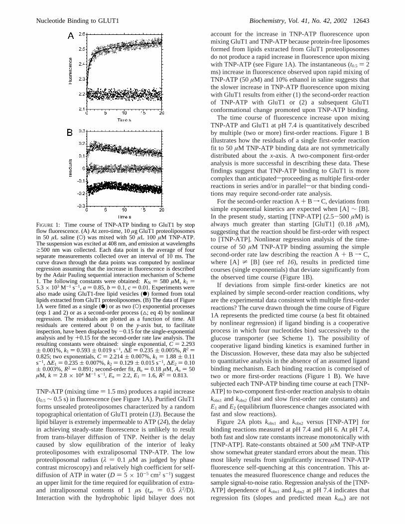
TNP-ATP (mixing time) 1.5 ms) produces a rapid increase(t0.5 ∼ 0.5 s) in fluorescence (see Figure 1A). Purified GluT1forms unsealed proteoliposomes characterized by a randomtopographical orientation of GluT1 protein (13). Because thelipid bilayer is extremely impermeable to ATP (24), the delayin achieving steady-state fluorescence is unlikely to resultfrom trans-bilayer diffusion of TNP. Neither is the delaycaused by slow equilibration of the interior of leakyproteoliposomes with extraliposomal TNP-ATP. The lowproteoliposomal radius (λ ) 0.1 µM as judged by phasecontrast microscopy) and relatively high coefficient for self-diffusion of ATP in water (D ) 5 × 10⁻⁵ cm2 s-1) suggestan upper limit for the time required for equilibration of extra-and intraliposomal contents of 1µs (tav ) 0.5 λ2/D).Interaction with the hydrophobic lipid bilayer does not
account for the increase in TNP-ATP fluorescence uponmixing GluT1 and TNP-ATP because protein-free liposomesformed from lipids extracted from GluT1 proteoliposomesdo not produce a rapid increase in fluorescence upon mixingwith TNP-ATP (see Figure 1A). The instantaneous (t0.5 ) 2ms) increase in fluorescence observed upon rapid mixing ofTNP-ATP (50µM) and 10% ethanol in saline suggests thatthe slower increase in TNP-ATP fluorescence upon mixingwith GluT1 results from either (1) the second-order reactionof TNP-ATP with GluT1 or (2) a subsequent GluT1conformational change promoted upon TNP-ATP binding.
The time course of fluorescence increase upon mixingTNP-ATP and GluT1 at pH 7.4 is quantitatively describedby multiple (two or more) first-order reactions. Figure 1 Billustrates how the residuals of a single first-order reactionfit to 50 µM TNP-ATP binding data are not symmetricallydistributed about thex-axis. A two-component first-orderanalysis is more successful in describing these data. Thesefindings suggest that TNP-ATP binding to GluT1 is morecomplex than anticipatedsproceeding as multiple first-orderreactions in series and/or in parallelsor that binding condi-tions may require second-order rate analysis.
For the second-order reaction A+ B f C, deviations fromsimple exponential kinetics are expected when [A]∼ [B].In the present study, starting [TNP-ATP] (2.5-500 µM) isalways much greater than starting [GluT1] (0.18µM),suggesting that the reaction should be first-order with respectto [TNP-ATP]. Nonlinear regression analysis of the time-course of 50µM TNP-ATP binding assuming the simplesecond-order rate law describing the reaction A+ B f C,where [A] * [B] (see ref 16), results in predicted timecourses (single exponentials) that deviate significantly fromthe observed time course (Figure 1B).
If deviations from simple first-order kinetics are notexplained by simple second-order reaction conditions, whyare the experimental data consistent with multiple first-orderreactions? The curve drawn through the time course of Figure1A represents the predicted time course (a best fit obtainedby nonlinear regression) if ligand binding is a cooperativeprocess in which four nucleotides bind successively to theglucose transporter (see Scheme 1). The possibility ofcooperative ligand binding kinetics is examined further inthe Discussion. However, these data may also be subjectedto quantitative analysis in the absence of an assumed ligandbinding mechanism. Each binding reaction is comprised oftwo or more first-order reactions (Figure 1 B). We havesubjected each TNP-ATP binding time course at each [TNP-ATP] to two-component first-order reaction analysis to obtainkobs1 andkobs2 (fast and slow first-order rate constants) andE1 andE2 (equilibrium fluorescence changes associated withfast and slow reactions).
Figure 2A plotskobs1 and kobs2 versus [TNP-ATP] forbinding reactions measured at pH 7.4 and pH 6. At pH 7.4,both fast and slow rate constants increase monotonically with[TNP-ATP]. Rate-constants obtained at 500µM TNP-ATPshow somewhat greater standard errors about the mean. Thismost likely results from significantly increased TNP-ATPfluorescence self-quenching at this concentration. This at-tenuates the measured fluorescence change and reduces thesample signal-to-noise ratio. Regression analysis of the [TNP-ATP] dependence ofkobs1 andkobs2 at pH 7.4 indicates thatregression fits (slopes and predicted meankobs) are not
FIGURE 1: Time course of TNP-ATP binding to GluT1 by stopflow fluorescence. (A) At zero-time, 10µg GluT1 proteoliposomesin 50 µL saline (O) was mixed with 50µL 100 µM TNP-ATP.The suspension was excited at 408 nm, and emission at wavelengthsg500 nm was collected. Each data point is the average of fourseparate measurements collected over an interval of 10 ms. Thecurve drawn through the data points was computed by nonlinearregression assuming that the increase in fluorescence is describedby the Adair Pauling sequential interaction mechanism of Scheme1. The following constants were obtained:KN ) 580 µM, k1 )5.3× 10³ M-1 s-1, a ) 0.85,b ) 0.1,c ) 0.01. Experiments werealso made using GluT1-free lipid vesicles (b) formed from totallipids extracted from GluT1 proteoliposomes. (B) The data of Figure1A were fitted as a single (b) or as two (O) exponential processes(eqs 1 and 2) or as a second-order process (4; eq 4) by nonlinearregression. The residuals are plotted as a function of time. Allresiduals are centered about 0 on they-axis but, to facilitateinspection, have been displaced by-0.15 for the single-exponentialanalysis and by+0.15 for the second-order rate law analysis. Theresulting constants were obtained: single exponential,C ) 2.293( 0.001%,k1 ) 0.593( 0.019 s-1, ∆E ) 0.235( 0.005%,R2 )0.825; two exponentials,C ) 2.214( 0.007%,k1 ) 1.88( 0.11s-1, ∆E1 ) 0.235( 0.007%,k2 ) 0.129( 0.015 s-1, ∆E2 ) 0.10( 0.003%,R2 ) 0.891; second-order fit,Bo ) 0.18µM, Ao ) 50µM, k ) 2.8 × 10⁴ M-1 s-1, Eo ) 2.2, E1 ) 1.6, R2 ) 0.813.
Nucleotide Binding to GLUT1 Biochemistry, Vol. 41, No. 42, 200212643
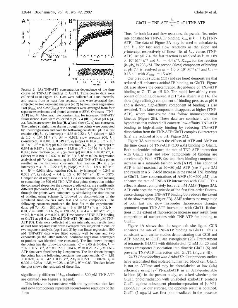
significantly different ifkobs obtained at 500µM TNP-ATPare omitted (see Figure 2 legend).
This behavior is consistent with the hypothesis that fastand slow components represent second-order reactions of theform
Thus, for both fast and slow reactions, the pseudo-first-orderrate constant for TNP-ATP binding,kobs, is k-1 + k1 [TNP-ATP]. The data of Figure 2A may be used to calculatek1
and k-1 for fast and slow reactions as the slope andy-intercept respectively of linear fits ofkobs versus [TNP-ATP]. At pH 7.4, the fast reaction is resolved ask1 ) 1.89× 10⁴ M-1 s-1 and k-1 ) 4.4 s-1. Kd(app) for the reaction(k-1/k1) is 233µM. The second (slow) component of bindingat pH 7.4 is resolved ask1 ) 1.0 × 10⁴ M-1 s-1 andk-1 )0.15 s-1 with Kd(app) ) 15 µM.
Our previous studies (11) (and see here) demonstrate thatreduced pH enhances azidoATP binding to GluT1. Figure2A also shows the concentration dependence of TNP-ATPbinding to GluT1 at pH 6.0. The rapid, low-affinity com-ponent of binding observed at pH 7.4 is absent at pH 6. Theslow (high affinity) component of binding persists at pH 6and a slower, high-affinity component of binding is alsorevealed. This latter component disappears at higher [TNP-ATP], where time-course data follow monoexponentialkinetics (Figure 2B). These data are consistent with thehypothesis that reduced pH converts low-affinity TNP-ATPbinding to high-affinity binding by reducing TNP-ATPdissociation from the TNP-ATP‚GluT1 complex (y-intercepts(k-1) are reduced at low pH; Figure 2A).
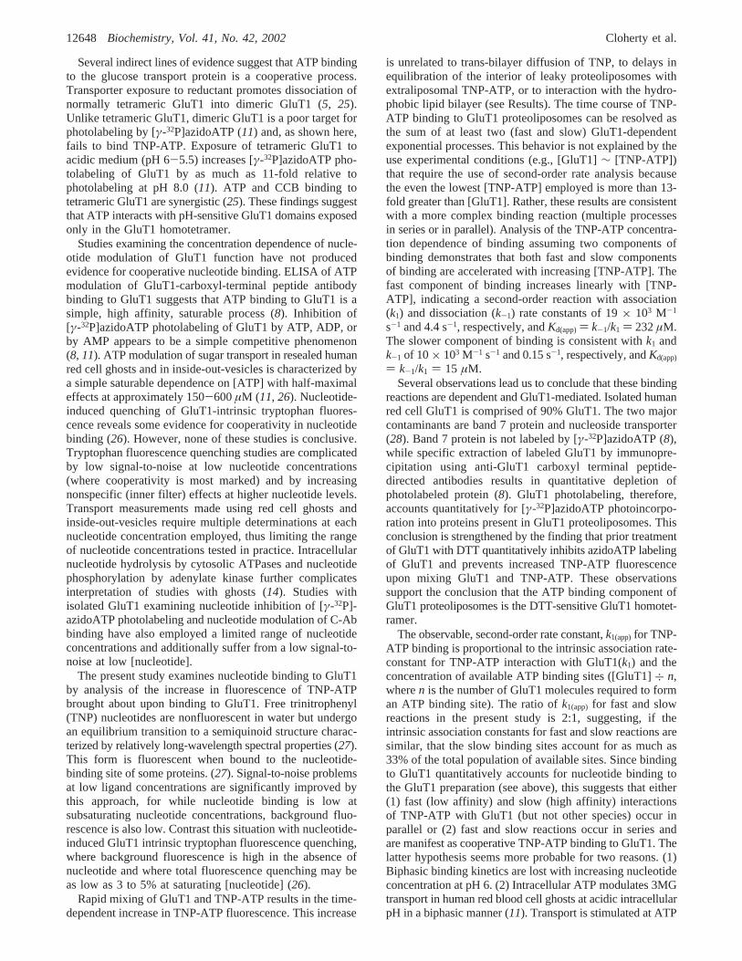
Figure 3A summarizes the effects of ATP and AMP onthe time course of TNP-ATP (100µM) binding to GluT1.Both nucleotides enhance the rate of TNP-ATP interactionwith GluT1 (fast and slow components of binding areaccelerated). With ATP, fast and slow binding componentsincrease in a saturable fashion with [ATP]. This action ofATP is half-maximal at 40-80 µM ATP (see Figure 3A)and results in a 5-7-fold increase in the rate of TNP bindingto GluT1. Low concentrations of AMP (50-500 µM) alsoenhance the rate of TNP-ATP interaction with GluT1. Thiseffect is almost completely lost at 2 mM AMP (Figure 3A).ATP enhances the magnitude of the fast first-order fluores-cence change upon TNP-ATP binding but reduces the extentof the slow reaction (Figure 3B). AMP reduces the magnitudeof both fast and slow first-order fluorescence changespromoted upon TNP-ATP interaction with GluT1. Reduc-tions in the extent of fluorescence increase may result fromcompetition of nucleotides with TNP-ATP for binding toGluT1.
Figure 4A shows that the sugar exit site ligand CCBenhances the rate of TNP-ATP binding to GluT1. This isconsistent with earlier studies demonstrating that CCB andATP binding to GluT1 are synergisitic (25). Pretreatmentof tetrameric GLUT1 with dithiothreitol (2 mM for 20 min)causes transporter dissociation into dimeric GluT1 (6) andprevents TNP-ATP interaction with GluT1 (Figure 4B).
GluT1 Photolabeling with AzidoATP.Our previous studieshave established that isolated human red blood cell GluT1is not an ATPase and may be photolabeled at low (4%)efficiency using [γ-32P]-azidoATP in an ATP-protectablefashion (8). In the present study, we asked whether priorGluT1 photoirradiation with unlabeled azidoATP protectsGluT1 against subsequent photoincorporation of [γ-32P]-azidoATP. To our surprise, the opposite result is obtained.GluT1 (1 µg/µL) was first photoirradiated in the presence
FIGURE 2: (A) TNP-ATP concentration dependence of the timecourse of TNP-ATP binding to GluT1. Time course data werecollected as in Figure 1A. Data were collected at 1 ms intervals,and results from at least four separate runs were averaged thensubjected to two exponent analysis (eq 2) by non linear regression.Fast (kobs1) and slow (kobs2) rate constants were averaged from fourseparate experiments and plotted at mean( SEM. Ordinate: [TNP-ATP] in µM. Abscissa: rate constant,kobs for increased TNP-ATPfluorescence. Data were collected at pH 7.4 (b, O) or at pH 6 (2,4). Results are shown for fast (b, 2) and slow (O, 4) rate constants.The dashed straight lines drawn through the points were computedby linear regression and have the following constants: pH 7.4, fastreaction (b), k-1 (y-intercept)) 4.36( 0.22 s-1, k1 (slope)) 18.9( 1.0 × 10³ M-1 s-1, R2 ) 0.982; slow reaction (O), k-1(y-intercept)) 0.049( 0.049 s-1, k1 (slope)) 10.4( 0.7 × 10³
M-1 s-1, R2 ) 0.972; pH 6.0, fast reaction (2), k-1 (y-intercept))0.674( 0.197 s-1, k1 (slope)) 14.8( 0.7 × 10³ M-1 s-1, R2 )0.996; slow reaction (4), k-1 (y-intercept)) 0.032( 0.007 s-1, k1(slope)) 0.198( 0.037× 10³ M-1 s-1, R2 ) 0.959. Regressionanalysis of pH 7.4 data omitting the 500µM TNP-ATP data pointsresulted in the following constants: fast reaction (b), k-1 (y-intercept)) 4.19( 0.22 s-1, k1 (slope)) 21.8( 1.9 × 10³ M-1
s-1, R2 ) 0.964; slow reaction (O), k-1 (y-intercept)) 0.249(0.061 s-1, k1 (slope)) 7.4 ( 0.5 × 10³ M-1 s-1, R2 ) 0.975.Comparison of regression fits of pH 7.4 experimental data lackingor containing the 500µM TNP-ATP data points shows that neitherthe computed slopes nor the average predictedkobsare significantlydifferent (two-tailedt-test,p < 0.05). The solid straight lines drawnthrough the points were computed by simulating the time courseof binding using Scheme 1, followed by deconvolution of thesimulated time courses into fast and slow components. Thefollowing constants produced the best fits to the experimentaldata: pH 7.4,KN ) 530µM, k1 ) 6 × 10³ M-1 s-1, a ) 0.2,b )0.01,c ) 0.001; pH 6,KN ) 120 µM, k1 ) 4.4 × 10³ M-1 s-1, a) 0.2,b ) 0.01,c ) 0.001. (B) Time course of TNP-ATP bindingto GluT1 at pH 6 at 250µM TNP-ATP (b) and at 500µM TNP-ATP (O). Data were collected at 1 ms intervals, and results fromat least four separate runs were averaged then subjected to one- ortwo exponent analysis (eqs 1 and 2) by non linear regression. 500µM TNP-ATP data were fitted equally well by one and twoexponents (in the latter instance, nonlinear regression convergedto produce two identical rate constants). The line drawn throughthe points has the following constants:C ) 2.05 ( 0.04%,k1 )7.92( 0.59 s-1, ∆E ) 0.27( 0.04%,R2 ) 0.79. 250µM TNP-ATP data were fitted best by 2-exponents. The line drawn throughthe points has the following constants two exponentials,C ) 1.81( 0.07%,k1 ) 3.42 ( 0.19 s-1, ∆E1 ) 0.221( 0.007%,k2 )0.376( 0.25 s-1, ∆E2 ) 0.25( 0.03%,R2 ) 0.83. The data belowthe plot shows the residuals of these fits.
GluT1 + TNP-ATPy\zk1
k-1GluT1.TNP-ATP
12644 Biochemistry, Vol. 41, No. 42, 2002 Cloherty et al.

or absence of 5 mM unlabeled azidoATP. Irradiated pro-teoliposomes were sedimented, washed several times inazidoATP-free medium then resuspended in medium con-taining 5.5µM [γ-32P]-azidoATP. Ultraviolet irradiation ofGluT1 in the presence of unlabeled azidoATP consistentlyenhances (relative to irradiation in the absence of unlabeledazidoATP) subsequent photoincorporation of [γ-32P]-azi-doATP by 2-3-fold (Figure 5). This study also confirmsearlier studies (11) demonstrating that GluT1 photolabelingby azidoATP is enhanced at pH 5.5 relative to labeling atpH 8.
Effects of Acidification on ATP-Modulation of 3MGTransport. Previous studies from this laboratory have
demonstrated that intracellular acidification modifies GluT1sensitivity to regulation by ATP (11). At pH 7.4, the affinityof the red cell sugar transporter for 3MG increases in asaturable manner with [ATP]. At pH 6, low concentrationsof ATP (<200µM) increase the affinity of GluT1 for 3MGand thus stimulate uptake of 100µM 3MG. Higher, physi-ological concentrations of ATP (2 mM) fail to stimulate 100µM 3MG uptake at pH 6. Several questions arise from theseobservations. (1) Does reduced pH promote GluT1 interac-tion with cellular factors that negate GluT1 regulation byATP? (2) Does reduced pH cause a general inhibition ofGluT1 mediated sugar transport?
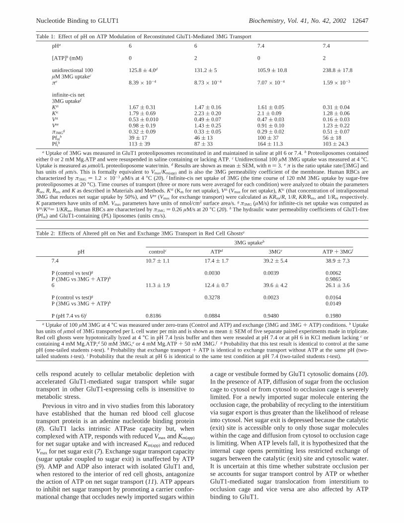
The potential involvement of cellular factors in pH-sensitive sugar transport regulation was investigated byreconstitution of GluT1 into proteoliposomes containing orlacking 4 mM Mg.ATP at pH 7.4 or pH 6. Table 1 showsthat 3MG uptake at subsaturating (100µM) 3MG isaccelerated by ATP at pH 7.4. ATP binding to GluT1 isincreased at pH 6 (see Figures 2 and 4 and ref11), but theeffect of ATP on net 3MG uptake is lost (Table 1). Infinite-cis net 3MG uptake (the time course of saturated 3MGuptake) by GluT1 proteoliposomes is also inhibited by ATPat pH 7.4 but is unaffected by ATP at pH 6 (Figure 6). Net
FIGURE 3: (A) Effects of ATP and AMP on TNP-ATP binding toGluT1. Ordinate: [TNP-ATP] inµM. Abscissa: rate constant,kobsfor increased TNP-ATP fluorescence. All data were collected atpH 7.4 using 100µM TNP-ATP. At least four runs were averagedat each competing [nucleotide], and the increase in TNP-ATPfluorescence was resolved as two exponentials characterized by fastand slow rate constants with corresponding fluorescence changes(∆Efast and ∆Eslow). The experiment was repeated at least threetimes, and results are shown as mean( SEM. Results are shownfor fast (O, 2) and slow (b, 4) rate constants. Results were obtainedusing ATP (b, O) or AMP (2, 4) as the competing nucleotide.With ATP, rate constants increase in a saturable manner with[nucleotide], and curves drawn through the points were computedby nonlinear regression, assuming thatkobs is described bykobs )ko + kmax [nucleotide]/(Ko.5 + [nucleotide]) whereko and kmaxrepresentkobs for TNP-ATP binding in the absence or presence ofsaturating nucleotide andK0.5 is that [nucleotide] producingkobs )ko + 0.5 kmax. The results are as follows: ATP, fast reactionko )4.73( 0.82 s-1, kmax ) 17.46( 0.98 s-1, K0.5 ) 86.7( 16.9µM;ATP, slow reactionko ) 0.24( 0.19 s-1, kmax ) 1.42( 0.35 s-1,K0.5 ) 41.3 ( 26.8 µM. With AMP, rate constants increase thendecrease with [nucleotide]. Insufficient data points exist to describethe results in a quantitative manner. The curves were drawn throughthe points by eye and have no theoretical significance. (B) Effectsof AMP and ATP on the extent of TNP-ATP fluorescence changeupon interaction with GluT1. The fluorescence changes (∆Efast and∆Eslow) calculated in 2B are plotted as a function of [ATP] (b, O)or [AMP] (2, 0). The curves drawn through the points were drawnby eye and have no theoretical significance.
FIGURE 4: Effects of CCB and DTT on TNP-ATP binding toGluT1. (A) Effect of 500 nM CCB and on 100µM TNP-ATPbinding to GluT1 (10µg). GluT1 was preincubated with 500 nMCCB (O) or saline (0) then mixed with 100µM TNP-ATP ( 500nM CCB. Five runs were averaged for each condition. CCBaccelerates TNP-ATP binding. The curves drawn through the pointswere computed by nonlinear regression according to Scheme 1 andhave the following constants: control,KN ) 124µM, k1 ) 15.7×10³ M-1 s-1, a ) 0.54,b ) 0.32,c ) 0.02; CCB,KN ) 89 µM, k1) 17.7× 10³ M-1 s-1, a ) 1.1,b ) 0.03,c ) 0.03. (B) Effect ofpreincubation with 2 mM DTT on 100µM TNP-ATP binding toGluT1 (10µg). GluT1 was preincubated with (O) or without (0)2 mM DTT for 30 min, sedimented at 14 000g, washed once in100 volumes DTT-free medium, and mixed rapidly with 100µMTNP-ATP. Five runs were averaged, and the curves drawn throughthe control points were computed by nonlinear regression accordingto Scheme 1 and have the following constants:KN ) 88.6µM, k1) 6.2 × 10³ M-1 s-1, a ) 0.67,b ) 0. 19,c ) 0.02. No TNP-ATP/GluT1 mixing induced increase in fluorescence was observedwhen GluT1 was first treated with DTT.
Nucleotide Binding to GLUT1 Biochemistry, Vol. 41, No. 42, 200212645

transport inhibition by ATP appears to result from (1)increased transporter affinity for 3MG (efflux saturates atlower [3MG]) and (2) reducedVmax for net transport (Table1). These results demonstrate that pH modulation of ATPregulation of GluT1-mediated sugar transport is recapitulatedin an in vitro reconstituted system.
We compared the fundamental characteristics of carrier-mediated 3MG transport at pH 6 and 7.4 by analysis of zero-trans and infinite-trans unidirectional 3MG uptake in resealedhuman red cell ghosts containing or lacking 4 mM ATP.Table 2 shows that the presence of 50 mM intracellular 3MGresults in accelerated unidirectional 3MG uptake at bothacidic and normal pH. ATP modulates zero-trans fluxes onlyat pH 7.4. The effects of ATP on exchange transport at pH7.4 and pH 6 are not significant at the p< 0.05 level (Table2).
DISCUSSION
The present study was formulated to answer two questions.(1) Is ATP binding to the glucose transporter a simple orcooperative process? (2) How does the ATP-liganded stateof the glucose transporter influence transporter catalyticefficiency? Our findings suggest a working hypothesis fornucleotide-dependent, allosteric regulation of GluT1-medi-ated sugar transport. This model may also explain why some
FIGURE 5: Effects of GluT1 UV-cross-linking GluT1 with unlabeledazidoATP on subsequent GluT1 UV cross-linking with [γ-32P]-azidoATP at pH 8 or 5.5. During conditioning, GluT1 (1µg/µL)was exposed to saline containing or lacking 5 mM unlabeledazidoATP at pH 8 or pH 5.5. The proteoliposomes were UVirradiated on ice for 90 s (two control samples at pH 8 and 5.5were not UV irradiated), collected by sedimentation at 14 000g for10 min, and washed twice in azidoATP-free medium. Subsequently,during labeling, membranes were exposed to 5.5µM [γ-32P]-azidoATP at pH 8 or pH 5.5 and UV irradiated on ice for 90 s.Following washing in label-free saline, proteins were resolved bySDS-PAGE, electrophoretically transferred to immobilon mem-branes, and exposed to film. The intensity of GluT1 [γ-32P]-azidoATP photoincorporation was quantitated by densitometry. Themembrane was then subjected to [GluT1] quantitation by immu-noblot analysis using C-Ab, and [γ-32P]-azidoATP photoincorpo-ration was normalized for the amount of immunodetectable GluT1in each lane. This experiment was repeated three times. Ordinate:relative [γ-32P]-azidoATP photoincorporation. Results are shownas mean( SEM. There is no significant difference in [γ-32P]-azidoATP labeling when samples are conditioned (in the absenceof azidoATP) with or without UV exposure. *Indicates that [γ-32P]-azidoATP photoincorporation at pH 5.5 is significantly greater thanlabel incorporation at pH 8.0 (p < 0.005). **Indicates that [γ-32P]-azidoATP photoincorporation is significantly greater (p < 0.005)following conditioning by UV-irradiation in the presence ofunlabeled azidoATP (5 mM) than it is following conditioning byUV-irradiation in the absence of azidoATP.
FIGURE 6: Effects of pH on GluT1-mediated infinite-cis net sugaruptake by reconstituted GluT1-proteoliposomes. Results are shownfor transport at pH 7.4 (A) and at pH 6 (B). GluT1 proteoliposomeswere prepared as described in materials and methods and wereinjected into saline containing 120 mM sucrose (0), 120 mM 3MG(O), or 120 mM 3MG plus 2 mM Mg.ATP (0). Proteoliposomesinjected into ATP-containing medium also contained 2 mMMg.ATP in the intraliposomal space. Light scattering at 450 nmwas averaged over 0.5 s intervals and plotted as a function of time.Each data set represents the average of three or more separate runs.Proteoliposomes first shrink due to osmotic water loss and thenswell as 3MG is transported into the intraliposomal space and waterfollows.
12646 Biochemistry, Vol. 41, No. 42, 2002 Cloherty et al.
cells respond acutely to cellular metabolic depletion withaccelerated GluT1-mediated sugar transport while sugartransport in other GluT1-expressing cells is insensitive tometabolic stress.
Previous in vitro and in vivo studies from this laboratoryhave established that the human red blood cell glucosetransport protein is an adenine nucleotide binding protein(8). GluT1 lacks intrinsic ATPase capacity but, whencomplexed with ATP, responds with reducedVmax andKm(app)
for net sugar uptake and with increasedKm(app)and reducedVmax for net sugar exit (7). Exchange sugar transport capacity(sugar uptake coupled to sugar exit) is unaffected by ATP(9). AMP and ADP also interact with isolated GluT1 and,when restored to the interior of red cell ghosts, antagonizethe action of ATP on net sugar transport (11). ATP appearsto inhibit net sugar transport by promoting a carrier confor-mational change that occludes newly imported sugars within
a cage or vestibule formed by GluT1 cytosolic domains (10).In the presence of ATP, diffusion of sugar from the occlusioncage to cytosol or from cytosol to occlusion cage is severelylimited. For a newly imported sugar molecule entering theocclusion cage, the probability of recycling to the interstitiumvia sugar export is thus greater than the likelihood of releaseinto cytosol. Net sugar exit is depressed because the catalytic(exit) site is accessible only to only those sugar moleculeswithin the cage and diffusion from cytosol to occlusion cageis limiting. When ATP levels fall, it is hypothesized that theinternal cage opens permitting less restricted exchange ofsugars between the catalytic (exit) site and cytosolic water.It is uncertain at this time whether substrate occlusion perse accounts for sugar transport control by ATP or whetherGluT1-mediated sugar translocation from interstitium toocclusion cage and vice versa are also affected by ATPbinding to GluT1.
Table 1: Effect of pH on ATP Modulation of Reconstituted GluT1-Mediated 3MG Transport
pHa 6 6 7.4 7.4
[ATP]b (mM) 0 2 0 2
unidirectional 100µM 3MG uptakec
125.8( 4.0d 131.2( 5 105.9( 10.8 238.8( 17.8
πe 8.39× 10⁻⁴ 8.73× 10⁻⁴ 7.07× 10⁻⁴ 1.59× 10⁻³
infinite-cis net3MG uptakef
Kzt 1.67( 0.31 1.47( 0.16 1.61( 0.05 0.31( 0.04Kic 1.79( 0.69 2.23( 0.20 2.1( 0.09 1.28( 0.06Vzt 0.53( 0.010 0.49( 0.07 0.47( 0.03 0.16( 0.03Vee 0.98( 0.19 1.43( 0.25 0.91( 0.10 1.23( 0.22π3MG
g 0.32( 0.09 0.33( 0.05 0.29( 0.02 0.51( 0.07Pfnt
h 39 ( 17 46( 13 100( 37 56( 18Pfth 113( 39 87( 33 164( 11.3 103( 24.3
a Uptake of 3MG was measured in GluT1 proteoliposomes reconstituted in and maintained in saline at pH 6 or 7.4.b Proteoliposomes containedeither 0 or 2 mM Mg.ATP and were resuspended in saline containing or lacking ATP.c Unidirectional 100µM 3MG uptake was measured at 4°C.Uptake is measured asµmol/L proteolioposome water/min.d Results are shown as mean( SEM, withn ) 3. e π is the ratio uptake rate/[3MG] andhas units ofµm/s. This is formally equivalent toVmax/Km(app) and is also the 3MG permeability coefficient of the membrane. Human RBCs arecharacterized byπ3MG ) 1.2 × 10⁻³ µM/s at 4°C (20). f Infinite-cis net uptake of 3MG (the time course of 120 mM 3MG uptake by sugar-freeproteoliposomes at 20°C). Time courses of transport (three or more runs were averaged for each condition) were analyzed to obtain the parametersRoo, R, Ree, andK as described in Materials and Methods.Kzt (Km for net uptake),Vzt (Vmax for net uptake),Kic (that concentration of intraliposomal3MG that reduces net sugar uptake by 50%), andVee (Vmax for exchange transport) were calculated asKRoo/R, 1/R, KR/Ree, and 1/Ree respectively.K parameters have units of mM.Vmax parameters have units of nmol/cm2 surface area/s.g π3MG (µM/s) for infinite-cis net uptake was computed asVzt/Kzt) 1/KRoo. Human RBCs are characterized byπ3MG ) 0.26µM/s at 20°C (20). h The hydraulic water permeability coefficients of GluT1-free(Pfnt) and GluT1-containing (Pft) liposomes (units cm/s).
Table 2: Effects of Altered pH on Net and Exchange 3MG Transport in Red Cell Ghostsa
3MG uptakeb
pH controlc ATPd 3MGe ATP + 3MGf
7.4 10.7( 1.1 17.4( 1.7 39.2( 5.4 38.9( 7.3
P (control vs test)g 0.0030 0.0039 0.0062P (3MG vs 3MG+ ATP)h 0.98656 11.3( 1.9 12.4( 0.7 39.6( 4.2 26.1( 3.6
P (control vs test)g 0.3278 0.0023 0.0164P (3MG vs 3MG+ ATP)h 0.0149
P (pH 7.4 vs 6)i 0.8186 0.0884 0.9480 0.1980a Uptake of 100µM 3MG at 4°C was measured under zero-trans (Control and ATP) and exchange (3MG and 3MG+ ATP) conditions.b Uptake
has units ofµmol of 3MG transported per L cell water per min and is shown as mean( SEM of five separate paired experiments made in triplicate.Red cell ghosts were hypotonically lyzed at 4°C in pH 7.4 lysis buffer and then were resealed at pH 7.4 or at pH 6 in KCl medium lackingc orcontaining 4 mM Mg.ATP,d 50 mM 3MG,e or 4 mM Mg.ATP+ 50 mM 3MG.f g Probability that this test result is identical to control at the samepH (one-tailed studentst-test).h Probability that exchange transport+ ATP is identical to exchange transport without ATP at the same pH (two-tailed studentst-test). i Probability that the result at pH 6 is identical to the same test condition at pH 7.4 (two-tailed studentst-test).
Nucleotide Binding to GLUT1 Biochemistry, Vol. 41, No. 42, 200212647
Several indirect lines of evidence suggest that ATP bindingto the glucose transport protein is a cooperative process.Transporter exposure to reductant promotes dissociation ofnormally tetrameric GluT1 into dimeric GluT1 (5, 25).Unlike tetrameric GluT1, dimeric GluT1 is a poor target forphotolabeling by [γ-32P]azidoATP (11) and, as shown here,fails to bind TNP-ATP. Exposure of tetrameric GluT1 toacidic medium (pH 6-5.5) increases [γ-32P]azidoATP pho-tolabeling of GluT1 by as much as 11-fold relative tophotolabeling at pH 8.0 (11). ATP and CCB binding totetrameric GluT1 are synergistic (25). These findings suggestthat ATP interacts with pH-sensitive GluT1 domains exposedonly in the GluT1 homotetramer.
Studies examining the concentration dependence of nucle-otide modulation of GluT1 function have not producedevidence for cooperative nucleotide binding. ELISA of ATPmodulation of GluT1-carboxyl-terminal peptide antibodybinding to GluT1 suggests that ATP binding to GluT1 is asimple, high affinity, saturable process (8). Inhibition of[γ-32P]azidoATP photolabeling of GluT1 by ATP, ADP, orby AMP appears to be a simple competitive phenomenon(8, 11). ATP modulation of sugar transport in resealed humanred cell ghosts and in inside-out-vesicles is characterized bya simple saturable dependence on [ATP] with half-maximaleffects at approximately 150-600µM (11, 26). Nucleotide-induced quenching of GluT1-intrinsic tryptophan fluores-cence reveals some evidence for cooperativity in nucleotidebinding (26). However, none of these studies is conclusive.Tryptophan fluorescence quenching studies are complicatedby low signal-to-noise at low nucleotide concentrations(where cooperativity is most marked) and by increasingnonspecific (inner filter) effects at higher nucleotide levels.Transport measurements made using red cell ghosts andinside-out-vesicles require multiple determinations at eachnucleotide concentration employed, thus limiting the rangeof nucleotide concentrations tested in practice. Intracellularnucleotide hydrolysis by cytosolic ATPases and nucleotidephosphorylation by adenylate kinase further complicatesinterpretation of studies with ghosts (14). Studies withisolated GluT1 examining nucleotide inhibition of [γ-32P]-azidoATP photolabeling and nucleotide modulation of C-Abbinding have also employed a limited range of nucleotideconcentrations and additionally suffer from a low signal-to-noise at low [nucleotide].
The present study examines nucleotide binding to GluT1by analysis of the increase in fluorescence of TNP-ATPbrought about upon binding to GluT1. Free trinitrophenyl(TNP) nucleotides are nonfluorescent in water but undergoan equilibrium transition to a semiquinoid structure charac-terized by relatively long-wavelength spectral properties (27).This form is fluorescent when bound to the nucleotide-binding site of some proteins. (27). Signal-to-noise problemsat low ligand concentrations are significantly improved bythis approach, for while nucleotide binding is low atsubsaturating nucleotide concentrations, background fluo-rescence is also low. Contrast this situation with nucleotide-induced GluT1 intrinsic tryptophan fluorescence quenching,where background fluorescence is high in the absence ofnucleotide and where total fluorescence quenching may beas low as 3 to 5% at saturating [nucleotide] (26).
Rapid mixing of GluT1 and TNP-ATP results in the time-dependent increase in TNP-ATP fluorescence. This increase
is unrelated to trans-bilayer diffusion of TNP, to delays inequilibration of the interior of leaky proteoliposomes withextraliposomal TNP-ATP, or to interaction with the hydro-phobic lipid bilayer (see Results). The time course of TNP-ATP binding to GluT1 proteoliposomes can be resolved asthe sum of at least two (fast and slow) GluT1-dependentexponential processes. This behavior is not explained by theuse experimental conditions (e.g., [GluT1]∼ [TNP-ATP])that require the use of second-order rate analysis becausethe even the lowest [TNP-ATP] employed is more than 13-fold greater than [GluT1]. Rather, these results are consistentwith a more complex binding reaction (multiple processesin series or in parallel). Analysis of the TNP-ATP concentra-tion dependence of binding assuming two components ofbinding demonstrates that both fast and slow componentsof binding are accelerated with increasing [TNP-ATP]. Thefast component of binding increases linearly with [TNP-ATP], indicating a second-order reaction with association(k1) and dissociation (k-1) rate constants of 19× 103 M-1
s-1 and 4.4 s-1, respectively, andKd(app)) k-1/k1 ) 232µM.The slower component of binding is consistent withk1 andk-1 of 10× 103 M-1 s-1 and 0.15 s-1, respectively, andKd(app)
) k-1/k1 ) 15 µM.Several observations lead us to conclude that these binding
reactions are dependent and GluT1-mediated. Isolated humanred cell GluT1 is comprised of 90% GluT1. The two majorcontaminants are band 7 protein and nucleoside transporter(28). Band 7 protein is not labeled by [γ-32P]azidoATP (8),while specific extraction of labeled GluT1 by immunopre-cipitation using anti-GluT1 carboxyl terminal peptide-directed antibodies results in quantitative depletion ofphotolabeled protein (8). GluT1 photolabeling, therefore,accounts quantitatively for [γ-32P]azidoATP photoincorpo-ration into proteins present in GluT1 proteoliposomes. Thisconclusion is strengthened by the finding that prior treatmentof GluT1 with DTT quantitatively inhibits azidoATP labelingof GluT1 and prevents increased TNP-ATP fluorescenceupon mixing GluT1 and TNP-ATP. These observationssupport the conclusion that the ATP binding component ofGluT1 proteoliposomes is the DTT-sensitive GluT1 homotet-ramer.
The observable, second-order rate constant,k1(app)for TNP-ATP binding is proportional to the intrinsic association rate-constant for TNP-ATP interaction with GluT1(k1) and theconcentration of available ATP binding sites ([GluT1]÷ n,wheren is the number of GluT1 molecules required to forman ATP binding site). The ratio ofk1(app) for fast and slowreactions in the present study is 2:1, suggesting, if theintrinsic association constants for fast and slow reactions aresimilar, that the slow binding sites account for as much as33% of the total population of available sites. Since bindingto GluT1 quantitatively accounts for nucleotide binding tothe GluT1 preparation (see above), this suggests that either(1) fast (low affinity) and slow (high affinity) interactionsof TNP-ATP with GluT1 (but not other species) occur inparallel or (2) fast and slow reactions occur in series andare manifest as cooperative TNP-ATP binding to GluT1. Thelatter hypothesis seems more probable for two reasons. (1)Biphasic binding kinetics are lost with increasing nucleotideconcentration at pH 6. (2) Intracellular ATP modulates 3MGtransport in human red blood cell ghosts at acidic intracellularpH in a biphasic manner (11). Transport is stimulated at ATP
12648 Biochemistry, Vol. 41, No. 42, 2002 Cloherty et al.
) 200 µM but is inhibited at higher [ATP]. Importantly,this effect cannot be modeled simply as the sum of ATPinhibition of one population of transporter plus ATP stimula-tion of a second GluT1 population (11).
Further evidence for cooperative nucleotide binding isavailable from studies in which progressive addition of ATPor AMP increases the rate at which TNP-ATP interacts withGluT1 while reducing the number of available sites forinteraction with TNP-ATP. Moreover, enhancement of[γ-32P]azidoATP photolabeling of GluT1 by prior irradiationin the presence of saturating unlabeled azidoATP suggeststhat low-efficiency, covalent attachment of unlabeled ligandto one or more nucleotide binding sites within a carriercomplex increases the efficiency of labeling of remainingsites for [γ-32P]azidoATP.
We have modeled TNP-ATP binding to the glucosetransporter as a cooperative process in which each GluT1protein of the tetramer presents a single nucleotide bindingdomain. At this time there are insufficient data to indicatewhether the nucleotide binding site:GluT1 molar ratio is 1:1or lower. Estimates of ethenoATP binding to GluT1 suggest1 mol nucleotide binding sites per mol CCB binding sites(8). However, since nonreduced GluT1 exposes only 1 molCCB binding sites per 2 mol GluT1 (5), tetrameric GluT1may normally present only two nucleotide binding sites pertransporter complex. The results of the present study are bestmodeled by the Adair-Pauling sequential interaction modelwhen three or more nucleotide binding sites per carriercomplex are assumed (see Scheme 1). Thus, a critical testof this hypothesis will be the accurate determination of GluT1nucleotide binding capacity. Assuming four nucleotidebinding sites per carrier complex and that all nucleotidebinding sites in the unliganded carrier adopt the low-affinitystate, binding of the first nucleotide to the tetramer enhancesTNP-ATP binding to the remaining nucleotide binding sitesby reducingKd (specifically by reducingk-1) for bindingby 5-fold. Progressive occupancy of available sites by TNP-ATP increasingly augments the affinity of the remaining site-(s) for TNP-ATP by 10-fold per nucleotide addition. Thismodel is compatible with the TNP-ATP binding data ofFigures 1 and 2.
pH-sensitive ATP binding may result from protonation ofside chains within the ATP binding domain. Histidine is theobvious candidate amino acid for side-chain protonation, aspH is lowered from 8.5 to 5.5 and GluT1 contains fivehistidine residues, of which four (H 160, 239, 337, and 484)are located within cytoplasmic domains. Exofacial His50 isa poor candidate since red cell sugar transport is insensitiveto altered extracellular pH (29). His337 lies within theintracellular portion of the GluT1 ATP binding domain(residues 301-361) identified previously by N-terminalsequence analysis of proteolytic fragments obtained fromazidoATP-labeled GluT1 (11). Protonation of His337 couldenhance ATP binding directly by increasing ionic interactionbetween ATP and His+ within the binding pocket. Alterna-tively, protonated His337 might now participate in a cation-πinteraction (30) with, for example, Phe326 or could interferewith a cation-π interactions through ionic repulsion ofcation-bearing side chains (e.g., Arg330, Arg3333, or Arg334).In the absence of detailed knowledge of the GluT1 ATPbinding domain and pH-dependent GluT1 conformationalchanges, these possibilities remain speculative. However, we
address these possibilities specifically by mutagenesis in theaccompanying paper (31).
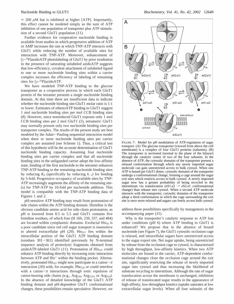
Why is the transporter’s catalytic response to ATP lostunder conditions (pH 6) where ATP binding to GluT1 isenhanced? We propose that in the absence of boundnucleotide (see Figure 7), the GluT1 cytosolic occlusion cageis relaxed, and intracellular sugars have unrestricted accessto the sugar export site. Net sugar uptake, being unrestrictedby release from the occlusion cage to cytosol, is characterizedby high throughput, low-affinity kinetics. When two ATPmolecules are bound to the carrier, ATP-dependent confor-mational changes close the occlusion cage around the exitsite, significantly restricting the release of newly importedsugar into cytosol and thus increasing the likelihood ofsubstrate recycling to interstitium. Although the rate of sugartranslocation across the membrane is unchanged, inhibitionof release of translocated sugar results in the appearance ofhigh-affinity, low-throughput kinetics (uptake saturates at lowextracellular sugar levels). When all four subunits of the
FIGURE 7: Model for pH modulation of ATP-regulation of sugartransport. (A) The glucose transporter (viewed from above the cellmembrane) is a complex of four GluT1 proteins (subunits). (B)The transporter is sectioned (normal to the plane of the bilayer)through the catalytic center of two of the four subunits. In theabsence of ATP, the cytosolic domains of the transporter present arelaxed conformation through which any newly imported sugarmolecule can gain unrestricted access to bulk cytosol. When oneATP is bound per GluT1 dimer, cytosolic domains of the transporterundergo a conformational change, forming a cage around the sugarexit sites which restricts access to bulk cytosol. A newly importedsugar now has a greater probability of being recycled to theinterstitium via translocation (eS1.e2f eS2.e1 conformationalchange) than release into cytosol. When a second ATP moleculeinteracts with the transporter, cytosolic domains of the transporteradopt a third conformation in which the cage surrounding the exitsite is once more relaxed and sugars can freely access bulk cytosol.
Nucleotide Binding to GLUT1 Biochemistry, Vol. 41, No. 42, 200212649

carrier are liganded with ATP, cooperative interactions resultin relaxation of the occlusion cage and facilitate the rapidrelease of imported sugars into the cell water.
We propose that only two ATP binding sites per trans-porter complex are complexed with ATP at pH 7.4. As pHis lowered to 6, the intrinsic affinity of the nucleotide bindingsite for ATP is increased by 5-fold, thereby promotingoccupation of all four nucleotide binding sites at physiologi-cal [ATP]. This model accounts for the effects of reducedpH on GluT1 photolabeling by azidoATP and can explainacidification-induced biphasic ATP-modulation of GluT1mediated sugar transport. At low pH and low [ATP], thetransporter is only partially complexed by ATP and is thusoccluded. As ATP levels are raised further, all 4 subunitsare occupied by ATP and the occlusion cage relaxes. Themodel is also consistent with demonstrations of unalteredexchange sugar transport at pH 6. Saturation of the occlusioncage with unlabeled sugars prevents the appearance of ATP-modulation of transport. Unlabeled saturating sugar competeswith newly imported labeled sugar for the exit site and thusincreases the probability of release of labeled sugar into thecytosol.
This model may also account for variable, cellular sugartransport responses to metabolic challenge. Some GluT1expressing cells respond to metabolic depletion with rapidacceleration of net sugar transport. This results from either(1) increased catalytic turnover of GluT1 (4, 7, 9, 14, 32-34) or (2) increased cell surface GluT1 content (35, 36).However, some GluT1-expressing cells are unresponsive tocellular metabolic stress (37, 38). The present study is mostgermane to regulation of GluT1 catalytic turnover. A lackof response to altered cellular metabolic status suggests thatGluT1 no longer senses cellular metabolic status or, that theeffector mechanism is altered. GluT1 in metabolism-insensi-tive cells may be converted to the high (ATP-binding) affinitydisoccluded state or may interact with other cellular factorsthat relax the occlusion cage.
REFERENCES
1. Zoccoli, M. A., Baldwin, S. A., and Lienhard, G. E. (1978) Themonosaccharide transport system of the human erythrocyte.Solubilization and characterization on the basis of cytochalasinB binding,J. Biol. Chem. 253, 6923-6930.
2. Kasahara, M., and Hinkle, P. C. (1977) Reconstitution andpurification of theD-glucose transporter from human erythrocytes,J. Biol. Chem. 253, 7384-7390.
3. Mueckler, M., Caruso, C., Baldwin, S. A., Panico, M., Blench, I.,Morris, H. R., Allard, W. J., Lienhard, G. E., and Lodish, H. F.(1985) Sequence and structure of a human glucose transporter,Science 229, 941-945.
4. Diamond, D., and Carruthers, A. (1993) Metabolic control of sugartransport by derepression of cell surface glucose transporters: aninsulin-independent, recruitment-independent mechanism of regu-lation, J. Biol. Chem. 268, 6437-6444.
5. Hebert, D. N., and Carruthers, A. (1992) Glucose transporteroligomeric structure determines transporter function. Reversibleredox-dependent interconversions of tetrameric and dimericGLUT1, J. Biol. Chem. 267, 23829-38.
6. Zottola, R. J., Cloherty, E. K., Coderre, P. E., Hansen, A., Hebert,D. N., and Carruthers, A. (1995) Glucose transporter function iscontrolled by transporter oligomeric structure. A single, intramo-lecular disulfide promotes GLUT1 tetramerization,Biochemistry34, 9734-47.
7. Cloherty, E. K., Diamond, D. L., Heard, K. S., and Carruthers,A. (1996) Regulation of GLUT1-mediated sugar transport by anantiport/uniport switch mechanism,Biochemistry 35, 13231-13239.
8. Carruthers, A., and Helgerson, A. L. (1989) The human erythrocytesugar transporter is also a nucleotide binding protein,Biochemistry28, 8337-8346.
9. Helgerson, A. L., Hebert, D. N., Naderi, S., and Carruthers, A.(1989) Characterization of two independent modes of action ofATP on human erythrocyte sugar transport,Biochemistry 28,6410-6417.
10. Heard, K. S., Fidyk, N., and Carruthers, A. (2000) ATP-dependentsubstrate occlusion by the human erythrocyte sugar transporter,Biochemistry 39, 3005-3014.
11. Levine, K. B., Cloherty, E. K., Fidyk, N. J., and Carruthers, A.(1998) Structural and physiologic determinants of human eryth-rocyte sugar transport regulation by adenosine triphosphate,Biochemistry 37, 12221-32.
12. Laemmli, U. K. (1970)Nature 270, 680-685.13. Sultzman, L. A., and Carruthers, A. (1999) Stop-flow analysis of
cooperative interactions between GLUT1 sugar import and exportsites,Biochemistry 38, 6640-50.
14. Hebert, D. N., and Carruthers, A. (1986) Direct evidence for ATPmodulation of sugar transport in human erythrocyte ghosts,J. Biol.Chem. 261, 10093-10099.
15. Carruthers, A., and Melchior, D. L. (1983) Asymmetric orsymmetric? Cytosolic modulation of human erythrocyte hexosetransfer,Biochim. Biophys. Acta 728, 254-266.
16. Tinoco Jr., I., Sauer, K., and Wang, J. C. (1985)PhysicalChemistry. Priciples and Applications in Biological Sciences, pp275-356, Prentice Hall, Englewood Cliffs.
17. Segel, I. H. (1975)Enzyme Kinetics, pp 355-385, Wiley, NewYork.
18. Stein, W. D. (1986)Transport and Diffusion Across Cell Mem-branes, Academic Press, New York.
19. Carruthers, A. (1991) Mechanisms for the facilitated diffusion ofsubstrates across cell membranes,Biochemistry 30, 3898-3906.
20. Carruthers, A., and Zottola, R. J. (1996) inHandbook of BiologicalPhysics. Transport Processes in Eukaryotic and ProkaryoticOrganisms. (Konings, W. N., Kaback, H. R., and Lolkema, J. S.,Eds.), pp 311-342, Elsevier, New York.
21. Hiratsuka, T. (1982) Biological activities and spectroscopicproperties of chromophoric and fluorescent analogs of adeninenucleoside and nucleotides, 2′,3′-O-(2,4,6-trinitrocyclohexadi-enylidene) adenosine derivatives,Biochim. Biophys. Acta 719,509-517.
22. Amler, E., Abbott, A., and Ball, W. J. J. (1992) Structural dynamicsand oligomeric interactions of Na+,K(+)-ATPase as monitoredusing fluorescence energy transfer,Biophys. J. 61, 553-568.
23. Hunsinger, R. N., and Cheung, H. C. (1986) Probe of the (Ca2++ Mg2+)-ATPase in erythrocyte membranes of cystic fibrosispatients,Clin. Chim. Acta 156, 165-177.
24. Guillen, E., and Hirschberg, C. B. (1995) Transport of adenosinetriphosphate into endoplasmic reticulum proteoliposomes,Bio-chemistry 34, 5472-6.
25. Cloherty, E. K., Levine, K. B., and Carruthers, A. (2001) The redblood cell glucose transporter presents multiple, nucleotidesensitive sugar exit sites,Biochemistry 40, 15549-15561.
26. Carruthers, A. (1986) Anomalous asymmetric kinetics of humanred cell hexose transfer: role of cytosolic adenosine 5′-triphos-phate,Biochemistry 25, 3592-3602.
27. Hiratsuka, T. (1976) Fluorescence properties of 2′ (or 3′)-O-(2,4,6-trinitrophenyl) adenosine 5′-triphosphate and its use in the studyof binding to heavy meromyosin ATPase,Biochim. Biophys. Acta453, 293-7.
28. Baldwin, S. A., Baldwin, J. M., and Lienhard, G. E. (1982) Themonosaccharide transporter of the human erythrocyte. Character-ization of an improved preparation,Biochemistry 21, 3836-3842.
29. Sen, A. K., and Widdas, W. F. (1962) Determination of thetemperature and pH dependence of glucose transfer across thehuman erythrocyte membrane measured by glucose exit,J.Physiol. 160, 392-403.
30. Gallivan, J. P., and Dougherty, D. A. (1999) Cation-pi interactionsin structural biology.,Proc. Natl. Acad. Sci. U.S.A. 96, 9459-9464.
31. Levine, K. B., Cloherty, E. K., Hamill, S., and Carruthers, A.(2002) Molecular determinants of sugar transport regulation byATP, Biochemistry 41, 12629-12638.
32. Wood, R. E., and Morgan, H. E. (1969) Regulation of sugartransport in avian erythrocytes,J. Biol. Chem. 244, 1451-1460.
33. Carruthers, A. (1986) ATP regulation of the human red cell sugartransporter,J. Biol. Chem. 261, 11028-11037.
12650 Biochemistry, Vol. 41, No. 42, 2002 Cloherty et al.
34. Jung, C. Y., Carlson, L. M., and Whaley, D. A. (1971) Glucosetransport carrier activities in extensively washed human red cellghosts,Biochim. Biophys. Acta 241, 613-627.
35. Fischer, Y., Thomas, J., Sevilla, L., Munoz, P., Becker, C.,Holman, G., Kozka, I. J., Palacin, M., Testar, X., Kammermeier,H., and Zorzano, A. (1997) Insulin-induced recruitment of glucosetransporter 4 (GLUT4) and GLUT1 in isolated rat cardiacmyocytes. Evidence of the existence of different intracellularGLUT4 vesicle populations,J. Biol. Chem. 272, 7085-92.
36. Sun, D., Nguyen, N., DeGrado, T. R., Schwaiger, M., and Brosius,F. C. r. (1994) Ischemia induces translocation of the insulin-responsive glucose transporter GLUT4 to the plasma membrane
of cardiac myocytes,Circulation 89, 793-8.37. Harrison, S. A., Buxton, J. M., and Czech, M. P. (1991) Suppressed
intrinsic catalytic activity of GLUT1 glucose transporters ininsulin-sensitive 3T3-L1 adipocytes,Proc. Natl. Acad. Sci. U.S.A.88, 7839-43.
38. Harrison, S. A., Buxton, J. M., Helgerson, A. L., MacDonald, R.G., Chlapowski, F. J., Carruthers, A., and Czech, M. P. (1990)Insulin action on activity and cell surface disposition of humanHepG2 glucose transporters expressed in Chinese hamster ovarycells,J. Biol. Chem. 265, 5793-801.
BI0259002
Nucleotide Binding to GLUT1 Biochemistry, Vol. 41, No. 42, 200212651
Copyright © 2022 FDOKUMEN