Factors Associated with Suicidal Behavior in Farmers ... - MDPI
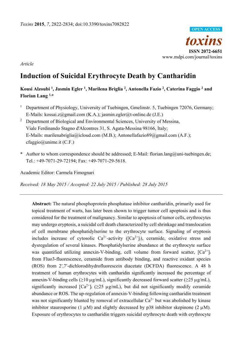
Toxins 2015, 7, 2822-2834; doi:10.3390/toxins7082822
toxins ISSN 2072-6651
www.mdpi.com/journal/toxins
Article
Induction of Suicidal Erythrocyte Death by Cantharidin
Kousi Alzoubi 1, Jasmin Egler 1, Marilena Briglia 2, Antonella Fazio 2, Caterina Faggio 2 and
Florian Lang 1,*
1 Department of Physiology, University of Tuebingen, Gmelinstr. 5, Tuebingen 72076, Germany;
E-Mails: [email protected] (K.A.); [email protected] (J.E.) 2 Department of Biological and Environmental Sciences, University of Messina,
Viale Ferdinando Stagno d'Alcontres 31, S. Agata-Messina 98166, Italy;
E-Mails: [email protected] (M.B.); [email protected] (A.F.);
[email protected] (C.F.)
* Author to whom correspondence should be addressed; E-Mail: [email protected];
Tel.: +49-7071-29-72194; Fax: +49-7071-29-5618.
Academic Editor: Carmela Fimognari
Received: 18 May 2015 / Accepted: 22 July 2015 / Published: 28 July 2015
Abstract: The natural phosphoprotein phosphatase inhibitor cantharidin, primarily used for
topical treatment of warts, has later been shown to trigger tumor cell apoptosis and is thus
considered for the treatment of malignancy. Similar to apoptosis of tumor cells, erythrocytes
may undergo eryptosis, a suicidal cell death characterized by cell shrinkage and translocation
of cell membrane phosphatidylserine to the erythrocyte surface. Signaling of eryptosis
includes increase of cytosolic Ca2+-activity ([Ca2+]i), ceramide, oxidative stress and
dysregulation of several kinases. Phosphatidylserine abundance at the erythrocyte surface
was quantified utilizing annexin-V-binding, cell volume from forward scatter, [Ca2+]i
from Fluo3-fluorescence, ceramide from antibody binding, and reactive oxidant species
(ROS) from 2′,7′-dichlorodihydrofluorescein diacetate (DCFDA) fluorescence. A 48 h
treatment of human erythrocytes with cantharidin significantly increased the percentage of
annexin-V-binding cells (≥10 g/mL), significantly decreased forward scatter (≥25 g/mL),
significantly increased [Ca2+]i (≥25 g/mL), but did not significantly modify ceramide
abundance or ROS. The up-regulation of annexin-V-binding following cantharidin treatment
was not significantly blunted by removal of extracellular Ca2+ but was abolished by kinase
inhibitor staurosporine (1 M) and slightly decreased by p38 inhibitor skepinone (2 M).
Exposure of erythrocytes to cantharidin triggers suicidal erythrocyte death with erythrocyte
OPEN ACCESS

Toxins 2015, 7 2823
shrinkage and erythrocyte membrane scrambling, an effect sensitive to kinase inhibitors
staurosporine and skepinone.
Keywords: phosphatidylserine; calcium; cell volume; staurosporine; kinase; eryptosis
1. Introduction
Cantharidin, a traditional Chinese natural product, has been successfully used for the treatment of
warts, molluscum contagiosum, and callus removal [1]. Cantharidin and its demethylated analogue
norcantharidin have more recently been shown to be effective against malignancy [2–4]. The anticancer
effects are in part attributed to their inhibitory effect on phosphoprotein phosphatases [4,5] and
stimulation of tumor cell apoptosis [6–9]. Further mechanisms invoked in (nor) cantheridine induced
apoptosis include mitochondrial dysregulation [10,11], cytosolic cytochrome c release [3], activation of
caspase-9 [3,12], induction of oxidative stress [10,13] and activation of the transcription factor p53 with
subsequent triggering of p53 dependent gene expression [13,14].
Erythrocytes lack mitochondria and nuclei, but are nevertheless able to enter suicidal cell death or
eryptosis, which is characterized by cell shrinkage [15] and translocation of phosphatidylserine to the outer
surface of the erythrocyte cell membrane [16]. Signaling involved in the stimulation of eryptosis includes
increased cytosolic Ca2+ activity ([Ca2+]i), ceramide [17], oxidative stress [16], caspase activation [16,18,19],
activation of casein kinase 1α, Janus-activated kinase JAK3, protein kinase C, p38 kinase, and PAK2
kinase [16] or inhibition of AMP activated kinase AMPK, cGMP-dependent protein kinase, and sorafenib
and sunitinib sensitive kinases [16]. Eryptosis is triggered by a wide variety of xenobiotics [16,20–42].
The present study explored whether cantharidin stimulates eryptosis. To this end, erythrocytes from
healthy volunteers were exposed to cantharidin, phosphatidylserine abundance at the erythrocyte surface
determined using annexin-V-binding and cell volume estimated from forward scatter in flow cytometry.
Moreover, [Ca2+]i was estimated utilizing Fluo3-fluorescence, ceramide abundance utilizing specific
antibodies, and abundance of reactive oxidant species utilizing 2′,7′-dichlorodihydrofluorescein
diacetate (DCFDA) fluorescence. The involvement of phosphorylation was tested utilizing kinase
inhibitors staurosporine and skepinone.
2. Results and Discussion
The present study explored whether cantharidin is capable to trigger eryptosis, the suicidal death
of erythrocytes, i.e., of cells lacking mitochondria and nuclei, organelles considered to play a major role
in the triggering of apoptosis. Hallmarks of eryptosis are phosphatidylserine translocation to the cell
surface and cell shrinkage.
Phosphatidylserine at the cell surface was detected utilizing binding of FITC-labeled annexin-V to
phosphatidylserine. The abundance of FITC-labeled annexin-V was determined by flow cytometry.
As illustrated in Figure 1, a 48 h exposure to cantharidin enhanced the percentage of annexin-V-binding
erythrocytes, an effect reaching statistical significance at 10 g/mL cantharidin concentration. As shown
in Figure 1, cantharidin did not modify hemolysis.

Toxins 2015, 7 2824
Figure 1. Effect of cantharidin on phosphatidylserine exposure. (A) Original histogram of
annexin-V-binding of erythrocytes following exposure for 48 h to Ringer solution without
(grey area) and with (black line) presence of 50 g/mL cantharidin. M1 indicates the
annexin-V-fluorescence defining the percentage of annexin-V-binding erythrocytes.
(B) Arithmetic means ± SEM of erythrocyte annexin-V-binding (n = 12) following
incubation for 48 h to Ringer solution without (white bar) or with (black bars) presence
of cantharidin (1–50 g/mL). For comparison, the effect of cantharidin on hemolysis is
shown (grey bars). *** (p < 0.001) indicates significant difference from the absence
of cantharidin (ANOVA).
Forward scatter was determined in flow cytometry as a measure of erythrocyte cell volume. As shown
in Figure 2, a 48 h cantharidin treatment was followed by a decrease of erythrocyte forward scatter, an
effect reaching statistical significance at 25 g/mL cantharidin concentration.
Figure 2. Effect of cantharidin on erythrocyte forward scatter: (A) Original histogram
of forward scatter of erythrocytes following exposure for 48 h to Ringer solution without
(grey area) and with (black line) presence of 50 g/mL cantharidin. (B) Arithmetic
means ± SEM (n = 12) of the geometric mean erythrocyte forward scatter (FSC) following
incubation for 48 h to Ringer solution without (white bar) or with (black bars) cantharidin
(1–50 g/mL). *** (p < 0.001) indicate significant difference from the absence of
cantharidin (ANOVA).

Toxins 2015, 7 2825
Figure 3. Effect of cantharidin on erythrocyte Ca2+ activity and Ca2+ sensitivity of
cantharidin-induced phosphatidylserine exposure: (A) Original histogram of Fluo3
fluorescence in erythrocytes following exposure for 48 h to Ringer solution without (grey area)
and with (black line) presence of cantharidin (50 g/mL). (B) Arithmetic means ± SEM (n = 12)
of the Fluo3 fluorescence (arbitrary units) in erythrocytes exposed for 48 h to Ringer solution
without (white bar) or with (black bars) cantharidin (1–50 g/mL). (C) Arithmetic means ±
SEM (n = 20) of annexin-V-binding of erythrocytes after a 48 h treatment with Ringer
solution without (white bars) or with 25 g/mL (grey bars) or 50 g/mL (black bars)
cantharidin in the presence (left bars, +Ca2+) and absence (right bars, −Ca2+) of Ca2+. ** (p < 0.01)
*** (p < 0.001) indicate significant difference from the absence of cantharidin (ANOVA).
(D) Arithmetic means ± SEM (n = 9) of the Fluo3 fluorescence (arbitrary units) in
erythrocytes exposed for 48 h to Ringer solution without (white bar) or with (black bars)
cantharidin (50 g/mL) and stained with Fluo3 AM in Ringer solution with (left bars) 5 mM
CaCl2 ± 1 mM sodium pyruvate, or with (right bars) 1 mM CaCl2 ± 1 mM sodium pyruvate.
*** (p < 0.001) indicate significant difference from the absence of cantharidin (ANOVA).
Both phospholipid scrambling of the erythrocyte membrane and cell shrinkage could be triggered by
activation of Ca2+ permeable cation channels with subsequent Ca2+ entry. Fluo3 fluorescence was thus
employed to test whether cantharidin influences cytosolic Ca2+ activity ([Ca2+]i). As illustrated in
Figure 3A,B, a 48 h exposure to cantharidin increased the Fluo3 fluorescence, an effect requiring 25 g/mL
cantharidin concentration for statistical significance. To test the effect of calcium concentration in the
staining solution while loading with Fluo3 and to test the potential toxic effects from released
formaldehyde as a byproduct of esterification [43,44], we treated erythrocytes for 48 h with Ringer
Toxins 2015, 7 2826
solution without or with cantharidin (50 g/mL) and then stained for 30 min with Fluo3 AM in Ringer
solution containing 1 or 5 mM CaCl2 in the presence and absence of 1 mM sodium pyruvate.
As illustrated in Figure 3D, the stimulatory effect of cantharidin on Fluo3 staining, in the presence
of 1 or 5 mM CaCl2, was similar in the presence or absence of pyruvate. A further series of experiments
explored whether cantharidin-induced translocation of phosphatidylserine to the cell surface required
entry of extracellular Ca2+. To this end, erythrocytes were incubated for 48 h in the absence or presence
of 25 or 50 g/mL cantharidin, both in the presence or nominal absence of extracellular Ca2+.
As illustrated in Figure 3C, removal of extracellular Ca2+ did not significantly blunt the effect of
cantharidin on annexin-V-binding. Instead, cantharidin significantly increased the percentage of
annexin-V-binding erythrocytes to similarly high levels in the absence and in the presence of
extracellular Ca2+. Thus, triggering of eryptosis did not require entry of extracellular Ca2+.
Eryptosis could be stimulated independently from increased [Ca2+]i by ceramide. Thus, specific antibodies
were utilized to quantify ceramide abundance at the erythrocyte surface. As a result, the ceramide abundance
was similar following a 48 h incubation in the absence of cantharidin (11.3 ± 1.3 a.u., n = 9), presence of
10 g/mL cantharidin (11.1 ± 1.4 a.u., n = 9) and presence of 50 g/mL cantharidin (11.2 ± 1.5 a.u., n = 9).
Thus, cantharidin did not enhance ceramide abundance.
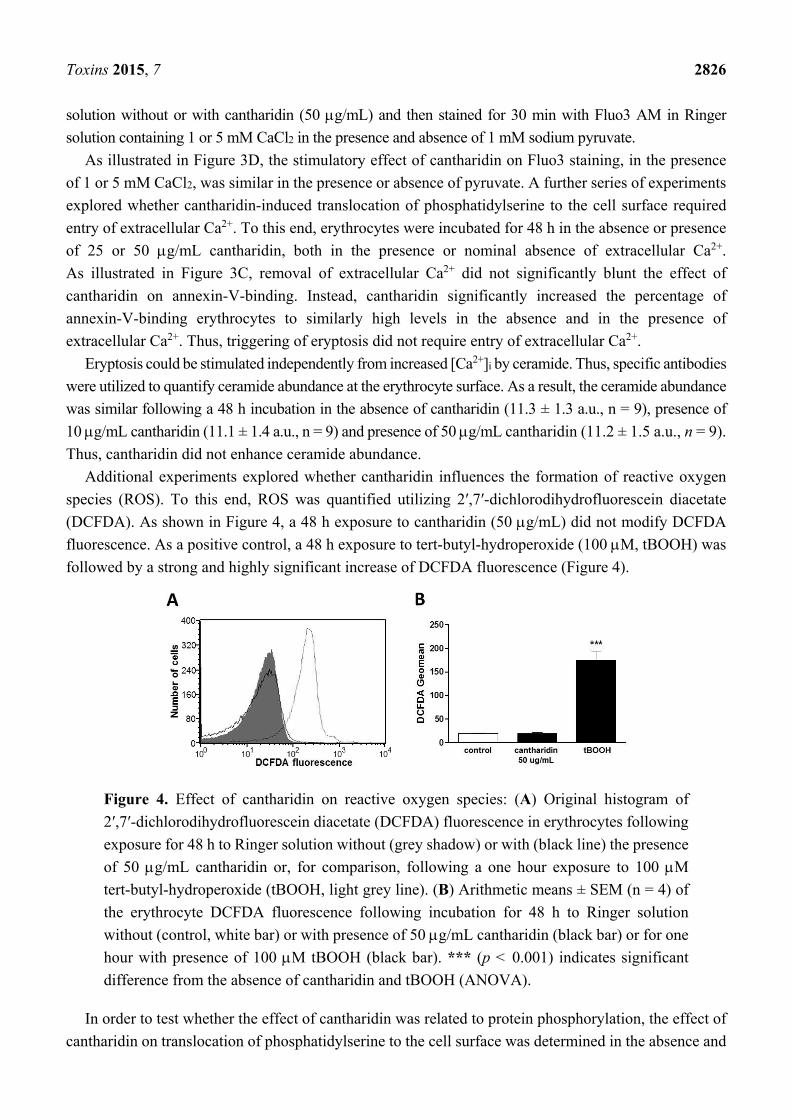
Additional experiments explored whether cantharidin influences the formation of reactive oxygen
species (ROS). To this end, ROS was quantified utilizing 2′,7′-dichlorodihydrofluorescein diacetate
(DCFDA). As shown in Figure 4, a 48 h exposure to cantharidin (50 g/mL) did not modify DCFDA
fluorescence. As a positive control, a 48 h exposure to tert-butyl-hydroperoxide (100 M, tBOOH) was
followed by a strong and highly significant increase of DCFDA fluorescence (Figure 4).
Figure 4. Effect of cantharidin on reactive oxygen species: (A) Original histogram of
2′,7′-dichlorodihydrofluorescein diacetate (DCFDA) fluorescence in erythrocytes following
exposure for 48 h to Ringer solution without (grey shadow) or with (black line) the presence
of 50 g/mL cantharidin or, for comparison, following a one hour exposure to 100 M
tert-butyl-hydroperoxide (tBOOH, light grey line). (B) Arithmetic means ± SEM (n = 4) of
the erythrocyte DCFDA fluorescence following incubation for 48 h to Ringer solution
without (control, white bar) or with presence of 50 g/mL cantharidin (black bar) or for one
hour with presence of 100 M tBOOH (black bar). *** (p < 0.001) indicates significant
difference from the absence of cantharidin and tBOOH (ANOVA).
In order to test whether the effect of cantharidin was related to protein phosphorylation, the effect of
cantharidin on translocation of phosphatidylserine to the cell surface was determined in the absence and
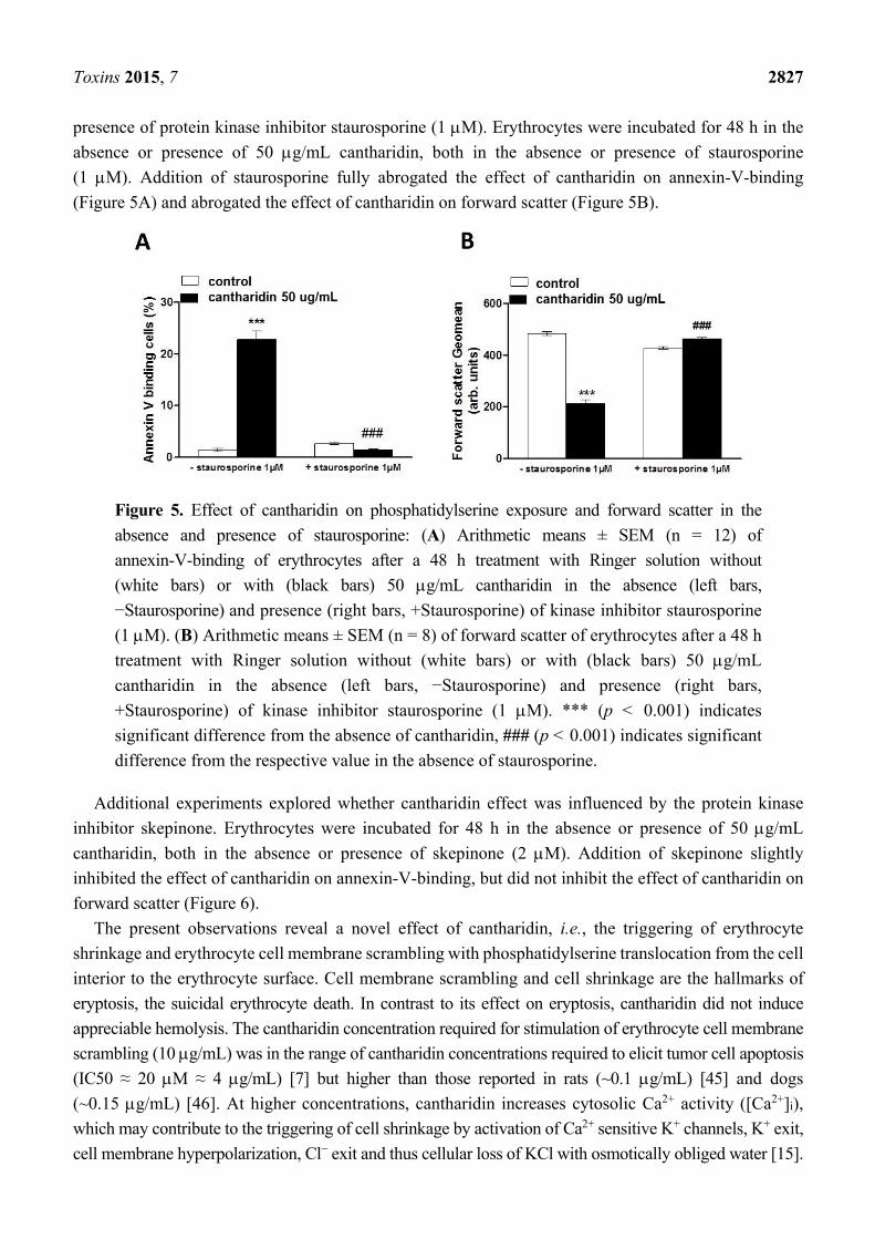
Toxins 2015, 7 2827
presence of protein kinase inhibitor staurosporine (1 M). Erythrocytes were incubated for 48 h in the
absence or presence of 50 g/mL cantharidin, both in the absence or presence of staurosporine
(1 M). Addition of staurosporine fully abrogated the effect of cantharidin on annexin-V-binding
(Figure 5A) and abrogated the effect of cantharidin on forward scatter (Figure 5B).
Figure 5. Effect of cantharidin on phosphatidylserine exposure and forward scatter in the
absence and presence of staurosporine: (A) Arithmetic means ± SEM (n = 12) of
annexin-V-binding of erythrocytes after a 48 h treatment with Ringer solution without
(white bars) or with (black bars) 50 g/mL cantharidin in the absence (left bars,
−Staurosporine) and presence (right bars, +Staurosporine) of kinase inhibitor staurosporine
(1 M). (B) Arithmetic means ± SEM (n = 8) of forward scatter of erythrocytes after a 48 h
treatment with Ringer solution without (white bars) or with (black bars) 50 g/mL
cantharidin in the absence (left bars, −Staurosporine) and presence (right bars,
+Staurosporine) of kinase inhibitor staurosporine (1 M). *** (p < 0.001) indicates
significant difference from the absence of cantharidin, ### (p < 0.001) indicates significant
difference from the respective value in the absence of staurosporine.
Additional experiments explored whether cantharidin effect was influenced by the protein kinase
inhibitor skepinone. Erythrocytes were incubated for 48 h in the absence or presence of 50 g/mL
cantharidin, both in the absence or presence of skepinone (2 M). Addition of skepinone slightly
inhibited the effect of cantharidin on annexin-V-binding, but did not inhibit the effect of cantharidin on
forward scatter (Figure 6).
The present observations reveal a novel effect of cantharidin, i.e., the triggering of erythrocyte
shrinkage and erythrocyte cell membrane scrambling with phosphatidylserine translocation from the cell
interior to the erythrocyte surface. Cell membrane scrambling and cell shrinkage are the hallmarks of
eryptosis, the suicidal erythrocyte death. In contrast to its effect on eryptosis, cantharidin did not induce
appreciable hemolysis. The cantharidin concentration required for stimulation of erythrocyte cell membrane
scrambling (10 g/mL) was in the range of cantharidin concentrations required to elicit tumor cell apoptosis
(IC50 ≈ 20 M ≈ 4 g/mL) [7] but higher than those reported in rats (~0.1 g/mL) [45] and dogs
(~0.15 g/mL) [46]. At higher concentrations, cantharidin increases cytosolic Ca2+ activity ([Ca2+]i),
which may contribute to the triggering of cell shrinkage by activation of Ca2+ sensitive K+ channels, K+ exit,
cell membrane hyperpolarization, Cl− exit and thus cellular loss of KCl with osmotically obliged water [15].

Toxins 2015, 7 2828
Figure 6. Effect of cantharidin on phosphatidylserine exposure and forward scatter in the
absence and presence of skepinone: (A) Arithmetic means ± SEM (n = 12) of annexin-V-binding
of erythrocytes after a 48 h treatment with Ringer solution without (white bars) or with
(black bars) 50 g/mL cantharidin in the absence (left bars, −skepinone) and presence (right bars,
+skepinone) of p38 kinase inhibitor skepinone (2 M). (B) Arithmetic means ± SEM
(n = 12) of forward scatter of erythrocytes after a 48 h treatment with Ringer solution without
(white bars) or with (black bars) 50 g/mL cantharidin in the absence (left bars, −skepinone)
and presence (right bars, +skepinone) of kinase inhibitor skepinone (2 M). *** (p < 0.001)
indicates significant difference from the absence of cantharidin, # (p < 0.05) indicates
significant difference from the respective value in the absence of skepinone.
Removal of extracellular Ca2+ did, however, not appreciably blunt the stimulation of annexin-V-binding
following cantharidin treatment. Even in the absence of extracellular Ca2+ cantharidin significantly
enhanced the phosphatidylserine abundance at the cell surface. Thus, the effect of cantharidin on
phosphatidylserine translocation did not require Ca2+ entry. The observation that cantharidin-induced
eryptosis occurred even in the absence of extracellular Ca2+ cannot be taken as evidence that cantharidin
was without effect on Ca2+ entry. As a matter of fact, cantharidin did increase [Ca2+]i, an effect
presumably due to stimulation of Ca2+ entry. Moreover, the increase of [Ca2+]i following cantharidin
treatment could have contributed to stimulation of eryptosis. However, cantharidin treatment was able
to trigger eryptosis even in the absence of Ca2+ entry, an observation pointing to the operation of a Ca2+
insensitive mechanism. Such a mechanism could have been ceramide. However, the cantharidin-induced
eryptosis does apparently not involve ceramide formation or translocation.
Cantharidin further did not trigger oxidative stress, a known stimulator of eryptosis [16]. In nucleated
cells, cantharidin does induce oxidative stress [10,13], an effect presumably requiring mitochondria [10,11]
and thus lacking in erythrocytes.
Instead, the effect of cantharidin on both, cell membrane scrambling and cell volume was apparently
related to its inhibitory effect on phosphoprotein phosphatases [4,5]. The effect of cantharidin on both,
cell membrane scrambling and cell volume, was completely abrogated by the kinase inhibitor staurosporine.
It is tempting to speculate that cantharidin prevents dephosphorylation of target proteins, which are
phosphorylated by staurosporine sensitive kinases. Regulators of erythrocyte cell volume include KCl
symport on the one hand and Na+,K+,2Cl− cotransport on the other, carriers under the control of several
kinases [47–49]. Thus, deranged activity of those kinases or carriers could contribute to cell volume loss.

Toxins 2015, 7 2829
Cell shrinkage and loss of cellular K+ in turn fosters cell membrane scrambling [15]. The sensitivity of
cantharidin induced eryptosis to staurosporine and skepinone indeed suggests a role of kinases in the
triggering of eryptosis. Staurosporine completely abrogated the effect of cantharidin on both,
phosphatidylserine translocation and cell volume, whereas skepinone only slightly blunted the effect of
cantharidin on phosphatidylserine translocation and did not appreciably modify the cantharidin-induced
cell shrinkage. Apparently, p38 kinase contributes to but does not account for cantharidine-induced
eryptosis. Clearly, additional experimental effort is required to dissect the specific kinase(s) and
phosphatase(s) involved in the cantharidin sensitive regulation of eryptosis.
The stimulation of eryptosis by cantharidin may add to the toxicity of the substance.
Phosphatidylserine exposing erythrocytes are engulfed by macrophages and thus rapidly cleared from
circulating blood and stimulation of eryptosis may thus lead to anemia [16]. Moreover, erythrocytes
exposing phosphatidylserine at their surface may adhere to endothelial cells of the vascular wall [50],
stimulate blood clotting and induce thrombosis [51–53]. Accordingly, stimulating phosphatidylserine
exposure of erythrocytes may impede microcirculation [17,51,54–57]. The toxic effect may be
augmented in clinical conditions associated with enhanced eryptosis, such as malignancy, hepatic
failure, diabetes, uremia, hemolytic uremic syndrome, sepsis, fever, dehydration, mycoplasma infection,
malaria, iron deficiency, sickle cell anemia, thalassemia, glucose-6-phosphate dehydrogenase
deficiency, and Wilson’s disease [16].
3. Experimental Section
3.1. Erythrocytes, Solutions and Chemicals
Fresh Lithium-Heparin-anticoagulated blood samples were kindly provided by the blood bank of the
University of Tübingen. The study was approved by the ethics committee of the University of Tübingen
(184/2003 V). The blood was centrifuged at 120 g for 20 min at 23 °C and the platelets and
leukocytes-containing supernatant was disposed. Erythrocytes were incubated in vitro for 48 h at a
hematocrit of 0.4% in Ringer solution containing (in mM) 125 NaCl, 5 KCl, 1 MgSO4,
32 N-2-hydroxyethylpiperazine-N-2-ethanesulfonic acid (HEPES), 5 glucose, and 1 CaCl2; the pH was
adjusted to 7.4 and the temperature kept at 37 °C. Where indicated, erythrocytes were exposed to
cantharidin (Sigma Aldrich, Hamburg, Germany).
3.2. Annexin-V-Binding and Forward Scatter
After incubation under the respective experimental condition, a 150 L cell suspension was washed
in Ringer solution containing 5 mM CaCl2 and then stained with Annexin-V-FITC (1:200 dilution;
ImmunoTools, Friesoythe, Germany) in this solution at 37 °C for 20 min under protection from light. In
the following, the geometric mean of the forward scatter (FSC) was determined, and annexin-V
fluorescence intensity was measured with an excitation wavelength of 488 nm and an emission
wavelength of 530 nm on a FACS Calibur (BD, Heidelberg, Germany). In some experiments erythrocytes
were preincubated in Ca2+ free solution. For determination of annexin-V-binding, addition of Ca2+ was
required during the 15 min incubation with FITC-annexin-V. Immediately thereafter measurements were
done so that the exposure to Ca2+ was too short to trigger significant phosphatidylserine translocation.

Toxins 2015, 7 2830
3.3. Hemolysis
For the determination of hemolysis, the samples were centrifuged (3 min at 1600 rpm, room temperature)
after incubation under the respective experimental conditions and the supernatants were harvested. As a
measure of hemolysis, the hemoglobin (Hb) concentration of the supernatant was determined
photometrically at 405 nm. The absorption of the supernatant of erythrocytes lysed in distilled water was
defined as 100% hemolysis.
3.4. Intracellular Ca2+
After incubation, a 150 L cell suspension was washed in Ringer solution and then loaded with
Fluo-3/AM (Biotium, Hayward, CA, USA) in Ringer solution containing 5 mM CaCl2 and 5 M
Fluo-3/AM. The cells were incubated at 37 °C for 30 min and washed twice in Ringer solution
containing 5 mM CaCl2. The Fluo-3/AM-loaded erythrocytes were resuspended in 200 L Ringer. Then,
Ca2+-dependent fluorescence intensity was measured with an excitation wavelength of 488 nm and an
emission wavelength of 530 nm on a FACS Calibur.
3.5. Reactive Oxidant Species (ROS)
Oxidative stress was determined utilizing 2′,7′-dichlorodihydrofluorescein diacetate (DCFDA). After
incubation, a 150 L suspension of erythrocytes was washed in Ringer solution and then stained with
DCFDA (Sigma, Schnelldorf, Germany) in Ringer solution containing DCFDA at a final concentration
of 10 M. Erythrocytes were incubated at 37 °C for 30 min in the dark and then washed three times in
Ringer solution. The DCFDA-loaded erythrocytes were resuspended in 200 L Ringer solution, and
ROS-dependent fluorescence intensity was measured at an excitation wavelength of 488 nm and an
emission wavelength of 530 nm on a FACS Calibur (BD).
3.6. Ceramide Abundance
For the determination of ceramide, a monoclonal antibody-based assay was used. After incubation,
cells were stained for 1 h at 37 °C with 1 g/mL anti ceramide antibody (clone MID 15B4, Alexis,
Grünberg, Germany) in PBS containing 0.1% bovine serum albumin (BSA) at a dilution of 1:10.
The samples were washed twice with PBS-BSA. Subsequently, the cells were stained for 30 min with
polyclonal fluorescein isothiocyanate (FITC) conjugated goat anti-mouse IgG and IgM specific antibody
(Pharmingen, Hamburg, Germany) diluted 1:50 in PBS-BSA. Unbound secondary antibody was
removed by repeated washing with PBS-BSA. The samples were then analyzed by flow cytometric
analysis with an excitation wavelength of 488 nm and an emission wavelength of 530 nm.
3.7. Statistics
Data are expressed as arithmetic means ± SEM. As indicated in the figure legends, statistical analysis
was made using ANOVA with Tukey’s test as post-test and t test as appropriate. n denotes the number
of different erythrocyte specimens studied. Since different erythrocyte specimens used in distinct

Toxins 2015, 7 2831
experiments are differently susceptible to triggers of eryptosis, the same erythrocyte specimens have
been used for control and experimental conditions.
4. Conclusions
Cantharidin stimulates erythrocyte cell membrane scrambling and cell shrinkage, both hallmarks of
eryptosis, the suicidal erythrocyte death. The effect of cantharidin on cell membrane scrambling and cell
shrinkage is abrogated by kinase inhibitor staurosporine and may thus be due to the known inhibitory
effect of cantharidin on protein phosphatases.
Acknowledgments
The authors acknowledge the meticulous preparation of the manuscript by Tanja Loch. The study was
supported by the Deutsche Forschungsgemeinschaft and the Open Access Publishing Fund of
Tuebingen University.
Author Contributions
F.L. designed the study and wrote the manuscript. K.A., J.E., M.B., A.F., and C.F. performed
experiments, analyzed and interpreted the results. All authors approved of the submitted manuscript.
Conflicts of Interest
The authors declare no conflict of interest.
References
1. Torbeck, R.; Pan, M.; DeMoll, E.; Levitt, J. Cantharidin: A comprehensive review
of the clinical literature. Dermatol. Online J. 2014, 20. Available online: http://escholarship.org/
uc/item/22845r22512w22860 (accessed on 27 July 2015).
2. Deng, L.P.; Dong, J.; Cai, H.; Wang, W. Cantharidin as an antitumor agent: A retrospective review.
Curr. Med. Chem. 2013, 20, 159–166.
3. Kok, S.H.; Cheng, S.J.; Hong, C.Y.; Lee, J.J.; Lin, S.K.; Kuo, Y.S.; Chiang, C.P.; Kuo, M.Y.
Norcantharidin-induced apoptosis in oral cancer cells is associated with an increase of proapoptotic
to antiapoptotic protein ratio. Cancer Lett. 2005, 217, 43–52.
4. Puerto Galvis, C.E.; Vargas Mendez, L.Y.; Kouznetsov, V.V. Cantharidin-based small molecules
as potential therapeutic agents. Chem. Biol. Drug Des. 2013, 82, 477–499.
5. Deng, L.; Dong, J.; Wang, W. Exploiting protein phosphatase inhibitors based on cantharidin
analogues for cancer drug discovery. Mini Rev. Med. Chem. 2013, 13, 1166–1176.
6. Honkanen, R.E.; Golden, T. Regulators of serine/threonine protein phosphatases at the dawn
of a clinical era? Curr. Med. Chem. 2002, 9, 2055–2075.
7. Huang, W.W.; Ko, S.W.; Tsai, H.Y.; Chung, J.G.; Chiang, J.H.; Chen, K.T.; Chen, Y.C.;
Chen, H.Y.; Chen, Y.F.; Yang, J.S. Cantharidin induces G2/M phase arrest and apoptosis in human
colorectal cancer colo 205 cells through inhibition of CDK1 activity and caspase-dependent
signaling pathways. Int. J. Oncol. 2011, 38, 1067–1073.

Toxins 2015, 7 2832
8. Liu, D.; Chen, Z. The effects of cantharidin and cantharidin derivates on tumour cells.
Anticancer Agents Med. Chem. 2009, 9, 392–396.
9. Pereira, S.R.; Vasconcelos, V.M.; Antunes, A. The phosphoprotein phosphatase family of Ser/Thr
phosphatases as principal targets of naturally occurring toxins. Crit. Rev. Toxicol. 2011, 41, 83–110.
10. Chang, C.; Zhu, Y.Q.; Mei, J.J.; Liu, S.Q.; Luo, J. Involvement of mitochondrial pathway in
NCTD-induced cytotoxicity in human hepG2 cells. J. Exp. Clin. Cancer Res. 2010, 29, 145.
11. Prasad, S.B.; Verma, A.K. Cantharidin-mediated ultrastructural and biochemical changes in
mitochondria lead to apoptosis and necrosis in murine dalton’s lymphoma. Microsc. Microanal.
2013, 19, 1377–1394.
12. Chen, Y.N.; Chen, J.C.; Yin, S.C.; Wang, G.S.; Tsauer, W.; Hsu, S.F.; Hsu, S.L. Effector
mechanisms of norcantharidin-induced mitotic arrest and apoptosis in human hepatoma cells.
Int. J. Cancer 2002, 100, 158–165.
13. Efferth, T.; Rauh, R.; Kahl, S.; Tomicic, M.; Bochzelt, H.; Tome, M.E.; Briehl, M.M.; Bauer, R.;
Kaina, B. Molecular modes of action of cantharidin in tumor cells. Biochem. Pharmacol. 2005, 69,
811–818.
14. Hong, C.Y.; Huang, S.C.; Lin, S.K.; Lee, J.J.; Chueh, L.L.; Lee, C.H.; Lin, J.H.; Hsiao, M.
Norcantharidin-induced post-G2/M apoptosis is dependent on wild-type p53 gene. Biochem. Biophys.
Res. Commun. 2000, 276, 278–285.
15. Lang, P.A.; Kaiser, S.; Myssina, S.; Wieder, T.; Lang, F.; Huber, S.M. Role of Ca2+-activated K+
channels in human erythrocyte apoptosis. Am. J. Physiol. Cell. Physiol. 2003, 285, C1553–C1560.
16. Lang, E.; Qadri, S.M.; Lang, F. Killing me softly—Suicidal erythrocyte death. Int. J. Biochem.
Cell Biol. 2012, 44, 1236–1243.
17. Abed, M.; Towhid, S.T.; Mia, S.; Pakladok, T.; Alesutan, I.; Borst, O.; Gawaz, M.; Gulbins, E.;
Lang, F. Sphingomyelinase-induced adhesion of eryptotic erythrocytes to endothelial cells.
Am. J. Physiol. Cell Physiol. 2012, 303, C991–C999.
18. Lau, I.P.; Chen, H.; Wang, J.; Ong, H.C.; Leung, K.C.; Ho, H.P.; Kong, S.K. In vitro effect of
CTAB- and PEG-coated gold nanorods on the induction of eryptosis/erythroptosis in human
erythrocytes. Nanotoxicology 2012, 6, 847–856.
19. Maellaro, E.; Leoncini, S.; Moretti, D.; del Bello, B.; Tanganelli, I.; de Felice, C.; Ciccoli, L.
Erythrocyte caspase-3 activation and oxidative imbalance in erythrocytes and in plasma of type 2
diabetic patients. Acta Diabetol. 2013, 50, 489–495.
20. Vota, D.M.; Maltaneri, R.E.; Wenker, S.D.; Nesse, A.B.; Vittori, D.C. Differential erythropoietin
action upon cells induced to eryptosis by different agents. Cell Biochem. Biophys. 2013, 65, 145–157.
21. Zbidah, M.; Lupescu, A.; Jilani, K.; Lang, F. Stimulation of suicidal erythrocyte death by
fumagillin. Basic Clin. Pharmacol. Toxicol. 2013, 112, 346–351.
22. Ahmed, M.S.; Langer, H.; Abed, M.; Voelkl, J.; Lang, F. The uremic toxin acrolein promotes
suicidal erythrocyte death. Kidney Blood Press. Res. 2013, 37, 158–167.
23. Ghashghaeinia, M.; Cluitmans, J.C.; Toulany, M.; Saki, M.; Koberle, M.; Lang, E.; Dreischer, P.;
Biedermann, T.; Duszenko, M.; Lang, F.; et al. Age sensitivity of nfkappab abundance and programmed
cell death in erythrocytes induced by nfkappab inhibitors. Cell. Physiol. Biochem. 2013, 32, 801–813.
24. Alzoubi, K.; Honisch, S.; Abed, M.; Lang, F. Triggering of suicidal erythrocyte death by
penta-O-galloyl-β-D-glucose. Toxins (Basel) 2014, 6, 54–65.

Toxins 2015, 7 2833
25. Jilani, K.; Enkel, S.; Bissinger, R.; Almilaji, A.; Abed, M.; Lang, F. Fluoxetine induced suicidal
erythrocyte death. Toxins (Basel) 2013, 5, 1230–1243.
26. Lupescu, A.; Bissinger, R.; Jilani, K.; Lang, F. Triggering of suicidal erythrocyte death by
celecoxib. Toxins (Basel) 2013, 5, 1543–1554.
27. Lupescu, A.; Jilani, K.; Zbidah, M.; Lang, F. Patulin-induced suicidal erythrocyte death.
Cell. Physiol. Biochem. 2013, 32, 291–299.
28. Abed, M.; Zoubi, K.A.; Theurer, M.; Lang, F. Effect of dermaseptin on erythrocytes. Basic Clin.
Pharmacol. Toxicol. 2013, 113, 347–352.
29. Arnold, M.; Lang, E.; Modicano, P.; Bissinger, R.; Faggio, C.; Abed, M.; Lang, F. Effect of
nitazoxanide on erythrocytes. Basic Clin. Pharmacol. Toxicol. 2014, 114, 421–426.
30. Oswald, G.; Alzoubi, K.; Abed, M.; Lang, F. Stimulation of suicidal erythrocyte death by ribavirin.
Basic Clin. Pharmacol. Toxicol. 2014, 114, 311–317.
31. Jacobi, J.; Lang, E.; Bissinger, R.; Frauenfeld, L.; Modicano, P.; Faggio, C.; Abed, M.; Lang, F.
Stimulation of erythrocyte cell membrane scrambling by mitotane. Cell. Physiol. Biochem. 2014,
33, 1516–1526.
32. Lupescu, A.; Bissinger, R.; Warsi, J.; Jilani, K.; Lang, F. Stimulation of erythrocyte cell membrane
scrambling by gedunin. Cell. Physiol. Biochem. 2014, 33, 1838–1848.
33. Abed, M.; Feger, M.; Alzoubi, K.; Pakladok, T.; Frauenfeld, L.; Geiger, C.; Towhid, S.T.;
Lang, F. Sensitization of erythrocytes to suicidal erythrocyte death following water deprivation.
Kidney Blood Press. Res. 2013, 37, 567–578.
34. Alzoubi, K.; Calabro, S.; Bissinger, R.; Abed, M.; Faggio, C.; Lang, F. Stimulation of suicidal
erythrocyte death by artesunate. Cell. Physiol. Biochem. 2014, 34, 2232–2244.
35. Arnold, M.; Bissinger, R.; Lang, F. Mitoxantrone-induced suicidal erythrocyte death.
Cell. Physiol. Biochem. 2014, 34, 1756–1767.
36. Bissinger, R.; Fischer, S.; Jilani, K.; Lang, F. Stimulation of erythrocyte death by phloretin.
Cell. Physiol. Biochem. 2014, 34, 2256–2265.
37. Bissinger, R.; Lupescu, A.; Zelenak, C.; Jilani, K.; Lang, F. Stimulation of eryptosis by
cryptotanshinone. Cell. Physiol. Biochem. 2014, 34, 432–442.
38. Bissinger, R.; Modicano, P.; Frauenfeld, L.; Lang, E.; Jacobi, J.; Faggio, C.; Lang, F.
Estramustine-induced suicidal erythrocyte death. Cell. Physiol. Biochem. 2013, 32, 1426–1436.
39. Malik, A.; Bissinger, R.; Calabro, S.; Faggio, C.; Jilani, K.; Lang, F. Aristolochic acid induced
suicidal erythrocyte death. Kidney Blood Press. Res. 2014, 39, 408–419.
40. Tesoriere, L.; Attanzio, A.; Allegra, M.; Cilla, A.; Gentile, C.; Livrea, M.A. Oxysterol mixture in
hypercholesterolemia-relevant proportion causes oxidative stress-dependent eryptosis.
Cell. Physiol. Biochem. 2014, 34, 1075–1089.
41. Voelkl, J.; Alzoubi, K.; Mamar, A.K.; Ahmed, M.S.; Abed, M.; Lang, F. Stimulation of suicidal
erythrocyte death by increased extracellular phosphate concentrations. Kidney Blood Press. Res.
2013, 38, 42–51.
42. Zhang, R.; Xiang, Y.; Ran, Q.; Deng, X.; Xiao, Y.; Xiang, L.; Li, Z. Involvement of calcium,
reactive oxygen species, and atp in hexavalent chromium-induced damage in red blood cells.
Cell. Physiol. Biochem. 2014, 34, 1780–1791.

Toxins 2015, 7 2834
43. Garcia-Sancho, J. Pyruvate prevents the atp depletion caused by formaldehyde or calcium-chelator
esters in the human red cell. Biochim. Biophys. Acta 1985, 813, 148–150.
44. Tiffert, T.; Garcia-Sancho, J.; Lew, V.L. Irreversible atp depletion caused by low concentrations
of formaldehyde and of calcium-chelator esters in intact human red cells. Biochim. Biophys. Acta
1984, 773, 143–156.
45. Dang, Y.J.; Zhu, C.Y. Oral bioavailability of cantharidin-loaded solid lipid nanoparticles.
Chin. Med. 2013, 8, 1–6.
46. Dang, Y.J.; Zhu, C.Y. Determination of trace cantharidin in plasma and pharmacokinetic study
in beagle dogs using gas chromatography-mass spectrometry. J. Anal. Toxicol. 2009, 33, 384–388.
47. Cossins, A.R.; Gibson, J.S. Volume-sensitive transport systems and volume homeostasis in
vertebrate red blood cells. J. Exp. Biol. 1997, 200, 343–352.
48. De Los Heros, P.; Alessi, D.R.; Gourlay, R.; Campbell, D.G.; Deak, M.; Macartney, T.J.;
Kahle, K.T.; Zhang, J. The WNK-regulated SPAK/OSR1 kinases directly phosphorylate and inhibit
the K+-Cl- co-transporters. Biochem. J. 2014, 458, 559–573.
49. Merciris, P.; Claussen, W.J.; Joiner, C.H.; Giraud, F. Regulation of K-Cl cotransport by Syk and
Src protein tyrosine kinases in deoxygenated sickle cells. Pflug. Arch. 2003, 446, 232–238.
50. Borst, O.; Abed, M.; Alesutan, I.; Towhid, S.T.; Qadri, S.M.; Foller, M.; Gawaz, M.; Lang, F.
Dynamic adhesion of eryptotic erythrocytes to endothelial cells via CXCL16/SR-PSOX. Am. J.
Physiol. Cell Physiol. 2012, 302, C644–C651.
51. Andrews, D.A.; Low, P.S. Role of red blood cells in thrombosis. Curr. Opin. Hematol. 1999, 6, 76–82.
52. Chung, S.M.; Bae, O.N.; Lim, K.M.; Noh, J.Y.; Lee, M.Y.; Jung, Y.S.; Chung, J.H.
Lysophosphatidic acid induces thrombogenic activity through phosphatidylserine exposure and
procoagulant microvesicle generation in human erythrocytes. Arterioscler. Thromb. Vasc. Biol.
2007, 27, 414–421.
53. Zwaal, R.F.; Comfurius, P.; Bevers, E.M. Surface exposure of phosphatidylserine in pathological
cells. Cell. Mol. Life Sci. 2005, 62, 971–988.
54. Closse, C.; Dachary-Prigent, J.; Boisseau, M.R. Phosphatidylserine-related adhesion of human
erythrocytes to vascular endothelium. Br. J. Haematol. 1999, 107, 300–302.
55. Gallagher, P.G.; Chang, S.H.; Rettig, M.P.; Neely, J.E.; Hillery, C.A.; Smith, B.D.; Low, P.S.
Altered erythrocyte endothelial adherence and membrane phospholipid asymmetry in hereditary
hydrocytosis. Blood 2003, 101, 4625–4627.
56. Pandolfi, A.; di Pietro, N.; Sirolli, V.; Giardinelli, A.; di Silvestre, S.; Amoroso, L.; di Tomo, P.;
Capani, F.; Consoli, A.; Bonomini, M. Mechanisms of uremic erythrocyte-induced adhesion of
human monocytes to cultured endothelial cells. J. Cell. Physiol. 2007, 213, 699–709.
57. Wood, B.L.; Gibson, D.F.; Tait, J.F. Increased erythrocyte phosphatidylserine exposure in sickle
cell disease: Flow-cytometric measurement and clinical associations. Blood 1996, 88, 1873–1880.
© 2015 by the authors; licensee MDPI, Basel, Switzerland. This article is an open access article
distributed under the terms and conditions of the Creative Commons Attribution license
(http://creativecommons.org/licenses/by/4.0/).
Copyright © 2022 FDOKUMEN