Asymmetric or symmetric? Cytosolic modulation of human erythrocyte hexose transfer
13
254 Biochimica et BiophysicaActa, 728 (1983) 254-266 Elsevier Biomedical Press BBA71543 ASYMMETRIC OR SYMMETRIC? CYTOSOLIC MODULATION OF HUMAN ERYTHROCYTE HEXOSE TRANSFER A. CARRUTHERS and D.L. MELCHIOR Department of Biochemistry, UMASS Medical Center, 55 Lake Avenue North, Worcester, MA 01605 (U.S.A.) (Received September 14th, 1982) Key words: Glucose transport," Hexose transfer," Asymmetric transfer," Inside-out vesicle," (Human erythrocyte) (1) The Michaelis-Menten parameters for hexose transfer in erythroctes, erythrocyte ghosts and inside-out vesicles at 20oC were determined using the light scattering method of Sen and Widdas ((1962) J. Physiol. 160, 392-403). (2) The external K m for infinite-cis exit of D-glucose in cells and ghosts is 3.6 ± 0.5 mM. (3) Dilution of cellular solute (up to 5<90 dilution) by lysing and resealing cells in varying volumes of lysate is without effect on the V m for net D-glucose exit. The K m for net exit, however, falls from 32.4 _ 3.7 mM in intact cells to 12.9 ± 2.3 mM in ghosts. This effect is reversible. (4) Infinite-cis net D-glucose uptake measurements in cells and ghosts reveal the presence of a low Km, high affinity internal site of 5.9 ± 0.8 mM. The V m for net glucose entry increases from 23.2 ± 3.7 mmol/I per min in intact cells to 55.4 S 6.3 mmol/I per min in ghosts. (5) The external K m for infinite-cis D-glucose exit in inside-out vesicles is 6.8 S 2.7 mM. The kinetics of zero-trans D-glucose exit from inside-out vesicles are changed markedly when cellular solute (obtained by lysis of intact cells) is applied to either surface of inside-out vesicles. When solute is present externally, the K m and Vma x for zero-trans exit are decreased by up to 10-fold. When solute is present at the interior of inside-out vesicles, Vma x for zero-trans exit is reduced; K m for exit is unaffected. In the nominal absence of cell solute, transfer is symmetric in inside-out vesicles. The orientation of transporter in the bilayer is unaffected by the vesicular±on procedure. (6) External application of cellular solute to ghosts reduces Vma x for D-glucose exit but is without effect on the external K m for infinite-cis exit. (7) The inhibitory potency of cell iysate on hexose transfer is lost following dialysis indicating that the factors responsible for transfer modulation are low molecular weight species. (8) We consider the hexose transfer in human erythrocytes is intrinsically symmetric and that asymmetry of transfer is conferred by interaction of the system with low molecular weight cytosolic factors. Introduction Hexose transfer in human erythrocytes is asym- metric (Widdas [1]). The K m and V m for net exit are some 10-fold greater than the corresponding values for influx (Geck [2]; Regen and Tarpley [3]; Baker and Widdas [4]). Hankin et al. [5], Foster et al. [6] and Ginsberg and Stein [7] have demon- strated the presence of a second, high affinity, low K m site at the inner surface of the membrane. The 0005-2736/83/0000-0000/$03.00 © 1983 Elsevier Biomedical Press presence of this site was confirmed by Baker and Naftalin [8] who showed that at 2°C the K m for zero-trans exit is some 5-times greater than the K m for infinite-trans exit of D-glucose into solutions containing saturating galactose concentrations. These observations are incompatible with the asymmetric form of the mobile carrier model for sugar transport. Naftalin and Holman [9] and Baker and Naftalin [8] have proposed an alterna- tive model for red cell hexose transfer which is
Transcript of Asymmetric or symmetric? Cytosolic modulation of human erythrocyte hexose transfer
254 Biochimica et BiophysicaActa, 728 (1983) 254-266 Elsevier Biomedical Press
BBA71543
ASYMMETRIC OR SYMMETRIC?
CYTOSOLIC M O D U L A T I O N OF H U M A N ERYTHROCYTE H E X O S E TRANSFER
A. CARRUTHERS and D.L. MELCHIOR
Department of Biochemistry, UMASS Medical Center, 55 Lake Avenue North, Worcester, MA 01605 (U.S.A.)
(Received September 14th, 1982)
Key words: Glucose transport," Hexose transfer," Asymmetric transfer," Inside-out vesicle," (Human erythrocyte)
(1) The Michaelis-Menten parameters for hexose transfer in erythroctes, erythrocyte ghosts and inside-out vesicles at 20oC were determined using the light scattering method of Sen and Widdas ((1962) J. Physiol. 160, 392-403). (2) The external K m for infinite-cis exit of D-glucose in cells and ghosts is 3.6 ± 0.5 mM. (3) Dilution of cellular solute (up to 5< 90 dilution) by lysing and resealing cells in varying volumes of lysate is without effect on the V m for net D-glucose exit. The K m for net exit, however, falls from 32.4 _ 3.7 mM in intact cells to 12.9 ± 2.3 mM in ghosts. This effect is reversible. (4) Infinite-cis net D-glucose uptake measurements in cells and ghosts reveal the presence of a low Km, high affinity internal site of 5.9 ± 0.8 mM. The V m for net glucose entry increases from 23.2 ± 3.7 m m o l / I per min in intact cells to 55.4 S 6.3 m m o l / I per min in ghosts. (5) The external K m for infinite-cis D-glucose exit in inside-out vesicles is 6.8 S 2.7 mM. The kinetics of zero-trans D-glucose exit from inside-out vesicles are changed markedly when cellular solute (obtained by lysis of intact cells) is applied to either surface of inside-out vesicles. When solute is present externally, the K m and Vma x for zero-trans exit are decreased by up to 10-fold. When solute is present at the interior of inside-out vesicles, Vma x for zero-trans exit is reduced; K m for exit is unaffected. In the nominal absence of cell solute, transfer is symmetric in inside-out vesicles. The orientation of transporter in the bilayer is unaffected by the vesicular±on procedure. (6) External application of cellular solute to ghosts reduces Vma x for D-glucose exit but is without effect on the external K m for infinite-cis exit. (7) The inhibitory potency of cell iysate on hexose transfer is lost following dialysis indicating that the factors responsible for transfer modulation are low molecular weight species. (8) We consider the hexose transfer in human erythrocytes is intrinsically symmetric and that asymmetry of transfer is conferred by interaction of the system with low molecular weight cytosolic factors.
Introduction
Hexose transfer in human erythrocytes is asym- metric (Widdas [1]). The K m and V m for net exit are some 10-fold greater than the corresponding values for influx (Geck [2]; Regen and Tarpley [3]; Baker and Widdas [4]). Hankin et al. [5], Foster et al. [6] and Ginsberg and Stein [7] have demon- strated the presence of a second, high affinity, low K m site at the inner surface of the membrane. The
0005-2736/83/0000-0000/$03.00 © 1983 Elsevier Biomedical Press
presence of this site was confirmed by Baker and Naftalin [8] who showed that at 2°C the K m for zero-trans exit is some 5-times greater than the K m for infinite-trans exit of D-glucose into solutions containing saturating galactose concentrations.
These observations are incompatible with the asymmetric form of the mobile carrier model for sugar transport. Naftalin and Holman [9] and Baker and Naftalin [8] have proposed an alterna- tive model for red cell hexose transfer which is
consistent with all the intact cell kinetic data pre- sently available. This model suggests that the asymmetry of hexose transfer results from factors extrinsic to the membrane. It is proposed that D-glucose binds slowly and non-specifically to haemoglobin. A consequence of this is that during rapid net exit, the free cytosolic sugar concentra- tion is overestimated and the K m for exit is over- estimated. Vma x for exit is unaffected by cytosolic compartmentalisation. Another consequence is that the rate of net sugar uptake will be rate-limited by the rate of association of sugar with haemoglobin. This reduces the operational Vma x for infinite-cis net entry and accounts for the asymmetry of net transport in the erythrocyte. This is an attractive model for hexose transfer and, in addition, may be tested directly by determining the kinetics of hexose-transfer in haemoglobin-free red cell ghosts.
We have examined this model using the Sen- Widdas [10] procedure (light-scattering) for hexose flux determinations. Our results show that as the cellular contents are diluted (by forming resealed ghosts) the K m for exit and Vma x for entry fall and increase, respectively. This confirms earlier predic- tions.
Furthermore, experiments with inside-out vesicles show that the kinetics of zero-trans exit (equivalent to entry in cells and ghosts) are af- fected markedly by the presence of cellular lysate at both sides of the membrane. Exit in the absence of lysate is identical to efflux from ghosts. How- ever, in the presence of lysate, exit resembles zero- trans entry in intact cells. We provide evidence to show that this effect is brought about by low molecular weight species present in the erythrocyte cytosol and conclude that hexose transfer in red cells may be intrinsically symmetric.
Methods
Solutions The following solutions were used. Tris-buffered
solution consisting of 150 mM KC1, 5 mM Tris- HC1, 0.2 mM EDTA, pH 7.4. Lysate consisting of 10 mM Tris-HC1, 4 mM EDTA, pH 7.2. Lysate vesiculation medium containing 10 mM Tris-HC1, 4 mM EDTA at pH 7.5. The pH of all solutions was adjusted using 1 M Tris base.
255
Erythrocytes, ghosts and inside-out vesicles Erythrocytes, obtained from freshly out-dated
blood bank whole blood, were washed three times in Tris-medium. Resealed ghosts were prepared by lysing 1 volume of packed, washed cells in 5 or more volumes of lysate. Lysis was carried out on ice and allowed to proceed for 10 min. KC1 (2 M, pH 7.2) was added to the lysate to restore isotonic- ity and the membrane suspension was incubated at 37°C for 40 min. Resealed ghosts were collected by centrifugation and were resuspended in Tris- medium. This procedure produces pink ghosts. Substantially haemoglobin-free ghosts (although still pink) were prepared by centrifugation of the lysate prior to resealing. Intact cells were lysed in 30 volumes of lysate, then centrifuged at 44000 x g for 15 min. The haemoglobin-rich supernatant was aspirated and the membranes resuspended in 30 volumes of ice-cold lysate. KC1 (2 M) was added to restore isotonicity and the lysate was incubated at 37°C for 40 min. The ghosts were collected by centrifugation and resuspended in Tris-medium.
Inside-out vesicles were prepared by the one- step method of Lew et al. [11]. Briefly, 1 volume of packed cells was lysed in 5 volumes of vesiculation medium and incubated on ice for 10 min. The lysate was then incubated at 37°C for 30 min and the resulting inside-out vesicles collected by centrifugation at 35 000 x g for 15 min. The vesicles were resuspended in 2 volumes of Tris medium and passed vigorously through a 27-gauge needle some four or five times. This step forms ion-tight inside-out vesicles. Phase contrast microscopy shows that these vesicles are 0.1-0.4/~m in diame- ter and are some 96-98% free of resealed ghosts. Sialic acid accessibility assays were performed using the procedure described by Steck and Kant [12] and sialic acid released quantitated by the method of Warren [13]. The results of these experi- ments show that 94% of membrane sialic acid groups in inside-out vesicles are inaccessible to the enzyme sialidase and that the inside-out vesicles are indeed inside-out.
Sugar transport measurements Sugar exit and entry were determined using the
light scattering method described by Sen and Wid- das [10] and, in more detail, by Miller [14]. Volume changes, induced by sugar entry and exit were
256
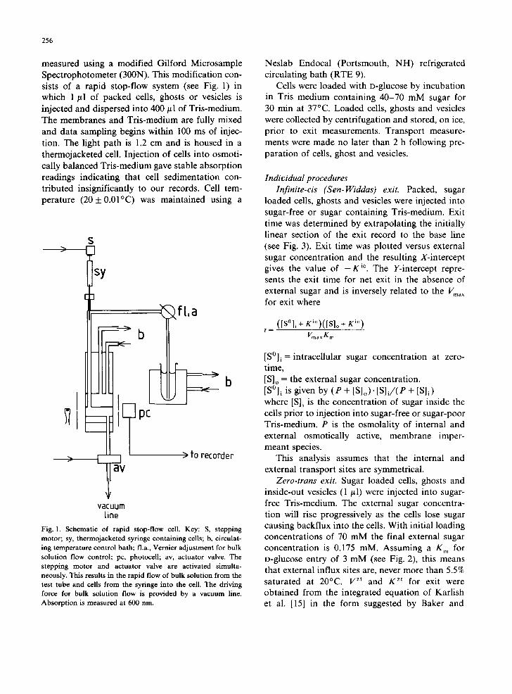
measured using a modified Gilford Microsample Spectrophotometer (300N). This modification con- sists of a rapid stop-flow system (see Fig. 1) in which 1 #1 of packed cells, ghosts or vesicles is injected and dispersed into 400/~1 of Tris-medium. The membranes and Tris-medium are fully mixed and data sampling begins within 100 ms of injec- tion. The light path is 1.2 cm and is housed in a thermojacketed cell. Injection of cells into osmoti- cally balanced Tris-medium gave stable absorption readings indicating that cell sedimentation con- tributed insignificantly to our records. Cell tem- perature (20+0.01°C) was maintained using a
sy
- ft,a
R pc
b
> to recorder
vacuum fine
Fig. 1. Schematic of rapid stop-flow cell. Key: S, stepping motor; sy, thermojacketed syringe containing cells; b, circulat- ing temperature control bath; fla., Vernier adjustment for bulk solution flow control; pc, photocell; av, actuator valve. The stepping motor and actuator valve are activated simulta- neously. This results in the rapid flow of bulk solution from the test tube and cells from the syringe into the cell. The driving force for bulk solution flow is provided by a vacuum line. Absorption is measured at 600 nm.
Neslab Endocal (Portsmouth, NH) refrigerated circulating bath (RTE 9).
Cells were loaded with o-glucose by incubation in Tris medium containing 40-70 mM sugar for 30 min at 37°C. Loaded cells, ghosts and vesicles were collected by centrifugation and stored, on ice, prior to exit measurements. Transport measure- ments were made no later than 2 h following pre- paration of cells, ghost and vesicles.
Individual procedures Infinite-cis (Sen-Widdas) exit. Packed, sugar
loaded cells, ghosts and vesicles were injected into sugar-free or sugar containing Tris-medium. Exit time was determined by extrapolating the initially linear section of the exit record to the base line (see Fig. 3). Exit time was plotted versus external sugar concentration and the resulting X-intercept gives the value of - K ic. The Y-intercept repre- sents the exit time for net exit in the absence of external sugar and is inversely related to the Vma X for exit where
t = (IS°l, + K'°)(IS]o + K 'c)
VmaxKm
[S°]i = intracellular sugar concentration at zero- time, [S]o = the external sugar concentration. [S°]i is given by (P + [S]o ) • [S] i / (P + [S]i ) where [S]i is the concentration of sugar inside the cells prior to injection into sugar-free or sugar-poor Tris-medium. P is the osmolality of internal and external osmotically active, membrane imper- meant species.
This analysis assumes that the internal and external transport sites are symmetrical.
Zero-trans exit. Sugar loaded cells, ghosts and inside-out vesicles (1/~1) were injected into sugar- free Tris-medium. The external sugar concentra- tion will rise progressively as the cells lose sugar causing backflux into the cells. With initial loading concentrations of 70 mM the final external sugar concentration is 0.175 mM. Assuming a K m for D-glucose entry of 3 mM (see Fig. 2), this means that external influx sites are, never more than 5.5% saturated at 20°C. V Zt and K zt for exit were obtained from the integrated equation of Karlish et al. [15] in the form suggested by Baker and
E
x
1
/
I, ~ / I I -6 0 6 12
[D-gluc0se] ° mM
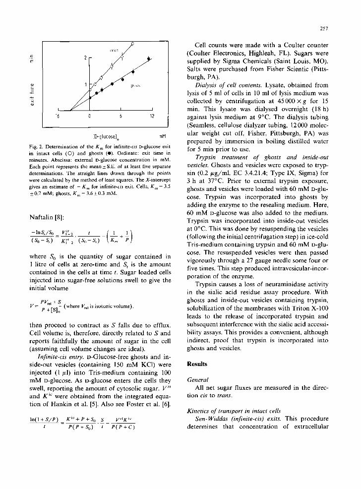
Fig. 2. Determination of the K m for infinite-cis D-glucose exit in intact cells (O) and ghosts (e). Ordinate: exit time in minutes. Abscissa: external D-glucose concentration in mM. Each point represents the mean_ S.E. of at least five separate determinations. The straight lines drawn through the points were calculated by the method of least squares. The X-intercept gives an estimate of - g m for infinite-cis exit. Cells, K m = 3.5 +0.7 mM; ghosts, K m = 3.6+0.3 mM.
Naftalin [8]:
-lnSt/So= V(t-2 t ( 1 1) (So-St) K z, (So-S,) ~ + P 1-2
where S O is the quantity of sugar contained in 1 litre of cells at zero-time and S t is the amount contained in the cells at time t. Sugar loaded cells injected into sugar-free solutions swell to give the initial volume
V= P Vre t + S P + [S]o (where Vr¢~ is isotonic volume),
then proceed to contract as S falls due to efflux. Cell volume is, therefore, directly related to S and reports faithfully the amount of sugar in the cell (assuming cell volume changes are ideal).
Infinite-cis entry. D-Glucose-free ghosts and in- side-out vesicles (containing 150 mM KC1) were injected (1 /~1) into Tris-medium containing 100 mM D-glucose. As D-glucose enters the cells they swell, reporting the amount of cytosolic sugar. V zt and K~C were obtained from the integrated equa- tion of Hankin et al. [5]. Also see Foster et al. [6].
In(I+S/P) Kic+p+So S VZtK ic t P ( P + S o ) t P ( P + C )
257
Cell counts were made with a Coulter counter (Coulter Electronics, Highleah, FL). Sugars were supplied by Sigma Chemicals (Saint Louis, MO). Salts were purchased from Fisher Scientic (Pitts- burgh, PA).
Dialysis of cell contents. Lysate, obtained from lysis of 5 ml of cells in 10 ml of lysis medium was collected by centrifugation at 45000 × g for 15 min. This lysate was dialysed overnight (18 h) against lysis medium at 9°C. The dialysis tubing (Seamless, cellulose dialyzer tubing, 12 000 molec- ular weight cut off, Fisher, Pittsburgh, PA) was prepared by immersion in boiling distilled water for 5 min prior to use.
Trypsin treatment of ghosts and inside-out vesicles. Ghosts and vesicles were exposed to tryp- sin (0.2/xg/ml, EC 3.4.21.4; Type IX, Sigma) for 3 h at 37°C. Prior to external trypsin exposure, ghosts and vesicles were loaded with 60 mM D-glu- cose. Trypsin was incorporated into ghosts by adding the enzyme to the resealing medium. Here, 60 mM D-glucose was also added to the medium. Trypsin was incorporated into inside-out vesicles at 0°C. This was done by resuspending the vesicles (following the initial centrifugation step) in ice-cold Tris-medium containing trypsin and 60 mM D-glu- cose. The resuspended vesicles were then passed vigorously through a 27 gauge needle some four or five times. This step produced intravesicular-incor- poration of the enzyme.
Trypsin causes a loss of neuraminidase activity in the sialic acid residue assay procedure. With ghosts and inside-out vesicles containing trypsin, solubilization of the membranes with Triton X-100 leads to the release of incorporated trypsin and subsequent interference with the sialic acid accessi- bility assays. This provides a convenient, although indirect, proof that trypsin is incorporated into ghosts and vesicles.
R e s u l t s
General All net sugar fluxes are measured in the direc-
tion cis to trans.
Kinetics of transport in intact cells Sen-Widdas (infinite-cis) exits. This procedure
determines that concentration of extracellular
258
sugar which reduces by half the saturated efflux of
sugar from loaded cells. In practice, this is done by determining the exit time for sugar efflux from
loaded cells (see methods) into solution of varying
sugar content. Fig. 2 shows the results of such experiments with D-glucose. The extracellular glu-
cose concentra t ion which inhibits exit of D-glucose half-maximally is 3.5 + 0.7 mM. This glucose con- centrat ion is a measure of the K m of infinite-trans influx. According to the Sen-Widdas [10] treat- ment, the exit time for D-glucose efflux into glu- cose-free medium corresponds to a Irma x for exit of 78.4 + 3.6 mmol /1 cell water per min. This analy-
sis assumes that the K m for exit and entry are identical.
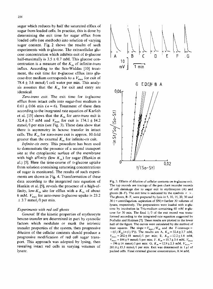
Zero-trans exit. The exit time for D-glucose efflux from intact cells into sugar-free medium is
0.61 + 0.06 min (n = 6). Trea tment of these data according to the integrated rate equat ion of Karl ish
et al. [15] shows that the g m for zero-trans exit is
32.4 + 3.7 mM and Vma X for exit is 174.1 + 14.2 mmol /1 per min (see Fig. 3). These data show that there is asymmetry in hexose transfer in intact
cells. The K m for zero-trans exit is approx. 10-fold greater than the external K m for infinite-cis exit.
Infinite-cis entry. This procedure has been used to demonstra te the presence of a second transport site at the cytoplasmic surface of the membrane
with high affinity (low Kin) for sugar (Hankin et al.) [5]. Here the time-course of D-glucose uptake
from solution conta in ing saturat ing concentra t ions of sugar is monitored. The results of such experi- ments are shown in Fig. 4. Transformat ion of these
data according to the integrated rate equat ion of Hank in et al. [5], reveals the presence of a high-af-
finity, low-K m site for efflux with a K m of about
6 mM. Vma x for zero-trans D-glucose upake is 23.2 + 3.7 m m o l / l per min.
Experiments with red cell ghosts General. If the kinetic properties of erythrocyte
hexose-transfer are determined in part by cytosolic factors which modula te or mask the intrinsic transfer properties of the system, then progressive di lut ion of the cellular contents should produce a progressive modificat ion of red cell sugar trans- port. This approach was adopted by lysing, then resealing intact red cells in varying volumes of lysate.
A 10
mV
B C
I' ' 1 min
D
k E F 5
0,06
A 4. . . -
I 0
v
¢-
I
(3 EDC/F B A
/
! I
t/(S0- St)
!
0,02
Fig. 3. Effects of dilution of cellular contents on D-glucose exit. The top records are tracings of the pen chart recorder records of cell shrinkage due to sugar exit in erythrocytes (A) and ghosts (B-F). The exit time is indicated by the symbols < >. The ghosts, B-F, were prepared by lysis in 5, 10, 15, 20, 30 and 30 (+ centrifugation, aspiration of SN)+ further 30 volumes of lysate, respectively. The preparations were loaded with D-glu- cose by incubation in Tris-medium containing 60 mM o-glu- cose for 30 min. The final 1/3 of the exit record was trans- formed according to the integrated rate equation suggested by Naftalin and Holman [7]. These results are plotted in the lower half of the figure. The curves were calculated by the method of least squares. The slope= Vmax/K m and the Y-intercept= -((1/Km)+(I/P)). The results are A, Kin= 32.4_+3.7 mM, Vma~=202+18 mmol/l per min; E, Km=12.2+1.8 mM, Vma ~ = 184+9 mmol/1 per min; F, K m = 15.7-+3.4 mM, Vma ~ = 196+ 14 mmol/1 per min; G, Km= 12.9+2.3 mM, Vm, . = 281.0+ 15.3 mmol/l per min. Exit was determined in 1 ~tl of packed cells. Final external glucose concentration, 0.14 mM,

0 ~
1 t i m e mln
0.2
CELLS " <
0 , ~ , ~ . . . . 50
S t / t m m o t / l / r m n
Fig. 4. In f in i t e -c i s D-glucose entry in erythrocytes (O) and ghosts (O). The figure to the left represents the time course of glucose uptake during the initial 60 s of the experiment. The points are derived from the influx records on the chart recorder and are mean values obtained from ten separate runs. The figure to the right of this is a transformation of the data according to the integrated rate equation of Hankin et al. [5]. The slope = ( K m + P + C o ) / ( P ( P + Co) ) and the Y-intercept = -- V m a x K m / P ( P + Co). K m is the so-called K m for infinite- cis uptake, K i. The curves drawn through the points were calculated by the least-squares method. The results are: cells, K i = 5.8+0.9 mM; Vma x = 23.2+3.7 mmol/1 per min; ghosts, K i = 5.9_+0.6 mM; Vma ~ = 55.4_+6.3 m m o l / l per rain. Influx determinations were made with 1 #1 of packed cells.
Infinite-cis (Sen-Widdas) exit. Fig. 2 shows that the external K m for infinite-cis glucose exit in pink ghosts (dilution of cellular haemoglobin = × 170) is 3.6 + 0.3 mM. This value is identical to that for infinite-cis exit in intact cells and shows that cyto- solic dilution is without effect on the affinity of the external site for sugar. Nevertheless, Fig. 2 also shows that exit times are reduced in ghosts. This supports the view that the kinetics of sugar exit are altered by cytosolic dilution.
Zero-trans exit. Fig. 3 shows the results of pro- gressive cytosolic dilution on the kinetics of zero- trans glucose exit from red cells. As dilution of the cytosol is increased from 0 to × 170, the exit time falls from 0.61 + 0.06 (n = 6) to 0.26 + 0.01 (n = 8) min. Treatment of these data according to the integrated rate equation for exit shows that cyto- solic dilution produces a progressive decrease in the K m for zero-trans exit from 32.4 + 3.7 mM in intact cells to 12.9 + 2.3 mM in ghosts. I/ma X for exit remains unchanged in pink ghost at approx. 200 mmol/1 per min. However, at higher degrees of cytosolic dilution, Vma x for exit increases to 281 + 15 m m o l / l per min. Fig. 5 summarizes the results of similar experiments with four different samples of human blood. The effect of cellular content dilution on the K m for zero-trans D-glu-
50
259
v E
0 0
::.
I I I i
100 I I
200
i -
X E
E
300
i ' : i
I i I I I
0 100 I I I
200
X[Hb] i di[ufion Fig. 5. The effects of cytosolic dilution on the kinetics of zero- t rans o-glucose exit from red cells. Upper graph. Ordinate: K m for exit in mM. Abscissa: dilution of cellular haemoglobin content. Lower graph. Ordinate; Vm~ for exit in m m o l / l cell water per min. Abscissa: dilution of cellular haemoglobin. Each point represents a single determination.
cose exit is half-maximal at about × 15 dilution. Infinite-cis entry. Fig. 4 summarizes such experi-
ments with intact cells and ghosts. Analysis of these data shows that the internal K m for infinite- cis entry is unchanged in ghosts but that Vma ~ for entry is increased from 23.2 _+ 3.7 mmol/1 per min in intact cells to 55.4 + 6.3 m m o l / l per rain in ghosts.
Reversibility. If the effects of cytosolic dilution on sugar exit are mediated by the loss of cytosolic factors, it should be possible to reverse this effect by adding these factors back to the cytosol. Table I summarizes the results of such experiments. Dilu- tion of cellular contents by up to 50-fold produces a progressive decrease in the K m for zero-trans glucose exit from 34 to 14.7 mM but is without
260
TABLE I
REVERSIBILITY OF CYTOSOLIC DILUTION EFFECTS
ON KINETICS OF EXIT
In Condition F cells from Condition E were lysed and released
in a × 10 dilution of cellular contents. The initial D-glucose
content of ghosts, 60 mmol/1.
Con- × Dilution Zero-trans D-glucose exit
dition [Hb] i
g m Vmax (mM) ( m m o l / l / m i n )
A 0 34.6 + 3.9 200 + 5
B 13.8 26.4+2.2 221 + 15 C 44.6 17.6+ 1.3 208+ 12
D 46.2 14.7+ 1.1 199+ 9
E 97.4 11.8 _ 0.8 287 _ 10
F 12.6 27.5 + 1.9 206 + 10
effect o n Vma x. If the cellular contents are further diluted to approx. 100-fold, Vma x for exit increases from about 200 to 287 mmol/1 per min and K m falls to 12 mM. When these ghosts are then lysed and resealed in solution containing cell contents (approximately × 10 dilution) the K m for exit in- creases and Vma x for exit decreases to 28 mM and 208 mmol/1 per min, respectively. These findings are important for they show that the effects of cell sol dilution on the kinetics of sugar exit are media- ted by the loss of cytosolic factors and not by the lysis and resealing procedures per se.
Experiments with inside-out vesicles General. Experiments with inside-out vesicles
offer a number of advantages over more conven- tional flux determinations with intact cells and ghosts. (1) They provide easy access to the cyto- plasmic surface of the erythrocyte plasma mem- brane. (2) They permit the infinite dilution of cellular solute exposed to the endofacial bilayer surface. The experiments described below were designed to determine the following. (1) Whether the internal high-affinity ( 1 o w - K m ) infinite-cis en- try site could be detected at the exterior of inside- out vesicles. (2) The extent to which cellular con- tents affect hexose-transfer when applied at both or either side of the membrane. To avoid confu- sion, it must be remembered that sugar exit in inside-out vesicles is equivalent to sugar entry in cells.
Infinite-cis (Sen-Widdas) exit. If the high-affin- ity internal infinite-trans exit site detected in cells and ghosts using the infinite-¢is entry procedure is an integral component of the transfer mechanism, it should be detected at the external surface of inside-out vesicles using the Sen-Widdas [10] pro- cedure. This experimental procedure in inside-out vesicles is analogous to infinite-cis entry in cells and ghosts. The K m obtained by the Sen-Widdas procedure is 6.8 _+ 2.7 mM (n = 4 , range 2.6-15 mM) which is in close agreement with the K m for infinite-trans exit in cells and ghosts (6 mM).
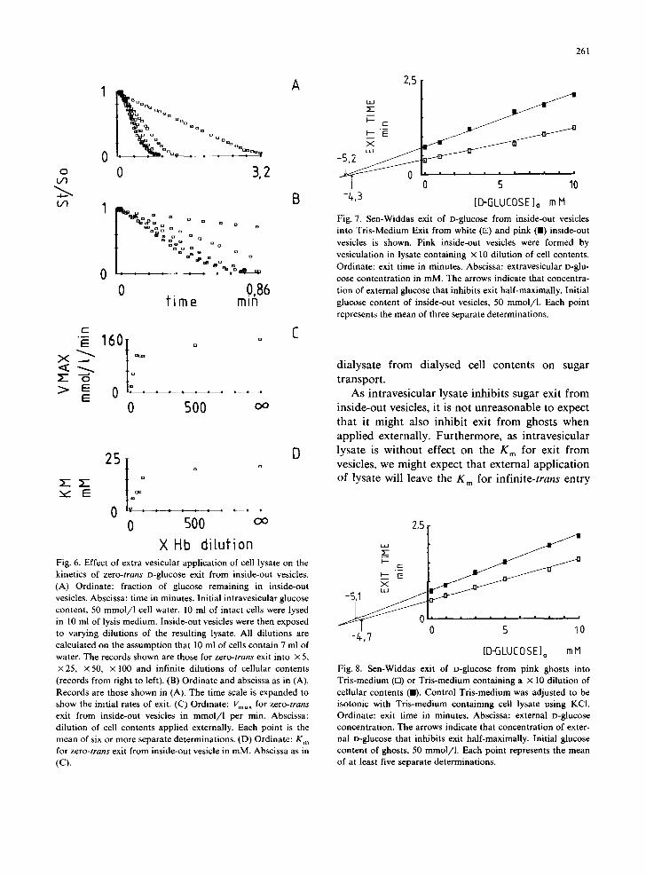
Zero-trans exit. The kinetics of zero-trans exit from inside-out vesicles are modified markedly by the presence of cytosolic solute at both the interior and exterior of the vesicles. Fig. 6 summarizes the results of experiments with inside-out vesicles where zero-trans exit was monitored into increas- ing dilutions of lysate obtained from the red cells during vesicle preparation. It is clear that exit from inside-out vesicles is inhibited by the pres- ence of cytosolic solute at the cytosolic surface of the bilayer. Fig. 6 also shows that the cytoplasmic solute reduces both the K m and Vma x for exit from inside-out vesicles. This dilution effect is half-max- imal at approximately × 45 dilution.
The presence of lysate at the interior of inside- out vesicles also inhibits sugar exit, but here, is without effect on the K m for exit. Furthermore, internal application of cellular solute is without effect on the external K m for infinite-cis exit. Fig. 7 and Table II summarize these results. Table II also reveals some information regarding the nature of the material which affects sugar transport so markedly. Lysate was collected from 5 ml of intact cells lysed in 10 ml of lysis medium. This was divided into equal volumes and one volume di- alysed overnight against lysis medium. The follow- ing day, fresh inside-out vesicles were prepared and the dialysed lysate and non-dialysed lysate were incorporated into separate batches of vesicles. The dialyzed lysate is without significant effect on sugar exit from inside-out vesicles ( P > 0 . 0 5 ) whereas non-dialysed lysate inhibits exit markedly ( P < 0.005). These data indicate that a low-molec- ular weight species (less than 12 000) is responsible for this effect on erythrocyte hexose transfer. We have not yet examined the effects of concentrated
o t ~
A D D m
~ D a ~ ~ ] D ° D m Q m ° m i n d m m mt~m o : n~. : : : , , or~
0 3,2
g
" o ~ : : o ° ° o m m Q m
lain D Q a
D EIn mo ~IB
0 0,86 t ime m~n
1,° I o o
0 ' : . . . . E 0 500 Do
D
2s I o o
0 500 D o
X Hb dilufi0n Fig. 6. Effect of extra vesicular application of cell lysate on the kinetics of z e r o - t r a n s D-glucose exit from inside-out vesicles. (A) Ordinate: fraction of glucose remaining in inside-out vesicles. Abscissa: time in minutes. Initial intravesicular glucose content, 50 mmol /1 cell water. 10 ml of intact cells were lysed in 10 ml of lysis medium. Inside-out vesicles were then exposed to varying dilutions of the resulting lysate. All dilutions are calculated on the assumption that 10 ml of cells contain 7 ml of water. The records shown are those for z e r o - t r a n s exit into × 5, × 25, × 50, × 100 and infinite dilutions of cellular contents (records from right to left). (B) Ordinate and abscissa as in (A). Records are those shown in (A). The time scale is expanded to show the initial rates of exit. (C) Ordinate: Vmax for z e r o - t r a n s
exit from inside-out vesicles in m m o l / l per min. Abscissa: dilution of cell contents applied externally. Each point is the mean of six or more separate determinations. (D) Ordinate: K m for z e r o - t r a n s exit from inside-out vesicle in mM. Abscissa as in (C).
261
2,5
-t~,3 0 5 10
[D-GLUCOSE]o m H
Fig. 7. Sen-Widdas exit of D-glucose from inside-out vesicles into Tris-Medium Exit from white (ra) and pink (11) inside-out vesicles is shown. Pink inside-out vesicles were formed by vesiculation in lysate containing × 10 dilution of cell contents. Ordinate: exit time in minutes. Abscissa: extravesicular D-glu- cose contentration in mM. The arrows indicate that concentra- tion of external glucose that inhibits exit half-maximally. Initial glucose content of inside-out vesicles, 50 mmol/1. Each point represents the mean of three separate determinations.
dialysate from dialysed cell contents on sugar transport.
As intravesicular lysate inhibits sugar exit from inside-out vesicles, it is not unreasonable to expect that it might also inhibit exit from ghosts when applied externally. Furthermore, as intravesicular lysate is without effect on the g m for exit from vesicles, we might expect that external application of lysate will leave the K m for infinite-trans entry
2,5
-/ , ,7 0 5 10
[D-GLUCOSE] o mH
Fig. 8. Sen-Widdas exit of D-glucose from pink ghosts into Tris-medium (E3) or Tris-medium containing a × 10 dilution of cellular contents (11). Control Tris-medium was adjusted to be isotonic with Tris-medium containing cell lysate using KCI. Ordinate: exit time in minutes. Abscissa: external D-glucose concentration. The arrows indicate that concentration of exter- nal D-glucose that inhibits exit half-maximally. Initial glucose content of ghosts, 50 retool/1. Each point represents the mean of at least five separate determinations.
262
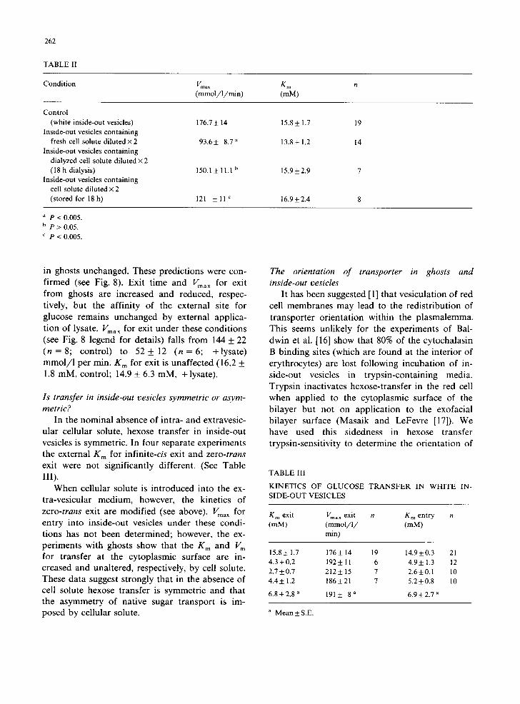
TABLE II
Condition Vma ~ K m n (mmol /1 /min) (mM)
Control
(white inside-out vesicles) 176.7 ± 14 15.8 ± 1.7 19
Inside-out vesicles containing
fresh cell solute d i lu t ed×2 93.6± 8.7 a 13.8+ 1.2 14 Inside-out vesicles containing
dialyzed cell solute diluted × 2
(18 h dialysis) 150.1± 11.1 b 15.9±2.9 7
Inside-out vesicles containing cell solute diluted × 2
(stored for 18h) 121 ± l l C 16.9±2.4 8
P < 0.005. b p > 0.05.
c p < 0.005.
in ghosts unchanged. These predictions were con- firmed (see Fig. 8). Exit time and Vma ~ for exit from ghosts are increased and reduced, respec- tively, but the affinity of the external site for glucose remains unchanged by external applica- tion of lysate. Vma x for exit under these conditions (see Fig. 8 legend for details) falls from 144 + 22 ( n = 8 ; control) to 5 2 + 12 ( n = 6 ; +lysate) mmol/1 per min. K m for exit is unaffected (16.2 + 1.8 mM, control; 14.9 + 6.3 mM, +lysate).
Is transfer in inside-out vesicles symmetric or asym- metric?
In the nominal absence of intra- and extravesic- ular cellular solute, hexose transfer in inside-out vesicles is symmetric. In four separate experiments the external K m for infinite-cis exit and zero-trans exit were not significantly different. (See Table III).
When cellular solute is introduced into the ex- tra-vesicular medium, however, the kinetics of zero-trans exit are modified (see above). Vma x for entry into inside-out vesicles under these condi- tions has not been determined; however, the ex- periments with ghosts show that the g m and V m for transfer at the cytoplasmic surface are in- creased and unaltered, respectively, by cell solute. These data suggest strongly that in the absence of cell solute hexose transfer is symmetric and that the asymmetry of native sugar transport is im- posed by cellular solute.
The orientation of transporter in ghosts and inside-out vesicles
It has been suggested [1] that vesiculation of red cell membranes may lead to the redistribution of transporter orientation within the plasmalemma. This seems unlikely for the experiments of Bal- dwin et al. [16] show that 80% of the cytochalasin B binding sites (which are found at the interior of erythrocytes) are lost following incubation of in- side-out vesicles in trypsin-containing media. Trypsin inactivates hexose-transfer in the red cell when applied to the cytoplasmic surface of the bilayer but not on application to the exofacial bilayer surface (Masaik and LeFevre [17]). We have used this sidedness in hexose transfer trypsin-sensitivity to determine the orientation of
TABLE l l I
KINETICS OF GLUCOSE TRANSFER IN WHITE IN- SIDE-OUT VESICLES
K m exit Vma x exit n K m entry (mM) ( m m o l / l / (mM)
min)
15.8±1.7 176±14 19 14.9±0.3 21
4 .3±0.2 192±11 6 4.9±1.3 12 2.7±0.7 212±15 7 2.6±0.1 10
4 .4±1.2 186±21 7 5.2±0.8 10
6.8±2.8 a 191± 8 a 6 .9±2.7 a
a Mean ± S.E.
263
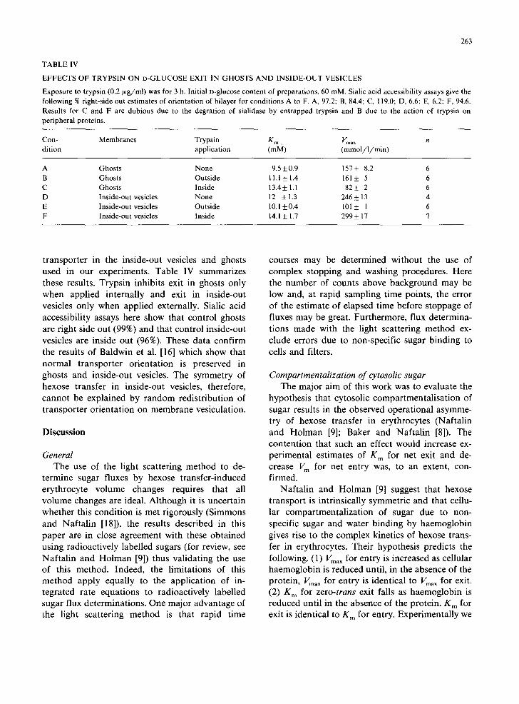
TABLE IV
EFFECTS OF TRYPSIN ON D-GLUCOSE EXIT IN GHOSTS AND INSIDE-OUT VESICLES
Exposure to trypsin (0.2/~g/ml) was for 3 h. Initial n-glucose content of preparations, 60 raM. Sialic acid accessibility assays give the following % right-side out estimates of orientation of bilayer for conditions A to F. A, 97.2; B, 84.4; C, 119.0; D, 6.6; E, 6.2; F, 94.6. Results for C and F are dubious due to the degration of sialidase by entrapped trypsin and B due to the action of trypsin on peripheral proteins.
Con- Membranes Trypsin K m • Vma x rt dition application (raM) (mmol / l /min )
A Ghosts None 9.5 + 0.9 157 + 8.2 6 B Ghosts Outside 11.1 + 1.4 161 + 5 6 C Ghosts Inside 13.4+ 1.1 82+_ 2 6 D Inside-out vesicles None 12 + 1.3 246_+ 13 4 E Inside-out vesicles Outside 10.1 +0.4 101 _+ I 6 F Inside-out vesicles Inside 14.1 + 1.7 299 + 17 7
transporter in the inside-out vesicles and ghosts used in our experiments. Table IV summarizes these results. Trypsin inhibits exit in ghosts only when applied internally and exit in inside-out vesicles only when applied externally. Sialic acid accessibility assays here show that control ghosts are right side out (99%) and that control inside-out vesicles are inside out (96%). These data confirm the results of Baldwin et al. [16] which show that normal transporter orientation is preserved in ghosts and inside-out vesicles. The symmetry of hexose transfer in inside-out vesicles, therefore, cannot be explained by random redistribution of transporter orientation on membrane vesiculation.
Discussion
General The use of the light scattering method to de-
termine sugar fluxes by hexose transfer-induced erythrocyte volume changes requires that all volume changes are ideal. Although it is uncertain whether this condition is met rigorously (Simmons and Naftalin [18]), the results described in this paper are in close agreement with these obtained using radioactively labelled sugars (for review, see Naftalin and Holman [9]) thus validating the use of this method. Indeed, the limitations of this method apply equally to the application of in- tegrated rate equations to radioactively labelled sugar flux determinations. One major advantage of the light scattering method is that rapid time
courses may be determined without the use of complex stopping and washing procedures. Here the number of counts above background may be low and, at rapid sampling time points, the error of the estimate of elapsed time before stoppage of fluxes may be great. Furthermore, flux determina- tions made with the light scattering method ex- clude errors due to non-specific sugar binding to cells and filters.
Compartmentalization of cytosolic sugar The major aim of this work was to evaluate the
hypothesis that cytosolic compartmentalisation of sugar results in the observed operational asymme- try of hexose transfer in erythrocytes (Naftalin and Holman [9]; Baker and Naftalin [8]). The contention that such an effect would increase ex- perimental estimates of K m for net exit and de- crease V m for net entry was, to an extent, con- firmed.
Naftalin and Holman [9] suggest that hexose transport is intrinsically symmetric and that cellu- lar compartmentalization of sugar due to non- specific sugar and water binding by haemoglobin gives rise to the complex kinetics of hexose trans- fer in erythrocytes. Their hypothesis predicts the following. (1) Vma x for entry is increased as cellular haemoglobin is reduced until, in the absence of the protein, Vma x for entry is identical to Vma x for exit. (2) K m for zero-trans exit falls as haemoglobin is reduced until in the absence of the protein, K m for exit is identical to K m for entry. Experimentally we
2 6 4
observe these effects. As the ghosts used in our experiments contained considerable amounts of haemoglobin, the incomplete reduction in asym- metry for transfer from approx. 10 to 3.6 might be due to incomplete haemoglobin removal.
A number of interesting findings, however, argue against the haemoglobin hypothesis. (1) Cel- lular solute (including haemoglobin), when applied to the exterior of ghosts or to the interior of inside-out vesicles is without effect on the K m for influx (efflux in inside-out vesicles). Yet the cellu- lar solute affects markedly both the K m for exit in ghosts and K m for exit in inside-out vesicles when applied to the cytoplasmic surface of the bilayer. That such an effect shows a sidedness (preference for the cytoplasmic surface of the bilayer) argues strongly against a simple interaction between sugar and haemoglobin. (2) Exposure of the normally exofacial surface of the bilayer to cellular solute inhibits transport by reducing the Vma x for exit in ghosts and inside-out vesicles. This effect is abolished by dialysing the cellular contents against lysis medium. As haemoglobin is still present fol- lowing dialysis, we consider that the protein is not responsible for the modulation of transport and that another, much lower molecular weight species somehow regulates or modifies the operational kinetics of hexose-transfer in red cells.
The symmetric transfer hypothesis has also been examined by Challis et al. [19]. These workers found that approx. 95% depletion of cytosolic haemoglobin is without significant effect on the kinetics of D-glucose exit from pink ghosts. These data are equivalent to the first few dilution condi- tions shown in here in Fig. 5. Vma x for exit remains unchanged by dilution, but K m for net exit falls from 32.4 + 3.7 mM in intact cells to 25.7 _+ 2.1 mM in ghosts containing a × 17 dilution of haemoglobin. These differences are small and may account for the apparent rejection of the symmet- ric transfer hypothesis by these workers [19]. Clearly, further dilution of cellular contents is required to observe the effects we describe here. In any case, our findings with inside-out vesicles confirm those of Taverna and Langdon [20] which suggest strongly that hexose transfer in these in- verted membrane vesicles is symmetric. This does not arise from the suggested [19] extensive leaki- ness of these preparations, for in our hands sugar
exit in inside-out vesicles is 98% inhibited by saturating (5/~M) levels of cytochalasin B.
The hexose-transfer reconstitution experiments of Wheeler and Hinkle [21] show that trypsin treatment of the reconstituted system restores hexose transfer asymmetry. These data are in ap- parent contradiction to our findings which argue for symmetry of hexose transfer in the native membrane. It is possible that these differences may result from isolation/purification and re- constitution procedures.
Models for hexose transfer in the erythrocyte Generally, models for erythrocyte hexose trans-
fer have assumed that the operational kinetics of transfer reflect the intrinsic properties of the trans- port mechanism. As such, these models have included asymmetric forms of the simple symmet- ric mobile carrier of Widdas [22], (Regen and Morgan [23]) and models in which the transporter contains two or more asymmetric binding sites exposed simultaneously at opposite sides of the membrane (Baker and Widdas [24]; Lieb and Stein [25]; Baker and Carruthers [26], Holman [27]). The observations we have made with inside-out vesicles in the absence of extravesicular cellular solute show that the K m for entry and exit from inside-out vesicles are almost identical. As such we consider hexose transfer in vesicles to be mediated by a symmetric process. We further suggest that our data are consistent with a transfer system in which the kinetics of transport may be regulated by cytosolic factors which confer asymmetry on the system. Thus in the absence of these factors, trans- fer is symmetric. In presence of these cytosolic factors the K m for exit increases, K m and Vma X for entry fall and Vma x for exit remains unchanged thus establishing transfer asymmetry. Thus all asymmetric forms of mobile carrier and multi-site models for human erythrocyte hexose transfer must be rejected. Here we will consider the symmetric mobile carrier and two component carrier models for hexose transfer.
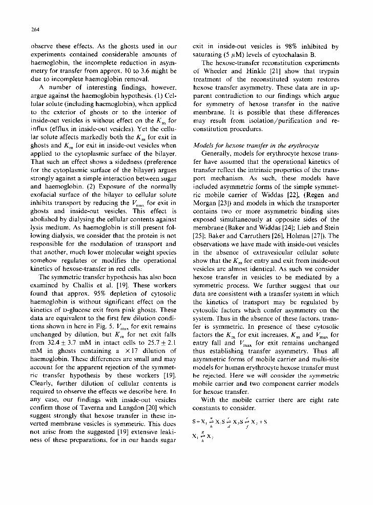
With the mobile carrier there are eight rate constants to consider.
s+x , ~ x~s ~ x2s ~ x 2 +s h d /
g X I ~ X 2
h
Where X is carrier and S is sugar. K m for flux from side 1 to 2 is:
(h + g)X ((e x(b + c))+(b x d) K'~2- ax((hx(d+e))+(c+(e+h)))
and Vma x for flux from 1 to 2 is
[ 1 1 1 + d
where aceh = bdfg and where n is a constant pro- portional to the membrane carrier density (Lieb and Stein [28]). Flux in the opposite direction is obtained by interchanging a ~ b, c ~ d, e ~ f and g ~ h. We have found that it is impossible to mimic our experimental findings without modify- ing individually the catalytic rate constants c, d, g, and h. Here the exchange flux of sugars would differ significantly from those reported experimen- tally (Naftalin and Holman [9]). We must, there- fore, reject this model.
Widdas [1] has suggested that the complex kinetics of erythrocyte hexose transfer may arise from substrate-induced redistribution of mobile carrier between both faces of the membrane. Thus the presence of a non-transportable competitive transport inhibitor in the cytoplasm would force a redistribution of carrier leading to the accumula- tion of carrier at the inner surface of the bilayer. This would reduce Vma x for sugar uptake and increase the K m for sugar exit leaving Vma x for exit unchanged. The observed decrease in K m for up- take by cytosolic factor is, however, incompatible with this hypothesis.
With the simple symmetric two component car- rier model we may suppose that the carrier is in rapid equilibrium with cytosolic factor X, internal and external sugar. The presence of X increases the K m for exit from K l t o K 2 leaving Vma x (VI) unchanged. The cytosolic factor X also reduces the K m for entry from K 3 to K 4 and Vma x for entry from V l to V z. If K I = K 3 then it can be shown that in the absence of X, hexose transfer is sym- metric and in the presence of saturating concentra- tions of X, hexose transfer is asymmetric. The asymmetry factor 0 = K 2 / K 4 = V I / V 2. This sim- ple model accounts readily for the effects of cyto- solic dilution on zero-trans sugar fluxes but fails to predict the lack of effect cytosolic dilution on the
265
g m a t the trans surface in infinite-cis flux de- terminations. To account for this effect we must introduce a further condition which states that occupancy of the cis-binding site of the carrier protects the trans-site from modulation by cyto- solic factor X without affecting the modulation of cis-site kinetics by X.
Due to its complexity such a kinetic formalism is useful only in a descriptive sense. Nevertheless the implication is that hexose transfer protein sugar binding sites are under allosteric control by both cytosolic factors and trans-sugar, possibly through substrate-induced conformational changes.
Conclusions
We believe that these data are consistent with the view of Naftalin and Holman [9] and Baker and Naftalin [8] that hexose transfer in human erythrocytes is mediated by an intrinsically sym- metric process. Nevertheless we have shown that this is unlikely to arise from interaction of sugar with haemoglobin. Rather, low molecular weight factors are present in the erythrocyte sol which confer asymmetry on the transport system perhaps by some allosteric interaction with the transport protein(s). This interaction is reflected operation- ally as decreased Vma x and K m for entry and increased K m for exit.
These findings are interesting for a number of reasons. (1) They constitute the first demonstra- tion of hexose transfer regulation in the human erythrocyte. (2) If similar factors are present in the cytosol of other cell types, they may provide a basis for hexose-transfer regulation. For example, the acceleration of hexose-transfer in muscle and adipocytes by insulin (Clausen [30]; Czech [31]; Baker and Carruthers [32]) could be brought about by switching transfer from the inhibited state (in- hibited by these factors) to the uninhibited state (inhibition of allosteric control of transfer by these factors). However, in the absence of experimental evidence, further speculation along these lines is unwarranted.
Acknowledgements
We would like to thank C. Rodgers and N. Frost for their excellent work in the design and manufacture of the rapid stop-flow apparatus used
266
in this work. A.C. gratefully acknowledges the award of a Welcome Trust Travel Grant. D.L.M. is recipient of an American Diabetes Association, Inc. Research and Development Award. The authors wish to thank the Juvenile Diabetes Foundation and the American Diabetes Associa- tion, Inc. for funding this work.
References
1 Widdas, W.F. (1980) Curr. Top. Membranes Transp. 14, 165-224
2 Geck, P. (1971) Biochim. Biophys. Acta 241,462-472 3 Regen, D.M. and Tarpley, H.L. (1974) Biochim. Biophys.
Acta 339, 218-233 4 Baker, G.K. and Widdas, W.F. (1978) J. Physiol. 278,
377-388 5 Hankin, B.C., Lieb, W.R. and Stein, W.D. (1972) Biochim.
Biophys. Acta 288, 114-126 6 Foster, D.M., Jacquez, J.A., Lieb, W.R. and Stein, W.D.
(1979) Biochim. Biophys. Acta 555, 349-351 7 Ginsburg, H. and Stein, W.D. (1975) Biochim. Biophys.
Acta 382, 353-368 8 Baker, G.K. and Naftalin, R.J. (1979) Biochim. Biophys.
Acta 550, 474-484 9 Naftalin, R.J. and Holman, G.D. (1979) in Membrane
Transport in Red Cells (Ellory, J.C. and Lew, V.C., eds.), Academic Press, New York
10 Sen, A.L. and Widdas, W.F. (1962) J. Physiol. 160, 392-403 11 Lew, V.C., Muallem, S. and Seymour, C.L. (1980) J. Phys-
iol. 307, 36P 12 Steck, T.L. and Kant, J.A. (1974) Methods Enzymol. 31,
172-180
13 Warren, L. (1959) J. Biol. Chem. 234, 1971-1775 14 Miller, D.M. (1966) Biophys. J. 5, 407-415 15 Karlish, S.J.D., Lieb, W.R., Ram, D. and Stein, W.D.
(1972) Biochim. Biophys. Acta 255, 126-132 16 Baldwin, J.M., Lienhard, G.E. and Baldwin, S.A. (1980)
Biochim. Biophys. Acta 599, 699-714 17 Masaik, S.J. and LeFevre, P.G. (1977) Biochim. Biophys.
Acta 465, 371-377 18 Simmons, N.L. and Naftalin, R.J. (1976) Biochim. Biophys.
Acta 419, 493-511 19 Challiss, J.R.A., Taylor, L.P. and Holman, G.D. (1980)
Biochim. Biophys. Acta 602, 155-166 20 Taverna, R.D. and Langdon, R.G. (1973) Biochim. Bio-
phys. Acta 323, 207-219 21 Wheeler, T.S. and Hinkle, P.C. (1981) J. Biol. Chem. 256,
8907-8914 22 Widdas, W.F. (1952) J. Physiol. 118, 23-39 23 Regen, D.M. and Morgan, H.E. (1964) Biochim. Biophys.
Acta 79, 151-166 24 Baker, G.F. and Widdas, W.F. (1973) J. Physiol. 231,
143-165 25 Lieb, W.R. and Stein, W.D. (1970) Biophys. J. 10, 585-605 26 Baker, P.F. and Carruthers, A. (1981) J. Physiol. 316,
503-525 27 Holman, G.D. (1980) Biochim. Biophys. Acta 599, 202-213 28 Lieb, W.R. and Stein, W.D. (1974) Biochim. Biophys. Acta
373, 178-196 29 Bloch, R. (1973) Biochemistry 12, 4799-4801 30 Clausen, T. (1977) FEBS Symposium, No. 42 (Semenza, G.
and Cavaboli, E., eds.), Springer Verlag, New York 31 Czech, M.P. (1977) Annu. Rev. Biochem. 46, 359-384 32 Baker, P.F. and Carruthers, A. (1983) J. Physiol. 336,
397-431