Reactions of nitrite in erythrocyte suspensions measured by membrane inlet mass spectrometry
18
Reactions of Nitrite in Erythrocyte Suspensions Measured by Membrane Inlet Mass Spectrometry Rose Mikulski a , Chingkuang Tu a , Erik R. Swenson b , and David N. Silverman a,* a Department of Pharmacology and Therapeutics, College of Medicine, University of Florida, Gainesville, FL 32610, USA b Departments of Medicine and Physiology, Pulmonary Section, University of Washington, Seattle, WA 98108 Abstract The reactions of nitrite with deoxygenated human erythrocytes were examined using membrane inlet mass spectrometry to detect the accumulation of NO in extracellular solution. In this method an inlet utilizing a silicon rubber membrane is submerged in cell suspensions and allows NO to pass from extracellular solution into the mass spectrometer. This provides a direct, continuous, and quantitative determination of nitric oxide concentrations over long periods without the necessity of purging the suspension with inert gas. We have not observed accumulation of NO when compared with controls on a physiologically relevant time scale and conclude that, within the limitations of the mass spectrometric method and our experimental conditions, erythrocytes do not generate a net efflux of NO following the addition of millimolar concentrations of nitrite. Moreover, there was no evidence at the mass spectrometer of the accumulation of a peak at mass 76 that would indicate N 2 O 3 , an intermediate that decays into NO and NO 2 . Inhibition of red cell membrane anion exchangers and aquaporins did not affect these processes. Keywords nitric oxide; nitrite; mass spectrometry; membrane inlet; erythrocytes; hemoglobin Introduction There has been great interest in the reactions of nitrite with erythrocytes following the initial reports of the reactions of nitrite with cell-free deoxyHb(Fe II ) [1]. These reactions are based on the well known one-electron reduction of nitrite by deoxyHb(Fe II ) resulting in the formation of metHb(Fe III ). The fate of the resulting NO has been heavily studied, but it is certain that most of it binds to deoxyHb(Fe II ). (1) © 2009 Elsevier Inc. All rights reserved. *Corresponding author: D. N. Silverman, Box 100267 Health Center, University of Florida, Gainesville, FL 32610-0267, USA. Fax: 352 392-9696. [email protected]. Publisher's Disclaimer: This is a PDF file of an unedited manuscript that has been accepted for publication. As a service to our customers we are providing this early version of the manuscript. The manuscript will undergo copyediting, typesetting, and review of the resulting proof before it is published in its final citable form. Please note that during the production process errors may be discovered which could affect the content, and all legal disclaimers that apply to the journal pertain. NIH Public Access Author Manuscript Free Radic Biol Med. Author manuscript; available in PMC 2011 January 15. Published in final edited form as: Free Radic Biol Med. 2010 January 15; 48(2): 325. doi:10.1016/j.freeradbiomed.2009.11.003. NIH-PA Author Manuscript NIH-PA Author Manuscript NIH-PA Author Manuscript
Transcript of Reactions of nitrite in erythrocyte suspensions measured by membrane inlet mass spectrometry
Reactions of Nitrite in Erythrocyte Suspensions Measured byMembrane Inlet Mass Spectrometry
Rose Mikulskia, Chingkuang Tua, Erik R. Swensonb, and David N. Silvermana,*aDepartment of Pharmacology and Therapeutics, College of Medicine, University of Florida,Gainesville, FL 32610, USAbDepartments of Medicine and Physiology, Pulmonary Section, University of Washington, Seattle,WA 98108
AbstractThe reactions of nitrite with deoxygenated human erythrocytes were examined using membrane inletmass spectrometry to detect the accumulation of NO in extracellular solution. In this method an inletutilizing a silicon rubber membrane is submerged in cell suspensions and allows NO to pass fromextracellular solution into the mass spectrometer. This provides a direct, continuous, and quantitativedetermination of nitric oxide concentrations over long periods without the necessity of purging thesuspension with inert gas. We have not observed accumulation of NO when compared with controlson a physiologically relevant time scale and conclude that, within the limitations of the massspectrometric method and our experimental conditions, erythrocytes do not generate a net efflux ofNO following the addition of millimolar concentrations of nitrite. Moreover, there was no evidenceat the mass spectrometer of the accumulation of a peak at mass 76 that would indicate N2O3, anintermediate that decays into NO and NO2. Inhibition of red cell membrane anion exchangers andaquaporins did not affect these processes.
Keywordsnitric oxide; nitrite; mass spectrometry; membrane inlet; erythrocytes; hemoglobin
IntroductionThere has been great interest in the reactions of nitrite with erythrocytes following the initialreports of the reactions of nitrite with cell-free deoxyHb(FeII) [1]. These reactions are basedon the well known one-electron reduction of nitrite by deoxyHb(FeII) resulting in the formationof metHb(FeIII). The fate of the resulting NO has been heavily studied, but it is certain thatmost of it binds to deoxyHb(FeII).
(1)
© 2009 Elsevier Inc. All rights reserved.*Corresponding author: D. N. Silverman, Box 100267 Health Center, University of Florida, Gainesville, FL 32610-0267, USA. Fax: 352392-9696. [email protected]'s Disclaimer: This is a PDF file of an unedited manuscript that has been accepted for publication. As a service to our customerswe are providing this early version of the manuscript. The manuscript will undergo copyediting, typesetting, and review of the resultingproof before it is published in its final citable form. Please note that during the production process errors may be discovered which couldaffect the content, and all legal disclaimers that apply to the journal pertain.
NIH Public AccessAuthor ManuscriptFree Radic Biol Med. Author manuscript; available in PMC 2011 January 15.
Published in final edited form as:Free Radic Biol Med. 2010 January 15; 48(2): 325. doi:10.1016/j.freeradbiomed.2009.11.003.
NIH
-PA Author Manuscript
NIH
-PA Author Manuscript
NIH
-PA Author Manuscript

(2)
The reduction of nitrite by deoxyHb(FeII) is a complex reaction the properties of which dependon the ratio of nitrite to hemoglobin, showing autocatalytic behavior and ratios of NOHb(FeII) to metHb(FeIII) that deviate from unity under specific and well described conditions[2–4]. The ratio of products, NOHbFe(II) and metHbFe(III), depends on the concentration ofnitrite. Doyle et al. [1] used excess nitrite and reported close to one equivalent each of NOHbFe(II) and metHbFe(III), confirmed in subsequent studies under specific conditions describedtherein [5,6]. However, as concentrations of nitrite decreases below about 400 µM there is anexcess of met-Fe(III) over NO-Fe(II), with the ratio Fe(III)/NO-Fe(II) as great as 1.8, whichthen approaches unity again at nitrite concentrations very low compared with heme [2]. Weemphasize that the reactions of nitrite with hemoglobin in this report are determined at highnitrite concentrations, much higher than under physiological conditions. To further emphasizethis, it was reported earlier that infusion of low concentrations of nitrite into the forearm causedincreased blood flow [7]. Subsequent studies, however, show that the extent and mechanismof nitrite bioactivity is controversial [8–10].
Based on studies showing vasoactivity of nitrite under hypoxic conditions, increasedconsumption of nitrite during normoxic exercise, and positive arterial-to-venous gradients ofnitrite, it has been suggested that the interactions of nitrite with erythrocytes could be a sourceof vasodilatory NO [4,7,11,12]. Prominent among explanations of the vasodilatory effect ofnitrite is the hypothesis that deoxyHb(FeII) in erythrocytes produces vasodilation throughmechanisms related to the reactions of nitrite with deoxyHb(FeII) [6,11,13]. Furthermore, ithas been proposed that the red cell membrane anion exchanger (AE-1) may help to mobilizeextracellular nitrite for intracellular utlilization [3] and that aquaporin-1 may act as a membranegas channel for NO permeation [14].
A significant problem with this scenario is that deoxyHb(FeII) avidly binds NO, and thisautocapture is expected to prevent appreciable NO from exiting the erythrocyte. This effectmay be mitigated by generation of an intermediate such as N2O3 which does not bind tightlyto deoxyHb(FeII) and could pass across the red cell membrane [13]. However, massspectrometric measurements showed no accumulation of N2O3 upon addition of nitrite tosolutions of cell-free deoxyHb(FeII), and no NO above that of the uncatalyzed dismutation[15]. Additional data are accumulating that the vasodilatory effect of nitrite in vivo arises fromnitrite reduction by the vascular endothelium and extravascular tissues. For example, studiesdetecting NO by EPR and chemiluminescence showed that nitrite caused appreciable increasesin NO production in tissues such as liver and heart but only trace amounts in blood [16]. Inaddition, reactions of nitrite to generate NO under anerobic conditions (in vitro) have beendemonstrated for myoglobin, xanthine oxidase, the bc1 complex of the mitochondrial electrontransport chain, cytochrome P450, and endothelial NO synthase enzyme [17]. Other studieshave suggested a role of S-nitrosothiols as a source of vasodilation, since potent S-nitrosatingagents including N2O3 may be formed from nitrite [11]. Physiological concentrations of nitritein humans are in a range up to 1 µM. The source of nitrite includes foods, the reactions ofnitrate in bacteria in the oral cavity and GI tract, and eNOS-mediated NO production withsubsequent oxidation which accounts for up to 70% of plasma nitrite [18].
In this report, we examine the reactions of nitrite with deoxygenated human erythrocytes usingmembrane inlet mass spectrometry (MIMS) to detect the accumulation of NO in extracellularsolution [19]. This follows a study using the same methods applied to cell-free deoxyHb(FeII) [15]. In this method an inlet utilizing a silicon rubber membrane is submerged in cellsuspensions and allows NO to pass from extracellular solution into the mass spectrometer. This
Mikulski et al. Page 2
Free Radic Biol Med. Author manuscript; available in PMC 2011 January 15.
NIH
-PA Author Manuscript
NIH
-PA Author Manuscript
NIH
-PA Author Manuscript
provides a direct, continuous, and quantitative determination of nitric oxide concentrationsover long periods without the necessity of purging the suspension with inert gas. We have notobserved accumulation of NO when compared with controls on a physiologically relevant timescale and conclude that, within the limitations of the mass spectrometric method and ourexperimental conditions, erythrocytes do not generate a net efflux of NO following the additionof millimolar concentrations of nitrite. Moreover, inhibition of red cell membrane anionexchangers and aquaporins does not affect these processes.
Experimental MethodsMaterials
Human blood was freshly collected and then diluted in isotonic buffer consisting of 50 mMsodium phosphate, 78 mM sodium chloride, 2.7 mM potassium chloride at pH 7.4. Cells werewashed by repeated cycles of centrifugation and dilution in isotonic buffer prior to use. Thewashed packed red cells were then diluted into the same isotonic buffer described above at pH6.7 supplemented with 2 mM of the chelator DTPA (diethylenetriamine-pentaacetic acid)within the reaction vessel. Experiments used 2.5 ml of red cells suspensions to which wasadded 0.5 µL of Antifoam A (Sigma). Degassed red cells were prepared from these samplesby bubbling of helium for 5 minutes prior to the analysis.
The anion exchange inhibitors 4-acetamido-4’-isothiocyanstilbene-2,2’-disulfonic aciddisodium salt (SITS), and 4,4'-diisothiocyano-stilbene-2,2'-disulfonic acid (DIDS) werepurchased from AnaSpec (San Jose, CA). The inhibitor 4,4'-dinitrostilbene-2,2'-disulfonicacid, disodium salt (DNDS) was purchased from Invitrogen Corporation (Carlsbad, CA). Theaquaporin channel blocker para-chloromercuribenzene sulfonic acid, sodium salt (PCMBS)was purchased from Toronto Research Chemicals, Inc. DIDS, SITS, and DNDS were incubatedin red cell suspensions up to 30 minutes before experiments. Red cells were incubated withPCMBS for 10 minutes prior to experiments.
Inlet probe and kinetic measurementsThe membrane inlet consisted of a length of silicon rubber (Silastic) tubing attached to a pieceof glass tubing leading into a dry-ice acetone water trap and then into an Extrel EXM-200 massspectrometer, as described elsewhere [19]. This membrane inlet was immersed in solutioncontained in a 3 ml glass reaction vessel modified for introduction of samples and inert gas,and sealable by injection septa and Teflon screw plugs [19]. When immersed in a red cellsuspension, this inlet detects free, unbound, extracellular NO based on the magnitude of them/z 30 peak (or N2O3 based on the m/z 76 peak if this species is present). The reaction vesselcontained a small magnetic spinner to continuously mix the reactions. Mass spectra wereobtained using electron impact ionization (70 eV) at an emission current of 1 mA. Sourcepressures were approximately 1 × 10⁻⁶ torr. The resulting mass scans were well resolved witha return of ion current (detector response) to the baseline separating each mass unit. Datacollected using the membrane inlet were calibrated by injecting solutions containing knownconcentrations of NO into buffered solutions in the reaction vessel [19]. The sample preparationinvolved deoxygenating the red cell suspension with helium, as described above, followed byaddition of nitrite through an injection septum.
ResultsWe have used membrane inlet mass spectrometry to observe the accumulation of free, unbound,extracellular NO caused by the addition of nitrite to deoxygenated suspensions of human redblood cells. In this method, the membrane inlet is submerged in the suspension and unchargedmolecules of low molecular weight pass across a permeable membrane into the mass
Mikulski et al. Page 3
Free Radic Biol Med. Author manuscript; available in PMC 2011 January 15.
NIH
-PA Author Manuscript
NIH
-PA Author Manuscript
NIH
-PA Author Manuscript

spectrometer. Inhibitors to block both anion exchange proteins and aquaporin were used toinvestigate mechanisms.
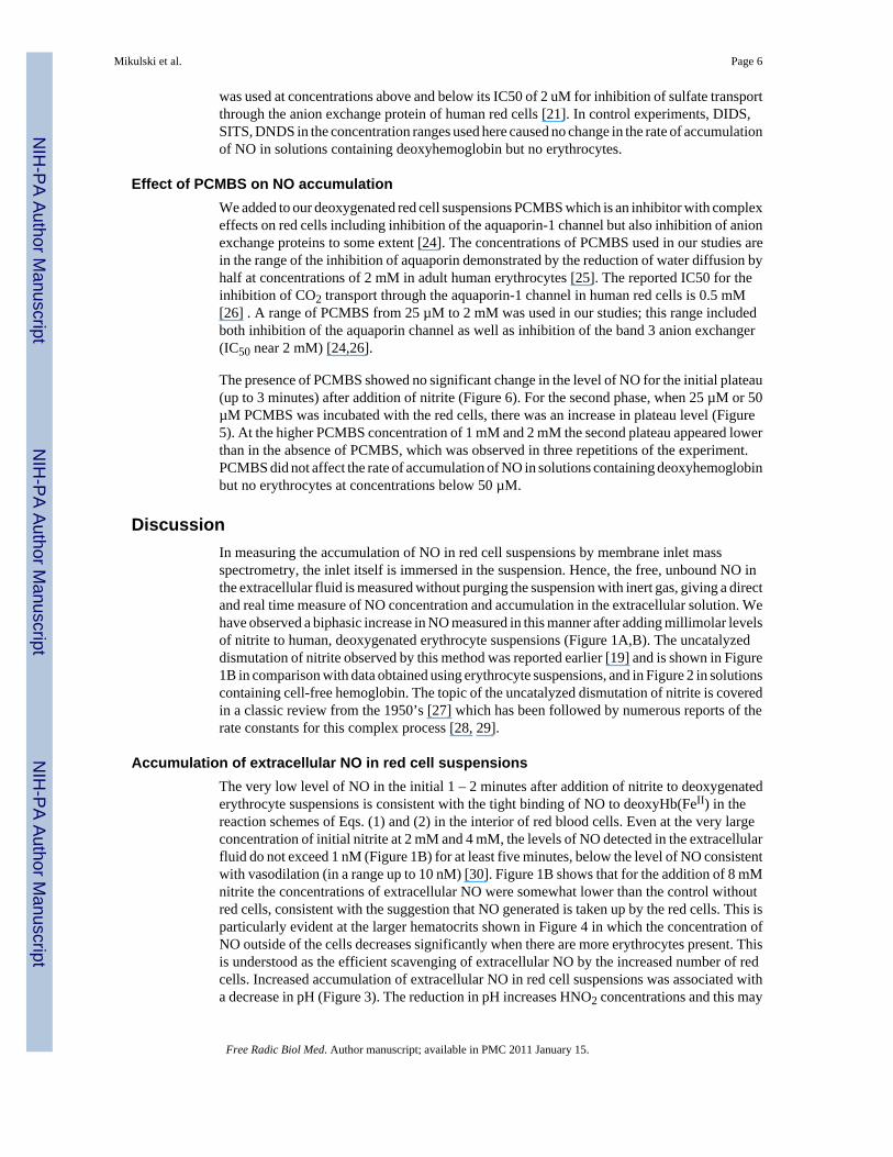
The addition of nitrite to a suspension of red cellsWe added nitrite at initial concentrations of 2 – 16 mM to a suspension of deoxygenated, humanred cells (0.8% hematocrit) and used the m/z 30 peak to detect extracellular NO. The appearanceof NO after the addition of nitrite at time zero was characterized by two phases (Figures 1A,B).Each of the two phases shows an increase in NO approaching a plateau, and the second phasefollows generally after the first phase has reached the plateau. For example, for the addition attime zero of 8 mM nitrite, the first phase extends from time zero to about 2.5 min, the secondphase begins about 2.5 minutes (Figures 1A,B).
The first phase showed accumulation of extracellular NO forming a low-level plateau near 1.3nM, for an initial nitrite concentration of 8.0 mM, with the plateau lasting over one minute(Figure 1B). This initial plateau was slightly less than about 2 nM formed in the uncatalyzeddismutation of an initial concentration of 8.0 mM (Figure 1B). The length of the initial plateaudecreased with increasing nitrite concentration. These data can be compared with theaccumulation of NO in solutions of cell-free hemoglobin (Figure 2) [15]. It is difficult tocompare these results from MIMS with studies which purge red cell suspensions with inert gasand detect NO in the gas phase using chemiluminescence [7, 16]. These methods and conditionsare different and so are results; for example, in measuring uncatalyzed dismutation of nitrite,MIMS allows equilibrium to establish and chemiluminescence removes NO with the inert gasas it is formed.
After this initial steady state plateau, there was a second phase in which extracellular NOconcentrations increased and approached a higher plateau of NO concentration, one thatexceeded the concentration of NO in the uncatalyzed reaction. In this second phase, the rateof increase of NO and level of the apparent second plateau increased with increasingconcentrations of nitrite (Figure 1A). The data shown in Figure 1 are for human red cells thatwere washed three times in isotonic buffer; however, the results were identical when anequivalent amount of blood was used without prior washing of the red cells.
The following experiments were performed to determine whether the second phase was dueto dissociation of NO from NOHb(FeII). MAHMA NONOate (Z-1-[N-methyl-N-[6-(N-methylammoniohexyl)amino]]-diazen-1-ium-1,2-diolate) at 25 µM was used to generate NOin a solution containing 40 µM deoxyHb(FeII) (heme concentration). Other conditions were asdescribed in Figure 1 except no nitrite was added. The solution was monitoredspectrophotometrically to observe the formation of the apparently fully nitrosylatedhemoglobin. A stream of helium gas was used to remove unbound NO, monitored byobservation of the m/z 30 peak. When this peak was near baseline, indicating the near completeremoval of unbound NO, the helium stream was stopped. MIMS showed no increase in the m/z 30 peak over a period of 12 minutes. This is consistent with no dissociation of NO fromNOHb(FeII) that could be detected.
When the pH of the external solution was decreased, we observed a decrease in the length ofthe first plateau accompanied by an increase in the rate of appearance of extracellular NO andapparent plateau level for the second phase (Figure 3). A qualitatively similar effect was alsoobserved in solutions of cell-free deoxyhemoglobin [15] and is related to the concentration ofHNO2, the conjugate acid of nitrite with a pKa of 3.3. As with solutions of cell-freedeoxyhemoglobin [15], the logarithm of the rates of NO accumulation in the second phase wasa linear function of pH (R2 = 0.97) with rates increasing as pH decreases.
Mikulski et al. Page 4
Free Radic Biol Med. Author manuscript; available in PMC 2011 January 15.
NIH
-PA Author Manuscript
NIH
-PA Author Manuscript
NIH
-PA Author Manuscript
We extended these studies to suspensions of larger hematocrit using additions of 8 mM nitrite(Figure 4). Increasing the amount of deoxygenated erythrocytes increased the length of theinitial plateau; at the larger hematocrits (25%, 50%) this initial phase showed no accumulationof NO. Also, increasing the hematocrit decreased the rate of extracellular NO accumulationand the level of the plateau in the second phase (Figure 4). We did not observe in theseexperiments an increase in the peak at m/z 76 that would correspond to the accumulation ofN2O3. The limitations of MIMS to detect N2O3 was discussed in our earlier report on reactionsof nitrite with hemoglobin [15]. This species is expected to have a lifetime in solution near 1ms and to dissociate rapidly into NO and NO2 [20]. It is entirely possible that MIMS as usedin this work would not detect an initial burst of N2O3. However, if N2O3 is escaping the redcell and rapidly decomposes to NO either in the red cell suspension or in the gas phase leadingto the mass spectrometer, then we should be able to measure it by MIMS as NO. In this sense,it does not matter what species escapes the red cell as a molecule that decays to NO, we havenot been able to detect it.
The generation of NO in conditions closer to (but clearly not) physiological was then examined.We used human, deoxygenated erythrocyte suspensions at 50% hematocrit in isotonic bufferat pH 7.4 and 37 °C. The suspension was then injected with an initial 1.0 mM nitriteconcentration and monitored for NO generation extracellularly. These conditions did notgenerate any detectable NO for up to 30 minutes following the addition of the nitrite.
Additional experiments were performed at varying levels of oxygenation under the conditionsof Figure 1. Mixing oxygenated and deoxygenated packed red cells in different ratios was usedto produce different levels of oxygenation. Completely oxygenated red cell suspensionsshowed no accumulation of NO upon addition of nitrite. Intermediate levels of oxygenationshowed the two-stage accumulation of NO as in Figure 1 with NO concentrations which wereintermediate between those observed for completely deoxygenated and oxygenated red cells.Further experiments were performed to force the transition between the relaxed state with highoxygen affinity and the tense state favoring deoxyhemoglobin in the red cell. The red cellswere oxygenated and then injected into doxygenated buffer in the reaction vessel (conditionsof Figure 1); at various times (up to 5 minutes) nitrite was added. The release of oxygen andaccumulation of NO was measured in the mass spectrometer. There was no indication of NOreleased in a burst following nitrite addition, instead there was a slow increase of NO, muchless than in the experiments using deoxygenated cells and on a slower time scale. When air oroxygen was bubbled into the reaction of completely deoxygenated red cells under conditionsof Figure 1 it resulted in a similar decreased generation of NO.
Effect of DIDS on NO accumulationWe tested several inhibitors of the band 3 anion exchanger (AE1) for their effect on theaccumulation of extracellular NO after introduction of nitrite to deoxygenated red cells.Concentrations of DIDS from 0.1 µM to 1.0 mM had no effect on the accumulation of NO inextracellular fluid, shown for four concentrations of DIDS in Figure 5. An apparent IC50 of0.1 µM DIDS was reported for the inhibition of the transport of sulfate by the anion exchangersin human red cells [21]. Results identical to Figure 5 were obtained when experiments wereperformed in the dark to prevent light induced isomerization of DIDS [22]. In additionalexperiments, we used solutions of DIDS containing potassium bicarbonate (0.1 mM) or DMSO(5% in final suspensions) to prevent the formation of polymeric DIDS [15]; results wereidentical to Figure 5.
Experiments under the same conditions using the anion exchange inhibitors SITS and DNDSalso showed no effect in the accumulation of NO (data not shown). SITS was examined in theconcentration range from 10 µM to 100 µM. An IC 50 of 50 µM for SITS is reported for theinhibition of the transport of sulfate by the anion exchangers in human red cells [23]. DNDS
Mikulski et al. Page 5
Free Radic Biol Med. Author manuscript; available in PMC 2011 January 15.
NIH
-PA Author Manuscript
NIH
-PA Author Manuscript
NIH
-PA Author Manuscript
was used at concentrations above and below its IC50 of 2 uM for inhibition of sulfate transportthrough the anion exchange protein of human red cells [21]. In control experiments, DIDS,SITS, DNDS in the concentration ranges used here caused no change in the rate of accumulationof NO in solutions containing deoxyhemoglobin but no erythrocytes.
Effect of PCMBS on NO accumulationWe added to our deoxygenated red cell suspensions PCMBS which is an inhibitor with complexeffects on red cells including inhibition of the aquaporin-1 channel but also inhibition of anionexchange proteins to some extent [24]. The concentrations of PCMBS used in our studies arein the range of the inhibition of aquaporin demonstrated by the reduction of water diffusion byhalf at concentrations of 2 mM in adult human erythrocytes [25]. The reported IC50 for theinhibition of CO2 transport through the aquaporin-1 channel in human red cells is 0.5 mM[26] . A range of PCMBS from 25 µM to 2 mM was used in our studies; this range includedboth inhibition of the aquaporin channel as well as inhibition of the band 3 anion exchanger(IC50 near 2 mM) [24,26].
The presence of PCMBS showed no significant change in the level of NO for the initial plateau(up to 3 minutes) after addition of nitrite (Figure 6). For the second phase, when 25 µM or 50µM PCMBS was incubated with the red cells, there was an increase in plateau level (Figure5). At the higher PCMBS concentration of 1 mM and 2 mM the second plateau appeared lowerthan in the absence of PCMBS, which was observed in three repetitions of the experiment.PCMBS did not affect the rate of accumulation of NO in solutions containing deoxyhemoglobinbut no erythrocytes at concentrations below 50 µM.
DiscussionIn measuring the accumulation of NO in red cell suspensions by membrane inlet massspectrometry, the inlet itself is immersed in the suspension. Hence, the free, unbound NO inthe extracellular fluid is measured without purging the suspension with inert gas, giving a directand real time measure of NO concentration and accumulation in the extracellular solution. Wehave observed a biphasic increase in NO measured in this manner after adding millimolar levelsof nitrite to human, deoxygenated erythrocyte suspensions (Figure 1A,B). The uncatalyzeddismutation of nitrite observed by this method was reported earlier [19] and is shown in Figure1B in comparison with data obtained using erythrocyte suspensions, and in Figure 2 in solutionscontaining cell-free hemoglobin. The topic of the uncatalyzed dismutation of nitrite is coveredin a classic review from the 1950’s [27] which has been followed by numerous reports of therate constants for this complex process [28, 29].
Accumulation of extracellular NO in red cell suspensionsThe very low level of NO in the initial 1 – 2 minutes after addition of nitrite to deoxygenatederythrocyte suspensions is consistent with the tight binding of NO to deoxyHb(FeII) in thereaction schemes of Eqs. (1) and (2) in the interior of red blood cells. Even at the very largeconcentration of initial nitrite at 2 mM and 4 mM, the levels of NO detected in the extracellularfluid do not exceed 1 nM (Figure 1B) for at least five minutes, below the level of NO consistentwith vasodilation (in a range up to 10 nM) [30]. Figure 1B shows that for the addition of 8 mMnitrite the concentrations of extracellular NO were somewhat lower than the control withoutred cells, consistent with the suggestion that NO generated is taken up by the red cells. This isparticularly evident at the larger hematocrits shown in Figure 4 in which the concentration ofNO outside of the cells decreases significantly when there are more erythrocytes present. Thisis understood as the efficient scavenging of extracellular NO by the increased number of redcells. Increased accumulation of extracellular NO in red cell suspensions was associated witha decrease in pH (Figure 3). The reduction in pH increases HNO2 concentrations and this may
Mikulski et al. Page 6
Free Radic Biol Med. Author manuscript; available in PMC 2011 January 15.
NIH
-PA Author Manuscript
NIH
-PA Author Manuscript
NIH
-PA Author Manuscript

be a predominant species entering the red cell to be reduced to NO. Moreover, the uncatalyzeddismutation of nitrite is more rapid at lower pH [15].
In these red cell suspensions, the low-level plateau of the first phase (up to 2–3 minutes)represents a complex steady state in extracellular NO concentration involving several processesincluding the uncatalyzed dismutation of nitrite, the flux of nitrite and HNO2 into the cellswhere it reacts with hemoglobin according to Eqs. (1) and (2), and the diffusion of NO acrossthe cell membrane. There are a number of other accompanying processes such as nitrosylationreactions, the possible role of other species of nitrogen oxides, other reactions of nitrite andNO in the cell, and the action of metHb(FeIII) reductase.
Nitrite concentrations used at 2 to 16 mM differ greatly from physiological concentrations ofnitrite, which are near 1 µM; however, the much larger concentrations of nitrite needed toobserve NO in red cell suspensions forms part of our conclusion. Addition of 1.0 mM nitriteunder more physiological conditions did not show any accumulation of NO that we couldmeasure. For the membrane inlet that we have used the half-time is near 5 seconds for responseto step increases in concentration of NO. We would not be able to observe a small, rapid pulseof NO increase occurring within seconds after the addition of nitrite.
Other cautions apply when interpreting our measurements. Detection of NO in our reactionvessel and experimental conditions does not reproduce conditions in an arteriole, in whichflowing red cells in plasma physiologically undergo a 25–75% reduction in hemoglobin-oxygen saturation with a simultaneous increase in the partial pressure of carbon dioxide (30 to50 mmHg) and fall in pH (7.5 to 7.3)1 with only a minor reduction of extracellular nitrite atconcentrations below 1 uM. Although our comparison of net NO accumulation withdeoxygenated cells and uncatalyzed dismutation (Figure 1B and 4) is a direct observation, NOaccumulation using deoxygenated compared with oxygenated red cells offers furtherinterpretation. The results with oxygenated and deoxygenated red cells were very similar tothose with cell free oxy- and deoxyHb(FeII) in Figure 2 where it is clear that initially NOaccumulation is greater with deoxyHb(FeII). The reaction of NO and oxyHb(FeII) is near 9 ×10⁷ M−1s−1 [31] and the value for the reaction of NO and deoxyHb(FeII) (Eq. (2)) is 2 × 10⁷
M−1s−1 [32]. These values are viewed as rather similar; hence, a major difference between oxyand deoxy conditions appears to be the nitrite reductase activity of deoxyHb(FeII). In thisinterpretation, the greater accumulation of NO under deoxy compared with oxy conditionssuggests efflux of NO from the cells. However, MIMS cannot detect the origin of the NOaccumulated outside the cells; a fraction may be generated by the uncatalyzed dismutation ofnitrite outside the cells and a fraction may be generated from the nitrite reductase activity ofdeoxyHb(FeII) with product NO that then exits the cell. However, direct observation shows nonet accumulation of NO in the initial phase outside the cells compared with uncatalyzed levelsof NO.
The second phase is an increase in the rate of extracellular NO accumulation that follows theinitial plateau and occurs many minutes after addition of nitrite to degassed erythrocytesuspensions (Figure 1A,B). The cause of this second phase of increase and apparent secondplateau is unclear; but it appears not due to the dissociation of NOHb(FeII). One possibility isthe reaction of nitrite with forms of nitrosylated hemoglobin. The resulting progress curve inthe second phase of the accumulation of NO in extracellular solution after addition of nitrite(Figure 1A) is very similar to that obtained by this method under similar conditions for solutionscontaining cell-free deoxyhemoglobin (Figure 2) [15]. Both experiments, using deoxygenated
1A limitation in the present experiments is the necessity for an absence of carbon dioxide and bicarbonate due to the generation in themass spectrometer of C18O (m/z 30) from the natural abundance of 18O masking the NO peak. We are currently exploring the use ofisotopic 15N-enriched nitrite to overcome this problem.
Mikulski et al. Page 7
Free Radic Biol Med. Author manuscript; available in PMC 2011 January 15.
NIH
-PA Author Manuscript
NIH
-PA Author Manuscript
NIH
-PA Author Manuscript

erythrocyte suspensions or solutions of cell-free deoxyHb(FeII), showed an initial phase of lowlevels of NO and a second phase of more rapid NO accumulation approaching a plateau.Moreover the time scales and magnitudes of NO observed are similar for the red cell andsolution phase studies with purified hemoglobin (Figure 1, Figure 2).
Inhibition of band 3 anion exchanger of red cellsThe exchange protein AE1 is a member of a family of anion exchangers that is located in theplasma membrane of erythrocytes as well as other tissues and is involved in the transport ofCO2 and bicarbonate in respiration [33,34]. Studies with intact red cells and ghosts show nitriteuptake blocked by DIDS, an inhibitor of anion exchange proteins including AE1 [35].However, other studies show no effect of DIDS on nitrite induced formation of met-Hb(FeIII) in erythrocytes [36,37]. There will be some diffusion of HNO2 (pKa 3.3) across themembrane, and studies indicate some uptake of nitrite by the sodium-dependent phosphatetransporter [37].
We observed no significant change caused by the inhibitors of anion exchange DIDS, SITS,or DNDS in the rate of extracellular NO accumulation in this work (shown for DIDS in Figure5). The disulfonic stilbene compounds including DIDS, SITS, and DNDS are a class of themost potent and commonly used inhibitors of the exchange protein AE1. These inhibitors inthe concentration range of their respective IC50 values had no effect on NO accumulationunder conditions of Figure 1. These results show that anion flux using the exchange proteinAE1 is not a predominant factor in the accumulation of extracellular NO caused by additionof large quantities of extracellular nitrite. The insensitivity to DIDS in Figure 5 indicates thatrate-limiting steps in the extracellular accumulation of NO do not involve AE1.
Inhibition of aquaporin-1 channel of red cellsBeing a small uncharged molecule, NO is expected to pass readily across membranes [38], andthere is evidence that some passes through the aquaporin channel [14]. Evidence that a largeamount of CO2 fluxes out of red cells through the aquaporin channel suggests that it may be aconduit for NO and other dissolved gases to enter or exit the red cell, although the significanceof this is uncertain [26].
PCMBS had no effect on the levels of extracellular NO in the initial plateau after addition ofnitrite (Figure 6). In addition, PCMBS at the concentrations of Figure 6 had no effect on theaccumulation of NO in solutions of cell-free deoxyhemoglobin (data not shown). These datashow that the aquaporin channel does not play a predominant role in the complex steady-stateprocesses that are involved in the initial response of red cells to large concentrations of nitrite.Rather, the steady state appears dominated by the flux of NO through other pathways and ofHNO2 followed by the autocapture of NO in the red cells. The complex behavior of PCMBStoward the second plateau in Figure 6 is unexplained at present. We can conclude however thatflux through the aquaporin channel is not a major factor for accumulation of extracellular NOin our experiments.
SummaryApplication of membrane inlet mass spectrometry showed no initial burst of extracellular NOor N2O3 caused by addition of millimolar levels of nitrite to suspensions of deoxygenated redcells at pH 6.7. With a sensitivity as low at 0.5 nM, this method detected no accumulation ofextracellular NO when 1.0 mM nitrite was added to red cells under more physiologicalconditions of hematocrit, pH, and temperature. These data support the hypothesis that, althoughthe reactions of nitrite with deoxyHb(FeII) apply, the autocapture of NO by deoxyHb(FeII)precludes efflux of NO. Inhibition of the anion exchange protein and the aquaporin channeldid not affect the accumulation of extracellular NO in these experiments; flux of nitrite and
Mikulski et al. Page 8
Free Radic Biol Med. Author manuscript; available in PMC 2011 January 15.
NIH
-PA Author Manuscript
NIH
-PA Author Manuscript
NIH
-PA Author Manuscript

NO across the membrane by these processes are not rate contributing towards appearance ofNO extracellularly.
AcknowledgmentsWe thank Dr. Bradley Fletcher for help with the experimental protocol. This work was supported in part by NIH grantGM25154 funds made available by the University of Florida.
Abbreviations
MIMS membrane inlet mass spectrometry
deoxyHb(FeII) the ferrous form of deoxyhemoglobin
NOHb(FeII) nitrosylated hemoglobin
m/z (m/e) is the mass-to-charge ratio detected with the mass spectrometer
AE1 anion exchanger-1
DTPA diethylenetriaminepentaacetic acid
SITS 4-acetamido-4’-isothiocyanstilbene-2,2’-disulfonic acid
DIDS 4,4'-diisothiocyano-stilbene-2,2'-disulfonic acid
DNDS 4,4'-dinitrostilbene-2,2'-disulfonic acid
pCMBS para-chloromercuribenzene sulfonate.
References1. Doyle MP, Pickering RA, DeWeert TM, Hoekstra JW, Pater D. Kinetics and mechanism of the
oxidation of human deoxyhemoglobin by nitrites. J Biol Chem 1981;256:12393–12398. [PubMed:7298665]
2. Angelo M, Singel DJ, Stamler JS. An S-nitrosothiol (SNO) synthase function of hemoglobin thatutilizes nitrite as a substrate. Proceedings of the National Academy of Sciences of the United Statesof America 2006;103:8366–8371. [PubMed: 16717191]
3. Huang Z, Shiva S, Kim-Shapiro DB, Patel RP, Ringwood LA, Irby CE, Huang KT, Ho C, Hogg N,Schechter AN, Gladwin MT. Enzymatic function of hemoglobin as a nitrite reductase that producesNO under allosteric control. J Clin Invest 2005;115:2099–2107. [PubMed: 16041407]
4. Kim-Shapiro DB, Gladwin MT, Patel RP, Hogg N. The reaction between nitrite and hemoglobin: therole of nitrite in hemoglobin-mediated hypoxic vasodilation. J Inorg Biochem 2005;99:237–246.[PubMed: 15598504]
5. Luchsinger BP, Rich EN, Yan Y, Williams EM, Stamler JS, Singel DJ. Assessments of the chemistryand vasodilatory activity of nitrite with hemoglobin under physiologically relevant conditions. Journalof Inorganic Biochemistry 2005;99:912–921. [PubMed: 15811508]
6. Huang Z, Shiva S, Kim-Shapiro D, Patel R, Ringwood L, Irby C, Huang K, Ho C, Hogg N, SchechterA, Gladwin M. Enzymatic function of hemoglobin as a nitrite reductase that produces NO underallosteric control. Journal of Clinical Investigation 2005;115:2099–2107. [PubMed: 16041407]
7. Cosby K, Partovi KS, Crawford JH, Patel RP, Reiter CD, Martyr S, Yang BK, Waclawiw MA, ZalosG, Xu XL, Huang KT, Shields H, Kim-Shapiro DB, Schechter AN, Cannon RO, Gladwin MT. Nitritereduction to nitric oxide by deoxyhemoglobin vasodilates the human circulation. Nature Medicine2003;9:1498–1505.
8. Deem S, Min JH, Moulding JD, Eveland R, Swenson ER. Red blood cells prevent inhibition of hypoxicpulmonary vasoconstriction by nitrite in isolated, perfused rat lungs. American Journal of Physiology-Heart and Circulatory Physiology 2007;292:H963–H970. [PubMed: 17012349]
Mikulski et al. Page 9
Free Radic Biol Med. Author manuscript; available in PMC 2011 January 15.
NIH
-PA Author Manuscript
NIH
-PA Author Manuscript
NIH
-PA Author Manuscript

9. Dalsgaard T, Simonsen U, Fago A. Nitrite-dependent vasodilation is facilitated by hypoxia and isindependent of known NO-generating nitrite reductase activities. American Journal of Physiology-Heart and Circulatory Physiology 2007;292:H3072–H3078. [PubMed: 17307993]
10. Hunter CJ, Dejam A, Blood AB, Shields H, Kim-Shapiro D, Machado RF, Tarekegn S, Mulla N,Hopper AO, Schechter AN, Power GG, Gladwin MT. Inhaled nebulized nitrite is a hypoxia-sensitiveNO-dependent selective pulmonary vasodilator. Nature Medicine 2004;10:1122–1127.
11. Singel DJ, Stamler JS. Chemical physiology of blood flow regulation by red blood cells: The role ofnitric oxide and S-nitrosohemoglobin. Annual Review of Physiology 2005;67:99–145.
12. Schechter AN, Gladwin MT. Hemoglobin and the paracrine and endocrine functions of nitric oxide.New England Journal of Medicine 2003;348:1483–1485. [PubMed: 12686706]
13. Basu S, Grubina R, Huang J, Conradie J, Huang Z, Jeffers A, Jiang A, He X, Azarov I, Seibert R,Mehta A, Patel R, King SB, Hogg N, Ghosh A, Gladwin MT, Kim-Shapiro DB. Catalytic generationof N2O3 by the concerted nitrite reductase and anhydrase activity of hemoglobin. Nature ChemicalBiology 2007;3:785–794.
14. Herrera M, Garvin JL. Novel role of AQP-1 in NO-dependent vasorelaxation. American journal ofphysiology. Renal physiology 2007;292:F1443. [PubMed: 17229677]
15. Tu CK, Mikulski R, Swenson ER, Silverman DN. Reactions of nitrite with hemoglobin measured bymembrane inlet mass spectrometry. Free Radical Biology & Medicine 2009;46:14–19. [PubMed:18848984]
16. Li HT, Cui HM, Kundu TK, Alzawahra W, Zweier JL. Nitric oxide production from nitrite occursprimarily in tissues not in the blood - Critical role of xanthine oxidase and aldehyde oxidase. Journalof Biological Chemistry 2008;283:17855–17863. [PubMed: 18424432]
17. Lundberg J, Weitzberg E, Gladwin M. The nitrate-nitrite-nitric oxide pathway in physiology andtherapeutics. Nat Rev Drug Discov. 2008 advanced online publication.
18. Kleinbongard P, Dejam A, Lauer T, Rassaf T, Schindler A, Picker O, Scheeren T, Godecke A,Schrader J, Schulz R, Heusch G, Schaub GA, Bryan NS, Feelisch M, Kelm M. Plasma nitrite reflectsconstitutive nitric oxide synthase activity in mammals. Free Radical Biology and Medicine2003;35:790–796. [PubMed: 14583343]
19. Tu CK, Swenson ER, Silverman DN. Membrane inlet for mass spectrometric measurement of nitricoxide. Free Radical Biology and Medicine 2007;43:1453–1457. [PubMed: 17936190]
20. Shaw AW, Vosper AJ. Dinitrogen Trioxide.9. Stability of Dinitrogen Trioxide in Solution. Journalof the Chemical Society a -Inorganic Physical Theoretical. 1971 1592-&.
21. Barzilay M, Ship S, Cabantchik ZI. Anion Transport in Red Blood-Cells.1. Chemical Properties ofAnion Recognition Sites as Revealed by Structure-Activity-Relationships of Aromatic Sulfonic-Acids. Membrane Biochemistry 1979;2:227–254. [PubMed: 229384]
22. Cabantchik ZI, Greger R. Chemical Probes for Anion Transporters of Mammalian-Cell Membranes.American Journal of Physiology 1992;262:C803–C827. [PubMed: 1566811]
23. Cabantch, Zi; Rothstei, A. Membrane Proteins Related to Anion Permeability of Human Red Blood-Cells.1. Localization of Disulfonic Stilbene Binding-Sites in Proteins Involved in Permeation.Journal of Membrane Biology 1974;15:207–226. [PubMed: 4838037]
24. Zhang ZH, Solomon AK. Effect of Pcmbs on Anion Transport in Human Red-Cell Membranes.Biochimica Et Biophysica Acta 1992;1106:31–39. [PubMed: 1316163]
25. Conlon T, Outhred R. Temperature-Dependence of Erythrocyte Water Diffusion Permeability.Biochimica Et Biophysica Acta 1978;511:408–418. [PubMed: 687621]
26. Endeward V, Musa-Aziz R, Cooper GJ, Chen LM, Pelletier MF, Virkki LV, Supuran CT, King LS,Boron WF, Gros G. Evidence that aquaporin 1 is a major pathway for CO2 transport across the humanerythrocyte membrane. Faseb Journal 2006;20:1974–1981. [PubMed: 17012249]
27. Turney TA, Wright GA. Nitrous acid and nitrosation. Chem Rev 1959;59:497–513.28. Park J-Y, Lee Y-N. Solubility and decomposition kinetics of nitrous acid in aqueous solution. J Phys
Chem 1988;92:6294–6302.29. Samouilov A, Kuppusamy P, Zweier JL. Evaluation of the magnitude and rate of nitric oxide
production from nitrite in biological systems. Arch Biochem Biophys 1998;357:1–7. [PubMed:9721176]
Mikulski et al. Page 10
Free Radic Biol Med. Author manuscript; available in PMC 2011 January 15.
NIH
-PA Author Manuscript
NIH
-PA Author Manuscript
NIH
-PA Author Manuscript

30. Carter TD, Bettache N, Ogden D. Potency and kinetics of nitric oxide-mediated vascular smoothmuscle relaxation determined with flash photolysis of ruthenium nitrosyl chlorides. British Journalof Pharmacology 1997;122:971–973. [PubMed: 9401757]
31. Herold S, Exner M, Nauser T. Kinetic and mechanistic studies of the NO center dot-mediatedoxidation of oxymyoglobin and oxyhemoglobin. Biochemistry 2001;40:3385–3395. [PubMed:11258960]
32. Cooper CE. Nitric oxide and iron proteins. Biochimica Et Biophysica Acta-Bioenergetics1999;1411:290–309.
33. Casey JR. Why bicarbonate? Biochem. Cell Biol 2006;84:930–939. [PubMed: 17215880]34. Sterling D, Casey JR. Bicarbonate transport proteins. Biochemistry and Cell Biology-Biochimie Et
Biologie Cellulaire 2002;80:483–497. [PubMed: 12440690]35. Shingles R, Roh MH, McCarty RE. Direct measurement of nitrite transport across erythrocyte
membrane vesicles using the fluorescent probe, 6-methoxy-N-(3-sulfopropyl) quinolinium. Journalof Bioenergetics and Biomembranes 1997;29:611–616. [PubMed: 9559862]
36. Zavodnik IB, Lapshina EA, Rekawiecka K, Zavodnik LB, Bartosz G, Bryszewska M. Membraneeffects of nitrite-induced oxidation of human red blood cells. Biochimica Et Biophysica Acta-Biomembranes 1999;1421:306–316.
37. May JM, Qu ZC, Xia L, Cobb CE. Nitrite uptake and metabolism and oxidant stress in humanerythrocytes. American Journal of Physiology-Cell Physiology 2000;279:C1946–C1954. [PubMed:11078710]
38. Liu X, Miller M, Joshi M, Sadowska-Krowicka H, Clark D, Lancaster JJ. Diffusion-limited Reactionof Free Nitric Oxide with Erythrocytes. Journal of Biological Chemistry 1998;273:18709. [PubMed:9668042]
Mikulski et al. Page 11
Free Radic Biol Med. Author manuscript; available in PMC 2011 January 15.
NIH
-PA Author Manuscript
NIH
-PA Author Manuscript
NIH
-PA Author Manuscript

Mikulski et al. Page 12
Free Radic Biol Med. Author manuscript; available in PMC 2011 January 15.
NIH
-PA Author Manuscript
NIH
-PA Author Manuscript
NIH
-PA Author Manuscript
Figure 1.(A) Time course of extracellular NO accumulation obtained from m/z 30 detected by membraneinlet mass spectrometry upon addition of sodium nitrite to a suspension degassed human redcells at 0.8% hematocrit. At time zero, NaNO2 was added at the concentrations identified onthe plot. Solutions also contained 50 mM sodium phosphate, 78 mM sodium chloride, 2.7 mMpotassium chloride, and 2 mM DTPA at pH 6.7 and 25 °C. (B) The same series of experimentsshowing an expansion of the early times. The dotted line represents the NO generated byaddition of NaNO2 at a concentration of 8.0 mM to the identical solution but containing no redcells.
Mikulski et al. Page 13
Free Radic Biol Med. Author manuscript; available in PMC 2011 January 15.
NIH
-PA Author Manuscript
NIH
-PA Author Manuscript
NIH
-PA Author Manuscript

Figure 2.The time course of dissolved NO concentrations (obtained from m/z 30) upon addition of nitriteto solutions containing (blue) 38 µM deoxy-Hb(FeII); or (red) 38 µM oxy-Hb(FeII); or (black)no hemoglobin. (These are heme concentrations.) At time zero, NaNO2 was added to attain aconcentration of 8 mM. Solutions also contained 50 mM phosphate buffer at pH 6.8, 110 mMNaCl, 2 mM EDTA at 23 °C. These data from ref [15].
Mikulski et al. Page 14
Free Radic Biol Med. Author manuscript; available in PMC 2011 January 15.
NIH
-PA Author Manuscript
NIH
-PA Author Manuscript
NIH
-PA Author Manuscript

Figure 3.The pH dependence of the accumulation of extracellular NO upon addition of sodium nitriteto a suspension of degassed human red cells at 0.8% hematocrit. At time zero, NaNO2 wasadded at a concentration of 8.0 mM at the pH identified on the plot. Other conditions were asdescribed in the legend to Figure 1.
Mikulski et al. Page 15
Free Radic Biol Med. Author manuscript; available in PMC 2011 January 15.
NIH
-PA Author Manuscript
NIH
-PA Author Manuscript
NIH
-PA Author Manuscript
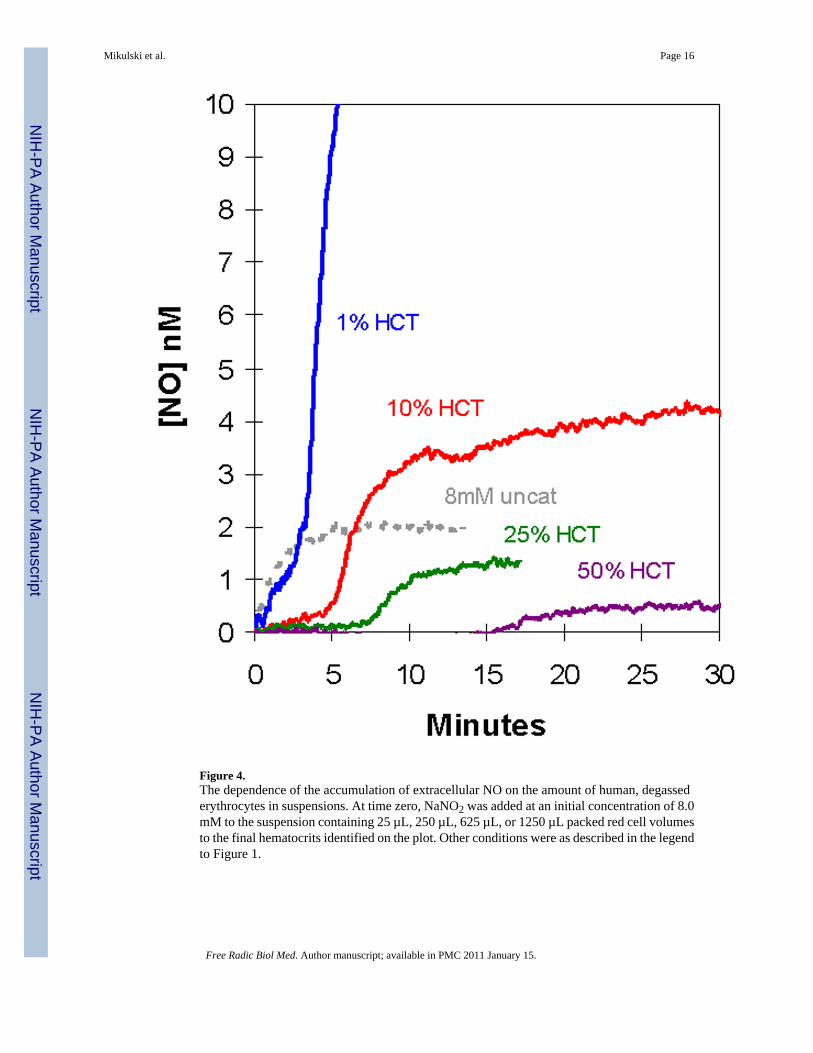
Figure 4.The dependence of the accumulation of extracellular NO on the amount of human, degassederythrocytes in suspensions. At time zero, NaNO2 was added at an initial concentration of 8.0mM to the suspension containing 25 µL, 250 µL, 625 µL, or 1250 µL packed red cell volumesto the final hematocrits identified on the plot. Other conditions were as described in the legendto Figure 1.
Mikulski et al. Page 16
Free Radic Biol Med. Author manuscript; available in PMC 2011 January 15.
NIH
-PA Author Manuscript
NIH
-PA Author Manuscript
NIH
-PA Author Manuscript

Figure 5.The effect of DIDS, an inhibitor of anion exchange, on accumulation of extracellular NO insuspensions of degassed human erythrocytes at 1.6% hematocrit. In each experiment 8.0 mMNaNO2 was added at time zero to a suspension containing the designated concentrations ofDIDS: (black) 0.1 µM; (green) 1 µM; (purple) 10 µM; (red) 100 µM; and (blue) no DIDS. Ineach case, DIDS was incubated with erythrocytes 10 minutes prior to addition of nitrite. Theother conditions were as described in the legend to Figure 1.
Mikulski et al. Page 17
Free Radic Biol Med. Author manuscript; available in PMC 2011 January 15.
NIH
-PA Author Manuscript
NIH
-PA Author Manuscript
NIH
-PA Author Manuscript

Figure 6.Effect of PCMBS, an inhibitor of the aquaporin channel, on the accumulation of extracellularNO concentration (determined from m/z 30) in suspensions of human erythrocytes at 0.8%hematocrit following the addition of an initial concentration of 8.0 mM nitrite. Suspensionscontained the following concentrations of PCMBS: (red) 25 µM; (green) 50 µM; (black) 100µM; (blue) no PCMBS: (brown) 1 mM; and (purple) 2 mM labeled A–F respectively on theplot. The data in grey labeled G show the accumulation of NO after addition of 8.0 mM nitritein solutions containing no erythrocytes. Other conditions were as described in the legend toFigure 1.
Mikulski et al. Page 18
Free Radic Biol Med. Author manuscript; available in PMC 2011 January 15.
NIH
-PA Author Manuscript
NIH
-PA Author Manuscript
NIH
-PA Author Manuscript