Crossed renal ectopia with fusion in a pelvic inlet area ...
7
CASE REPORT Open Access Crossed renal ectopia with fusion in a pelvic inlet area, atypical portal vein and coccygeal deformation in a young female cat Mateusz Hebel 1 , Jakub Jędrzej Ruszkowski 2* , Elżbieta Giza 1 and Małgorzata Pomorska-Mól 3 Abstract Background: The case report describes a rare congenital anomaly, crossed fused renal ectopia (CFRE), with coexistence of two other abnormalities – atypical portal vein and coccygeal vertebrae malformation in a domestic cat. The concomitance of those 3 congenital defects has not been described previously. Case presentation: An 8-month-old female, domestic cat suffering from chronic diarrhea was referred to the diagnostic imaging unit. The patient showed no other clinical symptoms. An abdominal ultrasonographic examination was performed in order to evaluate the condition of abdominal organs, particularly the gastrointestinal tract. The ultrasound examination showed an ectopic duplex kidney at instead of kidneys in their typical location. Computed tomography (CT) with angiographic phase and excretory urography was requested to evaluate the condition of the kidneys and ureters. The final diagnosis was CFRE, atypical portal vein and coccygeal deformation in an asymptomatic cat with no changes in renal function and normal blood parameters. Conclusions: Crossed fused renal ectopia is a rare congenital anomaly and is easily detectable by an abdominal ultrasonographic examination and CT, which allows more complete assessment of both anatomical relations and secretory function of the kidney. The occurrence of CFRE, abnormal portal vein and spinal malformation in a clinically healthy patient is the evidence that congenital malformations may simultaneously involve various, not directly related structures and systems, without significant influence on blood and urine parameters. Thus the most useful tool in the evaluation of the morphological and functional changes is the diagnostic imaging, especially contrast enhanced CT. Our results show that renal fusions should be considered in the differential diagnosis of caudal abdominal masses. Keywords: Cat, Congenital defect, Renal ectopia, CT © The Author(s). 2020 Open Access This article is licensed under a Creative Commons Attribution 4.0 International License, which permits use, sharing, adaptation, distribution and reproduction in any medium or format, as long as you give appropriate credit to the original author(s) and the source, provide a link to the Creative Commons licence, and indicate if changes were made. The images or other third party material in this article are included in the article's Creative Commons licence, unless indicated otherwise in a credit line to the material. If material is not included in the article's Creative Commons licence and your intended use is not permitted by statutory regulation or exceeds the permitted use, you will need to obtain permission directly from the copyright holder. To view a copy of this licence, visit http://creativecommons.org/licenses/by/4.0/. The Creative Commons Public Domain Dedication waiver (http://creativecommons.org/publicdomain/zero/1.0/) applies to the data made available in this article, unless otherwise stated in a credit line to the data. * Correspondence: [email protected] 2 Department of Animal Anatomy, Faculty of Veterinary Medicine and Animals Sciences, Poznan University of Life Sciences, Wojska Polskiego 71C, 60-625 Poznan, Poland Full list of author information is available at the end of the article Hebel et al. BMC Veterinary Research (2020) 16:314 https://doi.org/10.1186/s12917-020-02535-9
-
Upload
khangminh22 -
Category
Documents
-
view
2 -
download
0
Transcript of Crossed renal ectopia with fusion in a pelvic inlet area ...

CASE REPORT Open Access
Crossed renal ectopia with fusion in apelvic inlet area, atypical portal vein andcoccygeal deformation in a young femalecatMateusz Hebel1, Jakub Jędrzej Ruszkowski2* , Elżbieta Giza1 and Małgorzata Pomorska-Mól3
Abstract
Background: The case report describes a rare congenital anomaly, crossed fused renal ectopia (CFRE), withcoexistence of two other abnormalities – atypical portal vein and coccygeal vertebrae malformation in a domesticcat. The concomitance of those 3 congenital defects has not been described previously.
Case presentation: An 8-month-old female, domestic cat suffering from chronic diarrhea was referred to thediagnostic imaging unit. The patient showed no other clinical symptoms. An abdominal ultrasonographicexamination was performed in order to evaluate the condition of abdominal organs, particularly the gastrointestinaltract. The ultrasound examination showed an ectopic duplex kidney at instead of kidneys in their typical location.Computed tomography (CT) with angiographic phase and excretory urography was requested to evaluate thecondition of the kidneys and ureters. The final diagnosis was CFRE, atypical portal vein and coccygeal deformationin an asymptomatic cat with no changes in renal function and normal blood parameters.
Conclusions: Crossed fused renal ectopia is a rare congenital anomaly and is easily detectable by an abdominalultrasonographic examination and CT, which allows more complete assessment of both anatomical relations andsecretory function of the kidney. The occurrence of CFRE, abnormal portal vein and spinal malformation in aclinically healthy patient is the evidence that congenital malformations may simultaneously involve various, notdirectly related structures and systems, without significant influence on blood and urine parameters. Thus the mostuseful tool in the evaluation of the morphological and functional changes is the diagnostic imaging, especiallycontrast enhanced CT. Our results show that renal fusions should be considered in the differential diagnosis ofcaudal abdominal masses.
Keywords: Cat, Congenital defect, Renal ectopia, CT
© The Author(s). 2020 Open Access This article is licensed under a Creative Commons Attribution 4.0 International License,which permits use, sharing, adaptation, distribution and reproduction in any medium or format, as long as you giveappropriate credit to the original author(s) and the source, provide a link to the Creative Commons licence, and indicate ifchanges were made. The images or other third party material in this article are included in the article's Creative Commonslicence, unless indicated otherwise in a credit line to the material. If material is not included in the article's Creative Commonslicence and your intended use is not permitted by statutory regulation or exceeds the permitted use, you will need to obtainpermission directly from the copyright holder. To view a copy of this licence, visit http://creativecommons.org/licenses/by/4.0/.The Creative Commons Public Domain Dedication waiver (http://creativecommons.org/publicdomain/zero/1.0/) applies to thedata made available in this article, unless otherwise stated in a credit line to the data.
* Correspondence: [email protected] of Animal Anatomy, Faculty of Veterinary Medicine andAnimals Sciences, Poznan University of Life Sciences, Wojska Polskiego 71C,60-625 Poznan, PolandFull list of author information is available at the end of the article
Hebel et al. BMC Veterinary Research (2020) 16:314 https://doi.org/10.1186/s12917-020-02535-9
BackgroundUrinary system development is a complicated process ofinteractions between the embryonic precursors of kid-neys, ureters, bladder, and urethra [1]. Congenital mal-formations of the urinary system, especially the kidneys,are not the problem commonly described in veterinarymedicine, and they are rarely the cause of clinical prob-lems [2, 3]. Many authors refer to data from humanmedicine literature, where kidney malformations are de-scribed in more detail than in companion animals suchas dogs and cats [2–5]. Renal ectopia, including crossedfused renal ectopia (CFRE) and horseshoe kidney, can befound among these kidney malformations.Ectopic kidneys have previously been described as a
defect in humans [6], cattle [7], dogs [2, 8] and cats[3–5, 9, 10], most often as asymptomatic and appear-ing rarely. Until now, CFRE cases in cats were re-ported in both, males [3, 9] and females [4, 5, 10].Developmental defects, especially CFRE, may predis-pose to the occurrence of nephrolithiasis, hydrone-phrosis or urinary tract infections resulting inpyelonephritis [3].To date, five reports of CFRE in cats have been de-
scribed [3–5, 9, 10]. Modern methods of diagnostic im-aging allow for better detection of this type of congenitaldefects in vivo, and reports describing the condition canbe extremely helpful in the differential diagnosis of themasses in the caudal part of the abdominal cavity [3].
Case presentationAn eight-month, female domestic cat was referred forabdominal ultrasonographic (USG) examination due torecurrent diarrhoea. The previous medical history of pa-tient was unknown. Medical records provided by theowner lack the information on vaccinations and antipar-asitic prophylaxis. During the visit, the condition of thecat was normal: body condition score (BCS) 3/5, pinkmucous membranes, capillary refill time (CRT) < 2 s,and 38.5 °C of rectal temperature. Chest auscultation didnot reveal any abnormalities in the lungs or heart. Fem-oral pulse measured at femoral artery was 140 beats perminute (bpm), symmetrical, steady, even and full. Sys-tolic blood pressure measured by the doppler method onthe palmar surface of the left thoracic limb was 140mmHg. The palpation of the caudal abdomen revealedno kidneys in the lumbar region. However, in the caudalabdominal area, however, a solid, painless and non-shifting mass was found. The cat was negative for felineimmunodeficiency virus (FIV) antibody and feline leu-kaemia virus (FeLV) (FIV/FeLV Test SNAP, Idexx,Germany).Ultrasonographic examination of the abdomen was
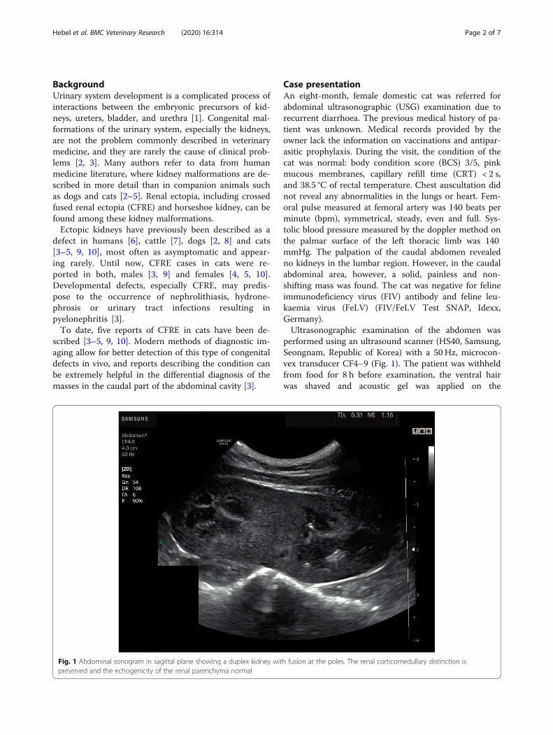
performed using an ultrasound scanner (HS40, Samsung,Seongnam, Republic of Korea) with a 50 Hz, microcon-vex transducer CF4–9 (Fig. 1). The patient was withheldfrom food for 8 h before examination, the ventral hairwas shaved and acoustic gel was applied on the
Fig. 1 Abdominal sonogram in sagittal plane showing a duplex kidney with fusion at the poles. The renal corticomedullary distinction ispreserved and the echogenicity of the renal parenchyma normal
Hebel et al. BMC Veterinary Research (2020) 16:314 Page 2 of 7

examined area. The patient was positioned in dorsal re-cumbency. Ultrasonographic examination confirmed nokidneys in their anatomical position. Instead, an ectopicduplex kidney with dimensions of about 50 mm × 25mm, located in the caudal abdomen, at the pelvic inletarea, between the spine and the rectum was revealed.There were no abnormalities in the underlying kidneystructure, corticomedullary differentiation of the kidneywas preserved, and the corticomedullary ratio was 1:1(Fig. 1). The kidney vascularization in the colour dopplertest was normal, the vascular resistance index (RI) mea-sured in the arcuate arteries at the renal poles werewithin the reference range, RI = 0.71. Two renal pelviseswere found, however, due to the location of the kidney,it was not possible to assess the ureters and their orificesin a standard USG examination. The ultrasound imageof the remaining abdominal organs was normal. The pa-tient was referred for contrast enhanced computed tom-ography (CT) of the abdominal and pelvic cavities toassess the kidney perfusion, secretory function and ur-eter morphology.
Blood morphology, biochemistry tests, and a ventro-dorsal radiograph (X-ray) of the abdominal cavityproceded the computed tomography examination. TheX-ray was performed using a Gierth HF 200 lamp(Gierth X-Ray International GbmH, Germany) with 60kVp and 8mAs and a Vivix-s 1417 N (Anyang, Republicof Korea) digital direct radiography detector. The resultsof hematological and biochemical parameters are sum-marized in Table 1. Urine and blood tests showed no ab-normalities. Urine specific gravity was 1.020 mOsm / kg(reference range 1.016–1.064).Computed tomography examination was performed
under general infusion anaesthesia with intravenous pro-pofol (2 mg/kg body weight (BW), after premedicationwith the use of dexmedetomidine (40 μg/kg BW, intra-muscularly) and intubation. The examination was car-ried out with a 2-slice helical scanner (SiemensSomatom Emotion Duo, Siemens, Germany), with scan-ning parameters 130 kVp and 100 mAs, pitch 2,0 and re-constructed slice thickness 1.25 mm in the soft tissuewindow level (WL) 40 Hounsfield Unit (HU) window
Table 1 Hematological (Horiba ABX, Horiba Medical, Japan) and biochemical (Mindray BS-120, Mindray Medical International Co.,Ltd., China) parameters of the patient
Parameter Result Reference values[11, 12]
Hematology
White blood cell (× 103/mm3) 8.5 5.5–19.5
Red blood cell (× 106/mm3) 7.39 5.8–10.7
Hemoglobin (g/dL) 12 9–15
Hematocrit (%) 34.7 30–47
Platelet (×103/mm3) 324 300–800
Mean corpuscular volume (mm3) 47 41–51
Mean corpuscular hemoglobin (pg) 16.2 13–18
Mean corpuscular hemoglobin concentration (g/dL) 34.6 31–35
Red blood cell distribution width (%) 17.3 17–22
Mean platelet volume (mm3) 10.6 6.5–15
Electrolytes
Sodium (mmol/L) 150 143–155
Potassium (mmol/L) 4.5 4.1–5.6
Chlorides (mmol/L) 115 102–118
Biochemistry
Alanine aminotransferase (U/L) 63 20–107
Aspartate aminotransferase (U/L) 56 6–44
Total Protein (g/dL) 7 6–8
Albumins (g/dL) 2.8 2.6–3.9
Urea (mg/dL) 22 25–70
Creatinine (mg/dL) 1.2 1–1.8
Calcium (mg/dL) 9.14 8–11
Phosphorus (mg/dL) 5.6 3–6.8
Hebel et al. BMC Veterinary Research (2020) 16:314 Page 3 of 7
width (WW) 350 HU and bone tissue windows (WL 300HU WW 1500 HU). The patient was placed in sternalrecumbency with legs directed towards CT gantry. Ascan of the abdominal cavity of the cat was performedprior to the administration of the contrast agent, within30 s after its administration to visualize the cortical per-fusion of the kidneys (nephrographic phase), and 5 minafter its administration to visualize the secretory func-tion of the kidney, together with the exact location ofthe renal pelvis and ureters. Iodine, non-ionic contrastagent (Ultravist 370, Bayer) at a dose of 700 mg iodine/kg BW was used, which corresponded to a dose of 2 ml/kg BW. The contrast was administered manually to thecephalic vein through a 20G intravenous cannula.Computed tomography examination revealed the pres-
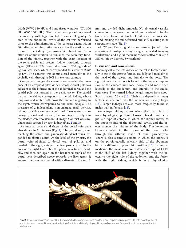
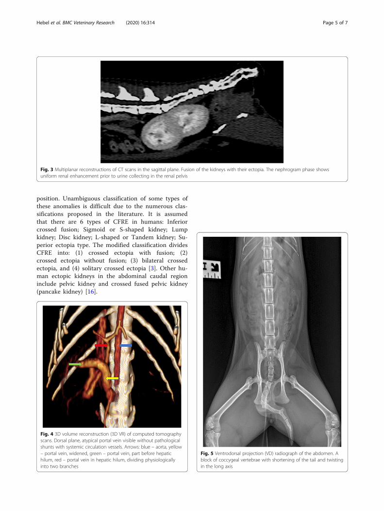
ence of an ectopic duplex kidney, whose cranial pole wasadjacent to the bifurcation of the abdominal aorta, and thecaudal pole was located in the pelvic cavity. The caudalpart of the kidney corresponds to the left kidney, whoselong axis and ureter both cross the midline migrating tothe right, which corresponds to the renal ectopia. Thepresence of 2 independent, non-enlarged renal pelvises,without calcifications was confirmed. Two ureters, non-enlarged, shortened, crossed, but running correctly intothe bladder were revealed on CT image. Contrast was sim-ultaneously secreted by each kidney (Figs. 2 and 3).An unusual course and shape of the portal vein were
also shown in CT images (Fig. 4). The portal vein, afterreaching the spleen and pancreatic-duodenal veins, ex-panded to about 5.5 mm. At the level of the pylorus, theportal vein adjoined to dorsal wall of pylorus, andheaded to the right, entered the liver parenchyma. In thearea of the right liver lobe, the portal vein turned caud-ally, and then run again on the broadened trunk of theportal vein described above towards the liver gates. Itentered the liver as a vessel with a diameter of about 3
mm and divided dichotomously. No abnormal vascularconnections between the portal and systemic circula-tions were found. A block of tail vertebrae was alsofound, making the tail deformed and stiff, resulting in itsserpentine shape (Fig. 5).All CT and X-ray digital images were subjected to the
analysis and post-processing using a dedicated imagingworkstation and digital medicine viewer software (OsiriXMD 64-bit by Pixmeo, Switzerland).
Discussion and conclusionsPhysiologically, the left kidney of the cat is located caud-ally, close to the gastric fundus, caudally and medially tothe head of the spleen, and laterally to the aorta. Theright kidney cranial pole is found in the hepatic impres-sion of the caudate liver lobe, dorsally and most oftenlaterally to the duodenum, and laterally to the caudalvena cava. The normal kidney length ranges from about3 cm to about 5.3 cm [13]. Their size depends on manyfactors, in neutered cats the kidneys are usually larger[14]. Larger kidneys are also more frequently found inmales than in females [13].An ectopic kidney occurs when the organ is in a
non-physiological position. Crossed fused renal ecto-pia is a type of ectopia in which the kidney moves tothe opposite side of the abdominal cavity, and the ur-eter crosses the midline of the body. The horseshoekidney consists in the fusion of the renal polesthrough the isthmus made of renal parenchyma.There is also a simple ectopia in which the kidney ison the physiologically relevant side of the abdomen,but in a different topographic position [15]. In humanmedicine, the most commonly described type of CFREis the shift of the left kidney, together with the ur-eter, to the right side of the abdomen and the fusionwith the right kidney, which is in a physiological
Fig. 2 3D volume reconstruction (3D VR) of computed tomography scans. Sagittal plane, nephrographic phase (30 s after contrast agentadministration), unusual kidney location (ectopia) visible, additionally: duplex kidney (yellow arrow). Deformation of the shape of the tail(red arrow)
Hebel et al. BMC Veterinary Research (2020) 16:314 Page 4 of 7
position. Unambiguous classification of some types ofthese anomalies is difficult due to the numerous clas-sifications proposed in the literature. It is assumedthat there are 6 types of CFRE in humans: Inferiorcrossed fusion; Sigmoid or S-shaped kidney; Lumpkidney; Disc kidney; L-shaped or Tandem kidney; Su-perior ectopia type. The modified classification dividesCFRE into: (1) crossed ectopia with fusion; (2)crossed ectopia without fusion; (3) bilateral crossedectopia, and (4) solitary crossed ectopia [3]. Other hu-man ectopic kidneys in the abdominal caudal regioninclude pelvic kidney and crossed fused pelvic kidney(pancake kidney) [16].
Fig. 3 Multiplanar reconstructions of CT scans in the sagittal plane. Fusion of the kidneys with their ectopia. The nephrogram phase showsuniform renal enhancement prior to urine collecting in the renal pelvis
Fig. 4 3D volume reconstruction (3D VR) of computed tomographyscans. Dorsal plane, atypical portal vein visible without pathologicalshunts with systemic circulation vessels. Arrows: blue – aorta, yellow– portal vein, widened, green – portal vein, part before hepatichilum, red – portal vein in hepatic hilum, dividing physiologicallyinto two branches
Fig. 5 Ventrodorsal projection (VD) radiograph of the abdomen. Ablock of coccygeal vertebrae with shortening of the tail and twistingin the long axis
Hebel et al. BMC Veterinary Research (2020) 16:314 Page 5 of 7

In crossed ectopia with fusion, the migration of the leftkidney to the right side is most often described in humans[3]. In the cases of CFRE reported to date in cats, the kid-ney fusion consisted in moving one of them to the contra-lateral one, located physiologically [3, 9, 10]. In thepresent case, both kidneys were ectopic, in addition a fu-sion and changes in the sides of the renal pelvis were ob-served. Moreover, the ureters were significantly shortened.Due to the multiplicity of types of ectopia described inhumans and the occurrence of common features of severaltypes of ectopia in the case being described, unambiguousnaming of the type of ectopia was more difficult. Accord-ing to the authors’ knowledge, renal ectopia co-occuringwith portal vein and spinal abnormalities in the same pa-tient has not been described in cats yet.The CT image allows us to classify the diagnosed defect
as S-shaped CFRE, but the fact that the ectopic kidneywas in the pelvic cavity also allows it to be classified as apancake kidney [3, 16]. Contrast enhanced CT examin-ation allows seeing the kidneys and ureters in variousplanes allowing much more detailed diagnosis than ultra-sound and excretory urography. This is the first descrip-tion of such a case in the literature. More than 50% ofpatients with CFRE are asymptomatic, but about 50% ofsymptomatic patients have complications such as nephro-lithiasis, hydronephrosis, urinary tract infections, or pyelo-nephritis [3]. By monitoring the clinical status of CFREpatients (i.e. capillary refill time, skin elasticity, sunkeneyes, blood pressure, creatinine and urea levels, abdominalUSG examination) the risk of these complications can bereduced. Measuring of the above mentioned parametersshould be conducted periodically and any abnormalitiesshould be corrected as soon as possible to prevent diseasedevelopment. In the case described, the results of bloodand urine tests as well as excretory urography did not in-dicate any disturbance of kidneys’ secretory function. Thepatient did not present clinical signs of renal dysfunction.In this case the detection of described abnormality was in-cidental, during a standard abdominal USG examination.It is possible that the lack of clinical symptoms and kidneydysfunction results from the shortening of the ureters, sothat they run straight and do not wind up as in the case ofsome types of CFRE. Imaging of the above mentioned le-sion may be helpful before abdominal surgery, as CFREmay be associated with various vascular and anatomicalabnormalities (i.e. different vessel patterns, abnormal ur-eter course), which were confirmed in this case.Apart from the confirmation of the defect in the renal
structure, the CT examination allowed for an incidentaldiagnosis referring to the unusual course of the portalvein, without the presence of portal anastomoses andwith a normal pattern of portal and venous livervascularization. The most frequently reported defects ofthe portal vein are congenital and acquired portal shunts
[17]. Rare cases of congenital absence of the portal vein(CAPV) have also been reported [17]. The anomaly diag-nosed in the present patient has not been reported previ-ously. Moreover, in this case, the third defect, in the formof a tail vertebral block and the deformation of the shapeof the tail was also observed. Among described earlieranomalies of caudal vertebrae in cats, there were lack ofthe tail or reduction of the tail’s length with deformationsin Manx breed [18, 19]. The most common abnormality istransitional vertebrae with the highest number of abnor-malities at the sacrocaudal junction [19]. All reported de-fects did not affect the general condition of cats and werenot associated with any clinical symptoms.Crossed fused renal ectopia is a rare congenital anomaly
and is easily detectable in an abdominal USG examinationand CT, which allows more complete assessment of bothanatomical relations and secretory function of the kidney.The occurrence of CFRE, abnormal portal vein and spinalmalformation in a clinically healthy patient is the evidencethat congenital malformations may simultaneously involvevarious structures and systems not directly related to eachother, such as in the case presented. In these cases, bloodand urine tests are of marginal significance, thus the diag-nostic imaging is the most important examination in theaccurate evaluation of the morphological and functionalchanges, especially contrast enhanced CT. Cats’ renal fu-sions should be considered in the differential diagnosis ofcaudal abdominal masses.
AbbreviationsBCS: Body condition score; bpm: Beats per minute; CAPV: Congenitalabsence of the portal vein; CFRE: Crossed fused renal ectopia; CRT: Capillaryrefill time; CT: Computed tomography; FIV: Feline immunodeficiency virus;FeLV: Feline leukemia virus; Res: Resolution; RI: Resistance index;USG: Ultrasonographic; X – ray: Radiograph
AcknowledgementsNot applicable.
Authors’ contributionsMH performed clinical examination, blood tests, ultrasound examination,computed tomography scan, collected the data, Writing – original draft,Writing - review & editing; JJR did research on available knowledge aboutthe topic, Writing – original draft, Writing - review & editing; EG - Writing -review & editing, MPM - Methodology, Writing - review & editing. All authorshave read and approved the manuscript.
FundingThis work was supported partially by the statutory funding No. 506.514.05.00of the Faculty of Veterinary Medicine and Animal Science Poznan Universityof Life Sciences, Poland; Department of Preclinical Sciences and InfectiousDiseases and by the Ministry of Science and Higher Education program“Regional Initiative Excellence” in years 2019–2022, Project No. 005/RID/2018/19. The funding body did not have any role in the design, analysis or writingof this study.
Availability of data and materialsThe data used to support the findings of this study are available from thecorresponding author upon request.
Ethics approval and consent to participateNot applicable.
Hebel et al. BMC Veterinary Research (2020) 16:314 Page 6 of 7

Consent for publicationWritten informed consent for publication of their clinical details and clinicalimages was obtained from the owner of the patient.
Competing interestsThe authors declare that there is no conflict of interest regarding thepublication of this paper.
Author details1Department of Internal Medicine and Diagnostics, Faculty of VeterinaryMedicine and Animal Sciences, Poznan University of Life Sciences, Wolynska35, 60-637 Poznan, Poland. 2Department of Animal Anatomy, Faculty ofVeterinary Medicine and Animals Sciences, Poznan University of Life Sciences,Wojska Polskiego 71C, 60-625 Poznan, Poland. 3Department of PreclinicalSciences and Infectious Diseases, Faculty of Veterinary Medicine and AnimalSciences, Poznan University of Life Sciences, Wolynska, 35 Poznań, Poland.
Received: 2 April 2020 Accepted: 23 August 2020
References1. Mario LC, Morais MP, Borghesi J, Favaron PO, Oliveira FD, Anunciação AR,
Miglino MA. Development of urinary organs in domestic cat during theembryonic and fetal periods. Microsc Res Tech. 2018;81:1286–94..
2. Choi J, Lee H, Lee Y, Choi H. Simple ectopic kidney in three dogs. J Vet MedSci. 2012;74:1373–5.
3. Seo SH, Lee HA, Suh SI, Choi R, Park IC, Hyun C. Crossed fused renal ectopiain a Persian cat. JFMS Open Rep. 2017;3:2055116917695875.
4. Rajabioun M, Salari Sedigh H, Mirshahi A. Bilateral simple ectopic kidney in acat. Vet Res Forum. 2017;8:175–7.
5. Story HE. A case of horseshoe kidney and associated vascular anomalies inthe domestic cat. Anat Rec. 1943;86:307–19.
6. Sharma V, Babu CSR, Gupta OP. Crossed fused renal ectopia multidetectorcomputed tomography study. Int J Anat Res. 2014;2:305–9.
7. Charan K, Pawaiya RVS. An unusual congenital anomaly: ectopic sigmoidkidney combined with hermaphroditism in a newly born calf. Anat HistolEmbryol. 1997;26:269–70.
8. Webb AI. Renal ectopia in a dog. Aust Vet J. 1974;50:519–21.9. Allworth MS, Hoffmann KL. Crossed renal ectopia with fusion in a cat. Vet
Radiol Ultrasound. 1999;40:357–60.10. Fulgêncio JQ, Miranda FG, Santos CJ, Moreira GD, Tôrres RCS, Nepomuceno
AC. Crossed renal ectopia with fusion in a female feline: case report. ArqBras Med Vet Zootec. 2019;71:833–6.
11. Klaassen JK. Reference values in veterinary medicine. Lab Med. 1999;30:194–7.12. Cottam YH, Caley P, Wamberg S, Hendriks WH. Feline reference values for
urine composition. J Nutr. 2002;132:1754S–6S.13. Debruyn K, Haers H, Combes A, Paepe D, Peremans K, Vanderperren K,
Saunders JH. Ultrasonography of the feline kidney: technique, anatomy andchanges associated with disease. J Feline Med Surg. 2012;14:794–803.
14. Shiroma JT, Gabriel JK, Carter RL, Scruggs SL, Stubbs PW. Effect of reproductivestatus on feline renal size. Vet Radiol Ultrasound. 1999;40:242–5.
15. Lulich JP, Osborne CA, Lawler DF, O'Brien TD, Johnston GR, O'Learyet TP.Urologic disorders of immature cats. Vet Clin North Am Small Anim Pract.1987;17:663–96.
16. Tiwari AK, Choudhary AK, Khowal H, Chaudhary P, Arora MP. Pancakekidney: a rare developmental anomaly. Can Urol Assoc J. 2014;8:E451–2.
17. Bertolini G. Anomalies of the portal venous system in dogs and cats as seenon multidetector-row computed tomography: an overview andsystematization proposal. Vet Sci. 2019;6:10.
18. Tomlinson BE. Abnormalities of the lower spine and spinal cord in Manxcats. J Clin Pathol. 1971;24:480.
19. Newitt A, German AJ, Barr FC. Congenital abnormalities of the felinevertebral column. Vet Radiol Ultrasound. 2008;49:35–41.
Publisher’s NoteSpringer Nature remains neutral with regard to jurisdictional claims inpublished maps and institutional affiliations.
Hebel et al. BMC Veterinary Research (2020) 16:314 Page 7 of 7