Infectious vaccine-derived rubella viruses emerge ... - PLOS
Upload
khangminh22Category
view
3download
0
REVIEW Open Access
The potential role of infectious agents andpelvic inflammatory disease in ovariancarcinogenesisKasper Ingerslev1* , Estrid Hogdall2, Tine Henrichsen Schnack3, Wojciech Skovrider-Ruminski4, Claus Hogdall3
and Jan Blaakaer1
Abstract
Background: The etiological cause of ovarian cancer is poorly understood. It has been theorized that bacterial orviral infection as well as pelvic inflammatory disease could play a role in ovarian carcinogenesis.
Aim: To review the literature on studies examining the association between ovarian cancer and bacterial or viralinfection or pelvic inflammatory disease.
Methods: Database search through MEDLINE, applying the medical subject headings: “Ovarian neoplasms”, AND“Chlamydia infections”, “Neisseria gonorrhoeae”, “Mycoplasma genitalium”, “Papillomaviridae”, or “pelvic inflammatorydisease”. Corresponding searches were performed in EMBASE, and Web of Science. The literature search identified935 articles of which 40 were eligible for inclusion in this review.
Results: Seven studies examined the association between bacterial infection and ovarian cancer. A single studyfound a significant association between chlamydial infection and ovarian cancer, while another study identifiedMycoplasma genitalium in a large proportion of ovarian cancer cases. The remaining studies found no association.Human papillomavirus detection rates varied from 0 to 67% and were generally higher in the Asian studies than instudies from Western countries. Cytomegalovirus was the only other virus to be detected and was found in 50%of cases in a case-control study. The association between ovarian cancer and pelvic inflammatory disease wasexamined in seven epidemiological studies, two of which, reported a statistically significant association.
Conclusions: Data indicate a potential association between pelvic inflammatory disease and ovarian cancer. Anassociation between ovarian cancer and high-risk human papillomavirus genotypes may exist in Asia, whereas anassociation in Western countries seems unlikely due to the low reported prevalence. Potential carcinogenic bacteriawere found, but results were inconsistent, and further research is warranted.
Keywords: Ovarian cancer, Bacterial infection, Viral infection, Pelvic inflammatory disease, Carcinogenesis
BackgroundOvarian cancer (OC) is a major threat to female health,with a global prevalence of 239,000 cases in 2012 [1]. Dueto the lack of symptoms in early stage OC, advanceddisease is often present at the time of diagnosis [2].Almost 90% of OC originates from epithelial cells,
which have been thought to arise from the surfacemesothelial lining of the ovaries. However, recent
research has indicated that a proportion of serousepithelial OC could originate in precancerous lesionscalled “serous tubal intraepithelial carcinomas” (STICs)located in the fimbriated end of the fallopian tubes [3].This is an important finding because previous theoriesof ovarian carcinogenesis have been unable to explainthe fact that precancerous lesions have never beenidentified in the ovaries [3].The female internal genitalia and the peritoneal cavity
are accessible to outside pathogens through the genitaltract. The mesothelial lining of the peritoneal cavity is
* Correspondence: [email protected] of Gynaecology and Obstetrics, Odense University Hospital,Denmark, Soendre Blvd. 29, 5000 Odense C, DenmarkFull list of author information is available at the end of the article
© The Author(s). 2017 Open Access This article is distributed under the terms of the Creative Commons Attribution 4.0International License (http://creativecommons.org/licenses/by/4.0/), which permits unrestricted use, distribution, andreproduction in any medium, provided you give appropriate credit to the original author(s) and the source, provide a link tothe Creative Commons license, and indicate if changes were made. The Creative Commons Public Domain Dedication waiver(http://creativecommons.org/publicdomain/zero/1.0/) applies to the data made available in this article, unless otherwise stated.
Ingerslev et al. Infectious Agents and Cancer (2017) 12:25 DOI 10.1186/s13027-017-0134-9

identical in both sexes. However, primary serous peri-toneal cancer, which may represent a continuum withserous OC [3], occurs almost exclusively in female pa-tients [4]. This suggests that extrinsic factors could playa crucial role in ovarian carcinogenesis. Furthermore,epidemiological studies have demonstrated that patientswith tubal factor infertility have a higher risk of OC [5].Since the fallopian tubes are often affected by pelvic in-flammatory disease (PID), it is therefore highly rele-vant to consider the potential role of infectiousagents in OC. Previous reviews have focused on hu-man papillomavirus (HPV) and OC, but this review isthe first to include studies on all bacterial and viralinfections as well as studies on pelvic inflammatorydisease (PID).
Material and methodsTo review the existing literature on OC and infectiousagents, we performed a systematic literature search bymeans of a database search for English language articlespublished in the period from 1980 to 2016. Using thePubmed research tool, the keyword: “ovarian neoplasms”was by means of the Boolean logical “AND”, combinedwith the following keywords: “Chlamydia infections”
(MeSH), “Neisseria gonorrhoeae” (MeSH), “Mycoplasmagenitalium” (MeSH), “Papillomaviridae” (MeSH), “pelvicinflammatory disease” (MeSH). Correspondent searcheswere performed in EMBASE and Web of Science. The ini-tial search identified 935 articles. After removal of 130duplicates, 805 studies remained for further analysis. Con-tents were outside the scope of the present review in740 articles that were excluded. The remaining 65articles were subjected to full-text screening. Ori-ginal studies, examining the association between in-fectious agents and ovarian cancer by use of tissue-based or serologic methods were included. Epidemio-logical studies examining the association betweenpelvic inflammatory disease and ovarian cancer werealso included. Case-reports and reviews were ex-cluded, leaving a total of 40 articles to be includedin the review. More detailed information on thesearch strategy is given in the “Additional file 1” tothis article.
Viral agents investigated in relation to ovariancarcinogenesisTable 1 summarises the viruses investigated in relationto OC, and their potential oncogenic mechanisms.
Table 1 Carcinogenic mechanisms of investigated viruses in relation to ovarian cancer
Virus Viral family Key viral transformingfactors
Mechanisms Associated cancers
Human papillomavirus Papillomaviridae E6
E7
Inhibition of apoptosisImmune evasionCell cycle arrestImmune evasion [8, 10, 11]
Anal/rectal cancerCervical cancerOro-pharyngeal cancer(Ovarian cancer)a
Penile cancerVaginalVulvar cancer
Epstein bar virus Herpesviridae EBNA 1EBNA 2EBNA-3ALatent membraneprotein 1
Inhibition of apoptosisB-cell growth transformationCell cycle disruptionCell cycle disruptionInhibition of apoptosisPromotion of cell immortalisation [48, 49]
Burkitt lymphomaGastric adenocarcinomaHodgkin lymphomaNasopharyngeal carcinomaNon-Hodgkin lymphoma(Ovarian cancer)Post-transplant lymphoproliferativedisease
Cytomegalovirus Herpesviridae IE1/IE2 Inhibition of antiviral immune responseInhibition of apoptosis
Mucoepidermoid carcinoma ofsalivary glands(Ovarian cancer)
US28 gene Enhanced proliferative signaling [44, 45]
John Cunningham virus/BK virus
Polyomaviridae T-antigen/t-antigen Cell cycle disruption Inhibition ofapoptosis [56]
(Ovarian cancer)
Hepatitis C virus Flaviviridae (Indirect) Chronic inflammation and oxidative stressInhibition of antioxidant systems
Hepatocellular carcinoma(Ovarian cancer)
(Direct)NS5BNOS2ANS3/4A protease
Cell cycle disruptionInhibition of apoptosisInhibition of DDR response [51–53]
DDR Double-stranded DNA Repair, EBNA Epstein Barr virus nuclear antigen, IE Immediate early genes, NOS2A Nitric oxide synthase 2A, NS3/4A/5B Nonstructuralprotein 3/4A/5BaCausality is controversial and is still under investigation
Ingerslev et al. Infectious Agents and Cancer (2017) 12:25 Page 2 of 10

Human papillomavirus infectionHPV infection is a prerequisite for cervical cancerdevelopment and is implicated in a range of othercancer forms such as vulvar, penile, anal, and oropha-ryngeal cancers. To date, The International Agency forResearch on Cancer has classified 12 HPV subtypes ashuman carcinogens: HPV16/18/31/33/35/39/45/51/52/56/58/59 [6]. In HPV-induced tumorigenesis, the viraloncogenes E6 and E7 are integrated into the host cellDNA, allowing viral oncoproteins to be expressed bythe cell [7]. The carcinogenic mechanisms of theseoncoproteins involve the ability of E6 to form atrimeric complex with P53 and the cellular E3 ubiqui-tin ligase, E6AP. This complex induces proteasomaldegradation of P53, which leads to inhibition of cell-regulatory mechanisms and apoptosis and, therefore,permits continuous cell proliferation despite irrepar-able DNA damage [8]. E7 can cause degradation ofretinoblastoma protein, which is an important regula-tor of the cell cycle [9]. Furthermore, E7 can interactwith several cell cycle regulators including cyclins,cyclin-dependent kinases, CDKs-inhibitors, cullin 2,histone deacetylase, AP-1, E2F1, and E2F6 [10]. Theseactions enable the virus to resume the replication-competent state within the infected cells, which isessential for viral DNA synthesis. E6 and E7 can alsoexhibit immunomodulatory effects by downregulatingexpression of various inflammatory cytokines and byreducing the production of antiviral interferons [11].HPV is the most studied infectious agent in relation to
OC, and the available studies are summarised below.Six case-control studies, with 11–50 cases of OC, were
identified that examined the relation between HPV andOC [12–17]. The applied methods of analysis wereeither tissue-based methods or serologic assays andincluded in-situ hybridisation, immunohistochemistry,enzyme-linked immunosorbent serologic assay (ELISA),southern blot hybridisation technique, and polymerasechain reaction (PCR)-based technologies. Only one studyfound a statistically significant association between HPVand OC [12]. This study was the largest and included 50cases with OC and a control group with 30 patients withnon- malignant ovarian lesions. In-situ hybridisation andimmunohistochemistry were used to detect the HPV 16E6 gene in the tumour specimens. The highest detectionrates were obtained by in-situ hybridisation, with aprevalence of 52% among cases as compared to 6.7%among controls (OR: 16.7 95% CI: 3.2–71.4). Theremaining smaller studies, found no or non-significantassociations [13–17].Twenty-two case-series and cross-sectional studies
reporting on the prevalence of HPV in OC tissue wereidentified (Table 2). HPV 16 was most frequently foundand was reported in eight studies, giving a combined
total of 60 cases. Ten studies did not find HPV in any ofthe samples [18–27]. The remaining studies reported aHPV prevalence ranging between 0.5 and 66.7% [28–39].Interestingly, the reported HPV prevalence varied ac-cording to the geographical region. Among the 572 casesof OC in Europe and USA combined, 18 cases (3.1%)were positive for HPV DNA. Studies from the MiddleEast and Asia, with 347 combined cases of OC, reported123 cases to be HPV DNA positive (35.4%).Recently, sequencing data from The Cancer Genome
Atlas (TCGA) database have been utilised to detect HPVoncogene expression in ovarian tumours. Using suchdata, Roos et al. concluded that six samples in 405(1.5%) were positive for HPV18 oncogene expression[40]. However, Khoury et al. performed a similar analysison 419 serous cystadenocarcinomas from the TCGAdatabase and did not find signs of HPV infection [41].Moreover, other studies present data that support poten-tial HPV 18 contamination of the TCGA sequencingdatabase and, consequently, question the validity of posi-tive findings [42, 43].
Cytomegalovirus infectionCytomegalovirus (CMV) is under increasing focus as acarcinogenic virus. It is speculated that rather than initi-ating malignant transformation, CMV can enhance thegrowth and migration of tumour cells through a processcalled “oncomodulation” [44]. For instance, the forma-tion of metastasis may be aided by the capacity of CMVto adhere to and disrupt the endothelial cell integrity byactivating β1α5 integrin on the surface of tumour cells[45]. However, data also indicate that CMV has inherentantiapoptotic and cell cycle modulatory effects [45]. Asingle study has examined the relation between OC andCMV. The study detected CMV infection in 12 (50%)out of 24 cases with OC and in three (50%) out of sixcases of borderline ovarian tumours. No signs of CMVinfection were found in the control group consisting ofnormal ovarian tissue [13]. These findings have not beenconfirmed by others.
Epstein-Barr virus and hepatitis C virus infectionEpstein-Barr virus (EBV) is present in more than 90% ofthe global adult population and causes latent infection inthe host. [46] It exhibits tropism for epithelial cells,lymphocytes, and mesenchymal cells and is associatedwith a range of human cancers, including nasopharyn-geal carcinoma (NPC) and lymphomas [47]. Its carcino-genic mechanisms include the actions of latent antigensthat display effects similar to those of high-risk HPVoncogenes E6 and E7. An example is EBV nuclear anti-gen (EBNA)-1 that can induce proteasomal degradationof P53 and corresponding inhibition of apoptosis [48].Likewise, EBNA-3A and -3C play an important role in
Ingerslev et al. Infectious Agents and Cancer (2017) 12:25 Page 3 of 10

cell cycle regulation and EBNA-2 antigen is suspectedto cause primary B-cell growth transformation andmaintain human B-cell immortalisation [49]. HepatitisC virus (HCV) is another investigated microorganismin relation to OC. Chronic HCV infection greatly in-creases the risk of subsequent hepatocellular carcin-oma [50]. The mechanisms are not fully understoodbut may entail different pathways. The chronic inflam-mation enhances a pro-carcinogenic microenviron-ment through the release of nitric oxide (NO) andreactive oxygen species (ROS). This process is furthermediated by the ability of HCV to increase the cellulargeneration of NO and ROS [51] and by its capacity toweaken the antioxidant defence of the host. [52] Amore direct pathway involves the ability of HCV toimpair phosphorylation of the ataxia telangiectasiamutated (ATM) kinase substrate, histone 2A.X, whichplays a role in initiating cellular response to double-stranded DNA breaks [53]. Despite the obvious car-cinogenic potential of EBV and HCV, only a singlestudy has investigated their potential role in relationto OC. However, this study did not detect EBV orHCV in 419 serous cystadenocarcinomas from theTCGA database [41].
Polymyoma virus infectionFinally, a single study has analysed tissue samples forthe presence of the BK virus and John Cunninghamvirus [18]. The oncogenic potential of polymyomaviruses has largely been attributed to viral proteins T-antigen and t-antigen through their interactions withvarious cellular proteins, including tumour suppressorproteins p53 and pRb [54, 55]. Furthermore, thecapability of John Cunningham virus T- antigen totransform rodent cells has previously been demon-strated [56]. In the included study, however, poly-myoma viruses were not detected in any of theovarian cancer tissue samples, nor in the benigncontrols.
Bacterial agents investigated in relation toovarian carcinogenesisIn relation to OC, previous research has focused on thepotential association between Chlamydia trachomatis(C. trachomatis), Mycoplasma genitalium (M. genitalium),and Neisseria gonorrhoeae (N. gonorrhoeae) [13, 18, 57–61].An overview of the potential carcinogenic mechanisms ofthese bacteria is provided in Table 3.
Table 2 Studies reporting on HPV prevalence in ovarian cancer tissue
Author & publication year Detection method HPV genotypesfound
Number of casesof EOC
Number of HPVpositive cases
HPV prevalence (%)
Ingerslev et al. (2016) [28] PCR 18 191 1 0.5
Al-Shabanah et al. (2013) [29] PCR 16,18,45 100 42 42.0
Malisic et al. (2012) [30] PCR 16 54 4 7.4
Bilyk et al. (2011) [31] PCR 16,18 53 9 16.9
Idahl et al. (2010) [18] PCR 52 0 0
Giordano et al. (2008) [32] PCR Not specified 50 1 2.0
Wentzensen et al. (2008) [19] PCR 74 0 0
Atalay et al. (2007) [33] PCR 16,33 94 8 8.5
Quirk et al. (2006) [20] PCR 16 0 0
Yang et al. (2003) [34] PCR 16,18 56 19 33.9
Ip et al. (2002) [35] PCR 16,18 60 6 10
Li et al. (2002) [36] PCR Not specified 39 26 66.6
Antilla et al. (1999) [21] PCR 98 0 0
Chen et al. (1999) [22] PCR 20 0 0
Strickler et al. (1998) [37] ELISA 16 16 1 6.2
Zimna et al. (1997) [38] PCR 18 18 7 38.8
Anwar et al. (1996) [23] PCR 3 0 0
Runnebaum et al. (1995) [24] PCR 26 0 0
Trottier et al. (1995) [25] PCR 22 0 0
Lai et al. (1994) [39] PCR 16,18 18 11 61.1
Beckmann et al. (1991) [26] PCR 18 0 0
de Villiers (1986) [27] Southern blot hybridisation 7 0 0
Ingerslev et al. Infectious Agents and Cancer (2017) 12:25 Page 4 of 10

Chlamydia trachomatis infectionC. trachomatis is a small, obligate intracellular bacter-ium that has developed several mechanisms to im-prove its own survival and replication within the host.It can upregulate the production of reactive oxygenspecies (ROS) through the Mitochondrial Nod-likeFamily Member NLRX1 [62]. ROS production is nor-mally a cellular defence mechanism against invadingmicroorganisms. However, C. trachomatis can subvertcellular defences and use ROS to improve its owngrowth through activation of caspase-1 [62]. Theresulting increased levels of ROS can lead to double-stranded DNA breaks in the infected cell. Further-more, C. trachomatis can impair the DNA damageresponse (DDR) through inhibition of DDR proteinspATM and 53BP1 [63]. Despite the DNA damage, thechlamydia-infected cells continue to proliferate due toincreased MAPK signalling and cyclin E expression[63]. Finally, C. trachomatis can initiate proteasomaldegradation of P53 through interaction with the phos-phorylated ubiquitin ligase Murine Double Minute 2,leading to inhibition of apoptosis [64]. Combined,these mechanisms promote bacterial survival, but theycan also potentially lead to genomic instability, dis-ruption of the cell cycle, and inhibition of apoptosis,all of which are among the hallmarks of cancer [65].Therefore, C. trachomatis is a suitable candidate forovarian carcinogenesis. However, the literature on thesubject is scarce, and an association between C. tra-chomatis and OC has been found in only two studies.One case-control study was based on fresh ovarian
tissues from 39 women undergoing laparotomy [13].The tissue samples, which included 24 cases of OC,six borderline tumours, and nine normal ovaries, weretested for the presence of C. trachomatis DNA byPCR techniques. C. trachomatis DNA was detected in87.5% of the malignant tumours and 50% of the bor-derline ovarian tumours as compared to none of thebenign ovarian tissue samples (OR = 32 (95% CI: 3.33,307.65).A study by Ness et al. included 117 women with
OC and 171 healthy controls [60]. Serologic testing
for IgG antibodies of the extracellular elementarybodies (EB) of C. trachomatis was performed. Patientswith EOC were more likely to have high levels of C.trachomatis EB than the controls in adjusted analysis.In a subgroup analysis, the probability of having OCwas insignificantly increased in patients with the high-est levels of IgG EB antibodies compared to thegroup with the lowest levels (OR = 1.9 95% CI 0.9–3.9). However, these findings are controversial, sincefour other serologic studies with a total of 626 cases ofOC found no association [57–59, 61]. Likewise, a singletissue- based study that included 51 OC cases did not findan association [18].
Neisseria gonorrhoeae & Mycoplasma genitaliuminfectionN. gonorrhoeae is a small, gram-negative diplococcusthat infects epithelial cells and causes cervicitis/urethritisand PID. It has been associated with prostate and blad-der cancer [66, 67]. One study has reported that N.gonorrhoeae can cause DNA strand breaks, affect the cellcycle progression, and can decrease the levels of P53within infected cells [68].M. genitalium is under increasing focus for its role in
PID. Like N. gonorrhoeae, its propensity for malignanttransformation of host cells is unclear. However, onestudy revealed that persistent infection of benign humanprostate cells with M. genitalium resulted in increasedmigration/invasion [69]. Furthermore, it induced theacquisition of anchorage-independent growth, whichallows cells to detach from the surrounding extracellularmatrix and metastasise. Finally, it induced karyotypicentropy and malignant transformation proven by theformation of xenograft tumours in immune-compromisedmice [69].Very few studies have investigated the association
between M. genitalium or N. gonorrhoeae and OC.Only one study reported an association, M. genitaliumDNA being detected in 16 (59.3%) out of 27 samplesby a combined PCR and ELISA technique. [70] How-ever, in two separate studies, Idahl et al. found no
Table 3 Carcinogenic mechanisms of investigated bacteria in relation to ovarian cancer
Microorganism Suspected mechanisms Potentially associated cancers
Chlamydia trachomatis Inhibition of apoptosisProduction of reactive oxygen speciesInhibition of DDR [62, 64]
Cervical squamous cell carcinomaOvarian cancer
Neisseria Gonorrhoeae DNA strand breaks. Inhibition of P53.Cell cycle disruption [68]
Bladder cancerProstate cancerOvarian cancer
Mycoplasma Genitalium Acquisition of anchorage-independent growthInhibition of apoptosis. Karyotypic entropy [69]
Ovarian cancerProstate cancer
DDR Double-stranded DNA Repair
Ingerslev et al. Infectious Agents and Cancer (2017) 12:25 Page 5 of 10
association between M. genitalium or N. gonorrhoeaeand OC [18, 57].
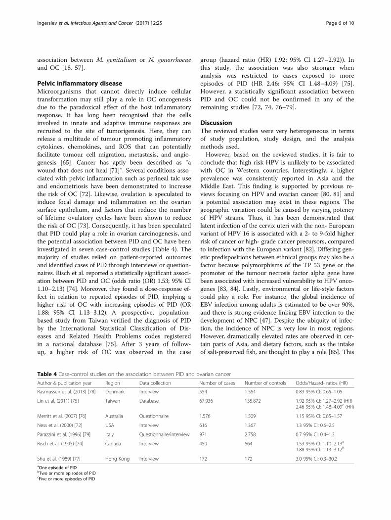
Pelvic inflammatory diseaseMicroorganisms that cannot directly induce cellulartransformation may still play a role in OC oncogenesisdue to the paradoxical effect of the host inflammatoryresponse. It has long been recognised that the cellsinvolved in innate and adaptive immune responses arerecruited to the site of tumorigenesis. Here, they canrelease a multitude of tumour promoting inflammatorycytokines, chemokines, and ROS that can potentiallyfacilitate tumour cell migration, metastasis, and angio-genesis [65]. Cancer has aptly been described as “awound that does not heal [71]”. Several conditions asso-ciated with pelvic inflammation such as perineal talc useand endometriosis have been demonstrated to increasethe risk of OC [72]. Likewise, ovulation is speculated toinduce focal damage and inflammation on the ovariansurface epithelium, and factors that reduce the numberof lifetime ovulatory cycles have been shown to reducethe risk of OC [73]. Consequently, it has been speculatedthat PID could play a role in ovarian carcinogenesis, andthe potential association between PID and OC have beeninvestigated in seven case-control studies (Table 4). Themajority of studies relied on patient-reported outcomesand identified cases of PID through interviews or question-naires. Risch et al. reported a statistically significant associ-ation between PID and OC (odds ratio (OR) 1.53; 95% CI1.10–2.13) [74]. Moreover, they found a dose-response ef-fect in relation to repeated episodes of PID, implying ahigher risk of OC with increasing episodes of PID (OR1.88; 95% CI 1.13–3.12). A prospective, population-based study from Taiwan verified the diagnosis of PIDby the International Statistical Classification of Dis-eases and Related Health Problems codes registeredin a national database [75]. After 3 years of follow-up, a higher risk of OC was observed in the case
group (hazard ratio (HR) 1.92; 95% CI 1.27–2.92)). Inthis study, the association was also stronger whenanalysis was restricted to cases exposed to moreepisodes of PID (HR 2.46; 95% CI 1.48–4.09) [75].However, a statistically significant association betweenPID and OC could not be confirmed in any of theremaining studies [72, 74, 76–79].
DiscussionThe reviewed studies were very heterogeneous in termsof study population, study design, and the analysismethods used.However, based on the reviewed studies, it is fair to
conclude that high-risk HPV is unlikely to be associatedwith OC in Western countries. Interestingly, a higherprevalence was consistently reported in Asia and theMiddle East. This finding is supported by previous re-views focusing on HPV and ovarian cancer [80, 81] anda potential association may exist in these regions. Thegeographic variation could be caused by varying potencyof HPV strains. Thus, it has been demonstrated thatlatent infection of the cervix uteri with the non- Europeanvariant of HPV 16 is associated with a 2- to 9-fold higherrisk of cancer or high- grade cancer precursors, comparedto infection with the European variant [82]. Differing gen-etic predispositions between ethnical groups may also be afactor because polymorphisms of the TP 53 gene or thepromoter of the tumour necrosis factor alpha gene havebeen associated with increased vulnerability to HPV onco-genes [83, 84]. Lastly, environmental or life-style factorscould play a role. For instance, the global incidence ofEBV infection among adults is estimated to be over 90%,and there is strong evidence linking EBV infection to thedevelopment of NPC [47]. Despite the ubiquity of infec-tion, the incidence of NPC is very low in most regions.However, dramatically elevated rates are observed in cer-tain parts of Asia, and dietary factors, such as the intakeof salt-preserved fish, are thought to play a role [85]. This
Table 4 Case-control studies on the association between PID and ovarian cancer
Author & publication year Region Data collection Number of cases Number of controls Odds/Hazard- ratios (HR)
Rasmussen et al. (2013) [78] Denmark Interview 554 1.564 0.83 95% CI: 0.65–1.05
Lin et al. (2011) [75] Taiwan Database 67.936 135.872 1.92 95% CI: 1.27–2.92 (HR)2.46 95% CI: 1.48–4.09c (HR)
Merritt et al. (2007) [76] Australia Questionnaire 1.576 1.509 1.15 95% CI: 0.85–1.57
Ness et al. (2000) [72] USA Interview 616 1.367 1.3 95% CI: 0.6–2.5
Parazzini et al. (1996) [79] Italy Questionnaire/interview 971 2.758 0.7 95% CI: 0.4–1.3
Risch et al. (1995) [74] Canada Interview 450 564 1.53 95% CI: 1.10–2.13a
1.88 95% CI: 1.13–3.12b
Shu et al. (1989) [77] Hong Kong Interview 172 172 3.0 95% CI: 0.3–30.2aOne episode of PIDbTwo or more episodes of PIDcFive or more episodes of PID
Ingerslev et al. Infectious Agents and Cancer (2017) 12:25 Page 6 of 10

underlines that, even though infection with oncogenic vi-ruses is common, cancer is a rare event that can arisefrom a combination of genetic, environmental, and life-style factors.Most of the epidemiological studies found an associ-
ation between OC and PID, but only two studies re-ported a statistically significant association [74, 75]. It isnoteworthy, however, that these studies also identified adose- response effect. This is one of the key points inHill’s criteria for causation that state that a biologicalgradient must be present, with more exposure to thesuspected agent leading to a larger effect [86]. Thesefindings are supported by studies that found evaluatedmarkers of inflammation such as CRP and interleukinsto be significantly associated with an increased risk ofOC [87]. A possible limitation to the reviewed studies isthat they relied primarily on patient-reported outcomes,which may be subject to recall bias. Furthermore, due tothe potential subclinical course of PID, patients can be un-aware of previous episodes. This is supported by studiesthat have demonstrated a relatively high seroprevalence ofchlamydia antibody titres in women that reported no his-tory of PID, and this issue would tend to produce bias to-wards the null [88, 89].Interestingly, CMV was reported in 50% of cases in a sin-
gle study, but this finding requires confirmation in futureand larger studies. No other viral agent was detected.The only detected bacteria were C. trachomatis and M.
genitalium. The results of this review do not support anassociation between C. trachomatis and OC, the majorityof studies reporting negative results. The association be-tween M. genitalium and OC was investigated and foundin only a single study and more research is needed.Several factors may account for the conflicting results.
First of all, there may be no connection between theinvestigated infectious agents and OC. Secondly, theheterogeneity of analysis methods, study populations, andstudy designs may play a part. However, other explanationsmust be discussed. Importantly, it is unclear to what extentsigns of microbiological agents are present in a tumour thatarises decades after infection. For some oncogenic viruses,like high-risk HPV, expression of viral oncogenes is obligatefor the maintenance of the malignant phenotype [90]. Inthese cases, viral genes will remain integrated and detect-able in tumour cell DNA. But another potential mechanismis debated in the “hit and run” hypothesis. Here, integrationof viral transforming genes and potential epigenetic repro-gramming of host cells initiate tumorigenesis, but the viralgene expression is subsequently lost during neoplastic de-velopment [91]. Accordingly, it has been demonstratedthat CMV oncogenes IE1 and IE2 can cooperate withadenovirus E1A gene to transform rat kidney cells. How-ever, expression of the transforming oncogenes was absentin the clonal cell lines from the transformed foci [92].
Finally, other oncogenic agents act more indirectly byinducing chronic inflammation that may function as aninitiator or promoter of carcinogenesis [93]. Consequently,the involved pathogen may no longer be present whencancer is diagnosed. For instance, despite the establishedrole of HCV in hepatocellular carcinoma, only a minorityof transformed hepatocytes contain viral RNA [94].These examples highlight the fact that the abscense
of microbiological agents in tumour tissue does notrule out their potential role in the transformation ofhost cells. These issues seem to strengthen the needfor serologic studies. However, serologic methods arelimited by the fact that antibody levels are likely to de-cline over time. In a study from Finland, 43% ofwomen had declining C. trachomatis antibody titres6 years after the diagnosis of PID [95]. Moreover, not allpatients with prior chlamydial infection will have detect-able antibodies [96].These obstacles call for a revised strategy in the
attempt to uncover the potential role of infectiousagents in OC. It is fair to assume that signs of micro-biological presence will be easier to detect when theinterval between primary infection and investigation isshort. Therefore, it seems more relevant to investigateprecursor lesions for the presence of potential micro-biological agents [91]. Until recently, this has beenimpossible due to the lack of a clearly defined prema-lignant lesion to OC. However, this has changed be-cause increasing evidence indicates that STIC lesionsin the distal fallopian tubes are precursor lesions toserous epithelial OC, which constitutes the majorityof OC cases [3]. Yet, no explanation has been formu-lated as to why STIC lesions arise. Since the fallopiantubes are often affected and damaged by chronic PID,it is biological plausible that pathogens with trans-forming capacities, or the chronic inflammation theyinduce, could lead to subsequent neoplastic trans-formation. We therefore recommend that future stud-ies focus on the detection of bacterial or viral agents infallopian tube tissue samples with STIC lesions verifiedthrough the “Sectioning and Extensively Examining of theFimbriated end” protocol.
ConclusionThe reviewed articles indicate a potential associationbetween PID and OC. An association between OC andhigh-risk HPV genotypes may exist in Asia, whereas anassociation in Western countries seems unlikely due tothe low reported prevalence. Two studies reported anassociation between bacterial agents and OC, but themajority of bacterial studies reported negative findings.However, the available literature was scarce and morestudies are warranted.
Ingerslev et al. Infectious Agents and Cancer (2017) 12:25 Page 7 of 10
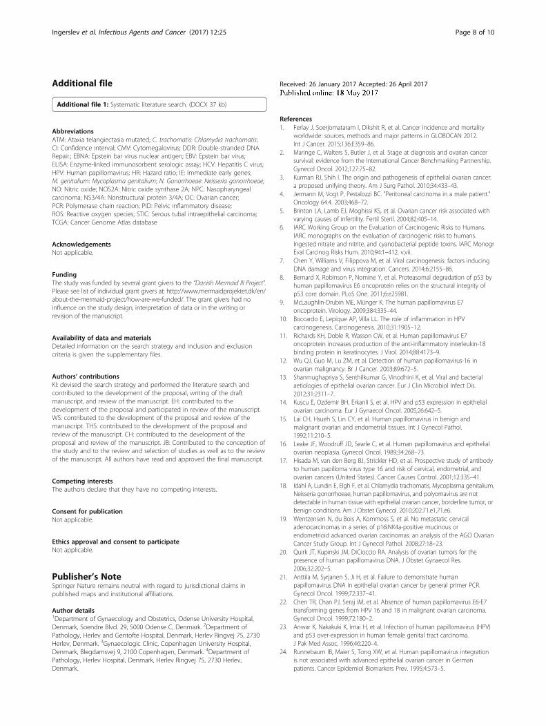
Additional file
Additional file 1: Systematic literature search. (DOCX 37 kb)
AbbreviationsATM: Ataxia telangiectasia mutated; C. trachomatis: Chlamydia trachomatis;CI: Confidence interval; CMV: Cytomegalovirus; DDR: Double-stranded DNARepair.; EBNA: Epstein bar virus nuclear antigen; EBV: Epstein bar virus;ELISA: Enzyme-linked immunosorbent serologic assay; HCV: Hepatitis C virus;HPV: Human papillomavirus; HR: Hazard ratio; IE: Immediate early genes;M. genitalium: Mycoplasma genitalium; N. Gonorrhoeae: Neisseria gonorrhoeae;NO: Nitric oxide; NOS2A: Nitric oxide synthase 2A; NPC: Nasopharyngealcarcinoma; NS3/4A: Nonstructural protein 3/4A; OC: Ovarian cancer;PCR: Polymerase chain reaction; PID: Pelvic inflammatory disease;ROS: Reactive oxygen species; STIC: Serous tubal intraepithelial carcinoma;TCGA: Cancer Genome Atlas database
AcknowledgementsNot applicable.
FundingThe study was funded by several grant givers to the “Danish Mermaid III Project”.Please see list of individual grant givers at: http://www.mermaidprojektet.dk/en/about-the-mermaid-project/how-are-we-funded/. The grant givers had noinfluence on the study design, interpretation of data or in the writing orrevision of the manuscript.
Availability of data and materialsDetailed information on the search strategy and inclusion and exclusioncriteria is given the supplementary files.
Authors’ contributionsKI: devised the search strategy and performed the literature search andcontributed to the development of the proposal, writing of the draftmanuscript, and review of the manuscript. EH: contributed to thedevelopment of the proposal and participated in review of the manuscript.WS: contributed to the development of the proposal and review of themanuscript. THS: contributed to the development of the proposal andreview of the manuscript. CH: contributed to the development of theproposal and review of the manuscript. JB: Contributed to the conception ofthe study and to the review and selection of studies as well as to the reviewof the manuscript. All authors have read and approved the final manuscript.
Competing interestsThe authors declare that they have no competing interests.
Consent for publicationNot applicable.
Ethics approval and consent to participateNot applicable.
Publisher’s NoteSpringer Nature remains neutral with regard to jurisdictional claims inpublished maps and institutional affiliations.
Author details1Department of Gynaecology and Obstetrics, Odense University Hospital,Denmark, Soendre Blvd. 29, 5000 Odense C, Denmark. 2Department ofPathology, Herlev and Gentofte Hospital, Denmark, Herlev Ringvej 75, 2730Herlev, Denmark. 3Gynaecologic Clinic, Copenhagen University Hospital,Denmark, Blegdamsvej 9, 2100 Copenhagen, Denmark. 4Department ofPathology, Herlev Hospital, Denmark, Herlev Ringvej 75, 2730 Herlev,Denmark.
Received: 26 January 2017 Accepted: 26 April 2017
References1. Ferlay J, Soerjomataram I, Dikshit R, et al. Cancer incidence and mortality
worldwide: sources, methods and major patterns in GLOBOCAN 2012.Int J Cancer. 2015;136:E359–86.
2. Maringe C, Walters S, Butler J, et al. Stage at diagnosis and ovarian cancersurvival: evidence from the International Cancer Benchmarking Partnership.Gynecol Oncol. 2012;127:75–82.
3. Kurman RJ, Shih I. The origin and pathogenesis of epithelial ovarian cancer:a proposed unifying theory. Am J Surg Pathol. 2010;34:433–43.
4. Jermann M, Vogt P, Pestalozzi BC. "Peritoneal carcinoma in a male patient."Oncology 64.4. 2003;468–72.
5. Brinton LA, Lamb EJ, Moghissi KS, et al. Ovarian cancer risk associated withvarying causes of infertility. Fertil Steril. 2004;82:405–14.
6. IARC Working Group on the Evaluation of Carcinogenic Risks to Humans.IARC monographs on the evaluation of carcinogenic risks to humans.Ingested nitrate and nitrite, and cyanobacterial peptide toxins. IARC MonogrEval Carcinog Risks Hum. 2010;94:1–412. v,vii.
7. Chen Y, Williams V, Filippova M, et al. Viral carcinogenesis: factors inducingDNA damage and virus integration. Cancers. 2014;6:2155–86.
8. Bernard X, Robinson P, Nomine Y, et al. Proteasomal degradation of p53 byhuman papillomavirus E6 oncoprotein relies on the structural integrity ofp53 core domain. PLoS One. 2011;6:e25981.
9. McLaughlin-Drubin ME, Münger K. The human papillomavirus E7oncoprotein. Virology. 2009;384:335–44.
10. Boccardo E, Lepique AP, Villa LL. The role of inflammation in HPVcarcinogenesis. Carcinogenesis. 2010;31:1905–12.
11. Richards KH, Doble R, Wasson CW, et al. Human papillomavirus E7oncoprotein increases production of the anti-inflammatory interleukin-18binding protein in keratinocytes. J Virol. 2014;88:4173–9.
12. Wu QJ, Guo M, Lu ZM, et al. Detection of human papillomavirus-16 inovarian malignancy. Br J Cancer. 2003;89:672–5.
13. Shanmughapriya S, Senthilkumar G, Vinodhini K, et al. Viral and bacterialaetiologies of epithelial ovarian cancer. Eur J Clin Microbiol Infect Dis.2012;31:2311–7.
14. Kuscu E, Ozdemir BH, Erkanli S, et al. HPV and p53 expression in epithelialovarian carcinoma. Eur J Gynaecol Oncol. 2005;26:642–5.
15. Lai CH, Hsueh S, Lin CY, et al. Human papillomavirus in benign andmalignant ovarian and endometrial tissues. Int J Gynecol Pathol.1992;11:210–5.
16. Leake JF, Woodruff JD, Searle C, et al. Human papillomavirus and epithelialovarian neoplasia. Gynecol Oncol. 1989;34:268–73.
17. Hisada M, van den Berg BJ, Strickler HD, et al. Prospective study of antibodyto human papilloma virus type 16 and risk of cervical, endometrial, andovarian cancers (United States). Cancer Causes Control. 2001;12:335–41.
18. Idahl A, Lundin E, Elgh F, et al. Chlamydia trachomatis, Mycoplasma genitalium,Neisseria gonorrhoeae, human papillomavirus, and polyomavirus are notdetectable in human tissue with epithelial ovarian cancer, borderline tumor, orbenign conditions. Am J Obstet Gynecol. 2010;202:71.e1,71.e6.
19. Wentzensen N, du Bois A, Kommoss S, et al. No metastatic cervicaladenocarcinomas in a series of p16INK4a-positive mucinous orendometrioid advanced ovarian carcinomas: an analysis of the AGO OvarianCancer Study Group. Int J Gynecol Pathol. 2008;27:18–23.
20. Quirk JT, Kupinski JM, DiCioccio RA. Analysis of ovarian tumors for thepresence of human papillomavirus DNA. J Obstet Gynaecol Res.2006;32:202–5.
21. Anttila M, Syrjanen S, Ji H, et al. Failure to demonstrate humanpapillomavirus DNA in epithelial ovarian cancer by general primer PCR.Gynecol Oncol. 1999;72:337–41.
22. Chen TR, Chan PJ, Seraj IM, et al. Absence of human papillomavirus E6-E7transforming genes from HPV 16 and 18 in malignant ovarian carcinoma.Gynecol Oncol. 1999;72:180–2.
23. Anwar K, Nakakuki K, Imai H, et al. Infection of human papillomavirus (HPV)and p53 over-expression in human female genital tract carcinoma.J Pak Med Assoc. 1996;46:220–4.
24. Runnebaum IB, Maier S, Tong XW, et al. Human papillomavirus integrationis not associated with advanced epithelial ovarian cancer in Germanpatients. Cancer Epidemiol Biomarkers Prev. 1995;4:573–5.
Ingerslev et al. Infectious Agents and Cancer (2017) 12:25 Page 8 of 10

25. Trottier AM, Provencher D, Mes-Masson AM, et al. Absence of humanpapillomavirus sequences in ovarian pathologies. J Clin Microbiol.1995;33:1011–3.
26. Beckmann AM, Sherman KJ, Saran L, et al. Genital-type human papillomavirusinfection is not associated with surface epithelial ovarian carcinoma. GynecolOncol. 1991;43:247–51.
27. de Villiers E, Schneider A, Gross G, et al. Analysis of benign and malignanturogenital tumors for human papillomavirus infection by labelling cellularDNA. Med Microbiol Immunol (Berl). 1986;174:281–6.
28. Ingerslev K, Hogdall E, Skovrider-Ruminski W, et al. High-risk HPV is notassociated with epithelial ovarian cancer in a Caucasian population. InfectAgent Cancer. 2016;11:1.
29. Al-Shabanah OA, Hafez MM, Hassan ZK, et al. Human papillomavirusgenotyping and integration in ovarian cancer Saudi patients. Virol J.2013;10:343. 422X-10-343.
30. Malisic E, Jankovic R, Jakovljevic K. Detection and genotyping of humanpapillomaviruses and their role in the development of ovarian carcinomas.Arch Gynecol Obstet. 2012;286:723–8.
31. Bilyk OO, Pande NT, Buchynska LG. Analysis of p53, p16(INK4a), pRb andCyclin D1 expression and human papillomavirus in primary ovarian serouscarcinomas. Exp Oncol. 2011;33:150–6.
32. Giordano G, D’Adda T, Gnetti L, et al. Role of human papillomavirus in thedevelopment of epithelial ovarian neoplasms in Italian women. J ObstetGynaecol Res. 2008;34:210–7.
33. Atalay F, Taskiran C, Taner MZ, et al. Detection of human papillomavirusDNA and genotyping in patients with epithelial ovarian carcinoma. J ObstetGynaecol Res. 2007;33:823–8.
34. Yang HJ, Liu VW, Tsang PC, et al. Comparison of human papillomavirus DNAlevels in gynecological cancers: implication for cancer development.Tumour Biol. 2003;24:310–6.
35. Ip SM, Wong LC, Xu CM, et al. Detection of human papillomavirus DNA inmalignant lesions from Chinese women with carcinomas of the uppergenital tract. Gynecol Oncol. 2002;87:104–11.
36. Li T, Lu Z-M, Guo M, et al. p53 Codon 72 polymorphism (C/G) and the risk ofhuman papillomavirus-associated carcinomas in China. Cancer. 2002;95:2571–6.
37. Strickler HD, Schiffman MH, Shah KV, et al. A survey of human papillomavirus 16antibodies in patients with epithelial cancers. Eur J Cancer Prev. 1998;7:305–13.
38. Zimna K, Poreba E, Kedzia W, et al. Human papillomavirus (HPV) in uppergenital tract carcinomas of women. Eur J Gynaecol Oncol. 1997;18:415–7.
39. Lai CH, Wang CY, Lin CY, et al. Detection of human papillomavirus RNA inovarian and endometrial carcinomas by reverse transcription/polymerasechain reaction. Gynecol Obstet Invest. 1994;38:276–80.
40. Roos P, Orlando PA, Fagerstrom RM, et al. In North America, some ovariancancers express the oncogenes of preventable human papillomavirusHPV-18. Sci Rep. 2015;5:8645.
41. Khoury JD, Tannir NM, Williams MD, et al. Landscape of DNA virusassociations across human malignant cancers: Analysis of 3,775 cases usingRNA-seq. J Virol. 2013;87:8916–26.
42. Kazemian M, Ren M, Lin JX, et al. Possible human papillomavirus 38contamination of endometrial cancer rna sequencing samples in the cancergenome atlas database. J Virol. 2015;89:8967–73.
43. Cantalupo PG, Katz JP, Pipas JM. HeLa nucleic acid contamination in thecancer genome atlas leads to the misidentification of human papillomavirus18. J Virol. 2015;89:4051–7.
44. Cinatl J, Scholz M, Kotchetkov R, et al. Molecular mechanisms of themodulatory effects of HCMV infection in tumor cell biology. Trends MolMed. 2004;10:19–23.
45. Michaelis M, Doerr HW, Cinatl J. The story of human cytomegalovirus andcancer: increasing evidence and open questions. Neoplasia. 2009;11:1–9.
46. de-The G, Day NE, Geser A, et al. Sero-epidemiology of the Epstein-Barrvirus: preliminary analysis of an international study - a review. IARC Sci Publ.1975;11 Pt 2:3–16.
47. Coghill AE, Hildesheim A. Epstein-Barr virus antibodies and the risk of associatedmalignancies: review of the literature. Am J Epidemiol. 2014;180:687–95.
48. Grywalska E, Rolinski J. Epstein-barr virus–associated lymphomas. SeminOncol. 2015;42:291–303.
49. Jha HC, Banerjee S, Robertson ES. The role of gammaherpesviruses incancer pathogenesis. Pathogens. 2016;5:18.
50. Donato F, Tagger A, Gelatti U, et al. Alcohol and hepatocellular carcinoma:the effect of lifetime intake and hepatitis virus infections in men andwomen. Am J Epidemiol. 2002;155:323–31.
51. Mitchell JK, Lemon SM, McGivern DR. How do persistent infections withhepatitis C virus cause liver cancer? Curr Opin Virol. 2015;14:101–8.
52. Choi J, Corder NL, Koduru B, et al. Oxidative stress and hepatic Nox proteinsin chronic hepatitis C and hepatocellular carcinoma. Free Radic Biol Med.2014;72:267–84.
53. Duong FH, Christen V, Lin S, et al. Hepatitis C virus–induced up‐regulationof protein phosphatase 2A inhibits histone modification and DNA damagerepair. Hepatology. 2010;51:741–51.
54. Khalili K, Del Valle L, Otte J, et al. Human neurotropic polyomavirus, JCV, andits role in carcinogenesis. Oncogene. 2003;22:5181–91.
55. Caracciolo V, Reiss K, Khalili K, et al. Role of the interaction between large Tantigen and Rb family members in the oncogenicity of JC virus. Oncogene.2006;25:5294–301.
56. Barbanti-Brodano G, Sabbioni S, Martini F, et al. BK virus, JC virus and SimianVirus 40 infection in humans, and association with human tumors.Polyomaviruses and human diseases. New York: Springer; 2006:319–41.
57. Idahl A, Lundin E, Jurstrand M, et al. Chlamydia trachomatis andMycoplasma genitalium plasma antibodies in relation to epithelial ovariantumors. Infect Dis Obstet Gynecol. 2011;2011:824627.
58. Ness RB, Shen C, Bass D, et al. Chlamydia trachomatis serology inwomen with and without ovarian cancer. Infect Dis Obstet Gynecol.2008;2008:219672.
59. Wong A, Maclean AB, Furrows SJ, et al. Could epithelial ovarian cancer beassociated with chlamydial infection? Eur J Gynaecol Oncol. 2007;28:117–20.
60. Ness RB, Goodman MT, Shen C, et al. Serologic evidence of past infectionwith Chlamydia trachomatis, in relation to ovarian cancer. J Infect Dis.2003;187:1147–52.
61. Martin DC, Khare VK, Miller BE, et al. Association of positive Chlamydiatrachomatis and Chlamydia pneumoniae immunoglobulin-gamma titerswith increasing age. J Am Assoc Gynecol Laparosc. 1997;4:583–6.
62. Abdul-Sater AA, Said-Sadier N, Lam VM, et al. Enhancement of reactiveoxygen species production and chlamydial infection by the mitochondrialNod-like family member NLRX1. J Biol Chem. 2010;285:41637–45.
63. Chumduri C, Gurumurthy RK, Zadora PK, et al. Chlamydia infectionpromotes host DNA damage and proliferation but impairs the DNAdamage response. Cell Host Microbe. 2013;13:746–58.
64. Gonzalez E, Rother M, Kerr MC, et al. Chlamydia infection depends on afunctional MDM2-p53 axis. Nat Commun. 2014;5:5201.
65. Hanahan D, Weinberg RA. Hallmarks of cancer: the next generation. Cell.2011;144:646–74.
66. Michaud D, Platz E, Giovannucci E. Gonorrhoea and male bladder cancer ina prospective study. Br J Cancer. 2007;96:169–71.
67. Lian WQ, Luo F, Song XL, et al. Gonorrhea and prostate cancer incidence:an updated meta-analysis of 21 epidemiologic studies. Med Sci Monit.2015;21:1902–10.
68. Vielfort K, Soderholm N, Weyler L, et al. Neisseria gonorrhoeae infectioncauses DNA damage and affects the expression of p21, p27 and p53 innon-tumor epithelial cells. J Cell Sci. 2013;126:339–47.
69. Namiki K, Goodison S, Porvasnik S, et al. Persistent exposure to Mycoplasmainduces malignant transformation of human prostate cells. PLoS One.2009;4:e6872.
70. Chan P, Seraj I, Kalugdan T, et al. Prevalence of Mycoplasma conserved DNAin malignant ovarian cancer detected using sensitive PCR-ELISA. GynecolOncol. 1996;63:258–60.
71. Dvorak HF. Tumors: wounds that do not heal. N Engl J Med. 1986;315:1650–9.72. Ness RB, Grisso JA, Cottreau C, et al. Factors related to inflammation of the
ovarian epithelium and risk of ovarian cancer. Epidemiology. 2000;11:111–7.73. Li J, Fadare O, Xiang L, et al. Ovarian serous carcinoma: recent concepts on
its origin and carcinogenesis. J Hematol Oncol. 2012;5:1.74. Risch HA, Howe GR. Pelvic inflammatory disease and the risk of epithelial
ovarian cancer. Cancer Epidemiol Biomarkers Prev. 1995;4:447–51.75. Lin HW, Tu YY, Lin SY, et al. Risk of ovarian cancer in women with pelvic
inflammatory disease: a population-based study. Lancet Oncol. 2011;12:900–4.76. Merritt MA, Green AC, Nagle CM, et al. Talcum powder, chronic pelvic
inflammation and NSAIDs in relation to risk of epithelial ovarian cancer.Int J Cancer. 2008;122:170–6.
77. Shu XO, Brinton LA, Gao YT, et al. Population-based case-control study ofovarian cancer in Shanghai. Cancer Res. 1989;49:3670–4.
78. Rasmussen CB, Faber MT, Jensen A, et al. Pelvic inflammatory disease andrisk of invasive ovarian cancer and ovarian borderline tumors. CancerCauses Control. 2013;24:1459–64.
Ingerslev et al. Infectious Agents and Cancer (2017) 12:25 Page 9 of 10
79. Parazzini F, La Vecchia C, Negri E, et al. Pelvic inflammatory disease and riskof ovarian cancer. Cancer Epidemiol Biomarkers Prev. 1996;5:667–9.
80. Svahn MF, Faber MT, Christensen J, et al. Prevalence of humanpapillomavirus in epithelial ovarian cancer tissue. A meta-analysis ofobservational studies. Acta Obstet Gynecol Scand. 2014;93:6–19.
81. Rosa MI, Silva GD, de Azedo Simoes PW, et al. The prevalence of humanpapillomavirus in ovarian cancer: a systematic review. Int J Gynecol Cancer.2013;23:437–41.
82. Hildesheim A, Wang SS. Host and viral genetics and risk of cervical cancer: areview. Virus Res. 2002;89:229–40.
83. Zhou X, Gu Y, Zhang S. Association between p53 codon 72 polymorphismand cervical cancer risk among Asians: a HuGE review and meta-analysis.Asian Pac J Cancer Prev. 2012;13:4909–14.
84. Liu L, Yang X, Chen X, et al. Association between TNF-α polymorphisms andcervical cancer risk: a meta-analysis. Mol Biol Rep. 2012;39:2683–8.
85. Chang ET, Adami HO. The enigmatic epidemiology of nasopharyngealcarcinoma. Cancer Epidemiol Biomarkers Prev. 2006;15:1765–77.
86. HILL AB. The environment and disease: association or causation? Proc R SocMed. 1965;58:295–300.
87. Charbonneau B, Goode EL, Kalli KR, et al. The immune system in thepathogenesis of ovarian cancer. Crit Rev Immunol. 2013;33:137–64.
88. Bjercke S, Purvis K. Characteristics of women under fertility investigationwith IgA/IgG seropositivity for Chlamydia trachomatis. Eur J Obstet GynecolReprod Biol. 1993;51:157–61.
89. Stamm WE, Holmes KK. Chlamydia trachomatis infections of the adult.Sex Transm Dis. 1999;3:407–22.
90. DeFilippis RA, Goodwin EC, Wu L, et al. Endogenous human papillomavirusE6 and E7 proteins differentially regulate proliferation, senescence, andapoptosis in HeLa cervical carcinoma cells. J Virol. 2003;77:1551–63.
91. Niller HH, Wolf H, Minarovits J. Viral hit and run-oncogenesis: genetic andepigenetic scenarios. Cancer Lett. 2011;305:200–17.
92. Shen Y, Zhu H, Shenk T. Human cytomagalovirus IE1 and IE2 proteins aremutagenic and mediate “hit-and-run” oncogenic transformation incooperation with the adenovirus E1A proteins. Proc Natl Acad Sci U S A.1997;94:3341–5.
93. Grivennikov SI, Greten FR, Karin M. Immunity, inflammation, and cancer. Cell.2010;140:883–99.
94. Kandathil AJ, Graw F, Quinn J, et al. Use of laser capture microdissection tomap hepatitis C virus–positive hepatocytes in human liver. Gastroenterology.2013;145:1404,1413. e10.
95. Puolakkainen M, Vesterinen E, Purola E, et al. Persistence of chlamydialantibodies after pelvic inflammatory disease. J Clin Microbiol. 1986;23:924–8.
96. Bas S, Muzzin P, Ninet B, et al. Chlamydial serology: comparative diagnosticvalue of immunoblotting, microimmunofluorescence test, and immunoassaysusing different recombinant proteins as antigens. J Clin Microbiol.2001;39:1368–77.
• We accept pre-submission inquiries
• Our selector tool helps you to find the most relevant journal
• We provide round the clock customer support
• Convenient online submission
• Thorough peer review
• Inclusion in PubMed and all major indexing services
• Maximum visibility for your research
Submit your manuscript atwww.biomedcentral.com/submit
Submit your next manuscript to BioMed Central and we will help you at every step:
Ingerslev et al. Infectious Agents and Cancer (2017) 12:25 Page 10 of 10
Copyright © 2022 FDOKUMEN