Infectious vaccine-derived rubella viruses emerge ... - PLOS
32
RESEARCH ARTICLE Infectious vaccine-derived rubella viruses emerge, persist, and evolve in cutaneous granulomas of children with primary immunodeficiencies Ludmila Perelygina ID 1 , Min-hsin Chen 1 , Suganthi Suppiah 1 , Adebola Adebayo 1 , Emily Abernathy 1 , Morna Dorsey 2 , Lionel Bercovitch 3 , Kenneth Paris 4 , Kevin P. White ID 5 , Alfons Krol ID 5 , Julie Dhossche ID 5 , Ivan Y. Torshin ID 6 , Natalie Saini 7 , Leszek J. Klimczak ID 8 , Dmitry A. Gordenin ID 7 , Andrey Zharkikh 9 , Stanley Plotkin 10 , Kathleen E. Sullivan 11 , Joseph Icenogle ID 1 * 1 Division of Viral Diseases, Centers for Disease Control and Prevention, Atlanta, Georgia, United States of America, 2 Department of Pediatrics, University of California, San Francisco, San Francisco, California, United States of America, 3 Department of Dermatology, Hasbro Children’s Hospital and Warren Alpert Medical School of Brown University, Providence, Rhode Island, United States of America, 4 Division of Allergy and Immunology, Children’s Hospital New Orleans, New Orleans, Louisiana, United States of America, 5 Department of Dermatology, Oregon Health & Science University, Portland, Oregon, United States of America, 6 Institute of Pharmacoinformatics, Federal Research Center “Computer Science and Control” of Russian Academy of Sciences, Dorodnicyn Computing Center, Moscow, Russian Federation, 7 Genome Integrity and Structural Biology Laboratory, National Institute of Environmental Health Sciences, US National Institutes of Health, Research Triangle Park, North Carolina, United States of America, 8 Integrative Bioinformatics Support Group, National Institute of Environmental Health Sciences, US National Institutes of Health, Research Triangle Park, North Carolina, United States of America, 9 Myriad Genetics, Inc., Salt Lake City, Utah, United States of America, 10 University of Pennsylvania Perelman School of Medicine, Philadelphia, Pennsylvania, United States of America, 11 Division of Allergy and Immunology, The Children’s Hospital of Philadelphia, Philadelphia, Pennsylvania, United States of America * [email protected] Abstract Rubella viruses (RV) have been found in an association with granulomas in children with pri- mary immune deficiencies (PID). Here, we report the recovery and characterization of infec- tious immunodeficiency-related vaccine-derived rubella viruses (iVDRV) from diagnostic skin biopsies of four patients. Sequence evolution within PID hosts was studied by compari- son of the complete genomic sequences of the iVDRVs with the genome of the vaccine virus RA27/3. The degree of divergence of each iVDRV correlated with the duration of per- sistence indicating continuous intrahost evolution. The evolution rates for synonymous and nonsynonymous substitutions were estimated to be 5.7 x 10 −3 subs/site/year and 8.9 x 10 −4 subs/site/year, respectively. Mutational spectra and signatures indicated a major role for APOBEC cytidine deaminases and a secondary role for ADAR adenosine deaminases in generating diversity of iVDRVs. The distributions of mutations across the genes and 3D hot- spots for amino acid substitutions in the E1 glycoprotein identified regions that may be under positive selective pressure. Quasispecies diversity was higher in granulomas than in recovered infectious iVDRVs. Growth properties of iVDRVs were assessed in WI-38 fibro- blast cultures. None of the iVDRV isolates showed complete reversion to wild type PLOS Pathogens | https://doi.org/10.1371/journal.ppat.1008080 October 28, 2019 1 / 32 a1111111111 a1111111111 a1111111111 a1111111111 a1111111111 OPEN ACCESS Citation: Perelygina L, Chen M-h, Suppiah S, Adebayo A, Abernathy E, Dorsey M, et al. (2019) Infectious vaccine-derived rubella viruses emerge, persist, and evolve in cutaneous granulomas of children with primary immunodeficiencies. PLoS Pathog 15(10): e1008080. https://doi.org/10.1371/ journal.ppat.1008080 Editor: Adam S. Lauring, University of Michigan, UNITED STATES Received: April 24, 2019 Accepted: September 13, 2019 Published: October 28, 2019 Copyright: This is an open access article, free of all copyright, and may be freely reproduced, distributed, transmitted, modified, built upon, or otherwise used by anyone for any lawful purpose. The work is made available under the Creative Commons CC0 public domain dedication. Data Availability Statement: All sequences of iVDRV genomes are available from the GenBank database (accession number(s) MK787188 - MK787191 and MK780807- MK780812) Funding: This work was supported by core funding from the Centers for Disease Control and Prevention, by a grant to K.E.S. from the National Institute of Health (R21-AI130967-01A1) and by the US National Institute of Health Intramural Research Program Project Z1AES103266 to D.A.G.
-
Upload
khangminh22 -
Category
Documents
-
view
0 -
download
0
Transcript of Infectious vaccine-derived rubella viruses emerge ... - PLOS
RESEARCH ARTICLE
Infectious vaccine-derived rubella viruses
emerge, persist, and evolve in cutaneous
granulomas of children with primary
immunodeficiencies
Ludmila PerelyginaID1, Min-hsin Chen1, Suganthi Suppiah1, Adebola Adebayo1,
Emily Abernathy1, Morna Dorsey2, Lionel Bercovitch3, Kenneth Paris4, Kevin P. WhiteID5,
Alfons KrolID5, Julie DhosscheID
5, Ivan Y. TorshinID6, Natalie Saini7, Leszek
J. KlimczakID8, Dmitry A. GordeninID
7, Andrey Zharkikh9, Stanley Plotkin10, Kathleen
E. Sullivan11, Joseph IcenogleID1*
1 Division of Viral Diseases, Centers for Disease Control and Prevention, Atlanta, Georgia, United States of
America, 2 Department of Pediatrics, University of California, San Francisco, San Francisco, California,
United States of America, 3 Department of Dermatology, Hasbro Children’s Hospital and Warren Alpert
Medical School of Brown University, Providence, Rhode Island, United States of America, 4 Division of
Allergy and Immunology, Children’s Hospital New Orleans, New Orleans, Louisiana, United States of
America, 5 Department of Dermatology, Oregon Health & Science University, Portland, Oregon, United
States of America, 6 Institute of Pharmacoinformatics, Federal Research Center “Computer Science and
Control” of Russian Academy of Sciences, Dorodnicyn Computing Center, Moscow, Russian Federation,
7 Genome Integrity and Structural Biology Laboratory, National Institute of Environmental Health Sciences,
US National Institutes of Health, Research Triangle Park, North Carolina, United States of America,
8 Integrative Bioinformatics Support Group, National Institute of Environmental Health Sciences, US National
Institutes of Health, Research Triangle Park, North Carolina, United States of America, 9 Myriad Genetics,
Inc., Salt Lake City, Utah, United States of America, 10 University of Pennsylvania Perelman School of
Medicine, Philadelphia, Pennsylvania, United States of America, 11 Division of Allergy and Immunology, The
Children’s Hospital of Philadelphia, Philadelphia, Pennsylvania, United States of America
Abstract
Rubella viruses (RV) have been found in an association with granulomas in children with pri-
mary immune deficiencies (PID). Here, we report the recovery and characterization of infec-
tious immunodeficiency-related vaccine-derived rubella viruses (iVDRV) from diagnostic
skin biopsies of four patients. Sequence evolution within PID hosts was studied by compari-
son of the complete genomic sequences of the iVDRVs with the genome of the vaccine
virus RA27/3. The degree of divergence of each iVDRV correlated with the duration of per-
sistence indicating continuous intrahost evolution. The evolution rates for synonymous and
nonsynonymous substitutions were estimated to be 5.7 x 10⁻³ subs/site/year and 8.9 x 10⁻⁴
subs/site/year, respectively. Mutational spectra and signatures indicated a major role for
APOBEC cytidine deaminases and a secondary role for ADAR adenosine deaminases in
generating diversity of iVDRVs. The distributions of mutations across the genes and 3D hot-
spots for amino acid substitutions in the E1 glycoprotein identified regions that may be
under positive selective pressure. Quasispecies diversity was higher in granulomas than in
recovered infectious iVDRVs. Growth properties of iVDRVs were assessed in WI-38 fibro-
blast cultures. None of the iVDRV isolates showed complete reversion to wild type
PLOS Pathogens | https://doi.org/10.1371/journal.ppat.1008080 October 28, 2019 1 / 32
a1111111111
a1111111111
a1111111111
a1111111111
a1111111111
OPEN ACCESS
Citation: Perelygina L, Chen M-h, Suppiah S,
Adebayo A, Abernathy E, Dorsey M, et al. (2019)
Infectious vaccine-derived rubella viruses emerge,
persist, and evolve in cutaneous granulomas of
children with primary immunodeficiencies. PLoS
Pathog 15(10): e1008080. https://doi.org/10.1371/
journal.ppat.1008080
Editor: Adam S. Lauring, University of Michigan,
UNITED STATES
Received: April 24, 2019
Accepted: September 13, 2019
Published: October 28, 2019
Copyright: This is an open access article, free of all
copyright, and may be freely reproduced,
distributed, transmitted, modified, built upon, or
otherwise used by anyone for any lawful purpose.
The work is made available under the Creative
Commons CC0 public domain dedication.
Data Availability Statement: All sequences of
iVDRV genomes are available from the GenBank
database (accession number(s) MK787188 -
MK787191 and MK780807- MK780812)
Funding: This work was supported by core funding
from the Centers for Disease Control and
Prevention, by a grant to K.E.S. from the National
Institute of Health (R21-AI130967-01A1) and by
the US National Institute of Health Intramural
Research Program Project Z1AES103266 to D.A.G.
phenotype but the replicative and persistence characteristics of iVDRVs were different from
those of the RA27/3 vaccine strain, making predictions of iVDRV transmissibility and terato-
genicity difficult. However, detection of iVDRV RNA in nasopharyngeal specimen and poor
neutralization of some iVDRV strains by sera from vaccinated persons suggests possible
public health risks associated with iVDRV carriers. Detection of IgM antibody to RV in sera
of two out of three patients may be a marker of virus persistence, potentially useful for identi-
fying patients with iVDRV before development of lesions. Studies of the evolutionary dynam-
ics of iVDRV during persistence will contribute to development of infection control strategies
and antiviral therapies.
Author summary
Primary immunodeficiency diseases (PID) are caused by genetic defects and lead to seri-
ous problems including chronic granulomas (abnormal collections (nodules) of inflam-
matory cells), sometimes lasting for decades and sometimes leading to severe ulcers.
Initial reports (2014–2016), including our report of a blinded study using ultrasensitive
virus detection in biopsies, proved the association between granuloma of the skin in PID
patients and rubella virus. The viruses in these reports and the current report were derived
from a widely used vaccine strain of the rubella virus. Work reported here shows that
these vaccine-derived viruses are biologically different from the vaccine virus and that
their genomes have changed. Genomic changes could be analyzed largely because the
exact sequence of starting vaccine virus genome was known. These genomic differences
are likely generated via mechanisms similar to those occurring during normal circulation
of wild type rubella. We present data that newly recognized mechanisms for generation of
sequence diversity in viruses (because of cellular deaminases) likely occurs in the genera-
tion of these vaccine-derived rubella viruses. Thousands of PID patients in the United
States are likely shedding these vaccine-derived rubella viruses. Our work presented here
characterizing viruses in diagnostic specimens highlights at least two areas where insuffi-
cient work has been done: 1) research on the properties of rubella virus (limited under-
standing of the antibody binding sites on the virus); 2) controlled research studies to
assess the public health impact of viruses in populations with high immunity.
Introduction
Rubella virus (RV) is an enveloped, single-stranded, positive-sense RNA virus in the Rubivirusgenus, which has been recently moved from the Togaviridae to a new family, Matonaviridae[1]. A total of 13 RV genotypes, which represent 2 clades, have been recognized, but 2 geno-
types, 1E and 2B, are currently the most common worldwide. RV replicates at low levels and
produces little cytopathology both in vitro and in vivo. A distinct feature of RV is the ability to
persist in the placenta and fetus and in immune privileged body sites of immunologically com-
petent individuals [2, 3]. Persistent RV infection is associated with a congenital rubella syn-
drome (CRS) and a number of less common pathologies such as rubella encephalitis and
Fuchs uveitis [4, 5]. The live attenuated vaccine strain, RA27/3 (a virus from the likely extinct
1a genotype and a part of the MMR vaccine), is currently used in the US and globally. It
has high immunogenicity, generates long-term immunity after a single dose, is effective in
preventing clinical disease, and has a very low rate of adverse events [6]. Worldwide,
Infectious vaccine-derived rubella viruses in immunodeficiencies
PLOS Pathogens | https://doi.org/10.1371/journal.ppat.1008080 October 28, 2019 2 / 32
K.E.S. was supported by the Wallace Chair of
Pediatrics. The findings and conclusions in this
report are those of the authors and do not
necessarily represent the official position of the
United States Centers for Disease Control and
Prevention. The funders had no role in study
design, data collection and analysis, decision to
publish, or preparation of the manuscript.
Competing interests: The authors have read the
journal’s policy and have the following conflicts:
Andrey Zharkikh is an employee of Myriad
Genetics. This company works in cancer genetics
and diagnostics without any relation to pathogen
studies and treatments. This does not alter our
adherence to all PLOS Pathogens policies on
sharing data and materials.
implementation of rubella vaccination programs has resulted in elimination of rubella and
CRS from the Americas and significant reduction in the burden of disease in some developed
countries [7]. Similar to wild type RV, RA27/3 can persist in immunologically competent indi-
viduals for a limited time causing mild complications, such as transient arthralgia or arthritis
in adult women [8]. The vaccine virus involvement in the pathology of Fuchs uveitis is also
suspected [5, 9]. The vaccine virus has not been associated with congenital defects, but asymp-
tomatic persistent infections of the fetus have been reported after inadvertent vaccination of
unknowingly pregnant women [10].
Primary immunodeficiency diseases (PID) are a group of hereditary disorders affecting dif-
ferent arms of the immune system [11]. PID patients usually have increased susceptibility to
infections and have difficulties eliminating pathogens. Live vaccines, including rubella vaccine,
are contraindicated for individuals with severe antibody deficiency, T-cell deficiencies or
innate immune defects because they may cause severe or chronic disease [12]. Unfortunately,
PID diagnosis often occurs after vaccination with MMR (usually given at the age of 12–15
months). Nevertheless, adverse outcomes related to MMR vaccination of children who are
diagnosed with PID are thought to be rare [13].
Granuloma formation, a well-recognized disease in PID patients, is an accumulation of his-
tiocytes and other immune cells near sites of chronic infection, which may persist for years
sometimes resulting in significant pathology [14]. The estimated granuloma prevalence in PID
patients is 1–4% and thus ~4,000 individuals in the US are expected to be affected [15]. RV
antigen and RNA have been recently found in association with granulomas at various body
sites (skin, liver, kidney, spleen, lung and bone periosteum) in children with a broad spectrum
of PIDs [16–19]. RV positive cutaneous granulomas have been reported to develop 2–152
weeks (average 48 weeks) after MMR vaccination typically near the vaccination site, but can
also appear at other body sites, e.g., face or legs, and then slowly spread [19]. Prominent T cell
deficiencies, often with concurrent antibody deficiencies, are common characteristics of PID
patients with RV positive granulomas [17, 19]. Immunohistochemical analysis of granuloma-
tous lesions revealed that M2 macrophages in the center of granulomas most commonly har-
bored RV antigen [17]. Previously, mutated RA27/3 RNA was detected in a few cases but
sequencing data were limited [16, 17]. As a result, little was known about the evolution of the
vaccine virus during persistent infection in PID patients.
Our initial attempt to isolate infectious virus from the RV-positive skin granuloma of a
single PID patient failed [17]. Accumulated deleterious mutations in the vaccine virus after
a 22-year-long persistence in this case may have caused loss of infectivity of that virus. How-
ever, it was unclear whether loss of infectivity is a common feature of RA27/3-derived
viruses within PID patients or a characteristic of vaccine virus evolution within that particu-
lar patient.
Here we report the isolation of infectious immunodeficiency-related vaccine-derived
rubella viruses (iVDRV) from the skin biopsies of four PID patients collected at different
times after vaccination. We have determined full genomic sequences of these iVDRV and
characterized the changes relative to the parental RA27/3 virus with the objective of charac-
terizing the RA27/3 evolution during persistent infection in PID patients. The replicative
and persistence properties of the recovered iVDRV were compared with those of RA27/3
and wild type RV (wtRV) in WI-38, the primary human fibroblasts used to culture RA27/3
during attenuation [20]. This study also documents iVDRV detection in nasopharyngeal
secretions raising the possibility of transmission of iVDRV strains to susceptible non-
immune contacts.
Infectious vaccine-derived rubella viruses in immunodeficiencies
PLOS Pathogens | https://doi.org/10.1371/journal.ppat.1008080 October 28, 2019 3 / 32
Results
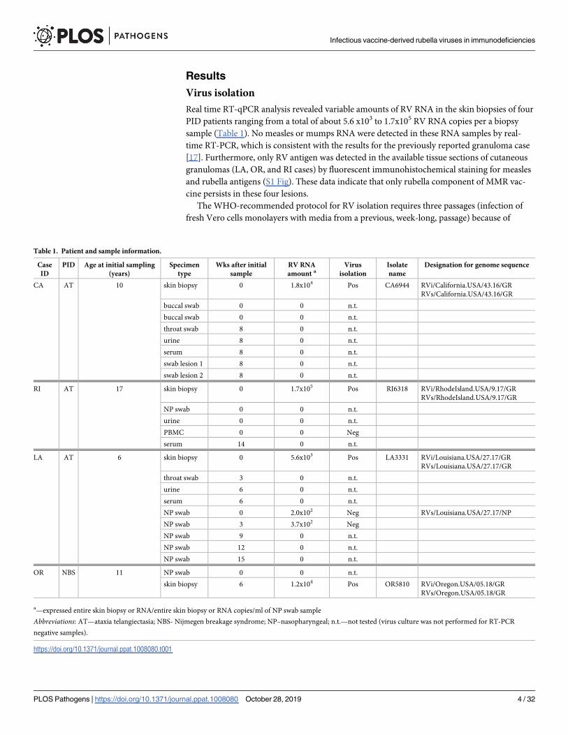
Virus isolation
Real time RT-qPCR analysis revealed variable amounts of RV RNA in the skin biopsies of four
PID patients ranging from a total of about 5.6 x10³ to 1.7x10⁵ RV RNA copies per a biopsy
sample (Table 1). No measles or mumps RNA were detected in these RNA samples by real-
time RT-PCR, which is consistent with the results for the previously reported granuloma case
[17]. Furthermore, only RV antigen was detected in the available tissue sections of cutaneous
granulomas (LA, OR, and RI cases) by fluorescent immunohistochemical staining for measles
and rubella antigens (S1 Fig). These data indicate that only rubella component of MMR vac-
cine persists in these four lesions.
The WHO-recommended protocol for RV isolation requires three passages (infection of
fresh Vero cells monolayers with media from a previous, week-long, passage) because of
Table 1. Patient and sample information.
Case
ID
PID Age at initial sampling
(years)
Specimen
type
Wks after initial
sample
RV RNA
amount aVirus
isolation
Isolate
name
Designation for genome sequence
CA AT 10 skin biopsy 0 1.8x10⁴ Pos CA6944 RVi/California.USA/43.16/GR
RVs/California.USA/43.16/GR
buccal swab 0 0 n.t.
buccal swab 0 0 n.t.
throat swab 8 0 n.t.
urine 8 0 n.t.
serum 8 0 n.t.
swab lesion 1 8 0 n.t.
swab lesion 2 8 0 n.t.
RI AT 17 skin biopsy 0 1.7x10⁵ Pos RI6318 RVi/RhodeIsland.USA/9.17/GR
RVs/RhodeIsland.USA/9.17/GR
NP swab 0 0 n.t.
urine 0 0 n.t.
PBMC 0 0 Neg
serum 14 0 n.t.
LA AT 6 skin biopsy 0 5.6x10³ Pos LA3331 RVi/Louisiana.USA/27.17/GR
RVs/Louisiana.USA/27.17/GR
throat swab 3 0 n.t.
urine 6 0 n.t.
serum 6 0 n.t.
NP swab 0 2.0x10² Neg RVs/Louisiana.USA/27.17/NP
NP swab 3 3.7x10² Neg
NP swab 9 0 n.t.
NP swab 12 0 n.t.
NP swab 15 0 n.t.
OR NBS 11 NP swab 0 0 n.t.
skin biopsy 6 1.2x10⁴ Pos OR5810 RVi/Oregon.USA/05.18/GR
RVs/Oregon.USA/05.18/GR
a—expressed entire skin biopsy or RNA/entire skin biopsy or RNA copies/ml of NP swab sample
Abbreviations: AT—ataxia telangiectasia; NBS- Nijmegen breakage syndrome; NP–nasopharyngeal; n.t.—not tested (virus culture was not performed for RT-PCR
negative samples).
https://doi.org/10.1371/journal.ppat.1008080.t001
Infectious vaccine-derived rubella viruses in immunodeficiencies
PLOS Pathogens | https://doi.org/10.1371/journal.ppat.1008080 October 28, 2019 4 / 32
typically low RV quantities in clinical samples. The failure to isolate infectious virus in the pre-
viously reported case prompted us to modify the culture protocol to enhance virus recovery
from biopsy specimens (see Methods). To estimate the number of infected cells in the cultures,
cells from each isolation were seeded onto chamber slides after each passage and then immu-
nostained for RV structural proteins E1, E2 and C. Almost all cells were positive in the 14-dpi
cultures of CA6499 and RI6318, whereas less than 1% RV-positive cells were detected in the
21-dpi cultures of the LA3331 and OR5810. This suggests the presence of low quantities of
infectious virus in the granulomas of the LA and OR cases and/or the reduced abilities of the
recovered viruses to infect and/or spread in Vero cell monolayers. Infectious rubella viruses
were harvested after 14 days (CA and RI cases) or 21 days (LA and OR cases) of culture in
Vero cells; the isolate designations are indicated in Table 1. Taken together, these data suggest
that infectious viruses with distinctive growth properties are present in the lesions of these
four patients.
Virus shedding
The presence of infectious virus in biopsies led us to assess virus shedding in urine samples,
NP swabs, buccal swabs and lesion scrapings by first testing for RV RNA by real-time RT-
qPCR. All samples were negative except the two sequential NP swabs taken three weeks apart
from the LA case patient (Table 1). The RV RNA concentration in both NP swabs was low, ~2-
4x10² copies/ml of the swab sample. Virus culture using 0.5 ml of both samples (~100 RV
RNA molecules/flask) was unsuccessful most probably due to the low number of infectious
particles in the sample. The ratio of foci forming units (ffu) per RV genome was previously
determined to be about 1/40 in both RA27/3 and wtRV-infected cell cultures [21] and was
lower in clinical samples. Thus we estimate only 2–3 ffu per flask or less in these two isolation
attempts. The presence of RV RNA in the sequential NP samples indicates that viral shedding
into a nasopharyngeal cavity can occur. Three sera and one PBMC sample from three patients
were all negative by RT-qPCR indicating a lack of viremia.
Sequence analysis of the iVDRVs genomes
Full genome sequences were obtained from granuloma biopsy specimens (RVs) and passage 1
(P1) virus isolates (RVi) by Sanger sequencing of overlapping RT-PCR products. The P1
CA6944 isolate was passaged three more times to obtain a high titer P4 virus stock. The con-
sensus sequences of the full RV genomes in the P1 and P4 stocks were identical showing con-
sensus sequence stability for at least three passages in Vero cells. Phylogenetic analysis of the
sequences obtained from primary granuloma samples (including previously described RVs/
Oulu.FIN/22.15/GR (GeneBank#KU958641.1), with full genomes of the WHO RV reference
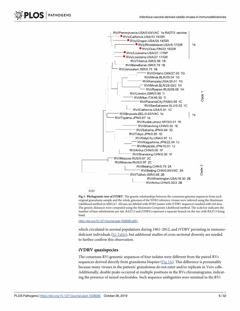
viruses showed that all sequences derived from the patients’ samples belong to genotype 1a
(Fig 1). RA27/3 vaccine strain was basal to all sequenced iVDRV. These viruses were somewhat
more distantly related to other vaccine strains, which are also genotype 1a.
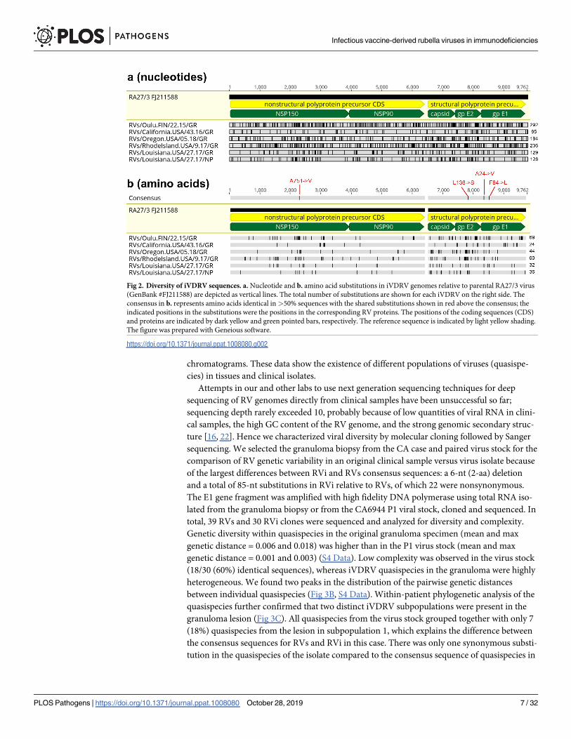
Comparative analysis of six iVDRV RVs and four RVi genomic sequences revealed multiple
(from 95 to 292) single nucleotide substitutions compared to the RA27/3 parental virus, many
of them nonsynonymous (Fig 2, S1 Table, S1 Data). Amino acid substitutions in the iVDRV
proteins were observed at a total 247 positions (S2 Table). The majority of substitutions (192
out of total 247, 76.5%) were found only in iVDRV strains, whereas the remaining substitu-
tions in 55 amino acid positions (23.5%) were also observed in various wtRV strains and may
represent reversions to wtRV. Notably, 142 substitutions (57.5%) occurred at invariant amino
acids in wtRV, which circulated worldwide during a period 1961–2012 (S2 Table, S2 and S3
Data). Thus, the spectrum of amino acid substitutions appears to be different between wtRV,
Infectious vaccine-derived rubella viruses in immunodeficiencies
PLOS Pathogens | https://doi.org/10.1371/journal.ppat.1008080 October 28, 2019 5 / 32
which circulated in normal populations during 1961–2012, and iVDRV persisting in immuno-
deficient individuals (S2 Table), but additional studies of cross sectional diversity are needed
to further confirm this observation.
iVDRV quasispecies
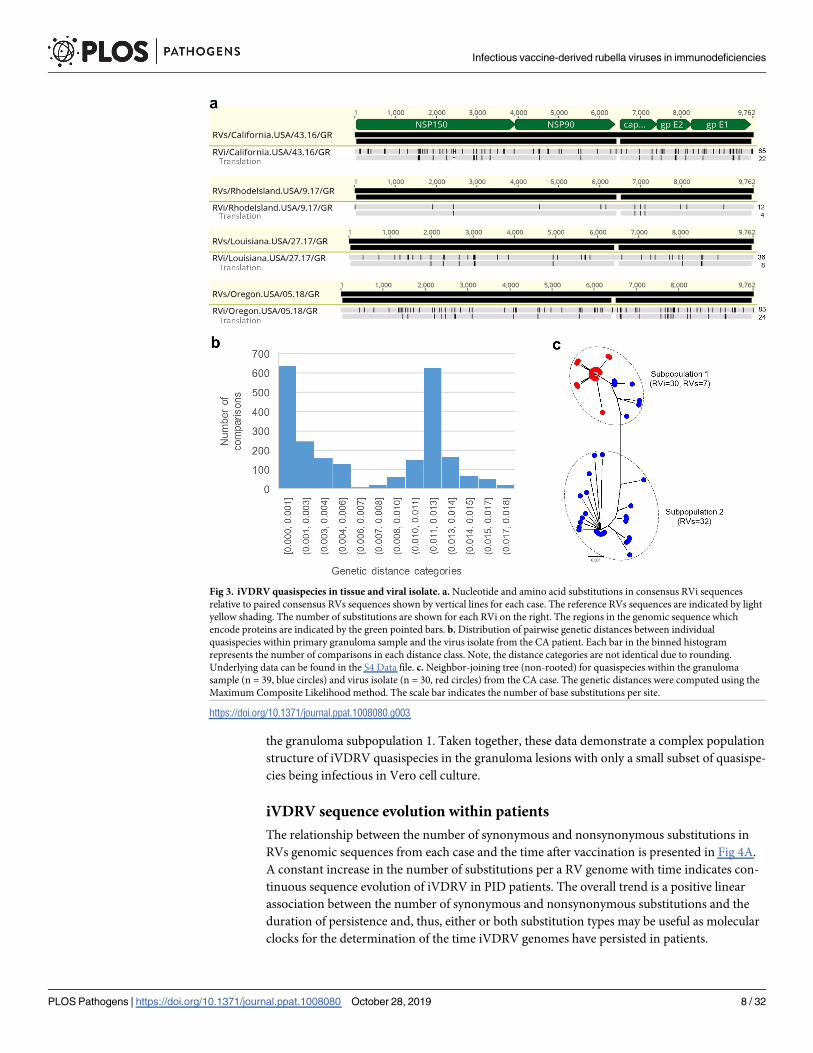
The consensus RVi genomic sequences of four isolates were different from the paired RVs
sequences derived directly from granuloma biopsies (Fig 3A). This difference is presumably
because many viruses in the patients’ granulomas do not enter and/or replicate in Vero cells.
Additionally, double peaks occurred at multiple positions in the RVs chromatograms, indicat-
ing the presence of mixed nucleotides. Such sequence ambiguities were minimal in the RVi
Fig 1. Phylogenetic tree of iVDRV. The genetic relationships between the consensus genome sequences from each
original granuloma sample and the whole genomes of the WHO reference viruses were inferred using the Maximum
Likelihood method in MEGA7. All taxa are labeled with WHO names with iVDRV sequences marked with red dots.
The genetic distances were computed using the Maximum Composite Likelihood method. The scale bar indicates the
number of base substitutions per site. RA27/3 and iVDRVs represent a separate branch on the tree with RA27/3 being
basal.
https://doi.org/10.1371/journal.ppat.1008080.g001
Infectious vaccine-derived rubella viruses in immunodeficiencies
PLOS Pathogens | https://doi.org/10.1371/journal.ppat.1008080 October 28, 2019 6 / 32
chromatograms. These data show the existence of different populations of viruses (quasispe-
cies) in tissues and clinical isolates.
Attempts in our and other labs to use next generation sequencing techniques for deep
sequencing of RV genomes directly from clinical samples have been unsuccessful so far;
sequencing depth rarely exceeded 10, probably because of low quantities of viral RNA in clini-
cal samples, the high GC content of the RV genome, and the strong genomic secondary struc-
ture [16, 22]. Hence we characterized viral diversity by molecular cloning followed by Sanger
sequencing. We selected the granuloma biopsy from the CA case and paired virus stock for the
comparison of RV genetic variability in an original clinical sample versus virus isolate because
of the largest differences between RVi and RVs consensus sequences: a 6-nt (2-aa) deletion
and a total of 85-nt substitutions in RVi relative to RVs, of which 22 were nonsynonymous.
The E1 gene fragment was amplified with high fidelity DNA polymerase using total RNA iso-
lated from the granuloma biopsy or from the CA6944 P1 viral stock, cloned and sequenced. In
total, 39 RVs and 30 RVi clones were sequenced and analyzed for diversity and complexity.
Genetic diversity within quasispecies in the original granuloma specimen (mean and max
genetic distance = 0.006 and 0.018) was higher than in the P1 virus stock (mean and max
genetic distance = 0.001 and 0.003) (S4 Data). Low complexity was observed in the virus stock
(18/30 (60%) identical sequences), whereas iVDRV quasispecies in the granuloma were highly
heterogeneous. We found two peaks in the distribution of the pairwise genetic distances
between individual quasispecies (Fig 3B, S4 Data). Within-patient phylogenetic analysis of the
quasispecies further confirmed that two distinct iVDRV subpopulations were present in the
granuloma lesion (Fig 3C). All quasispecies from the virus stock grouped together with only 7
(18%) quasispecies from the lesion in subpopulation 1, which explains the difference between
the consensus sequences for RVs and RVi in this case. There was only one synonymous substi-
tution in the quasispecies of the isolate compared to the consensus sequence of quasispecies in
Fig 2. Diversity of iVDRV sequences. a. Nucleotide and b. amino acid substitutions in iVDRV genomes relative to parental RA27/3 virus
(GenBank #FJ211588) are depicted as vertical lines. The total number of substitutions are shown for each iVDRV on the right side. The
consensus in b. represents amino acids identical in>50% sequences with the shared substitutions shown in red above the consensus; the
indicated positions in the substitutions were the positions in the corresponding RV proteins. The positions of the coding sequences (CDS)
and proteins are indicated by dark yellow and green pointed bars, respectively. The reference sequence is indicated by light yellow shading.
The figure was prepared with Geneious software.
https://doi.org/10.1371/journal.ppat.1008080.g002
Infectious vaccine-derived rubella viruses in immunodeficiencies
PLOS Pathogens | https://doi.org/10.1371/journal.ppat.1008080 October 28, 2019 7 / 32
the granuloma subpopulation 1. Taken together, these data demonstrate a complex population
structure of iVDRV quasispecies in the granuloma lesions with only a small subset of quasispe-
cies being infectious in Vero cell culture.
iVDRV sequence evolution within patients
The relationship between the number of synonymous and nonsynonymous substitutions in
RVs genomic sequences from each case and the time after vaccination is presented in Fig 4A.
A constant increase in the number of substitutions per a RV genome with time indicates con-
tinuous sequence evolution of iVDRV in PID patients. The overall trend is a positive linear
association between the number of synonymous and nonsynonymous substitutions and the
duration of persistence and, thus, either or both substitution types may be useful as molecular
clocks for the determination of the time iVDRV genomes have persisted in patients.
Fig 3. iVDRV quasispecies in tissue and viral isolate. a. Nucleotide and amino acid substitutions in consensus RVi sequences
relative to paired consensus RVs sequences shown by vertical lines for each case. The reference RVs sequences are indicated by light
yellow shading. The number of substitutions are shown for each RVi on the right. The regions in the genomic sequence which
encode proteins are indicated by the green pointed bars. b. Distribution of pairwise genetic distances between individual
quasispecies within primary granuloma sample and the virus isolate from the CA patient. Each bar in the binned histogram
represents the number of comparisons in each distance class. Note, the distance categories are not identical due to rounding.
Underlying data can be found in the S4 Data file. c. Neighbor-joining tree (non-rooted) for quasispecies within the granuloma
sample (n = 39, blue circles) and virus isolate (n = 30, red circles) from the CA case. The genetic distances were computed using the
Maximum Composite Likelihood method. The scale bar indicates the number of base substitutions per site.
https://doi.org/10.1371/journal.ppat.1008080.g003
Infectious vaccine-derived rubella viruses in immunodeficiencies
PLOS Pathogens | https://doi.org/10.1371/journal.ppat.1008080 October 28, 2019 8 / 32
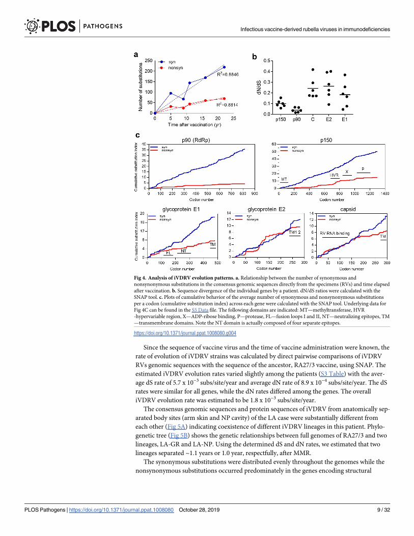
Since the sequence of vaccine virus and the time of vaccine administration were known, the
rate of evolution of iVDRV strains was calculated by direct pairwise comparisons of iVDRV
RVs genomic sequences with the sequence of the ancestor, RA27/3 vaccine, using SNAP. The
estimated iVDRV evolution rates varied slightly among the patients (S3 Table) with the aver-
age dS rate of 5.7 x 10⁻³ subs/site/year and average dN rate of 8.9 x 10⁻⁴ subs/site/year. The dS
rates were similar for all genes, while the dN rates differed among the genes. The overall
iVDRV evolution rate was estimated to be 1.8 x 10⁻³ subs/site/year.
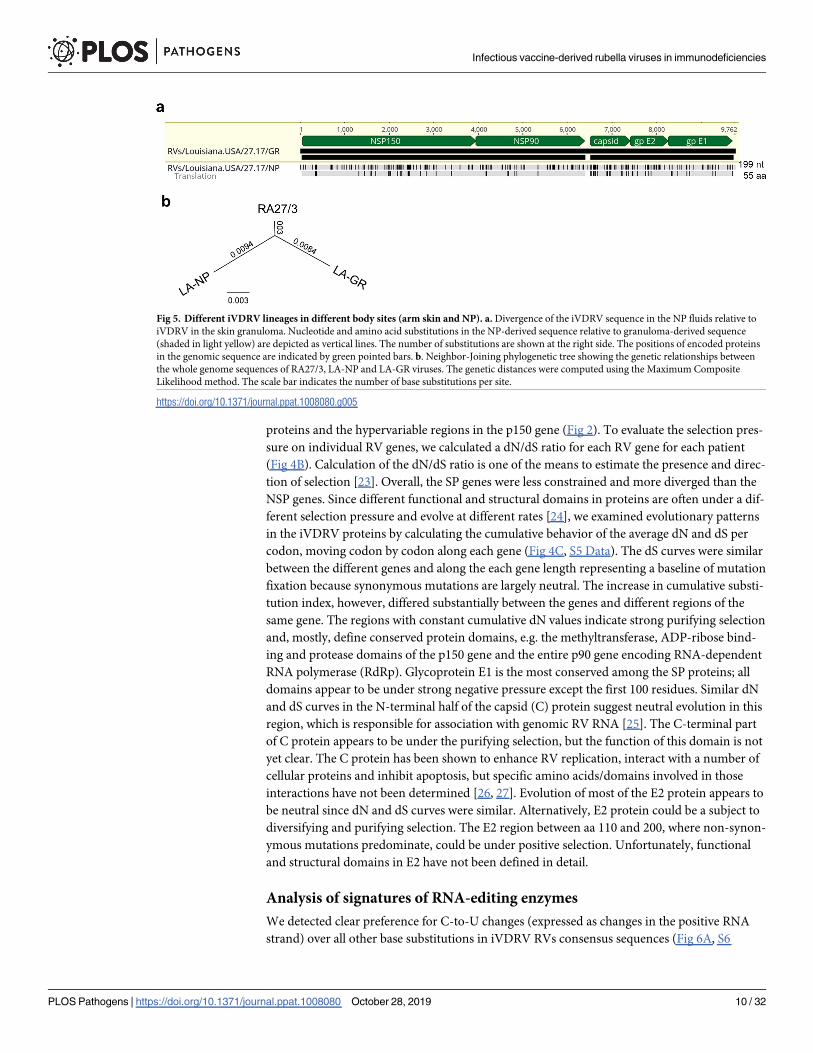
The consensus genomic sequences and protein sequences of iVDRV from anatomically sep-
arated body sites (arm skin and NP cavity) of the LA case were substantially different from
each other (Fig 5A) indicating coexistence of different iVDRV lineages in this patient. Phylo-
genetic tree (Fig 5B) shows the genetic relationships between full genomes of RA27/3 and two
lineages, LA-GR and LA-NP. Using the determined dS and dN rates, we estimated that two
lineages separated ~1.1 years or 1.0 year, respectfully, after MMR.
The synonymous substitutions were distributed evenly throughout the genomes while the
nonsynonymous substitutions occurred predominately in the genes encoding structural
Fig 4. Analysis of iVDRV evolution patterns. a. Relationship between the number of synonymous and
nonsynonymous substitutions in the consensus genomic sequences directly from the specimens (RVs) and time elapsed
after vaccination. b. Sequence divergence of the individual genes by a patient. dN/dS ratios were calculated with the
SNAP tool. c. Plots of cumulative behavior of the average number of synonymous and nonsynonymous substitutions
per a codon (cumulative substitution index) across each gene were calculated with the SNAP tool. Underlying data for
Fig 4C can be found in the S5 Data file. The following domains are indicated: MT—methyltransferase, HVR
-hypervariable region, X—ADP-ribose binding, P—protease, FL—fusion loops I and II, NT—neutralizing epitopes, TM
—transmembrane domains. Note the NT domain is actually composed of four separate epitopes.
https://doi.org/10.1371/journal.ppat.1008080.g004
Infectious vaccine-derived rubella viruses in immunodeficiencies
PLOS Pathogens | https://doi.org/10.1371/journal.ppat.1008080 October 28, 2019 9 / 32
proteins and the hypervariable regions in the p150 gene (Fig 2). To evaluate the selection pres-
sure on individual RV genes, we calculated a dN/dS ratio for each RV gene for each patient
(Fig 4B). Calculation of the dN/dS ratio is one of the means to estimate the presence and direc-
tion of selection [23]. Overall, the SP genes were less constrained and more diverged than the
NSP genes. Since different functional and structural domains in proteins are often under a dif-
ferent selection pressure and evolve at different rates [24], we examined evolutionary patterns
in the iVDRV proteins by calculating the cumulative behavior of the average dN and dS per
codon, moving codon by codon along each gene (Fig 4C, S5 Data). The dS curves were similar
between the different genes and along the each gene length representing a baseline of mutation
fixation because synonymous mutations are largely neutral. The increase in cumulative substi-
tution index, however, differed substantially between the genes and different regions of the
same gene. The regions with constant cumulative dN values indicate strong purifying selection
and, mostly, define conserved protein domains, e.g. the methyltransferase, ADP-ribose bind-
ing and protease domains of the p150 gene and the entire p90 gene encoding RNA-dependent
RNA polymerase (RdRp). Glycoprotein E1 is the most conserved among the SP proteins; all
domains appear to be under strong negative pressure except the first 100 residues. Similar dN
and dS curves in the N-terminal half of the capsid (C) protein suggest neutral evolution in this
region, which is responsible for association with genomic RV RNA [25]. The C-terminal part
of C protein appears to be under the purifying selection, but the function of this domain is not
yet clear. The C protein has been shown to enhance RV replication, interact with a number of
cellular proteins and inhibit apoptosis, but specific amino acids/domains involved in those
interactions have not been determined [26, 27]. Evolution of most of the E2 protein appears to
be neutral since dN and dS curves were similar. Alternatively, E2 protein could be a subject to
diversifying and purifying selection. The E2 region between aa 110 and 200, where non-synon-
ymous mutations predominate, could be under positive selection. Unfortunately, functional
and structural domains in E2 have not been defined in detail.
Analysis of signatures of RNA-editing enzymes
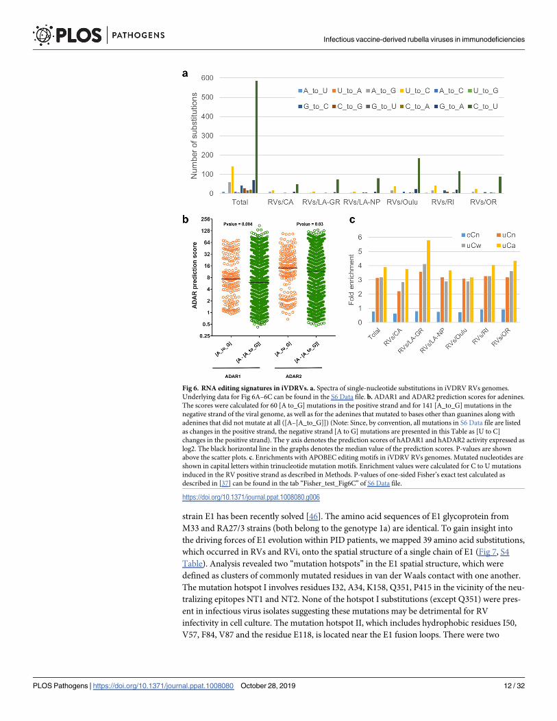
We detected clear preference for C-to-U changes (expressed as changes in the positive RNA
strand) over all other base substitutions in iVDRV RVs consensus sequences (Fig 6A, S6
Fig 5. Different iVDRV lineages in different body sites (arm skin and NP). a. Divergence of the iVDRV sequence in the NP fluids relative to
iVDRV in the skin granuloma. Nucleotide and amino acid substitutions in the NP-derived sequence relative to granuloma-derived sequence
(shaded in light yellow) are depicted as vertical lines. The number of substitutions are shown at the right side. The positions of encoded proteins
in the genomic sequence are indicated by green pointed bars. b. Neighbor-Joining phylogenetic tree showing the genetic relationships between
the whole genome sequences of RA27/3, LA-NP and LA-GR viruses. The genetic distances were computed using the Maximum Composite
Likelihood method. The scale bar indicates the number of base substitutions per site.
https://doi.org/10.1371/journal.ppat.1008080.g005
Infectious vaccine-derived rubella viruses in immunodeficiencies
PLOS Pathogens | https://doi.org/10.1371/journal.ppat.1008080 October 28, 2019 10 / 32
Data). The second major component of the mutation spectrum was U-to-C substitutions.
There was smaller number of C-to-U and U-to-C changes in the RV negative strand than in
the positive strand (expressed as G-to-A and A-to-G changes in the positive strand in the Fig
6A). The mutation profile and the strand bias agree with the previously suggested roles of
APOBEC (apolipoprotein B mRNA editing enzyme, catalytic polypeptide-like) cytidine deam-
inases and ADAR (Adenosine Deaminase Acting on RNA) in generating nucleotide diversity
in RNA viruses [28–30].
We used the web-based tool InosinePredict to generate prediction scores for ADAR1 and
for ADAR2 mutagenesis in each adenosine of the iVDRV RVs genomic sequences. In support
of these enzymes playing a role in mutating adenines in the viral DNA, the prediction scores
for positions containing A to G mutations exceeded prediction scores for adenines in positions
that were not mutated or contained mutations other than A to G (Fig 6B, S6 Data).
Most of mutations were in cytosines of the viral positive strand (Fig 6A). The capability for
cytidine deamination in RNA was well established for APOBEC1, while APOBEC 3 enzymes
are commonly viewed as acting exclusively in DNA [31]. However, there are recent reports
suggesting that the APOBEC3 subfamily might also act on pathogen and host RNA in certain
types of cells [32–35]. The preferred motif for APOBEC3G deamination is cCn (where n is any
nucleotide; mutated nucleotide capitalized), while the other members of the APOBEC3 family
as well as APOBEC1 are characterized by a preference for tCn motif with higher preference for
tCw (w = A or T) [29, 36]. The diagnostic signature for APOBEC3A and APOBEC3B was nar-
rowed to tCa trinucleotide [37]. Therefore, we explored enrichment with these mutation signa-
tures in C-to-U mutations induced in the positive strand of six iVDRV RVs sequences.
Interestingly, in each iVDRV strain as well as in the total mutation catalogue, there was
depletion of cCn motif characteristic of APOBEC3G. However, there was strong enrichment
with tCn-specific signatures, which constituted around 30% of all mutations in the positive
strand. Splitting tCn signature into sub-components that have even higher preference to APO-
BEC enzymes revealed that enrichment with tCa signature exceeded the enrichment with
either tCn or with tCw (Fig 6C, S6 Data). All enrichments with tCn, tCw and tCa signatures
were highly statistically significant with P-values of one-sided Fisher’s exact test =< 0.01 in
each case (see the tab “Fisher_test_Fig6C” of S6 Data file). This indicates that one or more t(u)
Cn-specific APOBECs play a role in generating diversity in iVDRV populations. Additional
studies are also needed to understand connections between the dynamics of viral RNA second-
ary structure and the relative impacts of APOBEC and ADAR editing activities, which act on
to mutually excluding substrates–single stranded and double stranded states in viral RNA
folds.
Structural context of amino acid substitutions in E1 protein
Many amino acid changes occurred in E1, E2, and C proteins, but interpretation of their func-
tional significance is limited by what is known about functional domains in the RV structural
proteins. The E1 glycoprotein plays a crucial role in RV infectivity by mediating receptor bind-
ing and membrane fusion [38, 39]. E1 is the better immunogen than E2 and C and four neu-
tralizing epitopes were mapped to this protein [40–42]. At least one weak neutralizing epitope
of E2 was identified but not precisely mapped [43]. Less is known about rubella-specific MHC
class I restricted CTL responses and only three CD8+ T cell epitopes have been identified so
far (two of them overlap), all located in the capsid protein [44, 45]. Changes in RV neutralizing
B cell epitopes and CD8+ T cell epitopes were detected in all cases except the RI case (Table 2).
Since data on the antigenic structure of E2 and C proteins are limited, the detailed analysis
of the mutations was only performed for E1. The three dimensional structure of the M33 RV
Infectious vaccine-derived rubella viruses in immunodeficiencies
PLOS Pathogens | https://doi.org/10.1371/journal.ppat.1008080 October 28, 2019 11 / 32
strain E1 has been recently solved [46]. The amino acid sequences of E1 glycoprotein from
M33 and RA27/3 strains (both belong to the genotype 1a) are identical. To gain insight into
the driving forces of E1 evolution within PID patients, we mapped 39 amino acid substitutions,
which occurred in RVs and RVi, onto the spatial structure of a single chain of E1 (Fig 7, S4
Table). Analysis revealed two “mutation hotspots” in the E1 spatial structure, which were
defined as clusters of commonly mutated residues in van der Waals contact with one another.
The mutation hotspot I involves residues I32, A34, K158, Q351, P415 in the vicinity of the neu-
tralizing epitopes NT1 and NT2. None of the hotspot I substitutions (except Q351) were pres-
ent in infectious virus isolates suggesting these mutations may be detrimental for RV
infectivity in cell culture. The mutation hotspot II, which includes hydrophobic residues I50,
V57, F84, V87 and the residue E118, is located near the E1 fusion loops. There were two
Fig 6. RNA editing signatures in iVDRVs. a. Spectra of single-nucleotide substitutions in iVDRV RVs genomes.
Underlying data for Fig 6A–6C can be found in the S6 Data file. b. ADAR1 and ADAR2 prediction scores for adenines.
The scores were calculated for 60 [A to_G] mutations in the positive strand and for 141 [A_to_G] mutations in the
negative strand of the viral genome, as well as for the adenines that mutated to bases other than guanines along with
adenines that did not mutate at all ([A–[A_to_G]]) (Note: Since, by convention, all mutations in S6 Data file are listed
as changes in the positive strand, the negative strand [A to G] mutations are presented in this Table as [U to C]
changes in the positive strand). The y axis denotes the prediction scores of hADAR1 and hADAR2 activity expressed as
log2. The black horizontal line in the graphs denotes the median value of the prediction scores. P-values are shown
above the scatter plots. c. Enrichments with APOBEC editing motifs in iVDRV RVs genomes. Mutated nucleotides are
shown in capital letters within trinucleotide mutation motifs. Enrichment values were calculated for C to U mutations
induced in the RV positive strand as described in Methods. P-values of one-sided Fisher’s exact test calculated as
described in [37] can be found in the tab “Fisher_test_Fig6C” of S6 Data file.
https://doi.org/10.1371/journal.ppat.1008080.g006
Infectious vaccine-derived rubella viruses in immunodeficiencies
PLOS Pathogens | https://doi.org/10.1371/journal.ppat.1008080 October 28, 2019 12 / 32
substitutions in E1 shared by iVDRV: F84 reversion to wt L84 occurred in five out of six
iVDRV and A24V substitution, which occurred in all six iVDRVs but not in wtRV strains (S2
Table). Many mutated residues (23 out of total 39) are exposed on the E1 surface (Fig 7B and
7C, S4 Table) and, thus, could be involved in direct interactions with host molecules.
Several substitutions occurred in E1 neutralizing epitopes NT2-NT4 in three iVDRV
strains, while NT1 epitope was invariant (Table 2). A260V substitution in the NT3 epitope and
two mutations in the NT4 epitope, S282A and I280T/M, were located in the proposed mem-
brane contact region of E1 in close proximity to the fusion loops [46]. The substitutions in aa
position 280 (the NT3 epitope) co-occurred in three iVDRV strains and, thus, might be impor-
tant for interactions with neutralizing antibody. The spatial location of the NT3 and NT4 epi-
topes near the fusion loops suggests that antibodies targeting these epitopes may neutralize RV
infectivity by interfering with the fusion process thus preventing cell entry. L245V substitution
was mapped to the NT2 epitope, which is located in close proximity to the NT1 epitope and
similar to the NT1 is not exposed on the surface of the trimer. The neutralization mechanism
of antibodies targeting NT2 is mostly likely similar to the mechanism proposed for NT1,
which is interference with the formation of E1 trimer, which is a pre-fusion form of E1 [46].
Taken together, these data suggest that granuloma-associated rubella virus is under selective
pressure from both neutralizing antibodies and T cells.
Growth properties of iVDRV strains
The replication and persistence properties of RA27/3 vaccine and clinical isolates form acute
RV infections are substantially different [21]. To test if vaccine-derived viruses in granulomas
have changed from the vaccine phenotype, we compared the growth properties of four iVDRV
strains with growth properties of RA27/3 and RV-Dz in WI-38 human fetal fibroblasts, the pri-
mary cell culture used for RA27/3 attenuation [47]. RV-Dz is a well-characterized wild type
strain of genotype 1E [48].
To compare virus yields and percentage of infected cells, cell monolayers were infected with
each virus strain at high (5 ffu/cell) and low (0.1 ffu/cell) multiplicity of infection (MOI). High
MOI allows comparing efficiency of virus entry and replication while low MOI provides infor-
mation on efficiency of cell-to-cell spread. At both MOIs after 2–3 days, RA27/3 infected
almost the entire WI-38 monolayers, producing an infectious titer of 5x10⁵ ffu/ml, while
Table 2. Substitutions in B and T cell epitopes in iVDRVs.
Epitope Sequence in RA27/3 Substitutions in iVDRV
RVs FIN RVs/RVi CA RVs/RVi RI RVs/RVi LA RVs
LA(NP)
RVs/RVi OR
Neutralizing B cell epitopes
NT1:E1221-239 LGSPNCHGPDWASPCQRHS - - - - - -
NT2:E1245-251 LVGATPE� - L245V - - - -
NT3:E1260-266 ADDPLLR - A260V - - - -
NT4:E1274-285 VWVTPVIGSQAR I280T I280M S282A - - - I280T
CD8+ T cell epitopes
C9-22 MEDLQKALETQSRA T18A - - R21C A22I -
C11-29 DLQKALETQSRALRAELAA T18A - - R21C A22I
A25E A28V
-
C264-272 RIETRSARH T267I - - - E266G -
�Mutated residues in the epitopes are shown in bold.
https://doi.org/10.1371/journal.ppat.1008080.t002
Infectious vaccine-derived rubella viruses in immunodeficiencies
PLOS Pathogens | https://doi.org/10.1371/journal.ppat.1008080 October 28, 2019 13 / 32
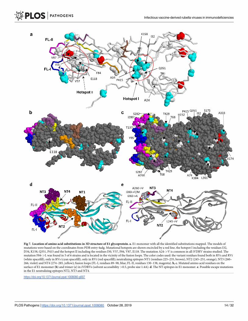
Fig 7. Location of amino acid substitutions in 3D structure of E1 glycoprotein. a. E1 monomer with all the identified substitutions mapped. The models of
mutations were based on the coordinates from PDB entry 4adg. Mutational hotspots are shown encircled by a red line, the hotspot I including the residues I32,
D34, K158, Q351, P415 and the hotspot II including the residues I50, V57, F84, V87, E118. The mutation A24->V is common in all iVDRV strains studied. The
mutation F84->L was found in 5 of 6 strains and is located in the vicinity of the fusion loops. The color codes used: the variant residues found both in RVs and RVi
(white spacefill), only in RVs (cyan spacefill), only in RVi (red spacefill); neutralizing epitopes NT1 (residues 225–235, brown), NT2 (245–251, orange), NT3 (260–
266, violet) and NT4 (274–285, yellow); fusion loops (FL-I, residues 89–98, blue; FL-II, residues 130–138, magenta). b, c. Mutated amino acid residues on the
surface of E1 monomer (b) and trimer (c) in iVDRVs (solvent accessibility>0.5, probe size 1.4A). d. The NT epitopes in E1 monomer. e. Possible escape mutations
in the E1 neutralizing epitopes NT2, NT3 and NT4.
https://doi.org/10.1371/journal.ppat.1008080.g007
Infectious vaccine-derived rubella viruses in immunodeficiencies
PLOS Pathogens | https://doi.org/10.1371/journal.ppat.1008080 October 28, 2019 14 / 32
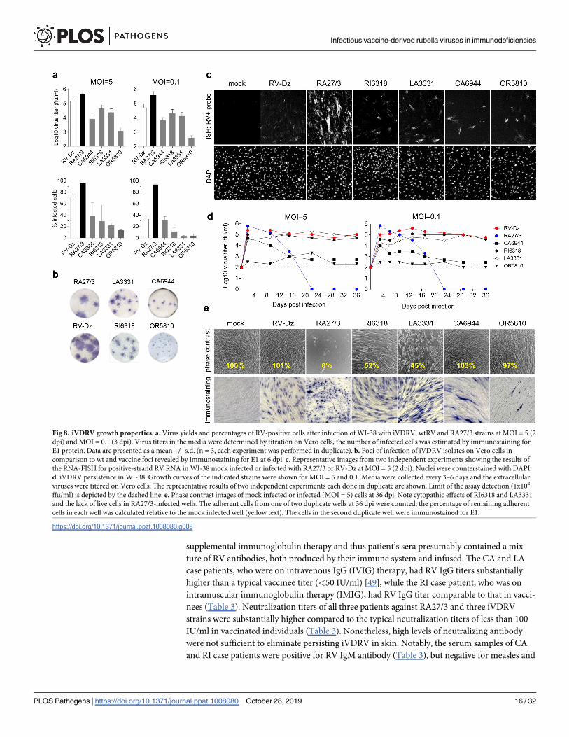
replication of RV-Dz was less effective (Fig 8A). In contrast, iVDRV infected fewer cells in the
monolayers (5–40%) producing 1–2.5 logs less infectious virus depending on the isolate. Simi-
lar to RV-Dz, iVDRV spread after low MOI infections was reduced compared to RA27/3. The
size of foci formed by iVDRV on Vero monolayers were smaller than that of RV-Dz and
RA27/3, confirming reduced abilities of iVDRVs to spread in cell culture (Fig 8B). The effi-
ciencies of cell spread varied depending on the iVDRV isolate with OR5810 being the least
efficient.
RV RNA replication on the single cell level was assessed using RNA-FISH. WI-38 cells were
mock infected or infected with the RV strains at MOI = 5 for two days and then genomic RV
RNA was detected by in situ hybridization with a probe set specific for the positive RNA strand
(Fig 8C). As previously observed in endothelial cells [21], the total amounts of genomic RV
RNA per a cell were substantially higher in RA27/3 than RV-Dz infected WI-38 cells. In
iVDRV infected monolayers, most cells produced low quantities of genomic RV RNA, similar
to that of RV-Dz, but isolated cells producing somewhat higher levels of cellular RV RNA than
RV-Dz were also observed (Fig 8C).
The abilities of the iVDRV isolates to persist in WI-38 cells were compared after high and
low MOIs. Virus titers in the medium and cytopathic effects (CPE) was monitored for 36 days.
No substantial differences were seen in persistence characteristics after different MOIs. After
the initial peak of replication, starting at day 2, RA27/3 began inducing cell death, which
resulted in complete destruction of the monolayers by 15 dpi (MOI = 5) and 17 dpi (MOI =
0.1) (Fig 8D and 8E). In contrast, RV-Dz persisted at the same level (~105 ffu/ml) for the dura-
tion of the experiments without causing any visible CPE. The continued virus production of
RI6318 and LA3331 was similar to that of RV-Dz, but unlike RV-Dz these viruses induced
CPE starting at ~14 dpi and resulting in 2-fold reduction of the cell number by 36 dpi. All cells
were RV-positive in the infected monolayers at 36 dpi except those infected with CA6944 and
OR5810. The fraction of CA6944 infected cells was reduced from ~40% initially to less than
10% at 36 dpi concurrent with 2-log titer reduction. The OR5810 virus production was consis-
tently low (<103 ffu/ml) and only a few infected cells were detected at 36 dpi. Consensus
genome sequences of all iVDRVs at 36 dpi were identical to the consensus sequence of the ini-
tial inoculums, although minor fractions of different variants would not have been detected by
the Sanger sequencing.
The results of these analyses clearly indicate that each iVDRV isolate has unique replicative
and persistence properties, which are different from each other and from those of both vaccine
and wtRV. Notably, all iVDRV isolates were less cytopathic in cell culture than RA27/3 and
had the capability to persist, indicating, at least partially, a wt phenotype in cell culture.
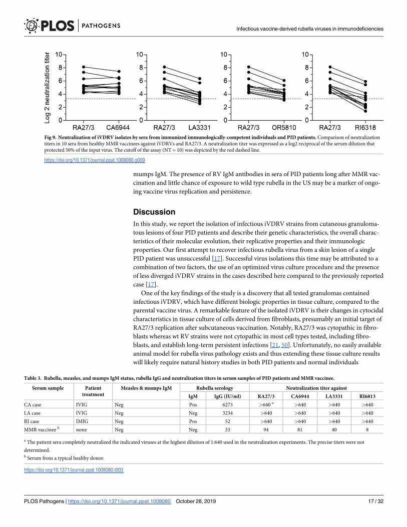
Neutralization of iVDRVs by sera from MMR vaccinees
Immunization with a RA27/3 vaccine strain protects against infections with different RV
genotypes. However, the mutation spectra of iVDRV and wtRV differ substantially (S2 Table).
To evaluate the protective capacity of rubella vaccination against iVDRV strains, we compared
the neutralization titers in 10 sera from healthy adult vaccinees against RA27/3 and against
each of the four iVDRV strains. In general all vaccinee sera contained RV neutralizing anti-
body that could better neutralize RA27/3 relative to iVDRV. The exception was CA6944 (Fig
9). The RI6318 isolate was the most resistant to neutralization since 7 out of 10 sera failed to
effectively neutralize it. These data suggest that additional research on the immune response of
vaccinated individuals against iVDRV is warranted.
Next we examined rubella antibody titers and neutralization titers against RA27/3 and
iVDRV strains in serum samples from three PID granuloma cases. All case patients were on
Infectious vaccine-derived rubella viruses in immunodeficiencies
PLOS Pathogens | https://doi.org/10.1371/journal.ppat.1008080 October 28, 2019 15 / 32
supplemental immunoglobulin therapy and thus patient’s sera presumably contained a mix-
ture of RV antibodies, both produced by their immune system and infused. The CA and LA
case patients, who were on intravenous IgG (IVIG) therapy, had RV IgG titers substantially
higher than a typical vaccinee titer (<50 IU/ml) [49], while the RI case patient, who was on
intramuscular immunoglobulin therapy (IMIG), had RV IgG titer comparable to that in vacci-
nees (Table 3). Neutralization titers of all three patients against RA27/3 and three iVDRV
strains were substantially higher compared to the typical neutralization titers of less than 100
IU/ml in vaccinated individuals (Table 3). Nonetheless, high levels of neutralizing antibody
were not sufficient to eliminate persisting iVDRV in skin. Notably, the serum samples of CA
and RI case patients were positive for RV IgM antibody (Table 3), but negative for measles and
Fig 8. iVDRV growth properties. a. Virus yields and percentages of RV-positive cells after infection of WI-38 with iVDRV, wtRV and RA27/3 strains at MOI = 5 (2
dpi) and MOI = 0.1 (3 dpi). Virus titers in the media were determined by titration on Vero cells, the number of infected cells was estimated by immunostaining for
E1 protein. Data are presented as a mean +/- s.d. (n = 3, each experiment was performed in duplicate). b. Foci of infection of iVDRV isolates on Vero cells in
comparison to wt and vaccine foci revealed by immunostaining for E1 at 6 dpi. c. Representative images from two independent experiments showing the results of
the RNA-FISH for positive-strand RV RNA in WI-38 mock infected or infected with RA27/3 or RV-Dz at MOI = 5 (2 dpi). Nuclei were counterstained with DAPI.
d. iVDRV persistence in WI-38. Growth curves of the indicated strains were shown for MOI = 5 and 0.1. Media were collected every 3–6 days and the extracellular
viruses were titered on Vero cells. The representative results of two independent experiments each done in duplicate are shown. Limit of the assay detection (1x10²
ffu/ml) is depicted by the dashed line. e. Phase contrast images of mock infected or infected (MOI = 5) cells at 36 dpi. Note cytopathic effects of RI6318 and LA3331
and the lack of live cells in RA27/3-infected wells. The adherent cells from one of two duplicate wells at 36 dpi were counted; the percentage of remaining adherent
cells in each well was calculated relative to the mock infected well (yellow text). The cells in the second duplicate well were immunostained for E1.
https://doi.org/10.1371/journal.ppat.1008080.g008
Infectious vaccine-derived rubella viruses in immunodeficiencies
PLOS Pathogens | https://doi.org/10.1371/journal.ppat.1008080 October 28, 2019 16 / 32
mumps IgM. The presence of RV IgM antibodies in sera of PID patients long after MMR vac-
cination and little chance of exposure to wild type rubella in the US may be a marker of ongo-
ing vaccine virus replication and persistence.
Discussion
In this study, we report the isolation of infectious iVDRV strains from cutaneous granuloma-
tous lesions of four PID patients and describe their genetic characteristics, the overall charac-
teristics of their molecular evolution, their replicative properties and their immunologic
properties. Our first attempt to recover infectious rubella virus from a skin lesion of a single
PID patient was unsuccessful [17]. Successful virus isolations this time may be attributed to a
combination of two factors, the use of an optimized virus culture procedure and the presence
of less diverged iVDRV strains in the cases described here compared to the previously reported
case [17].
One of the key findings of the study is a discovery that all tested granulomas contained
infectious iVDRV, which have different biologic properties in tissue culture, compared to the
parental vaccine virus. A remarkable feature of the isolated iVDRV is their changes in cytocidal
characteristics in tissue culture of cells derived from fibroblasts, presumably an initial target of
RA27/3 replication after subcutaneous vaccination. Notably, RA27/3 was cytopathic in fibro-
blasts whereas wt RV strains were not cytopathic in most cell types tested, including fibro-
blasts, and establish long-term persistent infections [21, 50]. Unfortunately, no easily available
animal model for rubella virus pathology exists and thus extending these tissue culture results
will likely require natural history studies in both PID patients and normal individuals
Fig 9. Neutralization of iVDRV isolates by sera from immunized immunologically-competent individuals and PID patients. Comparison of neutralization
titers in 10 sera from healthy MMR vaccinees against iVDRVs and RA27/3. A neutralization titer was expressed as a log2 reciprocal of the serum dilution that
protected 50% of the input virus. The cutoff of the assay (NT = 10) was depicted by the red dashed line.
https://doi.org/10.1371/journal.ppat.1008080.g009
Table 3. Rubella, measles, and mumps IgM status, rubella IgG and neutralization titers in serum samples of PID patients and MMR vaccinee.
Serum sample Patient
treatment
Measles & mumps IgM Rubella serology Neutralization titer against
IgM IgG (IU/ml) RA27/3 CA6944 LA3331 RI6813
CA case IVIG Neg Pos 6273 >640 a >640 >640 >640
LA case IVIG Neg Neg 3234 >640 >640 >640 >640
RI case IMIG Neg Pos 52 >640 >640 >640 >640
MMR vaccinee b none Neg Neg 33 94 81 40 8
a The patient sera completely neutralized the indicated viruses at the highest dilution of 1:640 used in the neutralization experiments. The precise titers were not
determined.b Serum from a typical healthy donor.
https://doi.org/10.1371/journal.ppat.1008080.t003
Infectious vaccine-derived rubella viruses in immunodeficiencies
PLOS Pathogens | https://doi.org/10.1371/journal.ppat.1008080 October 28, 2019 17 / 32
vaccinated with RA27/3. Changes in tissue tropism of iVDRVs would be a significant observa-
tion, especially if evidence of transmission to normal individuals is found.
Whole genome sequencing revealed that each of the recovered viruses contained multiple
predominantly virus-specific nucleotide and amino acid substitutions with respect to RA27/3,
which gives a molecular basis for the differences in their biological properties from each other
and vaccine virus. Our estimate of the overall evolutionary rate of iVDRV (1.8 x 10⁻³ subs/site/
year) based on the whole genome sequences is consistent with evolutionary rates reported for
other small RNA viruses [51] and somewhat higher that those reported for wtRV (genotype
1E: 9.16 × 10⁻⁴–1.04 × 10⁻³ subs/site/year) in populations with person-to-person transmission
[52]. Higher iVDRV evolution rates may be due to continuous viral replication and selection
during chronic infection in a single individual as opposed to intermittent replication and selec-
tion during disease outbreaks.
We have also documented for the first time that iVDRV persisted in a lesion as a diverse
population of heterogeneous but closely related quasispecies. Many persisting RNA viruses are
maintained in a host as quasispecies [53]. High quasispecies complexity allows viruses to
quickly adapt to changing conditions within a host and thus represents a challenge for antiviral
therapies, e.g. selection of drug-resistant virus variants. Indeed, mutant spectrum complexity
could be a factor in predicting disease progression and response to antivirals [54, 55]. Coexis-
tence of two genetically distinct quasispecies subpopulations in the CA patient granuloma and
two iVDRV lineages in the NP cavity and skin lesion of the LA patient which we report here
suggests that a viral population structure in chronically infected patients could be more com-
plicated than currently appreciated. Distinct lineages at each site of persistence and the lack of
viremia indicate that the virus spreads locally by cell-to-cell spread perhaps because circulating
virus (viremia) can be effectively neutralized by the high level of RV neutralizing antibodies
detected in the patients. Granulomas are often present in multiple body locations in the skin as
well in internal organs, e.g. liver, kidney or spleen [18, 19]. It would be interesting to compare
virus sequences from multiple granuloma sites in more patients to verify that multiple iVDRV
lineages may persist and evolve independently at different body sites and whether recombina-
tion between lineages occurs. Such compartmentalization of genetically and possibly antigeni-
cally distinct viral subpopulations, which has been reported for a number of viruses including
hepatitis C and HIV, may contribute to the maintenance of persistent infections [56, 57].
The error-prone RdRp is certainly a major contributor in generating sequence diversity in
many small RNA viruses [55]. However, our study provides evidence that the cellular factors,
one or more tCn-specific APOBECs and ADAR, are the most prominent source of mutations
in persisting iVDRV genomes (Fig 6), increasing genetic diversity and thus participating in
iVDRV evolution. Interestingly, high levels of expression of the APOBEC3 genes and APO-
BEC3-mediated C-to-U RNA editing activity of a subset of cellular RNAs have been detected
in monocytes and macrophages [33, 58], one of the target cells for RV in granulomas in PID
patients [17]. RNA editing by the host ADAR system has been proposed as a factor of evolu-
tion of many positive and negative-sense RNA viruses [30, 59], whereas the ability of APO-
BEC3 family members to restrict viral replication has been reported only for four RNA
viruses, measles, mumps, respiratory syncytial virus, and coronavirus [60, 61]. Inhibition of
viral replication for these 4 viruses was only demonstrated in vitro, in cell cultures overexpres-
sing APOBEC proteins, and was mediated by APOBEC activities other than cytidine deamina-
tion. To our knowledge, our report is the first study describing mRNA editing signature of
APOBECs in a genome of RNA virus after long-term persistence in vivo. However, more stud-
ies are needed to assign RV RNA editing activity to specific members of the APOBEC family.
Effective iVDRV population sizes in granulomas are expected to be smaller than those seen
in some other viral infections given low quantities of genomic RV RNA in the biopsies and
Infectious vaccine-derived rubella viruses in immunodeficiencies
PLOS Pathogens | https://doi.org/10.1371/journal.ppat.1008080 October 28, 2019 18 / 32
given the small foci of infection consisting only of several infected cells by IHC of PID associ-
ated granulomas [17]. Therefore, we expected that persisting iVDRV will undergo sequential
genetic bottlenecks when a limited number of founder viruses spreads within a host generating
independent virus population at different locations. Linear accumulation of substitutions,
both synonymous and nonsynonymous, in iVDRV genomes in different individuals and dif-
ferent body sites over the period of persistence is compatible with the prediction that random
genetic drift is the main force driving iVDRV genome evolution, at least during a chronic
phase of infection. Another known consequence of genetic drift is an appearance of numerous
atypical mutations, which could be the molecular basis of the fitness loss of persisting viruses
[55]. Indeed, the majority of amino acid substitutions were specific for iVDRV strains and
many of them occurred in the positions that are invariant in wtRV, e.g. mutations in the highly
conserved p150 methyltransferase domain and RdRp catalytic domain (S2 Table).
Strong negative selection was observed for nonstructural proteins p150 and p90 as evi-
denced by the dN/dS ratio of less than 0.2. The structural proteins were less constrained with
E1 being more conserved than C or E2. This is consistent with the data obtained for evolution
of wtRV in immunologically competent individuals [52, 62]. We have also identified several
possible regions under positive selection (mutated B and T cell epitopes, Table 2). Since adap-
tive evolution of viruses is often strongly coupled with their antigenic evolution, it is reason-
able to suggest that the mutations in the predicted B and T-cell epitopes in iVDRV have been
positively selected by the combined immune pressure of immunoglobulin therapy and an
ongoing cellular and humoral immune response from the patient. Data on poor neutralization
of iVDRV strains by some sera from vaccinated individuals further support the hypothesis that
the emergence of immune escape variants might be one of the mechanisms of iVDRV persis-
tence. There were, however, discrepancies between the presence of mutations in the E1 neu-
tralizing epitopes and the ability of some iVDRV to avoid neutralization by vaccinees sera. For
example, the virus most resistant to neutralization, RI6328, did not have mutations in known
E1 epitopes, whereas CA6944 had mutations in 3 out of 4 known epitopes, but was neutralized
effectively. It is a likely possibility that not all neutralizing sites in RV have been identified. The
number of known CTL epitopes is also small; only three CTL epitopes, all in C protein, have
been reported and it is not known to what extent sequence variability in those epitopes can be
tolerated [44, 45]. Overall, poor understanding of the antigenic structure of RA27/3 vaccine
and wtRV strains hampers investigations of possible mechanisms of iVDRV escape from
immune surveillance and its contribution to iVDRV persistence.
One puzzling observation was the detection of extremely high titers of RV neutralizing anti-
bodies in the PID patient sera. The observed lack of viremia in all three tested patients was
most likely due to continual removal of iVDRV from the bloodstream by these neutralizing
antibodies. It is presently unknown whether the neutralizing antibodies originated from sup-
plemental immunoglobulin therapy or were produced by the immune system of the chroni-
cally infected patients. RV neutralization titers (ranging from 40 to 2040) were reported for
commercial lots of immunoglobulin produced prior to introduction of rubella vaccine [63].
Determination of RV neutralizing titers in currently produced immunoglobulin preparations,
and in sequential serum samples collected at different times after initiation of immunoglobulin
therapy, may help to define the contributions of IVIG therapy and a patient’s own immune
system to the neutralizing capacity of patient sera. This work would likely enhance our under-
standing of the role of antibodies in clearance of other RV chronic infections.
In addition to the detection of the high levels of neutralizing antibody, we have also detected
RV-specific IgM antibody, but not measles or mumps specific IgM, in the sera of two PID
patients. It is unlikely that the RV-specific IgM antibody was from IVIG therapy, since com-
mercial immunoglobulin products contain primarily IgG from the plasma of a thousand or
Infectious vaccine-derived rubella viruses in immunodeficiencies
PLOS Pathogens | https://doi.org/10.1371/journal.ppat.1008080 October 28, 2019 19 / 32
more healthy blood donors with only trace amounts of IgM. Further investigations of RV IgM
levels in a larger group of PID patients should be considered to evaluate whether the persistent
RV IgM responses long after vaccination may serve as a marker of ongoing RV replication and
may predict granuloma formation prior to the development of lesions. Similarly, hepatitis C
specific IgM antibody has been observed in patients with chronic hepatitis and their levels cor-
relates with the level of viral replication and treatment outcome [64].
Detection of iVDRV RNA in nasopharyngeal secretions raises a concern about the possible
risk of iVDRV to non-immune individuals. Since only a low level of RV RNA was detected in
2 out of 5 sequential NP samples from one case out of three tested, the risk may be low. The
frequency and intensity of virus secretion into the NP cavity as well as transmissibility of these
viruses to non-immune contacts should be determined in a larger study group. In the US, an
estimated 4000 individuals with PID have granuloma complications and thus may be potential
iVDRV carriers [15]. Long-term iVDRV secretion by these individuals may have implications
for rubella and CRS elimination programs. Even if iVDRV carriers shed low virus quantities,
opportunities to transmit virus to susceptible contacts including non-immune siblings and
other patients in PID clinics may occur during decades-long iVDRV persistence.
One of the limitations of our study is the use of diagnostic skin samples, one per patient,
collected at different times after vaccination, rather than multiple samples from each patient
collected serially over time as would be used for a study of pathogen evolution within a patient.
A detailed understanding of rubella virus persistence in PID patients will require in part the
accumulation of information on the viruses from more patients and from serial samples from
individual patients. Unfortunately, the only reliable specimen so far known for iVDRV isola-
tion and sequencing is a skin biopsy. Obtaining sequential skin samples from PID patients,
most of them children, over an extended period of time would be challenging and would
require human subject oversight appropriate for such a research study. Nonetheless, important
conclusions regarding the overall trends of iVDRV evolution within a PID host and properties
of persisting iVDRV can be drawn from the analysis of the limited number of diagnostic
biopsy samples described here.
We already find it intriguing that most PID patients with RV positive persistent granuloma
have defects in their cellular immunity and that we find mutations in T-cell epitopes of viruses
from them. Furthermore, T-cell exhaustion is known to be important for other chronic viral
infections [65]. We hypothesize that cellular immune defects delay viral clearance and exacer-
bate what would be a limited persistent infection in persons with normal immunity, leading to
years or decades long infections and resulting in damage to multiple tissues in PID individuals.
However, we note that limitations in existing data leave open the possibility that other factors,
e.g. changing tissue tropism for the virus or antibody escape mutants, are the crucial factors
determining this extended persistence. In addition, since the most important property of
rubella viruses, their teratogenic potential, is poorly understood, we cannot accurately predict
the effect of mutations found here on teratogenic potential from the tissue culture results pre-
sented here.
When the disease associations for iVDRV [14, 17] and the sequence variation in iVDRVs
described here are compared with other prolonged viral infections in immunocompromised
persons, some similarities and some significant differences are found. The number of persons
with PID who have prolonged excretion of vaccine-derived poliovirus (iVDPV) is globally
only around 100 over the past 54 years and there are only a few case reports of such individuals
with disease/death associated with chronic polio vaccine replication [66]. There are thousands
of persons currently with cutaneous granulomas associated with iVDRVs predicted to be in
the United States alone [15], and the association between RV and granulomatous disease has
been established in blinded studies [17]. The median excretion time for prolonged poliovirus
Infectious vaccine-derived rubella viruses in immunodeficiencies
PLOS Pathogens | https://doi.org/10.1371/journal.ppat.1008080 October 28, 2019 20 / 32
in PID is only 1.3 years compared to decades-long RV replication in granuloma. Influenza
virus infections in immunocompromised patients, e.g. transplant patients, can become
chronic, but only for months, and the virus is usually cleared [67].
Comparisons of sequence divergence between viruses with very different replication prop-
erties and genome sequence stability is usually of limited utility; nevertheless some parallels
are observed between iVDRV genome evolution and other chronic RNA virus infections. For
example, the genomes of poliovirus and influenza virus are also under purifying selection but
selection for and against specific changes is found as well. With poliovirus, the accumulation
of mutations and genetic rearrangements likely increases the chances of reversion to neuro-
virulence, but the strains from immunocompromised persons are still vaccine-like [66]. We
report here changes in iVDRV in their cytopathologic characteristics, but the lack of an animal
model for rubella virus induced pathology limits further evaluation of the significance of these
tissue culture findings, in an experimental setting. For influenza virus, the recent report of par-
allel evolution of viruses in immunocompromised persons and in global circulation is intrigu-
ing [68], but since iVDRVs are not known to circulate, important parallels with this report are
unlikely. We report differences in the mutations in iVDRV and changes in wild type viruses
(S2 Table), but the significance of these differences will require analysis of more sequences. In
addition, unfortunately, the molecular surveillance for rubella viruses is far inferior to that for
influenza virus or poliovirus, which is also a significant constraint on testing similar parallel
evolution of rubella viruses in PID patients.
In conclusion, this study provides the first evidence for the presence of infectious vaccine
derived rubella viruses in granulomas of PID patients and evidence for RV genomic RNA in
their NP cavities. Our results strongly indicate the ongoing replication and evolution of the
rubella vaccine in PID persons resulted in emergence of iVDRV strains with biological proper-
ties distinct from RA27/3. To our knowledge, this is the first study detailing mRNA editing sig-
natures associated with APOBEC enzymes in an RNA virus. Furthermore, we provide
evidence that most of the iVDRV genome is under negative selection and random genetic drift
is the primary mechanism for the evolutions of iVDRV genomes with positive selection play-
ing a role. These results may provide information useful in designing therapies for PID
patients with granulomas associated with rubella vaccine. Finally, persistence of rubella virus
occurs in a number of serious diseases, e.g. CRS and Fuchs uveitis, and findings from persis-
tent rubella virus vaccine replication in PIDs may be useful in understanding these rubella
virus-associated diseases.
Methods
Patients and clinical samples
Detailed case descriptions have been published separately [69, 70]. Briefly, all four patients
received MMR vaccination at about one year of age and one patient received the second dose
at age 5 (the OR case). Three patients were diagnosed with ataxia telangiectasia and one patient
had diagnosis of Nijmegen Breakage Syndrome (Table 1). They developed cutaneous granulo-
matous lesions in different body sites, e.g. face, chest, arms or legs, at various times after vacci-
nation. No pathogenic organisms were identified by the histochemical staining or bacterial,
fungal and mycobacterial cultures. The CA and LA case patients were on IVIG therapy and RI
case patient received IMIG every 3 weeks. Topical and systemic administration of anti-inflam-
matory drugs and IgG therapies in all these patients did not result in any significant clinical
improvements. The RI case patient was deceased at age 19 years. Types of specimens collected
for diagnostic purposes and timing of collection are indicated in Table 1 for each patient.
Punch biopsies were taken from the affected skin, immediately snap frozen in liquid nitrogen,
Infectious vaccine-derived rubella viruses in immunodeficiencies
PLOS Pathogens | https://doi.org/10.1371/journal.ppat.1008080 October 28, 2019 21 / 32
and stored at -80˚C until testing. The slides cut from the archival formalin-fixes paraffin-
embedded (FFPE) tissue blocks of skin biopsies for LA, RI and OR case patients were also sub-
mitted to the CDC for testing.
Ethics statement
Diagnostic samples were obtained after provision of written informed consent (CA, RI, and
LA cases) by attending physicians. Verbal consent was obtained from the OR case patient and
parent for diagnostic skin biopsy and documented in the patient’s chart, which is a standard at
Oregon Health & Science University for this minimal risk procedure. The samples were sub-
mitted to the Rubella Laboratory (CDC, Atlanta, USA) by the state public health laboratories
for molecular testing, virus culture and rubella serology, which were performed as a part of the
reference and surveillance responsibilities of the CDC laboratory. Since RV analyses were con-
ducted for the purpose of public health response, this work was determined to not be research
in humans by the CDC Institutional Review Board (project determination numbers
P_2017_DVD_Icenogle_415 and P_2017_DVD_Icenogle_330). Serum samples from 10
healthy individuals with MMR immunization history were collected by the Emory University
donor services with written consent obtained from each participant.
Cell cultures
Vero cells (ATCC #CCL81) and human primary fetal fibroblasts WI-38 (Coriell Institute,
Camden, NJ) were maintained in DMEM supplemented with 5% and 10% Fetal Bovine Serum
(FBS, Atlanta Biologicals), respectively.
Virus culture
Half of each snap-frozen skin biopsy was used for virus culture and the other half for RNA iso-
lation (see below). Skin tissues were homogenized in 0.5 ml of DMEM/2% FBS/ 1% antibiotic-
antimycotic (Invitrogen) using zirconium beads as described elsewhere [17]. Tissue homoge-
nate or material from a swab was inoculated into subconfluent Vero cells monolayers in a T12
tissue culture flask, virus was adsorbed for 2 h, and then 3 ml of DMEM/2% FBS was added to
the flask. Medium was replaced the next day. Since RV is typically not cytopathic in Vero cells,
instead of using our standard protocol for three blind passages of culture media, all cells were
transferred from a T12 (passage 1, day 7 (P1-D7)) to a T25 flask after one week of cultivation.
Similarly, after an additional week, all cells from a T25 flask (P1-D14) were transferred to a
T75 flask and incubated for the third week (P1-D21). This modified virus isolation technique
allowed improved recovery of infectious virus from samples containing low amounts of RV.
To monitor the amount of infected cells, ~1x10⁴ cells were seeded on chamber slides after each
transfer of cells into a larger flask and immunostained for rubella structural proteins using
mouse monoclonal antibodies as described previously [48]. Presence of RV RNA in the culture
media was monitored by real time RT-qPCR [71, 72]. Depending on the virus concentrations
in the media, P1 virus stocks were harvested either at day 14 or day 21.
Preparation of high titer virus stocks
Preparation of high titer stocks (P4) of RA27/3 and RV-Dz (RVi/Dezhou.CHN/02, genotype
1E) using a bioreactor was described elsewhere [48]. The P1 iVDRV isolates were passaged in
Vero cells three additional times using our standard protocol and high titer P4 virus stocks
were then prepared by concentrating infected cell media with a Jumbosep 300K Centrifugal
Infectious vaccine-derived rubella viruses in immunodeficiencies
PLOS Pathogens | https://doi.org/10.1371/journal.ppat.1008080 October 28, 2019 22 / 32
Device (PALL Life Sciences) according to the manufacturer’s instructions. Viruses were titered
on Vero cells and foci of infection were detected by an immunocolorimetric assay [73].
Rubella, measles and mumps serology
Rubella IgG titers were measured by an enzyme-linked immunosorbent assay (EIA) with the
ZEUS ELISA Rubella IgG Test System (ZEUS Scientific, Branchburg, NJ) according to the
manufacturer’s instructions and expressed as IU/ml. Rubella IgM testing was performed with
a Rubella IgM Capture EIA Kit (Diamedix, Miami, FL). Measles and mumps IgM testing was
performed using in-house capture IgM EIA assays [74, 75].
Virus yield analysis
Vero or WI-38 cells were seeded in 48-well plates at 1x10⁵ cells/well. On the next day the cell
monolayers were infected with RV strains at an MOI of 5 ffu/cell or 0.1 ffu/cell (three experi-
ments with two replicate wells per each virus strain). Following 1-hr adsorption at 37˚C,
unbound virus was washed away with three changes of HBSS and then 0.5 ml of fresh medium
was added to the infected monolayers. Virus titers in the medium were determined by titration
on Vero cells using immunocolorimetric assay [73]. The cell monolayers were then fixed with
100% methanol, stained for E1 by immunofluorescence assay and the percentage of infected
cells was determined as previously described [48].
Growth curve analysis of persistence
Cell monolayers in 48-well plates were mock infected or infected with RV strains as described
above (two experiments with two replicate wells per each virus strain). After virus adsorption
and HBSS washes, the initial medium samples (0 hpi) were collected 10 minutes after fresh
medium addition and subsequent medium samples were collected at 3-6-day intervals. Virus
in the collected medium samples was titered on Vero cells in duplicate. At 36 dpi, phase con-
trast images were taken with a fluorescent inverted microscope AxioImager.A1 (Zeiss, Ober-
kochen, Germany). Then the remaining cells in mock infected and infected well were collected
by trypsinization and counted using a Scepter cell counter (Millipore, Billerica, MA).
Fluorescent in situ hybridization (RNA-FISH)
WI-38 cells on poly-lysine coated chamber slides (BD Biosciences) were mock infected or
infected with RV strains at MOI = 5 (two experiments with two replicate wells per each virus
strain). RV genomic RNA was detected with the positive strand-specific probe set using the
QuantiGene ViewRNA assay kit (Affymetrix, Cat # QV0096) as previously described [21].
Histological immunofluorescent staining
The presence of rubella antigen in FFPE tissue sections was detected by histological immuno-
fluorescent staining as described previously [17].
Rubella neutralization assay
Two-fold serial dilutions of heat-inactivated serum samples were prepared ranging from 1:5 to
1:640. Mixtures of 50 μl of each serum dilution and 50 μl of each virus strain (50–100 ffu) were
incubated at 37˚C for 1.5 hour and then added to Vero cell monolayers in 48-well plates. Fol-
lowing 1-hr virus adsorption, the cells were overlaid with DMEM (Thermo Fisher)/1% FBS/
1% Avicel (FMC BioPolymer; Newark, DE) and incubated at 37˚C, 5% CO2. Pooled serum
samples of a vaccinee from a rubella IgM and IgG Seroconversion Panel (Biomex GmbH,
Infectious vaccine-derived rubella viruses in immunodeficiencies
PLOS Pathogens | https://doi.org/10.1371/journal.ppat.1008080 October 28, 2019 23 / 32
Heidelberg, Germany) served as the positive control for the assay. A serum sample from a
donor lacking RV neutralizing antibody was included in each assay as a negative control.
Infected cells were immunostained for E1 at 2 dpi (RA27/3), 3 dpi (LA3331 and CA6944), 4
dpi (RI6318) or 6 dpi (OR5810) as described [73]. Foci of infection were counted by using an
ELISPOT analyzer (CTL; Cleveland, OH). The neutralization titer of a serum sample expressed
as the reciprocal of the dilution of the serum that neutralized 50% of added virus was calcu-
lated by a dose-response regression analysis using Graph-Pad Prism software v.6.0 (GraphPad
Software, Inc., San Diego, CA).
Molecular analyses
RNA was isolated from snap frozen skin samples using an RNeasy Fibrous Tissue Mini Kit
(Qiagen) and from swab samples or culture media using QIAamp Viral RNA Mini kit (Qia-
gen) according to the manufacturer’s instructions. Quantitative real-time RT-qPCR for
rubella, measles and mumps viruses, have been described elsewhere [71] [74] [76]. A detailed
description of the full-genome sequencing strategy for rubella virus has been published [62].
Briefly, five overlapping genomic fragments were produced by RT-PCR using primers listed in
Table 4. The PCR fragments were sequenced by the Sanger method. The sequences of genomic
ends were determined using the 5’/3’ RACE kit as directed by the manufacturer (Roche Diag-
nostics, Mannheim, Germany). The sequences were named according to the WHO RV nam-
ing convention [77] with modifications. The RVs prefix was used for the designation of
sequences obtained from total RNA isolated from a clinical sample, while the RVi prefix was
used for the designation of sequences obtained from recovered RV isolates; epi week in the
name indicated the date of sample collection instead of an onset date; the addition of GR or
NP indicated that the sequences originated from granuloma and NP swab, respectively. The
sequences were aligned using the Muscle algorithm [78] and alignments were annotated
according to the RA27/3 genome using Geneious software version 11.1.2 (Biomatters LTD).
Phylogenetic analyses were performed with Mega7 software [79]. The number of synonymous
substitutions per synonymous site (dS) and nonsynonymous substitution per nonsynonymous
site (dN), the dN/dS ratios and the average behavior of each codon for synonymous and non-
synonymous mutations were calculated for all pairwise comparisons of iVDRV RVs sequences
with the RA27/3 ancestor sequence by the method of Nei and Gojobory [80] using Synony-
mous Non-synonymous Analysis Program SNAP v2.1.1 [81] (https://www.hiv.lanl.gov/
content/sequence/SNAP/SNAP.html). This program adjusts for multiple hits by using the
Jukes-Cantor correction [82].
Molecular coning and Sanger sequencing for quasispecies analysis
An 904-nt fragment of the E1 gene (nt 8672–9577 in the RV genome) was amplified from
RNA isolated from a clinical sample or viral stock using the RV genotyping primers [71] and
Platinum SuperFi DNA Polymerase (> 100x Taq fidelity, Thermo Fisher). PCR fragments
were cloned into pCR-Blunt II-TOPO vector (Thermo Fisher). Plasmid DNA was isolated
using a ZymoPure Plasmid Miniprep Kit (Zymo Research) and the plasmid inserts were
sequenced with the M13 forward and reverse primers and the RV primers [71]. Within-subject
pairwise genetic distances between individual quasispecies were computed using the Maxi-
mum Composite Likelihood method with Mega7.
GenBank accession numbers
Complete sequences of iVDRV genomes have been deposited in the NCBI database under
accession numbers MK787188—MK787191 and MK780807- MK780812.
Infectious vaccine-derived rubella viruses in immunodeficiencies
PLOS Pathogens | https://doi.org/10.1371/journal.ppat.1008080 October 28, 2019 24 / 32
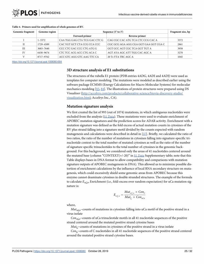
3D structure analysis of E1 substitutions
The structures of the rubella E1 protein (PDB entries 4ADG, 4ADI and 4ADJ) were used as
templates for computer modeling. The mutations were modeled as described earlier using the
software package ECMMS (Energy Calculations for Macro Molecular Systems) for molecular
mechanics modeling [83, 84]. The illustrations of protein structures were prepared using DS
Visualizer (http://accelrys.com/products/collaborative-science/biovia-discovery-studio/
visualization.html; Accelrys Inc., CA).
Mutation signature analysis
We first created the list of 993 (out of 1074) mutations, in which ambiguous nucleotides were
excluded from the analysis (S1 Data). These mutations were used to evaluate enrichment of
APOBEC mutation signatures and the prediction scores for ADAR activity. Enrichment with a
mutation signature was defined as the fold excess of actual mutation counts in cytosines of the
RV plus strand falling into a signature motif divided by the counts expected with random
mutagenesis and calculations were described in detail in [37]. Briefly, we calculated the ratio of
two ratios, the ratio of the number of mutations in cytosines falling into signature-specific tri-
nucleotide context to the total number of mutated cytosines as well as the ratio of the number
of signature specific trinucleotides to the total number of cytosines in the genomic back-
ground. For this background, we considered only the areas of 41 nucleotides centered around
the mutated base (column “CONTEXT(+/-20)” in S1 Data Supplementary table; note that this
Table displays bases in DNA format to allow compatibility and comparisons with mutation
signature outputs of APOBEC mutagenesis in DNA). This allowed us to minimize possible dis-
tortion of enrichment calculations by the influence of local RNA secondary structure on muta-
genesis, which could excessively shield some genomic areas from APOBEC because this
enzyme cannot deaminate cytosines in double stranded structures. The example of the formula
to calculate EuCa, Enrichment (i.e., fold-excess over random expectation) for uCa mutation sig-
nature is:
EuC a ¼MutuC a � ConC
MutC � ConuCa
where,
MutuCa−counts of mutations in cytosines falling into uCa motif of the positive strand in a
virus isolate
ConuCa−counts of uCa trinucleotide motifs in all 41 nucleotide sequences of the positive
strand centered around the mutated positive strand cytosine bases
MutC−counts of mutations in cytosines of the positive strand in a virus isolate
ConC−counts of C nucleotides in all 41 nucleotide sequences of the positive strand centered
around the mutated positive strand cytosine bases.
Table 4. Primers used for amplification of whole genomes of RV.
Genomic fragment Genome region Sequence (5’ to 3’) Fragment size, bp
Forward primer Reverse primer
I 1–3372 CAA TGG GAG CTA TCG GAC CTC G CAG CGC CAC ATG TCA CTC CCG CAC A 3372
II 1729–4589 CAC YGT GCT CTA CCG CCA CCC CGC GCG AGA AGG CGA GGT GAA GGT CGA C 2861
III 3683–7640 CCC CTC GAC CCC CTG ATG G GGT GCC AGT CGC TCA GGT TGT A 3958
IV 6430–9674 CTC TCC ACG ACC CTG ACA C AGT AYA AGC ATT TGG CAC AGC A 3270
V 8717–9762 ACC GTC AGG GTC AAG TTC CA 20 Ts YTA TRC AGC A 1045
https://doi.org/10.1371/journal.ppat.1008080.t004
Infectious vaccine-derived rubella viruses in immunodeficiencies
PLOS Pathogens | https://doi.org/10.1371/journal.ppat.1008080 October 28, 2019 25 / 32
Unlike the exquisitely specific to single stranded RNA and DNA APOBEC cytidine deami-
nases, ADARs act on adenines imbedded into double stranded RNA folds, so prediction of
ADAR deamination sites should account for combination of knowledge about experimentally
defined short nucleotide motifs and a potential to form double stranded structure. The predic-
tion scores for ADAR activity on adenines were generated using the web-based tool Inosine-
Predict (http://hci-bio-app.hci.utah.edu:8081/Bass/InosinePredict) and [85]. A one-sided
Mann-Whitney test for the prediction scores testing the hypothesis that scores for [A_to_G]
changes characteristic of ADAR would exceed scores for [A–[A_to_G]] nucleotides was per-
formed to obtain P-values shown above the scatter plots.
Supporting information
S1 Fig. RV antigen in chronic granulomatous lesions of LA, OR, and RI case patients. Dou-
ble immunofluorescent staining of granulomas with M2 macrophage-specific antibodies,
CD206 (green), and either RV capsid antibody (Abcam) (red) or measles (MeV) nucleoprotein
antibody 83KKII (Millipore) (red, not visible) was performed as described in Methods. Nuclei
were counterstained with DAPI. Note strong staining for RV antigen and the lack of staining
for measles antigen.
(TIF)
S1 Table. Nucleotide (nt) and amino acid (aa) substitutions in iVDRV genomes.
(DOCX)
S2 Table. Catalog of amino acid substitutions in iVDRV.
(XLSX)
S3 Table. Rates of synonymous (dS/year) and nonsynonymous (dN/year) substitutions in
iVDRV RVs genomes by gene.
(DOCX)
S4 Table. Mapping of the amino acid substitutions in RVs and RVi variants onto the E1
3D structure.
(DOCX)
S1 Data. The list of 993 (out of 1074) mutations in iVDRV RVs genomes with unambigu-
ously identified base substitutions. Sequences are shown in DNA format (T instead of U) to
maintain compatibility with other outputs of mutation signature R-script.
(XLSX)
S2 Data. Alignment of the nonstructural proteins of the 68 wtRV isolates, which circulated
worldwide during a period 1961–2012. The alignment was prepared with Mega7.
(MASX)
S3 Data. Alignment of the structural proteins of the 68 wtRV isolates, which circulated
worldwide during a period 1961–2012. The alignment was prepared with Mega7.
(MASX)
S4 Data. The list of pairwise genetic distances between individual quasispecies within pri-
mary granuloma sample (RVs) and the P1 CA6944 virus stock (RVi). Genetic distances was
computed using the Maximum Composite Likelihood method with Mega7.
(XLSX)
S5 Data. The average behavior of each codon for 6 pairwise comparisons to RA27/3 for
synonymous and nonsynonymous mutations, by gene. Data for each gene are located in a
Infectious vaccine-derived rubella viruses in immunodeficiencies
PLOS Pathogens | https://doi.org/10.1371/journal.ppat.1008080 October 28, 2019 26 / 32
separate sheet.
(XLSX)
S6 Data. RNA editing signatures.
(XLSX)
Acknowledgments
We thank the members of the CDC diagnostic team LiJuan Hao, Heather Colley and Kiana
Anthony for performing serological tests for rubella, mumps and measles IgG and IgM and to
Gimin Kim for performing real time PCR for measles and mumps RNA. We are grateful to
Yuri Wolf (NIH, NCBI) and Yury Khudyakov (CDC) for helpful discussions.
Disclaimer
The findings and conclusions in this report are those of the authors and do not necessarily rep-
resent the official position of the United States Centers for Disease Control and Prevention.
Author Contributions
Conceptualization: Ludmila Perelygina, Dmitry A. Gordenin, Stanley Plotkin, Kathleen E.
Sullivan, Joseph Icenogle.
Data curation: Ludmila Perelygina.
Formal analysis: Ludmila Perelygina, Min-hsin Chen, Ivan Y. Torshin.
Funding acquisition: Dmitry A. Gordenin, Kathleen E. Sullivan, Joseph Icenogle.
Investigation: Ludmila Perelygina, Min-hsin Chen, Suganthi Suppiah, Adebola Adebayo,
Emily Abernathy, Ivan Y. Torshin, Natalie Saini, Leszek J. Klimczak, Dmitry A. Gordenin,
Andrey Zharkikh.
Methodology: Ludmila Perelygina.
Project administration: Ludmila Perelygina, Kathleen E. Sullivan, Joseph Icenogle.
Resources: Morna Dorsey, Lionel Bercovitch, Kenneth Paris, Kevin P. White, Alfons Krol,
Julie Dhossche.
Supervision: Ludmila Perelygina, Dmitry A. Gordenin, Kathleen E. Sullivan, Joseph Icenogle.
Writing – original draft: Ludmila Perelygina, Ivan Y. Torshin, Dmitry A. Gordenin.
Writing – review & editing: Ludmila Perelygina, Min-hsin Chen, Suganthi Suppiah, Emily
Abernathy, Morna Dorsey, Lionel Bercovitch, Kevin P. White, Alfons Krol, Julie Dhossche,
Ivan Y. Torshin, Dmitry A. Gordenin, Andrey Zharkikh, Stanley Plotkin, Kathleen E. Sulli-
van, Joseph Icenogle.
References1. Approved proposal. Animal ssRNA+ viruses. 2018.013S.R.Matonaviridae 2019. Available from: https://
talk.ictvonline.org/files/ictv_official_taxonomy_updates_since_the_8th_report/m/animal-ssrna-viruses/
8087.
2. Rawls WE. Viral persistence in congenital rubella. Prog Med Virol. 1974; 18:273–88. PMID: 4608455
3. Lambert N, Strebel P, Orenstein W, Icenogle J, Poland GA. Rubella. Lancet. 2015 Jan 7. https://doi.org/
10.1016/S0140-6736(14)60539-0 PMID: 25576992.
Infectious vaccine-derived rubella viruses in immunodeficiencies
PLOS Pathogens | https://doi.org/10.1371/journal.ppat.1008080 October 28, 2019 27 / 32
4. Sherman FE, Michaels RH, Kenny FM. Acute encephalopathy (encephalitis) complicating rubella.
Report of cases with virologic studies, cortisol-production determinations, and observations at autopsy.
JAMA. 1965 May 24; 192:675–81. https://doi.org/10.1001/jama.1965.03080210019005 PMID:
14280514.
5. Kreps EO, Derveaux T, De Keyser F, Kestelyn P. Fuchs’ uveitis syndrome: no longer a syndrome?
Ocul Iammunol and Inflamm. 2015 Jul 29:1–10. https://doi.org/10.3109/09273948.2015.1005239
PubMed PMID: 26222767.
6. Plotkin SA. The history of rubella and rubella vaccination leading to elimination. Clin Infect Dis. 2006
Nov 1; 43 Suppl 3:S164–8. https://doi.org/10.1086/505950 PMID: 16998777.
7. Grant GB, Reef SE, Patel M, Knapp JK, Dabbagh A. Progress in rubella and congenital rubella syn-
drome control and elimination—Worldwide, 2000–2016. MMWR Morb Mortal Wkly Rep. 2017 Nov 17;
66(45):1256–60. https://doi.org/10.15585/mmwr.mm6645a4 PMID: 29145358. Pubmed Central
PMCID: PMC5726242.
8. Tingle AJ, Mitchell LA, Grace M, Middleton P, Mathias R, MacWilliam L, et al. Randomised double-blind
placebo-controlled study on adverse effects of rubella immunisation in seronegative women. Lancet.
1997 May 3; 349(9061):1277–81. https://doi.org/10.1016/S0140-6736(96)12031-6 PMID: 9142061.
9. Islam SM, El-Sheikh HF, Tabbara KF. Anterior uveitis following combined vaccination for measles,
mumps and rubella (MMR): a report of two cases. Acta Ophthalmol Scand. 2000 Oct; 78(5):590–2.
https://doi.org/10.1034/j.1600-0420.2000.078005590.x PMID: 11037921.
10. Plotkin S, Reef S, Cooper L, Alford CA. Rubella. In: J. R, Klein J, Wilson C, Nizet V, Maldonato Y, edi-
tors. Infectious diseases of the fetus and newborn infant. Philadelphia, PA: Elsveier; 2011. p. 861–98.
11. Picard C, Bobby Gaspar H, Al-Herz W, Bousfiha A, Casanova JL, Chatila T, et al. International Union of
Immunological Societies: 2017 primary immunodeficiency diseases committee report on inborn errors
of immunity. J Clin Immunol. 2018 Jan; 38(1):96–128. https://doi.org/10.1007/s10875-017-0464-9
PMID: 29226302. Pubmed Central PMCID: PMC5742601.
12. Shearer WT, Fleisher TA, Buckley RH, Ballas Z, Ballow M, Blaese RM, et al. Recommendations for live
viral and bacterial vaccines in immunodeficient patients and their close contacts. J Allergy Clin Immunol.
2014 Apr; 133(4):961–6. https://doi.org/10.1016/j.jaci.2013.11.043 PMID: 24582311. Pubmed Central
PMCID: PMC4009347.
13. Eibl MM, Wolf HM. Vaccination in patients with primary immune deficiency, secondary immune defi-
ciency and autoimmunity with immune regulatory abnormalities. Immunotherapy. 2015; 7(12):1273–92.
https://doi.org/10.2217/IMT.15.74 PMID: 26289364.
14. Nanda A, Al-Herz W, Al-Sabah H, Al-Ajmi H. Noninfectious cutaneous granulomas in primary immuno-
deficiency disorders: report from a national registry. Am J Dermatopathol. 2014 Oct; 36(10):832–7.
https://doi.org/10.1097/DAD.0000000000000112 PMID: 25062261.
15. Leung J, Sullivan KE, Perelygina L, Icenogle JP, Fuleihan RL, Lanzieri TM. Prevalence of granulomas
in patients with primary immunodeficiency disorders, United States: Data From national health care
claims and the US Immunodeficiency Network Registry. J Clin Immunol. 2018 Aug; 38(6):717–26.
https://doi.org/10.1007/s10875-018-0534-7 PMID: 30043271. Pubmed Central PMCID: PMC6155465.
16. Bodemer C, Sauvage V, Mahlaoui N, Cheval J, Couderc T, Leclerc-Mercier S, et al. Live rubella virus
vaccine long-term persistence as an antigenic trigger of cutaneous granulomas in patients with primary
immunodeficiency. Clin Microbiol Infect. 2014 Jan 30; 20(10):O656–63. https://doi.org/10.1111/1469-
0691.12573 PMID: 24476349.
17. Perelygina L, Plotkin S, Russo P, Hautala T, Bonilla F, Ochs HD, et al. Rubella persistence in epidermal
keratinocytes and granuloma M2 macrophages in patients with primary immunodeficiencies. J Allergy
Clin Immunol. 2016 Nov; 138(5):1436–9 e11. https://doi.org/10.1016/j.jaci.2016.06.030 PMID:
27613149. Pubmed Central PMCID: PMC5392721.
18. Neven B, Perot P, Bruneau J, Pasquet M, Ramirez M, Diana JS, et al. Cutaneous and visceral chronic
granulomatous disease triggered by a rubella virus vaccine strain in children with primary immunodefi-
ciencies. Clin Infect Dis. 2017 Jan 01; 64(1):83–6. https://doi.org/10.1093/cid/ciw675 PMID: 27810866.
19. Buchbinder D, Hauck F, Albert MH, Rack A, Bakhtiar S, Shcherbina A, et al. Rubella virus-associated
cutaneous granulomatous disease: a unique complication in immune-deficient patients, not limited to
DNA repair disorders. J Clin Immunol. 2019 Jan 3. https://doi.org/10.1007/s10875-018-0581-0 PMID:
30607663.
20. Plotkin SA, Farquhar JD, Katz M, Buser F. Attenuation of RA 27–3 rubella virus in WI-38 human diploid
cells. Am J Dis Child. 1969; 118(2):178–85. https://doi.org/10.1001/archpedi.1969.02100040180004
PMID: 5794813
21. Perelygina L, Adebayo A, Metcalfe M, Icenogle J. Differences in establishment of persistence of vaccine
and wild type rubella viruses in fetal endothelial cells. PLoS One. 2015; 10(7):e0133267. https://doi.org/
10.1371/journal.pone.0133267 PMID: 26177032.
Infectious vaccine-derived rubella viruses in immunodeficiencies
PLOS Pathogens | https://doi.org/10.1371/journal.ppat.1008080 October 28, 2019 28 / 32
22. Gonzales JA, Hinterwirth A, Shantha J, Wang K, Zhong L, Cummings SL, et al. Association of ocular
inflammation and rubella virus persistence. JAMA Ophthalmol. 2018 Dec 27. https://doi.org/10.1001/
jamaophthalmol.2018.6185 PMID: 30589932.
23. Yang Z, Bielawski JP. Statistical methods for detecting molecular adaptation. Trends Ecol Evol. 2000
Dec 1; 15(12):496–503. https://doi.org/10.1016/s0169-5347(00)01994-7 PMID: 11114436.
24. Echave J, Spielman SJ, Wilke CO. Causes of evolutionary rate variation among protein sites. Nat Rev
Genet. 2016 Feb; 17(2):109–21. https://doi.org/10.1038/nrg.2015.18 PMID: 26781812. Pubmed Cen-
tral PMCID: PMC4724262.
25. Liu Z, Yang D, Qiu Z, Lim KT, Chong P, Gillam S. Identification of domains in rubella virus genomic
RNA and capsid protein necessary for specific interaction. J Virol. 1996 Apr; 70(4):2184–90. PubMed
PMID: 8642641. Pubmed Central PMCID: 190057.
26. Chen MH, Icenogle JP. Rubella virus capsid protein modulates viral genome replication and virus infec-
tivity. J Virol. 2004 Apr; 78(8):4314–22. https://doi.org/10.1128/JVI.78.8.4314-4322.2004 PMID:
15047844. Pubmed Central PMCID: 374250.
27. Ilkow CS, Goping IS, Hobman TC. The rubella virus capsid as an anti-apoptotic protein that attenuates
the pore-forming ability of Bax. PLoS Pathog. 2011; 7(2):e1001291. https://doi.org/10.1371/journal.
ppat.1001291 PMID: 21379337
28. Petit V, Guetard D, Renard M, Keriel A, Sitbon M, Wain-Hobson S, et al. Murine APOBEC1 is a powerful
mutator of retroviral and cellular RNA in vitro and in vivo. J Mol Biol. 2009 Jan 9; 385(1):65–78. https://
doi.org/10.1016/j.jmb.2008.10.043 PMID: 18983852.
29. Harris RS, Dudley JP. APOBECs and virus restriction. Virology. 2015 May; 479-480(0):131–45. https://
doi.org/10.1016/j.virol.2015.03.012 PMID: 25818029. Pubmed Central PMCID: PMC4424171.
30. Tomaselli S, Galeano F, Locatelli F, Gallo A. ADARs and the balance game between virus infection and
innate immune cell response. Curr Issues Mol Biol. 2015; 17:37–51. PubMed PMID: 25502818.
31. Refsland EW, Harris RS. The APOBEC3 family of retroelement restriction factors. Curr Top Microbiol
Immunol. 2013; 371:1–27. https://doi.org/10.1007/978-3-642-37765-5_1 PMID: 23686230. Pubmed
Central PMCID: PMC3934647.
32. Niavarani A, Currie E, Reyal Y, Anjos-Afonso F, Horswell S, Griessinger E, et al. APOBEC3A is impli-
cated in a novel class of G-to-A mRNA editing in WT1 transcripts. PLoS One. 2015; 10(3):e0120089.
https://doi.org/10.1371/journal.pone.0120089 PMID: 25807502. Pubmed Central PMCID: 4373805.
33. Sharma S, Patnaik SK, Taggart RT, Kannisto ED, Enriquez SM, Gollnick P, et al. APOBEC3A cytidine
deaminase induces RNA editing in monocytes and macrophages. Nat Commun. 2015 Apr 21; 6:6881.
https://doi.org/10.1038/ncomms7881 PMID: 25898173. Pubmed Central PMCID: PMC4411297.
34. Sharma S, Patnaik SK, Taggart RT, Baysal BE. The double-domain cytidine deaminase APOBEC3G is
a cellular site-specific RNA editing enzyme. Sci Rep. 2016 Dec 15; 6:39100. https://doi.org/10.1038/
srep39100 PMID: 27974822. Pubmed Central PMCID: PMC5156925.
35. Prohaska KM, Bennett RP, Salter JD, Smith HC. The multifaceted roles of RNA binding in APOBEC
cytidine deaminase functions. Wiley Interdiscip Rev RNA. 2014 Jul-Aug; 5(4):493–508. https://doi.org/
10.1002/wrna.1226 PMID: 24664896. Pubmed Central PMCID: PMC4062598.
36. Lada AG, Krick CF, Kozmin SG, Mayorov VI, Karpova TS, Rogozin IB, et al. Mutator effects and muta-
tion signatures of editing deaminases produced in bacteria and yeast. Biochemistry (Mosc). 2011 Jan;
76(1):131–46. https://doi.org/10.1134/s0006297911010135 PMID: 21568845. Pubmed Central
PMCID: PMC3906858.
37. Chan K, Roberts SA, Klimczak LJ, Sterling JF, Saini N, Malc EP, et al. An APOBEC3A hypermutation
signature is distinguishable from the signature of background mutagenesis by APOBEC3B in human
cancers. Nat Genet. 2015 Sep; 47(9):1067–72. https://doi.org/10.1038/ng.3378 PMID: 26258849.
Pubmed Central PMCID: PMC4594173.
38. Yang D, Hwang D, Qiu Z, Gillam S. Effects of mutations in the rubella virus E1 glycoprotein on E1-E2
interaction and membrane fusion activity. J Virol. 1998 Nov; 72(11):8747–55. PubMed PMID: 9765418.
Pubmed Central PMCID: 110290.
39. Cong H, Jiang Y, Tien P. Identification of the myelin oligodendrocyte glycoprotein as a cellular receptor
for rubella virus. J Virol. 2011 Nov; 85(21):11038–47. https://doi.org/10.1128/JVI.05398-11 PMID:
21880773. Pubmed Central PMCID: 3194935.
40. Ho-Terry L, Terry GM, Cohen A, Londesborough P. Immunological characterisation of the rubella E 1
glycoprotein. Brief report. Arch Virol. 1986; 90(1–2):145–52. https://doi.org/10.1007/bf01314152 PMID:
2425779.
41. Chaye HH, Mauracher CA, Tingle AJ, Gillam S. Cellular and humoral immune responses to rubella
virus structural proteins E1, E2, and C. J Clin Microbiol. 1992 Sep; 30(9):2323–9. PubMed PMID:
1383269. Pubmed Central PMCID: 265500.
Infectious vaccine-derived rubella viruses in immunodeficiencies
PLOS Pathogens | https://doi.org/10.1371/journal.ppat.1008080 October 28, 2019 29 / 32
42. Wolinsky JS, McCarthy M, Allen-Cannady O, Moore WT, Jin R, Cao SN, et al. Monoclonal antibody-
defined epitope map of expressed rubella virus protein domains. J Virol. 1991; 65(8):3986–94. PMID:
1712855
43. Green KY, Dorsett PH. Rubella virus antigens: localization of epitopes involved in hemagglutination and
neutralization by using monoclonal antibodies. J Virol. 1986; 57(3):893–8. PMID: 2419590
44. Lovett AE, Hahn CS, Rice CM, Frey TK, Wolinsky JS. Rubella virus-specific cytotoxic T-lymphocyte
responses: identification of the capsid as a target of major histocompatibility complex class I-restricted
lysis and definition of two epitopes. J Virol. 1993 Oct; 67(10):5849–58. PubMed PMID: 7690412.
Pubmed Central PMCID: 238003.
45. Ou D, Mitchell LA, Decarie D, Gillam S, Tingle AJ. Characterization of an overlapping CD8+ and CD4+
T-cell epitope on rubella capsid protein. Virology. 1997 Sep 1; 235(2):286–92. https://doi.org/10.1006/
viro.1997.8704 PMID: 9281508.
46. DuBois RM, Vaney MC, Tortorici MA, Kurdi RA, Barba-Spaeth G, Krey T, et al. Functional and evolu-
tionary insight from the crystal structure of rubella virus protein E1. Nature. 2013 Jan 24; 493
(7433):552–6. https://doi.org/10.1038/nature11741 PMID: 23292515.
47. Plotkin SA, Buser F. History of RA27/3 rubella vaccine. Rev Infect Dis. 1985 Mar-Apr; 7 Suppl 1:S77–8.
https://doi.org/10.1093/clinids/7.supplement_1.s77 PMID: 3890107.
48. Perelygina L, Zheng Q, Metcalfe M, Icenogle J. Persistent infection of human fetal endothelial cells with
rubella virus. PLoS One. 2013; 8(8):e73014. https://doi.org/10.1371/journal.pone.0073014 PMID:
23940821. Pubmed Central PMCID: 3734309.
49. Seagle EE, Bednarczyk RA, Hill T, Fiebelkorn AP, Hickman CJ, Icenogle JP, et al. Measles, mumps,
and rubella antibody patterns of persistence and rate of decline following the second dose of the MMR
vaccine. Vaccine. 2018 Feb 1; 36(6):818–26. https://doi.org/10.1016/j.vaccine.2017.12.075 PMID:
29317117.
50. Miki NP, Chantler JK. Differential ability of wild-type and vaccine strains of rubella virus to replicate and
persist in human joint tissue. Clin Exp Rheumatol. 1992 Jan-Feb; 10(1):3–12. PubMed PMID: 1551276.
51. Duffy S, Shackelton LA, Holmes EC. Rates of evolutionary change in viruses: patterns and determi-
nants. Nat Rev Genet. 2008 Apr; 9(4):267–76. https://doi.org/10.1038/nrg2323 PMID: 18319742.
52. Zhu Z, Chen MH, Abernathy E, Icenogle J, Zhou S, Wang C, et al. Analysis of complete genomes of the
rubella virus genotypes 1E and 2B which circulated in China, 2000–2013. Sci Rep. 2016 Dec 13;
6:39025. https://doi.org/10.1038/srep39025 PMID: 27959338. Pubmed Central PMCID: PMC5154293.
53. Domingo E, Baranowski E, Ruiz-Jarabo CM, Martin-Hernandez AM, Saiz JC, Escarmis C. Quasispe-
cies structure and persistence of RNA viruses. Emerg Infect Dis. 1998 Oct-Dec; 4(4):521–7. https://doi.
org/10.3201/eid0404.980402 PMID: 9866728. Pubmed Central PMCID: PMC2640251.
54. Farci P, Shimoda A, Coiana A, Diaz G, Peddis G, Melpolder JC, et al. The outcome of acute hepatitis C
predicted by the evolution of the viral quasispecies. Science. 2000 Apr 14; 288(5464):339–44. https://
doi.org/10.1126/science.288.5464.339 PMID: 10764648.
55. Domingo E, Sheldon J, Perales C. Viral quasispecies evolution. Microbiology and molecular biology
reviews: MMBR. 2012 Jun; 76(2):159–216. https://doi.org/10.1128/MMBR.05023-11 PMID: 22688811.
Pubmed Central PMCID: PMC3372249.
56. Lemey P, Rambaut A, Pybus OG. HIV evolutionary dynamics within and among hosts. AIDS Rev. 2006
Jul-Sep; 8(3):125–40. PubMed PMID: 17078483.
57. Raghwani J, Rose R, Sheridan I, Lemey P, Suchard MA, Santantonio T, et al. Exceptional heterogene-
ity in viral evolutionary dynamics characterises chronic hepatitis C virus infection. PLoS Pathog. 2016
Sep; 12(9):e1005894. https://doi.org/10.1371/journal.ppat.1005894 PMID: 27631086. Pubmed Central
PMCID: PMC5025083.
58. Koning FA, Newman EN, Kim EY, Kunstman KJ, Wolinsky SM, Malim MH. Defining APOBEC3 expres-
sion patterns in human tissues and hematopoietic cell subsets. J Virol. 2009 Sep; 83(18):9474–85.
https://doi.org/10.1128/JVI.01089-09 PMID: 19587057. Pubmed Central PMCID: PMC2738220.
59. Liu Y, Ma T, Liu J, Zhao X, Cheng Z, Guo H, et al. Circulating type 1 vaccine-derived poliovirus may
evolve under the pressure of adenosine deaminases acting on RNA. J Matern Fetal Neonatal Med.
2015; 28(17):2096–9. https://doi.org/10.3109/14767058.2014.979147 PMID: 25330844.
60. Fehrholz M, Kendl S, Prifert C, Weissbrich B, Lemon K, Rennick L, et al. The innate antiviral factor APO-
BEC3G targets replication of measles, mumps and respiratory syncytial viruses. J Gen Virol. 2012 Mar;
93(Pt 3):565–76. https://doi.org/10.1099/vir.0.038919-0 PMID: 22170635.
61. Milewska A, Kindler E, Vkovski P, Zeglen S, Ochman M, Thiel V, et al. APOBEC3-mediated restriction
of RNA virus replication. Sci Rep. 2018 Apr 13; 8(1):5960. https://doi.org/10.1038/s41598-018-24448-2
PMID: 29654310. Pubmed Central PMCID: PMC5899082.
Infectious vaccine-derived rubella viruses in immunodeficiencies
PLOS Pathogens | https://doi.org/10.1371/journal.ppat.1008080 October 28, 2019 30 / 32
62. Abernathy E, Chen MH, Bera J, Shrivastava S, Kirkness E, Zheng Q, et al. Analysis of whole genome
sequences of 16 strains of rubella virus from the United States, 1961–2009. Virol J. 2013 Jan 25; 10
(1):32. https://doi.org/10.1186/1743-422X-10-32 PMID: 23351667.
63. Schiff GM, Sever JL, Huebner RJ. Rubella virus: neutralizing antibody in commercial gamma globulin.
Science. 1963 Oct 4; 142(3588):58–60. https://doi.org/10.1126/science.142.3588.58 PMID: 14057354.
64. Quiroga JA, van Binsbergen J, Wang CY, Pardo M, Navas S, Trines C, et al. Immunoglobulin M anti-
body to hepatitis C virus core antigen: correlations with viral replication, histological activity, and liver
disease outcome. Hepatology. 1995 Dec; 22(6):1635–40. PubMed PMID: 7489967.
65. Wherry EJ. T cell exhaustion. Nat Immunol. 2011 Jun; 12(6):492–9. PubMed PMID: 21739672.
66. Aghamohammadi A, Abolhassani H, Kutukculer N, Wassilak SG, Pallansch MA, Kluglein S, et al.
Patients with primary immunodeficiencies are a reservoir of poliovirus and a risk to polio eradication.
Front Immunol. 2017; 8:685. https://doi.org/10.3389/fimmu.2017.00685 PMID: 28952612. Pubmed
Central PMCID: PMC5468416.
67. Memoli MJ, Athota R, Reed S, Czajkowski L, Bristol T, Proudfoot K, et al. The natural history of influ-
enza infection in the severely immunocompromised vs nonimmunocompromised hosts. Clin Infect Dis.
2014 Jan; 58(2):214–24. https://doi.org/10.1093/cid/cit725 PMID: 24186906. Pubmed Central PMCID:
PMC3871797.
68. Xue KS, Stevens-Ayers T, Campbell AP, Englund JA, Pergam SA, Boeckh M, et al. Parallel evolution of
influenza across multiple spatiotemporal scales. Elife. 2017 Jun 27; 6. https://doi.org/10.7554/eLife.
26875 PMID: 28653624. Pubmed Central PMCID: PMC5487208.
69. Dhossche J, Johnson L, White K, Funk T, Leitenberger S, Perelygina L, et al. Cutaneous Granuloma-
tous Disease With Presence of Rubella Virus in Lesions. JAMA Dermatol. 2019 Jun 5. https://doi.org/
10.1001/jamadermatol.2019.0814 PMID: 31166586.
70. Perelygina L, Buchbinder D, Dorsey MJ, Eloit M, Hauck F, Hautala T, et al. Outcomes for nitazoxanide
treatment in a case series of patients with primary immunodeficiencies and rubella virus-associated
granuloma. J Clin Immunol. 2019 Jan 24. https://doi.org/10.1007/s10875-019-0589-0 PMID: 30680653.
71. Abernathy E, Cabezas C, Sun H, Zheng Q, Chen MH, Castillo-Solorzano C, et al. Confirmation of
rubella within 4 days of rash onset: comparison of rubella virus RNA detection in oral fluid with immuno-
globulin M detection in serum or oral fluid. J Clin Microbiol. 2009 Jan; 47(1):182–8. https://doi.org/10.
1128/JCM.01231-08 PMID: 19005151.
72. Namuwulya P, Abernathy E, Bukenya H, Bwogi J, Tushabe P, Birungi M, et al. Phylogenetic analysis of
rubella viruses identified in Uganda, 2003–2012. J Med Virol. 2014 Apr 4. https://doi.org/10.1002/jmv.
23935 PMID: 24700073.
73. Chen MH, Zhu Z, Zhang Y, Favors S, Xu WB, Featherstone DA, et al. An indirect immunocolorimetric
assay to detect rubella virus infected cells. J Virol Methods. 2007 Dec; 146(1–2):414–8. https://doi.org/
10.1016/j.jviromet.2007.08.021 PMID: 17919742.
74. Rota JS, Rosen JB, Doll MK, McNall RJ, McGrew M, Williams N, et al. Comparison of the sensitivity of
laboratory diagnostic methods from a well-characterized outbreak of mumps in New York city in 2009.
Clin Vaccine Immunol. 2013 Mar; 20(3):391–6. https://doi.org/10.1128/CVI.00660-12 PMID: 23324519.
Pubmed Central PMCID: PMC3592341.
75. Hummel KB, Erdman DD, Heath J, Bellini WJ. Baculovirus expression of the nucleoprotein gene of
measles virus and utility of the recombinant protein in diagnostic enzyme immunoassays. J Clin Micro-
biol. 1992 Nov; 30(11):2874–80. PubMed PMID: 1452657. Pubmed Central PMCID: PMC270545.
76. Hummel KB, Lowe L, Bellini WJ, Rota PA. Development of quantitative gene-specific real-time RT-PCR
assays for the detection of measles virus in clinical specimens. J Virol Methods. 2006 Mar; 132(1–
2):166–73. https://doi.org/10.1016/j.jviromet.2005.10.006 PMID: 16274752.
77. Standardization of the nomenclature for genetic characteristics of wild-type rubella viruses. Wkly Epide-
miol Rec. 2005 Apr 8; 80(14):126–32. PubMed PMID: 15850226.
78. Edgar RC. MUSCLE: multiple sequence alignment with high accuracy and high throughput. Nucleic
Acids Res. 2004; 32(5):1792–7. https://doi.org/10.1093/nar/gkh340 PMID: 15034147. Pubmed Central
PMCID: PMC390337.
79. Kumar S, Stecher G, Tamura K. MEGA7: Molecular Evolutionary Genetics Analysis Version 7.0 for Big-
ger Datasets. Mol Biol Evol. 2016 Jul; 33(7):1870–4. https://doi.org/10.1093/molbev/msw054 PMID:
27004904.
80. Nei M, Gojobori T. Simple methods for estimating the numbers of synonymous and nonsynonymous
nucleotide substitutions. Mol Biol Evol. 1986 Sep; 3(5):418–26. https://doi.org/10.1093/oxfordjournals.
molbev.a040410 PMID: 3444411.
Infectious vaccine-derived rubella viruses in immunodeficiencies
PLOS Pathogens | https://doi.org/10.1371/journal.ppat.1008080 October 28, 2019 31 / 32
81. Korber B. HIV signature and sequence variation analysis. In: Rodrigo AG, Learn GH, editors. Computa-
tional analysis of HIV molecular sequences. Dordrecht, Netherlands: Kluwer Academic Publishers;
2000. p. 55–72.
82. Jukes K, CAntor C. Evolution of protein molecules. In: Munro H, editor. Mammalian Protein Metabolism.
New York: Academic Press; 1969. p. 21–132.
83. Torshin IY. Computed energetics of nucleotides in spatial ribozyme structures: an accurate identifica-
tion of functional regions from structure. ScientificWorldJournal. 2004 Mar 26; 4:228–47. https://doi.org/
10.1100/tsw.2004.19 PMID: 15105962. Pubmed Central PMCID: PMC5956427.
84. Patrusheva I, Perelygina L, Torshin I, LeCher J, Hilliard J. B virus (Macacine Herpesvirus 1) divergence:
variations in glycoprotein D from clinical and laboratory isolates diversify virus entry strategies. J Virol.
2016 Oct 15; 90(20):9420–32. https://doi.org/10.1128/JVI.00799-16 PMID: 27512063.
85. Eggington JM, Greene T, Bass BL. Predicting sites of ADAR editing in double-stranded RNA. Nat Com-
mun. 2011; 2:319. https://doi.org/10.1038/ncomms1324 PMID: 21587236. Pubmed Central PMCID:
PMC3113232.
Infectious vaccine-derived rubella viruses in immunodeficiencies
PLOS Pathogens | https://doi.org/10.1371/journal.ppat.1008080 October 28, 2019 32 / 32