visual outcome in t cataract in childr rubella s
106
VISUAL OUT CATARACT I R Submitted M REGIONAL MA DR. M Dissertation on TCOME IN THE MANAGEME IN CHILDREN WITH CONGE RUBELLA SYNDROME in partial fulfillment of requirements o M.S. OPHTHALMOLOGY BRANCH - III L INSTITUTE OF OPHTHALMOLO ADRAS MEDICAL COLLEGE CHENNAI- 600 003 THE TAMILNADU M.G.R. MEDICAL UNIVERSITY CHENNAI APRIL 2013 ENT OF ENITAL of OGY
-
Upload
khangminh22 -
Category
Documents
-
view
0 -
download
0
Transcript of visual outcome in t cataract in childr rubella s
Dissertation on
VISUAL OUTCOME IN THE MANAGEMENT OFCATARACT IN CHILDREN WITH CONGENITAL
RUBELLA SYNDROME
Submitted in partial fulfillment of requirements of
M.S. OPHTHALMOLOGY
BRANCH - III
REGIONAL INSTITUTE OF OPHTHALMOLOGY
MADRAS MEDICAL COLLEGE
CHENNAI- 600 003
THE TAMILNADU
DR. M.G.R. MEDICAL UNIVERSITY
CHENNAI
APRIL 2013

CERTIFICATE
This is to certify that this dissertation entitled “Visual Outcome in
the Management of Cataract in Children with Congenital
Rubella Syndrome” is a bonafide record of the research work done by
Dr.S.CHITRA, post graduate in Regional Institute of Ophthalmology and
Government Ophthalmic Hospital, Madras Medical College and Government
General Hospital, Chennai-03, in partial fulfillment of the regulations laid
down by The Tamil Nadu Dr. M.G.R. Medical University for the award of
M.S. Ophthalmology Branch III, under my guidance and supervision during
the academic years 2010-2013.
Place:
Date :
Dr.K. NAMITHA BHUVANESWARI MS.,DO.
Chief – Squint, Neuro - Ophthalmology
and Paediatric Ophthalmology Dept.
RIO – GOH
Egmore, Chennai – 08
Dr.K.MARAGATHAM MS.,DO.
Director
RIO – GOH
Egmore, Chennai – 08
Dr.Prof. DR. V. KANAGASABAI M.D.,PhD.
Dean,
Madras Medical College.
and Government General Hospital
Chennai – 03

ACKNOWLEDGEMENT
I express my sincere thanks and gratitude to Prof. DR.V.
KANAGASABAI M.D., PhD. Dean, Madras Medical College and
Government General Hospital for permitting me to conduct this study.
I have great pleasure in thanking Prof. Dr. K. MARAGATHAM
M.S., DO. Director, RIO – GOH, Madras Medical College, for her valuable
advice in preparing this dissertation.
I express my profound gratitude to Prof. Dr. K. NAMITHA
BHUVANESWARI M.S., DO. my unit chief and my guide for her valuable
guidance and constant support at every stage throughout the period of this
study.
I am very grateful to my unit assistants Dr. B. PRAMILA M.S.,
DR. R. MUTHIAH M.S., and Dr. T.G. UMA MAHESWARI M.S., for
rendering their valuable advice and guidance for the study.
I wish to express my sincere thanks to all the professors, assistant
professors and all my colleagues who had helped me in bringing out this
study.


ABBREVIATIONS
RNA Ribonucleic acid
DNA Deoxyribo nucleic acid
ATP Adenosine triphosphate
TORCH Toxoplasma, Rubella, Cytomegalovirus, herpes simplex
IgG Immunoglobulin G
IgM Immunoglobulin M
kD Kilodalton
CMV Cytomegalovirus
PAS Periodic Acid Schiff
NADPH Nicotinamide adenine dinucleotide phosphate
CRS Congenital rubella syndrome
ELISA Enzyme linked immunoassay
HSV Herpes simplex virus
HAI Hemagglutination inhibition test
IOL Intraocular lens
PCO Posterior capsular opacity
CCC Continuous curvilinear capsulorrhexis
MMR Measles Mumps Rubella
RE Right Eye
LE Left eye

CONTENTS
S. NO TITLE PAGE. NO
PART - I
1 INTRODUCTION 1
2 REVIEW OF LITERATURE 2
3 EMBRYOLOGY 3
4 ANATOMY, PHYSIOLOGY AND BIOCHEMISTRY 6
5 CONGENITAL CATARACT 14
6 CONGENITAL RUBELLA SYNDROME 20
7 APPROACH TO A CHILD WITH CONGENITAL RUBELLA
CATARACT
33
8 MANAGEMENT 37
PART – II
6 OBJECTIVES 41
7 MATERIALS AND METHODS 41
8 OBSERVATION AND RESULTS 44
9 DISCUSSION 74
10 CONCLUSION 76
PART – III
11 BIBLIOGRAPHY 80
12 PROFORMA 85
13 MASTERCHART
14 KEY TO MASTERCHART
15 LIST OF SURGERIES PERFORMED

1
INTRODUCTION
Congenital cataract refers to a lens opacity present at birth. Incidence of
congenital cataract is one in every 2000 live births. Congenital cataracts are
aetiologically heterogeneous and show a great range of phenotypes depending on
their appearance and location in the lens.
Both genetic and non-genetic factors contribute to the cause of congenital
cataract. Viral infections, metabolic disorders, certain antibiotics and exposure to
certain ionizing radiation during pregnancy and rarely chromosomal abnormalities
contribute to the occurrence of congenital cataract. In majority of cases the
etiology is unknown.
Maternal infection with the rubella virus, a RNA toga virus cause fetal
damage if infection occurs during the first trimester of pregnancy. The term
rubella is a latin word meaning “little red”. Cataracts resulting from congenital
rubella syndrome are characterized by pearly white nuclear opacification. Cataract
may occur alone or part of classic triad of congenital rubella syndrome. Cataract is
the most common ocular sign in congenital rubella syndrome. The occurrence of
cataract is characteristic of the incidence of the rubella rash during the fifth or
sixth week of gestation when the primary fibres of the lens are being laid down.
Cataract removal in congenital rubella syndrome may be complicated by
excessive post operative inflammation caused by release of these live virus
particles. Live virus particles may be recovered from the lens as late as three years
after birth.

2
REVIEW OF LITERATURE
Synonyms of congenital rubella syndrome are Embryopathia rubeolaris,
Gregg-syndrome. First description of congenital rubella syndrome was given by
the Australian ophthalmologist Sir Norman McAlister Gregg in 1941.
According to Warburg,M in the year1986 maternal rubella in pregnancy
has been the single most common cause of congenital cataract .
In Dijk's Victorian Study 18 of the 81 rubella children had bilateral cataract in
addition to their hearing impairment.
Chess et al. - 1971 found 4 of 243 rubella children with visual defects
alone, 2 with severe, and two with moderate impairment
In a special ophthalmologic study on the ocular manifestations of the 1964-65
U.S.A rubella epidemic, other eye diseases which are often associated with
cataracts are reported.
1. Geltzer et al. – 1968 found that microphtalmia, retinopathy and
nystagmus occurred in half the cases studied.
2. Dijk, J. van 1982 - Iris hypoplasia, microcornea and glaucoma were
present in almost one quarter of the cases.
Desmond – 1975 Ocular defects occur in 30% to 60% of infants exposed
to rubella in the first trimester. The most common lesion is unilateral or bilateral
cataract, but Desmond also points to microphthalmia (frequently associated with
cataract), retinopathy, congenital glaucoma (less common), strabismus,
nystagmus. Enerstvedt – 1991 Collected data on 50 persons between 16 and 40
years of age who have a multi-sensory impairment caused by congenital rubella.

3
EMBRYOLOGY OF LENS
The formation of human crystalline lens begins around twenty five days of
gestation with two lateral evaginations called the optic vesicles from the forebrain
or diencephalon1. The ectodermal cells that overlie the optic vesicles thickened to
form the lens placode. The lens pit appears at 29 days of gestation as an
indentation of the lens placode. The lens pit deepens and invaginates to form the
lens vesicle which consists of a single layer of cells covered by a basal lamina
around 30 days of gestation. The cells of the posterior wall of the lens vesicle
rapidly elongate and get filled with proteins called crystallins.
The base of these densely packed cells remain anchored to the basal
laminae posteriorly and their apices grow towards the anterior lens epithelium
obliterating the lumen of lens vesicle2. These elongated transparent cells are
known as primary lens fibres.The primary lens fibres are formed up to the third
month of gestation and are preserved as the compact core of the lens, known as
embryonic nucleus.The equatorial cells of the anterior epithelium remain active
throughout the life and form the secondary lens fibres.The secondary lens fibres
are laid down concentrically so they has a laminated appearance. Depending upon
the period of development the secondary lens fibres are named as fetal nucleus,
infantile nucleus, adult nucleus.
Fetal nucleus refers to the secondary lens fibres formed from third to
eighth month of gestation. The initial lens fibres of fetal nucleus reach both the
anterior and posterior poles and they surround the embryonic nucleus. The
subsequent formed fibres of fetal nucleus can no longer extend from one pole to

4
the other3. Instead they meet at radiating lines or sutures that appear as an erect Y
anteriorly and inverted Y posteriorly.
Infantile nucleus refers to the secondary lens fibres formed during the last
weeks of fetal life to puberty. Adult nucleus is formed by the secondary lens fibres
formed after the puberty. Cortex consists of the recently formed superficial
secondary lens fibres4.
During embryonic and fetal development , the lens receives nourishment
via an intricate vascular capsule, the tunica vasculosa lentis that completely
encompasses the lens by approximately nine weeks5. It is formed from the
mesenchyme that surrounds the lens. In the earliest stages of development, it
receives an abundant blood supply from the hyaloids artery6. Later this blood
supply regresses, and the vascular capsule disappears before birth.

5
Embrology of lens
Figure- 1a Figure – 1b
Figure-1c
Histopathology of lens
Figure 1d – Arrangement of lens fibres

6
ANATOMY, PHYSIOLOGY AND BIOCHEMISTRY
OF THE LENS
The lens is biconvex structure located in the posterior chamber directly
behind the pupil. The crystalline lens forms the second refractive unit of the
human eye, adding 15 – 20 dioptre of plus power to the 45 dioptre created by the
cornea. As such, it must be perfectly clear otherwise light will not reach the retinal
sensory elements in an undisturbed state. Anterior-posterior diameter is four to
five mm and it gradually increases during life. Equatorial diameter being nine to
ten mm, remains unchanged. The weight of the human lens is 65 mg at birth. Lens
lacks nerve innervation as well as blood supply and it depends totally on aqueous
for its nutrition7.
A Cataract is an opacity in the lens, whether or not visual impairment
results. While congenital cataracts that impair vision are relatively uncommon,
many types of opacities like dots, clumps, lines, clefts, deposits, colours,
membranes, and discs have been described in the newborn lens.
Lens Histology:
Capsule:
The lens is surrounded by a typical PAS positive basement membrane
known as the lens capsule. The lens capsule is the thickest basement membrane in
the body. The anterior lens capsule is twice as thick as posterior capsule. The
capsule is created anteriorly by the epithelial cells and posteiorly by cortical
fibres. The capsule itself is non cellular having a structure composed largely of
glycoprotein – associated type IV collagen7. The mucopolysaccharide – heparin
sulphate make up less than one percent of the lens capsule but is considered very
important in determining the structure of the matrix which in turn is probably

7
critical in maintaining capsule clarity. The capsule acts as a barrier in keeping
back the vitreous and also acts as a barrier against fluorescein, bacteria and growth
factor7.
Epithelium:
The epithelium lies beneath the anterior and equatorial capsule but not
under posterior capsule. The epithelium has metabolic capacity to carry out the
normal cell activities including DNA, protein and lipid biosynthesis and to
generate sufficient ATP to meet the energy needs of the lens. The epithelial cell
are mitotic.
The highest activity of premitotic (replicative or S phase) DNA synthesis
occurs in a ring around the anterior aspect of the lens known as the germinative
zone. This newly formed cells migrate equatorially where they differentiate into
fibres.
Cortex and Nucleus
As the epithelial cells progress to the equatorial region, they change in
morphology and macromolecular synthetic activity to differentiate into lens fibres.
Most cell organelles diminish and ultimately disappear. New fibres are laid down
upon an increasing bundle of previously formed fibres. Oldest of these fibres
which are produced in the embryonic life persists in the very centre of the lens to
form the lens nucleus. Outer most new fibres form the cortex7. No distinct
morphological division differentiates the cortex from the nucleus.

8
Anatomy of lens
Figure 2
Histopathology of lens
Figure 3a
Figure 3b

9
Chemical composition of the lens:
Lens fibers
The chemical composition of the lens fibre plasma membrane suggests that
they are both very stable and rigid. A high content of saturated fatty acids, high
cholesterol: Phospholipid ratio and high concentration of sphingomyelin all
contribute for 1% of total lens mass.
Lens Proteins
Concentration : The human lens has a protein concentration of 33% of its wet
weight which is atleast twice that of most other tissues. Lens proteins are divided
into two groups based on water solubility. Water soluble fraction constitutes 80%
of lens protein, the major constituent of which is crystallins. Water insoluble
fraction is referred to as the albuminoid fraction.
The crystallins have been subdivided into major groups – alpha, beta and
gamma. Alpha crystallins constitutes about one third of lens protein. It is the
largest of the crystallins with an average molecular weight of 600 kilodaltons.
There are two alpha crystallin subunits alphaA and alphaB. Their primary function
appears to be to prevent complete denaturation and insolubilisation of other
crystallins8.
Beta and gamma crystallins are divided into two groups based on
molecular weight and isoelectric points. The beta crystallins constitutes 55% of
the water soluble proteins. By gel chromatography, beta crystalline can be
separated betaH (beta high molecular weight) and betaL (beta low molecular
weight fraction).
10
The gamma crystallins constitutes 15% of lens protein. They are the smallest of
the crystallins.
Water insoluble protein further divided in to urea soluble and urea
insoluble. The urea soluble fraction contains cytoskeletal proteins that provide the
structural framework of the lens. The urea- insoluble fraction contains the plasma
membranes of the lens fibre cells. 50% of the membrane proteins constitute urea
insoluble fraction which is known as major intrinsic protein. Major intrinsic
protein first appears in the lens as the fibres begin to elongate8. MIP has a
molecular weight of 28 KDa which undergoes proteolytic cleavage forming a 22
KDa protein fragment. This 22 KDa protein predominates in the nucleus. MIP is
the founding member of a class of protein called aquaporins.
With ageing, lens proteins aggregate to form very large particles that
become water soluble and that scatter light, thus increasing the opacity of the lens.
Conversion of the water - soluble proteins into water insoluble proteins appears to
be a natural process in lens fibre maturation, but it may occur to excess in
cataractous lenses. In cataracts with significant browning of lens nucleus, the
increase in the amount of water - insoluble protein correlates well with the degree
of opacification. Associated oxidative changes occur, including protein to protein
and protein- to - glutathione disulfide bond formation. This changes produce
decreased levels of the reduced form of glutathione and increased levels of
glutathione disulfide in the cytoplasm of the nuclear fibre cells. Depletion of the
reduced form of glutathione accelerates protein cross-linking, protein aggregation
and light scattering8.

11
Metabolic activities of the lens
Carbohydrate metabolism
The goal of lens metabolism is the maintanence of transparency. In the
lens, energy production largely depends on glucose metabolism. Glucose enters
the lens from the acqueous both by simple diffusion and by a mediated transfer
process called facilitated diffusion. Most of the glucose transported into the lens
is phosphorylated to a glucose-6-phosphate by the enzyme hexokinase. This
reaction is 70 to 1000 times slower than that of other enzymes involved in lens
glycolysis and is, therefore, rate limited in the lens. Once formed, glucose-6-
phosphate enters one of two metabolic pathway: anaerobic glycolysis or hexose
monophosphate shunt.
12
Sorbitol pathway: Normally less than five percent of lens glucose is
converted to sorbitol. Because Km – glucose (affinity constant) of aldose
reductase is 700 times more than that of hexokinase. When glucose increases in
the lens sorbitol is activated relatively more than glycolysis and therefore sorbitol
accumulates.
Protein metabolism:
Proteins are synthesized from free amino acids which are actively
transported from the aqueous. The formation of peptides from aminoacids requires

13
ATP and the appropriate RNA template. The ATP is acquired from glucose
metabolism. Incorporation of aminoacids into RNA to form lens protein occurs in
a slow rate.
Lens transparency
Lens transparency occurs due to
1. Single layer of epithelial cells which is not thick
2. Semipermeable character of the lens capsule
3. Sparsity and highly packed nature of lens cells
4. Regular arrangement of lens fibres and the uniform distribution and
paracrystalline status of proteins within the cell
5. Pump mechanism of the lens fibre which maintains relative dehydration of
the lens
6. Avascularity of the lens
7. Auto oxidation – High concentration of reduced glutathione in the lens
maintains the lens proteins in a reduced state and ensures the integrity of the
cell membrane pump.
14
CONGENITAL CATARACT
Congenital cataract refers to a lens opacity at birth. Infantile cataract
means lens opacity that develops during the first year of life10
. Congenital cataract
is the most common treatable cause of childhood visual impairment. The
incidence of congenital cataract is 3 in 10,000 live births11
. 33% are idiopathic
which may be unilateral or bilateral, 33% are inherited which are usually bilateral
and 33% are associated with systemic diseases which are usually bilateral12
.
Etiology of congenital and infantile cataract:
1. Mendelian inheritance
2. Intrauterine infection - Viruses and Protozoa
3. Prematurity
4. Metabolic disorders
5. Chromosomal disorders
6. Ocular anomalies
7. Systemic syndromes
8. Dermatologic disorders
9. Craniofacial dysostosis
Morphology of paediatric cataract9:
Embryonal nuclear cataract: It is also known as cataracta centralis pulverulenta.
Mostly it is bilateral and symmetrical. It consists of fine grey white punctate
opacification lying between anterior and posterior Y suture of the embryonic
nucleus. It interferes with vision earlier and severely.

15
Zonular cataract: It is also known as lamellar cataract. Most frequently occuring
cataract in children is zonular cataract. It constitutes about 50% of visually
significant congenital cataract. It is mostly bilateral and symmetrical. Morphology
shows zone of lamellar opacification surrounding clear zone. It may be nuclear
with or without surrounding riders. It has the tendency to increase in density rather
than size. If the disturbance is earlier, the opacity is smaller.
Complete congenital cataract: It is also known as total cataract. It is recognized
easily with naked eye. Total cataract is not entirely opaque until after birth9.
There is no view of the retina. As a result of liquefaction it may be reabsorbed in
the form of a membranous cataract. It may also be thin ,dense and sometimes
fibrotic.
Disc shaped cataract: It is also known lifebuoy cataract or ring cataract or
annular cataract. It results from absence of embryonic nucleus. It consists of
depressed opaque central area surrounded by a clear lens of normal size. It has an
area of thickness that forms a ring resembling a life preserver9. The depressed area
is not a hole since there is no communication between anterior and posterior
chamber. The central defect varies in size but is never larger than embryonic
nucleus.
Coronary cataract : It is usually bilateral and stationary. It interferes with vision.
It is often noted in early childhood or at puberty. It appears as wreath of club
shaped opacities in the periphery of the cortex near the equator of the lens like a
crown or corona. It is associated with cataracta cerulea13
.
16
Sutural cataract : It is also known as stellate cataract. It is usually bilateral
symmetrical. It is static and does not affect vision. It develops in the embryonic
nucleus. It appears as grey or white opacification in the anterior or posterior Y
shaped suture. It has a blue or greenish hue with direct or focal illumination.
Anterior Axial Embryonic cataract : It does not affect vision. This has an
opacity in the central zone of the anterior axial embryonic axis.
Anterior polar cataract : This is usually bilateral and symmetrical. It appears as
an sharply circumscribed, sometimes pyramidal opacity of anterior lens capsule.
The size of the opacity determines the visual loss9. It is associated with
microphthalmos, persistent pupillary membrane and anterior lenticonus.
Posterior polar cataract: This is usually bilateral and symmetrical. It consists of
dense large circular opacity covering the posterior pole closer to the nodal point.
It is usually stationary that occasionally progress. It causes marked decrease in
vision. It is associated with remnants of the tunica vasculosa lentis or posterior
lenticonus or lentiglobus or persistent anterior foetal vasculature.
Axial fusiform cataract : It is also known as spindle cataract. It consists of
anterior and posterior polar cataract united by thread like opacities extending
axially through the lens.
Blue Dot cataract: It is also known as cerulean cataract. It is bilateral. It increases
in intensity. It appears as small bluish opacities located in the lens cortex. It is non
progressive and does not affect the vision.
17
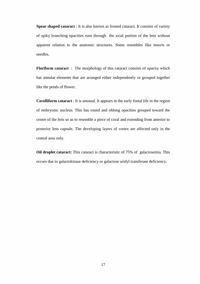
Spear shaped cataract : It is also known as frosted cataract. It consists of variety
of spiky branching opacities runs through the axial portion of the lens without
apparent relation to the anatomic structures. Some resembles like insects or
needles.
Floriform cataract : The morphology of this cataract consists of opacity which
has annular elements that are arranged either independently or grouped together
like the petals of flower.
Corolliform cataract : It is unusual. It appears in the early foetal life in the region
of embryonic nucleus. This has round and oblong opacities grouped toward the
centre of the lens so as to resemble a piece of coral and extending from anterior to
posterior lens capsule. The developing layers of cortex are affected only in the
central area only.
Oil droplet cataract: This cataract is characteristic of 75% of galactosemia. This
occurs due to galactokinase deficiency or galactose uridyl transferase deficiency.

18
Morphology of lens
Figure-4a: Axial embryonic nuclear cataract Figure-4b:zonular cataract
Figure-4c: Sutural cataract

19
Figure -4d:Anterior polar cataract Figure-4e: Posterior polar catract
Figure-4f:Blue dot cataract Figure-4g: Corraliform cataract
20
CONGENITAL RUBELLA SYNDROME
Congenital rubella syndrome occurs due to intra-uterine foetal infection
with rubella virus. During the short period of viremia in the pregnant women, the
virus crosses the placental barrier and infects the foetus within 10 – 12 days of
maternal infection 14
. The classic triad of congenital rubella syndrome includes
cataract, heart defects and deafness.
Disease burden
The first epidemic of rubella virus embryopathy occurred in the year 1941.
The second epidemic occurred during the period of 1963-1964 in which 12.5
million cases of rubella were detected (Graph-1). Of this 52,500 cases were
women in the first trimester of pregnancy. 10,500 cases of offspring were infected
15. Now after the wide spread use of rubella vaccine, incidence of rubella
embryopathy has drastically come down. Number of cases reduced to one in 1997
after which there were no new cases congenital rubella syndrome till 2002. Two
new cases were reported in 2003.

21
Graph-1 Disease burden of Rubella virus in the society
Timing of infection and the defects:
Infection that occurs during the 36th
day of gestation leads to cataract and
that occurs during 46th
day leads to heart defect. Infection occurring during 62nd
day to the end of 16th
gestation week causes defect in the inner ear. Infection
during the first one to six weeks of gestation leads to 56% of embryopathies 16
.
Infection occurring after 22 weeks of gestation does not lead to embryopathy.
Infection in the first trimester has a 50% risk of spontaneous abortion, 81% risk of
congenital infection and 69% of malformation. 50 – 80% fetuses who were
exposed to maternal rubella virus become infected prior to 8 weeks of gestation.
During second trimester only 10 – 20 % of fetuses become infected. During the
third trimester only 6 – 10% of fetuses get infected. Hematogenous spread of virus
results in the establishment of clones of virus infected cell. So children with
congenital rubella infection may excrete virus from birth to eighteen months of
age 17
.

22
Rubella Virus:
Rubella virus is a RNA toga virus belonging to the Rubivirus genera. The
virus is highly variable in morphology with surface projections. It is similar to
myxoviruses suggesting rubella virus as a paramyxovirus 18
. It measures 50 – 250
micron in cross section. It survives in pH range of 6.8-8.1. It can be destroyed by
ether, sodium desoxycholate, chloroform and formalin. It is resistant to
Idoxyuridin, tetracycline, fluorocarbons, thiomersal and sodium bisulphite. The
cytopathic effect of virus occurs in lens, cornea, iris, ciliary body and retinal
pigment epithelium. Rubella is a benign communicable exanthematous disease.
The virus is transmitted either by aerosol or transplacentally. Acquired rubella
commonly infects adolescents and adults particularly women. Incubation period is
14 – 21 days after exposure to rubella virus. This causes mild fever and
maculopapular rash often involving the occipital and post auricular
lymphadenopathy. Arthralgia or arthritis is common in adults especially in
women 19
. Signs and symptoms usually occurs 1 – 5 days before the onset of rash.
They include eye pain on lateral and upward movement, conjunctivitis, sore
throat, head ache, general body aches, low grade fever, chills, anorexia, nausea,
tender lymphadenopathy, and Forscheimer sign (pinpoint or large petechiae that
occurs in soft palate). The embryo infected with congenital rubella syndrome
contains foci of infected cells shedding virus which may affect the other cells in
the process of development 19
. So all tissues are not infected at the same time and
also the distribution of the infected cells is uneven which leads to wide variation
in the clinical picture of congenital rubella.

23
Figure 5 Rubella virus

24
CONGENITAL RUBELLA SYNDROME
Rubella infection in pregnancy upto twelth weeks of gestation
produces classic triad of heart defects, cataracts and inner ear defects which is
known as Gregg Syndrome 20
. Expanded congenital rubella syndrome includes in
addition to this triad, hepatosplenomegaly, thrombocytopenia purpura,
psychomotor retardation, dental deformity, bony lesions, general growth
retardation and inflammatory central nervous system lesions. Congenital rubella
syndrome manifestations may be transient such as purpura, or permanent
structural manifestations like deafness, congenital heart disease or late emerging
conditions like diabetes mellitus 21
.
Clinical manifestations of congenital rubella syndrome 22
1. General
a. Fetal loss (spontaneous abortion and stillbirths)
b. Low birth weight
c. Micrognathia
2. ENT
a. Sensorineural deafness: unilateral or bilateral
b. Central auditory deafness
3. Central Nervous System
a. Mental retardation
b. Speech defects
4. Cardiovascular system
a. Patent ductus arteriosus

25
b. Pulmonary arterial stenosis
c. Ventricular septal defects
5. Eyes
a. Retinopathy
b. Cataracts
c. Microphthalmos
6. Transient neonatal manifestations
a. Thrombocytopenia
b. Purpura
c. Hepatospenomegaly
d. Meningoencephalitis
e. Bony radiolucencies
f. Adenopathies
7. Late-emerging or developmental
a. Late-onset interstitial pneumonitis
b. Chronic diarrhoea
c. Insulin-dependent diabetes mellitus
Diagnostic criteria for congenital rubella syndrome
The diagnostic criteria includes cardiac, ear, ocular problems and central
nervous system disorders 23
. The cardiac defects are most frequently septum defect
and open ductus arteriosus (52%-80%). The ENT problems include inner ear
hearing loss, vestibular defects, imperfect formation of the middle and outer ear
(more than 50%). Ocular defects include visual deficit by congenital cataract,
chorioretinitis, glaucoma, microphtalmus (50%-55%). CNS problems are

26
microcephaly with psychomotoric retardation (40%-50%). Rubella is a common
cause of childhood rash. Hepatosplenomegaly (60%) and icterus, thrombopenia
(45%), skin haemorrhages, pneumonia, lymphnodeoedema, myocarditis,
exanthematous, structural changes of the long bones (30%) can also occur. Total
mortality is 13%-20%. Severe mortality (14%) occurs within a few months.
Ocular manifestations of congenital rubella infection
The ocular manifestations of congenital rubella include:
1. Cataracts
2. Glaucoma
3. Myopia
4. Microphthalmia
5. Strabismus, nystagmus, ptosis
Visual defects are graded according to degree of severity:
Mild defects - Unilateral or bilateral microphthalmia, esotropia, nystagmus, and
ptosis
Moderate defects - Unilateral cataract, glaucoma, and strabismus
Severe defects - Bilateral cataract, glaucoma, and strabismus
Congenital rubella cataract :
Rubella constitutes about 5% - 25% of childhood cataract in India.
Cataract occurs when the maternal rubella infection occurs between the second
and eleventh weeks of gestation which is the period of maximum blood supply to
the lens. In the first eight weeks primary and secondary fibres are formed. These
are the fibres that are involved due to the cytopathic effect of the virus which
causes delay in the development of fibres. The characteristic of the rubella

27
cataract is the retention of nuclei in the lens fibers which suggest this retarded
development. The lens capsule which develops during the eleventh week is not
visibly affected by rubella virus24
. There is large area of necrosis in the lens. The
cataract may extend upto peripheral as well as central cortical fibres which
sometimes progresses to total cataract. The liquefaction of the cortex and the area
of necrosis always tend to be central and surrounded by rim of normal capsule and
cortex 25
. There is little posterior migration of lens epithelium or variation in the
morphology of the anterior polar cells. The persistent nuclei which is present in
the central mass of the lens (which are normally devoid of nuclei) are typically
karyorrhectic. This is virtually pathognomic of rubella.
Morphology of rubella cataract:
Rubella cataract is usually swollen and pushes the iris forwards which
causes the depth of the anterior chamber to be reduced. Rubella cataract may also
be thin and membranous but it is rarely without the characteristic nuclei.
Congenital rubella cataract are mostly bilateral, central and dense. It may also be
lamellar, nuclear and membranous. Sometimes resorption of cataract occurs in the
centre which leads to clear pupillary axis. If the cataract is dense it indicates that
the infection occurs earlier in the pregnancy period. Dense cataract which occurs
in rubella infection is usually seen centrally or anteriorly with posterior clear zone.
The lens is usually microspherophakic in histology appearance. Sometimes the
liquefaction and necrosis of the lens occurs.

28
Other Ocular manifestations:
Corneal edema
Usually present at birth in patients with congenital rubella. It is transient
and is not obviously associated with glaucoma. The corneal transparency is
affected by inapparent viral involvement of the developing corneal endothelium
and delay in the elaboration of descement membrane.
Anterior chamber manifestations:
There is active virus in the lens, iris, aqueous and tears in later foetal life
and infancy which is evidenced by non granulomatous cellular response in the
anterior chamber. Atrophy of the iris, stromal hypoplasia or absence of dilator
muscle of the iris, vascularisation and focal necrosis of the pigment epithelium of
the iris and ciliary body are present.
Non granulomatous uveitis with diffuse and focal infiltration of anterior
uvea with lymphocytes, histiocytes and plasma cells are characteristic of
congenital rubella infection. This suggests an existence of chronic iritis in
congenital rubella infection.
Focal atrophy and cellular infiltration changes in the iris and ciliary body
suggest two periods of perinatal response to the virus 26
. The first period is
unhindered viral growth in the absence of foetal immunity. This causes slowing of
replication and even death of developing cells with resultant defective
organogenesis. The second period occurs in later gestation and is due to an
inflammatory response in whole or part of an attempt by the foetus as an immune
response to the presence of virus.

29
Failure of cleavage of the angle of the anterior chamber is a frequent
finding. This change indicates that there is an interruption of normal pattern of
development without evidence of inflammation which occurs before the
development of an immune response in the foetus .
Strabismus:
Most common ocular deviation in congenital rubella syndrome is
esotropia. It may be due to simply mechanical or most commonly sensory 27
. High
hyperopia also contributes to esotropia. Cerebral palsy contributes for the high
incidence of associated esotropia. Cerebral paralysis is frequently concomitant
with congenital rubella infection. Nystagmus may also be associated with
strabismus. Amblyopia is also one of the known precursor of convergent
strabismus. Strabismus is predominantly sensory (80%).
Glaucoma:
Incidence of Glaucoma in infants infected with rubella infection varies from two
to fifteen percent. Gonioscopy reveals
1. Irregular anterior insertion of the iris
2. Finely dispersed pigment on the surface of the trabeculae
3. Normal iridocorneal angle
The raised intraocular pressure is due to decreased outflow facility. The
glaucoma in rubella is an acute process and is not a permanent developmental
defect in the angle. It is due to a transient obstruction of outflow of acqueous
humour 28
. The obstruction is due to temporary defect in the development of
trabecular endothelium or temporary deposition of cellular debris over the surface

30
of the trabecular meshwork. The restoration of normal trabecular endothelial
lining and the disappearance of cellular debris leads to normal intraocular
pressure. Mild cases with corneal edema and raised intraocular pressure requires
medical therapy and follow up whereas progressive changes in the cornea
associated with persistent elevation of intraocular pressure needs surgical
intervention like goniotomy, filteration procedure and cyclodiathermy.
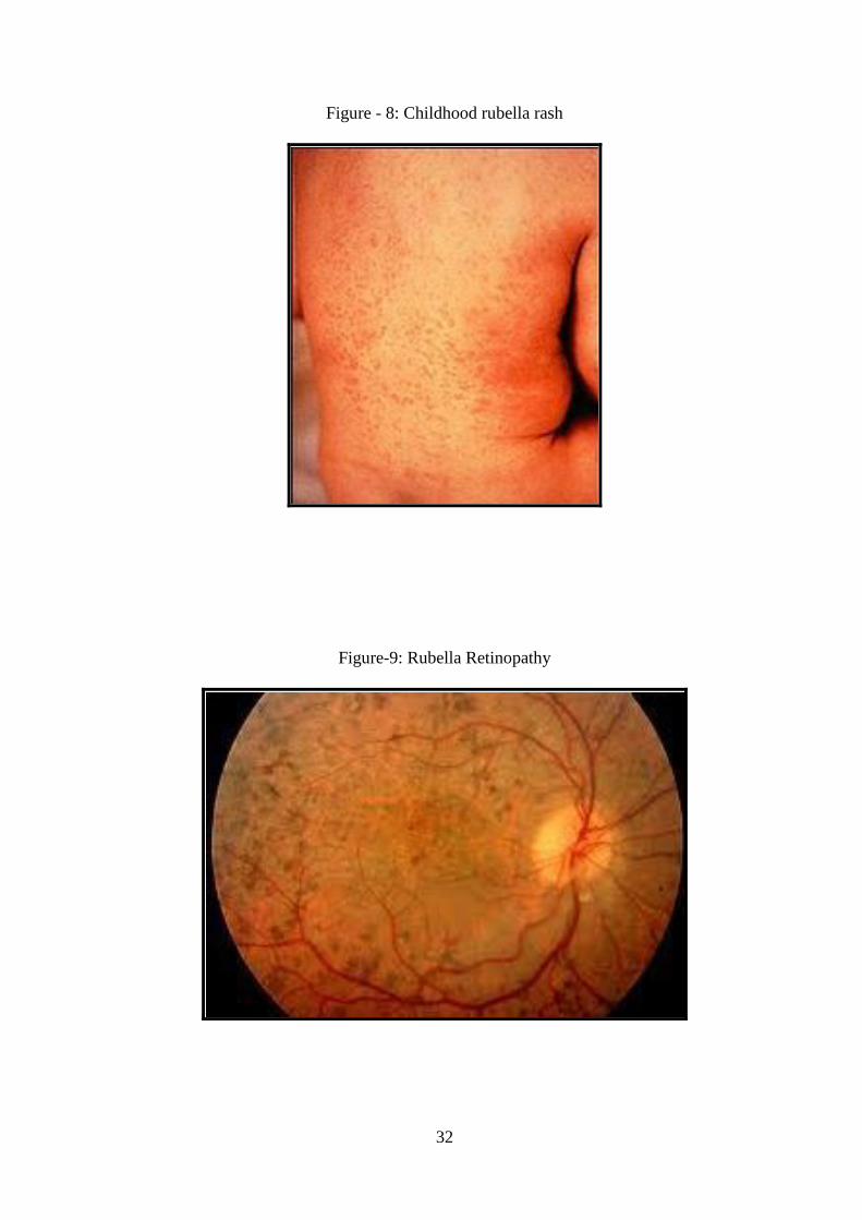
Rubella Retinopathy:
It is the commonest fundus finding in congenital rubella infected patients.
It is called as salt and pepper retinopathy because there are tiny flecks of dark
pigments mixed with fine areas of whitish area of depigmentation 29
. The pigment
alteration is usually diffuse. But it is most prominent either around the macula or
in the periphery of the retina. This fundus change reflects diffuse damage to the
retinal pigment epithelium. But the RPE is never damaged enough to interfere
with vision or to cause major abnormality in the electroretinogram 30
. Rubella
retinopathy is not considered as a handicap as it is a non-interfering defect .

31
Figure - 6: Congenital Rubella cataract – bilateral dense pearly white opacity
Figure-7: HPE showing liquefaction and necrosis of rubella cataract
32
Figure - 8: Childhood rubella rash
Figure-9: Rubella Retinopathy

33
APPROACH TO A CHILD WITH CONGENITAL
RUBELLA CATARACT
When a child with cataract is seen trauma should be ruled out especially in
unilateral cases.
History is usually elicited from parents. The following important history
related to symptoms are elicited 31
:
White spot in pupillary area
Diminution of vision and not able to recognize objects and parents
Unsteady eyes
Deviation of eyes
Associated symptoms of systemic disease, if present
Other relavant history which has to be asked include:
Low birth weight
Maternal infections
Maternal drug ingestion
Trauma
Parental consanguinity
Radiation exposure
Electric exposure
Family history of ocular disorder

34
Complete systemic examination is done to rule out other systemic
disorders. Ocular examination is done to detect the associated features like
glaucoma, strabismus, nystagmus, uveitis, corneal edema, and retinopathy
supplemented by B Scan to rule out the posterior segment pathology 32
.
Then the child should be screened for causes related to congenital cataract.
Screening for toxoplasmosis, rubella, cytomegalovirus and herpes (TORCH)
should be done. Congenital rubella syndrome is suspected if infants older than 3
months have rubella specific IgG antibodies and this does not decline at a rate
expected from passive transfer of maternal antibodies. Virus isolation from
nasopharyngeal swab, urine specimen and cerebrospinal fluid is confirmatory for
rubella infection.
Serum analysis of Galactose enzyme level in RBC, Glucose, Urea
nitrogen, Calcium, Phosphorous, electrolytes, Cholesterol is carried out. Urine
analysis for reducing substances, aminoacids, homocystine is done.
Cardiological evaluation to rule out septal defects, ENT examination to
look for sensorineural deafness, skeletal survey are done.
TORCH SCREENING:
With prior consent of the parents of the children, peripheral blood of 1-2ml
is collected by venipuncture 34
. Blood samples collected were tested for the
presence of specific IgG and IgM antibodies to toxoplasma gondii, rubella virus,
cytomegalovirus and herpes simplex virus by ELISA using commercially
available kits (eg. Bioelisa kits) following manufacturer’s instructions. The test for
the detection of specific IgM antibodies to toxoplasma gondii, RV, CMV, and

35
HSV is performed by the immune capture techniques. Immuno-capture ELISA is
performed by incubating the diluted test sera in microplates wells coated with
rabbit antibodies to human IgM. Following this the wells are washed to remove
the residual test sera and the specific antigen for toxoplasma gondii, RV, CMV
and HSV conjugated with enzyme are added. Sera for IgG and IgM antibodies are
tested with dilution of 1:100 with positive and negative controls. The final
readings are taken in an ELISA reader at 450 nm with reference at 650 nm 35
. The
positive results are those which shows presence of antibodies in dilution of more
than 1:100 .
The main stay of diagnosis of rubella infection is the serological
investigation. Recent rubella infection can be diagnosed by
1. Identification of rubella specific IgM or four fold increase in Ig G titre in
samples tested 2-3 weeks.
2. Rising titres of antibodies in Hemagglutination Inhibition test and ELISA
3. Seroconversion
Low or transient level of IgM can be present in cases of re infection 36
.
In congenital rubella infected children, the specific IgM is present upto
three months of age in all confirmed cases. It is present in 86% at the age of 3 – 6
months, 62% at 6 months to one year, and 42% at 12 – 18 months, rarely above
18 months. Maternally transferred rubella specific IgG disappears around six
months of age. Rubella specific IgG during the age of 1 – 2 yrs indicate congenital
infection and also the persistent level of high IgG level. Antibody levels are higher
after congenital infection than the vaccination. After rubella vaccination, IgM
persist in 76% upto 4 months but can be present in lower levels upto four years.

36
Here IgG can persist upto 21 years after vaccination in lower titres compared to
natural infection 37
(Graph-2).
Positivity of TORCH titre to one specific infectious agent is 16 – 19 %, to
two or more infectious agent is around 2-3% and that for all infection is about
20%.
Graph-2 Rubella antibody levels in congenital rubella infection

37
MANAGEMENT OF CONGENITAL CATARACT
Fixation develops between two to four months of age. Any paediatric
cataract in the visual axis should be treated as early as possible once the general
condition of the child becomes fit for anaesthesia. This is significant to prevent the
deprivation of the visual system and to provide optimal visual rehabilitation. In
cases of bilateral cataracts, surgeries should be separated by two weeks for
children younger than two years and by one month for children older than two
years. Small incision cataract surgery or phacoemulsification with intraocular lens
implantation with or without posterior capsulorrhexis is preferred.
Problems with paediatric cataract surgeries include:
IOL calculation due to change in axial length, corneal curvature and
lenticular refracting power
Smaller size of eye ball
More elastic capsule making CCC difficult
Less scleral rigidity
Increased tissue reactivity
Potential for amblyopia
Posterior capsular opacification
Secondary membrane formation
Proliferation of lens epithelium (Sommerring ring)
Glaucoma
Retinal detachment

38
In small incision cataract surgery, superior incision is preferred over
temporal incision because superior incision provides more secure incision in
children and so further risk for traumatic injury is reduced and also this helps to
maintain anterior chamber during the procedure. Although scleral incision is self
sealed, in paediatric cataract it is sutured due to less scleral rigidity. Since the
capsule is more elastic, while doing capsulorrhexis extension of capsulorrhexis is
more. So smaller capsulorrhexis is preferred.In children cortical matter is stickier
than adult cataract. So large port cannula is to be used for aspiration. Radial tears
are to be avoided to prevent IOL displacement.
Posterior capsular opacification is a major cause of gradual loss of vision
following uneventful paediatric cataract surgery. It occurs due to incomplete
removal of cortex and proliferation of lens fibres. The incidence of PCO depends
upon the amount of cortical matter left, and type of IOL implantation. So to
prevent PCO, complete removal of cortical matter, posterior capsulorrhexis with
anterior vitrectomy, use of biconvex IOL can be performed.
Regarding the choice of IOL in children, foldable hydrophobic acrylic
monofocal IOL is preferred. For the calculation of IOL power, modification of
SRK formula which is SRK II formula is used. SRK II formula includes a
correction factor according to the axial length which is added to the A constant
value.
Post operatively, antibiotics and steroids are given to hasten recovery. In
case of postoperative inflammation, cycloplegics are added. Post operative follow
39
up is given with regular visual assessment and rehabilitation. This includes
refractive error correction. In case of children who are not having full visual
improvement after refractive correction, amblyopia therapy is intensely given.
Risk of amblyopia is greater during the first year of life which declines rapidly
after five years of age. In case of amblyopia, occlusion therapy with best visual
correction is given. Occlusion is given for one day with one occlusion free day for
one year age group, two days occlusion with one day occlusion free day for two
year age group and till six year age group this rule applies. The occlusion is to be
more intense with increasing age group. Follow up is given once in two weeks to
monitor visual acuity of the occluding eye and the visual improvement of the
amblyopic eye. Improvement in visual acuity with amblyopia therapy is best
before seven years of age. This improvement gradually declines after seven years
of age. Still amblyopia therapy can be given till twelve years of age. This is
mainly due to the pliability of the central visual processes before seven years of
age. Occlusion improvement is noted in three months period.
In addition to above management of pediatric cataract, rubella cataract
extraction 39
is associated with severe post operative inflammation. Live rubella
virus has been isolated from the lens till three years of age. While performing
cataract surgery, the intralenticular virus are released which causes severe post
operative inflammation such as iritis. The post operative inflammation is due to
delayed hypersensitivity reaction or due to direct viral insult. The post operative
inflammation can be very severe that can to lead to pthisical eye also. The
intensive cycloplegics and topical steroids are given for a longer period to control
post operative inflammation 40
.
40
PREVENTION
PREVENTION IS BETTER THAN CURE. The disease burden of rubella
retinopathy can be reduced by proper vaccination of children 41
. As per
recommendations of Indian Academy of Paediatrics, MMR vaccine is advised to
all children at the age group of one and half years, with second dose being given
at 4-6 yrs age group. MMR is vaccination against measles, mumps and rubella.
Vaccine is a mixture of three attenuated virus. MMR vaccination is administered
subcutaneously 42
. After first dose, 95% of those who receive vaccine become
immune. After second dose, 99.7% develop immunity. Immunity is life long.
Adverse reactions are rarely serious. 10% of children develop fever, malaise and
rash. Other rare side effects include joint pain, aseptic meningitis 43
. After rubella
vaccine, IgM antibodies reach peak levels upto four months and can persist upto
four years. Ig G antibodies start appearing after five months of vaccination and
persist even upto 21 years. Rubella vaccination mainly aims at reducing and
preventing the congenital rubella syndrome.

41
OBJECTIVES OF THE STUDY
An analytical prospective study of visual outcome in the management of
cataract in children with Congenital Rubella Syndrome was carried out. The aims
of the study were
1. To evaluate the visual outcome of Congenital Rubella Cataract following
cataract surgery
2. To identify and treat post operative inflammation earlier following cataract
surgery.
METHODOLOGY (MATERIALS & METHODS)
Patient Selection :
Children with congenital cataract attending RIO-GOH during the period
June 2010 to June 2012 (two years) were evaluated.
Inclusion Criteria :
Children with congenital cataract who are found to be positive for rubella
infection in TORCH titre.
Exclusion Criteria :
Congenital cataract due to other intrauterine infections, metabolic
disorders and hereditary causes.
Methods:
Children with congenital cataract attending RIO-GOH were evaluated.
Visual acuity, slit lamp examination, and fundus examination were done. The

42
children were screened for TORCH infections. Those children with rubella IgG,
IgM or both were included in the study. The children were screened for systemic
manifestations of congenital rubella syndrome and the general condition of the
patients were assessed by paediatrician. Metabolic screening was carried out.
Ocular investigations including B Scan and IOL power calculation was
performed. Anaesthetic fitness for general anaesthesia was obtained. For those
children who were positive for rubella IgM, cataract surgery was postponed till
IgM disappeared.
Pre operative antibiotic eye drops were given. Cataract surgery was
performed by small incision cataract surgery or Phaco- emulsification with
intraocular lens implantation. Post operatively they were treated with topical
antibiotics, and topical steroids. Those children who developed iritis were treated
with cycloplegics, systemic steroids and intensive topical steroid treatment. Post
operative visual acuity and anterior segment were assessed. Children with more
inflammation were given follow up at shorter intervals till the inflammation
subsided.
Follow up:
The children were followed up at weekly intervals till six weeks, then
biweekly or monthly till six months depending on the anterior chamber reaction
and the visual acuity of the child. During each visit visual acuity assessment by
Snellen’s chart or paediatric picture chart depending upon the age of the child,
slit lamp examination and fundus examination were carried out. Children were
given refractive correction. Those children who had no full visual improvement
with refractive correction were advised occlusion therapy. Further these children

43
were followed up for improvement of vision with occlusion therapy. Those with
posterior capsular opacification during follow up were treated with either
surgical or laser capsulotomy depending upon the age of the child.
Those children who had bilateral cataract were advised other eye surgery
after two months. Some of the children did not get fitness for general anaesthesia.
Some children did not turn up for other eye surgery. Rest of the children were
operated for other eye. Surgery was carried out under cover of antibiotic drops.
Post operative inflammation was controlled by topical antibiotics, topical steroids
and cycloplegics. Follow up was given similar to the previous eye with visual
acuity assessment, slit lamp examination, fundus examination, refractive
management, amblyopia management and posterior capsular opacification
management.
Assessment of parameters :
Vision, Slit lamp examination, Fundus examination, Retinoscopy

44
OBSERVATION AND RESULTS
Thirty four eyes of twenty three patients with congenital rubella cataract
were evaluated.
AGE DISTRIBUTION
Table 1: Age distribution of the study patients
No. Of patients % of total
< 1 yr 5 21
1-3 yrs 3 13
4 – 6 yrs 7 31
7 – 9 yrs 5 22
10 – 12 yrs 3 13
Total 23 100
Figure 1: Age distribution of patients
21%
13%
31%
22%
13%
<1 yr
1-3yrs
4-6yrs
7-9yrs
10-12yrs
Among the 23 patients, five patients (21%) were in the age group of less
than one year. Three patients (13%) belonged to the age group of 1-3 years, seven
(31%) in 4 – 6 years, Five (22%) in 7 – 9 years, and three(13%) in 10 – 12 years.
(Figure 1) Majority of the patients were in the age group of 4 – 6 yrs. Only few
patients reported in 1-3 years and above 10 years of age (Table1).

45
SEX DISTRIBUTION
Table 2: Sex distribution of the study group
No. Of patients % of total
Male 14 61
Female 9 39
Total 23 100
Figure 2: Sex distribution of patients
61%
39%
Male
Female
Male children were 14 in number (61%) whereas female children were nine(39%)
(Table2). Male children dominated the study group. (Figure 2)

46
AGE SEX DISTRIBTUION OF PATIENTS
Table 3 Age sex distribution of the patients
Male patients % among the
age group
Female
patients
% among the
age group
< 1 yr 3 60 2 40
1 – 3 yrs 2 66.67 1 33.33
4-6 yrs 4 57.14 3 42.86
7-9 yrs 3 60 2 40
10-12 yrs 2 66.67 1 33.33
Figure 3: Age Sex distribution of the patients
3
2
4
3
22
1
3
2
1
0
1
2
3
4
5
<1yr 1-3yrs 4-6yrs 7-9yrs 10-12yrs
Male
Female
In less than one year age group three patients ( 60% ) were males, two
(40%) were females. In 1-3 years age two were males and one female. 4 -6 years
age group had four males and three females. (Table3) 7 – 9 years age group had
three (60%) males and two (40%) females, and 10- 12 years two (66.67%) males

47
and one (33.33%) female. Thus there was male dominance of presentation in all
age groups. (Figure 3)
PRESENTING FEATURES
Table 4: Presenting complaint of the study group
No. Of patients % of total
White Reflex 7 30
Not recognising faces 3 13
Deviation of eyes 4 17
Defective vision 11 48
Total 23 100
Figure 4: Presenting features of the study group
Seven (30%) patients presented with white reflex. Parents of three (13%)
children complained that their child was not recognising faces. (Figure 4) Four

48
(17%) children had deviation of eyes. 11 (48%) had defective vision. The most
common complaint was defective vision followed by white reflex. (Table4)
20 (87%) children had single complaint. Three had multiple complaints
(two double complaint and one triple complaint). (Figure 5)
Figure 5: Number of complaints of the patients
49
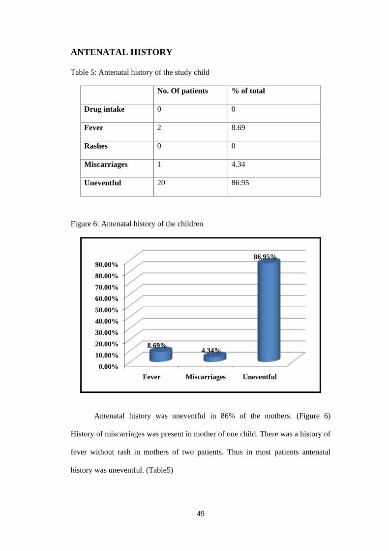
ANTENATAL HISTORY
Table 5: Antenatal history of the study child
No. Of patients % of total
Drug intake 0 0
Fever 2 8.69
Rashes 0 0
Miscarriages 1 4.34
Uneventful 20 86.95
Figure 6: Antenatal history of the children
0.00%
10.00%
20.00%
30.00%
40.00%
50.00%
60.00%
70.00%
80.00%
90.00%
Fever Miscarriages Uneventful
8.69%4.34%
86.95%
Antenatal history was uneventful in 86% of the mothers. (Figure 6)
History of miscarriages was present in mother of one child. There was a history of
fever without rash in mothers of two patients. Thus in most patients antenatal
history was uneventful. (Table5)
50
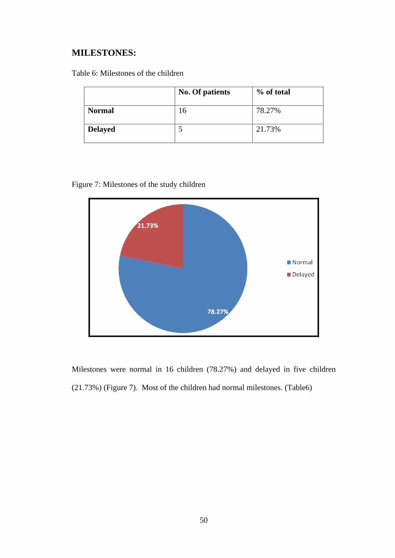
MILESTONES:
Table 6: Milestones of the children
No. Of patients % of total
Normal 16 78.27%
Delayed 5 21.73%
Figure 7: Milestones of the study children
Milestones were normal in 16 children (78.27%) and delayed in five children
(21.73%) (Figure 7). Most of the children had normal milestones. (Table6)

51
SYSTEMIC FEATURES:
Table 7: Systemic features in the study children
No. Of patients % of total
CVS 1 4.34%
CNS (Mental retardation) 2 8.69%
Hearing defect 2 8.69%
Rash 1 4.34%
Abdomen 0 0
Genitourinary abnormalities 0 0
Figure 8: Children presented with systemic features
0.00%
2.00%
4.00%
6.00%
8.00%
10.00%
4.34%
8.69% 8.69%
4.34%
One child had patent foramen ovale (4.34). Two children had mental
retardation. Two had sensorineural hearing defect. Rash present in one child.
(Figure 8). Six children had systemic abnormalities whereas others had no specific
systemic abnormality. (Table7)

52
VACCINATION STATUS
Table 8: Vaccination status of the study children
No. Of patients % of patients
GIVEN 5 30.43
NOT GIVEN 7 21.74
STATUS NOT KNOWN 11 47.83
23 100
Figure 9: Vaccination status of the study children
Status of rubella vaccination was elicited. Five children were vaccinated
(Table8). Seven children were not vaccinated. Status was not known in 11
children. (Figure 9)

53
LATERALITY
Table 9: Laterality in study group
No. Of cases % of total
Unilateral 5 21.73%
Bilateral 18 78.27%
Figure 10: Laterality in the study children
Cataract was unilateral in five children (21.73%) whereas it was bilateral
in 18 (78.27%) children(Table9). Bilateral cataracts were more common.
(Figure 10)

54
Figure 11: No. Of eyes operated
11
4
3
5 Both eyes operated
Lost follow up
GA fitness not
obtained
Unilateral cases
Figure 12 Children operated for single or both eyes
Among the 18 bilateral cataracts, after the first eye cataract surgery, 11
patients turn up for other eye cataract surgery. Three children did not get general
anaesthetic fitness and four did not turn up for follow up. So totally 34 eyes were
operated of which 12 were bilateral and 22 were unilateral. (Figure 11,12)

55
VISUAL ACUITY AT THE TIME OF PRESENTATION
Table 10: Visual acuity at the time of presentation
No. Of cases % of total eyes
Not fixing and following light 13 38.23
Hand movements 5 14.70
CFCF 6 17.64
1/60 – 5/60 9 26.47
6/60-6/36 1 2.96
Total 34 100
Figure 13: Visual acuity at the time of presentation
13
56
91
Not fixing
&following light
HM
CFCF
1/60-5/60
6/60-6/36
13 (38.23%) Children had visual acuity of not fixing and following
light. Five (14.70%) had hand movements. Six (17.64%) had CFCF vision. Nine
(26.47%) had visual acuity between 1/60-5/60. One child was with visual acuity
6/36 (Figure 13). Majority of the children had visual acuity of not fixing and
following light presumably because the visual acuity in those children could not
be tested by Snellen’s chart or picture chart due to the younger age of the children
(Table10).

56
MORPHOLOGY OF CATARCTS
Table 11: Morphology of cataracts
No. Of cases % of total
Central dense cataract 31 91.17%
Lamellar cataract 3 8.83%
34 100
Figure 14: Morphology of cataracts
91.17%
8.83%
Central Cataract
Lamellar Cataract
31 eyes (91.17%) had central dense cataract with three eyes of lamellar
(8.83%) cataract (Table11). Central dense cataract constitutes more than 90% of
total cataract (Figure 14).

57
ASSOCIATED OCULAR ANOMALIES
Table 12: Associated ocular features
No. Of cases % of total
Glaucoma 0 0
Strabismus 3 8.82%
Nystagmus 2 5.88%
Corneal opacity 0 0
Microphthalmos 0 0
Figure 15: Associated ocular features
0
0.5
1
1.5
2
2.5
3
Strabismus Nystagmus
3
2
Three children had strabismus (8.82%) of which two children were with
nystagmus (5.88%). (Table12 (Figure 15)
58
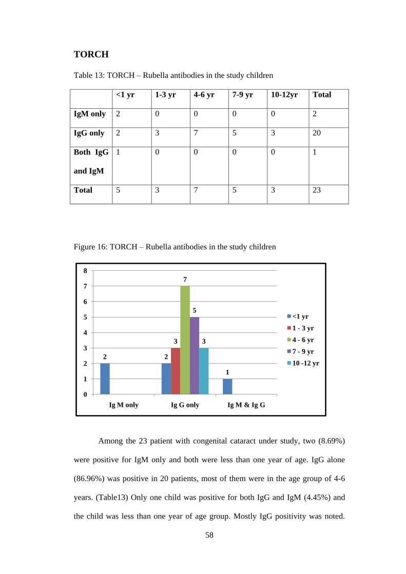
TORCH
Table 13: TORCH – Rubella antibodies in the study children
<1 yr 1-3 yr 4-6 yr 7-9 yr 10-12yr Total
IgM only 2 0 0 0 0 2
IgG only 2 3 7 5 3 20
Both IgG
and IgM
1 0 0 0 0 1
Total 5 3 7 5 3 23
Figure 16: TORCH – Rubella antibodies in the study children
2 2
1
3
7
5
3
0
1
2
3
4
5
6
7
8
Ig M only Ig G only Ig M & Ig G
<1 yr
1 - 3 yr
4 - 6 yr
7 - 9 yr
10 -12 yr
Among the 23 patient with congenital cataract under study, two (8.69%)
were positive for IgM only and both were less than one year of age. IgG alone
(86.96%) was positive in 20 patients, most of them were in the age group of 4-6
years. (Table13) Only one child was positive for both IgG and IgM (4.45%) and
the child was less than one year of age group. Mostly IgG positivity was noted.

59
All of them had very high titre excluding the probability of appearance of
antibodies from vaccination in which the titres were low. (Figure 16)
Associated toxoplasmosis was found in one child. Toxoplasma and CMV
were also positive in one child. One child was positive for all the TORCH
antibodies. Only Rubella positivity was noted in 20 children (Figure 17).
Figure 17: Positivity of other TORCH titres
Other investigations were done. Urine analysis done for metabolic
screening was negative in all the 23 children. B Scan showed normal posterior
segment finding in all 34 study eyes.

60
SURGERY DONE
Table 14: Type of cataract surgery done
% of total No. Of cases
SICS with IOL 19 55.88%
Phaco with IOL 15 44.12%
Figure 18: Type of surgery done
44.12%
55.88% Phaco
SICS
In 15 eyes of 34 eyes, Phacoemulsification was done (44.12%) and small
incision cataract surgery was done in 19 eyes (55.88%) (Table14). In all cases
IOL implantation was done. (Figure 18) Posterior capsulorrhexis was done in five
cases.

61
POST OPERATIVE VISUAL ACUITY
Table 15: Post operative visual acuity
Post Operative Visual Acuity
VISUAL ACUITY
No. Of
patients
Percentage of patients (%)
Not Fixing and Following light 11 32.35
Fixing and Following light 2 5.88
HM 0 0
CFCF 1 2.94
1/60 - 5/60 3 8.83
6/60- 6/36 9 26.47
6/24 - 6/18 7 20.59
6/12 - 6/6 1 2.94
Figure 19: Post operative visual acuity

62
In the immediate post operative period, 43% of the eyes were not fixing
and following light. 5% developed fixing and following light. 3% had counting
finger close to face. 8.8% had vision of 1/60-5/60 (Figure 19). Vision in 26.4%
was 6/60-6/36. 20.59% had vision of 6/24 – 6/18. 2% had vision of 6/12 – 6/6.
The improvement in vision compared with preoperative vision was statistically
significant at p<0.05. (Table15).
POST OPERATIVE INFLAMMATION
Figure 20: Post operative complications
Among the 34 eyes, six (17.64%) had corneal edema, 15 (44.11%) had
striate keratitis, 7 (20.58%) had iritis and 11 (32%) had no complications.
(Figure 20)

63
Post operative iritis vs TORCH titre:
Figure 21: Iritis Vs Rubella positivity
Among those with iritis (seven patients) three were positive for rubella IgM
(42.85%) only, three occured in those with IgG only (42.85%) and one occurred in
patient with both IgM and IgG (14.30%) (Figure 21). Among those who were IgG
positive 15% had iritis. Among those who were IgM positive and both IgG and
IgM positive iritis was present in all cases. This implies a high occurrence of iritis
in those with rubella IgM positivity.

64
Table 16: Iritis children
Patient No. Age Iritis IgM
positive
IgG
positive
IgG & M
positive
1 8/12 + + - -
5 9/12 ++ + - -
5(other
eye)
9/12 ++ + - -
12 10/12 + + + +
14 3 ++ - + -
19 3 ++ - + -
21 8 + - + -
Figure 22: Age distribution of iritis children
Among the seven cases of iritis, four were in the age group of less than one
year, two in 1-3 years and only one occurred in the 7-9 years of age group. So the
incidence of iritis was greater in younger age group compared to older age group.
(Figure 22)

65
POST OPERATIVE VISUAL ACUITY TWO WEEKS
Table 17: Post Operative Visual Acuity – two weeks
Post Operative Visual Acuity – two weeks
VISUAL ACUITY
No. Of
patients
Percentage of patients (%)
Not Fixing and Following light 8 23.53
Fixing and Following light 5 14.70
HM 0 0
CFCF 1 2.94
1/60 - 5/60 2 5.88
6/60- 6/36 5 14.70
6/24 - 6/18 10 29.43
6/12 - 6/6 3 8.82
Figure 23: Post Operative Visual Acuity – two weeks
8
5
1
25
10
3
Post operative Visual acuity - two weeks
Not fixing &following light
Fixing & following light
CFCF
1/60-5/60
6/60-6/36
6/24-6/18
6/12-6/6
66
After two weeks of follow up, those who were not fixing and following
light was eight (23.53%), fixing and following light five (14.70%) and CFCF was
in one(2.94%) children. Two patients (5.88%) had vision of 1/60 – 5/60. 6/60 –
6/36 was noted in five patients (14.70%). 6/24 – 6/18 was noted in 10 patients
(29.43%).6/12- 6/6 was in two patients (8.82%) (Table17). This improvement of
vision is statistically significant at P < 0.05% (Figure 23).
POST OPERATIVE 2 WEEKS ANTERIOR SEGMENT
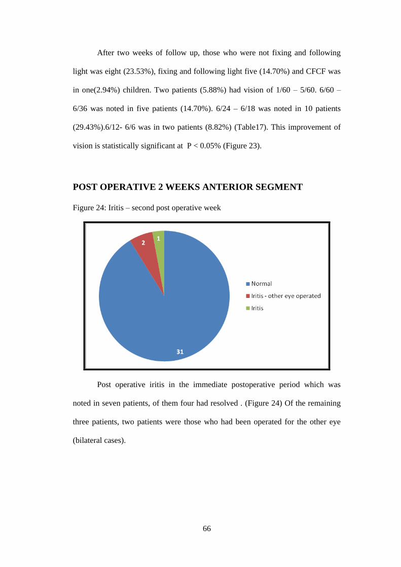
Figure 24: Iritis – second post operative week
Post operative iritis in the immediate postoperative period which was
noted in seven patients, of them four had resolved . (Figure 24) Of the remaining
three patients, two patients were those who had been operated for the other eye
(bilateral cases).

67
POST OPERATIVE VISUAL ACUITY – SIX WEEKS
Table 18 Post operative visual acuity – six weeks
Post Operative Visual Acuity – six weeks
VISUAL ACUITY No. Of patients Percentage of patients (%)
Not Fixing and Following
light
0 0
Fixing and Following light 13 38.23
HM 0 0
CFCF 0 0
1/60 - 5/60 1 2.94
6/60- 6/36 3 8.83
6/24 - 6/18 6 17.65
6/12 - 6/6 11 32.35
Figure 25: Post operative visual acuity – six weeks

68
After six weeks of surgery, no child was with not fixing and following
light (Table18). 13 developed fixing and following light (38.23%), one had vision
of 5/60 (2.94%), three had 6/60 – 6/36 (8.83%), and 6/24 – 6/18 in six patients
(17.65%). 11 patients had 6/12 – 6/6 (32.35%) (Figure 25). The p value is 0.009
which was statistically significant at p < 0.01.
POST OPERATIVE ANTERIOR SEGMENT AT SIX WEEKS
Iritis resolved in all the patients. PCO started appearing in three patients.
POST OPERATIVE FUNDUS AT SIX WEEKS
Among the thirty four patients fundus was normal in 30 patients. In three
patients with PCO fundus view was hazy, but disc and vessels appeared normal
through the hazy media. One patient had evidence of rubella retinopathy.
POST OPERATIVE VISUAL ACUITY AT SIX MONTHS
Table 19 Post operative visual acuity – six months
Post Operative Visual Acuity – six months
VISUAL ACUITY No. Of patients Percentage of patients (%)
Not Fixing and Following light 0 0
Fixing and Following light 13 38.23
HM 0 0
CFCF 0 0
1/60 - 5/60 1 2.94
6/60- 6/36 4 11.76
6/24 - 6/18 6 17.64
6/12 - 6/6 10 29.41
Did not turn up for follow up 1 2.94

69
Figure 26: Post operative visual acuity – six months
After follow up for six months, fixing and following light was present in
13 patients (38.23%), 5/60 in one patient (2.94%), 6/60 – 6/36 in four patients
(11.76%), 6/24-6/18 in six patients (17.64%). 6/12 – 6/6 was noted in 10 patients
(29.41%) (Figure 26). This was statistically significant at p < 0.01. (Table19)

70
SIX MONTHS ANTERIOR SEGMENT
Figure 27: Six months anterior segment
PCO was noted in six patients (17.64%). The posterior capsule was clear
in 22 patients (64.72%) (Figure 27). Of six patients, one had PCO even after
performing posterior capsulorrhexis during cataract surgery.
Figure 28: Age distribution of PCO
Two cases of PCO was noted in 4-6 years, two in less than one year, one in
7-9 years and one above 10years of age group. Incidence of PCO was noted more
in younger age group (Figure 28).
71
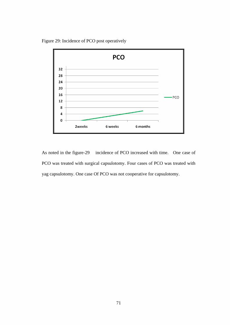
Figure 29: Incidence of PCO post operatively
As noted in the figure-29 incidence of PCO increased with time. One case of
PCO was treated with surgical capsulotomy. Four cases of PCO was treated with
yag capsulotomy. One case Of PCO was not cooperative for capsulotomy.

72
PRE AND POST OPERATIVE VISUAL ACUITY
IMPROVEMENT
Table 20 Pre and post operative visual acuity improvement
VISUAL
ACUITY
Pre op Post op 2 weeks 6 weeks 6 months
Not Fixing and
Following light
38.23 32.35 23.53 0 0
Fixing and
Following light
_ 5.88 14.70 38.23 38.23
HM 14.70 0 0 0 0
CFCF 17.64 2.94 2.94 0 0
1/60 - 5/60 26.47 8.83 5.88 2.94 2.94
6/60- 6/36 2.96 26.47 14.70 8.83 11.76
6/24 - 6/18 0 20.59 29.43 17.65 17.64
6/12 - 6/6 0 2.94 8.82 32.35 29.41
P value*** 0.019** 0.009* 0.006* 0.007*
Did not turn up
for follow up
_ _ _ _ 2.94
** statistically significant at p <0.05
* statistically significant at p < 0.01
***p value by chi square test
Comparing the visual acuity with preoperative status there was definite
improvement in visual acuity at statistically significant levels (Figure 30). The
proportion of children with good visual acuity after surgery was decreased in six
months follow up due to PCO (Table20).

73
Figure 30: Pre and post operative visual acuity improvement

74
DISCUSSION
Children with congenital cataract were screened for rubella antibodies by
TORCH. Those who were found to be positive for rubella antibodies were
followed up.
Among the children under study, most of the children were male (60%) .
This male preponderance was noted in all age groups. Many fall under the age
group of 4 – 6 years. This may partly because the difficulty in vision was noted at
school entry.
Most of the children presented with defective vision. Next the children
were reported with white reflex. This was mainly because of the central dense
nature of the cataract. When the child presents late, the type of cataract which
predominates was lamellar and the degree of visual loss was not so severe.
Nystagmus and strabismus were noted even in younger age group which may be
due to the dense cataract and severe vision deprivation. Most of the patients
presented with single complaint which was mainly defective vision.
Antenatal history was uneventful in most of the mothers of children except
for two who had fever and one with miscarriages. Although rubella rash
occurring in first trimester is most commonly associated with more severe
Cataractous changes, insignificant history was noted which may be due to missed
fever and rash.
Milestones were delayed in 20% of children with mental retardation.
Vaccination status was not known in 47% of kids. 30% of kids were vaccinated.

75
Vaccination can also result in raise in TORCH antibodies but the raise in antibody
titre due to infection is very high when compared to natural infection. Those with
high antibody titres presumably due to infection were taken into study.
Most of the children had very high IgG levels. Children with IgM
antibodies were in the younger age group.
General condition of the children were evaluated. In children with high
IgM levels surgery was postponed till IgM levels become negative so as to avoid
severe inflammation.
Cataract surgery with IOL implantation was done. Posterior
capsulorrhexis was done in five of 23 children. Post operative inflammation was
severe in children who were recently IgM positive. They were treated with
intensive cycloplegic and steroids. Visual improvement was good in all children.
11%of children did not have full visual recovery probably because of the long
duration of disease process and dense Cataractous changes going in for
amblyopia. Best visual correction was given to all children. Occlusion therapy
was given to amblyopic children.
Younger children had greater incidence of PCO which was treated with
capsulotomy either by yag or surgical. All children with rubella cataract should
be treated with cataract extraction as early as possible with intensive control of
post operative inflammation.

76
CONCLUSION
The most common age group in which the congenital rubella cataract noted
was 4 – 6 years of age. Least presentation was in the age group of 1- 3 years
and above 10 years .
Incidence of congenital cataract was more in males compared to females.
This dominance was noted in all age groups.
Defective vision was the most common presenting complaint. After this, the
most common presentation was white reflex.
Antenatal history was uneventful in most of the mothers of the study
children.
Milestone was delayed only in five patients with congenital rubella cataract.
In this study, the most common systemic abnormality was mental
retardation and hearing defect. Cardiovascular abnormality and rash was
found in one children each..
In most of the children rubella vaccination status was unknown.
Bilateral cataract was common compared to unilateral cataract.
The most common morphology of congenital rubella cataract was central
dense cataract.
Most often there was no associated ocular abnormalities.
Most of the children at the time of presentation had visual acuity of not
fixing and following light to 5/60.
Regarding TORCH titre, in congenital rubella cataract most of the patients
were positive for IgG only in high titres. IgM was noted in children less than
one year of age group.

77
All the cases were operated with intraocular lens implantation.
Incidence of iritis was more in the younger age group and those who were
initially IgM positive at the time of presentation . Resolution of all cases of
iritis was noted with intensive cycloplegic therapy and topical steroids.
Comparing the visual acuity pre operatively and post operatively there was
statistically significant improvement in vision at two weeks, six weeks and
six months.
The proportion of children with good visual acuity after surgery was
decreased in six months follow up due to PCO. Incidence of PCO increases
with time and more in the younger age group.
Visual rehabilitation was given to all children with best refractive error
correction and amblyopia therapy if needed.
To conclude, children with congenital rubella cataract should be operated as
early as possible under topical antibiotics coverage. Post operative
inflammation should be properly treated with intensive cycloplegics and
topical steroids. Proper follow up and visual rehabilitation should be done.

78
Post operative of patient No. 5 showing thin PCO
Profile photo of patient No.13
Slit lamp picture of left eye of the
same patient 13 showing lamellar
cataract
Bilateral dense cataract of patient No.5
Post operative profile picture of patient
No. 5

79
Lamellar cataract of patient No.23
Central dense cataract of patient No. 7
Post operative picture of patient No. 7
after cataract extraction with IOL
placement and posterior
capsulorrhexis

80
BIBLIOGRAPHY
1. Current Op. Gen. Dev. 9 209–216, Kondoh H 1999
Transcription factors for lens development assessed in vivo.
2. Mizuno N, Mochii M, Takagi Ch, Takahashi T C, Eguchi G and Okada T S
1998 A critical role for the optic vesicle in lens development; A
reinvestigation of free lens formation in Cynops pyrrhogaster; 247–252
3. Mizuno N, Mochii M, Yamamoto T S, Takahashi T C, Eguchi G and
Okada T S 1999 Pax-6 and Prox 1 expression during lens regeneration from
Cynops iris and Xenopus cornea: Evidence for a genetic program common
to embryonic lens development; 141–149
4. Coulombre, J. L., and Coulombre, A. J.:Lens development: Fiber
elongation and lens orientation, Science 142: 1489, 1963.
5. Woelworth, C. V.: The role of neural crest material and temperature in the
determination of the eye lens, Embryologia 6: 219, 1961
6. Hunt, H. H.: A study of the fine structure of the optic vesicle and lens
placode of the chick embryo during induction, Devel. Biol. 3: 175, 196
7. The Eye: Basic Sciences in Practice , John Forrester, Andrew Dick, Paul
McMenamin, William Lee p. 28
8. Experimental Eye Research O. Candia (2004). Electrolyte and fluid
transport across corneal, conjunctival and lens epithelia. 78 (3): 527-535.
9. Jain IS, Pillay P, Gangwar DN, Dhir SP, Kaul VK.27. Congenital cataract:
etiology and morphology. J Pediatr Ophthalmol Strabismus 1983; 20 :
238-42.

81
10. Shamannna BR, Muralikrishnan R. Childhood cataract: 5. Magnitude,
management, economics and impact. Community Eye Health 2004; 17 :
1-3.
11. Br J Ophthalmol 1996 Eckstein MB, Vijayalakshmi P, Killedar M, Gilbert
C, Foster 7. A. Aetiology of childhood cataract in south India; 80 : 628-32.
12. Indian J Med Sci 2004Johar KSR, Savalia NK, Vasavada AR, Gupta PD.
12. Epidemiology based etiological study of pediatric cataracts in western
India.; 58 : 115-21.
13. Dahan E. Pediatric cataract surgery. In: Yanoff M, Duker JS,
eds.Ophthalmology . 3rd ed. St. Louis, Mo: Mosby Elsevier; 2008:chap
5.13.
14. Eckstein MB, Brown DWG, Foster A, Richards AF, Gilbert 10. CE,
Vijayalakshmi P. Congenital rubella in south India: diagnosis using saliva
from infants with cataract. BMJ 1996; 312 : 161.
15. Zimmerman, L.E., Pathogenesis of rubella cataract: Gregg's syndrome,
Arch. Ophthal., 73:761, 1965.
16. Yanoff, M, Schaffer, D. B. and Schie, H.G., 1968, Trans. Amer. Acad.
Ophthalmol & Orolaryngol, 72:896.
17. Studies of rubella and its prevention with immune globulin, Doege, T.C.,
and K.S.W. Kim, JAMA, 200:584, 1967
18. Amer. J.Ophthal Bohigian, G.M., J. Fox, and E. Cother,
Immunofluorescent localization of rubella virus on the lens, retina and heart
of rubella infected rats,., 65:196, 1968.

82
19. Dudgeon, J.A., Teratogenic effect of rubella virus, Proc. Roy. Soc. Med.
63:1254, 1970.
20. Vijayalakshmi P, Kakkr G, Samprathi A, Banushree R. Ocular 24.
manifestations of congenital rubella syndrome in a developing country.
Indian J Ophthalmol 2002; 50 : 307
21. Yanoff, M., D.B. Schaeffer, and H.G. Scheie, Rubella ocular syndrome:
Clinical significance of viral and pathological studies, Trans. Amer. Acad.
Ophthal. Otolaryng., 72:896, 1968.
22. Frandsen, A.D., Occurrence of squint, Acta Ophthal. (Kobenhavn),
Suppl.,62, 1960.
23. Geltzer, A.I., D. Gruber, and M.L. Sears, Ocular manifestations of the
1964-65 rubella epidemic, Amer. J. Ophthal., 63:221, 1967.
24. Alford, C., Studies on antibody in congenital rubella infections, Amer. J.
Dis. Child., 110:455, 1965.
25. Alfano, J.E., Ocular aspects of maternal rubella syndrome, Trans. Amer.
Acad. Ophthal. Otolaryng., 70:235, 1966.
26. Bellanti, J.A., et al., Congenital rubella cinicopathologic, virologic and
immunologic studies, Amer. J. Dis. Child., 110:464, 1965..
27. Boniuk, M., and L.E. Zimmerman, Ocular pathology in the rubella
syndrome, Arch. Ophthal., 77:455, 1967.
28. Boniuk, V., and M. Boniuk, The congenital rubella syndrome, ed 2, in
D.M.
29. Gordon, ed., Internat. Ophthal. Clinics, 1968, vol. 8, p. 487.

83
30. Cordes, F.C., Cataract formation in the human embryo after rubella, Arch.
Ophthal., 42:596, 1949.
31. Gregg, N.McA. in Discussion of E.O. Marks, Pigmentary abnormality in
children congenitally deaf following german measles, Trans. Ophthal. Soc.
Aust., 6:122, 1946.
32. Gregg, N.McA., Further observations on congenital defects in infants
following maternal rubella, Trans. Ophthal. Soc. Aust., 4:119, 1944.
33. Guibor, G.P., Some eye defects seen in cerebral palsy, with some statistics,
Amer. J. Phys. Med., 32:342, 1953.
34. Banatvala, J.E., et al., Serological assessment of rubella during pregnancy,
Brit. Med. J., 3:247, 1970.
35. Kaur R, Gupta N, Nair D, Kakkar M, Mathur MD. Screening 2. for
TORCH infections in pregnant women: a report from Delhi. Southeast
Asian J Trop Med Public Health 1999; 30 : 284-6.
36. Shyamala G, Sowmya P, Madhavan HN, Malathi J. Relative 8. efficiency
of polymerase chain infection and enzyme linked immunosorbent assay in
determination of viral aetiology in congenital cataract in infants. J Postgrad
Med 2008; 54 : 17-20.
37. Desmyter, J., Serological diagnosis of rubella, Brit. J. Med., 4:119, 1970.
38. Boniuk, V., and M. Boniuk, The incidence of phthisis bulbi as a
complication of cataract surgery in the congenital rubella syndrome, Trans.
Amer. Acad. Ophthal. Otolaryng., 74:360, 1970.
39. Cordes, F.C., Surgery of congenital cataracts, Amer. J. Ophthal.,
31:1073,1948.

84
40. Horstmann, D.M., et al., Rubella: Reinfection of vaccinated and naturally
immune persons exposed in an epidemic, New Eng. J. Med., 283:771, 1970
41. Medeares, D.N., Rubella and immunologic response, Pediatrics, 43:322,
1969
42. Vaheri, A., et al., Transmission of the attenuated rubella vaccine to the
human fetus, Amer. J. Dis. Child., 118:243, 1969.

PROFORMA FOR CATARACT IN CHILDREN WITHCONGENITAL RUBELLA SYNDROME
NameAge/SexO.P./I.P.No.DateAddress
Contact No.UnitDiagnosisComplaints
History
Informant : Reliability:
Sl. No.1. Visual Symptoms Age of onset duration progression
of cataract
2 Antenatal HistoryDrug intakeFeverInfectionsRadiation exposureRashesMiscarriages
3. Birth HistoryTermMode of deliveryFirst cry of the childAPGARBirth weightNeonatal period

4. Developmental History(milestones)
5. H/o. Seizures
7. Immunization History
8. Family H/0.H/o. similar occurrence in parents, other siblings or relativesconsanguinity
9. Pedigree chart
7. Treatment H/0.
Other Relevant History:

EXAMINATION :
AGE : HEIGHT: WEIGHT: HEAD CIRCUMFERENCE:
GENERAL EXAMINATION :
Physical examination : Built
Nourishment
Temperature
Facial features
Posture
Skin lesions
Musculoskeletal system
CNS examination :
Abdomen examination :
Cardiovascular system :
Respiratory system :
Genito urinary system:

OCULAR EXAMINATION / SLIT LAMP EXAMINATION :
RE LEVisual AcuityIOPLidsConjunctivaCorneaACPupil - Direct
IndirectIrisLensExtra ocular movements
Fundus Examination:
Direct Ophthalmoscopy MediaOptic discCD ratio
Indirect Opthalmoscopy
Retinoscopy
AR Subjective
Orthoptic Evaluation
Paediatrician opinion :

Neurologist opinion:
Cardiologist opinion:
ENT opinion:
PROVISIONAL DIAGNOSIS

INVESTIGATIONS
Ocular Investigations :
1. A Scan and Keratometry
2. IOL power
3. B Scan

General investigations :
1. Complete hemogram
TCDCESRHb
2. TORCH
3. Metabolic Screening
4. CVS ECG
ECHO
5. ABDOMEN Ultrasonogram
6. CNS CT Brain and spinal cord
7. RS Chest X ray

FINAL DIAGNOSIS :
Management
Medical: PRE OP
POSTOP
Surgical:DAY OF SURGERY
OPERATIVE NOTES
Post op condition
POST OP VISION : 1 st day on discharge

FOLLOW UP :
Date Eye Vision SLE Fundus Refraction
RE
LE
RE
LE
RE
LE
RE
LE
RE
LE
RE
LE

Sl.NO. N
AM
E
AG
E
SEX
COM
PLA
INT
Ant
enat
al h
isto
ry
Mile
ston
es
Skin
- Ras
h
Hea
ring
def
ect
CVS
CNS
- MR
Abd
omen
Gen
itou
rina
ry
Late
ralit
y
TORC
H R
ubel
la Ig
M
TORC
H R
ubel
la Ig
G
TORC
H R
ubel
la b
oth
IgG
& Ig
M
TORC
H O
THER
S
Uri
ne a
naly
sis
Eye
for
surg
ery
VA
at P
rese
ntat
ion
Cata
ract
mor
phol
ogy
Ass
ocia
ted
Ocu
lar
feat
ures
B Sc
an
Surg
ical
trea
tmen
t
Post
CCC
PO -V
A
2 w
eeks
-VA
2 w
eeks
- an
teri
or s
egm
ent
six
wee
ks -
VA
six
wee
ks -
ante
rior
seg
emen
t
six
wee
ks -f
undu
s
6 m
onth
s - V
A
6 m
onth
s - A
nter
ior
segm
ent
Caps
ulot
omy
Iritis cor edema SK
1 Pasupathy 8/12 m a e a B B B B B B + - - - - RE FIX- Cen - N phaco done FIX- + - - FIX- - FIX+ N N FIX+ N -
2 Hemanthan 6 m d e a B A B B B B - + - - - LE CFCF Cen - N sics 6/60 - - + 6/36 - 6/18 N N 6/6 N -
3 Anitha 8 f d e a B B B B B B a - + - - - LE 3/60 Cen - N sics 6/24 - - + 6/24 - 6/24 N N 5/60 pco yag
4 Arasu 8 m d e a B B B B B B a - + - - - LE CFCF Cen - N sics 2/60 - - + 4/60 - 5/60 N N 6/36 N -
5 Tharunkumar 9/12 ma,b,
c e a B B B B B B + - - - - RE FIX- Cen StrNys N phaco FIX- ++ - - FIX- + FIX+ pco N FIX+ pco sur
6 Umavathy 10 f d e a A B B B B B - + - T,C,H - RE HM Cen - N phaco 3/60 - + - 5/60 - 6/60 N N 6/36 N -
7 Malleeswari 8 f d e a B B B B B B - + - - - RE CFCF Cen - N sics done 6/60 - - + 6/36 - 6/12 N N 6/6 N -
8 Mano 4 m c e b B B A A B B - + - - - LE CFCF Cen Str N phaco HM - - + HM - 6/24 N N 6/18 N -
9 Dhanalakshmi 9/12 f b d a B B B B B B a - + - - - RE FIX- Cen - N phaco FIX- - - - FIX+ - FIX+ pco N FIX+ pco -
10 Mangloo 6 f d e a B B B B B B - + - T - RE HM Cen - N sics done 6/60 - - - 6/36 - 6/60 pco N 6/60 pco yag
11 Rabiraj 2 m a b a B B B B B B - + - - - LE FIX- Cen - N sics FIX+ - + - FIX+ - FIX+ N N FIX+ N -
PO-a
nter
ior
segm
ent
11 Rabiraj 2 m a b a B B B B B B - + - - - LE FIX- Cen - N sics FIX+ - + - FIX+ - FIX+ N N FIX+ N -
12 NithyaPriya 10/12 f b e a B B B B B B - - + - - LE FIX- Cen - N phaco FIX- + - + FIX- - FIX+ N N FIX+ N -
13 Vijay 12 m d e a B B B B B B - + - - - RE 2/60 Lam - N sics 6/12 - - + 6/12 - 6/18 N N 6/24 pco yag
14 Kamal 3 m a e a B B B B B B - + - - - LE FIX- Cen - N phaco FIX+ - - - FIX+ - FIX+ N N FIX+ N -
15 Kamesh 11/12 m a, b e a B B B B B B - + - - - LE FIX- Cen - N phaco FIX- - - + FIX+ - FIX+ N N FIX+ N -
16 Rani 4 f a,d e a B B B B B B - + - - - RE CFCF Cen - N sics 6/24 - + - 6/18 - 6/12 N N 6/6 N -
17 Suresh 5 m a e a B B B B B B a - + - - - RE HM Cen - N phaco 6/24 - - - 6/18 - 6/12 N N 6/6 N -
18 Bala 5 m a e a B B B B B B - + - - - LE 3/60 Cen - N sics 2/60 - - + 6/18 - 6/12 N N 6/6 N -
19 Subashree 3 f a b b B A B A B B - + - - - LE FIX- Cen - N sics FIX- ++ - - FIX- - FIX+ N N FIX+ N -
20 Kalai 4 f d e a B B B B B B - + - - - RE 3/60 Cen - N phaco 6/60 - - - 6/18 - 6/12 N N 6/6 N -
21 Raja 8 m d e a B B B B B B - + - T,C - RE 1/60 Cen - N sics 6/36 + + + 6/18 - 612 N N 6/6 N -
22 Sundaram 9 m c e a B B B B B B - + - - - LE 2/60 Cen - N sics 6/60 - - + 6/36 - 6/12 N N 6/6 N -
23 Manish 11 m d e a B B B B B B a - + - - - RE 6/60 Lam - N sics 6/24 - - - 6/24 - 6/24 N N 618 N -

Sl.NO. N
AM
E
AG
E
SEX
Oth
er e
ye
VA
at P
rese
ntat
ion
Cata
ract
mor
phol
ogy
Ass
ocia
ted
Ocu
lar
feat
ures
B Sc
an
Tim
e be
twee
n2 s
urge
ries
Surg
ical
trea
tmen
t
Post
CCC
PO -V
A
2 w
eeks
-VA
2 w
eeks
- an
teri
or s
egm
ent
six
wee
ks -
VA
six
wee
ks -
ante
rior
sege
men
t
six
wee
ks-f
undu
s
6 m
onth
s - V
A
6 m
onth
s - A
nter
ior
segm
ent
Caps
ulot
omy
Iritis
Coredema SK
1 Pasupathy 8/12 m LE FIX-2 Hemanthan 6 m RE CFCF Cen - N 2mo sics 6/60 - - - 6/18 - 6/18 N N 6/18 N3 Anitha 8 f RE 6/6
PO-a
nter
ior
segm
ent
GA-
NANA
3 Anitha 8 f RE 6/64 Arasu 8 m RE 6/185 Tharunkumar 9/12 m LE FIX- Cen Str N 3mo phaco FIX- ++ - - FIX- + FIX+ N N FIX+ N6 Umavathy 10 f LE HM Cen - N 2mo sics done 6/24 - + + 6/18 - 6/12 N N 6/6 N -7 Malleeswari 8 f LE 3/608 Mano 4 m RE 4/609 Dhanalakshmi 9/12 f LE 6/6
10 Mangloo 6 f LE HM Cen - N 2mo sics 6/60 - - - 6/60 - 6/36 N N 6/60 pco yag11 Rabiraj 2 m RE FIX- Cen - N 1mo sics FIX- - - - FIX- - FIX+ N N FIX+ N -12 NithyaPriya 10/12 f RE FIX- Cen - N 3mo phaco FIX- - - + FIX- - FIX+ N N FIX+ N -13 Vijay 12 m LE 4/60 Lam - N 2mo sics 6/24 - - - 6/12 - 6/6 N N14 Kamal 3 m RE FIX- Cen - N 2mo phaco FIX- ++ + - FIX- + FIX+ N N FIX+ N -15 Kamesh 11/12 m RE FIX- Cen - N 3mo phaco done FIX- - - - FIX+ - FIX+ N N FIX+ N -16 Rani 4 f LE 2/6017 Suresh 5 m LE 6/618 Bala 5 m RE 4/60 Cen - N 1mo sics 6/18 - - + 6/12 - 6/12 N N 6/6 N19 Subashree 3 f RE FIX-20 Kalai 4 f LE 4/60 Cen - N 1mo phaco 6/36 - - + 6/18 - 6/6 N N 6/6 N -21 Raja 8 m LE 6/3622 Sundaram 9 m RE 1/6023 Manish 11 m LE 6/6
GA-
NANA
L
LNA
NA
L
LNA
GA-
L
KEY TO MASTER CHART
Complaint
White reflex - a
Not seeing faces-b
Deviation of eyes-c
Defective vision – d
Antenatal history
Drug intake - a
Fever - b
Rashes - c
Miscarriages – d
uneventful – e
Milestones
normal - a
delayed - b
Visual Acuity
a- not fixing and following light
a+ fixing and following light
HM - b
CFCF -c
1/60 - 5/60 - d
6/60- 6/36 - e
6/24 - 6/18 - f
6/12 - 6/6 -g
Cataract
Unilateral -a
Bilateral -b
Morphology
Central - Cen
lamellar - Lam
Associated ocular features
Corneal opacity - Cor
Microphthalmos - Mic
Glaucoma-Gla
Strabismus-Str
Nystagmus-Nys

TORCH
CYTOMEGALOVIRUS - C
TOXOPLASMOSIS- T
HERPES simplex-H
Urine Analysis +ve - a
negative - b
B Scan
Normal -N
Abormal -Ab
Surgical Treatment
Phaco with IOL -a
SICS with IOL - b
Post CCC – Posterior capsulorrhexis
Capsulotomy
yag-yag capsulotomy
sur - surgical capsulotomy
GA- GA fitness not obtained
L - lost follow up
NA - Not applicable
Anterior chamber-
Iri tis- Iri
Corneal Edema - Cor ede
Striate Keratitis - SK
Fundus
Normal-N
Abnormal- Ab
POVA - post operative visual acuity
PO - post operative
PCO - posterior capsular opacification
Present - A
Absent - B

Sl.NO. NAME AGE SEX OP/IP No. Diagnosis Eye SICS/PHACO IOL Post
CCC
Date OP/IP No. Other Eye SICS/PHACO IOL Post
CCC
Date1 Pasupathy 8/12 M 451275 Cataract RE Phaco Placed done 21-06-20102 Hemanthan 6 M 467542 Cataract LE SICS Placed 09-07-2010 476786 RE SICS Placed 14-10-20103 Anitha 8 F 456784 Cataract LE SICS Placed 14-07-20104 Arasu 8 M 478653 Cataract LE SICS Placed 09-09-20105 Tharunkumar 9/12 M 487689 Cataract RE Phaco Placed 22-11-2010 497689 LE Phaco Placed 04-03-20116 Umavathy 10 F 500021 Cataract RE Phaco Placed 15-12-2010 543560 LE SICS Placed done 09-03-20117 Malleeswari 8 F 492112 Cataract RE SICS Placed done 04-02-20118 Mano 4 M 453233 Cataract LE Phaco Placed 20-04-20119 Dhanalakshmi 9/12 F 512321 Cataract RE Phaco Placed 11-05-201110 Mangloo 6 F 519876 Cataract RE SICS Placed done 26-05-2011 521116 LE SICS Placed 13-07-201111 Rabiraj 2 M 5043 Cataract LE SICS Placed 25-07-2011 5043 RE SICS Placed 10-09-201112 NithyaPriya 10/12 F 4650 Cataract LE Phaco Placed 06-08-2011 4650 RE Phaco Placed 22-12-201113 Vijay 12 M 524534 Cataract RE SICS Placed 10-10-2011 526609 LE SICS Placed 25-01-201214 Kamal 3 M 6574 Cataract LE Phaco Placed 17-10-2011 6574 RE Phaco Placed 23-02-201215 Kamesh 11/12 M 519878 Cataract LE Phaco Placed 04-11-2011 520982 RE Phaco Placed done 18-01-201216 Rani 4 F 4309 Cataract RE SICS Placed 14-11-201116 Rani 4 F 4309 Cataract RE SICS Placed 14-11-201117 Suresh 5 M 546789 Cataract RE Phaco Placed 21-12-201118 Bala 5 M 547889 Cataract LE SICS Placed 17-01-2012 561788 RE SICS Placed 20-03-201219 Subashree 3 F 765890 Cataract LE SICS Placed 07-03-201220 Kalai 4 F 532566 Cataract RE Phaco Placed 21-03-2012 575645 LE Phaco Placed 17-05-201221 Raja 8 M 567855 Cataract RE SICS Placed 05-04-201222 Sundaram 9 M 2345 Cataract LE SICS Placed 25-04-201223 Manish 11 M 2092 Cataract RE SICS Placed 11-05-2012