High Diversity of Type I Polyketide Genes in Bacidia rubella as ...
25
Citation: Gerasimova, J.V.; Beck, A.; Werth, S.; Resl, P. High Diversity of Type I Polyketide Genes in Bacidia rubella as Revealed by the Comparative Analysis of 23 Lichen Genomes. J. Fungi 2022, 8, 449. https://doi.org/10.3390/jof8050449 Academic Editors: Cecile Gueidan and Garima Singh Received: 22 February 2022 Accepted: 22 April 2022 Published: 26 April 2022 Publisher’s Note: MDPI stays neutral with regard to jurisdictional claims in published maps and institutional affil- iations. Copyright: © 2022 by the authors. Licensee MDPI, Basel, Switzerland. This article is an open access article distributed under the terms and conditions of the Creative Commons Attribution (CC BY) license (https:// creativecommons.org/licenses/by/ 4.0/). Fungi Journal of Article High Diversity of Type I Polyketide Genes in Bacidia rubella as Revealed by the Comparative Analysis of 23 Lichen Genomes Julia V. Gerasimova 1,2, * , Andreas Beck 1,2 , Silke Werth 1,† and Philipp Resl 1,3,† 1 Systematics, Biodiversity and Evolution of Plants, LMU Munich, 80638 Munich, Germany; [email protected] (A.B.); [email protected] (S.W.); [email protected] (P.R.) 2 Botanische Staatssammlung München, SNSB-BSM, 80638 Munich, Germany 3 Institute of Biology, University of Graz, 8010 Graz, Austria * Correspondence: [email protected]; Tel.: +49-89-17861-268 † These authors contributed equally to this work. Abstract: Fungi involved in lichen symbioses produce a large array of secondary metabolites that are often diagnostic in the taxonomic delimitation of lichens. The most common lichen sec- ondary metabolites—polyketides—are synthesized by polyketide synthases, particularly by Type I PKS (TI-PKS). Here, we present a comparative genomic analysis of the TI-PKS gene content of 23 lichen-forming fungal genomes from Ascomycota, including the de novo sequenced genome of Bacidia rubella. Firstly, we identify a putative atranorin cluster in B. rubella. Secondly, we provide an overview of TI-PKS gene diversity in lichen-forming fungi, and the most comprehensive Type I PKS phylogeny of lichen-forming fungi to date, including 624 sequences. We reveal a high number of biosynthetic gene clusters and examine their domain composition in the context of previously characterized genes, confirming that PKS genes outnumber known secondary substances. Moreover, two novel groups of reducing PKSs were identified. Although many PKSs remain without functional assignments, our findings highlight that genes from lichen-forming fungi represent an untapped source of novel polyketide compounds. Keywords: lichen; secondary compounds; comparative genomics; fungi; polyketide synthases (PKS); Type I PKS 1. Introduction Fungi synthesize an extensive array of chemically and functionally diverse natural products, termed secondary metabolites, with roles in defense, self-protection and devel- opment [1,2]. Based on their properties and the core enzymes and precursors involved in their biosynthesis, four major groups of fungal secondary metabolites are distinguished: polyketides, non-ribosomal peptides (NRPS), terpenoids and tryptophan derivatives [3]. Most fungal secondary metabolites are encoded by genes located adjacent to each other (i.e., “clustered”) in the genome [2,4]. In lichen-forming fungi, polyketides are the most common class of secondary metabo- lites [5,6]. Although lichen polyketides can be produced by mycobionts grown axenically under appropriate conditions [7–10], they are usually formed in intact symbioses [11,12]. Many lichen polyketides are synthesized by Type I polyketide synthases (TI-PKS) [13], the closest structural and functional analogue of which is the mammalian fatty acid syn- thase [14]. In the minimal configuration, the domain structure of TI-PKSs always includes a ketoacyl synthase (KS), an acyltransferase (AT), and an acyl carrier protein (ACP). These domains are essential for polyketide synthesis [3]. This configuration can be supplemented with domains such as starter unit-ACP transacylase (SAT), ketoreductase (KR), dehydratase (DH), enoyl reductase (ER), methyltransferase (CMeT), and thioesterase (TE) [3]. The presence of optional domains has led to the classification of fungal PKSs into three subgroups based on their ability to perform redox reactions. The first subgroup J. Fungi 2022, 8, 449. https://doi.org/10.3390/jof8050449 https://www.mdpi.com/journal/jof
-
Upload
khangminh22 -
Category
Documents
-
view
0 -
download
0
Transcript of High Diversity of Type I Polyketide Genes in Bacidia rubella as ...

Citation: Gerasimova, J.V.; Beck, A.;
Werth, S.; Resl, P. High Diversity of
Type I Polyketide Genes in Bacidia
rubella as Revealed by the
Comparative Analysis of 23 Lichen
Genomes. J. Fungi 2022, 8, 449.
https://doi.org/10.3390/jof8050449
Academic Editors: Cecile Gueidan
and Garima Singh
Received: 22 February 2022
Accepted: 22 April 2022
Published: 26 April 2022
Publisher’s Note: MDPI stays neutral
with regard to jurisdictional claims in
published maps and institutional affil-
iations.
Copyright: © 2022 by the authors.
Licensee MDPI, Basel, Switzerland.
This article is an open access article
distributed under the terms and
conditions of the Creative Commons
Attribution (CC BY) license (https://
creativecommons.org/licenses/by/
4.0/).
FungiJournal of
Article
High Diversity of Type I Polyketide Genes in Bacidia rubella asRevealed by the Comparative Analysis of 23 Lichen GenomesJulia V. Gerasimova 1,2,* , Andreas Beck 1,2 , Silke Werth 1,† and Philipp Resl 1,3,†
1 Systematics, Biodiversity and Evolution of Plants, LMU Munich, 80638 Munich, Germany;[email protected] (A.B.); [email protected] (S.W.); [email protected] (P.R.)
2 Botanische Staatssammlung München, SNSB-BSM, 80638 Munich, Germany3 Institute of Biology, University of Graz, 8010 Graz, Austria* Correspondence: [email protected]; Tel.: +49-89-17861-268† These authors contributed equally to this work.
Abstract: Fungi involved in lichen symbioses produce a large array of secondary metabolitesthat are often diagnostic in the taxonomic delimitation of lichens. The most common lichen sec-ondary metabolites—polyketides—are synthesized by polyketide synthases, particularly by TypeI PKS (TI-PKS). Here, we present a comparative genomic analysis of the TI-PKS gene content of23 lichen-forming fungal genomes from Ascomycota, including the de novo sequenced genome ofBacidia rubella. Firstly, we identify a putative atranorin cluster in B. rubella. Secondly, we providean overview of TI-PKS gene diversity in lichen-forming fungi, and the most comprehensive Type IPKS phylogeny of lichen-forming fungi to date, including 624 sequences. We reveal a high numberof biosynthetic gene clusters and examine their domain composition in the context of previouslycharacterized genes, confirming that PKS genes outnumber known secondary substances. Moreover,two novel groups of reducing PKSs were identified. Although many PKSs remain without functionalassignments, our findings highlight that genes from lichen-forming fungi represent an untappedsource of novel polyketide compounds.
Keywords: lichen; secondary compounds; comparative genomics; fungi; polyketide synthases (PKS);Type I PKS
1. Introduction
Fungi synthesize an extensive array of chemically and functionally diverse naturalproducts, termed secondary metabolites, with roles in defense, self-protection and devel-opment [1,2]. Based on their properties and the core enzymes and precursors involved intheir biosynthesis, four major groups of fungal secondary metabolites are distinguished:polyketides, non-ribosomal peptides (NRPS), terpenoids and tryptophan derivatives [3].Most fungal secondary metabolites are encoded by genes located adjacent to each other(i.e., “clustered”) in the genome [2,4].
In lichen-forming fungi, polyketides are the most common class of secondary metabo-lites [5,6]. Although lichen polyketides can be produced by mycobionts grown axenicallyunder appropriate conditions [7–10], they are usually formed in intact symbioses [11,12].Many lichen polyketides are synthesized by Type I polyketide synthases (TI-PKS) [13],the closest structural and functional analogue of which is the mammalian fatty acid syn-thase [14]. In the minimal configuration, the domain structure of TI-PKSs always includesa ketoacyl synthase (KS), an acyltransferase (AT), and an acyl carrier protein (ACP). Thesedomains are essential for polyketide synthesis [3]. This configuration can be supplementedwith domains such as starter unit-ACP transacylase (SAT), ketoreductase (KR), dehydratase(DH), enoyl reductase (ER), methyltransferase (CMeT), and thioesterase (TE) [3].
The presence of optional domains has led to the classification of fungal PKSs intothree subgroups based on their ability to perform redox reactions. The first subgroup
J. Fungi 2022, 8, 449. https://doi.org/10.3390/jof8050449 https://www.mdpi.com/journal/jof
J. Fungi 2022, 8, 449 2 of 25
comprises non-reducing (NR) PKSs, which lack reductive domains and mainly producearomatic polyketides [13,15]. The second subgroup contains partially reducing (PR) PKSs,typically having a single KR or a KR and DH domain. Finally, reducing (R) PKSs contain acomplete set of reductive domains, viz. KR, DH, and ER. All three subgroups can be foundin lichen-forming fungi. Previous work suggests various ecological roles of polyketidesin lichens ranging from light-screening and chemical weathering to allelopathic effectsand herbivore defense [3,6,16,17]. More broadly, secondary metabolite profiles includingpolyketides are often characteristic of taxonomic groups and are thus extensively used toidentify lichens.
The first PKS gene from a lichen-forming fungus was cloned and analyzed byArmaleo et al. in 2011 [18], and in recent years secondary metabolite research has ben-efited from genome mining approaches in bacteria, fungi, and plants, uncovering hiddendiversity (e.g., [19–21]). The fact that genes encoding natural product biosynthetic path-ways are often clustered in the genome [2] facilitates the identification of biosynthetic geneclusters (BGCs) in whole genome sequences. In many cases, the chemical structure oftheir products can be predicted from the biosynthetic logic of enzymes encoded in a BGCand their similarity to known counterparts [22–25]. Despite this progress, the biosyntheticgenes for most secondary metabolites of lichen-forming fungi remain uncharacterized.Observations that lichen-forming fungi contain more biosynthetic genes than characterizedchemical compounds also complicates clear gene assignments to particular metabolites.Consequently, previous studies have focused only on a few well-known compounds,e.g., usnic acid, grayanic acid, and atranorin (e.g., [18,25–29]). However, the increasingavailability of lichen-forming fungal genomes provides a largely untapped resource foridentifying additional biosynthetic genes [30].
Here, we investigate the diversity of BGCs in the de novo sequenced Bacidia rubella(Hoffm.) A. Massal. (Ramalinaceae) genome within a two-level comparative genomicframework. We aim to identify genes involved in the biosynthesis of atranorin, a secondarymetabolite present in many lichens including B. rubella. To achieve this, we examine thesecondary metabolite biosynthetic potential of B. rubella by comparing its genome to thatof Ramalinaceae species: Bacidia gigantensis, Ramalina intermedia and R. peruviana. Thesecondary metabolites of these four closely-related species are known, and the chemicalsubstance profiles overlap. This facilitates comparison of the presence, absence and struc-ture of PKS genes, and enables the evaluation of BGCs for previously characterized genes,including those encoding for atranorin biosynthesis. We then extend this comparativeapproach to include an additional nineteen, publicly available fungal genomes from theLecanoromycetes. Most of these genomes were obtained from pure cultures, with highgenome completeness and little contamination. By employing an in-silico approach com-bined with phylogenetic reconstructions of previously characterized sequences, we gaininsight into the putative functions of TI-PKS BGCs in lichen-forming fungi. Our compar-ative genomic results are congruent with previous work, indicating a high diversity ofBGCs in lichen-forming fungi that extends beyond what can be observed from chemicalprofiles (e.g., [9,25]). This sheds new light on the potential of lichen–forming fungi toproduce different secondary metabolites, with direct relevance for natural product researchand production.
2. Materials and Methods2.1. In Vitro Cultivation of the Bacidia rubella Mycobiont
The lichen-forming fungus Bacidia rubella was axenically cultivated from a specimencollected from Germany (Bavaria, Lkr. Neuburg-Schrobenhausen, Markt Rennertshofen,south-east of Bertoldsheim, Naturwaldreservat “Mooser Schütt”; mixed forest, on barkof the trunk of Fraxinus sp., ca. 1.0 m above the ground, ca. 400 m asl.; M-0307710) inApril 2019. The mycobiont culture was obtained from a multispore discharge of a singleapothecium of B. rubella following the method of Yoshimura et al. [31]. Briefly, young andmiddle-aged apothecia were detached from the thallus and soaked in sterile water for about
J. Fungi 2022, 8, 449 3 of 25
an hour. Then, they were fixed to the top of a Petri dish lid using petroleum jelly whilekeeping the lid slightly open to let the apothecia dry slowly. We used Bold’s Basal Media(BBM) with doubled nitrate (25 g/L) as an initial substrate. Upon germination, the sporeswere transferred to a malt-yeast extract medium (Lichen medium) [32]. The mycobiontcultures were stored in a growth chamber (Wachstumsschrank Binder KBWF 720; BinderGmbH, Tuttlingen) at 16 ◦C and 60% relative humidity. They were subcultured every twoto three months until a sufficient biomass for genomic analysis was obtained (ca. one year).
2.2. DNA Isolation and Sequencing
To obtain concentrated high-molecular-weight genomic DNA, about 1 cm2 of myceliumwas taken and ground in liquid nitrogen with a pre-cooled pistil. Genomic DNA was iso-lated using the MagAttract HMW DNA Kit following the manufacturer’s protocol andsubsequent purification steps with magnetic beads (HMW genomic DNA from fresh orfrozen tissue protocol), resulting in a total yield of 1 µg. The final concentration was9.53 ng/µL, measured with a NanoDrop 1000 spectrophotometer (Peqlab Biotechnologie,GmbH, Erlangen, Germany) and Qubit 4 Fluorometer (ThermoFisher Scientific, Waltham,MA, USA) using 1X dsDNA HS Assay Kits. DNA extraction, PCR amplification and Sangersequencing (using nrITS primers) were performed to evaluate possible contamination andconfirm the cultures’ identities. First, a paired-end library was constructed using IlluminaDNA Prep (earlier known as Nextera DNA Flex Library Prep) and sequenced on NovaSeq6000 (NovaSeq SP 150 bp Paired-end Flow Cell, Illumina) at the Biomedical SequencingFacility (BSF, Vienna, Austria). A total concentration of 0.2 µg was used for Illumina librarypreparation. To supplement the Illumina data, we prepared several Oxford Nanoporelibraries using the SQK-LSK109 kit and sequenced them on a MinION sequencer usingR9.4.1 flow cells (altogether, four runs were conducted). The total yield of DNA was in therange of 0.12–0.31 µg.
2.3. Data Generation and Initial Read-Quality Assessment
To obtain a high-quality genome, we used a hybrid assembly approach using Illuminashort-reads and Oxford Nanopore long reads. In total, we produced 17 Gbp of raw paired-end Illumina reads with 500× coverage. Raw data inspection with FastQC v0.11.7 [33]indicated high-quality reads (quality score (Q) above 34) and no excessive adapter contam-ination or read duplication. Basecalling of Nanopore raw signals was performed usingFlappie v2.1.3 (git commit 4de542f; https://github.com/nanoporetech/flappie) into a totalof 22 Gbp of raw sequences up to 94.5 Kb of read length.
2.4. Genome Completeness and Quality Assessment
The paired-end Illumina reads and unpaired nanopore reads were assembled us-ing hybridSPAdes [34] with k-mer sizes of 33, 55, 77 and 127. To examine the assemblyfor potential non-target contigs, de novo assembly was subjected to BLASTX using DIA-MOND [35] against a custom database comprising the protein sets of the NCBI nr database(downloaded in July 2018). The results of this DIAMOND search were used as input forblobtools v1.1.1 [36]. The final results showed the absence of foreign contigs; therefore,further filtering was unnecessary. The quality of the assembly and genome statistics wereassessed using QUAST v5.0.2 [37]. The completeness of the genomes was assessed using allsingle-copy BUSCO genes of the ascomycota_odb9 set (part of the phylociraptor pipeline;git commit 4cfd3c4; accessed on 2 July 2021; see below).
2.5. Dataset Construction
The dataset consists of the de novo sequenced genome of B. rubella andtwenty-two additional representative genomes of Lecanoromycetes, the largest radiation oflichen-forming fungi. We included in our study twenty-three genomes obtained from purefungal cultures with the exception of B. gigantensis and R. intermedia, which were obtainedby sequencing the whole thallus (Table 1). The dataset aims to identify known and possible
J. Fungi 2022, 8, 449 4 of 25
unknown BGCs placing them together with previously characterized PKSs from other fungiand bacteria. The genomes were downloaded using phylociraptor, which automaticallydownloads genomes from NCBI and combines them with additionally specified genomesprovided by the user [38]. We kept the original taxon names from NCBI for convenience,even though the current taxonomic status might differ.
Table 1. Starting material, sequencing strategy and genome quality statistics of Ramalinaceaegenomes analyzed in this study.
Nuclear Genome Bacidia rubella Bacidia gigantensis Ramalina intermedia Ramalina peruviana
Substrate bark bark rock twig
Growth form crustose crustose fruticose fruticose
Source Axenic culture Whole thallus Whole thallus Axenic culture
Sequencing method Illumina NovaSeq SP;MinION PromethION 24 Illumina MiSeq Illumina MiSeq
Raw reads produced 17 Gbp; 22 Gbp(roughly) 32 Gbp 13.33 Gbp -
Coverage 500× 500× 290× 2.0×Assembly size (Mb) 33.52 33.11 26.19 25.53
Largest scaffold (bp) 2,353,056 3,530,911 898,913 694,821
Average scaffold (bp) 657,358 1,379,912 148,821 25,764
Number of scaffolds 51 24 176 991
N50 1,771,855 1,807,239 282,362 43,940
GC (%) 45.28 44.67 51.90 50.58
Num of predicted genes 8773 8451 7405 6756
Num of predicted proteins 8728 8400 7355 6706
Number of unique proteins 2514 2343 1099 1088
tRNAs 45 51 50 50
Proteins with at leastone ortholog 5860 5711 6111 5467
Single-copy orthologs 2345 2345 2345 2345
Secreted proteins (SignalP) 678 612 464 384
2.6. Genome Annotation
The publicly available genomes were sequenced with different sequencing technolo-gies at different times and are thus of varying quality. To make annotations comparable,we performed ab initio gene calling and functional annotations for all of them. The down-loaded genomes were analyzed using the smsi-funannotate (https://github.com/reslp/smsi-funannotate; git commit 398a144; accessed on 4 June 2021) pipeline based on funan-notate v.1.8.7 [39]. First, we removed duplicated identical contigs (funannotate clean) ineach assembly. To avoid long contig/scaffold names, we sorted our assembly by contiglength and then renamed the fasta headers (funannotate sort). Afterwards, we made asoft repeat masking of the assembly using tantan (funannotate mask) [40]. The follow-ing steps included gene prediction using the gene-callers Augustus v3.3.2 [41], snap [42],GlimmerHMM v3.0.4 [43] and Genemark ES v4.68 [44]. For Augustus, we used Aspergillusnidulans as a pre-trained species. All the pipelines were run on the cluster of the Universityof Graz and our in-house Linux Server at LMU Munich. The genome statistics are summa-rized in Table 4 (See Section 3.2. Biosynthetic Gene Composition in Twenty-Three AnnotatedFungal Genomes). The output files of the newly annotated genomes from NCBI and JGIused in this study are available on figshare (http://doi.org/10.6084/m9.figshare.19487837).

J. Fungi 2022, 8, 449 5 of 25
2.7. Annotation of Biosynthetic Gene Clusters
The identification, annotation and analysis of secondary metabolite BGCs in thestudied fungal genome sequences were performed with antiSMASH v6.0 as a part of thesmsi-funannotate pipeline and thus all BGC numbers are referred to antiSMASH prediction.We chose all BGCs from the annotation results that contained orthologous core TI-PKSs forour comparative genomic and phylogenetic analyses. The GBK output files with annotatedBGCs and predicted genes were visualized on the antiSMASH webserver (fungal version;accessed: November 2021) [45].
2.8. Identification of Atranorin Biosynthetic Gene Cluster and Phylogeny
To identify atranorin candidate genes, we downloaded sequences from PKS16of Cladonia grayi (GenBank: ADM79459; 2089 aa) and PKS23 of Stereocaulon alpinum(GenBank: QXF68953; 2500 aa). Both PKSs were previously reported as possiblecandidates involved in the biosynthesis of atranorin (see Section 3.1.1 Atranorin inSection 3 Results and Discussion for details). As atranorin synthesis involves anoxidation, the cluster must contain a cytochrome P450 as well as additional genes,O-methyltransferase (OMT), and transporter gene [29]. The presence of these geneswas verified by our annotation results, showing OMT (PF08241), cytochrome P450(PF00067), and transporter gene (PF00400) present in the putative atranorin BGC inB. rubella. The gene arrows plot, comparing two atranorin clusters in Bacidia rubella andCladonia rangiferina, was drawn using the gggenes v0.4.1 R package (https://github.com/wilkox/gggenes/; accessed on 20 April 2022).
We used BLASTP v2.9.0+ [46] to detect the orthologs of those characterized sequencesin all PKS sequences we identified on the twenty-three studied genomes. We filtered BLASToutput to retain sequences with a minimum identity of 30% over the alignment length anda minimum query coverage of 50%, sorted for the highest bit score and lowest e-value.Additionally, we used the best blast subject hits with the highest percent similarity tothe query gene. The best hits from the blast results (>30%) were selected for the PKS23alignment. The taxon selection was confirmed with the clade from the large TI-PKSphylogenetic tree.
We aligned the 29 identified amino acid sequences using MAFFT v7.480 [47] andcalculated the maximum likelihood using IQ-TREE v2.1.4 [48] with 1000 ultrafastbootstrap replicates, after selecting the best-fitting substitution model (LG+G8+F) withModelFinder [49]. The final alignment comprised 29 sequences from 16 taxa, with 2320amino acid sites, 2071 distinct patterns, 1756 parsimony-informative, 221 singletonsites, and 343 constant sites.
2.9. Topology Test to Confirm Atranorin Biosynthetic Genes
Our maximum-likelihood phylogeny recovered the B. rubella (Ramalinaceae) atra-norin gene (bacrubpred_000804) as sister to a clade comprised of Cladonia rangiferinaand S. alpinum (claranpred_005882 and QXF68953, respectively), which both belongto Cladoniaceae. Miadlikowska et al. [50] recovered Parmeliaceae as the closest rela-tive of Cladoniaceae, with a clade of these two families being sister to Ramalinaceae.Thus, we tested if the monophyly of a clade comprising Bacidia + Cladoniaceae issignificantly supported against the expected phylogenetic relationship comprisingParmeliaceae + Cladoniaceae. For this, we compared (1) an unconstrained ML tree torecover Bacidia + Cladoniaceae as monophyletic, and (2) a constrained tree with Bacidiasister to Cladoniaceae + Parmeliaceae (Figure 1).

J. Fungi 2022, 8, 449 6 of 25
J. Fungi 2022, 8, x FOR PEER REVIEW 6 of 26
Cladoniaceae, with a clade of these two families being sister to Ramalinaceae. Thus, we tested if the monophyly of a clade comprising Bacidia + Cladoniaceae is significantly sup-ported against the expected phylogenetic relationship comprising Parmeliaceae + Clado-niaceae. For this, we compared (1) an unconstrained ML tree to recover Bacidia + Cladoni-aceae as monophyletic, and (2) a constrained tree with Bacidia sister to Cladoniaceae + Parmeliaceae (Figure 1).
Figure 1. Two alternative topologies used for comparison in the topology test. Topology 1, recover-ing Bacidia + Cladoniaceae as monophyletic; Topology 2, recovering Cladoniaceae + Parmeliaceae as monophyletic.
For the topology test, we used PKS23 sequences in the strict sense, i.e., from the spe-cies known as atranorin produces with Gyalolechia flavorubescens and Xanthoria elegans as outgroups. The constrained tree can be multifurcating and need not contain all species; therefore, finally, we shortened our tree to the taxa we wanted to test. First, we performed a constrained search for both topologies using the LG model for the amino acid dataset. Then, we concatenated both trees and implemented tree topology tests, based on the Ki-shino-Hasegawa test [51], Shimodaira–Hasegawa test [52], Expected Likelihood Weight [53], and approximately unbiased (AU) test [54] using IQ-TREE (m LG -z concate-nated_trees.treels -n 0 -zb 10,000 -au). We considered tree topology to be unlikely if its test p-value was < 0.05 (marked with a “-” sign). The trees were visualized and examined using FigTree 1.3.1 [55].
2.10. Identification of Orthologues and Orthogroups with BUSCO and OrthoFinder Orthofinder uses a hybrid approach based on sequence similarity estimated by a
blast all-vs-all search with diamond and subsequent reconstruction of gene trees and a species tree to identify (single copy) orthologs and paralogs. To identify orthologues of extracted PKSs, we inferred orthogroups with Orthofinder v2.5.4 [56] for (1) all predicted proteins and (2) all extracted TI-PKSs, independently. The independent runs for two da-tasets were conducted to check for the concordance of the final results. As the results were
Figure 1. Two alternative topologies used for comparison in the topology test. Topology 1, recover-ing Bacidia + Cladoniaceae as monophyletic; Topology 2, recovering Cladoniaceae + Parmeliaceaeas monophyletic.
For the topology test, we used PKS23 sequences in the strict sense, i.e., from thespecies known as atranorin produces with Gyalolechia flavorubescens and Xanthoria elegansas outgroups. The constrained tree can be multifurcating and need not contain all species;therefore, finally, we shortened our tree to the taxa we wanted to test. First, we performed aconstrained search for both topologies using the LG model for the amino acid dataset. Then,we concatenated both trees and implemented tree topology tests, based on the Kishino-Hasegawa test [51], Shimodaira–Hasegawa test [52], Expected Likelihood Weight [53], andapproximately unbiased (AU) test [54] using IQ-TREE (-m LG -z concatenated_trees.treels-n 0 -zb 10,000 -au). We considered tree topology to be unlikely if its test p-value was <0.05(marked with a “-” sign). The trees were visualized and examined using FigTree 1.3.1 [55].
2.10. Identification of Orthologues and Orthogroups with BUSCO and OrthoFinder
Orthofinder uses a hybrid approach based on sequence similarity estimated by aBLAST all-vs-all search with DIAMOND and subsequent reconstruction of gene trees anda species tree to identify (single copy) orthologs and paralogs. To identify orthologues ofextracted PKSs, we inferred orthogroups with Orthofinder v2.5.4 [56] for (1) all predictedproteins and (2) all extracted TI-PKSs, independently. The independent runs for twodatasets were conducted to check for the concordance of the final results. As the resultswere not contradictory, we discussed the result of TI-PKS only. The Markov Cluster (MCL)inflation parameter was set up by default (1.5). The trees utilized by Orthofinder werereconstructed using Fasttree v2.1.10 (-m msa, -A muscle, and -S diamond (default)) [57].
2.11. Type I Iterative PKS Alignment
We performed a phylogenetic analysis of all TI-PKSs identified in twenty-three studiedgenomes of Lecanoromycetes, to assess the relationship of non-reducing PKSs (NR-PKS)and reducing PKSs (R-PKS) (including partly reducing PKSs). First, we prepared a listof all identified TI-PKS based on our functional annotations. We retrieved the sequencesbased on the corresponding gene ID numbers included in the gff3 file of each genomeusing a custom python script (select_transcript.py: https://github.com/reslp/genomics/blob/master/select_transcripts.py; accessed on 13 October 2021). We included predicted

J. Fungi 2022, 8, 449 7 of 25
ketoacyl synthase (KS) amino acid alignment reported by Kroken et al. [14] to enhance ourdataset. The inclusion of reference PKSs enables us to compare the tree topology proposedin Kroken et al. [14] to our larger sampling of putative PKS genes.
All TI-PKS sequences were combined into a single file and aligned using MAFFTv7.480 [47]. We chose the E-INS-i alignment strategy because it performs better whenaligning sequences with several conserved motifs interspersed in long, unalignableregions. We trimmed the alignment using trimAl v.1.4.rev15 [58]. Initial testing ofdifferent trimming settings (e.g., -gappyout, -strict, and -automated), showed thatparameter combination (-gt 0.70 -resoverlap 0.70 -seqoverlap 60) is a good trade-off be-tween removing ambiguously aligned sites and keeping phylogenetically informativesites of the PKS sequences. The final data matrix consisted of 624 amino acid sequenceswith 1049 amino acid sites, 1049 distinct patterns, and 1043 parsimony-informativesites from 61 taxa (23 studied genomes and 38 other fungal and bacterial taxa fromKroken et al. [14]). In addition, PKS23 from Stereocaulon alpinum (QXF68953) and PKS16from Cladonia grayi (ADM79459) from NCBI were included. The alignment is availableon figshare (http://doi.org/10.6084/m9.figshare.19487837).
We selected the best-fitting substitution model (LG+F+G4) according to the Akaikeand Bayesian Information Criteria using the ModelFinder implemented in IQ-TREE [49] onour TI-PKS alignment. We calculated a phylogenetic tree using maximum-likelihood (ML)analysis implemented in IQ-TREE v2.1.4 [48], with 1000 ultrafast bootstrap replicates [59].We also calculated a tree using RAxML-NG v1.0.3 [60] with parameters –all –bs-trees100 –model LG+F+G4 –threads 16 –data-type AA. The inferred phylogenetic tree was thenrooted with Bacterial Type II polyketide sequences using phyx [61].
To compare the congruence of the trees, we utilized Dendroscope v3.7.6 [62] usingTanglegram (Algorithms). The resulting tree was visualized using FigTree 1.3.1 [55] and acustom R script, with additional annotations added in Adobe Illustrator v24.0.3.
3. Results and Discussion3.1. General Characteristics of De Novo Bacidia rubella Genome
We report the de novo assembled genome of the lichen-forming fungusBacidia rubella obtained from an axenic fungal culture. Our hybrid approach to se-quencing, using Illumina short-read with Oxford Nanopore long-read, resulted in ahigh-quality genome assembly of B. rubella. The final assembly had a size of 33.52 Mb in51 scaffolds, an N50 of 1.77 Mb (Table 1), and was 98% BUSCO complete (fungi_odb9).Using multiple ab-initio gene-calling methods, 8773 genes were identified (Table 1).The similarity of standard genome metrics between B. rubella and B. gigantensis [63]suggests a high-quality B. rubella genome (Table 1).
The genome of B. rubella contains 31 BGCs (Table 2), including six non-reducing andfour reducing TI-PKS sequences. This TI-PKS biosynthetic arsenal is similar to that ofB. gigantensis, containing 11 identified TI-PKS genes (seven NR-PKSs and four R-PKSs,respectively; Table 2). Despite the overall similarity of TI-PKS gene numbers in B. rubellaand B. gigantensis, our BLAST-based comparison of these biosynthetic clusters showedonly two significant hits between the two species: one for R-PKS (bacgigpred_003963 andbacrubpred_000636), and one for NR-PKS (bacgigpred_008278 and bacrubpred_000202),with 62.8 and 63% similarity in amino-acid sequences, respectively. This result is notsurprising, given that both fungal species differ in their secondary metabolite profiles.The only secondary compound of B. rubella is atranorin, which is the most widespreadsecondary metabolite found in the genus Bacidia s. lat. (Ekman 1996) [64]. In contrast, B.gigantensis is currently the only known Bacidia species to produce homosekikaic acid [65].

J. Fungi 2022, 8, 449 8 of 25
Table 2. Overview of biosynthetic gene clusters and polyketide synthase families found in thetwenty-three studied fungal genomes. The occurrence of the major secondary substance of B. rubella,atranorin, is highlighted. All sequences except the de novo sequenced B. rubella genome are fromNCBI, if not otherwise specified in the first column.
Species(NCBI)
SpeciesTag
GenomeSize(Mb)
No ofClusters
Type IPKS
(Total)
Type INR-PKS
Type IR-PKS PR-PKS
HybridPKS-
NRPS
NRPS/PutativeNRPS
MetabolitesReported
Alectoria sarmentosa(ASM973377v1) alesarpred 39.5 36 12 5 7 0 1 2/12
Usnic acid,alectoronic acid
(major), thamnolic,squamatic andbarbatic acids
Bacidia gigantensis(ASM1945646v1) bacgigpred 33.1 31 11 7 4 0 1 5/10 Homosekikaic acid
Bacidia rubella(this study) bacrubpred 33.5 31 10 6 4 0 0 3/7 Atranorin
Cladonia grayi(JGI: Cgr/DA2myc/ss v2.0)
clagrapred 34.4 48 21 8 12 1 0 2/11
4-O-demethylgrayanic
acid, colensoic acid,confumarprotoce-
traric acid,divaronic acid,
fumarprotocetraricacid, grayanic acid,protocetraric acid,stenosporonic acid
Cladonia macilenta(Clmac_v1) clamacpred 36.8 55 25 15 10 0 2 3/9
Thamnolic acid,barbatic acid,didymic acid,
squamatic acid,usnic acid,
rhodocladonic acid
Cladoniametacorallifera(KoLRI002260_v2)
clametpred 36.6 51 24 10 14 0 1 1/11Usnic acid, didymicacid, squamatic acid,rhodocladonic acid
Cladonia rangiferina(ASM614605v1) claranpred 34.5 68 34 14 20 0 2 3/12
Atranorin,protocetraric acid,fumarprotocetraric
acid
Cladonia uncialis(ASM292778v1) clauncpred 30.7 61 30 15 14 1 2 1/11 Usnic acid,
squamatic acid
Cyanodermellaasteris(Astra)
cyaastpred 28.6 35 11 5 5 1 1 3/10 Astin, skyrin
Dibaeis baeomyces(JGI) dibbaepred 34.2 55 27 11 15 1 0 4/14 Baeomycic acid,
squamatic acid
Endocarponpusillum(Z07020)
endpuspred 36.2 32 14 4 9 1 1 3/4 Not reported
Evernia prunastri(ASM318436v1) eveprupred 40.2 86 36 16 19 1 4 3/20
Usnic acid,atranorin, and
chloroatranorin,evernic acid
Graphis scripta(JGI: CBS 132367) grascrpred 34.8 54 21 6 15 0 1 6/14 Not reported
Gyalolechiaflavorubescens(KoLRI002931)
gyaflapred 34.4 41 16 8 8 0 1 3/8 Parietin, emodin,fallacinal, fragilin
Lasallia hispanica(ASM325442v1) lashispred 39.7 28 15 8 6 1 1 0/3
Gyrophoric acid,lecanoric acid,
umbilicaric acid,skyrin
Lasallia pustulata(ASM863619v1) laspuspred 32.9 26 17 9 7 1 0 0/4
Gyrophoric acid,lecanoric acid,
hiascinic acid, skyrin
Letharia columbiana(Lecol_v1.0) letcolpred 52.2 43 14 7 7 0 2 3/6 Vulpinic acid and
atranorin

J. Fungi 2022, 8, 449 9 of 25
Table 2. Cont.
Species(NCBI)
SpeciesTag
GenomeSize(Mb)
No ofClusters
Type IPKS
(Total)
Type INR-PKS
Type IR-PKS PR-PKS
HybridPKS-
NRPS
NRPS/PutativeNRPS
MetabolitesReported
Letharia lupina(Lelup_v1.1) letluppred 49.2 48 18 11 7 0 2 3/11
Vulpinic acid andatranorin in the
cortex, withnorstictic acid in the
hymenium of theapothecia
Pseudeverniafurfuracea(Pfur_oli_TBG_2151)
psefurpred 37.8 48 26 8 18 0 3 3/11Atranorin, physodic
acid andoxyphysodic acid
Ramalina intermedia(RamPxa02_v1.0) ramintpred 26.2 54 31 13 17 1 3 5/9
Usnic acid (major);medulla with
homosekikaic acid(major), sekikaic
acid (major), 4’-O-methylnorhomosekikaic
acid (minor)
Ramalina peruviana(RamPxa01_v1) ramperpred 25.5 43 17 9 7 1 1 4/10
Usnic acid,homosekikaic acid(major), sekikaicacid (major), and
4’-O-methylnorhomosekikaic
acid and 4’-O-methylnorsekikaic
acid (minor)
Usnea hakonensis(Uhk_1.0) usnhakpred 40.4 70 23 10 12 1 3 5/22 Usnic and norstictic
acids
Xanthoria elegans(ASM1131630v1) xanelepred 44.2 63 25 7 18 0 1 7/17
Parietin (major),fallacinal, emodin,
teloschistin andparietinic acid
The two species Ramalina intermedia and R. peruviana belong to the same family as thegenus Bacidia but differ in having many more BGCs compared to B. gigantensis and B. rubella.In detail, R. intermedia contains 54 BGCs, including thirteen non-reducing, seventeenreducing and one partially reducing TI-PKS sequences. Ramalina peruviana, on the otherhand, contains 43 BGCs including nine non-reducing and seven reducing genes, and onepartially reducing gene (Table 2).
3.1.1. Atranorin
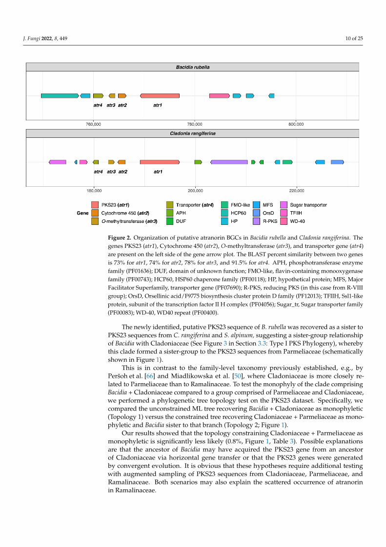
Atranorin is the main secondary compound known in B. rubella [64], and its biosyn-thetic pathway has received attention in previous studies on lichen-forming fungi [26,29].Our phylogenetic results—which included previously identified sequences of genes in-volved in atranorin production from Cladonia species and Stereocaulon alpinum—revealed aputative PKS23 homolog in B. rubella. This is consistent with the phylogenetic investigationof Kim et al. [29], where PKS23 sequences were reported to group together with sequencesfrom atranorin-producing lichen-forming fungi (see Section 3.3 Type I PKS phylogeny). Thedomain configuration of B. rubella PKS23 also supports our phylogenetic results, showingthe same organization of putative atranorin BGC in B. rubella as has been reported forCladonia rangiferina (Figure 2). The B. rubella PKS23 cluster contains a cytochrome P450domain (atr2) required for oxidation, as well as an O-methyltransferase (OMT) domain(atr3) and transporter gene (atr4), which are involved in atranorin biosynthesis [29]. ABLASTp search of the OMT domain (atr3) in B. rubella PKS23 resulted in 36% proteinsequence identity to Trt5 (UniProtKB accession no. Q0C8A3). This level of similarity isconsistent with previous findings by Kim et al. [29] for C. rangiferina PKS23.
J. Fungi 2022, 8, 449 10 of 25
J. Fungi 2022, 8, x FOR PEER REVIEW 10 of 26
rubella. In detail, R. intermedia contains 54 BGCs, including thirteen non-reducing, seven-teen reducing and one partially reducing TI-PKS sequences. Ramalina peruviana, on the other hand, contains 43 BGCs including nine non-reducing and seven reducing genes, and one partially reducing gene (Table 2).
3.1.1. Atranorin Atranorin is the main secondary compound known in B. rubella [64], and its biosyn-
thetic pathway has received attention in previous studies on lichen-forming fungi [26,29]. Our phylogenetic results—which included previously identified sequences of genes in-volved in atranorin production from Cladonia species and Stereocaulon alpinum—revealed a putative PKS23 homolog in B. rubella. This is consistent with the phylogenetic investiga-tion of Kim et al. [29], where PKS23 sequences were reported to group together with se-quences from atranorin-producing lichen-forming fungi (see Section 3.3 Type I PKS phy-logeny). The domain configuration of B. rubella PKS23 also supports our phylogenetic re-sults, showing the same organization of putative atranorin BGC in B. rubella as has been reported for Cladonia rangiferina (Figure 2). The B. rubella PKS23 cluster contains a cyto-chrome P450 domain (atr2) required for oxidation, as well as an O-methyltransferase (OMT) domain (atr3) and transporter gene (atr4), which are involved in atranorin biosyn-thesis [29]. A BLASTp search of the OMT domain (atr3) in B. rubella PKS23 resulted in 36% protein sequence identity to Trt5 (UniProtKB accession no. Q0C8A3). This level of simi-larity is consistent with previous findings by Kim et al. [29] for C. rangiferina PKS23.
Figure 2. Organization of putative atranorin BGCs in Bacidia rubella and Cladonia rangiferina. The genes PKS23 (atr1), Cytochrome 450 (atr2), O-methyltransferase (atr3), and transporter gene (atr4) are present on the left side of the gene arrow plot. The BLAST percent similarity between two genes is 73% for atr1, 74% for atr2, 78% for atr3, and 91.5% for atr4. APH, phosphotransferase enzyme family (PF01636); DUF, domain of unknown function; FMO-like, flavin-containing monooxygenase family (PF00743); HCP60, HSP60 chaperone family (PF00118); HP, hypothetical protein; MFS, Major Facilitator Superfamily, transporter gene (PF07690); R-PKS, reducing PKS (in this case from R-VIII group); OrsD, Orsellinic acid/F9775 biosynthesis cluster protein D family (PF12013); TFIIH, Ssl1-like protein, subunit of the transcription factor II H complex (PF04056); Sugar_tr, Sugar transporter fam-ily (PF00083); WD-40, WD40 repeat (PF00400).
Figure 2. Organization of putative atranorin BGCs in Bacidia rubella and Cladonia rangiferina. Thegenes PKS23 (atr1), Cytochrome 450 (atr2), O-methyltransferase (atr3), and transporter gene (atr4)are present on the left side of the gene arrow plot. The BLAST percent similarity between two genesis 73% for atr1, 74% for atr2, 78% for atr3, and 91.5% for atr4. APH, phosphotransferase enzymefamily (PF01636); DUF, domain of unknown function; FMO-like, flavin-containing monooxygenasefamily (PF00743); HCP60, HSP60 chaperone family (PF00118); HP, hypothetical protein; MFS, MajorFacilitator Superfamily, transporter gene (PF07690); R-PKS, reducing PKS (in this case from R-VIIIgroup); OrsD, Orsellinic acid/F9775 biosynthesis cluster protein D family (PF12013); TFIIH, Ssl1-likeprotein, subunit of the transcription factor II H complex (PF04056); Sugar_tr, Sugar transporter family(PF00083); WD-40, WD40 repeat (PF00400).
The newly identified, putative PKS23 sequence of B. rubella was recovered as a sister toPKS23 sequences from C. rangiferina and S. alpinum, suggesting a sister-group relationshipof Bacidia with Cladoniaceae (See Figure 3 in Section 3.3: Type I PKS Phylogeny), wherebythis clade formed a sister-group to the PKS23 sequences from Parmeliaceae (schematicallyshown in Figure 1).
This is in contrast to the family-level taxonomy previously established, e.g., byPeršoh et al. [66] and Miadlikowska et al. [50], where Cladoniaceae is more closely re-lated to Parmeliaceae than to Ramalinaceae. To test the monophyly of the clade comprisingBacidia + Cladoniaceae compared to a group comprised of Parmeliaceae and Cladoniaceae,we performed a phylogenetic tree topology test on the PKS23 dataset. Specifically, wecompared the unconstrained ML tree recovering Bacidia + Cladoniaceae as monophyletic(Topology 1) versus the constrained tree recovering Cladoniaceae + Parmeliaceae as mono-phyletic and Bacidia sister to that branch (Topology 2; Figure 1).
Our results showed that the topology constraining Cladoniaceae + Parmeliaceae asmonophyletic is significantly less likely (0.8%, Figure 1, Table 3). Possible explanationsare that the ancestor of Bacidia may have acquired the PKS23 gene from an ancestorof Cladoniaceae via horizontal gene transfer or that the PKS23 genes were generatedby convergent evolution. It is obvious that these hypotheses require additional testingwith augmented sampling of PKS23 sequences from Cladoniaceae, Parmeliaceae, andRamalinaceae. Both scenarios may also explain the scattered occurrence of atranorinin Ramalinaceae.

J. Fungi 2022, 8, 449 11 of 25
Table 3. Parameters for the topology test. Topology 1, recovering Bacidia + Cladoniaceae as mono-phyletic; Topology 2, recovering Cladoniaceae + Parmeliaceae as monophyletic. Value for theKishino–Hasegawa test (Kishino and Hasegawa, 1989), Shimodaira and Hasegawa, 1989), expectedlikelihood weights (Strimmer and Rambaut, 2002) and approximately unbiased (AU) test (Shimodaira,2002) are given.
Tree logL Deltal p-KH p-SH c-ELW p-AU
Bacidia + Cladoniaceae −28,054.52811 2.062 × 10⁻⁸ 0.495+ 0.814+ 0.496+ 0.566+
Cladoniaceae + Parmeliaceae −28,108.54142 54.013 0.0126− 0.0126− 0.00853− 0.0081−deltaL: logL difference from the maximal logl in the set. p-KH: p-value of one-sided Kishino–Hasegawa test (1989).p-SH: p-value of Shimodaira–Hasegawa test (2000). c-ELW: Expected Likelihood Weight (Strimmer and Rambaut2002). p-AU: p-value of approximately unbiased (AU) test (Shimodaira, 2002).
Even though a PKS16 gene from Cladonia grayi was first shown to be involved ingrayanic acid production by Armaleo et al. [18], a homolog of this gene from C. rangiferinawas later suggested to be involved in atranorin production by Elshobary et al. [26]. In ourphylogeny, these genes group together with genes from several other Cladonia and lichen-forming fungi (PKS16 clade, See Figure 3 in Section 3.3: Type I PKS Phylogeny), but notall of them are known as atranorin producers (Table 2). Moreover, atranorin is a B-orcinoldepside, and a corresponding biosynthetic gene would require a CMeT domain to add amethyl group to the depside ring. This is not the case for PKS16, which is thought to beinvolved in the synthesis of orcinol depsides [25]. Our comparison of known metabolites inlichen-forming fungi containing PKS16 does not reveal a clear pattern either (See Figure 3 insection: Type I PKS Phylogeny, Group NR-I, PKS16 clade; Table 2). As such, the biosyntheticrole of PKS16 genes remains elusive.
3.1.2. Homosekikaic Acid
Homosekikaic acid is a major secondary metabolite in Bacidia gigantensis as well as inboth Ramalina intermedia and R. peruviana. However, it has not been found in B. rubella. Toour knowledge, there was no putative PKS reported as being involved in the biosynthesis ofhomosekikaic acid, and its biosynthesis has not been characterized using gene expression orheterologous expression experiments. To identify candidate genes involved in the biosyn-thesis of homosekikaic acid in these three species, we used BLASTp on all predicted BGCsequences from the two Bacidia and Ramalina species. We identified three BGC candidateswith BLAST similarity ranging from 53 to 93%, but none of them were a TI-PKS. Instead,we recovered Type 3-PKS homologs from B. gigantensis (BGC 1.2), R. intermedia (BGC 6.1)and R. peruviana (BGC 223.1) as potential homosekikaic biosynthetic genes; however, noneof B. rubella BGCs showed a high similarity to them. Furthermore, our TI-PKS phylogenydid not reveal any clade with both Ramalina species and B. gigantensis that excluded B.rubella. A possible explanation for the observed pattern is that the BGC responsible forsynthesizing homosekikaic acid is present in the genome of B. rubella, but not expressed,and therefore homosekikaic acid is not produced in detectable amount. This hypothesisrequires testing with gene expression experiments and analyses of substance profiles.
3.1.3. Other Biosynthetic Genes Identified In Silico in the Two Bacidia Species
Using further in silico analyses with antiSMASH, we were able to identify genes en-coding enzymes to synthesize secondary metabolites such as clavaric acid (100% similarity)and squalestatin S1 (40% similarity). These terpenes have been identified in both Bacidia.Moreover, we identified a monascorubrin biosynthetic gene in B. rubella confirmed by100% BLAST identity to the monascorubrin biosynthetic gene from Talaromyces (Penicillium)marneffei (PKS3: HM070047) [67]. Apart from monascorubrin biosynthesis, PKS3 geneswere suggested to be involved in the production of the well-known toxin citrinin, as well asa yellow pigment, ankaflavin [67]. Monascorubrin and its related compounds are polyke-tides used as natural red colorants for food [68]. In B. rubella, monascorubrin could be

J. Fungi 2022, 8, 449 12 of 25
responsible for the characteristic orange to the orange-brown coloration of the apothecia.However, these substances have not yet been reported from B. rubella.
In the genome of B. gigantensis, we identified genes possessing high sequencesimilarity with genes involved in the production of pyranonigrin E (100% similarity toBGC0001124), naphthopyrone (100% similarity to BGC0000107), and melanin (100%similarity to BGC0001265). However, in most cases, only a part of the sequences showeda high percentage of similarity; therefore, our in-silico based report is provisional.Additional detailed studies are necessary to confirm these first functional assignments.
3.2. Biosynthetic Gene Composition in Twenty-Three Annotated Fungal Genomes
The basic statistics for all twenty-three studied genomes are provided in Table 4.According to BUSCO homology searches against the fungal dataset (fungi_odb9), mostgenomes were highly gene complete. We included only two genomes with completenessbelow 90%, namely Alectoria sarmentosa (75.8%) and Graphis scripta (88.6%). The numberof predicted genes was in the range of 6756 to 11,072. The lowest number of predictedgenes was observed for Ramalina peruviana (most likely due to issues in the quality of theassembly given the lower sequencing depth of 2×), and the highest number was observedfor Evernia prunastri (Table 4).
Table 4. Genome basics for twenty-three studied fungal genomes.
Assembly No. ofContigs
LargestContig (bp)
Total Length(Mb) GC (%) N50 (bp) N75 (bp) BUSCO
Completeness (%)GenesPredicted
ProteinsPredicted
Alectoriasarmentosa 915 400,628 39.9 40.22 93,085 44,808 75.8 8440 8406
Bacidiagigantensis 24 3,530,911 33.1 44.67 1,807,239 1,552,797 95.8 8451 8400
Bacidia rubella 246 2,353,056 33.7 45.25 1,771,855 1,480,693 97.9 8773 8728
Cladonia grayi 414 958,967 34.6 44.44 243,412 104,892 96.8 9215 9168
Cladoniamacilenta 240 2,265,542 37.1 44.68 1,469,036 1,071,353 96.8 8183 8135
Cladoniametacorallifera 30 2,400,105 36.6 44.91 1,591,850 1,304,658 97.5 8357 8313
Cladoniarangiferina 1008 751,829 35.6 45.46 273,041 142,056 98.2 9264 9218
Cladoniauncialis 2124 143,175 32.8 46.38 34,871 18,367 91.7 8706 8645
Cyanodermellaasteris 37 3,440,352 28.6 53.29 1,790,936 1,105,189 97.2 7946 7851
Dibaeisbaeomyces 1369 352,342 35.2 47.02 70,496 37,098 97.9 9799 9756
Endocarponpusillum 908 803,103 37.1 46.00 178,225 78,254 93.1 8446 8392
Evernia prunastri 277 732,541 40.3 48.97 264,454 154,311 97.9 11,072 10,979
Graphis scripta 1453 383,549 36.2 46.66 78,723 38,837 88.6 9808 9744
Gyalolechiaflavorubescens 36 2,816,824 34.4 41.89 1,693,300 1,515,355 97.5 8062 8008
Lasalliahispanica 1619 615,827 41.2 51.28 145,035 51,438 97.9 8218 8162
Lasalliapustulata 43 3,307,933 32.9 51.67 1,808,250 1,551,388 98.2 6973 6936
Lethariacolumbiana 161 2,188,364 52.2 39.57 666,803 377,091 92.0 9966 9890
Letharia lupina 31 3,031,725 49.2 38.73 2,098,233 1,574,492 96.2 9266 9206
Pseudeverniafurfuracea 46 3,053,396 37.7 47.86 1,178,799 859,355 97.2 9148 9082

J. Fungi 2022, 8, 449 13 of 25
Table 4. Cont.
Assembly No. ofContigs
LargestContig (bp)
Total Length(Mb) GC (%) N50 (bp) N75 (bp) BUSCO
Completeness (%)GenesPredicted
ProteinsPredicted
Ramalinaintermedia 196 898,913 26.2 51.89 273,318 142,876 97.5 7405 7355
Ramalinaperuviana 1657 694,821 26.9 50.75 40,431 15,829 90.0 6756 6706
Usneahakonensis 879 624,317 41.1 45.57 166,123 71,534 96.5 10,700 10,641
Xanthoriaelegans 261 990,773 44.3 40.70 385,707 188,494 98.2 9033 8911
We investigated the BGCs predicted in all twenty-three studied genomes that belongto different taxonomic groups and synthesize a plethora of secondary metabolites (Table 2;Table 4). Our results revealed a high number of BGCs, with an average of 48 clusters pergenome. The smallest number was recovered in the genome of Lasallia pustulata (26 BGCs),and the highest in Evernia prunastri (86 BGCs), which agrees with the results reported byCalchera et al. based on 15 lichenized genomes [6]. In nearly half the genomes, NR-PKSsare more common than R-PKS (Table 2). This evidence is in contrast to previous resultsreporting that R-PKS gene numbers exceed the number of NR-PKS genes [6]. As such, alarger data set is necessary to confirm whether R-PKS or NR-PKSs are more numerous in thegenomes of lichen-forming fungi. The total number of TI-PKSs identified across all studiedgenomes was 478. Of those, 44.35% were of the non-reducing (NR), 53.35% of reducing(R), and 2.3% of partly reducing (PR) PKSs. The highest number of PKS genes was foundin E. prunastri (36 TI-PKS clusters) and C. rangiferina (34 clusters) and the lowest numberswere in B. rubella (10 TI-PKS clusters), B. gigantensis (11 clusters), and Cyanodermella asteris(10 clusters) (Table 2).
Our broad genomic sampling provides phylogenetic context for the NR-PKS genesof B. rubella and B. gigantensis. The six TI-PKS genes from B. rubella are NR-PKSs (threein Subclade I and three in Subclade II), while for B. gigantensis seven TI-PKS genes areNR-PKSs (all are in Subclade I).
The discrepancy between the large number of recovered TI-PKS sequences and thefew experimentally verified secondary metabolites (Table 2) raises questions about therole these genes play in the secondary metabolism, and about the products they produce(e.g., [9,24,25,29]. However, results linking PKS genes to lichen secondary metabolitesbeyond in silico methods are still scarce.
3.3. Type I PKS Phylogeny
Our maximum-likelihood phylogeny of TI-PKS with a total of 624 sequences recoveredfrom twenty-three fungal genomes and supplemented with previously published sequencesis the largest analysis of biosynthetic gene content of various TI-PKS genes in lichen-formingfungi to date.
The TI-PKSs phylogenetic tree is divided into six main subgroups (Figure 3): BacterialType II PKS (including bacterial and mitochondrial ketoacyl-ACP-synthetases; used asoutgroup), Bacterial Type I PKS (also including some fungal sequences), animal fatty acidsynthase (FAS), PR-PKS, NR-PKS, and R-PKS. Lichen PKS genes are distributed across thethree of these main subgroups, viz. PR-PKS, NR-PKS, and R-PKS. These groups were alsorecognized by Kroken et al. [14] and Calchera et al. [6], but both used the single KS domainfor tree reconstruction, and the latter subsumed PR-PKS and R-PKS.

J. Fungi 2022, 8, 449 14 of 25
Nc_3_oxoacyl_ACP_synthetaseHomo_sapiens_3_oxoacyl_ACP_synth
Glycine_max_beta_ketoacyl_synth
Streptomyces_coelicolor_actStreptomyces_avermitilis_pks9
ales
arpr
ed_0
0571
3
psef
urpr
ed_0
0247
1us
nhak
pred
_007
651
evep
rupr
ed_0
0554
2
clagr
apre
d_00
2139
Clad
onia
_gra
yi_AD
M79
459
clam
etpr
ed_0
0706
2
clam
acpr
ed_0
0546
8
claun
cpre
d_00
2794
clara
npre
d_00
5276
ram
perp
red_
0041
81ev
epru
pred
_005
419
lasp
uspr
ed_0
0084
9
ales
arpr
ed_0
0805
3
evep
rupr
ed_0
0965
1
gyaf
lapr
ed_0
0120
7
letlu
ppre
d_00
0523
ram
intp
red_
0011
54
ram
perp
red_
0000
15
cyaa
stpr
ed_0
0330
4
7SKP_cN
1SKP_muiranegal_
muhcirtotelloCN
odul
ispo
rium
_sp_
PKS1
1SKP_sisneyozol_aeralG Bf
_PKS
12
lash
ispr
ed_0
0687
9la
spus
pred
_006
863
gras
crpr
ed_0
0466
4
Mon
ascu
s_pu
rpur
eus_
PKS1
bacg
igpr
ed_0
0827
8
bacr
ubpr
ed_0
0020
2
clara
npre
d_00
5223
clau
ncpr
ed_0
0311
5ra
min
tpre
d_00
2502
cyaa
stpr
ed_0
0297
8
dibb
aepr
ed_0
0316
8
bacg
igpr
ed_0
0000
5
clam
acpr
ed_0
0000
3cla
grap
red_
0087
25ev
epru
pred
_007
731
usnh
akpr
ed_0
0066
7ba
crub
pred
_003
401
clagr
apre
d_00
1062
clara
npre
d_00
1263
ram
intpr
ed_0
0732
0
usnh
akpr
ed_0
0194
5ba
cgigp
red_
0014
99
Bf_P
KS15
bacg
igpre
d_00
5589
lashis
pred
_007
714
gras
crpr
ed_0
0309
9
clara
npre
d_00
4556
Gz_P
KS13
lashis
pred
_008
042
cyaa
stpre
d_00
1401
clamac
pred
_005
709
clametp
red_
0065
66
clara
npre
d_00
6718
claun
cpre
d_00
2366
clamac
pred
_005
884
evep
rupr
ed_0
0845
8
dibba
epre
d_00
0578
dibba
epre
d_00
8406
alesa
rpred
_006
803
clametp
red_0
0465
2
claun
cpred
_000
655
claran
pred_
0086
17
evep
rupred
_003
002
usnh
akpre
d_00
8885
ramint
pred_
0050
33
rampe
rpred
_004
906
dibba
epred
_005
629
clamac
pred_
0038
75
evep
rupred
_002
837
letlup
pred_
0084
59
psefu
rpred
_006
184
dibba
epred
_007
987
laspu
spred
_002
561
Bf_PKS18
dibba
epred
_005
166
Bf_PSK19
evepru
pred_
0063
86
dibba
epred
_003
248
bacrubpred_000551
eveprupred_009395
xanelepred_006704
clamacpred_005650
clauncpred_005133
cyaastp
red_007142
gyaflapred_006654
letcolp
red_0
0504
6
letcolp
red_0
0981
1
letluppred_008051
xanelepred_001793
gyaflapred_002918
grascrpred_009639
bacrubpred_007642
letluppred_008553
ramintpred_005206
ramperpred_000360
xanelepred_008668
ramintpred_004776
clauncpred_000767
endpuspred_007430
psefurpred_006460
laspuspred_004907
clametpred_000650
ramintpred_005951
ramperpred_006064
cyaastpred_000084
eveprupred_007884
Bf_PKS17
dibbaepred_000127
letcolpred_007470
letluppred_004900
gyaflapred_006756
clamacpred_002961
Ch_PKS21
bacrubpred_000804claranpred_005882QXF68953_Stereocaulon_alpinumeveprupred_004607letcolpred_004160letluppred_003921
psefurpred_002298
clamacpred_000016
claranpred_006092
clauncpred_001517
letcolpred_000162letluppred_004273
eveprupred_000853
ramintpred_006122ramperpred_001854
usnhakpred_006848
dibbaepred_002187
Bf_PKS16
dibbaepred_000117letluppred_004909
Ch_PKS22Ch_PKS23
usnhakpred_002742
clagrapred_002409clamacpred_000566clauncpred_005817
claranpred_005675
eveprupred_001697usnhakpred_005239
letluppred_001919
gyaflapred_001052xanelepred_002515
Bf_PKS20
2254
00_d
erpt
nima
r00
3600
_der
palf
ayg
6975
00_d
erpt
nima
r28
3400
_der
ptni
mar ram
perpred_005635
alesarpred_003934
eveprupred_008370eveprupred_008369
psefurpred_008946
usnhakpred_004173clametpred_006401
clauncpred_004670claranpred_008275
eveprupred_008168dibbaepred_006935
gyaflapred_005082
xanelepred_007449Ch_PKS6
clagrapred_008572
clametpred_007955
psefurpred_008953
claranpred_006496
grascrpred_004065
ramperpred_000693endpuspred_007666
xanelepred_008501
laspuspred_000705
ramintpred_005521
ramperpred_003082
bacgigpred_000636
clamacpred_002224clametpred_003874claranpred_009248clauncpred_007217
ramintpred_004692ramperpred_004325dibbaepred_008748clauncpred_006517ramintpred_006159grascrpred_002484Gz_PKS2Gm_PKS2
grascrpred_000320
ramintpred_000325
xanelepred_008317
lashispred_007460
laspuspred_004629letcolpred_000734
dibbaepred_002400
dibbaepred_003279
endpuspred_005861
clagrapred_008282
clametpred_002134
clauncpred_001315Gm_PKS5
clamacpred_008134
laspuspred_001581dibbaepred_002621
psefurpred_002431xanelepred_000508
Ch_PKS8xanelepred_008120endpuspred_000621
xanelepred_005958eveprupred_007590
Ch_PKS9Gm_PKS6
bacgigpred_000006
clagrapred_008726
bacrubpred_003399
claranpred_009253clamacpred_000004
eveprupred_007732
usnhakpred_000668
dibbaepred_005590
bacgigpred_005587
clagrapred_001066
claranpred_001268
grascrpred_003097
Gz_PKS4lashispred_008043
Gz_PKS1
Ch_PKS5
Gz_PKS7
Gm_PKS7
claranpred_004977ram
intpred_004786
xanelepred_008454
laspuspred_000702
claranpred_007096clauncpred_006915
endpuspred_000840xanelepred_005791
clamacpred_002960
ramintpred_005956
psefurpred_003988
gyaflapred_006754
psefurpred_003987Nc_PKS3
gyaflapred_006755Ch_PKS3
Aspergillus_terreus_lovFPenicillium
_citrinum_m
lcB
alesarpred_007824
usnhakpred_005023alesarpred_008264
psefurpred_007717
eveprupred_004804
ramintpred_004931
ramintpred_005497
xanelepred_001687
dibbaepred_000872
clagrapred_001012
clametpred_008299
clauncpred_001467
eveprupred_003978
letcolpred_003111
letluppred_006416
eveprupred_000339
eveprupred_010419
ramperpred_005038
dibbaepred_005439
eveprupred_010217
eveprupred_010653
letcolpred_005666
psefurpred_008091
clagrapred_004542
claranpred_003561
Nc_PKS2cyaastpred_001560
Nc_PKS1Bf_PKS8Ch_PKS2
ramintpred_005164
ramperpred_002240
bacgigpred_003963bacrubpred_000636
eveprupred_005731
usnhakpred_002009eveprupred_003225
usnhakpred_006143
psefurpred_006406Ch_PKS7
Ch_PKS1Didym
ella_maydis_PKS1
grascrpred_002767
grascrpred_006832
clamacpred_003784
clametpred_004245
psefurpred_006748alesarpred_001071
eveprupred_009924usnhakpred_002781
usnhakpred_002782
letcolpred_000052letluppred_004158
alesarpred_007195
eveprupred_010069
clamacpred_005099
claranpred_009018
endpuspred_005198
gyaflapred_007767
Aspergillus_terreus_at4
dibbaepred_004513
Aspergillus_nidulans_wA
Aspergillus_fumigatus_alb1Gz_PK12
Gm_PKS4_CAC92399
Gm_PKS4
bacrubpred_002776
ramintpred_005964
grascrpred_002983
Gm_PKS3_CAC88775Gm_PKS3
Gz_PKS12
clagrapred_006320
claranpred_005443
Aspergillus_parasiticus_pksL1
Aspergillus_nidulans_pksST
clamacpred_006305
clametpred_005198
grascrpred_007614
clamacpred_000704
clametpred_001029
clauncpred_007239
eveprupred_005424
lashispred_004996
laspuspred_005311
lashispred_005798
laspuspred_000627
bacgigpred_001617
bacgigpred_005233
clametpred_001729
clauncpred_008273
usnhakpred_007976
xanelepred_007943
bacgigpred_001043
lashispred_000881
laspuspred_004359psefurpred_007645
Ch_PKS20
xanelepred_001628
Aspergillus_terreus_at1Bf_PKS14
clametpred_004736
clauncpred_007272claranpred_007975
gyaflapred_005508
xanelepred_000120
clametpred_006294
clamacpred_005292
lashispred_003313
laspuspred_003047Ch_PKS19
clauncpred_006694
endpuspred_001080
ramintpred_001718
ramperpred_002015
clauncpred_000071
letcolpred_007218
letluppred_008688ram
intpred_001878ram
intpred_004791
ramperpred_006583
lashispred_004596
laspuspred_000478clagrapred_001463
clauncpred_002890psefurpred_002946
grascrpred_004317
Aspergillus_terreus_at5Ch_PKS18
clagrapred_004008
claranpred_007176
clamacpred_003918
clametpred_004372
psefurpred_005449endpuspred_003222usnhakpred_005879
Bf_PKS13
alesarpred_007561
psefurpred_007016
letcolpred_003325
letluppred_006894
eveprupred_009466
usnhakpred_004695
clagrapred_008490
clamacpred_005628
clauncpred_002560claranpred_006571 35
7600
_der
ptni
mar
7592
00_d
erpr
epma
r
4313
00_d
erpa
lfay
g21
SKP_
ps_a
iral
yX
Homo_sapiens_FASGallus_gallus_FAS
Caenorhabditis_elegans_FASBombyx_mori_FAS_p270
alesarpred_007191psefurpred_008835
letluppred_001760eveprupred_001763
clagrapred_005518
claranpred_003419
clauncpred_002898
clamacpred_007697clametpred_008343
dibbaepred_001414laspuspred_004600
endpuspred_000968xanelepred_006641grascrpred_004833
clamacpred_007404
clametpred_008145claranpred_002808Gm_PKS9Bf_PKS5
Gm_PKS1_CAC44633
Gm_PKS1claranpred_009168letcolpred_004323
letluppred_005553
clauncpred_005612
clauncpred_006270
psefurpred_001125Ch_PKS16xanelepred_003338
xanelepred_000927
Aspergillus_terreus_lovB
Penicillium_citrinum_mlcA
Gz_PKS9
Bf_PKS4
ramintpred_002056Bf_PKS7Nc_PKS4
Bf_PKS3Bf_PKS6
Ch_PKS17Gz_PKS10Gm_PKS10
alesa
rpred
_005
485
psefu
rpred
_000
853
lashis
pred
_005
829
laspu
spre
d_00
6498
clamac
pred
_007
376
clametp
red_
0081
76
psefu
rpre
d_00
2096
clam
etpr
ed_0
0374
6
ram
intpr
ed_0
0172
7
claun
cpre
d_00
5597
xane
lepre
d_00
6042
Bf_P
KS10
dibba
epre
d_00
0831
endp
uspr
ed_0
0114
5
evep
rupr
ed_0
0672
0
gras
crpr
ed_0
0488
9
clara
npre
d_00
5080
Gm_PKS
13
ales
arpr
ed_0
0636
4ev
epru
pred
_009
188
psef
urpr
ed_0
0273
4
letlu
ppre
d_00
7084
clagr
apre
d_00
1823
clam
etpr
ed_0
0561
3
clara
npre
d_00
8950
xane
lepre
d_00
4960
clara
npre
d_00
0259
dibba
epre
d_00
1275
Ch_P
KS15
Ch_P
KS13
_pse
udog
ene
psef
urpr
ed_0
0111
4
clagr
apre
d_00
8921
gyaf
lapr
ed_0
0620
4cla
uncp
red_
0059
76ev
epru
pred
_006
580
lash
ispre
d_00
4056
lasp
uspr
ed_0
0449
6
ram
intp
red_
0057
50
Gm
_PKS
12
claun
cpre
d_00
2025
lash
ispre
d_00
5466
gras
crpr
ed_0
0736
0
dibb
aepr
ed_0
0721
8
gyaf
lapr
ed_0
0372
4
xane
lepr
ed_0
0344
4
Gz_
PKS1
1
xane
lepr
ed_0
0159
2Ch
_PKS
14
Ch_P
KS12
ram
intp
red_
0073
85Ch
_PKS
11_F
UM1
Gm
_FUM
1_AA
D435
62G
m_F
UM1gras
crpr
ed_0
0920
1
bacrubpred_006020
clamacpred_003651
clauncpred_007074
grascrpred_008377
Nc_PKS5
psefurpred_002161
cyaastp
red_002117
usnhakpred_005070
xanelepred_006659Gm_PKS15
dibbaepred_005397Bf_PKS9
clagrapred_006219
letcolpred_008508
letlup
pred_
0034
12
usnhakpred_005236cla
grapred_008233
clamacpred_007822
clametpred_004664Gm_PKS14
claran
pred_
0005
69
claran
pred_
0039
65
psefu
rpred
_000
173
claran
pred_
0052
22
claun
cpred
_003
114
ramint
pred_
0014
87
claran
pred_
0072
14
evep
rupred
_008
863
usnh
akpre
d_01
0014
endp
uspre
d_00
0617
Nc_PKS6
usnh
akpre
d_00
5206
endp
uspred
_005
250
grascr
pred_
0001
34
psefu
rpred
_000
935
letlup
pred_
0070
77
grascr
pred_
0003
63
grascr
pred_
0068
73
clagrapred_000956
letcolpred_001001
usnhakpred_000469claranpred_004628
dibbaepred_006309
clametpred_002147
xanelepred_008447claranpred_005884
cyaastpred_000672dibbaepred_008109
gyaflapred_002006Ch_PKS10
Bf_PKS11
bacr
ubpr
ed_0
0025
4C
h_PK
S4 627700_derptemalc
2SKP_fBla
shis
pred
_007
785
endp
uspr
ed_0
0760
4 065300_derprepmargy
afla
pred
_006
185
cyaa
stpr
ed_0
0202
3
cyaa
stpr
ed_0
0760
1G
z_PK
S5
gras
crpr
ed_0
0801
1
clagrapred_004712eveprupred_006464
dibbaepred_007182lashispred_004470
laspuspred_001869
Aspergillus_terreus_pksM
Aspergillus_parasiticus_pksL2
Penicillium_patulum_6MSAS
Byssochlamys_nivea_6MSAS
clauncpred_003027
usnhakpred_007455
endpuspred_006169
ramintpred_001715
ramperpred_002012
Ch_PKS25
cyaastpred_006073
Penicillium_griseofulvum_pks2
Steptomyces_viridochromogenes_aviM
Ch_PKS24_3p_of_nps7Pseudomonas_syringae_cfa6Pseudomonas_syringae_cfa7
Streptomyces_natalensis_PimS0Streptomyces_natalensis_PimS2_mod3Streptomyces_natalensis_PimS2_mod4
Sorangium_cellulosum_epoA_mod1
Microcystis_aruginosa_mycEStigmatella_aurantiaca_mxaB2_B1
Sorangium_cellulosum_epoF_mod9Stigmatella_aurantiaca_mxaC1Stigmatella_aurantiaca_mxaC2Stigmatella_aurantiaca_mxaE
Stigmatella_aurantiaca_mxaD
Stigmatella_aurantiaca_mxaC3
Mycobacterium_leprae_pksE
Mycobacterium_tuberculosis_ppsD
Mycobacterium_tuberculosis_mas
Mycobacterium_tuberculosis_ppsC
Bacillus_subtilis_pksK_mod2
Myxococcus_xanthus_ta1_mod2
Microcystis_aruginosa_mycG
Nostoc_sp_nosB
Sorangium_cellulosum_epoC_mod2
Mycobacterium_tuberculosis_ppsE
Bacillus_subtilis_mycA_pks1_mod2
Bacillus_subtilis_pksL_mod3
Bacillus_subtilis_pksK_mod3
Xanthomonas_albilineans_xabB_mod2
Bacillus_subtilis_pksL_mod1
Bacillus_subtilis_pksL_mod2
Xanthomonas_albilineans_xabB_mod3
Stigmatella_aurantiaca_mxaF
NR-Izearalenone,orsellinic acid NR-V
anthraquinones
NR-IImelanins
NR-IIIbikaverin
NR-IVaflatoxin, fusarubin
NR-VIusnic acid,mycophenolic acid
NR-VIIazaphilones
NR-VIIImelliolides, orsellinic acid
NR-IXatranorin
R-Ilovastatin/citrinindiketide
R-IIlovastatin/citrininnonaketide
R-IIIT- and PM- toxins
R-IVfumonisin
R-Vtoxins
R-VIIlovastatin/citrinindiketide
R-IX
R-X
PR−PKS
Bacterial and Fungal PKSs
Bacterial Type II PKSs
Animal FAS
Bacterial Type I PKSs
R-VIII
OrthogroupsOrthogroup 1Orthogroup 2Orthogroup 3Orthogroup 4Orthogroup 5Orthogroup 6Orthogroup 7Orthogroup 8Orthogroup 9
NR-I SAT−KS−AT−DH−ACP−[ACP]−TE
NR-V SAT−KS−AT−DH−ACP−[ACP]−TE
NR-II SAT−KS−AT−DH−ACP−[ACP]−TE
NR-VI SAT−KS−AT−DH−ACP−[ACP]−[HTH]−CMeT−[ADH or NAD]−[Peptidase S9]
NR-VIII SAT−KS−AT−DH−ACP−[ACP]−TE
NR-III SAT−KS−AT−DH−ACP−[ACP]−TE
NR-VII SAT−KS−AT−DH−ACP−[ACP]−[HTH]−CMeT−[ADH or NAD]−[Peptidase S9]
NR-IX SAT−KS−AT−DH−ACP−[ACP]−HTH−CMeT−ADH−[Peptidase S9]
NR-IV SAT−KS−AT−DH−ACP−[ACP]−TE
NR-PKS groupsR-I
R-II
R-III
R-IV
R-V
R-VII
R-IX
R-X
R-VIII
KS−AT−DH−[CMeT]−ER−KR−[ACP]
KS−AT−DH−[CMeT]−ER−KR−[ACP]
KS−AT−DH−[CMeT]−ER−KR−[ACP]
KS−AT−DH−[CMeT]−[ER]−[KR]−[ACP]
KS−AT−DH−ER−KR−[KR]−ACP
KS−AT−DH−[CMeT]−ER−KR−[ACP]
KS−AT−DH−[CMeT]−[ER]−KR−ACP−[Carn]
KS−AT−[DH]−[ER]−KR−[ACP]
KS−AT−DH−[CMeT]−ER−KR−[ACP]
R-PKS groups
PKS16
PKS13
PKS8
PKS23
PKS81
AGNPKS1
PKS5
PKS7
PKS7
PKS7
PKS7
PKS19
PKS19
FSR1
FSR1
PKS1
3
Figure 3. Maximum-likelihood phylogeny of Type I PKS genes inferred by IQ-TREE using Type IIBacterial PKSs as outgroup. Clades containing lichen-forming fungi are highlighted and the corre-sponding Orthogroups (1 to 9, respectively) are indicated by different colors. PR-PKS corresponds
J. Fungi 2022, 8, 449 15 of 25
to Orthogroup 1 (green); the nine NR-PKS groups (NR-I to NR-IX) belong to Orthogroups 2, 3and 4 (dark blue, pink and light green, respectively); the ten R-PKS groups (R-I to R-X) belongto Orthogroups 5, 6, 7, 8, and 9 (light blue, red, peach, orange, and lilac, respectively). Char-acteristic secondary substances for the groupings are given in the corresponding colored boxes.Groups not containing lichen-forming fungal genes are indicated by grey boxes. For each group, thedomain arrangement of PKS is highlighted with distinct colors: SAT—starter unit-ACP transacy-lase; KS—ketoacyl synthase; AT—acyltransferase; ACP—acyl carrier protein; KR—ketoreductase;DH—dehydratase; ER—enoyl reductase; CMeT—methyltransferase; TE—thioesterase; HTH—helix-to-helix; ADH—adhydrolase; NAD—NAD-binding; Carn—Choline/Carnitine O-acyltransferasedomain.
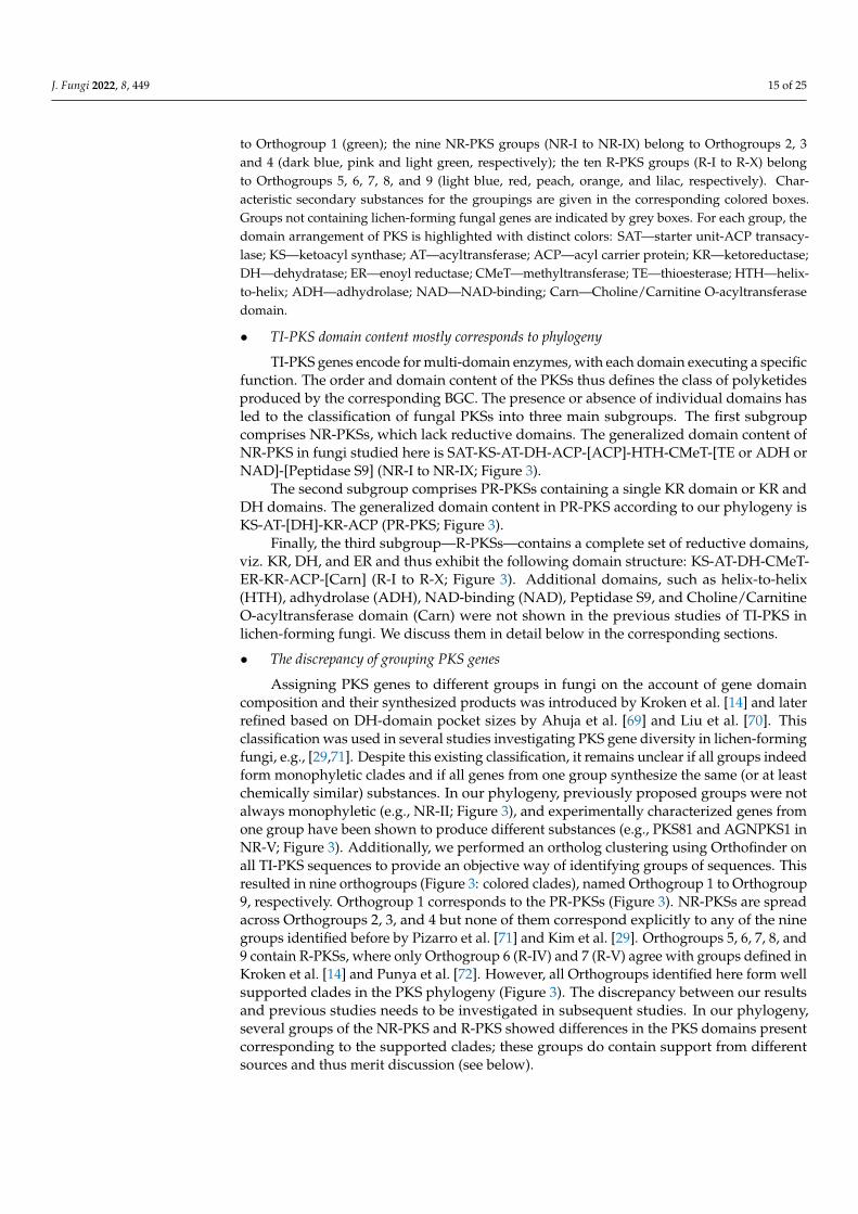
• TI-PKS domain content mostly corresponds to phylogeny
TI-PKS genes encode for multi-domain enzymes, with each domain executing a specificfunction. The order and domain content of the PKSs thus defines the class of polyketidesproduced by the corresponding BGC. The presence or absence of individual domains hasled to the classification of fungal PKSs into three main subgroups. The first subgroupcomprises NR-PKSs, which lack reductive domains. The generalized domain content ofNR-PKS in fungi studied here is SAT-KS-AT-DH-ACP-[ACP]-HTH-CMeT-[TE or ADH orNAD]-[Peptidase S9] (NR-I to NR-IX; Figure 3).
The second subgroup comprises PR-PKSs containing a single KR domain or KR andDH domains. The generalized domain content in PR-PKS according to our phylogeny isKS-AT-[DH]-KR-ACP (PR-PKS; Figure 3).
Finally, the third subgroup—R-PKSs—contains a complete set of reductive domains,viz. KR, DH, and ER and thus exhibit the following domain structure: KS-AT-DH-CMeT-ER-KR-ACP-[Carn] (R-I to R-X; Figure 3). Additional domains, such as helix-to-helix(HTH), adhydrolase (ADH), NAD-binding (NAD), Peptidase S9, and Choline/CarnitineO-acyltransferase domain (Carn) were not shown in the previous studies of TI-PKS inlichen-forming fungi. We discuss them in detail below in the corresponding sections.
• The discrepancy of grouping PKS genes
Assigning PKS genes to different groups in fungi on the account of gene domaincomposition and their synthesized products was introduced by Kroken et al. [14] and laterrefined based on DH-domain pocket sizes by Ahuja et al. [69] and Liu et al. [70]. Thisclassification was used in several studies investigating PKS gene diversity in lichen-formingfungi, e.g., [29,71]. Despite this existing classification, it remains unclear if all groups indeedform monophyletic clades and if all genes from one group synthesize the same (or at leastchemically similar) substances. In our phylogeny, previously proposed groups were notalways monophyletic (e.g., NR-II; Figure 3), and experimentally characterized genes fromone group have been shown to produce different substances (e.g., PKS81 and AGNPKS1 inNR-V; Figure 3). Additionally, we performed an ortholog clustering using Orthofinder onall TI-PKS sequences to provide an objective way of identifying groups of sequences. Thisresulted in nine orthogroups (Figure 3: colored clades), named Orthogroup 1 to Orthogroup9, respectively. Orthogroup 1 corresponds to the PR-PKSs (Figure 3). NR-PKSs are spreadacross Orthogroups 2, 3, and 4 but none of them correspond explicitly to any of the ninegroups identified before by Pizarro et al. [71] and Kim et al. [29]. Orthogroups 5, 6, 7, 8, and9 contain R-PKSs, where only Orthogroup 6 (R-IV) and 7 (R-V) agree with groups defined inKroken et al. [14] and Punya et al. [72]. However, all Orthogroups identified here form wellsupported clades in the PKS phylogeny (Figure 3). The discrepancy between our resultsand previous studies needs to be investigated in subsequent studies. In our phylogeny,several groups of the NR-PKS and R-PKS showed differences in the PKS domains presentcorresponding to the supported clades; these groups do contain support from differentsources and thus merit discussion (see below).

J. Fungi 2022, 8, 449 16 of 25
3.3.1. Lichen-Forming Fungi Contain Only a Few Partially Reducing PKSs in thePhylogenetic Neighbourhood to Bacterial PKSs
The PR-PKS sequences formed a well-supported clade sister to bacterial Type I PKSsequences with other fungal PKSs (PR-PKS; Figure 3). Their domain configuration is KS-AT-[DH]-KR-ACP, including only a single reductase domain (KR). In contrast to the largeNR- and R-PKS groups, PR-PKS contains genes mainly from Aspergillus and Penicillium,and only a few genes from lichen-forming fungi. Genes from the studied fungal genomeshave the typical PR-PKS domain composition, but in two genes from two Ramalina (ram-intpred_001715 and ramperpred_002012), the DH domain was missing. PR-PKS genesform a sister clade to Bacterial Type I PKSs (Figure 2). Bacterial T1-PKSs often possess aKR domain catalyzing the first step in the reductive modification of beta-carbonyl centersin the growing polyketide chain. This domain requires NADPH to reduce the keto- to ahydroxy group [73].
The “simple” domain configuration of Bacterial and PR-PKS genes compared to NR-and R-PKSs has not escaped our attention. Our study was not designed to specifically testhow PKS genes in Ascomycetes were acquired and how they diversified. However, theplacement of Bacterial and NR-PKS sequences as the earliest branches in our phylogenysupports the hypothesis suggested by Kroken et al. [14] that fungal TI-PKS genes couldhave been acquired by an ancient horizontal gene transfer event between bacteria andfungi. Additionally, our results suggest that ancestral TI-PKS genes may have been partiallyreducing. Under this scenario, NR- and R-PKS evolution could have been connected todomain structure modification (such as gaining SAT in NR-PKS) and subsequent func-tional diversification. However, this cannot be concluded with certainty without a greatlyexpanded sample of genomes from other fungal groups, including genomes from earlybranching fungal lineages and comprehensive ancestral state reconstructions.
3.3.2. Fungal PKSs Producing Non-Reduced Polyketides
The Diversity of NR-PKS Sequences
In previous studies, NR-PKSs were divided into nine major groups based on proteinsequence similarity and PKS domain content [14,29,69,71]. Similar to previous studies, weobserved characteristic domain configurations for the different clades in our phylogeny.The domain structure of NR-PKSs in the studied fungi can be generalized as SAT-KS-AT-DH-ACP-[ACP]-HTH-CMeT-[TE or ADH or NAD]-[Peptidase S9]; however, somePKS genes may deviate from this structure. The observed domain content variationsare not random but rather occur in two main patterns: either through the duplicationof the ACP domain, or through the addition of an N-terminal TE domain. PKS genespossessing an additional ACP domain are scattered throughout different NR-PKS clades,suggesting multiple independent gains. However, the functional significance of ACPdomain duplications is still unknown [14].
Most of the NR-PKS sequences belong to a large clade containing groups NR-I to NR-Vand NR-VIII, with domain configuration containing a TE domain at the N-terminal end.
The irregularly present additional TE domain is involved in a thioesterase-mediatedproduct release, which is the most common release mechanism in TI-PKS [74]. Itregularly extends to a C-C Claisen cyclization domain (TE/CLC domain), e.g., inAspergillus parasiticus PksA [75]. Although CYC domains have previously been reportedfrom various fungi [14,69,70], we could not identify them in our analyses and thus didnot indicate them in the phylogeny.
Subclade I of NR-PKSs (Including Groups NR-I to NR-V, and NR-VIII)
Group NR-I
The paraphyletic group NR-I contains several clades with previously characterizedsequences involved in the biosynthesis of aromatic compounds derived from orsellinic acid,such as grayanic acid (PKS16), physodic acid and olivetoric acid [24,25], lecanoric acid [76],

J. Fungi 2022, 8, 449 17 of 25
xanthones, aflatoxin, and naphthoquinones [14]. In addition, the clade also comprisesPKS13 sequences from several lichen-forming fungi and Gibberella zeae as well as a PKS15sequence from Botryotinia fuckeliana with unknown functions.
Group NR-II
Our results show that NR-II is a diverse group, comprised of several clades con-taining sequences from lichen-forming fungi interspersed with characterized PKS genesproposed to be involved in the biosynthesis of various substances. The earliest branch-ing clade contains genes involved in the 6-hydroxymellein biosynthesis (NR-II; Figure 3),a key intermediate in terrein biosynthesis [77]. Terrein, produced in large quantitiesby Aspergillus terreus, has phytotoxic activity and has the potential to serve as a novelantibiotic [78]. Our phylogeny revealed sequences of four lichen-forming fungi (cla-grapred_001463, clauncpred_002890, psefurpred_002946, and grascrpred_004317) clus-tering together with terA gene in A. terreus. A BLAST search of terA (GenBank: EAU38791)against these sequences revealed 58 to 67% similarity at the amino acid level, indicatinga high degree of conservation despite an approximate 350-million-year split of Euro-tiomycetes (Aspergillus) and Lecanoromycetes [79]. Terrein has not been reported fromlichen-forming fungi; therefore, without further experiments and detailed substance char-acterization, terrein production in lichen-forming fungi remains hypothetical.
Another important pattern is the occurrence of characterized melanin genes andmelanin precursors in different clades. Melanins are a diverse group of substances thatplay a role in virulence, morphogenesis or the response to environmental stress, and canbe synthesized via different pathways [80]. PKSs produce two forms of melanin: either1,8-dihydroxynaphtalene (DHN) melanin or Deoxybostrycoidein-melanin [81]. Melaninsare insoluble and thus cannot be studied by standard biochemical methods [80]. In the PKSgene survey by Kroken et al. [14], all melanin synthesizing genes were grouped togetherin a single monophyletic clade. In our analysis, they are recovered in at least three cladescontaining sequences from several Lecanoromycetes and Endocarpon pusillum. Sister toNR-II is a clade containing sequences from Colletotrichum lagenarium (PKS1), Glarea sp.,Nodulisporium sp., and C. heterostrophus (PKS18), which were also characterized as producersof melanin [14]. The high number of genes from lichen-forming fungi close to characterizedmelanin biosynthetic genes suggests an essential role of melanins in lichens.
Groups NR-III and NR-IV
The sister groups NR-III and NR-IV contain sequences of proteins producing largepolyketide chains, such as conidial yellow pigment Alb1 (NR-III) or aflatoxin/sterigmatocystin of A. nidulans (NR-IV) [14,71,82].
NR-III additionally contains characterized PKSs producing duclauxin (35–71% simi-larity) and naphthopyrone (100% similarity). Duclauxins are dimeric, heptacyclic fungalpolyketides with various activities [83], while bis-naphthopyrones act in herbivore defensein filamentous ascomycetes [84].
Several genes recovered in the NR-IV group are potentially involved in producingdifferent fungal pigments. The genes from Ramalina intermedia (ramintpred_005964) andCladonia grayi (clagrapred_006320) were assigned to an FSR1 gene, which is involved inproducing highly pigmented naphthoquinones fusarubins responsible for fruiting bodycoloration of Fusarium fujikuroi [85]. A BLAST search of the R. intermedia gene (ram-intpred_005964) revealed 72.3% similarity to a PKS of Cladonia metacorallifera (GenBank:QIX11499) that is involved in the biosynthesis of the red compound cristazarin, character-ized by antibacterial and antitumor activity [86]. Sequences of Bacidia rubella and severalCladonia species producing red pigments were recovered sister to the putative Ramalinaintermedia FSR1 gene. Interestingly, R. intermedia, at the same time, lacks red pigments [87].This close phylogenetic proximity raises the possibility that combinations of substancessynthesized by different PKS genes could be responsible for red-colored pigments (see alsodiscussion on monascorubrin above), particularly for the red fruiting bodies in B. rubella.

J. Fungi 2022, 8, 449 18 of 25
Group NR-V
According to the previous studies, NR-V contains PKSs without the TE domain [71];however, in the characterized PKSs in our phylogeny, the TE or R domains were present inall clades. The genes in the NR-V group are suggested to be involved in the production ofdifferent mycotoxins, such as desertorin (Aspergillus nidulans) and atrochrysone (Aspergillusfumigatus) [71,88,89].
The crown clades in the NR-V group include the annotated PKS81 orthologs fromCladonia metacorallifera, C. macilenta, Lasallia pustulata and L. hispanica as sister to a PKS19sequence from Cochliobolus heterostrophus and two clades with characterized Agnpks1 alsofrom several lichen-forming fungi. Agnpks1 (Atrochrysone carboxylic acid synthase) wasoriginally described from Penicillium divaricatum and is involved in the biosynthesis ofagnestins and dihydroxy-xanthone metabolites [90]. Xanthones and related benzophe-nones are produced by various filamentous fungi. They exhibit insecticide, antioxidant,antibacterial, anti-inflammatory, and anticancer activities [91]. Examples include desmethyl-sterigmatocystin, a key intermediate of the aflatoxin group of mycotoxins produced byAspergillus flavus, and a norlichexanthone from the lichen-forming Lecanora straminea [90].An additional BLAST search of norlichexanthone synthase from L. straminea (GenBank:D7PI15) against our annotated fungal sequences showed at least 37% similarity to sequencesfrom the clade containing PKS81 and Agnpks1. The relationship between these two genesremains unclear, but given our results, PKS81 might belong to or have evolved from Ag-npks1. Additional closer investigations will be necessary to show the role of PKS81 andAgnpks1 orthologs in lichen-forming fungi and their possible role in xanthone biosynthesis.
Subclade II of NR-PKSs (Including Groups NR-VI, NR-VII, and NR-IX)
The second largest clade of NR-PKSs includes the previously recovered groups NR-VI,NR-VII, and the recently defined group NR-IX [29] (Figure 3). The generalized domainarrangement in these groups is SAT-KS-AT-DH-ACP-[ACP]-[HTH]-CMeT-[ADH/NAD]-[Peptidase S9], deviating from the generalized arrangement for other NR-PKS (see above).Although previously reported from all NR-PKS groups except group NR-V [69,71], we didnot observe TE domains in groups NR-VI, NR-VII, and NR-IX. Instead, we observed NADand ADH domains—in some cases followed by a Peptidase S9 (PFAM: PF00326) domain.Peptidase S9 belongs to proteolytic enzymes that consume serine in their catalytic activity,and they are ubiquitously found in viruses, bacteria, and eukaryotes [92]. We also observeda helix-turn-helix domain (HTH) being inserted after one or two ACP domains in severalgroups. The HTH domain was previously found in PKSs in fungi but it has not been shownin the arrangement of the NR-PKSs of lichen-forming fungi before. Proteins with an HTHmotif are, for example, involved in DNA repair, RNA metabolism, and protein–proteininteraction [93], and could thus be involved in developmental or morphogenic processes.
Group NR-VI
Characterized PKSs in this group include PKS18 and PKS19 genes from Botryotiniafuckeliana. Genes from this group were suggested to be involved in usnic acid biosyn-thesis [71], and according to Kim et al. [29] they belong to PKS8. Our results show thatthis group is divided into two clades. The first clade contains genes of known usnic acidproducers such as Ramalina intermedia, R. peruviana, Cladonia metacorallifera, C. macilenta, C.uncialis, Evernia prunastri, Alectoria sarmentosa, Letharia columbiana, and Usnea hakonensis,interspersed with genes from lichen-forming fungi known not to produce usnic acid. Thesecond clade includes genes mostly from lichen-forming fungi not producing usnic acid,including a gene from B. rubella (bacrubpred_000551). However, sequences from Cladoniamacilenta, C. uncialis, and Evernia prunastri were also recovered in this clade.

J. Fungi 2022, 8, 449 19 of 25
Group NR-VII
The only genes characterized in this group are PKS17 from Botryotinia fuckeliana andPKS21 from Cochliobolus heterostrophus. These genes have been proposed to produce cit-rinin or lovastatin [14,94,95]. The other identified genes from the lichen-forming fungiin this group had 50 to 70% BLAST similarity to genes involved in the biosynthesis ofmonascorubrin, citrinin, conidial yellow, azanigerone A, and stipitatic acid (see also discus-sion on monascorubrin above). As mentioned above, the biosynthetic gene from B. rubella(bacrubpred_007642) has a 100% identity to the monascorubrin biosynthetic gene.
Group NR-IX
The recently recognized group NR-IX [29] contains several predicted PKS23 sequences,involved in the biosynthesis of atranorin (see sections on 3.1.1 Atranorin and 3.1.2 Ho-mosekikaic acid above) and methylated orsellinic acid derivatives. In contrast to the groupsNR-VI and NR-VII, NR-IX sequences always contain an ADH and lack a NAD domain afterCMeT, while Peptidase S9 is only sporadically present. It is necessary to note that only oneACP domain was present in the PKS23, including atranorin producers. In addition, othercharacterized PKSs of this group are from Cochliobolus heterostrophus (PKS22 and PKS23)and Botryotinia fuckeliana (PKS16 and PKS20).
3.3.3. Fungal PKSs Producing Reduced Polyketides
The Diversity of R-PKS Sequences
R-PKS genes are involved in synthesizing various reduced, usually linear polyketides.Many are precursors of toxins that are active in animals (e.g., lovastatin, citrinin, andfumonisin) or plants (e.g., T-toxin and PM-toxin) [96–99]. We follow this classificationin our discussion whenever possible, while also highlighting deviations. Compared toNR-PKSs, less is known about the possible roles of R-PKSs. In our phylogeny, R-PKSsformed a large clade sister to NR-PKSs with many well-supported subclades. Unlike whathas been shown in previous studies, all studied R-PKSs here lacked a second ACP domain,and in some cases, ACP was missing. According to Kroken et al. [14], four major groups(R-I to R-IV) of R-PKS genes can be distinguished based on their overall domain structureand produced compounds. This classification was later extended to groups R-V to R-VIIIby Punya et al. [72]. Over 70% of the studied fungal sequences could be assigned to groupsdesignated by Kroken et al. [14] and Punya et al. [72] with the general domain content:KS-AT-DH-[CMeT]-ER-KR-[ACP]. Two clades recovered by Punya et al. [72] are missingfrom our tree, namely R-VI and R-VIII. Clade VI contained uncommon PKS-NRPS hybridswith four additional domains after ACP, viz. condensation (C), adenylation (A), thiolation(T), and reductase (R) [72]. As our study focused on the TI-PKSs, it did not include hybridNRPS-PKS genes. The sequences from R-VIII were not present in our phylogeny, and arethus not feasible to identify with confidence.
Based on our phylogenetic results, we identified two R-PKS clades that did not matchany of the R-PKS groups reported in previous studies. Sequences in those two cladeshad characteristic domain structures and were recovered as distinct orthogroups in ourOrthofinder analysis. This leads us to propose these clades as new reducing PKS groups,R-IX and R-X, following the numbering scheme by Kroken et al. [14] and Punya et al. [72].
Groups of R-PKSs
Group R-I
Group R-I is the largest group of R-PKSs in our tree. It contains one of the R-PKSgenes synthesizing the diketide portion of lovastatin and citrinin, T-toxin, and PM-toxin [14]. Our results show that the genes of this group, recovered in a monophyleticclade by Kroken et al. [14], were nested in different clades in our analysis; therefore, R-Iassignment is not complete.

J. Fungi 2022, 8, 449 20 of 25
Group R-II
Group R-II forms a well-supported clade and it was previously characterized by theabsence of an ER domain [14]. In our phylogeny, however, ER domain was present inall lichen-forming fungi. The group includes many previously published sequences andorthologs from Cladonia and Letharia species, Xanthoria elegans, Pseudevernia furfuracea, andRamalina intermedia. The R. intermedia ortholog was assigned to PKS19. It was predicted tobe involved in the production of heptaketides with a role in a lesion of rice leaves formationin Magnaporthe oryzae [100]. Two characterized proteins from A. terreus lovB and P. citrinummlcA synthesize the cyclic nonaketide portion of lovastatin and citrinin.
Group R-III
Group R-III forms a clade comprised of four sequences previously assigned to thisgroup by Kroken et al. [14], and a sequence from Cyanodermella asteris. PKSs from this cladedo not possess a CMeT domain. The experimentally characterized gene in group R-III isC. heterostrophus PKS2, which, along with PKS1, is required for the synthesis of T-toxin [14].This group contains several sequences from Kroken et al. [14] interspersed with sequencesfrom various lichen-forming fungi.
Group R-IV
Genes belonging to R-IV may contain a conserved CMeT domain. Thus, their domainconfiguration somewhat resembles that of groups R-I and R-II. However, N-terminaldomains ER, KR, and ACP can be present or absent in R-IV. The only characterized PKSin this group is G. moniliformis FUM1 (Gm_FUM1_AAD43562), which makes the linearpolyketide precursor of the toxin fumonisin [98]. This group contains several sequencesfrom Kroken et al. [14] interspersed with sequences from various lichen-forming fungi.
Group R-V
Sequences in group R-V have similar domain composition like sequences from Or-thogroup 7: KS-AT-DH-ER-KR-[KR]-ACP. However, a second KR domain is only present inDibaeis baeomyces (dibbaepred_000831). We recovered genes from several lichen-formingfungi in this group. Again, experimentally characterized genes from Gibberella moniliformis(Gm_PKS13) and Botryotinia fuckeliana (Bf_PKS10) also recovered here indicate a possiblerole in toxin biosynthesis (Figure 3) [14].
Group R-VII
This group contains several previously characterized sequences as well as sequencesfrom R. intermedia, P. furfuracea, G. furfuracea, and C. macilenta where we could assign PKS5annotations. Additional sequences from lichen-forming fungi also recovered here had nofunctional annotations; as such the role of these genes remains unclear. The characterized se-quences of Aspergillus (Aspergillus_terreus_lovF) and Penicillium (Penicillium_citrinum_mlcB)indicate a role in the production of the diketide portion of lovastatin and citrinin, but theywere assigned to group R-I by Kroken et al. [14].
Novel group R-IX
A new group of reducing PKSs can be delineated based on a specific domain compo-sition and our orthogroup assignment. We tentatively call this group R-IX following thenaming scheme of Kroken et al. [14] and Punya et al. [72]. R-IX is sister to group R-VII anddiffers from all other groups by having a facultative Choline/Carnitine O-acyltransferasedomain (Carn) following the ACP domain: KS-AT-DH-[CMeT]-ER-KR-[ACP]-[Carn](Figure 3). The Carn domain can be found in several eukaryotic acetyltransferases andalso at the C terminus of a highly reducing polyketide synthase (SdnO), where it ispart of a gene cluster mediating the biosynthesis of glycoside antibiotics sordarin andhypoxysordarin [101].

J. Fungi 2022, 8, 449 21 of 25
Novel group R-X
Similar to R-IX, a second group of reducing PKSs can be identified based on a specificdomain composition and orthogroup assignment. This group R-X, recovered as Orthogroup9, contains only genes from lichen-forming fungi. Together with Group R-II, they share asister-group relationship to all other R-PKSs in our phylogeny (Figure 3). Genes from thisgroup may not have DH, ER and ACP domains, resulting in the following domain content:KS-AT-[DH]-[ER]-KR-[ACP]. Most fungal genes recovered here are missing the DH andER domain. However, the genes from Lasallia hispanica (lashispred_001760) and Everniaprunastri (eveprupred_001763) contain a complete domain configuration: KS-AT-DH-ER-KR-ACP.
4. Conclusions and Limitations
This study presents a comprehensive analysis of the PKS gene content of the de novosequenced genome of B. rubella and twenty-two publicly available genomes of mainlylichen-forming fungi. Our results reveal that a BGC including PKS23 is likely involved inbiosynthesis of atranorin in B. rubella. Similar to previous studies (e.g., [6,23,24,29]), weobserved a large diversity of PKS genes in lichen-forming fungi as well as B. rubella, muchlarger than expected based on their recorded secondary metabolite profiles. This highlightsthe potential of lichen-forming fungi to produce a much higher number of substances thanpreviously assumed, as mentioned by Calchera et al. [6] focusing on foliose lichens. Ourin silico approach provides first insights into the possible roles of PKS genes in crustoseB. rubella; however, we are aware that this comes with limitations. Only detailed studiesof individual BGCs combining gene expression analyses with analytical chemistry andmetabolomics will allow thorough testing of the hypotheses proposed here. The prospectsfor this are promising as an increasing number of studies succeeded in the heterologousexpression of lichen-forming fungal genes, (e.g., [29,76,86,102,103]). Subsequent in vivostudies of metabolite profiles in lichen-forming fungi supplemented by high-resolutionMS/MS spectra [104] or metabolic profiling based on stable isotope analysis [105] willcontribute to our knowledge on how secondary metabolites in lichen-forming fungi areproduced and what roles they play. Combined with the growing number of availablelichen-forming fungal genomes, we will be able to refine our understanding and formulatenovel hypotheses about the biosynthetic potential of lichens.
Author Contributions: Conceptualization, J.V.G., A.B., S.W. and P.R.; methodology, software andformal analysis, J.V.G. and P.R.; investigation, J.V.G. and A.B.; resources and funding acquisition, S.W.and A.B.; data curation, J.V.G. and P.R.; writing—original draft preparation, J.V.G.; writing—reviewand editing, J.V.G., A.B., S.W. and P.R.; visualization, J.V.G.; supervision, A.B., S.W. and P.R. Allauthors have read and agreed to the published version of the manuscript.
Funding: J.G. was supported by a BAYHOST fellowship from the Bayerische Staatsministerium fürBildung und Kultus, Wissenschaft und Kunst and an LMU travel grant (April 2021). Laboratorywork and genome sequencing were funded by the LMU Munich start-up funds of S.W. and by theStaatliche Naturwissenschaftliche Sammlungen Bayerns, grant SNSB innovativ to A.B.
Data Availability Statement: The data supporting the findings of this study are available open accessin figshare at http://doi.org/10.6084/m9.figshare.19487837. These include gff3 files and proteinsequences from twenty-two re-annotated genomes downloaded from the NCBI. In addition, thealignment used for the PKS tree calculation is deposited. The de novo sequenced genome of Bacidiarubella is available under the NCBI BioProject accession number PRJNA821848 and on the SequenceRead Archive (SRA).
Acknowledgments: We thank Diego F. Morales-Briones (LMU Munich) for his valuable help in thefigure preparation and Elizabeth Joyce (LMU Munich) for improving the English text. Andrea Brandland Tanja Ernst (both LMU Munich) helped with the lab work. We thank the government of theadministrative district of Upper Bavaria for the sampling permit.
Conflicts of Interest: The authors declare no conflict of interest.
J. Fungi 2022, 8, 449 22 of 25
References1. Keller, N.P. Fungal Secondary Metabolism: Regulation, Function and Drug Discovery. Nat. Rev. Microbiol. 2019, 17, 167–180.
[CrossRef] [PubMed]2. Rokas, A.; Mead, M.E.; Steenwyk, J.L.; Raja, H.A.; Oberlies, N.H. Biosynthetic Gene Clusters and the Evolution of Fungal
Chemodiversity. Nat. Prod. Rep. 2020, 37, 868–878. [CrossRef] [PubMed]3. Keller, N.P.; Turner, G.; Bennett, J.W. Fungal Secondary Metabolism—from Biochemistry to Genomics. Nat. Rev. Microbiol. 2005, 3,
937–947. [CrossRef] [PubMed]4. Keller, N.P.; Hohn, T.M. Metabolic Pathway Gene Clusters in Filamentous Fungi. Fungal Genet. Biol. 1997, 21, 17–29.
[CrossRef] [PubMed]5. Huneck, S.; Yoshimura, I. Identification of Lichen Substances. In Identification of Lichen Substances; Huneck, S., Yoshimura, I., Eds.;
Springer: Berlin/Heidelberg, Germany, 1996; pp. 11–123. ISBN 978-3-642-85243-5.6. Calchera, A.; Dal Grande, F.; Bode, H.B.; Schmitt, I. Biosynthetic Gene Content of the ‘Perfume Lichens’ Evernia prunastri and
Pseudevernia furfuracea. Molecules 2019, 24, 203. [CrossRef] [PubMed]7. Culberson, C.F.; Armaleo, D. Induction of a Complete Secondary-Product Pathway in a Cultured Lichen Fungus. Exp. Mycol.
1992, 16, 52–63. [CrossRef]8. Culberson, C.F.; Culberson, W.L.; Johnson, A. Characteristic Lichen Products in Cultures of Chemotypes of the Ramalina siliquosa
Complex. Mycologia 1992, 84, 705–714. [CrossRef]9. Hamada, N. The Effect of Various Culture Conditions on Depside Production by an Isolated Lichen Mycobiont. Bryologist 1989,
92, 310–313. [CrossRef]10. Stocker-Wörgötter, E.; Elix, J.A. Secondary Chemistry of Cultured Mycobionts: Formation of a Complete Chemosyndrome by the
Lichen Fungus of Lobaria spathulata. Lichenologist 2002, 34, 351–359. [CrossRef]11. Hamada, N.; Tanahashi, T.; Miyagawa, H.; Miyawaki, H. Characteristics of Secondary Metabolites from Isolated Lichen
Mycobionts. Symbiosis 2001, 31, 23–33.12. Molina, M.C.; Crespo, A.; Vicente, C.; Elix, J.A. Differences in the Composition of Phenolics and Fatty Acids of Cultured Mycobiont
and Thallus of Physconia distorta. Plant Physiol. Biochem. 2003, 41, 175–180. [CrossRef]13. Cox, R.J. Polyketides, Proteins and Genes in Fungi: Programmed Nano-Machines Begin to Reveal Their Secrets. Org. Biomol.
Chem. 2007, 5, 2010–2026. [CrossRef] [PubMed]14. Kroken, S.; Glass, N.L.; Taylor, J.W.; Yoder, O.C.; Turgeon, B.G. Phylogenomic Analysis of Type I Polyketide Synthase Genes in
Pathogenic and Saprobic Ascomycetes. Proc. Natl. Acad. Sci. USA 2003, 100, 15670. [CrossRef] [PubMed]15. Nicholson, T.P.; Rudd, B.A.M.; Dawson, M.; Lazarus, C.M.; Simpson, T.J.; Cox, R.J. Design and Utility of Oligonucleotide Gene
Probes for Fungal Polyketide Synthases. Chem. Biol. 2001, 8, 157–178. [CrossRef]16. Lawrey, J.D. Biological Role of Lichen Substances. Bryologist 1986, 89, 111–122. [CrossRef]17. Rundel, P.W. The Ecological Role of Secondary Lichen Substances. Biochem. Syst. Ecol. 1978, 6, 157–170. [CrossRef]18. Armaleo, D.; Sun, X.; Culberson, C. Insights from the First Putative Biosynthetic Gene Cluster for a Lichen Depside and Depsidone.
Mycologia 2011, 103, 741–754. [CrossRef]19. Mullins, A.J.; Murray, J.A.H.; Bull, M.J.; Jenner, M.; Jones, C.; Webster, G.; Green, A.E.; Neill, D.R.; Connor, T.R.; Parkhill, J.; et al.
Genome Mining Identifies Cepacin as a Plant-Protective Metabolite of the Biopesticidal Bacterium Burkholderia ambifaria. Nat.Microbiol. 2019, 4, 996–1005. [CrossRef]
20. Nielsen, J.C.; Grijseels, S.; Prigent, S.; Ji, B.; Dainat, J.; Nielsen, K.F.; Frisvad, J.C.; Workman, M.; Nielsen, J. Global Analysis ofBiosynthetic Gene Clusters Reveals Vast Potential of Secondary Metabolite Production in Penicillium Species. Nat. Microbiol. 2017,2, 17044. [CrossRef]
21. Lichman, B.R.; Godden, G.T.; Buell, C.R. Gene and Genome Duplications in the Evolution of Chemodiversity: Perspectives fromStudies of Lamiaceae. Physiol. Metab. 2020, 55, 74–83. [CrossRef]
22. Medema, M.H.; Fischbach, M.A. Computational Approaches to Natural Product Discovery. Nat. Chem. Biol. 2015, 11, 639–648.[CrossRef] [PubMed]
23. Navarro-Muñoz, J.C.; Selem-Mojica, N.; Mullowney, M.W.; Kautsar, S.A.; Tryon, J.H.; Parkinson, E.I.; De Los Santos, E.L.C.;Yeong, M.; Cruz-Morales, P.; Abubucker, S.; et al. A Computational Framework to Explore Large-Scale Biosynthetic Diversity.Nat. Chem. Biol. 2020, 16, 60–68. [CrossRef] [PubMed]
24. Singh, G.; Armaleo, D.; Dal Grande, F.; Schmitt, I. Depside and Depsidone Synthesis in Lichenized Fungi Comes into Focusthrough a Genome-Wide Comparison of the Olivetoric Acid and Physodic Acid Chemotypes of Pseudevernia furfuracea. Biomolecules2021, 11, 1445. [CrossRef] [PubMed]
25. Singh, G.; Calchera, A.; Merges, D.; Otte, J.; Schmitt, I.; Grande, F.D. A Candidate Gene Cluster for the Bioactive Natural ProductGyrophoric Acid in Lichen-Forming Fungi. bioRxiv 2022. [CrossRef]
26. Elshobary, M.E.; Becker, M.G.; Kalichuk, J.L.; Chan, A.C.; Belmonte, M.F.; Piercey-Normore, M.D. Tissue-Specific Localization ofPolyketide Synthase and Other Associated Genes in the Lichen, Cladonia rangiferina, Using Laser Microdissection. Phytochemistry2018, 156, 142–150. [CrossRef]
27. Abdel-Hameed, M.; Bertrand, R.L.; Piercey-Normore, M.D.; Sorensen, J.L. Identification of 6-Hydroxymellein Synthase andAccessory Genes in the Lichen Cladonia uncialis. J. Nat. Prod. 2016, 79, 1645–1650. [CrossRef]

J. Fungi 2022, 8, 449 23 of 25
28. Abdel-Hameed, M.; Bertrand, R.L.; Piercey-Normore, M.D.; Sorensen, J.L. Putative Identification of the Usnic Acid BiosyntheticGene Cluster by de novo Whole-Genome Sequencing of a Lichen-Forming Fungus. Fungal Biol. 2016, 120, 306–316. [CrossRef]
29. Kim, W.; Liu, R.; Woo, S.; Bin Kang, K.; Park, H.; Yu, Y.H.; Ha, H.-H.; Oh, S.-Y.; Yang, J.H.; Kim, H.; et al. Linking a Gene Clusterto Atranorin, a Major Cortical Substance of Lichens, through Genetic Dereplication and Heterologous Expression. mBio 2021,12, e01111-21. [CrossRef]
30. Boustie, J.; Tomasi, S.; Grube, M. Bioactive Lichen Metabolites: Alpine Habitats as an Untapped Source. Phytochem. Rev. 2011, 10,287–307. [CrossRef]
31. Yoshimura, I.; Yamamoto, Y.; Nakano, T.; Finnie, J. Isolation and Culture of Lichen Photobionts and Mycobionts. In Protocols inLichenology: Culturing, Biochemistry, Ecophysiology and Use in Biomonitoring; Kranner, I.C., Beckett, R.P., Varma, A.K., Eds.; Springer:Berlin/Heidelberg, Germany, 2002; pp. 3–33. ISBN 978-3-642-56359-1.
32. Honegger, R.; Zippler, U.; Gansner, H.; Scherrer, S. Mating Systems in the Genus Xanthoria (Lichen-Forming Ascomycetes). Mycol.Res. 2004, 108, 480–488. [CrossRef]
33. Andrews, S. FastQC: A Quality Control Tool for High Throughput Sequence Data. 2010. Available online: https://www.bioinformatics.babraham.ac.uk/projects/fastqc/ (accessed on 20 April 2022).
34. Antipov, D.; Korobeynikov, A.; McLean, J.S.; Pevzner, P.A. HybridSPAdes: An Algorithm for Hybrid Assembly of Short and LongReads. Bioinformatics 2016, 32, 1009–1015. [CrossRef] [PubMed]
35. Buchfink, B.; Reuter, K.; Drost, H.-G. Sensitive Protein Alignments at Tree-of-Life Scale Using DIAMOND. Nat. Methods 2021, 18,366–368. [CrossRef]
36. Laetsch, D.; Blaxter, M. BlobTools: Interrogation of Genome Assemblies [Version 1; Peer Review: 2 Approved with Reservations].F1000Research 2017, 6, 1287. [CrossRef]
37. Gurevich, A.; Saveliev, V.; Vyahhi, N.; Tesler, G. QUAST: Quality Assessment Tool for Genome Assemblies. Bioinformatics 2013, 29,1072–1075. [CrossRef] [PubMed]
38. Resl, P.; Hahn, C. Phylociraptor—Rapid Phylogenomic Tree Calculator. 2021. Available online: https://github.com/reslp/phylociraptor (accessed on 20 April 2022).
39. Palmer, J.M.; Stajich, J. Funannotate: Eukaryotic Genome Annotation; Zenodo. Available online: https://zenodo.org/record/4054262#.YmeT83ZBxPY (accessed on 15 April 2022).
40. Frith, M.C. A New Repeat-Masking Method Enables Specific Detection of Homologous Sequences. Nucleic Acids Res. 2011, 39, e23.[CrossRef] [PubMed]
41. Stanke, M.; Schöffmann, O.; Morgenstern, B.; Waack, S. Gene Prediction in Eukaryotes with a Generalized Hidden Markov ModelThat Uses Hints from External Sources. BMC Bioinform. 2006, 7, 62. [CrossRef]
42. Korf, I. Gene Finding in Novel Genomes. BMC Bioinform. 2004, 5, 59. [CrossRef]43. Majoros, W.H.; Pertea, M.; Salzberg, S.L. TigrScan and GlimmerHMM: Two Open Source Ab Initio Eukaryotic Gene-Finders.
Bioinformatics 2004, 20, 2878–2879. [CrossRef]44. Ter-Hovhannisyan, V.; Lomsadze, A.; Chernoff, Y.O.; Borodovsky, M. Gene Prediction in Novel Fungal Genomes Using an Ab
Initio Algorithm with Unsupervised Training. Genome Res. 2008, 18, 1979–1990. [CrossRef]45. Blin, K.; Shaw, S.; Kloosterman, A.M.; Charlop-Powers, Z.; van Wezel, G.P.; Medema, M.H.; Weber, T. AntiSMASH 6.0: Improving
Cluster Detection and Comparison Capabilities. Nucleic Acids Res. 2021, 49, W29–W35. [CrossRef]46. Camacho, C.; Coulouris, G.; Avagyan, V.; Ma, N.; Papadopoulos, J.; Bealer, K.; Madden, T.L. BLAST+: Architecture and
Applications. BMC Bioinform. 2009, 10, 421. [CrossRef] [PubMed]47. Katoh, K.; Standley, D.M. MAFFT Multiple Sequence Alignment Software Version 7: Improvements in Performance and Usability.
Mol. Biol. Evol. 2013, 30, 772–780. [CrossRef] [PubMed]48. Nguyen, L.-T.; Schmidt, H.A.; von Haeseler, A.; Minh, B.Q. IQ-TREE: A Fast and Effective Stochastic Algorithm for Estimating
Maximum-Likelihood Phylogenies. Mol. Biol. Evol. 2015, 32, 268–274. [CrossRef] [PubMed]49. Kalyaanamoorthy, S.; Minh, B.Q.; Wong, T.K.F.; von Haeseler, A.; Jermiin, L.S. ModelFinder: Fast Model Selection for Accurate
Phylogenetic Estimates. Nat. Methods 2017, 14, 587–589. [CrossRef]50. Miadlikowska, J.; Kauff, F.; Högnabba, F.; Oliver, J.C.; Molnár, K.; Fraker, E.; Gaya, E.; Hafellner, J.; Hofstetter, V.; Gueidan, C.; et al.
A Multigene Phylogenetic Synthesis for the Class Lecanoromycetes (Ascomycota): 1307 Fungi Representing 1139 InfragenericTaxa, 317 Genera and 66 Families. Mol. Phylogenet. Evol. 2014, 79, 132–168. [CrossRef]
51. Kishino, H.; Hasegawa, M. Evaluation of the Maximum Likelihood Estimate of the Evolutionary Tree Topologies from DNASequence Data, and the Branching Order in Hominoidea. J. Mol. Evol. 1989, 29, 170–179. [CrossRef]
52. Shimodaira, H.; Hasegawa, M. Multiple Comparisons of Log-Likelihoods with Applications to Phylogenetic Inference. Mol. Biol.Evol. 1999, 16, 1114. [CrossRef]
53. Strimmer, K.; Rambaut, A. Inferring Confidence Sets of Possibly Misspecified Gene Trees. Proc. R. Soc. Lond. B Biol. Sci. 2002, 269,137–142. [CrossRef]
54. Shimodaira, H. An Approximately Unbiased Test of Phylogenetic Tree Selection. Syst. Biol. 2002, 51, 492–508. [CrossRef]55. Rambaut, A. FigTree: Tree Figure Drawing Tool. 2009. Available online: http://tree.bio.ed.ac.uk/software/figtree/ (accessed on
21 February 2022).56. Emms, D.M.; Kelly, S. OrthoFinder: Phylogenetic Orthology Inference for Comparative Genomics. Genome Biol. 2019, 20, 238. [CrossRef]

J. Fungi 2022, 8, 449 24 of 25
57. Price, M.N.; Dehal, P.S.; Arkin, A.P. FastTree 2—Approximately Maximum-Likelihood Trees for Large Alignments. PLoS ONE2010, 5, e9490. [CrossRef] [PubMed]
58. Capella-Gutiérrez, S.; Silla-Martínez, J.M.; Gabaldón, T. TrimAl: A Tool for Automated Alignment Trimming in Large-ScalePhylogenetic Analyses. Bioinformatics 2009, 25, 1972–1973. [CrossRef] [PubMed]
59. Minh, B.Q.; Nguyen, M.A.T.; von Haeseler, A. Ultrafast Approximation for Phylogenetic Bootstrap. Mol. Biol. Evol. 2013, 30,1188–1195. [CrossRef] [PubMed]
60. Kozlov, A.M.; Darriba, D.; Flouri, T.; Morel, B.; Stamatakis, A. RAxML-NG: A Fast, Scalable and User-Friendly Tool for MaximumLikelihood Phylogenetic Inference. Bioinformatics 2019, 35, 4453–4455. [CrossRef] [PubMed]
61. Brown, J.W.; Walker, J.F.; Smith, S.A. Phyx: Phylogenetic Tools for Unix. Bioinformatics 2017, 33, 1886–1888. [CrossRef] [PubMed]62. Huson, D.H.; Scornavacca, C. Dendroscope 3: An Interactive Tool for Rooted Phylogenetic Trees and Networks. Syst. Biol. 2012,
61, 1061–1067. [CrossRef]63. Allen, J.L.; Jones, S.J.M.; McMullin, R.T.; Rokas, A. Draft Genome Sequence of the Lichenized Fungus Bacidia gigantensis. Microbiol.
Resour. Announc. 2021, 10, e00686-21. [CrossRef]64. Ekman, S. The Corticolous and Lignicolous Species of Bacidia and Bacidina in North America. Opera Bot. 1996, 127, 1–148.65. McMullin, R.T.; McCune, B.; Lendemer, J.C. Bacidia gigantensis (Ramalinaceae), a New Species with Homosekikaic Acid from the
North Shore of Lake Superior in Ontario, Canada. Bryologist 2020, 123, 215–224. [CrossRef]66. Peršoh, D.; Beck, A.; Rambold, G. The Distribution of Ascus Types and Photobiontal Selection in Lecanoromycetes (Ascomycota)
against the Background of a Revised SSU NrDNA Phylogeny. Mycol. Prog. 2006, 3, 103–121. [CrossRef]67. Woo, P.C.Y.; Lam, C.-W.; Tam, E.W.T.; Lee, K.-C.; Yung, K.K.Y.; Leung, C.K.F.; Sze, K.-H.; Lau, S.K.P.; Yuen, K.-Y. The
Biosynthetic Pathway for a Thousand-Year-Old Natural Food Colorant and Citrinin in Penicillium marneffei. Sci. Rep. 2014,4, 6728. [CrossRef] [PubMed]
68. Chen, W.; Chen, R.; Liu, Q.; He, Y.; He, K.; Ding, X.; Kang, L.; Guo, X.; Xie, N.; Zhou, Y.; et al. Orange, Red, Yellow: Biosynthesisof Azaphilone Pigments in Monascus Fungi. Chem. Sci. 2017, 8, 4917–4925. [CrossRef] [PubMed]
69. Ahuja, M.; Chiang, Y.-M.; Chang, S.-L.; Praseuth, M.B.; Entwistle, R.; Sanchez, J.F.; Lo, H.-C.; Yeh, H.-H.; Oakley, B.R.; Wang, C.C.C.Illuminating the Diversity of Aromatic Polyketide Synthases in Aspergillus nidulans. J. Am. Chem. Soc. 2012, 134, 8212–8221.[CrossRef] [PubMed]
70. Liu, L.; Zhang, Z.; Shao, C.-L.; Wang, J.-L.; Bai, H.; Wang, C.-Y. Bioinformatical Analysis of the Sequences, Structures andFunctions of Fungal Polyketide Synthase Product Template Domains. Sci. Rep. 2015, 5, 10463. [CrossRef]
71. Pizarro, D.; Divakar, P.K.; Grewe, F.; Crespo, A.; Dal Grande, F.; Lumbsch, H.T. Genome-Wide Analysis of Biosynthetic GeneCluster Reveals Correlated Gene Loss with Absence of Usnic Acid in Lichen-Forming Fungi. Genome Biol. Evol. 2020, 12,1858–1868. [CrossRef]
72. Punya, J.; Swangmaneecharern, P.; Pinsupa, S.; Nitistaporn, P.; Phonghanpot, S.; Kunathigan, V.; Cheevadhanarak, S.;Tanticharoen, M.; Amnuaykanjanasin, A. Phylogeny of Type I Polyketide Synthases (PKSs) in Fungal Entomopathogens andExpression Analysis of PKS Genes in Beauveria bassiana BCC 2660. Fungal Biol. 2015, 119, 538–550. [CrossRef]
73. Bonnett, S.A.; Whicher, J.R.; Papireddy, K.; Florova, G.; Smith, J.L.; Reynolds, K.A. Structural and Stereochemical Analysisof a Modular Polyketide Synthase Ketoreductase Domain Required for the Generation of a Cis-Alkene. Chem. Biol. 2013, 20,772–783. [CrossRef]
74. Du, L.; Lou, L. PKS and NRPS Release Mechanisms. Nat. Prod. Rep. 2010, 27, 255–278. [CrossRef]75. Korman, T.P.; Crawford, J.M.; Labonte, J.W.; Newman, A.G.; Wong, J.; Townsend, C.A.; Tsai, S.-C. Structure and Function of an
Iterative Polyketide Synthase Thioesterase Domain Catalyzing Claisen Cyclization in Aflatoxin Biosynthesis. Proc. Natl. Acad. Sci.USA 2010, 107, 6246–6251. [CrossRef]
76. Kealey, J.T.; Craig, J.P.; Barr, P.J. Identification of a Lichen Depside Polyketide Synthase Gene by Heterologous Expression inSaccharomyces cerevisiae. Metab. Eng. Commun. 2021, 13, e00172. [CrossRef]
77. Zaehle, C.; Gressler, M.; Shelest, E.; Geib, E.; Hertweck, C.; Brock, M. Terrein biosynthesis in Aspergillus terreus and its impact onphytotoxicity. Chem. Biol. 2014, 21, 719–731. [CrossRef] [PubMed]
78. Asfour, H.Z.; Awan, Z.A.; Bagalagel, A.A.; Elfaky, M.A.; Abdelhameed, R.F.A.; Elhady, S.S. Large-Scale Production of BioactiveTerrein by Aspergillus terreus Strain S020 Isolated from the Saudi Coast of the Red Sea. Biomolecules 2019, 9, 480. [CrossRef]
79. Beimforde, C.; Feldberg, K.; Nylinder, S.; Rikkinen, J.; Tuovila, H.; Dörfelt, H.; Gube, M.; Jackson, D.J.; Reitner, J.;Seyfullah, L.J.; et al. Estimating the Phanerozoic History of the Ascomycota Lineages: Combining Fossil and MolecularData. Mol. Phylogenet. Evol. 2014, 78, 386–398. [CrossRef] [PubMed]
80. Eisenman, H.C.; Casadevall, A. Synthesis and Assembly of Fungal Melanin. Appl. Microbiol. Biotechnol. 2012, 93, 931–940.[CrossRef] [PubMed]
81. Toledo, A.V.; Franco, M.E.E.; Yanil Lopez, S.M.; Troncozo, M.I.; Saparrat, M.C.N.; Balatti, P.A. Melanins in Fungi: Types,Localization and Putative Biological Roles. Physiol. Mol. Plant Pathol. 2017, 99, 2–6. [CrossRef]
82. Yu, J.H.; Leonard, T.J. Sterigmatocystin Biosynthesis in Aspergillus nidulans Requires a Novel Type I Polyketide Synthase. J.Bacteriol. 1995, 177, 4792–4800. [CrossRef]
83. Gao, S.-S.; Zhang, T.; Garcia-Borràs, M.; Hung, Y.-S.; Billingsley, J.M.; Houk, K.N.; Hu, Y.; Tang, Y. Biosynthesis of HeptacyclicDuclauxins Requires Extensive Redox Modifications of the Phenalenone Aromatic Polyketide. J. Am. Chem. Soc. 2018, 140,6991–6997. [CrossRef]
J. Fungi 2022, 8, 449 25 of 25
84. Xu, Y.; Vinas, M.; Alsarrag, A.; Su, L.; Pfohl, K.; Rohlfs, M.; Schäfer, W.; Chen, W.; Karlovsky, P. Bis-Naphthopyrone PigmentsProtect Filamentous Ascomycetes from a Wide Range of Predators. Nat. Commun. 2019, 10, 3579. [CrossRef]
85. Studt, L.; Wiemann, P.; Kleigrewe, K.; Humpf, H.-U.; Tudzynski, B. Tudzynski Bettina Biosynthesis of Fusarubins Accounts forPigmentation of Fusarium fujikuroi Perithecia. Appl. Environ. Microbiol. 2012, 78, 4468–4480. [CrossRef]
86. Jeong, M.-H.; Park, C.-H.; Kim, J.A.; Choi, E.D.; Kim, S.; Hur, J.-S.; Park, S.-Y. Production and Activity of Cristazarin in theLichen-Forming Fungus Cladonia metacorallifera. J. Fungi 2021, 7, 601. [CrossRef]
87. Bowler, P.A.; Rundel, P.W. The Ramalina intermedia Complex in North America. Bryologist 1974, 77, 617–623. [CrossRef]88. Galagan, J.E.; Calvo, S.E.; Cuomo, C.; Ma, L.-J.; Wortman, J.R.; Batzoglou, S.; Lee, S.-I.; Bastürkmen, M.; Spevak, C.C.;
Clutterbuck, J.; et al. Sequencing of Aspergillus nidulans and Comparative Analysis with A. fumigatus and A. oryzae. Nature2005, 438, 1105–1115. [CrossRef] [PubMed]
89. Lim, F.Y.; Hou, Y.; Chen, Y.; Oh, J.-H.; Lee, I.; Bugni, T.S.; Keller, N.P. Genome-Based Cluster Deletion Reveals an EndocrocinBiosynthetic Pathway in Aspergillus fumigatus. Appl. Environ. Microbiol. 2012, 78, 4117–4125. [CrossRef] [PubMed]
90. Szwalbe, A.J.; Williams, K.; Song, Z.; de Mattos-Shipley, K.; Vincent, J.L.; Bailey, A.M.; Willis, C.L.; Cox, R.J.; Simpson, T.J.Characterisation of the Biosynthetic Pathway to Agnestins A and B Reveals the Reductive Route to Chrysophanol in Fungi. Chem.Sci. 2019, 10, 233–238. [CrossRef]
91. Jung, H.-A.; Su, B.-N.; Keller, W.J.; Mehta, R.G.; Kinghorn, A.D. Antioxidant Xanthones from the Pericarp of Garcinia mangostana(Mangosteen). J. Agric. Food Chem. 2006, 54, 2077–2082. [CrossRef]
92. Rawlings, N.D.; Barrett, A.J. Families of Serine Peptidases. In Methods in Enzymology; Academic Press: Cambridge, MA, USA,1994; Volume 244, pp. 19–61, ISBN 0076-6879.
93. Roy, S.; Kundu, T.K. V-Chemical Principles of DNA Sequence Recognition and Gene Regulation. In Chemical Biology of the Genome;Roy, S., Kundu, T.K., Eds.; Academic Press: Cambridge, MA, USA, 2021; pp. 171–223. ISBN 978-0-12-817644-3.
94. Chiang, Y.-M.; Szewczyk, E.; Davidson, A.D.; Keller, N.; Oakley, B.R.; Wang, C.C.C. A Gene Cluster Containing Two FungalPolyketide Synthases Encodes the Biosynthetic Pathway for a Polyketide, Asperfuranone, in Aspergillus nidulans. J. Am. Chem.Soc. 2009, 131, 2965–2970. [CrossRef]
95. Gallo, A.; Ferrara, M.; Perrone, G. Phylogenetic Study of Polyketide Synthases and Nonribosomal Peptide Synthetases Involvedin the Biosynthesis of Mycotoxins. Toxins 2013, 5, 717–742. [CrossRef]
96. Hendrickson, L.; Davis, C.R.; Roach, C.; Nguyen, D.K.; Aldrich, T.; McAda, P.C.; Reeves, C.D. Lovastatin Biosynthesis inAspergillus terreus: Characterization of Blocked Mutants, Enzyme Activities and a Multifunctional Polyketide Synthase Gene.Chem. Biol. 1999, 6, 429–439. [CrossRef]
97. He, Y.; Cox, R.J. The Molecular Steps of Citrinin Biosynthesis in Fungi. Chem. Sci. 2016, 7, 2119–2127. [CrossRef]98. Proctor, R.H.; Brown, D.W.; Plattner, R.D.; Desjardins, A.E. Co-Expression of 15 Contiguous Genes Delineates a Fumonisin
Biosynthetic Gene Cluster in Gibberella moniliformis. Fungal Genet. Biol. 2003, 38, 237–249. [CrossRef]99. Yang, G.; Rose, M.S.; Turgeon, B.G.; Yoder, O.C. A Polyketide Synthase Is Required for Fungal Virulence and Production of the
Polyketide T-Toxin. Plant Cell 1996, 8, 2139–2150. [CrossRef] [PubMed]100. Jacob, S.; Grötsch, T.; Foster, A.J.; Schüffler, A.; Rieger, P.H.; Sandjo, L.P.; Liermann, J.C.; Opatz, T.; Thines, E. Unravelling the
Biosynthesis of Pyriculol in the Rice Blast Fungus Magnaporthe oryzae. Microbiology 2017, 163, 541–553. [CrossRef] [PubMed]101. Kudo, F.; Matsuura, Y.; Hayashi, T.; Fukushima, M.; Eguchi, T. Genome Mining of the Sordarin Biosynthetic Gene Cluster
from Sordaria araneosa Cain ATCC 36386: Characterization of Cycloaraneosene Synthase and GDP-6-Deoxyaltrose Transferase.J. Antibiot. 2016, 69, 541–548. [CrossRef] [PubMed]
102. Sinnemann, S.J.; Andrésson, Ó.S.; Brown, D.W.; Miao, V.P.W. Cloning and Heterologous Expression of Solorina crocea PyrG. Curr.Genet. 2000, 37, 333–338. [CrossRef] [PubMed]
103. Bertrand, R.L.; Sorensen, J.L. Transcriptional Heterologous Expression of Two Type III PKS from the Lichen Cladonia uncialis.Mycol. Prog. 2019, 18, 1437–1447. [CrossRef]
104. Olivier-Jimenez, D.; Chollet-Krugler, M.; Rondeau, D.; Beniddir, M.A.; Ferron, S.; Delhaye, T.; Allard, P.-M.; Wolfender, J.-L.;Sipman, H.J.M.; Lücking, R.; et al. A Database of High-Resolution MS/MS Spectra for Lichen Metabolites. Sci. Data 2019,6, 294. [CrossRef]
105. Kuhn, V.; Geisberger, T.; Huber, C.; Beck, A.; Eisenreich, W. A Facile in Vivo Procedure to Analyze Metabolic Pathways in IntactLichens. New Phytol. 2019, 224, 1657–1667. [CrossRef]