Infectious Diseases of the Nervous System
75
12 I n this chapter infectious diseases of the nervous system are discussed. These include bacterial, viral, fungal, spirochetal, and parasitic infections. Although the central ner- vous system (CNS) is protected from bacterial invasion by the intact blood-brain barrier, bacterial invasion is enhanced by the special surface properties of bacteria as well as host immune deficiencies. Similar to any type of infection of the nervous system, bacteria may involve any of the nervous system compartments: the epidural space (epidural abscess); the dura (pachymeningitis); the subdural space (subdural empyema); the leptomeninges and the subarachnoid space containing cerebrospinal fluid (meningitis or leptomeningitis); and the brain parenchyma (brain abscess). The clinical manifestations, pathogenesis, pathology, etiology, epidemiology, diagnosis, differential diagnosis, and treatment of these syndromes are presented. The list of viruses capable of causing neurologic disease is extensive. Most viral inf ections of the nervous system represent unusual complications of common systemic inf ections. After replication in extraneural tissue, viruses reach the CNS by the bloodstream or spread along nerve fibers. Although rabies and poliomyelitis have been known since antiquity, only in the early part of the twentieth century were they demonstrated to be caused by “filterable agents” (viruses). In the 1930s, arboviruses were isolated from the brains of patients dying of encephalitides (Eastern and Western equine, St. Louis, and Japanese encephalitis), and lymphocytic choriomeningitis virus was isolated from the spinal flu- id of patients with aseptic meningitis, being the first virus demonstrated to cause this syndrome. The coxsackie- and echoviruses were isolated and recognized to cause viral meningitis in the 1950s. The 1960s and 1970s were the decades during which slow virus infections were recognized, with conventional viruses and atypical agents (prions) iso- lated from chronic neurologic diseases. The 1980s ushered in the identification of the retroviruses with the AIDS epidemic and tropical spastic paraparesis. In the late 1990s, West Nile virus began to cause disease in North America. We have yet to discover what other viruses are unrecognized as causes of unusual neurologic diseases. The past 30 years have seen a steady increase in the frequency of fungal infections of the CNS, primarily due to the increased use of immunosuppressive drugs and the AIDS epidemic. Most fungal infections are caused by opportunistic organisms except those caused by the pathogenic fungi (histoplasmosis, blastomycosis, coccidioidomycosis, and paracoccidioidomycosis). In most fungal infections, spread to the CNS occurs after obvi- ous extraneural primary infection of the lungs, skin, and hair, the main exception being cryptococcosis. The spirochetal diseases that involve the nervous system include syphilis, Lyme disease, and leptospirosis. Syphilis and Lyme disease regularly cause both meningeal and paren- chymal disease; humans are the only host in syphilis and an important dead-end host in Lyme disease. Both of these diseases can be chronic and are relatively common; they are discussed in detail. Leptospirosis, in contrast, is a disease of both wild and domestic ani- mals with humans being incidental hosts. Human infection occurs through contact with infected animal tissue or urine or from exposure to contaminated ground water, soil, or vegetation. Leptospirosis is a self-limited illness that primarily manifests as aseptic men- ingitis. Rarely, encephalitis, myelitis, optic neuritis, and peripheral neuropathy have been reported. Penicillin (or tetracycline as an alternative therapy) is the antibiotic of choice; fewer than 100 cases of leptospirosis are reported per year. Burk Jubelt Infectious Diseases of the Nervous System CHAPTER © Springer Science+Business Media LLC 2009 R. N. Rosenberg (ed.), Atlas of Clinical Neurology Current Medicine Group LLC, part of
-
Upload
khangminh22 -
Category
Documents
-
view
3 -
download
0
Transcript of Infectious Diseases of the Nervous System

12
In this chapter infectious diseases of the nervous system are discussed. These include bacterial, viral, fungal, spirochetal, and parasitic infections. Although the central ner-vous system (CNS) is protected from bacterial invasion by the intact blood-brain
barrier, bacterial invasion is enhanced by the special surface properties of bacteria as well as host immune defi ciencies. Similar to any type of infection of the nervous system, bacteria may involve any of the nervous system compartments: the epidural space (epidural abscess); the dura (pachymeningitis); the subdural space (subdural empyema); theleptomeninges and the subarachnoid space containing cerebrospinal fl uid (meningitis or leptomeningitis); and the brain parenchyma (brain abscess). The clinical manifestations, pathogenesis, pathology, etiology, epidemiology, diagnosis, differential diagnosis, and treatment of these syndromes are presented.
The list of viruses capable of causing neurologic disease is extensive. Most viral infectionsof the nervous system represent unusual complications of common systemic infections. Afterreplication in extraneural tissue, viruses reach the CNS by the bloodstream or spread along nerve fi bers. Although rabies and poliomyelitis have been known since antiquity,only in the early part of the twentieth century were they demonstrated to be caused by “fi lterable agents” (viruses). In the 1930s, arboviruses were isolated from the brains of patients dying of encephalitides (Eastern and Western equine, St. Louis, and Japanese encephalitis), and lymphocytic choriomeningitis virus was isolated from the spinal fl u-id of patients with aseptic meningitis, being the fi rst virus demonstrated to cause this syndrome. The coxsackie- and echoviruses were isolated and recognized to cause viralmeningitis in the 1950s. The 1960s and 1970s were the decades during which slow virus infections were recognized, with conventional viruses and atypical agents (prions) iso-lated from chronic neurologic diseases. The 1980s ushered in the identifi cation of the retroviruses with the AIDS epidemic and tropical spastic paraparesis. In the late 1990s,West Nile virus began to cause disease in North America. We have yet to discover what other viruses are unrecognized as causes of unusual neurologic diseases.
The past 30 years have seen a steady increase in the frequency of fungal infections ofthe CNS, primarily due to the increased use of immunosuppressive drugs and the AIDSepidemic. Most fungal infections are caused by opportunistic organisms except thosecaused by the pathogenic fungi (histoplasmosis, blastomycosis, coccidioidomycosis, andparacoccidioidomycosis). In most fungal infections, spread to the CNS occurs after obvi-ous extraneural primary infection of the lungs, skin, and hair, the main exception being cryptococcosis.
The spirochetal diseases that involve the nervous system include syphilis, Lyme disease,and leptospirosis. Syphilis and Lyme disease regularly cause both meningeal and paren-chymal disease; humans are the only host in syphilis and an important dead-end host in Lyme disease. Both of these diseases can be chronic and are relatively common; they arediscussed in detail. Leptospirosis, in contrast, is a disease of both wild and domestic ani-mals with humans being incidental hosts. Human infection occurs through contact withinfected animal tissue or urine or from exposure to contaminated ground water, soil, orvegetation. Leptospirosis is a self-limited illness that primarily manifests as aseptic men-ingitis. Rarely, encephalitis, myelitis, optic neuritis, and peripheral neuropathy have beenreported. Penicillin (or tetracycline as an alternative therapy) is the antibiotic of choice;fewer than 100 cases of leptospirosis are reported per year.
Burk Jubelt
Infectious Diseases of the Nervous System
CHAPTER
© Springer Science+Business Media LLC 2009R. N. Rosenberg (ed.), Atlas of Clinical Neurology
Current Medicine Group LLC, part of
442 Atlas of Clinical Neurology
Parasitic infections can be divided into two major catego-ries: protozoan and helminthic (worms). Helminths include nematodes (roundworms), trematodes (fl ukes), and cestodes(tapeworms). Parasitic diseases occur worldwide but are most common in tropical and underdeveloped areas of the world,
where poverty and poor housing conditions contribute to their pathogenesis and spread. Tropical climates are also ide-al for the vectors that spread these infections. In these areas,parasitic infections are the most common infectious disease, and they exact a heavy toll on the human population.
BACTERIAL INFECTIONS
ACUTE BACTERIAL MENINGITIS
Age Group Symptoms Signs
Infants (≤ 2 years) Irritability Fever
Poor feeding Lethargy
Vomiting Stupor, coma
Unconsciousness Bulging fontanel
Respiratory symptoms Seizures
Apnea Petechial or purpuric rash
Children and adults Headache Fever
Neck stiffness or pain Nuchal rigidity
Unconsciousness Lethargy, confusion, stupor, coma
Nausea and vomiting Seizures
Photophobia Focal neurologic defi cits, including cranial nerve palsies
Respiratory symptoms Ataxia (in children)
Petechial or purpuric rash
Figure 12-1. Clinical manifestations of acute bacterial meningitisby age group. The symptoms and signs of bacterial meningitis in infants are nonspecifi c and typical of a severe systemic infectionincluding sepsis. In children and adults, the classic signs of men-ingeal irritation are nuchal rigidity, Kernig’s sign, and Brudzinski’ssign. Nuchal rigidity is present when the patient has resistance topassive fl exion of the neck. Kernig’s sign is elicited by fl exing the thigh and knee while the patient is in the supine position; in thepresence of meningeal infl ammation, there is resistance to passiveextension of the leg at the knee with the thigh fl exed. Brudzinski’ssign is positive when passive fl exion of the neck causes fl exion ofthe hips and knees. Neurologic complications are frequently asso-ciated with bacterial meningitis. Seizures occur in 40% of cases. Generalized seizures usually occur early due to fever, metabolic
derangements, or toxic factors (eg, alcohol withdrawal); focal sei-ggzures are more likely to occur after 4 to 10 days and are caused by arterial thrombosis, cortical vein thrombosis, or abscess formation. Cranial nerve (CN) palsies, especially of CN III, VI, VII, and VIII, aredue to purulent exudates in the arachnoid sheaths of the specifi ccranial nerve. Sensorineural hearing loss is a major complication in infants and children, occurring in 30% of cases. Cerebral edema and increased intracranial pressure may be due to noncommuni-cating hydrocephalus caused by basilar exudates, or to exudates inthe Virchow-Robin spaces invading the parenchyma. Focal cerebralsigns are most likely to occur at the end of the fi rst week of infec-tion but may occur later as well; they are due to arterial thrombosis causing infarction, cortical vein thrombosis with secondary hemor-rhagic infarction, or abscess formation.
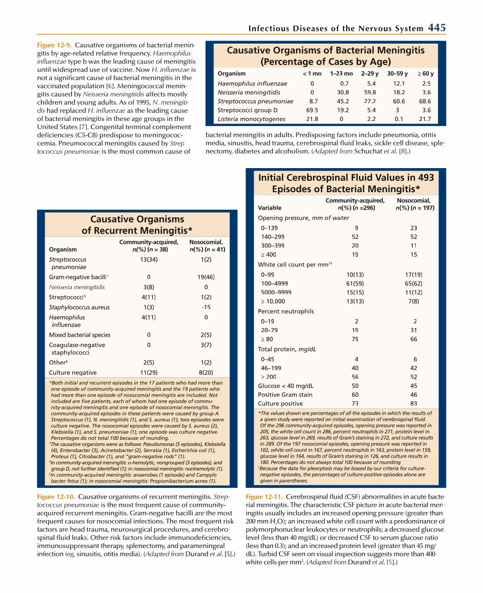
Figure 12-2. Meningococcal rash. Meningococcus is the only bac-terium that frequently causes a rash, which is probably the most important clue to the diagnosis of meningococcal meningitis. It usually begins as a diffuse erythematous maculopapular rash. Asthe rash evolves, petechiae and purpura appear primarily on the trunk and lower extremities. (From Roos et al. [1]; with permission.)

Infectious Diseases of the Nervous System 443
Increased intracranial pressure
Vasogenic edema Cytotoxic edema Interstitial edema
Hydrocephalus
Bacterial replication
Meningeal invasion
Bacteremia
Local invasion
Nasopharyngeal colonization
Cerebral vasculitisand/or infarction
Increased CSFoutflow resistance
Increased BBBpermeability
SAS inflammation
Figure 12-3. Pathogenesis of meningitis.For bacterial meningitis to occur, the hostusually acquires a new organism by colo-nization of the nasopharynx. This maylead to direct seeding of cerebral spinalfl uid (CSF) spaces, but more likely causeslocal spread to the sinuses or the lungs(pneumonia) or bacteremia, which then results in meningeal invasion. BBB—blood-brain barrier; SAS—subarachnoidspace. (Adapted from Roos et al. [1].)
Figure 12-4. Purulent exudate of bacterial meningitis at the base of the brain. The neurologic complications of cranial nerve palsies and increased intracranial pressure are often caused by infl ammation of the base of the brain. The increased intracranial pressure occurs because cerebrospinal fl uidpathways are blocked, resulting in obstructive hydrocephalus. (From Roos and Bonnin [2].)
Figure 12-5. Purulent exudate of leptomeningitis (infl ammation of pia and arachnoid spaces) over the convexities of the cere-bral cortex. This may result in the additional complications ofarterial or venous thrombosis with infarction and hemorrhage, both of which may lead to focal neurologic defects. Initiallyexudates over the convexity appear yellow, but later turn gray asthey become thicker. (From Kaplan [3]; with permission.)

444 Atlas of Clinical Neurology
Figure 12-6. Microscopic examination in bacterial meningitis. A, The meninges are thickened by both polymorphonuclear andmononuclear inflammatory cells. Thickening of blood vesselsmay eventually lead to thrombotic occlusion, cerebral infarcts,and focal neurologic deficits. B, Inflammatory cells in the Vir-
chow-Robin spaces around penetrating parenchymal vessels. The Virchow-Robin spaces are an extension of the subarachnoidspace. Occasionally, inflammation may extend into the perivascu-lar parenchyma, as shown here. (A, fromA Kaplan [3]; with permis-sion; B, from Wilson [4]; with permission.)
BA
Predisposing Factors in 404 SingleEpisodes of Bacterial Meningitis
FactorCommunity-acquired,
% (n = 253)Nosocomial, % (n = 151)
Acute otitis media 19 1
Chronic otitis media 7 0
Sinusitis 12 4
Pneumonia 15 8
Endocarditis 7 1
Head injury*
Recent 5 13
Remote 4 0
Recent neurosurgery* 0 68
Neurosurgical device† 1 32
Altered immune state 19 31
Diabetes mellitus 10 6
Alcoholism 18 5
Cerebrospinal fluid leak 8 13
None of the 13 factors 25 8
*Recent denotes head injury or neurosurgery within 1 month of the onset of meningitis; remote, more than 1 month before the onset of meningitis.
†Neurosurgical devices included ventriculostomy, ventriculoperitoneal or ventriculoatrial shunt, lumbar epidural catheter, lumboperitoneal catheter,and dorsal-column stimulator.
Figure 12-7. Predisposing factors in bacterial meningitis. Pre-disposing factors for community-acquired meningitis are somewhat different than those seen in nosocomial infections.Predisposing factors for nosocomial infections are primarilycaused by openings into the central nervous system. (Adapted from Durand et al. [5].)
Percentage of Causative Organisms in Single Episodes of Meningitis,
1962 Through 1988*
OrganismCommunity-acquired,
% (n = 253)Nosocomial, % (n = 151)
Streptococcus pneumoniae
38 5
Gram-negative bacilli† 4 38
Neisseria meningitidis 14 1
Streptococci‡ 7 9
Enterococcus 0 3
Staphylococcus aureus 5 9
Listeria monocytogenes 11 3
Haemophilus influenzae
4 4
Mixed bacterial species 2 7
Coagulase-negative staphylococci
0 9
Other§ 2 3
Culture negative 13 11
*Percentages do not always total 100 because of rounding.†In community-acquired meningitis, the causative organisms were Escherichiacoli (4 episodes), and species of Klebsiella (3), Enterobacter (1), and Proteus(1); in nosocomial meningitis, E. coli (17), Klebsiella (13), Pseudomonas (6),Acinetobacter (6), Enterobacter (5), Serratia (5), Citrobacter (2), Proteus (1), “coliform” bacteria (1), and “nonenteric gram-negative rods” (1).
‡In community-acquired meningitis, the causative organisms were groupA (4 episodes), group B (1), nonenterococcal group D (3), group D, not further identified (1), other groups (5), and nonhemolytic, nongrouped (3); in nosocomial meningitis, the causative organisms were group B (4), nonenterococcal group D (3), other groups (2), -hemolytic nongrouped (3), and nonhemolytic, nongrouped (1).
§In community-acquired meningitis, the causative organisms were anaerobes (3 episodes) and diphtheroids (1); in nosocomial meningitis, the causative organisms were micrococci (2), Neisseria species (1), propionibacteria (1),and diphtheroids (1).
Figure 12-8. Causative organisms of bacterial meningitis. The causative organisms are somewhat different for community-acquired as opposed to nosocomial meningitis. (Adapted fromDurand et al. [5].)
Infectious Diseases of the Nervous System 445
Causative Organisms of Bacterial Meningitis (Percentage of Cases by Age)
Organism < 1 mo 1–23 mo 2–29 y 30–59 y ≥ 60 y
Haemophilus influenzaeNeisseria meningitidisStreptococcus pneumoniaeStreptococci group DListeria monocytogenes
008.7
69.521.8
0.730.845.219.20
5.459.827.25.42.2
12.118.260.630.1
2.53.6
68.63.6
21.7
Causative Organisms of Recurrent Meningitis*
OrganismCommunity-acquired,
n(%) () n = 38)Nosocomial,n(%) (n = 41)
Streptococcuspneumoniae
13(34) 1(2)
Gram-negative bacilli† 0 19(46)
Neisseria meningitidis 3(8) 0
Streptococci‡ 4(11) 1(2)
Staphylococcus aureus 1(3) -15
Haemophilusinfluenzae
4(11) 0
Mixed bacterial species 0 2(5)
Coagulase-negativestaphylococci
0 3(7)
Other§ 2(5) 1(2)
Culture negative 11(29) 8(20)
*Both initial and recurrent episodes in the 17 patients who had more than one episode of community-acquired meningitis and the 19 patients who had more than one episode of nosocomial meningitis are included. Not included are five patients, each of whom had one episode of commu-nity-acquired meningitis and one episode of nosocomial meningitis. Thecommunity-acquired episodes in these patients were caused by group AStreptococcus (1), N. meningitidis (1), and S. aureus (1); two episodes wereculture negative. The nosocomial episodes were caused by S. aureus (2), Klebsiella (1), and S. pneumoniae (1); one episode was culture negative.Percentages do not total 100 because of rounding.
†The causative organisms were as follows: Pseudomonas (5 episodes), Klebsiella (4), Enterobacter (3), Acinetobacter (2), Serratia (1), Escherichia coli (1),Proteus (1), Citrobacter (1), and “gram-negative rods” (1).
‡In community-acquired meningitis: -hemolytic, nongrouped (3 episodes), and group D, not further identified (1); in nosocomial meningitis: nonhemolytic (1).
§In community-acquired meningitis: anaerobes (1 episode) and Campylo-bacter fetus (1); in nosocomial meningitis: Propionibacterium acnes (1).
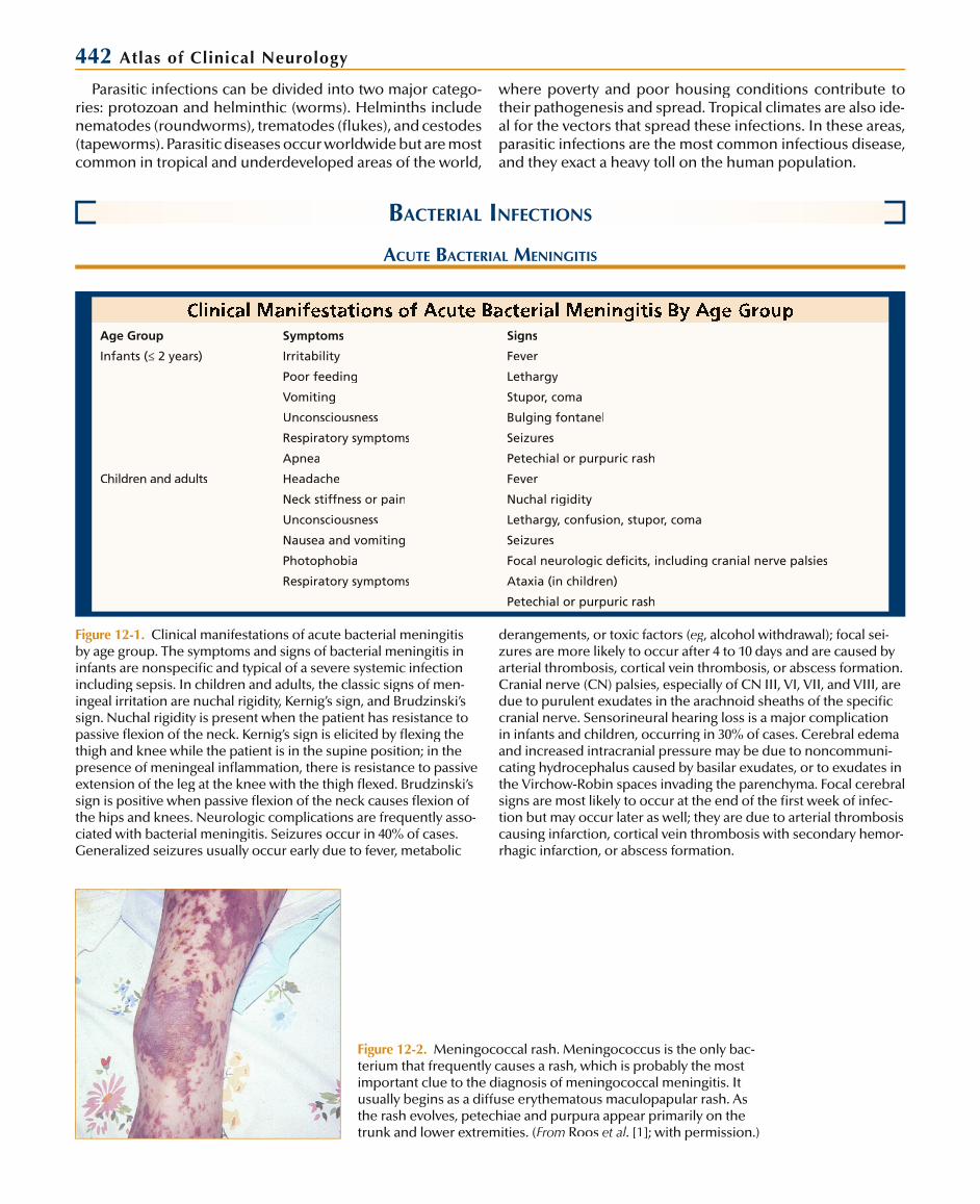
Figure 12-10. Causative organisms of recurrent meningitis. Strep-tococcus pneumoniae is the most frequent cause of community-acquired recurrent meningitis. Gram-negative bacilli are the most frequent causes for nosocomial infections. The most frequent risk factors are head trauma, neurosurgical procedures, and cerebro-spinal fluid leaks. Other risk factors include immunodeficiencies,immunosuppressant therapy, splenectomy, and parameningeal infection (eg, sinusitis, otitis media). (gg Adapted from(( Durand et al. [5].)
Initial Cerebrospinal Fluid Values in 493Episodes of Bacterial Meningitis*
VariableCommunity-acquired,
n(%) (n =296)Nosocomial, n(%) (n = 197)
Opening pressure, mm of water
0–139140–299300–399≥ 400
9522019
23521115
White cell count per mm3†
0–99100–49995000–9999≥ 10,000
10(13)61(59)15(15)13(13)
17(19)65(62)11(12)7(8)
Percent neutrophils
0–1920–79≥ 80
21979
23166
Total protein, mg/dL
0–4546–199≥ 200
Glucose < 40 mg/dLPositive Gram stainCulture positive
44056506073
64252454683
*The values shown are percentages of all the episodes in which the results of a given study were reported on initial examination of cerebrospinal fluid.Of the 296 community-acquired episodes, opening pressure was reported in 205, the white cell count in 286, percent neutrophils in 271, protein level in263, glucose level in 269, results of Gram’s staining in 272, and culture results in 289. Of the 197 nosocomial episodes, opening pressure was reported in 102, white cell count in 167, percent neutrophils in 163, protein level in 159,glucose level in 164, results of Gram’s staining in 126, and culture results in 180. Percentages do not always total 100 because of rounding.
†Because the data for pleocytosis may be biased by our criteria for culture-negative episodes, the percentages of culture-positive episodes alone aregiven in parentheses.
Figure 12-11. Cerebrospinal fluid (CSF) abnormalities in acute bacte-rial meningitis. The characteristic CSF picture in acute bacterial men-ingitis usually includes an increased opening pressure (greater than200 mm H2O); an increased white cell count with a predominance ofpolymorphonuclear leukocytes or neutrophils; a decreased glucose level (less than 40 mg/dL) or decreased CSF to serum glucose ratio (less than 0.3); and an increased protein level (greater than 45 mg/dL). Turbid CSF seen on visual inspection suggests more than 400white cells per mm3. (Adapted from(( Durand et al. [5].)
Figure 12-9. Causative organisms of bacterial menin-gitis by age-related relative frequency. Haemophilusinfluenzae type b was the leading cause of meningitisuntil widespread use of vaccine. Now H. influenzae is not a significant cause of bacterial meningitis in thevaccinated population [6]. Meningococcal menin-gitis caused by Neisseria meningitidis affects mostly children and young adults. As of 1995, N. meningiti-dis had replaced H. influenzae as the leading cause of bacterial meningitis in these age groups in theUnited States [7]. Congenital terminal complementdeficiencies (C5-C8) predispose to meningococ-cemia. Pneumococcal meningitis caused by Strep-tococcus pneumoniae is the most common cause of
bacterial meningitis in adults. Predisposing factors include pneumonia, otitis media, sinusitis, head trauma, cerebrospinal fluid leaks, sickle cell disease, sple-nectomy, diabetes and alcoholism. (Adapted from Schuchat et al. [8].)
446 Atlas of Clinical Neurology
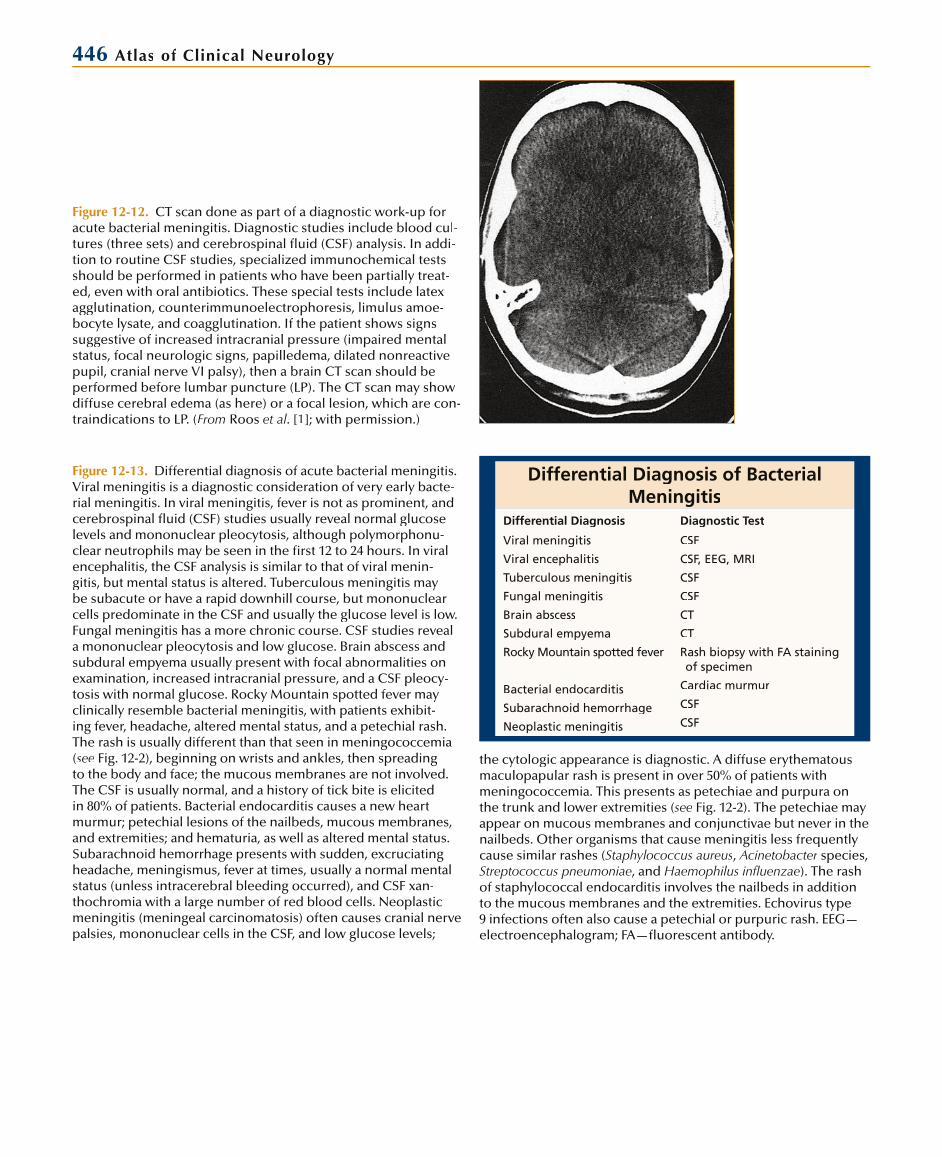
Figure 12-12. CT scan done as part of a diagnostic work-up for acute bacterial meningitis. Diagnostic studies include blood cul-tures (three sets) and cerebrospinal fluid (CSF) analysis. In addi-tion to routine CSF studies, specialized immunochemical testsshould be performed in patients who have been partially treat-ed, even with oral antibiotics. These special tests include latex agglutination, counterimmunoelectrophoresis, limulus amoe-bocyte lysate, and coagglutination. If the patient shows signssuggestive of increased intracranial pressure (impaired mentalstatus, focal neurologic signs, papilledema, dilated nonreactive pupil, cranial nerve VI palsy), then a brain CT scan should beperformed before lumbar puncture (LP). The CT scan may show diffuse cerebral edema (as here) or a focal lesion, which are con-traindications to LP. (From Roos et al. [1]; with permission.)
Differential Diagnosis of Bacterial Meningitis
Differential Diagnosis Diagnostic Test
Viral meningitis
Viral encephalitis
Tuberculous meningitis
Fungal meningitis
Brain abscess
Subdural empyema
Rocky Mountain spotted fever
Bacterial endocarditis
Subarachnoid hemorrhage
Neoplastic meningitis
CSF
CSF, EEG, MRI
CSF
CSF
CT
CT
Rash biopsy with FA stainingof specimen
Cardiac murmur
CSF
CSF
Figure 12-13. Differential diagnosis of acute bacterial meningitis.Viral meningitis is a diagnostic consideration of very early bacte-rial meningitis. In viral meningitis, fever is not as prominent, andcerebrospinal fluid (CSF) studies usually reveal normal glucose levels and mononuclear pleocytosis, although polymorphonu-clear neutrophils may be seen in the first 12 to 24 hours. In viralencephalitis, the CSF analysis is similar to that of viral menin-gitis, but mental status is altered. Tuberculous meningitis may be subacute or have a rapid downhill course, but mononuclearcells predominate in the CSF and usually the glucose level is low.Fungal meningitis has a more chronic course. CSF studies reveala mononuclear pleocytosis and low glucose. Brain abscess andsubdural empyema usually present with focal abnormalities on examination, increased intracranial pressure, and a CSF pleocy-tosis with normal glucose. Rocky Mountain spotted fever mayclinically resemble bacterial meningitis, with patients exhibit-ing fever, headache, altered mental status, and a petechial rash.The rash is usually different than that seen in meningococcemia (see Fig. 12-2), beginning on wrists and ankles, then spreading to the body and face; the mucous membranes are not involved. The CSF is usually normal, and a history of tick bite is elicitedin 80% of patients. Bacterial endocarditis causes a new heartmurmur; petechial lesions of the nailbeds, mucous membranes, and extremities; and hematuria, as well as altered mental status. Subarachnoid hemorrhage presents with sudden, excruciating headache, meningismus, fever at times, usually a normal mental status (unless intracerebral bleeding occurred), and CSF xan-thochromia with a large number of red blood cells. Neoplasticmeningitis (meningeal carcinomatosis) often causes cranial nervepalsies, mononuclear cells in the CSF, and low glucose levels;
the cytologic appearance is diagnostic. A diffuse erythematous maculopapular rash is present in over 50% of patients withmeningococcemia. This presents as petechiae and purpura on the trunk and lower extremities (see Fig. 12-2). The petechiae may appear on mucous membranes and conjunctivae but never in thenailbeds. Other organisms that cause meningitis less frequently cause similar rashes (Staphylococcus aureus, Acinetobacter species, rStreptococcus pneumoniae, and Haemophilus influenzae). The rashof staphylococcal endocarditis involves the nailbeds in additionto the mucous membranes and the extremities. Echovirus type 9 infections often also cause a petechial or purpuric rash. EEG—electroencephalogram; FA—fluorescent antibody.

Infectious Diseases of the Nervous System 447
Differential Diagnosis in Acute Bacterial Meningitis Based onTypical Cerebrospinal Abnormalities
Type of Infection Predominant Cells, per mm3 Glucose, mg/dL Stain for Organisms Diagnosis
Bacterial meningitis PMNs Very low (0–10) Gram stain Culture, CIE, LA, LLA, CoA
Tuberculous meningitis Mononuclear leukocytes Low to very low (10–20) Ziehl-Nelson Culture, PCR assay
Viral meningitis Mononuclear leukocytes Normal Culture, some PCR assays
Fungal meningitis Mononuclear leukocytes Low (15–30) Cryptococcus—India ink stain
Culture; various Ab and Ag tests
Parameningeal (serous) meningitis
Subacute and chronic: mononuclearleukocytes (usual picture); acute: PMNs (uncommon)
Normal CT, MRI; myelogram
Neoplastic meningitis Mononuclear leukocytes Low or normal (30–50) Cytologic studies
Figure 12-14. Differential diagnosis of cerebrospinal fluid(CSF) abnormalities in acute bacterial meningitis. Rarely in bacterial meningitis monocytes may predominate in theCSF (Listeria monocytogenes and especially brucellosis). In viral meningitis, polymorphonuclear leukocytes (PMNs) mayappear in the first 12 to 24 hours, and then there is a shift to mononuclear cells. Most parameningeal foci of infection (eg,
brain abscess, epidural abscess) cause a subacute to chroniccondition of mononuclear cells in the CSF. Subdural empyema,however, may cause an acute parameningeal CSF appear-ance of a large number of PMNs. Ab—antibody; Ag—antigen; CIE—counter immunoelectrophoresis; CoA—coagglutination; LA—latex agglutination; LLA—limulus lysate assay; PCR—poly-merase chain reaction
Empiric Antimicrobial Therapy for Bacterial MeningitisPopulation Antimicrobial Agent
Neonates Ampicillin plus cefotaxime
Infants and children Third-generation cephalosporin (cefotaxime or ceftriaxone) plus vancomycin*
Adults (15–50 y) Third-generation cephalosporin plus vancomycin*
Older adults Third-generation cephalosporin (ceftriaxone or ceftazidime) plus ampicillin plus vancomycin*
Neurosurgical procedure Third-generation cephalosporin (ceftazidime) plus vancomycin*
Immunocompromised state Ampicillin plus third-generation cephalosporin plus vancomycin*
Neutropenic state Cefepime
*Until susceptibility testing available.
Figure 12-15. Empiric antimicrobial thera-py for acute bacterial meningitis. Empiricantibiotic therapy must be given beforethe causative organism can be definitivelyidentified. The choice of the empiric agent depends on the patient’s age and associated conditions (such as neurosur-gical procedure, immunodeficiency), with modifications based on a positive Gramstain. To achieve adequate antibiotic lev-els in the cerebrospinal fluid, antibiotics should be given intravenously. (Adapted from Roos et al. [9].)
Antibiotic Therapy for Acute Bacterial MeningitisOrganism Antibiotics Organism Antibiotics
Streptococcus pneumoniae Enterobacteriaceae Third-generation cephalosporin*
Sensitive to penicillin Penicillin G or ampicillin Pseudomonas aeruginosa Cefepime, meropenem
Relatively resistant to penicillin Third-generation cephalosporin* Streptococcus agalactiae Ampicillin or penicillin G§
Resistant to penicillin Vancomycin plus a third-generation cephalosporin*†‡
Listeria monocytogenes Ampicillin or penicillin G§
Neisseria meningitidis Penicillin G or ampicillin‡ Staphylococcus aureus
Haemophilus influenzae Methicillin sensitive Nafcillin or oxacillin
ß-Lactamase negative Ampicillin‡ Methicillin resistant Vancomycin
ß-Lactamase positive Third-generation cephalosporin*‡ Staphylococcus epidermidis Vancomycin†
*Cefotaxime or ceftriaxone.†Addition of rifampin should be considered.†
‡Chloramphenicol is an option for penicillin-allergic patients.§Addition of an aminoglycoside should be considered.§
Figure 12-16. Specific antibiotic therapy for acute bacterial men-ingitis. Once the causative organism is cultured and sensitivitiesdetermined, therapy should be adjusted to be as narrow as pos-sible. The duration of treatment is somewhat empiric with the
following general recommendations: Neisseria meningitidis, 7 days; Haemophilus influenzae, 7 to 10 days; Streptococcus pneu-moniae, 10 to 14 days; gram-negative bacilli, 21 days. (Adapted from Roos et al. [9].)

448 Atlas of Clinical Neurology
Adjunctive Therapy and SupportiveCare for Bacterial Meningitis
Adjunctive dexamethasone: 0.15 mg/kg every 6 h for 4 days for children; 12 mg every 12 h for adults
Supportive care
Fluid and electrolyte balance: monitor for syndrome of inappropriate antidiuretic hormone
Maintenance of normal systemic blood pressure because ofloss of autoregulation
Intracranial pressure (ICP) monitoring for critically ill patients
Treatment for increased ICP
Elevate head of bed to 30 degrees
Hyperventilation to PaCO2 to 27–30 mm Hg
Hyperosmolar agents: mannitol, glycerol
Glucocorticoids: dexamethasone
Monitor and treat obstructive hydrocephalus
Seizure control: lorazepam, phenytoin, phenobarbital
Figure 12-17. Adjunctive therapy and supportive care of acute bacterial meningitis. Several studies have demonstrated that dexamethasone decreases sensorineural hearing loss and improves neurologic outcome in children older than 2 monthsof age [10] as well as adults [11]. The dexamethasone shouldbe started shortly before giving the first dose of antibioticsbecause the drug inhibits the production of inflammatory cytokines. It appears to be most useful for patients with pneu-mococcal or meningococcal meningitis. PaCO2—arterial carbondioxide pressure.
Mortality Rates of Treated Cases of Bacterial MeningitisCase Fatality Rate, %
Organism Episodes, n Meningitis Related, n Total, n
Streptococcus pneumoniae 120 25 28
Gram-negative bacilli 86 23 36
Neisseria meningitidis 40 10 10
Streptococci 36 17 25
Enterococcus 4 25 50
Staphylococcus aureus 36 28 39
Listeria monocytogenes 34 2111 32
Haemophilus influenzae 19 39 11
Mixed bacterial species 18 9 44
Coagulase-negative staphylococci 16 0 0
Other* 12 0 8
Culture negative 72 7 10
All causes 493 19 25
1962–1970 172 21 24
1971–1979 186 18 26
1980–1988 135 17 24
*Other organisms were as follows: anaerobes (4 episodes), propionibacteria (2), diphtheroids (2), micrococci (2), Neisseria species (1), and Campylobacter fetus (1).
Figure 12-18. Mortality rates of treated cases of acute bacterial meningitis. The mortality rate for treated cases of acute bacterial meningitis remains significantbecause of the numerous potential com-plications (increased intracranial pres-sure, hydrocephalus, focal neurologic deficits, seizures, brain abscess, subdural empyema, sepsis). Factors associated withsignificantly higher overall mortality rates were age of 60 years or older, obtunda-tion on admission, and seizures occurringwithin 24 hours of admission. (Adapted from Durand et al. [5].)

Infectious Diseases of the Nervous System 449
Vaccination for Acute Bacterial MeningitisHib Vaccine Recommendations Indicators for Meningococcal and Pneumococcal Vaccines
Vaccinate all infants at 2, 4, and 6 months of age
Unvaccinated infants 7–11 months of age receive two doses 2 months apart
Unvaccinated children 12–14 months of age receive onedose plus booster at 15 months
Unvaccinated children 15–60 months of age receive one dose
Children older than 5 years are vaccinated if increased disease risk (asplenia, sickle cell disease, immunodefi-ciency, or immunosuppression)
Children with history of invasive Hib disease or vaccinated at greater than 2 years with polyribosylribitol phosphate vaccinedo not need revaccination
Meningococcal quadrivalent vaccine
Vaccination during epidemic outbreaks due to represented serogroup
Travel to hyperepidemic areas
High-risk immunodeficient groups
Terminal coagulant deficiency
Properdin deficiency
Pneumococcal vaccine
Vaccinate all infants at 2, 4, and 6 months of age
Elderly over 65 years of age
Those with chronic cardiorespiratory conditions
Chronic alcoholics
Those with asplenic states, multiple myelemia, Wiskott-Aldrich syndrome
HIV infection
Those with diabetes mellitus or significant hepatic or renal disease
Figure 12-19. Vaccination for acute bacterial meningitis. Vaccinesare now available for Haemophilus influenzae type b (Hib), Neisseria meningitides, and Streptococcus pneumoniae. The routine use of Hib vaccine has greatly decreased the inci-dence of meningitis due to this agent [12]. Meningococcal andpneumococcal vaccines are used for specific circumstancesor “at risk” populations. Meningococcal vaccine is available for serogroups A, C, Y, and W135 (quadrivalent vaccine), but
the response is poor in young children, and there is no vaccine for serogroup B, which is responsible for over 50%of infections in the United States [13]. Pneumococcal vaccine is indicated for all infants in addition to high-risk groups older than 2 years of age [12]. It is recommended that close household, day care center, and medical personnel contactsfor meningococcal and H. influenzae meningitis be treatedprophylactically with rifampin.
Differential Diagnosis of Chronic MeningitisInfectious Causes Noninfectious Causes
Bacterial infections
Tuberculosis
Spirochetal (syphilis, Lyme disease, Leptospira infection)
Agents that form sinus tracts(Actinomyces, Arachnia, Nocardia)
Brucellosis
Listeria monocytogenes (rare cause)s
Nocardiosis
Fungal infections
Common (Candida, Coccidioides, Cryptococcus, Histoplasma)
Uncommon (Aspergillus, Blastomyces, Dematiaceous paracoccidioides,Pseudallescheria, Sporothrix,Mucormycetes)
Parasitic diseases
Cysticercosis
Granulomatous amebic meningoencephalitis(acanthamoeba)
Eosinophilic meningitis (angiostrongylus)
Toxoplasmosis
Coenurus cerebralis
Viral infections
Retrovirus (HIV-1, HTLV-1)
Enterovirus (in hypogammaglobulinemia)
Parameningeal infections (epidural abscess,subdural empyema, brain abscess)
Neoplasm
Sarcoidosis
Vasculitis
Primary central nervous system angiitis
Systemic: giant cell arteritis, systemic lupus erythematosus, Sjögren’s syndrome, rheuma-toid arthritis, lymphamatoid granulomatosis,polyarteritis nodosa, Wegener’s granuloma-tosis)
Behçet’s disease
Chemical meningitis
Endogenous
Exogenous
Chronic benign lymphocytic meningitis
Idiopathic hypertrophic pachymeningitis
Vogt-Koyanagi-Harada disease
CHRONIC BACTERIAL MENINGITIS
Figure 12-20. Differential diagnosis of chronic meningitis.Chronic meningitis accounts for about 10% of all meningitis cases. Clinical features include a subacute to chronic onset of various combinations of fever, headache, and stiff neck, often with signs of encephalitis (parenchymal involvement), mentalstatus changes, seizures, and focal deficits. Therefore, chronic meningitis is often referred to as a meningoencephalitis. Thecerebrospinal fluid (CSF) is abnormal with a pleocytosis (usu-ally mononuclear), elevated protein levels, and a moderatelydecreased glucose level (see Fig. 12-14 for comparison). Some
require that these manifestations persist for 4 weeks as a crite-rion for the diagnosis of chronic meningitis; however, the differ-ential diagnosis is usually considered before this period on thebasis of the suggestive CSF profile. The differential diagnosis is quite extensive and includes both infectious and noninfec-tious causes. The most common infectious causes of chronicmeningitis are tuberculosis, cryptococcosis, and toxoplasmosis; the common noninfectious causes are neoplasms and vasculitis. HTLV—human T-cell lymphotropic virus. (Adapted from Roos and Bonnin [2].)
450 Atlas of Clinical Neurology
Historical and Clinical Clues to Diagnosis of Chronic MeningitisHistory Examination
Exposure history Dermatologic lesions
To patient with tuberculosis (TB) Erythema chronicum migrans—Lyme disease
Ingestion of unpasteurized milk or dairy products (brucellosis) Depigmentation of skin (vitiligo) and hair (poliosis)—VKH
To farm animals or swimming in farm ponds (leptospirosis) Macular hyperpigmented lesions of trunk, palms, and soles—secondary syphilisTo deer ticks (Lyme disease)
Swimming in warm fresh water ponds (acanthamebiasis) Subcutaneous nodules, abscesses, draining sinuses—fungal aspergillosisSexual transmission (syphilis, retroviruses)
Intravenous drug use (retroviruses) Ophthalmologic disease
Travel and geographic history Uveitis—sarcoidosis, Behçet’s syndrome, VKH
Mexico and Latin America (cysticercosis) Choroidal tubercles—TB, sarcoidosis
Southeast Asia and Pacific (angiostrongylosis) Organ disease
US Northeast, North Central (Lyme disease) Primary disease—sarcoidosis, TB, histoplasmosis,aspergillosis, blastomycosisUS Midwest (histoplasmosis, blastomycosis)
US Southwest (coccidioidomycosis) Enlarged liver—potential biopsy sites for TB, histoplasmosis
US Southeast (acanthamebiasis) Muscle nodules—biopsy site for sarcoidosis, vasculitis
History of extraneural or systemic disease Adenopathy—biopsy site for TB, systemic fungi
Pulmonary disease (TB, histoplasmosis, sarcoidosis) Neurologic features
Polyarthritis (Lyme disease, Behçet’s syndrome, systemic lupuserythematosus, rheumatoid arthritis)
Cranial nerve involvement—sarcoidosis, Lyme disease,TB, fungal meningitis
Uveitis (sarcoidosis, Behçet’s syndrome, Vogt-Koyanagi-Harada [VKH, leptospirosis)
Focal lesions
Abscess—TB, fungal meningitis, toxoplasmosis
Skin lesions (syphilis, Lyme disease, VKH) Strokes—TB, aspergillosis, mucormycosis, vasculitis
Prior diagnosed disease (diabetes, malignancy, TB, syphilis, AIDS) Hydrocephalus—TB, fungal meningitis, especially cryptococcosis, cystinosisHistory of immunodeficiency
Congenital Peripheral neuropathy—Lyme disease, sarcoidosis, brucellosis, vasculitis Agammaglobulinemia (enteroviruses)
Acquired Multiple levels—carcinomatous meningitis
AIDS (toxoplasmosis, cryptococcosis, syphilis, TB, etc.)
Organ transplant immunosuppression (toxoplasmosis, listeriosis,candidiasis, nocardiosis, aspergillosis)
Chronic steroid use (TB, cryptococcosis, candidiasis)
Malignancy and chemotherapy (TB, cryptococcosis, listeriosis)
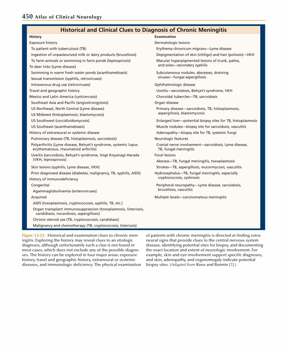
Figure 12-21. Historical and examination clues to chronic men-ingitis. Exploring the history may reveal clues to an etiologic diagnosis, although unfortunately such a clue is not found inmost cases, which does not exclude any of the possible diagno-ses. The history can be explored in four major areas: exposurehistory, travel and geographic history, extraneural or systemicdiseases, and immunologic deficiency. The physical examination
of patients with chronic meningitis is directed at finding extra-neural signs that provide clues to the central nervous systemdisease, identifying potential sites for biopsy, and documentingthe exact location and extent of neurologic involvement. Forexample, skin and eye involvement support specific diagnoses,and skin, adenopathy, and organomegaly indicate potentialbiopsy sites. (Adapted from Roos and Bonnin [2].)
Infectious Diseases of the Nervous System 451
Laboratory Tests in Chronic MeningitisBlood tests: CBC, serum chemistry studies, ANA, ANCA, ESR, VDRL, ACE
Cultures of draining skin lesions, sinuses, nodes, blood, sputum, urine, CSF
Multiple sites
Multiple times (≥ 3)
Skin testing
Intermediate PPD
Anergy battery
Antibody studies
Paired serum and CSF samples
CSF studies (× 3 if needed)
Cells, protein, glucose, antigen assays (fungus only), antibody assays, culture, cytologic analyses (Gram stain, India inkpreparations, acid-fast stains), PCR assays
Imaging
Chest radiography
Contrast-enhanced MRI preferred over CT
Angiography
Biopsy
Extraneural
Meningeal/cerebral
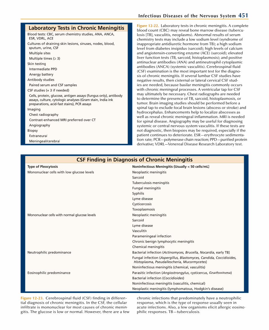
Figure 12-22. Laboratory tests in chronic meningitis. A completeblood count (CBC) may reveal bone marrow disease (tubercu-losis [TB], vasculitis, neoplasms). Abnormal results of serum chemistry tests may include a low sodium level (syndrome of inappropriate antidiuretic hormone from TB); a high sodiumlevel from diabetes insipidus (sarcoid); high levels of calcium and angiotensin-converting enzyme (ACE) (sarcoid); elevatedliver function tests (TB, sarcoid, histoplasmosis); and positiveantinuclear antibodies (ANA) and antineutrophil cytoplasmic antibodies (ANCA) (systemic vasculitis). Cerebrospinal fluid (CSF) examination is the most important test for the diagno-sis of chronic meningitis. If several lumbar CSF studies havenegative results, then cisternal or lateral cervical CSF stud-ies are needed, because basilar meningitis commonly occurs with chronic meningeal processes. A ventricular tap for CSFmay ultimately be necessary. Chest radiographs are needed to determine the presence of TB, sarcoid, histoplasmosis, or tumor. Brain imaging studies should be performed before a spinal tap to exclude focal brain lesions (abscess or stroke) and hydrocephalus. Enhancements help to localize abscesses as well as reveal chronic meningeal inflammation. MRI is neededfor spinal disease. Angiography may be useful for diagnosing systemic or central nervous system vasculitis. If these tests are not diagnostic, then biopsies may be required, especially if thepatient continues to deteriorate. ESR—erythrocyte sedimenta-tion rate; PCR—polymerase chain reaction; PPD—purified protein derivative; VDRL—Venereal Disease Research Laboratory test.
CSF Finding in Diagnosis of Chronic MeningitisType of Pleocytosis Noninfectious Meningitis (Usually < 50 cells/mL)
Mononuclear cells with low glucose levels Neoplastic meningitis
Sarcoid
Tuberculosis meningitis
Fungal meningitis
Syphilis
Lyme disease
Cysticercosis
Toxoplasmosis
Mononuclear cells with normal glucose levels Neoplastic meningitis
Sarcoid
Lyme disease
Vasculitis
Parameningeal infection
Chronic benign lymphocytic meningitis
Chemical meningitis
Neutrophilic predominance Bacterial infection (Actinomyces, Brucella, Nocardia(( , early TB)
Fungal infection (Aspergillus, Blastomyces, Candida, Coccidioides,((Histoplasma, Pseudallescheria, Mucormycetes)
Noninfectious meningitis (chemical, vasculitis)
Eosinophilic predominance Parasitic infection (Angiostrongylus(( , cysticercus, Gnathostoma)
Bacterial infection (Coccidioides)
Noninfectious meningitis (vasculitis, chemical)
Neoplastic meningitis (lymphomatous, Hodgkin’s disease)
Figure 12-23. Cerebrospinal fluid (CSF) finding in differen-tial diagnosis of chronic meningitis. In the CSF, the cellular infiltrate is mononuclear for most causes of chronic menin-gitis. The glucose is low or normal. However, there are a few
chronic infections that predominately have a neutrophilicresponse, which is the type of response usually seen inacute infections. Also, a few organisms elicit allergic eosino-philic responses. TB—tuberculosis.

452 Atlas of Clinical Neurology
CENTRAL NERVOUS SYSTEM TUBERCULOSIS
Clinical Staging of Tuberculous Meningitis
Stage I (early) Nonspecific symptoms and signs
No clouding of consciousness
No neurologic deficits
Stage II (intermediate) Lethargy or alteration in behavior
Meningeal irritation
Minor neurologic deficits
(such as cranial nerve palsies)
Stage III (advanced) Abnormal movements
Convulsions
Stupor or coma
Severe neurologic deficits (pareses)
Figure 12-24. Clinical staging of tuberculous meningitis.Because the clinical picture of meningitis due to tuberculosisvaries, especially by age at onset, a clinical staging system was introduced 50 years ago. Stage I patients have only a non-specific prodrome without neurologic manifestations, whichincludes headache, malaise, and low-grade fever. This stage usually lasts up to 2 weeks. Stage II is often referred to as themeningitic phase, as symptoms and signs of meningitis occur along with cranial nerve palsies. Behavior alteration and leth-argy may be seen. This stage progresses over days to weeksto stage III (advanced), in which seizures, stupor or coma, focal neurologic signs, and decorticate or decerebrate postur-ing occur. Without treatment, the course proceeds steadilydownhill to death in 6 to 12 weeks. Disease progression tends to occur faster in adults. Rarely, other forms of tuberculous meningitis may be seen. It can present acutely, similar to acutebacterial meningitis, with a more rapid course. Infrequently, ithas a more chronic course, with slowly developing hydroceph-
Symptoms and Signs of TuberculousMeningitis at Presentation
Manifestations Children, % Adults, %
Symptoms
Headache 20–50 50–60
Nausea/vomiting 50–75 8–40
Apathy/behavioral changes 30–70 30–70
Seizures 10–20 0–13
Prior history of tuberculosis 55 8–12
Signs
Fever 50–100 60–100
Meningismus 70–100 60–70
Cranial nerve palsy 15–30 15–40
Coma/altered consciousness 30–45 20–30
Purified protein derivative-positive 85–90 40–65
Figure 12-25. Symptoms and signs of tuberculous meningitis at presentation. The clinical presentation in children is some-what different than that seen in adults. Nausea and vomiting as well as behavioral changes are more common in children, whereas headache is clearly more common in adults. Childrenalso frequently complain of abdominal pain and constipation.In both groups, seizures increase in frequency with diseaseprogression. On examination, fever and meningismus are the most common signs in both age groups, although the frequency varies greatly. Cranial nerve palsies are present in some patientsat presentation but eventually occur in about half of all cases. The sixth cranial nerve is involved most commonly, followedby the third, fourth, and seventh cranial nerves. Examination of the optic fundus may reveal tubercles in a small percentage of patients. Funduscopic examination may also reveal papilledema due to increased intracranial pressure from hydrocephalus.Hydrocephalus correlates well with the duration of disease and eventually occurs in most cases.
Additional manifestations of central nervous system tuber-culosis include the following discussed below. Caseating granulomas of epithelioid cells and macrophages contain-ing mycobacteria may occur in the brain as single or mul-tiple focal lesions. Infrequently, caseating necrosis occurs, forming a tuberculous (cold) abscess. Both lesions oftenoccur without meningitis. Most often, the initial presenta-tion of tuberculoma and abscess is similar to that of a braintumor, with headaches from increased intracranial pressure, seizures, focal deficits, and altered mental status. Less fre-quently, seizure or focal deficits may be the first manifesta-tions. The most common form of tuberculosis of the spineis epidural compression of the thoracic cord from vertebral and disc destruction by caseating granulomas (tuberculousosteomyelitis). Less frequently the lumbar or cervical spinemay be affected. The clinical manifestations are those of chronic epidural cord compression with back pain increased
alus, similar to fungal meningitis. A stroke syndrome has alsobeen associated with tuberculous meningitis. (Adapted fromthe British Medical Research Council [14].)
with weight bearing; percussion tenderness over the spine;a spastic paraparesis, often with a sensory level; and bowel and bladder dysfunction. Localized and severe percussionspine tenderness with painful limitation of spinal motility isreferred to as a “spinal gibbus.” Tuberculous meningomyelitis is the rare occurrence of infection of the spinal leptomenin-ges without spine involvement; it has also been referred toas spinal meningitis, spinal arachnoiditis, and spinal radiculo-myelitis. Thick exudates and tubercles encase the nerve roots and spinal cord. This process presents as subacute to chronic radiculomyelitis or as cauda equina syndrome. It appearsmainly in highly endemic areas but has been reported inAIDS patients in the United States. Intramedullary tuberculo-mas are rare and have clinical presentations similar to other spinal cord tumors. (Adapted from Zuger and Lowy [15].)

Infectious Diseases of the Nervous System 453
Figure 12-26. Tuberculous basilar meningitis. The tubercle bacil-lus enters the human host through inhalation. Airborne dropletsreach the alveoli and multiply there or in alveolar and circulating macrophages. During this 2- to 4-week stage of infection, hema-togenous dissemination occurs, and delayed secondary hema-togenous dissemination may also occur. During dissemination, tubercles form in multiple organs, including the brain. Eventuallytubercles rupture into the subarachnoid space or ventricular system to cause meningitis.
The initial pathologic event after tubercle rupture is the forma-tion of a thick exudate in the subarachnoid space. This exudate initially begins at the base of the brain, where it is especially thick, and envelops cranial nerves, causing cranial nerve palsies. (From Wilson [4]; with permission.)
Figure 12-27. Tuberculous hydrocephalus. With the thick basilarexudate of tuberculous meningitis, often the foramina of Lusch-ka and Magendie become obstructed. Obstruction may also occur at the level of the aqueduct, causing noncommunicatinghydrocephalus, increased intracranial pressure, and papillede-ma. Communicating hydrocephalus caused by blockage of the basilar cisterns, interfering with the resorption of cerebrospinal fluid, may also occur. Either type of hydrocephalus may result in brain atrophy. (From Wilson [4]; with permission.)
Prevalence of CNS Tuberculosis in AIDS Patients
Location YearCases of Active TB/
Cases of AIDSCases of TB with
CNS Disease
Florida 1984 27/45 (60%) 2 of 27 (7%)
New Jersey 1986 52/420 (12%) 10 of 52 (19%)
New York City 1986 24/280 (9%) 1 of 24 (4%)
San Francisco 1987 35/1705 (2%) 2 of 35 (6%)
Barcelona, Spain 1988 Not available 5 of 65 (8%)
Figure 12-28. Prevalence of tuberculosis (TB) of the central nervous system (CNS) in AIDS patients. In the first half ofthe 20th century, autopsy studies revealed that 5% to 10% of patients with TB had CNS involvement. TB in AIDS patientsis thought to occur because of reactivation; most cases are pulmonary, but the incidence of extrapulmonary disease is
much greater than that of the general population. Rates of tuberculin purified protein derivative reactivity are lower thanthose in the general population, ranging from 33% to 50% as compared to 50% to 90%. The incidence of CNS involvementis similar to that of the general population. (Adapted fromZuger and Lowy [15].)
454 Atlas of Clinical Neurology
Laboratory Diagnosis of Tuberculous MeningitisTest Positivity, % Problems
CSF lymphocytic pleocytosiswith decreased glucose
~75 Nonspecific
AFB CSF staining ~32 Low sensitivity
Microscopic time dependent
CSF culture ~50 Low sensitivity
Too long for early diagnosis
CSF adenosine deaminase ~75 Low specificity
Not always available
PCR 50–98 Depends on sequence amplified
Tuberculin skin test Adults 40–65 Low sensitivity in adults
Children 85–90
Chest radiograph Adults 25–50 Low sensitivity
Children 15–20
CSF imaging (CT or MRI) 75–85 Nonspecific
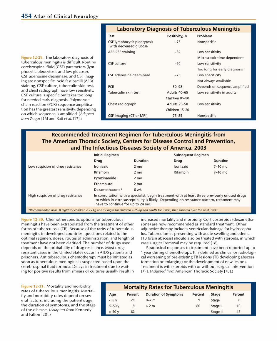
Figure 12-29. The laboratory diagnosis oftuberculous meningitis is difficult. Routinecerebrospinal fluid (CSF) parameters (lym-phocytic pleocytosis and low glucose), CSF adenosine deaminase, and CSF imag-ing are nonspecific. Acid fast bacilli (AFB)staining, CSF culture, tuberculin skin test, and chest radiograph have low sensitivity.CSF culture is specific but takes too long for needed early diagnosis. Polymerasechain reaction (PCR) sequence amplifica-tion has the greatest sensitivity, depending on which sequence is amplified. (Adapted from Zuger [16] and Rafi et al. [17].)
Recommended Treatment Regimen for Tuberculous Meningitis from The American Thoracic Society, Centers for Disease Control and Prevention,
and The Infectious Diseases Society of America, 2003Initial Regimen Subsequent Regimen
Drug Duration Drug Duration
Low suspicion of drug resistance Isoniazid 2 mo Isoniazid 7–10 mo
Rifampin 2 mo Rifampin 7–10 mo
Pyrazinamide 2 mo
Ethambutol 2 mo
Dexamethasone* 6 wk
High suspicion of drug resistance In consultation with a specialist, begin treatment with at least three previously unused drugsto which in vitro susceptibility is likely. Depending on resistance pattern, treatment may have to continue for up to 24 mo.
*Recommended dose: 8 mg/d for children < 25 kg and 12 mg/d for children > 25 kg and adults for 3 wks, then tapered over the next 3 wks.ee
Figure 12-30. Chemotherapeutic options for tuberculousmeningitis have been extrapolated from the treatment of other forms of tuberculosis (TB). Because of the rarity of tuberculousmeningitis in developed countries, questions related to the optimal regimen, doses, routes of administration, and length oftreatment have not been clarified. The number of drugs useddepends on the probability of drug resistance. Most drug-resistant cases in the United States occur in AIDS patients and prisoners. Antituberculous chemotherapy must be initiated assoon as tuberculous meningitis is suspected based upon thecerebrospinal fluid formula. Delays in treatment due to wait-ing for positive results from smears or cultures usually result in
increased mortality and morbidity. Corticosteroids (dexametha-sone) are now recommended as standard treatment. Other adjunctive therapy includes ventricular drainage for hydrocepha-lus. Tuberculomas presenting with acute swelling and edema(TB brain abscess) should also be treated with steroids, in whichcase surgical removal may be required [18].
Paradoxical responses to treatment have been reported up to1 year during chemotherapy. It is defined as clinical or radiologi-cal worsening of pre-existing TB lesions (TB developing abscess formation or enlarging) or the development of new lesions. Treatment is with steroids with or without surgical intervention[19]. (Adapted from American Thoracic Society [18].)
Mortality Rates for Tuberculous MeningitisAge Percent Duration of Symptoms Percent Stage Percent
< 5 y 20 0–2 m 9 Stage I 0
5–50 y 8 > 2 m 80 Stage II 10
> 50 y 60 Stage III 45
Figure 12-31. Mortality and morbidityrates of tuberculous meningitis. Mortal-ity and morbidity rates depend on sev-eral factors, including the patient’s age,the duration of symptoms, and the stageof the disease. (Adapted from Kennedy and Fallon [20].)
Infectious Diseases of the Nervous System 455
INTRACRANIAL EPIDURAL ABSCESS
Clinical Manifestations and Pathogenesis ofIntracranial Epidural Abscess
Clinical Manifestations Sources of Infection
Early Extension of contiguous infections
Fever Paranasal sinusitis
Symptoms related to the source of infection Orbital cellulitis
Sinusitis, otitis, etc. Otitis
Headache Mastoiditis
Localized skull tenderness from osteomyelitis Cranial defects
Scalp and face cellulitis from osteomyelitis Skull fracture
Cranial nerve palsies—rare Neurosurgic procedures
Late
Seizures
Focal neurologic deficits
Meningismus
Nausea and vomiting from increase ICP
Papilledema from increased ICP
Altered mental status from increased ICP
Cranial nerve palsies—rare
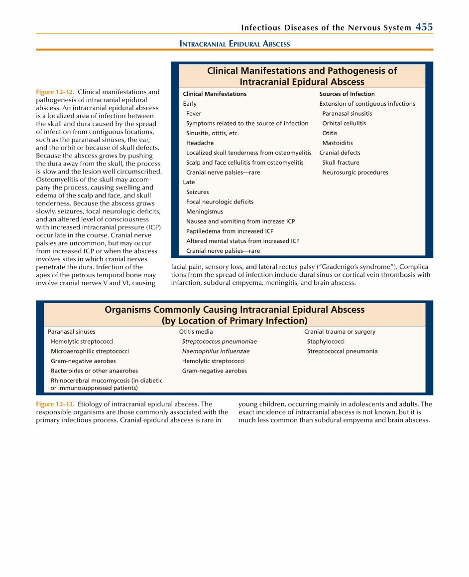
Figure 12-32. Clinical manifestations andpathogenesis of intracranial epidural abscess. An intracranial epidural abscessis a localized area of infection betweenthe skull and dura caused by the spread of infection from contiguous locations, such as the paranasal sinuses, the ear,and the orbit or because of skull defects.Because the abscess grows by pushing the dura away from the skull, the processis slow and the lesion well circumscribed. Osteomyelitis of the skull may accom-pany the process, causing swelling and edema of the scalp and face, and skulltenderness. Because the abscess growsslowly, seizures, focal neurologic deficits,and an altered level of consciousnesswith increased intracranial pressure (ICP)occur late in the course. Cranial nerve palsies are uncommon, but may occur from increased ICP or when the abscess involves sites in which cranial nervespenetrate the dura. Infection of the apex of the petrous temporal bone may involve cranial nerves V and VI, causing
Organisms Commonly Causing Intracranial Epidural Abscess(by Location of Primary Infection)
Paranasal sinuses Otitis media Cranial trauma or surgery
Hemolytic streptococci Streptococcus pneumoniae Staphylococci
Microaerophilic streptococci Haemophilus influenzae Streptococcal pneumonia
Gram-negative aerobes Hemolytic streptococci
Bacteroides or other anaerobes Gram-negative aerobes
Rhinocerebral mucormycosis (in diabetic or immunosuppressed patients)
Figure 12-33. Etiology of intracranial epidural abscess. Theresponsible organisms are those commonly associated with theprimary infectious process. Cranial epidural abscess is rare in
young children, occurring mainly in adolescents and adults. The exact incidence of intracranial abscess is not known, but it ismuch less common than subdural empyema and brain abscess.
facial pain, sensory loss, and lateral rectus palsy (“Gradenigo’s syndrome”). Complica-tions from the spread of infection include dural sinus or cortical vein thrombosis with infarction, subdural empyema, meningitis, and brain abscess.
456 Atlas of Clinical Neurology
Treatment of IntracranialEpidural Abscess
Empiric antibiotic therapy
Paranasal sinus or otitis source of infection
Ceftriaxone plus metronidazole
Cranial trauma or surgery
Vancomycin plus ceftazidime (or plus meropenem)
Surgical drainage and decompression
Gram stain and culture for bacteria and fungi
Craniectomy for osteomyelitis
Dural debridement; excision and grafting not usually required
Closure of any communication between sinus cavity and epidural space to prevent reaccumulation
Institute specific antibiotic therapy based on culture results
Figure 12-35. Treatment of intracranial epidural abscess.Therapy for intracranial epidural abscess consists of antibiotic therapy combined with neurosurgical drainage and decompres-sion. Antibiotic therapy should be continued for 4 to 6 weeksand for 8 weeks with associated osteomyelitis. The prognosis for these epidural infections is excellent, with no mortality in recent series, probably because the process is usually subacuteto chronic and CT and MRI are excellent diagnostic tools.
SPINAL EPIDURAL ABSCESS
Clinical Stages of Progression of Spinal Epidural AbscessStage I Severe localized back pain
Exquisite spinal percussion tenderness
Paraspinal muscle spasm
Stage II Nerve root irritation with radiating pain and paresthesia (radiculopathy)
Focal weakness or reflex changes
Stage III Spinal cord compression, with
Progressive weakness
Sensory loss
Bowel and bladder dysfunction
Stage IV Complete paralysis
Sensation is impaired below sensory level at or near the cord segmentof the lesion
Figure 12-36. Clinical manifestations of spinal epidural abscess by stages of pro-gression. The epidural space in the spinal cord is a true space, unlike the potential epidural intracranial space. In the spinalcord, the dura and arachnoid are closelyapproximated, so that the subdural spaceis only a potential space, and spinal sub-dural empyema or abscess is rare; spinal epidural abscess is much more common. Spinal epidural abscess is an emergency because spinal cord compression andparaplegia are possible rapid complica-tions that can occur over hours. It can beacute (symptoms are present less than 2weeks) or chronic (symptoms are present for more than 2 weeks); the acute form is more common. Four stages of progres-sion of spinal epidural abscess have beenrecognized. The acute form presents as an acute cord compression. Progression
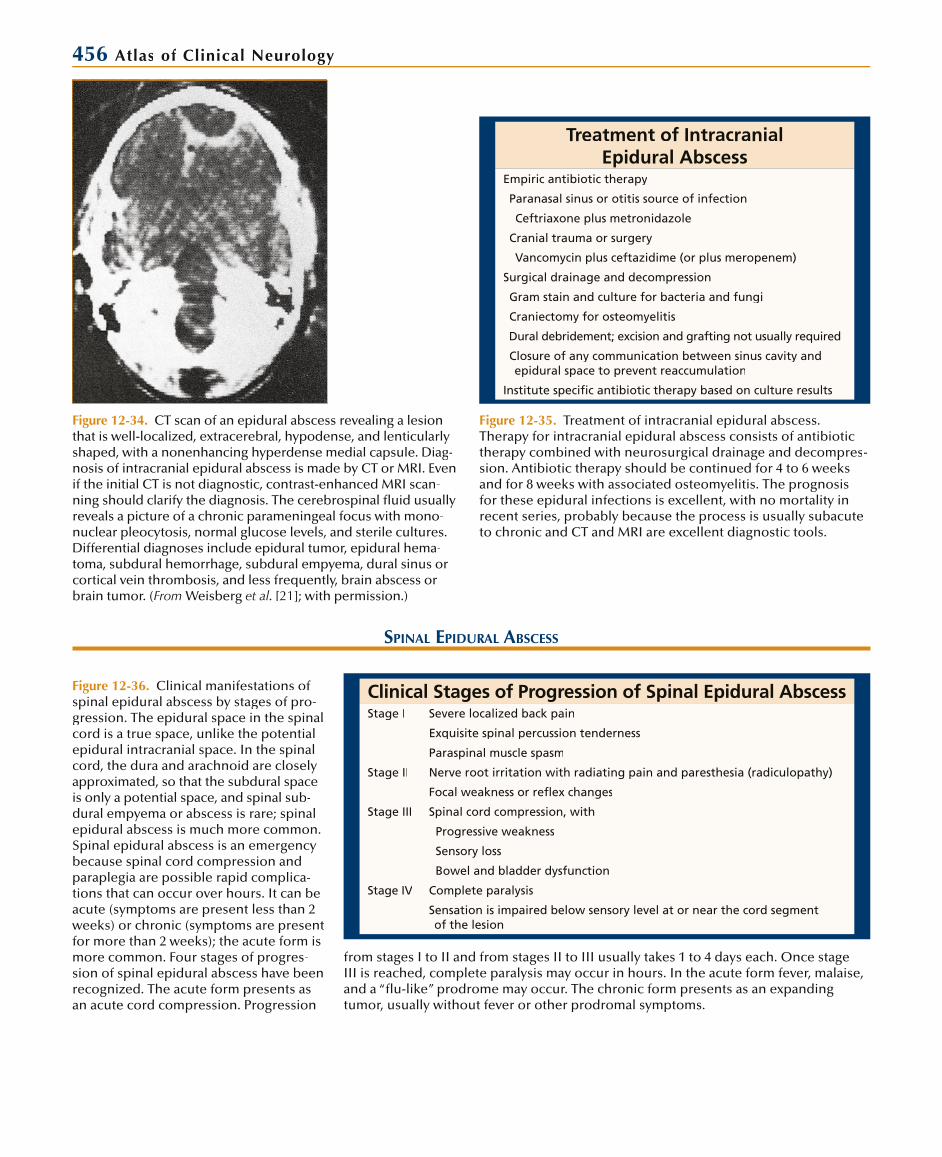
Figure 12-34. CT scan of an epidural abscess revealing a lesion that is well-localized, extracerebral, hypodense, and lenticularly shaped, with a nonenhancing hyperdense medial capsule. Diag-nosis of intracranial epidural abscess is made by CT or MRI. Evenif the initial CT is not diagnostic, contrast-enhanced MRI scan-ning should clarify the diagnosis. The cerebrospinal fluid usually reveals a picture of a chronic parameningeal focus with mono-nuclear pleocytosis, normal glucose levels, and sterile cultures.Differential diagnoses include epidural tumor, epidural hema-toma, subdural hemorrhage, subdural empyema, dural sinus orcortical vein thrombosis, and less frequently, brain abscess orbrain tumor. (From Weisberg et al. [21]; with permission.)
from stages I to II and from stages II to III usually takes 1 to 4 days each. Once stageIII is reached, complete paralysis may occur in hours. In the acute form fever, malaise,and a “flu-like” prodrome may occur. The chronic form presents as an expandingtumor, usually without fever or other prodromal symptoms.
Infectious Diseases of the Nervous System 457
Pathogenesis and Pathophysiology of SpinalEpidural Abscess: Source of
Infection of Spinal Epidural AbscessSource Percentage of Patients*
Hematogenous seeding 43
Skin and soft tissues 20
Abdomen and pelvis 7
Respiratory system 6
Intravenous drug use 5
Urinary tract 2
Cardiac system 2
Dental infection 1
Contiguous location 27
Vertebral osteomyelitis 8
Retroperitoneal or retromediastinal infection
7
Perinephric or psoas abscess 8
Decubitis ulcers 4
Surgery and trauma 5
No source identified 25
Total 100
*Estimates compiled from various series.
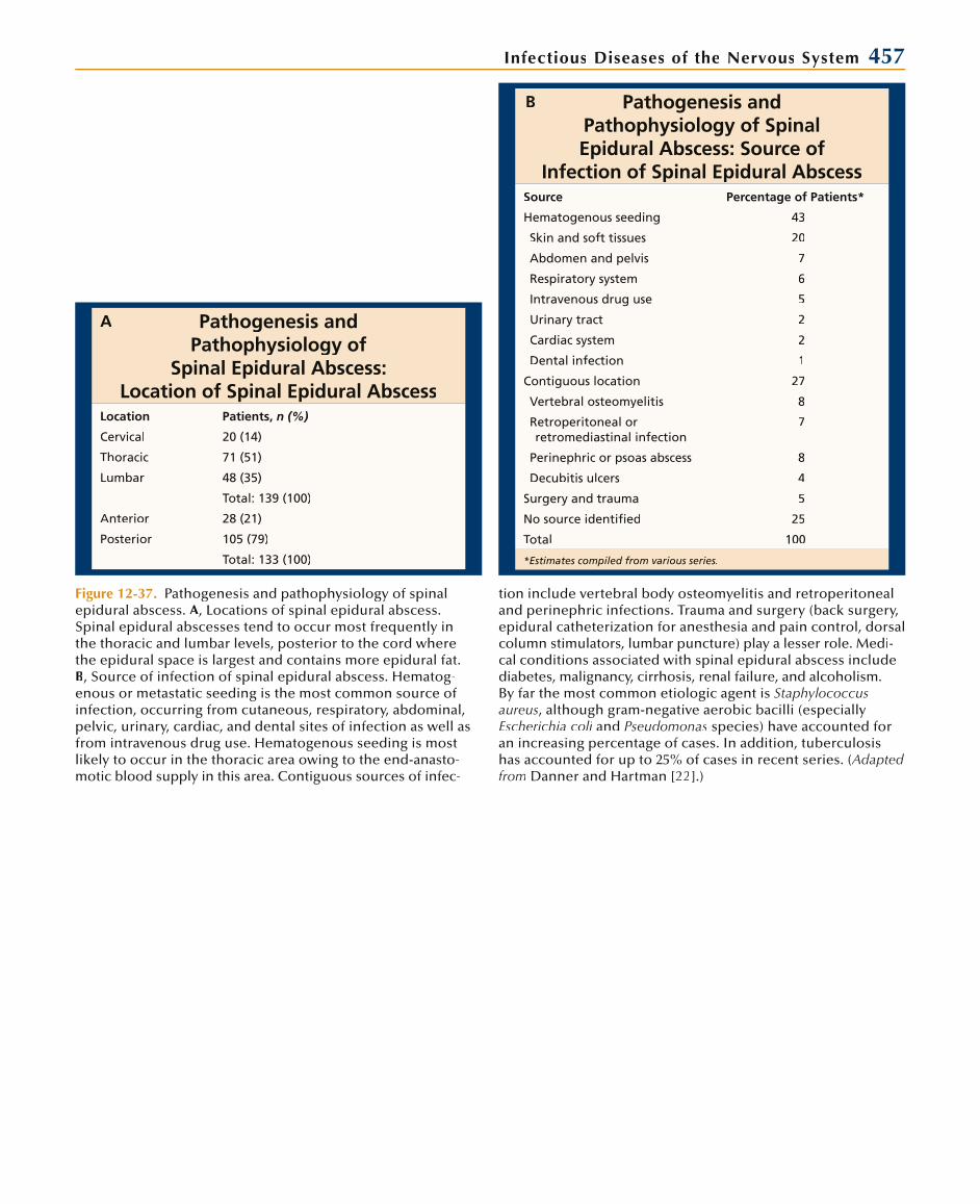
Figure 12-37. Pathogenesis and pathophysiology of spinal epidural abscess. A, Locations of spinal epidural abscess.Spinal epidural abscesses tend to occur most frequently in the thoracic and lumbar levels, posterior to the cord where the epidural space is largest and contains more epidural fat.B, Source of infection of spinal epidural abscess. Hematog-enous or metastatic seeding is the most common source of infection, occurring from cutaneous, respiratory, abdominal, pelvic, urinary, cardiac, and dental sites of infection as well asfrom intravenous drug use. Hematogenous seeding is most likely to occur in the thoracic area owing to the end-anasto-motic blood supply in this area. Contiguous sources of infec-
tion include vertebral body osteomyelitis and retroperitoneal and perinephric infections. Trauma and surgery (back surgery,epidural catheterization for anesthesia and pain control, dorsal column stimulators, lumbar puncture) play a lesser role. Medi-cal conditions associated with spinal epidural abscess includediabetes, malignancy, cirrhosis, renal failure, and alcoholism. By far the most common etiologic agent is Staphylococcus aureus, although gram-negative aerobic bacilli (especiallyEscherichia coli and i Pseudomonas species) have accounted for an increasing percentage of cases. In addition, tuberculosishas accounted for up to 25% of cases in recent series. (Adapted from Danner and Hartman [22].)
Pathogenesis and Pathophysiology of
Spinal Epidural Abscess:Location of Spinal Epidural Abscess
Location Patients, n (%)
Cervical 20 (14)
Thoracic 71 (51)
Lumbar 48 (35)
Total: 139 (100)
Anterior 28 (21)
Posterior 105 (79)
Total: 133 (100)
A
B
458 Atlas of Clinical Neurology
Figure 12-38. Contrast-enhanced T1-weighted MRI done as part of a diagnostic work-up for spinal epidural abscess. This scan reveals an epidural mass (arrow) extendingfrom the lower part of the L1 vertebral body to the upper part of the L4 vertebral body.Patients with acute spinal epidural abscess have a high peripheral leukocytic count from 12,000 to 15,000 cells per mm3. If the process is chronic, the peripheral leuko-cyte count may be normal. Cerebrospinal fluid (CSF) examination is consistent with parameningeal infection, with an elevated cell count, elevated protein level, normalglucose level, and negative cultures. When the process is acute, usually polymorpho-nuclear leukocytes predominate, with up to 100 to 200 cells per mm3; in chronic cases, mononuclear cells predominate, usually with fewer than 50 cells per mm3. CSF cul-tures are negative unless the organism has spread to the CSF and subsequently causedmeningitis. Blood cultures are positive 60% to 70% of the time. The definitive diagnos-tic studies, however, are CT myelograms and MRI scans, with MRI the study of choice. MRI scans directly visualize the extent of the abscess and should include T1-weightedimages before and after contrast enhancement and a T2-weighted image.
Treatment usually consists of surgical decompression, abscess drainage, and par-enteral antibiotics. Empirical treatment is with a combination of a third-generation cephalosporin (eg, ceftriaxone) with another antibiotic for methicillin-resistant staphy-lococci (vancomycin). Once the etiologic bacteria have been identified, the antibiotic regimen should be adjusted based on sensitivities. Antibiotic treatment should contin-ue for at least 4 weeks and 8 weeks with osteomyelitis. Corticosteroids have been used for cord compression, but their benefit has not been subjected to controlled studies. (From Gelfand et al. [23]; with permission.)
DIAGNOSIS
INTRACRANIAL SUBDURAL EMPYEMA
Clinical Manifestations ofSubdural Empyema
Signs/symptoms Patients, n* Percentage
Fever 420 77
Headache 467 74
Hemiparesis 389 71
Altered consciousness 544 69
Nuchal rigidity 385 63
Seizures 576 48
Papilledema 238 33
Altered speech 364 22
Other focal deficits 265 45
*Total number of patients assessed for the specific manifestation.
Figure 12-39. Clinical manifestations of intracranial subdural empy-ema. Subdural empyema is a fulminant, purulent infection thatspreads over the cerebral hemispheres in the existing subdural space. It is usually confined to one side of the brain by the anatom-ic barriers of the falx and tentorium. Undiagnosed and untreated,subdural empyema is rapidly fatal and therefore is a neurologic emergency. Usually patients have a nonspecific prodrome for sev-eral days to a week and then become acutely ill. After head traumaor surgery, the presentation may be milder and more subacute. The presenting manifestations usually include fever, headache,hemiparesis, nuchal rigidity, and seizures. As the process contin-ues, increased intracranial pressure causes papilledema and altera-tion of consciousness. At the end of the first week and into the second, cortical vein thrombosis begins to occur, causing infarcts and additional focal deficits. (Adapted from Hartman et al. [24].)
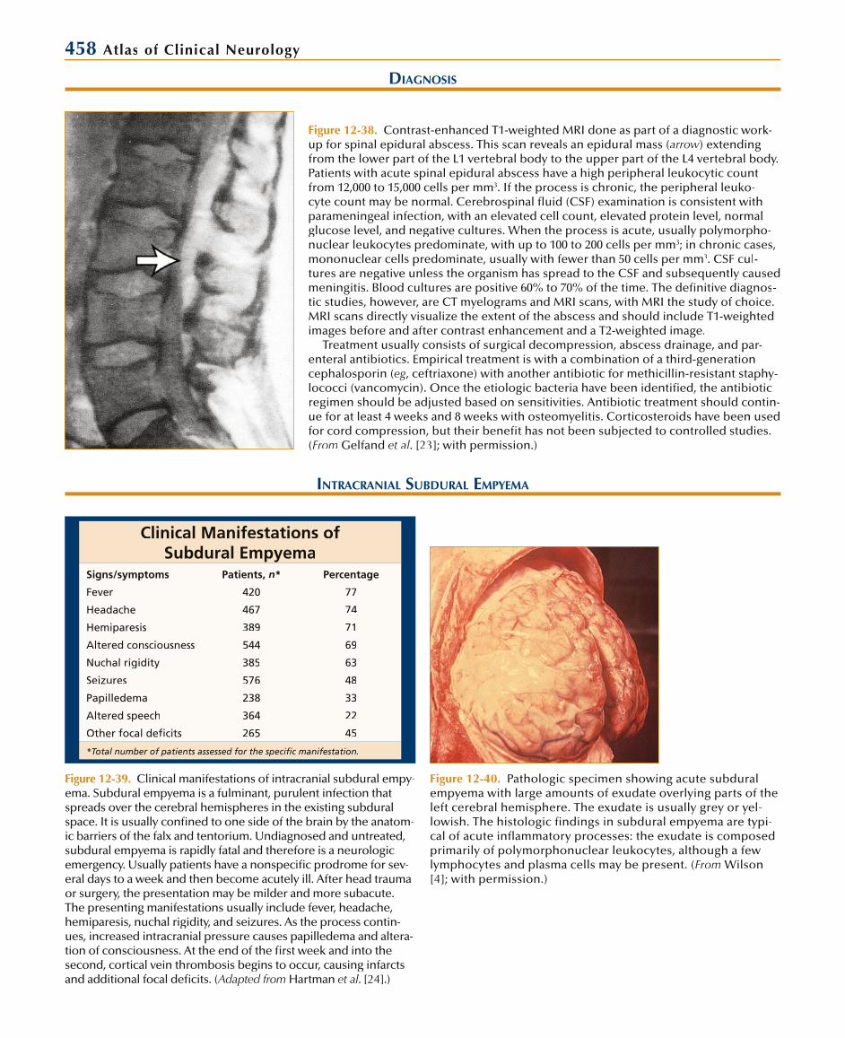
Figure 12-40. Pathologic specimen showing acute subduralempyema with large amounts of exudate overlying parts of the left cerebral hemisphere. The exudate is usually grey or yel-lowish. The histologic findings in subdural empyema are typi-cal of acute inflammatory processes: the exudate is composedprimarily of polymorphonuclear leukocytes, although a few lymphocytes and plasma cells may be present. (From Wilson[4]; with permission.)
Infectious Diseases of the Nervous System 459
Etiology of Adult Subdural EmpyemaOrganism Incidence*, %
Streptococci
Aerobic† 36
Anaerobic 10
Staphylococci
Coagulase-positive 9
Coagulase-negative 3
Aerobic gram-negative bacilli‡ 10
Other anaerobes 6
Sterile 29
*394 evaluated cases, total greater than 100% because of multipleisolates from single cases.
†Includes -hemolytic, -hemolytic, and nonhemolytic.‡Mostly enteric bacilli.
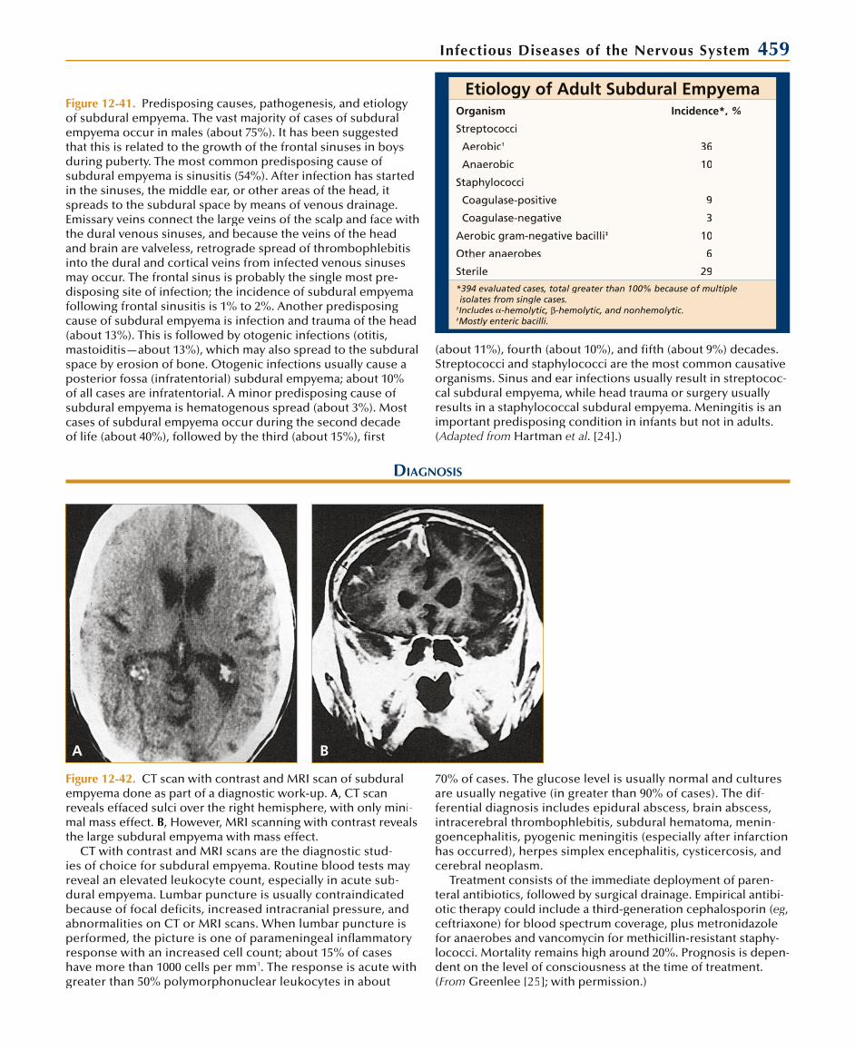
Figure 12-41. Predisposing causes, pathogenesis, and etiologyof subdural empyema. The vast majority of cases of subduralempyema occur in males (about 75%). It has been suggestedthat this is related to the growth of the frontal sinuses in boys during puberty. The most common predisposing cause of subdural empyema is sinusitis (54%). After infection has started in the sinuses, the middle ear, or other areas of the head, itspreads to the subdural space by means of venous drainage.Emissary veins connect the large veins of the scalp and face with the dural venous sinuses, and because the veins of the headand brain are valveless, retrograde spread of thrombophlebitis into the dural and cortical veins from infected venous sinuses may occur. The frontal sinus is probably the single most pre-disposing site of infection; the incidence of subdural empyemafollowing frontal sinusitis is 1% to 2%. Another predisposingcause of subdural empyema is infection and trauma of the head(about 13%). This is followed by otogenic infections (otitis,mastoiditis—about 13%), which may also spread to the subduralspace by erosion of bone. Otogenic infections usually cause aposterior fossa (infratentorial) subdural empyema; about 10% of all cases are infratentorial. A minor predisposing cause of subdural empyema is hematogenous spread (about 3%). Most cases of subdural empyema occur during the second decadeof life (about 40%), followed by the third (about 15%), first
DIAGNOSIS
Figure 12-42. CT scan with contrast and MRI scan of subdural empyema done as part of a diagnostic work-up. A, CT scan reveals effaced sulci over the right hemisphere, with only mini-mal mass effect. B, However, MRI scanning with contrast revealsthe large subdural empyema with mass effect.
CT with contrast and MRI scans are the diagnostic stud-ies of choice for subdural empyema. Routine blood tests mayreveal an elevated leukocyte count, especially in acute sub-dural empyema. Lumbar puncture is usually contraindicatedbecause of focal deficits, increased intracranial pressure, andabnormalities on CT or MRI scans. When lumbar puncture is performed, the picture is one of parameningeal inflammatory response with an increased cell count; about 15% of caseshave more than 1000 cells per mm3. The response is acute withgreater than 50% polymorphonuclear leukocytes in about
70% of cases. The glucose level is usually normal and culturesare usually negative (in greater than 90% of cases). The dif-ferential diagnosis includes epidural abscess, brain abscess, intracerebral thrombophlebitis, subdural hematoma, menin-goencephalitis, pyogenic meningitis (especially after infarction has occurred), herpes simplex encephalitis, cysticercosis, and cerebral neoplasm.
Treatment consists of the immediate deployment of paren-teral antibiotics, followed by surgical drainage. Empirical antibi-otic therapy could include a third-generation cephalosporin (eg, ceftriaxone) for blood spectrum coverage, plus metronidazole for anaerobes and vancomycin for methicillin-resistant staphy-lococci. Mortality remains high around 20%. Prognosis is depen-dent on the level of consciousness at the time of treatment. (From Greenlee [25]; with permission.)
A B
(about 11%), fourth (about 10%), and fifth (about 9%) decades. Streptococci and staphylococci are the most common causative organisms. Sinus and ear infections usually result in streptococ-cal subdural empyema, while head trauma or surgery usually results in a staphylococcal subdural empyema. Meningitis is animportant predisposing condition in infants but not in adults. (Adapted from Hartman et al. [24].)
460 Atlas of Clinical Neurology
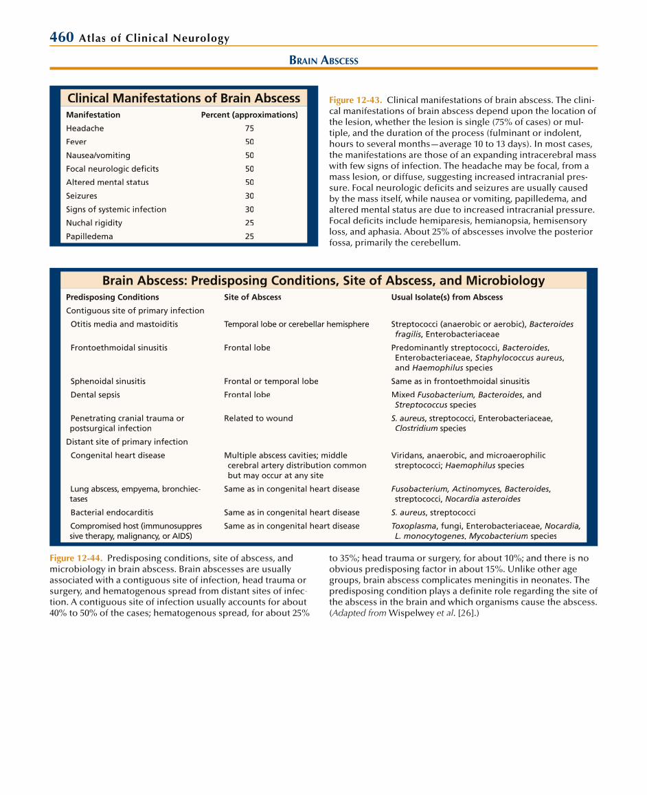
Clinical Manifestations of Brain AbscessManifestation Percent (approximations)
Headache 75
Fever 50
Nausea/vomiting 50
Focal neurologic deficits 50
Altered mental status 50
Seizures 30
Signs of systemic infection 30
Nuchal rigidity 25
Papilledema 25
BRAIN ABSCESS
Figure 12-43. Clinical manifestations of brain abscess. The clini-cal manifestations of brain abscess depend upon the location ofthe lesion, whether the lesion is single (75% of cases) or mul-tiple, and the duration of the process (fulminant or indolent,hours to several months—average 10 to 13 days). In most cases, the manifestations are those of an expanding intracerebral masswith few signs of infection. The headache may be focal, from a mass lesion, or diffuse, suggesting increased intracranial pres-sure. Focal neurologic deficits and seizures are usually causedby the mass itself, while nausea or vomiting, papilledema, andaltered mental status are due to increased intracranial pressure.Focal deficits include hemiparesis, hemianopsia, hemisensoryloss, and aphasia. About 25% of abscesses involve the posterior fossa, primarily the cerebellum.
Brain Abscess: Predisposing Conditions, Site of Abscess, and MicrobiologyPredisposing Conditions Site of Abscess Usual Isolate(s) from Abscess
Contiguous site of primary infection
Otitis media and mastoiditis Temporal lobe or cerebellar hemisphere Streptococci (anaerobic or aerobic), Bacteroides fragilis, Enterobacteriaceae
Frontoethmoidal sinusitis Frontal lobe Predominantly streptococci, Bacteroides, Enterobacteriaceae, Staphylococcus aureus, and Haemophilus species
Sphenoidal sinusitis Frontal or temporal lobe Same as in frontoethmoidal sinusitis
Dental sepsis Frontal lobe Mixed Fusobacterium, Bacteroides, and Streptococcus species
Penetrating cranial trauma or postsurgical infection
Related to wound S. aureus, streptococci, Enterobacteriaceae, Clostridium species
Distant site of primary infection
Congenital heart disease Multiple abscess cavities; middlecerebral artery distribution common but may occur at any site
Viridans, anaerobic, and microaerophilic streptococci; Haemophilus species
Lung abscess, empyema, bronchiec-tases
Same as in congenital heart disease Fusobacterium, Actinomyces, Bacteroides, streptococci, Nocardia asteroides
Bacterial endocarditis Same as in congenital heart disease S. aureus, streptococci
Compromised host (immunosuppres-sive therapy, malignancy, or AIDS)
Same as in congenital heart disease Toxoplasma, fungi, Enterobacteriaceae, Nocardia, L. monocytogenes, Mycobacterium species
Figure 12-44. Predisposing conditions, site of abscess, and microbiology in brain abscess. Brain abscesses are usuallyassociated with a contiguous site of infection, head trauma or surgery, and hematogenous spread from distant sites of infec-tion. A contiguous site of infection usually accounts for about 40% to 50% of the cases; hematogenous spread, for about 25%
to 35%; head trauma or surgery, for about 10%; and there is no obvious predisposing factor in about 15%. Unlike other age groups, brain abscess complicates meningitis in neonates. Thepredisposing condition plays a definite role regarding the site ofthe abscess in the brain and which organisms cause the abscess. (Adapted from Wispelwey et al. [26].)

Infectious Diseases of the Nervous System 461
Microbiologic Etiology of BrainAbscess in the Immunologically
Uncompromised HostEtiologic Organisms Isolation Frequency, %
Staphylococcus aureus 10–15
Enterobacteriaceae 23–33
Streptococcus pneumoniae < 1
Haemophilus influenzae < 1
Streptococci (S. intermedius group, including S. anginosus)
60–70
Bacteroides and Prevotella species 20–40
Fungi 10–15
Protozoa, helminths*
*Heavily dependent on geographic locale.
Microbiologic Etiology of BrainAbscess in the Immunologically
Compromised HostAbnormal Cell-mediated Immunity
Neutropenia or Neutrophil Defects
Toxoplasma gondii Aerobic gram-negative bacteria
Nocardia asteroides Aspergillus species
Cryptococcus neoformans Zygomycetes
Listeria monocytogenes Candida species
Mycobacterium species
Figure 12-45. Microbiologic etiology of brain abscess in the immunologically uncompromised host (A). In the preantibiotic era, Staphylococcus aureus, streptococci, and coliform bacteria were the common isolates from brain abscess. In the past 20years, however, anaerobes (streptococci intermedius group,Bacteroides species) are probably the most common cause. Microbiologic etiology of brain abscess in the immunologically
compromised host (B). Patients with AIDS, underlying malig-nancy, or those treated with immunosuppressive agents areat an increased risk for developing brain abscess. Fungi andparasites are also important diagnostic considerations in thesepatients. Neutropenia and neutrophilic defects are most oftendue to chemotherapy. (A, adapted from Wispelwey and Scheld[27]; B, adapted from Wispelwey et al. [26].)
Differential Diagnosis of Brain Abscess in the Immunologically
Uncompromised HostSubdural empyema
Pyogenic meningitis
Viral encephalitis (especially herpes simplex)
Cysticercosis
Cerebral infarction
Mycotic aneurysms
Epidural abscess
Cerebral neoplasms
Hemorrhagic leukoencephalitis
Echinococcosis
Cryptococcosis
Central nervous system vasculitis
Chronic subdural hematoma
Intracerebral thrombophlebitis
Differential Diagnosis of Brain Abscesses in Patients With AIDS
Toxoplasmosis
Primary central nervous system lymphoma
Mycobacterium tuberculosis
Mycobacterium avium-intracellulare
Progressive multifocal leukoencephalopathy
Cryptococcus neoformans
Candida species
Listeria monocytogenes
Nocardia asteroides
Salmonella group B
Aspergillus species
Figure 12-46. Differential diagnosis of brain abscess. A, Thedifferential diagnosis in the immunologically uncompromisedhost includes entities causing focal neurologic deficits, whichare usually seen early in the course, and diffuse manifestations,which are seen later in the course of brain abscess. B, In AIDS
patients, focal neurologic infections and processes of diverseand unusual etiologies have been recognized. The most commoncause of focal neurologic lesions is toxoplasmosis, followed by lymphoma. (A, adapted fromA Wispelwey [28]; B, adapted fromWispelwey et al. [26].)
A
B
A
B
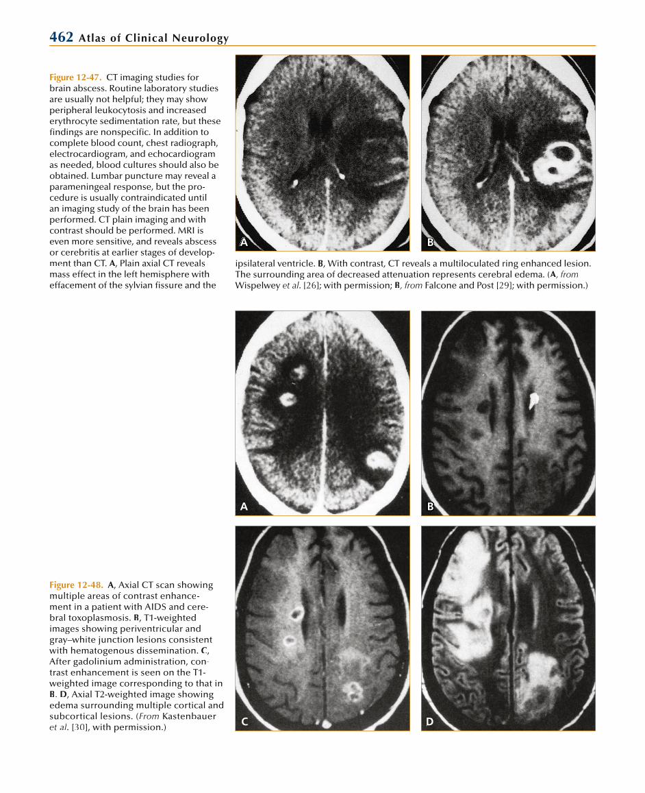
462 Atlas of Clinical Neurology
A B
Figure 12-47. CT imaging studies forbrain abscess. Routine laboratory studiesare usually not helpful; they may showperipheral leukocytosis and increasederythrocyte sedimentation rate, but thesefindings are nonspecific. In addition tocomplete blood count, chest radiograph, electrocardiogram, and echocardiogramas needed, blood cultures should also beobtained. Lumbar puncture may reveal a parameningeal response, but the pro-cedure is usually contraindicated untilan imaging study of the brain has beenperformed. CT plain imaging and withcontrast should be performed. MRI iseven more sensitive, and reveals abscessor cerebritis at earlier stages of develop-ment than CT. A, Plain axial CT reveals mass effect in the left hemisphere with effacement of the sylvian fissure and the
Figure 12-48. A, Axial CT scan showingmultiple areas of contrast enhance-ment in a patient with AIDS and cere-bral toxoplasmosis. B, T1-weighted images showing periventricular andgray–white junction lesions consistentwith hematogenous dissemination. C, After gadolinium administration, con-trast enhancement is seen on the T1-weighted image corresponding to that in B. D, Axial T2-weighted image showing edema surrounding multiple cortical andsubcortical lesions. (From Kastenbaueret al. [30], with permission.)
A B
C D
ipsilateral ventricle. B, With contrast, CT reveals a multiloculated ring enhanced lesion. The surrounding area of decreased attenuation represents cerebral edema. (A, fromAWispelwey et al. [26]; with permission; B, from Falcone and Post [29]; with permission.)
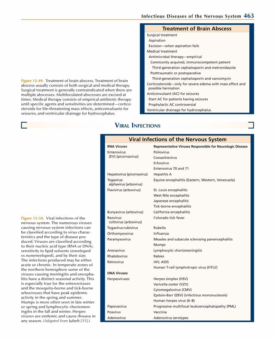
Infectious Diseases of the Nervous System 463
Treatment of Brain AbscessSurgical treatment
Aspiration
Excision—when aspiration fails
Medical treatment
Antimicrobal therapy—empirical
Community acquired, immunocompetent patient
Third-generation cephalosporin and metronidazole
Posttraumatic or postoperative
Third-generation cephalosporin and vancomycin
Corticosteroids—only for severe edema with mass effect and possible herniation
Anticonvulsant (AC) for seizures
Start AC for patients having seizures
Prophylactic AC controversial
Ventricular drainage for hydrocephalus
Figure 12-49. Treatment of brain abscess. Treatment of brain abscess usually consists of both surgical and medical therapy. Surgical treatment is generally contraindicated when there aremultiple abscesses. Multiloculated abscesses are excised attimes. Medical therapy consists of empirical antibiotic therapy until specific agents and sensitivities are determined—cortico-steroids for life-threatening mass effects, anticonvulsants for seizures, and ventricular drainage for hydrocephalus.
VIRAL INFECTIONS
Viral Infections of the Nervous SystemRNA Viruses Representative Viruses Responsible for Neurologic Disease
Enterovirus(EV) (picornavirus)
Poliovirus
Coxsackievirus
Echovirus
Enterovirus 70 and 71
Hepatovirus (picornavirus) Hepatitis A
Togavirus:alphavirus (arbovirus)
Equine encephalitis (Eastern, Western, Venezuela)
Flavivirus (arbovirus) St. Louis encephalitis
West Nile encephalitis
Japanese encephalitis
Tick-borne encephalitis
Bunyavirus (arbovirus) California encephalitis
Reovirus:coltivirus (arbovirus)
Colorado tick fever
Togavirus:rubivirus Rubella
Orthomyxovirus Influenza
Paramyxovirus Measles and subacute sclerosing panencephalitis
Mumps
Arenavirus Lymphocytic choriomeningitis
Rhabdovirus Rabies
Retrovirus HIV, AIDS
Human T-cell lymphotropic virus (HTLV)
DNA Viruses
Herpesviruses Herpes simplex (HSV)
Varicella-zoster (VZV)
Cytomegalovirus (CMV)
Epstein-Barr (EBV) (infectious mononucleosis)
Human herpes virus (6–8)
Papovavirus Progressive multifocal leukoencephalopathy (PML)
Poxvirus Vaccinia
Adenovirus Adenovirus serotypes
Figure 12-50. Viral infections of the nervous system. The numerous virusescausing nervous system infections canbe classified according to virus charac-teristics and the type of disease pro-duced. Viruses are classified according to their nucleic acid type (RNA or DNA), sensitivity to lipid solvents (enveloped vs nonenveloped), and by their size.The infections produced may be eitheracute or chronic. In temperate zones ofthe northern hemisphere some of theviruses causing meningitis and encepha-litis have a distinct seasonal activity. Thisis especially true for the enteroviruses and the mosquito-borne and tick-bornearboviruses that have peak epidemic activity in the spring and summer. Mumps is more often seen in late winter or spring and lymphocytic choriomen-ingitis in the fall and winter. Herpesviruses are endemic and cause disease in any season. (Adapted from Jubelt [31].)
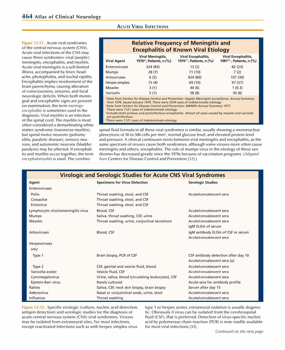
464 Atlas of Clinical Neurology
Relative Frequency of Meningitis and Encephalitis of Known Viral Etiology
Viral AgentViral Meningitis,
1976*, Patients, n (%)Viral Encephalitis,
1976† ‡, Patients, n (%)Viral Encephalitis,
1981‡ § , Patients, n (%)
EnterovirusesMumpsArbovirusesHerpes simplexMeaslesVaricella
324 (83)28 (7)6 (2)
15 (4)3 (1)5 (1)
13 (2)71 (10)424 (60)69 (10)44 (6)58 (8)
82 (23)7 (2)
107 (30)97 (27)1 (0.3)30 (8)
*Data from Centers for Disease Control and Prevention: Aseptic Meningitis Surveillance, Annual Summary from 1976. Issued January 1979. There were 2534 cases of indeterminate etiology.
†Data from Centers for Disease Control and Prevention: MMWR–Annual Summary 1977. There were 1121 cases of indeterminate etiology.
‡Includes both primary and postinfectious encephalitis. Almost all cases caused by measles and varicella are postinfectious.
§There were 1121 cases of indeterminate etiology.
ACUTE VIRAL INFECTIONS
Virologic and Serologic Studies for Acute CNS Viral SyndromesAgent Specimens for Virus Detection Serologic Studies
Enteroviruses
PolioCoxsackieEchovirus
Throat washing, stool, and CSFThroat washing, stool, and CSFThroat washing, stool, and CSF
Acute/convalescent sera
Lymphocytic choriomeningitis virusMumpsMeasles
Blood, CSFSaliva, throat washing, CSF, urineThroat washing, urine, conjuctival secretions
Acute/convalescent seraAcute/convalescent seraAcute/convalescent seraIgM ELISA of serum
Arboviruses Blood, CSF IgM antibody ELISA of CSF or serumAcute/convalescent sera
Herpesviruses
HSV
Type 1 Brain biopsy, PCR of CSF CSF antibody detection after day 10Acute/convalescent sera (±)
Type 2Varicella-zosterCytomegalovirusEpstein-Barr virus
RabiesAdenovirusInfluenza
CSF, genital and vesicle fluid, bloodVesicle fluid, CSFUrine, saliva, blood (circulating leukocytes), CSFRarely culturedSaliva, CSF, neck skin biopsy, brain biopsyNasal or conjunctival swab, urine, stoolThroat washing
Acute/convalescent seraAcute/convalescent seraAcute/convalescent seraAcute sera for antibody profileSerum after day 15Acute/convalescent seraAcute/convalescent sera
Figure 12-52. Specific virologic (culture, nucleic acid detection, antigen detection) and serologic studies for the diagnosis of acute central nervous system (CNS) viral syndromes. Virusesmay be isolated from extraneural sites. For most infections, except reactivated infections such as with herpes simplex virus
type 1 or herpes zoster, extraneural isolation is usually diagnos-tic. Obviously if virus can be isolated from the cerebrospinal fluid (CSF), that is preferred. Detection of virus-specific nucleicacid by polymerase chain reaction (PCR) is now readily available for most viral infections [33].
Continued on the next page
Figure 12-51. Acute viral syndromes of the central nervous system (CNS).Acute viral infections of the CNS may cause three syndromes: viral (aseptic) meningitis, encephalitis, and myelitis. Acute viral meningitis is a self-limited illness, accompanied by fever, head-ache, photophobia, and nuchal rigidity.Encephalitis implies involvement of thebrain parenchyma, causing alterationof consciousness, seizures, and focal neurologic deficits. When both menin-geal and encephalitic signs are present on examination, the term meningo-encephalitis is sometimes used in the diagnosis. Viral myelitis is an infection of the spinal cord. The myelitis is mostoften considered a demyelinating whitematter syndrome (transverse myelitis), but spinal motor neurons (poliomy-elitis, paralytic disease), sensory neu-rons, and autonomic neurons (bladderparalysis) may be affected. If encephali-tis and myelitis occur together, the term encephalomyelitis is used. The cerebro-
spinal fluid formula in all these viral syndromes is similar, usually showing a mononuclearpleocytosis of 50 to 500 cells per mm3, normal glucose level, and elevated protein level and pressure. A clinical continuum exists between viral meningitis and encephalitis, as thesame spectrum of viruses cause both syndromes, although some viruses more often cause meningitis and others, encephalitis. The role of mumps virus in the etiology of these syn-dromes has decreased greatly since the 1970s because of vaccination programs. (Adapted from Centers for Disease Control and Prevention [32].)

Infectious Diseases of the Nervous System 465Figure 12-52. (Continued) Antigen detection usually requiresthe use of biopsy material. Serologic studies require a fourfold increase between the acute and convalescent specimen to be con-sidered positive. Acute phase sera should be obtained immediatelyor as soon as infection is suspected. If the acute phase sera is notobtained until the end of the first week of the disease, the chancesof seeing a fourfold rise drops to 50%. Most viral infections of theCNS result in the intrathecal synthesis of specific antibody. When
analyzing CSF antibody synthesis one looks for an increased CSFto serum antibody ratio. Therefore, paired CSF and serum samplesare required. A correction should also be used for blood-brain barrier breakdown, which results in serum to CSF antibody leak-age. This can be done by using CSF to serum albumin or otherviral antibody ratios. Unfortunately, most antibody studies are not positive until at least 1 week after the onset of infection. ELISA—enzyme-linked immunosorbent assay. (Adapted from Jubelt [34].)
VIRAL MENINGITIS
Clinical Manifestations of Acute Viral MeningitisSystemic Manifestations Central Nervous System Manifestations
Fever
Malaise
Anorexia
Myalgia
Nausea and vomiting
Agent-specific pharyngitis, URI,abdominal pain, diarrhea
Headache, usually frontal or retro-orbital
Nuchal rigidity
Photophobia
Lethargy
Figure 12-53. Clinical manifestations ofacute viral meningitis. Acute viral menin-gitis is sometimes referred to as “aseptic” meningitis, but the terms are not synony-mous, because agents other than virusesalso cause aseptic meningitis, for example,parameningeal infection, autoimmunedisease, vasculitis, and chemicals. Acuteviral meningitis usually begins abruptlywith a combination of central nervoussystem signs of nuchal rigidity, headache,photophobia, and occasionally lethargyalong with systemic manifestations. If thealteration in the level of consciousness is more pronounced than lethargy, another diagnosis should be considered. The cere-brospinal fluid (CSF) profile is that of allviral syndromes, with lymphocytic pleo-
Differential Diagnosis of Acute Viral MeningitisBacterial meningitis
Early (0–24 h)
Also listeriosis, mycoplasmosis, brucellosis
Tuberculous meningitis
Fungal meningitis
Spirochetal meningitis: syphilis, Lyme disease, leptospirosis
Parasitic meningitis
Parameningeal infections
Neoplastic meningitis
Autoimmune and inflammatory diseases; lupus, sarcoid, vasculitis
Drug reactions: nonsteroidal anti-inflammatory agents, sulfamethoxazole, trim-ethoprim, trimethoprim-sulfamethoxazole, isoniazid, carbamazepine, azathioprine, intravenous immune globulin, intravenous monoclonal OKT3
Chemical meningitis: intrathecal drugs, central nervous system tumors, myelography,isotope cisternography
Figure 12-54. Differential diagnosis ofacute viral meningitis. It is importantto exclude nonviral causes of meningi-tis that may require specific therapy or more emergent therapy. During the first24 hours of viral meningitis polymorpho-nuclear leukocytes may appear in thecerebrospinal fluid sample, similar to bacterial meningitis. In partially treated bacterial meningitis, the glucose level may be normal. Usually in tuberculous, fungal, spirochetal, and parasitic men-ingitis, the glucose level is depressed. Cytologic examination should differenti-ate neoplastic meningitis, whereas sero-logic studies help exclude autoimmune disease. Imaging studies usually detectparameningeal inflammation.
cytosis, mildly elevated protein levels, and a normal glucose level. Because the intensity of the inflammatory response in the CSF is low (usually 0 to 500 cells per mm3), nuchal rigidity is usually the only sign of meningeal irritation, and Kernig’s and Brudzinski’ssigns are often absent. In a few cases, however, marked pleocytosis may be seen (greater than 1000 cells per mm3), often accompanied by Kernig’s and Brudzinski’s signs. Themeningitis is self-limited, resolving usually in 1 week. URI—upper respiratory infection.
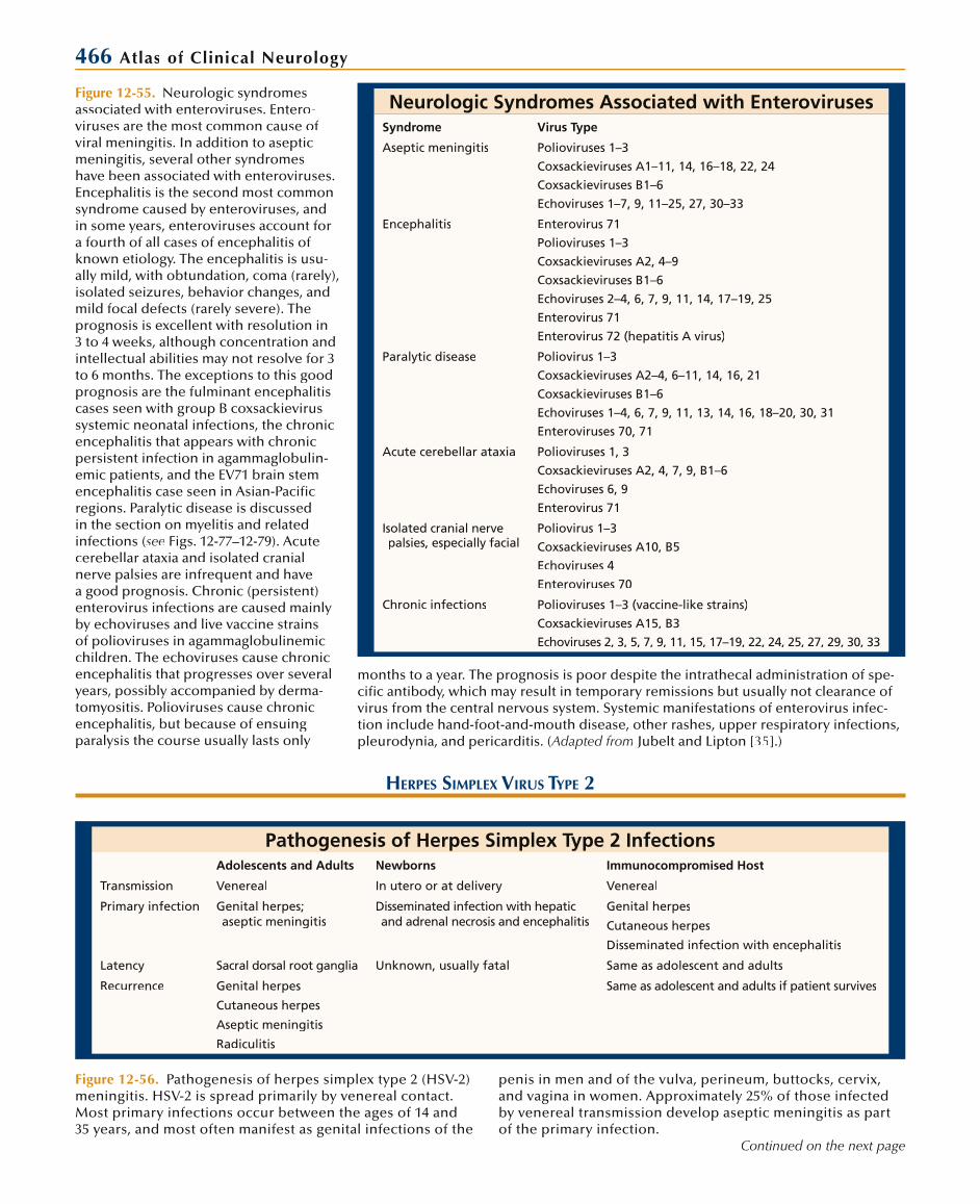
466 Atlas of Clinical Neurology
Neurologic Syndromes Associated with EnterovirusesSyndrome Virus Type
Aseptic meningitis Polioviruses 1–3
Coxsackieviruses A1–11, 14, 16–18, 22, 24
Coxsackieviruses B1–6
Echoviruses 1–7, 9, 11–25, 27, 30–33
Encephalitis Enterovirus 71
Polioviruses 1–3
Coxsackieviruses A2, 4–9
Coxsackieviruses B1–6
Echoviruses 2–4, 6, 7, 9, 11, 14, 17–19, 25
Enterovirus 71
Enterovirus 72 (hepatitis A virus)
Paralytic disease Poliovirus 1–3
Coxsackieviruses A2–4, 6–11, 14, 16, 21
Coxsackieviruses B1–6
Echoviruses 1–4, 6, 7, 9, 11, 13, 14, 16, 18–20, 30, 31
Enteroviruses 70, 71
Acute cerebellar ataxia Polioviruses 1, 3
Coxsackieviruses A2, 4, 7, 9, B1–6
Echoviruses 6, 9
Enterovirus 71
Isolated cranial nervepalsies, especially facial
Poliovirus 1–3
Coxsackieviruses A10, B5
Echoviruses 4
Enteroviruses 70
Chronic infections Polioviruses 1–3 (vaccine-like strains)
Coxsackieviruses A15, B3
Echoviruses 2, 3, 5, 7, 9, 11, 15, 17–19, 22, 24, 25, 27, 29, 30, 33
Figure 12-55. Neurologic syndromes associated with enteroviruses. Entero-viruses are the most common cause ofviral meningitis. In addition to asepticmeningitis, several other syndromeshave been associated with enteroviruses. Encephalitis is the second most common syndrome caused by enteroviruses, and in some years, enteroviruses account for a fourth of all cases of encephalitis ofknown etiology. The encephalitis is usu-ally mild, with obtundation, coma (rarely), isolated seizures, behavior changes, and mild focal defects (rarely severe). Theprognosis is excellent with resolution in3 to 4 weeks, although concentration andintellectual abilities may not resolve for 3 to 6 months. The exceptions to this goodprognosis are the fulminant encephalitiscases seen with group B coxsackievirus systemic neonatal infections, the chronic encephalitis that appears with chronic persistent infection in agammaglobulin-emic patients, and the EV71 brain stemencephalitis case seen in Asian-Pacific regions. Paralytic disease is discussedin the section on myelitis and relatedinfections (see Figs. 12-77–12-79). Acutecerebellar ataxia and isolated cranial nerve palsies are infrequent and have a good prognosis. Chronic (persistent)enterovirus infections are caused mainly by echoviruses and live vaccine strainsof polioviruses in agammaglobulinemicchildren. The echoviruses cause chronicencephalitis that progresses over severalyears, possibly accompanied by derma-tomyositis. Polioviruses cause chronicencephalitis, but because of ensuing paralysis the course usually lasts only
HERPES SIMPLEX VIRUS TYPE 2
Pathogenesis of Herpes Simplex Type 2 InfectionsAdolescents and Adults Newborns Immunocompromised Host
Transmission Venereal In utero or at delivery Venereal
Primary infection Genital herpes;aseptic meningitis
Disseminated infection with hepatic and adrenal necrosis and encephalitis
Genital herpes
Cutaneous herpes
Disseminated infection with encephalitis
Latency Sacral dorsal root ganglia Unknown, usually fatal Same as adolescent and adults
Recurrence Genital herpes
Cutaneous herpes
Aseptic meningitis
Radiculitis
— Same as adolescent and adults if patient survives
Figure 12-56. Pathogenesis of herpes simplex type 2 (HSV-2)meningitis. HSV-2 is spread primarily by venereal contact.Most primary infections occur between the ages of 14 and 35 years, and most often manifest as genital infections of the
penis in men and of the vulva, perineum, buttocks, cervix, and vagina in women. Approximately 25% of those infectedby venereal transmission develop aseptic meningitis as partof the primary infection.
Continued on the next page
months to a year. The prognosis is poor despite the intrathecal administration of spe-cific antibody, which may result in temporary remissions but usually not clearance ofvirus from the central nervous system. Systemic manifestations of enterovirus infec-tion include hand-foot-and-mouth disease, other rashes, upper respiratory infections,pleurodynia, and pericarditis. (Adapted from Jubelt and Lipton [35].)

Infectious Diseases of the Nervous System 467Figure 12-56. (Continued) Primary infection of a fetus can occur in utero or during delivery through an infected birthcanal, which may result in severe and often fatal disseminated infection with encephalitis of the neonate. A similar dissemi-nated infection may occur in the immunocompromised hostafter venereal transmission. Because the sacral ganglia receive sensory fibers from the external genitalia, the virus is trans-ported axonally to the ganglia, where it becomes latent; the
virus later reactivates and causes recurrent disease. Recur-rent disease usually is limited to the genitalia, but aseptic meningitis and radiculitis may occur. Recurrent meningitisis sometimes referred to as Mollaret’s meningitis. Radiculitisis manifested by dysesthesia, which is often painful, may be burning or lancinating in character, and may cause sciatica.Sacral radiculitis may also result in bladder and bowel dysfunction,such as retention or incontinence.
Acyclovir Treatment of Herpes SimplexType 2 Infections
Type of Infection Recommended Treatment
Primary infection
Genital lesions Oral acyclovir, 200 mg daily 5× for 10 d
Aseptic meningitis None or oral acyclovir (but not studied)
Disseminated infection withor without encephalitis
IV acyclovir, 10 mg/kg q8h for 21 d
Recurrent infection
Immunocompetent host
Infrequent No treatment
Frequent Oral acyclovir for suppression for up to 1 y,200 mg tid or qid
Immunocompromised host
Localized infection Oral or IV acyclovir
Disseminated infection IV acyclovir
Preventative Oral acyclovir, 200 to 400 mg 2× to 5× per day
Figure 12-57. Treatment of herpes simplex type 2 infections. Acyclovir is the majordrug in use today for treating herpes simplex infections. It is available in three formulations: intravenous (IV), oral, andtopical (5% ointment). For the primarygenital infection, oral acyclovir is indi-cated. Treatment for aseptic meningitis hasnot been studied, but it is a self-limiteddisease. Intravenous acyclovir is needed for disseminated infection with or without encephalitis. Frequent recurrences areusually treated in the immunocompetenthost with 1 year of oral acyclovir for sup-pression. The immunocompromised host requires treatment for each recurrenceand probably continuous oral treatment for suppression as a preventative measure.
VIRAL ENCEPHALITIS
Clinical Manifestations ofAcute Viral Encephalitis
Acute febrile illness of abrupt onset
Systemic manifestations—malaise, anorexia, myalgias, pain, nausea and vomiting, diarrhea
Meningeal irritation—headache, photophobia, nuchal rigidity
Parenchymal (brain) dysfunction
Altered level of consciousness
Lethargy to coma
Confusion, disorientation
Delirium
Mental changes—personality and behavioral changes, including agitation, hallucinations, and psychosis
Seizures—focal or generalized
Extensor plantar responses and hyperreflexia
Focal deficits—less frequent
Aphasia
Ataxia
Hemiparesis
Tremor
Cranial nerve palsies
Figure 12-58. Clinical manifestations of acute viral encephalitis.The term encephalitis implies involvement of the brain paren-chyma. Often the meningeal manifestations seen in acute viralmeningitis are also present in addition to the signs of braindysfunction (meningoencephalitis). The signs of brain dys-function may be focal or diffuse; these may manifest as mental status changes, seizures (in greater than 50% of cases), or hard focal signs. All types of focal deficits have been reported. Viralencephalitis can be divided into primary and secondary types.In primary encephalitis, there is viral invasion and infection of the brain parenchyma, usually of the gray matter. Secondary encephalitis is postinfectious encephalitis (or encephalomy-elitis) in which an immune-mediated attack against myelin andwhite matter apparently occurs. Despite the different locations(gray vs white matter) of these pathologic insults, one cannotdistinguish between the two based on the clinical symptomsand signs. The cerebrospinal fluid profile is similar to that of any acute viral syndrome, with lymphocytic pleocytosis (usually 5 to 500 cells per mm3), normal glucose levels, increased proteinlevels, and increased pressure.
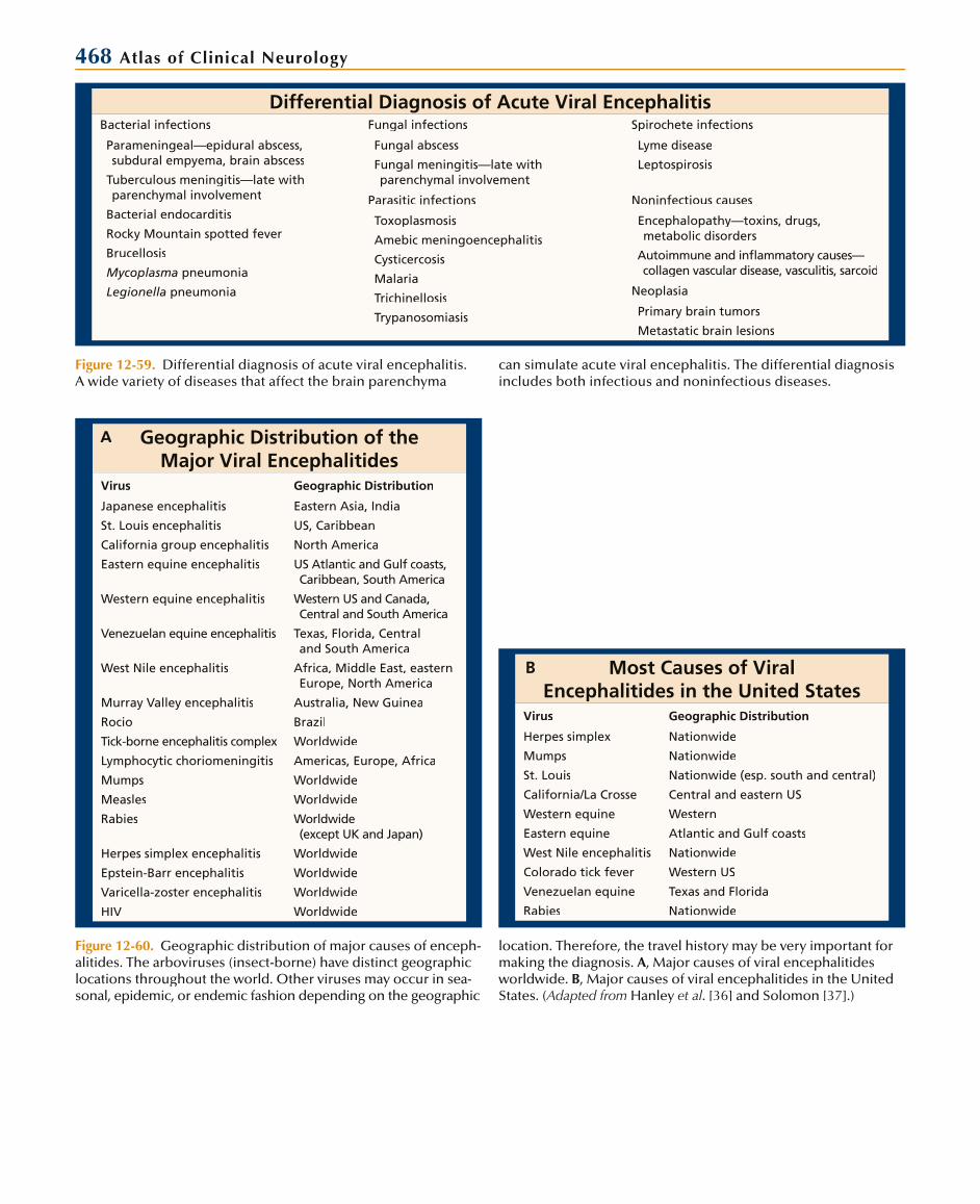
468 Atlas of Clinical Neurology
Differential Diagnosis of Acute Viral EncephalitisBacterial infections Fungal infections Spirochete infections
Parameningeal—epidural abscess, subdural empyema, brain abscess
Tuberculous meningitis—late with parenchymal involvement
Bacterial endocarditis
Rocky Mountain spotted fever
Brucellosis
Mycoplasma pneumonia
Legionella pneumonia
Fungal abscess
Fungal meningitis—late with parenchymal involvement
Lyme disease
Leptospirosis
Parasitic infections Noninfectious causes
Toxoplasmosis
Amebic meningoencephalitis
Cysticercosis
Malaria
Trichinellosis
Trypanosomiasis
Encephalopathy—toxins, drugs, metabolic disorders
Autoimmune and infl ammatory causes— collagen vascular disease, vasculitis, sarcoid
Neoplasia
Primary brain tumors
Metastatic brain lesions
Figure 12-59. Differential diagnosis of acute viral encephalitis.A wide variety of diseases that affect the brain parenchyma
can simulate acute viral encephalitis. The differential diagnosis includes both infectious and noninfectious diseases.
Geographic Distribution of the Major Viral Encephalitides
Virus Geographic Distribution
Japanese encephalitis
St. Louis encephalitis
California group encephalitis
Eastern equine encephalitis
Western equine encephalitis
Venezuelan equine encephalitis
West Nile encephalitis
Murray Valley encephalitis
Rocio
Tick-borne encephalitis complex
Lymphocytic choriomeningitis
Mumps
Measles
Rabies
Herpes simplex encephalitis
Epstein-Barr encephalitis
Varicella-zoster encephalitis
HIV
Eastern Asia, India
US, Caribbean
North America
US Atlantic and Gulf coasts,Caribbean, South America
Western US and Canada,Central and South America
Texas, Florida, Centraland South America
Africa, Middle East, easternEurope, North America
Australia, New Guinea
Brazil
Worldwide
Americas, Europe, Africa
Worldwide
Worldwide
Worldwide(except UK and Japan)
Worldwide
Worldwide
Worldwide
Worldwide
Most Causes of Viral Encephalitides in the United States
Virus Geographic Distribution
Herpes simplex
Mumps
St. Louis
California/La Crosse
Western equine
Eastern equine
West Nile encephalitis
Colorado tick fever
Venezuelan equine
Rabies
Nationwide
Nationwide
Nationwide (esp. south and central)
Central and eastern US
Western
Atlantic and Gulf coasts
Nationwide
Western US
Texas and Florida
Nationwide
Figure 12-60. Geographic distribution of major causes of enceph-alitides. The arboviruses (insect-borne) have distinct geographiclocations throughout the world. Other viruses may occur in sea-sonal, epidemic, or endemic fashion depending on the geographic
location. Therefore, the travel history may be very important formaking the diagnosis. A, Major causes of viral encephalitidesworldwide. B, Major causes of viral encephalitides in the United States. (Adapted from Hanley et al. [36] and Solomon [37].)
A
B
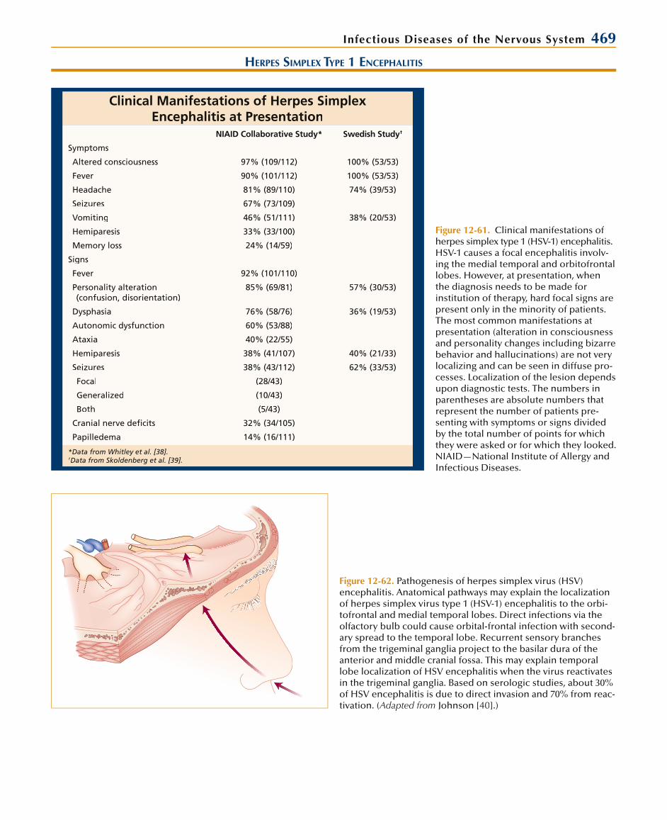
Infectious Diseases of the Nervous System 469
HERPES SIMPLEX TYPE 1 ENCEPHALITIS
Clinical Manifestations of Herpes Simplex Encephalitis at Presentation
NIAID Collaborative Study* Swedish Study†
Symptoms
Altered consciousness 97% (109/112) 100% (53/53)
Fever 90% (101/112) 100% (53/53)
Headache 81% (89/110) 74% (39/53)
Seizures 67% (73/109)
Vomiting 46% (51/111) 38% (20/53)
Hemiparesis 33% (33/100)
Memory loss 24% (14/59)
Signs
Fever 92% (101/110)
Personality alteration (confusion, disorientation)
85% (69/81) 57% (30/53)
Dysphasia 76% (58/76) 36% (19/53)
Autonomic dysfunction 60% (53/88)
Ataxia 40% (22/55)
Hemiparesis 38% (41/107) 40% (21/33)
Seizures 38% (43/112) 62% (33/53)
Focal (28/43)
Generalized (10/43)
Both (5/43)
Cranial nerve defi cits 32% (34/105)
Papilledema 14% (16/111)
*Data from Whitley et al. [38].†Data from Skoldenberg et al. [39].
Figure 12-61. Clinical manifestations ofherpes simplex type 1 (HSV-1) encephalitis.HSV-1 causes a focal encephalitis involv-ing the medial temporal and orbitofrontallobes. However, at presentation, when the diagnosis needs to be made for institution of therapy, hard focal signs arepresent only in the minority of patients. The most common manifestations atpresentation (alteration in consciousnessand personality changes including bizarrebehavior and hallucinations) are not very localizing and can be seen in diffuse pro-cesses. Localization of the lesion dependsupon diagnostic tests. The numbers inparentheses are absolute numbers thatrepresent the number of patients pre-senting with symptoms or signs dividedby the total number of points for whichthey were asked or for which they looked. NIAID—National Institute of Allergy andInfectious Diseases.
Figure 12-62. Pathogenesis of herpes simplex virus (HSV)encephalitis. Anatomical pathways may explain the localization of herpes simplex virus type 1 (HSV-1) encephalitis to the orbi-tofrontal and medial temporal lobes. Direct infections via theolfactory bulb could cause orbital-frontal infection with second-ary spread to the temporal lobe. Recurrent sensory branchesfrom the trigeminal ganglia project to the basilar dura of the anterior and middle cranial fossa. This may explain temporallobe localization of HSV encephalitis when the virus reactivatesin the trigeminal ganglia. Based on serologic studies, about 30%of HSV encephalitis is due to direct invasion and 70% from reac-tivation. (Adapted from Johnson [40].)

470 Atlas of Clinical Neurology
Figure 12-63. Gross anatomy of the brain in herpes simplexvirus encephalitis. There is a hemorrhagic necrotic lesion of theleft medial temporal lobe. As in this case, subarachnoid hemor-rhage is frequent and results in the large number of erythro-cytes often found in the cerebrospinal fluid. (From Hirano et al.[41]; with permission.)
Figure 12-64. Histologic slide showing microscopic anatomy of herpes simplex virus (HSV) encephalitis (hematoxylin-eosin stain). Shown is a type A intranuclear inclusion body within thenucleus of a small nerve cell. These eosinophilic inclusion bodiesmay be seen in other herpes virus infections and subacute mea-sles encephalitis. This section was taken from the temporal lobe cortex of a person with HSV encephalitis. The nuclear chromatin is marginated. These inclusion bodies can also be seen in glia and are a helpful diagnostic sign. (From Wilson [4]; with permission.)
Figure 12-65. CT scans of herpes simplex virus (HSV) encepha-litis. The initial diagnostic test should be a brain CT scan toexclude other lesions and to check for mass effect. Usuallythe CT scan does not become positive until the end of the first week of infection. If there is mass effect, treatment should commence with mannitol or steroids and acyclovir. Diagnosiscan then be confirmed by MRI scanning. If the CT is normal orthere is no mass effect, lumbar puncture should be performed.Within the first day or two of onset, 5% to 10% of cerebrospi-nal fluid (CSF) examinations are normal, but usually there is a lymphocytic pleocytosis, normal glucose, elevated protein, and pressure similar to other central nervous system viral infec-tions. However, in HSV encephalitis there may be significantnecrosis, hemorrhage, and erythrocytes in the CSF. The use of
polymerase chain reaction for amplification of HSV DNA from the CSF is a rapid and sensitive method of diagnosis. If the CSFis abnormal, MRI scanning should be performed, and acyclovir should be started if the MRI is consistent with herpes encepha-litis. If there is a delay in obtaining the MRI, then acyclovir should be started first. The electroencephalogram (EEG) can also be a useful test as it may reveal temporal lobe foci earlierthan CT scanning. If the diagnosis is not confirmed from the MRI and EEG, then a brain biopsy may be needed. A, CT scanon day 10 revealing low-density lesion in the right temporal and deep frontal lobes. B, The corresponding enhanced CT scan reveals gyral enhancement in the sylvian fissure and insularregions, which are greater on the right. (From Davis et al. [42];with permission.)
BA
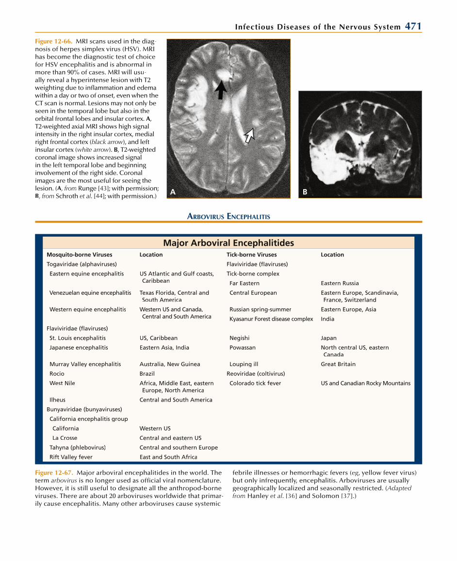
Infectious Diseases of the Nervous System 471
A B
Figure 12-66. MRI scans used in the diag-nosis of herpes simplex virus (HSV). MRIhas become the diagnostic test of choice for HSV encephalitis and is abnormal in more than 90% of cases. MRI will usu-ally reveal a hyperintense lesion with T2 weighting due to inflammation and edema within a day or two of onset, even when theCT scan is normal. Lesions may not only be seen in the temporal lobe but also in theorbital frontal lobes and insular cortex. A, T2-weighted axial MRI shows high signal intensity in the right insular cortex, medialright frontal cortex (black arrow), and leftwinsular cortex (white arrow). w B, T2-weightedcoronal image shows increased signal in the left temporal lobe and beginninginvolvement of the right side. Coronalimages are the most useful for seeing thelesion. (A, fromA Runge [43]; with permission; B, from Schroth et al. [44]; with permission.)
ARBOVIRUS ENCEPHALITIS
Major Arboviral EncephalitidesMosquito-borne Viruses Location Tick-borne Viruses Location
Togaviridae (alphaviruses) Flaviviridae (flaviruses)
Eastern equine encephalitis US Atlantic and Gulf coasts, Caribbean
Tick-borne complex
Far Eastern Eastern Russia
Venezuelan equine encephalitis Texas Florida, Central and South America
Central European Eastern Europe, Scandinavia, France, Switzerland
Western equine encephalitis Western US and Canada, Central and South America
Russian spring-summer Eastern Europe, Asia
Kyasanur Forest disease complex India
Flaviviridae (flaviruses)
St. Louis encephalitis US, Caribbean Negishi Japan
Japanese encephalitis Eastern Asia, India Powassan North central US, eastern Canada
Murray Valley encephalitis Australia, New Guinea Louping ill Great Britain
Rocio Brazil Reoviridae (coltivirus)
West Nile Africa, Middle East, eastern Europe, North America
Colorado tick fever US and Canadian Rocky Mountains
Ilheus Central and South America
Bunyaviridae (bunyaviruses)
California encephalitis group
California Western US
La Crosse Central and eastern US
Tahyna (phlebovirus) Central and southern Europe
Rift Valley fever East and South Africa
Figure 12-67. Major arboviral encephalitides in the world. Theterm arbovirus is no longer used as official viral nomenclature. However, it is still useful to designate all the anthropod-borne viruses. There are about 20 arboviruses worldwide that primar-ily cause encephalitis. Many other arboviruses cause systemic
febrile illnesses or hemorrhagic fevers (eg, yellow fever virus) but only infrequently, encephalitis. Arboviruses are usuallygeographically localized and seasonally restricted. (Adapted from Hanley et al. [36] and Solomon [37].)

472 Atlas of Clinical Neurology
A B
Figure 12-68. West Nile virus neuroinva-sive disease. West Nile virus (WNV) infec-tions appeared in New York City in August 1999. Since then, the virus has spread to allof the continental United States, Canada,and Mexico. WNV is now the most com-mon cause of epidemic viral encephalitis in the United States with nearly 20,000 confirmed cases. Symptomatic infections include generalized illness (West Nilefever) and West Nile neuroinvasive disease (WNND), which includes meningitis,encephalitis, brainstem encephalitis, andpoliomyelitis-like acute flaccid paralysis.On cerebrospinal fluid (CSF) analysis,about 40% of patients with WNND have aneutrophil predominance in the initial CSF
RABIES
Clinical Manifestations of Rabies
Finding %
Fever 73
Dysphagia 58
Altered mental state 55
Pain, paresthesia referable to site of exposure
45
Excitement, agitation 45
Paralysis, weakness 26
Hydrophobia 21
Hypersalivation 16
Nausea, vomiting 18.6
Malaise 16.3
Dyspnea 14
Headache 14
Convulsions, spasma 9.1
Coma 4.5
Miscellaneous (lethargy, dysuria, anorexia, hydrophobia)
16.3
No history of rabies exposure 16
Figure 12-69. Clinical manifestations of rabies, frequency during the course ofthe disease. At onset, about half of the patients have pain or paresthesia at the bite site. Other initial manifestationsinclude fever, malaise, anorexia, anddrowsiness. (Adapted from Robinson [46].)
Clinical Progression of RabiesStage Duration Clinical Manifestations
Incubationperiod
30–90 d: ~ 50% of cases
< 30 d: ~ 25%
> 90 d to 1 y: ~ 5%
> 1 y: ~ 5%
No clinical findings
Prodrome andearly clinical symptoms
2–10 d Paresthesia and/or pain at site of bite
Fever, malaise
Anorexia, nausea, vomiting
Headache
Acuteneurologic disease
2–7 d “Furious rabies” (80% of cases)
Hallucinations, bizarre behavior, anxiety, agitation, biting
Autonomic dysfunction
“Paralytic rabies” (20% of cases)
Flaccid paralysis
Paresis and plegias
Ascending paralysis
Coma 0–14 d SIADH
Diabetes insipidus
Multiorgan failure
Respiratory or cardiac failure
Death (common) Variable —
Recovery (rare) Variable Several sequelae
Figure 12-70. Clinical progression of rabies. The clinical course of rabies consists of fivestages. The prolonged incubation period lasts more than 90 days in 25% and more than1 year in 5% of cases. After the initial manifestations with lethargy, a state of excitability ensues, when external stimuli may cause focal or generalized convulsions. Spasmodiccontractions of the larynx and pharynx are precipitated by any attempt to drink or eat,thus the term hydrophobia. During this stage the temperature may reach 105° to 107° F. This hyperexcitability stage passes into the comatose stage, with generalized paralysis.Occasionally, the disease begins as “dumb rabies,” in which flaccid paralysis of one ormore limbs occurs rather than a hyperexcitable period. Death is usually caused by respi-ratory paralysis followed by cardiovascular collapse. SIADH—syndrome of inappropriate secretion of antidiuretic hormone. (Adapted from Rupprecht and Hemachudha [47].)
examination. Diagnosis depends primarily on finding specific IgM antibodies in the CSF.Neuroimaging studies may help with diagnosis. MRI abnormalities have been reported in 20% to 70% of cases and appear to increase over the first week of illness. Abnormali-ties are usually seen in deep gray matter structures (basal ganglia, thalami), brainstem,and cerebellum on T2, FLAIR, and diffusion-weighted images (DWI). MRI DWI images on day 6 of WNV encephalitis in thalami (A) and substantive nigra (B). There is no spe-cific treatment at the present time. (From Davis et al. [45]; with permission.)
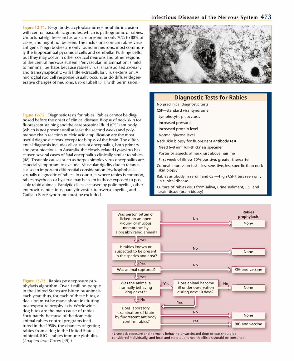
Infectious Diseases of the Nervous System 473Figure 12-71. Negri body, a cytoplasmic eosinophilic inclusionwith central basophilic granules, which is pathognomic of rabies. Unfortunately, these inclusions are present in only 70% to 80% ofcases, and might not be seen. The inclusions contain rabies virusantigens. Negri bodies are only found in neurons, most common-ly the hippocampal pyramidal cells and cerebellar Purkinje cells,but they may occur in other cortical neurons and other regions of the central nervous system. Perivascular infl ammation is mild to minimal, perhaps because rabies virus is transported axonallyand transsynaptically, with little extracellular virus extension. A microglial rod cell response usually occurs, as do diffuse degen-erative changes of neurons. (From Jubelt [31]; with permission.)
Diagnostic Tests for RabiesNo preclinical diagnostic tests
CSF—standard viral syndrome
Lymphocytic pleocytosis
Increased pressure
Increased protein level
Normal glucose level
Neck skin biopsy for fl uorescent antibody test
Need 6–8 mm full-thickness specimen
Posterior aspects of neck just above hairline
First week of illness 50% positive, greater thereafter
Corneal impression test—less sensitive, less specifi c than neckskin biopsy
Rabies antibody in serum and CSF—high CSF titers seen only in clinical disease
Culture of rabies virus from saliva, urine sediment, CSF andbrain tissue (brain biopsy)
Figure 12-72. Diagnostic tests for rabies. Rabies cannot be diag-nosed before the onset of clinical disease. Biopsy of neck skin for fl uorescent staining and the cerebrospinal fl uid (CSF) antibody(which is not present until at least the second week) and poly-merase chain reaction nucleic acid amplifi cation are the mostuseful diagnostic tests, except for biopsy of the brain. The differ-ential diagnosis includes all causes of encephalitis, both primaryand postinfectious. In Australia, the closely related Lyssavirus has caused several cases of fatal encephalitis clinically similar to rabies[48]. Treatable causes such as herpes simplex virus encephalitis areespecially important to exclude. Muscular rigidity due to tetanusis also an important differential consideration. Hydrophobia is virtually diagnostic of rabies. In countries where rabies is common, rabies psychosis or hysteria may be seen in those exposed to pos-sibly rabid animals. Paralytic disease caused by poliomyelitis, otherenterovirus infections, paralytic zoster, transverse myelitis, andGuillain-Barré syndrome must be excluded.
Was person bitten or licked on an openwound or mucous
membranes bya possibly rabid animal?
Is rabies known or suspected to be present in the species and area?
Was animal captured?
Was the animal anormally behaving
dog or cat?*None
NNoRIG and vaccine
YeYessYYes
YeYessYYes
Does laboratory examination of brain
by fluorescent antibodyconfirm rabies? Yes
Yes
Does animal becomeill under observationduring next 10 days?
NoNo
Livestock exposure and normally behaving unvaccinated dogs or cats should be*Livestock exposure and normally behaving unvaccinated dogs or cats should bed bconsidered individually, and local and state public health officials should be consulted.
Rabiesprophylaxis
YeYessYYes
NoNoNNo
NoneNoNo
NoneNo
NoneNo
RIG and vaccine
YYesYYesFigure 12-73. Rabies postexposure pro-phylaxis algorithm. Over 1 million peoplein the United States are bitten by animals each year; thus, for each of these bites, a decision must be made about instituting postexposure prophylaxis. Worldwide,dog bites are the main cause of rabies.Fortunately, because of the domesticanimal rabies control programs insti-tuted in the 1950s, the chances of gettingrabies from a dog in the United States isminimal. RIG—rabies immune globulin.(Adapted from Corey [49].)
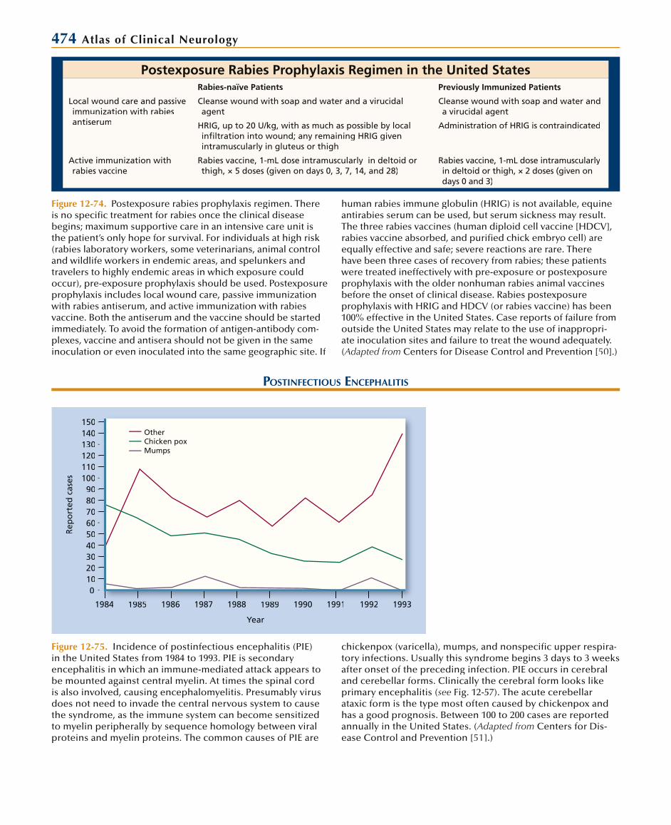
474 Atlas of Clinical Neurology
Postexposure Rabies Prophylaxis Regimen in the United StatesRabies-naïve Patients Previously Immunized Patients
Local wound care and passive immunization with rabiesantiserum
Cleanse wound with soap and water and a virucidalagent
Cleanse wound with soap and water anda virucidal agent
HRIG, up to 20 U/kg, with as much as possible by localinfi ltration into wound; any remaining HRIG given intramuscularly in gluteus or thigh
Administration of HRIG is contraindicated
Active immunization with rabies vaccine
Rabies vaccine, 1-mL dose intramuscularly in deltoid orthigh, × 5 doses (given on days 0, 3, 7, 14, and 28)
Rabies vaccine, 1-mL dose intramuscularlyin deltoid or thigh, × 2 doses (given on days 0 and 3)
Figure 12-74. Postexposure rabies prophylaxis regimen. There is no specifi c treatment for rabies once the clinical diseasebegins; maximum supportive care in an intensive care unit is the patient’s only hope for survival. For individuals at high risk(rabies laboratory workers, some veterinarians, animal control and wildlife workers in endemic areas, and spelunkers and travelers to highly endemic areas in which exposure could occur), pre-exposure prophylaxis should be used. Postexposureprophylaxis includes local wound care, passive immunizationwith rabies antiserum, and active immunization with rabiesvaccine. Both the antiserum and the vaccine should be started immediately. To avoid the formation of antigen-antibody com-plexes, vaccine and antisera should not be given in the sameinoculation or even inoculated into the same geographic site. If
human rabies immune globulin (HRIG) is not available, equineantirabies serum can be used, but serum sickness may result.The three rabies vaccines (human diploid cell vaccine [HDCV], rabies vaccine absorbed, and purifi ed chick embryo cell) areequally effective and safe; severe reactions are rare. Therehave been three cases of recovery from rabies; these patientswere treated ineffectively with pre-exposure or postexposureprophylaxis with the older nonhuman rabies animal vaccinesbefore the onset of clinical disease. Rabies postexposure prophylaxis with HRIG and HDCV (or rabies vaccine) has been 100% effective in the United States. Case reports of failure fromoutside the United States may relate to the use of inappropri-ate inoculation sites and failure to treat the wound adequately.(Adapted from Centers for Disease Control and Prevention [50].)
POSTINFECTIOUS ENCEPHALITIS
Figure 12-75. Incidence of postinfectious encephalitis (PIE)in the United States from 1984 to 1993. PIE is secondaryencephalitis in which an immune-mediated attack appears to be mounted against central myelin. At times the spinal cordis also involved, causing encephalomyelitis. Presumably virusdoes not need to invade the central nervous system to causethe syndrome, as the immune system can become sensitizedto myelin peripherally by sequence homology between viralproteins and myelin proteins. The common causes of PIE are
chickenpox (varicella), mumps, and nonspecifi c upper respira-tory infections. Usually this syndrome begins 3 days to 3 weeks after onset of the preceding infection. PIE occurs in cerebral and cerebellar forms. Clinically the cerebral form looks like primary encephalitis (see Fig. 12-57). The acute cerebellar ataxic form is the type most often caused by chickenpox and has a good prognosis. Between 100 to 200 cases are reportedannually in the United States. (Adapted from Centers for Dis-ease Control and Prevention [51].)
1984984 19851985 19861986 19871987 19881988 19891989 19901990 19911991 19921992 19931993
0102030405060708090
100110120130140150
Rep
ort
ed c
ases
Year
OtherChicken poxMumps

Infectious Diseases of the Nervous System 475
Figure 12-76. Proton-density MRI scan showing demyelination in postinfectious encephalitis (PIE) that occurred 2 weeks after a nonspecifi c upper respiratory infection. The best diagnostic test for PIE is MRI, which demonstrates white matter disease.Whether the form is cerebral or cerebellar, the cerebrospinal fl uid (CSF) profi le issimilar to that of other acute viral syndromes, with lymphocytic pleocytosis, normalglucose level, elevated protein level, and pressure. One CSF test that may be help-ful is the myelin basic protein level, which is usually elevated; unfortunately, in mostplaces, it takes 1 to 2 weeks to receive results. Treatment should be started once the diagnosis is made based on the clinical presentation, including causative disease, aCSF picture consistent with encephalitis, and a positive MRI scan. Treatment consistsof high-dose intravenous steroids (1.0 g methylprednisolone daily for 7 to 10 days) to stop the perivascular demyelination. Plasma exchange and IVIg have also been usedfor treatment [ 52 ]. (Courtesy of B. Jubelt, MD.)f
POLIOMYELITIS, MYELITIS, AND RADICULITIS
Figure 12-77. Correlation of clinical forms of poliomyelitis with the time of viral replication and antibody produc-tion. The term poliomyelitis is from the Greek words for gray marrow (“polio”) and spinal cord (“myelitis”): “the graymarrow of the spinal cord.” Sometimesthe term anterior is added as a reminderr that it is the anterior rather than the pos-terior horns that are infl amed in polio-myelitis. Only about 1% to 2% of thoseinfected with the virus develop paralysis(major illness) with or without the non-specifi c, systemic, febrile minor illness. Paralysis is usually asymmetric and fl ac-cid, and can occur in all four extremi-ties, as well as in the brain stem, causingbulbar polio with cranial nerve palsies, respiratory insuffi ciency, dysphagia, and coma. The use of oral polio vaccine haseradicated poliomyelitis caused by wildtype virus in the United States and otherdeveloped countries. Killed polio vaccineis now used in the United States for vac-cination. However, poliomyelitis is still a signifi cant problem in underdevelopedareas. CNS—central nervous system. (Adapted from Horstmann [ 53 ].)
Major illness(CNS involved)
Minor illness(nonspecific)
?
ersist for lifeP
May persistM2–17 weeks1
ersist 1–5 years(?)P
napparentIn
AbortiveA
rank casesF
Percent of allinfected
1%–2%
4%–8%
90%–95%
Virus present in:Blood
ThroatFeces
CNS (frank cases)
Antibodies presentNeutralizing
Composition fixing
Days after exposure
00 11 55 1010 1515 2020
Days after exposure
00 11 55 1010 1515 2020
Days after exposure
00 11 55 1010 1515 2020
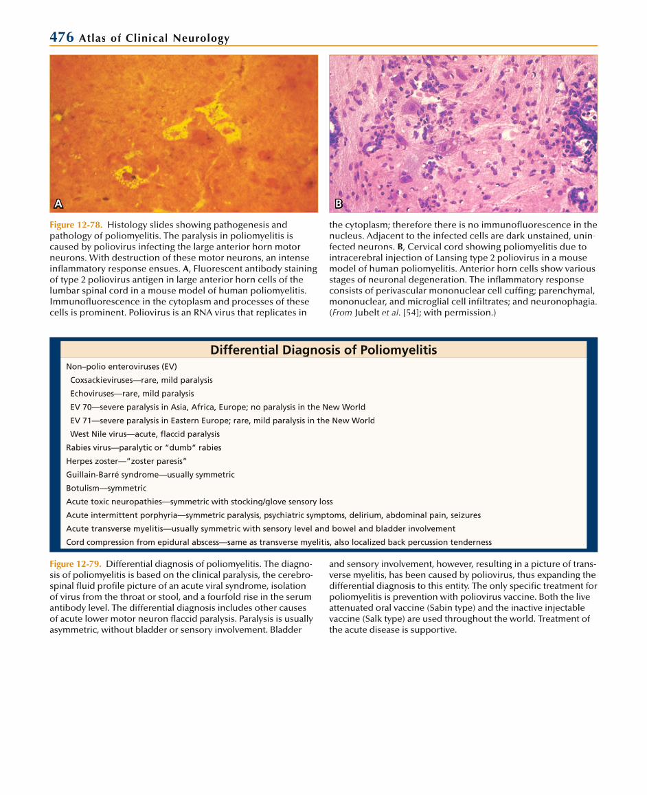
476 Atlas of Clinical Neurology
Figure 12-78. Histology slides showing pathogenesis andpathology of poliomyelitis. The paralysis in poliomyelitis is caused by poliovirus infecting the large anterior horn motorneurons. With destruction of these motor neurons, an intense inflammatory response ensues. A, Fluorescent antibody staining of type 2 poliovirus antigen in large anterior horn cells of the lumbar spinal cord in a mouse model of human poliomyelitis. Immunofluorescence in the cytoplasm and processes of these cells is prominent. Poliovirus is an RNA virus that replicates in
the cytoplasm; therefore there is no immunofluorescence in thenucleus. Adjacent to the infected cells are dark unstained, unin-fected neurons. B, Cervical cord showing poliomyelitis due to intracerebral injection of Lansing type 2 poliovirus in a mouse model of human poliomyelitis. Anterior horn cells show various stages of neuronal degeneration. The inflammatory response consists of perivascular mononuclear cell cuffing; parenchymal, mononuclear, and microglial cell infiltrates; and neuronophagia. (From Jubelt et al. [54]; with permission.)
BA
Differential Diagnosis of PoliomyelitisNon–polio enteroviruses (EV)
Coxsackieviruses—rare, mild paralysis
Echoviruses—rare, mild paralysis
EV 70—severe paralysis in Asia, Africa, Europe; no paralysis in the New World
EV 71—severe paralysis in Eastern Europe; rare, mild paralysis in the New World
West Nile virus—acute, flaccid paralysis
Rabies virus—paralytic or “dumb” rabies
Herpes zoster—”zoster paresis”
Guillain-Barré syndrome—usually symmetric
Botulism—symmetric
Acute toxic neuropathies—symmetric with stocking/glove sensory loss
Acute intermittent porphyria—symmetric paralysis, psychiatric symptoms, delirium, abdominal pain, seizures
Acute transverse myelitis—usually symmetric with sensory level and bowel and bladder involvement
Cord compression from epidural abscess—same as transverse myelitis, also localized back percussion tenderness
Figure 12-79. Differential diagnosis of poliomyelitis. The diagno-sis of poliomyelitis is based on the clinical paralysis, the cerebro-spinal fluid profile picture of an acute viral syndrome, isolation of virus from the throat or stool, and a fourfold rise in the serumantibody level. The differential diagnosis includes other causes of acute lower motor neuron flaccid paralysis. Paralysis is usually asymmetric, without bladder or sensory involvement. Bladder
and sensory involvement, however, resulting in a picture of trans-verse myelitis, has been caused by poliovirus, thus expanding thedifferential diagnosis to this entity. The only specific treatment forpoliomyelitis is prevention with poliovirus vaccine. Both the liveattenuated oral vaccine (Sabin type) and the inactive injectablevaccine (Salk type) are used throughout the world. Treatment ofthe acute disease is supportive.

Infectious Diseases of the Nervous System 477
Viruses Causing Acute Transverse Myelitis*Common Uncommon Rare
Herpes simplex virus type 2CytomegalovirusVaricella-zoster virusEpstein-Barr virus
MumpsInfluenzaRubella
PoliovirusCoxsackievirusEchovirusRabiesHepatitis A and BMeasles
*Viruses causing encephalomyelitis, in which myelitis occurs only with encephalitis, are not listed.
ACUTE TRANSVERSE MYELITIS
Figure 12-80. Viruses causing acute trans-verse myelitis. The syndrome of “acute transverse myelitis” consists of acute flaccid paralysis with hypoflexia (later may developspasticity with hyperreflexia), sensory loss usually with a sensory level, and bowel andbladder involvement. The paralysis is sym-metric and thus different from the usualasymmetric paralysis seen in poliomyelitis. In addition to viruses, this clinical syndromemay be caused by cord compression from epidural abscess or epidural tumor thathemorrhages; intraspinal abscess or intra-spinal hemorrhaging tumor; spinal cordinfarct; vasculitis (eg, systemic lupus erythegg -
Cytomegalovirus Neurologic InfectionsDisease and Features Host and Frequency
Cytomegalic inclusion body disease
EncephalitisMicroencephalySeizuresMental retardationPeriventricular calcifications
Disseminated disease
Neonate, congenital disease—rare
Encephalitis/ventriculitis
Subacute course (1–3 mo)
Immunocompromised patient (described primarilyin AIDS patients)—uncommon
Progressive mental status changesDisseminated disease in the immunocompromised patientMRI—periventricular hyperintensities, meningeal enhancement
Immunocompromised patient—rare
Polyradiculitis/polyradiculomyelitisPain and paresthesia in legs and perineumSacral hypesthesiaUrinary retentionSubacute ascending hypotonic paraparesisEventually ascend to cause myelitisCSF—pleocytosis (PMNs > lymphocytes), low glucose level, high protein level, CMV positive by culture or PCR
Usually disseminated diseaseMRI—lumbosacral leptomeningeal enhancement
Immunocompromised patient (described only inAIDS patients)—common
Multifocal neuropathyMarkedly asymmetricNumbness, painful paresthesia for months, followed by sensorimotor neuropathyUsually disseminated diseaseCMV positive in CSF by culture or PCR
Immunocompromised patient (described only inAIDS)—uncommon
CYTOMEGALOVIRUS POLYRADICULITIS
Figure 12-81. -or to the occurrence of the AIDS epidemic in the United States, CMV was known primarily for causing cytomegalic inclusionbody disease, an infrequent congenital disease of newborns. Cases of CMV encephalitis have been recognized rarely in immu-nocompetent individuals. CMV ventriculitis and encephalitisoccur most often as an opportunistic infection in AIDS patients. The most common syndrome caused by CMV today, however, is
the CMV polyradiculitis (polyradiculopathy)/polyradiculomyelitis (polyradiculomyelopathy). This syndrome occurs relatively late in the course of AIDS when the CD4+ T-lymphocyte count is usually less than 100. Less frequent and also late in the course of AIDS is CMV multifocal neuropathy. Ganciclovir and foscarnet have been reported to be beneficial in some postnatally acquired infections [56]. CSF—cerebrospinal fluid; PCR—polymerase chain reaction; PMNs—polymorphonuclear leukocytes.
matosus) causing infarction; postinfectious immune-mediated myelitis; multiple sclerosis;and remote effects of cancer. Treatment consists of the use of a specific viral agent whenthe agent is identified with our without high-dose intravenous methylprednisolone [55].

478 Atlas of Clinical Neurology
A B
Figure 12-82. Enhanced T1-weighted sagittal magnetic reso-nance image of nerve roots from L1 to L4 and the cauda equinaregion (arrows) in a patient with cytomegalovirus polyradiculi-tis/polyradiculomyelitis (A). In the right half of the picture, the pial lining of the sac is also enhanced. Usually these lesionsare visible only with enhancement (B). (From Talpes et al. [57];with permission.)
Figure 12-83. Herpes zoster. Herpes zoster is a distinctive syn-drome caused by the varicella-zoster virus (VZV), which also causes chickenpox (varicella). The chickenpox virus travels fromthe skin up the sensory nerves to become latent in the dorsalroot ganglia. Later in life, the virus reactivates, replicates in theganglia, and travels down the nerve to the skin to cause a derma-tomal vesicular eruption. The incidence of zoster increases with age and is more common in those with compromised cellular
immunity. Typically, patients experience dermatomal paresthesia or dysesthesias (itching, burning pain, tingling) for 1 to 2 days,after which the vesicular eruption occurs. Most patients havehypalgesia and hypesthesia in the affected dermatome. A, Herpes zoster in a thoracic dermatome. B, Herpes zoster ophthalmicus. Herpes zoster of the ophthalmic division of the trigeminal nerve. (A, fromA Murray et al. [58]; with permission; B, from Rosencrance[59]; with permission.)
BA
HERPES ZOSTER
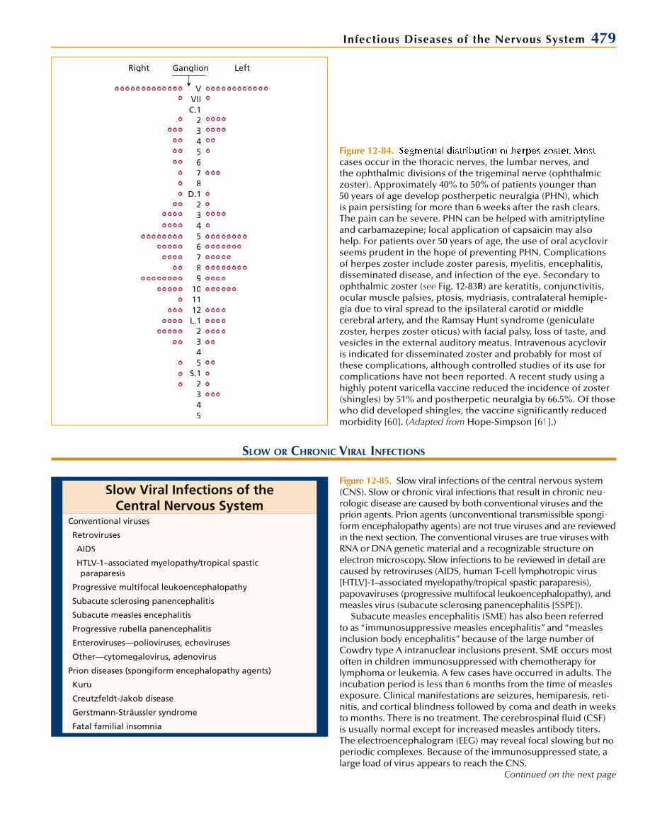
Infectious Diseases of the Nervous System 479
VVIIC.1
2345678
D.123456789
101112L.1
2345
S.12345
Ganglion LeftRight
Figure 12-84. cases occur in the thoracic nerves, the lumbar nerves, andthe ophthalmic divisions of the trigeminal nerve (ophthalmic zoster). Approximately 40% to 50% of patients younger than50 years of age develop postherpetic neuralgia (PHN), whichis pain persisting for more than 6 weeks after the rash clears. The pain can be severe. PHN can be helped with amitriptyline and carbamazepine; local application of capsaicin may also help. For patients over 50 years of age, the use of oral acyclovirseems prudent in the hope of preventing PHN. Complications of herpes zoster include zoster paresis, myelitis, encephalitis,disseminated disease, and infection of the eye. Secondary toophthalmic zoster (see Fig. 12-83B) are keratitis, conjunctivitis,ocular muscle palsies, ptosis, mydriasis, contralateral hemiple-gia due to viral spread to the ipsilateral carotid or middlecerebral artery, and the Ramsay Hunt syndrome (geniculatezoster, herpes zoster oticus) with facial palsy, loss of taste, andvesicles in the external auditory meatus. Intravenous acyclovir is indicated for disseminated zoster and probably for most of these complications, although controlled studies of its use forcomplications have not been reported. A recent study using ahighly potent varicella vaccine reduced the incidence of zoster (shingles) by 51% and postherpetic neuralgia by 66.5%. Of those who did developed shingles, the vaccine significantly reducedmorbidity [60]. (Adapted from Hope-Simpson [61].)
SLOW OR CHRONIC VIRAL INFECTIONS
Slow Viral Infections of the Central Nervous System
Conventional viruses
Retroviruses
AIDS
HTLV-1–associated myelopathy/tropical spastic paraparesis
Progressive multifocal leukoencephalopathy
Subacute sclerosing panencephalitis
Subacute measles encephalitis
Progressive rubella panencephalitis
Enteroviruses—polioviruses, echoviruses
Other—cytomegalovirus, adenovirus
Prion diseases (spongiform encephalopathy agents)
Kuru
Creutzfeldt-Jakob disease
Gerstmann-Sträussler syndrome
Fatal familial insomnia
Figure 12-85. Slow viral infections of the central nervous system (CNS). Slow or chronic viral infections that result in chronic neu-rologic disease are caused by both conventional viruses and the prion agents. Prion agents (unconventional transmissible spongi-form encephalopathy agents) are not true viruses and are reviewed in the next section. The conventional viruses are true viruses with RNA or DNA genetic material and a recognizable structure on electron microscopy. Slow infections to be reviewed in detail arecaused by retroviruses (AIDS, human T-cell lymphotropic virus[HTLV]-1–associated myelopathy/tropical spastic paraparesis),papovaviruses (progressive multifocal leukoencephalopathy), and measles virus (subacute sclerosing panencephalitis [SSPE]).
Subacute measles encephalitis (SME) has also been referred to as “immunosuppressive measles encephalitis” and “measles inclusion body encephalitis” because of the large number ofCowdry type A intranuclear inclusions present. SME occurs mostoften in children immunosuppressed with chemotherapy forlymphoma or leukemia. A few cases have occurred in adults. Theincubation period is less than 6 months from the time of measles exposure. Clinical manifestations are seizures, hemiparesis, reti-nitis, and cortical blindness followed by coma and death in weeksto months. There is no treatment. The cerebrospinal fluid (CSF) is usually normal except for increased measles antibody titers.The electroencephalogram (EEG) may reveal focal slowing but noperiodic complexes. Because of the immunosuppressed state, a large load of virus appears to reach the CNS.
Continued on the next page
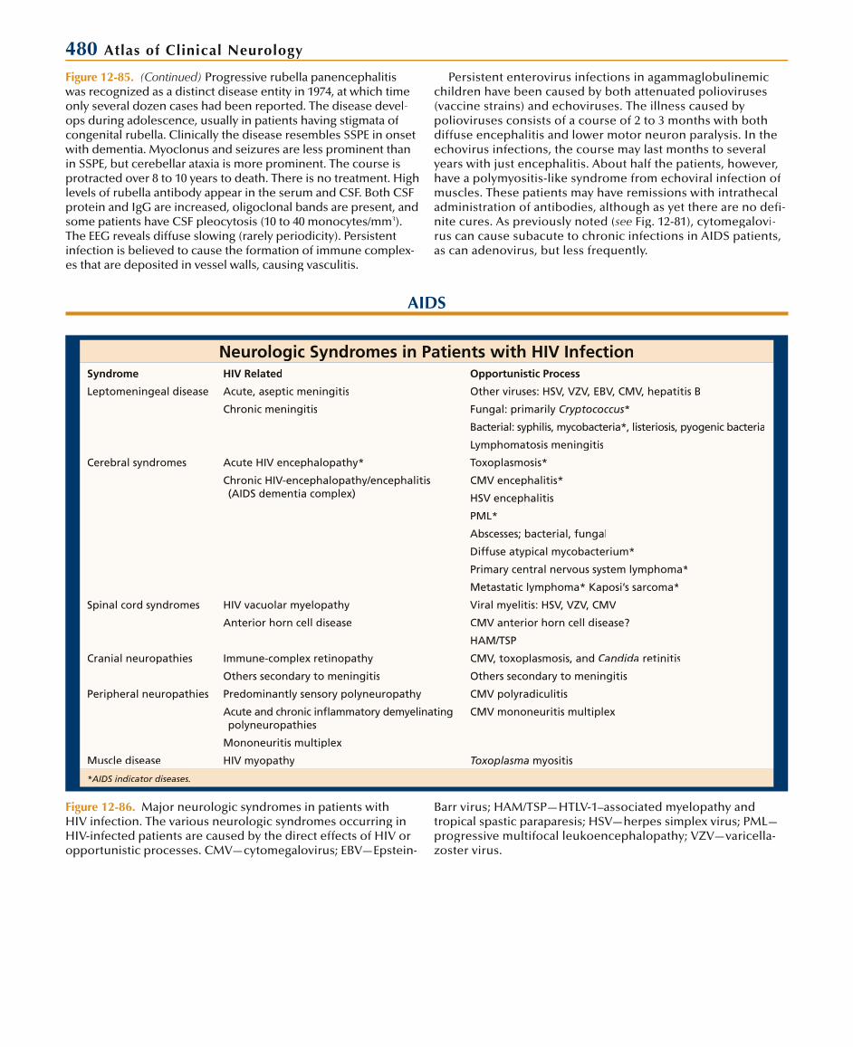
480 Atlas of Clinical Neurology
Figure 12-85. (Continued) Progressive rubella panencephalitis was recognized as a distinct disease entity in 1974, at which time only several dozen cases had been reported. The disease devel-ops during adolescence, usually in patients having stigmata ofcongenital rubella. Clinically the disease resembles SSPE in onset with dementia. Myoclonus and seizures are less prominent than in SSPE, but cerebellar ataxia is more prominent. The course is protracted over 8 to 10 years to death. There is no treatment. Highlevels of rubella antibody appear in the serum and CSF. Both CSF protein and IgG are increased, oligoclonal bands are present, and some patients have CSF pleocytosis (10 to 40 monocytes/mm3).The EEG reveals diffuse slowing (rarely periodicity). Persistent infection is believed to cause the formation of immune complex-es that are deposited in vessel walls, causing vasculitis.
Persistent enterovirus infections in agammaglobulinemicchildren have been caused by both attenuated polioviruses(vaccine strains) and echoviruses. The illness caused bypolioviruses consists of a course of 2 to 3 months with both diffuse encephalitis and lower motor neuron paralysis. In theechovirus infections, the course may last months to several years with just encephalitis. About half the patients, however,have a polymyositis-like syndrome from echoviral infection ofmuscles. These patients may have remissions with intrathecaladministration of antibodies, although as yet there are no defi-nite cures. As previously noted (see Fig. 12-81), cytomegalovi-rus can cause subacute to chronic infections in AIDS patients, as can adenovirus, but less frequently.
AIDS
Neurologic Syndromes in Patients with HIV InfectionSyndrome HIV Related Opportunistic Process
Leptomeningeal disease Acute, aseptic meningitis Other viruses: HSV, VZV, EBV, CMV, hepatitis B
Chronic meningitis Fungal: primarily Cryptococcus*
Bacterial: syphilis, mycobacteria*, listeriosis, pyogenic bacteria
Lymphomatosis meningitis
Cerebral syndromes Acute HIV encephalopathy* Toxoplasmosis*
Chronic HIV-encephalopathy/encephalitis(AIDS dementia complex)
CMV encephalitis*
HSV encephalitis
PML*
Abscesses; bacterial, fungal
Diffuse atypical mycobacterium*
Primary central nervous system lymphoma*
Metastatic lymphoma* Kaposi’s sarcoma*
Spinal cord syndromes HIV vacuolar myelopathy Viral myelitis: HSV, VZV, CMV
Anterior horn cell disease CMV anterior horn cell disease?
HAM/TSP
Cranial neuropathies Immune-complex retinopathy CMV, toxoplasmosis, and Candida retinitis
Others secondary to meningitis Others secondary to meningitis
Peripheral neuropathies Predominantly sensory polyneuropathy CMV polyradiculitis
Acute and chronic inflammatory demyelinatingpolyneuropathies
CMV mononeuritis multiplex
Mononeuritis multiplex
Muscle disease HIV myopathy Toxoplasma myositis
*AIDS indicator diseases.
Figure 12-86. Major neurologic syndromes in patients withHIV infection. The various neurologic syndromes occurring inHIV-infected patients are caused by the direct effects of HIV oropportunistic processes. CMV—cytomegalovirus; EBV—Epstein-
Barr virus; HAM/TSP—HTLV-1–associated myelopathy and tropical spastic paraparesis; HSV—herpes simplex virus; PML—progressive multifocal leukoencephalopathy; VZV—varicella-zoster virus.
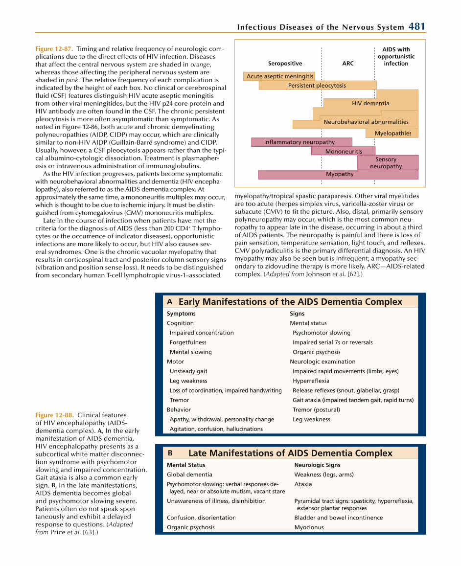
Infectious Diseases of the Nervous System 481
Seropositive ARC
AIDS withopportunistic
infection
Acute aseptic meningitis
Persistent pleocytosis
HIV dementia
Neurobehavioral abnormalities
Myelopathies
Inflammatory neuropathy
Mononeuritis
Myopathy
Sensoryneuropathy
Figure 12-87. Timing and relative frequency of neurologic com-plications due to the direct effects of HIV infection. Diseasesthat affect the central nervous system are shaded in orange, whereas those affecting the peripheral nervous system areshaded in pink. The relative frequency of each complication isindicated by the height of each box. No clinical or cerebrospinal fluid (CSF) features distinguish HIV acute aseptic meningitis from other viral meningitides, but the HIV p24 core protein and HIV antibody are often found in the CSF. The chronic persistentpleocytosis is more often asymptomatic than symptomatic. Asnoted in Figure 12-86, both acute and chronic demyelinating polyneuropathies (AIDP, CIDP) may occur, which are clinicallysimilar to non-HIV AIDP (Guillain-Barré syndrome) and CIDP.Usually, however, a CSF pleocytosis appears rather than the typi-cal albumino-cytologic dissociation. Treatment is plasmapher-esis or intravenous administration of immunoglobulins.
As the HIV infection progresses, patients become symptomaticwith neurobehavioral abnormalities and dementia (HIV encepha-lopathy), also referred to as the AIDS dementia complex. At approximately the same time, a mononeuritis multiplex may occur, which is thought to be due to ischemic injury. It must be distin-guished from cytomegalovirus (CMV) mononeuritis multiplex.
Late in the course of infection when patients have met thecriteria for the diagnosis of AIDS (less than 200 CD4+ T lympho-cytes or the occurrence of indicator diseases), opportunisticinfections are more likely to occur, but HIV also causes sev-eral syndromes. One is the chronic vacuolar myelopathy thatresults in corticospinal tract and posterior column sensory signs(vibration and position sense loss). It needs to be distinguishedfrom secondary human T-cell lymphotropic virus-1–associated
Early Manifestations of the AIDS Dementia ComplexSymptoms Signs
Cognition Mental status
Impaired concentration Psychomotor slowing
Forgetfulness Impaired serial 7s or reversals
Mental slowing Organic psychosis
Motor Neurologic examination
Unsteady gait Impaired rapid movements (limbs, eyes)
Leg weakness Hyperreflexia
Loss of coordination, impaired handwriting Release reflexes (snout, glabellar, grasp)
Tremor Gait ataxia (impaired tandem gait, rapid turns)
Behavior Tremor (postural)
Apathy, withdrawal, personality change Leg weakness
Agitation, confusion, hallucinations
Late Manifestations of AIDS Dementia ComplexMental Status Neurologic Signs
Global dementia Weakness (legs, arms)
Psychomotor slowing: verbal responses de-layed, near or absolute mutism, vacant stare
Ataxia
Unawareness of illness, disinhibition Pyramidal tract signs: spasticity, hyperreflexia, extensor plantar responses
Confusion, disorientation Bladder and bowel incontinence
Organic psychosis Myoclonus
Figure 12-88. Clinical featuresof HIV encephalopathy (AIDS-dementia complex). A, In the early manifestation of AIDS dementia,HIV encephalopathy presents as asubcortical white matter disconnec-tion syndrome with psychomotor slowing and impaired concentration. Gait ataxia is also a common early sign. B, In the late manifestations, AIDS dementia becomes global and psychomotor slowing severe. Patients often do not speak spon-taneously and exhibit a delayedresponse to questions. (Adapted from Price et al. [63].)
A
B
myelopathy/tropical spastic paraparesis. Other viral myelitidesare too acute (herpes simplex virus, varicella-zoster virus) or subacute (CMV) to fit the picture. Also, distal, primarily sensory polyneuropathy may occur, which is the most common neu-ropathy to appear late in the disease, occurring in about a third of AIDS patients. The neuropathy is painful and there is loss ofpain sensation, temperature sensation, light touch, and reflexes. CMV polyradiculitis is the primary differential diagnosis. An HIVmyopathy may also be seen but is infrequent; a myopathy sec-ondary to zidovudine therapy is more likely. ARC—AIDS-related complex. (Adapted from Johnson et al. [62].)
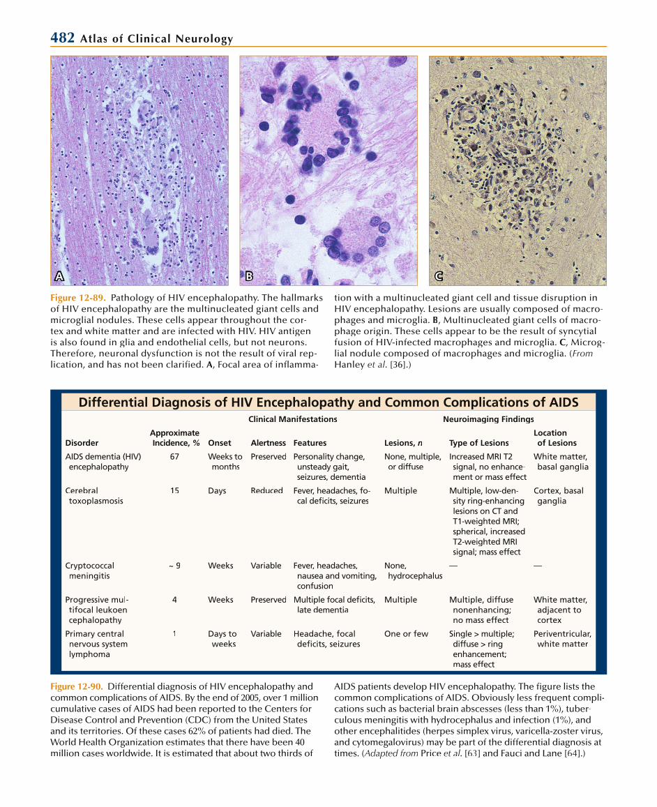
482 Atlas of Clinical Neurology
Figure 12-89. Pathology of HIV encephalopathy. The hallmarksof HIV encephalopathy are the multinucleated giant cells andmicroglial nodules. These cells appear throughout the cor-tex and white matter and are infected with HIV. HIV antigen is also found in glia and endothelial cells, but not neurons. Therefore, neuronal dysfunction is not the result of viral rep-lication, and has not been clarified. A, Focal area of inflamma-
tion with a multinucleated giant cell and tissue disruption in HIV encephalopathy. Lesions are usually composed of macro-phages and microglia. B, Multinucleated giant cells of macro-phage origin. These cells appear to be the result of syncytial fusion of HIV-infected macrophages and microglia. C, Microg-lial nodule composed of macrophages and microglia. (FromHanley et al. [36].)
A B C
Differential Diagnosis of HIV Encephalopathy and Common Complications of AIDS
DisorderApproximate Incidence, %
Clinical Manifestations Neuroimaging Findings
Onset Alertness Features Lesions, n Type of LesionsLocation of Lesions
AIDS dementia (HIV)encephalopathy
67 Weeks to months
Preserved Personality change, unsteady gait,seizures, dementia
None, multiple, or diffuse
Increased MRI T2signal, no enhance-ment or mass effect
White matter, basal ganglia
Cerebraltoxoplasmosis
15 Days Reduced Fever, headaches, fo-cal deficits, seizures
Multiple Multiple, low-den-sity ring-enhancing lesions on CT and T1-weighted MRI; spherical, increased T2-weighted MRI signal; mass effect
Cortex, basal ganglia
Cryptococcalmeningitis
~ 9 Weeks Variable Fever, headaches, nausea and vomiting,confusion
None, hydrocephalus
— —
Progressive mul-tifocal leukoen-cephalopathy
4 Weeks Preserved Multiple focal deficits, late dementia
Multiple Multiple, diffusenonenhancing; no mass effect
White matter, adjacent to cortex
Primary centralnervous system lymphoma
1 Days to weeks
Variable Headache, focal deficits, seizures
One or few Single > multiple;diffuse > ring enhancement; mass effect
Periventricular, white matter
Figure 12-90. Differential diagnosis of HIV encephalopathy andcommon complications of AIDS. By the end of 2005, over 1 millioncumulative cases of AIDS had been reported to the Centers forDisease Control and Prevention (CDC) from the United States and its territories. Of these cases 62% of patients had died. The World Health Organization estimates that there have been 40million cases worldwide. It is estimated that about two thirds of
AIDS patients develop HIV encephalopathy. The figure lists thecommon complications of AIDS. Obviously less frequent compli-cations such as bacterial brain abscesses (less than 1%), tuber-culous meningitis with hydrocephalus and infection (1%), andother encephalitides (herpes simplex virus, varicella-zoster virus,and cytomegalovirus) may be part of the differential diagnosis attimes. (Adapted from Price et al. [63] and Fauci and Lane [64].)
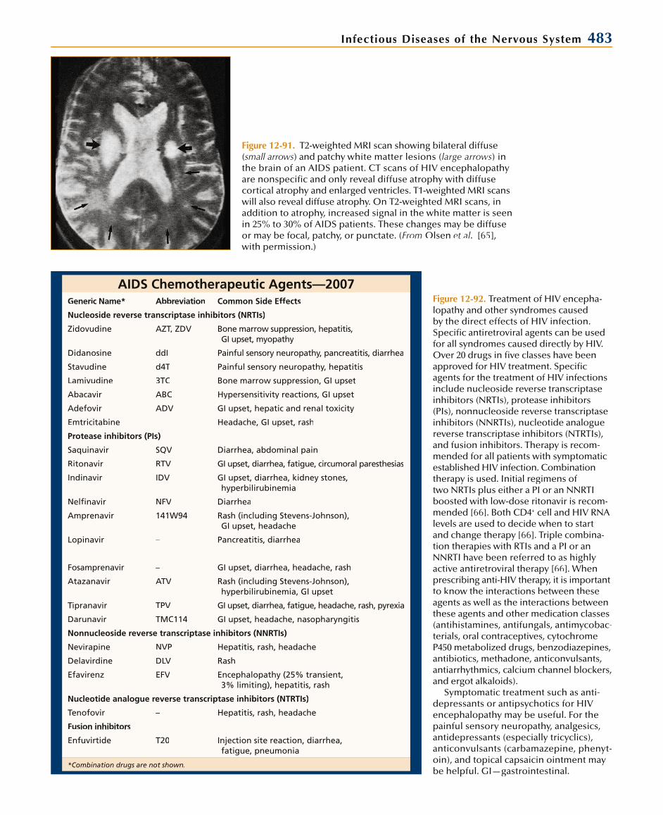
Infectious Diseases of the Nervous System 483
Figure 12-91. T2-weighted MRI scan showing bilateral diffuse(small arrows) and patchy white matter lesions (large arrows) in the brain of an AIDS patient. CT scans of HIV encephalopathyare nonspecific and only reveal diffuse atrophy with diffusecortical atrophy and enlarged ventricles. T1-weighted MRI scanswill also reveal diffuse atrophy. On T2-weighted MRI scans, in addition to atrophy, increased signal in the white matter is seenin 25% to 30% of AIDS patients. These changes may be diffuseor may be focal, patchy, or punctate. (From Olsen et al. [65],with permission.)
AIDS Chemotherapeutic Agents—2007Generic Name* Abbreviation Common Side Effects
Nucleoside reverse transcriptase inhibitors (NRTIs)
Zidovudine AZT, ZDV Bone marrow suppression, hepatitis,GI upset, myopathy
Didanosine ddI Painful sensory neuropathy, pancreatitis, diarrhea
Stavudine d4T Painful sensory neuropathy, hepatitis
Lamivudine 3TC Bone marrow suppression, GI upset
Abacavir ABC Hypersensitivity reactions, GI upset
Adefovir ADV GI upset, hepatic and renal toxicity
Emtricitabine – Headache, GI upset, rash
Protease inhibitors (PIs)
Saquinavir SQV Diarrhea, abdominal pain
Ritonavir RTV GI upset, diarrhea, fatigue, circumoral paresthesias
Indinavir IDV GI upset, diarrhea, kidney stones,hyperbilirubinemia
Nelfinavir NFV Diarrhea
Amprenavir 141W94 Rash (including Stevens-Johnson),GI upset, headache
Lopinavir – Pancreatitis, diarrhea
Fosamprenavir – GI upset, diarrhea, headache, rash
Atazanavir ATV Rash (including Stevens-Johnson),hyperbilirubinemia, GI upset
Tipranavir TPV GI upset, diarrhea, fatigue, headache, rash, pyrexia
Darunavir TMC114 GI upset, headache, nasopharyngitis
Nonnucleoside reverse transcriptase inhibitors (NNRTIs)
Nevirapine NVP Hepatitis, rash, headache
Delavirdine DLV Rash
Efavirenz EFV Encephalopathy (25% transient,3% limiting), hepatitis, rash
Nucleotide analogue reverse transcriptase inhibitors (NTRTIs)
Tenofovir – Hepatitis, rash, headache
Fusion inhibitors
Enfuvirtide T20 Injection site reaction, diarrhea,fatigue, pneumonia
*Combination drugs are not shown.
Figure 12-92. Treatment of HIV encepha-lopathy and other syndromes caused by the direct effects of HIV infection.Specific antiretroviral agents can be used for all syndromes caused directly by HIV.Over 20 drugs in five classes have beenapproved for HIV treatment. Specific agents for the treatment of HIV infectionsinclude nucleoside reverse transcriptaseinhibitors (NRTIs), protease inhibitors(PIs), nonnucleoside reverse transcriptase inhibitors (NNRTIs), nucleotide analogue reverse transcriptase inhibitors (NTRTIs),and fusion inhibitors. Therapy is recom-mended for all patients with symptomatic established HIV infection. Combination therapy is used. Initial regimens of two NRTIs plus either a PI or an NNRTIboosted with low-dose ritonavir is recom-mended [66]. Both CD4+ cell and HIV RNA levels are used to decide when to startand change therapy [66]. Triple combina-tion therapies with RTIs and a PI or anNNRTI have been referred to as highlyactive antiretroviral therapy [66]. When prescribing anti-HIV therapy, it is important to know the interactions between theseagents as well as the interactions between these agents and other medication classes(antihistamines, antifungals, antimycobac-terials, oral contraceptives, cytochrome P450 metabolized drugs, benzodiazepines,antibiotics, methadone, anticonvulsants, antiarrhythmics, calcium channel blockers,and ergot alkaloids).
Symptomatic treatment such as anti-depressants or antipsychotics for HIVencephalopathy may be useful. For the painful sensory neuropathy, analgesics,antidepressants (especially tricyclics),anticonvulsants (carbamazepine, phenyt-oin), and topical capsaicin ointment maybe helpful. GI—gastrointestinal.

484 Atlas of Clinical Neurology
Neurologic Signs of HAM/TSPAbnormal Signs Affected, %
Corticospinal signs
Legs
Spasticity 100
Weakness 90–100
Arms
Spasticity 60–90
Weakness 20–50
Increased jaw jerk 30–70
Bladder dysfunction 70–90
Impaired position, vibration sense 10–60
Root or cord sensation 20–65
Optic atrophy 2–20
Cerebellar signs 3–10
HTLV-1–ASSOCIATED MYELOPATHY/TROPICAL SPASTIC PARAPARESIS
Figure 12-93. Neurologic signs of human T-cell lymphotropic virus (HTLV)-1–associated myelopathy and tropical spastic para-paresis (HAM/TSP). HAM/TSP is characterized by a chronic pro-gressive spastic paraparesis, usually with a neurogenic bladder. Patients may have paresthesia and pain in the legs. Both poste-rior column and spinothalamic sensory loss have been seen but usually in a minority of cases. Infrequently, cranial nerve (CN) signs are seen, with optic atrophy the most common. Other CNsigns include nystagmus, diplopia, deafness, and facial paresis.Antibodies to HTLV-1 have been found in 80% to 100% of cases, and HTLV-1 has been isolated from the cerebrospinal fluid. The pathogenesis appears more likely to be an immune-mediated process, as cytotoxic T lymphocytes rather than HTLV-1 viraltiter levels are related to the demyelinating lesions. Pathologic changes include inflammation of the leptomeninges, perivas-cular cuffing, and inflammation of the cord parenchyma. Sym-metrical demyelination of the corticospinal tracts occurs, and toa lesser degree and frequency, demyelination of the posterior columns and spinothalamic and spinocerebellar tracts. Muchless frequently, patients with HTLV-1 infections have presentedwith an ALS-like picture, polyneuropathy, and inflammatory and noninflammatory myopathies [67].
Most cases of HAM/TSP occur in warm areas along theequator, where the virus is endemic. Isolated cases have occurred in more northern and southern latitudes, includingnorthern areas of the United States. The disease usually begins in the third or fourth decade and women are more commonly
Differential Diagnosis of HAM/TSPCervical or thoracic cord compression Nutritional disorders
Chiari malformations Combined systems disease(B12 deficiency)
Foramen magnum tumors Vitamin E (tocopherol) deficiency
Cervical spondylosis Lathyrisin
Cervical and thoracic herniated discs Neoplastic disease
Arteriovenous malformations Extramedullary (metastatic) or intramed-ullary tumors (primary or metastatic)
Motor neuron disease or related syndromes Paraneoplastic myelopathy
Amyotrophic lateral sclerosis Other disorders
Primary lateral sclerosis Adrenoleukodystrophy
Hereditary spastic paraplegia Hepatic myelopathy
Inflammatory disorders
Multiple sclerosis
Systemic lupus erythematosus
Sarcoid
Infectious diseases
HIV myelopathy
Bacterial disease
Syphilis
Tuberculosis (cord tuberculoma, compression)
Parasites
Schistosomiasis
Strongyloidosis
Figure 12-94. Differential diagnosis ofhuman T-cell lymphotropic virus (HTLV)-1–associated myelopathy and tropical spastic paraparesis (HAM/TSP). The dif-ferential diagnosis of HAM/TSP is that of a chronic spastic progressive paraparesis.The diagnosis can be made by analysis of the cerebrospinal fluid (CSF). Less thanhalf of patients have mild pleocytosis,while more than half have an elevatedprotein level. Specific CSF tests includethe presence of HTLV-1 antibody and HTLV-1 oligoclonal bands, virus isolation from the CSF, and the detection of the HTLV-1 genome using polymerase chain reaction amplification. MRI may reveal nonspecific demyelination in the spinal cord and brain. For treatment, prednisoneand danazol have been reported to bebeneficial but have not been tested in acontrolled fashion. In a recent double-blind, controlled study, however, two thirds of the patients reported a benefit from 3.0 mil-lion units per day of interferon- . (Adapted from Izumo et al. [69].)
affected, with a female to male ratio of about 2 to 1. The virusappears to be transmitted by vertical transmission from the mother to the infant through breastfeeding, sexual contact, intravenous drug use, and blood transfusions. For cases reportedfrom the United States, except for the endemic area of South Florida, 20% to 25% of the time the mode of transmission cannotbe determined. (Adapted from Rodgers-Johnson et al. [68].)

Infectious Diseases of the Nervous System 485
PROGRESSIVE MULTIFOCAL LEUKOENCEPHALOPATHY
Clinical Manifestations of Progressive Multifocal Leukoencephalopathy
Multifocal symptoms and signs
Hemiparesis
Hemianopsia
Hemisensory deficit
Aphasia
Limb ataxia
Gait ataxia
Dysarthria
Late mental status changes
Personality changes
Dementia
Figure 12-95. Clinical manifestations of progressive multifocalleukoencephalopathy (PML). PML is caused by the opportunisticJC papovavirus. Multiple areas of demyelination occur, lead-ing to the accumulation of different focal deficits (multifocal). When the lesions become extensive throughout the whitematter, mental status changes ensue. PML occurs in immunosup-pressed individuals. Before the AIDS epidemic, it was seen most often in those treated for lymphoproliferative disease (leuke-mia/lymphoma). PML is much more common in AIDS patients with a prevalence of about 2.5%. Recently, PML was reported in two multiple sclerosis patients enrolled in a clinical trial who received combination therapy of natalizumab and interferon- -1a[70]. The disease progresses to death in months. There is no specific treatment.
Figure 12-96. Photomicrograph showing the multiple pale areasof demyelination that are usually well-circumscribed in progres-sive multifocal leukoencephalopathy. These areas contain mac-rophages and virally infected astrocytes and oligodendroglia(lower power, myelin stain). The opportunistic JC papovavirus infects oligodendroglia of immunocompromised patients. This results in multiple areas of demyelination, which are greaterin the cerebral white matter than the brain stem and cerebel-lum. Hyperplasia of astrocytes also occurs, and eosinophilic intranuclear inclusions are visible in enlarged oligodendroglianuclei. (Courtesy of Richard Johnson, MD, Johns Hopkins Medif -cal School, Baltimore, MD.)
Figure 12-97. Diagnosis of progressive multifocal leukoencepha-lopathy (PML). T2-weighted axial MRI scan, with increased signal intensity in the left frontal lobe and corona radiata with extensionacross the midline in a patient with PML. As is characteristic, thesubcortical U-fibers are involved. No specific findings occur in the cerebrospinal fluid (CSF), which is usually normal. The elec-troencephalographic changes are nonspecific. Serologic testingis not helpful, as most adults already have antibody to JC virus. Polymerase chain reaction amplification of JC viral DNA from the CSF is positive in about 80% of patients with PML. CT scansreveal low-density nonenhancing lesions without mass effect.MRI scans reveal increased signal intensity of T2-weighted, FLAIR,or diffusion-weighted images; enhancement is minimal in 10% to15% of cases. Biopsy of the brain may be necessary for a defini-tive diagnosis. (From Whiteman et al. [71]; with permission.)

486 Atlas of Clinical Neurology
Clinical Stages of Subacute Sclerosing Panencephalitis (SSPE)Stage I: Cerebral signs (mental, behavioral) Stage III: Coma, opisthotonus
Irritability No responsiveness to any stimulus
Affectionate displays Extensor hypertonus
Lethargy Decerebrate rigidity
Forgetfulness Irregular, stertorous respiration
Indifference Stage IV: Mutism, loss of cerebral cortex function, myoclonus
Withdrawal Pathologic laughter, crying
Drooling Wandering of eyes
Regressive speech Flexion of upper and lower limbs
Slurred speech Hypotonia
Stage II: Convulsive, motor signs Turning of head to one side
Myoclonus of head, limbs, trunk Occasional limb myoclonus
Incoordination of trunk, limbs Startling by noise
Dyskinesia—choreoathetoid postures, movements, tremors
SUBACUTE SCLEROSING PANENCEPHALITIS
Figure 12-98. Clinical stages of subacute sclerosing panencepha-litis (SSPE). SSPE has become a rare disease in the United States,with fewer than 10 cases per year, since the introduction of measles vaccine. Most patients had their measles infection prior to the ageof 2 years, followed by a latent period before the onset of disease,which occurs between the ages of 5 to 15 years in 85% of patients.The disease usually begins with poor school performance, person-ality changes and then dementia (forgetfulness, regressive speech),which is referred to as stage I. Myoclonus, seizures, and movement disorders occur in stage II. Eventually the comatose and akinetic mutism stages ensue. Death usually occurs in 1 to 3 years, but hasranged from several months to 15 years.
SSPE apparently is caused by the lack of production of themeasles virus M protein, which is needed by the virus to bud out of the cell and spread efficiently to the next cell. This results
in a cell-associated infection. The measles virus can spread onlyby cell fusion, which is slow and inefficient. Diagnosis is made from clinical manifestations and the characteristic laboratoryabnormalities. One of the most characteristic laboratory testsis the electroencephalogram, which reveals periodic patterns of bursts occurring every 5 to 7 seconds followed by periods of background attenuation (burst-suppression). The cerebrospinal fluid gamma globulin and measles antibody titers are usu-ally elevated. CT and MRI performed late in the course reveal diffuse atrophy of the cortex and white matter, with ex vacuo ventricular enlargement. MRI usually reveals multifocal gray andwhite matter lesions. Although there is no specific treatment, intrathecal administration of interferon- with oral inosiplex has resulted in remissions. SSPE can be prevented with measles vaccination [72]. (Adapted from Ohya et al. [73].)
Prion Diseases of Humans and AnimalsDisease Host
Scrapie Sheep, goats
Transmissible mink encephalopathy Mink
Chronic wasting disease Mule deer, elk
Bovine spongiform encephalopathy Cattle
Feline spongiform encephalopathy Cats
Kuru Humans
CDJ Humans
nvCJD Humans
Gerstmann-Sträussler syndrome Humans
Fatal familial insomnia Humans
THE PRION DISEASES
Figure 12-99. Prion diseases of humans and animals. The priondiseases have also been referred to as transmissible spongiform encephalopathy agents. The first of these diseases, scrapie, wasrecognized to be transmissible in sheep in the 1930s. The lat-est epidemic of bovine spongiform encephalopathy in Britain,referred to by the lay public as “mad cow disease,” is believedto have been transmitted to humans via the ingestion of con-taminated beef resulting in a progressive dementia that has been named new variant Creutzfeldt-Jakob disease (nvCJD). Thedisease caused by nvCJD occurs in younger people and disease progression is slower [74]. Kuru is of historical interest becauseit was the first human prion disease to be recognized as trans-missible. It occurs in the Fore Indians of New Guinea beginning with gait, trunk, and limb ataxia and involuntary movements(myoclonus, chorea, tremor) followed by dementia at a later date. Because it is transmitted primarily through cannibalistic rituals, which are now restricted, kuru is rare.
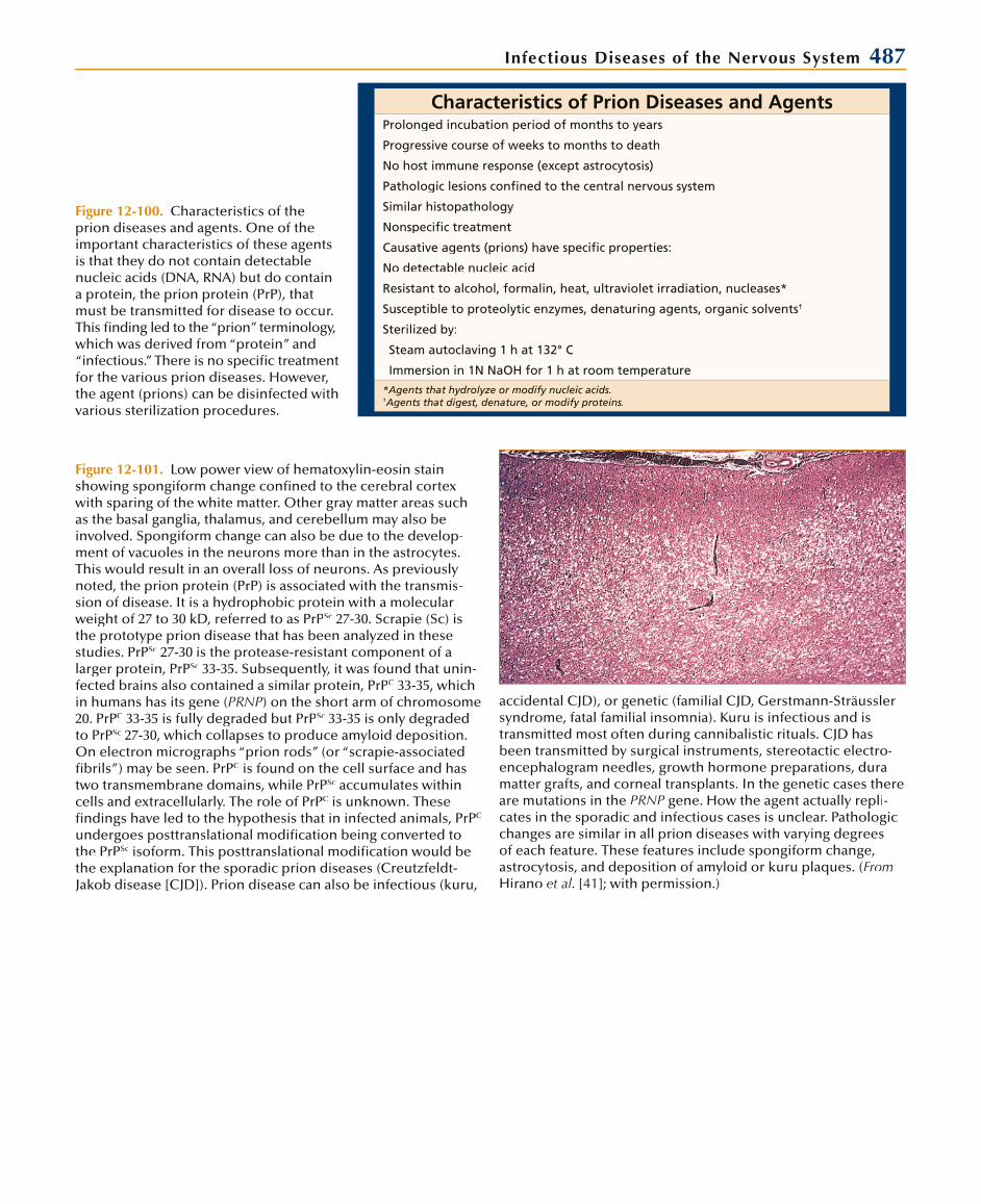
Infectious Diseases of the Nervous System 487
Characteristics of Prion Diseases and AgentsProlonged incubation period of months to years
Progressive course of weeks to months to death
No host immune response (except astrocytosis)
Pathologic lesions confined to the central nervous system
Similar histopathology
Nonspecific treatment
Causative agents (prions) have specific properties:
No detectable nucleic acid
Resistant to alcohol, formalin, heat, ultraviolet irradiation, nucleases*
Susceptible to proteolytic enzymes, denaturing agents, organic solvents†
Sterilized by:
Steam autoclaving 1 h at 132° C
Immersion in 1N NaOH for 1 h at room temperature
*Agents that hydrolyze or modify nucleic acids.†Agents that digest, denature, or modify proteins.†
Figure 12-100. Characteristics of the prion diseases and agents. One of theimportant characteristics of these agentsis that they do not contain detectablenucleic acids (DNA, RNA) but do containa protein, the prion protein (PrP), thatmust be transmitted for disease to occur. This finding led to the “prion” terminology,which was derived from “protein” and“infectious.” There is no specific treatmentfor the various prion diseases. However,the agent (prions) can be disinfected with various sterilization procedures.
Figure 12-101. Low power view of hematoxylin-eosin stainshowing spongiform change confined to the cerebral cortexwith sparing of the white matter. Other gray matter areas suchas the basal ganglia, thalamus, and cerebellum may also beinvolved. Spongiform change can also be due to the develop-ment of vacuoles in the neurons more than in the astrocytes. This would result in an overall loss of neurons. As previously noted, the prion protein (PrP) is associated with the transmis-sion of disease. It is a hydrophobic protein with a molecular weight of 27 to 30 kD, referred to as PrPSc 27-30. Scrapie (Sc) isthe prototype prion disease that has been analyzed in thesestudies. PrPSc 27-30 is the protease-resistant component of a larger protein, PrPSc 33-35. Subsequently, it was found that unin-fected brains also contained a similar protein, PrPC 33-35, which in humans has its gene (PRNP) on the short arm of chromosome 20. PrPC 33-35 is fully degraded but PrPSc 33-35 is only degraded to PrPSc 27-30, which collapses to produce amyloid deposition. On electron micrographs “prion rods” (or “scrapie-associatedfibrils”) may be seen. PrPC is found on the cell surface and hastwo transmembrane domains, while PrPSc accumulates within cells and extracellularly. The role of PrPC is unknown. Thesefindings have led to the hypothesis that in infected animals, PrPC
undergoes posttranslational modification being converted to the PrPSc isoform. This posttranslational modification would be the explanation for the sporadic prion diseases (Creutzfeldt-Jakob disease [CJD]). Prion disease can also be infectious (kuru,
accidental CJD), or genetic (familial CJD, Gerstmann-Sträusslersyndrome, fatal familial insomnia). Kuru is infectious and istransmitted most often during cannibalistic rituals. CJD hasbeen transmitted by surgical instruments, stereotactic electro-encephalogram needles, growth hormone preparations, duramatter grafts, and corneal transplants. In the genetic cases thereare mutations in the PRNP gene. How the agent actually repliP -cates in the sporadic and infectious cases is unclear. Pathologicchanges are similar in all prion diseases with varying degrees of each feature. These features include spongiform change, astrocytosis, and deposition of amyloid or kuru plaques. (FromHirano et al. [41]; with permission.)
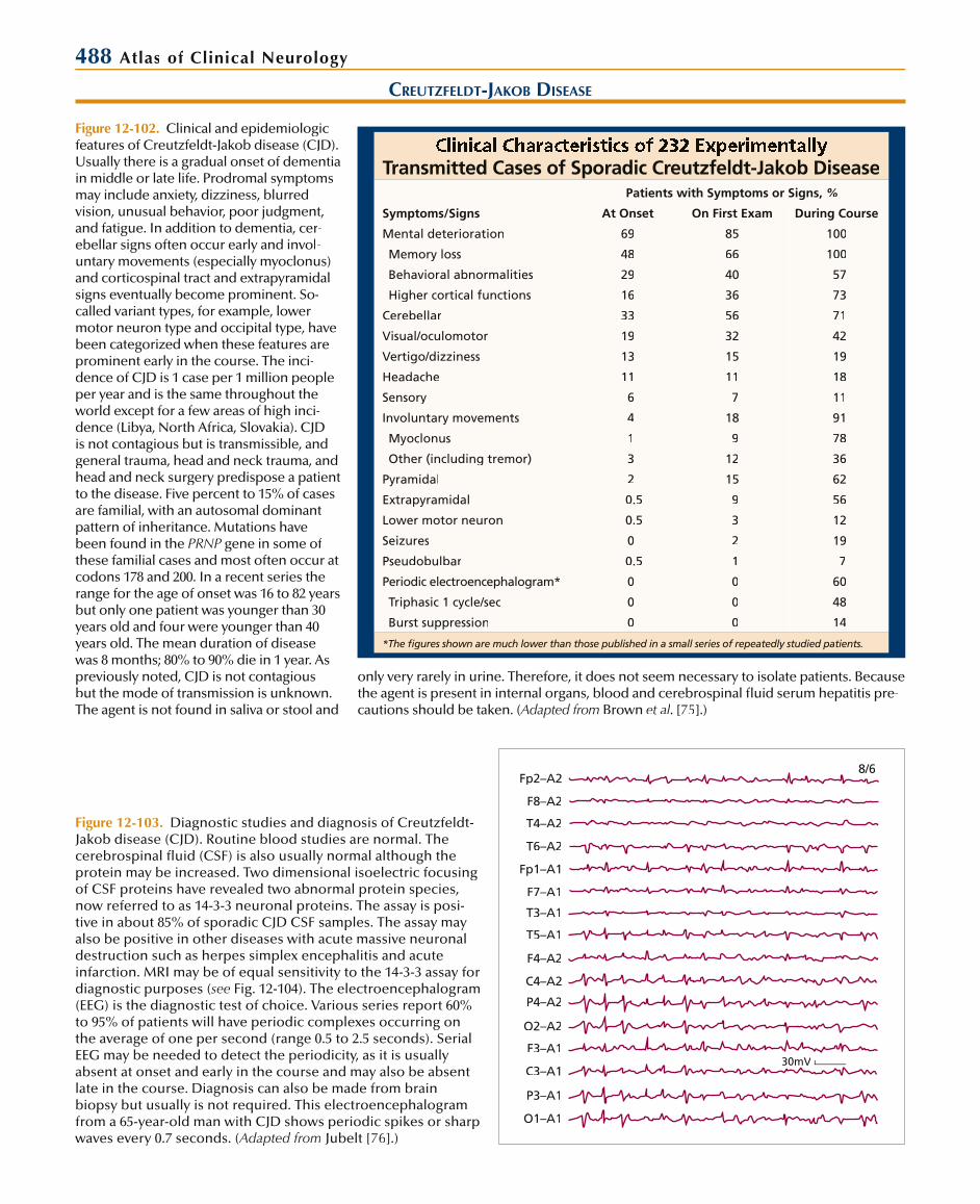
488 Atlas of Clinical Neurology
CREUTZFELDT-JAKOB DISEASE
Transmitted Cases of Sporadic Creutzfeldt-Jakob Disease
Symptoms/Signs
Patients with Symptoms or Signs, %
At Onset On First Exam During Course
Mental deterioration 69 85 100
Memory loss 48 66 100
Behavioral abnormalities 29 40 57
Higher cortical functions 16 36 73
Cerebellar 33 56 71
Visual/oculomotor 19 32 42
Vertigo/dizziness 13 15 19
Headache 11 11 18
Sensory 6 7 11
Involuntary movements 4 18 91
Myoclonus 1 9 78
Other (including tremor) 3 12 36
Pyramidal 2 15 62
Extrapyramidal 0.5 9 56
Lower motor neuron 0.5 3 12
Seizures 0 2 19
Pseudobulbar 0.5 1 7
Periodic electroencephalogram* 0 0 60
Triphasic 1 cycle/sec 0 0 48
Burst suppression 0 0 14
*The figures shown are much lower than those published in a small series of repeatedly studied patients.
Figure 12-102. Clinical and epidemiologicfeatures of Creutzfeldt-Jakob disease (CJD). Usually there is a gradual onset of dementiain middle or late life. Prodromal symptomsmay include anxiety, dizziness, blurredvision, unusual behavior, poor judgment, and fatigue. In addition to dementia, cer-ebellar signs often occur early and invol-untary movements (especially myoclonus) and corticospinal tract and extrapyramidal signs eventually become prominent. So-called variant types, for example, lower motor neuron type and occipital type, have been categorized when these features areprominent early in the course. The inci-dence of CJD is 1 case per 1 million peopleper year and is the same throughout theworld except for a few areas of high inci-dence (Libya, North Africa, Slovakia). CJDis not contagious but is transmissible, andgeneral trauma, head and neck trauma, andhead and neck surgery predispose a patientto the disease. Five percent to 15% of cases are familial, with an autosomal dominantpattern of inheritance. Mutations have been found in the PRNP gene in some ofPthese familial cases and most often occur atcodons 178 and 200. In a recent series therange for the age of onset was 16 to 82 years but only one patient was younger than 30 years old and four were younger than 40 years old. The mean duration of disease was 8 months; 80% to 90% die in 1 year. As previously noted, CJD is not contagious but the mode of transmission is unknown. The agent is not found in saliva or stool and
Fp2–A2
F8–A2
F4–A2
C4–A2
C3–A1
P3–A1
P4–A2
O2–A2
O1–A1
F3–A1
T4–A2
T6–A2
Fp1–A1
F7–A1
T3–A1
T5–A1
8/6
30mV
Figure 12-103. Diagnostic studies and diagnosis of Creutzfeldt-Jakob disease (CJD). Routine blood studies are normal. The cerebrospinal fluid (CSF) is also usually normal although the protein may be increased. Two dimensional isoelectric focusing of CSF proteins have revealed two abnormal protein species, now referred to as 14-3-3 neuronal proteins. The assay is posi-tive in about 85% of sporadic CJD CSF samples. The assay mayalso be positive in other diseases with acute massive neuronaldestruction such as herpes simplex encephalitis and acuteinfarction. MRI may be of equal sensitivity to the 14-3-3 assay for diagnostic purposes (see Fig. 12-104). The electroencephalogram (EEG) is the diagnostic test of choice. Various series report 60% to 95% of patients will have periodic complexes occurring onthe average of one per second (range 0.5 to 2.5 seconds). Serial EEG may be needed to detect the periodicity, as it is usuallyabsent at onset and early in the course and may also be absent late in the course. Diagnosis can also be made from brainbiopsy but usually is not required. This electroencephalogramfrom a 65-year-old man with CJD shows periodic spikes or sharpwaves every 0.7 seconds. (Adapted from Jubelt [76].)
only very rarely in urine. Therefore, it does not seem necessary to isolate patients. Because the agent is present in internal organs, blood and cerebrospinal fluid serum hepatitis pre-cautions should be taken. (Adapted from Brown et al. [75].)

Infectious Diseases of the Nervous System 489
Figure 12-104. MRI of Creutzfeldt-Jakob disease. Increasedsignal intensity may be seen on T2-weighted, FLAIR, and diffu-sion-weighted imaging (DWI). Two patterns may be seen. The first is a diffuse pattern primarily seen in the basal ganglia and occasionally in the thalamus. The second is a cortical ribbon orgyriform pattern in the cortex and cerebellum. DWI appears
to be more sensitive than either FLAIR or T2 imaging, with 90%to 100% sensitivity compared with about 50%. Three patternsof high intensity lesions were seen on DWI: striatal lesions (A), cerebral cortical lesions (B), and a combination of both lesions(C). (From Shiga et al. [77], with permission.)
A B C
Clinical Features of Variant Creutzfeldt-Jakob Disease
Symptoms/Signs
Patients with Symptomsor Signs, % (n = 35)
At Onset During Course
Psychiatric 63 97
Sensory symptoms 20 68
Limb pain 11 37
Ataxia 8 100
Forgetfulness 17 83
Involuntary movements 6 94
Dystonia 6 34
Chorea 0 57
Myoclonus 0 71
Upgaze paresis 0 40
Dementia 0 100
Akinetic 0 57
Figure 12-105. Clinical features of variant Creutzfeldt-Jakobdisease (vCJD). vCJD occurs at a younger age than classic CJD,with 89% dying before the age of 40. The duration of vCJD is also longer, with a median duration of illness of 14 months ascompared to 9 to 11 months for classic CJD. vCJD usually begins with psychiatric symptoms and painful sensory symptoms ratherthan memory loss. Similar to classic CJD, dementia, ataxia,and involuntary movements eventually occur (see Fig. 12-102).Another unusual feature of vCJD is that the typical periodic tri-phasic complexes seen on electroencephalograms from classicCJD patients has not been reported. MRI may reveal abnormali-ties in the thalamus, but this finding is not specific for vCJD. (Adapted from Will et al. [78].)
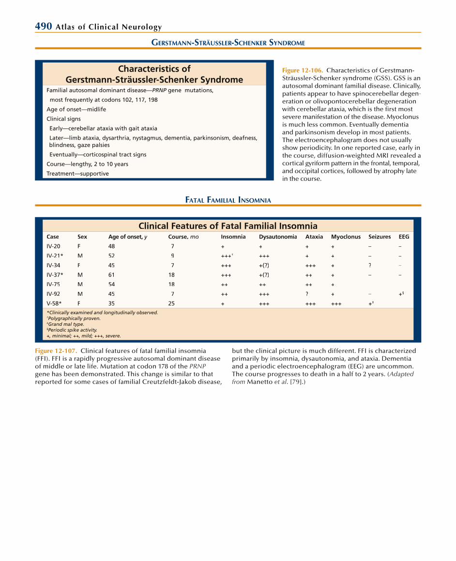
490 Atlas of Clinical Neurology
FATAL FAMILIAL INSOMNIA
Clinical Features of Fatal Familial InsomniaCase Sex Age of onset, y Course, mo Insomnia Dysautonomia Ataxia Myoclonus Seizures EEG
IV-20 F 48 7 + + + + – –
IV-21* M 52 9 +++† +++ + + – –
IV-34 F 45 7 +++ +(?) +++ + ? –
IV-37* M 61 18 +++ +(?) ++ + – –
IV-75 M 54 18 ++ ++ ++ + – –
IV-92 M 45 7 ++ +++ ? + – +§
V-58* F 35 25 + +++ +++ +++ +‡
*Clinically examined and longitudinally observed.†Polygraphically proven.‡Grand mal type.§Periodic spike activity.+, minimal; ++, mild; +++, severe.
Figure 12-107. Clinical features of fatal familial insomnia (FFI). FFI is a rapidly progressive autosomal dominant diseaseof middle or late life. Mutation at codon 178 of the PRNPgene has been demonstrated. This change is similar to that reported for some cases of familial Creutzfeldt-Jakob disease,
but the clinical picture is much different. FFI is characterized primarily by insomnia, dysautonomia, and ataxia. Dementia and a periodic electroencephalogram (EEG) are uncommon.The course progresses to death in a half to 2 years. (Adapted from Manetto et al. [79].)
GERSTMANN-STRÄUSSLER-SCHENKER SYNDROME
Characteristics of Gerstmann-Sträussler-Schenker Syndrome
Familial autosomal dominant disease—PRNP gene mutations,P
most frequently at codons 102, 117, 198
Age of onset—midlife
Clinical signs
Early—cerebellar ataxia with gait ataxia
Later—limb ataxia, dysarthria, nystagmus, dementia, parkinsonism, deafness, blindness, gaze palsies
Eventually—corticospinal tract signs
Course—lengthy, 2 to 10 years
Treatment—supportive
Figure 12-106. Characteristics of Gerstmann-Sträussler-Schenker syndrome (GSS). GSS is anautosomal dominant familial disease. Clinically,patients appear to have spinocerebellar degen-eration or olivopontocerebellar degenerationwith cerebellar ataxia, which is the first mostsevere manifestation of the disease. Myoclonusis much less common. Eventually dementia and parkinsonism develop in most patients.The electroencephalogram does not usuallyshow periodicity. In one reported case, early in the course, diffusion-weighted MRI revealed acortical gyriform pattern in the frontal, temporal, and occipital cortices, followed by atrophy late in the course.

Infectious Diseases of the Nervous System 491
FUNGAL INFECTIONS
Spectrum of Involvement for Fungi That Can Infect the CNSChief Pathologic Manifestations
Organisms Incidence Predilection to Involve the CNS* Meningitis Abscess or Inflammatory Mass Infarct
Cryptococcus Common ++++ ++++ + +
Coccidioides Common +++ ++++ + +
Candida Common ++ ++ ++ —
Aspergillus Occasional ++ + +++ ++++
Zygomycetes† Occasional ++ + +++ ++++
Histoplasma Occasional + + + +
Blastomyces Occasional + + + —
Sporothrix Occasional + + — —
Paracoccidioides Rare ± ± ± —
Dematiaceous fungi Rare +++ ± ++++ —
Pseudallescheria Rare + ++ ++ —
* Versus other body sites.†The class of Zygomycetes or Phycomycetes includes genera Rhizopus and Mucor.++++, common; ±, rare; —does not occur.
Figure 12-108. Clinical syndromes and frequencies of fungalinfections that can affect the central nervous system (CNS). The clinical syndromes caused by fungi invading the CNS can bedivided into meningitis, abscess or inflammatory mass (granu-loma formations), and arterial thrombosis causing infarction.Fungi exist in two forms: yeasts and molds. Yeasts are unicellular organisms that have a thick cell wall surrounded by a well-defined
capsule (see Fig. 12-112). Molds are composed of tubular filaments that sometimes have a branched form (hyphae). In the brain,dimorphic and fungal yeasts are more likely to cause meningitis, while molds are more likely to cause vasculitis with subsequentthrombosis and infarction. The major pathogenic molds are species of the genus Aspergillus and the class of Zygomycetes.(Adapted from Perfect [80].)
Factors Predisposing to Fungal CNS InfectionsPredisposing Factors Typical Organisms
Prematurity Candida
Inherited immune defects CGD, SCID, etc.
Candida, Cryptococcus, Aspergillus
Acquired immune defects
Corticosteroids Cryptococcus, Candida
Cytotoxic agents Aspergillus, Candida
HIV infection Cryptococcus, Histoplasma
Alcoholism Sporothrix
Hematologic malignancies Candida, Aspergillus, Cryptococcus, Histoplasma
Iron chelator therapy Zygomycetes
Deferoxamine
Intravenous drug abuse Zygomycetes, Candida
Diabetic ketoacidosis Zygomycetes, Candida
Trauma, surgery, foreign body, near-drowning
Candida, Pseudallescheria, dematiaceous fungi
Figure 12-109. Predisposing factors to fungal infections of the central nervous system (CNS). As previously noted, most fungal infections are opportunistic. Spe-cific factors predispose to specific fungal infections. CGD—chronic granulomatous disease; SCID—severe combined immunedeficiency. (Adapted from Tunkel and Crous [81] and Gozdasoglu et al. [82].)

492 Atlas of Clinical Neurology
Laboratory Studies in Non-AIDS and AIDS Patients
Laboratory Findings Non-AIDS, % AIDS, %
Positive blood culture — 30–63
Positive serum cryptococcal antigen 66 99
CSF opening pressure > 200 mm H2O 72 62
CSF glucose < 2.2 mmol/L (40 mg/dL) 73 33
CSF protein > 0.45 g/L (45 mg/dL) 89 58
CSF leukocytes > 20 × 106/L 70 23
Positive CSF India ink preparation 60 74
Positive CSF culture 96 95
Positive CSF cryptococcal antigen 86 91–100
Figure 12-112. An India ink preparation used in the diagnosisof cryptococcal meningitis that demonstrates the prominentcapsule of Cryptococcus neoformans. The India ink test is posi-tive 50% to 60% of the time, and slightly more frequently inpatients with AIDS (see Fig. 12-111B). (From Tunkel and Crous [81]; with permission.)
CRYPTOCOCCAL MENINGITIS
Figure 12-111. Clinical presentations (A) and laboratory stud-ies (B) of cryptococcal meningitis. Cryptococcus is the most frequent cause of fungal meningitis in both non-AIDS andAIDS patients. It is a chronic meningitis, but in AIDS patients it may progress even more slowly and present only with fever and headache instead of the usual manifestations of meningealsigns, mental status changes, and cranial nerve palsies.
The usual cerebrospinal fluid (CSF) profile in cryptococcalmeningitis, as well as most causes of fungal meningitis, is
that of mononuclear (lymphocytic) pleocytosis, a low glucoselevel, and an elevated protein level. AIDS patients, however,often do not fit this typical CSF picture. Most striking is the fact that 65% of AIDS patients have a normal CSF cell countof fewer than 5 cells/mm3. Diagnosis is confirmed by positive CSF culture result or a positive CSF cryptococcal antigen test.Polymerase chain reaction techniques are in development butappear to be more sensitive than culture. (Adapted from Tunkeland Scheld [84].)
FUNGAL MENINGITIS
Signs and Symptoms of Fungal MeningitisFungal Organism Fever (> 101° F) Headache Stiff Neck Change in Mentation Focal Signs Visual Disturbance
Blastomyces + +++ +++ + ++ +
Candida +++ +++ ++ + + +
Coccidioides + +++ + ++ ++ ++
Cryptococcus + +++ +++ + + +++
Histoplasma ++ + ++ + + +
Sporothrix + ++ ++ ++ + ?
+, rare; ++, occasionally to moderately frequently; +++, usually.
Figure 12-110. Signs and symptoms of fungal meningitis. Thesymptoms and signs of fungal meningitis, one of the many
causes of chronic meningitis, vary somewhat depending on the specific organism. (Adapted from Tucker and Ellner [83].)
Clinical Presentation in Non-AIDS and AIDS Patients
Clinical Presentation Non-AIDS, % AIDS, %
Headache 87 81
Fever 60 88
Nausea, vomiting, malaise 53 38
Mental status changes 52 19
Meningeal signs 50 31
Visual changes, photophobia 33 19
Seizures 15 8
No symptoms or signs 10 12
AB
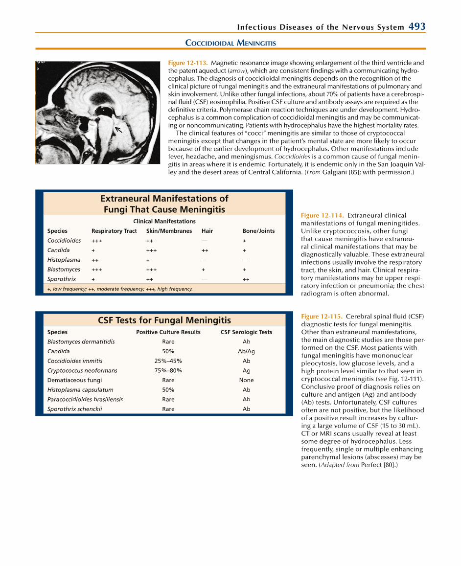
Infectious Diseases of the Nervous System 493
COCCIDIOIDAL MENINGITIS
Figure 12-113. Magnetic resonance image showing enlargement of the third ventricle and the patent aqueduct (arrow), which are consistent findings with a communicating hydrow -cephalus. The diagnosis of coccidioidal meningitis depends on the recognition of theclinical picture of fungal meningitis and the extraneural manifestations of pulmonary and skin involvement. Unlike other fungal infections, about 70% of patients have a cerebrospi-nal fluid (CSF) eosinophilia. Positive CSF culture and antibody assays are required as the definitive criteria. Polymerase chain reaction techniques are under development. Hydro-cephalus is a common complication of coccidioidal meningitis and may be communicat-ing or noncommunicating. Patients with hydrocephalus have the highest mortality rates.
The clinical features of “cocci” meningitis are similar to those of cryptococcalmeningitis except that changes in the patient’s mental state are more likely to occur because of the earlier development of hydrocephalus. Other manifestations include fever, headache, and meningismus. Coccidioides is a common cause of fungal menin-gitis in areas where it is endemic. Fortunately, it is endemic only in the San Joaquin Val-ley and the desert areas of Central California. (From Galgiani [85]; with permission.)
Extraneural Manifestations of Fungi That Cause Meningitis
Clinical Manifestations
Species Respiratory Tract Skin/Membranes Hair Bone/Joints
Coccidioides +++ ++ — +
Candida + +++ ++ +
Histoplasma ++ + — —
Blastomyces +++ +++ + +
Sporothrix + ++ — ++
+, low frequency; ++, moderate frequency; +++, high frequency.
CSF Tests for Fungal MeningitisSpecies Positive Culture Results CSF Serologic Tests
Blastomyces dermatitidis Rare Ab
Candida 50% Ab/Ag
Coccidioides immitis 25%–45% Ab
Cryptococcus neoformans 75%–80% Ag
Dematiaceous fungi Rare None
Histoplasma capsulatum 50% Ab
Paracoccidioides brasiliensis Rare Ab
Sporothrix schenckii Rare Ab
Figure 12-115. Cerebral spinal fluid (CSF)diagnostic tests for fungal meningitis. Other than extraneural manifestations, the main diagnostic studies are those per-formed on the CSF. Most patients withfungal meningitis have mononuclearpleocytosis, low glucose levels, and a high protein level similar to that seen in cryptococcal meningitis (see Fig. 12-111). Conclusive proof of diagnosis relies onculture and antigen (Ag) and antibody(Ab) tests. Unfortunately, CSF culturesoften are not positive, but the likelihood of a positive result increases by cultur-ing a large volume of CSF (15 to 30 mL).CT or MRI scans usually reveal at least some degree of hydrocephalus. Lessfrequently, single or multiple enhancing parenchymal lesions (abscesses) may be seen. (Adapted from Perfect [80].)
Figure 12-114. Extraneural clinical manifestations of fungal meningitides. Unlike cryptococcosis, other fungithat cause meningitis have extraneu-ral clinical manifestations that may be diagnostically valuable. These extraneuralinfections usually involve the respiratorytract, the skin, and hair. Clinical respira-tory manifestations may be upper respi-ratory infection or pneumonia; the chest radiogram is often abnormal.

494 Atlas of Clinical Neurology
Differential Diagnosis of Fungal Meningitis SyndromeInfectious Noninfectious
Bacterial Neoplasm
Tuberculosis Sarcoidosis
Spirochetal (Lyme disease, syphilis, Leptospira) Vasculitis
Agents that cause sinus tracts (Actinomyces, Arachnia, Nocardia(( ) Primary central nervous system angiitis
Brurcellosis Systemic (giant cell arteritis, systemic lupus erythematosus, Sjögren’s syndrome, rheumatoid arthritis, lymphomatoid granulomatosis,polyarteritis nodosa, Wegener’s granulomatosis)
Listeria monocytogenes Behçet’s disease
Fungal Chemical meningitis
Common (Candida, Coccidioides, Cryptococcus, Histoplasma) Endogenous
Uncommon (Aspergillus, Blastomyces,(( dematiaceous fungi,Paracoccidioides, Pseudallescheria, Sporothrix, Mucormycetes)ss
Exogenous
Intrathecal administration of systemic drugs
Parasitic Chronic benign lymphocytic meningitis
Cysticercosis Idiopathic hypertrophic pachymeningitis
Granulomatous amebic meningoencephalitis (acanthamoebiasis) Vogt-Koyanagi-Harada disease
Eosinophilic meningitis (angiostrongyloidiasis)
Toxoplasmosis
Coenurus cerebralis
Viral
Retrovirus (HIV-1, human T-cell lymphotropic virus [HTLV-1])
Enterovirus (in hypogammaglobulinemia)
Parameningeal infections—epidural abscess, subdural empyema, brain abscess
Figure 12-116. Differential diagnosis of fungal meningitis syndrome. Fungal meningitis is a subacute to chronic process with a course lasting over weeks to months, and the differen-
tial diagnosis of chronic meningitis is extensive. (Adapted fromHildebrand and Hildebrand [86].)
Primary Antifungal Therapy for Fungal MeningitidesAspergillus Voriconazole
Blastomyces dermatitidis Amphotericin B then itraconazole
Candida Amphotericin B plus 5-FC then fluconazole
Coccidioides immitis Fluconazole then amphotericin B
Cryptococcus neoformans Amphotericin B plus 5-FC then fluconazole
Histoplasma capsulatum Amphotericin B then itraconazole
Sporothrix schenckii Amphotericin B then itraconazole
Zygomycetes Amphotericin B or liposomal amphotericin B
Figure 12-117. Treatment of fungal men-ingitis. Amphotericin B has been the main drug used for treatment of fungal men-ingitis for over 30 years. It remains theagent of choice alone or in combination with other treatment for most species.Amphotericin is primarily given intrave-nously, rarely intrathecally, for Coccidi-oides. For many species, this is followedwith suppressive therapy with flucon-azole or itraconazole. 5-FC—5-fluorocyto-sine. (Adapted from Perfect [80].)

Infectious Diseases of the Nervous System 495
Etiology of CNS Fungal AbscessSpecies Distribution of Cases % Years of Survey Species Distribution of Cases,% Years of Survey
Total cases, 39 1964–1973 Total cases, 61* 1956–1985
Candida 49 Candida 44
Cryptococcus 23 Aspergillus 28
Mucormycoses 13 Cryptococcus 23
Aspergillus 5 Mucormycoses 3
Histoplasmosis 5 Histoplasmosis 2
Total cases, 11 1955–1971 Total cases, 57† 1984–1992
Aspergillus 64 Aspergillus
Mucormycoses 27 Candida
Candida 9 Mucormycoses
*Includes meningitis plus abscess cases.†Bone marrow transplant recipients.
FUNGAL ABSCESS AND INFARCTION
Figure 12-118. Etiology of fungal abscess of the central nervous system (CNS). Over the past 30 years fungal abscess has becomemore frequent due to the use of broad-spectrum antibiotics and immunosuppressive agents as well as the AIDS epidemic. Treat-
ment may require agents to decrease intracranial pressure(mannitol, corticosteroids), surgical decompression, andantifungal chemotherapy agents (see Fig. 12-117). (Adapted from Sepkowitz and Armstrong [87].)
Figure 12-119. Contrast-enhancedCT scan that shows an aspergillomain a 9-year-old boy with glioblastoma multiforme. For focal central nervoussystem fungal infections, CT and MRIare the diagnostic tests of choice. The cerebrospinal fluid analysis may becontraindicated because of increased intracranial pressure with a focal lesion. Culture specimens must be obtained at the time of surgical drainage and decompression. (From Sepkowitz andArmstrong [87]; with permission.)
Pathogenic Molds in the Central Nervous SystemAspergillus Species Mucorales Pseudallescheria boydii
Patients at risk Hematologic neoplasm Diabetes withketoacidosis (> 70%)
Near-drowning
Neutropenia on broad-spectrum antibiotics
Hematologic neoplasm Intravenous drug use
Cortiocosteroids Neutropenia on broad-spectrum antibiotics
Neutropenia on broad-spectrum antibiotics
Organ transplants Renal transplant Hematologic malignancy
Intravenous drug use Intravenous drug use Head trauma
Liver disease Deferoxamine therapy
Postcraniotomy Acidosis
Subtropical farming
Pathogenesis Hematogenous Rhinocerebral Hematogenous
Direct extension, including rhinocerebral (rare)
Hematogenous Direct extension
Traumatic implantation
Microscopicappearance
Septate hyphae Nonseptate hyphae Narrow septate hyphaewith rare branching
Acute branching Broad right-anglebranching
Narrow septate hyphaewith rare branching
Culture from CSF Rare Seldom Occasional
Treatment Surgery Surgery Surgery
Amphotericin B Amphotericin B Voriconazole
? ± Fluconazole
? Itraconazole
Figure 12-120. Pathogenic molds in the central nervous system. Molds may cause disease bydirect extension, including rhinocerebral disease, and by invading blood vessels causing infarc-tions. It is rarely possible to culture these organisms from the cerebrospinal fluid (CSF) sample,but CSF antigen and antibody assays are available for Aspergillus as is a CSF antibody assay forZygomycetes genera (Mucorales or Mucor). There are no CSF antigen or antibody tests for Pseudallescheria boydii. Aggressive treatment with surgery to remove necrotic tissue and antifungal chemotherapy are needed to cure these infections. (Adapted from(( Sepkowitz and Armstrong [87].)
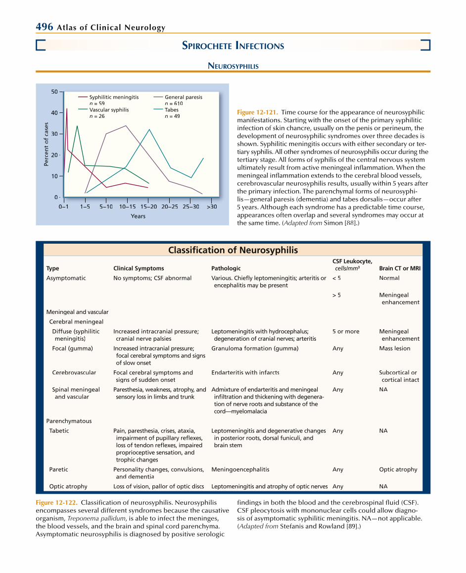
496 Atlas of Clinical Neurology
SPIROCHETE INFECTIONS
NEUROSYPHILIS
0
10
20
30
40
50
Perc
ent
of
case
s
0−10 1 1−51 5 5−105 10 10−1510 15 15−2015 20 20−2520 25 25−3025 30 >3030
Years
Syphilitic meningitisn = 59Vascular syphilisn = 26
General paresisn = 610Tabesn = 49
Figure 12-121. Time course for the appearance of neurosyphilicmanifestations. Starting with the onset of the primary syphiliticinfection of skin chancre, usually on the penis or perineum, thedevelopment of neurosyphilic syndromes over three decades isshown. Syphilitic meningitis occurs with either secondary or ter-tiary syphilis. All other syndromes of neurosyphilis occur during the tertiary stage. All forms of syphilis of the central nervous systemultimately result from active meningeal infl ammation. When the meningeal infl ammation extends to the cerebral blood vessels, cerebrovascular neurosyphilis results, usually within 5 years afterthe primary infection. The parenchymal forms of neurosyphi-lis—general paresis (dementia) and tabes dorsalis—occur after 5 years. Although each syndrome has a predictable time course,appearances often overlap and several syndromes may occur at the same time. (Adapted from Simon [88].)
Classifi cation of Neurosyphilis
Type Clinical Symptoms PathologicCSF Leukocyte, cells/mm3 Brain CT or MRI
Asymptomatic No symptoms; CSF abnormal Various. Chiefl y leptomeningitis; arteritis orencephalitis may be present
< 5 Normal
> 5 Meningeal enhancement
Meningeal and vascular
Cerebral meningeal
Diffuse (syphilitic meningitis)
Increased intracranial pressure; cranial nerve palsies
Leptomeningitis with hydrocephalus;degeneration of cranial nerves; arteritis
5 or more Meningeal enhancement
Focal (gumma) Increased intracranial pressure; focal cerebral symptoms and signsof slow onset
Granuloma formation (gumma) Any Mass lesion
Cerebrovascular Focal cerebral symptoms and signs of sudden onset
Endarteritis with infarcts Any Subcortical or cortical intact
Spinal meningeal and vascular
Paresthesia, weakness, atrophy, and sensory loss in limbs and trunk
Admixture of endarteritis and meningealinfi ltration and thickening with degenera-tion of nerve roots and substance of the cord—myelomalacia
Any NA
Parenchymatous
Tabetic Pain, paresthesia, crises, ataxia, impairment of pupillary refl exes,loss of tendon refl exes, impairedproprioceptive sensation, andtrophic changes
Leptomeningitis and degenerative changesin posterior roots, dorsal funiculi, and brain stem
Any NA
Paretic Personality changes, convulsions, and dementia
Meningoencephalitis Any Optic atrophy
Optic atrophy Loss of vision, pallor of optic discs Leptomeningitis and atrophy of optic nerves Any NA
Figure 12-122. Classifi cation of neurosyphilis. Neurosyphilisencompasses several different syndromes because the causative organism, Treponema pallidum, is able to infect the meninges,the blood vessels, and the brain and spinal cord parenchyma.Asymptomatic neurosyphilis is diagnosed by positive serologic
fi ndings in both the blood and the cerebrospinal fl uid (CSF).CSF pleocytosis with mononuclear cells could allow diagno-sis of asymptomatic syphilitic meningitis. NA—not applicable. (Adapted from Stefanis and Rowland [89].)

Infectious Diseases of the Nervous System 497
Cranial Nerve Palsies in Syphilitic Meningitis*
Cranial NervesPercent of Abnormalities
I 2
II 27
III 24
IV 2
V 12
VI 22
VII 41
VIII 42
IX–X 6
XI 1
XII 4
*354 cranial nerve palsies in 195 patients.
Figure 12-123. Cranial nerve palsies in syphilitic meningitis. Symptomatic meningeal syphilis usually occurs during the first 2 years after the primary infection. In approxi-mately 10% of cases, syphilitic meningitis occurs with the rash of secondary syphilis, but most cases occur during the tertiary stage. Patients present with headache, menin-gismus, and malaise. They may or may not have a low-grade fever. Lymphocytic pleo-cytosis of up to several hundred cells occurs; the cerebrospinal fluid (CSF) glucoselevel is reduced, but usually is greater than 25 mg/dL; CSF protein level is increased and may exceed 100 mg/dL; and the CSF pressure may be elevated. This syndromeis diagnosed with positive blood and CSF serologic tests (serologic tests for syphilis: Venereal Disease Research Laboratories, rapid plasma reagin). The syndrome may resolve on its own, or complications may ensue. Because the meningitis is most con-centrated at the base, cranial nerve palsies are often seen, sometimes bilaterally butusually asymmetrically. Obstruction of CSF pathways may result in subacute to chronic hydrocephalus. (Adapted from Merrit et al. [90].)
Figure 12-124. Anteroposterior view, left carotid angiogram, of a 26-year-old man with meningovascular syphilis, showing constriction (arrows) of the anterior and middle segments ofthe middle cerebral artery. Cerebrovascular syphilis occurswhen the inflammation in the subarachnoid space compromisesarteries traversing this space. Vasculitis of middle-sized vessels occurs, resulting in ischemia. The middle cerebral artery is affected most often, but any cerebral or spinal vessel may beinvolved. Stroke syndromes occurring in neurosyphilis are no different than those seen from other causes; focal clinical manifestations are determined by which vessel is involved. Thepatients exhibit risk factors for venereal disease and are usuallyyounger than those with arthrosclerotic infarction. Differentia-tion depends upon the blood and cerebrospinal fluid serologicresults. CT or MRI scans of the brain reveal cortical or subcortical infarction. If the meningeal inflammation is intense enough, aprodrome of headache and personality change may precede the stroke by weeks (meningovascular syphilis). Penicillineffectively cures the infection, preventing further infarctions. (From Simon and Bayne [91]; with permission.)
Figure 12-125. Pathologic specimen from a brain with pareticneurosyphilis. Paretic neurosyphilis has also been referred toas general paresis of the insane, syphilitic meningoencephalitis, dementia paralytica, and syphilitic dementia. Symptoms usually begin 5 to 15 years after the primary infection, with a range of 3to 30 years. Paretic neurosyphilis was common in the preantibi-otic era but is now infrequent. The symptoms and signs of general paresis are similar to any organic brain syndrome. Progressivedementia with personality changes is the most common feature. Behavioral changes with psychosis and grandiose delusional statesare unusual. Seizures also are seen. Masked facies; tremors ofthe face, tongue, lips, and extremities; dysarthria; and hyperactive reflexes may also occur. Pathologically there is chronic meningo-encephalitis with cortical atrophy, enlarged ventricles, thickened meninges, and granular ependymitis. Microscopic lesions includemononuclear inflammatory cells, a prominent microglial rod cellresponse, and the presence of the organisms. Diagnosis depends on cerebrospinal fluid abnormalities and serologic results. Usually patients are younger (less than 50 years of age) than those with other causes of dementia. Penicillin is an effective therapy and usu-ally arrests the progression of the dementia but will not reverse the existing damage. (From Merrit et al. [90]; with permission.)

498 Atlas of Clinical Neurology
Figure 12-126. Symptoms and signs of tabetic neurosyphilis.Tabetic neurosyphilis is also referred to as tabes dorsalis because of the degeneration of the posterior columns. Tabes usuallybegins 10 to 20 years after the primary infection, but some cases have had onset after 30 years (range, 5–50 years). A, The mostcommon and classic symptoms of tabes are the lancinating orlightning-like pains, ataxia, and bladder dysfunction. Prominent signs include abnormal pupils including Argyll-Robertson pupils,absent reflexes in the lower extremities, loss of posterior column sensations, Romberg sign, and gait ataxia. As the disease progresses, bladder dysfunction and the sensory gait ataxia usually becomethe most disabling problems. Loss of deep pain sensation may occur with Abadie’s sign (loss or delayed recognition of pain when the Achilles tendon is squeezed). Charcot joints and distal
extremity ulcerations may be seen. B, Pathologically, degenera-tion of the posterior columns and dorsal roots occurs. Diagnosis is relatively easy because of the classic clinical manifestations,with positive serologic results, at least in the blood samples. Thedisease may arrest spontaneously or may be arrested with antibi-otic treatment. The latter is more likely to occur when there are signs of active inflammation in the cerebrospinal fluid (CSF). Even after treatment, and even when there is no active CSF inflamma-tion (as regards CSF pleocytosis), many of the manifestations maycontinue to progress. The pathogenesis of tabes is not under-stood. In contrast to paretic cortical lesions, spirochetes are notfound in the affected areas of the spinal cord, which may explainwhy antibiotic therapy may not stop progression. (A, adapted fromAMerrit et al. [90]; B, from Wilson [4]; with permission.)
Frequency of Different Formsof Symptomatic Neurosyphilis*
Preantibiotic Era Antibiotic Era AIDS Era
HIV (-) HIV (+) or AIDS
1 2 3 4 5 6 7
Tabetic 45 48 45 15 11 5 0
Paretic 17 48 8 12 4 9 4
Taboparetic 4 7 9 23 23 — —
Vascular 15 19 9 19 61 41 38
Meningeal 8 8 19 23 0 23 46
Optic neuritis 4 — — — — 14 42
Spinal cord 4 — 10 8 — — —
* Numbers are a percentage of cases.1 Merritt, Adams, Solomon, 1946 (457 patients).2 Kierland et al., 1942 (2019 patients).3 Wolters, 1987 (518 patients, 1930–1940).4 Wolters, 1987 (121 patients, 1970–1984).5 Burke, Schaberg, 1985 (26 patients).6, 7 Katz et al., 1993.
Figure 12-127. Frequency of differentforms of symptomatic neurosyphilis. Fol-lowing the introduction of penicillin, the frequency of neurosyphilis per hospital admission fell from 5.9 per 100,000 popu-lation in 1942 to 0.1 per 100,000 in 1965.Beginning in the 1980s and coincident with the AIDS epidemic, this trend hasbeen reversed. The incidence of neuro-syphilis is unknown, but the incidenceof primary and secondary syphilis hasrisen from 13.7 per 100,000 population in1981 to a peak of 20.1 per 100,000 in 1990.Since then the incidence has dropped significantly, perhaps because of bettersurveillance and education. The overall frequency of neurosyphilis in HIV-posi-tive and AIDS patients is estimated to be 2%. Also during the 1980s and 1990s, ashift occurred toward meningeal and vas-cular forms of neurosyphilis and a decline in the parenchymal forms. This may be related to cerebrospinal fluid abnormali-
BBBBB
Symptoms and Signs of Tabetic Neurosyphilis (Analysis of 150 Cases)
Symptoms % Signs %
Lancinating pain 75 Abnormal pupils 94
Ataxia 42 Argyll-Robertson 48
Bladder disturbance 33 Other abnormalities 46
Paresthesia 24 Absent reflexes
Gastric or visceral crises 18 Ankle jerks 94
Optic atrophy 16 Knee jerks 81
Rectal incontinence 14 Biceps and triceps 11
Deafness 7 Romberg sign 55
Impotence 4 Impaired sensations
Vibratory sense 52
Position sense 45
Touch and pain 13
Ocular palsy 10
Charcot joints 7
A
ties of neurosyphilis in HIV-infected patients, which are more intense than those ofnon–HIV-infected patients. (Adapted from Stefanis and Rowland [89].)

Infectious Diseases of the Nervous System 499
CSF Findings in Neurosyphilis by Clinical Syndrome (Subtype)VDRL
Neurosyphilic Syndrome OP mm H2O Leukocyte Level, per mm3 Glucose Level, mg/dL Protein Level, mg/dL Blood CSF
Meningitic 210–400* 100–500† 20–40 45–200 1:64 1:4
(< 200 to >400)
(< 10–2000) (< 20 to > 80) (< 45–400)
Cerebrovascular < 200 (>200–250)
10–100 (< 10 to > 100) Normal to mildlydecreased
100–200 (15–260) 1:512 1:16
Paretic < 200 (<200–300)
10–100 (< 10 to > 100) Normal tomildly decreased
45–100 (29–500) 1:128 1:8
Tabetic < 200 Active 5–50 Normal 45–100 Active 1:15 1:28
(< 200–300) (5–165) (14–250) Inactive 1:16 1:2
Inactive 0–5
* Numbers without parentheses are the common range. Numbers in parentheses are the overall range.†CSF pleocytosis is usually 80% to 100% lymphocytic mononuclear.
Figure 12-128. Abnormalities of cerebrospinal fluid (CSF) inneurosyphilis. The diagnosis of active neurosyphilis is based upon a compatible clinical syndrome, an inflammatory CSF profile, and reactive serologic tests in the blood (treponemal antibody test) and CSF (nontreponemal test). CSF pleocytosis(primarily lymphocytic) is the best measure of disease activity. The number of cells varies with each clinical subtype, being
maximal in the earlier acute-like stage of syphilitic meningitis. The glucose level is usually low in syphilitic meningitis and is more likely to be normal for other subtypes. CSF protein levels are usually elevated for all subtypes. The CSF gamma globulinlevel may be increased and oligoclonal bands may be present.OP—opening pressure, VDRL—Venereal Disease ResearchLaboratory. (Adapted from Simon and Bayne [91].)
Serologic Tests Used in the Diagnosis of Neurosyphilis
TestAbnormality Required for Diagnosis False/Positive Tests
FTA-ABS orMHA-TP
+ in blood required Rare
- excludes diagnosis Other spirochete diseases
Autoimmune diseases, especiallysystemic lupus erythematosus (SLE)
VDRL + in CSF required for diagnosis Contamination of CSF by blood
CSF paraprotein
- in CSF inactive or no neurosyphilis Very high CSF protein level
Autoimmune disease (such as SLE)
Strong immunologic stimulus
Acute bacterial or viral infections
Early HIV infection
Vaccination
Central nervous system neoplasia
Drug addiction
Pregnancy
Chronic liver disease
Figure 12-129. Serologic tests used in the diagnosis of neurosyphilis. Diagnosis of neurosyphilis depends on a compat-ible clinical syndrome, an inflammatorycerebrospinal fluid (CSF) profile, andreactive serologic tests. The treponemalantibody tests are the fluorescent trepo-nemal antibody-absorption (FTA-ABS)test and the microhemagglutination test for Treponema pallidum (MHA-TP). Positive blood FTA-ABS and MHA-TP are diagnostic for syphilis, as they are highlyspecific and remain positive for years. Ifthese tests are negative, the diagnosisof neurosyphilis is essentially excluded;they are not useful, however, for follow-ing disease activity because they do not revert with successful treatment. Thesetreponemal antibody tests are not usefulfor CSF analysis. The serologic diagno-sis of neurosyphilis requires a positive blood serologic result and a reactive CSF serologic test for syphilis (STS). The twoSTS tests used currently are the Venereal
Disease Research Laboratory (VDRL) test and the rapid plasma reagin (RPR) test. TheRPR test cannot be used on the CSF. The CSF VDRL test can be used to follow disease activity after treatment but is not as reliable as the CSF pleocytosis.
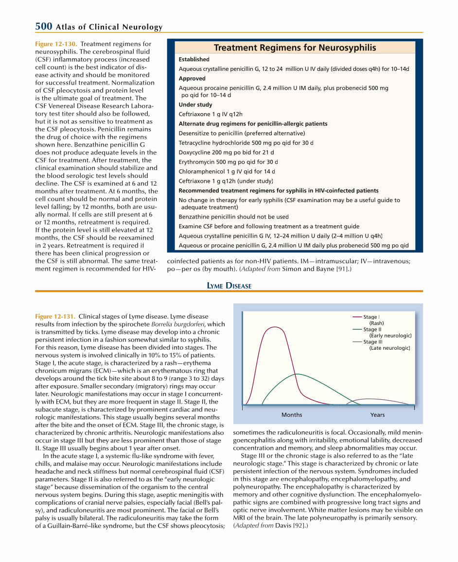
500 Atlas of Clinical Neurology
Treatment Regimens for NeurosyphilisEstablished
Aqueous crystalline penicillin G, 12 to 24 million U IV daily (divided doses q4h) for 10–14d
Approved
Aqueous procaine penicillin G, 2.4 million U IM daily, plus probenecid 500 mgpo qid for 10–14 d
Under study
Ceftriaxone 1 g IV q12h
Alternate drug regimens for penicillin-allergic patients
Desensitize to penicillin (preferred alternative)
Tetracycline hydrochloride 500 mg po qid for 30 d
Doxycycline 200 mg po bid for 21 d
Erythromycin 500 mg po qid for 30 d
Chloramphenicol 1 g IV qid for 14 d
Ceftriaxone 1 g q12h (under study)
Recommended treatment regimens for syphilis in HIV-coinfected patients
No change in therapy for early syphilis (CSF examination may be a useful guide toadequate treatment)
Benzathine penicillin should not be used
Examine CSF before and following treatment as a treatment guide
Aqueous crystalline penicillin G IV, 12–24 million U daily (2–4 million U q4h)
Aqueous or procaine penicillin G, 2.4 million U IM daily plus probenecid 500 mg po qid
Figure 12-130. Treatment regimens for neurosyphilis. The cerebrospinal fl uid (CSF) infl ammatory process (increasedcell count) is the best indicator of dis-ease activity and should be monitoredfor successful treatment. Normalization of CSF pleocytosis and protein level is the ultimate goal of treatment. TheCSF Venereal Disease Research Labora-tory test titer should also be followed,but it is not as sensitive to treatment as the CSF pleocytosis. Penicillin remainsthe drug of choice with the regimensshown here. Benzathine penicillin Gdoes not produce adequate levels in the CSF for treatment. After treatment, the clinical examination should stabilize andthe blood serologic test levels should decline. The CSF is examined at 6 and 12months after treatment. At 6 months, the cell count should be normal and proteinlevel falling; by 12 months, both are usu-ally normal. If cells are still present at 6 or 12 months, retreatment is required. If the protein level is still elevated at 12months, the CSF should be reexamined in 2 years. Retreatment is required ifthere has been clinical progression or the CSF is still abnormal. The same treat-ment regimen is recommended for HIV-
LYME DISEASE
Stage I (Rash)Stage II (Early neurologic)Stage III (Late neurologic)
Months Years
Figure 12-131. Clinical stages of Lyme disease. Lyme diseaseresults from infection by the spirochete Borrelia burgdorferi, which iis transmitted by ticks. Lyme disease may develop into a chronicpersistent infection in a fashion somewhat similar to syphilis.For this reason, Lyme disease has been divided into stages. Thenervous system is involved clinically in 10% to 15% of patients. Stage I, the acute stage, is characterized by a rash—erythemachronicum migrans (ECM)—which is an erythematous ring that develops around the tick bite site about 8 to 9 (range 3 to 32) days after exposure. Smaller secondary (migratory) rings may occurlater. Neurologic manifestations may occur in stage I concurrent-ly with ECM, but they are more frequent in stage II. Stage II, the subacute stage, is characterized by prominent cardiac and neu-rologic manifestations. This stage usually begins several monthsafter the bite and the onset of ECM. Stage III, the chronic stage, is characterized by chronic arthritis. Neurologic manifestations alsooccur in stage III but they are less prominent than those of stageII. Stage III usually begins about 1 year after onset.
In the acute stage I, a systemic fl u-like syndrome with fever, chills, and malaise may occur. Neurologic manifestations include headache and neck stiffness but normal cerebrospinal fl uid (CSF)parameters. Stage II is also referred to as the “early neurologicstage” because dissemination of the organism to the centralnervous system begins. During this stage, aseptic meningitis with complications of cranial nerve palsies, especially facial (Bell’s pal-sy), and radiculoneuritis are most prominent. The facial or Bell’s palsy is usually bilateral. The radiculoneuritis may take the formof a Guillain-Barré–like syndrome, but the CSF shows pleocytosis;
coinfected patients as for non-HIV patients. IM—intramuscular; IV—intravenous;po—per os (by mouth). (Adapted from Simon and Bayne [91].)
sometimes the radiculoneuritis is focal. Occasionally, mild menin-goencephalitis along with irritability, emotional lability, decreased concentration and memory, and sleep abnormalities may occur.
Stage III or the chronic stage is also referred to as the “late neurologic stage.” This stage is characterized by chronic or latepersistent infection of the nervous system. Syndromes included in this stage are encephalopathy, encephalomyelopathy, and polyneuropathy. The encephalopathy is characterized bymemory and other cognitive dysfunction. The encephalomyelo-pathic signs are combined with progressive long tract signs and optic nerve involvement. White matter lesions may be visible onMRI of the brain. The late polyneuropathy is primarily sensory. (Adapted from Davis [92].)

Infectious Diseases of the Nervous System 501
Ixodees damminniIxodees scapularisIxodees pacificuus
Figure 12-132. Epidemiology of Lyme disease. Lyme diseaseaccounts for about 90% of the vector-borne infections in theUnited States. In 2005, over 23,000 cases were reported to theCenters for Disease Control and Prevention. Borrelia burgdorferi, the spirochete that causes Lyme disease, is transmitted by Ixodesspecies (hard body) ticks. Up to 50% of human infections areasymptomatic. The infection rate of different Ixodes species carrying the spirochete depends on the species. Ixodes dam-mini (deer tick) is the principal vector in the Northeast (30% ito 60% infection rate) and the Midwest (10% to 15% infection rate). Ixodes pacifi cus (western black-legged tick) is the vec-tor in the West (1% to 5% infection rate). In the southeastern United States, Ixodes scapularis is the vector but its infection rate is much lower even than that of I. pacifi cus. The various infection rates of the different Ixodes species explains why 10states account for almost 90% of the cases: New York (40%), followed by Connecticut, New Jersey, Pennsylvania, Rhode Island,
CSF Analysis in Lyme Disease Encephalomyelitis
Test CSF Findings
Opening pressure* Normal
Total leukocytes/mm3 166 (15–700)†
Percent lymphocytes 93 (40–100)
Glucose, mg/dL‡ 49 (33–61)
Protein, mg/dL 79 (8–400)
IgG/albumin ratio (n = 20) 0.18 (0.9–0.44)
Oligoclonal bands (n = 4) Present
Myelin basic protein (n = 5) Absent
VDRL (n = 20) Negative
*n = 38, except where noted.†Median (range).‡Serum glucose = 95 (87–113).
Figure 12-133. Cerebrospinal fl uid (CSF) analysis in Lyme disease encephalomyelitis. CSF is usually abnormal in cen-tral nervous system Lyme disease and generally normal inperipheral nervous system syndromes unless there is radicular involvement. CSF analysis is usually abnormal and has a higherdiagnostic yield than electroencephalography (usually normal or nonspecifi c) or MRI (25% have small cerebral white matterlesions). CSF analysis also offers the opportunity to test for the intrathecal production of Borrelia burgdorferi antibodies. iVDRL—Venereal Disease Research Laboratory. (Adapted fromPachner and Steere [94].)
Causes of False-positive andFalse-negative Lyme Serologic Tests
False-positive
Cross-reactive spirochetal infection (eg, mouth treponemes, syphilis, leptospirosis, relapsing fever)
Severe bacterial infections
Hypergammaglobulinemia
Epstein-Barr virus
Autoimmune disorders with high autoantibody titers
HIV infection
Unreliably easy
False-negative
Too early in the infection
Early antibiotics with blurred humoral response
Unreliably easy
Figure 12-134. Causes of false-positive and false-negative Lymedisease serologic tests. Serum antibodies, when present, proveexposure to the agent but cannot be used to determine when the infection occurred. Most antibody assays are now performed byan enzyme-linked immunosorbent assay technique. Western blot testing is required to confi rm the diagnosis. Polymerase chain reaction testing is still experimental. The demonstration of elevatedBorrelia burgdorferi antibodies in the cerebrospinal fl uid is essential-ily diagnostic. About 95% of Lyme disease patients are seropositive, but false-positive and false-negative test results may occur. (Adapted ((from Coyle [95].)
Massachusetts, Maryland, Wisconsin, Minnesota, and California.In the fi gure, the circles indicate collection sites outside the main range of I. dammini. (Adapted from Anderson [93].)

502 Atlas of Clinical Neurology
Neurologic Conditions in WhichLyme Disease Should Be Considered
Central Nervous System Peripheral Nervous System
Acute aseptic meningitis Cranial neuritis (Bell’s palsy)
Chronic lymphocytic meningitis Mononeuritis simplex or multiplex
Acute meningoencephalitis Radiculoneuritis
Acute focal encephalitis Plexitis
Brainstem encephalitis Distal axonal neuropathy
Progressive encephalomyelitis Demyelinating neuropathy
Cerebral demyelination, including multiple sclerosis Carpal tunnel syndrome
Cerebral vasculitis Focal myositis
Dementia
Transverse myelitis
Figure 12-135. Differential diagnosis ofLyme disease. The combination of men-ingitis, neuritis, and radiculitis withoutfever is highly suggestive of Lyme disease.If a history of tick exposure or erythemachronicum migrans is obtained, thediagnosis can be made with confidence.Because of the involvement of both the peripheral and the central nervous sys-tems, the differential diagnosis is varied and extensive. (Adapted from Reik [96].)
Antibiotic Therapy for NeurologicSymptoms of Lyme Disease
Manifestations Treatment
ECM and systemic symptoms Doxycycline, 100 mg po bid for 14–28 d
Adults Amoxicillin, 500 mg po tid for 14–28 d(plus probenecid, 500 mg po tid)*
Cefuroxime axetil, 500 mg po bid for 14–28 d†
Children (≤ 8 y) Amoxicillin, 25–50 mg/kg po daily in 3 divideddoses for 14–28 d
Cefuroxime axetil, 250 mg po bid for 14–28 d†
Neurologic involvement
Facial palsy alone Oral antibiotics as for ECM
All others
Adults Ceftriaxone, 2 g IV daily for 14–28 d
Cefotaxime, 2 g IV tid for 14–28 d
Penicillin G, 20–24 million U IV daily for 10–14 d
Doxycycline, 200 mg po bid for 14–28 d
Children Ceftriaxone, 75–100 mg/kg IV daily for 14–28 d
Penicillin G, 300,000 U/kg IV q 4 h for 10–14 d
*Optional.†Alternative.†
Figure 12-136. Antibiotic therapy for neurologic syndromes of Lyme disease. The antibiotic therapy for neurologic syndromes depends on the specific stageof Lyme disease. Ceftriaxone IV is nowusually considered the drug of choicefor stages II (early neurologic stage) or III (late neurologic stage). Most symptoms resolve with antibiotic treatment, butmotor signs may last for 7 to 8 weeks. The duration of therapy has never been clarified. A postinfectious syndrome withsymptoms of fatigue, headache, and mus-cle and joint pain may last for months to several years. ECM—erythema chronicummigrans; IV—intravenous; po—per os (by mouth). (Adapted from Cadavid [97].)
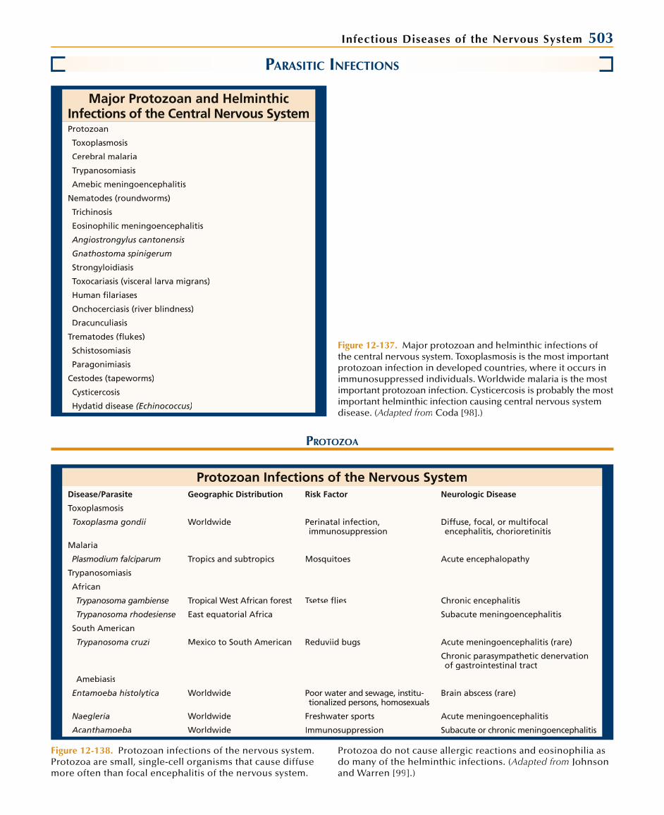
Infectious Diseases of the Nervous System 503
PARASITIC INFECTIONS
Major Protozoan and Helminthic Infections of the Central Nervous SystemProtozoan
Toxoplasmosis
Cerebral malaria
Trypanosomiasis
Amebic meningoencephalitis
Nematodes (roundworms)
Trichinosis
Eosinophilic meningoencephalitis
Angiostrongylus cantonensis
Gnathostoma spinigerum
Strongyloidiasis
Toxocariasis (visceral larva migrans)
Human filariases
Onchocerciasis (river blindness)
Dracunculiasis
Trematodes (flukes)
Schistosomiasis
Paragonimiasis
Cestodes (tapeworms)
Cysticercosis
Hydatid disease (Echinococcus)
Figure 12-137. Major protozoan and helminthic infections ofthe central nervous system. Toxoplasmosis is the most importantprotozoan infection in developed countries, where it occurs inimmunosuppressed individuals. Worldwide malaria is the mostimportant protozoan infection. Cysticercosis is probably the mostimportant helminthic infection causing central nervous systemdisease. (Adapted from Coda [98].)
PROTOZOA
Protozoan Infections of the Nervous SystemDisease/Parasite Geographic Distribution Risk Factor Neurologic Disease
Toxoplasmosis
Toxoplasma gondii Worldwide Perinatal infection, immunosuppression
Diffuse, focal, or multifocal encephalitis, chorioretinitis
Malaria
Plasmodium falciparum Tropics and subtropics Mosquitoes Acute encephalopathy
Trypanosomiasis
African
Trypanosoma gambiense Tropical West African forest Tsetse flies Chronic encephalitis
Trypanosoma rhodesiense East equatorial Africa Subacute meningoencephalitis
South American
Trypanosoma cruzi Mexico to South American Reduviid bugs Acute meningoencephalitis (rare)
Chronic parasympathetic denervation of gastrointestinal tract
Amebiasis
Entamoeba histolytica Worldwide Poor water and sewage, institu-tionalized persons, homosexuals
Brain abscess (rare)
Naegleria Worldwide Freshwater sports Acute meningoencephalitis
Acanthamoeba Worldwide Immunosuppression Subacute or chronic meningoencephalitis
Figure 12-138. Protozoan infections of the nervous system. Protozoa are small, single-cell organisms that cause diffusemore often than focal encephalitis of the nervous system.
Protozoa do not cause allergic reactions and eosinophilia as do many of the helminthic infections. (Adapted from Johnsonand Warren [99].)

504 Atlas of Clinical Neurology
TOXOPLASMOSIS
Clinical Manifestationsof Toxiplasmosis
Congenital Disease % Immunocompromised Host %
Retinochoroiditis 87 Altered mental state 75
Abnormal cere-brospinal fluid
55 Fever 10–72
Anemia 51 Seizures 33
Convulsions 50 Headache 56
Intracranial calcifications
50 Focal neurologic signs 60
Jaundice 29 Motor deficits
Fever 25 Cranial nerve palsies
Splenomegaly 21 Movement disorders
Hepatomegaly 17 Dysmetria
Visual field loss
Aphasia
Figure 12-139. Clinical manifestations of toxoplasmosis. Con-genital toxoplasmosis is a systemic illness causing chorioretinitis,microencephaly, seizures, mental retardation (from encephalitis), and cerebral calcifications in newborns. Cases in immunocom-promised patients are frequent, thus, toxoplasmosis is a com-mon opportunistic infection in AIDS patients that usually occursbecause of reactivation. (Adapted from Luft and Remington [100].)
leukocytetransfusion
Organ transplantation
Laboratoryaccident
Trophozoites
Cysts
Cysts
Cysts
Oocysts
Trophozoites
Cysts
Oocysts
Oocysts
Trophozoites
Figure 12-140. Transmission of toxoplas-mosis. Most often Toxoplasma gondiiinfection occurs by eating undercooked meat or other foods contaminated with cat feces containing oocysts. Com-mon-source outbreaks have occurred in families from contaminated food.Unpasteurized milk and water are alsopossible sources of infection. In addition to exposure to cats and eating under-cooked meats, warm humid climates andpoor sanitation correlate with a greaterprevalence of infection. Primary infectionin the immunocompetent host is usually asymptomatic. Rarely, symptomatic severe primary infection occurs. Usually tissuecysts persist and reactivation is preventedunless immunity wanes, as occurs inAIDS or other immunosuppressive events(such as therapy for cancer and transplan-tation). (Adapted from Berger [101].)
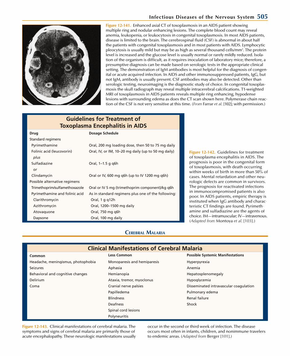
Infectious Diseases of the Nervous System 505Figure 12-141. Enhanced axial CT of toxoplasmosis in an AIDS patient showing multiple ring and nodular enhancing lesions. The complete blood count may revealanemia, leukopenia, or leukocytosis in congenital toxoplasmosis. In most AIDS patients, disease is limited to the brain. The cerebrospinal fluid (CSF) is abnormal in about half the patients with congenital toxoplasmosis and in most patients with AIDS. Lymphocyticpleocytosis is usually mild but may be as high as several thousand cells/mm3. The protein level is increased and the glucose level is usually normal or rarely mildly reduced. Isola-tion of the organism is difficult, as it requires inoculation of laboratory mice; therefore, a presumptive diagnosis can be made based on serologic tests in the appropriate clinical setting. The demonstration of IgM antibodies is most helpful for the diagnosis of congen-ital or acute acquired infection. In AIDS and other immunosuppressed patients, IgG, butnot IgM, antibody is usually present. CSF antibodies may also be detected. Other than serologic testing, neuroimaging is the diagnostic study of choice. In congenital toxoplas-mosis the skull radiograph may reveal multiple intracerebral calcifications. T1-weighted MRI of toxoplasmosis in AIDS patients reveals multiple ring enhancing, hypodenselesions with surrounding edema as does the CT scan shown here. Polymerase chain reac-tion of the CSF is not very sensitive at this time. (From Farrar et al. [102]; with permission.)
Guidelines for Treatment ofToxoplasma Encephalitis in AIDS
Drug Dosage Schedule
Standard regimens
Pyrimethamine Oral, 200 mg loading dose, then 50 to 75 mg daily
Folinic acid (leucovorin) Oral, IV, or IM, 10–20 mg daily (up to 50 mg daily)
plus
Sulfadiazine Oral, 1–1.5 g q6h
or
Clindamycin Oral or IV, 600 mg q6h (up to IV 1200 mg q6h)
Possible alternative regimens
Trimethoprim/sulfamethoxazole Oral or IV 5 mg (trimethoprim component)/kg q6h
Pyrimethamine and folinic acid As in standard regimens plus one of the following:
Clarithromycin Oral, 1 g q12h
Azithromycin Oral, 1200–1500 mg daily
Atovaquone Oral, 750 mg q6h
Dapsone Oral, 100 mg daily
Figure 12-142. Guidelines for treatmentof toxoplasma encephalitis in AIDS. The prognosis is poor in the congenital formof toxoplasmosis, with death occurringwithin weeks of birth in more than 50% of cases. Mental retardation and other neu-rologic defects are common in survivors. The prognosis for reactivated infections in immunocompromised patients is alsopoor. In AIDS patients, empiric therapy isinstituted when IgG antibody and charac-teristic CT findings are found. Pyrimeth-amine and sulfadiazine are the agents ofchoice. IM—intramuscular; IV—intravenous. (Adapted from Montoya et al. [103].)
CEREBRAL MALARIA
Clinical Manifestations of Cerebral MalariaCommon Less Common Possible Systemic Manifestations
Headache, meningismus, photophobia Monoparesis and hemiparesis Hyperpyrexia
Seizures Aphasia Anemia
Behavioral and cognitive changes Hemianopia Hepatosplenomegaly
Delirium Ataxia, tremor, myoclonus Hypoglycemia
Coma Cranial nerve palsies Disseminated intravascular coagulation
Papilledema Pulmonary edema
Blindness Renal failure
Deafness Shock
Spinal cord lesions
Polyneuritis
Figure 12-143. Clinical manifestations of cerebral malaria. The symptoms and signs of cerebral malaria are primarily those of acute encephalopathy. These neurologic manifestations usually
occur in the second or third week of infection. The disease occurs most often in infants, children, and nonimmune travelersto endemic areas. (Adapted from Berger [101].)
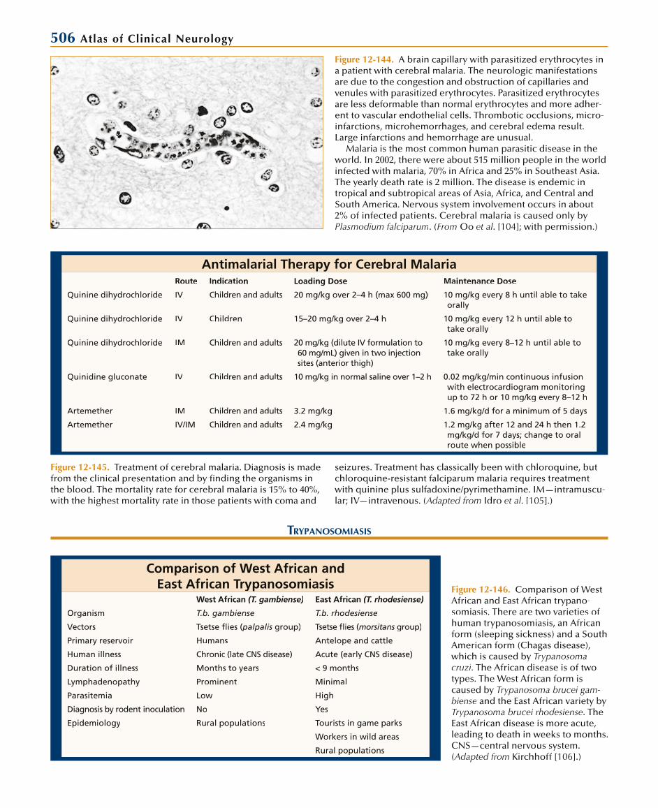
506 Atlas of Clinical Neurology
Figure 12-144. A brain capillary with parasitized erythrocytes ina patient with cerebral malaria. The neurologic manifestations are due to the congestion and obstruction of capillaries andvenules with parasitized erythrocytes. Parasitized erythrocytesare less deformable than normal erythrocytes and more adher-ent to vascular endothelial cells. Thrombotic occlusions, micro-infarctions, microhemorrhages, and cerebral edema result. Large infarctions and hemorrhage are unusual.
Malaria is the most common human parasitic disease in theworld. In 2002, there were about 515 million people in the world infected with malaria, 70% in Africa and 25% in Southeast Asia.The yearly death rate is 2 million. The disease is endemic intropical and subtropical areas of Asia, Africa, and Central andSouth America. Nervous system involvement occurs in about2% of infected patients. Cerebral malaria is caused only by Plasmodium falciparum. (From Oo et al. [104]; with permission.)
Antimalarial Therapy for Cerebral MalariaRoute Indication Loading Dose Maintenance Dose
Quinine dihydrochloride IV Children and adults 20 mg/kg over 2–4 h (max 600 mg) 10 mg/kg every 8 h until able to take orally
Quinine dihydrochloride IV Children 15–20 mg/kg over 2–4 h 10 mg/kg every 12 h until able to take orally
Quinine dihydrochloride IM Children and adults 20 mg/kg (dilute IV formulation to 60 mg/mL) given in two injectionsites (anterior thigh)
10 mg/kg every 8–12 h until able to take orally
Quinidine gluconate IV Children and adults 10 mg/kg in normal saline over 1–2 h 0.02 mg/kg/min continuous infusion with electrocardiogram monitoringup to 72 h or 10 mg/kg every 8–12 h
Artemether IM Children and adults 3.2 mg/kg 1.6 mg/kg/d for a minimum of 5 days
Artemether IV/IM Children and adults 2.4 mg/kg 1.2 mg/kg after 12 and 24 h then 1.2 mg/kg/d for 7 days; change to oralroute when possible
Figure 12-145. Treatment of cerebral malaria. Diagnosis is made from the clinical presentation and by finding the organisms in the blood. The mortality rate for cerebral malaria is 15% to 40%,with the highest mortality rate in those patients with coma and
seizures. Treatment has classically been with chloroquine, butchloroquine-resistant falciparum malaria requires treatmentwith quinine plus sulfadoxine/pyrimethamine. IM—intramuscu-lar; IV—intravenous. (Adapted from Idro et al. [105].)
TRYPANOSOMIASIS
Comparison of West African and East African Trypanosomiasis
West African (T. gambiense) East African (T. rhodesiense)
Organism T.b. gambiense T.b. rhodesiense
Vectors Tsetse flies (palpalis( group) Tsetse flies (morsitans group)s
Primary reservoir Humans Antelope and cattle
Human illness Chronic (late CNS disease) Acute (early CNS disease)
Duration of illness Months to years < 9 months
Lymphadenopathy Prominent Minimal
Parasitemia Low High
Diagnosis by rodent inoculation No Yes
Epidemiology Rural populations Tourists in game parks
Workers in wild areas
Rural populations
Figure 12-146. Comparison of West African and East African trypano-somiasis. There are two varieties ofhuman trypanosomiasis, an African form (sleeping sickness) and a SouthAmerican form (Chagas disease), which is caused by Trypanosoma cruzi. The African disease is of two types. The West African form is caused by Trypanosoma brucei gam-biense and the East African variety by Trypanosoma brucei rhodesiense. The East African disease is more acute, leading to death in weeks to months. CNS—central nervous system. (Adapted from Kirchhoff [106].)
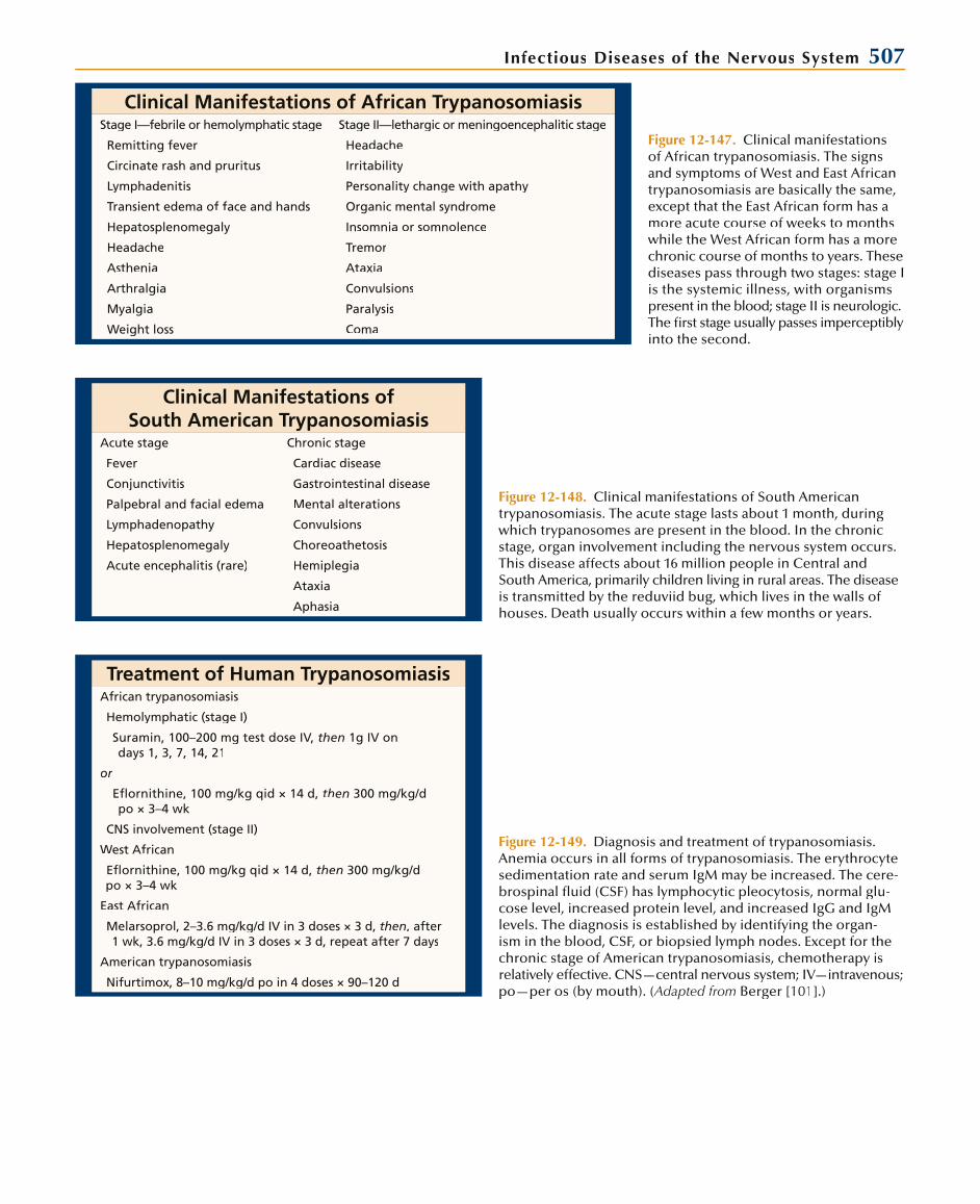
Infectious Diseases of the Nervous System 507
Clinical Manifestations of African TrypanosomiasisStage I—febrile or hemolymphatic stage Stage II—lethargic or meningoencephalitic stage
Remitting fever Headache
Circinate rash and pruritus Irritability
Lymphadenitis Personality change with apathy
Transient edema of face and hands Organic mental syndrome
Hepatosplenomegaly Insomnia or somnolence
Headache Tremor
Asthenia Ataxia
Arthralgia Convulsions
Myalgia Paralysis
Weight loss Coma
Figure 12-147. Clinical manifestations of African trypanosomiasis. The signsand symptoms of West and East African trypanosomiasis are basically the same, except that the East African form has amore acute course of weeks to months while the West African form has a morechronic course of months to years. These diseases pass through two stages: stage I is the systemic illness, with organisms present in the blood; stage II is neurologic. The first stage usually passes imperceptibly into the second.
Clinical Manifestations ofSouth American Trypanosomiasis
Acute stage Chronic stage
Fever Cardiac disease
Conjunctivitis Gastrointestinal disease
Palpebral and facial edema Mental alterations
Lymphadenopathy Convulsions
Hepatosplenomegaly Choreoathetosis
Acute encephalitis (rare) Hemiplegia
Ataxia
Aphasia
Figure 12-148. Clinical manifestations of South Americantrypanosomiasis. The acute stage lasts about 1 month, during which trypanosomes are present in the blood. In the chronic stage, organ involvement including the nervous system occurs.This disease affects about 16 million people in Central andSouth America, primarily children living in rural areas. The disease is transmitted by the reduviid bug, which lives in the walls of houses. Death usually occurs within a few months or years.
Treatment of Human TrypanosomiasisAfrican trypanosomiasis
Hemolymphatic (stage I)
Suramin, 100–200 mg test dose IV, then 1g IV on days 1, 3, 7, 14, 21
or
Eflornithine, 100 mg/kg qid × 14 d, then 300 mg/kg/dpo × 3–4 wk
CNS involvement (stage II)
West African
Eflornithine, 100 mg/kg qid × 14 d, then 300 mg/kg/dpo × 3–4 wk
East African
Melarsoprol, 2–3.6 mg/kg/d IV in 3 doses × 3 d, then, after 1 wk, 3.6 mg/kg/d IV in 3 doses × 3 d, repeat after 7 days
American trypanosomiasis
Nifurtimox, 8–10 mg/kg/d po in 4 doses × 90–120 d
Figure 12-149. Diagnosis and treatment of trypanosomiasis. Anemia occurs in all forms of trypanosomiasis. The erythrocyte sedimentation rate and serum IgM may be increased. The cere-brospinal fluid (CSF) has lymphocytic pleocytosis, normal glu-cose level, increased protein level, and increased IgG and IgMlevels. The diagnosis is established by identifying the organ-ism in the blood, CSF, or biopsied lymph nodes. Except for thechronic stage of American trypanosomiasis, chemotherapy is relatively effective. CNS—central nervous system; IV—intravenous;po—per os (by mouth). (Adapted from Berger [101].)
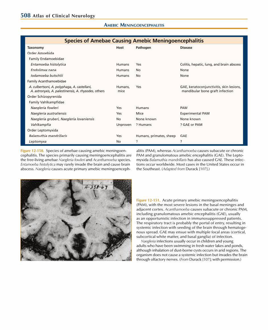
508 Atlas of Clinical Neurology
AMEBIC MENINGOENCEPHALITIS
Species of Amebae Causing Amebic MeningoencephalitisTaxonomy Host Pathogen Disease
Order Amoebida
Family Endamoebidae
Entamoeba histolytica Humans Yes Colitis, hepatic, lung, and brain abscess
Endolimax nana Humans No None
Iodamoeba butschlii Humans No None
Family Acanthamoebidae
A. culbertsoni, A. polyphaga, A. castellani, A. astronyxis, A. palestinensis, A. rhysodes, otherss
Humans, mice
Yes GAE, keratoconjunctivitis, skin lesions, mandibular bone graft infection
Order Schizopyrenida
Family Vahlkampfidae
Naegleria fowleri Yes Humans PAM
Naegleria australiensis Yes Mice Experimental PAM
Naegleria gruberi, Naegleria lovaniensis No None known None known
Vahlkampfia Unproven ? Humans ? GAE or PAM
Order Leptomyxida
Balamuthia mandrillaris Yes Humans, primates, sheep GAE
Leptomyxa No ? ?
Figure 12-150. Species of amebae causing amebic meningoen-cephalitis. The species primarily causing meningoencephalitis arethe free-living amebae Naegleria fowleri and i Acanthamoeba species. Entamoeba histolytica may rarely invade the brain and cause brain abscess. Naegleria causes acute primary amebic meningoenceph-
alitis (PAM), whereas Acanthamoeba causes subacute or chronicPAM and granulomatous amebic encephalitis (GAE). The Lepto-myxida Balamuthia mandrillaris has also caused GAE. These infec-tions occur worldwide. Most cases in the United States occur in the Southeast. (Adapted from Durack [107].)
Figure 12-151. Acute primary amebic meningoencephalitis (PAM), with the most severe lesions in the basal meninges and adjacent cortex. Acanthamoeba causes subacute or chronic PAM,including granulomatous amebic encephalitis (GAE), usually as an opportunistic infection in immunosuppressed patients. The respiratory tract is probably the portal of entry, resulting insystemic infection with seeding of the brain through hematoge-nous spread. GAE may ensue with multiple focal areas (cortical, subcortical white matter, and basal ganglia) of infection.
Naegleria infections usually occur in children and youngadults who have been swimming in fresh water lakes and ponds, although inhalation of dust-borne cysts occurs in arid regions. Theorganism does not cause a systemic infection but invades the brain through olfactory nerves. (From Durack [107]; with permission.)
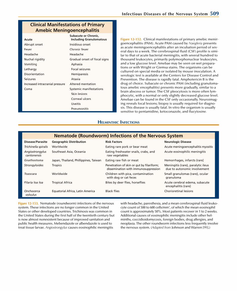
Infectious Diseases of the Nervous System 509
Clinical Manifestations of Primary Amebic Meningoencephalitis
AcuteSubacute or Chronic, Including Granulomatous
Abrupt onset Insidious onset
Fever Chronic fever
Headache Headache
Nuchal rigidity Gradual onset of focal signs
Vomiting Aphasia
Lethargy Focal seizures
Disorientation Hemiparesis
Seizures Ataxia
Increased intracranial pressure Altered mentation
Coma Systemic manifestations
Skin lesions
Corneal ulcers
Uveitis
Pneumonitis
Figure 12-152. Clinical manifestations of primary amebic menin-goencephalitis (PAM). Acute PAM caused by Naegleria presentsas acute meningoencephalitis after an incubation period of sev-eral days to a week. The cerebrospinal fluid (CSF) profile is simi-lar to that of acute bacterial meningitis, with several hundred to thousand leukocytes, primarily polymorphonuclear leukocytes, and a low glucose level. Amebae may be seen on wet prepara-tions or with Wright or Giemsa stains. The organisms can becultured on special media or isolated by mouse inoculation. Aserologic test is available at the Centers for Disease Control and Prevention. The disease is rapidly fatal. Amphotericin B is thedrug of choice. Subacute or chronic PAM (including granuloma-tous amebic encephalitis) presents more gradually, similar to a brain abscess or tumor. The CSF pleocytosis is more often lym-phocytic, with a normal or only slightly decreased glucose level. Amebae can be found in the CSF only occasionally. Neuroimag-ing reveals focal lesions; biopsy is usually required for diagno-sis. This disease is usually fatal. In vitro the organism is usually sensitive to pentamidine, ketoconazole, and flucytosine.
HELMINTHIC INFECTIONS
Nematode (Roundworm) Infections of the Nervous SystemDisease/Parasite Geographic Distribution Risk Factors Neurologic Disease
Trichinella spiralis Worldwide Eating rare pork or bear meat Acute meningoencephalitis myositis
Angiostrongylus cantonensis
Southeast Asia, Oceania Eating freshwater snails, crabs, and raw vegetables
Acute eosinophilic meningitis
Gnathostoma Japan, Thailand, Phillippines, Taiwan Eating raw fish or meat Hemorrhages, infarcts (rare)
Strongyloides Tropics Penetration of skin or gut by filariform; dissemination with immunosuppression
Meningitis (rare), paralytic ileusdue to autonomic involvement
Toxocara Worldwide Children with pica, contamination with dog or cat feces
Small granulomas (rare), oculargranuloma
Filaria loa loa Tropical Africa Bites by deer flies, horseflies Acute cerebral edema, subacuteencephalitis (rare)
Onchocerca volvulus
Equatorial Africa, Latin America Black flies Chorioretinal lesions
Figure 12-153. Nematode (roundworm) infections of the nervous system. These infections are no longer common in the United States or other developed countries. Trichinosis was common inthe United States during the first half of the twentieth century but is now almost nonexistent because of improved sanitation and public health measures. Mebendazole or albendazole is used totreat tissue larvae. Angiostrongylus causes eosinophilic meningitis
with headache, paresthesia, and a mean cerebrospinal fluid leuko-cyte count of 500 to 600 cells/mm3, of which the mean eosinophil count is approximately 50%. Most patients recover in 1 to 2 weeks. Additional causes of eosinophilic meningitis include other hel-minths, coccidioidomycosis, foreign bodies, drug allergies, and neoplasia. The other roundworm infections less frequently involvethe nervous system. (Adapted from Johnson and Warren [99].)
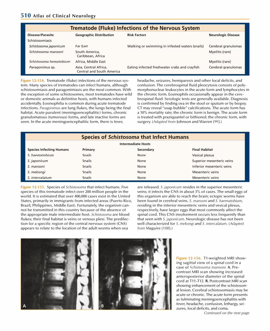
510 Atlas of Clinical Neurology
Trematode (Fluke) Infections of the Nervous SystemDisease/Parasite Geographic Distribution Risk Factors Neurologic Disease
Schistosomiasis
Schistosoma japonicum Far East Walking or swimming in infested waters (snails) Cerebral granulomas
Schistosoma mansoni South America,Caribbean, Africa
Myelitis (rare)
Schistosoma hematobium Africa, Middle East Myelitis (rare)
Paragonimus sp. Asia, Central Africa,Central and South America
Eating infected freshwater crabs and crayfish Cerebral granulomas
Figure 12-154. Trematode (fluke) infections of the nervous sys-tem. Many species of trematodes can infect humans, althoughschistosomiasis and paragonimiasis are the most common. Withthe exception of some schistosomes, most trematodes have wildor domestic animals as definitive hosts, with humans infected accidentally. Eosinophilia is common during acute trematode infections. Paragonimus are lung flukes, the lungs being the finalhabitat. Acute purulent (meningoencephalitic) forms, chronicgranulomatous (tumorous) forms, and late inactive forms areseen. In the acute meningoencephalitic form, there is fever,
headache, seizures, hemiparesis and other focal deficits, and confusion. The cerebrospinal fluid pleocytosis consists of poly-morphonuclear leukocytes in the acute form and lymphocytes in the chronic form. Eosinophils occasionally appear in the cere-brospinal fluid. Serologic tests are generally available. Diagnosis is confirmed by finding ova in the stool or sputum or by biopsy.CT may reveal “soap bubble” calcifications. The acute form hasa 10% mortality rate; the chronic form is benign. The acute formis treated with praziquantel or bithionol; the chronic form, with surgery. (Adapted from Johnson and Warren [99].)
Species of Schistosoma that Infect HumansIntermediate Hosts
Species Infecting Humans Primary Secondary Final Habitat
S. haematobium Snails None Vesical plexus
S. japonicum Snails None Superior mesenteric veins
S. mansoni Snails None Inferior mesenteric veins
S. mekongi Snails None Mesenteric veins
S. intercalatum Snails None Mesenteric veins
Figure 12-155. Species of Schistosoma that infect humans. Five species of this trematode infect over 200 million people in theworld. It is estimated that over 400,000 cases exist in the UnitedStates, primarily in immigrants from infected areas (Puerto Rico,Brazil, Philippines, Middle East). Fortunately, the organism can-not be transmitted in this country because of the absence of the appropriate male intermediate host. Schistosoma are bloodflukes; their final habitat is veins or venous plexi. The predilec-tion for a specific region of the central nervous system (CNS)appears to relate to the location of the adult worms when ova
are released. S. japonicum resides in the superior mesentericveins; it infects the CNS in about 3% of cases. The small eggs of this organism are able to reach the brain; ectopic worms have been found in cerebral veins. S. mansoni andi S. haematobium, residing in the inferior mesenteric veins and vesical plexus,respectively, have larger eggs that most commonly affect thespinal cord. This CNS involvement occurs less frequently than that seen with S. japonicum. Neurologic disease has not been well characterized for S. mekongi andi S. intercalatum. (Adapted from Maguire [108].)
A B
Figure 12-156. T1-weighted MRI show-ing sagittal view of a spinal cord in a case of Schistosoma mansoni. A, Pre-contrast MRI scan showing increasedanteroposterior diameter of the spinalcord at T11–T12. B, Postcontrast MRI scan showing enhancement of the schistosom-al lesion. Cerebral schistosomiasis may beacute or chronic. The acute form presents as fulminating meningoencephalitis with fever, headache, confusion, lethargy, sei-zures, focal deficits, and coma.
Continued on the next page

Infectious Diseases of the Nervous System 511Figure 12-156. (Continued) The presentation of the chroniccerebral form is similar to a tumor, with focal deficits, seizures,increased intracranial pressure, and papilledema. The spinalcord disease is almost always acute, presenting as incompletetransverse myelitis. There is a peripheral leukocytosis witheosinophilia except in the chronic cerebral form. The cerebro-spinal fluid shows slight to moderate pleocytosis, sometimeswith eosinophilia. Cerebral lesions may be seen with CT or
MRI; spinal lesions are seen with MRI or myelography. Diagno-sis can be made by finding ova in the stool or urine, by usingserologic tests, and rectal mucosal biopsy. Treatment includes the use of praziquantel, corticosteroids for edema, anticonvul-sants for seizure, and often decompressive laminectomy for aspinal block. Oxamniquine may need to be added for S. man-soni species resistant to praziquantel. (i From Selwa et al. [109];with permission.)
Cestode (Tapeworm) Infections of the Nervous SystemDisease/Parasite Geographic Distribution Risk Factors Neurologic Disease
Cysticercosis
Taenia solium Central and South America, Asia,Africa, East Europe
Ingestion of eggs in humanfecal contamination
Small cysts or basilar arachnoiditis with hydrocephalus; ocular lesions
Hydatid disease
Echinococcus granulosus Worldwide Ingestion of eggs in caninefecal contamination
Large cysts
Coenurosis
Multiceps multiceps Europe, Americas Ingestion of eggs in carnivorefecal contamination
Budding cysts (rare)
Figure 12-157. Cestode infections of the nervous system. Cestode, or human tapeworm, infections can be divided into two groups. In the first, humans are the definitive host and the adult worms (Taenia saginata and others) live in the gastrointestinal tract and the central nervous system (CNS) is not involved. In the second group, humans are the intermediate host and the larvae spread to the tissues, including the CNS (echinococcosis, coenurosis, and others less common). In Taenia solium, the pork tapeworminfection, humans may be either the definitive host (T. solium) orthe intermediate host (cysticercosis).
Ingestion of undercooked pork containing the encysted larvae (Cysticercus cellulosae; tissue larval stage) results in infection of
the human intestine by the adult tapeworm (definitive host). There are usually no symptoms at this stage. The terminal gravidproglottids of the worm are excreted in the feces with thousandsof ova. These ova contaminate the environment, where they are ingested by pigs or humans (intermediate hosts). The shells ofthese eggs are digested by gastric juices, liberating the embryos(oncospheres), which penetrate the intestinal wall, migrate to tis-sues, and become encysted (cysticerci). In humans they primarily localize to the brain and CNS. Cysticercosis is clearly the most important cestode infection of humans. Coenurosis is the rarelarval disease caused by the dog tapeworm, Taenia (Multiceps) multiceps. (Adapted from Johnson and Warren [99].)
Clinical Manifestationsof Neurocysticercosis
Symptoms and Signs Approximate Frequency, %
Headache 23–98
Seizures 37–92
Papilledema 48–84
Meningeal signs 29–33
Nausea/vomiting 74–80
Altered mental status 9–47
Dementia 1–6
Psychosis 1–17
Focal sensory or motor deficits 3–36
Cranial nerve palsies 1–36
Altered vision 5–34
Ataxia 5–24
Spinal cord compression < 1
Figure 12-158. Clinical manifestations of neurocysticercosis. The manifestations of neurocysticercosis depend on the loca-tion of the lesions. The clinical disease can be divided into fourtypes based on the anatomic location of infection: parenchymal,subarachnoid (meningitic), intraventricular, and spinal. In the parenchymal form, the manifestations are related to the loca-tion of the cysts. Focal seizures and focal neurologic deficits areseen in the parenchymal form. The meningitic form results in headache, nuchal rigidity, and communicating hydrocephalus.Intraventricular disease may result in obstructive hydrocepha-lus. Spinal disease may result in arachnoiditis and subarachnoidblock. (Adapted from Cameron and Durack [110].)
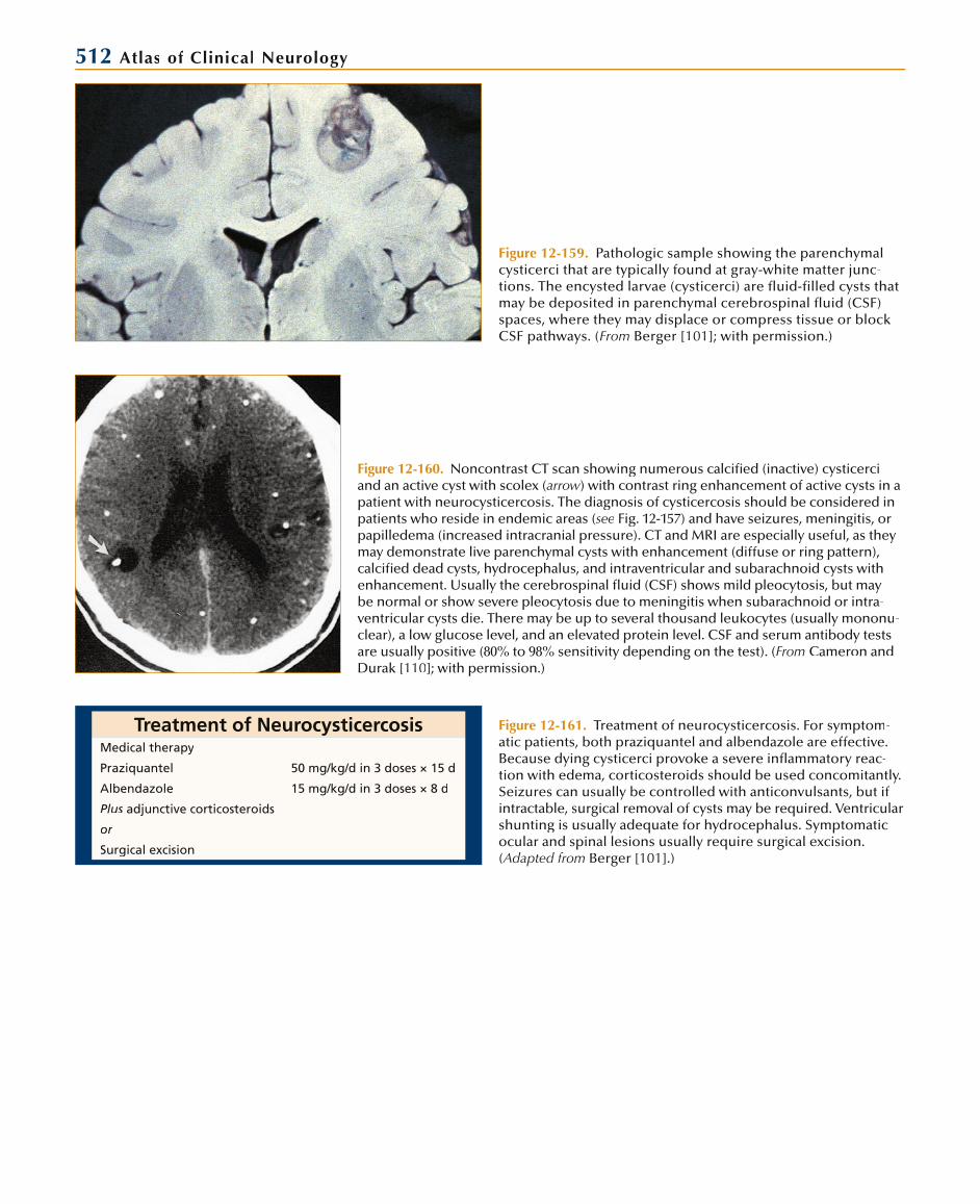
512 Atlas of Clinical Neurology
Figure 12-159. Pathologic sample showing the parenchymalcysticerci that are typically found at gray-white matter junc-tions. The encysted larvae (cysticerci) are fluid-filled cysts that may be deposited in parenchymal cerebrospinal fluid (CSF)spaces, where they may displace or compress tissue or block CSF pathways. (From Berger [101]; with permission.)
Figure 12-160. Noncontrast CT scan showing numerous calcified (inactive) cysticerci and an active cyst with scolex (arrow) with contrast ring enhancement of active cysts in a patient with neurocysticercosis. The diagnosis of cysticercosis should be considered inpatients who reside in endemic areas (see Fig. 12-157) and have seizures, meningitis, orpapilledema (increased intracranial pressure). CT and MRI are especially useful, as theymay demonstrate live parenchymal cysts with enhancement (diffuse or ring pattern),calcified dead cysts, hydrocephalus, and intraventricular and subarachnoid cysts with enhancement. Usually the cerebrospinal fluid (CSF) shows mild pleocytosis, but maybe normal or show severe pleocytosis due to meningitis when subarachnoid or intra-ventricular cysts die. There may be up to several thousand leukocytes (usually mononu-clear), a low glucose level, and an elevated protein level. CSF and serum antibody tests are usually positive (80% to 98% sensitivity depending on the test). (From Cameron and Durak [110]; with permission.)
Treatment of NeurocysticercosisMedical therapy
Praziquantel 50 mg/kg/d in 3 doses × 15 d
Albendazole 15 mg/kg/d in 3 doses × 8 d
Plus adjunctive corticosteroids
or
Surgical excision
Figure 12-161. Treatment of neurocysticercosis. For symptom-atic patients, both praziquantel and albendazole are effective.Because dying cysticerci provoke a severe inflammatory reac-tion with edema, corticosteroids should be used concomitantly. Seizures can usually be controlled with anticonvulsants, but if intractable, surgical removal of cysts may be required. Ventricular shunting is usually adequate for hydrocephalus. Symptomatic ocular and spinal lesions usually require surgical excision.(Adapted from Berger [101].)
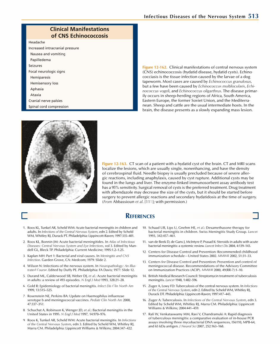
Infectious Diseases of the Nervous System 513
Clinical Manifestationsof CNS Echinococcosis
Headache
Increased intracranial pressure
Nausea and vomiting
Papilledema
Seizures
Focal neurologic signs
Hemiparesis
Hemisensory loss
Aphasia
Ataxia
Cranial nerve palsies
Spinal cord compression
Figure 12-162. Clinical manifestations of central nervous system (CNS) echinococcosis (hydatid disease, hydatid cysts). Echino-cocciasis is the tissue infection caused by the larvae of a dogtapeworm. Most cases are caused by Echinococcus granulosus,but a few have been caused by Echinococcus multilocularis, Echi-nococcus vogeli, and i Echinococcus oligarthrus. The disease primar-ily occurs in sheep-herding regions of Africa, South America,Eastern Europe, the former Soviet Union, and the Mediterra-nean. Sheep and cattle are the usual intermediate hosts. In the brain, the disease presents as a slowly expanding mass lesion.
Figure 12-163. CT scan of a patient with a hydatid cyst of the brain. CT and MRI scanslocalize the lesions, which are usually single, nonenhancing, and have the density of cerebrospinal fluid. Needle biopsy is usually precluded because of severe aller-gic reactions, including anaphylaxis, caused by cyst rupture. Additional cysts may be found in the lungs and liver. The enzyme-linked immunosorbent assay antibody testhas a 95% sensitivity. Surgical removal of cysts is the preferred treatment. Drug treatmentwith albendazole may decrease the size of the cysts, but it should be started beforesurgery to prevent allergic reactions and secondary hydatidosis at the time of surgery.(From Abbassioun et al. [111]; with permission.)
REFERENCES
1. Roos KL, Tunkel AR, Scheld WM: Acute bacterial meningitis in children and adults. In Infections of the Central Nervous System, edn 2. Edited by ScheldWM, Whitley RJ, Durack PT. Philadelphia: Lippincott-Raven; 1997:335–401.
2. Roos KL, Bonnin JM: Acute bacterial meningitides. In Atlas of Infectious Diseases: Central Nervous System and Eye Infections, vol 3. Edited by Man-dell GL, Bleck TP. Philadelphia: Current Medicine; 1995:1.2–1.25.
3. Kaplan MH: Part 1: Bacterial and viral causes. In Meningitis and CNSInfection. Garden Grove, CA: Medcom; 1979: Slide 2.
4. Wilson N: Infections of the nervous system. In Neuropathology: An Illus-trated Course. Edited by Duffy PE. Philadelphia: FA Davis; 1977: Slide 12.
5. Durand ML, Calderwood SB, Weber DJ, et al.: Acute bacterial meningitis in adults: a review of 493 episodes. N Engl J Med 1993, 328:21–28.d
6. Gold R: Epidemiology of bacterial meningitis. Infect Dis Clin North Am1999, 13:515–525.
7. Rosenstein NE, Perkins BA: Update on Haemophilus influenzaeserotype b and meningococcal vaccines. Pediatr Clin North Am 2000,47:337–352.
8. Schuchat A, Robinson K, Wenger JD, et al.: Bacterial meningitis in theUnited States in 1995. N Engl J Med 1997, 14:970–976.d
9. Roos K, Tunkel AR, Scheld WM: Acute bacterial meningitis. In Infectionsof the Central Nervous System, edn 3. Edited by Scheld WM, Whitley RJ,Marra CM. Philadelphia: Lippincott Williams & Wilkins; 2004:347–422.
10. Schaad UB, Lips U, Gnehm HE, et al.: Dexamethasone therapy for bacterial meningitis in children. Swiss Meningitis Study Group. Lancet1993, 342:457–461.
11. van de Beek D, de Gans J, McIntyre P, Prasad K: Steroids in adults with acute bacterial meningitis: a systemic review. Lancet Infect Dis 2004, 4:139–143.
12. Centers for Disease Control and Prevention: Recommended childhoodimmunization schedule—United States 2002. MMWR 2002, 51:31–33.
13. Centers for Disease Control and Prevention: Prevention and control of meningococcal disease. Recommendations of the Advisory Committee on Immunization Practices (ACIP). MMWR 2000, 49(RR-7):1–10.
14. British Medical Research Council: Streptomycin treatment of tuberculosis meningitis. Lancet 1948, 1:482–596.t
15. Zuger A, Lowy FD: Tuberculosis of the central nervous system. In Infections of the Central Nervous System, edn 2. Edited by Scheld WM, Whitley RJ, Durack DT. Philadelphia: Lippincott-Raven; 1997:417–443.
16. Zuger A: Tuberculosis. In Infections of the Central Nervous System, edn 3. Edited by Scheld WM, Whitley RJ, Marra CM. Philadelphia: LippincottWilliams & Wilkins; 2004:441–459.
17. Rafi W, Venkataswamy MM, Ravi V, Chandramuki A: Rapid diagnosisof tuberculous meningitis: a comparative evaluation of in-house PCRassays involving three mycobacterial DNA sequences, IS6110, MPB-64,and 65 kDa antigen. J Neurol Sci 2007, 252:163–168.i
514 Atlas of Clinical Neurology
18. American Thoracic Society joint statement. Treatment of tuberculosis. Am J Respir Crit Care Med 2003, 167:603–662.d
19. Kumar R, Prakash M, Jha S: Paradoxical response to chemotherapy inneurotuberculosis. Pediatr Neurosurg 2006, 42:214–222.g
20. Kennedy DH, Fallon RJ: Tuberculous meningitis. JAMA 1979, 241:264–268.
21. Weisberg L, Nice C, Katz M: Infectious inflammatory conditions. InCerebral Computed Tomography: A Text Atlas, edn 2. Philadelphia: WBSaunders; 1984:229–248.
22. Danner RL, Hartman BJ: Update of spinal epidural abscess: 35 cases and review of the literature. Rev Infect Dis 1987, 9:265–274.
23. Gelfand MS, Bakhtian BJ, Simmons BP: Spinal sepsis due to Streptococcus milleri: two cases and review. Rev Infect Dis 1991, 13:559–563.
24. Hartman BJ, Helfgott DC, Weingarten K: Subdural empyema and suppu-rative intracranial phlebitis. In Infections of the Central Nervous System, edn 3. Edited by Scheld WM, Whitley RJ, Marra CM. Philadelphia: LippincottWilliams & Wilkins; 2004:524–535.
25. Greenlee JE: Subdural empyema. In Principles and Practice of Infectious Diseases, edn 4. Edited by Mandell GL, Bennet JE, Dolin R. New York:Churchill Livingstone; 1995:900–903.
26. Wispelwey B, Dacey RG Jr, Scheld WM: Brain abscess. In Infections of theCentral Nervous System, edn 2. Edited by Scheld WM, Whitley RJ, DurackDT. Philadelphia: Lippincott-Raven; 1997:463–493.
27. Wispelwey B, Scheld WM: Brain abscess. In Principles and Practice of Infectious Diseases, edn 4. Edited by Mandell GL, Douglas RG Jr, Bennett JE. New York: Churchill Livingstone; 1995:889–891.
28. Wispelwey B: Brain abscess. In Atlas of Infectious Diseases: Central NervousSystem and Eye Infections, vol. 3 Edited by Mandell GL, Bleck TP. Philadel-phia: Current Medicine; 1995:4.2–4.14.
29. Falcone S, Post MJ: Encephalitis, cerebritis, and brain abscess: pathophysi-ology and imaging findings. Neuroimaging Clin North Am 2000, 10:333–353.
30. Kastenbauer S, Pfister H–W, Wispelwey B, Scheld WM: Brain abscess. In Infections of the Central Nervous System, edn 3. Edited by Scheld WM, Whit-ley RJ, Marra CM. Philadelphia: Lippincott Williams & Wilkins; 2004:479–507.
31. Jubelt B: Viral infections. In Merritt’s Textbook of Neurology, edn 9. Edited yyby Rowland LP. Philadelphia: Lippincott Williams & Wilkins; 2005:175–210.
32. Centers for Disease Control and Prevention: Annual summary 1981:reported morbidity and mortality in the United States. MMWR 1982, 30(54).
33. Read SJ, Kurtz JB: Laboratory diagnosis of common viral infectios of the central nervous system by using a single multiplex PCR screening assay. J Clin Microbiol 1999, 37:1352–1355.l
34. Jubelt B: The diagnosis of viral meningitis and encephalitis. In Neurology and Neurosurgery Update Series, vol 2, no 30. Edited by Scheinberg P, Davidoff RA, Arnason BGW. Princeton NJ: Education Center; 1981.
35. Jubelt B, Lipton HL: Enterovirus infections. In Handbook of Clinical Neu-rology, vol 12. Edited by Vinken PJ, Bruyn GW, Klawans HL. Amsterdam:yyElsevier Science Publishing; 1989:326.
36. Hanley DF, Glass JD, McArthur JC, Johnson RT: Viral encephalitis and related conditions. In Atlas of Infectious Diseases: Central Nervous Systemand Eye Infections. Edited by Mandell GL, Bleck TB. Philadelphia: CurrentMedicine; 1995:3.2–3.40.
37. Solomon T: Flavivirus encephalitis. N Engl J Med 2004, 351:370–378.d
38. Whitley RJ, Alford CA, Hirsch MS, et al.: Vidarabine versus acyclovir therapy in herpes simplex encephalitis. N Engl J Med 1986, 314:144–149.d
39. Skoldenberg B, Forsgren M, Alestig K, et al.: Acyclovir versus vidarabinein herpes simplex encephalitis. Randomised multicenter study in con-secutive Swedish patients. Lancet 1984, 2:707–711.t
40. Johnson RT: Viral Infections of the Nervous System. New York: Raven Press; 1998:133–168.
41. Hirano A, Iwato M, Kato T, et al.: Color Atlas of Pathology of the Nervous System, edn 2. Edited by Hirano A. Tokyo: Igaku-Shoin; 1998:133–168.
42. Davis JM, Davis KR, Kleinman GM, et al.: Computed tomography of herpes simplex encephalitis with clinicopathological correlation.Radiology 1978, 129:409–417.
43. Runge VM: Skull and its contents. In Magnetic Resonance Imaging of theBrain. Philadelphia: JB Lippincott; 1994:180.
44. Schroth G, Gawehn J, Thron A, et al.: Early diagnosis of herpes simplex encephalitis by MRI. Neurology 1987, 37:179–183.
45. Davis LE, DeBiasi R, Goade DE, et al.: West Nile virus neuroinvasive disease. Ann Neurol 2006, 60:286–300.l
46. Robinson P: Rabies. In Infectious Diseases. Edited by Gorbach SL, Bartlett JG, Blacklow NR. Philadelphia: WB Saunders; 1992:1269–1277.
47. Rupprecht CE, Hemachudha T: Rabies. In Infections of the Central Nervous System, edn 3. Edited by Scheld WM, Whitley RJ, Marra CM.Philadelphia: Lippincott Williams & Wilkins; 2004:243–259.
48. Hanna JN, Carney IK, Smith GA, et al.: Australian bat lyssavirus infection: a second human case, with a long incubation period. Med J Aust 2000, t172:597–599.
49. Corey L: Rabies, rhabdoviruses, and Marburg-like agents. In Harrison’s Principles of Internal Medicine, edn 13. Edited by Isselbacher KJ, Braunwald E, Wilson JD. New York: McGraw Hill; 1994:834.
50. Centers for Disease Control and Prevention: Human rabies prevention—United States, 1999. Recommendations of the Advisory Practices (ACIP).MMWR 1999, 48:1–21.
51. Centers for Disease Control and Prevention: Summary of notifiabledisease, United States, 1993. MMWR 1993, 42(53):27.
52. Mihai C, Jubelt B: Post-infections encephalomyelitis. Curr Neurol Neurosci Rep 2005, 5:440–445.
53. Horstmann DM: Epidemiology of poliomyelitis and allied diseases—1963.Yale J Biol Med 1964, 36:5–26. d
54. Jubelt B, Gallez-Hawkins G, Narayan O, Johnson RT: Pathogenesis of human poliovirus infection in mice. I. Clinical and pathological studies.J Neuropathol Exp Neurol 1980, 39:138–148. l
55. Kincaid O, Lipton HL: Viral myelitis: an update. Curr Neurol Neurosci Rep 2006, 6:469–474.
56. Anders HJ, Goebel FD: Cytomegalovirus polyradiculopathy in patientswith AIDS. Clin Infect Dis 1998, 27:345–352.
57. Talpes D, Tien RD, Hesselink JR: Magnetic resonance imaging of AIDS-related polyradiculopathy. Neurology 1991, 41:1995–1997.
58. Murray RP, Kobayashi GS, Pfaller MA, Rosenthal KD: Picorna viruses. In Medical Microbiology, edn 2.. St. Louis: Mosby; 1994:583. yy
59. Rosencrance G: Images in clinical medicine. Herpes zoster. N Engl J Med1994, 330:906.
60. Oxman MN, Levin MJ, Johnson GR, et al.: A vaccine to prevent herpes zoster and postherpetic neurologia in older adults. N Engl J Med 2005, d352:2271–2284.
61. Hope-Simpson R: The nature of herpes zoster: a long-term study and anew hypothesis. Proc Roy Soc Med 1965, 58:1–12.d
62. Johnson RT, McArthur JC, Narayan O: The neurobiology of HIV infections. FASEB J 1988, 2:2970–2981. J
63. Price RW, Brew BJ, Roke M: Central and peripheral nervous systemcomplications of HIV-1 infections and AIDS. In AIDS: Etiology, Diagnosis,Treatment, and Prevention. Edited by DeVita VT, Hellman S, RosenbergSA. Philadelphia: JB Lippincott; 1992:237–254.
64. Fauci AS, Lane HC: Human immunodeficiency virus disease: AIDS and related disorders. In Harrison’s Principles of Internal Medicine, edn 16. Edited by Kasper DL, Braunwald E, Fauci AS, et al. New York: McGraw-Hill;2005:1076–1139.
65. Olsen WL, Longo FM, Mills CM, Norman D: White matter disease in AIDS: findings at MR imaging. Radiology 1988, 169:445–448.
66. Hammer SM, Saag MS, Schechter M, et al.: Treatment for adult HIV infec-tion: 2006 recommendations of the International AIDS Society—USA panel. JAMA 2006, 296:827–843.
67. Grindstaff P, Gruener G: The peripheral nervous system complica-tions of HTLV-1 myelopathy (HAM/TSP) syndromes. Semin Neurol 2005,l25:315–327.

Infectious Diseases of the Nervous System 51568. Rodgers-Johnson PEB, Ono SG, Asher DM, Gibbs CL: Tropical spastic
paraparesis and HTLV-1 myelopathy: clinical features and pathogenesis.In Immunologic Mechanisms in Neurologic and Psychiatric Disease. Editedby Waksman BD. New York: Raven Press; 1990.
69. Izumo S, Goto MD, Itoyama MD, et al.: Interferon-alpha is effectivein HTLV-1–associated myelopathy: a multicenter, randomized, doubleblind, controlled trial. Neurology 1996, 46:1016–1021.
70. Khalili K, White MK, Lublin F, et al.: Reactivation of JC virus and develop-ment of PML in patients with multiple sclerosis. Neurology 2007, 68:985–990.y
71. Whiteman ML, Post MJ, Berger JK, et al.: Progressive multifocal leuko-encephalopathy in 47 HIV-seropositive patients: neuroimaging with clinical and pathologic correlation. Radiology 1993, 187:233–240.y
72. Bale JF Jr: Subacute sclerosing panencephalitis. A preventable disease that needs to be prevented. Neurology 2004, 63:1352–1353.y
73. Ohya T, Martinez AJ, Jabbour JT, et al.: Subacute sclerosing panencephalitis.Correlation of clinical, neurophysiologic and neuropathologic findings.Neurology 1974, 24:211–218.y
74. Weihl CC, Roos RP: Creutzfeldt-Jakob disease, new variant Creutzfeldt-Jakob disease, and bovine spongiform encephalopathy. Neurol Clin1999, 17:835–859.
75. Brown P, Gibbs CJ Jr, Rodgers-Johnson P, et al.: Human spongiform enceph-alopathy: the NIH series of 300 cases of experimentally transmitted disease. Ann Neurol 1994, 35:513–529. l
76. Jubelt B: Prion diseases. In Merritt’s Textbook of Neurology, edn 11.Edited by Rowland LP. Philadelphia: Lippincott Williams & Wilkins;2005:264–270.
77. Shiga Y, Miyazawa K, Sato S, et al.: Diffusion-weighted MRI abnormalities as an early diagnostic marker for Creutzfeldt-Jakob disease. Neurology2004, 63: 443–449.
78. Will RG, Zeidler M, Stewart GE, et al.: Diagnosis of new variantCreutzfeldt-Jakob disease. Ann Neurol 2000, 47:575–582.l
79. Manetto V, Medori R, Cortelli P, et al.: Fatal familial insomnia: clinical and pathologic study of five new cases. Neurology 1992, 42:312–319.y
80. Perfect JR: Fungal meningitis. In Infections of the Central Nervous System, edn 3. Edited by Scheld WM, Whitley RJ, Marra CM. Philadelphia: LippincottWilliams & Wilkins; 2004:691–712.
81. Tunkel AR, Crous SE: Subacute and chronic meningitides. In Atlas of Infectious Diseases: Central Nervous System and Eye Infections, vol 3. Editedby Mandell GL, Bleck TP. Philadelphia: Current Medicine; 1995:2.13.
82. Gozdasoglu S, Ertem M, Buyukkececi Z, et al.: Fungal colonization andinfection in children with acute leukemia and lymphoma during induction therapy. Med Ped Oncol 1999, 32:344–348.l
83. Tucker T, Ellner JJ: Chronic meningitis. In Infections of the Central NervousSystem. Edited by Scheld WM, Whitley RJ, Durack DT. New York: RavenPress; 1991:703–728.
84. Tunkel AR, Scheld WM: Central nervous system infections in the com-promised host. In Clinical Approach to Infection in the Compromised Host,edn 3. Edited by Rubin RH, Young LS. New York: Plenum; 1994:187.
85. Galgiani JN: Coccidioidomycosis. West J Med 1993, 159:153–171. d
86. Hildebrand J, Hildebrand M: Chronic meningitis. In Infections of theCentral Nervous System, edn 3. Edited by Scheld WM, Whitley RJ, Marra CM. Philadelphia: Lippincott Williams & Wilkins; 2004:829–841.
87. Sepkowitz K, Armstrong D: Space-occupying fungal lesions. In Infectionsof the Central Nervous System, edn 2. Edited by Scheld WM, Whitley RJ,Durack DT. Philadelphia: Lippincott-Raven Publishing; 1997:741–762.
88. Simon RP: Neurosyphilis. Arch Neurol 1995, 42:606–613.l
89. Stefanis L, Rowland LP: Spirochete infections: neurosyphilis. In Merritt’sTextbook of Neurology, edn 11. Edited by Rowland LP. Philadelphia: Lip-pincott Williams & Wilkins; 2005:235–244.
90. Merrit HH, Adams RD, Solomon HC: Neurosyphilis. New York: OxfordUniversity Press; 1946:24–67.
91. Simon R, Bayne L: Neurosyphilis. In Infectious Diseases of the Central Nervous System. Edited by Tyler KL, Martin JB. Philadelphia: FA Davis; 1993:237–255.
92. Davis E: Spirochetal disease. In Diseases of the Nervous System: Clinical Neurobiology, edn 2. Edited by Asbury AK, McKhann GM, McDonald WI.yyPhiladelphia: WB Saunders; 1992:1359–1370.
93. Anderson JF: Epizootiology of Borrelia in Ixodes tick vectors and reservoirhosts. Rev Infect Dis 1989, 11(suppl 6):S1451–S1459.
94. Pachner AR, Steere AC: The triad of neurologic manifestations of Lymedisease, meningitis, cranial neuritis and radiculoneuritis. Neurology1985, 35:47–53.
95. Coyle PK: Lyme disease. In Current Diagnosis in Neurology. Edited by Feldman E. St. Louis: CV Mosby; 1994:113.
96. Reik L Jr: Lyme disease. In Infections of the Central Nervous System, edn2. Edited by Scheld WM, Whitley RJ, Durack DT. Philadelphia: Lippin-cott-Raven; 1997:685–718.
97. Cadavid D: Lyme disease and relapsing fever. In Infections of the Central Nervous System, edn 3. Edited by Scheld WM, Whitley RJ, Marra CM.Philadelphia: Lippincott Williams & Wilkins; 2004:659–690.
98. Coda GC: Protozoan and helminthic infections. In Infections of theCentral Nervous System. Edited by Lambert HP. Philadelphia: BC Decker; 1991:264–282.
99. Johnson RT, Warren KS: Parasitic infections. In Diseases of the Nervous System: Clinical Neurobiology, edn 2. Edited by Asbury AK, McKhann yyGM, McDonald WI. Philadelphia: WB Saunders; 1992:1350–1358.
100. Luft BJ, Remington JS: Toxoplasmosis of the central nervous system. In Current Topics in Infectious Diseases, vol 6. Edited by Remington SJ,Swartz MN. New York: McGraw-Hill; 1985:315–358.
101. Berger JR: Parasitic diseases of the nervous system. In Atlas of Infec-tious Diseases: Central Nervous System and Eye Infections. Edited byMandell GL, Bleck TP. Philadelphia: Current Medicine; 1995:5.4–5.25.
102. Farrar WE, Wood MJ, Innes JA, Tubbs H: Infectious Diseases: Text and Color Atlas, edn 2. London: Gower Medical Publishers; 1992:3.30.
103. Montoya JG, Kovacs JA, Remington JS: Toxoplasma gondii. In Principles and Practice of Infectious Diseases, edn 6. Edited by Mandell GL, Bennett JE, Dolin R. Philadelphia: Elsevier; 2005:3170–3198.
104. Oo MM, Aikawa M, Than T, et al.: Human cerebral malaria: a pathologicalstudy. J Neuropathol Exp Neurol 1987, 46:223–231. l
105. Idro R, Jenkins NE, Newton CR: Pathogenesis, clinical features,and neurological out come of cerebral malaria. Lancet Neurol 2005, l4:827–840.
106. Kirchhoff LV: Trypanosomiasis. In Infections of the Central Nervous System, edn 3. Edited by Scheld WM, Whitley RJ, Marra CM. Philadelphia:Lippincott Williams & Wilkins; 2004:777–789.
107. Durack DT: Amebic infections. In Infections of the Central Nervous System, edn 2. Edited by Scheld WM, Whitley RJ, Durack DT. Philadelphia:Lippincott-Raven; 1997:831–844.
108. Maguire JH: Trematodes (Schistosomes) and other flukes. In Principles and Practices of Infectious Diseases, edn 6. Edited by Mandell GL,Bennett JE, Dolin R. Philadelphia: Elsevier; 2005:3276–3285.
109. Selwa LM, Brumberg JA, Mandell SH, Garofalo EA: Spinal cord schisto-somiasis: a pediatric case mimicking intrinsic cord neoplasm. Neurology1991, 41:755–757.
110. Cameron ML, Durack DT: Helminthic infections. In Infections of the Central Nervous System, edn 2. Edited by Scheld WM, Whitley RJ,Durack DT. New York: Raven Press; 1997:845–878.
111. Abbassioun K, Rahmat H, Ameli NO, Tafazoli M: Computerized tomog-raphy in hydatid cyst of the brain. J Neurosurg 1978, 49:408–411.g