Surveillance of infectious diseases in animals and ... - SVA
127
SURVEILLANCE OF INFECTIOUS DISEASES IN ANIMALS AND HUMANS IN SWEDEN 2017
-
Upload
khangminh22 -
Category
Documents
-
view
0 -
download
0
Transcript of Surveillance of infectious diseases in animals and ... - SVA

SURVEILLANCE OF INFECTIOUSDISEASES IN ANIMALS AND HUMANS
IN SWEDEN 2017

Editors: Ann Lindberg, Thomas RosendalDepartment of Disease Control and Epidemiology, Naional Veterinary Insitute (SVA), SE-751 89 Uppsala,Sweden.
Authors: Anton Andreasson, Charlote Axén, Mia Brying, Jonas Carlsson, Erika Chenais, Arianna Comin,Helena Eriksson, Linda Ernholm, Eva Forsgren, Gitan Gröndahl, Gunilla Hallgren, Gete Hestvik, MarikaHjertqvist, Mia Holmberg, Helena Höök, Cecilia Hultén, Cecilia Jernberg, Jerker Jonsson, Pontus Juréen, ElinaLahi, Ann Lindberg, Mats Lindblad, Ylva Lindgren, Emma Löf, Margareta Löfdahl, Anna Lundén, Marie Jans-sonMörk, Oskar Nilsson, Maria Nöremark, Faruk Otman, Karin Persson-Waller, Moa Rehn, Thomas Rosendal,Christoffer Sjölund, Kaisa Sörén, Karl Ståhl, Lena Sundqvist, Magnus Thelander, Kersin de Verdier, CatrinVesterlund-Carlson, HeleneWahlström, AndersWallensten, PerWallgren, StefanWidgren, Elsie Ydring, BethYoung, Nabil Yousef, Siamak Zohari, Erik Ågren
Cover Photo: Karin Bernodt
Copyright of map data: ©EuroGeographics for the administraive boundaries
Layout: The producion of this report coninues to be accomplished using a primarily open-source toolset.The method allows the source text, produced by authors, to be edited independently of the template forthe layout which can be modified and reused for future reports. Specifically, the chapter texts are authoredin Microsot Word and then converted using pandoc to the LaTeX typeseing language. All figures andmaps are produced using R sotware for staisical compuing. Development for 2017 further formalisedthe report generaion tool in an R-package available on GitHub which has streamlined the report buildingand integrates quality control into the process. The report generaing toolset and process was designed andwriten by Thomas Rosendal and Stefan Widgren.
Print: TMG Tabergs AB.
Prining and part of the producion was financed by the Swedish Board of Agriculture. Text, tables, figuresand maps may be cited and reprinted only with reference to this report.
Suggesion citaion: Surveillance of infecious diseases in animals and humans in Sweden 2017, NaionalVeterinary Insitute (SVA), Uppsala, Sweden. SVA:s rapportserie 52 ISSN 1654-7098.
This report may be subject to updates and correcions. The latest version is always available for downloadat www.sva.se.
visiing address. Ulls väg 2B address. 751 89Uppsalatelephone. +46 18-67 40 00 fax. +46 18-30 91 62e-mail. [email protected] web. www.sva.se

ContentsIntroducion 3
Overview of acive surveillance 2009-2017 4
Livestock populaion and tradein live animals 5
Animal registers and datasourcesused in surveillance 9
Insituions, organisaions andlaboratories involved in monitoring 10
Disease Surveillance 2017 13
Atrophic rhiniis 14
Aujeszkys disease 15
Bluetongue 17
Bovine spongiform encephalopathy 19
Bovine viral diarrhoea 21
Brucellosis 23
Campylobacteriosis 25
Chronic wasing disease 29
Classical swine fever 31
Coccidiosis and clostridiosis 33
Echinococcosis 34
Alveolar echinococcosis 34
Cysic echinococcosis 36
Enzooic bovine leucosis 37
Footrot 38
Infecious bovine rhinotracheiis 40
Influenza 41
Avian influenza 41
Swine influenza 45
Leptospirosis 48
Listeriosis 50
Maedi-visna 53
Nephropathia epidemica 55
Paratuberculosis 57
Porcine reproducive and respiratorysyndrome 60
Psitacosis 62
Q fever 63
Rabies 65
Salmonellosis 67
Scrapie 79
Strangles 81
Tick borne encephaliis 83
Trichinellosis 85
Tuberculosis 87
Tularaemia 90
Verotoxinogenic Escherichia coli 92
Yersiniosis 96
Addiional Surveillance 2017 99
Clinical surveillance 100
Poultry health control programme 103
Infecious diseases in wild boars 106
Infecious diseases and parasites inhoneybees 108
Infecious diseases in fish crustaceansand molluscs 112
Examinaions of aborions in foodproducing animals 116
Post mortem examinaions in foodproducing animals 117
Post mortem examinaions in wildlife 120
Animicrobial resistance in bacteriafrom animals and food 122

Photo: Karin Bernodt
2 BACKGROUND

IntroducionSurveillance of infectious diseases in animals and hu-mans 2017 is the annual report on surveillance activitiescarried out in Sweden during the year, and their output. Thereport covers surveillance for important animal diseases andzoonotic agents in humans, food, feed and animals, carriedout and compiled by public and private actors with surveil-lance mandates along the entire food chain, from stable totable.
The report is subject to constant improvement and de-velopment. This year we have introduced a chapter whichprovides a 10-year retrospective overview of active surveil-lance, as well as a new chapter on strangles; a disease whichis endemic in horses in Sweden and constitutes a recurringthreat to the horse industry. Furthermore, a chapter on in-fectious diseases and parasites in honeybees has been addedto describe the extensive activities conducted to maintain agood health status in this important animal species.
The information generated by animal disease surveil-lance is of key importance to demonstrate the good healthand welfare of Swedish animals. Some beneits of surveil-lance activities are inherent, such as the prevention of ani-mal disease and promotion of public health. However, manysurveillance activities are in place primarily to ensure safetrade and movement of animals, thereby facilitating tradeand giving access to foreign markets. This is also wherethe major costs can appear as a result of outbreaks of reg-ulated diseases; by the restrictions put in place to maintaintrust between trading partners. Consequently, to reinstate afavourable status it is necessary to provide evidence of highquality surveillance data that the disease is once again absentfrom the country, region or sector, or at least under control.
Surveillance activities require resources for planningand design as well as for implementation, to organise theof the collection of samples or other types of data from dif-ferent groups in a representative way; to identify and useaccurate and timely diagnostics; and inally, to analyse thedata and communicate it to relevant stakeholders for deci-sion making. Investments in surveillance are costly, and maybe diicult to justify, in particular when the disease burdenor the perceived threat is low. This is sometimes referred toas the good health status paradox , where it is challengingto motivate investments to maintain a favourable disease sit-uation. Surveillance investments are similar to insurance inthat the beneit accrues in the appearance of negative events.Therefore, a national surveillance plan is being put in placewith the purpose to ensure that long-term needs to main-tain a favourable situation are balanced with more short-termneeds to manage emerging issues.
Surveillance activities also have to evolve to incorporatenew diagnostic methods, new knowledge of the disease and
new technology for information capture and analysis. Sev-eral national and international projects are currently run-ning where the developments will contribute to more ei-cient surveillance in the future. Of particular importance isa 5-year One Health European Joint Programme (OHEJP)that, from the Swedish side, involves both the National Vet-erinary Institute (SVA), the National Food Agency and thePublic Health Agency of Sweden. The irst two integrativeprojects launched under this programme focus on improvingthe interoperability of animal and public health surveillancesystems, as well as the capacity to conduct joint risk assess-ments. The Swedish partners in these projects receive co-funding from the Swedish Civil Contingencies Agency, whosupports the development of crisis preparedness within theSwedish food chain. Research into new innovative surveil-lance methods is also underway within the OHEJP, with SVAas coordinator.
The need for further strengthening an integrated OneHealth surveillance and response in Sweden can be exem-pliied by the large outbreak of human campylobacteriosis,which started in 2016 and lasted into 2017. The coordinatedand joint collection, isolation and subtyping of isolates fromhumans, animals and chicken meat provided a richer under-standing of the associations, and contributed strongly to trig-gering the necessary action.
Also in focus during 2017 was the planning for surveil-lance of Chronic Wasting Disease (CWD), which appearedfor the irst time in Europe in early 2016, in Norway. Surveil-lance targeting fallen and road-killed wildlife as well asclinical suspicions is in place, and during 2018 additionalsurveillance components targeting deer populations enter-ing the food chain are being implemented. Other exampleswhere wildlife surveillance is of great importance for earlywarning and protection of domestic populations are AfricanSwine Fever (ASF) and highly pathogenic avian influenza(HPAI). The former advanced into new European territoryduring 2017, and has continued to spread during 2018. Mea-sures to prevent further spread are now imperative, with theaim to increase awareness among the public as well as to en-gage the hunting community to strengthen the capacity forearly detection.
As an EU member state, Sweden shares the implicationsand consequences of exotic disease introduction with manyother European countries. We are part of a pan-Europeansurveillance system, where our eforts contribute, directlyand indirectly, to the understanding of risks that emerg-ing diseases pose to other EU countries. Openness, trans-parency and pro-activeness are key for efective early warn-ing and control, and it is important for trust and for joint Eu-ropean preparedness to which we actively contribute. In linewith this, our understanding of the Swedish disease situationin 2017 is provided in this report.
BACKGROUND 3

Overview of acive surveillance 2009-2017BACKGROUNDSince 2009, Sweden has reported the outcome of its activesurveillance programmes in an annual report on surveillanceof infectious diseases in animals and humans. This yearlydescription of active disease surveillance programmes is im-portant as it contributes to the international community’sunderstanding of how Sweden’s animal and zoonotic dis-ease status is determined. While passive surveillance forimportant diseases occurs continuously, active surveillancefor each disease does not necessarily occur on an annual ba-sis. Surveillance activities are regularly evaluated and the
decision to conduct active surveillance for a speciic dis-ease in any given year is based on a number of factors,such as the indings of previous years’ surveillance activi-ties, changes in the disease status of other countries and theemergence of new diseases. Table 1 provides information onthe years in which, active surveillance was undertaken forvarious diseases of importance. More detailed informationabout the active surveillance that was conducted during aspeciic year between 2009-2016 can be found by consultingthat year’s annual surveillance report, which can be found atwww.sva.se.
Table 1: Historical overview of acive surveillance aciviies from 2009-2017. Filled circles ( ) indicate that acive surveillance was carried out.
Disease Year
2009 2010 2011 2012 2013 2014 2015 2016 2017
African swine fever # # # # # # # #
Atrophic rhiniis
Aujeszkys disease
Bluetongue
Bovine spongiform encephalopathy
Bovine viral diarrhoea
Brucellosis
Campylobacteriosis
Chronic wasing disease # # # # #
Classical swine fever
Coccidiosis and clostridiosis
Echinococcosis
Enzooic bovine leucosis
Footrot
Infecious bovine rhinotracheiis
Avian influenza
Swine influenza # # # # # #
Leptospirosis # # # #
Listeriosis # # # # # # # # #
Maedi-visna
Nephropathia epidemica # # # # # # # # #
Paratuberculosis
Porcine reproducive and respiratory syndrome
Psitacosis # # # # # # # # #
Q fever # # # # # #
Rabies
Salmonellosis
Swine vesicular disease # # # # # # #
Schmallenbergvirus # # # # # # # #
Scrapie
Strangles # # # # # # # # #
Tick borne encephaliis # # # # # # # #
Transmissible gastroenteriis # # # # # # #
Trichinellosis
Tuberculosis
Tularaemia # # # # # # #
Verotoxigenic Escherichia coli # # # # # #
Yersiniosis # # # # # # #
4 BACKGROUND

Livestock populaions and tradein live animals
0
500,000
1,000,000
1,500,000
2,000,000
2,500,000
1997
1999
2001
2003
2005
2007
2009
2011
2013
2015
2017
Year
Num
ber
of a
nim
als
Cattle Sheep and lambs Pigs
Figure 1: Number of Swedish livestock 1996-2017.
The Swedish agriculture industry is concentrated in thesouthern and central parts of the country. The largest sec-tors are meat and dairy and in northern Sweden, farms aremainly small. During the last decade the number of hold-ings with livestock has decreased, but the average size ofthose remaining has increased. In the current description ofthe livestock industry we deine a holding as livestock pro-duction under a single management.
Figures 1, 2, 3 and 4 give an overview of the livestockpopulation in Sweden in 2017. The statistics for aquaculturereflect 2016.
CATTLEThere are approximately 16,700 holdings with a total num-ber of 1.5 million cattle (dairy cows, cows for calf produc-tion, heifers, bulls, steers and calves younger than one year)in Sweden (Figure 2).
The number of holdings with dairy cows as well as thenumber of dairy cows has decreased consistently over a longperiod. In 2017, there were 322,000 dairy cows in 3,600holdings with an average of 89 cows per herd. Eight percent
of the holdings have 200 or more dairy cows. The number ofcows for calf production was 207,600; this is an increasingnumber, with an average herd size of 20 cows.
In total, approximately 392,000 adult cattle and 14,400calves were slaughtered during 2017. The total milk deliv-ered in 2017 was 2,817 million kg, which is a decrease of5% since 2016 and the lowest production since 1995.
PIGSThe total number of pigs was 1,362,000 (Figure 3) in 2017.The total number of pigs has decreased over a long period oftime, but in recent years the decline has stopped. The num-ber of holdings with pigs was 1,272 of which 1,014 held fat-tening pigs. About 2,576,000 pigs were slaughtered during2017.
SHEEPIn 2017, there were 9,278 sheep holdings with a total of301,468 ewes and rams (Figure 4). Sheep holdings in Swe-den are usually small-scale enterprises with an average herdsize of 33 adult sheep. During 2017, approximately 261,000sheep were slaughtered of which 224,000 were lambs.
BACKGROUND 5

< 1
1−4
4−9
9−14
14−18
18−20
Figure 2: Number of catle per km2 in 21 Swedishcounies as of June 2017.
< 1
1−4
4−9
9−14
14−18
18−38
Figure 3: Number of pigs per km2 in 21 Swedishcounies as of June 2017.
< 1
1−4
4−9
9−14
14−18
18−23
Figure 4: Number of sheep per km2 in 21 Swedish counies asof June 2017.
6 BACKGROUND

GOATSThe reported number of goats in January 2018 was 17,498.They were kept on 2,478 diferent holdings.
POULTRYTo provide animals for the broiler industry, grandparentstock (Ross, Kobb) and parents (other hybrids) are broughtto Sweden. For the egg industry parent stock are broughtinto the country. These animals are the top of the commer-cial breeding pyramid in Sweden.
The number of fowl has increased continuously duringthe last two decades.
In June 2017, there were 7.3 million hens (chicken notincluded) in 2,900 commercial holdings, which indicatesthat the population decreased but the total number of hold-ings remained stable compared to the previous year.
Eggs delivered to wholesalers amounted to 118.2 millionkg during 2017.
The number of holdings with broiler production in June2017 was 283 and approximately 102 million chickens weresent for slaughter during the year. During 2017, 526,000turkeys were slaughtered.
The production of geese and ducks is very small. In2017, 15,330 geese and 11,822 ducks and no guineafowlwere slaughtered.
FISH AND SHELLFISHRainbow trout are the most common farmed ish in Sweden,followed by char, brown trout, eel and salmon where salmonand brown trout are mainly for restocking of feral popula-tions. Swedish shellish production is dominated by culti-vated blue mussels; 2,317 tonnes were produced in 2016.
In 2016, there were 64 holdings with production of foodish, 56 holdings with ish for restocking, 17 with crayishfor consumption and ive with crayish for restocking. Therewere 17 holdings with production of blue mussels and twowith oyster production.
The production was 11,417 tonnes of food ish, whichwhen converted to round fresh weight is the equivalent of13,451 tonnes. This production represents a 25% increasefrom the previous year. The increase is due to some recentlyestablished holdings as well as increased production in somelarge holdings. Rainbow trout represented the largest pro-duction, with 86% of the total production of ish for con-sumption. The total production of ish for restocking wasestimated to 860 tonnes; a decrease of 20% from 2015. Themost common species produced for restocking was also therainbow trout.
To compensate for a decrease in natural reproductioncaused by the establishment of hydroelectric power plants,2.9 million salmon fry and sea trout were released, mainlyin rivers running into the Baltic sea.
REINDEERIn 2017, there were 254,275 reindeer in Sweden including61,676 calves. During the season 2016/2017, 58,740 rein-deer were slaughtered. There are no wild reindeer in Swe-den, only semi-domesticated. There is cross-border reindeer
husbandry between Sweden and Norway.
HORSESIn 2016, there were approximately 355,500 horses in Swe-den of which 18,300 were held at riding schools and 101,000at agricultural holdings. The number of premises withhorses on 2 June, 2016 was 77,800. Approximately, 2,000horses were slaughtered in Sweden in 2017.
BEESIn 2017, the number of apiaries in Sweden was 17,409 andthe number of colonies was 92,954. These igures are ap-proximated by the bee inspectors and in the last ive yearsthe bee population has increased.
TRADE IN LIVE ANIMALS (LIVESTOCK)The trade of livestock into and from Sweden is limited. In2017, 137 pigs were brought into Sweden from Norway and13 pigs from UK, 35 cattle came from Denmark and 8 sheepfrom the Netherlands, 19 sheep from UK, 19 sheep fromGermany, all ARR/ARR. Furthermore, 493 reindeer werebrought from Finland and two alpacas from Germany andCzech Republic. Furthermore, 114 pigs from Denmark werebrought to be used for scientiic purposes.
Approximately 300,000 day-old chicks (Gallus gallus)were brought into Sweden from France and Great Britain.
In addition, 8,528 turkeys (Meleagris gallopavo) fromGreat Britain and 6,400 ducks from Denmark and theNetherlands, were brought in as day-old chicks. The tradewith adult poultry is infrequent, only 12 adult poultry, 16geese and 16 adult ducks were brought from Germany toSweden.
Hatching eggs of diferent species were broughtto Sweden from Germany (Gallus gallus (SPF)),Poland(Phasanidae), France (Phasanidae) and Denmark(Phasanidae, Anas sp). Data on the quantities of hatchingeggs brought into Sweden was not available.
Approximately 121,600 bees were brought to Swedenfor breeding purposes from Denmark, Italy, Malta, Norway,Austria, Poland, Slovenia and Germany. The consignmentfrom Poland included 120,000 bees.
The number of animals that left Sweden for intra-Uniontrade during 2017 were: 69 cattle, 4 pigs, 41 sheep, 8 alpacasand 889 reindeer. In addition, 219 reindeer were exportedto Norway. There is cross-border reindeer farming betweenSweden and Norway.
Altogether, 5.2 million day-old chicks were sent fromSweden to Denmark, Estonia, Lithuania, Poland, Germany,Latvia, the Netherlands and Finland. About 306,000 livepoultry (Gallus gallus) were sent to Poland and the Nether-lands.
Hatching eggs (Gallus gallus) were sent to Poland, Fin-land Denmark, Belgium, Germany, Spain, Hungary, Norwayand Russia from Sweden. No data was available on quanti-ties of exported eggs.
In total, 361,226 bees (Apis mellifera) left Sweden forintra-union trade, of which the great majority (360,000) be-longed to the same consignment.
BACKGROUND 7

REFERENCESTRACES (TRAde Control and Expert System), a trans-European network, developed by EU COM, for veterinaryhealth which notiies, certiies and monitors imports, ex-ports and trade in animals and animal products. Data fromTRACES was extracted by SBA.
Personal communication (goats) Jonas Jonsson, Swedishboard of Agriculture, Mars 2018
Jordbruksverkets statistikdatabas (available at: http://statis-tik.sjv.se/PXWeb/)
Statistiska meddelanden JO 20 SM 1702, Livestock in June2017 (available at: https://www.jordbruksverket.se )
Aquaculture in Sweden in 2016, JO 60 SM 1701, SBA(available at: https://www.jordbruksverket.se )
Livsmedelsverket (statistics on poultry slaughter)
Sametinget (https://www.sametinget.se)
Bitillsyn 2017 (available 20180509 at: http://www.jord-bruksverket.se)
8 BACKGROUND

Animal registers and data sourcesused in surveillanceTHE CENTRAL REGISTER OF HOLDINGSThe Swedish Board of Agriculture is responsible for theCentral Register of Holdings (PLATS). Each holding is allo-cated a unique identiication number (holding number). Theregister contains information on holdings with bovine ani-mals, pigs, sheep, goats, laying hens and other poultry. De-tails on holding number, address, type of production, capac-ity and the geographical coordinates of the holding are in-cluded, as well as the name, address and telephone numberof the keeper. All egg producers with a capacity of at least350 laying hens and all those selling eggs for consumptionmust be registered. The register contains speciic informa-tion about production method, capacity and the number ofhouses and sections on the holding.
THECENTRALDATABASEOFPIG, SHEEPANDGOATMOVEMENTSThe Swedish Board of Agriculture is responsible for theCentral Database of animal movements. It contains data onall holdings with pigs, sheep and goats and their movementsbetween holdings. The data encompasses date, address andholding number as well as name and telephone number ofthe keeper. The database also contains information from thekeepers and the abattoirs. It is also possible to register move-ments in the database via the internet, or in paper form. An-imals are registered in groups in the database when moved.For sheep and goats both the keeper who dispatches the an-imals, and the keeper who receives the animals, are respon-sible for reporting to the database, within seven days of themovement.
THE CENTRAL DATABASE FOR BOVINEANIMALSThe Swedish Board of Agriculture is responsible for theCentral Database for Bovine animals (CDB), to which allbovine births, deaths and movements must be reported. Thekeeper is responsible for reporting any changes within sevendays of the occurrence. The purpose of the register is to al-low swift and eicient tracing of a contagious disease, ver-iication of the country of origin of a meat product, as wellas control and administration of cross compliance. The sys-tem enables the scanning of animal disease forms into thedata system. For herds enrolled in the national milk record-ing scheme, managed by Växa Sverige, all reporting to theCentral Database for Bovine Animals is done via reportingto the Database for Dairy Herds (see below).
THE SLAUGHTER REGISTERThe Slaughter Register (SLAKT) is administrated by theSwedish Board of Agriculture. The abattoirs are responsiblefor reporting all slaughtered animals including wild game.
The organisation number or personal identiication numberof the producer must be reported for all species except wildgame. The holding number of the supplier is compulsoryinformation for all species except horses and wild game. Re-ports must be made every week.
THE DATABASE OF DAIRY HERDSThe national coordinating organisation for dairy and beefproduction is Växa Sverige. The organisation is responsi-ble for the database for dairy herds (Ko-databasen). Thedatabase includes milk recordings, fertility results and dis-ease recordings for all animals at the dairy farm, as well as,meat inspection records. It forms the basis for the develop-ment of diferent management tools used by the farmers, ad-visers and veterinarians. It is also a valuable tool for researchon topics such as: feeding, animal health and genetics. Ap-proximately 85% of all dairy cows in Sweden are includedin this recording program.
THE ANIMAL HEALTH DATABASEThe Swedish board of Agriculture is responsible for the An-imal health database (Vet@) which is used by the veterinaryservices for the documentation of the health situation onfarms, including details about health status, treatment andvaccinations of individual animals. It is based on reportsfrom practitioners to the Swedish Board of Agriculture. Allveterinarians are obliged to continuously report activities oftheir veterinary practice on production animals. The pur-pose of the database is to monitor the animal health situationin Sweden and use it as a basis for preventive measures.
CENTRAL AQUACULTURE REGISTERAll aquaculture premises authorised by the County Adminis-trative Boards are registered in the Central Aquaculture Reg-ister. The register is administered by the Swedish Board ofAgriculture. The data encompasses name and coordinates ofthe premise as well as type of production and species kept.It also contains results from oicial controls, information onthe farm’s water supply and discharge as well as date infor-mation on health status.
THE POULTRY REGISTERThe Swedish Board of Agriculture is responsible for thepoultry register, which includes data on commercial hold-ings with ducks, pigeons, pheasants, geese, mallard ducks,chickens, turkeys, guinea fowl, partridges, ratites or quails.The purpose of the register is to allow swift and eicienttracing of contagious diseases (i.e. avian influenza and New-castle disease). The register encompasses information aboutthe location of the holding, contact information, type of pro-duction, species, maximum capacity, number of units on thesite and more.
BACKGROUND 9

Insituions, organisaions andlaboratories involved in surveillanceSWEDISH BOARD OF AGRICULTUREThe Swedish Board of Agriculture (SBA) is an expert au-thority under the Ministry of Innovation and Enterprise, cov-ering the ield of agricultural and food policy, and is re-sponsible for agriculture, aquaculture and horticulture, in-cluding animal and plant health. This includes monitoring,analysing and reporting to the Government on developmentsin these areas, and implementing policy decisions within itsdesignated ield of activities. The aim is to fulil the over-all goals of the agro-food policy and promote food produc-tion that is competitive, adapted to environmental and ani-mal welfare concerns, and that beneits consumers.
The SBA promotes animal health by prevention and con-trol of contagious animal diseases. This includes feed, ani-mal by-products and animal health personnel. SBA is alsothe central authority for animal welfare issues. The SBAdistrict veterinarians represent a substantial part of the or-ganisation, and constitute the principal body for perform-ing oicial veterinary controls and for emergency measuresto combat contagious diseases. In addition to their oicialtasks, the district veterinarians also do clinical work and areinvolved in preventive health care.
NATIONAL VETERINARY INSTITUTEThe National Veterinary Institute, SVA, is a Swedish na-tional expert authority with a mission to follow and com-municate the situation regarding infectious diseases and an-timicrobial resistance in domestic and wild animals, nation-ally and internationally. SVA strives for good animal andhuman health, a good environment and sustainable foodproduction. The authority lies under the Swedish Min-istry of Enterprise and Innovation, and is the nation’s lead-ing knowledge centre for infectious diseases in veterinarymedicine with expertise within pathology, microbiology, di-agnostics, risk assessment, prevention and control of conta-gious animal diseases and other serious transmissible haz-ards including zoonotic agents and antimicrobial resistance.SVA maintains 24/7 epizootic disease preparedness, has Na-tional Laboratory functions for several zoonotic and epi-zootic pathogens, and is also the EU reference laboratory(EURL) for Campylobacter.
Several control- and monitoring programmes are con-ducted in cooperation with stakeholder organisations andrelevant authorities. SVA prepares the national surveillanceplan that is conirmed and enacted by the SBA.
THE PUBLIC HEALTH AGENCY OF SWEDENThe Public Health Agency of Sweden is a governmentagency under the Ministry of Social Afairs. This au-thority operates across the public health spectrum and in-tegrates communicable disease control with other publichealth work. It aims to identify and highlight public health
issues where efective interventions can be made. The au-thority collaborates with other authorities, county councilsand municipalities to develop a national knowledge supportand to follow up interventions. The Public Health Agency ofSweden promotes health and prevents diseases by support-ing communicable disease control with epidemiological andmicrobiological analyses. The authority also focuses on pre-paredness for outbreaks of severe infectious diseases, bothwithin the country and outside the borders. Diagnostic anal-yses of diferent bacteria, viruses and parasites, as well aswater and environmental analyses are carried out by the au-thority.
NATIONAL FOOD AGENCYThe National Food Agency (NFA) is a national agency underthe Ministry for Enterprise and Innovation. The NFA worksin the interest of the consumer to ensure food safety, to pro-mote fair practices in food trade and to promote healthy eat-ing habits. To accomplish this mission, the agency developsand issues regulations, advice and information as well as co-ordinates and carries out control. As a basis for these activi-ties the agency performs risk and beneits analyses, collectsdata on food consumption and composition, and carries outmicrobiological, chemical and nutritional analyses on foodand water. The NFA is also responsible for environmentalissues, emergency preparedness, and co-ordination of drink-ing water control.
COUNTY ADMINISTRATIVE BOARDSSweden is divided into 21 counties, each of which has itsown County Administrative Board (CAB) and County Gov-ernor. The CAB is an important link between the peopleand the municipal authorities on one hand and the govern-ment, parliament and central authorities on the other. TheCABs have coordinating functions for prevention, surveil-lance and eradication of contagious animal diseases. Sevenof the CAB’s has a regional responsibility for bee health.They set the borders of the inspection districts and are re-sponsible for appointing bee inspectors in all of the CABcounties. Collaboration with the County Medical Oicersand veterinarians in clinical work in terms of zoonoses andOne Health approach are also carried out by the CAB
as well as regional supervision regarding animal health andwelfare.
DAIRY SWEDENDairy Sweden is the national industry organisation forSwedish dairy farmers and the Swedish dairy industry.Dairy Sweden works on behalf of its owners, who are the sixlargest dairy companies in Sweden. These companies repre-sent more than 98% of Swedish milk production, includingthree livestock cooperatives (one of them is Växa Sverige).
10 BACKGROUND

DAIRY SWEDEN
FARM & ANIMAL HEALTH
LUNDEN ANIMAL
HEALTH ORGANIZATION
SWEDISH POULTRY MEAT
ASSOCIATION
SWEDISH EGG ASSOCIATION
SVA
THE PUBLIC HEALTH AGENCY
OF SWEDEN
NATIONAL FOOD AGENCY
COUNTY ADMINISTRATIVE BOARDS
Animal Health Surveillance
SBARisk Manager
Risk Assessor
Human Health Surveillance
Design and implementation
of active surveillance
Management of suspected
epizootic diseases
Farm Render / Post mortemAbattoir Milk quality lab
sources
Roles, responsibiliies and relaions between organisaions involved in acive surveillance in domesic livestock populaions (catle, pigs, poultry, sheep and goat),and their sources of animal health informaion. Infographic: Arianna Comin
Dairy Sweden gathers, develops and communicates knowl-edge relating to the entire chain from cow to consumer, in-cluding animal health. Växa Sverige is further organisingthe surveillance programmes for bovine leucosis and infec-tious bovine rhinotracheitis. It is also organising the eradica-tion programme for bovine viral diarrhoea virus and a volun-tary control programme for salmonellosis in bovines. Sincethe autumn of 2015 the programme for salmonellosis gradu-ally is replaced with a more general biosecurity programmefor bovines (Smittsäkrad besättning).
FARM & ANIMAL HEALTHFarm & Animal Health is an advisory company owned bythe main meat producing companies and the farmer organ-isations for pigs, beef and sheep in Sweden. The aim is tomaintain a high level of health in an efective proitable pro-duction in the pig, beef and sheep sectors. The company’sbusiness idea originates from the 1960’s and is to promotehealthy animals for proitable farming. Focus is to preventanimal health problems for pigs, cattle (for meat production)and sheep as well as to improve animal welfare.
The activities are performed with a clear national fo-cus and the consulting services are open to all farmers. Alarge part of the activities and services are based on oi-cially approved animal health programmes for pigs, cattleand sheep. In addition, Farm & Animal Health is assigned
by the Swedish Board of Agriculture to perform speciicdisease control and surveillance programmes. Examples ofsuch programmes are surveillance of porcine reproductiveand respiratory syndrome virus in pigs, the control of maedi-visna in sheep and Johne’s disease in cattle, monitoring ofantimicrobial resistance in disease-causing bacteria and thenational post mortem programme of livestock animals.
Applied research and development are important partsof the business and projects are often performed in collabo-ration with the National Veterinary Institute and the SwedishUniversity of Agricultural Sciences.
LUNDEN ANIMAL HEALTH ORGANISATIONLunden Animal Health Organisation is a veterinary consult-ing company working with pig health and welfare. The ob-jective is to gather, develop and communicate knowledgeon pig issues. The organisation is involved in the nationalsurveillance programme for pig diseases and is assigned bythe Swedish Board of Agriculture to perform health controlas well as the on-farm national biosecurity programme.
SWEDISH POULTRY MEAT ASSOCIATIONSwedish Poultry Meat Association (SPMA) represents99.5% of the poultry meat production of chicken and 95-97% of the turkey meat production in Sweden, with mem-bers from the entire production chain. The members are
BACKGROUND 11

obligated to participate in the animal welfare and healthprogrammes, administered by SPMA such as control forSalmonella, Campylobacter, coccidiosis and clostridiosis, tomeet high standards for food hygiene and safety.
The SPMA is multifunctional; the major task is the workassociated with economic and political industry related mat-ters important to its members. SPMA receives legislativereferrals from the Swedish public authorities and EU institu-tions. The organisation also initiates and economically sup-ports research.
THE SWEDISH EGG ASSOCIATIONThe Swedish Egg Association is the national organisationfor Swedish egg producers, hatcheries, rearing companies,egg packing stations and feeding companies and represents94% of the total Swedish egg production.
The Swedish Egg Association is responsible for the or-ganisation of the the surveillance programmes for animalhealth and welfare in layers and for the voluntary Salmonellacontrol programme. The objective is to support proitableegg production, with a high standard of animal welfare, foodhygiene and safety.
SWEDISH UNIVERSITY OF AGRICULTURAL SCI-ENCESThe Swedish University of Agricultural Sciences (SLU) de-velops the understanding and sustainable use and manage-ment of biological natural resources.
The Ecology Centre at SLU, conducts research on sus-tainable agriculture, forest production and biological conser-vation. This includes both fundamental and applied researchon communities and ecosystems and the influences of landuse and climate on animals, plants, soil nutrient status andgreenhouse gas balance. Active dissemination, outreach andfrequent contacts with stakeholders are key activities.
Activities also include developing the topic of bee healthand how it is afected by pathogens, environmental factors,pesticides and beekeeping methods. Also included is the Na-tional Reference Laboratory for bee health whose activitiesare carried out in close cooperation with relevant authoritiesand beekeepers.
BEE INSPECTORSAt the local level, bee inspectors (bitillsynsmän) are experi-enced beekeepers that are specially trained to examine beecolonies for disease. The main duties of bee inspectors areto examine bee colonies to detect diseases in case of dis-ease suspicion; in connection with requests of moving beecolonies or in connection with the annual inspection. Beeinspectors also issue move-permits and carry out or imposecontrol measures for speciic diseases and inform beekeep-ers about Varroa treatment. Seven of the CAB’s have a re-gional responsibility for bee health. They set the bordersof the inspection districts and are responsible for appointingbee inspectors in all of the CAB counties. Sweden is dividedinto a little less than 500 bee districts and the bee inspectorsare responsible for the practical control in each of these. Thebee inspector system aims at combating American foulbroodand varroa mite.
REFERENCESAnton Andreasson, Livsmedelsverket (www.slv.se)
Viveca Eriksson, Länsstyrelsen i Hallands län(www.lansstyrelsen.se)
Lena Hult, Jordbruksverket (www.slv.se)
Jonas Carlsson, Växa Sverige (www.vxa.se)
Andrea Holmström, Gård & Djurhälsan (www.gardochd-jurhalsan.se)
Anders Wallensten, Folkhälsomyndigheten (www.folkhal-somyndigheten.se)
Pia Gustafsson, Svensk Fågel (www.svenskfagel.se)
Magnus Jeremiasson, Svenska Ägg (www.svenskaagg.se)
Erik Lindahl, Lundens djurhälsovård (www.lundens.com)
Ingrid Karlsson, Jordbruksverket
12 BACKGROUND

Disease Surveillance 2017
BACKGROUND 13

Atrophic rhiniisBACKGROUNDAtrophic rhinitis (AR) is caused by toxin-producing strainsof Pasteurella multocida. Since P. multocida is a secondaryinvader and not capable of penetrating an intact mucosa, itis dependent on other infections. Traditionally, Bordetellabronchiseptica has been considered the most important pre-cursor, but other bacteria and viruses may also precede P.multocida infection. Atrophic rhinitis was a common dis-ease in pig production but improvements in rearing and dis-ease prevention have caused the disease to gradually fadeaway. Farm & Animal Health administers a control pro-gramme which has been in place since 1995.
DISEASEWhen P. multocida penetrates the nasal mucosa, its toxinscan afect the bone building process and the snout may pro-gressively become twisted. Afected pigs will also show re-tarded growth. P. multocida toxins can also damage the nasalepithelium and cilia causing inhaled air to reach the respira-tory organs without being iltered or warmed, which in turnincreases the risk for other respiratory infections.
LEGISLATIONAtrophic rhinitis is a notiiable disease according to SJVFS2013:23.
SURVEILLANCEThe purpose of the control programme is to declare herdsselling breeding stock free from infection with toxigenic P.
multocida, and thereby decrease the incidence of AR in allherds. Nucleus and multiplying herds are actively controlledfor the presence of toxigenic P. multocida at least once a yearand every time there is clinical suspicion of AR. Eradicationof P. multocida is not realistic since it is an ubiquitous bac-terium that can afect all mammals. However, anytime AR issuspected in a herd, tests should be performed for the pres-ence of toxigenic P. multocida. If toxigenic P. multocida isdetected, the health declaration is withdrawn and restrictionson the sale of pigs are put in place until the herd is sanitisedand declared free from the disease. Diagnostic tools devel-oped by DAKO (Copenhagen, Denmark) and evaluated atSVA during the late 1980s and early 1990s ofered the pos-sibility to combat AR in an efective way. Nasal swabs arecultured on a special media overnight. The entire microbialgrowth is harvested and diluted in water and the presence ofthe P. multocida toxin is assessed by an ELISA system.
RESULTS AND DISCUSSIONAtrophic rhinitis used to be a common disease, but the dis-ease is now very rare due to eforts made in the early 1990sand the control programme that was initiated in 1995. Thelatest Swedish herd was diagnosed with AR in 2005 (Table2). In 2009, P. multocida was detected in 10 out of 34 im-ported Norwegian boars in quarantine. These boars wereisolated and found negative for P. multocida at re-samplingbefore moved to a boar station as intended.
Table 2: The total number of samples and the outcome of nasal swabs analysed for P. multocida 2005-2017. The samples have been collectedin all nucleus and muliplying herds, as well as in producion herds suspected for AR.
Year Samples Posiive samples Diagnosed herds
2005 2,413 29 22006 1,836 2 02007 1,878 1 0
2008 462 0 02009 1,724 10 12010 1,523 0 0
2011 1,323 0 02012 1,431 0 02013 1,027 0 0
2014 1,050 0 02015 844 0 02016 976 0 0
2017 1,294 0 0
14 DISEASE SURVEILLANCE 2017

Aujeszky's disease
Blood samples from hunted wild boars are used for acive surveillance of exposure to Aujeszky’s disease virus. In 2017, 136 animals were invesigated, withnegaive result. Photo: Torsten Mörner
BACKGROUNDAujeszky’s disease (AD) is caused by a herpes virus withthe capacity to infect several species, but pigs are the naturalhosts. The disease is of importance for pig production world-wide although it is controlled in many countries, at least inthe domestic pig population. AD is widespread in the wildboar populations in Europe and wild boars are reported todevelop clinical signs of disease and could act as reservoirs,but their role in transmitting the disease is not well known.Other species, including cattle, sheep, goats, dogs and cats,develop clinical signs but are not of importance for the trans-mission of the disease, but rather considered as dead-endhosts. A few cases of human infection have been reportedbut AD is not considered a zoonotic disease.
Sweden has been oicially free from AD since 1996(Commission Decision 96/725/EU with amendments). Thisstatus was achieved following a national, government-supported control programme, that was introduced in 1991and operated by Farm & Animal Health. Farm & AnimalHealth is also responsible for the ongoing active surveillanceprogramme inanced by the Swedish Board of Agriculture.
DISEASEThe clinical presentation of AD is diferent depending on theage of the infected animal. The most severe clinical signsdevelop in newborn or very young piglets in which infec-tion leads to neurological signs and nearly 100% mortality,whereas adult pigs show only mild respiratory signs and in-appetence. In addition to the mild clinical signs, pregnantsows can abort as a consequence of the infection. Speciesother than pigs develop neurological signs including severeitch ( mad itch ) and die within 1-2 days.
LEGISLATIONThe disease is included in the Swedish Act of Epizootic dis-eases (SFS 1999:657 with amendments) and is thereby no-tiiable on clinical suspicion for all veterinarians and farm-ers. Sweden has been granted certain additional guaranteesregarding AD by the European Commission, in order to pro-tect the Swedish pig health status (Decision 2008/185/EC).
SURVEILLANCEThe purpose of the surveillance is to document contin-ued freedom from the disease. Samples are analysed forantibodies against the AD virus using a blocking ELISA(SvanovirTM, PRV-gB-Ab ELISA, Svanova) and in the case
DISEASE SURVEILLANCE 2017 15

of clinical suspicion also for virus or viral genome. All anal-yses are performed at the National Veterinary Institute.
Passive surveillanceAs AD is notiiable on clinical suspicion for both veterinar-ians and farmers, cases with clinical signs consistent withAD will be investigated following notiication to the SwedishBoard of Agriculture. The investigation includes samplingof sick or dead animals and examination of the herd for pres-ence of clinical signs and analyses of production results. Thefarm is placed under restrictions during the investigation.
Acive surveillanceIn 2017, the samples used for active surveillance of AD,were collected in the abattoir sampling component of thesurveillance for porcine respiratory and reproductive syn-drome virus (PRRSV), carried out by Farm & AnimalHealth. This programme is designed using a between-herddesign prevalence of 0.5%, a within-herd design prevalenceof 40% and a risk of introduction of 1 in 5 years. The num-ber of samples needed is calculated yearly taking the out-come of the surveillance in the previous years into account.For 2017 the calculated number of samples needed for PRRSfrom the abattoir sampling was 2400 (3 samples per holdingfrom 800 holdings). All these samples were also used forAD. See chapter on PRRS for details on sampling and thetarget population.
In addition to the surveillance of AD in domestic pigsthere is also an active surveillance of AD in wild boar, seechapter Infectious diseases in wild boars.
RESULTSPassive surveillanceDuring 2017, no clinical suspicions of AD were investigated.
Acive surveillanceIn 2017, 2,625 samples, corresponding to 3 samples per herdat 875 sampling occasions, were analysed within the activesurveillance programme. Each herd was, as a rule, sampled1-2 times during the year. All samples were negative for an-tibodies to the AD virus.
DISCUSSIONThe purpose of the surveillance is to document freedomfrom the disease and to contribute to the maintenance of thissituation by detection of an introduction of the disease be-fore it is widely spread in the swine population. The designof the active surveillance has been changed several timessince 2007. Since 2011, the AD surveillance is based solelyon abattoir sampling in the PRRS surveillance programme.The efects on probability of freedom and sensitivity of thesurveillance of these changes have not been evaluated. How-ever, since the risk of introduction is considered lower forAD than for PRRS the result of the surveillance indicatesthat the probability of freedom of AD is high (Table 3).
Table 3: Number of samples and sampling populaion included in the acive surveillance of Aujeszky’s disease 2007-2017.
Year Sampling populaion Number of samples
2007 Boars and sows at slaughter 4,5292008 Boars and sows at slaughter 3,6122009 Boars and sows at slaughter 776
2009 Fateners at slaughter 2,7122010 Field sampling of nucleus herds, muliplying herds and sow pools 1,0702010 Abatoir sampling 4,371
2011 Abatoir sampling 2,3082012 Abatoir sampling 2,1522013 Abatoir sampling 1,548
2014 Abatoir sampling 2,0282015 Abatoir sampling 2,3832016 Abatoir sampling 2,418
2017 Abatoir sampling 2,625
16 DISEASE SURVEILLANCE 2017

Bluetongue
BACKGROUNDBluetongue is a vector borne disease of ruminants andcamelids caused by any of 27 serotypes of bluetongue virus(BTV). The virus is transmitted by haematophagous midges(Culicoides spp).
Until 1998, bluetongue had not been detected in any Eu-ropean country but since then, outbreaks of several serotypeshave frequently been detected in the Mediterranean coun-tries. In August 2006, BTV-8 appeared in the Netherlands.During 2006 and 2007 this outbreak spread to a large num-ber of countries in Northern and Western Europe. In 2008,further cases were reported and vaccination campaigns werelaunched in most of EU as soon as inactivated vaccines be-came available. In September 2008, the irst case of BTV-8infection in Sweden was conirmed. A vaccination cam-paign and intensive surveillance activities were initiated na-tionally, with focus on the southern part of the country. Fol-lowing the detection of infected animals in new areas, thezones were adjusted accordingly. Vaccination and surveil-lance activities continued in 2009. In the irst quarter of2009 transplacental infection was detected in three newborncalves, all three cases originating from infections of theirdams in autumn 2008.
In December 2010, after extensive surveillance, Swe-den was declared free from BTV-8. After that, surveillanceaccording to Commission Regulation (EC) No 1266/2007,with amendments, has been carried out annually.
DISEASEBTV causes clinical disease in ruminants, mainly in sheep.The diferent serotypes appear to vary in their ability tocause clinical signs in diferent animal species and also inthe severity of clinical signs in the same species. The signsinclude fever, lesions in the mucous membranes of the mouthand nostrils, inflammation of the coronary band, swollenhead and oedema in various body tissues.
LEGISLATIONThe control, monitoring, surveillance and restriction ofmovements of certain animals of susceptible species are gov-erned by Regulation 1266/2007 with amendments. Blue-tongue is a notiiable disease and is included in the SwedishAct of Epizootic diseases (SFS 1999:657 with amendments).
SURVEILLANCEAll diagnostic testing, as outlined below, was performed atthe National Veterinary Institute with the purpose of demon-strating sustained freedom from BTV in Swedish cattle.Serum samples were analysed with a competitive ELISA(ID Screen Bluetongue Competition ELISA) and milk sam-ples were analysed with an indirect ELISA (ID Screen Blue-tongue Milk). Organs and blood were analysed with real-time pan-PCR detecting 24 serotypes.
A positive case is deined as an animal giving rise to a
positive PCR-product or an unvaccinated animal without re-maining maternal antibodies giving a signiicant antibodytitre.
Passive surveillanceSuspicions based on clinical signs must be reported to theSwedish Board of Agriculture and will be subsequently in-vestigated.
Acive surveillanceVectorsVector surveillance was initiated in 2007 in order to docu-ment the activity of relevant Culicoides spp. throughout thediferent seasons of the year. The programme was discon-tinued in 2011 after Sweden was declared free from BTV-8.
AnimalsIn the 2017 bluetongue surveillance, approximately 1,330animals from 133 dairy herds were selected for testing. Thenumber of holdings to test were distributed among the statedistrict veterinarians in accordance with the cattle densityin each county. The district veterinarians selected the test-herds based on convenience sampling criteria. Ten animalsfrom each holding were selected for testing by the samplingveterinarian according to the following inclusion criteria:lactating, unvaccinated and having grazed (been exposed tothe vector) during the last season. The number of testedherds was suicient to detect 2% prevalence with 95% con-idence. The sampling took place after the vector season,from December 2017 until February 2018 and samples wereanalysed with the milk ELISA routinely used.
In addition to the ield testing, serological testing forbluetongue prior to import and export, and at breeding cen-tres was performed.
RESULTSTwo clinically suspect cases were investigated and testedduring 2017, and found negative. The outcome of all othertesting performed in 2017 was also negative.
DISCUSSIONIn summary, no clinical suspicions of bluetongue were con-irmed nor was there any indication of viral circulation dur-ing 2017, conirming the continued sustained freedom fromBTV in Sweden.
Competent vectors are present in Sweden and mayspread the infection. Reintroduction of the virus to Swedenmay occur by infected animals, infected vectors or other yetunidentiied means.
At present, there are no indications of BTV-8 circula-tion in neighbouring countries. However in 2015, Francereported that BTV-8, of the Northern European strain from2007, had re-emerged in the central parts of the country.Since September 2015 several thousands of cases (animal
DISEASE SURVEILLANCE 2017 17

found positive for BTV with real time PCR) have been re-ported by France. Most of these cases are animals found pos-itive within active surveillance activities. During the vectorseason of 2016, 59 out of in total 1,740 conirmed cases wereanimals with clinical signs of BTV. During the vector sea-son of 2017, the United Kingdom (UK) and Switzerland re-ported outbreaks of BTV-8. The outbreak in the UK refersto one shipment with cattle imported from France in which7 out of 32 animals were found positive for BTV-8 with realtime PCR. The animals did not express any clinical signs.In Switzerland, two outbreaks, each involving one animal,was located close to the French border and presumed to becaused by vector migration.
During 2017, BTV-4 was detected in France, involvingseveral outbreaks on the island of Corsica and subsequentspread to mainland France via movement of live animals.
During 2012, BTV-14 was detected in cattle in Estonia,Latvia, Lithuania, Poland and Russia. Sequencing was per-formed and indicated that the positive cases were derivedfrom a common source and suggested signiicant spread ofthe virus in the ield. The strain was identiied as a BTV-14reference or vaccine strain, possibly indicating the use of alive BTV-14 vaccine.
The detection of BTV-8 in France in 2015 after severalyears of silence, and the numerous cases detected during2017, again demonstrates that BTV may spread and becomeestablished in livestock populations in Europe. Moreover,
as the prevalence of seropositive animals decline, the popu-lation will again become susceptible to BTV-8. Therefore,new introductions of this serotype, or any remaining foci inpreviously infected countries, could pose a threat. Likewise,new serotypes could emerge in the Mediterranean region orstart circulating worldwide, underlining how the situationcan rapidly change.
REFERENCESÅgren, E.C., Burgin, L., Sternberg Lewerin, S., Gloster,J., Elvander, M., 2010. Possible means of introduction ofbluetongue virus serotype 8 (BTV-8) to Sweden in Au-gust 2008 - comparison of results from two models for at-mospheric transport of the Culicoides vector. VeterinaryRecord 167:484-488.
Sternberg Lewerin, S., Hallgren, G., Mieziewska, K.,Treiberg Berndtsson, L., Chirico, J., Elvander, M., 2010.Infection with bluetongue serotype 8 in Sweden 2008. Vet-erinary Record 167:165-170.
Nielsen S. A., Nielsen B. O., Chirico J., 2009. Monitoringof biting midges (Diptera: Ceratopogonidae: CulicoidesLatreille) on farms in Sweden during the emergence ofthe 2008 epidemic of bluetongue. Parasitology Research106:1197-1203.
18 DISEASE SURVEILLANCE 2017

Bovine spongiform encephalopathyBACKGROUNDClassical bovine spongiform encephalopathy (BSE) belongsto a group of diseases called transmissible spongiform en-cephalopathies (TSE). It was irst described in cattle in theUK in 1986 and from there the disease spread to a large num-ber of European countries as well as countries outside Eu-rope. The current theory about the causative agent is theprotein-only hypothesis. This theory assumes that misfoldedprions (small proteins) induce the same misfolded struc-ture in normal proteins in the body of the host, resultingin accumulation of prions and cellular damage without in-volvement of any microorganism. Classical BSE primarilyspread through contaminated meat and bone meal (MBM),i.e. MBM containing parts of animals infected with BSE.However, the primary source of the epidemic was never es-tablished.
In 1996, the disease became a public health concern, af-ter the detection of a new variant of Creuzfeldt-Jacob Dis-ease in humans (vCJD), likely to be linked to classical BSEin cattle. This resulted in actions taken to prevent transmis-sion to humans through removal of speciied risk material(such as brain and spinal cord) from cattle at slaughter, re-strictions related to feed to avoid recycling of infectious ma-terial to ruminants through infected MBM and an intensi-ied surveillance which started in 2001 after rapid diagnostictests became available.
Atypical strains of BSE, which show diagnostic dissimi-larities with classical BSE, have been described. These atyp-ical BSE cases probably occur spontaneously and possiblelinks to classical BSE and potential zoonotic aspects are be-ing discussed.
Sweden has historically had a low risk of introduction ofclassical BSE and a low risk of recirculation of the diseaseif it had been introduced, due to an early ban on the use offallen stock in production of feed for livestock and limitedimports. This has been assessed by the Scientiic SteeringCommittee, by the European Food Safety Authority (EFSA),and later by the OIE Scientiic Commission and expressed interms of the Geographical Bovine spongiform encephalopa-thy Risk (GBR). Sweden is currently, recognised as havinga negligible risk for classical BSE, as a result of a resolutionadopted by the OIE International Committee.
One case of BSE has been detected in cattle in Sweden.This was in 2006 in a beef cow born in 1994. This case wasconirmed to be atypical BSE of the H-type, i.e. not classicalBSE.
DISEASEThe incubation period is long, from two years up to severalyears. Clinical signs are related to the neurological systemand include altered behaviour and sensation as well as af-fected movement and posture. The clinical state can last forweeks or months. The disease is progressive and always fa-tal.
Samples of feed and imported rawmaterial for feed producion are analysed forthe presence of processed animal protein (PAP). The image shows a bone frag-ment of from a terrestrial vertebrate, which is characterisic of the presence ofPAP. Photo: SVA
LEGISLATIONSurveillance and control of BSE is regulated through theRegulation (EC) No 999/2001 of the European Parliamentand of the Council of 22 May 2001. The surveillance de-sign is in accordance with Annex III and Sweden appliesderogation for remote areas with low cattle density (Com-mission Decision 2008/908), where there is no collection offallen stock. The cattle population in these areas does notexceed 10% of the bovine population in Sweden. On thenational level, the sampling is regulated by SJVFS 2010:9,last amended through SJVFS 2013:3. BSE is a notiiabledisease under the Swedish Act of Epizootic diseases (SFS1999:657, with amendments). Feed controls are regulatedthrough Regulation (EC) 152/2009.
SURVEILLANCEFeedIn order to investigate compliance with the feed bans, sam-ples of feed and imported raw material for feed productionare collected at feed mills, points of retail and at the farmlevel and analysed for the presence of processed animal pro-tein (PAP) using microscopy. This is part of the oicial con-trols and the Swedish Board of Agriculture and the CountyAdministrative Boards are responsible.
AnimalsThe Swedish Board of Agriculture is responsible for thesurveillance programme. It is carried out in cooperationwith the National Veterinary Institute, which is the NationalReference Laboratory (Regulation (EC) 999/2001). Sam-ples are analysed at the National Veterinary Institute.
Passive surveillanceAll suspicions of BSE (bovine animals not responding totreatment, with clinical signs that are consistent with a BSE
DISEASE SURVEILLANCE 2017 19

diagnosis) must be reported to the authorities. The obliga-tion to report applies to animal owners, veterinarians and ev-eryone else who is responsible for the animals. Samples areanalysed with Bio-Rad TeSeE short assay protocol (SAP) incombination with Bio-Rad TeSeE Western Blot.
Acive surveillanceThe following categories were sampled in the active surveil-lance:
• Cattle of Swedish origin, above 48 months of age, thathave remarks at antemortem inspection before slaugh-ter or are emergency slaughtered.
• Cattle of other than Swedish origin above 24 monthsof age that have remarks at antemortem inspection be-fore slaughter or are emergency slaughtered.
• All healthy slaughtered cattle above 30 months of agethat originate in a country other than Sweden, whichdoes not have negligible risk for BSE.
• All fallen stock (animals dead or killed on farm but notslaughtered for human consumption) above 48 monthsof age that originate from Sweden. For cattle that orig-inate from a country other than Sweden which doesnot have a negligible risk for BSE, the age limit forsampling fallen stock is 24 months. The fallen stockare sampled by employees at the rendering plants orby veterinarians or veterinary assistants at necropsy.
All samples were examined with Bio-Rad TeSeE SAP.In case of positive or inconclusive results the material wasprepared and examined with Bio-Rad TeSeE Western Blot.
RESULTSFeedIn 2017, 25 feed samples were taken at feed mills and onefrom retail; 14 of these were from feed (12 were cattle feed)and 12 from raw materials for feed production. All of thesesamples were negative. No samples were collected in pri-mary production during 2017.
AnimalsPassive surveillanceIn 2017, two bovines were examined due to clinical suspi-cion, both with negative results.
Acive surveillanceIn 2017, 8,317 samples were examined for BSE. All sam-ples were negative. Of these samples 8,182 were from fallen
stock, 17 samples were from animals with remarks at ante-mortem inspection before slaughter and 172 samples werefrom emergency slaughtered animals.
DISCUSSIONNo positive BSE cases were detected in Sweden in 2017.Preventive measures have been in place for many years andthe fact that no cases were detected supports that these mea-sures have been efective. The low number of clinical sus-picions may be an indication of a lower degree of aware-ness among farmers and veterinarians compared to 10-15years ago. Reports of prion transmission studies, includ-ing several passages in diferent species, have shown thatprion-strains do not always remain stable through these pas-sages. The source of the large epidemic of classical BSEhas not been determined and atypical cases cannot be ex-cluded as the source. Thus, the atypical cases may be a po-tential source of a new epidemic. As the number of casesof classical BSE is decreasing within the European Union,surveillance is decreasing and suggestions have been madeto once again allow the use of MBM in feed within the EU.However, strict separation and bans of these feeding prac-tices must be kept in place to avoid any possibility of recir-culation of BSE, if the disease agent were to enter the systemagain. Recent international reports of a few cases of classi-cal BSE in young animals born long after implementationof the strict feed ban either indicates problems with the banor there are other causes of classical BSE that we do not yetunderstand.
REFERENCESGavier-Widén D, Nöremark M, Langeveld JP, Stack M, Bi-acabe AG, Vulin J, Chaplin M, Richt JA, Jacobs J, Acín C,Monleón E, Renström L, Klingeborn B, Baron TG. Bovinespongiform encephalopathy in Sweden: an H-type variant.,J Vet Diagn Invest. 2008 Jan;20(1):2-10.
Capobianco R et al. (2007) PLoS Pathog. Conversion ofthe BASE prion strain into the BSE strain: the origin ofBSE? Mar;3(3):e31.
EFSA Scientiic Report on the Assessment of the Ge-ographical BSE-Risk (GBR) of Sweden. Adopted July2004 (Question No EFSAQ-2003-083). Last updated 8September 2004. Publication Date 20 August 2004 http://www.efsa.europa.eu/en/science/tse_assessments/gbr_assessments/572.html
20 DISEASE SURVEILLANCE 2017

Bovine viral diarrhoea
Surveillance of dairy herds for BVD is performed by sampling bulk milk in conjuncion with milk quality tesing and analysing the milk for anibodies. In this way,dairy herds can be invesigated for many different diseases in a cost-efficient manner, without conducing any herd visits. The bulk-milk collecion system isadministered by Växa Sverige. It was originally developed for EBL surveillance, with support from public funding. Photo: Maria Adlerborn
BACKGROUNDBovine viral diarrhoea (BVD) is caused by bovine viral di-arrhoea virus (BVDV), which is classiied in the genus Pes-tivirus and the family Flaviviridae. Cattle are the primaryhost of BVDV, but most even-toed ungulates are likely tobe susceptible to the disease. Cattle that are persistently in-fected serve as a natural reservoir for the virus. The virusmay spread between animals via direct or indirect routes. Avoluntary surveillance and control programme with the ob-jective to eradicate BVD without vaccination was launchedin 1993. The programme is managed by Växa Sverige whilethe government and the farmers share the costs for samplingand testing. Since June 2001, there is also a compulsorycontrol programme requiring all cattle herds to be tested forBVDV on a regular basis. Since 2014, Sweden is consideredfree from BVD. In 2016, two herds were antibody positivebut were considered to be non-infected after investigation.
DISEASEBVDV may induce disease of varying severity, duration andclinical signs after an incubation period of 6-12 days. Fever,depression, respiratory distress, diarrhoea are typical signsof acute BVD. In pregnant cattle, infection may result in re-productive failure such as abortion, stillbirth or the birth ofcalves that are persistently infected with the virus. A moreuncommon form of BVD is mucosal disease, that may occur
in an acute or chronic form in persistently infected animals.
LEGISLATIONBVD is a notiiable disease according to SJVFS 2013:23.The voluntary control is regulated through SJVFS 1993:42and the compulsory control in SJVFS 2002:31.
SURVEILLANCEHerds are individually risk categorised based on the numberof herds they have purchased from and sold to during thepreceding 12-month period.
Surveillance of dairy herds is performed by samplingbulk milk in conjunction with milk quality testing. The lab-oratory gets an order from Växa Sverige about which herdsto sample. All samples are marked using bar code labels.Surveillance of beef herds is performed by blood samplingat slaughter. Field testing can also be carried out as a backupcomponent if herds to be tested cannot be accessed throughthe abattoir or through sampling of bulk milk. The scheme isdesigned to detect the presence of infection at a herd designprevalence of 0.2%, with 99% conidence. The within-herddesign prevalence is set to 30%. In case of re-appearanceof BVD, herds that are infected will be screened, and persis-tently infected virus carriers identiied and removed. Detailson numbers of samples and herds tested 2017 are given inTables 4 and 5.
DISEASE SURVEILLANCE 2017 21

Diagnostic testing is performed at the National Veteri-nary Institute. For screening, an indirect antibody ELISA(Svanovir® BVDV-Ab ELISA) is used on serum, milk andbulk milk samples. Presence of virus is analyzed by an in-house IPX (immunoperoxidase) or PCR tests.
RESULTSNumbers of antibody positive bulk milk, slaughter, and ieldsamples tested in 2017 are given in Table 4. As shown inTable 5, two herds (both beef herds) were antibody positiveduring the year. These herds were investigated and consid-ered to be non-infected. In 2017, no newly infected herdswere identiied and no virus positive animals were born.
DISCUSSIONAll herds in Sweden were ailiated to the voluntary or com-pulsory programmes during 2017. At the end of the year, noherd was diagnosed to have an ongoing BVDV-infection. Anewly infected herd has not been detected since 2011, andthe last virus positive animal was born in an infected dairyherd in 2012. Since 2014, Sweden is considered free fromBVDV. Continued surveillance is necessary to maintain con-idence in freedom from the disease.
REFERENCESVäxa Sverige, Statistics for 2017.
Table 4: Total numbers of samples with different contents of BVDV anibodies tested in 2017.
Sample type Class/Finding Herds Animals
Bulk milk 0-1A 2,388 -Bulk milk 2-3A 0 -
Blood sample at slaughter Negaive - 11,402Blood sample at slaughter Posiive - 2
Field sample Negaive - 347Field sample Posiive - 0A Class 0-1 = no or very low levels of anibodies; Class 2-3 = moderate or high levels of anibod-ies.
Table 5: Dairy and beef herd results from tesing of BVDV anibodies in bulk milk or blood samples in 2017 divided by herd level risk
Herd level riskA Herd numbers (N) Producion type
Dairy Beef
Low risk N of herds 2,456 7,009N of herds tested 963 1,891N posiive 0 0
Medium risk N of herds 1,270 1,626N of herds tested 1,156 947N posiive 0 1
High risk N of herds 331 1260N of herds tested 278 331N posiive 0 1
A Based on the number of herds they have purchased from and sold to during the preceding 12month period
22 DISEASE SURVEILLANCE 2017

BrucellosisBACKGROUNDBrucellosis is caused by a zoonotic, gram-negative bac-terium belonging to the genus Brucella. Most human casesare caused by four species, each having a preferred animalhost. Brucella melitensis occurs mainly in sheep and goats,Brucella abortus in cattle Brucella suis in pigs and Brucellacanis in dogs. The infection is transmitted by contact withplacenta, foetus, foetal fluids and vaginal discharges frominfected animals and may also be found in milk, urine, se-men and faeces. In utero infections occur, however, venerealtransmission seems to be uncommon. Humans are usuallyinfected through contact with infected animals or contami-nated animal products, such as cheese made of unpasteurisedmilk.
Brucellosis was eradicated from the Swedish cattle pop-ulation during the irst half of the last century. The lastSwedish bovine case was recorded in 1957. Brucellosis inhumans has been a notiiable disease in Sweden since 2004.Between 4 and 19 human cases have been reported annu-ally. Most of these patients have acquired the infection out-side Sweden or via consumption of products from countrieswhere brucellosis is endemic.
DISEASEAnimalsIn animals, brucellosis mainly causes reproductive disorderssuch as abortion, orchitis and epididymitis. Arthritis is oc-casionally seen in both sexes. Systemic signs and deaths arerare, except in the foetus or newborn. The period between in-fection and abortion or other reproductive signs is variable.Infected asymptomatic females may shed the organism inmilk and uterine discharges.
HumansB. melitensis is considered to be the most severe humanpathogen in the genus. Brucellosis in humans is commonlycharacterised by fever periods that wax and wane (undulantfever) with headache, malaise and fatigue. Untreated bru-cellosis can continue for months and progress to meningitis,cardiac infections, bone and joint infections. If left untreatedthe mortality rate is around 2%.
LEGISLATIONAnimalsBrucellosis in food-producing animals is included in theSwedish Act of Epizootic diseases (SFS 1999:657 withamendments). Vaccination is prohibited and notiication ofsuspect cases is mandatory. Sweden’s bovine brucellosisfree status has been oicially stated in EU legislation since1994, Decision 2003/467/EC. Ovine brucellosis is coveredby Directive 91/68/EEC. Sweden was declared oicially freefrom brucellosis in sheep and goats in 1995 by Decision94/972/EC.
Current surveillance standards for bovine and ovinebrucellosis are given in the EU legislation, Directive
64/432/EEC and Directive 91/68/EEC, respectively.Brucellosis in non-food-producing animals is not in-
cluded in the Swedish Act of Epizootic diseases but is stillnotiiable.
HumansBrucellosis has been a notiiable disease since 2004 accord-ing to the Communicable Disease Act (SFS 2004:168 withthe amendments of SFS 2013:634).
SURVEILLANCEAnimalsThe purpose of the surveillance activities is to documentfreedom from bovine and ovine brucellosis in Sweden inaccordance with the EU legislation, and also to documentfreedom from the disease in the Swedish pig population.The Swedish Board of Agriculture inances the surveillance,which is planned and executed by the National VeterinaryInstitute. Since the start of the screenings, no samples havebeen conirmed positive. All diagnostic testing is performedat the National Veterinary Institute. Bovine samples (serumand milk) are tested with an ELISA, and porcine, ovine andcaprine samples (serum) are tested with the Rose Bengal Test(RBT). In case of positive reactions in the ELISA or RBT,serum samples are conirmed with a Complement FixationTest (CFT). For positive bovine milk samples, serum sam-ples are requested for re-testing with the ELISA.
Diagnostic tests for animals with clinical signs suggest-ing brucellosis or animals that are to be exported/importedwill often be tested with the same diagnostic tests as usedin the Swedish surveillance programme. Samples from ani-mals (foetuses) included in the passive post mortem surveil-lance programme are cultured. For rare species, CFT is mostcommonly used and Rapid Slide Agglutination Test (RSAT)is the most common test for dogs. A positive case is deinedas an animal from which Brucella spp. has been isolated, orin some cases an animal with a conirmed positive serolog-ical reaction.
HumansDiagnosis of human cases is made by detection of Brucellaspecies in blood, bone marrow, bronchoalveolar lavage,biopsy, pleural efusion or urine or, commonly for non-acuteinfections, by detection of antibodies in blood. Clinical sam-ples from acute infections are tested by direct real-time PCRin parallel by culture. Positive colonies are investigated bymicroscopy, MALDI-TOF and repeated PCR.
Passive surveillanceAnimalsSuspicions based on clinical signs in food producing animalsmust be reported to the Swedish Board of Agriculture andwill be subsequently investigated. In addition, culture forBrucella spp. is included in the enhanced passive surveil-lance of aborted foetuses of ruminants and pigs (Page 116).
DISEASE SURVEILLANCE 2017 23

Brucellosis in dogs is not included in the Swedish Act ofEpizootic diseases and the zoonotic potential of B. canis isconsidered to be signiicantly smaller than that of B. abor-tus, B. melitensis or B. suis. Nevertheless, conirmed casesof infection with B. canis are notiiable and cases have alsobeen investigated and put under restrictions by the SwedishBoard of Agriculture.
HumansSurveillance in humans is passive.
Acive surveillanceAnimalsScreening for B. abortus has been conducted regularly inSweden since 1988, for B. melitensis since 1995 and for B.suis since 1996.
Ongoing serological testing of all susceptible speciesprior to export, and in bulls and boars at semen collectioncentres, adds to the active disease surveillance of Brucellaspp.
Surveillance for brucellosis in catleThis sampling is, since 2010, conducted every third year andwas thus not performed in 2017. From 1997 and onwards,approximately 3,000 samples (bulk milk and/or serum sam-ples) have been tested each year for antibodies against B.abortus. Samples are selected by systematic random sam-pling every 6th serum and every 8th milk sample evenly dis-tributed throughout the sampling period from samples col-lected in the surveillance programmes for bovine viral diar-rhoea and enzootic bovine leucosis.
Surveillance for brucellosis in sheep and goatsSerum samples were tested for antibodies against B. meliten-sis. The sheep serum samples were collected within thesurveillance programme for Maedi/Visna and the goat serumsamples were collected within the Caprine Arthritis En-cephalitis programme. The samples were selected by sys-tematic random sample by collecting the irst 5 samples sub-mitted from each herd in these surveillance programmes.
The ovine and caprine surveillance of 2017 was designedwith a between-herd design prevalence of 0.2%, a within-herd prevalence of 40% and a risk of introduction of 1 in 25years. Sample size is calculated on a yearly basis to reacha probability of freedom of 95% at the end of the year. Toreach this target, 2,000 samples (5 samples per herd from400 herds per year) is required.
Surveillance for brucellosis in pigsFrom 1996 until 2008 approximately 3,000 serum samplesfrom pigs have been tested for antibodies against B. suis eachyear. Beginning in 2009, serum samples are tested every sec-ond year, and accordingly, this sampling was performed in2017.
RESULTSPassive surveillanceAnimalsDuring 2017, no clinical suspicions of brucellosis were seenin any food-producing animal species.
Within the surveillance of aborted foetuses, 22 bovine,20 ovine, two caprine, one water bufalo and six pig foetuseswere examined for Brucella spp. All samples were negative.
HumansFor many years, no domestic cases were reported and Swe-den is therefore considered free from brucellosis. However,since 2010 there has been approximately one domestic casereported annually. Predominantly these cases have, or weresuspected to have, consumed unpasteurized milk productsfrom endemic countries. During the time period, one con-genital and one laboratory acquired Brucella infection werealso reported.
In 2017, 15 cases were reported. The most commoncountry of infection was Iraq, which represented 10 of thecases. In addition, the only case without a travel history toendemic countries also reported to have consumed unpas-teurised milk products from Iraq.
Acive surveillanceAnimalsDuring 2017, 2,000 ovine and caprine serum samples from376 individual holdings were analysed for B. melitensis and750 pig serum samples were analysed for B. suis within theactive surveillance programme. The pig samples originatedfrom 750 sampling occasions and each herd was as a rulesampled 1-2 times during the year.
One sheep in one herd tested positive in the ovinesurveillance. There were some clinical signs in this herd butno epidemiological links suggesting possible routes of intro-duction. Serological samples from all twelve animals in theherd were tested for B. melitensis, all with negative resultsand altogether infection was ruled out.
All samples from the serological testing prior to exportand from bulls at semen collection centres were also nega-tive.
DISCUSSIONIn summary, Brucella infection was not detected in cattle,sheep, goats or pigs during 2017. The long standing andextensive serological screenings performed without indingany infection and the very low number of human cases, onlyoccasionally domestically acquired, conirms that Brucellais not present in Swedish food-producing animals. The en-hanced passive surveillance in aborted foetuses from food-producing animals is an important part of the surveillancesystem.
An unknown number of stray dogs from countries whereB. canis is endemic are brought into Sweden every year. Itis important to be aware of the risk this group of dogs rep-resents, for Brucella infection as well as for other diseases.Imported non-stray dogs, or dogs mated abroad are seen as arisk factor for introduction of B. canis into Sweden as well.During the past six years ive dogs have tested positive for B.canis using bacterial culture and/or serology. All these dogswere imported or had close contact with imported dogs.
24 DISEASE SURVEILLANCE 2017

Campylobacteriosis
A naional outbreak of human campylobacteriosis in 2016-2017 was temporally associated with an increase in the prevalence of Campylobacter in broiler flocksfrom one large abatoir. Subtyping of isolates from humans, animals and chicken meat confirmed the temporal associaion. Photo: Désirée Jansson
BACKGROUNDThermophilic Campylobacter spp. are gram negative curvedrods, and are the most common cause of human bacterialgastroenteritis in many countries. Campylobacter was irstisolated from human diarrhoea in 1972, although spiral bac-teria had earlier been seen microscopically in human stoolsamples. Most human infections are caused by C. jejuni, fol-lowed by C. coli and a few by other Campylobacter species.
Birds are considered the principal reservoir althoughCampylobacter can colonise the intestinal tract of manyother animal species. The bacteria are excreted in faeces.Campylobacter spp. are fragile organisms but are able tosurvive in water for longer periods. The infectious dose forhumans is low. A seasonal peak in the summer months isobserved in most European countries. Most human infec-tions are considered sporadic, which makes identiication ofthe source of infection diicult in the absence of molecu-lar typing. Risk factors for infection include consumption orhandling of undercooked contaminated meat products (espe-cially poultry), consuming contaminated unpasteurised milkand other dairy products, drinking from contaminated watersupplies, travelling abroad and contact with farm animalsand pets.
During the last two decades, the incidence of humancampylobacteriosis has varied between 67 and 110 cases per100,000 inhabitants (Figure 5). Of these, approximately 20-60% have been reported as domestic. In recent years, theproportion of domestic cases has increased.
DISEASEAnimalsAsymptomatic carriage of thermophilic Campylobacter iscommon in several animal species, including poultry, cattle,pigs, sheep and dogs. The prevalence is higher in youngeranimals.
HumansCampylobacteriosis is an acute, usually self-limiting entericdisease that resolves within a week. In some individuals, thesymptoms may last longer. The symptoms are mild to se-vere: diarrhoea, fever, abdominal pain, nausea and malaise.The infection can be complicated by reactive arthritis, irri-table bowel syndrome as well as the neurological disorderGuillain-Barré syndrome.
LEGISLATIONAnimalsFindings of thermophilic Campylobacter spp. in meat-producing poultry are notiiable in Sweden, according toSJVFS 2012:24. In addition, Campylobacter fetus subsp.venerealis, which causes bovine genital campylobacteriosis,is notiiable.
FoodDetection of Campylobacter spp. in food is not notiiable.
DISEASE SURVEILLANCE 2017 25

HumansInfection with Campylobacter is notiiable according to theCommunicable Disease Act (SFS 2004:168 with the amend-ments of SFS 2013:634).
SURVEILLANCEAnimalsA monitoring programme for broiler chicken has been oper-ated by the Swedish Poultry Meat Association since 1991.The programme is co-inanced by the Swedish Board ofAgriculture (SJVFS 2015:17, K152). The goal of the pro-gramme is an overall annual Campylobacter prevalence ofless than 10% of the batches of slaughter chicken. Prior to2017, the goal was 5%. In 2017, the guidelines of the pro-gramme were reviewed.
The programme covers 99% of the broilers slaughteredin Sweden. Since 2006, sampling is performed by collectingintact caeca from 10 birds from each slaughter batch at themajor abattoirs. When thinning is applied, samples are takenfrom both batches. The caeca are pooled into one compos-ite sample per batch. Samples are analysed according to ISO10272 part 1.
In 2017, all Campylobacter isolates collected in themonitoring programme during two periods of 2.5 weeks,starting week-8 and week-31, were subjected to wholegenome sequencing. The time frames were selected to pre-cede the collection of human domestic isolates.
FoodNo oicial surveillance programme exists. Sampling maybe performed by national and local authorities as part of ex-tended oicial controls, or as targeted projects. In 2017,300 samples of fresh chicken meat were taken at retail.These samples were taken and analysed by the National FoodAgency during spring (March and May) and late summer(August).
HumansA trace back investigation is performed for all domesticcases of campybacteriosis. Since 2017, the Public HealthAgency of Sweden requests isolates from all domestic casesreported during week 11 (low season) and week 34 (highseason) for whole genome sequencing. The periods for col-lection were chosen to reflect the diversity in diferent sea-sons. The aims of the typing are to assess the diversity ofdomestic strains and identify clusters.
RESULTSAnimalsIn 2017, thermophilic Campylobacter spp. were detected in474 (10.7%) of the 4,419 conventionally produced broilerchicken batches tested at slaughter (Figure 6), which is lessthan the year before. However, the monthly prevalenceof Campylobacter in chicken slaughter batches varied be-tween 1.4% and 18.6% with the highest prevalence in Febru-ary. The prevalence during the irst six months of the yearwas higher than during the previous years. Campylobac-ter prevalence varied between the abattoirs. The largest
Swedish chicken abattoir, which produces approximately50% of the national supply, had problems with contamina-tion of Campylobacter and thus an unusually high preva-lence.
Typing by whole genome sequencing was performed onall isolates collected in one period in late winter/early spring(48 isolates) and in one period in the summer (59 isolates).During the irst period, more than 90% of the isolates wereof the same strain and originated from one abattoir. Typingperformed on isolates collected during the summer revealeda much higher diversity of strains. However, the dominat-ing strain from the irst period still represented 25% of theisolates analysed in the second period.
FoodIn spring and late summer, Campylobacter was detectedin 43% and 50% of the samples, respectively. In spring2017, most indings (69%) of Campylobacter were fromsamples originating from the largest Swedish chicken abat-toir, whereas most Campylobacter positive samples in latesummer originated from organic chicken (88%).
In addition, 276 samples were also collected by local au-thorities. These were mostly taken as part of a survey (201of 276). Campylobacter were detected in 3 of all 276 sam-ples. One of the three positive samples in which Campy-lobacter was detected, was taken as part of an investigationof a complaint or a suspected food poisoning, while two pos-itive samples were taken for another reasons. The positivesamples were from broiler meat.
HumansA total of 10,608 cases of campylobacteriosis were reportedin 2017. Of the reported cases, 58% (6,023 cases) were do-mestic. The incidence in domestic cases decreased by 14%from the year before to 59.5/100,000 inhabitants, which islower than in 2016 but higher than recent years. Seen overa longer period there is an increasing trend in the domesticincidence.
Among the domestic cases in 2017, the median age was46 years with a spread from about 0 to 98 years. Like 2016,the domestic incidence was highest in adults above the ageof 20, and there were more men (54%) than women reportedwith campylobacteriosis. The incidence was higher amongmen in all age groups.
Of the 4,358 cases infected abroad, Spain was the mostcommon country of infection (931 cases), followed by Thai-land (629 cases) and Greece (248 cases). In contrast to theincreasing trend in the domestic incidence, the incidence intravel related infections does not show a statistically signii-cant change over time. However, the analysis was done usingpopulation size as an exposure and did not account for dif-ferences in travel between years.
During August 2016 to June 2017 an outbreak of campy-lobacteriosis was occurring in Sweden. This outbreak wasthe largest in Sweden so far and was estimated to have causedapproximately 5,000 notiied cases.
26 DISEASE SURVEILLANCE 2017

0
10
20
30
40
50
60
70
1997
1998
1999
2000
2001
2002
2003
2004
2005
2006
2007
2008
2009
2010
2011
2012
2013
2014
2015
2016
2017
Year
Num
ber
of c
ases
per
100
,000
Imported cases Domestic cases
Figure 5: Noified incidence (per 100,000 inhabitants) of human cases of campylobacteriosis in Sweden, 1997-2016. Imported cases are thosewhere the paient has reported travel to another country during the incubaion period prior to clinical presentaion. Domesic casesare paients that have not recently travelled outside Sweden.
In 2017, Campylobacter was included in the microbialsurveillance programme of the Public Health Agency. Inthe active surveillance conducted in March (week 11), morethan 80% of the isolates belonged to the outbreak strain. Theoutbreak strain was still identiied in August (week 34) andconstituted the largest cluster.
DISCUSSIONDuring the last ifteen years, the number of reported humandomestic cases of campylobacteriosis has increased. Al-though most campylobacteriosis cases are considered spo-radic, outbreaks do occur. This was observed in 2012, whenstored human isolates were subtyped together with strainsfrom suspected sources and matches were discovered. Thesubtyping showed to be a useful tool in the outbreak iden-tiications. Moreover, the large increase in human cases inthe winter months during the last three years, and its linkto poultry, shows that national outbreaks of campylobacte-riosis do occur. The exceptional increase in domestic casesin 2016-2017 was temporally associated with an increase inthe prevalence of Campylobacter in broiler flocks from oneabattoir. Subtyping of isolates from humans, animals andchicken meat has conirmed the temporal association.
In 2017, the annual prevalence of Campylobacter inbroiler chicken batches was lower than the year before (Fig-ure 6). However, during the irst six months of the year, thelargest Swedish chicken abattoir had problems with Campy-lobacter contamination. This abattoir covers approximately
50% of the national market supply of chicken meat. Campy-lobacter prevalence varies considerably between abattoirs,with only a few indings in some abattoirs and high preva-lences at others. Chicken production has increased in Swe-den and the industry has started to apply practices that hasincreased the prevalence of Campylobacter infected broilerflocks.
The increase in domestic cases was due to a correspond-ing increase in the proportion of Campylobacter infectedpoultry flocks from one domestic abattoir. This could, inturn, be explained by an incorrect installation of a new wash-ing equipment for transport cages in combination with thepractice of thinning as well as shorter empty periods betweenrounds of flocks. After the error with the washing equipmentwas discovered, it also took a long time to reduce the infec-tion pressure at the broiler farms. The outbreak strain wasfound in human samples, in chicken caecal samples and inchicken meat samples, from this speciic abattoir, collectedfrom diferent stores.
In 2017, the guidelines for the surveillance programmewere renewed. Reducing Campylobacter prevalence at thefarm level decreases the risk of human infection. Over theyears, applying strict biosecurity measures has decreased thenumber of Campylobacter positive broiler slaughter batchesin Sweden. However, there has been a marked change in theproduction system and more efort is needed to decrease thenumber of infected broiler flocks.
Broiler carcasses are easily contaminated at slaughter
DISEASE SURVEILLANCE 2017 27

which necessitates that consumers apply good hygiene prac-tices. Strict hygiene in the kitchen is essential to avoid cross-contamination between contaminated raw meat and food thatwill not be heated such as raw vegetables.
In 2013, a national strategy plan for Campylobacter waspublished in cooperation between the Swedish Board ofAgriculture, National Food Agency, Public Health Agencyof Sweden, the National Board of Health and Welfare and theNational Veterinary Institute to decrease human incidenceof campylobacteriosis. Several measures to control the in-fection were proposed in the strategy document, which isnow scheduled for revision in 2018. As the Campylobacteroutbreak caused by domestically produced chicken was chal-lenging to control, the Swedish authorities asked Finnish ex-perts to evaluate the actions taken. The recommendations ofthe experts will be considered in the revision of the nationalCampylobacter strategy.
REFERENCESFolkhälsomyndigheten. Mikrobiologisk övervakningav campylobacter. 2018, https://www.folkhalsomyn-digheten.se/mikrobiologi-laboratorieanalyser/mikrobiella-och-immunologiska-overvakningsprogram/overvakning-av-campylobacter/.
Hansson I., Nyman A., Lahti E., Gustafsson P., Olsson En-gvall E., Associations between Campylobacter levels on
chicken skin, underlying muscle, caecum and packaged il-lets, Food Microbiol., 2015, 178-181.
Hansson I, Vågsholm I, Svensson L, Olsson Engvall E.Correlations between Campylobacter spp. prevalence in theenvironment and broiler flocks. J Appl Microbiol. 2007;103:640-9.
Harvala H, Rosendal T, Lahti E, Olsson Engvall E, BryttingM, Wallensten A, Lindberg A. Epidemiology of Campy-lobacter jejuni infections in Sweden, November 2011-October 2012: is the severity of infection associated withC. jejuni sequence type? Infect. Ecol. Epidemiol. 2016;6:31079. doi: 10.3402/iee.v6.31079.
Lindqvist R, Lindblad M. Quantitative risk assessment ofthermophilic Campylobacter spp. and cross-contaminationduring handling of raw broiler chickens evaluating strategiesat the producer level to reduce human campylobacteriosis inSweden. Int J Food Microbiol. 2008; 121:41-52.
Newell, DG, Elvers KT, Dopfer D, Hansson I, Jones P, JamesS, Gittins J, Stern NJ, Davies R, Connerton I, Pearson D,Salvat G, Allen VM.
Biosecurity-based interventions and strategies to reduceCampylobacter spp. on poultry farms. Appl Environ Mi-crobiol 77(24): 8605-8614n.
0
1000
2000
3000
4000
2002
2003
2004
2005
2006
2007
2008
2009
2010
2011
2012
2013
2014
2015
2016
2017
Year
Num
ber
of fl
ocks
No. of flocks tested Flocks positive for Campylobacter
0 %
5 %
10 %
15 %
20 %
2002
2003
2004
2005
2006
2007
2008
2009
2010
2011
2012
2013
2014
2015
2016
2017
Year
Pro
port
ion
of fl
ocks
pos
itive
Figure 6: Prevalence of Campylobacter in broiler flocks in 2002-2016.
28 DISEASE SURVEILLANCE 2017

Chronic wasing diseaseBACKGROUNDChronic wasting disease (CWD) is a transmissible spongi-form encephalopathy (TSE) afecting cervid species. Thedisease was irst described in Colorado in 1967 and in1978 identiied as a transmissible spongiform encephalopa-thy (TSE). The disease has spread, and is now conirmedpresent in at least 22 states in the USA, and in two Cana-dian provinces (CDC, 2018). Through export of live cervids,CWD has also spread to South Korea.
Until 2016, CWD had not been reported in Europe. Butin spring of 2016, the irst case in Europe was detected inwild reindeer in the region of Nordfjella in Norway (Benes-tad et al, 2016). As a consequence of the inding, surveil-lance in Norway was intensiied and this has so far (April2018) resulted in the detection of the disease in three mooseclose to the Swedish border (in Selbu and Lierne), one reddeer (in Gjemnes) and detection of 17 further cases in thereindeer flock of Nordfjella. The cases in reindeer showsimilarities with the cases found in North America, whilstthe cases in moose and red deer have been shown to diferfrom the cases in reindeer. What this means in terms of dif-ferences in e.g. the disease transmission pattern is still un-known. The origin of the outbreaks has not been conirmed.
In March 2018, the irst case of CWD in Finland was re-ported. It was a ifteen-year old moose that was found deadin Kuhmo in the eastern parts of Finland. The case showedsimilarities with the cases in moose and red deer in Norway.
Wildlife, including cervid animals, cross the border be-tween Sweden and Norway. Some semidomesticated rein-deer also cross the border between the countries. In Swe-den, reindeer herding is an essential part of the Sami cul-ture; there are no wild reindeer and only Sami people havethe rights of reindeer husbandry. Moose and roedeer live inthe wild (with few exceptions) and many people are involvedin hunting of these species. The farmed cervid species inSweden are mainly fallow deer and red deer, as well as a lownumber of moose.
Due to similarities with BSE, which is linked to vari-ant Creuzfeldt Jacobs disease in humans, and the knownfact that many transmissible spongiform encephalopathiesexperimentally can be transmitted between several diferentspecies, there has been a suspicion that Chronic WastingDisease may be a zoonotic disease. Currently, there is notenough data to exclude that CWD could be zoonotic, how-ever, the risk is deemed to be very low (VKM 2016 and 2017,Wadell 2017). In areas where CWD is endemic, people arerecommended not to consume animals displaying clinicalsigns consistent with CWD or animals with positive test re-sults for CWD.
HISTORYThe disease has so far (April 2018) not been detected inSweden. However, with exception of an EU-regulated active
surveillance in 2007-2010, and a retrospective study exam-ining 270 frozen brains from cervids sent for necropsy be-tween 2008 to irst part of 2016, surveillance has only beenpassive, i.e. based on reporting of animals displaying clini-cal signs. Since the disease has not been known to occur inEurope, the awareness of the disease in the ield has been lowand as a consequence very few animals have been examined.
DISEASEThe incubation period is long, over a year. The diseasespreads through direct contact between animals but alsothrough body excretions which can contaminate the envi-ronment. The predominant clinical signs are behaviouralchanges, change of locomotion and loss of body condition.The disease is fatal.
The currently accepted theory of TSEs is that they aretransmitted through small proteins, prions with abnormalstructural conformation. These prions induce a structuraltransformation of normal prion-proteins in the body of therecipient. Thus, the disease is not caused by parasites, bacte-ria, fungi or viruses, but by proteins. The full details of theseprocesses are not yet understood. Prions accumulate in bodytissues, especially the brain where damage can be observedwhen studying tissue in a microscope. Although TSEs ex-ist in other ruminant species, i.e. bovine spongiform en-cephalopathy (BSE) in cattle and Scrapie in sheep and goats,there are essential diferences when it comes to spread ofdisease and distribution of prions in the body. Spontaneouscases of TSEs seem to occur both in human and animals.
LEGISLATIONCWD is a notiiable disease under the Swedish Act of Epi-zootic diseases (SFS 1999:657, with amendments) and thereis a scheme to compensate farmers for losses due to eradica-tion measures. CWD is also regulated through the Commis-sion Regulation (EU) 2017/1972 of 30 October 2017 amend-ing Annexes I and II to Regulation (EC) No 999/2001 of theEuropean Parliament and of the Council as regards a surveil-lance programme for chronic wasting disease in cervids inEstonia, Finland, Latvia, Lithuania, Poland and Sweden andrepealing Commission Decision 2007/182/EC.
SURVEILLANCEAs mentioned above, CWD is a notiiable disease. However,since the disease has not been known to be present in Europeprior to 2016, the awareness of the disease has been low andvery few suspect cases have been reported. In response tothe detection of CWD in Norway, information was sent tostakeholder organisations encouraging them to be observantand notify animals displaying clinical signs of CWD. To fur-ther raise the awareness of CWD amongst concerned parties,a workshop was organised by SVA in October 2017. Mediahas also shown a growing interest in CWD during 2017.
General sampling of all adult cervids sent for necropsy
DISEASE SURVEILLANCE 2017 29

to SVA started during summer 2016. In response to the ad-ditional inding of a CWD positive moose in Norway closeto the Swedish border a limited active surveillance was con-ducted in the county of Jämtland where samples were col-lected during the moose hunting period. Brainstem andlymph node samples were analysed with Bio-Rad TeSeEshort assay protocol (SAP) at the National Veterinary Insti-tute which is the National Reference Laboratory (Regulation(EC) 999/2001) for TSEs.
RESULTSOne investigation following clinical suspicion of CWD (ina moose) was carried out in 2017, with negative result. Inaddition, 239 cervids (191 moose, 13 roe deer, 6 red deer,8 fallow deer and 21 reindeer) were examined for CWD atSVA during 2017, all with negative results. Around 60 of themoose were examined as a result of the active surveillancein the county of Jämtland.
DISCUSSIONThe number of animals examined so far has been limited andnot well represented geographically. The current CWD sta-tus of the country is therefore largely unknown. As a nextstep, large scale surveillance, ensuring geographic coverage,is needed. A surveillance programme, as regulated in Com-mission Regulation (EU) 2017/1972, will be conducted dur-ing the years 2018 to 2020.
If the disease is present or introduced into the country,it could have large consequences for reindeer, wild cervid
populations and farmed cervids. Consequently, the dis-ease could also have large consequences for people involvedin activities related to, or making their living from, thesespecies.
The experience from North America is that CWD is verydiicult to eradicate, and to have a chance, early detection isneeded while the prevalence is still low.
REFERENCESCenters for Disease Control and Prevention, https://www.cdc.gov/prions/cwd/occurrence.html (accessed 2018-03-26)
Benestad SL, Mitchell G, Simmons M, Ytrehus B, VikørenT., First case of chronic wasting disease in Europe ina Norwegian free-ranging reindeer.Vet Res. 2016 Sep15;47(1):88. doi: 10.1186/s13567-016-0375-4.
VKM (2016). CWD in Norway. Opinion of the Panel on bi-ological hazards, ISBN: 978-82-8259-216-1, Oslo, Norway.
VKM (2017). CWD – update statement. Opinion of thePanel on biological hazards, ISBN: 978-82-8259-283-3,Oslo, Norway.
Waddell L, Greig J, Mascarenhas M, Otten A, CorrinT, Hierlihy K., Current evidence on the transmissibilityof chronic wasting disease prions to humans-A system-atic review.Transbound Emerg Dis. 2017 Jan 30. doi:10.1111/tbed.12612.
30 DISEASE SURVEILLANCE 2017

Classical swine fever
The acute form of the classical swine fever includes clinical signs such as high fever (<42°C), shivering, weak hind legs, purple discolouraion of the skin anddiarrhoea. At post mortem, muliple and disseminated petechiae (small bleedings) in the kidney are a common finding. Photo: Emil Wikström
BACKGROUNDClassical swine fever (CSF) is a disease of pigs caused bya pestivirus closely related to bovine virus diarrhoea virusand border disease virus. The acute clinical form of CSFcannot be distinguished from the clinical manifestation ofAfrican swine fever (ASF), although these two viruses arenot related. CSF is considered one of the most importantand devastating pig diseases worldwide. During 1997-98 anextensive outbreak occurred in the Netherlands, Germany,Belgium and Spain. Since then, outbreaks in Europe havebeen conined to more limited geographic regions althoughthe outbreaks in Lithuania 2009 and 2011 involved verylarge farms and are thus considered extensive. In 2012 and2014, CSF was reported in domestic pigs in Latvia and wasstill present in the wild boar population there during 2015.Ukraine also reported CSF in wild boar in 2015 and CSFV isalso present in Russia as well as in Asia and South America.CSF has not been diagnosed in Sweden since 1944 and Swe-den received oicial status as a historically CSF free countryissued by OIE in February 2015.
Classical swine fever is a highly contagious disease thatis transmitted by direct and indirect contact between ani-mals. Feeding pigs swill contaminated with CSFV is con-sidered the main route of spreading the disease to new areas.
Because of this, swill feeding of pigs is prohibited in the Eu-ropean Union.
DISEASECSF appears in diferent clinical forms; acute, chronic anda mild form with reproductive disorders as the main clini-cal manifestation. The incubation period is 2-14 days andthe acute form of the disease includes high fever (<42°C),shivering, weak hind legs, purple discolouring of the skinand diarrhoea. Chronically infected animals exhibit a moredifuse clinical picture with intermittent fever, anorexia andstunted growth. In the mild form, abortion is the main clin-ical sign.
LEGISLATIONCSF is included in the Swedish Act of Epizootic diseases(SFS 1999:657 with amendments) and the control of thedisease is regulated in detail through Council Directive2001/89/EC with amendments.
SURVEILLANCEThe purpose of the surveillance programme is to documentfreedom from CSF in the Swedish pig population and to con-tribute to the maintenance of this situation by early detec-tion of an introduction. The National Veterinary Institute
DISEASE SURVEILLANCE 2017 31

is responsible for surveillance design, sample analysis andreporting to the Swedish Board of Agriculture. Serologicalanalyses for CSF, PCR analyses for the presence of CSF viralgenome and CSFV culturing are performed at the NationalVeterinary Institute. CSF serology is done using a commer-cial kit (IDEXX® HerdChek CSFV Antibody Test Kit) andin case of positive ELISA results, a conirming serum neu-tralisation (SN) test for detection of antibodies against CSFVis performed.
Passive surveillanceBecause CSF is notiiable on clinical suspicion for both vet-erinarians and farmers, cases with clinical signs consistentwith CSF will be investigated following a notiication to theSwedish Board of Agriculture. The investigation includesrestrictions on the farm during investigation, sampling ofsick or dead animals and examination of the herd for pres-ence of clinical signs and analyses of production results.Due to the similarity of clinical signs, samples are anal-ysed for both CSF and ASF. This strategy is strongly rec-ommended by the EU.
In addition, analyses for the CSFV genome with PCRare included in the enhanced passive surveillance of abortedfoetuses (Page 116).
Acive surveillanceSamples collected for the abattoir sampling part of thesurveillance carried out by the Farm & Animal Healthfor porcine reproductive and respiratory syndrome (PRRS)were used for the active surveillance (See chapter on PRRSfor details on sampling and population). The surveillancewas designed using a design prevalence of 0.5% betweenherd, 40% within herd, and a risk of introduction of 1 in 25years. The number of samples needed to achieve a prob-ability of freedom of >99% is calculated yearly taking thesurveillance results of previous years into account. For2017, the number of samples needed was 2,000. Typically,all three samples from each holding from the abattoir sam-pling for PRRS were used. Temporarily, two samples perholding were analysed for CSF depending on the sampleflow in the PRRS surveillance.
In addition to the surveillance of CSF in domestic pigsthere is also active surveillance of CSF in wild boar (Page106)
RESULTSPassive surveillanceThree investigations following clinical suspicion of CSFwere carried out during 2017, two in domestic pigs and onein wild boar. The clinical manifestations included suddendeaths, neurological signs and circulatory disorders includ-ing organ hemorrhages and purple discoloration of the skin.Following further investigations, including sampling, theherds and the wild boar could be declared negative for CSF(the investigations also included testing for African swinefever).
Within the surveillance of aborted foetuses, 6 foetusesfrom 4 herds were examined for the CSF viral genome andall samples were negative.
Acive surveillanceSerum samples from 1,992 pigs were analysed and in noneof them antibodies to CSFV could be found. Taking thesurveillance outcome from 2016 into account, the probabil-ity of freedom based on the summarised surveillance during2017, was >99%.
DISCUSSIONThe results from the passive and active surveillance for CSFin Sweden during 2017 add to the documentation of free-dom from this infection in the Swedish commercial pig pop-ulation. During recent years the Swedish pig industry hasundergone heavy structural changes leading to a rapidly de-clining number of herds and extensive changes in the mar-ket and in the habits of farmers. The active surveillance, interms of planning design and number of samples, is there-fore evaluated yearly and adjusted accordingly if needed.
Although the situation regarding CSF in the EU has suc-cessively improved in recent years, occasional outbreaks indomestic pigs in countries close to Sweden and the exten-sive movement of products and people, including labour inthe animal production sector, emphasises the need for bothpassive and active surveillance for CSF.
32 DISEASE SURVEILLANCE 2017

Coccidiosis and clostridiosisBACKGROUNDCoccidiosis and clostridiosis are intestinal diseases thatcommonly afect broiler chickens worldwide. Both diseasesare major causes of economic losses and reduced welfare.
DISEASECoccidiosis is caused by microscopic parasites (genus Eime-ria) that invade the intestinal epithelium. Eimeria spp. areubiquitous, resilient and host speciic parasites that are easilytransmitted between birds by the faecal-oral route, especiallywhen birds are kept on litter at a high stocking density. Theseverity of the intestinal lesions is influenced by parasite andhost factors, such as parasite species, infectious dose, hostage and level of immunity. Generally, young broiler chick-ens are highly susceptible.
Clostridiosis is a multifactorial disease and the patho-genesis is not well understood. Clostridiosis is associatedwith proliferation of the bacterium Clostridium perfringenstype A, which together with management factors and lossof mucosal integrity cause lesions in the intestines (necroticenteritis) and liver (cholangiohepatitis).
Clinical signs of coccidiosis and clostridiosis range fromclinical disease with signiicantly increased mortality ratesto mild or subclinical forms, which are associated with re-duced weight gain and impaired feed conversion. Clostrid-iosis is also a cause of condemnation at slaughter due toliver lesions. Both diseases may be prevented by in-feedionophorous anticoccidials.
LEGISLATIONThe health control programme for coccidiosis and clostrid-iosis in broilers is regulated in Swedish legislation (SJVFS1998:131) and is administered by the Swedish Poultry MeatAssociation.
SURVEILLANCEThe purpose of the surveillance is to document that the an-ticoccidials eiciently protect broilers from disease. Thelongterm goal is to replace anticoccidials by other preven-tive measures such as vaccines.
Intesinal lesion scoringField control of anticoccidial eicacy is performed by a le-sion scoring method in broiler chickens from 20 selectedfarms originating from regions served by diferent feedmills. The flock selection is performed by the Swedish Poul-try Meat Association. From each selected farm, intestinallesion scoring (scale 0-4) is completing on 5 birds at 2 oc-casions during the year when the birds are between 22-24
days of age. If the mean total lesion score of an individualflock exceeds a certain level (2.5), an analysis of the feed forthe concentration of anticoccidial is performed and an on-farm investigation of management and general health statusis carried out.
Condemnaion due to liver lesionsThe occurrence of hepatic lesions is registered at the abat-toir, and if more than 0.5% of the birds in a flock are afected,samples are sent for histological examination to the NationalVeterinary Institute. Data on the level of condemnations dueto liver lesions are compiled on a quarterly basis, from allabattoirs.
RESULTS AND DISCUSSIONIn 2017, 11 broiler flocks were investigated, and no lesionscores above 2.5 were identiied.
Samples for histological examination of the liver weresubmitted from abattoirs originating from 58 broiler flockswith > 0.5% condemnation due to liver lesions. Lesions con-sistent with clostridiosis (i.e. cholangiohepatitis) were ob-served in all flocks.
It was concluded that there is currently no indication ofreduced eicacy of anticoccidials in Sweden, despite the in-creasing occurrence of hepatic lesions at the abattoirs. Nolongterm trends towards reduced anticoccidial eicacy or in-creased prevalence of coccidiosis were observed.
During 2017, the Animal Health Board who is responsi-ble for this programme has reviewed and assessed this con-trol programme and proposed essential changes in the pro-gramme from 2018 as follows:
• The cutof for submitting samples for histological ex-amination, based on liver lesion-associated condem-nation rates, will be increased from 0.5 to 2%.
• On-farm investigations will be initiated when con-demnation due to liver lesions has exceeded 1% onmore than two occasions. It is anticipated that in-depth investigations, focusing on possible causes ofclostridiosis-associated liver lesions, will be carriedout on 3-5 farms per year.
• Intestinal lesions will be removed as an indicator,since it is no longer recorded in the abattoirs.
REFERENCESJohnson J, Reid WM (1970). Anticoccidial drugs: lesionscoring techniques in battery and floor-pen experiments withchickens. Exp. Parasitol 28, 30-36.
DISEASE SURVEILLANCE 2017 33

EchinococcosisBACKGROUNDEchinococcosis is a common name for diferent diseases in humans caused by tapeworms belonging to the genus Echinococ-cus. Although the genus contains several species, only the species of E. granulosus and multilocularis exist in Europe.The life cycles of these parasites are completely diferent but both require two hosts: a deinitive and an intermediate host.Humans are dead-end hosts of these parasites and may become infected by accidental ingestion of the eggs.
Alveolar echinococcosisBACKGROUNDEchinococcus multilocularis is endemic in large parts of Eu-rope and has a reported increasing geographical range. Al-though a rare disease in humans, alveolar echinococcosis isof considerable public health concern due to its high mortal-ity if untreated as well as high treatment costs. The deini-tive hosts of this parasite are mainly foxes, but raccoon dogs,dogs, coyotes and wolves can also act as deinitive hosts. Ro-dents, mainly voles, serve as intermediate hosts. Foxes con-tract E. multilocularis by eating infected rodents.
HISTORYPrior to 2010, E. multilocularis had not been detected, andno case of alveolar echinococcosis had been reported in Swe-den. As a response to inding E. multilocularis in foxesin Denmark, an active monitoring programme of the redfox (Vulpes vulpes) was implemented in Sweden in 2000.From 2000 to 2009, a total of 2,962 red foxes, 68 raccoondogs (Nyctereutes procyonoides) and 35 wolves (Canis lu-pus) were examined for E. multilocularis, all with negativeresults. Samples from the majority of foxes (n=2,675) wereexamined by ELISA (CoproAntigen ELISA) at the Institutefor Parasitology, Zurich University, for the presence of theE. multilocularis coproantigen. The remaining samples andthose that were ELISA positive, were examined using thesedimentation and counting technique (SCT) (n=726). Allsamples from raccoon dogs and wolves were examined bySCT.
During 2010, 304 foxes were examined for E. multiloc-ularis. A total of 103 were tested by SCT and 201 by eggPCR. One fox, shot in south-west Sweden (Västra Götaland)and analysed in 2011 was found to be positive.
During the spring of 2011, a national surveillance pro-gramme was implemented where 2,985 hunter-shot foxeswere analysed with the segmental sedimentation and count-ing technique (SSCT). Three foxes were found positive: onein Västra Götaland, one in Södermanland and one in DalarnaCounty. In addition, 119 faecal samples from hunting dogscollected in the region of the irst positive inding were anal-ysed with egg PCR and all were negative. In the same area236 rodents were necropsied and all potential lesions exam-ined by an in-house PCR without any positive inding.
To obtain a better prevalence estimate in a known in-fected area, fox scats were collected, by a systematic sam-pling procedure, from a circular area with a diameter of 25km surrounding a positive inding in Södermanland county.The samples were collected in 2011 and analysed in 2012,using semi-automated magnetic capture probe based DNAextraction and real-time PCR method (MC-PCR). Six out of790 (0.8%) faecal samples were positive.
A second national screening was initiated in 2012 andcontinued in 2013 and 2014. In all, a total of 2,779 foxscat samples were analysed and three positive fox scats wereidentiied, one from Gnesta, one from Katrineholm (both inthe county of Södermanland) and one from the county ofVästra Götaland.
From the ive known infected areas (including Kro-noberg county which was identiied as an infected area in2014), hunters were asked to submit 30 foxes from each cir-cular area with a diameter of 40 km. The aim was to followup the positive indings, and to collect parasites from anypositive cases, for further subtyping. Sampling was initiatedin 2012. Sampling was inalized in 2016. In Västra Götalandtwo foxes were positive, in Södermanland three foxes fromKatrineholm and one from Gnesta were positive, whereas nofoxes from Dalarna or Kronoberg were positive.
Within the Emiro research project (http://www.emiro.org/), inalized in 2016, and the FoMA Zoonosis moni-toring programme (http://www.slu.se/en/environment)at the Swedish University of Agricultural Sciences (SLU),the parasite was found for the irst time in an intermedi-ate host; voles caught in Södermanlands county in 2013(Gnesta/Nyköping). One out of 187 Microtus agrestis andeight out of 439 Arvicola amphibius were positive. Pres-ence of protoscoleces were conirmed in the infected Micro-tus agrestis and in three out of eight Arvicola amphibius. Nolesions were found in Myodes glareolus (n=655) and Apode-mus spp. (n=285). Within this project, a new infected areawas identiied in 2014; Växjö region in Kronoberg county.
In 2012, alveolar echinococcosis was diagnosed in hu-mans in Sweden for the irst time. There were two humancases with clinical symptoms and both were considered tohave been infected abroad. No human cases were diagnosedin 2013 to 2015. In 2016, one case was reported.
34 DISEASE SURVEILLANCE 2017

DISEASEAnimalsIn the deinitive animal host, the infection is asymptomatic.The main intermediate hosts, rodents, will usually die fromthe infection if not captured by a predator.
HumansIn humans, alveolar echinococcosis may develop into a se-rious, potentially fatal disease characterised by iniltrativetumour-like lesions in the afected organ. The incubationperiod for developing alveolar echinococcosis in humans isassumed to be between 5 and 15 years. Because of the longincubation period, the disease is most frequently seen inadults. The most common site of localisation is the liverbut other organs can also be afected. Symptoms depend onthe site and size of the lesion.
LEGISLATIONAnimalsDetection of the parasite is notiiable according to Swedishlegislation (SJVFS 2013:23). Before 2012, all importeddogs and cats (except from certain countries) were requiredto be de-wormed with praziquantel before entering Swedenas a preventive measure. Because E. multilocularis has beendetected in Sweden, there is presently no legal requirementto deworm pets entering the country. However, as the preva-lence of the parasite in foxes is very low in Sweden comparedto many European countries, dog owners are still encouragedto deworm their dogs prior to entry to Sweden.
HumansInfection with Echinococcus spp. has been notiiable since2004 according to the Communicable Disease Act (SFS2004:168) with the amendments of SFS 2013:634. However,notiication at the species level is not required. If cases of E.multilocularis occur in humans, the data will be presented inthe annual report at the website of the Public Health Agencyof Sweden (http://www.folkhalsomyndigheten.se). Be-fore 2004, Echinococcus spp. was reported on a voluntarybasis by the laboratories.
SURVEILLANCEAnimalsAs E. multilocularis does not cause clinical signs in thedeinitive host, surveillance in these species must be ac-tive. All free-living wolves submitted to necropsy at the Na-tional Veterinary Institute were tested with semi-automatedmagnetic capture probe-based DNA extraction and real-timePCR test (MC-PCR).
HumansSurveillance in humans is passive.
RESULTSAnimalsDuring 2017, 60 wolves (Canis lupus lupus) and seven wolf/domestic dog hybrids, one raccoon dog (Nyctereutes procy-onoides) and one dog were tested with the MC-PCR and allwere negative.
HumansIn 2017, there were four cases of alveolar echinococcosisreported. They were all considered to have acquired theirinfection abroad, either in the respective country of originor while traveling.
DISCUSSIONE. multilocularis is considered to be endemic albeit at a verylow prevalence in Sweden. It is not known how and whenthe parasite was introduced into the country. The nationalscreening inalised in 2014, can be used as a baseline es-timate of the national prevalence, against which the futuretrend can be assessed. It is well known that the prevalenceof this parasite varies geographically. Regional screeningshave previously shown a prevalence of more than 1% in apart of Södermanlands county and within the Emiro researchproject and FoMA Zoonosis monitoring programme 18 of80 (20%) of fox scats were found to be positive in one offour investigated small areas. However, the true geograph-ical distribution is unknown. No positive cases have beenfound north of Dalarna County. At present, a total of iveareas have been found infected.
E. multilocularis was found for the irst time in an in-termediate host in 2014, within the Emiro research project.This inding increases our knowledge about in which bio-types the life cycle of the parasite can be completed. It wassuggested that the absence of Microtus arvalis in Swedenmay be a contributing factor to the low prevalence of theparasite. However, in small restricted areas, prevalence hasbeen reported to be higher and more research is needed toclarify which intermediate host(s) are most important.
Based on the studies that exist today, the risk that humanswill become infected in Sweden is considered negligible.
REFERENCESIsaksson, M., Hagström, A., Armua-Fernandez, M.,Wahlström, H., Ågren, E., Miller, A., Holmberg, A., Lukacs,M., Casulli, A., Deplazes, P., Juremalm, M. (2014). A semi-automated magnetic capture probe based DNA extractionand real-time PCR method applied in the Swedish surveil-lance of Echinococcus multilocularis in red fox (Vulpesvulpes) faecal samples. Parasites & vectors 7, 583.
Miller, A. The role of rodents in the transmission ofEchinococcus multilocularis and other tapeworms in a lowendemic area. Doctoral Thesis 2016. Faculty of Veteri-nary Medicine and Animal Sciences, Swedish University ofAgricultural Sciences, Uppsala, Sweden.
Wahlström, H., Comin, A., Isaksson, M., Deplazes, P.(2016). Detection of Echinococcus multilocularis by MC-PCR: evaluation of diagnostic sensitivity and speciicitywithout gold standard. Infect Ecol Epidemiol 6: 30173.
Wahlström H, Lindberg A, Lindh J, Wallensten A, LindqvistR, et al. (2012) Investigations and actions taken during 2011due to the irst inding of Echinococcus multilocularis inSweden. Eurosurveillance 17(28).
DISEASE SURVEILLANCE 2017 35

Cysic echinococcosisBACKGROUNDCystic echinococcosis is caused by Echinococcus granulo-sus. Domestic dogs and wolves are the most frequent mainhosts. Eggs of the parasite are excreted in faeces into the en-vironment where they can infect intermediate hosts such ascattle, horses and wild ruminants. The eggs develop into thelarval stage (hydatid cyst) mainly in the liver and occasion-ally in other organs of the intermediate host. The main hostsget the infection when consuming organs containing larvalcysts.
HistoryEchinococcosis was quite common in reindeer in the north-ern parts of Scandinavia in the irst half of the 20th century.In the 1990’s, single cases of E. granulosus were detected inmoose and reindeer in Sweden.
DISEASEAnimalsIn animals, the infection is usually asymptomatic.
HumansIn humans, the main site for cystic echinococcosis is theliver. However, the lungs, brain or other tissues may alsobe involved. Infected patients may remain asymptomatic foryears or permanently. Clinical signs of disease depend onthe number of cysts, their size, localisation and pressure ex-erted on surrounding organs or tissues. The incubation pe-riod for developing cystic echinococcosis ranges from oneto several years.
LEGISLATIONAnimalsDetection of the parasite is notiiable in all animals accord-ing to (SJVFS 2013:23).
HumansEchinococcosis has been notiiable according to the Com-municable Disease Act since 2004 (SFS 2004:168) withthe amendments of SFS 2013:634. However, notiicationon species level is not required. If cases of E. granulo-sus occur in humans, the data will be presented in the an-nual report at the website of the Public Health Agency ofSweden (http://www.folkhalsomyndigheten.se). Before2004 Echinococcus spp. was voluntarily reported by the lab-oratories.
SURVEILLANCEAnimalsAll animals are inspected for cysts during routine meatinspection. Semi-domesticated reindeer are inspected atslaughter, but not all free-ranging hunted cervids are in-spected. If cysts are noted in liver or lung, samples wouldin some, but not all cases, be sent to the National VeterinaryInstitute for diagnosis.
HumansSurveillance in humans is passive.
RESULTSAnimalsDuring the slaughter season of 2016-2017, 58,740 reindeerwere slaughtered and inspected. The statistics for the 2017-2018 season are not yet available. E. granulosus was notdetected in any animals in 2017.
HumansIn 2017, 31 cases of cystic echinococcosis were reported.Annually around 15-30 cases are reported in Sweden. In2017, the reported cases ranged in age from 18 to 83 years(median 38 years). Thirteen cases were women and 18 weremen. They were all considered to have been infected abroadin areas where the parasite is endemic. The most frequentlyspeciied country of infection was Iraq with 7 cases.
DISCUSSIONE. granulosus has not been detected in Sweden in animalssince the late 1990s, when it was reported in two reindeerin the northernmost regions of Sweden, bordering Norwayand Finland. The parasite is prevalent in several Europeancountries. In Finland it has been detected in wildlife (wolves,moose and reindeer). In other European countries it is iden-tiied mainly in a cycle between dogs and farm animals.
In humans, cystic echinococcosis is a rare disease seenin immigrants or other people who have resided in endemiccountries. In Sweden, no domestically acquired humancases have been reported since the infection became no-tiiable. In Finland, on the other hand, pulmonary cysticechinococcosis (Echinococcus canadensis) was conirmedin 2015 in an eight year old child from the eastern parts ofthe country with no history of travelling abroad. The infec-tion was presumably transmitted by hunting dogs.
36 DISEASE SURVEILLANCE 2017

Enzooic bovine leucosisBACKGROUNDEnzootic bovine leucosis (EBL) is caused by bovineleukaemia virus, which is an oncovirus in the family Retro-viridae. The viral infection is transmitted by infected lym-phocytes via contact with contaminated biological materialfrom an infected animal. Sweden was declared oicially freefrom EBL by the European Union (EU) in January 2001 (for-mer Decision 2001/28/EC, currently Decision 2003/467/EClast amended by Decision 2005/764/ EC). Before this, a vol-untary control programme had started in 1990 and a manda-tory eradication programme had been running since the au-tumn of 1995.
DISEASEEBL is characterized by multiple cases of multicentric lym-phosarcoma in adult cattle within a herd after an incubationperiod of 4-5 years. The tumours can develop rapidly inmany sites, which may cause variable clinical signs depend-ing on the site. Persistent lymphocytosis, without clinicalsigns, develops earlier but rarely before 2 years of age.
LEGISLATIONEBL is included in the Swedish legislation for notiiable dis-eases (SJVFS 2013:23). Current surveillance standards aregiven in EU legislation, Directive 64/432/EEC.
SURVEILLANCEThe purpose of the surveillance is to document freedomfrom EBL in accordance with Directive 64/432/EEC. VäxaSverige (former Swedish Dairy Association) is responsiblefor this surveillance, which is inanced by the Swedish Boardof Agriculture.
From 2010 onwards, surveillance in dairy herds has beenperformed by random sampling. The between-herd designprevalence is 0.2% and the within-herd design prevalence15%, with a 99% conidence, given known freedom of infec-tion the previous year. To achieve this, approximately 1,500herds need to be randomly sampled per year. Bulk milk sam-ples are collected within the quality control programmes ofthe dairies. The surveillance in beef herds is performed withan aim to random sample 1-3 animals per herd in 2,000 herdsevery year. Serum is collected from slaughtered cattle above2 years of age originating from sampled herds. Details onnumbers of herds and animals tested in 2017 are given inTable 6.
Diagnostic testing is performed at the National Veteri-nary Institute. Both milk and sera are analyzed using an an-tibody ELISA (Svanovir® BLV GP-51 ELISA).
RESULTSNo positive samples were found in 2017.
DISCUSSIONSweden was declared free from EBL in 2001 (CommissionDecision 2001/28 EC), and has had a very stable disease-free situation since then. In 2012 one slaughtered animalabove 2 years of age was positive for EBL. All animals over6 months in the herd from which the positive animal orig-inated were tested for EBL in spring 2013 and all sampleswere negative. The herd was thereafter cleared from suspi-cions of EBL infection.
REFERENCESVäxa Sverige, Statistics for 2017.
Table 6: Total numbers of herds and animals tested for EBL anibodies in 2017.
Herd type (sample type) Herds Animals
Dairy herds (1 bulk milk sample per herd) 1,247 -
Beef herds (blood from 1-3 animals per herd) 1,633 3,939
DISEASE SURVEILLANCE 2017 37

Footrot
A control programme with the aim to eliminate footrot from affected sheep flocks and to provide cerificaion of freedom from footrot for the sheep trade hasbeen in place in Sweden since 2009. The programme is implemented by Farm & Animal Health. Photo: Ulrika König
BACKGROUNDFootrot is a globally distributed contagious disease in sheepand goats. The causative agent is Dichelobacter nodosus(D. nodosus). The disease is characterised by inter-digitaldermatitis, and predisposing factors are humid and warmweather conditions. The severity of footrot can vary by thestrain of D. nodosus and the environmental conditions.
The irst case of footrot in Swedish sheep was identiiedin 2004. Data on all afected flocks within the programmehave been recorded since 2004. A study on the prevalencein slaughter lambs was performed in 2009 and a voluntary
control programme for footrot ( Klövkontrollen ) was es-tablished by Farm & Animal Health in 2009.
DISEASEThe clinical signs of the disease are typically foot lesions,and lameness due to the painful lesions. However, lamenessis not a consistent clinical sign in all afected sheep. Footrotvaries greatly in severity from inflammation of the interdig-ital skin to complete underrunning of hoof horn.
38 DISEASE SURVEILLANCE 2017

LEGISLATIONFootrot is a notiiable disease (SJVFS 2013:23).
SURVEILLANCEThe aim of the control programme is to eliminate footrotfrom afected sheep flocks and to provide certiication offreedom from footrot for the sheep trade. Another importantpart of the programme is training of veterinarians and non-veterinary staf to perform clinical inspection and footrotscoring. The feet of sheep are inspected by veterinariansand farmers on an annual basis. The inspections are per-formed during August 15 to October 15, when the risk offootrot is highest due to the weather conditions. If no signsof footrot are detected, the flock is certiied free from footrot(F-status). However, if signs of footrot are documented thefollowing measures are taken: foot baths, moving of animalsto clean pasture and culling of chronically infected sheep.Flocks with a history of footrot can be certiied as free, atthe earliest ten months after the last signs of infection.
Diagnostic testing of samples from interdigital skin isperformed at the National Veterinary Institute. The develop-ment of additional diagnostic tools is also linked to the con-trol programme. Recent improvements to the programmeinclude testing of strains for virulence and pooling of sam-ples (results published 2017). A total of 382 sheep flocksare ailiated to the control programme.
For all newly ailiated flocks and for all ailiated-to-beflocks with clinical signs suspecting footrot, a real-time PCRis used for detecting D. nodosus and determining strain vir-ulence.
RESULTSDuring 2017, 6 new flocks were detected with footrot (Fig-ure 7). In 1 of the 6 flocks, virulent strains of D. nodosuswere detected. In the programme, 376 flocks were certiiedfree from footrot (F-status). Most of the Swedish D. nodosus
strains are benign, and the virulent type is uncommon.
DISCUSSIONThe awareness of disease control has been enhanced in thesheep farming community, and their agreement on a tradeban between certiied and non-certiied flocks has been es-sential to the programme’s success. Good collaboration be-tween authorities, the sheep farming community and indi-vidual sheep farmers has resulted in a cost-efective controlprogramme. The new real-time PCR can discriminate be-tween benign and virulent strains. This typing might makeit possible in the future to limit mandatory notiication tovirulent strains of footrot.
REFERENCESFrosth, S., König, U., Nyman, A.K., and Aspán, A. Samplepooling for real-time PCR detection and virulence deter-mination of the footrot pathogen Dichelobacter nodosus.Veterinary Research Communications, 2017, 41(3), 189-193
Frosth S, König U, Nyman AK, Pringle M, Aspán A. Char-acterisation of Dichelobacter nodosus and detection of Fu-sobacterium necrophorum and Treponema spp. in sheepwith diferent clinical manifestations of footrot. Vet Micro-biol 2015, 179(1-2), 82-90.
Frosth S, Slettemeås JS, Jørgensen HJ, Angen O, AspánA: Development and comparison of a real-time PCR assayfor detection of Dichelobacter nodosus with culturing andconventional PCR: harmonisation between three laborato-ries. Acta Veterinaria Scandinavica 2012, 54:6
König U, Nyman A-K J, de Verdier K: Prevalence of footrotin Swedish slaughter lambs. Acta Veterinaria Scandinavica2011, 53:27.
0
10
20
30
40
50
2004
2005
2006
2007
2008
2009
2010
2011
2012
2013
2014
2015
2016
2017
Year
Num
ber
of flocks
Figure 7: Number of sheep flocks detected with footrot 2004-2017
DISEASE SURVEILLANCE 2017 39

Infecious bovine rhinotracheiisBACKGROUNDInfectious bovine rhinotracheitis (IBR) is caused by Bovineherpes virus 1. The same virus can afect diferent organsystems causing respiratory, abortive, genital or conjuncti-val disease. Transmission is mainly by aerosol for the res-piratory form and by venereal transmission for the genitalform.
Examination of Swedish bulk milk samples during theearly nineties showed the presence of a small number ofseropositive herds. No signs of clinical disease were presentin these herds. An eradication programme was initiated in1994 and the last seropositive animal was found in 1995.
DISEASEThe incubation period of IBR is 3-21 days, but the viruscan be silently present in the host animal and be reactivatedby stress or immunosuppression. The clinical picture variesby subtype of the virus but also with the environmental andmanagement factors. Several manifestations of the diseasecan be present during the same outbreak in the same herd.However, the clinical signs are typically concentrated eitherto the respiratory tract, reproductive organs or the eyes.
LEGISLATIONThe Swedish IBR eradication programme was approvedin 1994 (Decision 73/94/ COL and Decision 95/71/EC).Sweden was allowed additional guarantees by the EU toreduce the chance of IBR introduction in 1995 (Deci-sion 95/109/EC) and was oicially declared free from IBRin 1998 (former Decision 98/362/ EC, current Decision2004/558/ EC). Since 2004, all neighbouring Nordic coun-tries have additional guarantees from the EU relating to thisdisease (Decision 74/94/ COL and Decision 95/71/EC). IBRis included in the Swedish Act of Epizootic diseases (SFS1999:657 with amendments). Vaccination is prohibited andnotiication of clinical suspicion is mandatory.
SURVEILLANCEAll diagnostic testing was performed at the National Vet-erinary Institute. Milk and sera were analysed for the pres-ence of antibodies using an indirect ELISA (SVANOVIR™IBRab, Svanova®). A blocking-ELISA IBR/BHV-1 gB AbELISA kit (IDEXX) was used for conirmatory testing. Se-men and organ samples were tested with a real time PCR(Wang et al, 2007). A positive case is deined as an animalwith a positive PCR result or a conirmed positive serologi-cal reaction for IBR.
Passive surveillanceSuspicions based on clinical signs must be reported to theSwedish Board of Agriculture and will be subsequently in-vestigated.
Acive surveillanceThe purpose of the surveillance is to document freedomfrom IBR. The Swedish Board of Agriculture is responsiblefor the surveillance, which is implemented by Växa Sverige(the former Swedish Dairy Association). Within the surveil-lance programme, dairy herds are tested by bulk milk sam-ples and in farms with more than 60 cows, pooled milk sam-ples from individual cows are used. The sampling is con-ducted twice a year within the Växa Sverige’s quality con-trol programme and synchronised with the programmes forbovine viral diarrhoea and enzootic bovine leucosis. Thesurveillance also includes serum samples from beef cattle.The sample size for dairy herds is calculated based on a herddesign prevalence of 0.2% and a conidence level of 99%,and for beef cattle on a herd design prevalence of 0.2%, ananimal design prevalence of 10% (beef cattle) and a coni-dence level of 99%.
In addition to the oicial active surveillance programme,bulls are tested within health schemes at semen collectioncentres and all cattle (and other potentially susceptible ru-minants) are tested before export and import.
RESULTSWithin the active surveillance, 3,427 bulk milk samples and5,629 serum samples from beef cattle were examined. 270cattle, 162 reindeer, 48 alpaca, 8 moose, 1 roe deer, 1 cameland 4 European bison were tested as part of health schemes,prior to export, or for research purposes. All samples weretested negative. No herds were investigated due to clinicalsuspicions of IBR.
DISCUSSIONIn summary no herd or individual animal was diagnosedwith IBR infection during 2017. This supports Sweden’sIBR free status.
REFERENCESWang et al., 2007. Validation of a real-time PCR assay forthe detection of bovine herpesvirus 1 in bovine semen. JVirol Methods 144, 1-2, pp 103-108. doi.org/10.1016/j.jvi-romet.2007.04.002
40 DISEASE SURVEILLANCE 2017

InfluenzaBACKGROUNDInfluenza type A is a viral disease afecting both birds and mammals, including humans. The causative agent is an RNA-virus of the family Orthomyxoviridae with a marked ability to change over time. New strains are created both throughaccumulation of point mutations (’antigenic drift’) and through genetic reassortment (antigenic shift). Influenza type Aviruses are classiied into diferent subtypes based on the surface glycoproteins: hemagglutinin (H) and neuraminidase (N).
The main mode of transmission of influenza type A virus is by aerosols containing virus from the airways of infectedindividuals of the same species. Occasionally influenza type A virus can be transmitted from one species to another, like inthe case of avian influenza infecting humans, but typically, each host species has its own influenza type A viruses.
Avian InfluenzaBACKGROUNDAvian influenza (AI) viruses are divided into diferent anti-genic subtypes based on the combination of two surface gly-coprotein (HxNy). Currently, 18 HA and 11 NA variantshave been identiied. Except for the H17N10 and H18N11,which have only been found in bats, all other possible com-binations can be found in the aquatic wild bird reservoir.The disease is highly contagious and is spread both directlyand indirectly. Wild birds are reservoirs for low pathogenicviruses (LPAIV) including subtypes H5 and H7, which upontransmission and further adaptation to poultry may mutateand become highly pathogenic (HPAIV).
The detection of highly pathogenic avian influenza(HPAI) H5N1 in Hong Kong in the middle of 1990s, withthe ability to cause disease in humans, highlighted the po-tential threat of avian influenza to human and animal health.
In May 2005, an outbreak of H5N1 led to the death ofover 6,000 migratory waterfowl in Qinghai Lake in west-ern China. This was the irst sustained major outbreakwith H5N1 viruses within wild bird populations since 1997.Subsequently, H5N1 outbreaks in wild birds or in poultrywere reported in Siberia (July 2005), Mongolia and Kaza-khstan (August 2005), Romania, Croatia, and Turkey (Oc-tober 2005). Wild bird infections with or without poultrydisease were also noted in several other countries in Europeincluding Sweden, in 2006. The outbreak of HPAIV-H5N1in Sweden led to deaths among several species of wild birds,one infected farmed mallard in a game bird holding and amink.
In early 2014, highly pathogenic avian influenzaA(H5N8) viruses belonging to clade 2.3.4.4 of the Gs/Gd-like lineage were detected in wild birds and poultry irst inthe Republic of Korea, China, Japan and Russian Federation.By autumn the same year, the group A (Buan-like) virus ofclade 2.3.4.4 was detected in commercial poultry in Canadaand later in December, strains of HPAI were also detectedin wild birds and poultry in the United States of America(USA). By the middle of 2015, over 50 million poultry weredead or culled because of the outbreak with the estimatedeconomy-wide loses of 3.3 billion dollars. In November2014, almost simultaneously A(H5N8) Buan-like viruseswere also detected in The Netherlands, Germany, Italy, theUnited Kingdom and Northern Ireland and in Hungary. In
2014-2015, outbreaks in Europe were limited to a few com-mercial poultry holdings and only sporadic cases in wildbirds. The last reported detection during the 2014/2015 Eu-ropean outbreaks was two mute swans in Sweden in Febru-ary 2015.
In May 2016, a new H5N8 subtype belonging to clade2.3.4.4 group B (Gochang-like) viruses were detected inwild migratory birds in the Tyva Republic, southern Rus-sia. This was the starting point of a new intercontinentalwave of transmission by H5 viruses within the Gs/GD/96-lineage causing multiple outbreaks of disease in poultry andwild birds across Europe, Asia and Africa and was by far themost severe in terms of the number of countries afected.
On 27 October 2016, an infected wild swan with HPAIvirus A(H5N8) was reported from Hungary. On 4 Novem-ber, Hungary reported the irst outbreak of HPAI H5N8 inpoultry. The virus spread rapidly across central Europewith multiple notiications in wild birds, poultry and captivebirds.
In November 2016, H5N8 virus was detected in a deadcommon goldeneye (Bucephala clangula) in Skåne countyin the southern part of Sweden. Shortly after, a high-biosecurity establishment of laying hens also in Skåne be-came infected, and the 210,000 animals had to be destroyed.Further cases with H5N8 viruses were found subsequentlyduring 2017, as described in this chapter.
DiseaseAnimalsMorbidity in birds infected with HPAIV may be as high as100%, but depends on the species afected, co-infections,virulence of the virus and other factors. In general, gal-linaceous birds, including turkeys and chickens, sufer amore severe disease than waterfowl such as ducks and geese,which may exhibit only minor or no clinical disease. LPAIVinfections most often cause asymptomatic infections or mildrespiratory disease. HPAIV infections cause variable clini-cal signs such as cyanosis, respiratory distress, diarrhoea,nervous signs, depression, decreased food and water in-take and decreased egg production with altered egg quality.Sometimes the only clinical sign is the sudden death of alarge numbers of birds.
DISEASE SURVEILLANCE 2017 41

HumansSince 2003, more than 859 human cases of HPAI H5N1 in-fection have been identiied worldwide with a death rate of53%. According to the WHO, most of the positive caseshave been diagnosed in Egypt, Indonesia and Vietnam. Adecrease of cases was noted during the last years, three caseswere determined in Egypt and one case in Indonesia during2017. The majority of human cases of H5N1 infection havebeen associated with direct or indirect contact with infectedlive or dead poultry.
More than 1,560 laboratory-conirmed cases of humaninfection with LPAI H7N9 viruses, including 39% deaths,have been reported since 2013. The irst wave in spring 2013(weeks 7-2013 to 40-2013) resulted in 135 cases, the sec-ond wave (weeks 41-2013 to 40-2014) led to 320 cases, thethird wave (weeks 41-2014 to 40-2015) caused 223 cases,the fourth wave (weeks 41-2015 to 40-2016) caused 120cases, the ifth wave (weeks 41-2016 to 40-2017) resultedin 766 cases, and the sixth wave which started on week 40-2017 has resulted in one case as of 31 December 2017. Anincrease of human cases of H7N9 has been noted during theifth winter season. During this wave, the number of humancases is higher than in previous waves and accounts for 49%of the human cases reported so far. This increased numberis most likely due to increased environmental contaminationin live bird markets and increased circulation of the virusamong poultry. In February 2017, a new A(H7N9) viruswith mutations in the haemagglutinin gene indicating highpathogenicity in poultry was detected in three patients, aswell as in environmental and poultry samples. During theifth wave, 28 human cases with HPAI A(H7N9) virus werereported in China.
Since 1998, 43 laboratory-conirmed cases of human in-fection with LPAI H9N2 virus, including one death, havebeen reported globally. Cases occurred in China (36), Egypt(4) and Bangladesh (3). During 2017 were six cases ofH9N2 reported from China.
A total of 19 laboratory-conirmed cases of human infec-tion with HPAI H5N6 virus, including 6 deaths, have beendetected in China since 2014. During 2017 were two casesH5N6 reported from China. Controlling the disease in do-mestic animals is the irst step in decreasing the risk to hu-mans.
LEGISLATIONAnimalsHighly pathogenic avian influenza of all subtypes as wellas LPAI of H5 and H7 subtypes are included in the SwedishAct of Epizootic diseases (SFS 1999:657 with amendments)and cases are notiiable upon suspicion. If AI is suspected orconirmed on a farm, measures will be taken to combat thedisease and to prevent further spread according to CouncilDirective 2005/94/EC.
HumansAll laboratory conirmed cases of influenza are notiiableaccording to SFS 2015:587, and H5N1 infection is noti-
iable according to the Communicable Disease Act (SFS2004:168).
SURVEILLANCEThe Swedish Avian Influenza surveillance programme inpoultry and wild birds 2017 is based on Council directive2005/94/EC and Commission decision 2010/367/EU.
Surveillance programmes have been carried out annuallyin all member states since 2002 to determine the prevalenceof avian influenza viruses, in particular the subtypes H5 andH7. The aim of the surveillance in poultry is to detect infec-tions of avian influenza virus subtype H5 and H7 in diferentspecies of poultry. Surveillance of wild birds contributes tothe knowledge of the threats from wildlife to domestic ani-mal health and serves as an early warning system for avianinfluenza threat to domestic poultry flocks. From 2006-2010there was active surveillance of 2,000-4,500 wild birds an-nually. Since 2011, the surveillance has been conducted ondead birds submitted for necropsy only.
PoultryIn 2017, sampling was performed in kept game birds (mal-lard ducks and pheasants), layers, breeders, small-scalebroiler production, turkeys, geese, ducks, and ratites. Tenblood samples from each holding were collected except forholdings with geese, ducks or mallards where 20 samplesfrom each flock were collected. In flocks with fewer individ-uals than the above mentioned sample size, all individualswhere sampled. In total, 2,187 blood samples were taken.Table 7 gives an overview of all poultry flocks sampled in2008 to 2017. In addition to the surveillance programme,samples were taken on clinical suspicion of avian influenza.On clinical suspicion of AI or Newcastle disease, laboratoryanalyses for both diseases are generally performed.
The surveillance programme for 2017 was based on rep-resentative sampling and the serological analyses were per-formed at the National Veterinary Institute. All poultry sam-ples were collected at slaughter, except breeders and gamebirds. Blood samples from these categories of birds werecollected at their holdings. Breeders were sampled late intheir production period. Samples were analysed using anELISA (IDEXX Influenza A Ab Test). Positive results wereconirmed with haemagglutination inhibition tests (for sub-types H5, H7 and H5N8) in accordance to the OIE guide-lines.
Wild birdsThe surveillance in wild birds is passive and based on birdsfound dead or diseased and submitted for post mortem ex-amination. The geographical distribution of wild birds ex-amined for avian influenza is shown in igure 8. Swab sam-ples (both cloacal and tracheal) taken from these birds wereanalysed for the detection of avian influenza viral genome byusing an M-gene qRT-PCR. Samples found positive for thematrix gene were further analyzed by qRT-PCR speciic forthe haemagglutinin gene of H5 and H7 and virus pathotyp-ing by amplicon sequencing.
42 DISEASE SURVEILLANCE 2017

HumansEvery year during the influenza surveillance season 1,500-2,000 samples are collected from sentinel patients withinfluenza-like illness. These samples are analysed for in-fluenza A and B. If influenza A is detected, further subtyp-ing is performed into A(H1N1)pdm09 and A/H3N2. If in-fluenza A positive samples cannot be subtyped further char-acterisation is performed to rule out zoonotic influenza A. Afurther 200-300 of the influenza positive samples from thediagnostic laboratory are subtyped/characterised. The Pub-lic Health Agency of Sweden, also performs a speciic PCRfor H5N1, H5N6 and H7N9 if requested.
RESULTSPoultryIn 2017, antibodies against influenza virus subtype H5 or H7was not detected in any poultry holding sampled within theactive surveillance programme (Table 7).
Avian Influenza was investigated following 28 clinicalsuspicions in poultry or captive birds. Clinical signs as sus-picion arose included; increased mortality, production lossesand/or eggshell abnormalities. Twenty-two of the suspicionswere in commercial flocks (one in broiler, eight in pullets,one in game birds (mallards) and 13 in layer flocks). Six ofthe suspicions were in small hobby flocks with hens or mixedspecies. All suspicions were investigated by PCR on swaband/or organ samples. One layer farm, two small hobbyflocks and a small bird exhibition in a public park were con-irmed as HPAI H5N8; the other 24 suspicions were PCR-negative for influenza. In the AI positive cases the symptomsraising the suspicions were increased mortality.
Wild birdsAutumn migrations of wild birds have been implicated in theincursion of HPAIV into Europe in 2005, 2014 and 2016.Wild birds play a key role in the long-distance spread, in-troduction into new areas or countries and further local am-pliication and spread of HPAIV. For 2017 the bird migra-tion seemed to be later than usual, probably due to warmerweather.
In 2017, the HP H5N8 epizootic from 2016 continuedwith positive indings in wild birds over the winter monthswith a peak in February and the last inding for the season inMarch. Cases were found in wild birds along the south andeastern coast of Sweden, including the islands of Öland andGotland.
Within the passive surveillance programme, 452 wildbirds of 69 diferent species were sampled of which 97 indi-vidual birds were waterfowl or shorebirds. Thirty-nine wildbirds were PCR-positive for HPAI H5N8 (22 water fowl, 15bird of prey and 2 corvids), all during the period January –March. One northern hawk-owl (Surnia ulula) was positivefor avian influenza but not for the notiiable H5 or H7 type.All other birds where negative for Influenza A virus.
Humans
No cases of zoonotic influenza were identiied among thesamples characterised during 2017 in Sweden.
●
●●
●●●
●
●
●
●
●
●●
●●●
●
●
●
●
●
●●●
●
Figure 8: Geographical locaion of the wild birds analysed for avianinfluenza in 2017. Point sizes are scaled by the number ofbirds sampled at a given locaion. A total of 39 birds wereidenified posiive for influenza H5N8 in 2017.©EuroGeographics for the administraive boundaries
DISEASE SURVEILLANCE 2017 43

DISCUSSIONThe irst large outbreak of HPAI in wild birds was reportedfrom China in May 2005. Thereafter wild birds infected withHPAI have been detected in Europe. HPAI may cause dis-ease and death in wild birds, though there seem to be a host-species dependent susceptibility. Wild birds, especially wa-terfowl, may be infected with LPAI without the presence ofclinical symptoms. Considering the capacity of the virusto mutate and become highly pathogenic (HPAI), wild birdsmay pose a potential risk to poultry since they may host andintroduce LPAI into poultry flocks, where the virus may cir-culate, mutate and become HPAI.
A recent development in the evolution of HPAI H5N1virus is the emergence of HPAI H5N8 virus. A HPAI H5N8virus with genes from viruses of the influenza A(H5N1)A/Goose/Guangdong/1/1996 lineage was irst detected inbirds on live bird markets in China in 2010. This HPAIH5N8 virus is a reassortant virus with the HA gene segmentof HPAI H5N1 virus and other gene segments of multipleother AI viruses circulating in eastern China, and is catego-rized in the new HPAI H5 virus clade 2.3.4.4 (WHO 2015).
This virus caused a large AI outbreak in poultry in SouthKorea in the winter of 2013/2014, and subsequently spreadto Japan, North America, and Europe, causing AI outbreaksthere between autumn 2014 and spring 2015. The H5N8viruses involved in the 2014/2015 outbreaks in Europe andNorth America belonged to group A(Buan-like) viruses ofclade 2.3.4.4, while the current 2016/2017 outbreaks in Eu-rope belongs to group B(Gochang-Like) in clade 2.3.4.4.Since the irst detection of H5N8 clade 2.3.4.4 group B-Gochang-like viruses at the Ubsu-Nur Lake in May 2016,closely related viruses continued to spread throughout the
autumn, winter and spring of 2016–2017, eventually afect-ing more than 50 countries in Asia, the Middle East, West-ern, Eastern and Southern Africa and Europe by the June2017. Countries in the European Union reported a total of874 outbreaks of HPAI in poultry or captive birds in 24 coun-tries and 1,146 reports by 19 countries on indings in wildbirds. In winter and spring of 2017 H5N8 virus detectioncontinued to occur among wild and domestic birds acrossthe south and eastern coast of Sweden.
Wild birds have played an important role in the arrivaland subsequent spread of the H5N8 in 2016-2017. Duringthis period one commercial farm, two small hobby flocksand a small bird exhibition in a public park were conirmedas HPAI H5N8. All of the cases in domestic birds occurredin areas in close proximity to wetlands and had a wild birdsoutbreak reported in close proximity to afected farm.
The ongoing event further signiies the need for aware-ness and improved biosecurity in poultry holdings to preventthe introduction of the virus from wild birds.
Influenza viruses are unpredictable and changes by mu-tation or reassortment occur. This might enable the virus tobecome more transmissible among humans. Monitoring ofhuman infections with these viruses is also critically impor-tant to assess their pandemic potential.
REFERENCESEuropean Commission, ADNS
OIE - WAHID database.
WHO 2015 www.who.int/influenza/gisrs_labora-tory/h5_nomenclature_clade2344/en/
WHO Feb. 2018 www.who.int/influenza/vac-cines/virus/201802_zoonotic_vaccinevirusupdate.pdf?ua=1
Table 7: Number of flocks of different poultry categories sampled in 2008-2017.
2008 2009 2010 2011 2012 2013 2014 2015 2016 2017
Laying hensA 65 61 62 61 52 44 58 68 62 68Free range laying hensA - - - 30 27 16 23 23 30 43Turkeys 23 17 21 22 19 26 16 18 18 16
Ducks 8 3 4 6 3 1 3 3 4 1Geese 30 13 11 20 20 13 9 9 7 5BroilersB 28 27 24 39 34 26 12 22 33 23
Raites 10 6 4 5 3 2 3 3 3 2Breeding hens (parents) 42 33 34 36 36 36 32 31 34 35Breeding turkeys (parents) 2 4 3 3 3 3 2 3 3 3
Game birds (mallards) 6 6 7 9 7 7 5 6 7 2Game birds (pheasants) 23 20 17 15 16 16 12 13 9 13Backyard flocks(geese, ducks) 0 6 0 0 0 0 0 0 0 0AUnil 2011 sampling of all laying hens were reported under the same category regardless of housing system. From 2011, free-range (organic)laying hens are reported separately while the category ’laying hens’ includes hens in furnished cages and indoor liter-based housing systems.B Small-scale producion.
44 DISEASE SURVEILLANCE 2017

Swine influenza
Swine influenza has occasionally infected pigs by transmission from humans. In 2017, influenza A virus was detected in 2 herds. Complete genome sequencingand phylogeneic analysis of the obtained sequences indicated zoonoic transmission of the virus from humans to pigs. Photo: Anna Sollén
BACKGROUNDSwine influenza (SI) has a worldwide distribution and causesan acute upper respiratory disease characterised by fever,lethargy, anorexia, weight loss and laboured breathing inpigs. The most commonly occurring subtypes of swine in-fluenza virus (SIV) worldwide are H1N1, H1N2 and H3N2.Of these, the H1N1 SIV was reported to infect pigs in NorthAmerica already in 1918. In 2009, a new triple reassortanttype of influenza H1N1, partly of porcine origin, began cir-culating among people. In a number of countries includingSweden, this virus has occasionally infected pigs by trans-mission from humans. This reassortant H1N1 virus becameknown as influenza A(H1N1)pdm09.
AnimalsInfluenza H1N1 was isolated from Swedish pigs for the irsttime in 1982. The clinical signs were severe in the pre-viously naïve pig population, but waned over time. Since1982, H1N1 virus has been considered endemic in Sweden.Influenza H3N2 is also present in the Swedish pig popula-tion. Antibodies to H3N2 were irst detected in 1999, butthe clinical signs were not as evident as when H1N1 was in-troduced. Actually, antibodies to H3N2 were irst detected
in a screening of apparently healthy animals, and it is there-fore less clear when this subtype was introduced. However,H3N2 has since 1999 occasionally been correlated with se-vere respiratory disease in pigs.
Another swine influenza A type (H1N2) that spreadthrough Europe, was diagnosed for the irst time in Swedenin a large multisite unit with respiratory disease in growersduring the winter of 2009. Since the irst report of the de-tection of pandemic influenza A(H1N1)pdm09 in early May2009 in pigs in Canada, H1N1pdm09 has been isolated frompigs throughout the world including several European coun-tries including Germany, Italy, Denmark, Norway, Icelandand Finland. This virus is well adapted to humans and clin-ical signs of disease in pigs were sparse. In 2013, a newvariant of this influenza virus was identiied in Swedish pigswhere the HA gene revealed high nucleotide identity withcontemporary human pH1 strains, suggesting that a recenthuman to pig transmission was the most likely route of in-fection in the pigs. The isolate expressed a human pan-demic H1N1-pdm09 like HA and a H3N2 SIV-like NA thatwas closely related to Avian like H1N2 SIV NA from iso-lates collected in Sweden since 2009. The internal geneswere entirely of pandemic H1N1-pdm09 origin which is welladopted to humans. Although the pH1N2 subtype influenza
DISEASE SURVEILLANCE 2017 45

A virus was exclusively prevalent in the Swedish pig popu-lation in 2014, the clinical signs of the disease were minor,as later also seen in other countries.
There has not been a regular monitoring of influenza inpigs in Sweden, but serological screenings were performedin 1999, 2002, 2006 and 2010. On each occasion, 1,000porcine sera were analysed for H1N1, H3N2 and H1N2. Thescreening in 2006 also included analyses for antibodies to H5and H7. During the past ive years, 10-15 herds have beensampled annually with special focus on influenza; in theseherds influenza virus has been demonstrated in 3-5 herds peryear (Table 8).
Infection with influenza virus can produce clinical res-piratory disease including dyspnoea, sometimes with nasaldischarge and coughing, accompanied by fever, inappetenceand lethargy. The disease can afect pigs of varying ages andthe severity of clinical signs varies from severe respiratorydisease to subclinical infection. The morbidity of afectedherds is generally high but mortality is low.
HumansGlobally, 5-10 human cases of influenza virus infectionswith influenza from pigs are reported yearly. Since 2005,434 humans have become infected with A(H3N2)v in USAand Canada. In 2016, 18 cases of human infection withA(H3N2)v virus were detected in North America and in2017, 63 cases were reported from USA. Since 2005, 13 hu-mans have become infected with A(H1N2)v in USA. Dur-ing 2017, there were two cases of A(H1N2)v diagnosed inUSA. In total three laboratory conirmed human cases ofA(H1N1)v were determined in Italy, Switzerland and USAduring 2017. Human infection with swine influenza hasbeen associated with agricultural fairs where people are inclose contact with potentially infected pig populations.
LEGISLATIONAll laboratory conirmed influenza is notiiable according toSFS 2015:587.
SURVEILLANCEAnimalsPassive surveillanceDuring the period from 2009 to 2015, samples from pigherds with respiratory signs consistent with influenza werecollected and analysed for presence of the pandemic in-fluenza A (H1N1)pdm09 virus using a polymerase chain re-action (PCR) method. From each afected herd, 5-10 nasalswab samples were collected and analysed irst for swine in-fluenza A and if positive, samples were further analysed forpandemic influenza A(H1N1)pdm09. These samples werealso investigated for other influenza A types.
Acive surveillanceThe surveillance in 2010 included 1,008 pig sera collectedat slaughter. These sera were randomly selected from theporcine reproductive and respiratory syndrome control pro-gramme and included a maximum of 4 sera per herd and
sampling occasion. These sera were tested for antibod-ies to swine influenza types H1N1, H1N2 and H3N2 usinghaemagglutination inhibition tests (HI). Titres of ≥1:64 wereinterpreted as signiicant levels of serum antibodies. For therecently demonstrated influenza H1N2-virus, two HI-testswere carried out, one using a traditional strain and one basedon the strain isolated in Sweden (the 9706-strain).
In 2015, the National Veterinary Institute (SVA) and thePublic Health Agency of Sweden initiated a study on thetransmission of human and swine influenza among farmers,veterinarians and pigs. In collaboration with the industry,ten ield veterinarians were asked to select pig farms thatwere representative of the pig production systems in Swedenand that were owned by producers interested in participatingin the study. All workers on the pig farms with a daily con-tact with pigs, pig farmers and their families were asked tocollect nasal swabs from themselves every third week andwhenever they had influenza-like symptoms. Concurrently,samples were collected from the pigs at these farms. Partic-ipants were asked to complete a health questionnaire aboutthe type of symptoms, duration of illness, and possible ex-posures to infected pigs. The participants were also askedif they had been vaccinated against seasonal influenza Aviruses.
Starting from the last week of January 2015, participat-ing farms were visited every third week for 6 consecutivevisits by the ield veterinarian. A total of 15 nasal swabsamples from pigs were collected at each farm during eachvisit. During the visit, the age of the pigs and any respira-tory clinical signs (absence or presence of sneezing, cough-ing and nasal secretion) among the sampled individuals wasrecorded.
The nasal swabs and submission sheets from animals andhumans were shipped overnight to SVA or the Public HealthAgency, respectively.
Nasal swab samples were initially screened for influenzaA virus by real-time reverse transcription PCR(rRT-PCR)selective for the matrix gene. Samples positive by rRT-PCRwere further analysed for determination of subtype, includ-ing the influenza A(H1N1)pdm09 virus using rRT-PCR spe-ciic for hemagglutinin gene of influenza A(H1N1)pdm09virus. The hemagglutinin and neuraminidase fragmentsfrom all positive pig and human isolates were sequenced bythe Sanger sequencing method.
HumansIn Sweden, 1,500-2,000 samples are collected annually frompatients with influenza like illness during the influenza sea-son in a sentinel surveillance system for influenza. Thesesamples are analysed at the Public Health Agency of Swe-den for influenza A and B. If influenza A is detected, furthersubtyping is performed into A(H1N1)pdm09 and A(H3N2).If Influenza A positive samples cannot be subtyped, furthercharacterisation is performed to rule out zoonotic influenzaA. A further 200-300 influenza positive samples from thediagnostic laboratory are subtyped/characterised.
46 DISEASE SURVEILLANCE 2017

RESULTSAnimalsPassive surveillanceSamples from 9 herds with respiratory signs were analysedfor swine influenza virus in 2017 (Jan 1st to Dec 31st, 2017).In two of these herds, influenza A virus was detected. Thepandemic A(H1N1)pdm09 virus was demonstrated in bothherds. Complete genome sequencing and phylogenetic anal-ysis of the obtained sequences of the virus isolated fromone of these herds revealed a close relationship betweenthe swine A(H1N1)pdm09 virus and the concomitant hu-man A(H1N1)pdm09 viruses circulating in 2017 indicatingzoonotic transmission of the virus from human to pigs.
Acive surveillanceNo active surveillance was performed in 2017.
The surveillance in 2010 revealed low frequencies ofpigs with signiicant levels of antibodies to swine influenzatypes H1N1, H1N2 and H3N2 using HI tests (Table 8). Itis, however, notable that the prevalence of pigs with signii-cant levels of antibodies to H1N2 increased somewhat whenthe analysis was based on the recent Swedish isolate of thestrain.
No pigs with clinical disease were observed during the 6visits to 10 farms as part of the study on the transmission ofhuman and swine influenza among farmers, veterinaries andpigs. Out of ten participating farms, four farms had at leastone positive result during this period and two farms weretested positive on at least two occasions. In total, 825 swabscollected from pigs and 330 swabs collected from humanswere analysed for the presence of influenza A viruses. Ofthese, 19 samples (2%) were positive for influenza A viruseswith rRT-PCR.
HumansNo cases of zoonotic influenza were identiied among thecharacterised samples during 2017 in Sweden.
DISCUSSIONThe results indicate presence, but no large impact, of swineinfluenza in the Swedish pig population. In the serologi-cal screening carried out in 2010, the incidence of influenzaH1N1 and H3N2 was low. The prevalence of pigs with sig-niicant levels of serum antibodies was lower during 2010than 2006. Also the prevalence of pigs with signiicant levelsof serum antibodies to H1N2 was low, regardless of the ori-gin of viral strain used for the analysis. The reactions deinedas low, indicate unspeciic reactions rather than true antibod-ies to the influenza strains analysed for. Still, the diferencein results depending on H1N2-viral strain used for analysing,illustrates the necessity to include relevant influenza strains(Table 8) in the testing protocol.
In last ive years two new influenza A viruses were de-tected in the Swedish pig population. Both of these viruseswere the result of multiple reassortments between avianor/and human and swine influenza A viruses. Influenza Aviruses are unpredictable and changes (mutations or reas-sortment) might be induced. This could enable the virusto be more transmissible among humans. The veterinarymedical importance and the public health signiicance of in-fluenza A virus in pigs should not be underestimated. Mon-itoring of human infections caused by these viruses is criti-cally important to assess their pandemic potential.
REFERENCESKiss, I., A. Bálint, G. Metrevelli, E. Emmoth, F. Widén,S. Bélak and P. Wallgren (2010). Swine influenza isolatedin 1983, 2002 and 2009 in Sweden examplify diferent lin-eages. Acta Vet Scand. 52:65.
Wallgren, P, Paulsson, M. Gerth Löfstedt, M. (2009). Nyinfluensastam, H1N2, påvisad hos gris i Sverige (InfluenzaH1N2 demonstrated in Swedish pigs). Svensk VetTidn. 61(14) 11-17.
Table 8: Reactors from the serological surveys performed in 2006 and 2010. The table shows the prevalence of significant seroreactors to thethree porcine adapted strains of influenza present in the country. The table also shows the prevalences with low reacion in the HItests. Note the difference in prevalences depending on strain used for anibody detecion for H1N2 in 2010.
Seroposiive samples H1N1 H3N2 H1N2-standard H1N2 new (9706strain)
Significant levels of anibodies (≥1:64)2006 (n=999) 33.0% 6.7% 0.6% -
2010 (n=1,008) 0.6% 3.7% 0.1% 0.9%
Low levels of anibodies (≤1:32)2006 (n=999) 15.1% 18.8% 7.0% -
2010 (n=1,008) 2.3% 9.6% 1.3% 5.1%
DISEASE SURVEILLANCE 2017 47

LeptospirosisBACKGROUNDSeveral species of the spirochetal bacterium Leptospira cancause leptospirosis. All mammals including humans, aresusceptible to one or several Leptospira serovars. Lep-tospirosis occurs worldwide but the dominant serovars varyby region. Cattle are considered the reservoir for L. Hardjoand pigs for L. Pomona. Between 1994 and 2006 samplingand testing for antibodies to L. Hardjo and L. Pomona in cat-tle and pigs, respectively, was performed each year and after2006 every third year.Leptospira may be transmitted directly between animals orindirectly in the environment. The bacteria do not multiplyoutside the host, but may survive for long periods in the en-vironment.
DISEASEAnimalsL. Hardjo is one of several pathogenic serovars and is asso-ciated with disease in cattle, sheep, goats and horses. Infec-tions may be acute or chronic; asymptomatic, mild or severe.Acute disease is more often seen in calves. Disease in adultsmay go unnoticed, because the early clinical signs of feverand depression are often transient and mild. Infected herdsmay have problems with abortions, decreased fertility anddecreased milk yield as well as increased mortality in calves.The clinical signs in sheep and goats are similar to those incattle. Sheep and cattle can act as reservoir hosts becausethe disease may be asymptomatic. Leptospira infections inpigs may also be asymptomatic or may give rise to repro-ductive failure. In piglets, fever, gastrointestinal disordersand jaundice may be present. The clinical presentations indogs infected with Leptospira range from subclinical to se-vere clinical illness afecting the kidneys and liver.Clinical manifestations in dogs include fever and acute liveror kidney afection.
HumansLeptospirosis in humans ranges from asymptomatic or mildinfluenza-like illness to a severe infection with renal andhepatic failure, pulmonary distress and death.
LEGISLATIONAnimalsSince 2004, leptospirosis is a notiiable disease in Sweden(SJVFS 2013:23), in all animal species concerned.
HumansLeptospirosis in humans is notiiable according to the Com-municable Disease Act (SFS 2004:168 with the amendmentsof SFS 2013:634).
SURVEILLANCEAnimalsPassive surveillance in animals involves mandatory case re-porting of laboratory conirmed cases. Surveillance in dogsis passive only.
The active surveillance in cattle is focused on L. Hardjo andis based on serum and bulk milk samples selected by system-atic random sampling from the surveillance programme forbovine viral diarrhoea virus (BVDV) and evenly distributedthroughout the sampling period. See chapter on BVDV fordetails on sampling and population. The surveillance wasdesigned using a between-herd design prevalence of 0.2%,a within-herd design prevalence of 40% and a risk of intro-duction of 1 in 50 years.In domestic pigs, the active surveillance is based on samplescollected for the abattoir sampling part of the surveillancecarried out by Farm & Animal Health for porcine reproduc-tive and respiratory syndrome (PRRS). See chapter on PRRSfor details on sampling and population. The surveillance isfocused on L. Pomona and the surveillance was designed us-ing a between-herd design prevalence of 0.5%, a within-herddesign prevalence of 40% and a risk of introduction of 1 in25 years. Active surveillance in cattle and pigs is at presentperformed every third year. Animals sampled for export andin breeding centres adds to the active surveillance.The serological analyses are performed at the National Vet-erinary Institute. The diagnostic test used for L. Hardjo isan indirect ELISA (Prio- CHECK L. Hardjo, Antibody de-tection ELISA, Lelystad, Holland) for both blood and bulkmilk samples. Positive blood samples are further tested withMAT (Microscopic agglutination test) with results reportedas positive at 1:100 or above. For positive or doubtful ELISAresults on bulk milk samples, an investigation is carried outin the herd and additional individual samples are taken. An-tibodies against L. Pomona are analysed using the micro-scopic agglutination test (MAT) with results reported as pos-itive at 1:100 or above.
HumansThe surveillance in humans is passive.
RESULTSAnimalsIn 2017, 36 cases of Leptospira infection were reported indogs and one in a horse.No active surveillance was performed in cattle and pigs. Seeprevious reports for surveillance results from 2016 and ear-lier.
HumansIn 2017, four cases of leptospirosis were reported. Three ofthe cases had acquired their infections in Asia. Cases in-fected outside Sweden have often acquired their infectionsduring leisure activities in contact with water. One case wasmost likely infected in Sweden, but the source of infectioncould not be determined. In 2017, all the cases were adultsand three out of four were men.
48 DISEASE SURVEILLANCE 2017

DISCUSSIONLeptospirosis occurs worldwide, but the predominantserovars vary by geographic region. The disease is associ-ated with reproductive losses in cattle and signiicant eco-nomic costs worldwide. Certain Leptospira serovars arepresent in Sweden. Occasional cases of pigs serologicallypositive to Leptospira spp (other than L. Pomona) are di-agnosed in Sweden, mostly to an indigenous serovar of L.Sejroe, L. Bratislava and L. Ichterohaemorrhagiae. An evenlower prevalence to the indigenous strain of L. Sejroe in cat-tle has been recorded.
Swedish cattle and the commercial pig population areconsidered free from L. Hardjo and L. Pomona based on onlynegative results from the surveillance system since 1994.Since 2006, the surveillance programme in cattle and pigsis no longer performed on a yearly basis as the serologi-cal screening of Leptospira is considered of less importancecompared to screening programmes of other contagious an-imal diseases. Also, human infections are mainly travel-associated. The Swedish Board of Agriculture can decide
to initiate an epidemiological investigation in case of clini-cal disease consistent with leptospirosis in animals.
The number of reported Leptospira cases in dogs ishigher 2017 compared to previous years and, although notfurther investigated, this could be indicative of an increasein the incidence. The sources of infection have not been in-vestigated in these cases, but rodent contact could be onepossible source.
REFERENCESLindahl E, Boqvist s, Artursson K, Magnusson U, 2011. Aield-study on Leptospira seroprevalence in dairy cows infour geographical areas in Sweden. Acta Veterinaria Scan-dinavica 53(1), 53.
Boqvist S, Eliasson-Selling L, Bergström K, MagnussonU, 2012. The association between rainfall and seropositiv-ity to Leptospira in outdoor reared pigs. The VeterinaryJournal 193(1), 135-9.
DISEASE SURVEILLANCE 2017 49

ListeriosisBACKGROUNDThe genus Listeria contains several species but Listeriamonocytogenes is the only zoonotic species and was irstdescribed in 1926. Previously, sporadic cases of listerio-sis were reported, often in employees in contact with dis-eased animals but since the 1980s outbreaks of listeriosishave been traced to food products.
Listeria bacteria are widely distributed in the environ-ment, such as in soil, silage and water. They can survivefor long periods in the environment and tolerate disinfec-tion and also grow at refrigeration temperatures, in vac-uum packed food and in modiied atmospheres. L. mono-cytogenes is often found as an environmental contaminantin food premises. These properties make elimination of L.monocytogenes diicult. The main sources of human liste-riosis are contaminated food products, such as smoked orgravad vacuum-packaged ish products, meat products andsoft cheeses or other ready-to-eat foods with a long shelf-life. L. monocytogenes is destroyed by heating (pasteuri-sation and cooking). The infection can also be transmit-ted from infected animals to humans or via person-to-personcontact.
The environment and animals serve as important reser-voirs of the pathogen. The main sources of listeriosis foranimals are feed or environment. To prevent listeriosis in ru-minants it is essential to feed animals with a silage of goodquality (low pH and without contamination with soil) as theless acidic pH enhances multiplication of L monocytogenes.
In Sweden, during the last ten years approximately 50-120 human cases have been reported annually. Outbreakshave been associated with vacuum-packaged ish (1995-1996, 2013-15), with cheese made of unpasteurized goat’smilk (2001), cold cuts (2013-2014) and with frozen corn(2016-2017). During 2017 the incidence of listeriosis in-creased compared to the year before and the overall pictureis an increasing trend of cases of listeriosis in Sweden (Fig-ure 9).
AnimalsL. monocytogenes can infect a wide range of animal species,both domestic and wild. Animals may be asymptomatic car-riers and shed the organism but especially sheep may de-velop clinical disease, such as neurological symptoms, abor-tions, mastitis or septicaemia.
HumansListeriosis can be manifested either as a milder noninvasiveform or as a severe invasive disease. The non-invasive formis mainly febrile gastroenteritis. The severe form most oftenoccurs in immunocompromised persons, newborns, preg-nant women and elderly people. Symptoms of invasive liste-riosis are septicaemia, meningitis and meningoencephalitis.For those with severe infection, the mortality rate is high(20-40%). The infection can lead to miscarriage, premature
delivery or neonatal death. The incubation period of liste-riosis varies from 3-70 days, with an average incubation of21 days.
LEGISLATIONAnimalsListeriosis is a notiiable disease in animals according toSJVFS 2013:23.
FoodCriteria for L. monocytogenes in foods are speciied in theEU-regulation on microbiological criteria (EC 2073/2005).Food business operators shall ensure that foodstufs are incompliance with the regulation. Diferent criteria apply toready-to-eat (RTE) foods in which growth of L. monocyto-genes can occur and in RTE foods in which growth of L.monocytogenes will not occur during their shelf-life.
HumansThe invasive form of listeriosis has been a notiiable diseasein Sweden since 1960. It is notiiable according to the Com-municable Disease Act (SFS 2004:168 with the amendmentsof SFS 2014:1549).
SURVEILLANCEAnimalsSurveillance in animals is passive. Notiications are basedon clinical cases and laboratory analyses. The diagnosis canbe based on histological indings at necropsy or by detec-tion of the organism by cultivation methods using enrich-ment in selective broth followed by culture on selective andnon-selective agar. Identiication is made by biochemicalmethods. The Swedish Board of Agriculture can decide onepidemiological investigations if needed.
FoodNo oicial control programme exists, but sampling can beconducted as part of oicial controls completed by nationaland local authorities, mainly at retail level but also at produc-tion units. Sampling performed by the industry is normallynot reported to the authorities. Analysis is based on culti-vation methods according to EN/ISO 11290-1 and 11290-2or NMKL 136 or other methods available at accredited lab-oratories. The ISO-standard was revised and published in2017. Laboratories need to adapt their processes to the newstandard within a three-year period.
HumansThe surveillance in humans is passive. Isolates from hu-man cases are sent to the Public Health Agency of Swedenfor typing using whole genome sequencing (WGS) to verifymolecular serotype and for cluster detection. As a conven-tional nomenclature tool, not only the serotype but also theMulti Locus Sequence Typing (MLST) type, ie. ST-type, isdeined by WGS.
50 DISEASE SURVEILLANCE 2017

0.0
0.5
1.0
1.5
1997
1999
2001
2003
2005
2007
2009
2011
2013
2015
2017
Year
Num
ber
of c
ases
per
100
,000
Total cases Domestic cases
Figure 9: Noified incidence (per 100,000 inhabitants) of human cases of listeriosis in Sweden 1997-2017.
RESULTSAnimalsIn 2017, listeriosis was reported in 22 sheep, three cattle,one goat and one forest reindeer.
FoodResults from oicial sampling by local authorities at food en-terprises showed that 491 samples from various food prod-ucts were analysed. Out of them, 208 samples were takenas part of a project. There were 110 samples that were anal-ysed both qualitatively and quantitatively. L. monocytogeneswas detected in 13 of the 491 samples. Of these 13 samplesthere were two samples from raw ish, three samples frommeat from pig, and two samples from cheese made from un-speciied milk.
HumansIn 2017, 81 cases of listeriosis were reported (incidence 0.8cases per 100,000 inhabitants). (Figure 9). This was an in-crease in number of cases compared to the year before when68 cases were notiied. The majority of the cases reportedwith listeriosis belong to the older age groups. In 2017, themedian age was 75 years and 68% were people over 70 years.As previous years, the highest incidence was found in the agegroup over 80 years (6.2 cases per 100,000 inhabitants). Ofthe reported cases, 56% were women. In total 35% of thereported cases died within one month from diagnosis.
Listeriosis is most often a domestic infection. During2017, 77 cases (95%) were reported with Sweden as countryof infection. Three cases were reported as infected abroadand one case had missing information about country of in-fection.
In 2017, all but ive (94%) of the human isolates were
sent in to the Public Health Agency of Sweden for typing.The most common molecular serotypes were IIa (68%), IVb(24%), IIb (5%) and IIc (3%). In addition to serotypes, se-quence types (ST) are also identiied through WGS. Difer-ent ST can belong to the same serotype and during 2017the most common ST were ST-37 and ST-451 belonging toserotype IIa and ST-6 belonging to serotype IVb. Throughmolecular comparison within the same ST, cluster of iden-tical isolates can be identiied and also be associated with asuspected source of infection. Identical isolates within thesame ST have been identiied during several years. Thismight indicate that some of these strains have been estab-lished in production facilities and occasionally contaminatefood products causing illness in patients.
During 2017, a cluster of serotype IVb ST-6 was iden-tiied belonging to a European outbreak of listeriosis. Thecluster included six cases between May 2016 and December2017. All six cases were over 70 years. In total 28 cases in2015-2017 from ive diferent countries are included in theoutbreak which was identiied through WGS. Investigationsshowed that frozen corn, produced in Hungary and packagedin Poland, was the suspected source of infection. In Sweden,the outbreak strain could be identiied in frozen corn froma large retailer and all suspected batches were recalled fromthe market. The outbreak is ongoing and additional caseshave been detected in 2018.
In addition to the outbreak, a single case with serotypeIIa ST155 could be associated with a soft cheese producedat a local farm.
DISCUSSIONDuring 2017, the incidence of listeriosis increased comparedto the year before and the overall picture is an increasing
DISEASE SURVEILLANCE 2017 51

trend of listeriosis since 1983. (Figure 9). The same trendhas been observed in other European countries. The reasonsfor the increase remain unclear but are most likely relatedto a combination of factors such as an ageing population,a widespread use of immunosuppression medications andconsumer preference changes to more ready-to-eat foods.The European Centre for Disease Prevention and Control(ECDC) collaborate with the member states to strengthenthe molecular surveillance to be able to detect cross-borderclusters and outbreaks of L. monocytogenes. This collabo-ration, also including the European Food Safety Authority(EFSA), was essential in the investigation of the Europeanoutbreak associated with frozen corn. Through subtyping ofisolates from both humans and food it was possible to linkcases from diferent countries to the suspected food product.
The case-fatality rate of listeriosis is high. Approxi-mately one third of the patients die within three months.Since most of the patients sufer from severe underlying dis-eases the impact of listeriosis is diicult to estimate. Themicrobiological criteria for L. monocytogenes, set in 2005,determine the standard the industry has to achieve for theirproducts to be considered safe for consumers.
Continued surveillance of L. monocytogenes in humansand in food and food processing environments will be essen-tial for understanding the sources for human infection andproviding tools to prevent infections. For identiication ofpossible links between human cases and food products, sub-typing of isolates is essential.
REFERENCESLopez-Valladares G, Tham W, Parihar VS, Helmersson S,Andersson B, Ivarsson S, Johansson C, Ringberg H, Tjern-berg I, Henriques-Normark B, Danielsson-Tham ML. Hu-man isolates of Listeria monocytogenes in Sweden during
half a century (1958-2010). Epidemiol Infect. 2014 Nov;142(11):2251-60
Thisted Lambertz S, Ivarsson S, Lopez-Valladares G, Sid-stedt M, Lindqvist R. Subtyping of Listeria monocytogenesisolates recovered from retail ready-to-eat foods, processingplants and listeriosis patients in Sweden 2010. Int J FoodMicrobiol. 2013
Thisted Lambertz S, Nilsson C, Brådenmark A, Sylvén S,Johansson A, Jansson L-M and Lindblad M. Prevalenceand level of Listeria monocytogenes in ready-to-eat foods inSweden 2010 2012;160:24-31.
Parihar VS, Lopez-Valladares G, Danielsson-Tham ML,Peiris I, Helmersson S, Unemo M, Andersson B, ArnebornM, Bannerman E, Barbuddhe S, Bille J, Hajdu L, JacquetC, Johansson C, Löfdahl M, Möllerberg G, Ringberg H,Rocourt J, Tjernberg I, Ursing J, Henriques-Normark B,Tham W. Characterization of human invasive isolates ofListeria monocytogenes in Sweden 1986-2007. FoodbornePathogens and Dis. 2008;5:6.
Carrique-Mas J, Hökeberg I, Andersson Y, Arneborn M,Tham W, Danielsson-Tham ML, Osterman B, Leler M,Steen M, Eriksson E, Hedin G, Giesecke J. Febrile gastroen-teritis after eating on-farm manufactured fresh cheese - anoutbreak of listeriosis? Epidemiol Infect. 2003;130(1):79-86.
52 DISEASE SURVEILLANCE 2017

Maedi-visna
At the end of 2017, 4,062 flocks with 148,563 sheep were enrolled in the Swedish maedi-visna programme, administered by Farm & Animal Health. A total of3,753 of these flocks were declared free from the disease. Photo: Karin Bernodt
BACKGROUNDMaedi-visna (MV) is a globally distributed contagious dis-ease in sheep, irst described in Iceland in 1939. Thecausative agent is a lentivirus in the Retrovirus family.Transmission between animals occurs most commonly viathe oral route (mainly via milk), but may also occur via in-halation of infected aerosol droplets. The incubation periodis long; often 4-5 years. The irst case of MV in Swedishsheep was oicially reported in 1974. Fifteen years later,the flock-level seroprevalence was 8.2% as demonstrated bysampling of randomly selected sheep at abattoirs. A volun-tary control programme for MV was launched by Farm &Animal Health in 1993, and in 2005 an additional simpli-ied version started, with single sampling of sheep and goatsto identify diseased flocks and then in the next step enrolthem into the control programme. The simpliied versionis not regulated within the Swedish legislation and does notrequire the same obligations from the farmers. The controlprogramme and the simpliied version of it are running inparallel.
Data from all sampled and controlled flocks have beenrecorded since 1993.
DISEASEOnly the maedi form of MV is occurring in Swedish sheepflocks; a progressive viral pneumonia. The disease typicallyremains latent in the flock for several years before appear-ing with clinical manifestations. In an advanced stage of thedisease the typical clinical signs are severe emaciation andrespiratory distress in older ewes. In highly infected flocksclinical signs can also appear in younger sheep. After the ap-pearance of clinical signs the outcome is always fatal withinweeks to months.
LEGISLATIONMV is a notiiable disease (SJVFS 2013:23). The con-trol programme is regulated through SJVFS 1993:42 (Jord-bruksverkets föreskrifter om organiserad hälsokontroll avhusdjur (K 152).
DISEASE SURVEILLANCE 2017 53

SURVEILLANCEThe purpose of the control programme is to detect and erad-icate MV from Swedish sheep flocks. Documentation of theMV status in the flocks is essential. By identifying infectedflocks for disease control and taking measures, the spread ofMV stops and eradication is possible. Prevention of intro-duction of MV into flocks is crucial.
The programme is based on serological testing of sheepat farm level. A flock speciic Maedi status is achieved byrepeated blood sampling and testing. Participating farmerssign an agreement that all sheep in the flock are individuallyidentiied and recorded. Purchase of sheep is only allowedfrom flocks with a similar or higher MV status.
Serological testing is performed on all sheep older thanone year. Negative serology grants the flock an M1-status.A second sampling performed 12-16 months later grantsan M2-status if all samples are negative for MV antibod-ies. This procedure is repeated 12-16 months later and anegative result grants an M3-status, which means that theflock is declared free from MV. The MV free status is main-tained by an assurance of the animal keeper. An indirectcontrol of the M3 status holdings is performed by testing ofsheep from holdings entering the programme as these newanimals are mainly bought from M3 status flocks. If anti-bodies are detected in a flock, either the whole herd is culledor other eradication measures including selective slaughteris performed, depending on the prevalence of positive sheepwithin the flock.
Goats and goat herds can also be enrolled in the MV pro-gramme.
The programme is based on serological examinationof blood samples for antibodies against MV virus with anAGID-test (agar gel immunodifusion) for which the anti-gen is purchased from the Animal and Plant Health Agency,Weybridge, UK. Samples with inconclusive or seropositiveresults are retested with an ELISA (Elitest MVV/ CAEV,Synbiotic), which is also used for flocks under partial erad-ication and for very small flocks with less than ive sheep.
Post mortem examinations and histopathology are stillimportant tools to detect MV. Diagnostic testing is per-formed at the National Veterinary Institute. Serum samples
collected in the MV-programme are also used for other ac-tive surveillance in sheep (eg. brucellosis and tuberculosis).
RESULTSDuring 2017, approximately 12,000 samples from sheep(and a few goat) flocks were analysed in the MV control pro-gramme for antibodies against MV virus.
At the end of 2017, 4,062 flocks with 148,563 sheep wereenrolled in the programme and 3,753 of these flocks weredeclared free from MV. For goats, 240 flocks with 2,453goats were enrolled in the MV/CAE programme. This cor-responds to about 50% of the Swedish sheep population, andabout 25% of the goat population.
Within the simpliied programme, 920 samples from 129flocks were analysed.
In total during 2017, six flocks were considered positiveof which ive were goat flocks. Four were detected in thesimpliied programme.
DISCUSSIONIt is now 25 years since the MV programme was launched.A series of measures have been taken in order to inalise theprogramme. A revision of the MV programme was madeduring 2013 by Farm & Animal Health and the NationalVeterinary Institute. Since July 2014, the programme wasfurther reined to increase sampling in risk areas and higherrisk flocks and reduce sampling in long term MV free andwell documented flocks. Studies on the routes of introduc-tion and appearance of MV in sheep flocks are desired tofurther understand how to conclude the programme, as wellas investigations into the advantages and disadvantages ofdiferent diagnostic tests.
REFERENCESKampen AH, Mork J, Hopp P, Klevar S. The surveillanceand control programme for maedi in Norway 2012. Surveil-lance and control programmes for terrestrial and aquaticanimals in Norway. Annual report 2012. Oslo: NorwegianVeterinary Institute 2013
Lindqvist Å. Kontrollprogram hos maedi-visna hos får.Svensk veterinärtidning 1993, 11, 463-5
54 DISEASE SURVEILLANCE 2017

Nephropathia epidemica
The human incidence of nephropathia epidemica shows a considerable interannual variaion coupled to the 3-4 year populaion cycle of the bank vole. A majorityof the 158 cases reported in 2017 were from the four northernmost counies in Sweden. Photo: Ian Preston CC BY 2.0
BACKGROUNDNephropathia epidemica (NE) is caused by Puumala virus,a member of the Hantavirus genus in the Bunyaviridae fam-ily. Hantaviruses are the cause of rodent-borne haemor-rhagic fevers with renal syndrome (HFRS) and hantaviruspulmonary syndrome (HPS). Puumala virus is likely to bethe most prevalent hantavirus in Europe. The virus is ex-creted in saliva, urine and faeces from its natural reservoir,the bank vole. Puumala virus can remain infectious in bankvole cage bedding for two weeks. Transmission to humansoften occurs in an aerosolised form. Humans may be ex-posed to virus aerosols during occupational or recreationalactivities, such as working with hay, cleaning barns or sum-mer cottages, cutting wood and entering buildings contami-nated with rodent excretions.
Nephropathia epidemica was irst described by twoSwedish physicians ,independently, in 1934. The linkage tothe bank vole was suggested many years later. The virus wasirst isolated in 1982 in Puumala, a municipality in south-eastern Finland.
In Sweden, between 50 and 600 cases are reported each
season with a considerable interannual variation coupled tothe 3-4 year population cycle of the bank vole. During thewinter seasons 2006-2007 and 2007-2008 the number of no-tiied cases rose to 1,400, where most of the cases occurredin 2007 (Figure 10). It is hypothesised that a parallel occur-rence of a peak in the bank vole population and lack of snowcover in December 2006 caused bank voles to seek refugein buildings and barns, hence increasing their contact withhumans.
DISEASEAnimalsIn the bank vole, the infection is understood to be subclini-cal.
HumansThe clinical picture is characterised by a sudden onset ofhigh fever, headache, backache and abdominal pain. Thesymptoms range from sub-clinical to renal failure requiringintensive care and dialysis, but fatal cases are rare. The in-cubation period varies from 2 to 6 weeks.
DISEASE SURVEILLANCE 2017 55

LEGISLATIONAnimalsHantaviruses are not notiiable in animals.
HumansNephropathia epidemica has been notiiable since 1989 ac-cording to the Communicable Disease Act (SFS 2004:168with the amendments of SFS 2013:634).
SURVEILLANCEAnimalsThere is no surveillance in animals.
HumansThe surveillance in humans is passive.
RESULTSHumansIn 2017, 158 cases of NE were reported, which was a in-crease in comparison to the previous year (Figure 10). Mostreported cases were in the age category between 50 and 69years and the median age was 54 years. Consistent with pre-vious years, more cases were reported in men (63%) than inwomen. The reason for this diference in incidence betweenage groups and sexes is not completely understood, but be-haviour is most likely an important factor.
Most of the reported NE cases have acquired their infec-tions in Sweden. In 2017, there was only two cases infectedabroad, in Russia and in Norway respectively.
A majority of the cases (63%) were reported from thefour northernmost counties in Sweden. In Västernorrlandthe incidence was highest (18.7 cases per 100,000 inhab-itants) and in the counties of Jämtland, Norrbotten andVästerbotten there were 5.2-10.4 cases per 100,000 inhabi-tants. This regional pattern is consistent with previous years.There were not so many cases reported during the irst halfof 2017. The highest number of cases were reported in Au-gust and September respectively.
DISCUSSIONDuring recent years, fluctuations in the bank vole populationhave coincided with increases and decreases in the numberof human cases of Puumala virus infections. The 3-4 yearnatural population cycle and variations in the climatic con-ditions impact the rodent populations.
REFERENCESEvander, M and Ahlm, C. Milder winters in northern Scan-dinavia may contribute to larger outbreaks of haemorrhagicfever virus. Global Health Action 2009.
0
5
10
15
20
25
1999
2000
2001
2002
2003
2004
2005
2006
2007
2008
2009
2010
2011
2012
2013
2014
2015
2016
2017
Year
Num
ber
of c
ases
per
100
,000
Total cases Domestic cases
Figure 10: Noified incidence (per 100,000 inhabitants) of human Nephropathia epidemica inSweden 1998-2017.
56 DISEASE SURVEILLANCE 2017

ParatuberculosisBACKGROUNDParatuberculosis is a common disease of ruminants in mostparts of the world caused by Mycobacterium avium subsp.paratuberculosis (MAP). Sweden has a unique situation,where the prevalence of the disease is extremely low, or notpresent at all. Sporadic cases have previously occurred inbeef cattle, all of them connected directly or indirectly toimported animals. The latest case was detected in 2005.Throughout the 20th century, detection of such cases hasbeen followed by whole herd stamping-out, tracing and san-itation measures, with the goal to eradicate the disease andto prevent spread of infection.
Paratuberculosis has never been detected in dairy cattle,other ruminant species or wildlife in Sweden. The overallpurpose of the surveillance and the voluntary control pro-gramme in beef herds is to document freedom from bovineparatuberculosis and to prevent possible spread by early de-tection of the infection.
In a study by Frössling and co-workers (2013), the prob-ability of freedom and sensitivity of the surveillance systemfor MAP was estimated. Results showed that, at the end of2008, there was a high probability that the Swedish cattlepopulation was free from or had a very low prevalence ofMAP.
Previous acive surveillanceTracings and several screenings in cattle were initiated afterdetection of a positive beef cow in 1993:
• Screening of 200 dairy herds in the years of 2000,2003 and 2005.
• Since 2004 all ruminants above one year of age, sub-mitted for necropsy, are sampled and cultured forMAP. Sampled animals also include exotic ruminantslike bufalo and camelids.
• Screening of sheep herds during the years 1993- 2011,irst with serology, then with faecal culture. Thescreening of sheep was discontinued in 2012.
• In 2007-2009 faecal culture screening of beef herdswith animals imported during 1990-2005. In 2012,another screening of beef herds with animals importedduring 2005-2011 was conducted.
• Risk-based screening of older cows at abattoirs in2009-2010, including cows older than six years withsigns of weight loss, resulted in 1,211 sampled cows.
In 2012-2013, a campaign to raise the awareness of thedisease among farmers and veterinarians was initiated to im-prove the passive surveillance. Bovine practitioners were en-couraged to look for and sample cows with low bodyweight,with or without diarrhoea. The 258 samples were analyzedby faecal PCR.
DISEASEParatuberculosis, also known as Johne’s disease, is an in-testinal infection in ruminants caused by Mycobacteriumavium subsp. paratuberculosis (MAP). The bacteria is ex-creted in the faeces of an infected animal and the normaltransmission route is faecal to oral. It causes chronic di-arrhoea and emaciation resulting in sufering and death. Ifpresent, the disease causes great economic losses due to re-duced milk production, reproductive losses and increased re-placements of afected animals.
The incubation period is several years. In areas with en-demic infection, clinical disease is most commonly seen atthe age of 2-5 years. There is no reliable method to detectthe infection in the individual animal during the incubationperiod.
The zoonotic potential of MAP is a recurring questionand there are ongoing discussions about MAP as a possiblecontributing factor to the development of Crohn’s disease inhumans.
LEGISLATIONParatuberculosis (Johne’s disease) has been included inthe Swedish Act of Epizootic diseases since 1952 (SFS1999:657 with amendments). Vaccination is prohibited bylaw and notiication of the infection is mandatory on clinicalsuspicion. Whole-herd slaughter with subsequent sanitationand tracing of animal trade is performed if MAP is detectedin a herd.
SURVEILLANCEDiagnosic testsCultures were pre-treated with HPC and double incuba-tion. Samples were subsequently cultured on modiiedLöwenstein-Jensen medium supplemented with mycobactinand on Herrolds Egg Yolk medium for up to 4 months. Fae-cal samples from sheep were cultured for up to 6 months, onboth modiied L-J with mycobactin and modiied Middle-brook 7H10 with mycobactin. Direct PCR on a new prepa-ration from the stored samples was performed on samplesthat had mould overgrowth in the culture.
Samples collected because of clinical suspicion and indi-vidual faecal samples from the beef herd control programmeare analysed with direct PCR.
All tests for detection of MAP bacteria are performed atthe National Veterinary Institute
Passive surveillanceNotiication, sampling and diagnostic testing are mandatoryin animals of any ruminant species exhibiting clinical signsthat lead to suspicion of paratuberculosis. Sampling in-cludes faecal samples from live animals and post mortemsamples from dead or culled animals. The latter consistsof samples from the ileal wall, ileal contents and ileocae-cal lymph nodes as well as any macroscopic lesions in the
DISEASE SURVEILLANCE 2017 57

intestines. Wildlife is sampled when MAP is suspected atnecropsy.
Post mortem examinaionsSampling is performed on all ruminants above one year ofage submitted for post mortem examinations as part of theenhanced passive surveillance for MAP. Samples are takenfrom the ileal wall, ileal contents and ileocaecal lymph nodesand submitted to the National Veterinary Institute.
Acive surveillanceControl programme for surveillance in beef catleIn the voluntary control programme, the target population isbeef herds that sell animals for breeding. The programmeis managed by Farm & Animal Health and inanced by theSwedish Board of Agriculture. In total, at the end of 2017,the control programme for bovine paratuberculosis encom-passed 449 herds, of which 423 are of the highest status. Thecontrol programme includes all main beef breeding herdsand a smaller number of dairy herds selling calves to beefherds within the program.
In ailiated herds, individual faecal samples are col-lected annually for three consecutive years, from all cattleover two years of age and all purchased animals from oneyear of age. Ailiated herds are only allowed to trade withherds of the same status or higher to maintain their levelwithin the programme. After three years of negative testresults, the faecal sampling is replaced by necropsy of alleuthanised or deceased cattle on the premises where paratu-berculosis cannot be excluded as a cause of culling.
Health controls for export reasonsTwenty-three cattle were tested for export reasons, 16 byserology, 8 by PCR on semen and 1 by PCR on faeces. Someof the animals have been tested by more than one method ormore than one time. Ten alpacas and 18 wisent (Europeanbison) were tested by faecal PCR. The choice of analysis de-pends on the recipient country.
RESULTSIn 2017, four cattle were investigated by faecal PCR due toclinical suspicion of MAP. One sheep was included in thepassive surveillance due to pathological changes detected atpost mortem examination and tested by PCR. In addition,one bull had a serological reaction at sampling for semen ex-port and was followed up by faecal PCR. All animals weretested by PCR with negative results. In 2017, 34 herds weresampled within the control programme for surveillance inbeef herds, resulting in 1,130 individual samples (1,067 cat-tle from 30 herds, 60 sheep from 4 herds and 3 water bufalofrom one herd).
Three hundred and forty-nine animals were sampled atpost-mortem examination; 218 cattle, 114 sheep, 8 goats, 6alpacas, 1 mulon sheep and two moose. The mulon andmoose were kept at wildlife parks. No cases of MAP weredetected in any of the examinations completed in 2017 (Ta-bles 9, 10 and 11).
DISCUSSIONThe prevalence of MAP in Swedish ruminants remains at avery low level, if present at all.
The screenings of beef herds with cattle imported from1990-2011 was targeting the highest risk group of animalsfor MAP in Sweden; MAP has been detected in no otherbreeds or species than beef cattle and all cases have beentraced back to imported animals with the latest case back in2005.
Fallen stock is considered a risk category for MAP andtherefore all ruminants older than one year of age, submittedfor post mortem examination, are sampled for MAP and ex-amined by culture. All herds ailiated with the control pro-gramme must send fallen stock for post mortem examinationif paratuberculosis cannot be ruled out as a cause for death orculling. The post mortem sampling also includes other sus-ceptible species, like sheep, goats and exotic ruminants. Theexotic ruminants are sometimes imported, or kept in herdswith other exotic ruminants imported from countries whereMAP is prevalent.
A recent update of the evaluation of the paratuberculo-sis surveillance programme indicates that the surveillancesensitivity in the last years has decreased. This year workhas been initiated to evaluate the possibility of utilising bulkmilk samples to increase the surveillance in the dairy cattlepopulation to improve the surveillance sensitivity.
REFERENCESFrössling, J, H. Wahlström, E. Ågren, A. Cameron, A. Lind-berg, S. Sternberg-Lewerin. Surveillance system sensitivi-ties and probability of freedom from Mycobacterium aviumsubsp paratuberculosis infection in Swedish cattle. Preven-tive Veterinary Medicine, Volume 108, Issue 1, 1 January2013.
Rockström, U. 2011. Slutrapport; Undersökning avseendeparatuberkulos i nötkreatursbesättningar som varit sistabesättning till djur, vilka importerats till Sverige under 1990till 2005. Jordbruksverkets diarienr 33-4861/11.
Sternberg Lewerin, S., E Ågren, J Frössling, G Bolske,A Holmstrom, A Lindberg, E Szanto, D Viske. Control ofparatuberculosis in Sweden. In the proceedings of: 9th ICP,Japan, Nov 2007.
58 DISEASE SURVEILLANCE 2017

Table 9: Catle sampled in 2017.
Surveillance in catle No. of sampled animals No. of herds
Beef herd surveillance programmeA 1,070 31Catle sampled at post mortem examinaions 218 177Catle sampled for export 23 1A Including 3 water buffalo from one herd
Table 10: Sheep and goats sampled in 2017.
Surveillance in sheep and goats No. of sampled sheep No. of herds
Sheep sampled in catle herds within the beef herd surveillanceprogramme
60 4
Sheep sampled at post mortem examinaions 114 98Goats sampled at post mortem examinaions 8 8
Table 11: Exoic ruminants sampled in 2017.
Surveillance in exoic ruminants No. of sampled animals No. of herds
Exoic and wild kept ruminants sampled at post mortem examinaion 9 8Exoic and wild kept ruminants sampled for exportA 28 3A 10 alpacas and 18 wisent.
DISEASE SURVEILLANCE 2017 59

Porcine reproducive and respiratory syndromeBACKGROUNDPorcine reproductive and respiratory syndrome (PRRS) iscaused by an enveloped RNA-virus belonging to the familyArteriviridae and the disease afects domestic pigs. PRRSis a highly contagious disease transmitted between pigsthrough both direct and indirect contact.
Seropositive feral pigs and wild boars have been de-scribed but there is no evidence of wild boar being a reser-voir for PRRSV. The disease was irst described in USAin 1987 and the virus was subsequently identiied in 1991.Since then, PRRSV has spread and is endemic in most ofthe pig populations of the world. It is considered to be oneof the most economically important viral diseases in swineproduction. In 2006, an atypical variant of PRRSV was re-ported from Asia. This variant causes more severe clinicalsigns and higher mortality than previously described geno-types of the virus.
In 1998, Farm & Animal Health launched a surveil-lance programme for PRRSV in which the Farm & AnimalHealth is responsible for the sampling and the National Vet-erinary Institute performs the analyses. The irst case ofPRRS in Sweden was conirmed in July 2007. Until then,Sweden was one of few countries that had declared them-selves free of PRRSV. The outbreak was detected throughthe active surveillance programme. Since the disease wasnot widespread at the time of detection, a decision was madeto control the outbreak through a modiied stamping out pro-cedure. The actions taken to eradicate the disease proved tobe efective and following extensive surveillance during thefall of 2007, Sweden was declared free from the disease witha high probability in the beginning of 2008. Despite exten-sive investigation, the source of the outbreak could not beestablished.
After the outbreak in 2007, the surveillance programmewas revised in order to enable even earlier detection of anintroduction of PRRSV. Another revision of the programmewas done in 2012 following extensive changes in the pig pro-duction in Sweden.
DISEASEInfection with PRRSV causes varying clinical signs depend-ing on the age of the infected animals. The incubation periodis 2-7 days (usually 2-3 days) and in adult swine the clinicalsigns are usually mild, consisting of fever and inappetencefor a few days. The devastating efect of PRRSV infectionin this category of animals is that it causes reproductive fail-ure including abortions, mummiied foetuses, small littersand increased incidence of non- pregnant sows. In fatteningpigs the infection mainly causes respiratory signs.
The atypical variant of PRRSV may cause high fever,discolouration of the skin and high mortality rates in all agegroups.
LEGISLATIONThe disease was included in the Swedish Act of Epizooticdiseases in 1999 (SFS 1999:657 with amendments) and isconsequently notiiable on suspicion. Notiication will thenlead to investigations.
SURVEILLANCEThe purpose of the surveillance is to document freedomfrom PRRSV and to detect introduction of the virus be-fore it is widespread in the population. Both detection ofviral genome and antibodies against PRRSV are used inthe surveillance. All samples are analysed at the NationalVeterinary Institute. To detect antibodies against PRRSV,a commercial ELISA-method (IDEXX PRRS X3 Ab Test,Idexx Laboratories) is used and presence of the viral genomeis analysed using an in-house PCR method (modiied fromKleiboeker et al, 2005). Samples positive for PRRSV anti-bodies in the ELISA-test are analysed at the Danish Techni-cal University using an immunoperoxidase monolayer assay(IPMA) for conirmation.
Passive surveillanceBecause PRRS is notiiable on clinical suspicion for bothveterinarians and farmers, cases with suspect clinical signswill be investigated following notiication to the SwedishBoard of Agriculture. The investigation includes samplingof sick or dead animals and examination of the herd for pres-ence of clinical signs and analyses of production results.During the investigation the farm is placed under restric-tions.
In addition, analyses for the PRRSV genome with PCRare included in the enhanced passive surveillance of abortedfetuses (See Page ).
Acive surveillanceThe active surveillance programme revised 2012 and putinto efect in 2013, comprises ield sampling of all Swedishnucleus herds, multiplying herds and sow pools twice a year.In addition, randomly selected production herds are sam-pled continuously at slaughter. In nucleus herds, multiply-ing herds and sow pools eight samples per herd are taken oneach sampling occasion, and at slaughter, three samples perherd are collected.
The revised programme was designed to take into con-sideration an increased risk of introduction, the changes inthe structure of the pig production and to keep the probabil-ity of freedom of PRRS on the same level as after demon-stration of freedom after the outbreak in 2007. To achievethis, the programme is designed using a between-herd de-sign prevalence of 0.5%, a within-herd design prevalence of40% and a risk of introduction of 1 in 5 years. The numberof samples needed is calculated yearly taking the outcomeof the surveillance in the previous years into account. For2017, the calculated number of samples required was 2,400from the abattoir sampling in addition to the ield samplingdescribed above.
60 DISEASE SURVEILLANCE 2017

RESULTSPassive surveillanceTwo investigations following clinical suspicion of PRRSwere conducted during 2017. Respiratory disorder was themain clinical manifestation in both cases. The number ofanimals sampled and the methods chosen varied dependingon the nature of the suspicion in terms of clinical manifesta-tion and how widespread the clinical signs were in the herd.Following sampling and testing, both herds were declarednegative for PRRSV.
Within the surveillance of aborted foetuses, 6 foetusesfrom 4 herds were examined for the PRRSV genome and allsamples were negative.
Acive surveillanceIn 2017, 826 samples from 54 nucleus herds, multiplyingherds and sow pools and 2,625 samples from the abattoirsampling were analysed. The samples from the abattoir sam-pling originated from 875 sampling occasions and each herdwas, as a rule, sampled 1-2 times during the year. For com-parison, the number of samples collected per year since thePRRSV outbreak is given in Table 12.
One investigation following a positive sample was per-formed. It included, in addition to further sampling, exami-nation of the herd for clinical signs of PRRS and assessmentof production results. No clinical signs of PRRS were de-tected and all additional samples were negative for PRRSantibodies and the investigation concluded the positive sam-ple to be a singleton reactor and not due to infection withPRRS in the herd.
Taking the surveillance outcome from 2016 into ac-count, the probability of freedom based on the surveillanceduring 2017, was >99%.
DISCUSSIONBefore the outbreak of PRRS in 2007, the active surveil-lance programme was based on ield sampling in all nucleusherds, multiplying herds, sow pools and 50 production herdsonce a year, usually clustered in time. This surveillance de-sign had the drawback of being expensive, having a low sen-sitivity and a risk of poor timeliness. After the outbreak,
the surveillance was further developed employing continu-ous abattoir sampling and a more efective ield sampling innucleus herds, multiplying herds and sow pools to improveearly detection of a PRRSV introduction and to increase thesensitivity of the surveillance. The evaluation of the pro-gramme in 2012 indicated that the probability of freedomand the sensitivity of surveillance were declining over timeand the changes that were suggested aimed at breaking thistrend. The main reason for the declining probability of free-dom was the decreasing number of samples. During recentyears, the Swedish pig industry has undergone substantialstructural changes leading to a rapidly declining number ofherds and extensive changes in the market and in the habits offarmers. These changes emphasise the need for continuousmonitoring of surveillance performance over the year and ayearly evaluation of performance and design. The presentdesign with continuous sampling and testing over the yearin combination with the clinical surveillance, increase theprobability of early detection compared to the strategy usedbefore the outbreak.
REFERENCESU. Carlsson, et al. Emergence of Porcine Reproductiveand Respiratory Syndrome in Detection, Response andEradication. Transboundary and Emerging diseases. Pub-lished Online: Feb 20 2009 10:37AM DOI: 10.1111/j.1865-1682.2008.01065.
Frössling J, et al. Probability of freedom from disease af-ter the irst detection and eradication of PRRS in Sweden:Scenario-tree modeling of the surveillance system. Preven-tive Veterinary Medicine 91(2-4),137-45
Hultén C, 2012. Översyn av den aktiva övervakningen avporcine reproductive and respiratory syndrome (PRRS) iSverige. SVA D-nr 2012/50 (In Swedish)
Lindberg A, 2008. PRRS-översyn av övervakningsprogram-met. SVA D-nr 2008/429 (In Swedish)
Table 12: Number of samples and herds tested in the acive PRRS surveillance 2008-2017 in relaion to the number of registered swine herds
Year Field sampling Abatoir sampling Total number ofsamples
Number ofregistered swineherds inSwedenANumber of
samplesCorrespondingnumber ofsampled herds
Number ofsamples
Correspondingnumber of sampledherdsB
2009 1,106 69 2,712 904 3,818 2,0272010 2,012 126 4,424 1,475 6,436 1,6952011 1,240 78 2,308 770 3,548 1,515
2012 1,055 66 2,145 717 3,200 1,3182013 1,024 64 1,548 516 2,572 1,2812014 912 57 2,028 676 2,940 1,300
2015 824 52 2,382 521 3,206 1,2282016 875 60 2,446 815 3,321 1,2522017 826 54 2,625 875 3,451 1,082A Sources: Jordbruksverket, Staisikrapport 2018:01B Some herds were sampled more than once
DISEASE SURVEILLANCE 2017 61

PsitacosisBACKGROUNDPsittacosis is caused by Chlamydophila psittaci, an intracel-lular bacterium. In 1879, psittacosis was described for theirst time when an outbreak of pneumonia associated withexposure to tropical pet birds was detected among Swiss pa-tients. The organism was identiied in the 1930s. Since then,outbreaks have been described worldwide.
The main reservoir is birds and the organism is excretedin faeces and nasal discharges. Birds may become carriersof the organism and shed it intermittently for years withoutany clinical signs. People acquire the infection mainly viainhalation of contaminated dust or through contact with in-fected birds. In birds, the infection is transmitted via contact,by ectoparasites or contaminated equipment. C. psittaci maypersist in dry faecal material for months.
Control of psittacosis is very diicult. As the organismexists in both domestic and wild birds, eradication is impos-sible.
DISEASEAnimalsBirds commonly develop clinical signs when stressed orwhen their immune system is suppressed. Clinical signs inbirds range from an asymptomatic infection to conjunctivi-tis, sneezing, pneumonia and generalised infection. Adultbirds recover from the infection but mortality can be up to90% among young birds.
HumansIn humans, the symptoms often include fever, headache,rash, myalgia, chills and upper or lower respiratory tract in-fection. The disease is usually mild or moderate, but can besevere especially in untreated elderly persons. Most humancases are sporadic, many infections are probably not diag-nosed. The incubation period is usually around 10 days butcan vary from 1 to 4 weeks.
LEGISLATIONAnimalsC. psittaci is notiiable in animals according to SJVFS2013:23.
HumansPsittacosis has been a notiiable disease since 1969 accord-ing to the Communicable Disease Act (SFS 2004:168) withthe amendments of SFS 2013:634.
SURVEILLANCEAnimalsSurveillance in animals is passive. Notiication is basedon detection of the organism by PCR targeting all mem-bers of the Chlamydiaceae family, including both genera ofChlamydia and Chlamydophila. Species identiication canbe performed by sequencing the PCR fragment.
HumansThe surveillance in humans is passive. For laboratory veri-ication of the infection serology and PCR are the methodsused.
RESULTSAnimalsIn 2017, six domestic birds from four diferent epidemio-logical units were tested for C. psittaci. All animals testednegative.
HumansPsittacosis is mainly a domestic infection. Of the 43 casesreported during 2017 only 1 was infected abroad (in Spain).Of the 14 cases who were women the median age was 63.The median age for men was 71. A majority of the cases re-ported that they had been in contact with birds or bird drop-pings. For the remaining cases there were no obvious routeof transmission. All cases except for two were reported fromthe south of Sweden.
DISCUSSIONAt present, C. psittaci does not occur in Swedish poultry.The organism is occasionally reported in captive birds, butpsittacosis is considered common in both captive birds andwild birds. Still, the few birds sampled over the year alltested negative for C. psittaci.
In the 1980s around 100 human cases were reported eachyear. However, during the last decade the number of re-ported cases has decreased considerably.
One of the reasons for the decrease in the number ofcases is believed to be changes in diagnostic methods. Themethods used today are more accurate and many of the earlycases are believed to be incorrectly reported as psittacosis.
Another reason that may have contributed to the decreasewere amendments in hygiene requirements for pet stores.One of the new requirements were that imported birds had tobe treated with prophylactic antibiotics to prevent infections.
REFERENCESRehn M, Ringberg H, Runehagen A, Herrmann B, Olsen B,Petersson AC, Hjertqvist M, Kühlmann-Berenzon S, Wal-lensten A, 2013. Unusual increase of psittacosis in southernSweden linked to wild bird exposure, January to April 2013.Euro Surveill. 18 (19), 20478.
Blomqvist M, Christerson L, Waldenström J, HerrmannB, Olsen B. Chlamydia psittaci in Swedish wetland birds: arisk to zoonotic infection? Avian Dis. 2012; 56 (4):737-40.
Blomqvist M, Christerson L, Waldenström J, Lindberg P,Helander B, Gunnarsson G, Herrmann B, Olsen B. Chlamy-dia psittaci in birds of prey, Sweden. Infect Ecol Epidemiol.2012; 2.
62 DISEASE SURVEILLANCE 2017

Q fever
Coxiella burneii is shed in foetal and vaginal fluids, and consequently the pathogen can be transmited in conjuncion with calving and lambing. However, C. burneiiinfecion in sheep seems to be very rare in Sweden, whereas it is common in catle. Photo:Bengt Ekberg
BACKGROUNDQ fever is a zoonotic disease caused by the bacterium Cox-iella burnetii. Because of its tolerance to heat, dryness andmany disinfectants, the organism is diicult to eradicate.Cattle, sheep and goats are considered to be the main reser-voirs of the organism, but pets such as dogs and cats may alsobecome infected. The agent is shed through several routes,such as milk, foetal and vaginal fluids, faeces, urine and se-men. C. burnetii has also been isolated from ticks.
Transmission to humans is mainly considered to bethrough inhalation of contaminated aerosols and dust.Therefore, contact with dusty animal products and environ-ments, such as wool, hay and bedding material may pose arisk. Also, consumption of unpasteurised milk may be a riskto susceptible individuals. In humans, immunosuppression,predisposing valvular heart disease and pregnancy may in-crease susceptibility to Q fever.
Larger outbreaks of Q fever, when reported, are princi-pally associated with small ruminants, whereas cattle canbe a source of sporadic cases. In many countries, Q feveris seen as an occupational hazard for professionals in con-tact with domestic ruminants and their environments, suchas farmers, veterinarians and abattoir workers.
The presence of C. burnetii in domestic animal popula-tions in Sweden has been known since the early 1990s. Thebacterium was irst isolated from a sheep placenta in a herdon the isle of Gotland. In 2008/2009, a national survey ofdairy cattle herds showed that 8% of the herds were anti-body positive in bulk milk. There were large regional difer-ences with the highest prevalence on the isles of Gotland andÖland (59% and 35%, respectively). In 2010, national sur-veys of sheep and dairy goat herds showed a very low preva-lence of antibodies; 0.6% (n=518 herds) and 1.7% (n=58herds), respectively. In addition, goat bulk-milk was alsoanalysed for the agent; C. burnetii was not detected. In 2011,80 sheep farms were investigated by analysing vaginal swabsamples from sheep taken in conjunction with lambing with-out detecting the agent in any of the samples. The resultssupport that C. burnetii is a rare pathogen in the Swedishsheep and goat populations. In a survey of 99 Swedishmoose during 2008-2010 no positive samples were found,indicating that C. burnetii is rare also in this wild species.
In humans, only two domestic cases were reported inthe 1980s and 1990s. During the same period, a serologi-cal survey in humans identiied 28% of sheep farmers and13% of veterinarians to be antibody positive, indicating a
DISEASE SURVEILLANCE 2017 63

larger extent of the exposure. However, a prospective studyon cases of endocarditis showed that only one of 329 pa-tients had antibodies to C. burnetii indicating that chronic Qfever endocarditis is rare. Since Q fever became notiiable inhumans in 2004, one to three cases have been reported an-nually until 2008, when an increase was observed. Only onecase was classiied as domestic during the period from 2004-2009. In 2010, the situation changed as eight of the totally11 reported cases claimed to have been infected in Sweden.All these domestic cases were linked to a farm in southernSweden, which was included in a national survey on dairyherds and where the bulk milk from the cows was shown tobe antibody positive for C. burnetii. No active surveillancehas been conducted after 2011.
DISEASEAnimalsQ fever in animals is usually asymptomatic but can also leadto reproductive failures such as abortions or still/weakborncalves. In herds where the agent has been proven to bepresent, the investigation of reproductive problems shouldstill exclude other causes before reproductive failures are at-tributed to C. burnetii infection.
HumansIn humans the infection can vary from asymptomatic or flu-like illness to acute pneumonia. Liver complications and ob-stetric complications can also occur. Most patients recoverbut some may develop a chronic illness. The incubation pe-riod varies depending on the number of organisms inhaledbut is usually 2-3 weeks.
LEGISLATIONAnimalsQ fever is a notiiable disease (SJVFS 2013:23). Notiicationof a primary case of Q fever in animals is based on detectionof the agent C. burnetii or increased antibody levels in pairedsamples.
HumansQ fever has been notiiable according to the Communica-ble Disease Act since 2004 (SFS 2004:168) with the amend-ments of SFS 2013:634.
SURVEILLANCEAnimalsSurveillance for Q fever in animals is passive. Limited test-ing was done in 2017 on cattle and alpacas mainly for exportreasons. Blood samples from 13 cattle and 5 alpacas were
analysed for the presence of antibodies by complement ix-ation test or ELISA. Animals from one herd were tested forC. burnetii in bulk milk by PCR.
HumansThe surveillance in humans is passive. For laboratory veri-ication of the infection, serology and PCR are used.
RESULTSAnimalsBulk milk from one cattle herd tested positive for Q feverwith PCR. All other samples that were submitted for testingwere negative.
HumansSince the 1980s, few domestically acquired cases of Q feverhave been reported apart from the cluster in 2010. Most re-ported cases have been infected in Mediterranean countries.In 2017, two cases of Q fever were reported. Both cases werereported as males that were infected in the southern parts ofEurope.
During the period when Q fever has been a notiiabledisease, only about 20% of the reported cases have beenwomen. A similar diference in gender distribution has beendescribed from other countries, but the cause is not clear.
DISCUSSIONAfter four years (2008-2011) of active surveillance for Qfever, as well as other related studies, the present surveil-lance in animals is passive. It is notable that awarenessand concern with Q fever as a diferential diagnosis has de-creased. Due to the nature of the infection, this situationis not likely to change as long as the surveillance remainspassive, i.e. dependent on the health or veterinary care seek-ing behaviour of individuals. Any future prioritisation of Qfever for active surveillance will most likely remain a func-tion of the international situation.
REFERENCESECDC. Technical report: Risk assessment on Q fever. [46pp] doi:10.2900/28860. Available online: http://www.ecdc.europa.eu/en/publications/Publications/1005_TER_Risk_Assessment_Qfever.pdf
EFSA Panel on Animal Health and Welfare (AHAW); Sci-entiic Opinion on Q Fever. EFSA Journal 2010; 8(5):1595.[114 pp.]. doi:10.2903/j.efsa.2010.1595. Available online:http://www.efsa.europa.eu
64 DISEASE SURVEILLANCE 2017

Rabies
Since 2004, there has been an increasing problem with illegal importaion of pets, mostly dogs. Illegally imported dogs from rabies-endemic countries are probablythe greatest threat to the rabies-free status of Sweden. Photo: Bengt Ekberg
BACKGROUNDRabies is caused by a lyssavirus in the family Rhabdoviri-dae, and can infect all warm-blooded animals. The diseaseoccurs worldwide with some exceptions. Rabies is transmit-ted through contact with saliva, typically via animal bites.Most human cases are caused by bites from infected dogs.The reservoir animal species for rabies in endemic countriesare most notably among carnivores of the family Canidae.In Europe, the reservoir species are red foxes and raccoondogs.
Bats in Europe may carry another type of lyssaviruscalled European Bat Lyssavirus (EBLV), but not classicalrabies. Sweden has been free from classical animal rabiessince 1886. Findings suggest that EBLV is present in Swe-den, but virus has never been isolated.
DISEASEHumans and animalsRabies virus infects the central nervous system of humansand mammals. Early symptoms of rabies are non-speciic,consisting of fever, headache, and general malaise. As thedisease progresses, neurological symptoms appear and may
include: insomnia, severe anxiety, confusion, slight or par-tial paralysis, excitation, hallucinations, agitation, hypersali-vation and diiculties in swallowing. The incubation periodof rabies is usually 3-6 weeks, but may vary from ive daysto one year.
There are still knowledge gaps on how EBLV infectionsafect bats. Experimentally infected bats have shown clini-cal signs as weight loss, disorientation, lack of coordination,muscle spasms and aggression. Some infected bats may stillbe normal in behavior.
LEGISLATIONAnimalsRabies is included in the Swedish Act of Epizootic diseases(SFS 1999:657 with amendments) and is notiiable on sus-picion. If rabies is suspected or conirmed, measures will betaken to combat the disease and to prevent further spread.
To prevent the introduction of rabies, dogs and cats mustbe vaccinated against rabies before entering Sweden. Inaddition, depending on the country of origin, some musthave their antibody titre tested. The rules are set in SJVFS2011:49 (with amendments of SJVFS 2014:47) and in theEU Regulation 576/2013.
DISEASE SURVEILLANCE 2017 65

HumansRabies in humans is notiiable according to the Communi-cable Disease Act (SFS 2004:168 with the amendments ofSFS 2013:634).
SURVEILLANCEAnimalsPassive surveillanceAnimals with clinical signs where rabies cannot be excludedare tested on suspicion by fluorescent antibody test (FAT).
Acive surveillanceIllegally imported pets that are detected and come fromcountries with endemic rabies, are euthanized and examinedfor rabies to exclude the possible spread of rabies in Sweden.
HumansThe surveillance in humans is passive.
RESULTSAnimalsIn 2017, four cats and two dogs were examined for rabies dueto clinical suspicion. Furthermore, two illegally introducedanimals, one dog and one tenrec, were examined for rabiesdue to sudden death. The dog was at the time kept in homeisolation after decision by the Swedish Board of Agriculture.
In addition, 23 illegally introduced euthanized dogs andtwo cats were examined after decision by the Swedish Boardof Agriculture. The diagnostic method used was PCR. Noneof the animals had presented clinical signs associated withrabies. All animals tested negative for rabies.
HumansNo human cases were reported during the year.
DISCUSSIONDuring the recent decades, two people have been hospi-talised for rabies in Sweden. In 1974, a Swedish man fellill after having become infected in India. In 2000, a womanfell ill after a visit to Thailand. Both patients had most
probably been infected by rabid dogs. Since Sweden is freefrom classical rabies, the risk of acquiring the disease fromSwedish animals is negligible. However, since 2004, therehas been an increasing problem with illegal importation ofpets, mostly dogs. Illegally imported dogs from endemiccountries are probably the greatest threat to the rabies-freestatus of Sweden. During 2014, SVA made a risk assess-ment on rabies. The results suggest that the probability ofintroducing rabies with illegally imported pets is very low,but not negligible. The results are similar to the results froma previous risk assessment conducted in 2005. The Boardof Agriculture changed the risk management of illegally im-ported pets during 2015 which resulted in fewer euthanizeddogs. Instead the dogs are kept under the owner’s control.The rabies situation in many countries, especially in the EU,is improving due to control and eradication programmes.All countries in the EU are now considered low-risk coun-tries. EU co-inances control, eradication and surveillanceprogrammes in member states as well as in some third coun-tries adjacent to EU.
Almost every year since 1998, an enhanced passivesurveillance programme where dead bats are examined forthe presence of rabies has been implemented. Since 2016this surveillance activity is performed every third year. Inaddition, from 2008 to 2013 an active surveillance pro-gramme for EBLV was performed in diferent regions inSweden.
Antibodies to EBLV have been detected in specimensfrom live Daubenton’s bats as part of the active surveil-lance programme, suggesting that EBLV is present in Swe-den. Daubenton’s bats (Myotis daubentonii), associatedwith EBLV-2, are common and may be found from the southup to the county of Ångermanland in the north. Six otherMyotis species may also be found in Sweden. The SerotineBat (Eptesicus serotinus), associated with indings of EBLV-1 in Europe, is found in certain habitats in the south of Swe-den. The Northern Bat (Eptesicus nilssonii), which is relatedto the Serotine Bat, is the most common bat in Sweden, andmay be found all over the country.
66 DISEASE SURVEILLANCE 2017

SalmonellosisBACKGROUNDSalmonellosis is one of the most important bacterialzoonoses. The genus is divided into two species: S. enter-ica and S. bongori. Most Salmonella belong to S. entericasubspecies enterica. More than 2,500 diferent serovars be-longing to this subspecies have been described. Salmonellacan infect reptiles, all warm-blooded animals as well as hu-mans. Humans are infected by contaminated food productsof various types, through contact with infected animals, viaperson-to-person transmission or via a contaminated envi-ronment.
A severe domestic outbreak of S. Typhimurium in 1953that involved more than 9,000 people prompted the need fora control programme for Salmonella. Since then, the strat-egy for control has been to prevent Salmonella in all partsof the production chain, from feed to food of animal ori-gin. When Sweden joined the European Union in 1995, theSwedish Salmonella control programme was accepted. Swe-den obtained additional guarantees that food products of ani-mal origin from countries with a non-equivalent Salmonellastatus should be tested for the presence of Salmonella beforebeing placed on the Swedish market. These additional guar-antees constitute an important safeguard to Swedish publichealth.
The past ten years, an average of 3,000 human casesof salmonellosis have been reported annually to the PublicHealth Agency of Sweden. A majority (70-80%) of thesecases were infected abroad. During this period, the total in-cidence has decreased. This is due to a decrease in cases in-fected abroad, whereas the domestic incidence remains con-stant. The low proportion of domestic infections is uniqueto Sweden compared to many other countries. A few largeroutbreaks have been reported. The source, when identiied,is often imported food. The contribution to the human dis-ease burden from domestic animals is low.
DISEASEAnimalsInfected animals are often asymptomatic. However,Salmonella can cause clinical illness with diarrhoea, abor-tions and fever, and even lead to death. In Sweden, clinicalsigns are frequently seen in cattle, horses and cats, but in-fected pigs and poultry are most commonly asymptomatic.
HumansSalmonella infects the gastrointestinal tract and causes anacute gastrointestinal illness. The symptoms can range fromasymptomatic and mild to severe. The incubation periodis typically between 1 and 3 days but can vary from 6hours to 10 days. Most patients recover from the illnessspontaneously but sequelae such as reactive arthritis occurin approximately 1-15% of the patients. Excretion of thepathogen normally lasts for four to six weeks but prolongedasymptomatic excretion occurs.
LEGISLATIONFeedControl of animal feed is an integrated and essential part ofthe control programme for Salmonella in primary produc-tion. The feed business operator is responsible for produc-ing Salmonella-free feed. Poultry feed must be heat treatedaccording to the legislation. A major part of cattle and pigfeed is also heat-treated. The production of feed is super-vised by the Swedish Board of Agriculture which carriesout announced and unannounced inspections at feed mills.The control of Salmonella in feed is regulated in nationallegislation (SJVFS 2006:81) as well as in an EU regulation(Commission Regulation (EU) No142/2011).
AnimalsInvestigation is required upon clinical suspicion ofsalmonellosis and any inding of Salmonella, regardlessof serovar, is notiiable and action is taken to eliminatethe infection or contamination. Vaccination is not used inSweden. The Salmonella control programme is governedby the Swedish Act on Zoonoses (SFS 1999:658) and itsregulations. The aim of the programme is that animalssent for slaughter and animal products should be free fromSalmonella.
FoodAny inding of Salmonella in food is notiiable and a contam-inated food product is considered unit for human consump-tion. However, there is one exception, which is Salmonelladiarizonae serovar 61:(k):1,5(7) in sheep meat, as thisserovar is not considered to be of public health importance,(LIVFS 2005:20 with amendments).
HumansSalmonellosis in humans is notiiable according to the Com-municable Disease Act (SFS 2004:168 with amendments,SFS 2013:634).
SURVEILLANCEFeedIn the control programme for feed, the emphasis is on controlof feed raw materials, the heat treatment process and preven-tive measures for preventing recontamination of heat-treatedfeed. Suspected feed-borne infections are also investigated.
Surveillance of feed raw materialsRaw materials are the most important risk factor in feedproduction. In the domestic legislation, feed materials areclassiied according to the empirical risk of being contam-inated, and high-risk feed materials must test negative forSalmonella contamination before being used for feed pro-duction. All consignments of intra-community traded or im-ported feed materials classiied as a risk have to be sampledfor Salmonella. The sampling plan is designed to detecta Salmonella contamination in 5% of the batch with 95%probability.
DISEASE SURVEILLANCE 2017 67

Surveillance of feed millsThe purpose of the surveillance is to ensure the absence ofSalmonella in the production lines as well as in the feed millenvironment. A safety management system is applied in theprocessing line according to HACCP (Hazard Analysis andCritical Control Points). The management system coversseveral speciic GMP (Good Manufacturing Practices) re-quirements, according to Swedish legislation. A minimumof ive samples from feed mills manufacturing compoundfeeding stufs for poultry and a minimum of two samplesfrom those manufacturing compound feeding stufs for otherfood-producing animals must be collected in the processingline on a weekly basis. These samples are analysed at theNational Veterinary Institute (using MSRV, amendment toISO 6579:2002 Draft 251004) and any inding of Salmonellais reported to the Swedish Board of Agriculture. The feedmanufacturers also take additional samples from the pro-cessing line and the feed mill environment as part of theirown process quality control.
AnimalsPoultryThe programme comprises a compulsory part and a volun-tary part. All poultry species are included in the compulsorypart, which sets the rules for mandatory sampling.
Compulsory programmeAll breeding flocks with more than 250 birds are tested (Ta-ble 13). Grandparents of Gallus gallus broilers are importedas day-old chicks. Laying hens, turkeys, geese and ducks areimported as parents. Samples consist of sock samples or fae-cal samples taken from all parts of the building or the depart-ment where the bird flock is kept. From rearing flocks, twopairs of sock samples are taken and pooled into one whereasive pairs pooled to two are taken from production flocks ofbreeders.
All holdings selling eggs for consumption are sampled(Table 13). All poultry flocks having more than 500 birds, ir-respective of species, are tested 1-2 weeks before slaughter.In practice, all poultry flocks are tested prior to slaughter.The results must be available before slaughter.
The producers pay the costs for laboratory analyses andthe visits to the farms. Only accredited laboratories are al-lowed to perform the analyses. County Veterinary Oicerssupervise the poultry control programme regionally. Thelaboratory sends the test results to the County VeterinaryOicer on a quarterly basis. According to regulations, theCounty Veterinary Oicer must send a report on the test re-sults of all poultry holdings to the Swedish Board of Agri-culture once a year.
Voluntary programmeThe voluntary programme has been in place for more than 40years. Producers ailiated to the voluntary programme re-ceive higher compensation in case of a Salmonella outbreak.
All broiler producers belonging to the Swedish Poultry As-sociation are ailiated to the voluntary programme (approx-imately 99% of the slaughtered broilers). The voluntary pre-ventive programme includes all-in all-out production, hy-gienic measures and a high standard for poultry houses, suchas hygienic barriers between the clean and unclean parts.Purchases of animals may only occur from holdings aili-ated to the voluntary programme and only heat-treated feedis allowed. The poultry houses must be cleaned and disin-fected before introduction of a new flock. The broiler pro-ducer has to make an application to be accepted into thevoluntary programme. An oicial veterinarian inspects theholding at least once a year.
Catle and pig herdsThe programme includes a compulsory and a voluntary part.
Compulsory programmeThe compulsory part consists of annual faecal samplingfrom breeding pig herds and gilt-producing herds and bian-nual sampling from sow pools. There is no compulsorytesting in cattle. Salmonella is also tested for in conjunc-tion with post mortem investigations if an infection is sus-pected by macroscopic indings. All imported animals aretested. On clinical suspicion, herds or single animals shouldbe tested for Salmonella.
Voluntary programmeThe voluntary programme is a preventive hygienic pro-gramme aiming at decreasing the risk of Salmonella. Hold-ings ailiated to the programme receive higher compensa-tion in case of positive indings. In addition, ailiated hold-ings are entitled to apply for a commercial Salmonella in-surance. The majority of all breeding herds and many of thelarge dairy herds are ailiated to the programme.
Other animalsAnimals are tested for Salmonella on suspicion or as part oftrace-back investigations. Wild animals necropsied at theNational Veterinary Institute are tested for Salmonella onsuspicion.
All samples from animals (poultry, cattle and pigs andother animals) are analysed using the MSRV (EN-ISO6579:2002/A1: 2007: Amendment 1: Annex D) method.
FoodControl of Salmonella is an important part of in-house qual-ity control programmes in most food enterprises in Sweden.All indings must be reported to the competent authority.
Oicial sampling by local authorities at food enterprises,other than abattoirs and cutting plants, is at a level of approx-imately 1,000 samples per year and samples are analysedmainly using NMKL (nr 71:1999) and Vidas-SLM methods.
68 DISEASE SURVEILLANCE 2017

Surveillance at abatoirs and cuing plantsAccording to the Swedish Salmonella control programme,samples from intestinal lymph nodes and swabs from car-casses are taken from cattle and swine and neck skin sam-ples are taken from slaughtered poultry. Sampling is propor-tional to slaughtering capacity. Altogether, approximately20,000 samples from cattle, adult pigs, fattening pigs andpoultry are collected at abattoirs annually.
At red meat cutting plants, approximately 5,000 samplesare taken annually from crushed meat and meat scrapingsand approximately 900 samples are taken in poultry meatcutting plants. The samples are analysed by regional labo-ratories using the current edition of the NMKL (nr 71:1999)method, except for approximately 700 samples analysed byVidas-SLM.
HumansSalmonella infection is notiiable in humans. A traceback investigation is completed for all domestic cases ofSalmonella. All isolates sent to the Public Health Agencyof Sweden are analysed according to the guidelines of theWHO Collaborating Centre for Reference and Research onSalmonella. Institute Pasteur, Paris, France Grimont, P. A.D. and Weill, F-X, 2007.
MEASURES IN CASE OF POSITIVE FINDINGSIsolatesAll suspected index isolates of Salmonella from non-humansources are sent to the National Veterinary Institute for con-irmation, serotyping, resistance testing, and further typing.Index isolates of Salmonella from domestic human cases aresent to the Public Health Agency of Sweden for serotyp-ing and further molecular typing. A subset of isolates fromtravel-associated cases are also typed. Already in 2013,phage typing of S. Typhimurium was completely replacedby MLVA (multi-locus variable number tandem repeat anal-ysis). During 2016 MLVA was introduced also for S. Enter-itidis.
FeedFindings of Salmonella in intra-community traded or im-ported feed materials and compound feeds are reported inthe Rapid Alert System for Food and Feed (RASFF). Mea-sures are always taken when Salmonella is detected in feedsamples. Salmonella positive feed materials are usuallytreated with organic acids. After acid treatment the feedmaterial must be re-tested negative before use in feed pro-duction. Finished feed containing Salmonella must be with-drawn from the market. Extended sampling and cleaningare done in the production line if Salmonella is detectedin the weekly surveillance. If Salmonella is found beforeheat treatment the contaminated part of the production lineis thoroughly cleaned and disinfected, usually by dry clean-ing, followed by disinfection. If Salmonella is found afterheat treatment, the feed mill has to be thoroughly cleanedand disinfected. Environmental sampling must show nega-tive results before production is resumed.
AnimalsIf Salmonella is suspected in an animal, a veterinarian is ob-ligated to take samples and implement measures to preventfurther transmission. When Salmonella is detected the lab-oratory must notify the Swedish Board of Agriculture andthe County Veterinary Oicer. When detected in a food-producing animal, the County Veterinary Oicer informs theoicial veterinarian at the abattoir involved. When relevant,other persons are informed before conirmation.
When Salmonella is conirmed on a farm, the holdingis put under restrictions, an epidemiological investigation isperformed and a plan to eradicate Salmonella from the hold-ing is deined. Animal movements to and from the holdingare stopped.
All Salmonella positive poultry flocks are euthanised ir-respective of serovar. The poultry house involved, and allpossible contaminated areas are thoroughly cleaned and dis-infected. Before introduction of new birds, all environmen-tal samples must be negative for Salmonella.
In pigs and cattle, a combination of partial herd depopu-lation and hygienic measures controlled by repeated sam-pling is usually practiced. Cattle herds under restrictionsfor Salmonella are monitored by a combination of serologi-cal and bacteriological testing. Hygienic measures can in-clude reducing the number of animals, control of animalfeed and manure management on the farm and reductionof Salmonella contamination in the environment by clean-ing and disinfection. Animals from restricted herds may beslaughtered after sampling with negative results. The re-strictions are lifted when the cleaning and disinfection havebeen completed and Salmonella cannot be detected by cul-ture from whole-herd sampling at two occasions performedfour weeks apart.
If Salmonella is detected in companion animals advice isgiven to the owners. If Salmonella is detected in horses, thestables and/or the paddocks at risk are put under restrictionsand follow up investigations are performed on the positivehorse(s).
FoodProducts released on the market will be withdrawn and con-taminated products will be destroyed or sent for special treat-ment to eliminate the Salmonella bacteria which the excep-tion of Salmonella diarizonae serovar 61:(k):1,5(7) in sheepmeat.
Findings in imported consignments are reported in theRASFF system and the consignments will be returned to thecountry of origin, destroyed or sent for special treatment asapplicable. RASFF is also used for informing about contam-inated Swedish food products released on the EU-market.
In food enterprises where Salmonella has been detected,appropriate follow-up measures will be applied, such ascareful cleaning and disinfection and environmental sam-pling.
DISEASE SURVEILLANCE 2017 69

RESULTSFeedEleven major feed mills produce approximately 95% of thefeed for food producing animals. In the weekly surveillanceof feed mills, 8,052 samples were analysed for Salmonellawith 12 samples (0.1%) being positive. Eight serovars weredetected; S. Typhimurium was the most common (n = 4) (Ta-ble 14).
In addition, Salmonella was detected in 14 out of 1,830analysed batches from feed materials of vegetable ori-gin. The most common serovar was S. Senftenberg (n=5).Salmonella was not detected in any of the 1,242 batchestested from feed materials of animal origin and from petfood.
AnimalsPoultrySalmonella was detected in 2 (0.04%) of 4,723 broiler flockstested in routine sampling before slaughter (Table 15) 13).Salmonella was not detected in any of the 675 flocks of lay-ers tested or in any breeding flocks. Salmonella was neitherdetected in any flocks of turkeys, geese or ducks.
S. Pullorum was detected in two hobby flocks.
CatleIn total, Salmonella was detected in three new herds in 2017(Table 16);
By the end of 2017, six cattle herds were under restric-tion for Salmonella.
Salmonella was isolated from two (0.06%) of 3,629mesenteric lymph nodes from cattle at slaughter (Table 17and Figures 11 and 12).
PigsIn 2017, Salmonella was not detected in any pig herd (Figure14).
Salmonella was detected from one (0.03%) of 3,091lymph node samples taken from adult pigs and from two(0.08%) of 2,886 lymph node samples from fattening pigs(Table 17, Figures 11 and 12).
Other animalsIn 2017, Salmonella was detected in one large stable with 70horses.
Salmonella was also detected in 129 (30.7%) cats of 420tested (Table 18), which was less than the year before. In2016, 54.2% of the 951 tested samples yielded Salmonella.Of the 96 serotyped cat isolates 93 belonged to the serovarTyphimurium.
Also, Salmonella was detected in seven dogs, 11 wildbirds (mainly passerine), one squirrel and one porpoise (Ta-ble 18).
FoodIn the Swedish Salmonella control programme, swab sam-ples were taken from 5,089 pig carcasses and 3,656 cat-tle carcasses. Neck skins were sampled from 4,033 poul-try carcasses. Salmonella was not detected in any of the
samples from carcasses of pig, cattle or poultry (Table 17).Salmonella was detected in one of the 5,147 samples of redmeat taken at cutting plants, but in none of the 1,026 samplesof poultry meat taken at cutting plants (Table 17 and Figures11 and 12).
In addition to the sampling performed within the con-trol programme, 802 samples were taken by local authori-ties. Salmonella was detected in ive of these 802 samples.Three of these were from meat products from either pig orbovine animals.
HumansIn 2017, a total of 2,279 cases of salmonellosis were re-ported, compared to 2,246 cases in 2016 (Figure 17). Do-mestic cases increased by 24% from 645 cases in 2016 to 798cases in 2017, giving an incidence of 7.9 cases per 100,000inhabitants. The domestic incidence varies from year to yearbut is at a stable level over a longer period.
Most of the cases (n=1,465, 64%) were infected abroad.Since 2008, a steep decrease in the number of travel-associated cases has been noted, despite an increase in in-ternational travel. Travel-associated cases have decreasedsince the early 2000s. The observed decrease has been mostapparent among those travelling in Europe. As in previousyears, Salmonella infection was most commonly acquired inThailand (341 cases). The number of cases infected in Spainincreased from 109 to 156, while cases infected in Greeceand Turkey decreased (from 131 to 89 and from 151 to 77,respectively)
Among the domestic cases, the median age was 39 years(0-98 years). Children below 10 years of age accountedfor 344 of all reported cases (both the domestic and thetravel-related cases). Among domestic cases 48% were men.Among travel-related cases 53% were men.
Of the isolates from domestic cases, 93% wereserotyped. The most common serotype from domestic caseswas S. Typhimurium (22%) and S. Typhimurium (21%) fol-lowed by S. Enteritidis (19%). Of the isolates of S. Ty-phimurium, MLVA proile 3-19-11-N-311 (72 cases) wasthe most common, followed by 2-5-N-13-212 (9 cases).MLVA proile 3-13-10-N-211 (27 cases) and 3-14-9-N-211(17 cases) were most common among isolates of monopha-sic S. Typhimurium and MLVA-proile 3-10-5-4-1 and 2-10-7-3-2 (33 and 19 cases respectively) was the most com-mon among isolates of S. Enteritidis.
A clear seasonal variation of domestic salmonellosisis usually observed, with most cases during the summermonths. In 2017, due to a large outbreak, the highest numberof domestic cases were observed in September. Most travel-associated cases of salmonellosis are normally reportedfrom January to March when travelling to warmer destina-tions is common. Also, a clear peak in travel-associatedcases is usually observed during the summer season whenmany people have holidays. These two seasonal peaks incases infected abroad were also observed in 2016.
During 2017, the Public Health Agency was involved
70 DISEASE SURVEILLANCE 2017

in the investigation of 8 outbreaks of Salmonella. A Eu-ropean outbreak of S. Enteritidis linked to Polish egg de-tected in 2016 continued in 2017. Also, in a domestic out-break with 10 cases Polish eggs were found to be the ve-hicle of infection. The largest domestic outbreak with 72cases was caused by S. Typhimurium with MLVA-proile 3-19-11-N-311. The outbreak afected 10 counties in Swedenduring September to November. An epidemiological inves-tigation pointed out a speciic salami product as the commonexposure among cases. The product was recalled already atthe suspicion of infection and the same strain was then iden-tiied in unopened packages. An outbreak of S. Kentuckywas detected in October and continued in 2018. Almost allcases had recent hospital care or stayed in nursing homesprior to or during testing positive for Salmonella infection.The infection seems to be quite mild and most cases do nothave a date for onset of disease, either due to their precondi-tion or because the salmonellosis has passed asymptomatic.Therefore, a case-control study has not been considered fea-sible. Still, based on the dates for hospitalisation, a timeframe where it is likely that the cases were infected has beenidentiied. A questionnaire was sent out to all relevant hos-pital and nursing home kitchens to identify what foods theyhave in common. Several food items have been analysed, butthe source has not been identiied.
DISCUSSIONThe low proportion of domestic human infections is uniqueto Sweden, Norway and Finland when compared to most Eu-ropean countries. To trace and further control the sources ofinfection it is important that both the total incidence and do-mestic incidence in humans continue to be reported. Thetotal notiied incidence in 2016, 22.5 cases per 100,000 in-habitants, is considerably higher than the domestic incidenceof 6.5 cases per 100,000 inhabitants. The Swedish situationwith few domestic human cases reflects the low Salmonellaburden in domestic animals and food.
In the feed sector, data from 2017 showed that severaldiferent serovars were isolated in the weekly surveillanceof feed mills where S. Typhimurium was the most common(n=4). More or less all the indings were in the feed materialintake area in a number of diferent feed mills, even if thetrend of indings has been decreasing the last years. This il-lustrates the importance to handle feed materials in a properway even if the feed materials have been tested negative forSalmonella contamination.
In 2017, Salmonella was detected in only two broilerflocks, which was less than previous years. However,Salmonella was detected in chicken originating from aSwedish parent flock exported to Finland. In Sweden, aninvestigation was performed which lead to detection of oneinfected broiler flock (included in the Swedish igures). Thishighlights a continuous need for stringent biosafety routines.However, a comparison of indings of Salmonella in indoorand outdoor poultry production did not indicate a higher riskof Salmonella in outdoor production in Sweden.
The poultry registries maintained by the Swedish Board
of Agriculture are not suiciently updated which compli-cates supervision of the control as well as outbreak investi-gations. In addition, this leads to uncertain estimates of thepoultry population. Thus, the Swedish igures on the num-ber of flocks within the programme can only be consideredas estimates. Approximately 20% of the poultry flocks lackan annual veterinary inspection. To exercise supervision ofsalmonella control in poultry some County Veterinary Oi-cers have created their own poultry databases.
In the summer of 2017, an outbreak of S. Pullorum wasdetected in a small hobby flock. Detection was made at postmortem examination of recently bought chickens that haddied. The chickens originated from another larger hobbyflock which sold newly hatched chickens. This larger hobbyflock was believed to be the source of the infection. TheSwedish Board of Agriculture decided not to sample anyfurther contact flocks found in the trace-forward investiga-tion, as this was hobby flocks only, with no connection tocommercial poultry.
In 2012, Salmonella Dublin was detected for the irsttime in decades in cattle herds in the county of Skåne. Al-together, 13 infected herds were detected in 2012-2015. Allbut one of them are located within a radius of 10 km, in acattle dense area of Skåne. The only infected herd outsidethis region had purchased cattle from one of these herds. Nonewly infected herds were detected during 2016 or 2017 andat the end of the year, only three herds remained under re-strictions, thus the outbreak may reasonably be consideredto be in decline. Except this declining outbreak there werethree newly detected cattle herds, two infected with S. Dublinand one with S. infantis.
In order to present a context for the history of SalmonellaDublin in Sweden, data was obtained for the period 1958-1967 (Robertsson, 1985) (Fig. 16). This indicates thatSalmonella Dublin did not become dominant in Sweden un-til a dramatic rise in the proportion of positive herds in 1963,when 102 cattle herds were detected with this serotype thatstill today is a challenge for the industry.
In 2017, Salmonella was not detected in any pig herd(Fig. 14). This is consistent with the low incidence ofSalmonella in pigs in previous years. However, the decreasein the number of pig herds in Sweden during the last fewyears may also play a role in the low incidence.
In 2017, Salmonella Infantis was detected in a stablewith 70 horses. Detection was made at examination of fae-cal samples from diseased and dead foals. In total, four foalsdied within the outbreak. The same farm also has a smallcattle herd, and these animals were Salmonella infected aswell. Two horses were imported to the farm during the year,but none of them tested positive for salmonella when sam-pled during the outbreak. The source of the infection couldnot be determined.
Reported domestic human cases of Salmonella varyfrom year to year depending on the number of outbreaks.The largest decrease over the past ten years was seen for thetravel-associated cases, especially from European countries.This decrease in Salmonella cases has been seen in countries
DISEASE SURVEILLANCE 2017 71

throughout the EU and is the result of the successful imple-mentation of harmonised Salmonella control programmes inpoultry across the union.
Thailand is the most common country for travel-associated salmonellosis, although the number of cases hasdecreased in the past years. However, it is still necessary toinform travellers about the risks of contracting Salmonellaand other infectious diseases in order to further decrease theincidence. Also, information on how to prevent secondarytransmission to other persons, to the environment and to an-imals when returning back to Sweden is crucial.
Routine MLVA typing and comparison of S. Ty-phimurium isolates from humans, animals, food, feed andthe environment has proved to be a useful tool to detect clus-ters and outbreaks.
The Swedish Salmonella control programme has beenin place for decades and resulted in a very low Salmonellaburden in domestic animals (Figures 16, 14 and 15). How-ever, the programme is costly and could be modernised. TheSwedish Board of Agriculture, the National Food Agency,Public Health Agency of Sweden, the National Board ofHealth and Welfare and the National Veterinary Institutehave jointly published a common national strategy for thecontrol and monitoring of Salmonella for the entire chainfrom animal feed to humans. The strategy includes goalsand proposals for important actions to achieve goals, includ-ing how the control programme should be made more costefective.
REFERENCESRobertsson JÅ, Salmonella infections in cattle – Cellular andhumoral immune reactivity against O-antigens and porinsafter infection and vaccination with killed and live vaccines.(Page 8, igure 3). Swedish University of Agricultural Sci-ences, College of Veterinary Medicine, Department of Vet-erinary Microbiology, Uppsala, Sweden 1985.
Sternberg Lewerin S, Skog L, Frössling J, Wahlström H.Geographical distribution of Salmonella infected pig, cattleand sheep herds in Sweden 1993-2010. Acta Vet Scand.
2011 Oct 5;53:51. doi: 10.1186/1751-0147-53-51.
Sundström K, Wahlström H, Ivarsson S, Sternberg LewerinS. Economic efects of introducing alternative Salmonellacontrol strategies in Sweden. PLoS One. 2014 May15;9(5):e96446.
Sörén K, Lindblad M, Jernberg C, Eriksson E, Melin L,Wahlström H, Lundh M. Changes in the risk management ofSalmonella enterica subspecies diarizonae serovar 61:(k):1,5, (7) in Swedish sheep herds and sheep meat due to theresults of a prevalence study 2012. Acta Vet Scand. 2015Feb 3;57:6.
Wahlström H, Andersson Y, Plym-Forshell L, Pires SM.Source attribution of human Salmonella cases in Sweden.Epidemiol Infect. 2011;139(8):1246-53.
Wahlström H, Sternberg Lewerin S, Sundström K, IvarssonS. Estimation of the expected change in domestic humanSalmonella cases in Sweden in 2010, given a hypotheticalrelaxation of the current Salmonella control programme.PLoS One. 2014 Mar 3;9(3):e89833.
Wierup M, Wahlström H, Lahti E, Eriksson H, Jansson DS,Odelros Å, Ernholm L. Occurrence of Salmonella spp.: acomparison between indoor and outdoor housing of broilersand laying hens. Acta Vet Scand. 2017 Feb 21;59(1):13.doi: 10.1186/s13028-017-0281-4.
Wierup M, Widell S. Estimation of costs for control ofSalmonella in high-risk feed materials and compoundfeed. Infect Ecol Epidemiol. 2014 Jun 12;4. doi:10.3402/iee.v4.23496. eCollection 2014.
Ågren EC, Johansson J, Frössling J, Wahlström H, Emanuel-son U, Sternberg-Lewerin S. Factors afecting costs for on-farm control of Salmonella in Swedish dairy herds. ActaVet Scand. 2015 Jun 6;57:28.
72 DISEASE SURVEILLANCE 2017
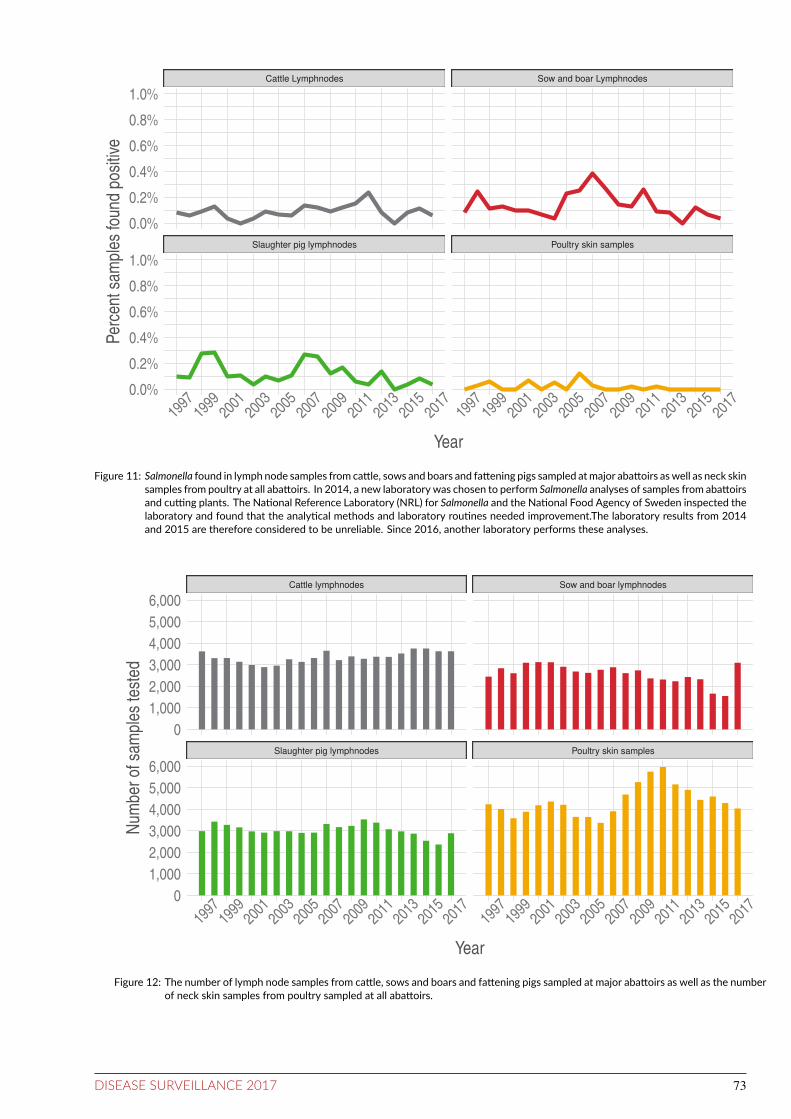
Slaughter pig lymphnodes Poultry skin samples
Cattle Lymphnodes Sow and boar Lymphnodes
1997
1999
2001
2003
2005
2007
2009
2011
2013
2015
2017
1997
1999
2001
2003
2005
2007
2009
2011
2013
2015
2017
0.0%
0.2%
0.4%
0.6%
0.8%
1.0%
0.0%
0.2%
0.4%
0.6%
0.8%
1.0%
Year
Per
cent
sam
ples
foun
d po
sitiv
e
Figure 11: Salmonella found in lymph node samples from catle, sows and boars and fatening pigs sampled atmajor abatoirs aswell as neck skinsamples from poultry at all abatoirs. In 2014, a new laboratory was chosen to perform Salmonella analyses of samples from abatoirsand cuing plants. The Naional Reference Laboratory (NRL) for Salmonella and the Naional Food Agency of Sweden inspected thelaboratory and found that the analyical methods and laboratory rouines needed improvement.The laboratory results from 2014and 2015 are therefore considered to be unreliable. Since 2016, another laboratory performs these analyses.
Slaughter pig lymphnodes Poultry skin samples
Cattle lymphnodes Sow and boar lymphnodes
1997
1999
2001
2003
2005
2007
2009
2011
2013
2015
2017
1997
1999
2001
2003
2005
2007
2009
2011
2013
2015
2017
0
1,000
2,000
3,000
4,000
5,000
6,000
0
1,000
2,000
3,000
4,000
5,000
6,000
Year
Num
ber
of s
ampl
es te
sted
Figure 12: The number of lymph node samples from catle, sows and boars and fatening pigs sampled at major abatoirs as well as the numberof neck skin samples from poultry sampled at all abatoirs.
DISEASE SURVEILLANCE 2017 73

0
10
20
30
40
50
1969
1971
1973
1975
1977
1979
1981
1983
1985
1987
1989
1991
1993
1995
1997
1999
2001
2003
2005
2007
2009
2011
2013
2015
2017
Year
Num
ber
of c
ases
S. Typhimurium S. Other
Figure 13: Frequency of noificaions of Salmonella in broiler holdings during 1968-2017, breeding flocks included.
0
10
20
30
40
50
60
1969
1971
1973
1975
1977
1979
1981
1983
1985
1987
1989
1991
1993
1995
1997
1999
2001
2003
2005
2007
2009
2011
2013
2015
2017
Year
Num
ber
of in
fect
ed h
erds
S. Derby S. Choleraesuis S. Typhimurium S. Other
Figure 14: Frequency of noificaions of Salmonella in swine herds during 1968-2017. In 2016 and 2017, Salmonella was not detected in anyherd.
74 DISEASE SURVEILLANCE 2017

0
5
10
15
20
25
30
1969
1971
1973
1975
1977
1979
1981
1983
1985
1987
1989
1991
1993
1995
1997
1999
2001
2003
2005
2007
2009
2011
2013
2015
2017
Year
Num
ber
of c
ases
S. Gallinarum/Pullorum S. Livingstone S. Enteritidis S. Typhimurium S. Other
Figure 15: Frequency of noificaions of Salmonella in layer holdings during 1968-2017.
0
50
100
150
200
250
1959
1961
1963
1965
1967
1969
1971
1973
1975
1977
1979
1981
1983
1985
1987
1989
1991
1993
1995
1997
1999
2001
2003
2005
2007
2009
2011
2013
2015
2017
Year
Num
ber
of in
fect
ed h
erds
S. Typhimurium S. Dublin S. Other
Figure 16: Frequency of noificaions of Salmonella in Swedish catle herds during 1958-2017. Data from 1958 through 1967 is extracted froma graph presented by J.Å. Robertsson (1985).
DISEASE SURVEILLANCE 2017 75

0
10
20
30
40
50
60
1998
1999
2000
2001
2002
2003
2004
2005
2006
2007
2008
2009
2010
2011
2012
2013
2014
2015
2016
2017
Year
Num
ber
of c
ases
per
100
,000
Total cases Domestic cases
Figure 17: Incidence (per 100,000) of noified cases of human salmonellosis in Sweden, 1998-2017.
Table 13: Sampling scheme for poultry in the compulsory Swedish Salmonella programme.
Category of poultry Sampling frequency Sample type Sampling beforeslaughter
Officialveterinarian
Breeders in rearing 1 d, 4 weeks, 2 weeks prior torearing or moving
2 pairs sock samples 14 d beforeslaughter
Once a year
Breeders inproducion
every 2nd week 5 pairs sock samples 14 d beforeslaughter
3 imes underproducion
Layers in rearing 2 weeks prior to moving 2 pairs sock samples or 2faecal samples of 75 g
14 d beforeslaughter
Once a year
Layers in producion every 15th week (start at22-26 weeks)
2 pairs sock samples or 2faecal samples of 75 g
14 d beforeslaughter
Once a year
Poultry for meatproducion (allspecies)
2 pairs sock samples or 2faecal samples of 75 g
14 d beforeslaughter
Once a year
76 DISEASE SURVEILLANCE 2017

Table 14: Serotypes of Salmonella isolated in feed control in 2017.
Serotype Feedmaterial ofanimaloriginA
Pet food Feedmaterial ofoil seedoriginB
Feedmaterial ofcereal grainorigin
OtherplantsC
Processcontrol feedmills
Processcontrolrapeseedcrushingplant
S. Agona - - - - 2 - -S. Enteriidis - - - - - 1 -S. Hviingfoss - - - - - 1 -S. Kedougou - - 1 - - - -S. Kentucky - - - - - 1 -S.Mbandaka - - 4 - - 2 1S. Newport - - - - - 1 -S. Sentenberg - - 5 - - - -S. Tennessee - - 2 - - 1 -S. Typhimurium - - - - 1 4 -S. enterica subspeciesenterica
- - 1 - - - -
Non-typeable - - - - - 1 -
Total Posiive 0 0 11D 0 3 12 1total number ofsamples
1,037 205 1,171 618 41 8,052 824
AMeat and bonemeal, animal fat, fishmeal, greaves, proteinmeal, meat meal, milk products, poultry offal meal and animal by-products.B Derived from palm kernel, rape seed, soya bean, linseed, peanut and sunflower seed.C Peas, algae, leaves (dried), sugar beets, buckwheat and herbs (dried).D In two of the units posiive for Salmonella two different serotypes were found in each unit.
Table 15: Results from the Salmonella control programme in poultry flocks
Animal species Producion type Producion stage No. flockstested
No.posiives
Percentage Serotype
Gallus gallus Meat producion Adult Grand Parent 24 0 0.00%Gallus gallus Meat producion Adult Parent 130 0 0.00%Gallus gallus Meat producion Producion 4,723 2 0.04% S. Typhimurium
Turkeys Meat producion Adult Parent 4 0 0.00%Turkeys Meat producion Producion 278 0 0.00%Gallus gallus Egg producion Adult Parent 20 0 0.00%
Gallus gallus Egg producion Producion 675 0 0.00%Geese Meat producion Producion 25 0 0.00%
Ducks Meat producion Producion 22 0 0.00%
Table 16: Catle herds under restricion for Salmonella infecion in 2017
Primary serotype Restricted since Restricions lited Reason for sampling
S. Aarhus 2015 2017 NecropsyS. Dublin 2012 2017 NecropsyS. Dublin 2013 2017 Necropsy
S. Dublin 2014 - Trace-backS. Dublin 2014 - Trace-backS. Dublin 2015 - Necropsy
S. Dublin 2017 - NecropsyS. Dublin 2017 - Clinical suspicionS. Typhimurium 2015 2017 Trace-back ater a human case
S. Typhimurium 2015 2017 Trace-backS. Typhimurium 2015 2017 NecropsyS. infanis 2017 - Trace-back ater horse case
DISEASE SURVEILLANCE 2017 77

Table 17: Results from the Salmonella control programme at slaughterhouses and cuing plants in 2017
Animal species Sample type No. samples Posiive PercentPosiive
Serotypes
Catle Lymph node 3,629 2 0.06% S. Duesseldorf, S.Typhimurium
Carcass swab 3,656 0 0.00%
Breeding swine Lymph node 3,091 1 0.03% S. TyphimuriumCarcass swab 3,059 0 0.00%
Slaughter swine Lymph node 2,886 2 0.07% S. TyphimuriumCarcass swab 2,840 0 0.00%
Catle and swine Meat scrapings 4,442 0 0.00%
Poultry Neck skin 4,033 0 0.00%Meat scrapings 1,026 0 0.00%
A Isolaion from pooled samples of lymph nodes
Table 18: Reported cases of Salmonella in cats, dogs, horses, sheep, wild birds and wild mammals in 2017
Serotype Cats
Dogs
Horses
Sheep
Wild
birds
Wild
animalsA
S. Agona - 1 - - - -S. Derby - 1 - - - -S. Enteriidis 1 - - - - -
S. Hessarek 1 - - - - -S. Indiana - - - - - -S. Infanis - 2 1 - - -
S.Mbandaka - - - - - -S. Typhimurium 33 2 - - 8 1Salmonella enterica sp diarizonae =61:-1,5 - - - 13 - -
Salmonella enterica sp diarizonae 1 - - - - -Salmonella enterica sp enterica (I) = 4,5:-:H5- - - - - 1 -Salmonella enterica sp enterica (I) = 4,5:i:- - - - - 1 -
Salmonella enterica sp enterica (I) = 4,5:-:H5 - - - - 1 -Salmonella enterica sp enterica (I) =20:-:Z6 - 1 - - - -Salmonella enterica sp enterica ST-416 - - - - - 1
Salmonella, not serotyped 93 - - - - -
Total 129 7 1 13 11 2A One squirrel and one porpoise.
78 DISEASE SURVEILLANCE 2017
ScrapieBACKGROUNDScrapie belongs to a group of diseases called Transmissi-ble Spongiform Encephalopathies (TSE) and was irst de-scribed more than 250 years ago. The current theory aboutthe causative agent is the protein-only hypothesis. This the-ory assumes that misfolded prions (small proteins) inducethe same misfolded and pathological structure in normalproteins of the host, resulting in accumulation of prions andcellular damage without involvement of any microorganism.Susceptibility to scrapie is genetically related. All routes oftransmission have not been established, however, it is clearthat transmission of classical scrapie occurs within a flock atlambing and that pastures can be contaminated for long peri-ods of time. Scrapie has, based on epidemiological data, notbeen considered a zoonotic disease, however, the question isregularly raised.
Classical scrapie has been detected in Sweden once, ina single flock in 1986. The whole flock was culled and theorigin of the disease was never established.
After classical BSE became a disease of public healthconcern (see chapter on BSE), and the existence of BSEin small ruminants was suspected, both surveillance andcontrol of TSE in small ruminants was increased withinthe European Union in 2002. Since the start of the in-creased surveillance, more than 75,000 sheep have beentested without any positive cases of classical scrapie de-tected. In 2014, Sweden sent an application to the Euro-pean Commission to obtain status as country with negligi-ble risk for classical scrapie. The dossier contained detailedinformation about the population, imports (which were lim-ited), education about the disease, the EU-approved na-tional control programme as well as results of estimates ofthe probability that Sweden is free from classical scrapie.The Commission evaluated the dossier and also asked theEuropean Food Safety Authority (EFSA) for an opinion(doi:10.2903/j.efsa.2015.4292). In August 2016, the appli-cation was approved and Sweden was granted the status neg-ligible risk for classical scrapie through Commission regu-lation (EC) 2016/1396.
In 1998, an atypical variant of scrapie was detected inNorway (Nor98), and this variant was also detected in Swe-den in 2003. Since then, a number of cases have beendetected in Sweden. Although atypical scrapie is experi-mentally transmissible, epidemiological studies on the Eu-ropean level indicate that atypical scrapie probably is a spon-taneously occurring disease. When transmitted experimen-tally, atypical scrapie can cause disease indistinguishablefrom classical scrapie.
DISEASEThe incubation period is long, up to several years. Clini-cal signs of classical scrapie are related to the neurologicalsystem and include altered behaviour and sensation, afectedmovement and posture, as well as pruritus and skin lesions.The disease is progressive and always fatal.
LEGISLATIONSurveillance and control is regulated through Regulation(EC) 999/2001 of the European Parliament and of the Coun-cil of 22 May 2001. At the national level, the surveil-lance scheme and control were, until 2016, also regulated byan EU-approved national scrapie control programme whichfrom 2003 also formed the basis for additional guaran-tees related to trade within the union (Commission Reg-ulation (EC) 546/2006). The current rules in regulation(EC) 999/2001, cover both trade and surveillance require-ments for countries with negligable risk. After 2016, whenSweden was granted the status: negligible risk for classi-cal scrapie through Commission regulation (EC) 2016/1396amending Regulation (EC) 999/2001, the rules in 999/2001replace both the additional guarantees and previous surveil-lance scheme in the national programme.
Sampling at the national level is regulated by SJVFS2010:9, last amended through SJVFS 2013:3. Scrapie is anotiiable disease under the Swedish Act of Epizootic dis-eases (SFS 1999:657, with amendments).
SURVEILLANCEThe Swedish Board of Agriculture is responsible for thesurveillance programme. It is carried out in coopera-tion with the National Veterinary Institute, which is ap-pointed the National Reference Laboratory (Regulation (EC)999/2001). Samples are analysed at the National VeterinaryInstitute.
Passive surveillanceAll suspicions of scrapie must be reported to the authorities.The obligation to report applies to animal owners, veterinar-ians and everyone else who is responsible for the animals.Samples from animals with clinical suspicion of scrapie areexamined with Bio-Rad TeSeE short assay protocol (SAP)in combination with Bio-Rad TeSeE Western Blot.
Acive surveillanceThe design of the surveillance programme is in accordancewith Regulation (EC), 999/2001 Annex III and was untilthe approval of the negligible risk status, also in accordancewith the EU-approved Swedish national control programme.Within that programme, all dead sheep and goats over 18months of age that were not slaughtered for human con-sumption have been sampled. As part of the programme,the Swedish Board of Agriculture inanced the collection offallen sheep and goats above 18 months at the farm level andthe farmers did, until 2016, not have any costs when sendingfallen animals for rendering. This was changed in 2017 andthe farmers now pay to have their fallen animals collected.
After Sweden was granted negligible risk status for clas-sical scrapie in 2016, the surveillance was adapted in 2017with the target to sample 1,500 fallen sheep and goats abovethe age of 18 months, and with the target that the samplesshould representative for the population. The carcasses are
DISEASE SURVEILLANCE 2017 79

sampled at rendering plants and at necropsy, by employees atthe rendering plant or veterinarians of veterinary assistantsat necropsy.
Farms with conirmed cases of atypical scrapie are ob-ligated to have increased surveillance in the herd for twoyears. In addition to fallen stock, healthy slaughtered ani-mals above 18 months of age are examined from these flocks.
The samples from active surveillance were examinedwith Bio-Rad TeSeE short assay protocol (SAP) at the Na-tional Veterinary Institute in accordance with Regulation(EC) 999/2001. In case of positive or inconclusive resultsthe material was examined by Bio-Rad TeSeE Western Blot.
RESULTSPassive surveillanceIn 2017, no sheep or goats were examined due to clinicalsuspicion of scrapie.
Acive surveillanceSheepIn 2017, the National Veterinary Institute examined 1,411sheep from fallen stock for scrapie. Out of these, all sam-ples were negative for classical scrapie and one was positivefor atypical scrapie Nor98.
GoatsIn 2017, the National Veterinary Institute examined 152goats from fallen stock for scrapie. All were negative bothfor classical scrapie and for atypical scrapie.
DISCUSSIONClassical scrapieAfter Sweden was granted status negligible risk and thesurveillance programme was adapted, the Swedish Boardof Agriculture decided not to fully subsidise collection offallen stock on the farm. This had the consequence that thenumber of collected animals decreased dramatically and thesampling plan, which was based on data from previous years,needed to be revised at several occasions with the primarytarget to reach suicient numbers, while not allowing for ad-justments to ensure representativeness. Meetings were heldwith all stakeholders to identify the reasons. In parallel withdecreased number of fallen stock, there was an increase inthe number of slaughtered ewes. There was also a reported
higher demand for sheep meat and thus increased price, alsofor older ewes. Previously, the compensation for older eweshas been very low and there has been no inancial beneit forfarmers to send them to slaughter. However, when the pricefor sending animals for rendering increased, the conclusionis that farmers, to a higher extent, send old ewes to slaughter.
In 2018, the sampling scheme will be adapted taking thenew data into account.
Atypical scrapieSince the irst case of atypical scrapie was detected in Swe-den in 2003, approximately 50 cases have been detected.Out of these, two were detected through passive surveil-lance and the rest through active surveillance. Currently,the flocks are put under intensiied monitoring in accordancewith the regulation (EC) 999/2001. No additional cases ofatypical scrapie have been found in the positive flocks. Atthe European level, two epidemiological studies have con-cluded that the prevalence is similar in diferent countriesand that the prevalence in positive flocks does not diferfrom the prevalence in the rest of the sampled population.This pattern difers from the way contagious disease are nor-mally distributed in the population and support the hypoth-esis that atypical scrapie is spontaneously occurring. Al-though within flock transmission between animals seems tobe very low (if it exists) other routes of spread and the po-tential zoonotic aspect are being discussed.
REFERENCESFediaevsky A, Maurella C, Nöremark M, Ingravalle F,Thorgeirsdottir S, Orge L, Poizat R, Hautaniemi M, Liam B,Calavas D, Ru G, Hopp P. BMC Vet Res. 2010 Feb 7;6:9.The prevalence of atypical scrapie in sheep from positiveflocks is not higher than in the general sheep population in11 European countries.
Fediaevsky A, Tongue SC, Nöremark M, Calavas D, RuG, Hopp P. BMC Vet Res. 2008 Jun 10;4:19. A descriptivestudy of the prevalence of atypical and classical scrapie insheep in 20 European countries.
Elvander M, Engvall A, Klingeborn B. Acta vet Scand02/1988. 29(3-4):509-10. Scrapie in sheep in Sweden.
80 DISEASE SURVEILLANCE 2017

StranglesBACKGROUNDStrangles is a very contagious disease in horses, caused byStreptococcus equi subsp. equi (S equi), belonging to Lance-ield’s group C streptococci. Strangles normally resolveswithout antibiotic treatment but can cause severe complica-tions or persistent infection. To control and eradicate stran-gles, systematic surveillance by testing is necessary, and in-formation must be disseminated about the disease and howit can be prevented, detected and stopped. It is equally im-portant to understand and combat the social stigma associ-ated with this disease. During the last decade, an array ofnew diagnostic tools has become available, to the beneit ofsurveillance and control.
DISEASEStrangles afects horses, including donkeys and zebras.Common clinical signs include: fever, nasal discharge, de-pression, cough and enlarged submandibular or cervicallymph nodes. Other signs that may appear are: inappe-tence, dysphagia, painful movements, ruptured abscesses,dyspnoea and swollen limbs; and less commonly: spreadof infection to other organs, so called bastard strangles .Complications of strangles may be severe and lead to death.
So called atypical strangles with mild clinical signsis probably more typical than previously understood, whichmay lead to unnecessary large outbreaks due to delayed diag-noses. Also, recent indings indicate that subclinical infec-tions with S equi after an acute outbreak may be far morecommon than previously understood, and microbiologicalconirmation of the absence of S. equi can be required torule out the horse being a carrier.
LEGISLATIONStrangles is a notiiable disease in Sweden (SJVFS 2013:23).It is notiiable already on clinical suspicion.
SURVEILLANCE
In Sweden, surveillance for strangles is passive; samplingand diagnostic testing is primarily performed on clinical sus-picion. Typically, samples from airways and lymph nodesare submitted for bacterial analysis (culture or qPCR) to theNational Veterinary Institute. Numbers from other veteri-nary laboratories were not available.
A yearly summary of notiied, conirmed cases of stran-gles per county is given by the Board of Agriculture and pre-sented below (Figure 18).
Detection of S. equi is influenced by site of specimencollection (nasal passage, nasopharynx, guttural pouch orabscess), method of sampling (flocked swab, rayon swab,or wash), and type of diagnostic test (culture or qPCR), aswell as target gene for the PCR and the DNA ampliicationmethod that is performed. Timing of sampling is also cru-cial. Serology for serum antibodies against antigens A and
C of S. equi has been suggested for screening of subclinicalS. equi carriers, but has limitations in both sensitivity andspeciicity.
RESULTSThe number of samples received by the National VeterinaryInstitute as a result of clinical surveillance has doubled since2013 (Figure 19). In 2017, 1,448 samples were submittedfrom over 1,200 horses, of which 11% were positive to S.equi (Figure 19). The percentage of horses positive for stran-gles has increased 3-fold during the last ive years (Figure19). Nasopharyngeal lavage sampling, introduced in 2013,quickly became popular and now comprises almost one-thirdof the passive surveillance samples.
In 2017, there were 100 oicially reported index casesof strangles in Sweden, each representing an outbreak in afarm. During the last 9 years, the number of afected farmsincreased from a level of under 50 per year during the years2009-2014, to around 100 the last three years (Figure 18).
DISCUSSIONThe passive surveillance results indicate that strangles is en-demic in the Swedish horse population, and that the trendhas been increasing during recent years. The observed lowernumber of cases detected through passive surveillance be-tween 2009 to 2014 was possibly a reflection of the eco-nomic recession, which led to decreased breeding and tradein the horse industry at the time, resulting in less diseasetransmission. After 2015, the industry returned to higher ac-tivity, coincidentally with a return to strangles rates similarto before 2009 (Figure 18).
Other possible factors for a change in notiications overtime might be varying knowledge about the disease, vary-ing willingness to sample animals, and/or methodologicalchanges over time, such as the increase in use of nasopha-ryngeal lavage. Substantial efort has been made to increaseawareness among veterinarians and horse owners about newdiagnostic developments and proper ways to investigate theinfection. At the same time, the social media culture in soci-ety has led to more openness in private and delicate matters.It has contributed to a decrease in the stigma associated withstrangles, and possibly increased compliance by owners totest horses, which is a beneit for the surveillance and for thepossibility to control the infection.
Investigations of outbreaks point to a need for screen-ing horses that recently have been moved for trade purposes,as these horses appear to be involved in most of the investi-gated acute outbreaks. It would also be of beneit with a pro-gramme for tracking the spread of strangles, by DNA char-acterisation of diferent isolates.
REFERENCESSwedish Board of Agriculture, Statistics of index cases ofnotiiable animal diseases, http://www.jordbruksverket.se
DISEASE SURVEILLANCE 2017 81

0
20
40
60
80
100
120
2001
2002
2003
2004
2005
2006
2007
2008
2009
2010
2011
2012
2013
2014
2015
2016
2017
Year
Num
ber
of outb
reaks
Figure 18: Reported index cases (farm outbreaks) of Streptococcus equi infecions in horses in Sweden during years 2001-2017. Source:Swedish Board of Agriculture
0
500
1000
1500
2013
2014
2015
2016
2017
Num
ber
of S
ampl
es
Negative Positive
0%
2%
4%
6%
8%
10%
12%
2013
2014
2015
2016
2017
Year
Per
cent
pos
itive
Figure 19: Numbers of individual horses sampled from airways and/or related lymph nodes for bacterial diagnosis, and the proporions of S.equi posiive horses, Naional veterinary insitute, 2013-2017
82 DISEASE SURVEILLANCE 2017

Tick-borne encephaliis
0
50
100
150
200
250
300
350
400
1987
1989
1991
1993
1995
1997
1999
2001
2003
2005
2007
2009
2011
2013
2015
2017
Year
Num
ber
of cases
Figure 20: Number of noified cases of TBE in humans 1987-2017.
BACKGROUNDTick-borne encephalitis virus (TBEV) belongs to the genusflavivirus in the family Flaviviridae. TBE virus is endemicin an area ranging from northern China and Japan, throughfar-eastern Russia to Europe. The virus may cause a neuro-logical infection which may lead to long-term sequelae in theafected patients. The virus is spread by ticks (Ixodes ricinusand I. persulcatus), which are infected when they suck bloodfrom infected rodents. Wild rodents are the natural reser-voir for TBEV. The virus also circulates in the tick popula-tion through transovarial transmission without involvementof vertebrate hosts. Large mammals, predominantly ungu-lates, are important to the maintenance of large tick popula-tions. Humans typically become infected via ticks, althoughunpasteurised cow’s and goat’s milk and milk products havealso been reported as sources. Vaccination of persons living,visiting or working in endemic areas is recommended.
Three sub-types of TBEV are described: the Western ,Siberian and Far eastern subtypes. In Sweden, only theWestern subtype has been identiied.
The irst case of TBE infection in Sweden was reportedin 1954. During the following three decades, 10-40 annualcases were reported. From the mid-1980s a clearly increas-ing trend has been observed. In recent years about 150-400cases have been reported annually. A majority of the casesacquire their infections in Sweden. Most have been infectedon the east coast of Sweden and the Stockholm archipelagobut in recent decades cases have been observed regularly onthe west coast of the country. The age distribution is widebut most of the cases are between 30 and 70 years. There is aslight over-representation of men. A majority of the patientsare diagnosed in July to October.
DISEASEAnimalsA few conirmed cases of disease in dogs have been reported.Seroconversion has been demonstrated in grazing goats andcows. Most authors consider these animals to be a dead-endhosts for the viral infection. Wild rodents are the naturalreservoir for TBEV but are not reported to contract the dis-ease. Roe deer have been shown to seroconvert and they haveconsequently been suggested as an indicator of the preva-lence of the virus. However, there have been no reports ofdisease in this species.
HumansIn humans, a biphasic course of the disease is common. Theirst, viraemic phase lasts for about four days. After an in-terval of about a week, a meningoencephalitic phase ap-pears in about one third of the patients. The symptoms mayinclude fever, headache, nausea, cognitive dysfunctions orspinal paresis. The mortality is low, about 0.5%. The incu-bation period of TBE is usually between 7 and 14 days.
LEGISLATIONAnimalsDemonstration of TBE virus or antibodies in animals is notnotiiable.
HumansTBE in humans is notiiable as a viral meningoencephali-tis since 2004, according to the Communicable Disease Act(SFS 2004:168 with the amendments of SFS 2013:634).
DISEASE SURVEILLANCE 2017 83

SURVEILLANCEAnimalsThe surveillance in animals is passive. During 2017, sixdogs were tested for TBE antibodies.
HumansThe surveillance is passive in humans.
RESULTSAnimalsOne of the dogs was positive; it is however not known ifclinical signs in this dog were caused by TBEV infection.
HumansIn 2017, 391 cases of TBE were reported, which is an in-crease of 64% from the year before, and more cases thanhave ever been reported in a single year (Figure 20). On alonger term, since 1983 the TBE incidence has shown a sig-niicantly rising trend of 7% each year.
More men (63%) than women were reported with TBE.The incidence was highest among people in the age group50-69 years, but there were cases reported from 1 to 95 yearsof age.
All but ten cases had acquired their infections in Swe-den. The majority of imported cases had been infected inFinland (six cases).
The irst TBE cases became ill in mid-April and the lastin mid-December. The peak occurred in August, but therewere unusually many cases reported during the whole periodfrom July until October.
The geographic distribution of the disease was mainly, asin previous years, concentrated in the coastal areas of Stock-holm, Södermanland and Uppsala counties, both along thelake of Mälaren and the Baltic Sea (Figure 21). The in-cidence was highest in the counties of Uppsala (13 casesper 100,000 inhabitants) and Södermanland (12 cases per100,000 inhabitants). However, the infection also occurs inmany other parts of the country; from the county of Skånein the south to southern Gävleborg and Dalarna in the north.TBE is successively spreading westwards and in 2017 therewere unusually many cases infected in for example, VästraGötaland and Värmland.
DISCUSSIONThe TBE incidence has shown a signiicantly rising trendduring the last three decades, but there were still consider-ably more cases reported in 2017 than expected.
This general, long term increase is probably due to sev-eral interacting factors. The most important cause is pre-sumably the very dense population of ticks, a consequence ofa large roe deer population from the 1980s up until the recentsnowy winters. This situation in combination with a highpopulation of small host animals such as bank voles, andoptimal weather for both virus spread and humans spendingtime outdoors, could explain the large number of cases re-ported. It is unknown why the incidence was unexpectedlyhigh in 2017.
Number of Cases
1
5
10
Figure 21: The geographic distribuion of the place of infecion ofcases of TBE during 2017. The area of each circle markeris relaive to the number of cases idenified, the smallestmarkers are a single case.©EuroGeographics for the administraive boundaries.
84 DISEASE SURVEILLANCE 2017

Trichinellosis
Producion sites without controlled housing condiions should test all their slaughtered domesic pigs. Although Trichinella occurs in wild carnivores and wild boarin Sweden, the risk of becoming infected from domesic pigs and horses is negligible. Photo: Bengt Ekberg
BACKGROUNDTrichinellosis is caused by parasitic nematodes of the genusTrichinella. The parasites can be hosted by diferent mam-mals including domestic pigs and horses but the main reser-voirs are wild carnivores and omnivores. Humans typicallyacquire the infection by eating raw or inadequately heatedinfected meat and meat products, often cold-smoked, fer-mented sausages. In Western Europe, the wild boar appearsto be the main source of human infection.
In Europe, T. spiralis and T. britovi are the dominantcauses of human infections. In Sweden, these species arealso detected as well as T. nativa and T. pseudospiralis. T.pseudospiralis is mainly isolated from wild boars. In the gut,Trichinella larvae develop into adults and mate. After mat-ing, the female releases larvae which penetrate the intestinalmucosa and travel via the bloodstream to various organs andmuscles. In striated muscles the larvae may survive in anencapsulated form for years.
In Sweden, Trichinella has been monitored at slaughterin domestic pigs since the 20th century. From 1970-1990sporadic cases were detected in domestic pigs, but since1994 there have been no cases. The parasite is endemic inSwedish wildlife.
The disease is extremely rare in Sweden and detected hu-man cases are usually infected abroad. During 2013 to 2016three human cases with conirmed infection with Trichinellawere reported, all infected abroad with country of infectionbeing Poland, Eritrea and Georgia. Also, two cases werereported with clinical symptoms indicating infection withTrichinella, although the diagnoses could not be laboratoryconirmed.
DISEASEAnimalsAnimals rarely develop a clinical infection, although bothpigs and rodents can exhibit clinical signs.
HumansThe disease in humans can range from subclinical infectionto fatal disease. The incubation period varies from 5-15days. Symptoms initially involve diarrhoea and abdominalpain and later muscle pain, fever, oedema of the upper eye-lids and photosensitivity. Intestinal stages of the disease re-spond well to treatment. Cardiac and neurological compli-cations may occur 3-6 weeks post infection. Trichinella isnot transmitted between humans.
DISEASE SURVEILLANCE 2017 85
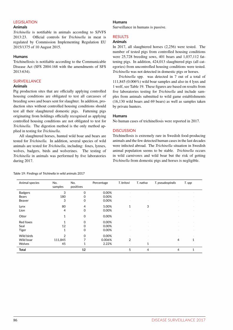
LEGISLATIONAnimalsTrichinella is notiiable in animals according to SJVFS2013:23. Oicial controls for Trichinella in meat isregulated by Commission Implementing Regulation EU2015/1375 of 10 August 2015.
HumansTrichinellosis is notiiable according to the CommunicableDisease Act (SFS 2004:168 with the amendments of SFS2013:634).
SURVEILLANCEAnimalsPig production sites that are oicially applying controlledhousing conditions are obligated to test all carcasses ofbreeding sows and boars sent for slaughter. In addition, pro-duction sites without controlled housing conditions shouldtest all their slaughtered domestic pigs. Fattening pigsoriginating from holdings oicially recognised as applyingcontrolled housing conditions are not obligated to test forTrichinella. The digestion method is the only method ap-plied in testing for Trichinella.
All slaughtered horses, hunted wild boar and bears aretested for Trichinella. In addition, several species of wildanimals are tested for Trichinella, including: foxes, lynxes,wolves, badgers, birds and wolverines. The testing ofTrichinella in animals was performed by ive laboratoriesduring 2017.
HumansSurveillance in humans is passive.
RESULTSAnimalsIn 2017, all slaughtered horses (2,256) were tested. Thenumber of tested pigs from controlled housing conditionswere 25,728 breeding sows, 401 boars and 1,037,112 fat-tening pigs. In addition, 424,013 slaughtered pigs (all cat-egories) from uncontrolled housing conditions were tested.Trichinella was not detected in domestic pigs or horses.
Trichinella spp. was detected in 7 out of a total of111,845 (0.006%) wild boar samples and also in 4 lynx and1 wolf, see Table 19. These igures are based on results fromive laboratories testing for Trichinella and include sam-ples from animals submitted to wild game establishments(16,130 wild boars and 69 bears) as well as samples takenby private hunters.
HumansNo human cases of trichinellosis were reported in 2017.
DISCUSSIONTrichinellosis is extremely rare in Swedish food-producinganimals and the few detected human cases in the last decadeswere infected abroad. The Trichinella situation in Swedishanimal population seems to be stable. Trichinella occursin wild carnivores and wild boar but the risk of gettingTrichinella from domestic pigs and horses is negligible.
Table 19: Findings of Trichinella in wild animals 2017
Animal species No.samples
No.posiives
Percentage T. britovi T. naiva T. pseudospiralis T. spp
Badgers 3 0 0.00% - - - -Bears 180 0 0.00% - - -Beaver 3 0 0.00% - - - -
Lynx 80 4 5.00% 1 3 - -Lion 4 0 0.00% - - - -
Oter 1 0 0.00% - - - -
Red foxes 1 0 0.00% - - -Seal 12 0 0.00% - - - -Tiger 1 0 0.00% - - - -
Wild birds 2 0 0.00% - - - -Wild boar 111,845 7 0.006% 2 - 4 1Wolves 45 1 2.22% - 1 - -
Total 12 5 4 4 1
86 DISEASE SURVEILLANCE 2017

Tuberculosis
The voluntary tuberculosis control programme in alpacaswas launched in 2015. It is based on serological tesing since the tradiional intradermal skin fold tuberculintest is less sensiive in new world camelids. Photo: Public domain
BACKGROUNDTuberculosis (TB) is a serious disease in humans and ani-mals caused by bacteria included in the Mycobacterium tu-berculosis complex. Mycobacterium bovis causes bovine tu-berculosis in several animal species as well as in humans.Historically, the reservoir has been cattle but many otherwild and domestic species can also maintain the infection.Wildlife reservoirs including badgers, deer and wild boarcause persistent problems in some countries. Humans usu-ally acquire M. bovis infection via unpasteurised milk orvia inhalation. The predominant cause of human tubercu-losis is however Mycobacterium tuberculosis. In countrieswhere human tuberculosis caused by M. tuberculosis is com-mon, this bacterium is also frequently isolated from variousspecies of animals.
Sweden was declared oicially free from bovine tuber-culosis in 1958. Since then, sporadic cases have occurredin cattle, the most recent in 1978. Compulsory tuberculintesting of all cattle was abolished in 1970 and the nationaltuberculosis control in cattle is now based on meat inspec-tion and clinical surveillance.
When Sweden joined the European Union in 1995, thestatus of OTF (oicially tuberculosis free) was obtained.
In 1987, M. bovis infection was introduced into the
farmed deer population. A control programme for tubercu-losis in farmed deer was introduced in 1994 and made com-pulsory in 2003. The last case of tuberculosis in farmed deerwas identiied in 1997.
The yearly incidence among humans in Sweden in theearly 1940’s was above 300/100,000 inhabitants. This wasfollowed by a rapid decline, beginning before efective treat-ment was available in the early 1950’s. Currently, the yearlyincidence is 5.3/100,000 inhabitants, which is among thelowest in the world. Around 90% of the cases are born out-side of Sweden and the vast majority of them are immigrantsoriginating from countries that still have a high incidence oftuberculosis. The yearly incidence among people born inSweden is 1/100,000 inhabitants.
DISEASEThe symptoms caused by tuberculosis in both humans andanimals depend largely on the localisation of the infection.The disease progresses slowly and clinical signs may take along time to develop, even in cases with substantial lesions.Weight loss and sometimes coughing (in cases with respi-ratory tract infection), ascites (due to infection in intestinallymph nodes or liver) or mastitis (mainly in cattle with udderinfection) can be seen. The incubation period varies fromweeks to years.
DISEASE SURVEILLANCE 2017 87

LEGISLATIONAnimalsSuspect and conirmed cases of infection with Mycobac-terium bovis, M. tuberculosis, or other mycobacteria in theM. tuberculosis-complex, are notiiable in all animal speciesaccording to the Swedish Act of Epizootic diseases (SFS1999:657, with amendments).
HumansTuberculosis in humans is a notiiable disease accordingto the Communicable Disease Act (SFS 2004:168 with theamendments of SFS 2013:634). Contact tracing is compul-sory and the treatment is free of charge. Refusing treatmentas a patient when being contagious can lead to detention.
SURVEILLANCEAnimalsFrom suspect animal cases, samples from organs withmacroscopic lesions and adjacent lymph nodes are collected.In case of positive tuberculin test reactors samples from or-gans with macroscopic lesions and lymph nodes from ivediferent areas (retropharyngeal, submandibular, mediasti-nal, mesenteric and inguinal) are collected. Histology anddirect smears are performed on all materials. If TB cannotbe ruled out by histology or if direct smears are positive, cul-ture is performed. Cultures are performed on solid media(Löwenstein-Jensen and Stonebrink’s) at the National Vet-erinary Institute and cultured for up to twelve weeks. Sus-pected colonies are tested with PCR and, if necessary, withsequencing of a speciic gene. Isolates suspected to belongto the M. tuberculosis-complex or where the M. tuberculo-sis-complex cannot be ruled out are sent for conirmationto the Norwegian Veterinary Institute or the Public HealthAgency of Sweden. Positive isolates are further subtyped.
Skin fold tuberculin tests are performed according to EC1226/2002 (amending annex B of EC 64/432) and SJVFS2003:33, (K62). The comparative intradermal test is used,mostly at the neck site. In case of a positive tuberculin test,the animal is culled and sampled as stated above. Culture isperformed on all samples.
Since 2012, testing of alpacas for tuberculosis has beendone using a serological test instead of an intradermal testas the intradermal test has a demonstrated low sensitivity inalpacas.
HumansIn humans, culture on sputum smear is the standard testwhen pulmonary tuberculosis is suspected. Otherwise cul-ture from urine, faeces, blood or liquor is also a possibility,or biopsies from suspected site of infection.
Passive surveillanceAnimalsAs TB is notiiable on clinical suspicion, clinical signs inanimals or lesions detected at necropsy of an animal promptoicial investigations, including sampling for bacteriology,
tuberculin testing of contact animals and epidemiological in-vestigations. A positive inding of M. bovis or M. tubercu-losis in animals would generate contacts with public healthrepresentatives to ensure that possible exposure of humanscan be investigated.
HumansThe surveillance in humans is mainly passive but contacttracing from diagnosed cases is compulsory and asylumseekers from high incidence countries are ofered health ex-amination where screening for TB is included.
Acive surveillanceAnimalsSurveillance for TB is mainly performed by meat inspec-tion at slaughter of food producing animals. Oicial inspec-tors from the National Food Agency perform the inspections.Suspect lesions are sent to the National Veterinary Institutefor histology and bacteriology.
The control programme in farmed deer was, until Octo-ber 2012, based on regular whole-herd tuberculin testing, orwhole-herd slaughter and meat inspection. Since October2012, tuberculin tests are no longer performed in TB-freeherds, but inspections at slaughter and necropsy of animalsfound dead or euthanized are still required.
A voluntary control programme in alpacas was launchedby Farm & Animal Health in 2015. All adult animals in theherd are serologically tested and all animal purchases andcontacts with other herds are recorded.
Furthermore, tuberculin tests are performed at artiicialinsemination centres and at export of animals as required ac-cording to EU-legislation (Council Directive 64/432/EEC).
RESULTSAnimalsDue to lesions detected at slaughter, three cattle, eightsheep, six pigs and one deer were investigated by histol-ogy and, where relevant, by culture. From these samplesNTM (Non-tuberculous mycobacteria), from the Mycobac-terium avium/intracellulare-complex were isolated in onepig. No other samples yielded any mycobacteria. Due toclinical suspicions or lesions found at necropsy, samplesfrom one cow, two deer, one camel, three dogs and two catswere investigated. None of these samples yielded any my-cobacteria. Due to a positive tuberculin test, one moose calfwas euthanized, necropsied and cultures from relevant or-gans were performed. The NTM Mycobacterium kansasiiwere isolated from one sample; no other sample yielded anymycobacteria. An epidemiological investigation was per-formed and the mother of the calf, which was the only animalthat had been in contact with the calf for considerable time,were tuberculin tested with negative result.
In 2017, there were 308 holdings with farmed deer thatwere considered active and had obtained TB free status.Eight herds were not tested. These herds are exempted fromregular testing and instead they must slaughter 20% of theherd yearly with meat inspections and necropsies for 15years and thereby obtain a free status. TB was not detected
88 DISEASE SURVEILLANCE 2017

in any farmed deer in Sweden during 2017.During 2017, 53 alpacas, 2 llamas and 18 wisents were
tested serologically before export or import. Two of the al-pacas had positive inal results, and they are now isolatedand subject to further sampling. All other animals had neg-ative inal results. Within the voluntary control programme,514 alpacas and 7 llamas were tested, all with negative inalresults.
HumansThree cases of M. bovis were reported in humans in 2017;all three cases with extrapulmonary TB, one with periph-eral lymphadenitis and two with skeletal involvement. Thecase with lymphadenitis was a young woman from Eritrea,most likely infected in her country of origin, and the caseswith skeletal TB were both born in Sweden in the 1930’s andprobably infected in their childhood.
DISCUSSIONAnimalsThe oicially free status for bovine tuberculosis has beenmaintained during 2017. The overall TB situation in ani-mals and humans remains favourable. No cases of TB weredetected in Swedish animals during 2017. Although thesurveillance is mainly dependent on inspections of slaugh-tered animals, this is considered suicient. However, thesubmission rates of lesions from slaughtered ruminantsshould be improved. Passive surveillance based on clinicalsuspicions and necropsy indings will always have a low sen-sitivity as clinical symptoms and massive lesions are mainlyseen in late stages of the infection.
The eradication eforts in farmed deer have been suc-cessful and the probability that Swedish farmed deer are TB
free is high. The aim is to eventually declare the remainingdeer herds oicially free.
HumansThe rapid decline of tuberculosis in humans in the 1940’scoincided with the eradication of tuberculosis in cattle andstarted before the introduction of efective treatment in the1950’s. A much larger part of the human population lived inclose contact with domestic animals. This change in contactbetween humans and animals possibly played a role in thechanging TB incidence in humans. Today, Sweden has oneof the lowest incidences of human tuberculosis in the world.
REFERENCESAlvarez J, Bezos J, Juan Ld, Vordermeier M, Rodriguez S,Fernandez-de-Mera IG, Mateos A, Domínguez L. Diagno-sis of tuberculosis in camelids: old problems, current solu-tions and future challenges. Transbound Emerg Dis. 2012Feb;59(1):1-10. doi: 10.1111/j.1865-1682.2011.01233.x.Epub 2011 Jun 2
Rhodes S, Holder T, Cliford D, Dexter I, Brewer J, SmithN, Waring L, Crawshaw T, Gillgan S, Lyashchenko K,Lawrence J, Clarke J, de la Rua-Domenech R, Vorder-meier M. Evaluation of gamma interferon and antibodytuberculosis tests in alpacas. Clin Vaccine Immunol. 2012Oct;19(10):1677-83. Epub 2012 Aug 22.
Wahlström, H., Frössling, J., Sternberg Lewerin, S., Ljung,A., Cedersmyg, M. and Cameron, A., 2010. Demonstrat-ing freedom from infection with Mycobacterium bovis inSwedish farmed deer using non-survey data sources. Pre-ventive Veterinary Medicine 94:108-118.
DISEASE SURVEILLANCE 2017 89

TularaemiaBACKGROUNDThe bacterium Francisella tularensis is the causative agentof tularaemia, a disease afecting many animal species, in-cluding humans. There are several subtypes of F. tularensisof variable virulence. F. tularensis subsp. holarctica (typeB) is the main subspecies responsible for human and animalinfection in Europe.
F. tularensis is capable of surviving for weeks at lowtemperatures in water, moist soil, or decaying plant and ani-mal matter. Although many diferent animal species can beinfected, tularaemia is typically found in hares and small ro-dents.
Humans become infected through a variety of mecha-nisms such as bites of infected insects or other arthropods,handling infected or dead animals, ingesting contaminatedfood or water, and inhaling aerosols of bacteria. Clinical dis-ease is variable and dependent on the route of transmission.The infection is more often reported in men than in women,which might be attributed to their leisure and professionalactivities. The age group of 40-79 years is the most afectedin both sexes. Tularaemia may occur during the whole year,but it is most frequent during late summer and early autumn.
Sweden has reported cases of tularaemia in humans andanimals since 1931. Ever since the irst Swedish tularaemiacase was reported, endemic areas have been identiied innorthern and central Sweden.
The mountain hare and the European brown hare are theanimal species in which tularaemia has most frequently beenidentiied. Diseased animals have been found in the tradi-tionally endemic areas in northern and central Sweden, aswell as in regions south of these areas.
The annual numbers of reported human cases range froma few cases to more than 2,700 cases in 1967.
DISEASEF. tularensis is highly infectious, as few as 10-50 colonyforming units may cause infection. The incubation periodis usually 3-5 days. Tularaemia can be manifested in difer-ent forms depending on the route of transmission and on thevirulence of the organism.
AnimalsIn Swedish hares, and in many rodent species that die of tu-laraemia, the pathological presentation of the disease is adisseminated multi-organ septicaemic form.
HumansThe ulceroglandular form is the most common form , andis more frequently seen than the typhoidal form. The pneu-monic, oculoglandular and oropharyngeal forms are rarelydiagnosed. In the ulceroglandular form, a local ulcer usu-ally appears at the site of infection and the adjacent lymphnodes are enlarged. The general symptoms of tularaemia arehigh fever, headache and nausea.
LEGISLATIONAnimalsTularaemia is notiiable in animals (SJVFS 2013:23).
HumansTularaemia has been a notiiable disease since 1970 accord-ing to the Communicable Disease Act (SFS 2004:168) withthe amendments of SFS 2013:634.
SURVEILLANCEAnimalsSurveillance in animals is passive. It is based on voluntarysubmission of animals found dead or euthanised by huntersand the general public. Detection is based on PCR or im-munohistochemistry of the animal sample.
HumansThe surveillance is passive. For laboratory veriication ofthe infection, serology, PCR and isolation of the bacteria canbe used.
RESULTSAnimalsIn 2017, 31 European brown hares and eight mountain hareswere examined. F. tularensis subsp. holarctica was de-tected in seven European brown hares and none of the moun-tain hares. The seven hares had all died of an acute dis-ease spread to several organs, and inally ending with sepsis.All tularaemic hares originated from central-eastern parts ofSweden (one from Stockholm, two from Uppsala, and the re-maining four from Östergötland), and one from Västra Gö-taland in the southwest part of the country. The number ofcases in 2017 is approximately at the same level as otheryears without outbreaks, for example six cases in 2016 andtwo in 2014. This could be compared to the outbreak year2015 when tularaemia was diagnosed in 31 hares, the ma-jority coming from the outbreak area.
HumansIn 2017, 87 human cases of tularaemia were reported. Asa rule, there are large natural fluctuations in the number oftularaemia cases observed between years and in diferent re-gions. This is probably due to several factors among oth-ers are the number of reservoirs and mosquitoes, as well asweather conditions. Even though the tularaemia incidencehas varied a lot between years, the increasing incidence-trend for the last 25 years is signiicant diferent in compar-ison to the same time-period before 1992.
More men (73%) than women were reported to be in-fected in 2017, which is in accordance with previous years.The incidence of tularaemia was highest in the age group40-79 years, which is also similar to previous years. Theuneven distribution among age groups and sexes might par-tially be attributed to their somewhat diferent leisure and
90 DISEASE SURVEILLANCE 2017

professional activities.As in previous years, except for a few sporadic cases,
tularaemia was only reported from the northern, westernand central parts of Sweden. During 2017, the incidencewas highest in the county of Värmland with 6.1 cases per100,000 inhabitants, followed by the counties of Örebro withive cases per 100,000 inhabitants. In 2017, two cases werereported to have been infected in Denmark and Ukraine re-spectively.
More than half of the cases for whom a route of trans-mission had been speciied, an insect bite was reported. Thetrue number may be both greater or smaller as the infectionroute is diicult to determine, both for the patient and theclinician. In 2017, 16 cases were assumed to have been in-fected through direct contact with animals.
During the irst half of the year, just a few cases were re-ported each month. The number of cases started to increasein June and peaked in October. During the last two monthsof the year the number of cases subsided.
DISCUSSIONTularaemia has been endemic in northern and central Swe-den at least since the early 20th century with a marked an-nual variation. Years with high numbers of cases are of-ten followed by periods when the disease is virtually absent.There is no obvious explanation for these fluctuations. Thereservoir for the bacterium between outbreaks has not beenclearly identiied. During the last two decades, the epidemi-ology of tularaemia has changed and the number of reportedcases in humans and animals, mainly European brown hares,infected south of the previous endemic region has increased.In animals, outbreaks of tularaemia have in some countriesbeen associated with rises in rodent and hare populations,but this has not been conirmed in Sweden. It is possiblethat the European brown hare has become an important car-rier of F. tularensis in many areas, but its epidemiologicalrole remains unclear.
0.0
2.5
5.0
7.5
10.0
1997
1998
1999
2000
2001
2002
2003
2004
2005
2006
2007
2008
2009
2010
2011
2012
2013
2014
2015
2016
2017
Year
Num
ber
of c
ases
per
100
,000
Total cases Domestic cases
Figure 22: Incidence of noified human cases of tularaemia in Sweden 1997-2017
DISEASE SURVEILLANCE 2017 91

Verotoxinogenic Escherichia coliBACKGROUNDVerotoxinogenic Escherichia coli (VTEC) may cause se-rious intestinal infections in humans. When these bacte-ria cause hemorrhagic diarrhoea they are called EHEC (en-terohaemorrhagic E. coli). More than 380 diferent VTECserotypes have been associated with human illness. Shigatoxin-producing Escherichia coli (STEC) is used synony-mously with VTEC. The toxin can be divided into two maingroups, shigatoxin 1 (stx1) and shigatoxin 2 (stx2), and thenfurther divided into several subtypes, for example, stx1a.Previously, many outbreaks and severe disease were causedby serotype O157:H7, but in recent years other serogroupshave emerged. Often the strains associated with severe dis-ease carry the stx2 gene. Other common serotypes caus-ing gastrointestinal illness are O26, O103, O111, O121 andO145. Cattle are the main reservoir of VTEC associatedwith human disease although other animal species may alsocarry the organisms. The infectious dose is low, probablyless than 100 bacteria. Not only foods of bovine origin butalso vegetable food items and drinking water have been im-plicated in outbreaks. The infection can also be transmittedthrough direct or indirect animal contact, via environmentor person-to-person contacts.
VTEC was only sporadically detected in Sweden before1995, when 114 human cases of EHEC O157:H7 were no-tiied. In 1996, VTEC O157 was isolated from Swedishcattle for the irst time and human EHEC O157 infectionwas traced to a cattle herd. In 2002, an outbreak of EHECO157:H7 in the county of Skåne, afecting 30 persons, wascaused by consumption of cold-smoked fermented sausage.The largest Swedish outbreak so far occurred in the summerof 2005 when 135 reported cases, including 11 (8%) HUS(haemolytic uraemic syndrome) cases, were infected withO157:H7 after eating contaminated fresh lettuce. The let-tuce had been irrigated with water from a local stream pos-itive for stx 2 at the time of harvest. Indistinguishable iso-lates from humans and cattle faeces from a farm upstreamconirmed the implicated source, and control measures thatlead to the termination of the outbreak were implemented.
Between 250-550 cases (3-6 cases per 100,000 inhab-itants) of EHEC infections have been reported in Swedenannually, of which 50-60% are domestically acquired. Mostof the domestic cases are reported during the period July toSeptember.
Management of zoonotic agents requires collaborationbetween several authorities within the veterinary and pub-lic health sector. A national strategy document containinga plan to reduce the risk of domestic EHEC cases was pub-lished in 2014 by the Swedish Board of Agriculture, the Na-tional Food Agency, the Public Health Agency of Sweden,the National Board of Health and Welfare and the NationalVeterinary Institute. The document is based on a synthesisof current knowledge and identiies what actions the authori-ties consider as important that should be prioritised to reduce
the risk of domestic infection with VTEC in humans.
DISEASEAnimalsAnimals do not develop clinical disease.
HumansThe clinical picture may vary from asymptomatic infectionto non-haemorrhagic or haemorrhagic diarrhoea associatedwith abdominal cramps. Most patients fully recover. In re-cent years, approximately 3% of the cases in Sweden havedeveloped HUS, which is characterised by acute renal fail-ure, thrombocytopenia, and microangiopathic haemolyticanaemia; a condition that may lead to death. A large pro-portion of the patients are young children, and severe com-plications are most common in this age group, as well asamong elderly people.
LEGISLATIONAnimalsSince 1999, VTEC O157 indings in animals are notiiablewhen associated with human infection (SJVFS 2013:23).
HumansEHEC O157 has been notiiable for both clinicians and labo-ratories under the Swedish Communicable Disease Act since1996. All EHEC serotypes that are pathogenic to humanshave been notiiable since 1 July 2004 (SFS 2004:168 withthe addition of SFS 2013:634). A laboratory conirmed casecan also include cases that are only positive by PCR i.e.where no isolate has be obtained.
SURVEILLANCEAnimalsSurveillance of VTEC in animals is active and consistsof traceback investigations from human EHEC cases andprevalence surveys of VTEC in abattoirs.
Passive - Traceback from human casesIf a County Medical Oicer suspects an association betweena human case of EHEC infection and animals, or a farm withanimals, the County Veterinary Oicer will be informed. Arequest will be made to the Swedish Board of Agriculturefor a trace back investigation and sampling of suspected an-imals, and/or the environment of the animals.
AciveBetween 1997 and 2002, annual prevalence studies of VTECin cattle at abattoirs were conducted. Since 2002, prevalencestudies have been performed every third year. In the stud-ies conducted during 2011-2012 and 2014-2015, all positiveVTEC O157:H7 were also examined for a variant of VTECO157:H7, called clade 8. This variant is often isolated fromcattle farms associated with human cases. A baseline study
92 DISEASE SURVEILLANCE 2017

on cattle carcasses was done in 2006-2007 and a prevalencestudy in sheep was done at nine abattoirs in 2007-2008. Re-sults from a slaughter prevalence survey from 1998 showedthat 0.1% of the pigs were positive for VTEC O157:H7.
HumansSurveillance in humans is passive. Isolates from humancases are sent to the Public Health Agency of Sweden fortyping using whole genome sequencing (WGS) to verifymolecular serotype and for cluster detection. As a conven-tional nomenclature tool, not only the serotype but also theMulti Locus Sequence Typing (MLST) type, i.e. ST-type, isdeined by WGS.
RESULTSAnimalsPassive - Traceback from human casesDuring 2017, 16 cattle farms were investigated as suspectedsources for human infection. An epidemiological associa-tion was established in four cases of VTEC O157:H7, onecase of VTEC O26 and inally, one case of VTEC O121.
AciveVTEC O157 was detected in nine (1.8%) of 492 faecal and2 (1.9%) of 105 ear samples from sheep in a survey per-formed in 2007-2008. In cattle, surveys during 1997-2002showed a prevalence of approximately 1%. In the study donein 2005-2006, VTEC O157 was detected in 3.4% of fae-cal samples. In the abattoir survey conducted in 2008-2009VTEC O157 was detected in 3.3% of 1993 faecal and 8.2%of 500 ear samples in cattle. In the study conducted during2011-2012, VTEC O157 was detected in 73 of 2,376 fae-cal samples (3.1%) from cattle. Clade 8 was detected in 15of the 73 positive samples. In the study conducted during2014-2015, VTEC O157 was detected in 33 of 1,492 faecalsamples (2.2%) from cattle. Clade 8 was detected in 5 of the33 positive samples. In these studies, VTEC O157:H7 haspredominantly been isolated from cattle in southern Swe-den but rarely from the northern two thirds of the country.The collected samples during 2011-2012 were also analysedfor VTEC O26 and VTEC O103. VTEC O26 was detectedin 8 of 1,308 faecal samples (0.6%) and in 15 of 336 cattleear samples (4.5%). VTEC O103 was detected in three of1,000 faecal samples (0.3%) and in three of 500 ear samples(0.6%).
FoodAvailable results from oicial sampling by local authoritiesshowed that analysis for E. coli O157 was done for 8 sam-ples. The samples have been taken from diferent kinds offood. Out of these, 4 samples were taken as part of the inves-tigation of food poisoning/complaints. All 8 samples werenegative. There were also another 23 samples analysed withgene detection methods. Out of these, 18 were taken as partof the investigation of food poisoning/complaints. Five ofthese twenty-three samples were positive. Those ive pos-itive samples have been taken from meat and milk. Sam-pling at the border inspection posts, generated 126 samples;
all negative for genes associated with virulence. However,isolation of living strains with virulence genes was not suc-cessful.
HumansIn 2017, 504 human cases were reported of which 296 weredomestic (59%); this corresponds to an overall incidence of5.0 cases per 100,000 inhabitants. The domestic incidencein 2017 was 2.9 cases per 100,000 inhabitants compared to4.7 cases per 100,000 inhabitants in 2016 (Figure 23). Asin previous years, the age group of 1-4 years had the highestproportion of cases (19%).
EHEC normally has a seasonal variation with most casesreported during the summer months. In 2017, 38% of thedomestic cases were reported from July to September.
The domestic incidence was lower in most counties in2017 compared to 2016. In 2017 the domestic incidence washighest in the county of Västerbotten (8.6 cases per 100,000inhabitants) followed by Örebro (7.7 cases per 100,000 in-habitants) and Halland (7.4 cases per 100,000 inhabitants).There are regional diferences regarding when and how fae-cal samples are analysed for EHEC. For example, in someregions only children up to 10 are routinely screened, oronly when bloody diarrhoea is present etc. Also, there isan ongoing change into using multiplexed PCR panels whenfaecal samples are analysed for gastrointestinal pathogens inlocal clinical microbiological laboratories (Folkhälsomyn-digheten, 2015). This change will result in more samplesbeing analysed for EHEC, as seen in the county of Örebro in2017.
Of the total number of human cases, 40% were infectedabroad and Turkey was the most common country of in-fection (20 cases) followed by Egypt (17) and Spain (16).Turkey and Egypt are usually the countries outside Swe-den where most Swedes become infected with EHEC, al-though the numbers of infected cases from these countrieshave declined in recent years. This is probably a reflectionof changed travel patterns.
A total of 18 cases of EHEC-associated HUS were re-ported of which 13 were domestically acquired infections.Eleven of the HUS cases were children under the age of 10.For nine of the HUS cases an isolate could be retrieved andthereby serotyped (Table 20). Six of the domestic HUS casesbelonged to serotype O157:H7, clade 8 which is associatedwith more severe disease.
In 64% of the domestic EHEC cases, an isolate couldbe retrieved and thereby serotyped. The most com-mon serotypes were O26:H11, O157:H7, O103:H2 andO121:H9.
The majority of reported human cases are sporadic andthe source of infection unknown. In recent years there hasbeen a number of large outbreaks. In 2017 however, no largeoutbreak occurred although two outbreaks starting in 2016continued in the beginning of 2017. In one of the outbreaksminced beef meat was found to be the vehicle of infection.In the other outbreak the source of infection was not found.
DISEASE SURVEILLANCE 2017 93

DISCUSSIONAlthough there was a lower domestic incidence of EHEC in2017 compared to 2016, there is an increasing trend since2005. The ongoing change toward using multiplexed PCRpanels when faecal samples are analysed for gastrointestinalpathogens will likely increase the number of detected cases.It is therefore important to follow these changing screeningand analysis procedures to understand fluctuations in dataover time.
Several investigations were performed based on sus-pected associations with farms and food items. Most re-ported cases from humans are in counties with high cattledensity, typically in the southern parts of Sweden. The high-est screening frequency of EHEC in faecal samples of chil-dren with diarrhoea has, in a previous investigation, beenshown to also be the highest in the southern parts. Thehigher numbers of cases infected abroad, which can also befound in these parts of Sweden, can partly be explained bythe diferences in screening routines, but the cause of thisdiference has not been fully investigated.
The prevalence among cattle, based on samples takenat slaughter, has since 2005 been in the range of 2.2-3.4%.In these studies, VTEC O157:H7 has predominantly beenisolated from cattle in southern Sweden and rarely from thenorthern two thirds of the country. In the latest survey, pos-itive VTEC O157 samples were also analysed for the sub-group clade 8. There is a tendency for geographical cluster-ing of clade 8.
REFERENCESFrank C, Werber D, Cramer JP, Askar M, Faber M, an derHeiden M, Bernard H, Fruth A, Prager R, Spode A, Wadl M,
Zoufaly A, Jordan S, Kemper MJ, Follin P, Muller L, KingLA, Rosner B, Buchholz U, Stark K, Krause G. HUS Inves-tigation Team. Epidemic Proile of Shiga-Toxin-ProducingEscherichia coli O104:H4 Outbreak in Germany, N. Engl JMed 2011; 365:1771-1780.
Ivarsson S, Jernberg C, Björkholm B, Hedenström I, LopezG. Regional Investigation Team, Rapid detection and com-munication of Swedish cases provided early puzzle piecesin the German STEC O104 outbreak, poster Escaide confer-ence 2011.
Sartz L, De Jong B, Hjertqvist M, Plym Forshell L, Al-sterlund R, Löfdahl S, Osterman B, Ståhl A, Eriksson E,Hansson H-B, Karpman D. An outbreak of Escherichiacoli O157:H7 infection in southern Sweden associated withconsumption of fermented sausage; aspects of sausage pro-duction that increase the risk of contamination, Epidemiol.Infect. 2008: 136; 370-380.
Söderström A, Österberg P, Lindqvist A, Jönsson B, Lind-berg A, Blide Ulander S, Welinder-Olsson C, Löfdahl S,Kaijser B, De Jong B, Kühlmann-Berenzon S, Boqvist S,Eriksson E, Szanto E, Andersson S, Allestam G, Heden-ström I, Ledet Muller L, Andersson Y. A large Escherichiacoli O157 outbreak in Sweden associated with locally pro-duced lettuce, Foodborne Pathog. Dis., Vol 5, Nr 3 2008.
National guidelines: Infektion med EHEC/VTEC Ett na-tionellt strategidokument. Available from: www.folkhal-somyndigheten.se
94 DISEASE SURVEILLANCE 2017

0
1
2
3
4
5
6
7
1997
1998
1999
2000
2001
2002
2003
2004
2005
2006
2007
2008
2009
2010
2011
2012
2013
2014
2015
2016
2017
Year
Num
ber
of c
ases
per
100
,000
Total cases Domestic cases
Figure 23: Noified incidence per 100,000 inhabitants of human EHEC cases in Sweden, 1997-2017.
Table 20: Distribuion of serotypes and shigatoxin-subtypes in HUS (haemolyic uraemic syndrome) cases in 2017.
Subtype of Shigatoxin
Serotype stx1a+stx2a stx1c+stx2b stx2a stx2a+stx2c
O157:H7, clade 8 - - 1 -O157:H7, clade 8 - - - 5O157:H7 1 - - -O113:H4 - 2 - -
DISEASE SURVEILLANCE 2017 95

YersiniosisBACKGROUNDThe genus Yersinia has been associated with human and ani-mal diseases for centuries. Two enteropathogenic species ofthe genus are zoonotic: Yersinia enterocolitica and Yersiniapseudotuberculosis. Pigs are considered the main reservoirof Y. enterocolitica. Yersinia bacteria are widespread in na-ture, among which nonpathogenic strains are most frequent.The most common human pathogenic bioserotype is Y. en-terocolitica 4/O:3.
Wild animals, especially rodents and birds are consid-ered the principal reservoir of Y. pseudotuberculosis. BothY. enterocolitica and Y. pseudotuberculosis are frequentlyfound in pig tonsils and porcine intestinal contents. Infec-tions caused by Y. enterocolitica are thought to be foodborneand pigs are considered the main source of infection. Thesources and vehicles of Y. pseudotuberculosis infections inhumans remain unclear but infections caused by consump-tion of contaminated carrots and iceberg lettuce have beendescribed in Finland. Yersinia bacteria are killed by heat-ing (pasteurisation and cooking); however, they are able togrow at refrigerator temperature and can therefore grow infood that is kept cool. In addition, they can grow in vacuumand modiied atmosphere packages.
Y. pseudotuberculosis was isolated from diseased guineapigs in the 1880s. Mainly sporadic cases of yersiniosis werereported in humans until a large outbreak of Y. enterocolit-ica associated with chocolate milk occurred in the USA in1976. The irst food and waterborne outbreaks of Y. pseudo-tuberculosis were reported in 1980s.
During 2014-2015, a survey of the presence of Y. ente-rocolitica on Swedish inishing pig farms was completed. Aherd level prevalence of 30.5% was found from 105 farms,and the identiied bioserotypes were ail-gene positive 4/O:3and 2/O:9, which are considered to be human pathogens.These results indicate that the Swedish domestic pig pop-ulation has a similar Y. enterocolitica status to other pig pro-ducing countries in Europe. In the 2016 longitudinal studyof 8 previously positive pig herds, all herds were identiied aspositive again for Y. enterocolitica in at least one of the sam-ples collected indicating that Yersinia is persistent in positivepig herds.
DISEASEAnimalsPigs are asymptomatic intestinal carriers of pathogenic Y.enterocolitica and Y. pseudotuberculosis. Infection with Y.pseudotuberculosis in other animals may vary from asymp-tomatic to severe mesenteric lymphadenitis and lead to sep-ticaemia and death. Y. enterocolitica has occasionally beenisolated from cats and dogs with diarrhoea.
HumansY. enterocolitica causes gastrointestinal symptoms in hu-mans ranging from mild self-limiting diarrhoea to acute
mesenteric lymphadenitis, which might be diicult to dif-ferentiate from appendicitis. Prolonged carriage has beenreported in children as well as in adults. In adults, gastroin-testinal symptoms are usually mild and the clinical signsare dominated by the longterm sequelae including reactivearthritis, uveitis and glomerulonephritis which occasionallyoccurs.
LEGISLATIONAnimalsY. enterocolitica and Y. pseudotuberculosis are not notiiablein animals.
FoodFindings of Y. enterocolitica and Y. pseudotuberculosis infood are not notiiable.
HumansYersiniosis (isolation or identiication by PCR of Y. ente-rocolitica (other than biotype 1A) or Y. pseudotuberculosisfrom a clinical sample) is notiiable according to the Com-municable Disease Act (SFS 2004:168 with the amendmentsof SFS 2013:634). Diagnosis of yersiniosis by serology isnot notiiable.
SURVEILLANCEAnimalsSurveillance for Yersinia was not conducted during 2017.
FoodThere is no active surveillance in food, but complaints maylead to sampling and testing in conjunction with investiga-tion of food-borne outbreaks.
HumansThe surveillance in humans is passive.
RESULTSAnimalsThere are no results for surveillance of Yersinia in animalsin 2017.
FoodIn 2017, one sample was taken by a local authority. Thesample was taken from meat as a part of an investigation offood poisoning and consumer complaints. The result of thisinvestigation was negative for Yersinia.
HumansYersiniosis is mainly an infection of domestic origin. Of the243 cases reported in 2017, 72% were infected in Sweden.Of the 57 cases infected abroad, eight cases were infected ineach of Spain and Cuba and a further ive in Greece. Fromother countries, only a few cases were reported from each.
96 DISEASE SURVEILLANCE 2017

During the years 2000-2004, the number of domesticcases of yersiniosis increased until 2004 when 594 domes-tic cases were reported (Figure 24). Since 2004, the totalnumber of cases has decreased.
Similar to previous years, the incidence was highestamong children younger than ive years. The incidence was6.0 (cases per 100,000 inhabitants) for infants and 6.4 forchildren 1-4 years old, compared to 2.4 for all cases.
The summer months May-August usually have a higherproportion of reported domestic cases compared with therest of the year. However, deviations from that pattern oc-cur, some years, where there have been peaks in January. In2017, a peak was observed in November.
In 2017, the Public Health Agency was involved in oneoutbreak investigation of yersiniosis. The outbreak was lo-cated in the county of Örebro. Seventeen people fell ill afterattending a conference. Insuiciently fried minced meat wassuspected to be the source of infection but that could not beconirmed.
DISCUSSIONEnteric infection with Yersinia is one of the zoonoses withthe highest number of reported domestic human cases inSweden. However, since 2004, the number of reported caseshas decreased not only in Sweden but also in entire EU. Thisdecrease has occurred without any active interventions in thefood chain.
In 2012, the case deinition for notiication of yersiniosiswas revised and infection with Y. enterocolitica biotype 1Awas excluded. Since 2013, notiication was also extended toinclude both culture and PCR identiication. The new casedeinition is thought to have had marginal efect on the de-crease in the number of reported cases.
Yersiniosis in humans is considered foodborne. Out-breaks are rare, and most infections seem to be sporadic butunder-reporting may be considerable. Approximately 75%
of the infected cases are domestic. Case-control studies sug-gest that consumption of pork products is a risk factor. Thus,good slaughter hygiene and good manufacturing practices infood processing are essential for control of Yersinia.
In 2013, a national 5-year strategy plan for humanpathogenic Y. enterocolitica was published to help identifymeasures that should be prioritised to decrease human in-cidence of yersiniosis. The strategy was developed in co-operation between the Swedish Board of Agriculture, Na-tional Food Agency, the Public Health Agency of Sweden,the National Board of Health and Welfare and the NationalVeterinary Institute. The knowledge raising surveillance ac-tivities conducted in 2014-2016 are a result of this strategyplan.
REFERENCESSannö A, Aspán A, Hestvik G, Jacobson M. Presence ofSalmonella spp., Yersinia enterocolitica, Yersinia pseudo-tuberculosis and Escherichia coli O157:H7 in wild boars.Epidemiol Infect. 2014; 142(12):2542-2547.
Lindblad M, Lindmark H, Thisted Lambertz S, LindqvistR. Microbiological baseline study of swine carcasses atSwedish slaughterhouses. J Food Prot. 2007;70(8):1790-7.
Boqvist S, Pettersson H, Svensson A, Andersson Y Sourcesof sporadic Yersinia enterocolitica infection in children inSweden, 2004: a case-control study. Epidemiol Infect.2009;137(6):897-905.
Swedish Board of Agriculture, National Food Agency, thePublic Health Agency of Sweden, the National Board ofHealth and Welfare and the National Veterinary Institute. In-fektion med Yersinia enterocolitica – ett nationellt strategi-dokument ISBN 978-91-7555-116-6 Artikelnr 2013-11-18
0.0
2.5
5.0
7.5
10.0
1997
1998
1999
2000
2001
2002
2003
2004
2005
2006
2007
2008
2009
2010
2011
2012
2013
2014
2015
2016
2017
Year
Num
ber
of c
ases
per
100
,000
Total cases Domestic cases
Figure 24: Noified incidence (per 100,000 inhabitants) of human cases of yersiniosis in Sweden, 1997-2017
DISEASE SURVEILLANCE 2017 97


Addiional Surveillance 2017
DISEASE SURVEILLANCE 2017 99
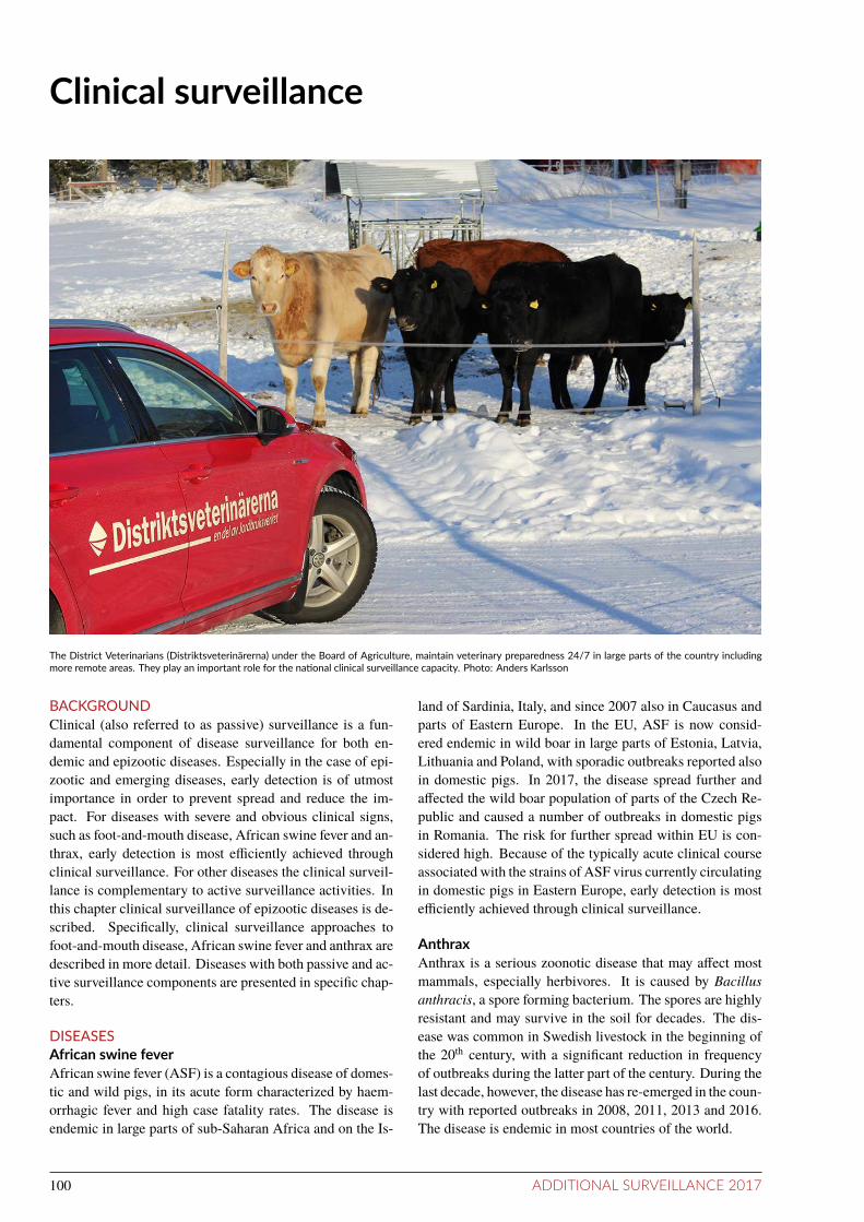
Clinical surveillance
The District Veterinarians (Distriktsveterinärerna) under the Board of Agriculture, maintain veterinary preparedness 24/7 in large parts of the country includingmore remote areas. They play an important role for the naional clinical surveillance capacity. Photo: Anders Karlsson
BACKGROUNDClinical (also referred to as passive) surveillance is a fun-damental component of disease surveillance for both en-demic and epizootic diseases. Especially in the case of epi-zootic and emerging diseases, early detection is of utmostimportance in order to prevent spread and reduce the im-pact. For diseases with severe and obvious clinical signs,such as foot-and-mouth disease, African swine fever and an-thrax, early detection is most eiciently achieved throughclinical surveillance. For other diseases the clinical surveil-lance is complementary to active surveillance activities. Inthis chapter clinical surveillance of epizootic diseases is de-scribed. Speciically, clinical surveillance approaches tofoot-and-mouth disease, African swine fever and anthrax aredescribed in more detail. Diseases with both passive and ac-tive surveillance components are presented in speciic chap-ters.
DISEASESAfrican swine feverAfrican swine fever (ASF) is a contagious disease of domes-tic and wild pigs, in its acute form characterized by haem-orrhagic fever and high case fatality rates. The disease isendemic in large parts of sub-Saharan Africa and on the Is-
land of Sardinia, Italy, and since 2007 also in Caucasus andparts of Eastern Europe. In the EU, ASF is now consid-ered endemic in wild boar in large parts of Estonia, Latvia,Lithuania and Poland, with sporadic outbreaks reported alsoin domestic pigs. In 2017, the disease spread further andafected the wild boar population of parts of the Czech Re-public and caused a number of outbreaks in domestic pigsin Romania. The risk for further spread within EU is con-sidered high. Because of the typically acute clinical courseassociated with the strains of ASF virus currently circulatingin domestic pigs in Eastern Europe, early detection is mosteiciently achieved through clinical surveillance.
AnthraxAnthrax is a serious zoonotic disease that may afect mostmammals, especially herbivores. It is caused by Bacillusanthracis, a spore forming bacterium. The spores are highlyresistant and may survive in the soil for decades. The dis-ease was common in Swedish livestock in the beginning ofthe 20th century, with a signiicant reduction in frequencyof outbreaks during the latter part of the century. During thelast decade, however, the disease has re-emerged in the coun-try with reported outbreaks in 2008, 2011, 2013 and 2016.The disease is endemic in most countries of the world.
100 ADDITIONAL SURVEILLANCE 2017

Foot-and-mouth diseaseFoot-and-mouth disease (FMD) is a highly contagious dis-ease of cloven-hoofed animals such as pigs, cattle, sheep andgoats. The case fatality rate in FMD is low, but morbid-ity very high and convalescence is extended, which makesthis disease especially important in countries previously freefrom the disease. FMD is endemic in many parts of theworld, but since 2011 the disease is absent in Europe. How-ever, the major FMD epidemics that afected several Euro-pean countries during the last decade demonstrated that thecontinent is continuously at risk for FMD virus introduction,and that early detection is crucial.
LEGISLATIONClinical suspicions of epizootic diseases must be notiiedto the Swedish Board of Agriculture in accordance withthe Swedish Act of Epizootic diseases (SFS 1999:657 withamendments). This obligation applies to animal keepers,oicial and private veterinarians, veterinary laboratories,and other relevant stakeholders. Suspicions are investigatedafter consultation with disease experts at the National Vet-erinary Institute and following notiication to the SwedishBoard of Agriculture.
SURVEILLANCEEvery year, hundreds of suspicions of serious infectiousdiseases are reported by ield veterinarians, animal ownersor private veterinary pathologists to the experts at the Na-tional Veterinary Institute. Many of these suspicions canbe ruled out already based on anamnesis and initial clini-cal investigation, whereas others require notiication to theSwedish Board of Agriculture and further investigation in-cluding sampling of sick or dead animals, with movementrestrictions imposed on the farms during the investigation.Also, in cases in which an epizootic disease is not primarilysuspected, but where it cannot be excluded based on clinicalinvestigation, samples can be submitted for laboratory inves-tigation to exclude a diagnosis. This can only be done afterdiscussions with experts at the National Veterinary Instituteand in consultation with the Swedish Board of Agriculture.This approach serves to reduce the threshold for submittingsamples for analysis of notiiable diseases, and thereby in-creasing the sensitivity of the system. The Swedish Board ofAgriculture covers all costs for veterinary visits, transports,and diagnostic analyses related to the investigation.
African swine feverReported cases of increased mortality or serious morbidity,with clinical signs such as haemorrhagic disorders or repro-ductive failures in pigs are considered suspicions of ASF un-til ruled out through further clinical investigation, with orwithout sampling of afected animals. Due to clinical simi-larity, samples from domestic pigs collected for ASF are alsoanalysed for CSF. This strategy is strongly recommended bythe EU.
Given the current situation in Eastern Europe as regardsASF in wild boar, the clinical surveillance is enhanced, andSwedish hunters are encouraged to report all indings of dead
wild boar. If possible, carcasses or samples are taken in andinvestigated to rule out ASF as the cause of death (see alsospeciic chapter on infectious diseases in wild boars).
AnthraxCases with a history of sudden deaths in one or more animalson the premise are considered suspicions of anthrax. Clin-ical signs such as fever, bloody discharges from the nose,mouth, anus or vagina, uncoagulated blood, subcutaneousoedematous swellings and lack of rigor mortis, as well as re-cent site disturbances (dredging or digging) strengthens thesuspicion. In addition, cases with gross pathological lesionssuggestive of anthrax found at post mortem are consideredsuspicions of anthrax.
During 2017, the clinical surveillance in the area afectedby anthrax during 2016 was enhanced. All cattle, sheep andwild ruminants found dead in the area, with no obvious causeof death, were investigated to rule out anthrax.
Foot-and-mouth diseaseReported cases of disease in cattle, pigs, sheep or goatswhich presents with vesicular lesions of the feet, buccal mu-cosa or mammary glands, are considered suspicions of FMDuntil ruled out through further clinical investigation, with orwithout sampling of afected animals.
RESULTSThe suspicions of epizootic diseases that were reported andfurther investigated based on sampling of sick or dead ani-mals in 2017 are compiled in Table 21.
Two clinical suspicions of ASF in domestic pigs were in-vestigated. Samples were collected and sent to the NationalVeterinary Institute for PCR analyses with negative results.Samples were also analysed for CSF with negative results.Sixteen samples from wild boar found dead were also anal-ysed for ASF with negative results.
Twenty clinical suspicions of anthrax in cattle, one insheep, one in fallow deer and one in a dog were reported andinvestigated. In addition, four suspicions in cattle and one insheep, with pathological lesions suggestive of anthrax, wereraised at post mortem, and three cattle, two roe deer and onemoose found dead without further clinical signs were inves-tigated as part of the enhanced clinical surveillance in thearea afected by anthrax during 2016. All suspected caseswere bled or sampled, and samples were sent to the Na-tional Veterinary Institute for examination using multiplexRT-PCR. Carcasses were left on the premises, covered toprevent any direct contact with the carcass and possibly con-taminated surfaces. In none of the cases, anthrax could beconirmed.
No clinical suspicion of FMD was investigated during2017.
DISCUSSIONClinical surveillance constitutes a fundamental part of theanimal disease surveillance system and is particularly im-portant as regards early detection of epizootic and/or emerg-ing diseases. This surveillance component depends on the
ADDITIONAL SURVEILLANCE 2017 101

level of cooperation and trust between the relevant stake-holders in the ield (including animal keepers and oicialand private veterinarians, among others) and the central vet-erinary authorities, but also on the level of knowledge andawareness among all involved. In Sweden, cooperation be-tween the relevant stakeholders is long-standing at a highlevel, and the level of knowledge and awareness as regardsepizootic diseases as well as the obligation to report suspi-cions thereof is considered good. Based on this and giventhe relatively high numbers of suspicions of epizootic dis-eases investigated each year, the performance of the clinicalsurveillance is considered adequate. However, a systematicevaluation of this performance has never been carried out.Therefore, to get a better understanding of the coverage andrepresentativeness of the clinical surveillance and thus theperformance, and to identify gaps, an evaluation of the clin-ical surveillance using data from the last ten years will becarried out during 2018.
Given the current situation in Eastern Europe as regardsASF in wild boar with a continuous albeit slow spread west-wards, the risk for introduction also to Swedish wild boaris considered increased. In case of introduction, early de-tection is crucial in order to prevent a longer-term establish-ment of the disease. The timeliness of detection depends, toa large extent, on the capacity of Swedish hunters to detect,
and their willingness to report, indings of dead wild boar.Despite information and awareness campaigns carried outin this regard during the last few years, targeting the hunt-ing community, and in-spite of a well-established networkof hunters, which is fundamental for the general surveillancefor diseases in wildlife (also described in the speciic chap-ter on Post mortem examinations in wildlife), less than 20wild boar found dead are investigated annually as part of thesurveillance for ASF. Given the population size of Swedishwild boar (estimated at 250 000) and the expected number ofwild boar that would die from other causes than hunting, andthus constitute the potential sampling frame for the surveil-lance, this number is not adequate. Further measures willtherefore be taken during 2018 to increase the numbers.
REFERENCESEFSA AHAW Panel (EFSA Panel on Animal Healthand Welfare), 2015. Scientiic opinion on Africanswine fever. EFSA Journal 2015;13(7):4163, 92 pp.doi:10.2903/j.efsa.2015.4163
DG(SANCO) 2013-6780. Report from an audit carriedout in Sweden on Animal health - disease contingencyplans; protection during depopulation for disease control,pp 14-17. http://ec.europa.eu/food/audits-analysis/act_get-PDF.cfm?PDF_ID=11146
Table 21: Number of suspicions of epizooic diseases reported through the clinical surveillance system during 2017 and invesigated by expertsat the Naional Veterinary Insitute ater noificaion to the Swedish Board of Agriculture.
Disease InvesigatedA Confirmed
African swine fever 18B 0Anthrax 34 0Aujesky’s disease 0 0
Avian influenza 28C 4CBluetongue 2 0BSE 2 0
CWD 1 0Classical swine fever 3 0FMD 0 0
Lumpy skin disease 0 0Newcastle disease 29C 3CParatuberculosis 5 0
PRRS 2 0Rabies 6 0Tuberculosis 9 0
West Nile fever 2 0A Inmany cases clinical suspicionswere invesigated for several diseaseswith similar clinical picture (e.g. ASF/CSF/PRRS,AI/ND)B Includes 16 wild boars found dead also described in the specific chapter on infecious diseases in wild boarC Does not include surveillance of, or cases in, wild birds
102 ADDITIONAL SURVEILLANCE 2017

Poultry Health Control ProgrammeBACKGROUNDThe aim of the Poultry Health Control Programme is to doc-ument freedom from the included diseases, to prevent the in-troduction and further spread of diseases and to allow tradefrom the participating companies.
The Poultry Health Control Programme is based on pro-visions (SJVFS 2010:58) issued by the Swedish Board ofAgriculture. The programme is mandatory for all hatcheriesproducing more than 50,000 day-old chicks per year andall breeding establishments (grandparent and parent flocksof layers, broilers and turkeys) delivering hatching eggs tothese hatcheries. In addition to serological sampling for sev-eral infectious diseases, the programme consists of biosecu-rity requirements, standards for poultry houses, managementand clinical surveillance.
LEGISLATION AND DISEASESAll diseases covered by the programme, except for My-coplasma synoviae, are notiiable according to provisions is-sued by the Swedish Board of Agriculture (SJVFS 2013:23).The diseases included in the programme during 2017 arebriefly described below.
Fowl typhoid and pullorum diseaseFowl typhoid and pullorum disease are two poultry dis-eases caused by Salmonella enterica subspecies entericaserovar Gallinarum biovar Gallinarum (Salmonella Galli-narum, fowl typhoid) and biovar Pullorum (Salmonella Pul-lorum, pullorum disease) respectively. These two biovars ofthe same serovar are specially adapted to poultry and verti-cal transmission (from the hen to the chicken via the egg) isan important feature in addition to the common horizontalspread. Pullorum disease mainly afects foetuses and chick-ens up to 3 weeks of age while Salmonella Gallinarum com-monly infects and causes disease (diarrhoea, inappetence,production losses and mortality) in older birds. Both biovarsare included in the Swedish zoonosis legislation (SJVFS2004:2) as well as in the European legislation on trade inpoultry and hatching eggs (Council Directive 2009/158/EC).The diseases were eradicated from the Swedish commercialpoultry population in the beginning of the 1960’s. A sin-gle case of fowl typhoid (Salmonella Gallinarum) was de-tected in a backyard flock in 1984 but has not been diagnosedsince then. Salmonella Pullorum is however present in theSwedish backyard poultry population; the last outbreak wasdiagnosed in 2017.
Mycoplasma gallisepicum, Mycoplasma synoviae andMycoplasma meleagridisM. gallisepticum, M. synoviae and M. meleagridis are im-portant poultry pathogens. However, M. meleagridis is onlypathogenic for turkeys. These three mycoplasmas can spreadboth horizontally and vertically. They mainly cause respira-tory disease and egg production losses. M. gallisepticum and
M. synoviae may also cause arthritis and are present in thebackyard poultry population in Sweden. Testing of breedingflocks for M. gallisepticum and M. meleagridis (only turkeyflocks) is included in the European legislation on trade inpoultry and hatching eggs (Council Directive 2009/158/EC).Due to its potential to cause disease and production losses,testing for M. synoviae was included in the programme be-tween 1995 and 2010. During a revision of the programmethe agent was excluded but is since 1 June 2015 includedagain. In 2016, testing for M. synoviae was further intensi-ied.
Avian avulavirus 1Avian avulavirus 1 (previously paramyxovirus type 1) maycause outbreaks of Newcastle disease, with egg productionlosses, increased mortality, nervous signs and respiratorydisease; the severity of the disease may vary. The virusis transmitted through direct and indirect contacts with in-fected birds and for shorter distances also with the wind.Wild birds are an important reservoir. Since 1995, sixteenoutbreaks of Newcastle Disease have occurred in Sweden.The disease is included in the Swedish Act of Epizooticdiseases (SFS 1999:657 with amendments). Since all out-breaks have been successfully eradicated, Sweden has a sta-tus of Newcastle free country without vaccination accordingto Commission Decision 95/98/EEC.
Egg drop syndromeEgg drop syndrome. The virus is a naturally occurring aden-ovirus in waterfowl (including the wild population) in whichit does not cause any clinical disease. In chickens, the clin-ical signs are only seen during the production period as de-creased egg production in an otherwise clinically healthyflock. The virus is able to spread both vertically and hor-izontally. The Swedish poultry breeding population is freefrom the disease.
SURVEILLANCESerological screening within the programme is administeredby the National Veterinary Institute and inanced by theSwedish Board of Agriculture and the participating compa-nies. In 2017, seven breeding companies participated in theprogramme; ive broiler-, two laying hen- and one turkeybreeding company (one company with both broiler- and lay-ing hen parent flocks). In accordance with the provisions,sixty blood samples were taken from the breeding flocks in-cluded in the programme, once during the rearing period andseveral times during the production period. The blood sam-ples were sent by mail to the National Veterinary Institutewhere serological tests were performed. The sampling andtesting schemes are presented in tables 22 and 23.
ADDITIONAL SURVEILLANCE 2017 103

RESULTSTable 24 gives an overview of all samples taken in breedingflocks of chickens and turkeys, and the laboratory methodsused, during 2017. All analysed samples tested negative forM. gallisepticum, M. meleagridis and avian avulavirus 1.
Antibodies to M. synoviae were detected in sevenchicken flocks (ive parent flocks and two grandparentflocks). One of the flocks was sampled at 60 weeks of ageand no additional samples were available from this flock. Intwo parent and one grandparent flock new samples obtainedtwo weeks later were also positive for M. synoviae. For theother three flocks the positive samples were considered asunspeciic serological reactions after testing new samplesfrom these flocks.
Seven chicken parent flocks were further investigateddue to a few positive samples for egg drop syndrome. In ad-dition, two chicken parent flocks were investigated due to afew positive samples for Salmonella Gallinarum/SalmonellaPullorum. No clinical signs were seen in these flocks and af-ter testing new samples from these flocks, the previous pos-itive samples were considered as unspeciic serological re-actions.
DISCUSSIONIn conclusion, the results from the serological screeningin the Poultry Health Control Programme in 2017 supportthe status of freedom from several important infectious dis-eases in the Swedish breeding poultry population. However,the indings of M. synoviae antibodies in chicken breedingflocks and possible implications on animal health and pro-duction both in breeding and ofspring flocks need to befurther considered. M. synoviae may spread both horizon-tally and vertically (from the hen to the chicken via the egg),hence infection in breeders may have consequences for thenext generation as well. Infection may result in respiratorysigns, articular disease and egg production losses. In addi-tion, egg shell abnormalities associated with infection withM. synoviae have been reported.
Finally, the clinical surveillance of the poultry breedingpopulation is also of utmost importance.
Table 22: Sampling schedule for chicken grandparent and parent flocks. Number of blood samples tested at different weeks of age.
Agent Age in weeks
16 24 36 48 60
S. Pullorum/ S. Gallinarum - 60 - - -Mycoplasma gallisepicum 60 60 60 60 60Mycoplasma synoviae 60 60 60 60 60Avian avulavirus 1 - - - 60 -Egg drop syndrome-virus - 30 - - -
Table 23: Sampling schedule for turkey parent flocks. Number of blood samples tested at different weeks of age.
Agent Age in weeks
20 32 44 56
S. Pullorum/ S. Gallinarum - 60 - -Mycoplasma gallisepicum 60 60 60 60Mycoplasma meleagridis 60 60 60 60Mycoplasma synoviae 60 60 60 60Avian avulavirus 1 - - - 60
104 ADDITIONAL SURVEILLANCE 2017

Table 24: Number of sampling occasions for grandparent (GP) and parent (P) flocks of chickens and turkeys and total number of samples testedduring 2017.
Agent No. of sampling occasions No. of samples Method
Chickens Turkeys Chickens Turkeys
GP P P GP P P
S. Pullorum /S. Gallinarum 16 90 4 960 5,400 240 Serum plate aggluinaion test,anigen, ID.Vet
Mycoplasma gallisepicum /Mycoplasma synoviae
79 445 16 4,740 26,700 960 Mycoplasma gallisepicum/synoviaeAnibody Test Kit, ID.Vet
Mycoplasma meleagridis 0 0 16 0 0 960 Serum plate aggluinaion test,anigen, ID.Vet
Avian avulavirus 1 16 85 4 960 5,100 240 NDV screen compeiion ELISA,ID.Vet
Egg drop Syndrome-virus 16 90 0 480 2,700 0 Anibody haemaggluinaioninhibiion test, anigen, in-house(Unil May 2017. From May 2017anigen from GD Animal Health)
ADDITIONAL SURVEILLANCE 2017 105

Infecious diseases in wild boarsBACKGROUNDWild boars are susceptible to contagious diseases that af-fect domestic pigs and therefore they have a potential role inspreading diseases to and from domestic pigs. This is par-ticularly the case for classical swine fever which has beentransmitted between wild boars and domestic pigs in severalEuropean countries. Also, the ongoing spread of Africanswine fever (ASF) in Eastern Europe and within the EU in-volves wild boar, and the direct and indirect contacts be-tween domestic pigs and wild boar in these areas hamperthe control and management of the disease. The Swedishwild boar population is increasing rapidly and is presentlyestimated at 250,000 animals before the reproductive sea-son of 2018. The northern border of the wild boar habitatis extending and has at present passed the level of the riverDalälven. Since the year 2000, hunted wild boars from dif-ferent parts of the country have been blood sampled yearlyfor surveillance purposes. The samples have been sent to theNational Veterinary Institute for analysis for antibodies to in-fectious agents that are of importance for the domestic pigproduction. Due to the worrying situation regarding ASF inEastern Europe and within EU, passive surveillance for thedisease in wild boars found dead has been included in thesurveillance programme since 2013.
LEGISLATIONThe infections investigated in the wild boar surveillance pro-gramme of 2017 are all included in the Swedish Act of Epi-zootic diseases (SFS 1999:657 with amendments) and arenotiiable on suspicion. If any of them are suspected or con-irmed, measures will be taken to control the disease and toprevent further spread.
SURVEILLANCEPassive surveillanceOrgan samples from, or whole carcasses of, wild boar founddead were submitted for post mortem examination at the Na-tional Veterinary Institute. All were subjected to Africanswine fever virus genome analysis irrespective of patholog-ical lesions.
All wild boar with clinical or post mortem signs leadingto suspicion of a disease included in the Act of EpizooticDiseases are sampled and investigated.
Acive surveillanceBlood samples from hunted wild boars were used for activesurveillance of antibodies to Aujeszky’s disease virus, clas-sical swine fever virus and Brucella suis. The samples werecollected voluntarily by hunters recruited through informa-tion on the webpage of the National Veterinary Institute,in hunter’s magazines and through using informal networksincluding information meetings. The surveillance was de-signed to detect the investigated diseases at 1% prevalencewith 99% conidence level. To reach this level of conidence
500 samples were needed. The samples were analysed usingthe serological methods described in the respective diseasechapters in this report.
RESULTSPassive surveillanceOne clinical suspicion of classical or African swine feverin a wild boar was investigated during 2017 due to postmortem indings including severe circulatory disorder withorgan hemorrhages. Following investigation, including sam-pling, the wild boar could be declared negative for CSF andASF.
Sixteen wild boars found dead were examined forAfrican swine fever virus genome and all analyses werenegative. The geographical distribution of the sampleddead wild boars is visualised in Figure 25. Additional postmortem indings in these wild boars are reported in the chap-ter Post mortem examinations in wildlife in this report.
Acive surveillanceIn 2017, 136 samples were collected from hunted wild boarsand analysed for antibodies to Aujeszky’s disease virus andclassical swine fever virus. All samples were negative forantibodies to these two pathogens. Antibodies to Brucellasuis were analysed in 100 of the samples. In three of thesamples the results were inconclusive due to hemolysis, theremaining samples were negative. The geographical distri-bution of sampled wild boars was roughly correlated to thedistribution and density of the Swedish wild boar popula-tion (Figure 25). The goal of 500 samples was not met, butthe surveillance evidence was suicient to indicate that theprevalence of the investigated diseases in the wildboar pop-ulation was <2% with a certainty of 98%.
DISCUSSIONThe Swedish wild boar population is growing and the bound-ary of the population is moving north. In areas where wildboars are already present, the population is also becomingmore dense, which increases the risk of direct and indirectcontact between wild boars and domestic pigs. The area inSweden populated by wild boars is surrounded by the sea.Therefore, there is no risk of wild boars migrating into Swe-den. Instead, the role of the wild boar in disease spreadmight be to pick up infectious agents introduced into Swe-den by other routes. It is possible that wild boars couldgain access to infected meat or other infected animal prod-ucts for example in garbage or following indirect spread byother means from people, vehicles or equipment. All dis-eases monitored in 2017 are or have recently been presentin neighbouring countries or in close proximity to Sweden.The unfavourable development of the African swine feversituation in Russia, Eastern Europe and within EU is of spe-cial concern and calls for eicient approaches to early detec-tion of disease in the wild boar population.
106 ADDITIONAL SURVEILLANCE 2017

0
1 − 4
5 − 15
15 − 24
25 − 34
over 34
●Found dead and
tested for ASF
●●
●
●
●
●●
●●
●●
●●
●●●
Figure 25: Geographical distribuion per county of huntedwild boars that were sampled in 2017. Thewhite points indicate the locaions wheredead wild boars tested for African Swine Fever (ASF) were found. ©EuroGeographics.
ADDITIONAL SURVEILLANCE 2017 107

Infecious diseases and parasites inhoneybeesBACKGROUNDEvery beekeeper in Sweden has the responsibility to preventthe spread of bee diseases and are obligated to register the lo-cation of their apiaries to the County Administrative Boards(CABs). There is no national bee register, but the number ofapiaries and colonies must be reported by the beekeepers andrecorded by the respective CABs. The health of honeybeesis controlled by local bee inspectors appointed and given theresponsibility over local inspection districts, by seven of theCABs. The country is divided into approximately 500 beedistricts and the bee inspectors are responsible for the actualcontrol of the apiaries located in the district. The SwedishBoard of Agriculture (SBA), is the central authority for thecontrol of bee diseases. The SBA is responsible for the reg-ulations and guidelines for management and control of thehoneybee diseases regulated in SJVFS 1992:38 includingAmerican foulbrood and Varroa mite infestation. There areregulations for the import and export of bees, bee-relatedproducts and beekeeping equipment to prevent contagiousbee diseases from entering the country and reduce furtherspread. Applications for permits to import bees must bemade to the SBA at each point of entry. The conditions forimport are the same in all EU Member States. If bees areintroduced without permission, it is considered to be a vio-lation of the law on smuggling of goods.
DISEASES AND LEGISLATIONAll veterinarians, as well as laboratories analyzing sam-ples from honeybee colonies, are obligated to notify theSBA if American or European foulbrood, tracheal mite in-festation/ acariosis (Acarapis woodi), Varroa mite infesta-tion/varroosis (Varroa destructor), Tropilaelaps mite infes-tation (Tropilaelaps spp) or the small hive beetle (Aethinatumida) are found. This is regulated in the law of bee dis-eases (1974:211), the regulation of bee diseases (1974:212)and the SBA’s regulation on the control of American foul-brood, AFB, and Varroa mites (SJVFS 1992:38). A bee-keeper needs a permit issued by a bee inspector to move thebees out of a parish which has been declared infected withAFB by the SBA. Visual inspection of clinical symptoms ofAFB and Varroa mites are carried out at the same time. Incase of an outbreak of AFB or if Varroa mites are reportedfrom an area or region where it has not been present earlier,the bee inspector notiies the CAB, which in turn notiies theSBA. The SBA then declares the parish in which the apiary issituated infected/infested. Bee inspectors can send samplesof diseased brood, larvae or pupae to the National ReferenceLaboratory for Bee Health, NRL, at the Swedish Universityof Agricultural Sciences, SLU, where the diagnosis of hon-eybee diseases included in the legislation is performed onbehalf of the SBA. This is a yearly, laboratory-based, pas-sive surveillance of honeybee diseases.
American foulbroodAmerican foulbrood (AFB) of honeybees is a contagiousbacterial disease caused by the spore-forming bacterium,Paenibacillus larvae. The disease is widely distributedacross the world causing great economic losses in apiculture.The disease is classiied as an epizootic and is notiiable inmost countries. As the name indicates, the disease only af-fects the larval stages of honeybees. AFB is highly infec-tious, lethal to the individual honeybee larva and potentiallylethal to infected colonies. AFB is a statutory notiiable dis-ease in the European Union in the framework of trade andexport requirements (Directive 92/65/EEC). In many Euro-pean countries, Sweden included, the disease is controlledthrough burning of symptomatic colonies and the use of bee-keeping management techniques to avoid the spread of theinfectious agent to uninfected hives. Current legislation donot allow European beekeepers to use antibiotics since thereis no maximum residue limit (MRL) set for the antibioticsubstances used to control AFB (oxytetracycline and tylo-sine). No antibiotics can be legally used since there is azero tolerance limit to antibiotic residues in honey. Swedenhas strict rules for movement of bees, apiculture equipmentand honeybee products from areas where AFB has been re-ported. The bee inspectors burn any colony with clinicalsigns of American foulbrood and inspect all other apiarieswithin a 3-km radius from the infected apiary.
European foulbroodEuropean foulbrood (EFB), is a serious disease of honeybeescaused by the bacterium Melissococcus plutonius. EFB af-fects mainly young honeybee larvae usually between 4 and5 days old. A massive loss of brood resulting from severeinfection, weakens the colony and can lead to its collapse.Regional variations in disease burden have been reportedand recent decades have seen dramatic increases in the in-cidence of EFB in parts of Europe. Large disease outbreakshave been identiied in areas previously thought to be diseasefree, such as Norway.
Tracheal mite infestaion (acariosis)The honeybee tracheal mite Acarapis woodi is an inter-nal parasite of the respiratory system of adult honeybees.The tracheal mite has spread through global beekeeping ex-changes and has been reported from all European countriesexcept Sweden. It is therefore regulated in Swedish legisla-tion.
Varroa mite infestaion (varroosis) andassociated virus infecionsThe honeybee parasitic mite, Varroa destructor, was origi-nally conined to the Eastern honeybee, Apis cerana, wherea stable host-parasite relationship exists due to a long period
108 ADDITIONAL SURVEILLANCE 2017

of coevolution. After a shift in the last century, from the na-tive host to the Western honeybee, Apis mellifera, the mitedispersed around the globe and is currently considered thegreatest threat to honeybees and apiculture worldwide. Themite was reported in Europe in the late 1970s, was found onGotland in 1989 and in the county of Skåne, in 1991. Theregulations from the SBA has since been aimed at limitingthe spread of the Varroa mite in the country. Varroa miteshave so far not been reported from the northern half of Swe-den (Västerbotten, Jämtland, as well as most of Västernor-rland, Norrbotten, Dalarna and Gävleborg) except close tothe Finnish border. The rest of the country has a varyinglevel of infestation.
Honeybee viruses such as Deformed wing virus (DWV)and Acute bee paralysis virus (ABPV) are associated withthe Varroa mite and DWV is the actual cause of the clini-cal signs observed in connection with high Varroa numbers.The mite acts as a biological vector for both viruses.
Tropilaelaps mite infestaionMites of the genus Tropilaelaps afect both developing broodand adult bees mainly in Asia. Tropilaelaps mercedesae andTropilaelaps clareae are the only species found reproduc-ing on brood of A. mellifera. The distribution of the emerg-ing mite is currently restricted to tropical and subtropicalregions of Asia and Africa but is regulated within the EUand honeybee queen imports are visually inspected for theoccurrence Tropilaelaps mites. The mite has not been re-ported from Europe.
The small hive beetleThe small hive beetle, Aethina tumida, is endemic to sub-Saharan Africa, but has spread to many other locations, in-cluding North America, Australia, the Philippines and wasrecently reported in Italy. The small hive beetle can be adestructive pest of honeybee colonies, causing damage tocomb, stored honey and pollen. The primary damage tocolonies and stored honey is caused through the activityof the larvae tunneling through honey combs, feeding anddefecating, causing discolouration and fermentation of thehoney. If a beetle infestation is suiciently heavy, they maycause bees to abandon their hive.
SURVEILLANCEPassive surveillancePassive disease surveillance of honeybee diseases and para-sites is done through diagnostics related to disease outbreaksand reported by the NRL to the SBA yearly (Table 25).
Enhanced passive surveillance by visual inspection ofclinical symptoms of AFB is done when a beekeeper needs apermit issued by the bee inspector in order to move the beesout of a parish which is declared infected by the SBA. Thisis reported by the bee inspectors to the CABs (Figure 26).
Acive surveillanceActive surveillance for the occurrence of the tracheal mite,A. woodi completed by the SBA in 1993 and 2010-11. Todate, A. woodi has not been detected in Sweden.
In 2016, a base-line study of the prevalence of Var-roa mites, the viruses DWV, ABPV, the bacteria M. plu-tonius and P. larvae was completed by the NRL for beehealth in collaboration with the National Veterinary Institute(SVA). Samples of adult bees were collected from honey-bee colonies in 382 randomly selected apiaries distributedthroughout the country. The goal was to sample 385 api-aries (≤5 colonies per apiary). Samples were collected dur-ing the 2016 beekeeping season and the samples were sentto the NRL at SLU for analysis. The samples were examinedmacroscopically for Varroa mites and analyzed by molecu-lar methods (RT-qPCR for ABPV and CBPV and qPCR forM. plutonius) and by microbial culturing (P. larvae).
Paenibacillus larvae, the causative agent of AFB was de-tected in 6% of all sampled apiaries (Figure 30).
Melissococcus plutonius, the causative agent of EFB,was detected in 2 apiaries in Östergötland (Figure 29).
Varroa mites was detected in 53% of the sampled api-aries and in all counties except Jämtlands, Västerbottens andNorrbottens county (Figure 31) which reinforces earlier ob-servations and reports from bee inspectors. DWV was de-tected in 30% of the investigated apiaries and in all countiesexcept Jämtlands, Västerbottens, Norrbottens and Väster-norrlands counties and ABPV was detected in one apiaryin Skåne and one apiary on Gotland (Figures 28, 27).
DISCUSSIONThe reporting of AFB incidences, thus far, has been basedon the information that the bee inspectors report to the CABsbased on visual observation of clinical signs (Figure 26). Inthe 2016 base-line study, microbiological cultivation of P.larvae from samples of adult bees was used. This methodwas previously shown to be well correlated with clinicalsigns of disease. Only young larvae develop clinical signs,but adult bees are carriers of the bacterium. In the base-linestudy, we investigated the subclinical presence of the bac-terium in a selection of the country’s apiaries. The bacteriacould not be detected in the majority of the examined api-aries (94%), which is an important argument in the ongoingdiscussion between beekeepers and the regulatory authori-ties about simplifying the regulations on the managementand movement of bee colonies in the country. It is impor-tant to highlight that there are many apiaries in areas freefrom this pathogen and that this status is worth preserving.
The bacteria Melissococcus plutonius that causes EFBwas detected in only two apiaries in Östergötland. Histori-cally, EFB has been considered to be less serious than AFBbut reports of more aggressive forms of the bacterium andmore serious disease outbreaks have become increasinglycommon in recent years. A few years ago, Norway had anoutbreak of EFB that led to extensive investigations and san-itation which highlights the value in continued monitoringfor this disease to prevent outbreaks in Sweden.
After the introduction of the Varroa mite in Sweden, theSBA introduced regulations to prevent or at least slow downthe spread of the mite in the country. This has not com-pletely prevented the spread but led to the fact that we stillhave apiaries in the northern parts of the country that are
ADDITIONAL SURVEILLANCE 2017 109

apparently free from Varroa mites. This can be further con-irmed by the results of this survey which reinforces earlierobservations and reports from bee inspectors. In Norrbot-ten, however, there have been indings of Varroa mites inHaparanda, Övertorneå, Kalix and Luleå, which may be aresult of introduction of the disease from northern Finlandwhere the mite is present. The Varroa mite acts as a bio-logical vector for viruses like DWV and ABPV. In the sur-vey, DWV was detected in all counties except Västerbot-ten, Jämtland, Norrbotten and Västernorrland. The spreadof DWV coincides with the presence of Varroa and followsthe spread of the mite. In Västernorrland, the mite has re-cently been introduced and the virus infection has not yetbeen spread. Another virus associated with Varroa is ABPVwhich was detected only in a single apiary on Gotland andone in Skåne. It is possible that the virus is so virulent that itkills its host faster than it can efectively spread. This couldexplain why the less virulent virus DWV has such a highincidence while ABPV is rare. It is also worth noting that
the counties where ABPV is detected, Gotland and Skåne,are the counties where Varroa was irst introduced into thecountry. At that time (late 80s, early 90s), ABPV was themost dominant Varroa-associated virus in Europe before be-ing surpassed by DWV. Perhaps it is that ABPV was estab-lished in parts of the honeybee population in these countiesbefore DWV became more widely spread.
The lack of a national bee register makes it diicult toorganize and collect samples of bees. There has been resis-tance to a central bee register from some beekeepers, but aninitiative has now been taken by the CABs and the SBA to es-tablish a register. This would facilitate disease surveillancein the future and is a prerequisite for being able to follow thecontingency plans for certain exotic pests in honeybees. Insummary, the health situation for Swedish honeybees is goodand we should continue to promote regular disease monitor-ing as a basis for legislation and prevention measures regard-ing honeybee health.
Table 25: Number of samples analysed in the Swedish honeybee populaion during 2017. Tesing conducted basedmainly on clinical suspicions.
Disease/parasite No. of testedbeekeepingoperaions
No. ofinfected/infestedoperaions
No. of testedbee hives
No. ofinfected/infestedbee hives
AFB 95 31 168 51EFB 8 0 8 0A. woodi 13 0 47 0
Varroa mites 34 29 87 61Tropilaelaps 0 0 0 0A. tumida 2 0 2 0
0
50
100
150
200
250
300
2005
2007
2009
2011
2013
2015
2017
Year
Num
ber
of c
olon
ies/
apia
ries
Colonies Apiaries
Figure 26: Number of new cases of American foulbrood during 2005-2017 in bee colonies and apiaries based on reports from bee inspectorsto the County Administraive Boards
110 ADDITIONAL SURVEILLANCE 2017

0 %
0−1 %
1−2 %
2−5 %
> 5 %
Figure 27: Prevalence of ABPV in 2016
0 %
0−6 %
6−10 %
10−15 %
> 15 %
Figure 28: Prevalence of DWV in 2016
0 %
< 5 %
5−10 %
> 10 %
Figure 29: Prevalence ofM. plutoniusin 2016
0 %
< 5 %
5−10 %
10−25 %
> 25 %
Figure 30: Prevalence of P. larvaein 2016
0 %
0−10 %
10−25 %
25−40 %
> 40 %
Figure 31: Prevalence of Varroa destructorin 2016
ADDITIONAL SURVEILLANCE 2017 111

Infecious diseases in fish, crustaceansand molluscs
European/flat oyster, Ostrea edulis, top shell half to the let, botom shell half to the right. The botom shell half is atached to a Litorina shell.Photo: Anders Aljorden
BACKGROUNDAll registered aquaculture farming sites are obligated to par-ticipate in the Oicial Health Control Programme, regulatedin accordance with SJVFS 2014:4, and by Council Direc-tive 2006/88/EG. Sweden has a very healthy aquacultureas well as wild populations of ish and shellish. None ofthe serious diseases that occur throughout Europe are preva-lent in Sweden. A restrictive approach to imports of liveish for restocking/farming, an early introduction of healthcontrol in farms and the presence of hydroelectric dams inmost Swedish rivers (acting as migration barriers for feralish from the coastal zone) all contribute to maintaining thishealth status. The presence of dams also results in a diferenthealth status at the coast compared to the more disease freecontinental zone. To maintain this situation, all transport oflive ish from the coast to the inland zone is forbidden andSweden has a national restocking programme for salmonidsto compensate for the lack of natural migration.
LEGISLATION AND DISEASESAll Swedish ish farms have participated in surveillance forthe diseases mentioned below since the late 1980’s in ac-cordance with EU Directives 2001/183 (now replaced by2015/1554) and 2006/88. Sweden has an approved dis-ease free zone status (2002/308/EC) for Viral haemorrhagicsepticaemia (VHS) and Infectious haematopoietic necrosis
(IHN) (2008/427/ EG). Additional guarantees are in placefor the whole country for Spring Viraemia of Carp (SVC)and for the continental zone for Infectious Pancreatic Necro-sis (IPN) (2010/221/EC). The zone has an eradication pro-gramme for Renibacteriosis/bacterial kidney disease (BKD)and the coastal zone for IPN (2010/221/EU). These diseasesare included in the Swedish legislation of notiiable dis-eases (SJVFS 2013:23). Further, IHN, VHS, IPN (other thanserotype ab) and SVC are included in the Swedish Act of epi-zootic diseases (SFS 1999:657 with amendments). In addi-tion, testing is routinely done for Koi herpes virus (KHV) inimported, quarantined koi, and for Crayish plague in cray-ish. These diseases are also regulated by the Swedish leg-islation for notiiable diseases (SJVFS 2013:23). Other no-tiiable diseases such as furunculosis (Aeromonas salmoni-cida salmonicida/ASS), yersiniosis/Enteric redmouth dis-ease (ERM), Marteiliosis and Bonamiosis (shellish) andWhitespot disease (crayish) are not actively tested for withinsurveillance programmes.
Infecious haematopoieic necrosis (IHN) and viralhaemorrhagic sepicaemia (VHS)Both diseases are caused by rhabdoviruses and occur fre-quently in Europe. They are transferred horizontally, but ver-tical transmission cannot be completely ruled out for IHN.
112 ADDITIONAL SURVEILLANCE 2017

Both diseases have greatest impact in aquaculture of rain-bow trout (Oncorhynchus mykiss) in freshwater, but havealso been detected in several other species. Infected ishexhibit behavioral changes, lethargy and abnormal swim-ming (whirling). The ish are anemic with varying degreesof hemorrhage in multiple organs. VHS is found in a marineform, and a low frequency in wild populations of sensitivespecies cannot be excluded in the Swedish coastal zone.
Infecious pancreaic necrosis (IPN)IPN is caused by a Birnavirus that is highly infectious tojuvenile salmonids. Susceptibility declines with increasingage. Fish that survive infection become subclinical carriers.In addition to salmonids, virus has been detected in severalother species. The virus is transmitted both horizontally andvertically.
The disease has large consequences, with high mortalityin young ish, and is considered as one of the most costly inseveral European countries. Symptoms include darkening,abdominal distension and corkscrew swimming. Petechialhemorrhage in abdominal fat and internal organs are themost dominant internal indings. Mortality rates can varybetween 10-90%.
Renibacteriosis (BKD)BKD is caused by a gram positive bacterium, Renibacteriumsalmoninarum. The infection can be transmitted both hori-zontally and vertically. The disease favours low water tem-peratures, and outbreaks occur mainly at temperatures be-tween 7-15°C.
Salmon and arctic char are most susceptible to BKD andmortality can reach 80%. In rainbow trout, the disease ischronic with a continuous low mortality of about 5-10%,however outbreaks of up to 40% mortality can occur. In-fected ish may have reduced growth and disease can resultin a deterioration of the meat quality.
Spring viraemia of carp (SVC)SVC is caused by a rhabdovirus. The disease occurs in Asiaand several European countries. The virus has been detectedin several ish species in the cyprinid family. The virus istransmitted horizontally. The clinical signs are usually gen-eral, such as darkening, exophthalmia and a slow breathing.The ish swim lazily with sporadic periods of hyperactivity.Other common indings are pale gills, ascites and hemor-rhage in the skin and gills. Internally, bleedings are foundin various organs including muscle, swim bladder and thebrain.
Koi Herpes virus (KHV) infecionKHV is a DNA virus and afects common carp (Cyprinuscarpio) and variants thereof, including koi. The virus wasirst detected in 1998 and has since then been reported fromall continents except Australia. The virus is transmitted hor-izontally. KHV can cause severe problems and is associatedwith high mortality. Infected ish usually swim at the sur-face and have an increased breathing frequency. Symptomsinclude enophtalmia, spotted gills and secondary bacterial
or parasitic infections on gills and skin. Surviving carps canbecome subclinical carriers.
Crayfish plagueCrayish plague is caused by an aquatic fungus(Aphanomyces astaci), which spread to Europe in the late1800’s from the United States with live crayish. The dis-ease occurs throughout Europe and North America. Thefungus reproduces by spores spread in the water. When thespores infect crayish they grow through the skin and attackthe underlying tissues.
The signal crayish becomes subclinically infected andmay exhibit black (melaninated) areas in the shell adjacentto the presence of the fungus in the skin. The spots will dis-appear when the shell is shed, but may gradually reappear.
When noble crayish is infected the irst sign is high mor-tality in afected populations. Disease in the individual ischaracterised by behavioural changes such as moving dur-ing daytime, reduced coordination and balance diiculties.
White spot disease (WSD)WSD is caused by a Whispovirus (WSSv) that can infect awide range of aquatic crustaceans including marine, brack-ish and freshwater prawns, crabs, crayish and lobsters. Out-breaks occur at water temperatures of 18-30°C. The mostcommon clinical sign is white spots in the exoskeleton, butthe disease can occur without obvious external signs.
The virus is transmitted both horizontally and verticallyand has a long survival time outside the host animal. Thevirus is present in imported frozen raw giant shrimps. Thereis a non-negligible risk that the virus will be introduced tothe aquatic environment by anglers using these shrimps forbait. The consequences are diicult to predict but may havea negative impact on Swedish crustacean populations.
MarteiliosisMarteiliosis, a disease in oysters and blue mussels, is causedby a unicellular parasite (Marteilia refringens in oysters andM. pararefringens in blue mussels). The parasite needs acrustacean (Paracartia grani) as an intermediate host. Thedisease causes reduced itness, impaired growth and resorp-tion of the gonads and hence reduced reproductive capacity.When the animals weaken, they cannot keep the shell halvesclosed.
BonamiosisBonamiosis is a disease in oysters caused by the protistanparasite Bonamia ostreae. The parasite invades and destroysthe haemocytes. Usually the only sign of disease is increasedmortality in the infected oyster population. B. ostreae isfound along the European Atlantic coast as far up as Den-mark, where it has now been found in Limfjorden.
SURVEILLANCEWithin the Oicial Control Programme, there is activesurveillance for the viruses causing IHN, VHS, IPN andSVC, and also for renibacteriosis/BKD. Sampling frequency
ADDITIONAL SURVEILLANCE 2017 113

is based on classiication of each farm into one of three cat-egories (high (I), medium (II) or low risk (III)) after a riskanalysis, based on the risk for the farm becoming infected,the risk that the farm will further spread the pathogen andthe impact of the pathogen. The risk categorization is per-formed by the Board of Agriculture. Farms within risk cate-gory I and II are tested every year and every second year, re-spectively, but farms within risk category III are only testedupon suspicion of disease. The aim of the Oicial ControlProgramme is to document freedom from disease and to con-tribute to the maintenance of this status.
There is also active surveillance in imported quarantinedish (eel - IPN and koi/carp - KHV). Active surveillance isalso done when potential invasive alien species - like themarble crayish - are discovered.
Crayish plague is monitored by passive surveillance andtesting is done based on suspicion of disease outbreaks.
Except for the control program, passive disease surveil-lance has been done through diagnostics related to diseaseoutbreaks in farms and wild ish.
DIAGNOSTIC PROCEDURESAll diagnostic analyses are performed according to recom-mendation by EU or OIE at the Swedish reference labora-tory, the National Veterinary Institute. VHS, IHN, IPN andSVC are tested for on pooled organ material (spleen, kid-ney, heart/brain) by a cell culturing method. A pool consistsof organs from up to ten ish. A cell culture is deined asvirus positive if a cytopathogenic efect is detected withintwo weeks, after which the virus is identiied by ELISA andconirmed by PCR or in some cases by serum neutralisation(SN test). KHV is tested for on individual ish (pooled gilland kidney) by PCR. Thirty ish are sampled in regular ishfarms, and in restocking farms all females are sampled afterstripping of roe. In the case of carp/koi, only a few ish maybe sampled. In eel quarantine, 120 glass eels are sampled atarrival and after 2 months, 120 co-habitated rainbow troutare sampled for detection of virus.
BKD is tested for on kidney tissue from individual ishand demonstrated by an ELISA method. Veriication is doneby PCR. Thirty ish are sampled in regular farms, and in re-stocking farms up to 120 ish are sampled after stripping ofroe.
A. astaci is demonstrated by light microscopy and culti-vation and veriied by realtime (rt) PCR, and WSSv is de-tected by rt-PCR. The number of sampled animals variesfrom case to case.
RESULTSOfficial health programme for fish farmers and crus-tacean surveillanceThe number of samples analysed and results are shown intable 26. In summary, the active surveillance detected (onecase=one outbreak):
• 1 case of IPN serogroup 2 in the coastal zone
• 2 cases of BKD in arctic char, one of these in a newlocation
• 1 case of BKD in rainbow trout
• 1 case of eel virus European X (EVEX) in quarantinedglass eels
• 5 cases of Crayish plague
Voluntary health programme for fish farmersThere was one recorded outbreak of other notiiable diseasesin ish during 2017, when furunculosis (ASS) was identiiedin an inland farm with concurrent BKD infection.
One farm imported 2,000 siberian sturgeon fry fromItaly and the fry was infected with Acipenser iridovirusEuropean–I. The mortality was 95% in 2.5 months. The re-maining 5% were then euthanized to clean and disinfect thefarm.
Flavobacteriosis due to Flavobacterium psychrophilumcontiunes to be the dominant cause of production diseasein fry and young ish. Resistance against oxolinic acid andoxytetracycline is becoming more and more common in thebacterium. The cause for this is not known. Usually florfeni-col (for which there is yet no resistance) is used for treatmentof the bacterium, and the other two antibiotics are rarely usedin aquaculture.
Voluntary health control in wild fishDue to the detection of IPN genogroup 6 in broodstock troutfrom Lake Vänern in December 2016, the County boards ofVärmland and Västra Götaland took an initiative to samplewild trout and salmon from the lake. A total of 144 adultsalmon and trout, plus ive organ pools without stated num-ber of ish (1 to max. 10 ish) and two pools of yearlingsfrom upper Klarälven have so far been investigated by viruscell culture. No virus has been detected. The investigationswill continue in 2018 in an aim to reach 500 sampled indi-viduals.
Outbreaks in wild fish, crustaceans and molluscsInvestigations into mortality in the freshwater pearl mussel(Margaritifera margaritifera) were continued in two addi-tional sites. The cause of the disease has not yet (May 2018)been fully understood and investigations continue.
DISCUSSIONThe number of farms that were sampled are listed in table26. Swedish aquaculture has a good health status, where allsevere diseases of importance are absent.
The most problematic disease to control is renibacterio-sis/BKD, due to its vertical transmission and variable clini-cal presentation. Prolonged time from diagnosis to slaughtercan lead to secondary health issues and increased antibioticuse as well as decreased welfare. As an example: the last twoyears ASS has been causing problems in one BKD infectedfarm and mortalities continued despite antibiotic treatment.The reason is probably the underlying BKD infection, facil-itating the ASS infection and itself being accelerated by theconcurrent ASS infection. Control of BKD is expected to beimproved by modiied sampling and improved methodology,from today’s post mortem sampling to an in vivo method.
114 ADDITIONAL SURVEILLANCE 2017

Additional resources must be invested in risk-based analysisof individual aquaculture farms to get a more reliable assess-ment for health surveillance. One new geographic locationwas found to be infected in 2017. The other two cases werere-infections of previously sanitised farm sites. In additionto detected cases, one sample from another farm was pos-itive by ELISA, but negative by PCR although the ELISAOD value was high. The farm is suspected to be positive,and the cause of the negative PCR could be localised infec-tion in the kidney, meaning that the PCR swab did not pickup any bacterial DNA.
The detected IPN case was due to reinfection of a farmsite that was sanitised a few years ago. Genogroup 2 is oflow pathogenicity, and the ish in the farm are outside theage range where serious symptoms generally occur.
EVEX in glass eel was detected by virus cell culturefrom eels sampled when the ish had just arrived at the quar-antine. SVA noted lethargy in the eels, which are usually
very lively upon arrival to the lab. Mortalities in the quar-antine occurred at the time of arrival but were thought to bedue to poor transport conditions. EVEX has been detectedin imported glass eel once before. Usually import is donefrom Great Britain, but on both of these occasions, glass eelwas imported from France instead. Since EVEX is a notii-able disease (rhabdoviruses other than VHSV), discussionsabout the fate of the eels were held several times betweenthe diferent involved authorities. The eels were doing well,and the Swedish Agency for Marine and Water managementwas about to allow restocking in the wild at the end of quar-antine time, when mortalities started to occur again. In theend, 2 million eels were euthanized, causing a major loss inthe Swedish restocking programme.
The number of identiied crayish plague outbreaks areat the same level as in 2016.
Table 26: Samples taken in the Swedish surveillance programmes for noifiable diseases in fish, crustaceans and molluscs
Disease No. of sampledproducion sites
No. of infectedproducion sites
No. of testedindividuals
No. of testedpools
No. of infectedindividuals/pools
Fish
VHS 64 0 - 417 -/0IHN 64 0 - 417 -/0IPN 64 1 - 417 -/1B
SVC 2 0 - 7 -/0KHV 1 0 4 - 0/0BKD 85 3 3,418 - 43C/-
Crustaceans
Aphanomyces astaci 18A 5 39 0 15/0WSSv 0A 0 0 0 0/-
Molluscs
Bonamia ostreaeD 5 0 150 0 0/-Marteilia refringensD 5 0 150 0 0/-
A Wild fish/crayfishB Virus isolaion in cell culture, virus idenified by ELISA and confirmed by RT-PCR and sequencing.C By ELISA. Infecion was confirmed in 16/18 fish that were further tested by qPCR.D This sampling was performed as part of a project withing the European Sea and Fisheries Fund
Abbreviaions:VHS Viral hemorrhagic sepicemiaIHN Infecious Haematopoieic NecrosisIPN Infecious pancreaic necrosisISAV Infecious salmon anaemia virusSVC Spring viraemia of carpSAV Salmonid alphavirus (Pancreas disease)PRV1 Piscine reovirus (PRV1 causes Heart and sceletal muscle inflammaion(HSMI))KHV Koi herpesvirusBKD Bacterial Kidney Disease
ADDITIONAL SURVEILLANCE 2017 115

Examinaion of aborions in foodproducing animalsBACKGROUNDPost mortem examinations are considered important forearly detection and national surveillance for infectious andemerging disease. As mentioned in the chapter PostMortem examinations in food producing animals , theSwedish Board of Agriculture has for the past 20 yearsinanced a programme to encourage such examinations.Many infections, however, show no macroscopic lesions orcause nonspeciic changes not detected at necropsy. Bru-cellosis, porcine reproductive and respiratory syndrome(PRRS) and classical swine fever (CSF) are examples of in-fections that may be present without speciic macroscopicindings. Moreover, the clinical picture in the herd may benon-speciic, which may cause a delay before the suspicionof these diseases occurs in clinical surveillance.
SURVEILLANCEThis surveillance component was introduced in 2008. It in-cludes examinations for brucellosis in all ruminant foetusesand for brucellosis, PRRS and CSF in all pig foetuses sub-mitted for necropsy as part of the post mortem examina-tion programme. During the second half of 2012 and 2013,Schmallenberg virus (SBV) was tested for as well. These in-fections often cause abortion, therefore sampling of abortedfoetuses means sampling within a risk group and increasesthe chance of detecting the infectious agent if present in thecountry. The Swedish Board of Agriculture inances sam-pling and testing of foetuses for Brucella, PRRS and CSF.All diagnostic testing was performed at the National Veteri-nary Institute. The foetuses were analysed for the CSFV and
PRRS genome with PCR and for Brucella by bacterial cul-ture.
RESULTSSince the start in 2008, a varying number of foetuses ofdiferent species have been examined each year (Table 27).The numbers for 2012 and 2013 were extraordinarily high,most likely because of increased attention due to the newlyidentiied infection with Schmallenberg virus (SBV). Dur-ing 2017, the lowest number of foetuses since 2008 weresubmitted to the programme (Table 27).
All analysed samples were negative for Brucella, PRRSand CSF.
DISCUSSIONThe post mortem examinations and sampling of foetuses arean important part of the national surveillance for infectiousand emerging diseases, as illustrated by the detection of in-fections with Schmallenberg virus in 2012 and 2013. Testingfor SBV ended in 2013 because the disease, at that time, hadbecome established in Sweden and therefore was consideredendemic. During the last four years, the number of exami-nations has been less than the anticipated, approximation of140 foetal examinations per year. Actions have been takento increase the numbers during 2017, for example improv-ing awareness by reminding about the possibility to submitfoetuses for examination to herd veterinarians. These ac-tions will continue and will be complemented by awareness-raising activities directed towards farmers during 2018.
Table 27: Number of aborted foetuses examined in the surveillance since the start in 2008
Species 2008 2009 2010 2011 2012 2013 2014 2015 2016 2017
Catle 14 15 62 21 63 114 32 29 34 20Goat 0 0 9 3 5 4 2 0 2 2Sheep 0 29 70 45 79 89 28 31 16 22
Alpaca 0 2 5 0 0 4 0 2 1 0Bison 0 0 0 0 1 1 0 0 1 0Gnu 0 0 0 0 0 1 0 0 0 0
Visent 1 1 0 2 1 0 0 0 0 0Pig 37 79 61 51 54 46 31 17 43 6Water buffalo 0 0 0 0 0 0 0 0 0 1
Total 52 126 207 122 203 259 93 79 97 51
116 ADDITIONAL SURVEILLANCE 2017

Post mortem examinaions in foodproducing animals
Post mortem examinaions are an important part of early detecion surveillance, and key in understanding and describing new emerging threats. In 2017, almost500 pigs were invesigated. Photo: Bengt Ekberg
BACKGROUNDEarly detection of infectious diseases is of utmost impor-tance to prevent negative efects. For diseases with severeclinical signs the irst line of defence is the detection of dis-ease by animal owners, ield veterinarians and pathologists.International and national experience show that post mortemexaminations remain a vital part in disease control and de-tection of emerging diseases.
As post mortem examinations are considered an impor-tant part in the early detection and national disease surveil-lance, a speciic programme for such examinations startedin the early nineties. The Swedish Board of Agriculture i-nances the programme, complemented by fees from the an-imal owners. Farm & Animal Health is responsible for theorganisation of the post mortem examination programme.
SURVEILLANCEThe programme subsidises post mortem examinations in allfood producing animals, including poultry. The latter wereincluded in the programme in 2007. Since 2008, domes-ticated exotic ungulates are also included. Approximately
3,000 animals have been examined yearly within the pro-gramme since 1999. In conjunction with post mortem exam-inations, samples are routinely collected from deined cate-gories of animals for surveillance of salmonellosis, paratu-berculosis, PRRS, CSF, brucellosis, TSE and antimicrobialresistance.
The programme also includes further training of veteri-narians and the veterinary employees at the post mortem fa-cilities. Yearly courses are held and quarterly newsletters areproduced.
Transportation of the carcasses to the laboratories is ar-ranged and inanced by the owner. This can be a problemfor large animals, particularly when the distance between thefarm and post mortem facility is long.
RESULTSDuring 2017, post mortem examinations were performed ative diferent sites, all located in the southern half of Swe-den: Skara (Animalycen AB), Kristianstad (Farm & AnimalHealth), Uppsala (the National Veterinary Institute and theSwedish University of Agricultural Sciences (SLU)), Visby(Farm & Animal Health) and Karlskoga (Farm & AnimalHealth). Large animals, such as adult cattle, were examined
ADDITIONAL SURVEILLANCE 2017 117

at four of these sites, Uppsala, Kristianstad, Karlskoga andVisby. A total of 3,283 post mortem examinations were per-formed within the programme during 2017.
The distribution of species examined over the last 10years are shown in table 28. The variation in the number ofanimals submitted for post mortem examination within thelargest livestock producing sectors (pigs, cattle, sheep andpoultry) is illustrated in igure 32.
In 2017, 88 cases were diagnosed with a notiiable dis-ease at post mortem examination. Table 29 shows the num-ber of reported index cases of notiiable diseases.
To facilitate timely necropsies of large animals in re-mote areas of Sweden, a project inanced by the Boardof Agriculture and carried out by SLU has trained sixteenSwedish veterinarians in a ield necropsy method, developedby the Feedlot Health Management Services in Canada. Themethod Remote Digital Autopsy (RDA) utilises a processwhere a simpliied gross post mortem examination is doneat the farm. Digital photographs of key organs are takenand, together with available anamnestic information sent toa pathologist for a presumptive diagnosis.
DISCUSSIONPost mortem examinations are a vital part of the nationalsurveillance for infectious and emerging diseases, as illus-trated by the detection of 88 index cases of notiiable diseasein 2017. Post mortem examination is also an important toolfor the veterinarians to solve animal health problems at theindividual farm. During the last decade, the number of postmortem examinations has been around 3,000 per year witha shift in species examined. Pigs were on a steady declinebut seems to have settled at around 500 animals per year.The number of cattle and sheep are stable around 800 and500 animals examined respectively. This year poultry hada substantial increase in numbers examined. This increaseof submissions may reflect both the increasing populationof poultry and by raised awareness due to disease outbreaks.For 2017, approximately 2/3 of the examined poultry comesfrom commercial flocks and the rest from hobby flocks.
A regional imbalance can be seen in that more exami-nations are done in the regions closer to post mortem ex-amination facilities. The highest numbers of examinationsare performed in regions with high animal density and ac-cess to a regional laboratory performing post mortem ex-aminations. If the RDA-method is found to be useful underSwedish conditions, this method may be a valuable comple-ment to increase post mortem examinations in more remoteareas.
Distance, and transportation method to facilities wherethorough post mortem examinations can be performed, isimportant for quality reasons. A long delay before cold stor-age and examination will result in more cadaverous changesand will influence the quality of the post-mortem examina-tion negatively. A project inanced by the Swedish CivilContingency Agencies on improving transportation and lo-gistics for transportation of dead animals submitted for postmortem, to improve quality of the examinations, was initi-ated and carried out in 2014 - 2015. The project resultedin better logistics and better post mortem examinations dueto less carcasses afected by cadaverous changes. The trans-portation project has since then become a permanent solu-tion. The designated transports have, in part, been fundedby an extra fee for the farmers using the service and by theprogramme. In 2017, this transport became more frequentlyused and due to funding constraints the service is now lim-ited to three days a week, reduced from ive previously.
REFERENCESWahlström, H. 2003. Sjukdomsövervakning hos ani-malieproducerande djur - en översyn på uppdrag av Jord-bruksverket och Köttböndernas Forskningsprogram.
Redovisning av uppdrag om veterinär obduktionsverk-samhet. veterinär obduktionsverksamhet (SJV Dnr 33-10225/10)
Personal communication, Ulrika Rockström Swedish Farm& Animal Health.
Table 28: Number of submissions to post mortem examinaion of food producing species, 2008-2017.
Year Pigs Catle Sheep Goat Farmeddeer
Poultry Exoicungulates
Reindeer Other Total
2008 1,173 646 613 15 43 480 10 0 1 2,9812009 1,112 655 510 11 10 656 18 0 5 2,9772010 932 773 637 24 13 391 25 0 2 2,797
2011 737 707 611 23 11 460 28 0 1 2,5782012 862 826 749 35 11 630 37 0 1 3,1512013 667 983 840 34 18 749 43 0 2 3,338
2014 502 747 548 14 11 1,006 40 0 0 2,8682015 529 707 557 21 3 778 42 0 3 2,6402016 651 845 617 34 17 642 31 0 0 2,837
2017 498 777 458 17 15 1478 36 4 0 3,283
118 ADDITIONAL SURVEILLANCE 2017

Sheep Poultry
Pigs Cattle
2007
2008
2009
2010
2011
2012
2013
2014
2015
2016
2017
2007
2008
2009
2010
2011
2012
2013
2014
2015
2016
2017
0
250
500
750
1,000
1,250
1,500
0
250
500
750
1,000
1,250
1,500
Year
Num
ber
of n
ecro
psie
d an
imal
s
Figure 32: Number of post mortem examinaions by selected animal species over a 10 year period
Table 29: Number of index cases of a noifiable disease 2013-2017, diagnosed from samples taken at post mortem examinaion.
Disease 2013 2014 2015 2016 2017
Anthrax 0 0 0 1 0Avian rhinothracheiis 1 0 0 0 0Blackleg 7 4 19 26 25
Bovine Malignant Catarrhal fever 3 1 1 6 6Chorioptes (sheep/goat) - - - - 1Duck Viral EnteriisA - 1 0 0 0
Fowl Cholera (pasturellosis) 0 0 0 3 4Fowl typhoid (S. Gallinarum) 0 0 0 0 3Gumboro (Very virulent IBDV) - - - - 5
Infecious Bronchiis 0 0 0 0 0Infecious laryngotracheiis 36 35 26 26 17Influenza, pigs 1 1 0 0 1
Influenza A typ (H1N1) 2009 3 0 0 0 1Listeriosis 49 31 22 20 22Lymphoma (not EBL) 0 0 0 2 0
Mycoplasma gallisepicum 0 4 4 0 1Mycoplasma, poultry (not gallisepicum) 0 0 0 2 0Necroic haemorrhagic enteriis (Clostridium perfringenstype C)
0 0 0 0 0
Salmonellosis 2 3 3 1 2
Total 102 80 75 87 88
Staisics from Farm & Animal Health.A This disease was not diagnosed in Sweden prior to 2014
ADDITIONAL SURVEILLANCE 2017 119

Post mortem examinaions in wildlife
White-tailed eagle (Haliaeetus albicilla), in a rehabilitaion center. Several eagles were affected by the H5N8 avian influenza outbreak in 2017. Photo: S. Sköldås.
BACKGROUNDA general surveillance programme for diseases of wildlifebased on pathology and ancillary testing was established inSweden in the late 1940s. The surveillance programme is i-nanced partly by annual state hunting permit fees, and partlyby governmental funding. The aim of the general and tar-geted wildlife disease surveillance programmes is to monitorthe health status of wildlife in Sweden, as well as presence orabsence of diseases. The mission is to diagnose and acquireknowledge on present and emerging diseases in Swedishwildlife. The disease surveillance and diagnostics providekey information for wildlife management. It is also part of
zoonotic and epizootic disease surveillance eforts and canserve as an indicator of environmental and ecosystem health.The National Veterinary Institute (SVA) is the only labora-tory in Sweden where post mortem examination of fallenwildlife is performed. SVA is also the national wildlife fo-cal point for OIE and submits reports of OIE-listed diseasesin wildlife, as well as OIE-speciied non-listed wildlife dis-eases.
SURVEILLANCEThe general public, local authorities, and especially hunterssubmit wildlife that is found dead, or found sick and then
120 ADDITIONAL SURVEILLANCE 2017

euthanized, to the National Veterinary Institute for examina-tion. This includes fallen wildlife and standard samples col-lected from hunted large carnivores or other game specieswithin research projects and bio-bank sampling. Hunter-harvested wild boar and brown bear (Ursus arctos) samplesfor Trichinella analysis are not included in these numbers.All large carnivores: brown bear, lynx (Lynx lynx), wolf(Canis lupus) and wolverine (Gulo gulo), found dead, euth-anized or shot in licensed hunting are submitted to SVA fornecropsy as skinned carcasses or tissue samples. Wheneverpossible, disease causing agents are identiied and cause ofdeath established.
RESULTSIn 2017, whole carcasses or parts of 2,312 wild animalswere submitted and examined at the Department of Pathol-ogy and Wildlife Diseases. Some notable wildlife diseaseswere cases of high pathogenic avian influenza virus typeH5N6, outbreaks of pigeon paramyxovirus, and local out-breaks of rabbit viral hemorrhagic disease type 2. Chronic
Wasting Disease (CWD) screening of fallen or euthanizedsick cervids continued in Sweden pending decision from theEU on compulsory CWD testing. In all, over 400 Swedishcervids have been tested since 2016, all have been negative.For more details, see the CWD chapter.
DISCUSSIONThe general disease surveillance in wildlife is based on cit-izen science, with the interested public and hunters espe-cially, reporting and submitting samples. A high public in-terest in wildlife health and conservation continues to makethis work possible, together with state inancing. Among thehealth care community and relevant authorities, it is wellrecognised that wildlife disease surveillance is an integralpart of the One Health concept. The surveillance results (Ta-ble 30) show that Sweden has few serious infectious diseasethreats in wildlife.
REFERENCESSjukdomsövervakning av Vilda Djur i Sverige 2017. EditorsErik Ågren, Gete Hestvik; SVA:s rapportserie:2018.
Table 30: OIE non-listed wildlife diseases and number of outbreaks/cases reported to the OIE for 2017.
Disease Number of cases Species affected
Avian influensa (H5N8) 34 Goshawk (2), Mallard (4), White-tailed eagle (7), Crow (1), Buzzard (1),Rook (2), Peregrine falcon (2), Northern hawk-owl (1)
Meningeal worm 6 MooseMyxomatosis 6 Wild rabbit
Paramyxovirus (PMV-1) 6 Rock pigeonPasteurellosis 2 Fallow deerPox virus 1 Porpoise
Pseudotuberculosis 1 Mountain hareRabbit Hemorrhagic Disease (RHD) 10 Wild rabbit (43), Mountain hare (1)Sarcopic mange 17 Lynx (7), Wolf (5), Wild boar (1), Red fox (3), Raccoon dog (1)
”Salmonellosis” 13 Bullfinch (5), Common redpoll (1), Siskin (2), Green woodpecker (1),Great spoted woodpecker (1), Common kestrel (1), Gray owl (1), Redsquirrel (1), Porpoise (1)
Trichomoniasis 24 Rock pigeon (1), Common wood pigeon (4), Chaffinch (1),Greenfinch (17), Common kestrel (1)
Trichinellosis 12 Lynx (4), Wolf (1), Wild boar (7)
Total 139
ADDITIONAL SURVEILLANCE 2017 121

Anibioic resistance in bacteriafrom animals and foodBACKGROUNDThe National Veterinary Institute (SVA) has the mandatefrom the Government to monitor and analyse the develop-ment of antimicrobial resistance in bacteria from animalsand from food of animal origin. Also, the European Com-mission has decided on mandatory harmonised monitoringof antibiotic resistance in bacteria from food-producing ani-mals and food thereof. The monitoring activities are carriedout through the Swedish Veterinary Antibiotic ResistanceMonitoring Programme (Svarm), which has been runningsince 2000.
The objectives of Svarm are to detect changes in trendsin resistance and to provide a basis for recommendations onthe use of antibiotics in animals. Three types of bacteriaare monitored: zoonotic bacteria, speciic animal pathogensand indicator bacteria from healthy animals and meat. Inaddition, intestinal content from healthy farm animals andfresh meat thereof are screened for E. coli producing ex-tended spectrum beta-lactamases (ESBL), AmpC-enzymesand carbapenemases. The rationale for monitoring indicatorbacteria, i.e. commensal Escherichia coli and Enterococ-cus spp. from the normal intestinal flora of healthy animals,is that resistance among these bacteria reflects the selectionpressure caused by the of use of antibiotics in an animal pop-ulation. These commensal bacteria can also be a reservoirof mobile resistance genes that can reach humans throughthe food chain. Thus, the prevalence of resistance in bacte-ria that contaminate meat indicates the magnitude of the po-tential human exposure to such reservoirs in food-producinganimals.
The Svarm programme adheres to the instructions forthe mandatory monitoring of resistance in EU accord-ing to directive (2003/99/EG) and subsequent decisions(2013/652/EU). According to the directive, resistance inSalmonella, Campylobacter jejuni and in indicator bacteriashall be regularly monitored in broilers, pigs and cattle usingharmonised methodology. Briefly, for Sweden, this impliesthat each year, isolates of Salmonella from all notiied out-breaks in food-producing animals, as well as, 100-200 iso-lates of Campylobacter from either broilers, pigs or calvesare tested for antibiotic susceptibility. Also, each year 170isolates of E. coli from intestinal content of healthy broilersor from pigs and cattle are tested. In addition, each year 300samples of intestinal content and 300 samples of fresh retailmeat from either broilers or from pigs and cattle are screenedfor ESBL/AmpC- and carbapenemase producing E. coli.
In addition to this mandatory monitoring, Svarm is com-plemented with data on resistance in clinical isolates ofbacteria from the routine testing of clinical submissions atSVA. Svarm is also complemented with data from researchprojects and speciically from the SvarmPat project focus-ing on resistance in animal pathogens from farm animals.SvarmPat is run in cooperation with Farm & Animal Health
and is inanced by the Swedish Board of Agriculture.Results of Svarm, i.e. data on antimicrobial resistance
in bacteria from animals and food are presented in a yearlyreport together with data on sales of antimicrobials for usein animals. These results are published together with cor-responding data for human medicine from the Swedres pro-gramme at the Public Health Agency of Sweden in an inte-grated report - Swedres-Svarm - available at www.folkhal-somyndigheten.se or at www.sva.se. The diferent datasources compiled in this report are illustrated schematicallyin Figure 33.
SUMMARY OF RESULTSThe situation in Sweden regarding antibiotic resistance inbacteria from humans and animals is still favourable froman international perspective. This conirms that strategies topromote the rational use of antibiotics and to limit the spreadof antibiotic resistance are efective. In the last decades, theconsumption of antibiotics in Sweden has decreased in bothhumans and in veterinary medicine. In addition, the salesof broad-spectrum antibiotics have decreased while the useof narrow-spectrum antibiotics has increased. Despite this,some of the monitored types of antibiotic resistance havecontinued to increase.
Anibioic sales in veterinary medicineIn 2017, reported sales of antibiotics for animals were10,310 kg, of which 57% were benzyl penicillin. The corre-sponding igures for 2008 were 16,364 kg and 47%, respec-tively. These igures include products for intramammarytreatment.
Since the withdrawal of growth-promoting antibioticsfrom the market in 1986, the total sales of antibiotics havedecreased by two thirds when corrected for diferent popu-lation sizes over time. During the 1990s, sales of veterinaryproducts for medication of groups of animals decreased, andin the past decade there has also been a decrease in sales ofproducts for use in individual animals (Figure 34).
Extended spectrum beta-lactamase (ESBL) producingEnterobacteriaceaeESBL-producing Enterobacteriaceae are, with the excep-tion of broilers, rare among animals in Sweden. In 2017,the occurrence of ESBL-producing E. coli in intestinal sam-ples from pigs, samples of pork and beef, and in intestinalsamples from broilers was investigated with screening meth-ods. Such bacteria were isolated from 4% of the intestinalsamples from pigs, 0 and <1% of the pork and beef samplesof Swedish origin, and 34% of the intestinal samples frombroilers. The occurrence in intestinal samples from broilerswas comparable with previous years. Changes in the screen-ing methodology prevent any direct comparisons with theigures from previous years.
122 ADDITIONAL SURVEILLANCE 2017

Figure 33: A schemaic illustraion of data included in the Swedres-Svarm report.
Methicillin resistant Staphylococcus aureus (MRSA)MRSA is notiiable in animals in Sweden. The occurrenceis still low, which limits the spread from animals to humans.In 2017, MRSA was isolated from the animal species horse,dog, cat, rabbit, cattle, goat and sheep. MRSA with mecCwas isolated from several animals in a goat herd. In compan-ion animals, the same types of MRSA as in humans domi-nate, indicating a human source of MRSA in these animals.In horses, livestock-associated MRSA CC398 is the mostcommon type but other types occur. On two occasions in2017, spread of MRSA was suspected between horses inequine clinic facilities.
Methicillin resistant Staphylococcus pseudinter-medius (MRSP)In 2017, the number of notiied cases of MRSP was on thesame level as 2016. In total, 47 cases were notiied in 2017,which can be compared to 55 cases in 2016 and 60 cases in2015. All cases in 2017 were dogs. In previous years, theclone ST71 has dominated among Swedish cases, but nowthe picture is becoming more diverse. The clones ST71 and
ST258 are most common but several other types were alsodetected in 2017.
Resistance in zoonoic pathogensSalmonella is rare in animals in Sweden, and few incidentsinvolve antibiotic-resistant strains. Strains with ESBL resis-tance have never been found in isolates from animals in Swe-den, and resistance to fluoroquinolones is rare. Usually hu-mans diagnosed with Salmonella in Sweden has contractedthe infection abroad or through imported foodstufs. This ismost likely the explanation to the higher levels of resistance,e.g. to fluoroquinolones, in isolates from humans than inisolates from Swedish animals.
Campylobacter from animals in Sweden are mostly sus-ceptible, and resistance to erythromycin, for example, ismost uncommon.
Resistance in animal clinical isolatesBacteria causing clinical disease in animals are mostly sus-ceptible to antibiotics relevant for treatment. Respiratory
ADDITIONAL SURVEILLANCE 2017 123

pathogens from farm animals and horses are generally sus-ceptible to bensylpenicillin, but penicillin resistance is com-mon in Staphylococcus pseudintermedius from dogs and oc-curs in S. aureus from horses and Staphylococcus felis fromcats. Resistance in E. coli occurs in all animals but is mostprominent in enteric isolates from young calves. Suscepti-bility testing for guidance in antibiotic therapy is warranted,especially for staphylococci, E. coli and Brachyspira spp.
Resistance in indicator bacteria from healthy animalsAntibiotic resistance in E. coli from the intestinal flora ofhealthy animals serves as an indicator for the presence ofresistance in an animal population. The prevalence of ac-quired resistance in such commensal bacteria also indirectlyindicates the magnitude of the selective pressure from theuse of antibiotics in an animal population. The prevalenceof resistance in indicator bacteria from animals in Swedenis low, and the situation is favourable in an international per-spective.
0
10
20
30
40
50
1980
1982
1984
1986
1989
1991
1993
1995
1997
1999
2001
2003
2005
2007
2009
2011
2013
2015
2017
Year
mg/
PC
U
Growth promotion Group treatment Individual treatment
Figure 34: Sales of anibioics for animals expressed as mg per populaion correcion unit (PCU). Data from 2010-2015 are uncertain becauseof a lack of completeness mainly affecing injectable products (Indicated in a lighter grey). In the present figure, all products (includ-ing tablets) are included while in data presented in the European surveillance of veterinary animicrobial consumpion tables areexcluded when calculaing mg/PCU.
124 ADDITIONAL SURVEILLANCE 2017

visiing address. Ulls väg 2B address. 751 89 Uppsalatelephone. +46 18-67 40 00 fax. +46 18-30 91 62e-mail. [email protected] web. www.sva.se