
Infectious disease and group size: more than just a numbers game
Upload
khangminh22Category
view
4download
0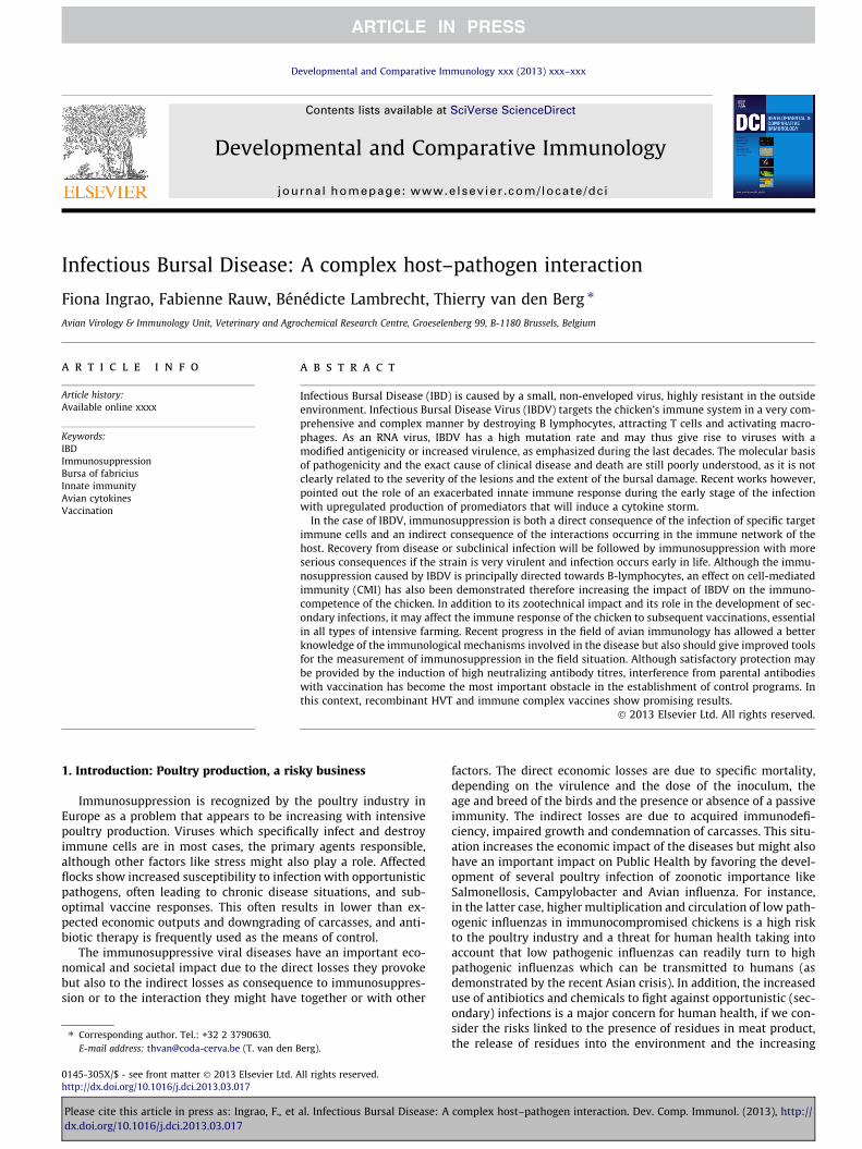
Developmental and Comparative Immunology xxx (2013) xxx–xxx
Contents lists available at SciVerse ScienceDirect
Developmental and Comparative Immunology
journal homepage: www.elsevier .com/locate /dci
Infectious Bursal Disease: A complex host–pathogen interaction
0145-305X/$ - see front matter � 2013 Elsevier Ltd. All rights reserved.http://dx.doi.org/10.1016/j.dci.2013.03.017
⇑ Corresponding author. Tel.: +32 2 3790630.E-mail address: [email protected] (T. van den Berg).
Please cite this article in press as: Ingrao, F., et al. Infectious Bursal Disease: A complex host–pathogen interaction. Dev. Comp. Immunol. (2013),dx.doi.org/10.1016/j.dci.2013.03.017
Fiona Ingrao, Fabienne Rauw, Bénédicte Lambrecht, Thierry van den Berg ⇑Avian Virology & Immunology Unit, Veterinary and Agrochemical Research Centre, Groeselenberg 99, B-1180 Brussels, Belgium
a r t i c l e i n f o a b s t r a c t
Article history:Available online xxxx
Keywords:IBDImmunosuppressionBursa of fabriciusInnate immunityAvian cytokinesVaccination
Infectious Bursal Disease (IBD) is caused by a small, non-enveloped virus, highly resistant in the outsideenvironment. Infectious Bursal Disease Virus (IBDV) targets the chicken’s immune system in a very com-prehensive and complex manner by destroying B lymphocytes, attracting T cells and activating macro-phages. As an RNA virus, IBDV has a high mutation rate and may thus give rise to viruses with amodified antigenicity or increased virulence, as emphasized during the last decades. The molecular basisof pathogenicity and the exact cause of clinical disease and death are still poorly understood, as it is notclearly related to the severity of the lesions and the extent of the bursal damage. Recent works however,pointed out the role of an exacerbated innate immune response during the early stage of the infectionwith upregulated production of promediators that will induce a cytokine storm.
In the case of IBDV, immunosuppression is both a direct consequence of the infection of specific targetimmune cells and an indirect consequence of the interactions occurring in the immune network of thehost. Recovery from disease or subclinical infection will be followed by immunosuppression with moreserious consequences if the strain is very virulent and infection occurs early in life. Although the immu-nosuppression caused by IBDV is principally directed towards B-lymphocytes, an effect on cell-mediatedimmunity (CMI) has also been demonstrated therefore increasing the impact of IBDV on the immuno-competence of the chicken. In addition to its zootechnical impact and its role in the development of sec-ondary infections, it may affect the immune response of the chicken to subsequent vaccinations, essentialin all types of intensive farming. Recent progress in the field of avian immunology has allowed a betterknowledge of the immunological mechanisms involved in the disease but also should give improved toolsfor the measurement of immunosuppression in the field situation. Although satisfactory protection maybe provided by the induction of high neutralizing antibody titres, interference from parental antibodieswith vaccination has become the most important obstacle in the establishment of control programs. Inthis context, recombinant HVT and immune complex vaccines show promising results.
� 2013 Elsevier Ltd. All rights reserved.
1. Introduction: Poultry production, a risky business
Immunosuppression is recognized by the poultry industry inEurope as a problem that appears to be increasing with intensivepoultry production. Viruses which specifically infect and destroyimmune cells are in most cases, the primary agents responsible,although other factors like stress might also play a role. Affectedflocks show increased susceptibility to infection with opportunisticpathogens, often leading to chronic disease situations, and sub-optimal vaccine responses. This often results in lower than ex-pected economic outputs and downgrading of carcasses, and anti-biotic therapy is frequently used as the means of control.
The immunosuppressive viral diseases have an important eco-nomical and societal impact due to the direct losses they provokebut also to the indirect losses as consequence to immunosuppres-sion or to the interaction they might have together or with other
factors. The direct economic losses are due to specific mortality,depending on the virulence and the dose of the inoculum, theage and breed of the birds and the presence or absence of a passiveimmunity. The indirect losses are due to acquired immunodefi-ciency, impaired growth and condemnation of carcasses. This situ-ation increases the economic impact of the diseases but might alsohave an important impact on Public Health by favoring the devel-opment of several poultry infection of zoonotic importance likeSalmonellosis, Campylobacter and Avian influenza. For instance,in the latter case, higher multiplication and circulation of low path-ogenic influenzas in immunocompromised chickens is a high riskto the poultry industry and a threat for human health taking intoaccount that low pathogenic influenzas can readily turn to highpathogenic influenzas which can be transmitted to humans (asdemonstrated by the recent Asian crisis). In addition, the increaseduse of antibiotics and chemicals to fight against opportunistic (sec-ondary) infections is a major concern for human health, if we con-sider the risks linked to the presence of residues in meat product,the release of residues into the environment and the increasing
http://
2 F. Ingrao et al. / Developmental and Comparative Immunology xxx (2013) xxx–xxx
antibiotic resistances. Viral diseases represent the predominantpathology in commercial flocks and vaccination is therefore essen-tial for control. Furthermore, immunosuppression is one of themost common causes for a failure of the general vaccination pro-gram and successful IBD vaccination is considered as a key factorin the establishment of a satisfactory control schedule.
Very recent reviews have focussed on the disease and its caus-ative agent (Maghoub, 2012) and vaccination (Muller et al., 2012).General information can be found in the IBDV specific chapter ofthe 12th edition of Diseases of Poultry (Eterradossi and Saif,2008) and other textbooks. In this review, according to the generaltopic of this special issue, we will concentrate on the immune re-sponse to IBDV that is responsible for the pathogenesis and the ac-quired immunosuppression, with a focus on the host response andits measurement.
Fig. 1. Electron micrograph of negatively stained IBDV. Scale bar 100 nm. One of thenucleocapsids is damaged (arrow) explaining the presence of isolated capsomers(circles). Courtesy of Dr. J. Mast (CODA-CERVA).
2. Short description of the etiological agent: When a virusattacks the defenses of the host
Infectious Bursal Disease (IBD), or Gumboro disease, is a viralinfection that was described for the first time in the 60’s in Gum-boro, Delaware, United States (Eterradossi and Saif, 2008) andnow occurs worldwide. Two serotypes have been recognized sofar by the use of monospecific neutralizing antisera: serotype 1causing disease in chicken and serotype 2 which is apathogenic(McFerran et al., 1980). Only chickens (Gallus gallus), develop aninfectious and highly contagious disease after infection with path-ogenic serotype 1 virus.
IBDV is highly infectious in young chickens, and is characterizedby the destruction of the lymphoid organs, and in particular, theBursa of Fabricius (BF), which is the site where B-lymphocyte mat-uration and differentiation occurs in birds. Indeed, the target cell isthe B lymphocyte in an immature stage, and the infection, whennot fatal, causes an immunosuppression, in most cases temporary,the degree of which is often difficult to determine. Until 1987, theisolated strains were of low virulence and caused only 1–2% of spe-cific mortality in the field. As of 1987, however, an increase in spe-cific mortality was described in different parts of the world. In theUnited States, strains with new antigenic properties and responsi-ble for up to 5% specific mortality were described (Jackwood andSaif, 1987). At the same time, in Europe and subsequently in Japan,mortality rates of up to 100% in SPF (specific pathogen-free) chick-ens, 50–60% in laying hens, and 25–30% in broilers were observedand reproduced in isolators, due to very virulent (vvIBDV) strainsisolated in the field (Chettle et al., 1989; Nunoya et al., 1992; vanden Berg et al., 1991). These strains have now spread all over theworld, with the exception of Australia. The first cases of vvIBDVinfections in the United States were only identified at the end of2008 (Stoute et al., 2009) but apparently, they did not spreadthroughout California.
The virus responsible for IBD is a member of the family Birnavi-ridae, within the Avibirnavirus genus (Muller et al., 1979). It is non-enveloped with a single-shelled icosahedral capsid and has a diam-eter between 55 and 60 nm (Fig. 1). This relatively simple structureconfers the virus high resistance in the outside environment, andrepresents a key issue in the control of the disease. The genomeof the virus consists of two segments, A and B, of double-strandedRNA. Segment A encodes the viral structural proteins VP2, VP3 andVP4 as well as VP5 a protein of regulatory function. As the externalcapsid protein, VP2 elicits neutralizing antibody and represents themolecular basis for antigenicity with variation in the encodingnucleotide sequences resulting in antigenic variants. As the inter-nal capsid protein, VP3 induces group-specific antibodies. SegmentB encodes VP1, the viral polymerase, involved in replication andtranscription of the virus and thus plays a role in the virulence of
Please cite this article in press as: Ingrao, F., et al. Infectious Bursal Disease: Adx.doi.org/10.1016/j.dci.2013.03.017
the virus (for a review on the viral structure and gene organization,see Coulibaly et al., 2005; Delmas et al., 2004; van den Berg, 2000).
The molecular basis for IBDV pathogenicity is still poorly under-stood. To illustrate this, nine strains of IBDV, isolated at differenttimes and from different geographic regions of Europe and China,were characterized. Batches of all strains were prepared followingstandardized protocols and checked for the absence of contaminat-ing viruses. Criteria used for their characterization were: (i) thenucleotide sequence of the VP2 variable region, (ii) binding to a pa-nel of neutralizing monoclonal antibodies in antigen capture en-zyme-linked immunosorbent assays (ELISA), and (iii) virulence inspecific pathogen free chickens after infection with a standardizednumber of median embryo infective doses. Based on the first twocriteria, two of nine strains were classified as classical virulent(cv) IBDV and five as very virulent (vv) IBDV strains. Remarkably,although a clear-cut difference was demonstrable between Euro-pean cvIBDV and vvIBDV strains, there was a continuum in thepathogenicity of Chinese vvIBDVs. These results indicated thelikely existence of differences in virulence within IBDV lineagesdetermined on the basis of antigenic typing using monoclonal anti-bodies and the alignment of the VP2 sequences. This emphasizedlimitation in the analysis of IBDV pathotypes based on the VP2gene and indicated that virulence is a polygenic trait (van den Berget al., 2004).
The polygenic nature of IBDV pathogenicity has been furtherdemonstrated by the isolation of a vvIBDV strain with reducedpathogenicity in a rare natural segment-B-reassorted isolate (LeNouen et al., 2006) and, by using reverse genetics, a clear role ofthe VP1 polymerase could be established confirming that both gen-ome segments influence vvIBDV pathogenicity and may providenew targets for the attenuation of vvIBDVs (Escaffre et al., 2012;Le Nouën et al., 2012). Interestingly, the latter work suggested pos-sible interactions between VP1 and another, as yet unidentified,host molecule.
Recent progress regarding our understanding of IBDV epidemi-ology have illustrated that it is probably only a matter of time untilvvIBDVs are replaced by an emerging strain with new antigenic orpathotypic properties. This raises the question of possible sourcesfor the introduction of new IBDV strains.
3. Pathogenesis: A growing evidence for a role ofproinflammatory cytokines in acute IBD
Pathogenesis can be defined as the method used by IBDVto cause injury to the host with mortality, disease and/or
complex host–pathogen interaction. Dev. Comp. Immunol. (2013), http://
F. Ingrao et al. / Developmental and Comparative Immunology xxx (2013) xxx–xxx 3
immunosuppression as a consequence. These injuries can be eval-uated at different levels: the host, the organ and the cell, and areexacerbated in the acute forms of the disease. The main character-istic of vvIBDVs is their increased virulence. A better understandingof the mechanisms of pathogenicity is thus essential for the controlof the disease.
Host range. IBDV is highly host specific. The selected host forIBDV is the young chicken where a clinical disease occurs whilstin older birds the infection is essentially subclinical. As alreadymentioned, only chickens (Gallus gallus) develop IBD after infectionby serotype 1 viruses and inoculation of IBDV in other avian spe-cies fails to induce disease. Turkeys (Meleagris gallopavo) and Pek-ing ducks (Anas peking) may be asymptomatic carriers of serotype1 viruses whose pathogenicity is ill defined (McFerran et al., 1980).Experimental inoculation of game birds (quails, partridges, pheas-ants and guinea fowls) with serotype 1 viruses failed to induce anyclinical sign or disease, even when a very virulent IBDV strain wasused for challenge (van den Berg et al., 2001).
Varying susceptibility of different chicken breeds has been de-scribed with higher mortality rates in light than in heavier breeds(Bumstead et al., 1993; Nielsen et al., 1998). Studies with vvIBDVdemonstrated the exacerbated susceptibility of Leghorn-type SPFchickens (Rauw et al., 2007). More recent studies have confirmeda significant influence of chicken’s genetic background on IBD out-come and, more closely, its association with the early immune re-sponse during the acute phase after infection with vvIBDV(Aricibasi et al., 2010; Ruby et al., 2006). Furthermore, the differen-tial immunopathogenesis of IBDV was investigated in differentconventional layer and broiler type chickens in comparison tohighly susceptible SPF layers often used for experimental studies.Layer-type chickens of all genetic backgrounds showed signifi-cantly higher IBDV antigen loads in the BF, clinical signs and deathrate compared to broiler type birds (Tippenhauer et al., 2012).
Symptomatology & lesions. Disease severity and clinical signs de-pend on the age and sensitivity of the infected birds, the virulenceof the strain, and the degree of passive immunity transmitted bythe parents. Initial infection in a given farm is generally very acute,with very high mortality rates if a very virulent strain is involved. Ifthe virus persists on the farm and is transmitted to successiveflocks, the clinical forms of the disease appear earlier and are thengradually replaced by subclinical forms. Nonetheless, acute epi-sodes may still occur (van den Berg, 2000). Moreover, a primaryinfection may also be unapparent if maternal antibodies are pres-ent. Acute IBDV infections are characterized by severe clinical signsand high mortality. Indeed, vvIBDVs produce disease signs similarto classical type 1 infections but the acute phase is exacerbated andmore generalized in the affected flock. The incubation period isvery short: 2–3 days. In acute cases, the animals are exhausted,prostrated, dehydrated, suffer from aqueous diarrhea, and theirfeathers are ruffled. Mortality commences on the third day of infec-tion, reaches a peak, then drops rapidly, and the surviving chickensrecover a state of apparent health after 5–7 days. In the case ofvvIBDV infection, the incubation period seems to be shortened(60 h instead of 72) which would be consistent with a faster repli-cation in the affected animals (van den Berg, unpublished observa-tions). The age susceptibility is extended, covering the entiregrowing period in broilers and the peaks of mortality show sharpdeath curves followed by rapid recovery (Chettle et al., 1989; Nu-noya et al., 1992; Tsukamoto et al., 1992; van den Berg et al., 1991).
Compared with classical virulent strains, vvIBDVs induce highermortality rates in fully susceptible chickens. But the exact cause ofclinical disease and death is still unknown, as it is not clearly re-lated to the severity of the lesions and the extent of the bursaldamage. Indeed, after infection, some birds with few bursal lesionscan be found dead while others can survive despite extensive bur-sal damage. Moreover, mortality rates are often variable and the
Please cite this article in press as: Ingrao, F., et al. Infectious Bursal Disease: Adx.doi.org/10.1016/j.dci.2013.03.017
establishment of the lethal dose 50 (LD50) for the standardizationof challenge has always been difficult. This variability must be re-lated to host factors.
On post mortem examination of birds that died during the acutephase of vvIBD, the BF is the principal diagnostic organ: it is turgid,oedematous and sometimes haemorrhagic and turns atrophicwithin 7–10 days. In addition, dehydration and nephrosis withswollen kidneys are common and ecchymotic hemorrhages inthe muscle and the mucosa of the proventriculus are observed inmany affected birds (Eterradossi and Saif, 2008). The damage istransient and the structure of the BF is restored and repopulatedwith lymphocytes; the duration of the recovery process dependson the age at infection and the virulence of the strain (Sharmaet al., 2000; Vervelde and Davison, 1997). Depletion of lymphoidcells is observed not only in the BF but also in the non-bursal lym-phoid tissues. Pathogenicity of vvIBDV has been associated withvirus distribution in the BF but also in non-bursal lymphopoieticand hematopoietic organs. Indeed, using various immunostainingmethods, a higher frequency of antigen-positive cells could bedemonstrated after infection of birds with vvIBDV than with otherstrains, in the thymus (Inoue et al., 1994; Nunoya et al., 1992;Sharma et al., 1993), the spleen and the bone marrow (Inoueet al., 1999; Tanimura et al., 1995; Tsukamoto et al., 1995). In par-ticular, atrophy of the thymus has been associated with the acutephase of the disease and might be indicative of the virulence ofthe isolate, although it is not associated with extensive viral repli-cation in thymic cells (Sharma et al., 1993).
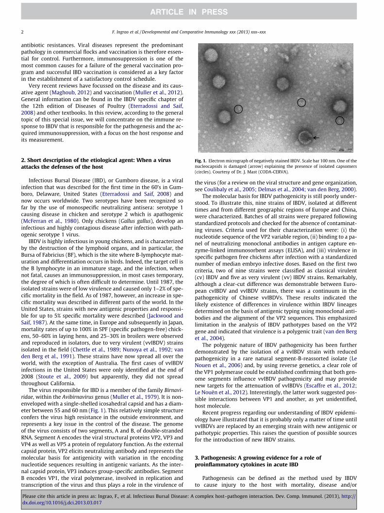
Target cells. All compartments of the bird’s immune system willbe affected during infection with IBDV. IBDV targets the chicken’simmune cells in a very comprehensive and complex manner bydestroying B lymphocytes, attracting T cells and activating macro-phages (Fig. 2).
The target organ for IBDV is the Bursa of Fabricius (BF) at itsmaximum development, which is a specific source for mature B-lymphocytes in avian species. Bursectomy can prevent illness inchicks infected with virulent virus (Hiraga et al., 1994). The sever-ity of the disease is directly related to the number of susceptiblecells present in the BF; therefore, the highest age susceptibility isbetween 3 and 6 weeks, when the BF is at its maximum rate ofdevelopment. This age susceptibility is extended in the case ofvvIBDV infection (Nunoya et al., 1992; van den Berg et al., 1991).Depletion of lymphoid B cells in the Bursa of Fabricius after IBDVinfection is due to both necrosis and apoptosis. Actively dividing,surface immunoglobulin M-bearing B cells are lysed by IBDV infec-tion (Hirai and Calnek, 1979; Hirai et al., 1981; Rodenberg et al.,1994). Apoptosis, characterized by nuclear fragmentation and cel-lular breakdown into apoptotic vesicles also plays an importantrole in IBDV pathogenesis. A high level of apoptosis can be evi-denced in peripheral blood lymphocytes of chickens infected withserotype 1 IBDV (Vasconcelos and Lam, 1994). Very virulent strainscausing increased pathology and earlier mortality also induce alsoa higher level of chIFN-c mRNA in bursal tissue (Eldaghayes et al.,2006; Liu et al., 2010). The viral proteins VP2 and especially VP5have been suspected to play a crucial role in IBDV replication byinducing cell death (Fernandez-Arias et al., 1997; Liu and Vakharia,2006). It was shown in vitro that IBDV infection activates effectorcaspase 3 and the initiation caspase 9 as well as nuclear factor-jB (NFjB), likely through the accumulation of oxygen reactive spe-cies, resulting in apoptosis late in the infective cycle (Liu andVakharia, 2006). More recently, VP5 was confirmed to be a majorapoptosis inducer by interacting with the voltage-dependent anionchannel 2 (VDAC2) in the mitochondrion (Li et al., 2012). Addition-ally, the RNA-binding VP3 polypeptide likely ensures the continu-ity of the IBDV replication cycle by inhibiting PKR-mediatedapoptosis (Busnadiego et al., 2012). On the other hand, apoptosishas also been observed in viral antigen-negative bursal cells
complex host–pathogen interaction. Dev. Comp. Immunol. (2013), http://
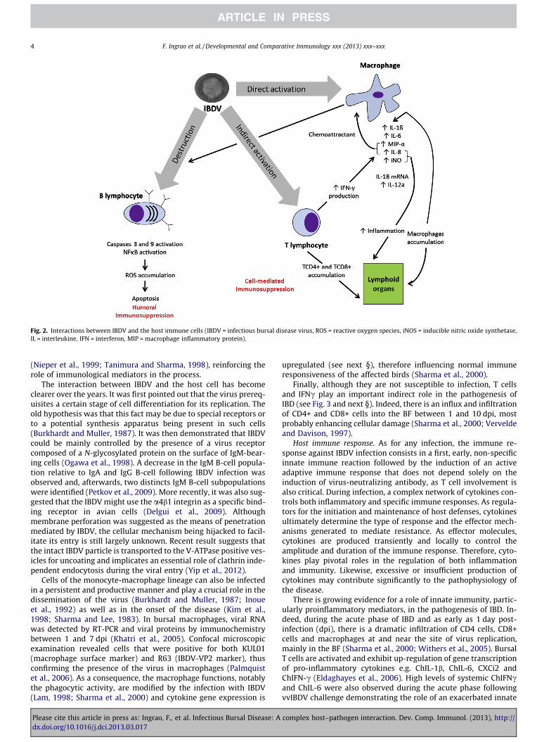
Fig. 2. Interactions between IBDV and the host immune cells (IBDV = infectious bursal disease virus, ROS = reactive oxygen species, iNOS = inducible nitric oxide synthetase,IL = interleukine, IFN = interferon, MIP = macrophage inflammatory protein).
4 F. Ingrao et al. / Developmental and Comparative Immunology xxx (2013) xxx–xxx
(Nieper et al., 1999; Tanimura and Sharma, 1998), reinforcing therole of immunological mediators in the process.
The interaction between IBDV and the host cell has becomeclearer over the years. It was first pointed out that the virus prereq-uisites a certain stage of cell differentiation for its replication. Theold hypothesis was that this fact may be due to special receptors orto a potential synthesis apparatus being present in such cells(Burkhardt and Muller, 1987). It was then demonstrated that IBDVcould be mainly controlled by the presence of a virus receptorcomposed of a N-glycosylated protein on the surface of IgM-bear-ing cells (Ogawa et al., 1998). A decrease in the IgM B-cell popula-tion relative to IgA and IgG B-cell following IBDV infection wasobserved and, afterwards, two distincts IgM B-cell subpopulationswere identified (Petkov et al., 2009). More recently, it was also sug-gested that the IBDV might use the a4b1 integrin as a specific bind-ing receptor in avian cells (Delgui et al., 2009). Althoughmembrane perforation was suggested as the means of penetrationmediated by IBDV, the cellular mechanism being hijacked to facil-itate its entry is still largely unknown. Recent result suggests thatthe intact IBDV particle is transported to the V-ATPase positive ves-icles for uncoating and implicates an essential role of clathrin inde-pendent endocytosis during the viral entry (Yip et al., 2012).
Cells of the monocyte-macrophage lineage can also be infectedin a persistent and productive manner and play a crucial role in thedissemination of the virus (Burkhardt and Muller, 1987; Inoueet al., 1992) as well as in the onset of the disease (Kim et al.,1998; Sharma and Lee, 1983). In bursal macrophages, viral RNAwas detected by RT-PCR and viral proteins by immunochemistrybetween 1 and 7 dpi (Khatri et al., 2005). Confocal microscopicexamination revealed cells that were positive for both KUL01(macrophage surface marker) and R63 (IBDV-VP2 marker), thusconfirming the presence of the virus in macrophages (Palmquistet al., 2006). As a consequence, the macrophage functions, notablythe phagocytic activity, are modified by the infection with IBDV(Lam, 1998; Sharma et al., 2000) and cytokine gene expression is
Please cite this article in press as: Ingrao, F., et al. Infectious Bursal Disease: Adx.doi.org/10.1016/j.dci.2013.03.017
upregulated (see next §), therefore influencing normal immuneresponsiveness of the affected birds (Sharma et al., 2000).
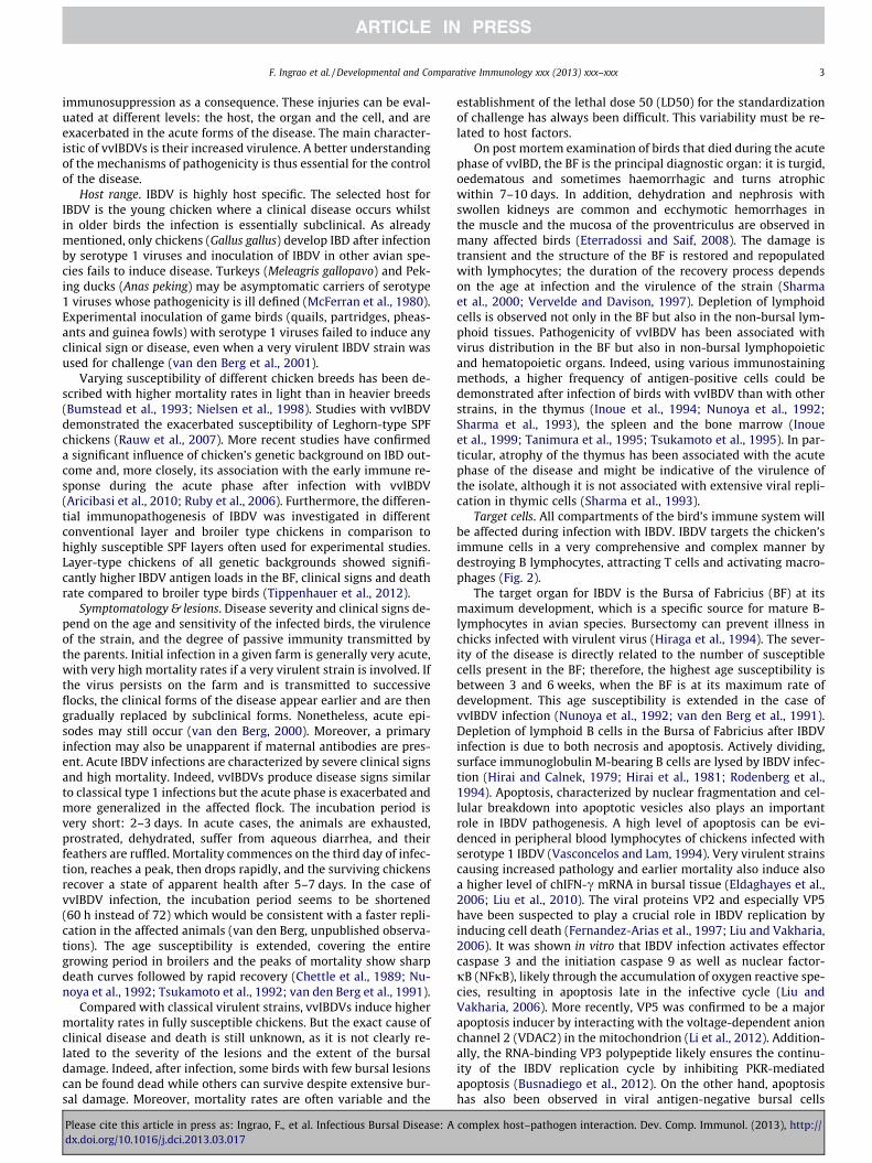
Finally, although they are not susceptible to infection, T cellsand IFNc play an important indirect role in the pathogenesis ofIBD (see Fig. 3 and next §). Indeed, there is an influx and infiltrationof CD4+ and CD8+ cells into the BF between 1 and 10 dpi, mostprobably enhancing cellular damage (Sharma et al., 2000; Verveldeand Davison, 1997).
Host immune response. As for any infection, the immune re-sponse against IBDV infection consists in a first, early, non-specificinnate immune reaction followed by the induction of an activeadaptive immune response that does not depend solely on theinduction of virus-neutralizing antibody, as T cell involvement isalso critical. During infection, a complex network of cytokines con-trols both inflammatory and specific immune responses. As regula-tors for the initiation and maintenance of host defenses, cytokinesultimately determine the type of response and the effector mech-anisms generated to mediate resistance. As effector molecules,cytokines are produced transiently and locally to control theamplitude and duration of the immune response. Therefore, cyto-kines play pivotal roles in the regulation of both inflammationand immunity. Likewise, excessive or insufficient production ofcytokines may contribute significantly to the pathophysiology ofthe disease.
There is growing evidence for a role of innate immunity, partic-ularly proinflammatory mediators, in the pathogenesis of IBD. In-deed, during the acute phase of IBD and as early as 1 day post-infection (dpi), there is a dramatic infiltration of CD4 cells, CD8+cells and macrophages at and near the site of virus replication,mainly in the BF (Sharma et al., 2000; Withers et al., 2005). BursalT cells are activated and exhibit up-regulation of gene transcriptionof pro-inflammatory cytokines e.g. ChIL-1b, ChIL-6, CXCi2 andChIFN-c (Eldaghayes et al., 2006). High levels of systemic ChIFNcand ChIL-6 were also observed during the acute phase followingvvIBDV challenge demonstrating the role of an exacerbated innate
complex host–pathogen interaction. Dev. Comp. Immunol. (2013), http://
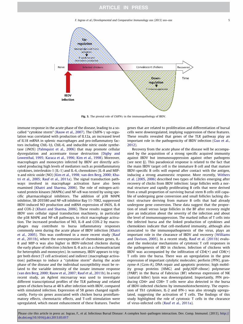
Fig. 3. The pivotal role of ChIFNc in the immunopathology of IBDV.
F. Ingrao et al. / Developmental and Comparative Immunology xxx (2013) xxx–xxx 5
immune response in the acute phase of the disease, leading to a so-called ‘‘cytokine storm’’ (Rauw et al., 2007). The ChIFN-c up-regu-lation was correlated with production of IL12a, an increased levelof IL18 mRNA in splenic macrophages and pro-inflammatory fac-tors including ChIL-1b, ChIL-6, and inducible nitric oxide synthe-tase (iNOS) (Palmquist et al., 2006) that may promote cellulardysregulation and accentuate tissue destruction (Digby andLowenthal, 1995; Karaca et al., 1996; Kim et al., 1998). Moreover,macrophages and monocytes infected by IBDV are directly acti-vated producing high levels of mediators such as proinflammatorycytokines, interleukin-1 (IL-1) and IL-6, chemokines (IL-8 and MIP-a and nitric oxide (NO) (Kim et al., 1998; van den Berg, 2000; Kha-tri et al., 2005; Rauf et al., 2011a). The signal transduction path-ways involved in macrophage activation have also beenexamined (Khatri and Sharma, 2006). The role of mitogen-acti-vated protein kinases (MAPKs) and NF-jB was tested by using spe-cific pharmacological inhibitors. The addition of p38 MAPKinhibitor, SB-203580 and NF-jB inhibitor Bay 11-7082, suppressedIBDV-induced NO production and mRNA expression of iNOS, IL-8and COX-2 (Khatri and Sharma, 2006). These results suggest thatIBDV uses cellular signal transduction machinery, in particularthe p38 MAPK and NF-jB pathways, to elicit macrophage activa-tion. The increased production of NO, IL-8 and COX-2 by macro-phages may contribute to bursa inflammatory responsescommonly seen during the acute phase of IBDV infection (Khatriet al., 2005). This was confirmed in a more recent study (Raufet al., 2011b), where the overexpression of chemokines genes, IL-8 and MIP-a was also higher in IBDV-infected chickens duringthe early phase of infection (chicken IL-8 acts as a chemoattractantfor heterophils and monocytes). In summary, IBDV appears to trig-ger both direct (T cell activation) and indirect (macrophage activa-tion) pathways to induce a ‘‘cytokine storm’’ during the acutephase of the disease and the individual susceptibility must be re-lated to the variable intensity of the innate immune response(van den Berg, 2000; Rauw et al., 2007; Rauf et al., 2011b). In a veryrecent study, an Agilent microarray was used to investigatedifferent transcriptional profiles of the TLR pathway and relatedgenes of chicken bursa at 48 h after infection with IBDV, comparedwith simulated infection. Expression of 58 genes changed signifi-cantly. Forty-six genes associated with chicken bursa proinflam-matory effects, chemotactic effects, and T-cell stimulation wereupregulated, which meant enhancement of these features. Twelve
Please cite this article in press as: Ingrao, F., et al. Infectious Bursal Disease: Adx.doi.org/10.1016/j.dci.2013.03.017
genes that are related to proliferation and differentiation of bursalcells were downregulated, implying suppression of these features.These results revealed that genes of the TLR pathway play animportant role in the pathogenicity of IBDV infection (Guo et al.,2012).
Recovery from the acute phase of the disease will be accompa-nied by the acquisition of a strong specific acquired immunityagainst IBDV but immunosuppression against other pathogens(see next §). This paradoxical response is related to the fact thatthe main IBDV target cell is the immature B cell and that matureIBDV-specific B cells will expend after contact with the antigen,inducing a strong anamnestic response. More recently, Witherset al. (2005, 2006) described two types of follicles emerging afterrecovery of chicks from IBDV infection: large follicles with a nor-mal structure and rapidly proliferating B cells that were derivedfrom a small proportion of surviving bursal stem B cells still capa-ble of undergoing gene conversion and small follicles lacking dis-tinct structure deriving from mature B cells that had alreadyundergone gene conversion. These data suggest that the propor-tion of small versus large follicles in the BF after recovery mightgive an indication about the severity of the infection and aboutthe level of immunosuppression. The marked influx of T cells intothe infected bursa and the linked production of cytokines andchemokines indicate that cell-mediated immunity, although alsoassociated to the immunopathogenesis of the virus, plays animportant role in the clearance of IBDV and recovery (Williamsand Davison, 2005). In a recent study, Rauf et al. (2011b) evalu-ated the molecular mechanisms of cytotoxic T cell responses inthe pathogenesis of IBD in chickens. Infection of chickens withIBDV was accompanied by the infiltration of CD4(+) and CD8(+)T cells into the bursa. There was an upregulation in the geneexpression of important cytolytic molecules; perforin (PFN), gran-zyme-A (Gzm-A), DNA repair and apoptotic proteins; high mobil-ity group proteins (HMG) and poly(ADP-ribose) polymerase(PARP) in the Bursa of Fabricius (BF) whereas expression of NK(natural killer) lysin was downregulated. Importantly, PFN pro-ducing CD4+ and CD8+ T cells were also detected in the bursaof IBDV-infected chickens by immunohistochemistry. The expres-sion of Th1 cytokines, IL-2 and IFN-c was also strongly upregu-lated, suggesting the activation of T cells. The findings of thisstudy highlighted the role of cytotoxic T cells in the clearanceof virus-infected cells (Rauf et al., 2011a).
complex host–pathogen interaction. Dev. Comp. Immunol. (2013), http://
6 F. Ingrao et al. / Developmental and Comparative Immunology xxx (2013) xxx–xxx
4. Immunosuppression: The hidden enemy
Immunosuppression has been defined as a ‘‘state of temporaryor permanent dysfunction of the immune response resulting fromdamage to the immune system and leading to increased suscepti-bility to disease’’ (Dohms and Saif, 1984) and often a suboptimalantibody response (Lutticken, 1997). However, this definition con-siders more the consequences (increased disease incidence) thanthe causes, of which mechanisms, beyond the destruction of a spe-cific cell type, are still not fully understood.
The destruction of the BF by IBDV creates an immunosuppres-sion, which will be all the more serious the younger the infectedbird. In addition to its zootechnical impact and its role in the devel-opment of secondary infections, it may affect the immune responseof the chicken to subsequent vaccinations, essential in all types ofintensive farming.
The immunosuppression has been most often evidenced usingexperimental models based on the measurement of humoral re-sponses induced by different antigens such as Brucella abortus,sheep red blood cells, or Newcastle disease vaccines (Allan et al.,1972; Giambrone et al., 1976; Giambrone, 1979). The best assess-ment is clearly the measurement of vaccinal protection against achallenge infection by the Newcastle virus as described in theOIE Manual of Standards for Diagnostic Tests and Vaccines since itconstitutes a measurement of both humoral and cellular immu-nity. However, they only give a partial picture of the immunosup-pression as, according to the clonal nature of immunity, it willdepend on the number of NDV-specific clones that will be de-stroyed. Using such tests, the most serious and longest-lastingimmunosuppression was described when day-old chicks were
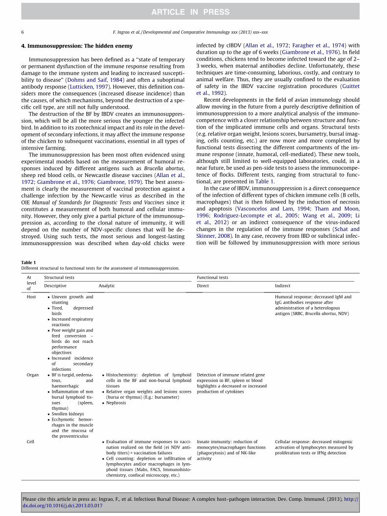
Table 1Different structural to functional tests for the assessment of immunosuppression.
Atlevelof
Structural tests
Descriptive Analytic
Host � Uneven growth andstunting� Tired, depressed
birds� Increased respiratory
reactions� Poor weight gain and
feed conversion –birds do not reachperformanceobjectives� Increased incidence
of secondaryinfections
Organ � BF is turgid, oedema-tous, andhaemorrhagic� Inflammation of non
bursal lymphoid tis-sues (spleen,thymus)� Swollen kidneys� Ecchymotic hemor-
rhages in the muscleand the mucosa ofthe proventriculus
� Histochemistry: depletion of lymphoidcells in the BF and non-bursal lymphoidtissues� Relative organ weights and lesions scores
(bursa or thymus) (E.g.: bursameter)� Nephrosis
Cell � Evaluation of immune responses to vacci-nation realized on the field (ei NDV anti-body titers) > vaccination failures� Cell counting: depletion or infiltration of
lymphocytes and/or macrophages in lym-phoid tissues (Mabs, FACS, Immunohisto-chemistry, confocal microscopy, etc.)
Please cite this article in press as: Ingrao, F., et al. Infectious Bursal Disease: Adx.doi.org/10.1016/j.dci.2013.03.017
infected by cIBDV (Allan et al., 1972; Faragher et al., 1974) withduration up to the age of 6 weeks (Giambrone et al., 1976). In fieldconditions, chickens tend to become infected toward the age of 2–3 weeks, when maternal antibodies decline. Unfortunately, thesetechniques are time-consuming, laborious, costly, and contrary toanimal welfare. Thus, they are usually confined to the evaluationof safety in the IBDV vaccine registration procedures (Guittetet al., 1992).
Recent developments in the field of avian immunology shouldallow moving in the future from a purely descriptive definition ofimmunosuppression to a more analytical analysis of the immuno-competence with a closer relationship between structure and func-tion of the implicated immune cells and organs. Structural tests(e.g. relative organ weight, lesions scores, bursametry, bursal imag-ing, cells counting, etc.) are now more and more completed byfunctional tests dissecting the different compartments of the im-mune response (innate, humoral, cell-mediated). These new tools,although still limited to well-equipped laboratories, could, in anear future, be used as pen-side tests to assess the immunocompe-tence of flocks. Different tests, ranging from structural to func-tional, are presented in Table 1.
In the case of IBDV, immunosuppression is a direct consequenceof the infection of different types of chicken immune cells (B cells,macrophages) that is then followed by the induction of necrosisand apoptosis (Vasconcelos and Lam, 1994; Tham and Moon,1996; Rodriguez-Lecompte et al., 2005; Wang et al., 2009; Liet al., 2012) or an indirect consequence of the virus-inducedchanges in the regulation of the immune responses (Schat andSkinner, 2008). In any case, recovery from IBD or subclinical infec-tion will be followed by immunosuppression with more serious
Functional tests
Direct Indirect
Humoral response: decreased IgM andIgG antibodies response afteradministration of a heterologousantigen (SRBC, Brucella abortus, NDV)
Detection of immune related geneexpression in BF, spleen or bloodhighlights a decreased or increasedproduction of cytokines
Innate immunity: reduction ofmonocytes/macrophages functions(phagocytosis) and of NK-likeactivity
Cellular response: decreased mitogenicactivation of lymphocytes measured byproliferation tests or IFNg detection
complex host–pathogen interaction. Dev. Comp. Immunol. (2013), http://
F. Ingrao et al. / Developmental and Comparative Immunology xxx (2013) xxx–xxx 7
consequences if the strain is very virulent and infection occursearly in life.
Although the immunosuppression caused by IBDV is principallydirected towards B-lymphocytes, an indirect effect on cell-medi-ated immunity (CMI) has also been demonstrated (Cloud et al.,1992a,b; Sharma and Fredericksen, 1987; Sharma et al., 1989)therefore increasing the impact of IBDV on the immunocompe-tence of the chicken. The infiltrated T cells constituting the major-ity of the bursal population after IBDV infection are unresponsiveto mitogen activation at days 4 and 9 days pi (McNeilly et al.,1999; Rauw et al., 2007). Moreover splenocytes from IBDV-infectedchickens were also shown to be deficient in secretion of ChIL-2(Kim et al., 1998; Sharma and Fredericksen, 1987). It is now wellaccepted that macrophages are the central effector cells of the in-nate immune system and influence the nature of the adaptive im-mune response. In a study performed at 3 and 5 dpi, spleens ofvirus-exposed chickens had fewer macrophages than those ofvirus-free controls; the robust expression of proinflammatorycytokine transcripts, along with a decrease in macrophage num-bers, suggest that IBDV activates and may lead to a reduction ofresident macrophages in vivo (Palmquist et al., 2006). However, itremains unclear how these changes play a role in immunosuppres-sion (Schat and Skinner, 2008). Mechanisms like the developmentof suppressor macrophages and the impairment of helper T-cellshave been suggested to explain this clear impairment of the activa-tion capability (Sharma and Fredericksen, 1987; Vervelde and Dav-ison, 1997). As described previously, IBDV is a potent inductor ofChIFNc production, which has been demonstrated as able to inhibitthe in vitro mitogenic proliferation of T cells (Sharma et al., 2000)and ChIFNc production of splenocytes from naive chickens (Rauwet al., 2007). In mice, it has been shown that IFNc is responsible forthe decrease of IL-2 production by splenocytes (Gajewski et al.,1988; Bradley et al., 1996). Thus, IBDV modulates T cell functions:activated T cells will overproduce ChIFN-c that, in turn, will stim-ulate macrophages for the production of pro-chemokines and cyto-kines. Afterwards, this up-regulation in T cell activation is followedby a feedback down-regulation in convalescent chickens, leadingto T cell immunosuppression, of which the duration and impor-tance will depend on the strain and dose of virus as well as onthe age and breed susceptibility of the chickens. It can thus reason-ably be concluded that overproduction of ChIFN-c plays a pivotal
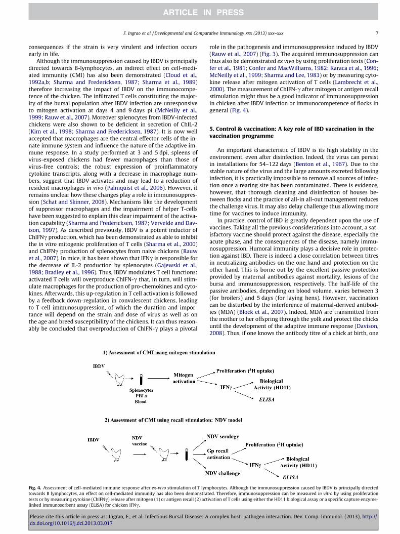
Fig. 4. Assessment of cell-mediated immune response after ex-vivo stimulation of T lymtowards B lymphocytes, an effect on cell-mediated immunity has also been demonstrattests or by measuring cytokine (ChIFNc) release after mitogen (1) or antigen recall (2) actilinked immunosorbent assay (ELISA) for chicken IFNc.
Please cite this article in press as: Ingrao, F., et al. Infectious Bursal Disease: Adx.doi.org/10.1016/j.dci.2013.03.017
role in the pathogenesis and immunosuppression induced by IBDV(Rauw et al., 2007) (Fig. 3). The acquired immunosuppression canthus also be demonstrated ex vivo by using proliferation tests (Con-fer et al., 1981; Confer and MacWilliams, 1982; Karaca et al., 1996;McNeilly et al., 1999; Sharma and Lee, 1983) or by measuring cyto-kine release after mitogen activation of T cells (Lambrecht et al.,2000). The measurement of ChIFN-c after mitogen or antigen recallstimulation might thus be a good indicator of immunosuppressionin chicken after IBDV infection or immunocompetence of flocks ingeneral (Fig. 4).
5. Control & vaccination: A key role of IBD vaccination in thevaccination programme
An important characteristic of IBDV is its high stability in theenvironment, even after disinfection. Indeed, the virus can persistin installations for 54–122 days (Benton et al., 1967). Due to thestable nature of the virus and the large amounts excreted followinginfection, it is practically impossible to remove all sources of infec-tion once a rearing site has been contaminated. There is evidence,however, that thorough cleaning and disinfection of houses be-tween flocks and the practice of all-in all-out management reducesthe challenge virus. It may also delay challenge thus allowing moretime for vaccines to induce immunity.
In practice, control of IBD is greatly dependent upon the use ofvaccines. Taking all the previous considerations into account, a sat-isfactory vaccine should protect against the disease, especially theacute phase, and the consequences of the disease, namely immu-nosuppression. Humoral immunity plays a decisive role in protec-tion against IBD. There is indeed a close correlation between titresin neutralizing antibodies on the one hand and protection on theother hand. This is borne out by the excellent passive protectionprovided by maternal antibodies against mortality, lesions of thebursa and immunosuppression, respectively. The half-life of thepassive antibodies, depending on blood volume, varies between 3(for broilers) and 5 days (for laying hens). However, vaccinationcan be disturbed by the interference of maternal-derived antibod-ies (MDA) (Block et al., 2007). Indeed, MDA are transmitted fromthe mother to her offspring through the yolk and protect the chicksuntil the development of the adaptive immune response (Davison,2008). Thus, if one knows the antibody titre of a chick at birth, one
phocytes. Although the immunosuppression caused by IBDV is principally directeded. Therefore, immunosuppression can be measured in vitro by using proliferationvation of T cells using either the HD11 biological assay or a specific capture enzyme-
complex host–pathogen interaction. Dev. Comp. Immunol. (2013), http://
8 F. Ingrao et al. / Developmental and Comparative Immunology xxx (2013) xxx–xxx
can determine the time of maximum susceptibility to the vaccine.This determination is very important when establishing vaccina-tion programs (De Herdt et al., 2005). Neutralizing antibodies areprotective and these can be provided by immunizing chickens withlive attenuated vaccines given in the drinking water. In addition, alive vaccine will induce a strong CMI response, the role of which isstill unclear but important. Indeed, protection in the absence ofvirus-specific antibodies and studies with T cell compromisedchickens have indicated that functional T cells are needed to con-trol IBDV replication during the acute phase of infection (Yehet al., 2002; Rautenschlein et al., 2002). It has been recently shownthat a dominant fragment of VP2 can induce humoral and cellularimmunity against IBD and elicits a protective immune response inchickens better than the available attenuated viral strains and thuscould be used as or in a vaccine (Pradhan et al., 2012).
To obtain high levels of MDA in the progeny, parent stock arevaccinated between 4 and 10 weeks of age with live vaccine andagain at approximately 16 weeks with inactivated oil-adjuvantedvaccine. In the progeny, the MDA levels wane with time, but mayprotect against virulent challenge up to between 2, 5 and 3,5 weeks of age. Mild vaccine strains that cause no bursal lesionscannot be used effectively in chicks with MDA until about 4 weeksof age as they are neutralized. Moderately virulent vaccine (‘inter-mediate’) strains that are less affected by MDA can be given withsome success as early as 2–3 weeks of age, depending upon MDAtitres. As MDA levels may vary within a flock and between flocksrepeated vaccination is practiced by some in order to ensure thatchicks are actively immunized as soon as the MDA levels havewaned to a level at which they do not neutralize the vaccine. Inter-mediate and hot vaccine strains can induce bursal lesions andcause immunosuppression (Mazariegos et al., 1990). The intensiveuse of this kind of vaccines increased preoccupations about resid-ual pathogenicity and about the decreasing effectiveness of vac-cines against other diseases.
The ideal IBDV vaccine should thus be safe and capable of prim-ing an immune response after a single inoculation in ovo or athatching in the presence of MDA. Different approaches have beeninvestigated to achieve these goals but, so far, only two kinds ofnew generation vaccines have been successfully developed andcommercialized: herpes virus of turkey (HVT) recombinant vectorvaccines and immune complexes.
The first successful new concept was the establishment of im-mune complex vaccines (Icx). These vaccines consist of a mixtureof specific hyperimmune neutralizing antiserum (or ‘‘virus neutral-izing factor’’) with a vaccine virus under conditions that are notsufficient to neutralize the vaccine virus but which are sufficientfor delaying the pathological effects of the vaccine alone. This al-lows young chicks to be vaccinated even with a strain that wouldbe too virulent for use in ovo or at hatching (Whitfill et al., 1995).Other advantages of the Icx vaccines are that they are effective inthe presence of maternally derived antibodies (Giambrone et al.,2001) and can be delivered by subcutaneous injection at 1 dayold in the hatchery (Ivan et al., 2005) or, alternatively when theegg injection equipment is available, they are also suitable for inovo vaccination at day 18 of incubation (Haddad et al., 1997).Although the mechanism of action is still poorly understood, suchIBDV complexing with specific antibodies cause a delay in virusdetection of approximately 5 days, thus decreasing the viral loadin the bursa and therefore, the bursal lesions, with a remarkablelow level of depletion of bursal and splenic B lymphocytes (Ivanet al., 2005; Jeurissen et al., 1998).
The second concept is based on the widely used Marek’s diseasevaccine made of the serotype 3 HVT, which is well known to besafe and poorly sensitive to MDA interference, and this is why itwas developed as a vector for IBD (Le Gros et al., 2009). Indeed,it has been shown in different comparative studies that MDA inter-
Please cite this article in press as: Ingrao, F., et al. Infectious Bursal Disease: Adx.doi.org/10.1016/j.dci.2013.03.017
fere with humoral response in vaccination with intermediate livevaccines but do not have any impact on immunization with HVTrecombinant vector vaccines containing the VP2 sequence (Bublotet al., 2007; Le Gros et al., 2009; Zorman Rojs et al., 2011). Remark-ably, it was also observed that this vector vaccine provided protec-tion from a challenge with variant IBDV (Perozo et al., 2009). Thisvectored vaccine removes the dilemma of considering safety ver-sus efficacy for IBD vaccination that poultry veterinarians currentlyface with the use of classical IBDV vaccines. Furthermore, it alsoprotects against MD, and its use in the hatchery, where the vacci-nation procedure is very well controlled, reduces the need toadminister vaccine by drinking water on poultry farms.
Other approaches are still in experimental phase and have notbeen brought to practice so far (for review, see Maghoub, 2012;Muller et al., 2012). Among these, other vectors have been engi-neered to express VP2 or the polyprotein VP4-2-3 such as Newcas-tle disease virus (NDV), fowlpox (FPV) or adenovirus but theysuffer from safety or efficacy often related to interference of MDAissues. Indeed, although recombinant vaccines possess the advan-tage of a bivalent vaccination when the vector is a vaccine strain,they have the limitations of the vector in terms of safety and effi-cacy and, if the vector is sensitive to MDA, so will the recombinantbe. In addition, depending on the site of insertion and expression,some interference against the insert might also be observed. Thismight be particularly critical and is probably one of the reasonswhy the only recombinant vaccine commercialized so far is theHVT vector expressing VP2. Likewise, sub-unit vaccines (producedin prokaryotes, yeast or baculovirus) and DNA vaccines haveshown promises but also limitations. Although the first were ex-pected to advantageously replace inactivated vaccines, they cannotbe used for priming and they still suffer from the costs of produc-tion. DNA vaccines are safe and insensitive to MDA but they are notsufficiently immunogenic to replace attenuated vaccines. Indeed,in addition to individual variability in the immune response andgeneral low humoral response, the results showed that a singleDNA delivery without a boost vaccination was not always suffi-cient to induce protective immunity and therefore necessitate mul-tiple administrations or boost with another vaccine, thus raisingthe cost issue.
6. Conclusion: ‘‘There is nothing permanent except change’’(Heraclitus)
A clinical picture of IBD has dominated the field in differentparts of the world since more than two decades, with high mortal-ity rates and considerable economic losses. Antigenically andgenetically homogeneous vvIBDV-like strains have apparentlyspread in most countries and are now frequently isolated world-wide from acute cases. This sudden and dramatic emergence hasstimulated research in IBDV due to the need of new adequate con-trol measures. Important progress has been made in the range ofmolecular virology, epidemiology of the disease, pathogenesisand development of new vaccines.
The role of T cells, macrophages and dendritic cells in the dis-ease has been demonstrated. Particularly, T lymphocytes have re-cently been shown to contribute to the onset of the acutedisease. Although their role in clearance of the virus and recoveryfrom the disease is essential, their function is exacerbated afterinfection with vvIBDVs. Hopefully, current and future research inthe field of avian immunology will allow a better understandingof the immunological mechanisms involved in the disease but alsogive tools for the measurement of immunosuppression in the fieldsituation and, therefore, a better identification of protective crite-ria. This would allow early recognition of problem flocks and willfacilitate further studies to determine the cause of the condition.
complex host–pathogen interaction. Dev. Comp. Immunol. (2013), http://
F. Ingrao et al. / Developmental and Comparative Immunology xxx (2013) xxx–xxx 9
Recent increases in understanding of immune modulation and therapid development of recombinant products for manipulation ofimmune responses could potentially offer a means of treatmentof these conditions, once an accurate diagnosis has been reached.
However, additional research is still needed to overcome someof the current obstacles. Particularly, the identification of a viru-lence marker, through a better understanding of the host–patho-gen interactions and of the underlying molecular mechanisms, isessential. In this regards, the reverse genetic system, providingthe tool to construct chimeric viruses, will be decisive for the iden-tification of virulence markers and the genetic attenuation ofstrains. Likewise, future research needs to be focusing on themolecular mechanisms between viral proteins and host cells, andparticularly cytokine regulation. This should allow the develop-ment of more appropriate control measures in order to afford ahigher degree of protection to young birds that carry maternalimmunity.
References
Allan, W.H., Faragher, J.T., et al., 1972. Immunosuppression by the infectious bursalagent in chickens immunised against Newcastle disease. Vet. Rec. 90 (18), 511–512.
Aricibasi, M., Jung, A., Heller, E.D., Rautenschlein, S., 2010. Differences in geneticbackground influence the induction of innate and acquired immune responsesin chickens depending on the virulence of the infecting infectious bursal diseasevirus (IBDV) strain. Vet. Immunol. Immunopathol. 135, 79–92.
Benton, W.J., Cover, M.S., et al., 1967. Physicochemical properties of the infectiousbursal agent (IBA). Avian Dis. 11 (3), 438–445.
Block, H., Meyer-Block, K., et al., 2007. A field study on the significance ofvaccination against infectious bursal disease virus (IBDV) at the optimal timepoint in broiler flocks with maternally derived IBDV antibodies. Avian Pathol.36 (5), 401–409.
Bradley, L.M., Dalton, D.K., et al., 1996. A direct role for IFN-gamma in regulation ofTh1 cell development. J. Immunol. 157 (4), 1350–1358.
Bublot, M., Pritchard, N., Le Gros, F.X., Goutebroze, S., 2007. Use of vectored vaccineagainst infectious bursal disease of chickens in the face of high-titredmaternally derived antibody. J. Comp. Pathol. 137, S81–S84.
Bumstead, N., Reece, R.L., Cook, J.K.A., 1993. Genetic differences in susceptibility ofchicken lines to infection with infectious bursal disease virus. Poult. Sci. 72 (3),403–410.
Burkhardt, E., Muller, H., 1987. Susceptibility of chicken blood lymphoblasts andmonocytes to infectious bursal disease virus (IBDV). Arch. Virol. 94 (3–4), 297–303.
Busnadiego, I., Maestre, A.M., et al., 2012. The infectious bursal disease virus RNA-binding VP3 polypeptide inhibits PKR-mediated apoptosis. PLoS One 7 (10),e46768.
Chettle, N.J., Stuart, J.C., Wyeth, P., 1989. Outbreaks of virulent infectious bursaldisease in East Anglia. Vet. Rec. 125, 271–272.
Cloud, S.S., Lillehoj, H.S., et al., 1992a. Immune dysfunction following infection withchicken anemia agent and infectious bursal disease virus. I. Kinetic alterationsof avian lymphocyte subpopulations. Vet. Immunol. Immunopathol. 34 (3–4),337–352.
Cloud, S.S., Rosenberger, J.K., et al., 1992b. Immune dysfunction following infectionwith chicken anemia agent and infectious bursal disease virus. II. Alterations ofin vitro lymphoproliferation and in vivo immune responses. Vet. Immunol.Immunopathol. 34 (3–4), 353–366.
Confer, A.W., MacWilliams, P.S., 1982. Correlation of hematological changes andserum and monocyte inhibition with the early suppression ofphytohemagglutinin stimulation of lymphocytes in experimental infectiousbursal disease. Can. J. Comp. Med. 46 (2), 169–175.
Confer, A.W., Springer, W.T., et al., 1981. Sequential mitogen stimulation ofperipheral blood lymphocytes from chickens inoculated with infectious bursaldisease virus. Am. J. Vet. Res. 42 (12), 2109–2113.
Coulibaly, F., Chevalier, C., Gutsche, I., Pous, J., Navaza, J., Bressanelli, S., Delmas, B.,Rey, F.A., 2005. The birnavirus crystal structure reveals structural relationshipsamong icosahedral viruses. Cell 120 (6), 761–772.
Davison, T.F., 2008. The importance of the avian immune system and its uniquefeatures. In: Davison, T.F., Kaspers, B., Schat, K.A. (Eds.), Avian Immunology.Academic press and Elsevier Ltd, Amsterdam, London, pp. 299–322.
De Herdt, P., Jagt, E., Paul, G., Van Colen, S., Renard, R., Destrooper, C., van den Bosch,G., 2005. Evaluation of the enzyme-linked immunosorbent assay for thedetection of antibodies against infectious bursal disease virus (IBDV) and theestimation of the optimal age for IBDV vaccination in broilers. Avian Pathol. 34(6), 501–504.
Delgui, L., Oña, A., et al., 2009. The capsid protein of infectious bursal disease viruscontains a functional a4b1 integrin ligand motif. Virology 386 (2), 360–372.
Delmas, B., Kibenge, F.S.B., Leong, J.C., Mundt, E., Vakharia, V.N., Wu, J.L., 2004.Birnaviridae. In: Fauquet, C.M., Mayo, M.A., Maniloff, J., Desselberger, U., Ball,A.L. (Eds.), Virus Taxonomy. Academic Press, London, pp. 561–569.
Please cite this article in press as: Ingrao, F., et al. Infectious Bursal Disease: Adx.doi.org/10.1016/j.dci.2013.03.017
Dohms, J.E., Saif, Y.M., 1984. Criteria for evaluating immunosuppression. Avian Dis.28 (2), 305–310.
Digby, M.R., Lowenthal, J.W., 1995. Cloning and expression of the chickeninterferon-gamma gene. J. Interferon Cytokine Res. 15 (11), 939–945.
Eldaghayes, I., Rothwell, L., et al., 2006. Infectious bursal disease virus: strains thatdiffer in virulence differentially modulate the innate immune response toinfection in the chicken bursa. Viral Immunol. 19 (1), 83–91.
Escaffre, O., Le Nouen, C., 2012. Both genome segments contribute to thepathogenicity of very virulent infectious bursal disease virus. J. Virol..
Eterradossi, N., Saif, Y.M., 2008. Infectious bursal disease. In: Saif, Y.M., Fadly, A.M.,Glisson, J.R., McDougald, L.R., Nolan, L.K., Swayne, D.E. (Eds.), Disease of Poultry,12th ed. Blackwell Publishing, Ames Iowa, USA, pp. 185–208.
Faragher, J.T., Allan, W.H., et al., 1974. Immunosuppressive effect of infectiousbursal agent on vaccination against Newcastle disease. Vet. Rec. 95 (17), 385–388.
Fernandez-Arias, A., Martinez, S., et al., 1997. The major antigenic protein ofinfectious bursal disease virus, VP2, is an apoptotic inducer. J. Virol. 71 (10),8014–8018.
Gajewski, T.F., Goldwasser, E., et al., 1988. Anti-proliferative effect of IFN-gamma inimmune regulation. II. IFN-gamma inhibits the proliferation of murine bonemarrow cells stimulated with IL-3, IL-4, or granulocyte-macrophage colony-stimulating factor. J. Immunol. 141 (8), 2635–2642.
Giambrone, J.J., Eidson, C.S., et al., 1976. Effect of infectious bursal agent on theresponse of chickens to Newcastle disease and Marek’s disease vaccination.Avian Dis. 20 (3), 534–544.
Giambrone, J.J., 1979. Effects of early infectious bursal disease virus infection onimmunity to Newcastle disease in adult chickens. Poult. Sci. 58 (4), 794–798.
Giambrone, J.J., Dormitorio, T., Brown, T., 2001. Safety and efficacy of in ovoadministration of infectious bursal disease viral vaccines. Avian Dis. 45, 144–148.
Guittet, M., Le Coq, H., Picault, J.P., Eterradossi, N., Bennejean, G., 1992. Safety ofinfectious bursal disease vaccines: assessment of an acceptability threshold.Dev. Biol. Stand. 79, 147–152.
Guo, X., Wang, L., Cui, D., Ruan, W., Liu, F., Li, H., 2012. Differential expression of theToll-like receptor pathway and related genes of chicken bursa afterexperimental infection with infectious bursa disease virus. Arch. Virol. 157,2189–2199.
Haddad, E.E., Whitfill, C.E., Avakian, A.P., et al., 1997. Efficacy of a novel infectiousbursal disease virus immune complex vaccine in broiler chickens. Avian Dis. 41,882–889.
Hiraga, M., Nunoya, T., Otaki, Y., Tajima, M., Saito, T., Nakamura, T., 1994.Pathogenesis of highly virulent infectious bursal disease virus infection inintact and bursectomized chickens. J. Vet. Med. Sci. 56, 1057–1063.
Hirai, K., Calnek, B.W., 1979. In vitro replication of infectious bursal disease virus inestablished lymphoid cell lines and chicken B lymphocytes. Infect. Immun. 25(3), 964–970.
Hirai, K., Funakoshi, T., et al., 1981. Sequential changes in the number of surfaceimmunoglobulin-bearing B lymphocytes in infectious bursal disease virus-infected chickens. Avian Dis. 25 (2), 484–496.
Inoue, M., Yamamoto, H., Matuo, K., Hihara, H., 1992. Susceptibility of chickenmonocytic cell lines to infectious bursal disease virus. J. Vet. Med. Sci. 54, 575–577.
Inoue, M., Fukuda, M., Miyano, K., 1994. Thymic lesions in chicken infected withinfectious bursal disease virus. Avian Dis. 38 (4), 839–846.
Inoue, M., Fujita, A., Maeda, K., 1999. Lysis of myelocytes in chickens infected withinfectious bursal disease virus. Vet. Pathol. 36 (2), 146–151.
Ivan, J., Velhner, M., Ursu, K., et al., 2005. Delayed vaccine virus replication inchickens vaccinated subcutaneously with an immune complex infectious bursaldisease vaccine: quantification of vaccine virus by real-time polymerase chainreaction. Can. J. Vet. Res. 69, 135–142.
Jackwood, D.H., Saif, Y.M., 1987. Antigenic diversity of infectious bursal diseaseviruses. Avian Dis. 31 (4), 766–770.
Jeurissen, S.H., Janse, E.M., Lehrbach, P.R., Haddad, E.E., Avakian, A., Whitfill, C.E.,1998. The working mechanism of an immune complex vaccine that protectschickens against infectious bursal disease. Immunology 95, 494–500.
Karaca, K., Kim, I.J., et al., 1996. Nitric oxide inducing factor as a measure of antigenand mitogen-specific T cell responses in chickens. J. Immunol. Methods 192 (1–2), 97–103.
Khatri, M., Palmquist, J.M., et al., 2005. Infection and activation of bursalmacrophages by virulent infectious bursal disease virus. Virus Res. 113 (1),44–50.
Khatri, M., Sharma, J.M., 2006. Infectious bursal disease infection inducesmacrophage activation via p38 MAPK and and NF-kappaB pathways. VirusRes. 118, 70–77.
Kim, I.J., Karaca, K., et al., 1998. Enhanced expression of cytokine genes in spleenmacrophages during acute infection with infectious bursal disease virus inchickens. Vet. Immunol. Immunopathol. 61 (2–4), 331–341.
Lam, K.M., 1998. Alteration of chicken heterophil and macrophage functions by theinfectious bursal disease virus. Microbiol. Pathog. 25 (3), 147–155.
Lambrecht, B., Gonze, M., Meulemans, G., van den Berg, T.P., 2000. Production ofantibodies against chicken interferon-g: demonstration of neutralizing activityand development of a quantitative ELISA. Vet. Immunol. Immunopathol. 74,137–144.
Le Gros, F.X., Dancer, A., et al., 2009. Field efficacy trial of a novel HVT-IBD vectorvaccine for 1-day-old broilers. Vaccine 27 (4), 592–596.
complex host–pathogen interaction. Dev. Comp. Immunol. (2013), http://
10 F. Ingrao et al. / Developmental and Comparative Immunology xxx (2013) xxx–xxx
Le Nouen, C., Rivallan, G., et al., 2006. Very virulent infectious bursal disease virus:reduced pathogenicity in a rare natural segment-B-reassorted isolate. J. Gen.Virol. 87 (Pt 1), 209–216.
Le Nouën, C., Toquin, D., Müller, H., Raue, R., Kean, K.M., et al., 2012. Differentdomains of the RNA polymerase of infectious bursal disease virus contribute tovirulence. PLoS One 7 (1), e28064. http://dx.doi.org/10.1371/journal.pone.0028064.
Li, Z., Wang, Y., et al., 2012. Critical role for voltage-dependent anion channel 2 ininfectious bursal disease virus-induced apoptosis in host cells via interactionwith VP5. J. Virol. 86 (3), 1328–1338.
Liu, M., Vakharia, V.N., 2006. Nonstructural protein of infectious bursal disease virusinhibits apoptosis at the early stage of virus infection. J. Virol. 80 (7), 3369–3377.
Liu, H., Zhang, M., et al., 2010. Comparison of the expression of cytokine genes in thebursal tissues of the chickens following challenge with infectious bursal diseaseviruses of varying virulence. J. Virol. 7, 364.
Lutticken, D., 1997. Viral diseases of the immune system and strategies to controlinfectious bursal disease by vaccination. Acta Vet. Hung. 45 (3), 239–249.
Maghoub, H.A., 2012. An overview of infectious bursal disease. Arch. Virol. 157,2047–2057.
Mazariegos, L.A., Lukert, P.D., et al., 1990. Pathogenicity and immunosuppressiveproperties of infectious bursal disease ‘‘intermediate’’ strains. Avian Dis. 34 (1),203–208.
McFerran, J.B., McNulty, M.S., et al., 1980. Isolation and serological studies withinfectious bursal disease viruses from fowl, turkeys and ducks: demonstrationof a second serotype. Avian Pathol. 9 (3), 395–404.
McNeilly, F., Walker, I., et al., 1999. Bursal lymphocyte proliferation in the presenceof phorbol myristate acetate: effect of IBDV strains on the proliferationresponse. Avian Pathol. 28 (3), 301–303.
Muller, H., Scholtissek, C., et al., 1979. The genome of infectious bursal disease virusconsists of two segments of double-stranded RNA. J. Virol. 31 (3), 584–589.
Muller, H., Mundt, E., et al., 2012. Current status of vaccines against infectiousbursal disease. Avian Pathol. 41 (2), 133–139.
Nielsen, O.L., Sorensen, P., Hedemand, J.E., Laursen, S.B., Jorgensen, P.H., 1998.Inflammatory response of different chicken lines and B haplotypes to infectionwith infectious bursal disease virus. Avian Pathol. 27, 181–189.
Nieper, H., Teifke, J.P., Jungmann, A., Löhr, C.V., Müller, H., 1999. Infected andapoptotic cells in the IBDV-infected Bursa of Fabricus studied by double-labelling techniques. Avian Pathol. 28, 279–285.
Nunoya, T., Otaki, Y., et al., 1992. Occurrence of acute infectious bursal disease withhigh mortality in Japan and pathogenicity of field isolates in specific-pathogen-free chickens. Avian Dis. 36 (3), 597–609.
Ogawa, M., Yamaguchi, T., et al., 1998. Some characteristics of a cellular receptor forvirulent infectious bursal disease virus by using flow cytometry. Arch. Virol. 143(12), 2327–2341.
Palmquist, J.M., Khatri, M., et al., 2006. In vivo activation of chicken macrophages byinfectious bursal disease virus. Viral Immunol. 19 (2), 305–315.
Perozo, F., Villegas, A.P., Fernandez, R., Cruz, J., Pritchard, N., 2009. Efficacy of singledose recombinant herpesvirus of turkey infectious bursal disease virus (IBDV)vaccination against a variant IBDV strain. Avian Dis. 53, 624–628.
Petkov, D.I., Linnemann, E.G., et al., 2009. Identification and characterization of twodistinct bursal B-cell subpopulations following infectious bursal disease virusinfection of White Leghorn chickens. Avian Dis. 53 (3), 347–355.
Pradhan, S.N., Prince, P.R., et al., 2012. Protective immune responses of recombinantVP2 subunit antigen of infectious bursal disease virus in chickens. Vet.Immunol. Immunopathol. 148 (3–4), 293–301.
Rauf, A., Khatri, M., Murgia, M.V., Jung, K., Saif, Y.M., 2011a. Differential modulationof cytokine, chemokine and Toll like receptor expression in chickens infectedwith classical and variant infectious bursal disease virus. Vet. Res. 42 (1), 85.
Rauf, A., Khatri, M., Murgia, M.V., Saif, Y.M., 2011b. Expression of perforin-granzymepathway genes in the bursa of infectious bursal disease virus-infected chickens.Dev. Comp. Immunol. 35 (5), 620–627.
Rautenschlein, S., Yeh, H.Y., Njenga, M.K., Sharma, J.M., 2002. Role of intrabursal Tcells in infectious bursal disease virus (IBDV) infection: T cells promote viralclearance but delay follicular recovery. Arch. Virol. 147, 285–304.
Rauw, F., Lambrecht, B., et al., 2007. Pivotal role of ChIFNgamma in the pathogenesisand immunosuppression of infectious bursal disease. Avian Pathol. 36 (5), 367–374.
Rodenberg, J., Sharma, J.M., et al., 1994. Flow cytometric analysis of B cell and T cellsubpopulations in specific-pathogen-free chickens infected with infectiousbursal disease virus. Avian Dis. 38 (1), 16–21.
Rodriguez-Lecompte, J.C., Nino-Fong, R., et al., 2005. Infectious bursal disease virus(IBDV) induces apoptosis in chicken B cells. Comp. Immunol. Microbiol. Infect.Dis. 28 (4), 321–337.
Ruby, T., Whittaker, C., Withers, D.R., Chelbi-Alix, M.K., Morin, V., Oudin, A., Young,J.R., Zoorob, R., 2006. Transcriptional profiling reveals a possible role for thetiming of the inflammatory response in determining susceptibility to a viralinfection. J. Virol. 80, 9207–9216.
Schat, K.A., Skinner, M.A., 2008. Avian immunosuppressive diseases and immuneevasion. In: Davison, T.F., Kaspers, B., Schat, K.A. (Eds.), Avian Immunology.Academic press and Elsevier Ltd, Amsterdam, London, pp. 299–322.
Please cite this article in press as: Ingrao, F., et al. Infectious Bursal Disease: Adx.doi.org/10.1016/j.dci.2013.03.017
Sharma, J.M., Lee, L.F., 1983. Effect of infectious bursal disease on natural killercell activity and mitogenic response of chicken lymphoid cells: role ofadherent cells in cellular immune suppression. Infect. Immun. 42 (2), 747–754.
Sharma, J.M., Dohms, J.E., et al., 1989. Comparative pathogenesis of serotype 1 andvariant serotype 1 isolates of infectious bursal disease virus and their effect onhumoral and cellular immune competence of specific-pathogen-free chickens.Avian Dis. 33 (1), 112–124.
Sharma, J.M., Fredericksen, T.L., 1987. Mechanism of T cell immunosuppression byinfectious bursal disease virus of chickens. Prog. Clin. Biol. Res. 238, 283–294.
Sharma, J.M., Dohms, J., Walser, M., Snyder, D.B., 1993. Presence of lesions withoutvirus replication in the thymus of chickens exposed to infectious bursal diseasevirus. Avian Dis. 37 (3), 741–748.
Sharma, J.M., Kim, I.J., et al., 2000. Infectious bursal disease virus of chickens:pathogenesis and immunosuppression. Dev. Comp. Immunol. 24 (2–3), 223–235.
Stoute, S.T., Jackwood, D.J., et al., 2009. The diagnosis of very virulent infectiousbursal disease in California pullets. Avian Dis. 53 (2), 321–326.
Tanimura, N., Tsukamoto, K., et al., 1995. Association between pathogenicity ofinfectious bursal disease virus and viral antigen distribution detected byimmunohistochemistry. Avian Dis. 39 (1), 9–20.
Tanimura, N., Sharma, J.M., 1998. In-situ apoptosis in chickens infected withinfectious bursal disease virus. J. Comp. Pathol. 118 (1), 15–27.
Tham, K.M., Moon, C.D., 1996. Apoptosis in cell cultures induced by infectiousbursal disease virus following in vitro infection. Avian Dis. 40 (1), 109–113.
Tippenhauer, M. et al., 2012. The host genotype influences infectious bursal diseasevirus pathogenesis in chickens by modulation of T cells responses and cytokinegene expression. Dev. Comp. Immunol.. http://dx.doi.org/10.1016/j.dci.2012.10.013.
Tsukamoto, K., Tanimura, N., et al., 1992. Isolation of virulent infectious bursaldisease virus from field outbreaks with high mortality in Japan. J. Vet. Med. Sci.54 (1), 153–155.
Tsukamoto, K., Tanimura, N., Mase, M., Imai, K., 1995. Comparison of virusreplication efficiency in lymphoid tissues among three infectious bursaldisease virus strains. Avian Dis. 39 (4), 844–852.
van den Berg, T.P., Gonze, M., Meulemans, G., 1991. Acute infectious bursal diseasein poultry: isolation and characterisation of a highly virulent strain. AvianPathol. 20 (1), 133–143.
van den Berg, T.P., 2000. Acute infectious bursal disease in poultry: a review. AvianPathol. 29 (3), 175–194.
van den Berg, T.B., Ona, A., Morales, D., Rodriguez, J.F., 2001. Experimentalinoculation of game/ornamental birds with a very virulent strain of IBDV. In:Kaleta, E., Heffels-Redmann, U., (Eds.), Proceedings IIth InternationalSymposium on Infectious Bursal Disease and Chicken Infectious Anaemia,Rauischholzhausen, pp. 236–246.
van den Berg, T.P., Morales, D., Eterradossi, N., et al., 2004. Assessment of genetic,antigenic and pathotypic criteria for the characterization of IBDV strains. AvianPathol. 33 (5), 470–476.
Vasconcelos, A.C., Lam, K.M., 1994. Apoptosis induced by infectious bursal diseasevirus. J. Gen. Virol. 75 (Pt 7), 1803–1806.
Vervelde, L., Davison, T.F., 1997. Comparison of the in situ changes in lymphoid cellsduring infection with infectious bursal disease virus in chickens of differentages. Avian Pathol. 26 (4), 803–821.
Wang, Y., Qi, X., et al., 2009. Comparative study of the replication of infectiousbursal disease virus in DF-1 cell line and chicken embryo fibroblasts evaluatedby a new real-time RT-PCR. J. Virol. Methods 157 (2), 205–210.
Whitfill, C.E., Haddad, E.E., Ricks, C.A., Skeeles, J.K., Newberry, L.A., Beasley, J.N.,Andrews, P.D., Thoma, J.A., Wakenell, P.S., 1995. Determination of optimumformulation of a novel infectious bursal disease virus (IBDV) vaccineconstructed by mixing bursal disease antibody with IBDV. Avian Dis. 39, 687–699.
Williams, A.E., Davison, T.F., 2005. Enhanced immunopathology induced by veryvirulent infectious bursal disease virus. Avian Pathol. 34 (1), 4–14.
Withers, D.R., Young, J.R., Davison, T.F., 2005. Infectious bursal disease virus-induced immunosuppression in the chick is associated with the presence ofundifferentiated follicles in the recovering bursa. Viral Immunol. 18 (1), 127–137.
Withers, D.R., Davison, T.F., Young, J.R., 2006. Diversified bursal medullary B cellssurvive and expand independently after depletion following neonatal infectiousbursal disease infection. Immunology 117, 558–565.
Yeh, H.Y., Rautenschlein, S., Sharma, J.M., 2002. Protective immunity againstinfectious bursal disease virus in chickens in the absence of virus-specificantibodies. Vet. Immunol. Immunopathol. 89, 149–158.
Yip, C.W., Hon, C.C., Zeng, F., Leung, F.C., 2012. Cell culture-adapted IBDV usesendocytosis for entry in DF-1 chicken embryonic fibroblasts. Virus Res. 165, 9–16.
Zorman Rojs, O., Krapez, U., et al., 2011. Field efficacy of different vaccinesagainst infectious bursal disease in broiler flocks. Acta Vet. Hung. 59 (3),385–398.
complex host–pathogen interaction. Dev. Comp. Immunol. (2013), http://
Copyright © 2022 FDOKUMEN