Equine infectious anaemia and mechanical transmission
12
Equine infectious anaemia and mechanical transmission: man and the wee beasties C.J. Issel (1)* & L.D. Foil (2) (1) Wright-Markey Chair of Equine Infectious Diseases, University of Kentucky, Department of Veterinary Science, Gluck Equine Research Center, Lexington, KY 40506-0099, United States of America (2) Pennington Chair of Wildlife Research, Louisiana State University, LSU College of Agriculture, Department of Entomology, Baton Rouge, LA 70803, United States of America *Corresponding author: [email protected] Summary There is no credible evidence that the lentivirus that causes equine infectious anaemia (EIA) replicates in invertebrates. The virus persistently infects its equid hosts and is often present in blood in significant quantities. Blood-feeding arthropods thus have the potential to transfer the virus between hosts, especially if their feeding on the first host is interrupted and immediately continued on a second host. The general details and dynamics of mechanical transmission are included in this paper, as this agent presents an excellent model. Mechanical transmission can be effectively controlled if the dynamics and interactions of the host, virus and vector populations are understood. Efficient transmission is proportional to the amount of agent found in the source material, the environmental survival of the agent, the number of vector feedings, the number of interrupted feedings, vector refeeding, the proximity of infected and naive hosts, host population density, and the length of time during which vectors and hosts are in contact. Establishing firm quantitative risk estimates for EIA is impossible, mainly because the virus content in blood can change exponentially from day to day. The EIA virus can be transmitted by horse flies for at least 30 minutes after feeding on a horse with acute signs of EIA, but the probability of a horse fly being interrupted and completing its blood feeding on a second host at a distance of 50 m is very low, and the separation of infected and uninfected equids by 200 m breaks transmission. The statements above assume that human interactions are absent or do not contribute to the risk of virus transmission; however, the risk from human interventions, such as the too-often-used procedure of administering >200 ml of plasma to foals, can easily be more than 10 7 times greater than the risk posed by a single horse fly. Controlling EIA depends upon the identification of persistently infected equids by serological testing because other methods of identifying infective virus or viral genetic material are less accurate or impractical. Keywords Deer fly – Equid – Equine infectious anaemia – Horse – Horse fly – Lentivirus – Mechanical transmission – Tabanid – Vector behaviour. Rev. Sci. Tech. Off. Int. Epiz., 2015, 34 (2), 513-523 Equine infectious anaemia The agent and its persistence in the host Equine infectious anaemia virus (EIAV) is a retrovirus in the lentivirus genus. The EIAV genome is a dimer of ss+RNA and is among the simplest of the known lentiviruses. It contains sequences coding for gag, pol, and env genes, which code for structural proteins (and viral enzyme functions), and three short open reading frames coding for tat, rev, and S2 functions (Fig. 1) (Table I). Detailed descriptions of EIAV genetic organisation and virus proteins can be seen in a recent review (1). The virus infects species in the family Equidae and induces persistent lifelong infections. It can be found primarily in cells of the reticulo-endothelial system, and active virus replication occurs predominantly in macrophages. The blood of equids infected with EIAV carries the virus either free in the plasma (more frequent during acute episodes), attached to circulating cells, or in
-
Upload
khangminh22 -
Category
Documents
-
view
0 -
download
0
Transcript of Equine infectious anaemia and mechanical transmission
Equine infectious anaemia and mechanical transmission: man and the wee beasties
C.J. Issel (1)* & L.D. Foil (2)
(1) Wright-Markey Chair of Equine Infectious Diseases, University of Kentucky, Department of Veterinary Science, Gluck Equine Research Center, Lexington, KY 40506-0099, United States of America(2) Pennington Chair of Wildlife Research, Louisiana State University, LSU College of Agriculture, Department of Entomology, Baton Rouge, LA 70803, United States of America*Corresponding author: [email protected]
Summary There is no credible evidence that the lentivirus that causes equine infectious anaemia (EIA) replicates in invertebrates. The virus persistently infects its equid hosts and is often present in blood in significant quantities. Blood-feeding arthropods thus have the potential to transfer the virus between hosts, especially if their feeding on the first host is interrupted and immediately continued on a second host. The general details and dynamics of mechanical transmission are included in this paper, as this agent presents an excellent model. Mechanical transmission can be effectively controlled if the dynamics and interactions of the host, virus and vector populations are understood. Efficient transmission is proportional to the amount of agent found in the source material, the environmental survival of the agent, the number of vector feedings, the number of interrupted feedings, vector refeeding, the proximity of infected and naive hosts, host population density, and the length of time during which vectors and hosts are in contact. Establishing firm quantitative risk estimates for EIA is impossible, mainly because the virus content in blood can change exponentially from day to day. The EIA virus can be transmitted by horse flies for at least 30 minutes after feeding on a horse with acute signs of EIA, but the probability of a horse fly being interrupted and completing its blood feeding on a second host at a distance of 50 m is very low, and the separation of infected and uninfected equids by 200 m breaks transmission. The statements above assume that human interactions are absent or do not contribute to the risk of virus transmission; however, the risk from human interventions, such as the too-often-used procedure of administering >200 ml of plasma to foals, can easily be more than 107 times greater than the risk posed by a single horse fly. Controlling EIA depends upon the identification of persistently infected equids by serological testing because other methods of identifying infective virus or viral genetic material are less accurate or impractical.
KeywordsDeer fly – Equid – Equine infectious anaemia – Horse – Horse fly – Lentivirus – Mechanical transmission – Tabanid – Vector behaviour.
Rev. Sci. Tech. Off. Int. Epiz., 2015, 34 (2), 513-523
Equine infectious anaemiaThe agent and its persistence in the host
Equine infectious anaemia virus (EIAV) is a retrovirus in the
lentivirus genus. The EIAV genome is a dimer of ss+RNA
and is among the simplest of the known lentiviruses. It
contains sequences coding for gag, pol, and env genes, which
code for structural proteins (and viral enzyme functions),
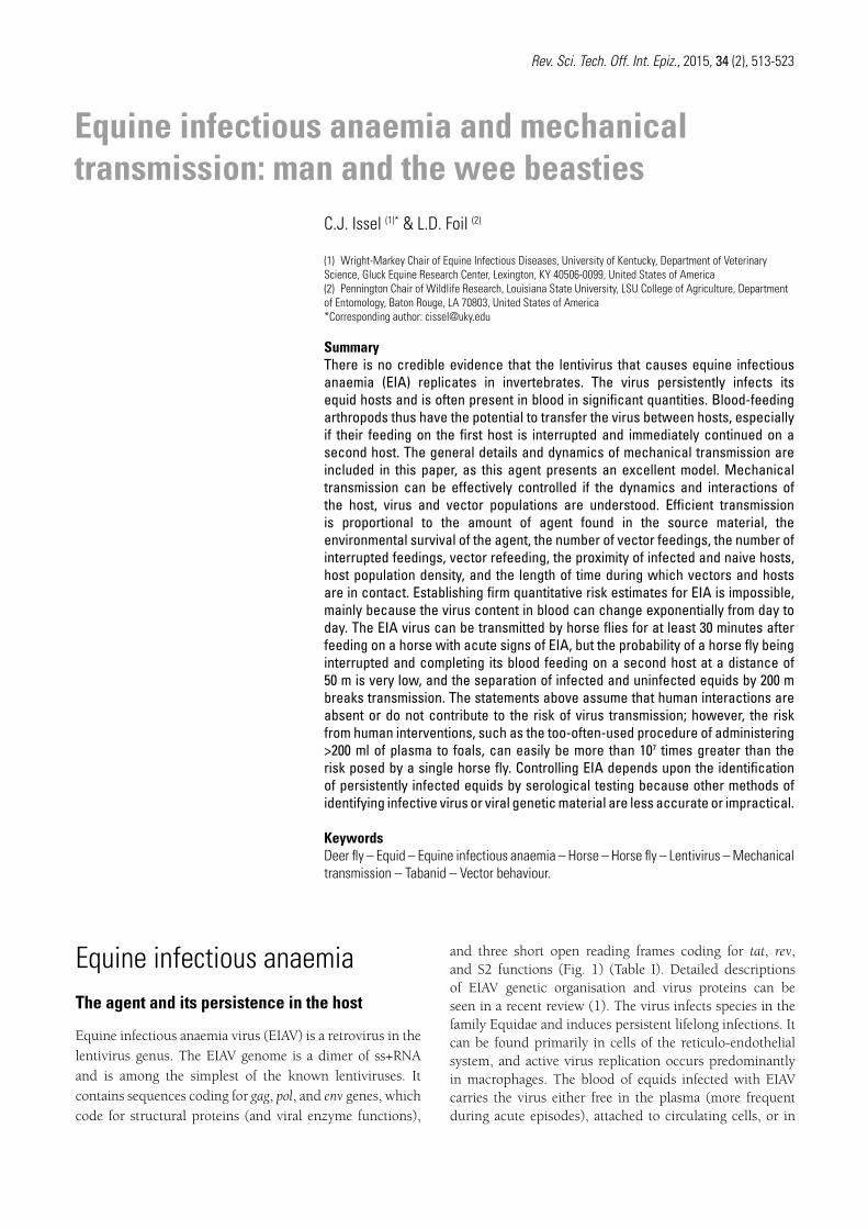
and three short open reading frames coding for tat, rev, and S2 functions (Fig. 1) (Table I). Detailed descriptions of EIAV genetic organisation and virus proteins can be seen in a recent review (1). The virus infects species in the family Equidae and induces persistent lifelong infections. It can be found primarily in cells of the reticulo-endothelial system, and active virus replication occurs predominantly in macrophages. The blood of equids infected with EIAV carries the virus either free in the plasma (more frequent during acute episodes), attached to circulating cells, or in
514 Rev. Sci. Tech. Off. Int. Epiz., 34 (2)
a latent form as pro-viral DNA within monocytes (more common during periods without clinical signs).
At this time, practical methods for the laboratory isolation of EIAV have not been perfected. Although techniques for demonstrating sequences of EIAV genetic material have improved recently and have proven useful in field situations, their routine application is currently limited to research (1). A major impediment to the routine use of polymerase chain reaction (PCR) for EIA is the diversity and rapid mutation of EIAV genomes in nature. When these constraints are taken together, proof of infection with this persistent lentivirus is almost always limited to indirect proof, i.e. the presence of antibodies against the virus is evidence of active infection.
Reverse transcriptase-RT, p66
Integrase-IN, p30
dUTPase -DU, p15
Protease-PR, p12
Matrix-MA, p15
Envelope:
SU, gp90
TM, gp45
Nucleocapsid-NC, p11
Lipid bilayer
Viral RNA
Core-CA, p26
Fig. 1 Equine infectious anaemia virusa) Cartoon of the equine infectious anaemia virion structure showing the location and identity of structural proteinsb) The 8-kilobase pair equine infectious anaemia virus pro-virus is shown, with long terminal repeats and protein-coding regions (gag, pol, env, tat, S2 and rev) with protein names added
a)
Viral genome coding regions with 3 short open reading frames (tat, S2 and rev)
LTR tat tat
S2 Rev Rev
LTR MA CA p9 NC SU/gp90 TM/gp45
PR RT-RH DU IN
env pol gag
b)
Table IMajor protein antigens of equine infectious anaemia virus used in current commercial test kits (all p26-based*)
Molecular weight
Nomenclature Tests using this antigen
p26 Core antigen AGID, ELISA, immunoblot**
gp45 Transmembrane ELISA, immunoblot
gp90 Surface unit Immunoblot
AGID: agar gel diffusion ELISA: enzyme-linked immunosorbent assay* One commercial ELISA kit includes p26 and a determinant of gp45, but discrimination between reactants is not made** The immunoblot test is mainly a research test today but can detect immune responses against all three major proteins of equine infectious anaemia virus
515Rev. Sci. Tech. Off. Int. Epiz., 34 (2)
Infection and disease
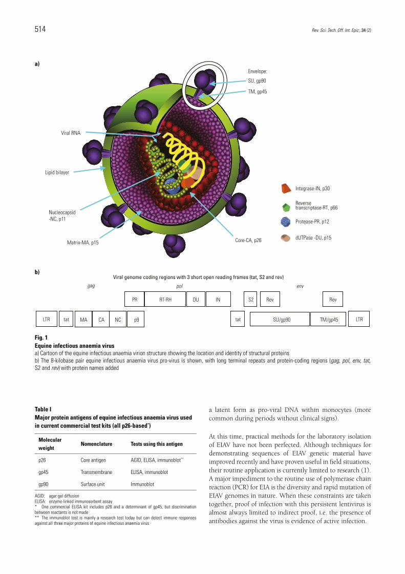
Infection is usually initiated by the percutaneous introduction of infective blood mediated by the interrupted feeding of arthropods or through human actions. Once the virus is introduced, it attaches itself to the cells of the monocyte-macrophage lineage where its RNA can be reverse-transcribed into pro-viral double-stranded DNA and enter the nucleus where it can persist. If the cell is activated and matures into a macrophage, a productive infection results and virions are released. After the intravenous introduction of 103 median horse infective doses of a horse-pathogenic laboratory strain of EIAV into a horse (Fig. 2), virus can be detected in the blood within 7–10 days post infection (PI). Peak levels of EIAV (105–107 median horse infective doses) are seen 15–20 days PI and the first febrile episode is coincident or follows shortly afterwards. The first antibodies against EIAV are seen at the same time as the resolution of the first febrile episode or shortly after. By 28 days PI, with this strain of EIAV, horses become positive in serological tests for EIA, by agar gel immunodiffusion (AGID), enzyme-linked immunosorbent assay (ELISA) or immunoblot. In the majority of cases with this strain of EIAV, horses develop recurring febrile episodes (2).
Responses to other strains of EIAV in nature, and with other doses and routes of exposure, should be expected to vary among the different equid species. One species may be more resistant to infection/disease than another; the
strain may replicate with less efficiency, depending on its passage history; the host may respond with more or less clinical disease, and the host may produce lower levels of antibodies to EIAV. As an example, the same horse-pathogenic strain used above causes no clinical signs in donkeys, peak viraemia titres are 100–1,000-fold lower, and antibodies may be seen for the first time 35–42 days PI, and at such low levels that diagnosis by AGID alone may not be possible (3).
For convenience the clinical forms of infection with EIA have been labelled acute, chronic and inapparent (Fig. 2).
Most equids which test positive for antibodies against EIAV are found as a result of routine surveillance testing and have no clinical signs of EIA. The authors refer to this group as ‘inapparent carriers’ of EIAV and they may remain clinically quiescent if left undisturbed. Under conditions of extreme environmental stress, or with the administration of immunosuppressive drugs, immune control over EIAV replication may be lifted sufficiently to release high levels of virus into the blood of the equid, often associated with mild-to-severe clinical signs (4). It is during these periods of active virus replication that the risk of transmission by blood-feeding insects increases exponentially.
The risk of insect transmission is greatest during the ‘acute phase’ of the infection (and during subsequent
Inapparent Chronic Acute
42
38
40
37
39
41
Platelets x 1,000/µl
Rect
al te
mpe
ratu
re (C
o )
Stress/immunosuppression
Time 4–12 months
200
0
100
50
150
Vira
l RN
A lo
g 10
1
10 102103104105106107108109
Disease episodes are characterised by rectal temperatures of ≥39°C and a platelet count below 100,000/µl Platelets x 1,000/µl
Rectal temperature
+
Fig. 2 Kinetics of events after the intravenous inoculation of a horse with 103 median horse infective doses of a horse-pathogenic laboratory strain of equine infectious anaemia virusVirus load (plasma viraemia in copies of viral RNA/ml) is designated by cartoons of virus particles. The first positive test for antibody against equine infectious anaemia virus is shown as a + on the timeline, shortly after the first febrile episode
516 Rev. Sci. Tech. Off. Int. Epiz., 34 (2)
acute episodes), while the viraemia is at its highest (5, 6). The authors’ definition of ‘acute’ includes the initial responses of equid hosts to pathogenic strains of EIAV. These are usually characterised by marked increases in body temperature, associated with high levels of free virus in the plasma, before antibodies against the virus can be found in the blood. As many of these episodes are short-lived, relatively mild and may not be followed by additional episodes, they are infrequently observed by owners and veterinarians unless daily body temperature monitoring is routine. The failure to detect antibody against EIAV at this time is not unexpected; a second sample 7–14 days later will be positive.
By far the majority of clinically ill equids with EIA are in what is referred to as the ‘chronic form’ of the disease. In these cases, the equid has usually experienced multiple febrile episodes, is profoundly affected and frequently demonstrates anaemia, cachexia, petechial haemorrhages, extremely low platelet counts, etc. and is positive in all serological tests for EIA (see below).
Diagnosis and control
Although clinical signs may be suggestive of EIA, none is pathognomonic. The only practical and accurate way to diagnose the infection is through serological tests. Currently, the only serological test for EIA that has proven to be positively correlated with the presence of the virus is the AGID test, also colloquially known as the Coggins test (7, 8, 9). The AGID test received rapid international acceptance and has remained the confirmatory test in laboratories throughout the world, where testing has been adopted by regulatory bodies.
Since the 1980s, a number of commercially available ELISA test kits have proven of value, and all use the AGID test as the final arbiter for confirmation. When applied to research samples, the immunoblot test has also proven useful, especially in cases where AGID and ELISA test results do not agree (10, 11, 12). Recently, immunoblotting has been used in a practical, three-tiered laboratory system, with ELISA tests first, followed by confirmation by AGID test, and immunoblot tests on samples with positive ELISA results but negative AGID test interpretations (13). In that survey, nearly 20% of the samples that were ultimately judged positive for EIA were interpreted as negative in the AGID test. Although false-positive ELISA test results were almost ten times more frequent than false-negative AGID test results, the three-tiered system is considered imperative if the goal is the highest possible rate of accuracy (with both high sensitivity and high specificity). A limited longitudinal study of mules with positive ELISA and immunoblot reactions, but which were interpreted as negative in AGID tests, confirmed that such equids are carrying EIAV and may remain negative by AGID, despite high virus RNA
levels following immunosuppression (14). Detection of EIAV in these types of equids requires an approach such as the three-tiered system described above.
The spread of EIAV can be controlled by identifying and segregating test-positive equids at a distance of at least 200 m from other equids. This distance was empirically considered to be a safe distance to break transmission by insect vectors, and is consistent with the results from studies on vector feeding behaviour (see below).
Transmission and practical field control
Equine infectious anaemia is best thought of as a blood-borne infection because EIAV can be consistently demonstrated in 250 ml+ of whole blood of test-positive horses when transfused into naive horses. Not all inapparent carriers of EIAV will have infective virus in 1 ml of blood and the level can vary exponentially from day to day (15). As no prognostic laboratory tests have been identified to differentiate infected equids by risk, the regulatory community has made the assumption that all test-positive equids pose the same risk for transmission.
When an equid with severe clinical signs of EIA is sampled, it is likely that EIAV will be demonstrated in all secretions and excretions, especially as blood vessel integrity may be compromised (low platelet counts and petechial haemorrhages may be widespread). Although the importance of these materials for transmission is considered negligible over all, it is possible that, under extreme conditions, such material could initiate outbreaks. One such example occurred during a hospital outbreak in 2006 in which intense anamnesis was conducted (16). In that case, the horse, which was not suspected of having EIA, was haemorrhaging from the nostrils and aerosol transmission appeared to be a likely route. Thus, transmission through secretions or excretions cannot be entirely excluded.
Some reviewers have suggested that an infective agent cannot be maintained in nature by mechanical transmission alone. For several reasons, in the authors’ opinion, EIAV is an agent that defies that comment. The authors maintain that the probability of mechanical transmission of EIAV is sufficiently high to maintain the virus because it persists in its host for life, because the host’s life span is relatively long, and the host’s behaviour includes herding. One cannot ignore the fact that certain types of intraspecies interactions induce severe trauma that could add to the risk of transmission (e.g. biting between stallions during the establishment of dominance for harems). For example, in an outbreak of EIA in ‘wild’/feral horses, with little chance of human interaction, the rate of infection with EIAV in mature stallions (with many battle scars) was noted to be significantly higher than in mature mares (17).
517Rev. Sci. Tech. Off. Int. Epiz., 34 (2)
The overwhelming importance of human activity
The historical documentation of EIA as an important disease of horses is almost coincident with the invention and use of syringes and hypodermic needles. Since that time, humans have undoubtedly been the major contributors to the spread of EIA locally, nationally and internationally. Despite educational efforts, humans are still considered to be a major factor in numerous outbreaks, including a widely publicised outbreak in 2006, in which transmission through human intervention was considered the major route (16, 18, 19).
Few long-term studies have been conducted on true ‘free-living’ populations of equids to accurately assess the routes and vectors/fomites involved in transmission scenarios. Limited field studies and anecdotal accounts of populations tested for the first time suggest the following:
– transmission is associated with periods of insect vector feeding activity
– transmission occurs infrequently from inapparent carriers to naive ones
– inapparent carriers can often have long productive lives
– in the absence of humans, the time between transmission ‘explosions’ may be quite long.
The detailed coverage of the dynamics and quantitative factors in mechanical transmission given below will give the reader a perspective on the relative importance of human activities and the justification for continued and expanded educational efforts to reduce the impact of humans on the transmission of this lentivirus.
Mechanical transmission of disease agentsThe two types of horizontal mechanical transmission are ‘direct mechanical transmission’, which is the direct transfer of an agent between hosts, and ‘indirect or contaminative transmission’, which is when vectors are contaminated by secretory or excretory products of infected hosts before transfer to a second host. Foil and Gorham (20) provide a comprehensive review of the role of arthropods in both direct and indirect mechanical transmission of disease agents. The purpose of this section is to provide information on the epizootiology of blood-borne pathogens when they are mechanically transmitted to susceptible hosts via portals of entry in the skin. The percentage of the overall transmission of various agents for this scenario varies widely depending upon different routes; however, when examining the amount of underlying mechanical transmission, the
conclusion resides in a probability (21) relative to the variables discussed below.
Equine infectious anaemia virus is a model for this mechanism of transmission because:
– transmission most frequently occurs via this method, associated with insects or iatrogenic vectors
– equids are the only host of the virus
– there is no biological vector of the virus
– there are no effective drugs or vaccines that might mask a transmission event.
Other examples of agents that can be transmitted by this route include:
– bovine leukaemia virus and Bacillus anthracis, which both have other major routes of transmission
– Anaplasma marginale, Francisella tularensis and Trypanosoma vivax, which also have biological vectors
– Trypanosoma evansi, which has reservoir hosts.
All of these agents have been shown to be transmitted by tabanids, which are the prototype insect mechanical vectors, in the absence of other routes of transmission, biological vectors or reservoir hosts (22, 23).
Understanding the dynamics of transmission, the transmission routes, and the mechanisms of pathogen persistence has been central to developing effective epidemiological methods for reducing endemic/enzootic transmission and controlling epidemics/epizootics. For EIAV, mechanical transmission of the virus in the blood via insect and iatrogenic vectors is the major route. When the infectious source of an epizootic of EIA is an infected blood product or drug administered on farms, the source is often easily identified from patient profiles, the absence of heavy insect attack, and/or the use of techniques which contribute to iatrogenic transmission, such as the multiple use of needles or surgical tools, as discussed above. The important elements that initially led to the first successes in controlling EIAV transmission were the development of a dependable diagnostic tool (9) and information regarding the weights of arthropod and iatrogenic transmission. The slogan for such preventive efforts was: ‘One needle, one horse’.
The spatial and temporal distribution of epizootics (in the absence of iatrogenic transmission) and the prevalence of EIAV demonstrate the landscape epizootiology of a pathogen which is mechanically transmitted by insects. In the United States, the association of peaks of tabanid and
518 Rev. Sci. Tech. Off. Int. Epiz., 34 (2)
stable fly populations with EIAV outbreaks had been made by the early 1900s and the common name, ‘swamp fever’, ties the occurrence of EIAV to the plentiful tabanid larval habitats that exist in the semi-tropical states, particularly Louisiana (24, 25). These associations were possible due to the short incubation period before fatal acute cases of EIAV. The magnitude of the tabanid attack rate on animals that are confined close to tabanid larval habitats, such as seasonally flooded hardwoods and swamps, can be significant. For example, Foil et al. (26, 27) marked approximately 2,000 tabanids (belonging to 17 species and five genera) per day for eight days, feeding on steers at two sites approximately 50 m apart in a wildlife refuge in central Louisiana. Much higher landing rates, up to 1,000 tabanids in 1 h, have been recorded at other locations in south Louisiana. Issel et al. (28) conducted a prospective study with febrile, chronically infected horses co-mingled with negative sentinels at two locations over an eight-year period. Over the first four years at farm 1, eight of 27 negative sentinels became seropositive. The tabanid season was seven months long over the year, but epizootics consistently occurred in association with population peaks of different tabanid species. Over the second four years, at farm 2, none of 36 sentinels became infected; mosquito populations were high and stable fly populations were low at both farms, but the tabanid populations at farm 2 were less than 2% of those at farm 1. At farm 1, the present authors observed that foals were exempt from transmission events and then showed that the tabanid burden of foals was less than 3% of that of adult horses (29). Younger equids have more intense defensive behaviours against tabanid attack, a fact which encourages flies to redistribute to older animals in the herd, and helps to explain part of the age bias for EIAV cases. These studies show that there are predictable elements of epizootics of the mechanical transmission of pathogens by insects, but the probability of a transmission event is affected by a number of factors, summarised below.
One variable that contributes to the mechanical transmission of blood-borne agents is the quantity of the agent transferred to the susceptible host by the insect or iatrogenic vector. This depends upon the titre of the agent in the donor’s blood, the volume of blood transferred to and inoculated into the susceptible host via the vector, and the time required for the transfer versus the persistence of the agent on the vector. For EIAV, the persistence of the virus is different between biological and inanimate surfaces. Hawkins et al. (5) reported that transmission events with horse flies only occurred when transfers were made within 30 min, while Williams et al. (30) found that the virus was viable for up to 96 h on needles but only for 1 h on mosquito mouthparts. Foil and Gorham (20) summarised studies that estimated the amount of blood retained or transferred on insect mouthparts after an interrupted meal and on needles. Approximately 1,000 nl are transferred upon the second use of a 22-gauge needle after blood
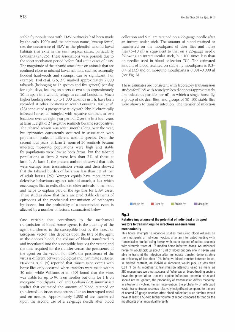
collection and 9 nl are retained on a 22-gauge needle after an intramuscular stick. The amount of blood retained or transferred on the mouthparts of deer flies and horse flies (5–10 nl) is equivalent to that on a 22-gauge needle following an intramuscular stick, but 100 times less than on needles used in blood collection (31). The estimated amount of blood retained on stable fly mouthparts is 0.3–0.4 nl (32) and on mosquito mouthparts is 0.001–0.000 nl (see Fig. 3).
These estimates are consistent with laboratory transmission studies for EIAV with acutely infected donors (approximately one infectious particle per nl), in which a single horse fly, a group of six deer flies, and groups of 50–100 stable flies were shown to transfer infection. The transfer of infection
Horse fly Deer fly Stable fly Mosquito
Fig. 3 Relative importance of the potential of individual arthropod vectors to transmit equine infectious anaemia virus mechanicallyThis figure attempts to reconcile studies measuring blood volumes on the mouthparts of individual vectors after an interrupted feeding with transmission studies using horses with acute equine infectious anaemia with viraemia titres of 106 median horse infective doses. An individual horse fly would pick up about 10 nl of blood but only one in seven was able to transmit the infection after immediate transfer, demonstrating an efficiency of less than 10% infective blood transfer between hosts. In marked contrast, an individual mosquito would pick up less than 0.01 nl on its mouthparts; transmission attempts using as many as 200 mosquitoes were not successful. Whereas all blood-feeding vectors have the potential to transmit equine infectious anaemia virus and should not be ignored, the probability of transmission differs markedly. In situations involving human intervention, the probability of arthropod vector transmission becomes relatively insignificant compared to the use of shared 22-gauge needles after blood collection; such fomites would have at least a 50-fold higher volume of blood compared to that on the mouthparts of an individual horse fly
519Rev. Sci. Tech. Off. Int. Epiz., 34 (2)
has never been demonstrated using mosquitoes (5, 24, 33). The virus titre in a febrile, EIAV-infected horse is known to fluctuate up to 1,000-fold, reaching a maximum of one infectious particle per 100 nl. The infectious titres of a febrile donor in horse fly transmission studies have not been reported, but Issel et al. (6) showed that transmission occurred with a group of 25 horse flies (estimates of blood transfer would be more than 100 nl), using a febrile donor and an inapparent carrier that had experienced a clinical attack of EIA nine months earlier. In a study on the transmission of bovine leukaemia virus (BLV) by horse flies, Foil et al. (34) established the volume of blood containing an infectious dose for two BLV-infected donor cows with persistent lymphocytosis. Groups of flies ranging from 10–100 were fed on each donor cow, interrupted after partial blood feeding and transferred to goats and allowed to feed to repletion. The infectious titre of donor 1 was 10–3.5 and, based upon an estimate of 6 nl of blood to be transferred by the flies, Foil et al. (35) predicted that a group of 53 flies would transfer the infection; groups of 10–25 flies did not transfer the virus while groups of 50–100 did. The infectious titre of BLV for donor 2 was 10–2.5; the predicted successful fly group size was 533 and no virus was transferred by up to 100 flies per group.
We can say with some certainty that the physical variables required to predict the probability of a mechanical transmission event occurring under defined conditions, as discussed above, are now available. However, vector density and behaviour, as well as host herd age structure, affect the probability of a transmission event occurring under natural conditions. The incidence of immunity and infection within a herd or on a farm (what percentage of the herd is at risk), as well as the population density of potential insect vectors (multiplier of events), are obviously important elements for a mathematical model of the mechanical transmission of pathogens (21). The proximity of susceptible and infected horses within or between herds at different locations and the feeding behaviour of the potential vectors are two other important variables that affect the probability of mechanical transmission and are tied together. Studies on tabanid behaviour can be used to estimate how these two variables
interact but there are over 4,000 species of tabanids, which complicates any attempt to make generalisations.
Foil (23) used various mark-recapture methods, such as marking feeding tabanids with non-toxic paint to measure the percentage of interrupted feeding – resulting in a transfer to a different host (mixed feeding) – that occurred for different species. He also examined the effects of spatial separation of horses on the percentage of tabanid mixed feeding. When four horses were tethered in a 9 m square and feeding tabanids were marked (a different colour for each horse), and allowed to continue feeding, 2–3% of the smaller tabanids, such as Tabanus lineola, transferred to other horses rather than completing the blood meal on the initial host, while the larger flies transferred more frequently (e.g. 12% for T. petiolatus). Given a moderate tabanid burden of four flies feeding at one time on a horse, and an average feeding time of 2.5 min (36), approximately 1,000 flies would feed on this animal in a 10 h day and, conservatively, 20–30 flies would take a mixed meal. Since 25 tabanids have been shown to transmit EIAV from a febrile donor (6), transmission events are highly probable when infected and susceptible horses are co-mingled. Foil (23) also conducted assays to determine the effects of host separation on the percentage of mixed feeding and predicted that only 1% of the flies that left the host before completion of the blood meal would transfer to a second host that was 50 m away. Later, these results were confirmed in a similar study in Brazil (37). These studies have been used to recommend separating infected and susceptible horses by at least 200 m to prevent the mechanical transmission of EIAV by horse flies, a strategy shown to be effective in field situations in numerous jurisdictions (C.J. Issel, personal communication).
520 Rev. Sci. Tech. Off. Int. Epiz., 34 (2)
Anémie infectieuse équine et transmission mécanique : l’homme et les bestioles
C.J. Issel & L.D. Foil
Résumé Aucun élément crédible n’indique que le lentivirus responsable de l’anémie infectieuse équine se réplique dans l’organisme des invertébrés du. L’infection virale est persistante chez les hôtes équins, le virus étant souvent présent en grandes quantités dans le sang. Cela facilite le transfert du virus d’un hôte à l’autre par les arthropodes hématophages, en particulier lorsqu’un premier repas de sang est interrompu et se poursuit immédiatement sur un deuxième hôte. Les auteurs décrivent les étapes détaillées et la dynamique de la transmission mécanique de ce virus, qu’ils considèrent comme un excellent modèle d’étude. La transmission mécanique peut être efficacement contenue si sa dynamique et les interactions entre l’hôte, le virus et les populations de vecteurs sont bien comprises. La réussite de la transmission du virus est fonction de la quantité de virus présent dans les matériels source, de la survie de l’agent pathogène dans l’environnement, du nombre de repas de sang pris par les vecteurs, du nombre de repas interrompus et poursuivis sur d’autres hôtes, de la proximité entre les hôtes infectés et d’autres hôtes naïfs, de la densité de la population d’hôtes et de la durée de l’exposition des hôtes aux vecteurs. Il est impossible de procéder à des estimations quantitatives définitives du risque d’anémie infectieuse équine, d’autant que le volume de virus présent dans le sang peut changer exponentiellement d’un jour sur l’autre. Les taons peuvent transmettre le virus de l’anémie infectieuse équine au cours des 30 minutes ou plus suivant le repas de sang pris sur un cheval présentant des signes d’anémie infectieuse équine aiguë ; la probabilité qu’un taon interrompe son repas de sang et le poursuive sur un second hôte à une distance de 50 mètres est néanmoins très faible, de sorte qu’il suffit de séparer les chevaux infectés en les maintenant à une distance d’au moins 200 mètres des chevaux non infectés pour briser la chaîne de transmission. Ce constat suppose toutefois qu’aucune interaction humaine n’ait lieu ou ne contribue au risque de transmission virale. Or, le risque associé aux interventions humaines, notamment la pratique bien trop répandue d’administrer plus de 200 ml de plasma aux poulains s’avère plus de dix millions de fois supérieur au risque posé par un taon. L’anémie infectieuse équine peut être contrôlée efficacement si l’on soumet les équidés infectés persistants à un dépistage au moyen de tests sérologiques, car les autres méthodes d’identification du virus infectieux ou du matériel génétique viral sont moins précises ou peu pratiques.
Mots-clésAnémie infectieuse équine – Cheval – Comportement des vecteurs – Équidé – Lentivirus – Tabanidé – Taon du genre Chrysops – Taon du genre Tabanus – Transmission mécanique.
521Rev. Sci. Tech. Off. Int. Epiz., 34 (2)
Anemia infecciosa equina y transmisión mecánica: el hombre y esos bichos…
C.J. Issel & L.D. Foil
ResumenNo hay pruebas fidedignas de que el lentivirus causante de la anemia infecciosa equina (AIE) se replique en invertebrados. El virus provoca una infección persistente en los équidos y a menudo está presente en el torrente sanguíneo en cantidades importantes. Los artrópodos que se alimentan de sangre tienen por tanto la posibilidad de transferir el virus entre hospedadores, sobre todo cuando interrumpen la toma en un primer hospedador para proseguirla de inmediato en un segundo. Los autores exponen los aspectos generales y la dinámica de la transmisión mecánica, de la que este agente ofrece un excelente modelo. Es posible controlar eficazmente esta transmisión a condición de entender la dinámica y las relaciones entre las poblaciones de hospedador, virus y vector. La eficacia de la transmisión es proporcional a: la cantidad de agente presente en la sangre de origen; la supervivencia ambiental del agente; el número de tomas del vector; el número de tomas interrumpidas y de tomas sucesivas del vector; la proximidad entre hospedadores infectados y hospedadores no expuestos; la densidad de población del hospedador; y el tiempo de contacto entre vectores y hospedadores. Es imposible realizar estimaciones cuantitativas sólidas del riesgo de AIE, principalmente porque la concentración de virus en sangre puede cambiar exponencialmente de un día para otro. Los tábanos pueden transmitir el virus de la AIE durante al menos 30 minutos después de alimentarse de un caballo con signos de AIE aguda, pero la probabilidad de que un tábano sea interrumpido en su toma y vaya a completarla alimentándose de la sangre de un segundo hospedador situado a 50 m de distancia es muy escasa, por lo que una separación de 200 m entre los équidos infectados y los no infectados basta para interrumpir la transmisión. Estas aseveraciones parten de la premisa de que no hay intervención humana o de que esta no contribuye al riesgo de transmisión. Sin embargo las intervenciones humanas, como la demasiado socorrida administración a los potros de más de 200 ml de plasma, pueden fácilmente acarrear un riesgo más de 107 veces mayor que el que presenta un solo tábano. La lucha contra la AIE reposa en la detección de équidos aquejados de infección persistente mediante pruebas serológicas, pues los otros métodos para detectar la presencia de virus infecciosos o de su material genético resultan menos exactos o inviables.
Palabras claveAnemia infecciosa equina – Caballo – Comportamiento del vector – Équido – Lentivirus – Tabánido – Tábano – Tábanos Chrysops – Transmisión mecánica.
522 Rev. Sci. Tech. Off. Int. Epiz., 34 (2)
References 1. Cook R.F., Leroux C. & Issel C.J. (2013). – Equine infectious
anemia and equine infectious anemia virus in 2013: a review. Vet. Microbiol., 167 (1–2), 181–204.
2. Leroux C., Issel C.J. & Montelaro R.C. (1997). – Novel and dynamic evolution of equine infectious anemia virus genomic quasispecies associated with sequential disease cycles in an experimentally infected pony. J. Virol., 71 (12), 9627–9639.
3. Cook S.J., Cook R.F., Montelaro R.C. & Issel C.J (2001). – Differential responses of Equus caballus and Equus asinus to infection with two pathogenic strains of equine infectious anemia virus. Vet. Microbiol., 79 (2), 93–109.
4. Kono Y., Hirasawa K., Fukunaga Y. & Taniguchi T. (1976). – Recrudescence of equine infectious anemia by treatment with immunosuppressive drugs. Natl Inst. Anim. Hlth Q. (Tokyo), 16 (1), 8–15.
5. Hawkins J.A., Adams W.V. Jr, Wilson B.H., Issel C.J. & Roth E.E. (1976). – Transmission of equine infectious anemia virus by Tabanus fuscicostatus. JAVMA, 168 (1), 63–64.
6. Issel C.J., Adams W.V.J., Meek L. & Ochoa R. (1982). – Transmission of equine infectious anemia virus from horses without clinical signs of disease. JAVMA, 180 (3), 272–275.
7. Coggins L. (1971). – Immunodiffusion test for equine infectious anemia. Proc. US Livest. Sanit. Assoc., 75, 254–257.
8. Coggins L. & Norcross N.L. (1970). – Immunodiffusion reaction in equine infectious anemia. Cornell Vet., 60 (2), 330–335.
9. Coggins L., Norcross N.L. & Nusbaum S.R. (1972). – Diagnosis of equine infectious anemia by immunodiffusion test. Am. J. Vet. Res., 33 (1), 11–18.
10. Issel C.J., Rwambo P.M. & Montelaro R.C. (1987). – Evolution of equine infectious anemia diagnostic tests: recognition of a need for detection of anti-EIAV glycoprotein antibodies. In Equine infectious diseases, V. University of Kentucky Press, Lexington, 196–200.
11. Issel C.J. & Cook R.F. (1993). – A review of techniques for the serologic diagnosis of equine infectious anemia. J. Vet. Diagn. Invest., 5 (1), 137–141.
12. Issel C.J., Cook S.J., Cook R.F. & Cordes T.R. (1999). – Optimal paradigms to detect reservoirs of equine infectious anemia virus (EIAV). J. Equine Vet. Sci., 19 (11), 728–732.
13. Issel C.J., Scicluna M.T., Cook S.J., Cook R.F., Caprioli A., Ricci I., Rosone F., Craigo J.K., Montelaro R.C. & Autorino G.L. (2012). – Challenges and proposed solutions for more accurate serological diagnosis of equine infectious anaemia. Vet. Rec., 172, 210. doi:10.1136/vr-2012-100735.
14. Scicluna M.T., Issel C.J., Cook F.R., Manna G., Cersini A., Rosone F., Frontoso R., Caprioli A., Antonetti V. & Autorino G.L. (2013). – Is a diagnostic system based exclusively on agar gel immunodiffusion adequate for controlling the spread of equine infectious anaemia? Vet. Microbiol., 165 (1–2), 123–134.
15. Issel C.J., Adams W.V. Jr, Meek L. & Ochoa R. (1982). – Transmission of equine infectious anemia virus from horses without clinical signs of disease. JAVMA, 180 (3), 272–275.
16. More S.J., Aznar I., Bailey D.C., Larkin J.F., Leadon D.P., Lenihan P., Flaherty B., Fogarty U. & Brangan P. (2008). – An outbreak of equine infectious anaemia in Ireland during 2006: the modes of transmission and spread in the Kildare cluster. Equine Vet. J., 40 (7), 709–711.
17. Issel C.J., Cook S.J., Howell D., Nitschke Sinclear J., Gardner D., Mathis J.G., Marshall M.R. & Roger L.E. (1998). – Equine infectious anemia in wild free-roaming horses in Utah. Proc. US Livest. Sanit. Assoc., 102, 376–384.
18. More S.J., Aznar I., Myers T., Leadon D.P. & Clegg T.A. (2006). – An outbreak of equine infectious anaemia in Ireland during 2006: investigation methodology, initial source of infection, diagnosis and clinical presentation, modes of transmission and spread in the Meath cluster. Equine Vet. J., 40 (7), 706–708.
19. More S.J., Brangan P., Aznar I., Bailey D.C., Larkin J., Myers T., Leadon D.P., Lenihan P., Flaherty B. & Clegg T.A. (2008). – Successful eradication of equine infectious anemia from Ireland during 2006. In Proc. 54th Annual Convention of the American Association of Equine Practitioners, 6–10 December, San Diego, California, 306–307.
20. Foil L.D. & Gorham J.R. (2000). – Mechanical transmission of disease agents by arthropods. In Medical entomology: a textbook on public health and veterinary problems caused by arthropods (B.F. Idridge & J.D. Edman, eds). Kluwer Academic Publishers, Dordrecht, the Netherlands, 461–514.
21. Desquesnes M., Biteau-Coroller F., Bouyer J., Dia M.L. & Foil L. (2009). – Development of a mathematical model for mechanical transmission of trypanosomes and other pathogens of cattle transmitted by tabanids. Int. J. Parasitol., 39 (3), 333–346.
22. Krinsky W.L. (1976). – Animal disease agents transmitted by horse flies and deer flies (Diptera: Tabanidae). J. Med. Entomol., 13 (3), 225–275.
23. Foil L.D. (1989). – Tabanids as vectors of disease agents. Parasitol. Today, 5 (3), 88–96.
24. Foil L.D. & Issel C.J. (1991). – Transmission of retroviruses by arthropods. Annu. Rev. Entomol., 36, 355–381.
523Rev. Sci. Tech. Off. Int. Epiz., 34 (2)
25. Foil L.D. & Hogsette J.A. (1994). – Biology and control of tabanids, stable flies and horn flies. In Ectoparasites of animals and control methods (G. Uilenberg, ed.). Rev. Sci. Tech. Off. Int. Epiz., 13 (4), 1125–1158.
26. Foil L.D. (1983). – A mark-recapture method for measuring effects of spatial separation of horses on tabanid (Diptera) movement between hosts. J. Med. Entomol., 20, 301–305.
27. Foil L.D., Leprince D.J. & Byford R.L. (1991). – Survival and dispersal of horse flies (Diptera: Tabanidae) feeding on cattle sprayed with a sublethal dose of fenvalerate. J. Med. Entomol., 28 (5), 663–667.
28. Issel C.J., Adams W.V. Jr & Foil L.D. (1985). – Prospective study of progeny of inapparent equine carriers of equine infectious anemia virus. Am. J. Vet. Res., 46 (5), 1114–1116.
29. Foil L.D., Stage D., Adams W.V. Jr & Issel C.J. (1985). – Observations of tabanid feeding on mares and foals. Am. J. Vet. Res., 46 (5), 1111–1113.
30. Williams D.L., Issel C.J., Steelman C.D., Adams W.V. Jr & Benton C.V. (1981). – Studies with equine infectious anemia virus: transmission attempts by mosquitoes and survival of virus on vector mouthparts and hypodermic needles, and in mosquito tissue culture. Am. J. Vet. Res., 42 (9), 1469–1473.
31. Scoles G.A., Miller J.A. & Foil L.D. (2008). – Comparison of the efficiency of biological transmission of Anaplasma marginale (Rickettsiales: Anaplasmataceae) by Dermacentor andersoni Stiles (Acari: Ixodidae) with mechanical transmission by the horse fly, Tabanus fuscicostatus Hine (Diptera: Muscidae). J. Med. Entomol., 45 (1), 109–114.
32. Scoles G.A., Broce A.B., Lysyk T.J. & Palmer G.H. (2005). – Relative efficiency of biological transmission of Anaplasma marginale (Rickettsiales: Anaplasmataceae) by Dermacentor andersoni (Acari: Ixodidae) compared with mechanical transmission by Stomoxys calcitrans (Diptera: Muscidae). J. Med. Entomol., 42 (4), 668–675.
33. Foil L.D., Meek C.L., Adams W.V. & Issel C.J. (1983). – Mechanical transmission of equine infectious anemia virus by deer flies (Chrysops flavidus) and stable flies (Stomoxys calcitrans). Am. J. Vet. Res., 44 (1), 155–156.
34. Foil L.D., Seger C.L., French D.D., Issel C.J., McManus J.M., Ohberg C.L. & Ramsey R.T. (1988). – Mechanical transmission of bovine leukemia virus by horse flies (Diptera: Tabanidae). J. Med. Entomol., 25 (5), 374–376.
35. Foil L.D., Adams W.V., McManus J.M. & Issel C.J. (1987). – Bloodmeal residues on mouthparts of Tabanus fuscicostatus (Diptera: Tabanidae) and the potential for mechanical transmission of pathogens. J. Med. Entomol., 24 (6), 613–616.
36. Foil L.D., Leprince D.J. & Byford R.L. (1990). – Sublethal effects and mortality of tabanids (Diptera, Tabanidae) induced by fenvalerate treatment of cattle. J. Entomol. Sci., 25 (2), 294–302.
37. Barros A.T. & Foil L.D. (2007). – The influence of distance on movement of tabanids (Diptera: Tabanidae) between horses. Vet. Parasitol., 144 (3–4), 380–384.