Eyeblink conditioning anomalies in bipolar disorder suggest cerebellar dysfunction
Upload
independentCategory
view
1download
0
Ultra-Deep Pyrosequencing of Partial Surface ProteinGenes from Infectious Salmon Anaemia Virus (ISAV)Suggest Novel Mechanisms Involved in Transition toVirulenceTurhan Markussen1*, Hilde Sindre1, Christine Monceyron Jonassen1¤a, Torstein Tengs1, Anja B.Kristoffersen2, Jon Ramsell1¤b, Sanela Numanovic1, Monika J. Hjortaas1, Debes H. Christiansen3, OleBendik Dale1, Knut Falk2
1 Department of Laboratory Services, Norwegian Veterinary Institute, Oslo, Norway, 2 Department of Health Surveillance, Norwegian Veterinary Institute, Oslo,Norway, 3 National Reference Laboratory for Fish Diseases, Food and Veterinary Authority, Torshavn, Faroe Islands
Abstract
Uncultivable HPR0 strains of infectious salmon anaemia viruses (ISAVs) infecting gills are non-virulent putativeprecursors of virulent ISAVs (vISAVs) causing systemic disease in farmed Atlantic salmon (Salmo salar). Thetransition to virulence involves two molecular events, a deletion in the highly polymorphic region (HPR) of thehemagglutinin-esterase (HE) gene and a Q266→L266 substitution or insertion next to the putative cleavage site (R267) inthe fusion protein (F). We have performed ultra-deep pyrosequencing (UDPS) of these gene regions from healthyfish positive for HPR0 virus carrying full-length HPR sampled in a screening program, and a vISAV strain from an ISAoutbreak at the same farming site three weeks later, and compared the mutant spectra. As the UDPS data shows thepresence of both HE genotypes at both sampling times, and the outbreak strain was unlikely to be directly related tothe HPR0 strain, this is the first report of a double infection with HPR0s and vISAVs. For F amplicon reads, mutationfrequencies generating L266 codons in screening samples and Q266 codons in outbreak samples were not higher thanat any random site. We suggest quasispecies heterogeneity as well as RNA structural properties are linked totransition to virulence. More specifically, a mechanism where selected single point mutations in the full-length HPRalter the RNA structure facilitating single- or sequential deletions in this region is proposed. The data providesstronger support for the deletion hypothesis, as opposed to recombination, as the responsible mechanism forgenerating the sequence deletions in HE.
Citation: Markussen T, Sindre H, Jonassen CM, Tengs T, Kristoffersen AB, et al. (2013) Ultra-Deep Pyrosequencing of Partial Surface Protein Genes fromInfectious Salmon Anaemia Virus (ISAV) Suggest Novel Mechanisms Involved in Transition to Virulence. PLoS ONE 8(11): e81571. doi:10.1371/journal.pone.0081571
Editor: Maureen J. Donlin, Saint Louis University, United States of America
Received July 13, 2013; Accepted October 14, 2013; Published November 26, 2013
Copyright: © 2013 Markussen et al. This is an open-access article distributed under the terms of the Creative Commons Attribution License, which permitsunrestricted use, distribution, and reproduction in any medium, provided the original author and source are credited.
Funding: This work was supported by The Norwegian Research Council (project no. 207024/E40). The funders had no role in study design, data collectionand analysis, decision to publish, or preparation of the manuscript.
Competing interests: The authors have declared that no competing interests exist.
* E-mail: [email protected]
¤a Current address: Department of Laboratory Medicine, Østfold Hospital Trust, Fredrikstad, Norway¤b Current address: Department of Medical Sciences, Uppsala University, Uppsala, Sweden
Introduction
Infectious salmon anaemia virus (ISAV) is an orthomyxovirusthat has caused systemic infection and disease in farmedAtlantic salmon (Salmo salar) in Norway, Canada, Scotland,Shetland Islands, the Faroe Islands, USA and Chile [1-8]. Thevirus’ two major surface glycoproteins are the hemagglutinin-esterase (HE), responsible for receptor binding and release(RDE), and the fusion protein (F), responsible for virus uptakethrough the fusion of viral and cellular membranes [9-12]. The
HEs from different virulent ISAV strains (vISAVs) often vary inlength as determined by the size of their highly polymorphicregion (HPR) located in the stalk immediately upstream of thetransmembrane domain of the protein. The shortening of thestalk is thought to arise from differential deletions from a full-length precursor HE (HPR0) [13]. Recombination throughtemplate switching during replication has been proposed as analternative mechanism for generating the deletions [14]. Thedeletions in the HE stalk region could be analogues to thevarying lengths in the influenza A virus neuraminidase stalk,
PLOS ONE | www.plosone.org 1 November 2013 | Volume 8 | Issue 11 | e81571

which has been associated with host switching [15-18]. Ashortening of the HE stalk could also affect the functionalbalance between the HE receptor-binding and -destroyingactivities similar to that found between the hemagglutinin andneuraminidase of influenza A viruses through a number ofstudies [19-23].
ISAV HPR0 genotypes containing full-length HEs are non-virulent, non-cultivable and are primarily found in gills [24-26].They have been detected in apparently healthy wild andfarmed Atlantic salmon in most regions with Atlantic salmonfarming [24-28]. Frequent findings of HPR0s in fish farms inNorway and the Faroe Islands, the former with few diseaseoutbreaks and the latter with no ISA outbreaks since 2005,suggests the transition to virulence is an infrequent occurrence[27,29]. In addition to a full-length HE, all HPR0s also contain aglutamine in position 266 (Q266) next to the one of the twoputative cleavage sites in the F protein (R267) [30]. In contrast,all vISAVs carry a leucine in this position (L266), except for a fewstrains which have small sequence insertions close to this site[30-32]. F gene insertions through template switching, as wellas reassortment of gene segments, have both been linked tovirulence [30,31]. For the F protein, the single amino acidsubstitution or insertion close to R267 may be analogous to thatin highly pathogenic avian influenza A virus subtypes H5 andH7, where pathogenicity is acquired through the mutationalchange of cleavage specificity resulting in altered tissuetropism [33,34]. These differences between HPR0s andvISAVs may represent viral adaptation leading to disease indensely populated industrial farming operations.
RNA viruses are known to exist in the host as a swarm ofclosely related mutant genomes known as quasispecies[35-37]. The molecular basis for this genomic heterogeneity isthat replication of RNA viruses is highly error prone since viralRNA-dependent RNA polymerases do not possessproofreading-repair activity [38-40]. Such a mutant spectrumwithin an individual host, combined with the ability to recombineand reassort their gene segments, can enable these viruses torapidly adapt in response to selective pressures [36,37]. Thisgenetic diversity can also be viewed as a measure of thefitness of the virus and be linked directly to pathogenesis[41-43]. Ultra-deep pyrosequencing (UDPS) allows for thesimultaneous analysis of thousands of clonally amplified PCRamplicons, enabling the detection of the within-host minorityvariants [44-49]. The technology is now well established inmany fields within virology, including viral evolution andpathogenesis, detection of antiviral resistance markers anddiagnostics. For ISAV, aside from a recent publicationdescribing the presence of quasispecies in the non-codingregions of the viral genome, based on traditional cloning andSanger sequencing [50], the within-host genomic heterogeneityfor this virus has not been investigated.
An ISA epidemic from 2007 to 2010 in Astafjord of Norwaywas monitored by both disease diagnostics and screening ofapparently healthy fish. Sequence-based analysis of partialHE- and F genes combined with epidemiological informationconcluded that a single virulent ISAV strain had transmittedhorizontally between proximal fish farms and caused all 17 ISAoutbreaks in the region from 2007 to 2009 (i.e. Astafjord strain)
[28,51]. Here we present results from UDPS of amplicons fromgene regions spanning the HPR in HE and the putative R267
cleavage site in the F protein. From a site in Astafjord 2010,mutant spectra originating from gill tissue samples foundscreening positive for a non-virulent HPR0 are compared withthat of a vISAV from an ISA outbreak at the same farming sitethree weeks later. We report, for the first time, a doubleinfection involving a HPR0 and a vISAV. F reads containingL266- and Q266 codons in screening and outbreak samples,respectively, were present at mutational frequenciescomparable to that found at any random site in the amplicons.Similarly, HE reads from screening samples containingdeletions within the full-length HPR, differing from the HPRdeletion pattern of the outbreak strain (delHPR, present at verylow levels) were not observed at all. The results from UDPSdata analyses, together with RNA structure predictions, providestronger support for the HE deletion hypothesis overrecombination in creating the sequence deletions in vISAVs,and we propose a mechanism in which selected single pointmutations in the full-length HPR alter the RNA structure in sucha way that it facilitates single- or sequential deletions in thisregion of the HE gene.
Materials and Methods
Field SamplesFour HPR0-positive gill samples from healthy Atlantic salmon
taken from a commercial farming site (hereafter named Site 10)in Astafjord (Norway) during screening in May 2010 (screeningsamples 1-4), and four gill samples taken three weeks laterduring the early phases of an ISA outbreak at the same site(outbreak samples 1-4), were chosen for UDPS. Sampling fromboth screening and disease outbreak, which were fromdifferent fish and different locations at the site, were performedon order by the Norwegian Food Safety Authority. Approvalfrom Institutional Animal care and Use Committee (IACUC) orethics was not required. No experiments that involved fish wereperformed. The screening samples were among 8 of 23 gillsamples ISAV positive by real-time RT-PCR (Ct’s 28-38, notshown). Traditional (Sanger) sequencing of HE- and F genes,covering the HPR and the putative encoded R267 cleavage site,respectively, showed all to be of one non-virulent HPR0genotype [29]. The ISA outbreak fish had pathology consistentwith ISA, and the systemic infection was verified by real-timeRT-PCR and Sanger sequencing of the HE gene from kidneysamples of ten fish tested (Ct’s 14-18, not shown). Real-timeRT-PCR to determine viral loads in gill samples selected forUDPS was performed as previously described [28].
Preparation of PCR-generated AmpliconsGill tissue was homogenized and total RNA extracted using
RNeasy Mini Kit (Qiagen, Hilden, Germany). cDNA synthesiswas performed separately for each sample using randomhexamers together with Superscript III reverse transcriptase,following the protocol recommended by the manufacturer(Invitrogen, Carlsbad, USA). Following first-strand synthesis,cDNA samples were used as templates in two separate PCRreactions to generate gene segment 5 (F gene) and segment 6
UDPS of Partial HE- and F Protein Genes
PLOS ONE | www.plosone.org 2 November 2013 | Volume 8 | Issue 11 | e81571

(HE gene) amplicons. Primer sequences are shown in TableS1. PCR was performed using high fidelity AccuPrime Pfx DNApolymerase (Invitrogen, Carlsbad, USA) with 2 µl cDNA in eachreaction. PCR cycling conditions were 95°C/2 min, followed by40 cycles of 95°C/45 sec, 55°C/45 sec and 68°C/2 min and afinal extension step at 72°C/7 min. After PCR, a small volumefrom each reaction was run on 1.5 % agarose gelelectrophoresis and the products visualized by ethidiumbromide staining. In some cases the 2100 Bioanalyzer (AgilentTechnologies) was also used to evaluate the quality of PCRproducts. For most samples, single PCRs produced sufficientamount of product, except for screening samples 2, 3 and 4where mixing products from several parallel PCRs wasnecessary in order obtain the recommended amount of eachproduct for pyrosequencing. The same cDNA sample was usedin each PCR parallel. Samples from PCR were treated withExoSAP-IT to remove unincorporated dNTPs and primers,following manufacturer instructions (Affymetrix). Purificationwas performed using the QIAquick PCR Purification kit(Qiagen, Hilden, Germany). Quantification was performedusing Nanodrop 2000 (Thermo Scientific). In addition topyrosequencing each amplicon in both directions, the eightpurified HE- and F gene products were adjusted to equimolaramounts for multiplexing. Finally, two single samples(screening sample 1 and outbreak sample 1) and two pooledsamples (screening samples 2-4 and outbreak samples 2-4)were prepared and distributed over four regions on a GenomeSequencer FLX (GS-FLX) Titanium platform (Roche) at theNorwegian Sequencing Centre, University of Oslo.
Data AnalysisThe CLC Genomics Workbench program (CLC bio) was
used for assembly of sequences. Prior to in-depth analysis ofthe pyrosequencing data, the reads were split only acceptingreads containing 0 errors in the multiplex identifiers (MIDs).Sequences were then de-multiplexed, and adaptor sequences,keys, MIDs and primer ends trimmed away. This would, whencompared to the Sanger sequence lengths (reference),correspond to amplicon sizes of 250 nucleotides (nts) (HPROHE), 187 nts (delHPR HE) and 194 nts (F). The comparativeaspect between the screening and outbreak samples was themain focus of the present work. In order to avoid low-frequencymutational artifacts from the plasmid RT-step and risk of crosscontamination during UDPS setup, which could mask truequasispecies present at low frequencies, in vitro transcribedplasmid cloned RT-PCR-amplified templates were not includedas controls. The “RNA environments” as represented by shortidentical RNAs (from controls) and total RNA (from samples)could also introduce differential mutational artifacts followingRT-PCR amplification. Therefore, a site by site manualcomparison of observed frequency of nucleotides differing fromthe Sanger sequences was chosen as an alternative for allreads from screening and outbreak samples. Based on theresults from these analyses a mutational cutoff value of 0.05%was chosen in order to identify mutations occurring only atparticular high frequencies. Potential sequencing errors relatedto homopolymers was addressed by excluding variable sitespresent as stretches of four or more identical nucleotides. In
addition, mutation frequencies above cutoff, as well as readscontaining sequence deletions and/or insertions (see below),would have to be present in both forward and reversedirections in order to be considered. When an estimation of thenumber of reads containing a particular deletion was to bedetermined, the minimum length fraction setting was always setto 1.0 while the minimum similarity fraction was set to 0.99.This would allow no flexibility with regards to length of thereads, but would allow for 1 to 3 mismatches, thus providing anecessary flexibility with regards to single nucleotidedifferences in the reads when determining the number of readsthat contained the particular deletion type. CLC MainWorkbench (CLC bio) and Align X (Vector NTI Advance™ 11Package, Invitrogen) were used as additional tools forcomparisons between individual sequence reads. RNAsecondary structure predictions (minimal free-energy models)of viral (+)/(-)RNAs were performed using the Mfold program(version 2.3) [52]. Default parameters were used in thepredictions except for temperature, which was set to 15°C,considered the optimal temperature for ISAV replication [53].
Real-Time PCR on AmpliconsAs there was a theoretical possibility that the detection of
delHPR reads in screening samples and full-length HPR readsin outbreak samples could have originated from cross-contamination during UDPS setup, TaqMAN real-time PCRwas performed on all HE amplicon samples (except screeningsample 2, no more sample). The assays were designedspecifically to distinguish between full-length HPRs anddelHPRs by placing the two reverse primers in this region.First, 1 µl of the amplicon samples used for UDPS was run ona 2% agarose gel for separation of the two size variants.Bands, both visible and estimated, corresponding to full-length-or delHPR, were excised and purified using QIAquick GelExtraction Kit following manufacturers protocol (Qiagen, Hilden,Germany). Real-time PCR was performed using 5 µl of sampleand TaqMAN Universal PCR Master Mix (Invitrogen, Carlsbad,USA). Primer- and probes used are shown in Table S1. Real-time PCR was run on a Stratagene Mx3005P (AgilentTechnologies) with cycling conditions 95°C/10 min followed by40 cycles of 95°C/15 sec, 55°C or 60°C/1 min and 72°C/45sec. Real-time PCR products were run on 2 % agarose gel,bands excised and purified as above, and sequenced usingprimer s6 HPR_F (Table S1) on a ABI 3130 Genetic Analyser(Invitrogen).
Statistical ApproachTo evaluate whether events like mutations or deletions
appear more frequently in one region compared to another, achi-squared test was developed. Here, the actual number ofevents observed in each region, and the possible numbers ofsites represented by the amplicon sizes of each of the regions,were compared. The null hypothesis tested each time waswhether the frequencies of events were similar in the twoamplicons.
UDPS of Partial HE- and F Protein Genes
PLOS ONE | www.plosone.org 3 November 2013 | Volume 8 | Issue 11 | e81571

Accession NumbersThe Sanger nucleotide sequences obtained and used in this
study have GenBank accession numbers KC907278-KC907293. The UDPS dataset has been deposited intoBioProject database (NCBI) with the accession numberPRJNA221196.
Results
This paper describes the results obtained from UDPS of thevariable regions in ISAV HE- and F genes from non-virulentHPR0-positive fish collected from screening, and a virulentstrain sampled from an ISA outbreak at the same farming sitethree weeks later. From UDPS, mutant spectra were comparedand sequence data linked to predicted RNA structuralproperties of the two genes. Table 1 shows the total number ofreads (25695 to 88172) obtained from bidirectional UDPS ofHE- and F amplicons following trimming and de-multiplexing.On average, the number of reads identical to Sanger sequencewas 90% for both HE and F. In the remaining 10%, mutations,deletions and insertions were observed.
HE Gene AmpliconsThe majority of high frequency mutations in HE reads
were found within the HPR0 full-length HPR. Generally, theprevalence (i.e. # of different mutational sites compared toSanger sequence) and frequency (i.e. # of reads containing aparticular mutation differing from Sanger sequence) of
Table 1. Raw data from UDPS of partial ISAV HE- and Fgene amplicons.
ISAV Sample(s) Sequence direction No. of trimmedreads
Percent of readsidentical to Sangersequence
SCN1a HE gene forward 50552 82.4 HE gene reverse 69684 91.4 F gene forward 44498 91.9 F gene reverse 49118 89.2SCN pool HE gene forward 25695 78.3(SCN2, SCN3,SCN4)
HE gene reverse 68481 92.2
F gene forward 31230 92.1 F gene reverse 35799 89.5OBK1b HE gene forward 52038 92.4 HE gene reverse 88172 93.0 F gene forward 31546 91.4 F gene reverse 46835 89.7OBK pool HE gene forward 40458 92.1(OBK2, OBK3,OBK4)
HE gene reverse 51772 92.8
F gene forward 45909 91.9 F gene reverse 44498 91.9a. SCN=screening sampleb. OBK=outbreak sampledoi: 10.1371/journal.pone.0081571.t001
mutations above 0.05 % in reads from HE amplicons (i.e. highfrequency mutations) seemed higher for screening- comparedto outbreak samples, as illustrated in Figure 1a and Table S2.There were 22 sites in total with mutational frequency above0.05%, 18 in screening- and 5 in outbreak samples (Table S2).A chi-squared test correcting for HPRO HE and delHPR HEamplicon sizes (250 and 187 nts respectively) gave p = 0.075,supporting the tendency for a higher number of mutational sitesoccurring in HE amplicons from screening- compared to thosefrom outbreak. There were a total of 4209 reads containingmutations in HE amplicons with frequency above 0.05%, ofwhich 3319 were from screening samples (Table S2).Comparing the total number of mutations, correcting fordifferences in amplicon size, showed that HE amplicons fromscreening samples had a higher frequency of mutational sitescompared to those from outbreak (p < 0.001, chi-squared test).The majority of high frequency mutational sites were foundwithin the HPR0 full-length HPR which constitutes 63/250 ntsof the amplicon. In the single sample from screening, nine siteswere in the full-length HPR, six in the rest of the amplicon (p =0.008, chi-squared test). In the pooled sample thecorresponding number was five of eight sites in total (p = 0.051,chi-squared test) (Figure 1a, Table S2).
Half of the deletion types in HE reads are located at or inclose proximity to the T1043-C1044 site. Insertions were notobserved in any HE reads with the exception of the low-frequency findings of the non-deleted full-length HPR inoutbreak samples (the opposite, the outbreak delHPR patternin screening samples was also observed, see separatesection). On the other hand, a number of different types ofdeletions were observed in HE reads, especially from outbreaksamples. Although present in low frequencies (4-45 reads) theyappeared mostly to have a non-random distribution. A total offourteen different deletion types were observed for HE fromoutbreak samples (Figure 1a, 2b, Table S2). Two of these arefurther deletions from T1043-C1044 (positions relative to start ofopen reading frame). This is the site in the HE gene were aputative precursor full-length HE is hypothesized to haveundergone a deletion generating the outbreak delHPR pattern(i.e. the site were the Sanger sequences of full-length HPR anddelHPR differ in a multiple sequence alignment). Furthermore,reads containing three other deletion types occurringimmediately-, four- and fourteen nts upstream of this site, andtwo deletion types starting further upstream on the ampliconand ending immediately downstream of the T1043-C1044 site,were observed as well (Figures 1a, 2b, Table S2). The latterdeletion type was present in both the single sample fromoutbreak and the pooled sample. Together, these sevendeletion types (“7-dels”) all occur within fourteen nts of theT1043-C1044 site, constituting half of all deletion types observed inHE reads from outbreak samples (p < 0.001, chi-squared test).Although in some cases the differences were only a fewnucleotides, none of the above deletion types produceddelHPR patterns identical to that observed in outbreak strains.HE reads containing deletions starting downstream of T1043-C1044 was not observed. The remaining deletion types, bothfrom screening- and outbreak samples, are shown in Figures1a, 2a, 2b and Table S2.
UDPS of Partial HE- and F Protein Genes
PLOS ONE | www.plosone.org 4 November 2013 | Volume 8 | Issue 11 | e81571
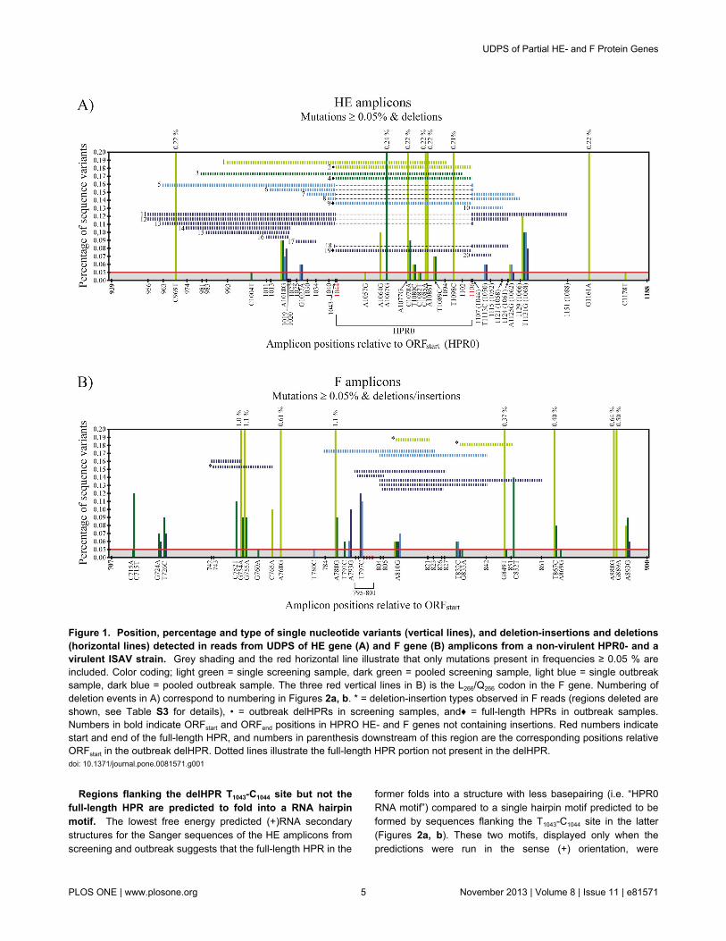
Regions flanking the delHPR T1043-C1044 site but not thefull-length HPR are predicted to fold into a RNA hairpinmotif. The lowest free energy predicted (+)RNA secondarystructures for the Sanger sequences of the HE amplicons fromscreening and outbreak suggests that the full-length HPR in the
former folds into a structure with less basepairing (i.e. “HPR0RNA motif”) compared to a single hairpin motif predicted to beformed by sequences flanking the T1043-C1044 site in the latter(Figures 2a, b). These two motifs, displayed only when thepredictions were run in the sense (+) orientation, were
Figure 1. Position, percentage and type of single nucleotide variants (vertical lines), and deletion-insertions and deletions(horizontal lines) detected in reads from UDPS of HE gene (A) and F gene (B) amplicons from a non-virulent HPR0- and avirulent ISAV strain. Grey shading and the red horizontal line illustrate that only mutations present in frequencies ≥ 0.05 % areincluded. Color coding; light green = single screening sample, dark green = pooled screening sample, light blue = single outbreaksample, dark blue = pooled outbreak sample. The three red vertical lines in B) is the L266/Q266 codon in the F gene. Numbering ofdeletion events in A) correspond to numbering in Figures 2a, b. * = deletion-insertion types observed in F reads (regions deleted areshown, see Table S3 for details), • = outbreak delHPRs in screening samples, and♦ = full-length HPRs in outbreak samples.Numbers in bold indicate ORFstart and ORFend positions in HPRO HE- and F genes not containing insertions. Red numbers indicatestart and end of the full-length HPR, and numbers in parenthesis downstream of this region are the corresponding positions relativeORFstart in the outbreak delHPR. Dotted lines illustrate the full-length HPR portion not present in the delHPR.doi: 10.1371/journal.pone.0081571.g001
UDPS of Partial HE- and F Protein Genes
PLOS ONE | www.plosone.org 5 November 2013 | Volume 8 | Issue 11 | e81571

consistently present with varying input sequence lengths up to800 nts (the largest input length allowed in the program) (not
shown). Comparing UDPS data for HE from outbreak sampleswith predicted RNA structure shows that the 7-dels all start or
Figure 2. RNA secondary structure predictions of HE amplicons using mFold. The lowest free energy predicted structures ofthe RNAs in the sense (+) orientations are shown. A) Sanger sequence of non-virulent HPR0 strain, B) Sanger sequence of thevirulent ISAV strain, and C) partial Sanger sequence from screening with a single A1086→T1086 mutation (position indicated by redcircle in A) and C)). Default settings were used in the predictions, except for temperature which was set to 15°C. Sequencenumbering is according to ORFstart. The nucleotides constituting the HPR0 full-length HPR in A) and C) are shown in green color,and the site were this region is missing in the sequence from the virulent strain (between T1043-C1044) in B) is indicated by a largearrow and “delHPR”. Numbers in brackets indicate the 5’-flanking positions of deletion sites. Here, the numbering, symbols andcolor coding correspond to that used in Figure 1a.doi: 10.1371/journal.pone.0081571.g002
UDPS of Partial HE- and F Protein Genes
PLOS ONE | www.plosone.org 6 November 2013 | Volume 8 | Issue 11 | e81571

end within the predicted hairpin motif, with four starting at orclose to the T1043-C1044 site in the top half of this structure(Figure 2b). As a higher proportion of high frequency mutationsites (11) were present within the full-length HPR fromscreening samples, we also checked whether any of thesesingle nucleotide variants would alter the predicted RNAstructure of the Sanger sequence in this region. Testedseparately, only the A1086→T1086 mutation predicted a significantstructural change in (+)RNA, transforming most of the predictedHPR0 RNA motif into a single hairpin structure similar to thatpredicted for the delHPR (Figures 2a, c). In the pooledsamples, not a single read containing the A1086→T1086 mutationwas present.
F Gene AmpliconsF reads from the single screening sample contain sites
with high mutation frequencies. As observed for HE reads,the prevalence and frequency of mutations in F reads abovecutoff (≥ 0.05 %) were higher for screening samples comparedto outbreak samples (Figure 1b, Table S3). There were 25sites in total with mutational frequency above 0.05%, 22 inscreening- and 7 in outbreak samples (Table S3) (p = 0.013,chi-squared test) supporting also for F the tendency for ahigher number of mutational sites occurring in ampliconsamples from screening- compared to those from outbreak.There were a total of 7584 reads containing mutations in Famplicons with frequency above 0.05%, of which 6799 werefrom screening samples (Table S3). Similar to HE, F ampliconsfrom screening samples has a higher frequency of mutationalsites compared to those from outbreak (p < 0.001, chi-squaredtest). Opposed to all other samples though, the single samplefrom screening contained several sites displaying exceptionallyhigh variability (≥ 1%) (Figure 1b, Table S3). These sites werealso present in high frequency in the pooled sample, althoughwith a prevalence approximately 10x lower.
Frequencies of L266 codon in screening samples and Q266
codon in outbreak samples were not higher than at anyother random site. As the codon immediately upstream of theputative encoded cleavage sites R267 in the F gene marks acrucial difference between HPR0s and vISAVs it was of interestto see whether the codon for this site displayed highervariability. We found that the mutation frequency in this codon,CA797G↔CT797G (Q266↔L266) was not higher than the mutationfrequency at any random site in the two amplicon types(<<0,05%). Also, the A797 vs. T797 does not introduce changesin predicted (+)RNA or (-)RNA structures. In contrast, the onlyother site that differs between the F gene Sanger sequencesfrom screening and outbreak, G755 vs. A755, does introducesignificant changes in predicted RNA structure in theimmediate region in both (+)- and (-) RNA (not shown). Hence,the contribution of this nt difference to the differing mutantspectra profiles between the two types of F amplicons, cannotbe excluded.
Both deletions and insertions are found in F reads, themajority located in proximity to the R267 codon. ISAVstrains with small insertions (originating from other parts of theISAV genome) close to the putative encoded cleavage site R267
in the F gene have been documented [30-32]. In the present
study, reads containing sequence insertions and/or deletionswere observed in low frequency from both screening- andoutbreak samples (4-30 reads). These are all shown in Figure1b and Table S3. Of a total of three deletion-insertion events,two are from the single screening sample where one, a 23 ntinsertion, is located only 6 positions downstream of whereinsertions have been found in some ISAV outbreak strains. Incontrast, the number of reads containing deletions of varyinglengths was higher. Five of a total of eight deletion types (“5-dels”), all from outbreak samples, start at the same position (3),immediately upstream (1) or downstream (1) of were insertionshave been observed in outbreak strains (at two sites separatedby 11 nts). These 5-dels occur within a 12 nt region, which is ahigher occurrence compared to the rest of the amplicon region(p < 0.001, chi-squared test). RNA secondary structurepredictions on the Sanger sequences using mFold suggest thatthe 5-dels, and the 23 nt insertion type from the screeningsample may, similar to that seen for the HE from outbreak (seeabove), occur within hairpin structures (not shown). Foramplicons from screening samples, the hairpin motif was mostprominent when the prediction was run on (-)RNA. Of the 5-dels from outbreak, three were located at the distal tip of apredicted hairpin, the structural feature displayed in all lowestenergy predicted (+/-)RNA structures (up to the maximumallowed input sequence length of 800 nts).
Double Infection with HPR0 and vISAVDelHPR’s in screening samples and vice
versa. Sequencing (Sanger) of upstream regions of the HEgenes from one screening sample (screening sample 3) andone outbreak sample (outbreak sample 3) verified that theoutbreak strain from Site 10 was identical to the outbreak strainfrom two years earlier at the same site. Together withinformation previously obtained from the Astafjord region [28],the sequencing showed that the HPR0 genotype at this sitewas not the likely precursor of the outbreak strain that emergedthree weeks later (not shown). From UDPS, both the single-and pooled sample from screening, low frequency reads(21-30) containing deletion pattern identical to the Site 10delHPR Sanger sequence, were observed (Figure 1a, TableS2). The opposite was also found, reads (22,23) containing full-length HPR in outbreak samples. To confirm their presenceand rule out the possibility of sample cross-contamination, tworeal-time PCR assays using reverse primers differentiatingbetween the full-length HPR and the delHPR were run on allsamples prior to pooling and shipping for UDPS (Table S1).The results, including sequencing of the real-time PCRproducts, verified the presence of these low frequency reads(not shown).
Discussion
UDPS of non-virulent ISAV HPR0- and virulent ISAV strainsis presented for the first time. Gill tissues were sampled fromhealthy fish positive for the HPR0 strain from a screeningregime, and a virulent (v) ISAV strain from a subsequent ISAdisease outbreak at the same fish farming site three weekslater. HE- and F gene regions including the HPR and the codon
UDPS of Partial HE- and F Protein Genes
PLOS ONE | www.plosone.org 7 November 2013 | Volume 8 | Issue 11 | e81571

for the putative encoded R267 cleavage site were amplified byPCR, pyrosequenced and detailed analysis of the sequencedata was performed. The raw UDPS data revealed that roughly10% of the reads in each sample differed from the Sangersequences. Mutant spectra from the two ISAV strains werecompared and linked to predicted viral RNA structures.
Different mutant spectra patterns were observed for both HE-and F amplicons between the HPR0- and vISAV strains, with ahigher number of high frequency mutation sites in screeningthan outbreak samples (see below). Moreover, HE reads fromscreening samples revealed a strong preference for highfrequency mutations within the full-length HPR, suggesting thisregion to be more prone to mutations compared to the flankingregions of the amplicon. For many RNA viruses, studies haveshown that nucleotide sites not forming molecular base pairstend to show higher variability compared to those who do [54].It should therefore not be excluded that the predicted RNAstructure characteristics of the loop-containing HPR0 motifcontributes to the higher frequency of mutations observed inthis region. In F reads from single screening sample, theexceptionally high mutational frequency (≥ 1.0 %) observed atseveral positions may correlate with the fact that this sampleinitially contained a viral load 10-100x higher, as estimated byreal-time RT-PCR (not shown), compared to the individualsamples included in the pooled screening sample. On the otherhand, the CT797G vs. CA797G codons (i.e. the L266 vs. Q266
virulence marker) were not found in higher frequencies inscreening vs. outbreak samples, respectively, than themutational frequency at any random position.
Reads containing deletions were observed in both HE- and Freads while insertions were only observed in the latter. Therewas a higher number of different deletion types in HE readsthan in F reads. For HE, half of the deletion types fromoutbreak samples (7 of 14) start or end within a fourteen ntregion, where two are further deletions from T1043-C1044, the sitewhere the original deletion from a precursor HPR0 genotypecan be hypothesized to have occurred. Similarly, of a total often different insertion-deletion- or deletion types found in Freads, three were found to start at the same position were mostF gene insertions have been found in outbreak strains [30-32].It cannot be excluded that several of these events have beenartificially generated during the PCR amplification step orduring UDPS [55,56]. However, studies have indicated that thein vitro recombination rate in UDPS is low [56]. For HE, thissuggests a tendency towards further deletions from T1043-C1044
following a primary deletion event, possibly linked to increasedviral fitness associated with larger deletions. In fact, closelyrelated vISAVs isolated in the course of the same outbreak atthe same fish farming site have been found to differ only by thesize of their deletions in the HPR, where one isolate is likely tohave originated from a further deletion of the other [57].
The results obtained from UDPS suggest a link betweenmutant spectra profiles and the predicted hairpin motifsobserved with Sanger sequences of HE (+)RNA and F(+/-)RNAs from outbreak, and F (-)RNA from screening. Such aRNA structure may facilitate mutational events such asinsertions (F) and deletions (HE) through mechanisms such asRNA polymerase jumping- and/or template switching during
replication. For influenza A viruses, jumping of the RNApolymerase has been suggested as a mechanism forgenerating deletions and insertions in hemagglutinin andneuraminidase genes [58]. The involvement of RNA structurein the generation of the polybasic cleavage site in highlypathogenic avian influenza A strains, by the polymeraseslipping during strand synthesis upon arrival at a region ofhigher stability, has also been suggested [59]. For F genes, asequence-based non-homologous replicase-driven templateswitch, as observed in positive-stranded RNA viruses, hasbeen proposed as the most likely mechanism behind thesequence insertions observed in some vISAVs [30,60]. Theinserted sequences in F genes were all found to originate fromother parts of the ISAV genome, and not the host, we believemost likely because these sequences are in close proximity toone another, associated with replicase complex, during ISAVreplication in the nucleolus [61].
From the present UDPS data, only one deletion-insertionevent (F reads, pooled outbreak sample) and one deletionevent (HPRO HE reads, single screening sample) seem tohave been generated through the involvement of extensivesequence homology (not shown), although whether these weregenerated artificially or are true quasispecies, is not known. Ingeneral though, recombination is known to be rare in negativestrand- compared to positive-stranded RNA viruses likePicornaviridae and Coronaviridae [60,62]. Our results fromUDPS data- and RNA secondary structure prediction analysissuggests that RNA structural properties may be involved ingenerating the F gene insertions, supported also by the factthat non-homologous recombination is very rare inorthomyxoviruses. For ISAV HE, based on the results fromboth UDPS and RNA structure predictions, a plausablemechanism for creating the deletions in HE is likely to involvethe replicase complex bypassing a sequence stretch on thesame template or jumping to a homologous templatereinitiating strand synthesis further downstream. This event ishypothesized to be driven through alterations in RNA structure(i.e. hairpin structure) following point mutations in the full-lengthHPR. The distributions of deletions and the total absence ofinsertions in HE reads together with current epidemiologicalinformation on vISAVs supports this hypothesis. Compared toprevious publications on this issue, the present results stronglysuggest that the recombination mechanisms creating thedeletions do not involve insertions nor recombination betweenHPR regions between different virulent strains [14].
The high frequency of mutations within the full-length HPR ofscreening samples prompted investigation of whether any ofthese mutations (11 in all) tested individually would change thepredicted (+/-)RNA structure of the Sanger HPR0 motif. Resultsfrom several studies suggest a direct link between the structureof viral RNA and the fidelity of the RNA polymerase [54]. Onlyone mutation, A1086 to T1086, found in reads from the singlescreening sample resulted in a change in the predictedstructure, transforming most of the predicted HPR0 motif intohairpin-like structure. Hence, single mutations in the full-lengthHPR may have the potential to transform this RNA motif intoone resembling the regions flanking the delHPR site, potentiallyfacilitating deletions in this region. In fact, for avian influenza A
UDPS of Partial HE- and F Protein Genes
PLOS ONE | www.plosone.org 8 November 2013 | Volume 8 | Issue 11 | e81571

viruses the generation of the polybasic cleavage sitecharacteristic of high pathogenic strains has been suggestedalso to be mediated by a mechanism in which single nucleotidechanges alter RNA secondary structure facilitating theintroduction of small sequence insertions in this region [63].Here, aside from the fact that the HPR from the singlescreening sample contained roughly twice the amount of highfrequency mutation sites compared to the pooled sample, it isnoteworthy that not a single read from the pooled samplecontained the T1086 mutation. This might reflect a link betweenthe severity of infection by a HPR0 strain, possibly influencedby a high-stress fish farming environment, and the compositionof mutant spectra. Studies have shown that differences inHPR0 viral loads between individuals is linked to the point intime of infection and that the virus may be present, althoughtransiently, in the population for some time, with no clinicalsigns of ISA [27]. The effect may thus be a broadening of themutant spectra in HPR0 quasispecies in spite of low replicationrate for these non-virulent strains. In contrast, upon mutationleading to vISAVs, one sequence that harbours the mutationwill outcompete the other sequences and spread as a singlestrain displaying narrower mutant spectra, at least in the earlystages of the disease outbreak. In the present study, samplingof the vISAV strain was performed in the early phases of thedisease outbreak and displayed many magnitudes higher viralloads compared to the samples from screening.
We hypothesize that the mutant spectra displayed by HPR0infections in a fish farming environment is directly linked to thepotential for deletions in the full-length HPR, and that RNAstructure plays a central role in this transition. Also, theobserved differences in mutant spectra by the two ISAV strains(like the higher number of high frequency mutation sites inscreening vs. outbreak samples) may not only be linked todifferences in infection period but also to replication rates, aswell as to possible variations in the viral RdRps that may affectboth replication rate and replication fidelity, thus changingquasispecies populations and hence viral fitness [42,64-68].Hence, the potential involvement of sequence differences ininternal genes on the differential mutational frequenciesobserved between the HPR0- and vISAV strains cannot beexcluded. Also, the putative gill tropism displayed by HPR0s infarmed and wild Atlantic salmon compared to the systemicnature of vISAV infections, possibly involving different celltypes in the gills [69] and different parts of the immune system,may also be a contributing factor to the observed differences inthe mutant spectra. As ISA has not been observed in wildAtlantic salmon, a future comparative study between mutantspectra from HPR0-positive wild salmon with that obtained inthe present study could provide valuable insight into themechanisms of ISAV transition into virulence. It should also beconsidered that Atlantic salmon may not be the natural host forHPR0s and the transition to vISAV could be viewed as anadaptation of the virus in this particular host.
This study represents the first documentation of a doubleinfection involving non-virulent HPR0s and vISAVs. The lownumber of delHPR reads in screening samples correspondswell with the number of reads containing L266 codons, as do thelow number of full-length HPRs vs. Q266 codons from outbreak
samples. Although statistically unlikely, it cannot be excludedthat one or several of the deletions observed in HE- and Famplicons are linked to the low frequency presence of the otherISAV variant. For single site mutations though, the thresholdlevel set eliminates this potential misinterpretation. FromSanger sequencing of larger portions of the HE genes it wasdeemed unlikely that the HPR0 strain from the screeningsamples was the direct precursor of the vISAV strain in thisparticular fish farm. Hence, many individuals showing no signsof ISA disease were already infected with a virulent strain atthe time of screening, suggesting that infection with a HPR0strain may not protect the fish from ISA disease. Also,vaccination against ISA has shown little or no protectionagainst infection with HPR0s [27]. The opposite finding, i.e. thepresence of full-length HPRs in outbreak samples, could eitherbe due to the fact that sampling was performed in the earlystages of the disease outbreak, or that an earlier infection witha HPR0 virus persists for some time at basal levels in ISA-diseased individuals caused by a virulent strain. Such doubleinfections may actually play an important role in ISAVevolution.
UDPS technology has shown itself to be a valuable tool indetecting minority within-host mutant variants of viral genomesnot detected by traditional means, such as low-levelpersistence of drug resistant mutations in individuals followingprophylactic treatment, or double infections [44,70-73]. Thelatter is also the case here, where the low-frequency presenceof delHPRs in screening samples were not detected throughthe standard diagnostic procedures involving RT-PCR andSanger sequencing, thus demonstrating the future potentialUDPS technology may have in ISAV screening regimes anddiagnostics.
In conclusion, we have performed UDPS of HE- and Fvariable gene regions from samples containing a non-virulentHPR0 strain and a virulent ISAV strain. Detailed analysis andcomparisons of mutant spectra from both individual and pooledsamples revealed marked differences between the two virusstrains. We propose a new hypothesis were the introduction ofselected mutations in the full-length HPR can alter the RNAstructure in such a way that facilitates deletion events in thisgene region, making both quasispecies composition and RNAstructure important factors involved in the two known molecularevents leading to ISAV virulence. The results further strengthenthe hypothesis that deletions, and not recombination, form thevariability of delHPRs in virulent ISAVs and supportobservations from Norway and the Faroe Islands that thistransition to virulence is an infrequent event.
Supporting Information
Table S1. PCR primers and probes.(DOCX)
Table S2. Type, number and prevalence of mutationspresent in ≥ 0.05% of reads and deletions as detected byUDPS of HE amplicons.(DOCX)
UDPS of Partial HE- and F Protein Genes
PLOS ONE | www.plosone.org 9 November 2013 | Volume 8 | Issue 11 | e81571

Table S3. Type, number and prevalence of mutationspresent in ≥ 0.05% of reads, deletions and insertions asdetected by UDPS of F amplicons.(DOCX)
Author Contributions
Conceived and designed the experiments: TM HS TT MJH.Performed the experiments: TM SN MJH. Analyzed the data:TM CMJ ABK. Wrote the manuscript: TM HS CMJ TT ABK JRDHC OBD KF.
References
1. Thorud KE, Djupvik HO (1988) Infectious anemia in Atlantic salmon(Salmo salar L.). Bull Eur Assoc Fish Pathol 8: 109-111
2. Bouchard D, Brockway K, Giray C, Keleher W, Merrill PL (2001) Firstreport of infectious salmon anaemia (ISA) in the United States. Bull EurAssoc Fish Pathol 21: 86-88.
3. Lovely JE, Dannevig BH, Falk K, Hutchin L, MacKinnon AM et al.(1999) First identification of infectious salmon anaemia virus in NorthAmerica with haemorrhagic kidney syndrome. Dis Aquat Organ 35:145-148. doi:10.3354/dao035145. PubMed: 10092978.
4. Mullins J, Groman D, Wadowska D (1998) Infectious salmon anemia insalt water Atlantic salmon (Salmo salar L.) in New Brunswick, Canada.Bull Eur Assoc Fish Pathol 18: 110-114
5. Rodger HD, Richards RH (1998) Haemorrhagic smolt syndrome: asevere anaemic condition in farmed salmon in Scotland. Vet Rec 142:538-541. doi:10.1136/vr.142.20.538. PubMed: 9637379.
6. Lyngrøy C (2003) Infectious salmon anaemia in Norway and theFaeroe Islands: an industrial approach. In: O MillerRC Cipriniano.International Response to Infectious Salmon Anaemia: Prevention,Control and Eradication. pp. 97-109.
7. Rowley HM, Campbell SJ, Curran WL, Turnbull T, Bryson DG (1999)Isolation of infectious salmon anaemia virus (ISAV) from Scottishfarmed Atlantic salmon, Salmo salar L. J Fish Dis 22: 483-487. doi:10.1046/j.1365-2761.1999.00190.x.
8. Godoy MG, Aedo A, Kibenge MJ, Groman DB, Yason CV et al. (2008)First detection, isolation and molecular characterization of infectioussalmon anaemia virus associated with clinical disease in farmedAtlantic salmon (Salmo salar) in Chile. BMC. Vet Res 4: 28.
9. Falk K, Aspehaug V, Vlasak R, Endresen C (2004) Identification andcharacterization of viral structural proteins of infectious salmon anemiavirus. J Virol 78: 3063-3071. doi:10.1128/JVI.78.6.3063-3071.2004.PubMed: 14990725.
10. Rimstad E, Mjaaland S, Snow M, Mikalsen AB, Cunningham CO (2001)Characterization of the infectious salmon anemia virus genomicsegment that encodes the putative hemagglutinin. J Virol 75:5352-5356. doi:10.1128/JVI.75.11.5352-5356.2001. PubMed:11333916.
11. Krossøy B, Devold M, Sanders L, Knappskog PM, Aspehaug V et al.(2001) Cloning and identification of the infectious salmon anaemia virushaemagglutinin. J Gen Virol 82: 1757-1765. PubMed: 11413388.
12. Aspehaug V, Mikalsen AB, Snow M, Biering E, Villoing S (2005)Characterization of the infectious salmon anemia virus fusion protein. JVirol 79: 12544-12553. doi:10.1128/JVI.79.19.12544-12553.2005.PubMed: 16160182.
13. Mjaaland S, Hungnes O, Teig A, Dannevig BH, Thorud K et al. (2002)Polymorphism in the infectious salmon anemia virus hemagglutiningene: importance and possible implications for evolution and ecology ofinfectious salmon anemia disease. Virology 304: 379-391. doi:10.1006/viro.2002.1658. PubMed: 12504577.
14. Devold M, Falk K, Dale B, Krossøy B, Biering E, et al. (2001) Strainvariation, based on the hemagglutinin gene, in Norwegian ISA virusisolates collected from 1987 to 2001: indications of recombination. DisAquat Organ 47: 119-128
15. Castrucci MR, Kawaoka Y (1993) Biologic importance ofneuraminidase stalk length in influenza A virus. J Virol 67: 759-764.PubMed: 8419645.
16. Hossain MJ, Hickman D, Perez DR (2008) Evidence of expanded hostrange and mammalian-associated genetic changes in a duck H9N2influenza virus following adaptation in quail and chickens. PLOS ONE3: e3170. doi:10.1371/journal.pone.0003170. PubMed: 18779858.
17. Li J, Cardona CJ (2010) Adaptation and transmission of a wild duckavian influenza isolate in chickens. Avian Dis 54: 586-590. doi:10.1637/8806-040109-ResNote.1. PubMed: 20521699.
18. Sorrell EM, Song H, Pena L, Perez DR (2010) A 27-amino-acid deletionin the neuraminidase stalk supports replication of an avian H2N2influenza A virus in the respiratory tract of chickens. J Virol 84:11831-11840. doi:10.1128/JVI.01460-10. PubMed: 20826691.
19. Xu R, Zhu X, McBride R, Nycholat CM, Yu W et al. (2012) Functionalbalance of the hemagglutinin and neuraminidase activitiesaccompanies the emergence of the 2009 H1N1 influenza pandemic. JVirol 86: 9221-9232. doi:10.1128/JVI.00697-12. PubMed: 22718832.
20. de Vries E, de Vries RP, Wienholts MJ, Floris CE, Jacobs MS et al.(2012) Influenza A virus entry into cells lacking sialylated N-glycans.Proc Natl Acad Sci U S A 109: 7457-7462. doi:10.1073/pnas.1200987109. PubMed: 22529385.
21. Lu B, Zhou H, Ye D, Kemble G, Jin H (2005) Improvement of influenzaA/Fujian/411/02 (H3N2) virus growth in embryonated chicken eggs bybalancing the hemagglutinin and neuraminidase activities, usingreverse genetics. J Virol 79: 6763-6771. doi:10.1128/JVI.79.11.6763-6771.2005. PubMed: 15890915.
22. Mitnaul LJ, Matrosovich MN, Castrucci MR, Tuzikov AB, Bovin NV et al.(2000) Balanced hemagglutinin and neuraminidase activities are criticalfor efficient replication of influenza A virus. J Virol 74: 6015-6020. doi:10.1128/JVI.74.13.6015-6020.2000. PubMed: 10846083.
23. Shtyrya Y, Mochalova L, Voznova G, Rudneva I, Shilov A et al. (2009)Adjustment of receptor-binding and neuraminidase substratespecificities in avian-human reassortant influenza viruses. Glycoconj J26: 99-109. doi:10.1007/s10719-008-9169-x. PubMed: 18661232.
24. Cook-Versloot M, Griffiths S, Cusack R, McGeachy S, Richie R (2004)Identification and characterization of infectious salmon anaemia virus(ISAV) haemagglutinin gene highly polymorphic region (HPR) type 0 inNorth America. Bull Eur Assoc Fish Pathol 4: 203-208.
25. Cunningham CO, Griffiths S, Black J, Simpson I, Raynard RS (2002) Anovel variant of the infectious salmon anemia virus (ISAV)haemagglutinin gene suggests mechanisms for virus diversity. Bull EurAssoc Fish Pathol 22: 366-374.
26. McBeath AJ, Bain N, Snow M (2009) Surveillance for infectious salmonanaemia virus HPR0 in marine Atlantic salmon farms across Scotland.Dis Aquat Organ 87: 161-169. doi:10.3354/dao02128. PubMed:20099410.
27. Christiansen DH, Østergaard PS, Snow M, Dale OB, Falk K (2011) Alow-pathogenic variant of infectious salmon anemia virus (ISAV-HPR0)is highly prevalent and causes a non-clinical transient infection infarmed Atlantic salmon (Salmo salar L.) in the Faroe Islands. J GenVirol 92: 909-918. doi:10.1099/vir.0.027094-0. PubMed: 21148272.
28. Lyngstad TM, Hjortaas MJ, Kristoffersen AB, Markussen T, Karlsen ETet al. (2011) Use of molecular epidemiology to trace transmissionpathways for infectious salmon anaemia virus (ISAV) in Norwegiansalmon farming. Epidemics 3: 1-11. doi:10.1016/j.epidem.2010.11.001.PubMed: 21420655.
29. Lyngstad TM, Kristoffersen AB, Hjortaas MJ, Devold M, Aspehaug V etal. (2012) Low virulent infectious salmon anaemia virus (ISAV-HPR0) isprevalent and geographically structured in Norwegian salmon farming.Dis Aquat Organ 101: 197-206. doi:10.3354/dao02520. PubMed:23324416.
30. Markussen T, Jonassen CM, Numanovic S, Braaen S, Hjortaas M et al.(2008) Evolutionary mechanisms involved in the virulence of infectioussalmon anaemia virus (ISAV), a piscine orthomyxovirus. Virology 374:515-527. doi:10.1016/j.virol.2008.01.019. PubMed: 18280528.
31. Devold M, Karlsen M, Nylund A (2006) Sequence analysis of the fusionprotein gene from infectious salmon anemia virus isolates: evidence ofrecombination and reassortment. J Gen Virol 87: 2031-2040. doi:10.1099/vir.0.81687-0. PubMed: 16760406.
32. Kibenge FS, Godoy MG, Wang Y, Kibenge MJ, Gherardelli V et al.(2009) Infectious salmon anaemia virus (ISAV) isolated from the ISAdisease outbreaks in Chile diverged from ISAV isolates from Norwayaround 1996 and was disseminated around 2005, based on surfaceglycoprotein gene sequences. Virol J 6: 88. doi:10.1186/1743-422X-6-88. PubMed: 19558648.
33. Bosch FX, Garten W, Klenk HD, Rott R (1981) Proteolytic cleavage ofinfluenza virus hemagglutinins: primary structure of the connectingpeptide between HA1 and HA2 determines proteolytic cleavability andpathogenicity of Avian influenza viruses. Virology 113: 725-735. doi:10.1016/0042-6822(81)90201-4. PubMed: 7023022.
UDPS of Partial HE- and F Protein Genes
PLOS ONE | www.plosone.org 10 November 2013 | Volume 8 | Issue 11 | e81571

34. Senne DA, Panigrahy B, Kawaoka Y, Pearson JE, Süss J et al. (1996)Survey of the hemagglutinin (HA) cleavage site sequence of H5 and H7avian influenza viruses: amino acid sequence at the HA cleavage siteas a marker of pathogenicity potential. Avian Dis 40: 425-437. doi:10.2307/1592241. PubMed: 8790895.
35. Ojosnegros S, Perales C, Mas A, Domingo E (2011) Quasispecies as amatter of fact: viruses and beyond. Virus Res 162: 203-215. doi:10.1016/j.virusres.2011.09.018. PubMed: 21945638.
36. Domingo E, Martin V Perales C, Grande-Perez A, Garcia-Arriaza J etal. (2006) Viruses as quasispecies: biological implications. Curr TopMicrobiol Immunol 299: 51-82. doi:10.1007/3-540-26397-7_3. PubMed:16568896.
37. Domingo E, Sheldon J, Perales C (2012) Viral quasispecies evolution.Microbiol Mol Biol Rev 76: 159-216. doi:10.1128/MMBR.05023-11.PubMed: 22688811.
38. Steinhauer DA, Domingo E, Holland JJ (1992) Lack of evidence forproofreading mechanisms associated with an RNA virus polymerase.Gene 122: 281-288. doi:10.1016/0378-1119(92)90216-C. PubMed:1336756.
39. Ferrer-Orta C, Arias A, Pérez-Luque R, Escarmís C, Domingo E et al.(2007) Sequential structures provide insights into the fidelity of RNAreplication. Proc Natl Acad Sci U S A 104: 9463-9468. doi:10.1073/pnas.0700518104. PubMed: 17517631.
40. Menéndez-Arias L (2002) Molecular basis of fidelity of DNA synthesisand nucleotide specificity of retroviral reverse transcriptases. ProgNucleic Acid Res Mol Biol 71: 91-147. doi:10.1016/S0079-6603(02)71042-8. PubMed: 12102562.
41. Vignuzzi M, Stone JK, Arnold JJ, Cameron CE, Andino R (2006)Quasispecies diversity determines pathogenesis through cooperativeinteractions in a viral population. Nature 439: 344-348. doi:10.1038/nature04388. PubMed: 16327776.
42. Pfeiffer JK, Kirkegaard K (2005) Increased fidelity reduces poliovirusfitness and virulence under selective pressure in mice. PLoS Pathog 1:e11. doi:10.1371/journal.ppat.0010011. PubMed: 16220146.
43. Jerzak GV, Bernard K, Kramer LD, Shi PY, Ebel GD (2007) The WestNile virus mutant spectrum is host-dependant and a determinant ofmortality in mice. Virology 360: 469-476. doi:10.1016/j.virol.2006.10.029. PubMed: 17134731.
44. Hoffmann C, Minkah N, Leipzig J, Wang G, Arens MQ et al. (2007)DNA bar coding and pyrosequencing to identify rare HIV drugresistance mutations. Nucleic Acids Res 35: e91. doi:10.1093/nar/gkm435. PubMed: 17576693.
45. Mitsuya Y, Varghese V, Wang C, Liu TF, Holmes SP et al. (2008)Minority human immunodeficiency virus type 1 variants in antiretroviral-naive persons with reverse transcriptase codon 215 revertantmutations. J Virol 82: 10747-10755. doi:10.1128/JVI.01827-07.PubMed: 18715933.
46. Eriksson N, Pachter L, Mitsuya Y, Rhee SY, Wang C et al. (2008) Viralpopulation estimation using pyrosequencing. PLoS Comput Biol 4:e1000074. PubMed: 18437230.
47. Nishijima N, Marusawa H, Ueda Y, Takahashi K, Nasu A et al. (2012)Dynamics of hepatitis B virus quasispecies in association withnucleos(t)ide analogue treatment determined by ultra-deep sequencing.PLOS ONE 7: e35052. doi:10.1371/journal.pone.0035052. PubMed:22523569.
48. Kuroda M, Katano H, Nakajima N, Tobiume M, Ainai A et al. (2010)Characterization of quasispecies of pandemic 2009 influenza A virus(A/H1N1/2009) by de novo sequencing using a next-generation DNAsequencer. PLOS ONE 5: e10256. doi:10.1371/journal.pone.0010256.PubMed: 20428231.
49. Deng YM, Caldwell N, Hurt A, Shaw T, Kelso A et al. (2011) Acomparison of pyrosequencing and neuraminidase inhibition assays forthe detection of oseltamivir-resistant pandemic influenza A(H1N1) 2009viruses. Antiviral Res 90: 87-91. doi:10.1016/j.antiviral.2011.02.014.PubMed: 21376084.
50. Kulshreshtha V, Kibenge M, Salonius K, Simard N, Riveroll A et al.(2010) Identification of the 3' and 5' terminal sequences of the 8 rnagenome segments of European and North American genotypes ofinfectious salmon anemia virus (an orthomyxovirus) and evidence forquasispecies based on the non-coding sequences of transcripts. Virol J7: 338. doi:10.1186/1743-422X-7-338. PubMed: 21092282.
51. Bornø G, Sviland C, Jensen BB, Biering E, Johansen R, et al. (2010)Helsesituasjonen hos laksefisk 2010 (Health situation in farmed Atlanticsalmon 2010; in Norwegian). In: Fiskehelserapporten 2010 (Fish HealthReport 2010). Report from the Norwegian Veterinary Institute, Norway.pp. 1-36
52. Zuker M (2003) Mfold web server for nucleic acid folding andhybridization prediction. Nucleic Acids Res 31: 3406-3415. doi:10.1093/nar/gkg595. PubMed: 12824337.
53. Falk K, Namork E, Rimstad E, Mjaaland S, Dannevig BH (1997)Characterization of infectious salmon anemia virus, an orthomyxo-likevirus isolated from Atlantic salmon (Salmo salar L.). J Virol 71:9016-9023
54. Sanjuán R, Bordería AV (2011) Interplay between RNA structure andprotein evolution in HIV-1. Mol Biol Evol 28: 1333-1338. doi:10.1093/molbev/msq329. PubMed: 21135148.
55. Görzer I, Guelly C, Trajanoski S, Puchhammer-Stöckl E (2010) Theimpact of PCR-generated recombination on diversity estimation ofmixed viral populations by deep sequencing. J Virol Methods 169:248-252. doi:10.1016/j.jviromet.2010.07.040. PubMed: 20691210.
56. Mild M, Hedskog C, Jernberg J, Albert J (2011) Performance of ultra-deep pyrosequencing in analysis of HIV-1 pol gene variation. PLOSONE 6: e22741. doi:10.1371/journal.pone.0022741. PubMed:21799940.
57. Lyngstad TM, Jansen PA, Sindre H, Jonassen CM, Hjortaas MJ et al.(2008) Epidemiological investigation of infectious salmon anaemia(ISA) outbreaks in Norway 2003-2005. Prev Vet Med 84: 213-227. doi:10.1016/j.prevetmed.2007.12.008. PubMed: 18243376.
58. Fields S, Winter G (1982) Nucleotide sequences of influenza virussegments 1 and 3 reveal mosaic structure of a small viral RNAsegment. Cell 28: 303-313. doi:10.1016/0092-8674(82)90348-8.PubMed: 7060132.
59. Perdue ML, García M, Senne D, Fraire M (1997) Virulence-associatedsequence duplication at the hemagglutinin cleavage site of avianinfluenza viruses. Virus Res 49: 173-186. doi:10.1016/S0168-1702(97)01468-8. PubMed: 9213392.
60. Nagy PD, Simon AE (1997) New insights into the mechansims of RNArecombination. Virology 235: 1-9. doi:10.1006/viro.1997.8681. PubMed:9300032.
61. Goić B, Bustamante J, Miquel A, Alvarez M, Vera MI et al. (2008) Thenucleoprotein and the viral RNA of infectious salmon anemia virus(ISAV) are localized in the nucleolus of infected cells. Virology 379:55-63. doi:10.1016/j.virol.2008.05.036. PubMed: 18632128.
62. Lai MM (1992) RNA recombination in animal and plant viruses.Microbiol Rev 56: 61-79. PubMed: 1579113.
63. García M, Crawford JM, Latimer JW, Rivera-Cruz E, Perdue ML (1996)Heterogeneity in the haemagglutinin gene and emergence of the highlypathogenic phenotype among recent H5N2 avian influenza virusesfrom Mexico. J Gen Virol 77: 1493-1504. doi:10.1099/0022-1317-77-7-1493. PubMed: 8757992.
64. Arias A, Arnold JJ, Sierra M, Smidansky ED, Domingo E et al. (2008)Determinants of RNA-dependent RNA polymerase (in)fidelity revealedby kinetic analysis of the polymerase encoded by a foot-and-mouthdisease virus mutant with reduced sensitivity to ribavirin. J Virol 82:12346-12355. doi:10.1128/JVI.01297-08. PubMed: 18829745.
65. Coffey LL, Beeharry Y, Bordería AV, Blanc H, Vignuzzi M (2011)Arbovirus high fidelity variant loses fitness in mosquitoes and mice.Proc Natl Acad Sci U S A 108: 16038-16043. doi:10.1073/pnas.1111650108. PubMed: 21896755.
66. Agudo R, Ferrer-Orta C, Arias A, de la Higuera I, Perales C et al.(2010) A multi-step process of viral adaptation to a mutagenicnucleoside analogue by modulation of transition types leads toextinction-escape. PLOS Pathog 6: e1001072.
67. Vignuzzi M, Wendt E, Andino R (2008) Engineering attenuated virusvaccines by controlling replication fidelity. Nat Med 14: 154-161. doi:10.1038/nm1726. PubMed: 18246077.
68. Andreoni M (2004) Viral phenotype and fitness. New Microbiol 27:71-76. PubMed: 15646067.
69. Aamelfot M, Dale OB, Weli SC, Koppang EO, Falk K (2012) Expressionof the infectious salmon anemia virus receptor on atlantic salmonendothelial cells correlates with the cell tropism of the virus. J Virol 86:10571-10578. doi:10.1128/JVI.00047-12. PubMed: 22811536.
70. Deyde VM, Gubareva LV (2009) Influenza genome analysis usingpyrosequencing method: current applications for a moving target.Expert Rev Mol Diagn 9: 493-509. doi:10.1586/erm.09.21. PubMed:19580433.
71. Pingen M, Nouwen JL, Dinant S, Albert J, Mild M et al. (2012) Therapyfailure resulting from superinfection by a drug-resistant HIV variant.Antivir Ther 17: 1621-1625. doi:10.3851/IMP2267. PubMed: 22846173.
72. Ko SY, Oh HB, Park CW, Lee HC, Lee JE (2012) Analysis of hepatitisB virus drug-resistant mutant haplotypes by ultra-deep pyrosequencing.Clin Microbiol Infect 18: E404-E411. doi:10.1111/j.1469-0691.2012.03951.x. PubMed: 22757653.
73. Newman RM, Kuntzen T, Weiner B, Berical A, Charlebois P et al.(2013) Whole Genome Pyrosequencing of Rare Hepatitis C VirusGenotypes Enhances Subtype Classification and Identification ofNaturally Occurring Drug Resistance Variants. J Infect Dis 208: 17-31.PubMed: 23136221.
UDPS of Partial HE- and F Protein Genes
PLOS ONE | www.plosone.org 11 November 2013 | Volume 8 | Issue 11 | e81571

Table S1: PCR primers and probes.
Assay type Primer/probe name Primer/probe sequence (5’-3’)
Pyrosequencinga
s6 HPR_Fpyro CGTATCGCCTCCCTCGCGCCATCAGACGAGTG
CGTG913ACCAGACAAGCTTAGGTAACACAGA
s6 HPR_Rpyro CTATGCGCCTTGCCAGCCCGCTCAGACGCTCGA
CAG1217ATGGTGGAATTCTACCTCTAGACTTGTA
s5 cleav_Fpyro CGTATCGCCTCCCTCGCGCCATCAGAGACG
CACTCG683AGTAGTGCCGTTCCATTCTGTAC
s5 cleav_Rpyro CTATGCGCCTTGCCAGCCCGCTCAGAGC
ACTGTAGT920CTTCTGCGGATGCTGCACC
Real-time PCR 1b s6HPR_F GACCAGACAAGCTTAGGTAACACAGA
s6delHPR_R CATAGAAATGAGCTGAGGTGGGAT
s6delHPR_MGB (6-FAM)ACCTCCCTCATGATAAGT(MGBNFQ)
Real-time PCR 2c s6HPR0_F CTCAGCTGAACCAAACATTCAATACA
s6HPR0_R GAAATGAAGATGTTACTCAACACAGATGT
s6HPR0_MGB (6-FAM)ACCAAGTAGAGCAACCTG(MGBNFQ)
aTemplate specific sequences in segment 6 (HE gene) and segment 5 (F gene) primers are in
bold. The remaining are sequences necessary for pyrosequencing in both directions and
multiplexing; adaptor sequences (normal font), library keys (italics) and multiplex identifiers
(MIDs) (underlined). Lower case letters indicate positions in HE HPR0- and F gene ORFs,
respectively.
bReal-time PCR for detection of low-frequency delHPR reads in screening samples.
cReal-time PCR for detection of low-frequency full-length HPR reads in outbreak samples.

Table S2: Type, number and prevalence of mutations present in 0.05% of reads and
deletions as detected by UDPS of HE amplicons.
HE amplicon Shared mutations in forward and reverse Deletions in readsd
sample(s) reads with prevalence 0.05%
(direction of read: # of reads/% frequency)c
SCN1a C969T (F:141/0.28, R:155/0.22) p993p1094 (1)
A1018G (F:52/ 0.10, R 63/0.09) p1044 p1106 (2)e
A1057G (F:25,/0.05, R 38/0.05)
A1064C (F:60/0.12, R:69/0.10)
A1077G (F:134/0.27, R:151/0.22)
T1080C (F:41/0.08, R:42/0.06)
C1082T (F:27/0.05, R:45/0.06)
C1085A (F:133/0.26, R:151/0.22)
A1086T (F:136/0.27, R:152/0.22)
T1089C (F:50/0.10, R:48/0.07)
T1098C (F:135/0.27, R:149/0.21)
A1125G (F:31/0.06, R:55/0.08)
T1131G (F:63/0.12, R:81/0.12)
G1161A (F:112/0.22, R:164/0.24)
C1178T (F:25/0.05, R:40/0.06)
SCN pool C1004T (F:14/0.05, R: 46/0.07) p981p1102 (3)
(SCN2, SCN3, SCN4) A1018G (F: 26/0.10, R: 59/0.09) p1044 p1106 (4)f
A1067G (F:61/0.24, R:164/0.24)
C1078A (F:28/0.11, R:(65/0.09)
T1080C (F:27/0.11, R:38/0.06)
C1082T (F:18/0.07, R:35/0.05)
T1089C (F:27/0.11, R:46/0.07)
T1131G (F:26/0.10, R:71/0.10)
OBK1b A1018G (F:37/0.07, R:74/0.08) p963p1044 (5)
G1027A (F:40/0.08, R:55/0.06) p1013p1044 (6)
T1050C (F:30/0.06, R:55/0.06) p1030p1062 (7)
A1062G (F:42/0.08, R:54/0.06) p1040p1066 (8)
T1068G (F:54/0,10, R:95/0,11) p1044 p1106 (9)g
p1044p1058 (10)
OBK pool A1018G (F:33/0.08, R:50/0.10) p956p1088 (11)
(OBK2, OBK3, OBK4) G1027A (F:24/0.06, R:40/0.08) p956p1061 (12)
T1050C (F:34/0.08, R:30/0.06) p963p1044 (13)
A1062G (F:21/0.05, R:28/0.05) p974p1022 (14)
T1068G (F:31/0.08, R:63/0.12) p983p1020 (15)
p1011p1019 (16)
p1025p1034 (17)
p1043p1061 (18)
p1044 p1106 (19)h
p1044p1052 (20) aSCN = screening sample. Mutations within the HPR0 full-length HPR are shown in bold.

bOBK = outbreak sample.
cNumbering according to start of open reading frame (ORFstart).
dReads containing insertions were not observed.
e,fSite 10 outbreak delHPR.
g,hFull-length HPR (numbering according to ORFstart for HPR0s).
Numbers and symbols in parenthesis in the right column (1-20) correspond to that used in
Figures 1a, 2a and 2b.

Table S3: Type, number and prevalence of mutations present in 0.05% of reads, deletions
and insertions as detected by UDPS of F amplicons.
F amplicon Shared mutations in forward and reverse Deletions/insertions
sample(s) reads with prevalence 0.05%. in reads
(direction of read: # of reads/% frequency)c
SCN1a G754A (F:455/1.00, R:534/1.10) p810p821 (*)
d
G755A (F:478/1.10, R:536/1.10) p833p851 (*)e
C765A (F:46/0.10, R:53/0.11)
A768G (F:272/0.61, R:321/0.65)
A788G (F:450/1.01, R:534/1.10)
A810G (F:29/0.07, R:31/0.06)
G849T (F:166/0.37, R:193/0.39)
T867C (F:178/0.40, R:202/0.41)
A888G (F:287/0.64, R:346/0.70)
G889A (F:222/0.50, R:315/0.64)
A893G (F:35/0.08, R:46/0.09)
SCN pool C715A (F:22/0.07, R:19/0.05)
(SCN2, SCN3, SCN4) C715T (F:37/0.12, R:51/0.14)
G724A (F:27/0.09, R:26/0.07)
T726C (F:27/0.09, R:31/0.09)
C752T (F:42/0.13, R:38/0.11)
G754A (F:30/0.10, R:34/0.09)
G755A (F:29/0.09, R:52/0.15)
G760A (F:32/0.10, R:18/0.05)
A788G (F:29/0.09, R:42/0.11) -
T791C (F:18/0.06 R:24/0.07)
A810G (F:31/0.10, R:22/0.06)
T832C (F:19/0.06, R:27/0.08)
G833A (F:25/0.08, R:19/0.05)
G849T (F:16/0.05, R:29/0.08)
C852T (F:48/0.15, R:51/0.14)
T867C (F:24/0.08, R:37/0.10)
A869G (F:16/0.05, R:21/0.06)
A893G (F:28/0.09, R:29/0.08)
OBK1b T780C (F:16/0.05, R:24/0.05) p784p823
A793G (F:18/0.06, R:35/0.07) p804p842
T797C (F:37/0.12, R:52/0.11)
A810G (F:21/0.07, R:33/0.07)
T832C (F:27/0.09, R:29/0.06)
OBK pool G724A (F:27/0.06, R:31/0.07) p742p754

(OBK2, OBK3, OBK4) T726C (F:31/0.07, R:29/0.07) p743p765 (*)f
A793G (F:44/0.10, R:44/0.10) p795p827
T797C (F:56/0.12, R:56/0.13) p796p805
A810G (F:29/0.06, R:32/0.07) p804p861
T832C (F:23/0.05, R:30/0.07) p804p842
A893G (F:29/0.06, R:32/0.07) p805p826 aSCN = screening sample.
bOBK = outbreak sample.
cNumbering according to start of open reading frame (ORFstart).
dDeletion replaced by 23 nt insertion.
eDeletion replaced by 25 nt insertion.
fDeletion replaced by 20 nt insertion.
Asterisks in parenthesis in the right column correspond to that used in Figure 1b.
Copyright © 2022 FDOKUMEN