Ultrastructure of equine morbillivirus
15
~:, "All ELSEVIER Virus Research 43 (1996) 1-15 Virus Ultrastructure of equine morbillivirus Alex D. Hyatt*, Paul W. Selleck Australian Animal Health Laboratory, C.S.I.R.O., P.O. Bag 24, Geelong, Victoria 3220~ Australia Received 5 December 1995; accepted 28 February 1996 Abstract The ultrastructure of the equine morbillivirus (EMV) which was implicated in the death of one human and fourteen horses in Queensland, Australia during September 1994 and a 36 year old man from Queensland in October 1995 is described. The ultrastructure of the virus and the intracellular virus-specific structures are characteristic for the family Paramyxoviridae. Cytoplasmic nucleocapsids were observed within the infected cells monolayers, endothelial cells (lung) of infected horses and the neurons within the brain of the 36 year old Queensland man. Aggregates of smaller nucleocapsid-like structures were also observed within the brain of the same man; these did not react with sera from recovered EMV-infected horses or from a recovered EMV-infected human. Co-examination of rinderpest virus (RPV), bovine parainfluenza-3 (BPIV-3), human respiratory virus (HRSV) and Sendai virus revealed that their envelope-associated surface projections are equivalent in length to the 15 nm spikes of EMV. EMV differed from these other viruses in that the majority of virions possessed surface projections of two distinct lengths (18 and 15 nm). Further ultrastructural examinations of plaque purified EMV revealed a small percentage of EM viruses possessed a mixed array of surface projections indicating that the 'double-fringed' (DF) particles may be the result of a post-translational modification(s). Keywords: Equine morbillivirus; Paramyxoviridae; Acute equine respiratory syndrome; Transmission electron mi- croscopy; Ultrastructure; Immunogold-labelling I. Introduction On the 7th of September 1994 a pregnant mare was removed from a paddock near Brisbane, Queensland, Australia, to a horse stable in the suburbs. The horse was observed to be ill and died two days later. By the 26th of September a total of 21 horses on two properties had become ill suffering from a disease known as acute equine * Corresponding author. respiratory syndrome (Murray et al., 1995a,b). Of these horses, 14 died or were euthanased (case 1). Six days after the index case died a stable-hand and horse trainer, both of whom worked in one of the stables and who had close contact with the index case, became ill with an influenza-like illness (Selvey et al., 1995). The stable-hand recovered but the trainer was admitted to hospital where he later died (case 1). During September/October 1995 another human (case 2) was identified to have antibodies against EMV (O'Sullivan et al., 1996). This 36 year old man from Queensland had 0168-1702/96/$15.00 © 1996 Elsevier Science B.V. All rights reserved PII SO 168-1702(96)01307-X
Transcript of Ultrastructure of equine morbillivirus
~:, "All
E L S E V I E R Virus Research 43 (1996) 1-15
V i r u s
Ultrastructure of equine morbillivirus
Alex D. Hyatt*, Paul W. Selleck Australian Animal Health Laboratory, C.S.I.R.O., P.O. Bag 24, Geelong, Victoria 3220~ Australia
Received 5 December 1995; accepted 28 February 1996
Abstract
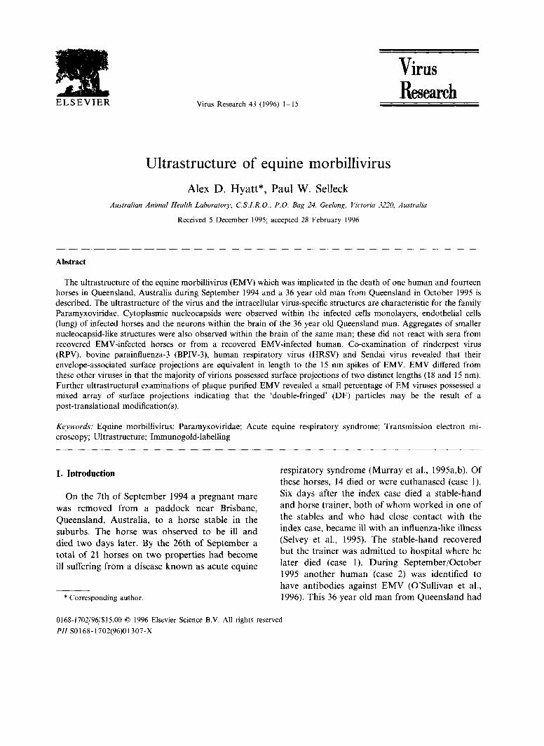
The ultrastructure of the equine morbillivirus (EMV) which was implicated in the death of one human and fourteen horses in Queensland, Australia during September 1994 and a 36 year old man from Queensland in October 1995 is described. The ultrastructure of the virus and the intracellular virus-specific structures are characteristic for the family Paramyxoviridae. Cytoplasmic nucleocapsids were observed within the infected cells monolayers, endothelial cells (lung) of infected horses and the neurons within the brain of the 36 year old Queensland man. Aggregates of smaller nucleocapsid-like structures were also observed within the brain of the same man; these did not react with sera from recovered EMV-infected horses or from a recovered EMV-infected human. Co-examination of rinderpest virus (RPV), bovine parainfluenza-3 (BPIV-3), human respiratory virus (HRSV) and Sendai virus revealed that their envelope-associated surface projections are equivalent in length to the 15 nm spikes of EMV. EMV differed from these other viruses in that the majority of virions possessed surface projections of two distinct lengths (18 and 15 nm). Further ultrastructural examinations of plaque purified EMV revealed a small percentage of EM viruses possessed a mixed array of surface projections indicating that the 'double-fringed' (DF) particles may be the result of a post-translational modification(s).
Keywords: Equine morbillivirus; Paramyxoviridae; Acute equine respiratory syndrome; Transmission electron mi- croscopy; Ultrastructure; Immunogold-labelling
I. Introduction
On the 7th of September 1994 a pregnant mare was removed from a paddock near Brisbane, Queensland, Australia, to a horse stable in the suburbs. The horse was observed to be ill and died two days later. By the 26th of September a total of 21 horses on two properties had become ill suffering from a disease known as acute equine
* Corresponding author.
respiratory syndrome (Murray et al., 1995a,b). Of these horses, 14 died or were euthanased (case 1). Six days after the index case died a stable-hand and horse trainer, both of whom worked in one of the stables and who had close contact with the index case, became ill with an influenza-like illness (Selvey et al., 1995). The stable-hand recovered but the trainer was admitted to hospital where he later died (case 1). During September/October 1995 another human (case 2) was identified to have antibodies against EMV (O'Sullivan et al., 1996). This 36 year old man from Queensland had
0168-1702/96/$15.00 © 1996 Elsevier Science B.V. All rights reserved PII SO 168-1702(96)01307-X

2 A.D. Hyatt, P.W. Selleck / Virus Research 43 (1996) 1-15
assisted his wife (a veterinarian) in the necropsies of horses which were later identified to have been infected with EMV (Hooper et al., 1996a).
Viral nucleocapsids, characteristic of viruses within the family Paramyxoviridae, were iden- tified within the lungs of clinically affected horses (Murray et al., 1995a,b) and from the brain of the afflicted 36 year old Queensland man (O'Sullivan et al., 1996). Viruses from the original outbreak were isolated in cell cultures which had been inoculated with the lung homogenates from horses and a kidney homogenate from the de- ceased trainer. No virus was isolated from the second case (O'Sullivan et al., 1996). When the cell cuiture virus and original horse lung ho- mogenates were administered to experimental horses, all animals became ill with an acute res- piratory illness and died or were euthanased 8-12 days post inoculation. The subsequent identifica- tion of virus within the lung tissues and virus isolation and identification from cell culture demonstrated that the aetiological agent belonged to the family Paramyxoviridae (Murray et al., 1995a,b). However, when the virus was tested against antisera to a range of paramyxoviruses, morbilliviruses and pneumoviruses, all results were negative except for weak reactions against rinderpest antisera (Murray et al., 1995a). The virus was also unusual in that it did not possess any detectable neuraminidase activity and did not agglutinate erythrocytes from a range of species (Murray et al., 1995b). The virus was identified as being distantly related to the genus Morbillivirus by sequencing regions of the matrix (M), fusion (F) and phospho (P) proteins (Murray et al., 1995a; Gould, 1996) and is now referred to as equine morbillivirus (EMV), although Gould (1996) states that as EMV has many features with viruses from the genera Morbillivirus and Paramyxovirus it may fulfil the role in bridging these two genera or be a separate genus.
Limited experimental studies on this virus (Murray et al., 1995a; Westbury et al., 1995) have shown that it can infect a range of tissue culture cells including those derived from mammals, fish, reptiles, amphibians and birds and also has the potential to lethally infect domestic cats and guinea pigs. The zoonotic nature of EMV, it's
apparent potential to infect a range of organism and lack of serological cross-reactivity with viruses from the different genera of the family Paramyxoviridae makes this pathogen an impor- tant scientific, medical and veterinary agent for further studies. There is a need for further infor- mation on the transmission and pathogenesis of the virus, molecular biology of the genome and ultrastructure of the virus.
The original reports of this disease (Murray et al., 1995a, b) included a brief ultrastructural de- scription of the virus. In this paper we describe and illustrate the ultrastructure of plaque purified EMV, EMV-infected cells, EMV within the origi- nal and experimental horse lungs and compare the ultrastructure of the virus with representative viruses from the family Paramyxoviridae. The pa- per also describes the presence of varied nude- ocapsid structures within the brain of the 36 year old Queensland man and presents immuno-elec- tron microscopical evidence which indicates that at least one of these structures is antigenically related to EMV.
2. Materials and methods
2.1. Virus isolation and cell culture (case 1)
Tissue homogenates from the spleen and lung from two of the affected Queensland horses and from the lung, liver, kidneys and lymph nodes of experimental horses were inoculated onto a range of cell culture monolayers including VERO (ATCC CCL 81), BHK-21 (ATCC CCL 10), RK- 13 (ATCC CCL 37), MDBK (ATCC CCL 22) and primary foetal equine kidney cells. Similarly, lung and kidney homogenates from the trainer were inoculated onto confluent VERO cell layers. All cells were grown in Eagle's minimum essential medium (EMEM) with Eagle's salts supplemented with 10% foetal bovine serum. When a cytopathic effect (CPE) was observed the cells and superna- tant were examined by fluorescence, negative con- trast electron microscopy and prepared for examination via ultra-thin sections (refer below).
A.D. Hyatt, P.W. Selleck / Virus Research 43 (1996) 1-15 3
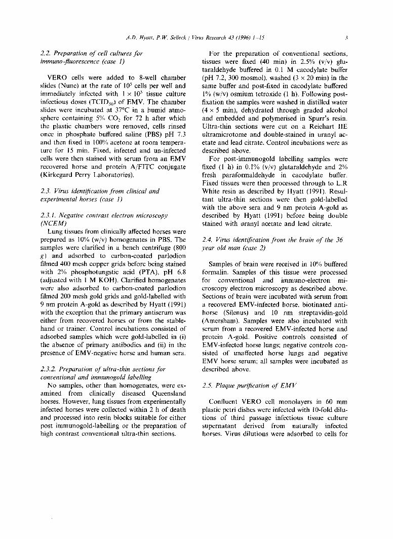
2.2. Preparation of cell cultures for immuno-fluorescence (case 1)
VERO cells were added to 8-well chamber slides (Nunc) at the rate of 105 cells per well and immediately infected with 1 x 10 3 tissue culture infectious doses (TCIDso) of EMV. The chamber slides were incubated at 37°C in a humid atmo- sphere containing 5% CO2 for 72 h after which the plastic chambers were removed, cells rinsed once in phosphate buffered saline (PBS) pH 7.3 and then fixed in 100% acetone at room tempera- ture for 15 rain, Fixed, infected and un-infected cells were then stained with serum from an EMV recovered horse and protein A/FITC conjugate (Kirkegard Perry Laboratories).
2.3. Virus identification from clinical and experimental horses (case 1)
2.3.1. Negative contrast electron microscopy (NCEM)
Lung tissues from clinically affected horses were prepared as 10% (w/v) homogenates in PBS. The samples were clarified in a bench centrifuge (800 g) and adsorbed to carbon-coated parlodion filmed 400 mesh copper grids before being stained with 2% phosphotungstic acid (PTA), pH 6.8 (adjusted with 1 M KOH). Clarified homogenates were also adsorbed to carbon-coated parlodion filmed 200 mesh gold grids and gold-labelled with 9 nm protein A-gold as described by Hyatt (1991) with the exception that the primary antiserum was either from recovered horses or from the stable- hand or trainer. Control incubations consisted of adsorbed samples which were gold-labelled in (i) the absence of primary antibodies and (ii) in the presence of EMV-negative horse and human sera.
2.3.2. Preparation of ultra-thin sections for conventional and immunogold labelling
No samples, other than homogenates, were ex- amined from clinically diseased Queensland horses. However, lung tissues from experimentally infected horses were collected within 2 h of death and processed into resin blocks suitable for either post immunogold-labelling or the preparation of high contrast conventional ultra-thin sections.
For the preparation of conventional sections, tissues were fixed (40 min) in 2.5% (v/v) glu- taraldehyde buffered in 0.1 M cacodylate buffer (pH 7.2, 300 mosmol), washed (3 × 20 min) in the same buffer and post-fixed in cacodylate buffered 1% (w/v) osmium tetroxide (1 h). Following post- fixation the samples were washed in distilled water (4 x 5 min), dehydrated through graded alcohol and embedded and polymerised in Spurr's resin. Ultra-thin sections were cut on a Reichart liE ultramicrotome and double-stained in uranyl ac- etate and lead citrate. Control incubations were as described above.
For post-immunogold labelling samples were fixed (1 h) in 0.1% (v/v) glutaraldehyde and 2% fresh paraformaldehyde in cacodylate buffer. Fixed tissues were then processed through to L.R White resin as described by Hyatt (1991). Resul- tant ultra-thin sections were then gold-labelled with the above sera and 9 nm protein A-gold as described by Hyatt (1991) before being double stained with uranyl acetate and lead citrate.
2.4. Virus identification from the brain of the 36 year oM man (case 2)
Samples of brain were received in 10% buffered formalin. Samples of this tissue were processed for conventional and immuno-electron mi- croscopy electron microscopy as described above. Sections of brain were incubated with serum from a recovered EMV-infected horse, biotinated anti- horse (Silenus) and l0 nm streptavidin-gold (Amersham). Samples were also incubated with serum from a recovered EMV-infected horse and protein A-gold. Positive controls consisted of EMV-infected horse lungs; negative controls con- sisted of unaffected horse lungs and negative EMV horse serum; all samples were incubated as described above.
2.5. Plaque purification of E M V
Confluent VERO cell monolayers in 60 mm plastic petri dishes were infected with 10-fold dilu- tions of third passage infectious tissue culture supernatant derived from naturally infected horses. Virus dilutions were adsorbed to cells for

4 A.D. Hyatt, P.W. Selleck/ Virus Research 43 (1996) 1-15
60 min at 37°C with frequent rocking, inoculums removed and cell monolayers washed with PBS. The infected cell cultures were overlaid with growth medium containing 1% bacteriological agar and incubated at 37°C in 5% CO2 for 72 h, after which they were stained with 1% agar con- taining 0.01% neutral red. After a further overnight incubation plaques from the highest dilution were selected for further passage. Selected plaques were inoculated onto confluent VERO cell monolayers in tissue culture flasks and incu- bated at 37°C until the CPE was complete. A further two cycles of plaquing were performed resulting in a total of 125 plaques. Five of these plaques were selected at random and examined by NCEM.
2.6. Growth and examination of paramyxoviruses, morbilliviruses and pneumoviruses
Bovine parainfluenza virus 3 (BPIV-3, genus Paramyxovirus) and Rinderpest virus (RPV, genus Morbillivirus) were grown in MDBK cell monolayers; human respiratory syncytial virus (HRSV, genus Pneumovirus) in HeLa cell (ATCC CCL 2) monolayers and Sendai virus (genus Paramyxovirus) in embryonated chicken eggs.
Supernatants from the infected cell monolayers were prepared for NCEM as described above. Supernatants from EMV-infected cells and any one of the above were mixed and adsorbed to a single grid. As nucleocapsids of HRSV were difficult to observe in HRSV-infected cell superna- tants and disrupted cells, grid cell-cultures (GCCs) of HeLa cells and HRSV were prepared as described by (Hyatt et al., 1987). GCCs were harvested at four days post-infection and fixed and stained as described by (Hyatt et al., 1987). Sendai virus was propagated by inoculation into the allantoic cavity of nine-day-old embryonated chicken eggs. After five days incubation at 37°C the allantoic fluid was harvested, clarified by cen- trifugation (2000 g) and the virus pelleted by centrifugation (80000 g). The pellet was resus- pended in TNE buffer (Tris 0.01 M, NaCI 0.1 M, EDTA 0.001 M) pH 7.2 to a final volume of 1% of the original.
All micrographs were recorded from a Hitachi H600 scanning transmission electron microscope at 75 kV. Magnification was standardised with catalase crystals and a 2160 line/mm diffraction grating replica; photographs were recorded in as- cending magnification.
3. Results
3.1. Identification of virus nucleocapsids within lung homogenates of clinical and experimental horses (case 1)
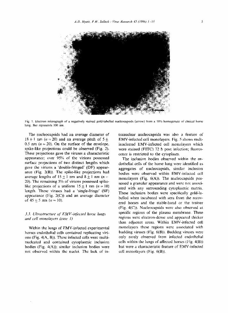
No virus particles were observed within the lung homogenates from clinically and experimen- tally affected horses. Nucleocapsids were, how- ever, observed in all homogenates and were specifically gold-labelled following incubation with serum from recovered EMV-infected horses and both the trainer and stable-hand (Fig. 1). When the homogenates were incubated with EMV-negative sera (human and equine) no gold- labelling occurred (data not shown). The nucle- ocapsids had an approximate diameter of 18 nm and a pitch of 5 nm.
3.2. Identification of viruses and nucleocapsids within the supernatant of EMV-infected cell monolayers (case 1)
Within the supernatant of cells which were inoculated with organ homogenates from the trainer and clinically and experimentally infected horses, viruses and nucleocapsids were observed.
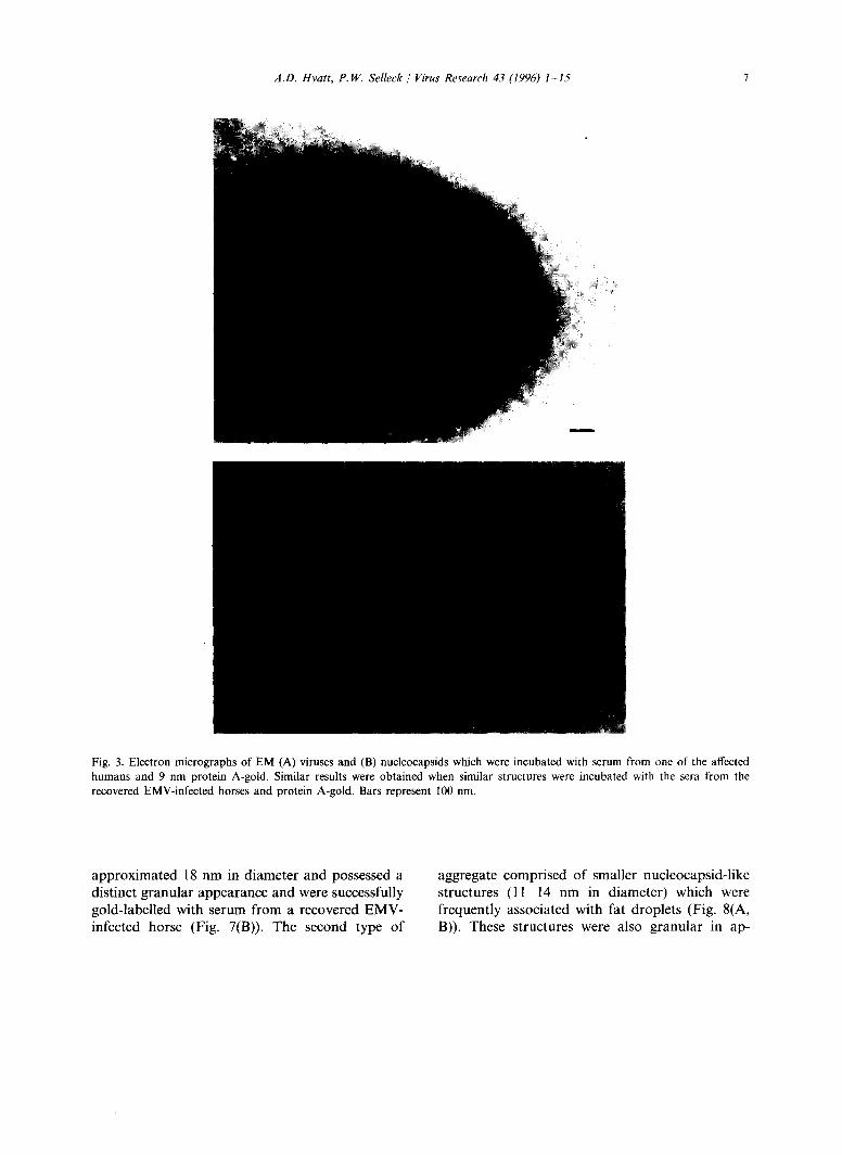
The viruses ranged in size from approximately 40 to 600 nm and were pleomorphic in shape varying from spherical to filamentous forms. Viri- ons possessed a limiting envelope which enclosed a nucleocapsid. It was not uncommon to observe ruptured virions and flee-lying nucleocapsids (Fig. 2(A)). Both the viruses and nucleocapsids were specifically gold-labelled following incuba- tion with sera from recovered EMV-infected horses and from the stable-hand and trainer (Fig. 3); incubations in the absence of primary antibody and with EMV-negative sera (refer above) failed to gold-label either of the structures (data not shown).
A . D . H y a t t , P . W . S e l l e c k / Virus Research 43 (1996) 1 - 1 5 5
~ i~ii~ ~ i; iiiiii~ili
, <!!~ ? i ~ ¸ ¸
Fig. 1. Electron micrograph of a negatively stained gold-labelled nucleocapsids (arrow) from a 10% homogenate of clinical horse lung. Bar represents 100 nm.
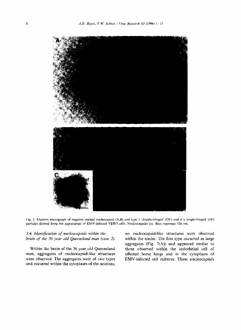
The nucleocapsids had an average diameter of 18 _+ 1 nm (n = 20) and an average pitch of 5 _+ 0.5 nm (n = 20). On the surface of the envelope, spike-like projections could be observed (Fig. 2). These projections gave the viruses a characteristic appearance; over 95% of the virions possessed surface projections of two distinct lengths which gave the virions a 'double-fringed' (DF) appear- ance (Fig. 2(B)). The spike-like projections had average lengths of 15 _+ 1 nm and 8 + 1 nm (n = 20). The remaining 5% of virions possessed spike- like projections of a uniform 15 + 1 nm (n = 10) length. These viruses had a 'single-fringe' (SF) appearance (Fig. 2(C)) and an average diameter of 4 5 ± 5 n m ( n = 1 0 ) .
3.3. Ultrastructure of EMV-infected horse lungs and cell monolayers (case I)
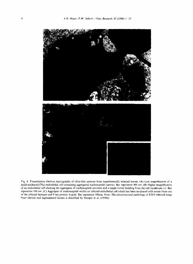
Within the lungs of EMV-infected experimental horses endothelial cells contained replicating viri- ons (Fig. 4(A, B)). These infected cells were multi- nucleated and contained cytoplasmic inclusion bodies (Fig. 4(A)); similar inclusion bodies were not observed within the nuclei. The lack of in-
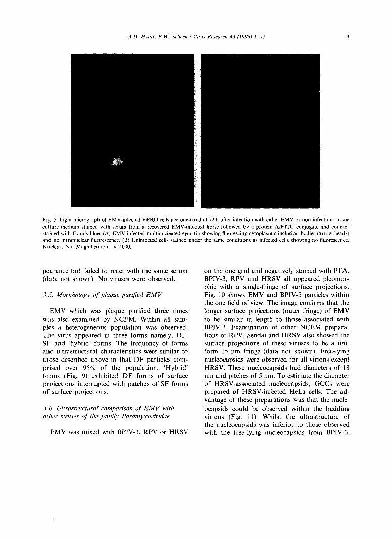
tranuclear nucleocapsids was also a feature of EMV-infected cell monolayers. Fig. 5 shows mult- inucleated EMV-infected cell monolayers which were stained (FITC) 72 h post infection; fluores- cence is restricted to the cytoplasm.
The inclusion bodies observed within the en- dothelial cells of the horse lung were identified as aggregates of nucleocapsids, similar inclusion bodies were observed within EMV-infected cell monolayers (Fig. 6(A)). The nucleocapsids pos- sessed a granular appearance and were not associ- ated with any surrounding cytoplasmic matrix. These inclusion bodies were specifically gold-la- belled when incubated with sera from the recov- ered horses and the stable-hand or the trainer (Fig. 4(C)). Nucleocapsids were also observed at specific regions of the plasma membrane. These regions were electron-dense and appeared thicker than adjacent areas. Within EMV-infected cell monolayers these regions were associated with budding viruses (Fig. 6(B)). Budding viruses were only rarely observed from infected endothelial cells within the lungs of affected horses (Fig. 4(B)) but were a characteristic feature of EMV-infected cell monolayers (Fig. 6(B)).
6 A.D. Hyatt, P.W. Selleck/ Virus Research 43 (1996) 1--15
Fig. 2. Electron micrograph of negative stained nucleocapsid (A,B) and type l 'double-fringed' (DF) and (C) 'single-fringed' (SF) particles derived from the supernatant of EMV-infected VERO cells. Nucleocapsids (n). Bars represent 100 nm.
3.4. Identification of nucleocapsids within the brain of the 36 year old Queensland man (case 2)
Within the brain of the 36 year old Queensland man, aggregates of nucleocapsid-like structures were observed. The aggregates were of two types and occurred within the cytoplasm of the neurons;
no nucleocapsid-like structures were observed within the nuclei. The first type occurred in large aggregates (Fig. 7(A)) and appeared similar to those observed within the endothelial cell of affected horse lungs and in the cytoplasm of EMV-infected cell cultures. These nucleocapsids
A.D. Hyatt, P.W. Selleck / Virus Research 43 (1996) 1-15 7
• ~,• • . . . . . . . ~i~ I
Fig. 3. Electron micrographs of EM (A) viruses and (B) nucleocapsids which were incubated with serum from one of the affected humans and 9 nm protein A-gold. Similar results were obtained when similar structures were incubated with the sera from the recovered EMV-infected horses and protein A-gold. Bars represent I00 nm.
a p p r o x i m a t e d 18 nm in d i ame te r and possessed a dis t inct g ranu la r appea rance and were successfully go ld- labe l led with serum f rom a recovered E M V - infected horse (Fig. 7(B)). The second type o f
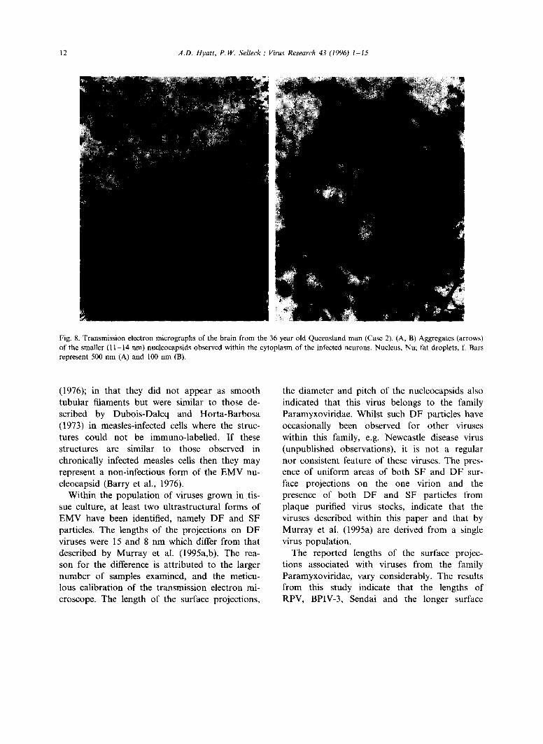
aggregate compr i sed o f smal ler nuc leocaps id- l ike s t ructures ( 1 1 - 1 4 nm in d iameter ) which were f requent ly associa ted with fat d rop le t s (Fig. 8(A, B)). These s t ructures were also g ranu la r in ap-
8 A.D. Hyatt, P.W. Selleck / Virus Research 43 (1996) 1-15
Fig. 4. Transmission electron micrographs of ultra-thin sections from experimentally infected horses. (A) Low magnification of a multi-nucleated (Nu) endothelial cell containing aggregated nucleocapsids (arrow). Bar represents 500 nm. (B) Higher magnification of an endothelial cell showing the aggregates of nucleocapsids (arrows) and a single virion budding from the cell membrane (v). Bar represents 100 rim. (C) Aggregate of nucleocapsids within an infected endothelial cell which has been incubated with serum from one of the infected humans and 9 nm protein A-gold. Bar represents 100nm. Note: The ultrastructural pathology of EMV-infected lungs from clinical and experimental horses is described by Hooper et al. (1996b).
A.D. Hyatt, P.W. Selleck / Virus Research 43 (1996) 1-15 9
Fig. 5. Light micrograph of EMV-infected VERO cells acetone-fixed at 72 h after infection with either EMV or non-infectious tissue culture medium stained with serum from a recovered EMV-infected horse followed by a protein A/FITC conjugate and counter stained with Evan's blue. (A) EMV-infected multinucleated syncitia showing fluorescing cytoplasmic inclusion bodies (arrow heads) and no intranuclear fluorescence. (B) Uninfected cells stained under the same conditions as infected cells showing no fluorescence. Nucleus, Nu. Magnification, x 2000.
pearance but failed to react with the same serum (data not shown). No viruses were observed.
3.5. Morphology o f plaque purified E M V
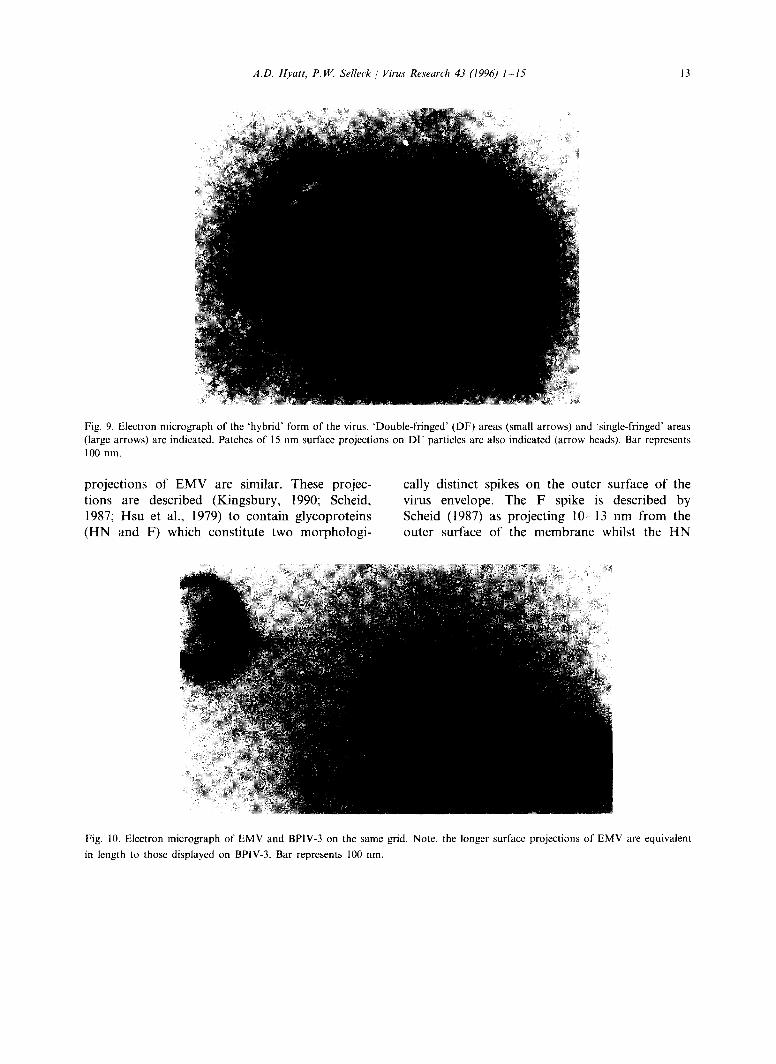
EMV which was plaque purified three times was also examined by NCEM. Within all sam- ples a heterogeneous population was observed. The virus appeared in three forms namely, DF, SF and 'hybrid ' forms. The frequency of forms and ultrastructural characteristics were similar to those described above in that DF particles com- prised over 95% of the population. 'Hybr id ' forms (Fig. 9) exhibited D F forms of surface projections interrupted with patches of SF forms of surface projections.
3.6. Ultrastructural comparison o f E M V with other viruses of the family Paramyxoviridae
EMV was mixed with BPIV-3, RPV or HRSV
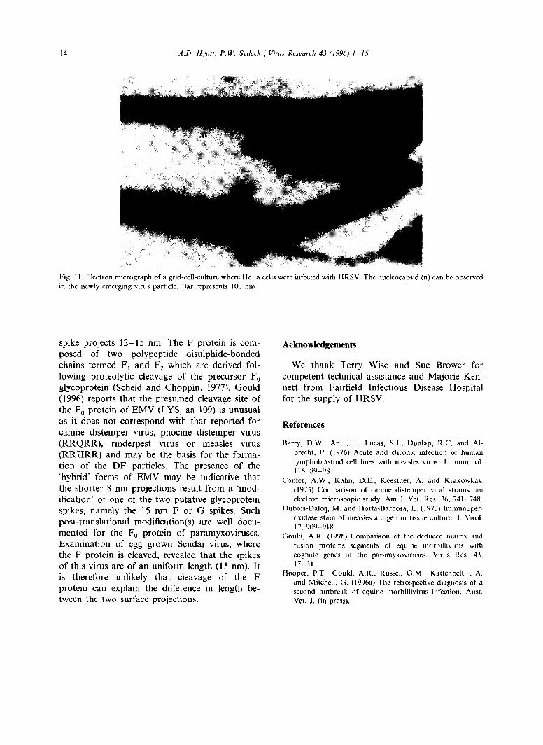
on the one grid and negatively stained with PTA. BPIV-3, RPV and HRSV all appeared pleomor- phic with a single-fringe of surface projections. Fig. 10 shows EMV and BPIV-3 particles within the one field of view. The image confirms that the longer surface projections (outer fringe) of EMV to be similar in length to those associated with BPIV-3. Examination of other N C E M prepara- tions of RPV, Sendai and HRSV also showed the surface projections of these viruses to be a uni- form 15 nm fringe (data not shown). Free-lying nucleocapsids were observed for all virions except HRSV. These nucleocapsids had diameters of 18 nm and pitches of 5 nm. To estimate the diameter of HRSV-associated nucleocapsids, GCCs were prepared of HRSV-infected HeLa cells. The ad- vantage of these preparations was that the nucle-
ocapsids could be observed within the budding virions (Fig. 11). Whilst the ultrastructure of the nucleocapsids was inferior to those observed with the free-lying nucleocapsids from BPIV-3,

Fig. 6. Transmission electron micrographs of ultrathin sections derived from EMV-infected VERO cells. (A) Multinucleated (Nu) cell with cytoplasmic aggregations of nucleocapsids (ncs). (B) Region of an infected cell showing EMV budding (arrows) from the cell membrane, nucleocapsids (n) can be observed with the membrane. Thickened areas of the membrane (large arrow) represent the analogous M protein of paramyxoviruses. Lysosomes, Ly. Bars represent 100 nm.
4~
7"

A.D. Hyatt, P.W. Selleck / Virus Research 43 (1996) 1-15 11
Fig. 7. Transmission electron micrographs of the brain from the 36 year old Queensland man (Case 2). (A) Nucleocapsids (18 nm) observed within the cytoplasm of the infected neurons. (B) Similar nucleocapsids incubated with serum from a recovered EMV-infected horse, biotinated anti-horse and streptavidin gold. Bars represent 100 nm.
RPV and EMV, the diameter could be estimated at 15 ±1 (n = 10); the pitch could not be mea- sured.
4. Discussion
The ultrastructure of EMV-infected cells was consistent with that described for members of the family Paramyxoviridae. Within both tissue culture cells and the lungs of EMV-infected horses, aggregations of nucleocapsids were ob- served within the cytoplasm of the multi-nucle- ated cells. Nucleocapsids were also observed adjacent to electron dense or thickened regions of the plasma membrane. Unlike other represen- tatives of the genus Morbillivirus, nucleocapsids
were not observed within the nuclei of infected cells (Tajima et al., 1971; Nunoya et aL 1990; McLean and Doane, 1971; Confer et al., 1975). This may be due to the short time course of infection (72 h) within tissue culture cells (Raine et al., 1969; Barry et al., 1976). However the nuclei of (a) endothelial cells from the lungs of infected horses and (b) the neurons within the brain of the 36 year old Queensland man were also devoid of nucleocapsids indicating that in- tranuclear nucleocapsids are not a prominent feature of EMV-infection.
The cytoplasm of the neurons did, however, contain aggregates of smaller nucleocapsid-like structures. These structures differed to those de- scribed by Raine et al. (1969); Oyanagi et al. (1970); Raine et al. (1971) and Barry et al.
12 .,I.D. Hyatt, P.W. Selleck / Virus Research 43 (1996) 1-15
Fig. 8. Transmission electron micrographs of the brain from the 36 year old Queensland man (Case 2). (A, B) Aggregates (arrows) of the smaller (11-14 nm) nucleoeapsids observed within the cytoplasm of the infected neurons. Nucleus, Nu; fat droplets, f. Bars represent 500 nm (A) and 100 nm (B).
(1976); in that they did not appear as smooth tubular filaments but were similar to those de- scribed by Dubois-Dalcq and Horta-Barbosa (1973) in measles-infected cells where the struc- tures could not be immuno-labeUed. If these structures are similar to those observed in chronically infected measles cells then they may represent a non-infectious form of the EMV nu- cleocapsid (Barry et al., 1976).
Within the population of viruses grown in tis- sue culture, at least two ultrastructural forms of EMV have been identified, namely DF and SF particles. The lengths of the projections on DF viruses were 15 and 8 nm which differ from that described by Murray et al. (1995a,b). The rea- son for the difference is attributed to the larger number of samples examined, and the meticu- lous calibration of the transmission electron mi- croscope. The length of the surface projections,
the diameter and pitch of the nucleocapsids also indicated that this virus belongs to the family Paramyxoviridae. Whilst such DF particles have occasionally been observed for other viruses within this family, e.g. Newcastle disease virus (unpublished observations), it is not a regular nor consistent feature of these viruses. The pres- ence of uniform areas of both SF and D F sur- face projections on the one virion and the presence of both DF and SF particles from plaque purified virus stocks, indicate that the viruses described within this paper and that by Murray et al. (1995a) are derived from a single virus population.
The reported lengths of the surface projec- tions associated with viruses from the family Paramyxoviridae, vary considerably. The results from this study indicate that the lengths of RPV, BPIV-3, Sendai and the longer surface
A.D. Hyatt, P.W. Selleek / Virus Research 43 (1996) 1-15 13
Fig. 9. Electron micrograph of the 'hybrid' form of the virus. 'Double-fringed' (DF) areas (small arrows) and 'single-fringed' areas (large arrows) are indicated. Patches of 15 nm surface projections on DF particles are also indicated (arrow heads). Bar represents 100 nm.
pro jec t ions o f E M V are similar. These projec- t ions are descr ibed (Kingsbury , 1990; Scheid, 1987; Hsu et al., 1979) to con ta in g lycopro te ins ( H N and F) which cons t i tu te two m o r p h o l o g i -
cally dis t inct spikes on the ou te r surface o f the virus envelope. The F spike is descr ibed by Scheid (1987) as p ro jec t ing 10-13 nm f rom the ou te r surface o f the m e m b r a n e whilst the H N
Fig. 10. Electron micrograph of EMV and BPIV-3 on the same grid. Note, the longer surface projections of EMV are equivalent in length to those displayed on BPIV-3. Bar represents 100 rim.
14 A.D. Hyatt, P, W. Selleck / Virus Research 43 (1996) 1 15
Fig. 11. Electron micrograph of a grid-cell-culture where HeLa cells were infected with HRSV. The nucleocapsid (n) can be observed in the newly emerging virus particle. Bar represents 100 nm.
spike projects 12-15 nm. The F protein is com- posed of two polypeptide disulphide-bonded chains termed F~ and F2 which are derived fol- lowing proteolytic cleavage of the precursor F0 glycoprotein (Scheid and Choppin, 1977). Gould (1996) reports that the presumed cleavage site of the F 0 protein of EMV (LYS, aa 109) is unusual as it does not correspond with that reported for canine distemper virus, phocine distemper virus (RRQRR), rinderpest virus or measles virus (RRHRR) and may be the basis for the forma- tion of the DF particles. The presence of the 'hybrid' forms of EMV may be indicative that the shorter 8 nm projections result from a 'mod- ification' of one of the two putative glycoprotein spikes, namely the 15 nm F or G spikes. Such post-translational modification(s) are well docu- mented for the F0 protein of paramyxoviruses. Examination of egg grown Sendal virus, where the F protein is cleaved, revealed that the spikes of this virus are of an uniform length (15 nm). It is therefore unlikely that cleavage of the F protein can explain the difference in length be- tween the two surface projections.
Acknowledgements
We thank Terry Wise and Sue Brower for competent technical assistance and Majorie Ken- nett from Fairfield Infectious Disease Hospital for the supply of HRSV.
References
Barry, D.W., An, J.L., Lucas, S.J., Dunlap, R.C. and Al- brecht, P. (1976) Acute and chronic infection of human lymphoblastoid cell lines with measles virus. J. Immunol. 116, 89-98.
Confer, A.W., Kahn, D.E., Koestner, A. and Krakowkas. (1975) Comparison of canine distemper viral strains: an electron microscopic study. Am J. Vet. Res. 36, 741 748.
Dubois-Dalcq, M. and Horta-Barbosa, L. (1973) lmmunoper- oxidase stain of measles antigen in tissue culture. J. Virol. 12, 909-918.
Gould, A.R. (1996) Comparison of the deduced matrix and fusion proteins segments of equine morbillivirus with cognate genes of the paramyxoviruses. Virus Res. 43, 17--31.
Hooper, P.T., Gould, A.R., Russel, G.M., Kattenbelt, J.A. and Mitchell, G. (1996a) The retrospective diagnosis of a second outbreak of equine morbillivirus infection. Aust. Vet. J. (in press).
A.D. Hyatt, P.W. Selleck / Virus Research 43 (1996) 1-15 15
Hooper, P.T., Gould., A.R., Mitchell, G., Russel, G.W. and Kattenbelt, J.A. (1996b) The retrospective diagnosis of a second outbreak of equine morbillivirus infection. Aust. Vet. J. (in press).
Hyatt, A.D. (1991) Immunogold labelling techniques. In: Robin Harris (Ed.), Electron Microscopy in Biology, A practical Approach. IRL Press, Oxford, pp. 59-81.
Hyatt, A.D., Eaton, B.T. and Lunt, R. (1987) The grid-cell- culture technique: the direct examination of virus-infected cells and progeny viruses. J. Microsc. (Oxford) 145, 97 L 106.
Hsu, M-C., Scheid, A. and Choppin, P.W. (1979) Reconstitu- tion of membranes with individual paramyxovirus glyco- proteins and phospholipid in cholate solution. Virology 95, 476-491.
Kingsbury, D.W. (1990) Paramyxoviridae and their replica- tion. In: B.N. Fields, D.M. Knipe, R.M. Chanock, M.S. Hirsch, J.L. Melnick, T.P. Monath and B. Roizman (Eds.), Fields Virology, vol. 1. Raven Press, New York, pp. 945-962.
McLean, A.M. and Doane F.W. (1971) The morphogenesis and cytopathology of bovine parainttuenza type 3 virus. J. Gen. Virol. 12, 271-279.
Murray, K., Selleck, P., Hooper, P., Hyatt. A., Gould, A., Gleeson, L. et al. (1995) A morbillivirus that caused fatal disease in horses and humans. Science 268, 94 97.
Murray, K., Rogers, R., Selvey, L., Selleck, P., Hyatt. A., Gould, A. et al. (1995) A novel morbillivirus pneumonia of horses and its transmission to humans. Emerging Infect. Dis. 1. 31-33.
Nunoya, T., Tajima, M., Ishikawa, Y., Samejima, T., Ishikawa, H. and Hasegawa, K. (1990) Occurrence of a canine distemper-like disease in aquarium seals. Nippon Juigaku Zasshi 52, 469-477.
O'Sullivan, J.D., Allworth, A.M., Paterson, D.L., Sama- ratunga, H., Boots, R., Gleeson, L., Snow, T. and Bradfield, J. (1996) Fatal encephalitis due to a novel mor- billivirus transmitted from horses. Lancet (submitted).
Oyanagi, S., Meulenter V., Muller, D., Katz, M. and Ko- prowski, H. (1970) Electron microscopic observations in subacute sclerosing panencephalitis brain cell cultures: their correlation with cytochemical and immunocytological findings. J. Virol. 6, 370-379,
Raine, C.S., Feldman, A., Sheppard, R.D. and Bornstein, M.B. (1969) Ultrastructure of measles virus in cultures of hamster cerebellum. J. Virol. 4, 169 181.
Raine, C.S., Feldman, A., Sheppard, R.D. and Bornstein, M.B. (1971) Ultrastructural study of long-term measles infection in cultures of hamster dorsal root ganglion. J. Virol. 8, 318-~329.
Scheid, A. (1987) Paramyxoviridae. ln: M.V. Nermut and A.C. Steven (Eds.), Animal virus structure, Elsevier, Am- sterdam, pp. 233-252.
Scheid, A. and Choppin, P.W. (1977) Two disulfide-linked polypeptide chains constitute the active F protein of paramyxoviruses. Virology 80, 54-66.
Selvey, L.A., Wells, R.M., McCormack, J.G., Ansford, A., Murray, K., Rogers, R.J. et al. (1995) Infection of humans and horses by a newly described morbillivirus. Med. J. Aust. 162, 642-645.
Tajima, M., Motohashi, T., Kishi, S. and Nakamura, J. (1971) A comparative electron microscopic study on the morpho- genesis of canine distemper and rinderpest viruses. Jap. J. Vet. Sci. 33, 1 10.
Westbury, H.A., Hooper, P.T., Selleck, P.W. and Murray, P.K. (1995) Equine morbillivirus pneumonia: susceptibility of laboratory animals to the virus. Aust. Vet. J. 72(7), 278- 279.