EQUINE edition of the Compendium of Animal Reproduction ...
85
-
Upload
khangminh22 -
Category
Documents
-
view
1 -
download
0
Transcript of EQUINE edition of the Compendium of Animal Reproduction ...

Table of Contents
2
ISBN 90-801886-6-2*
Publisher: Intervet International bv
*This number references the entire Compendium of Animal Reproduction, of which this book is a segment.
060200.12.09
11th revised edition, 2015
Preface
The animals we call our patients make our world a much better place in which to live and work. They enrich our lives and serve us in so many different capacities. They allow us to go beyond our own abilities and technologies to help and comfort the disabled. They expand our ability to work and do things we could not do without them. They give us enjoyment, companionship, transportation and power.
Without reproduction there would be no production. Animal reproduction is an essential element in the continued interaction between humans and animals.
It is with great pleasure that I present to you the EQUINE edition of the Compendium of Animal Reproduction, 11th edition.
The objective of the EQUINE edition is to update and inspire those interested in the management of reproduction in horses and to provide usable solutions to challenges in the everyday life of working veterinarians and their clients.
It would have been impossible to accomplish this EQUINE edition without the help of colleagues who devoted much time and effort to this project. I would like to express my gratitude to Dr. Andy Skidmore, Dr. Marc-Antoine Driancourt and Dr. Bryant Craig for their help in editing the content included in this EQUINE edition.
Reproduction in horses is a very complex and dynamic world. I hope that you find the EQUINE edition of the compendium to be useful and stimulating. This only scratches the surface. There is so much more to know and to explore. Go forward and have a great adventure.
Linda J. I. Horspool BVMS, PhD, Dip.ECVPT, MRCVS Editor, EQUINE section, Compendium of Animal Reproduction, 11th edition

33
Table of Contents
1 Physiology of Reproduction in Mammals 5
1.1 Introduction 5
1.2 Endocrine, paracrine, and autocrine regulation of reproduction; regulatory loops and feedback mechanisms 6 1.2.1 Definitions 6 1.2.2 Regulation of reproduction in the female 8 1.2.3 Regulation of reproduction in the male 13
1.3 Regulation of follicular growth, maturation, and ovulation 15 1.3.1 Endocrine and autocrine regulation of follicular growth 15 1.3.2 Endocrine, paracrine, and autocrine regulation of
follicular steroidogenesis 17 1.3.3 Endocrine, paracrine, and autocrine regulation of
oocyte function 19
1.4 Regulatory mechanisms involved in seasonality 20 1.4.1 A few facts about seasonality 20 1.4.2 The cascade blocking reproductive function in
seasonal anoestrus (Figure 8) 21
1.5 Regulatory mechanisms involved in postpartum anoestrus 22 1.5.1 A few facts about postpartum anoestrus 22 1.5.2 The cascade blocking reproductive function during
postpartum anoestrus (Figure 9) 23
1.6 Quality of sperm 24 1.6.1 Features of sperm that may be related to its fertilising ability 25 1.6.2 Sperm biotechnologies and fertility 26
1.7 Further Reading 27

44
Table of Contents
2 Equine Reproduction 28
2.1 Physiology 28 2.1.1 The mare: a seasonal breeder 28 2.1.2 Physiology of the oestrous cycle in the cyclic mare 29 2.1.3 Initiation of pregnancy, pregnancy maintenance, and pregnancy loss 32 2.1.4 Seasonal regulation of reproductive activity in the mare 34
2.2 Tools available to optimise reproduction management 36 2.2.1 Oestrus detection 36 2.2.2 Ultrasonography 37 2.2.3 Mating 37 2.2.4 Artificial insemination 38 2.2.5 Reproductive tools to breed high-value and problem mares 41 2.2.6 Pregnancy diagnosis 43
2.3 Solutions for efficient horse breeding 45 2.3.1 Breeding during the transition period 45 2.3.2 Breeding season - solutions for optimising fertility and
obtaining one foal/year 47 2.3.3 Solutions for synchronising oestrus in groups of mares 49 2.3.4 Solutions for inducing ovulation and timing insemination 50 2.3.5 Solutions for breeding late-foaling mares efficiently during
early summer or late autumn 53 2.3.6 Solutions for twin pregnancies 54 2.3.7 Solutions for inducing parturition 55 2.3.8 Solutions for suppressing oestrus in competing mares 56
2.4 Reproductive pathologies: prevention and treatment 57 2.4.1 Endometritis and endometriosis 57 2.4.2 Early embryonic death and abortion 64 2.4.3 Retained foetal membranes 71
2.5 The stallion 72 2.5.1 Reproductive performance evaluation 73 2.5.2 Cryptorchidism 74 2.5.3 Sexual behavioural disorders 76 2.5.4 Testicular degeneration 77 2.5.5 Hemospermia and urospermia 77
2.6 References 78

Table of Contents
55
Physiology of Reproduction in Mammals
1.1 Introduction
Reproductive performance in breeding females is the key to economic performance. The three prerequisites to reach this target are well known:
•Amaximumproportionoffemalesinaherdshoulddisplaycyclicovarian activity at the time of breeding. While this is straightforward in species in which seasonal or postpartum anoestrus does not occur, this may be more challenging in herds where cycling and noncycling females are mixed, without any easy way to identify these two subpopulations.
•Thereshouldbeclosesynchronybetweeninseminationandovulation. Such synchrony is very easily obtained in the few species (eg, rabbits, cats, camelids) where ovulation is induced by mating. In other species, the occurrence of oestrus helps to get some synchrony between mating and ovulation. However, in some species, such as horses, which display long oestrus periods with ovulation at the end of oestrus, the beginning of oestrus is a poor indicator of the optimal time for mating.
•Usespermofhighfertilisingabilityforinseminationorhighlyfertile males with high libido and in numbers suitable for the numbers of females to be mated.
This chapter presents a summary of the mechanisms controlling reproduction and reviews the processes involved in:
•Folliculargrowth,maturation,andovulation •Seasonalanoestrus •Postpartumanoestrus •Qualityofsperm
Species-specific features as well as manipulation of these processes are presented in the chapters dedicated to each species.
Table of Contents
66
Physiology of Reproduction in Mammals
1.2 Endocrine, paracrine, and autocrine regulation of reproduction; regulatory loops and feedback mechanisms
1.2.1 Definitions
There are three types of hormonal regulation – endocrine, paracrine, and autocrine. All three types of regulation are modulated by feedback loops. Feedback loops are very well understood for all endocrine mechanisms (see below). Feedback loops may be negative, when they slow down the process initiated by the initial action, or positive, when they increase the initial action. The intracellular mechanisms involved in these different regulatory loops are numerous and complex and are outside the scope of this review.
a. Endocrine regulation (Figure 1) In endocrine regulation, the hormone is synthesised in an endocrine gland and released into the bloodstream, which transports it to its target organ, often distant from the source. The endocrine control of gonadal function by the hypothalamic-pituitary axis through the release of follicle stimulating hormone (FSH) and luteinizing hormone (LH) by the pituitary is a good example of an endocrine control mechanism. Usually, there are regulatory feedback loops that maintain the balance between stimulation and inhibition.
Interestingly, recent research has demonstrated that there are a number of endocrine regulatory mechanisms that involve organs outside of the classic hypothalamic-pituitary-gonadal axis, which are also involved in the control of gonadal function. Just to name a few, insulin (produced by the pancreas), leptin (produced by fat), and ghrelin (produced by the stomach) have all been shown to modulate gonadal function (Figure 1).

Table of Contents
77
Physiology of Reproduction in Mammals
b. Paracrine regulation (Figure 1) Paracrine regulation is said to occur when two neighboring tissues interact. Such interactions are well documented in the ovary (cross-talk between the granulosa and theca cells and between the oocyte and the granulosa cells) as well as in the testicle (cross-talk between Leydig and Sertoli cells and between Sertoli cells and germ cells). Another example of paracrine regulation is the cross-talk between the large and small luteal cells, not only to maximise progesterone synthesis but also during the regression of the corpus luteum.
c. Autocrine regulation (Figure 1) Autocrine regulation involves the action of compounds produced by a specific tissue on the same tissue. A good example of autocrine regulation is the role of oestradiol (produced by granulosa cells) in the differentiation process (measured, for example, by the presence of LH receptors) of these same cells.
Oocyte
Theca cellGranulosa cell
Paracrineregulators
Autocrineregulators
Endocrine signals from fat (leptin)
Intracellular signaling(eg, protein kinase
(PK) A, PKC)
Cell proliferation or apoptosis
Endocrine signals fromthe stomach (ghrelin)
Endocrine signals fromthe pituitary (FSH, LH)
Endocrine signalsfrom the liver (IGF1)
Oestradiol production
Figure 1 Endocrine, paracrine, and autocrine regulation and the interactions between the oocyte and its somatic (granulosa and theca) cells modulating granulosa cell proliferation, apoptosis, and differentiation

Table of Contents
88
Physiology of Reproduction in Mammals
1.2.2 Regulation of reproduction in the female
a. The hypothalamic-pituitary axis and follicle function Gonadotropin-releasing hormone (GnRH), a ten-amino acid peptide (decapeptide), is released into the hypothalamic-hypophyseal portal system and transported to the anterior lobe of the pituitary, its target organ, where it acts on specific cells to stimulate the synthesis and release of the gonadotropins FSH and LH. As GnRH is secreted in a pulsatile way (ie, rapid bursts separated by a quiescent period) by GnRH neurons, it is not surprising that LH secretion by the pituitary is also pulsatile. In contrast, the pulsatile nature of FSH secretion is usually less obvious. It is the amplitude and frequency of GnRH pulses that convey the endocrine signals to the pituitary-ovarian axis. Both internal factors (through feedback loops) and external factors (eg, photoperiod, pheromones, nutrition, and metabolic status) exert their primary effect on reproduction through the modulation of pulsatile secretion of GnRH by the hypothalamus. This ensures that the target organ is always exposed to efficient hormonal stimuli. Indeed, constant stimulation by high concentrations of GnRH results in desensitisation of the target cells in the pituitary. This is most probably caused by a decrease in the number of GnRH receptors on the cell membrane of the target cells.
The pituitary gonadotropins, FSH and LH, belong to the superfamily of glycoprotein hormones. They have two different subunits, alpha and beta, which are noncovalently associated. The two hormones are not secreted synchronously in vivo since they are regulated independently. GnRH is of major importance in controlling the secretion of LH. It acts by triggering both the release and the biosynthesis of LH in order to replenish stores of it in the pituitary. The LH content of the pituitary of most mammalian species is up to ten times higher than that of FSH. In contrast, FSH synthesis is mainly modulated by various gonadal factors (eg, oestradiol and members of the inhibin family ie, inhibin, activin, and follistatin), although GnRH is also involved. The pituitary stores of FSH are low, and its secretion mirrors the rate and extent of its biosynthesis.
Table of Contents
99
Physiology of Reproduction in Mammals
At the ovarian level, FSH has two main roles. The first is to sustain growth of recruited follicles (see Section 1.3.1) until the gonadotropin dependence is transferred to LH, usually around the time of selection of the dominant follicle. The second is the induction of aromatase in the granulosa cells (see Section 1.3.2). Aromatase is the enzyme that converts androgens into oestrogens. Its successful induction is a prerequisite for further maturation of the dominant follicle. The granulosa cells of the dominant follicle also produce inhibin, which acts by negative feedback on FSH release from the pituitary. This negative feedback loop prevents hyperstimulation of the ovary by FSH.
In the theca interna, LH stimulates the synthesis of androstenedione from cholesterol and progestagens (progestins). Androstenedione is converted into testosterone, which is transferred to the granulosa cells to be converted into oestradiol-17β by aromatase (see Section 1.3.2). Oestradiol, when its concentrations exceed a certain threshold, exerts positive feedback on the hypothalamus to induce the LH surge that triggers ovulation. The time interval between the LH surge and ovulation is very consistent within a species but quite variable between species. For example, it is around 40 hours in swine and horses but only 24-28 hours in cattle and sheep. An additional effect of oestradiol is the induction of the signs of oestrus. Oestrus can be described as the behavioural and physical signs that signal to other animals that the female is in the fertile phase of its cycle and will allow mating.
Interestingly, androgens and oestrogens as well as members of the inhibin family are involved in paracrine and autocrine regulation that modulates endocrine signaling in the ovary. An example of paracrine interaction is detailed in Section 1.3.2.

Table of Contents
1010
Physiology of Reproduction in Mammals
b. The hypothalamic-pituitary axis and corpus luteum function Changes in progesterone concentrations after ovulation follow the same pattern in all species. Progesterone is produced by the corpus luteum, and concentrations start to rise in the days following ovulation and steadily increase until around day 6 postovulation. Progesterone concentrations then plateau for 7 to 12 days, depending on the duration of the luteal phase of the species concerned. Progesterone concentrations during this period are species specific, ranging from around 5 ng/mL in sheep to about 40 ng/mL in swine. Following the initiation of luteolysis (see Section 1.2.2c), progesterone concentrations quickly decline to very low levels (below 0.5 ng/mL), allowing a new follicular phase to start.
Progesterone is the hormone responsible for the maintenance of pregnancy. It is produced and secreted jointly by large and small luteal cells. Large luteal cells are derived from granulosa cells and have a low sensitivity to LH and a high sensitivity to prostaglandins. Small luteal cells are derived from theca interna cells and are highly sensitive to LH. LH alone, or together with prolactin (in some species of rodent), is the key hormone supporting the formation of the corpus luteum and the initiation of progesterone production. Progesterone acts on several targets. Firstly, it prepares the oviduct and endometrium to accommodate the freshly fertilised, young embryo (oviduct) and later the developing embryo when it enters the uterus at the blastocyst stage. Secondly, by exerting negative feedback, it slows down GnRH release at the level of the hypothalamus and reduces the concentrations of LH available to support terminal follicular growth, thereby preventing return to oestrus and new ovulation(s).
In pregnant females, interferon tau production by the developing embryo (in cattle) or oestrogen production by the multiple embryos (in swine) acts to maintain the corpus luteum, thereby allowing pregnancy initiation (see Section 1.2.2c).

Table of Contents
1111
Physiology of Reproduction in Mammals
c. Interactions between the uterus, embryo, and corpus luteum in the control of luteolysis Prostaglandin (PG) F2α initiates the regression of the corpus luteum, known as luteolysis. The luteolytic signal is increased pulsatile secretion of PGF2α. Uterine venous PGF2α concentrations begin to increase on days 11-13 in sheep, days 13-14 in swine, and days 16-17 postoestrus in cattle (reviewed by Weems et al., 2006). The mechanism by which PG induce luteolysis has not been completely elucidated, but it involves a reduction of the blood supply to the corpus luteum by vasoconstriction, as well as direct inhibition of luteal steroidogenesis coupled to increased cell death (apoptosis) of luteal cells. It is generally assumed that functional luteolysis (ie, a drop in progesterone production) precedes morphological luteolysis (ie, a reduction in size leading to a corpus albicans). The primary site for the initiation of luteolysis is the large luteal cells of the aging corpus luteum. Oxytocin produced in the corpus luteum is believed to be the first signal triggering luteolysis. Binding of oxytocin to its receptor in the uterine endometrium of nonpregnant cattle and sheep stimulates the pulsatile secretion of PGF2α. Oestrogens increase expression of uterine oxytocin receptors, while progesterone has the opposite effect. This is why it is possible to postpone luteolysis by preventing the growth of large oestrogen-active follicles (See Luteolysis, Section 2.1.4c). During the initiation of pregnancy in cattle, luteolysis is prevented through increased interferon tau production by the embryo before pulsatile PGF2α secretion is initiated. In pregnant swine, luteolysis is stopped by embryonic oestradiol production that diverts PGF2α away from the ovarian circulation, thus preventing it from reaching the ovaries.
Figure 2 summarises the interactions between the different levels of the hypothalamic-pituitary-ovarian-uterine axis involved in the control of reproduction and endocrine mediators.
Figure 3 presents an overview of the changes in gonadotropin and steroid hormone (progesterone and oestradiol) concentrations during the bovine oestrous cycle.

Table of Contents
1212
Physiology of Reproduction in Mammals
Hormoneconcentration
OV
FSH
Oestrus LH OV
Progesterone
Oestradiol Ovulation
OV
Luteal phaseFollicular phase Follicular phase
Progestational changes
Oestrus changes
Maturation
Luteolysis
ProgesteroneOestrogens
Oestrus behaviour
Growth
Ovulation
PGF2α
Progesterone feedbackFSH
OVARY
ANTERIORPITUITARY
LH
UTERUS
HYPOTHALAMUS
GnRH
Oestradiolfeedback
Follicle
Inhibinfeedback onFSH release
Maturation
Oxytocin
Luteinisation
+ -
Figure 3 Schematic profile of the changes in gonadotropin (FSH and LH) and steroid hormone concentrations (progesterone and oestradiol) during the bovine oestrous cycle
Figure 2 The hypothalamic-pituitary-ovarian-uterine axis and the endocrine regulators of follicular growth, corpus luteum formation, and luteolysis

Table of Contents
1313
Physiology of Reproduction in Mammals
1.2.3 Regulation of reproduction in the male
In males, the testicles or testes produce sperm and male steroid hormones (mainly androgens). This requires interactions between the two constitutive compartments of the testis, the seminiferous tubules, in which germ cells and Sertoli cells are located, and interstitial tissue, which includes Leydig cells. In rodents, the volume of interstitial tissue (Leydig cells) does not exceed 5% of the total testicular volume; however, this proportion reaches 10% in sheep and is far higher in swine and horses. Seminiferous tubules therefore represent 60% (swine, horses) to 90% (rodents) of the testicular volume. The time needed for production of a spermatid from a quiescent (A0) spermatagonium is species specific and ranges from 35 days (mice) to 41 days (swine), 45 days (sheep), and 54 days (cattle). Daily sperm production by both testicles (in billions of spermatozoa) averages 5.2 (horses), 7.5 (cattle), and 16.2 (swine), but the daily sperm production per gram of testicular tissue appears quite consistent across species (at around 12-20 million/g).
In the seminiferous tubules, the germ cells divide by mitosis, generating several generations of spermatogonia, and initiate meiosis when they reach the spermatocyte stage. They are released into the lumen of the seminiferous tubules when they become spermatids. All steps of germ cell proliferation and maturation occur with the different generations of germ cells in close proximity to the Sertoli cells that line the basal membrane of the seminiferous tubules. Sertoli cells also produce regulatory proteins, such as inhibin, that reduce FSH concentrations by exerting negative feedback on the pituitary.
In the interstitial tissue, Leydig cells actively produce the testicular androgens. In addition, the testis also produces limited amounts of oestradiol.
Control of reproduction in the male involves the same types of regulation (ie, endocrine, paracrine, and autocrine) as in females, and the endocrine regulatory loops are generally very similar to those described in Section 1.2.2.

Table of Contents
1414
Physiology of Reproduction in Mammals
In the prepubertal animal, FSH stimulates proliferation of Sertoli cells, with their final number reached at puberty. Around puberty, FSH is responsible for the maturation of the Sertoli cells, resulting in increased inhibin and androgen-binding protein (ABP) production. The pubertal increase in pulsatile LH secretion stimulates androgen production by the Leydig cells, followed by its possible aromatisation to oestradiol in Sertoli cells. In sheep, the endocrine control of germ-cell proliferation is well understood (Courot and Ortavant, 1981) and involves the actions of FSH, LH, and androgens at specific steps of the mitotic and meiotic processes. For example, differentiation of A0 to A1 spermatogonia is controlled by LH, while transition from A1 to intermediate spermatogonia is controlled by testosterone. The last spermatogonial divisions, changing intermediate spermatogonia into primary spermatocytes, are under the control of FSH. Once the leptotene stage is reached, the prophase of meiosis and spermatogenesis is controlled by androgens. As germ cells display oestradiol receptors, this steroid hormone is also likely to modulate the actions of FSH, LH, and androgens.
Endocrine regulation also involves positive and negative feedback regulatory loops. Inhibin produced by Sertoli cells acts by negative feedback on FSH. Androgens produced by Leydig cells act by negative feedback on LH secretion. There are also numerous paracrine and autocrine regulatory mechanisms controlling the function of the testis. Examples of paracrine regulation include the stimulatory effects on Sertoli cells of growth factors (such as insulin-like growth factor (IGF) 1) produced by the Leydig cells and by the germinal epithelium at specific stages and stimulating (epidermal growth factor (EGF)) or inhibiting Sertoli cell production of inhibin. Autocrine regulation also actively modulates testicular function. A good example of this is the local stimulatory effect of IGF-1 in the amplification of the Leydig cell response to LH and in sustaining the stimulatory effects of LH on several steps of testicular steroidogenesis.
Figure 4 shows a schematic representation of the endocrine, paracrine, and autocrine regulation involved in the control of testicular function.

Table of Contents
1515
Physiology of Reproduction in Mammals
1.3 Regulation of follicular growth, maturation, and ovulation
1.3.1 Endocrine and autocrine regulation of follicular growth
Large follicles (seen using ultrasonography or on the surface of ovaries collected at slaughter) are the tip of a large iceberg. The ovary of most farm animals (ie, cattle, sheep, goats, and swine) contains a large store of tiny primordial follicles (around 50,000 to 100,000) that are formed during foetal life. The size of this follicular store is large enough to ensure ovulation throughout the reproductive life of the female; there is no equivalent to menopause (the end of menstruation) in animals. The growth process from the primordial follicle (measuring about 0.04 mm (40 microns) in diameter) to the preovulatory stage lasts around 3-5 months. The mechanisms involved in the control of follicular growth between 0.04 mm and 1 mm in diameter are not fully understood (Scaramuzzi et al., 2011). In contrast, terminal follicular growth has been extensively studied in all species using ultrasonography. Terminal follicular growth starts when follicles become acutely dependent on gonadotropin support (ie, 2 mm in sheep and swine, 4 mm in cattle, and around 10 mm in horses). During terminal follicular growth, recruitment of a cohort of gonadotropin-dependent follicles is followed a few days later by
SpermatogenesisTestosterone
Inhibin
ANTERIORPITUITARY
HYPOTHALAMUS
GnRH
Leydigcells
Sertolicells
LH FSH
+
+
++ -
-
Figure 4 Endocrine, paracrine, and autocrine regulation involved in testis function (sperm and testosterone production)

Table of Contents
1616
Physiology of Reproduction in Mammals
the selection of the dominant follicle (Figure 5). This dominant follicle will continue growing and matures until it produces enough oestradiol to trigger oestrus and ovulation. The other follicles from the cohort will regress and there is apoptosis of their somatic cells (Figure 5). It is noteworthy that a single ultrasound scan does not allow growing, potentially dominant follicles to be distinguished from regressing, apoptotic follicles. Repeated daily ultrasound scans are needed. Several studies in experimental paradigms when LH and/or FSH concentrations were manipulated (cattle: Gong et al., 1996; Crowe et al., 2001; sheep: Picton et al., 1991; swine: Driancourt et al., 1995) have shown that it is around the time of selection that follicles transfer their gonadotropin dependence from FSH to LH. In all models, large follicles rely on the consecutive exposure to FSH and then LH to grow to preovulatory size (Figure 6). The steps that are FSH- or LH-dependent in cattle, sheep, swine, and horses are presented in Figure 6. While the range of follicular diameter needing FSH or LH appears to be species specific, it is interesting to note that the FSH/LH sequence is common to all species (Figure 6). This is why hormones with both FSH and LH activity (such as pregnant mare serum gonadotropin (PMSG, also called equine chorionic gonadotropin, eCG) are potent stimulators of follicular growth in all species.
RECRUITMENT
SELECTION
Cohort ofrecruitedfollicles
OvulationLarge
dominantfollicle
Follicular size (mm)
DOMINANCE
Atretic folliclesfrom the cohort
Days of the cycle
2
8
16
Figure 5 Main events occurring during terminal follicular growth (after Driancourt, 2001). In species with a single ovulation (cattle, horses), recruitment of several follicles is followed by selection of a single follicle that becomes dominant and fully matures and ovulates. All other recruited follicles regress by apoptosis. In multiovulatory species (swine), the same events occur, but the number of recruited and dominant follicles is higher
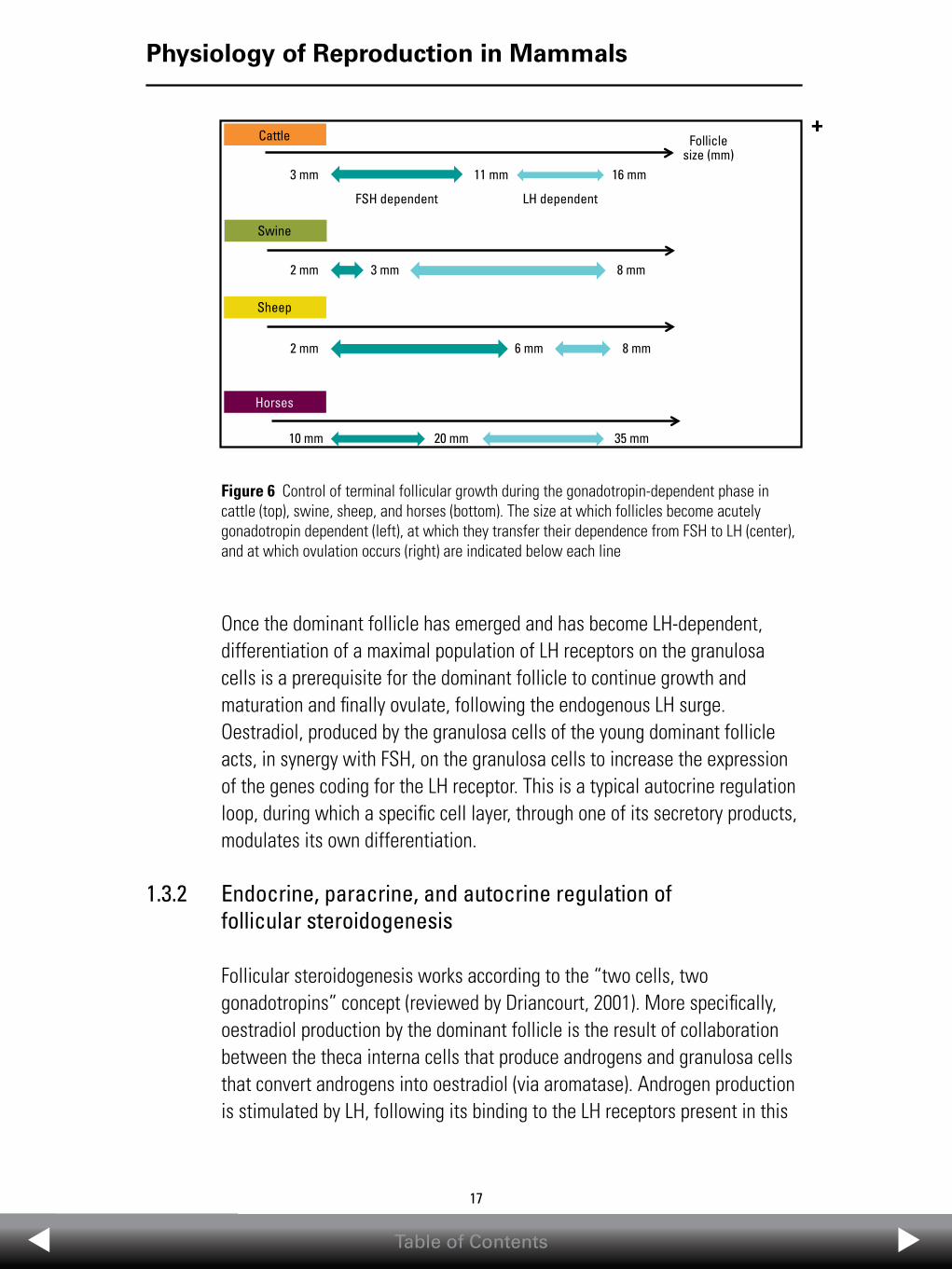
Table of Contents
1717
Physiology of Reproduction in Mammals
Once the dominant follicle has emerged and has become LH-dependent, differentiation of a maximal population of LH receptors on the granulosa cells is a prerequisite for the dominant follicle to continue growth and maturation and finally ovulate, following the endogenous LH surge. Oestradiol, produced by the granulosa cells of the young dominant follicle acts, in synergy with FSH, on the granulosa cells to increase the expression of the genes coding for the LH receptor. This is a typical autocrine regulation loop, during which a specific cell layer, through one of its secretory products, modulates its own differentiation.
1.3.2 Endocrine, paracrine, and autocrine regulation of follicular steroidogenesis
Follicular steroidogenesis works according to the “two cells, two gonadotropins” concept (reviewed by Driancourt, 2001). More specifically, oestradiol production by the dominant follicle is the result of collaboration between the theca interna cells that produce androgens and granulosa cells that convert androgens into oestradiol (via aromatase). Androgen production is stimulated by LH, following its binding to the LH receptors present in this
Cattle
Swine
Sheep
Horses
3 mm
2 mm
2 mm
10 mm
6 mm 8 mm
8 mm
35 mm
16 mm
Follicle size (mm)
LH dependentFSH dependent
11 mm
3 mm
20 mm
Figure 6 Control of terminal follicular growth during the gonadotropin-dependent phase in cattle (top), swine, sheep, and horses (bottom). The size at which follicles become acutely gonadotropin dependent (left), at which they transfer their dependence from FSH to LH (center), and at which ovulation occurs (right) are indicated below each line
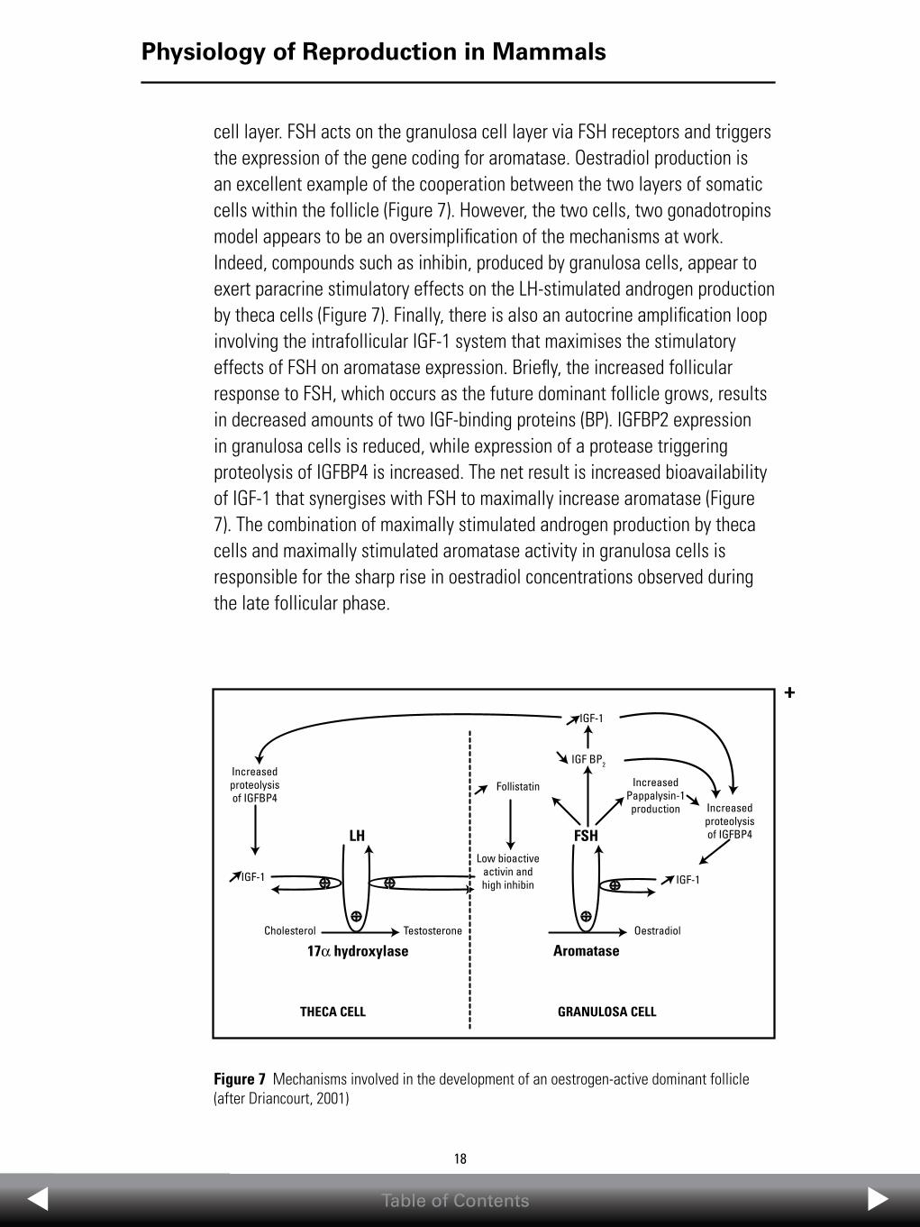
Table of Contents
1818
Physiology of Reproduction in Mammals
cell layer. FSH acts on the granulosa cell layer via FSH receptors and triggers the expression of the gene coding for aromatase. Oestradiol production is an excellent example of the cooperation between the two layers of somatic cells within the follicle (Figure 7). However, the two cells, two gonadotropins model appears to be an oversimplification of the mechanisms at work. Indeed, compounds such as inhibin, produced by granulosa cells, appear to exert paracrine stimulatory effects on the LH-stimulated androgen production by theca cells (Figure 7). Finally, there is also an autocrine amplification loop involving the intrafollicular IGF-1 system that maximises the stimulatory effects of FSH on aromatase expression. Briefly, the increased follicular response to FSH, which occurs as the future dominant follicle grows, results in decreased amounts of two IGF-binding proteins (BP). IGFBP2 expression in granulosa cells is reduced, while expression of a protease triggering proteolysis of IGFBP4 is increased. The net result is increased bioavailability of IGF-1 that synergises with FSH to maximally increase aromatase (Figure 7). The combination of maximally stimulated androgen production by theca cells and maximally stimulated aromatase activity in granulosa cells is responsible for the sharp rise in oestradiol concentrations observed during the late follicular phase.
GRANULOSA CELL
17α hydroxylase
THECA CELL
AromataseCholesterol Testosterone Oestradiol
IGF-1IGF-1
LH FSH
Follistatin
IGF-1
IGF BP2Increasedproteolysisof IGFBP4
Low bioactiveactivin andhigh inhibin
Increasedproteolysisof IGFBP4
IncreasedPappalysin-1production
Figure 7 Mechanisms involved in the development of an oestrogen-active dominant follicle (after Driancourt, 2001)
Table of Contents
1919
Physiology of Reproduction in Mammals
1.3.3 Endocrine, paracrine, and autocrine regulation of oocyte function
There are two prerequisites for an oocyte to be fertilised. It needs to become competent to undergo nuclear maturation and secondly to have completed cytoplasmic maturation.
Nuclear maturation is the process whereby the oocyte nuclear material completes meiosis, moving from the fourth stage of the prophase of meiosis (diplotene stage) to the metaphase II stage. This normally occurs when the preovulatory follicle containing the oocyte is exposed to the LH surge. In vitro the ability of oocytes removed from their follicles to resume meiosis increases with the increasing size of the follicle. It reaches 100% for oocytes obtained from 2-3 mm (cattle) and 1-2 mm (sheep) diameter follicles. Oocytes originating from smaller follicles may resume meiosis in vitro but commonly fail to complete it properly, generally remaining at the metaphase I stage. Nuclear maturation is controlled by the balance between stimulatory signals, such as the LH surge, and inhibitory ones produced by the granulosa cells surrounding the oocyte (the cumulus cells) or by the theca cells. The exact paracrine mediators produced by the somatic cells of the follicle modulating nuclear maturation of the oocyte have not been fully clarified but may include purines, such as hypoxanthine, produced by theca cells. Purines act by maintaining high cyclic adenosine monophosphate (cAMP) concentrations within the oocyte.
Cytoplasmic maturation is the process where the oocyte stores a number of messenger ribonucleic acids (mRNAs) and proteins needed for survival and the first rounds of cleavage following fertilisation (before activation of the embryonic genome). Organelles are also widely redistributed within the oocyte during this process. In all species, full cytoplasmic maturation is acquired gradually and progresses in synchrony with follicular development. In cattle, oocytes that are enclosed in follicles around 4-6 mm in diameter are thought to have completed cytoplasmic maturation. Proper cytoplasmic maturation of the oocyte, such as occurs during terminal follicular growth in vivo, is associated with a high rate of embryonic development (around 60% blastocyst development rate) following in vitro fertilisation (IVF) followed by culture (IVC). In contrast, the culture conditions applied during in vitro

Table of Contents
2020
Physiology of Reproduction in Mammals
maturation (IVM) partly interfere with the quality of cytoplasmic maturation, markedly reducing the blastocyst production rate (to around 30% in good IVM conditions). It is likely that cytoplasmic maturation of the oocyte is controlled mainly by paracrine regulators (including cAMP) transferred from the cumulus cells to the oocyte via gap junctions (Gilchrist and Thompson, 2007).
1.4 Regulatory mechanisms involved in seasonality
1.4.1 A few facts about seasonality
In temperate latitudes, recurrent, seasonal changes in temperature, climate, and food availability influence reproductive activity. One of the common features of most wild and some domesticated species is the development of a reproductive pattern favoring birth at an optimal time of year, usually spring, which allows the newborn to grow under optimal conditions of climate and food availability.
This means that periods of sexual activity (oestrus) alternate with periods of sexual inactivity (anoestrus). Among domesticated species, sheep, goats, and horses display the strongest seasonality. In sheep, sexual activity begins as the day length becomes shorter (short-day breeders). In horses, sexual activity starts when day length increases (long-day breeders). In both species, young are born in the spring, when the environmental conditions are optimal for their growth and survival.
All species that display seasonal anoestrus may display either “shallow” or “deep” anoestrus, a feature that is typical of a breed, season, and nutritional status. Shallow anoestrus is characterised by a limited reduction in GnRH secretion, with the hypothalamus still generating infrequent LH pulses that may partly support terminal follicular growth but fail to support follicular maturation. This explains why sheep in shallow anoestrus may be induced to ovulate within 24 hours of exposure to a sexually active male. In contrast, deep anoestrus is characterised by a profound inhibition of the pulsatile secretion of GnRH, resulting in very low pulses of LH that prevent terminal follicular growth and maturation. In such females, ovulation cannot be

Table of Contents
2121
Physiology of Reproduction in Mammals
induced by exposure to a sexually active male, but administration of an exogenous gonadotropin (ie, PMSG) induces the growth of preovulatory follicles.
It is obvious that females that display deep anoestrus will transition through periods of shallow anoestrus when entering anoestrus and moving toward the breeding season. It is also noteworthy that shallow anoestrus may change to deep anoestrus in underfed females or females that display postpartum anoestrus and give birth outside of the breeding season (eg, sheep lambing in April-May).
1.4.2 The cascade blocking reproductive function in seasonal anoestrus (Figure 8)
Seasonality of reproduction is linked to the duration of daylight and darkness. There is general agreement that the eye is the window that reads the daylight/darkness information. The downstream mediator of this information is melatonin produced by the pineal gland. High concentrations of melatonin are observed during darkness, while its concentrations are very low during daylight. As days become shorter, the exposure to melatonin increases. Secondly, treatment of anoestrus sheep and goats with melatonin implants can induce the resumption of oestrous cycles. Until recently, the links between melatonin and the hypothalamic centers responsible for GnRH secretion were unknown, as melatonin receptors had not been demonstrated on GnRH neurons. Two other types of neurons, kiss and RF-amide-related peptide-3 (RFRP3) neurons, are now believed to be the ones that form a bridge between melatonin and GnRH release. Receptors for kisspeptin (kiSS-1-derived peptide receptor or GPR 54) and gonadotropin-inhibitory hormone (GnIH) (RFRP3 receptors or GPR 147) have been detected on GnRH neurons. Kiss appears to increase the pulsatile secretion of GnRH, while RFRP3 has the opposite effect. During the breeding season of sheep, expression of kiss and GPR 54 in the hypothalamus increases, while the number of connections between RFRP3 and GnRH neurons drops.

Table of Contents
2222
Physiology of Reproduction in Mammals
At the initiation of anoestrus, the balance between kiss and RFRP3 activities shifts and a strong activity of RFRP3 neurons reduces the pulsatile nature of GnRH secretion. Clear support for this hypothesis has been provided by the demonstration that an infusion of kiss successfully induced ovulation in all treated sheep (Caraty et al., 2007).
While such a balance perfectly explains seasonality in sheep, it does not appear to be valid for mares, where kiss treatment during seasonal anoestrus does not stimulate follicular growth and ovulation.
1.5 Regulatory mechanisms involved in postpartum anoestrus
1.5.1 A few facts about postpartum anoestrus
Postpartum anoestrus commonly occurs in the weeks following parturition. Most farm animal species (cattle, sheep, goats, and swine) do not display oestrus or ovulate for a variable period after parturition, during which milk production is maximized and young suckled following the development of a strong bond between the dam and its offspring. The purpose of this is to
Long days
Low melatonin concentrations (short nights)
RFRP3 neurons kiss neurons
+ -
Low-level stimulation of GnRH neurons by kiss
Strong inhibition of GnRH neurons by RFRP3
GPR147 GPR54
GnRH neurons
Reduction in pulsatile GnRH secretion
Reduced pulsatile LH secretion by the pituitary
No terminal follicular growth or ovulation
GPR147
Figure 8 Mechanisms explaining why long days cause seasonal anoestrus in sheep
Table of Contents
2323
Physiology of Reproduction in Mammals
optimise the survival of the newborn, and initiation of a new pregnancy has a lower priority. There are two exceptions to this: horses return to oestrus in the 2 weeks following parturition, and rabbits can be successfully mated on the day of parturition.
As in seasonal anoestrus, interactions between the female and its environment modulate the occurrence and depth of postpartum anoestrus. In dairy cattle, the duration of postpartum anoestrus is increased when the depth and/or duration of the period of negative energy balance is increased. This is a period when the nutrients ingested do not compensate for the energy requirements of milk production. In beef cattle and ewes, in which the udder is stimulated repeatedly by suckling throughout the day, the duration and/or depth of postpartum anoestrus is longer and/or deeper than in dairy cattle. This also applies to swine, where oestrus and ovulation are generally prevented before weaning. In all species that raise their offspring, maternal bonding between the dam and its offspring also interferes with the resumption of cyclic reproductive activity. Initiation of this bonding for the first time in primiparous females contributes to the increased length and/or depth of postpartum anoestrus in these animals.
1.5.2 The cascade blocking reproductive function during postpartum anoestrus (Figure 9)
During the postpartum period, nutritional, metabolic, and behavioural factors affect reproductive function at multiple levels of the hypothalamic-pituitary-ovarian axis. For example, in cattle, negative energy balance has been shown to reduce the frequency of GnRH pulses produced by the hypothalamus. This results in a reduced frequency of LH pulses available to support terminal follicular growth. In addition, negative energy balance appears to reduce circulating concentrations of IGF-1, thereby limiting its positive effects on follicular steroidogenesis. The maximisation of androgen production by thecal cells, which is a result of synergy between LH and IGF-1, fails to occur. Furthermore, oestradiol production from granulosa cells is limited by lack of induction of aromatase because the low IGF-1 concentrations do not synergise with FSH. Finally, the high nonesterified fatty acid (NEFA) concentrations produced by mobilisation of body reserves reduce granulosa cell proliferation and limit terminal follicular growth. Hence, during the period of negative energy balance, the blunted growth of

Table of Contents
2424
Physiology of Reproduction in Mammals
follicles, together with their incomplete maturation, explain why there is no oestradiol surge and therefore no LH surge followed by ovulation. Additional details on the nutrition-reproduction interface have been reviewed by Scaramuzzi et al. (2011).
1.6 Quality of sperm
Sperm quality has the ability to strongly modulate reproductive performance. This is why semen quality is regularly checked in most of the males used for natural mating. In addition, the semen collected for use in artificial insemination (AI) is carefully assessed before being released for use in the field. However, the identification of the key factors modulating sperm quality and use of this information to optimise reproductive performance are not easy tasks. Indeed, in species where there is a single ovulation, such as cattle and horses, sperm quality is defined by an all-or-nothing response (ie, pregnant or nonpregnant) that leaves little room to relate this information to in vitro markers of sperm quality. In contrast, in species with multiple ovulations, such as swine, where a wide range of litter sizes may be
Negative energy balance
Increased nonesterified fatty acid (NEFA) concentrations
Reduced pulsatile secretion of GnRH
Low concentrations of IGF-1 produced by the liver
Reduced amountsof aromatase ingranulosa cells
Reduced pulsatile secretion of LHby the pituitary
Failure of the follicles to grow past 10-12 mm
Reduced androgen production by
theca cells
Reduced oestradiol production
No increase in plasma oestradiol
concentrationsNo ovulation
Increased steroid clearance
by the liver
Figure 9 Mechanisms explaining why negative energy balance triggers postpartum anoestrus in cattle

Table of Contents
2525
Physiology of Reproduction in Mammals
obtained, it may be easier to identify useful markers of sperm quality. The section that follows focuses on swine.
1.6.1 Features of sperm that may be related to its fertilising ability
There are six groups of features and associated technologies that provide relevant information on the potential quality of a semen sample.
•Themostobviousfeature,whichhasbeenmeasuredsincetheearlyyears of AI, is the proportion of live and morphologically normal spermatozoa. This can easily be assessed under the microscope. This approach is mainly used to identify semen samples that would be unfit for use in the field.
•Theabilityofspermtoswimandmoveforward.While,inthepast, this was evaluated under the microscope, this is now done using computer-assisted technologies (CASA) (Amman and Waberski, 2014). This is now the most widely used approach to assess semen samples for release for use in the field. However, it must be clear that this technology, while allowing discarding of semen samples of limited quality, does not predict the fertilising ability of the semen. Indeed, motility parameters only explain 9%-10% of the variation in the fertility of swine (Broekhuisje et al., 2012).
•Theabilitytoundergocapacitationwhenexposedtosuitable environments. Capacitation of sperm is a prerequisite for successful fertilisation. The response of sperm to in vitro capacitating agents can be monitored under the microscope. This parameter is only useful for the identification of semen samples that display a poor capacitation response.
•Theabilityofspermtobindtozonapellucida(ZP)proteins.Thisisan obvioustestsystem,assementhatisunabletobindtoZPproteinsis unfitforuse.However,monitoringthisrequiresaccesstoasourceofZP along with conditions similar to those used for in vitro fertilisation (IVF). In addition, the value of this test may be limited, as there is not a consistent relationship between the test results and fertility.
Table of Contents
2626
Physiology of Reproduction in Mammals
•Afurtherrefinementofinvitrotestsistoassessandmonitorfertilisation and development rates following IVF and in vitro culture (IVC). Sperm penetration into the oocyte, decondensation of the sperm nucleus, fertilisation, and embryo cleavage may be evaluated. This test has value in that decondensation of the sperm nucleus explains between 12% and 17% of the variation in fertility in vivo (Foxcroft et al., 2008). Although this is certainly the most informative system, it requires a proper IVF/IVC setting to generate relevant information.
•Recently,researchhasfocusedontheidentificationofspecificproteins in seminal plasma that may be markers of the fertilising ability of semen. A few proteins have been found to be consistently present in the semen of swine with low fertility (Dyck et al., 2011). These findings need to be confirmed in larger and more diverse populations.
While all such tests may provide useful information, it must be remembered that fertility is a multifactorial trait generated by a heterologous sperm population resulting from several spermatogenic waves and variable durations of storage in the epididymis. It should therefore not be surprising that it has proven difficult to characterise the fertility of sperm using a single in vitro test. In addition, insemination with very large numbers of spermatozoa (eg, 3 billion in swine) may not allow the identification of males with the least fertile semen.
1.6.2 Sperm biotechnologies and fertility
While describing the different semen biotechnologies is out of the scope of this chapter, it is worth remembering that for a specific semen sample, the steps needed to store the semen (cooling or freezing) or split the sperm population into X- and Y-bearing spermatozoa (semen sexing) may strongly alter its fertilising features. For example, it is well known that:
•Useofsexedsemenincattleiscommonlyassociatedwitha10%-15% drop in fertility compared to control animals inseminated with the same semen not submitted to the sexing procedure.

Table of Contents
2727
Physiology of Reproduction in Mammals
•Inswine,unlesstheinsemination-to-ovulationintervalisnomore than 4 hours, the use of frozen, thawed semen results in a drop in both farrowing rate and prolificacy. The window of opportunity for obtaining high reproductive performance using frozen, thawed semen is therefore much narrower than with fresh semen.
•Semenfromspecifichorses,whichisnormallyfertilewhenfreshsemen is used immediately following collection, does not withstand conservation under cooled conditions or the freeze-thaw cycle.
1.7 Further readingAmman RP, Waberski D. Computer assisted sperm analysis (CASA): Capabilities and potential developments. Theriogenology. 2014;81:5–17.
Broekhuisje MLWJ, Feitsma H, Gadella BM. Artificial insemination in pigs: predicting male fertility. Vet Q. 2012;32:151–157.
Caraty A, Smith JT, Lomet D, et al. Kisspeptin synchronizes preovulatory surges in cyclic ewes and causes ovulation in seasonally acyclic ewes. Endocrinology. 2007;148:5258–5267.
Courot M, Ortavant R. Endocrine control of spermatogenesis in the ram. J Reprod Fertil. 1981;63:47– 60.
Crowe MA, Kelly P, Driancourt MA, Boland M, Roche JF. Effects of FSH with and without LH on serum hormone concentrations, follicle growth and aromatase activity in gonadotropin-releasing hormone immunized heifers. Biol Reprod. 2001;64:368–374.
Driancourt MA. Regulation of ovarian follicular dynamics in farm animals: Implications for manipulation of reproduction. Theriogenology. 2001;55:1211–1239.
Driancourt MA, Prunier A, Locatelli A. Effects of gonadotropin deprivation on follicular growth in gilts. Reprod Nutr Dev. 1995;35:663–673.
Dyck MK, Foxcroft GR, Novak S, et al. Biological markers of boar fertility. Reprod Domest Anim. 2011;46 (Suppl 2):55–58.
Foxcroft GR, Dyck MK, Ruiz-Sanchez A, Novak S, Dixon WT. Identifying usable semen. Theriogenology. 2008;70:1324–1336.
Gilchrist RB, Thompson JG. Oocyte maturation: Emerging concepts and technologies to improve developmental potential in vitro. Theriogenology. 2007;67:6–15.
Gong JG, Campbell BK, Bramley TA, Gutierrez CG, Peters AR, Webb R. Suppression in the secretion of FSH and LH and ovarian follicle development in heifers continuously infused with a GnRH agonist. Biol Reprod. 1996;55:68–74.
Picton HM, Mc Neilly AS. Effect of basal and pulsatile LH release on FSH stimulated follicle growth in ewes chronically treated with gonadotrophin releasing hormone agonist. J Endocrinol. 1991;128:449–456.
Scaramuzzi RJ, Baird DT, Campbell BK, et al. Regulation of folliculogenesis and the determination of ovulation rate in ruminants. Reprod Fertil Dev. 2011;23:1–24.
Weems CW, Weems YS, Randel RD. Prostaglandins and reproduction in female farm animals. Vet J. 2006;171:206–228.

Table of Contents
28
Equine Reproduction
28
2.1 Physiology
2.1.1 The mare: a seasonal breeder
Reproductive activity in the horse is seasonal; the natural breeding season of mares extends from early spring to late summer—April to September in the Northern Hemisphere and October to March in the Southern Hemisphere. The normal cyclic activity of horses is activated primarily by increasing day length (longer photoperiod) in early spring, while in the late summer and early autumn, decreasing day length (shorter photoperiod) triggers the end of the breeding season. Hence, a mare that is not pregnant will display winter anoestrus, followed by a transition to the breeding season in early spring, usually between March and May. In most mares, the breeding season, with its associated regular oestrous cycles, will continue until the beginning of the transition to anoestrus in the autumn. Some mares (around 25%) develop a persistent corpus luteum in late summer. Some mares do not display seasonal anoestrus.
Figure 1 shows the association between changes in photoperiod and seasonal reproductive activity in mares. There is considerable interindividual variation in the number of oestrous cycles at which a mare can be bred. It should also be remembered that this pattern of reproductive activity may only be obvious in maiden and barren mares. This pattern is not seen in mares that conceive in consecutive years because conception at foal heat, within 6-8 days of foaling, is followed by pregnancy (around 11 months).
The age of a horse is measured from January 1, irrespective of its actual birth date; therefore, particularly in the racing industry, it is important that foals are born as early as possible in the year so that they are as mature as possible (in terms of muscle and bone and ability to withstand stress and effort) by the time they start to compete. Racehorses and trotters mainly perform when 2 and 3 years old. Foals that are born early in the year have advantages in terms of maturity at the start of their show or race career and/or their value at yearling sales. This is a challenge due to the seasonal pattern of ovarian activity in mares. However, there are a number of ways to achieve this successfully.

Table of Contents
29
Equine Reproduction
29
Figure 1 Changes in the proportion of mares displaying anoestrus (in red), a persistent corpus luteum (in yellow), and those cycling throughout the year
% of females at a specific physiological stage
Month ofthe yearJan April July Oct Dec
50
100
75
25
Cyclicity
Persistent corpus luteumAnoestrus Anoestrus
2.1.2 Physiology of the oestrous cycle in the cyclic mare
During the breeding season, mares come into oestrus (“heat”) on average every 21 days (range 18-24 days). This can be split into luteal and follicular phases. The luteal phase, when progesterone is produced by the corpus luteum, typically lasts 13-15 days, from ovulation until the regression of the corpus luteum. The follicular phase, from luteolysis until ovulation, lasts around 7 days, during which growth and maturation of the ovulatory follicle takes place. However, its duration can be variable, ranging from 4 to 5 days at the end of spring to over 15 days at the end of winter. The timing of ovulation after the beginning of oestrus is not predictable. Thus, the wide variation in the duration of the follicular phase poses a major hurdle in the optimisation of the breeding management of mares. Figure 2 summarises the patterns of corpus luteal and follicular growth and regression during a 21-day cycle.
In most mares, there is only one period of follicular growth (“follicular wave”), starting around days 12-14 and culminating in ovulation on day 21. Two follicular waves have also been detected in some mares, with the first one starting after ovulation, reaching a maximal size during the luteal phase, and regressing around day 12 and the second one, starting around days 12-

Table of Contents
30
Equine Reproduction
30
First day of oestrus
Large-diameter, 35-mm follicle present
Artificial insemination
Pregnancy diagnosis
60 days
10 days
3 days
7-9 days
1 day
14 days
hCG injection
Oral altrenogest treatment
Light treatment (16 hours/day)
Figure 2 A proposed treatment strategy to efficiently breed maiden and barren mares
14, generating a preovulatory follicle. During a follicular wave, recruitment of a group of medium-sized follicles (10-20 mm in diameter) is usually followed by the selection of a single follicle that becomes dominant and completes terminal follicular growth and maturation before ovulation. Oestrus generally starts around the time when the largest follicle reaches around 25 mm in diameter. The dominant follicle grows at a rate of 3 mm/day, reaching approximately 35 mm at the preovulatory stage. All of the other follicles undergo atresia and regress.
Factors affecting the size of the preovulatory follicle include the age of the mare (larger in young mares), season (larger in late winter than in summer), and the number of preovulatory follicles (smaller if a double ovulation) (Davies Morel et al., 2010). Preovulatory follicle diameter appears to be highly repeatable within an individual (Cuervo-Arango and Newcombe, 2008). Sometimes two follicles become dominant, and this is followed by a double ovulation. Factors affecting the incidence of double ovulation include breed (estimated to range from 2% in ponies to 25% in Thoroughbreds) (Ginther et al., 2008), reproductive status, and age. Double ovulation is strongly repeatable in individual mares.

Table of Contents
31
Equine Reproduction
31
The changes in the concentrations of the two main gonadotropins controlling ovarian function (follicle stimulating hormone [FSH] and luteinising hormone [LH]) during the oestrous cycle in mares are well characterised.
FSH concentrations rise around the mid-late luteal phase (days 10-14 of the oestrous cycle) and act as the trigger for the recruitment of follicles. As the wave of follicles grows, it starts to produce increasing amounts of inhibin and oestradiol, which lead, through negative feedback, to a progressive reduction in circulating concentrations of FSH, with the lowest concentration reached in the presence of the dominant follicle. If there is a double ovulation, FSH concentrations are lowest during the late luteal and follicular phases (Ginther et al., 2008).
The pattern of LH secretion in the mare is rather unique in contrast to what has been demonstrated in most other species. Firstly, LH secretion does not appear to be pulsatile. This may be related to the longer half-life of equine LH. Secondly, the LH surge is spread over at least 5 days; LH concentrations start to rise 3 days before ovulation, peak around the time of ovulation, and do not return to basal levels before 3 days after ovulation. It is not clear why the LH surge is so prolonged in this species.
There appears to be general consensus that the rise in FSH concentrations in the late luteal phase causes the recruitment of small follicles and supports their growth from 10 mm to around 20-25 mm in diameter. By this stage, the two or three largest follicles have developed LH receptors on their granulosa cells (Fay and Douglas, 1987), which allows these follicles to shift from FSH- to LH-dependence and to survive while FSH concentrations are decreasing. The dominant follicle is the follicle with the highest response to LH, likely mediated via an increase in free insulin-like growth factor-1 (IGF-1) concentrations in the follicular fluid, which maximises the response of granulosa cells to LH (Checura et al., 2010). Increased vascularisation of the dominant follicle may also help its development through the preferential delivery of hormones and nutrients. As soon as a follicle becomes dominant, there is an increase in oestradiol concentrations due to peak, LH-stimulated production of androgens by thecal cells and conversion of androgens into oestradiol by the highly active aromatase enzyme in granulosa cells.

Table of Contents
32
Equine Reproduction
32
Peak plasma concentrations of oestradiol are generally detected 2 days before ovulation (Ginther et al., 2006). However, there is still debate as to whether this oestradiol peak exerts positive feedback on the hypothalamic-pituitary axis, as in other species.
2.1.3 Initiation of pregnancy, pregnancy maintenance, and pregnancy loss
Fertilisation takes place in the oviduct up to 30 hours after ovulation. Transit of the young embryo through the oviduct into the uterus takes about 6 days, by which time it has reached the blastocyst stage. It reaches 2 mm in diameter around day 10 and becomes large enough to be visualised by ultrasonography (as a round, 20-mm diameter vesicle generally in one uterine horn) around days 13-14 after ovulation. Consecutive ultrasound scans in an individual mare show that the embryo moves freely throughout the uterus during this period, a key part of maternal recognition of pregnancy, which occurs around day 17 (Allen, 2001a). Any pathological changes within the endometrium, or large endometrial cysts or septae, can contribute to insufficient maternal recognition of pregnancy. To date, the exact embryonic signal involved in maintenance of equine pregnancy is not fully understood. In pregnant mares, luteolysis does not occur because there is no release of prostaglandin (PG) F2α from the endometrium due to an absence of cyclical upregulation of endometrial oxytocin receptors (Stout et al., 2000). This means that progesterone concentrations remain high from days 16 to 21 and then decrease slightly between days 21 and 40. Progesterone concentrations increase again around days 40-50 following the formation of accessory corpora lutea induced by pregnant mare serum gonadotropin (PMSG, also known as equine chorionic gonadotropin, eCG).
Foetal heartbeats can first be detected by ultrasonography between days 25 and 35 after ovulation. Around days 36-38, cells from the trophoblast migrate deep into the maternal endometrium to form structures, unique to Equidae, called endometrial cups. Large amounts of PMSG are produced and secreted by the endometrial cups between days 40 and 70 (Allen, 2001a).There is close synchrony between the appearance of PMSG in the peripheral circulation and the formation of accessory corpora lutea, although a direct

Table of Contents
33
Equine Reproduction
33
cause-effect relationship remains uncertain. Starting around day 70, the endometrial cups begin to degenerate and plasma concentrations of PMSG reach a plateau (at 100 international units (IU) per mL). Finally, at around day 100-120, the necrotic endometrial cups detach from the surface of the endometrium and PMSG concentrations decrease, becoming undetectable around day 120. At this stage, the placenta has gained the ability to produce steroid hormones and synthesises large amounts of progesterone or progestins, as well as the oestrogen equilenin. As the corpora lutea regress around day 160, pregnancy is maintained by the high placental output of 5α-pregnanes, a specific class of progestins.
Pregnancy in the mare lasts for around 11 months (average 335 days, range 310-365 days). The variability in the duration is due to a number of factors, including season (pregnancies started in winter and spring are around 10 days longer), body condition (pregnancy is 4 days shorter for mares in good body condition), and the sex of the foal (pregnancy is 2-3 days longer for male foals).
The development of ultrasonography for pregnancy diagnosis (Palmer and Driancourt, 1980) allowed monitoring of embryonic and foetal survival from the first diagnosis of pregnancy (usually before day 20) and foaling. Studies (Ginther, 1985; Woods et al., 1987; Chevalier-Clément, 1989) have established conclusively that about 5%-7% of pregnancies are lost between days 20 and 50 (embryo loss), while about 9% of pregnancies are lost between day 50 and foaling (foetal loss or abortion). Factors increasing pregnancy loss include the presence of twins (twofold increase), old age (twofold increase in mares older than 15 years), and abnormal embryos (sixfold increase). The abortion rate increases considerably in the presence of twins (sixfold increase) and old age (threefold increase in mares older than 20 years) (Chevalier-Clément, 1989). There is no consensus on the possible effect of the physiological status of the mare when bred (lactating or barren) and the rate of embryonic loss or abortion.
The first oestrus after parturition (“foal heat”) starts 6-8 days after parturition (range 6-15 days), and most mares ovulate around 10-15 days after parturition. Mares foaling during periods of short day length (winter) tend to display a longer foaling to first ovulation interval (15
Table of Contents
34
Equine Reproduction
34
days) than those foaling in spring (10 days) (Macpherson and Blanchard, 2005). Given that a short parturition to conception interval is required to maintain the annual production of offspring, breeding mares at foal heat can enhance reproductive efficiency. Fertility at foal heat appears to be higher when ovulation occurs after day 10. Breeding at foal heat should be avoided in mares where uterine involution is incomplete (uterine fluid on ultrasonography) or there are periparturient problems (dystocia or retained placenta) as well as in older mares (slower uterine involution). In addition, the benefit of breeding early needs to be weighed against reduced fertility at the foal heat compared to the following heat.
2.1.4 Seasonal regulation of reproductive activity in the mare
A maiden mare may experience three different transition periods in any given year. In early spring, there is a transition from anoestrus to regular oestrous cycles. In summer, the development of a persistent corpus luteum leads to a cessation of cyclic ovarian activity. In mid-autumn, the mare may revert to anoestrus.
Day length (photoperiod) plays a key role in the regulation of seasonal reproductive activity in mares. Exposure to increasing day length during winter triggers the resumption of cyclic ovarian activity. Other factors affecting the duration of anoestrus are age (young mares more commonly display anoestrus), breed (anoestrus is more common in ponies than in horses), and body condition (anoestrus is longer in lean animals).
As in other species, melatonin, synthesised in the pineal gland (or epiphysis) from the neurotransmitter serotonin (5-hydroxytryptamine) by the enzyme serotonin N-acetyltransferase, forms the link between day length and the hypothalamic-pituitary axis. Rates of synthesis and release of melatonin are low during daylight and peak during darkness. In sheep, melatonin has been shown to act indirectly through a complex neuroendocrine network involving the hypothalamic kisspeptin (Kp) family of peptides and RFamide-related peptide-3 (RFRP-3) (see Chapter 1, Section 1.4.2) (Malpaux et al., 1999). However, although a bolus injection or infusion of equine Kp-10 (eKp10) consistently and transiently increased peripheral concentrations of LH and FSH in pony mares, it did not induce ovulation, irrespective of when
Table of Contents
35
Equine Reproduction
35
it was administered (Decourt et al., 2014). Thus, the link between melatonin concentrations and hypothalamic-pituitary axis activity in mares is not clear and appears to be different than in sheep.
Prolactin may also be involved in seasonal breeding in mares. Indeed, prolactin concentrations are low in the winter months (Evans et al., 1991), while they are high in summer. Treatment of mares with prolactin or dopamine receptor antagonists (eg, sulpiride) can hasten the first ovulation of spring (Besognet et al., 1997). However, the highly variable response of mares to dopamine receptor antagonists (Daels et al., 2000) appears to suggest that prolactin may act more as a modulator of seasonal breeding in mares.
Resumption of ovarian activity during the spring transition period occurs in a stepwise manner (Donadeu and Watson, 2007). Initially, ovarian follicular growth starts and follicles reaching 25-35 mm in diameter appear. However, this is not associated with oestrus behaviour, and these follicles regress after a few days. These blunted follicular waves are typically associated with high FSH and low LH concentrations. The changes in the pattern of GnRH secretion responsible for this transition, which typically lasts 30-90 days, have not been well characterised, mainly due to the difficulty in collecting hypophyseal portal blood from mares. This is followed by a second period that occurs during the weeks preceding the first ovulation of the breeding season. The pituitary, possibly due to increased activity of GnRH neurons, regains its ability to produce and release LH (Donadeu and Watson, 2007). Increased LH concentrations support terminal follicular growth up to a preovulatory size, promote steroid hormone production by this follicle (by increasing androgen production by thecal cells), and may increase follicle sensitivity to LH (by reducing the concentrations of IGF binding proteins present in the follicular fluid). Eventually, this large-diameter follicle starts producing enough oestradiol to initiate oestrus. The rising oestradiol concentrations increase the sensitivity of the pituitary to GnRH and the follicle to LH, therefore starting the loop that triggers ovulation.
A number of studies have reported that the last LH surge in the breeding season is smaller and that in autumn the first failure to ovulate is associated with the absence of an LH surge (Ginther et al., 2003). The neuronal mechanisms suppressing GnRH secretion and therefore preventing

Table of Contents
36
Equine Reproduction
36
increases in LH are unknown. Interestingly, the transition to anoestrus is gradual, with an initial stage where follicles continue to grow to large sizes (without ovulating) followed by a stage where follicular growth is blunted and no follicles grow to greater than 20-25 mm in diameter. It is possible that the mechanisms involved are the opposite of those that occur in the spring transition period.
The physiological mechanisms involved in the development of persistent corpora lutea during summer have been partly clarified (Kindahl et al., 2000). PGF2α concentrations fail to increase, possibly due to a decrease in uterine sensitivity to oxytocin and/or ability to secrete PGs.
2.2 Tools available to optimise reproduction management
2.2.1 Oestrus detection
The most common method of detecting oestrus in mares is called “teasing”; on exposure to a stallion, the mare exhibits external signs of oestrus. Mares that are not in oestrus will pull back their ears, keep their tails down, and try to kick when approached from behind by an interested stallion. A mare in oestrus will tolerate and may even encourage the advances of a stallion. The mare squats, raises its tail, urinates, everts its clitoris (“clitoral wink”), and stands still, as the stallion calls, nibbles, licks, and even bites or threatens it. Nibbling of the mare’s stifles and hocks by the stallion may lead the mare to tilt its pelvis even further. The rounded-back posture (kyphosis) of equine oestrus is unlike the arched-back posture (lordosis) seen in other species (eg, cats, dogs, cattle, and rodents).
The external signs can be subtle early in oestrus, but gradually become more marked as the time of ovulation approaches. Other external stimuli, such as the presence of a foal or an unfamiliar environment, can reduce the demonstration of oestrus signs. Under these circumstances, the judicious use of a “twitch” can lead to these signs becoming more obvious.
While some mares may display obvious signs of oestrus even in the absence of a stallion, detecting oestrus (particularly in the early stages) may be very challenging in the absence of a stallion in “shy” mares. For such mares, palpation of the tonicity of the reproductive tract, as well as ultrasound

Table of Contents
37
Equine Reproduction
37
scanning of the ovaries and uterus, may provide very valuable information. The observation of a large-diameter follicle (greater than 35 mm in diameter) on one of the ovaries, together with an “orange slice” aspect to the uterine horns (also known as uterine oedema), is clear evidence of oestrus.
2.2.2 Ultrasonography
Ultrasound has been used to monitor follicular growth and diagnose pregnancy since the early 1980s (Palmer and Driancourt, 1980) and is now used routinely by equine veterinarians and on many stud farms. It relies on the fact that the ultrasound emitted by probes bounces back differently depending on whether tissue (grey images) or fluid (eg, follicles - black images) is encountered. Information on tissue density is reflected by differences in the depth of grey visualised. Depending on the type of probe used, it is possible to visualise small, 5- to 10-mm, follicles (high-frequency probes) or medium-sized, 10- to 15-mm, follicles (3-MHz probes). Similarly, by using a high-frequency probe, pregnancy can be diagnosed 1 or 2 days earlier (days 12-13 after ovulation) than with a 3-MHz probe.
During an ultrasound scan, the diameter of the largest follicle on each ovary and the number of follicles in specific size classes are recorded. Daily scans allow the growth of the dominant follicle to be monitored. However, ultrasonography is not able to indicate how close a preovulatory dominant follicle is to ovulation. Monitoring the softening of this follicle by rectal palpation is still the best way to get an insight into the likelihood of ovulation occurring within the next 24 hours; very soft follicles are close to ovulation. However, this technique requires skill so as not to induce ovulation during handling of the ovary through the rectal wall.
2.2.3 Mating
To maximise fertility, mating needs to occur close to ovulation. Oestrus behaviour is not an accurate predictor of the time of ovulation due to the variability in the interval between the beginning of oestrus and ovulation.
Two different strategies are employed. Where there are a limited number of mares scheduled for the season (no more than 40), each mare in oestrus is usually bred every other day until the end of oestrus. Where stallions are

Table of Contents
38
Equine Reproduction
38
heavily booked (some Thoroughbred stallions mate more than 150 mares during the 6-month breeding season), there is usually allow only one mating per oestrus per mare. Under such conditions, careful ultrasound monitoring of follicular growth, possibly combined with induced ovulation, is usually used to try to make sure that this single mating closely coincides with ovulation.
2.2.4 Artificial insemination
Artificial insemination (AI) is quite common, depending on the studbook and country, and offers clear advantages in terms of management and health. It allows stallions of high genetic merit to be used to breed a larger number of mares. The risk of injury (associated with transport and natural mating) and infection, and the costs associated with transport, are reduced since mares can be inseminated at home. A mare can be inseminated with frozen, thawed semen from a stallion from a different country, which may also have advantages for the gene pool.
AI can be carried out using fresh, cooled or frozen, thawed semen (Table 1). Fresh, cooled semen is used when there is only a short time (up to around 24 hours) between semen collection and insemination. This technique is now well established, and many stallion owners have made fresh, cooled semen available in response to breeder demand. However, not all stallions produce ejaculates suitable for cooling, and the logistics of semen management must be very well managed, owing to the relatively short viability of fresh, cooled semen (24-48 hours). Typically, insemination within around 1 hour of collection uses semen with 500 million progressively motile spermatozoa (PMS), while 1 billion PMS are used for semen stored for 24 hours at 5°C. It is common for fertility to be high (up to 60% of mares pregnant after AI).
The use of frozen, thawed semen allows for a longer delay between semen collection and AI and usually results in acceptable conception rates (Table 1). The use of frozen, thawed semen is generally thought to allow the widest choice of genetics from the best-performing stallions. However, it is critical to remember that the quality of the semen (number and viability of spermatozoa in the straw post-thawing) and the care used in preparing the mare for insemination may strongly modulate conception rates. One of the main reasons that frozen, thawed semen is not used more

Table of Contents
39
Equine Reproduction
39
widely is individual variability in the capacity of sperm to tolerate freezing and thawing. The semen from only 25% of stallions generates pregnancy rates comparable to those for fresh, cooled semen or natural mating, even when healthy mares are inseminated at the optimal time (Vidament et al., 1997). Mares to be bred with frozen, thawed semen should be monitored beforehand for regular and normal, cyclical reproductive activity. All mares (except maiden mares younger than 6 years old) should have uterine samples taken for cytology and microbial culture at least once. Maiden mares with any evidence of uterine fluid accumulation must undergo the same procedure. Fertility following the use of frozen, thawed semen in aging (older than 12 years of age) mares is better than in the past due to improvements in diagnostic tools and semen freezing, but can still be disappointing.
1. Fresh, cooled semen (after Sieme et al., 2003)
2. Frozen, thawed semen (after Sieme et al., 2003)
AI to ovulation interval (hours)
Number of cycles Pregnancy rate (%)
AI to ovulation interval (hours)
Number of cycles Pregnancy rate (%)
0 to +12 24 45.8
-12 to 0 28 53.6
-24 to -12 88 59.1
-36 to -24 7 28.6
-48 to -36 22 18.2
0 to +12 48 50.0
-12 to 0 75 41.3
-24 to -12 26 30.8
Table 1 Links between the interval between insemination and ovulation and fertility following a single insemination with fresh, cooled semen (1) and frozen, thawed semen (2)

Table of Contents
40
Equine Reproduction
40
It is important that there is very close synchrony between insemination with frozen, thawed semen and ovulation to obtain acceptable conception rates. This is usually achieved by inducing ovulation with human chorionic gonadotropin (hCG) or the gonadotropin releasing hormone (GnRH) agonist deslorelin once a large dominant follicle has been detected by ultrasonography. Ovulation occurs 36-38 hours after treatment, making it quite easy to set the time of insemination to optimise the insemination to ovulation interval. Typically, frozen, thawed semen containing 400 million-800 million spermatozoa is used. There is still some controversy about whether mares should be inseminated just prior to or just after ovulation.
For many years, the standard procedure was to deposit the frozen, thawed semen in the uterine body. However, several groups have reported differences in pregnancy rates when mares were bred with reduced numbers of sperm placed at the uterotubal junction ipsilateral to the ovary that contained the preovulatory follicle. It appears that inseminating deep in the uterine horn or close to the uterotubal junction maximises sperm usage, increasing the number of sperm in the oviduct, which could result in higher pregnancy rates.
The average pregnancy rate per cycle with frozen, thawed semen is around 30%-40% with 1.8-2.0 oestrous cycles per pregnancy. However, it is not uncommon for pregnancy rates per oestrous cycle to vary between 0 and 100% (Loomis, 2001; Samper, 2001). It is recommended that insemination should take place between 6 hours before and 6 hours after ovulation (Samper and Morris, 1998). However, one retrospective study suggested that this did not lead to significant differences in pregnancy rates (Barbacini et al., 1999). More than one insemination results in a slightly, but consistently, higher pregnancy rate than a single insemination (Vidament et al., 1997).
Recently, low-dose insemination and insemination by hysteroscopy have been used. Insemination using low doses of equine semen can be carried out using manual guidance through the rectal wall or by using an endoscope. Insemination by hysteroscopy, using small numbers of fresh or frozen, thawed spermatozoa, has been used for stallions whose semen is in short supply.

Table of Contents
41
Equine Reproduction
41
2.2.5 Reproductive tools to breed high-value and problem mares
a. Embryo transfer Embryo transfer in horses is a technique for maximising foal production from mares with high genetic merit. The main candidates for embryo transfer include older mares that are unable to carry a foal, mares that are competing, and very young mares with the highest genetic merit prior to entering the sport. In all cases, embryos are collected nonsurgically (around 6-8 days after ovulation) and transferred to surrogate mares.
The majority of equine embryos collected originate from spontaneous single ovulations. Following collection, by flushing the donor’s uterine lumen 7-8 days after ovulation (Squires et al., 2003), the embryos are placed in appropriate culture media, which includes protein and antimicrobial agents, to ensure a high rate of embryo survival and eliminate any bacterial contamination. Morphological evaluation is used to decide whether an embryo is suitable for transfer.
As in other species, the success of embryo transfer is heavily dependent on the management of the recipient. The highest pregnancy rates are obtained when the recipient ovulates 1 day before to 3 days after the donor mare. Donor-recipient synchrony is relatively easily achieved by a single administration of a PGF2α analog administered to the donor and, 1-2 days later, recipient mares.
Using fresh embryos, pregnancy rates of up to around 50% can be obtained in mares. Embryos can be stored at 5°C before transport to another site for transfer into the recipient mare. Embryos can also be stored frozen for extended periods. This is more difficult than in other species due to the capsule surrounding young equine embryos that limits the penetration of cryoprotectants, thereby reducing survival following thawing. Vitrification, a simple cryopreservation process that is used for equine embryos, uses a kit and minimal equipment (reviewed by Carnevale, 2006). The most critical step is the collection of embryos less than 300 nm in diameter, at the morula or early blastocyst stage.

Table of Contents
42
Equine Reproduction
42
The collection of embryos from unstimulated mares means that usually only one embryo can be collected per mare (Logan et al., 2007; Squires et al., 2003; Squires and McCue, 2007). Superovulation, using equine pituitary extracts, FSH, or combinations of FSH and LH, allows the collection of more embryos per mare per oestrous cycle. However, in contrast to other species, it has proven difficult to increase the number of ovulations above three to five, irrespective of treatment regimen used. There are good responses from some mares (with up to eight embryos); there are also many poor responses (one or two embryos). In addition, even in the mares with multiple ovulations, there is a large gap between the number of ovulations and the number of embryos collected (two to three). This strongly suggests that this type of treatment reduces oocyte quality. Combination treatment with recombinant FSH and LH appears to minimise this gap (Meyers-Brown et al., 2011).
b. In vitro fertilisation, intracytoplasmic sperm injection, and gamete intrafallopian transfer In vitro fertilisation (IVF), gamete intrafallopian transfer (GIFT), and intracytoplasmic sperm injection (ICSI) have proven difficult to develop for use in mares.
i. In vitro fertilisation (IVF) There are two main issues facing embryo production, which have limited the number of foals born following IVF in mares. Firstly, the rate of nuclear maturation of oocytes to the stage where successful fertilisation is possible (metaphase II) is far lower in mares than in other species. Secondly, in vitro capacitation of stallion sperm has been difficult to achieve (Allen, 2005).
ii. Gamete intrafallopian transfer GIFT is a technique that helps to bypass the difficulties linked to IVF in mares. It relies on the collection of an oocyte from a donor mare, maturation of this oocyte in vitro to metaphase stage II, followed by transfer back to the oviduct of a recipient mare. Immediately before intrafallopian transfer of the donor oocyte, the recipient mare is inseminated and its large-diameter follicle aspirated. GIFT is particularly valuable in mares with high genetic merit that have blocked oviducts or damaged uteri and has led to the production of several live foals. GIFT is not in widespread use and is unlikely to become available commercially.

Table of Contents
43
Equine Reproduction
43
iii. Intracytoplasmic sperm injection ICSI, a technique where a single spermatozoon is injected into the cytoplasm of a metaphase II oocyte, has helped to solve the in vitro capacitation issue (reviewed by Squires, 2005). Advantages of ICSI include the ability to use frozen, thawed, and even sex-sorted semen. ICSI is not uncommon and may become available commercially.
2.2.6 Pregnancy diagnosis
The two main factors affecting reproductive efficiency are: 1) fertility per oestrus 2) number of oestrous cycles used for breeding
Early pregnancy diagnosis is an essential tool to maximise the number of opportunities for breeding a mare. It is used to detect those mares in which breeding has not been successful. It is also used to detect twins.
There are a number of methods used for pregnancy diagnosis in mares – lack of return to oestrus, measurement of hormone concentrations, and ultrasonography – that are summarised below.
a. Lack of return to oestrus This method is simple, if there is access to a teaser stallion, but very unreliable, as the intensity of oestrus signs varies considerably between mares. This means that oestrus may not be detected in “shy” mares, and mares with a persistent corpus luteum may wrongly be assumed to be pregnant.
b. Measurement of hormone concentrations
i. Progesterone Plasma progesterone can be measured by radio-immunoassay (RIA) or enzyme-linked immunosorbent assay (ELISA). ELISA kits are available for use in practice. Between 17 and 22 days after ovulation, progesterone concentrations should be greater than 2 ng/mL in pregnant mares and less than 1 ng/mL in nonpregnant mares. One limitation of this approach is that it is indirect; it confirms the absence of pregnancy when concentrations are low. However, high progesterone concentrations may
Table of Contents
44
Equine Reproduction
44
be associated with a persistent corpus luteum, pregnancy, or sampling the mare outside the appropriate window (17-22 days after ovulation).
ii. Pregnant mare serum gonadotropin PMSG appears in the blood of pregnant mares around day 40 after ovulation. Peak concentrations (very high but variable) are reached around 60-80 days after ovulation. PMSG concentrations decrease and are undetectable around day 120. Measuring PMSG would have value as a direct test for pregnancy between 40 and 120 days or to confirm pregnancy in mares already diagnosed as pregnant using ultrasonography. However, there is no standard, commercially available test for PMSG. Locally available tests for PMSG should only be used after confirming that they have been validated.
iii. Placental oestrogens Circulating concentrations of oestrone sulfate increase from day 65 of pregnancy and peak at around day 200, remaining high beyond day 300. The measurement of high oestrone sulfate concentrations is therefore a useful test for confirming ongoing pregnancy. High oestrone sulfate concentrations are also a good indicator of foetal viability. However, there is no standard, commercially available test for oestrone sulfate.
iv. Ultrasonography Ultrasonography (using a rectal probe) is the most accurate and useful method for diagnosing pregnancy (see Section 2.2.2). It is possible to diagnose pregnancy in mares as early as 13-16 days after ovulation. When using repeated scans, it is possible to measure changes in the size of the embryo and check its growth rate (Bucca et al., 2005). In addition, this method can detect twin pregnancies soon enough to take action.
The ideal method for pregnancy diagnosis in an individual mare should take at least three parameters into account: 1) The time interval between conception and the test (the precocity of the test) 2) The sensitivity, specificity, and positive and negative predictive values of the method selected 3) Whether the test is direct (detection of the embryo) or indirect (where negative results are informative)

Table of Contents
45
Equine Reproduction
45
Based on this, the optimal test is ultrasonography because it is a direct test with high sensitivity and specificity that can be performed during the first 3 months of pregnancy and provides information on embryo viability.
2.3 Solutions for efficient horse breeding
There are several reasons why it is useful to try to shift the normal pattern of reproduction and breed successfully as early as possible.
2.3.1 Breeding during the transition period
Breeding as early as possible in the breeding season is a prerequisite to maximise reproductive efficiency of maiden and barren mares. The methods used include: •Alteringdaylength(photoperiod) •Progestins •Gonadotropin-releasinghormone
The response depends on the ovarian activity prior to treatment (Squires et al., 1983; Webel and Squires, 1982). There is a better response in the late transition period (ie, after March 15 in the Northern Hemisphere) than in the early transition period. The lowest response is seen in mares in deep anoestrus (Allen et al., 1980). Thus, regardless of the type of treatment used, it must be remembered that only mares in the mid-to-late transition period (generally defined as those with follicles 25 mm in diameter or larger) may respond favourably to treatment.
a. Altering day length It has been known for more than 30 years that winter anoestrus can be terminated and the time of first ovulation and conception advanced by altering the photoperiod. This is usually done by exposing a mare to artificially long days (at least 14-16 hours of light) for about 60 days, starting from the shortest day (Palmer and Driancourt, 1983). An alternative, more cost-effective and energy-saving regimen uses incremental increases in day length to mimic what occurs naturally. This starts with the addition of, for example, 3 hours of supplemental light in the evening, starting in early December in the Northern Hemisphere, and adds 30 minutes to the day

Table of Contents
46
Equine Reproduction
46
length every week until a day length of 14-16 hours is reached. The exact length of the photosensitive phase within the day-night time sequence and the minimum level of light exposure needed to achieve the best results are also well established (Nagy et al., 2000). Although light treatment is easy, there is considerable interindividual variation in the interval from starting treatment to first ovulation and to conception. This may be related to the “depth” of anoestrus when this treatment is initiated.
A combination of light exposure and GnRH has been shown to reduce the variation in the response to treatment compared to light alone (Lowis and Hyland, 1991). However, to date, the use of a GnRH analogue has not proven to be cost effective enough for widespread use.
b. Progestins Progestins have been used widely to hasten the onset of cyclic ovarian activity during the winter to spring transition period in mares for many years (Allen et al., 1980; Squires et al., 1979; Squires, 1993; Webel and Squires, 1982). Progestins prevent irregular oestrus behaviour (without active follicular growth), which commonly occurs during anoestrus and can be very misleading to breeders. Progestins, through negative feedback on the hypothalamic-pituitary axis, prevent the release of LH from the pituitary. FSH concentrations increase midway through treatment (Squires et al., 1983). After treatment, LH is released, supporting follicular growth, initiation of oestrus, and ovulation.
Progestins can either be administered orally or by intramuscular injection. Altrenogest (allyl trenbolone 0.22%, 0.044 mg/kg once daily) can be administered orally for 10 or 15 days to mares with significant follicular activity (ie, with follicles of at least 20-25 mm present at the beginning of treatment) during the transitional period. Any irregular oestrus behavior is suppressed within 1-3 days of starting treatment (Squires et al., 1983). Treatment in the spring transition period significantly increases the size of follicles and shortens oestrus and the interval from ovulation to conception (Squires et al., 1983; Webel and Squires, 1982). This is often combined with day length manipulation (Figure 2). In addition, regular oestrous cycles occur in more mares (75%) when treatment is in the late transition period rather than the early transition period (55%) or untreated controls (57%) (Webel and Squires, 1982). The mean interval to oestrus after the end of treatment

Table of Contents
47
Equine Reproduction
47
is 4.4 days (Webel and Squires, 1982). Approximately 90% of mares show signs of oestrus within 5 days, and 60% of mares ovulate 11-14 days after the end of treatment. Fertility at the first posttreatment oestrus reaches around 50% to 60% (Allen et al., 1980).
Progesterone-releasing intravaginal devices (PRIDs) developed for use in cattle have also been used in mares (Ataman et al., 2000; Klug and Jöchle, 2001; Handler et al., 2006). This can be associated with low retention rates and vaginal discharge.
c. Gonadotropin-releasing hormone As winter anoestrus is mainly associated with low LH concentrations, it is tempting to assume that treatment with GnRH may help to initiate cyclic ovarian activity and ovulation during the spring transition period, at the end of winter. However, a major hurdle in implementing this approach is the very short half-life of GnRH (a few minutes) and GnRH agonists (around 1-3 hours). This means that either an injection needs to be administered several times per day for at least the full duration of the growth of the dominant follicle (at least 10 days) or an intravenous infusion used. Recent studies have looked at the use of osmotic mini-pumps (Thorson et al., 2014a, b) and confirmed that continuous GnRH delivery for 28 days, started in February or March, induced ovulation in 60% and 90% of mares, respectively. The intervals to ovulation (19.3 days) and conception (28.6 days) were shorter than in the control group (51.8 days and 65.3 days, respectively). However, only about 50% of the mares that failed to conceive following insemination continued to display cyclic ovarian activity after pregnancy diagnosis confirmed that they were not pregnant (Thorson et al., 2014a, b). Using GnRH is complex and expensive; therefore, methods using photoperiodic stimulation and/or progestins are likely to remain the most cost effective and applicable for managing the spring transition period.
2.3.2 Breeding season - solutions for optimising fertility and obtaining one foal/year
Obtaining a maximum proportion of mares pregnant at the end of the breeding season may be attained by: •Achievinghighfertilityperoestrouscycle •Maximisingthenumberofoestrouscyclesusedperannum

Table of Contents
48
Equine Reproduction
48
a. Achieving high fertility per oestrous cycle This target can be reached by closely synchronising insemination and ovulation and by using high-quality semen.
The first of these two aims may be easily reached by monitoring the growth of the preovulatory follicle using ultrasound and by inducing ovulation (using hCG or GnRH, see Section 2.3.4) when the follicle has reached 30-35 mm in diameter.
Obtaining details on fertility of individual stallions can be a bit more challenging. A few important factors should be remembered. Simple semen treatment (such as dilution before cooling and transport) may strongly affect the fertilising ability of semen from specific stallions. It is always useful to have sound information on the fertilising ability of fresh, cooled and transported semen before choosing to use it. Frozen, thawed semen should not be used in older mares. Reducing the number of straws of frozen, thawed semen used for insemination is a gamble. Repeated insemination should be avoided in mares that are prone to endometritis after breeding.
b. Maximising the number of oestrous cycles used per annum The optimal strategy depends on the type of mare involved. There are two strategies.
For maiden and barren mares, the following sequence is likely to maximise the number of breeding opportunities per year (as well as getting conception early in the year). The mares should be exposed to long day length, starting in early December in the Northern Hemisphere, to ensure that cyclic ovarian activity has been initiated by the middle of February. Following the detection of oestrus, together with a soft preovulatory follicle (ultrasonography and palpation), hCG (1500-2500 IU) can be used to induce ovulation, with a single insemination performed 30 hours after treatment (ie, 6-10 hours before ovulation). Ultrasonography should be conducted 14-17 days after ovulation so that any mares that are not pregnant can be bred again as soon as they return to oestrus. For the induction of ovulation at this second oestrus, hCG or a GnRH analogue may be used. Mares that are diagnosed pregnant at the first ultrasound scan should have this confirmed before day 30-35 of gestation. Specific attention should be paid to the possible presence of twins during any ultrasound examination.

Table of Contents
49
Equine Reproduction
49
For mares that foal during the breeding season, the challenge is to get them pregnant again within a limited time frame (from foaling to the end of the season). There are two strategies that are usually helpful. The first is to breed the mare at the foal heat (to minimise the foaling to conception interval), acknowledging the risk of lower fertility associated with this heat. Early pregnancy diagnosis using ultrasonography (14-17 days after ovulation) is an essential part of this strategy so that there can be an immediate response to any mare that is not pregnant. The second strategy allows the mare to have a foal heat and ovulate without being bred, with the useful associated effect of oestrogens on the quality of the uterine environment, and develop a corpus luteum. PG can then be administered 5-7 days after ovulation, leading to luteolysis and regression of the corpus luteum (Oxender et al., 1975). This is followed within 3 days by oestrus at which the mare can be bred. The remaining steps are similar to those described in the first strategy.
There are two factors that veterinarians can use to help make the best choice. Firstly, the later in the breeding season the mare foals, the stronger the incentive to breed during the foal heat. The older the mare is, the more the second strategy is favored, based on slower uterine involution in these animals. In addition, the second strategy should be used for any mare with uterine fluid accumulation or uterine pathology.
2.3.3 Solutions for synchronising oestrus in groups of mares
Synchronising oestrus in groups of mares is used either as a management aid (to minimise the number of trips to the stud) or for embryo transfer (to synchronise donor and recipient mares). There are two main strategies.
a. Progestins Synchronisation of oestrus is usually done using a progestin (synthetic or natural) for 10-12 days. This relies on the inhibitory effects of progestins on LH, leading to regression of follicles larger than 25 mm in diameter (Driancourt and Palmer, 1982). This follicular turnover is associated with a rise in FSH concentrations in the last days of treatment. This initiates a new wave of follicular growth. The rise in LH concentration triggered by the end of treatment supports follicular growth and the emergence of a dominant follicle. The maximum size of a follicle at the end of treatment is inversely

Table of Contents
50
Equine Reproduction
50
related to the interval to ovulation and can be used to predict when ovulation may occur.
Synchronisation of oestrus mares may be achieved by administering progesterone (using a PRID developed for use in cattle, a progestin (eg, daily oral administration of altrenogest at 0.044 mg/kg), or a controlled internal drug release (CIDR) device developed for use in cattle) for 10-12 days. At the end of treatment, PG is administered by injection to induce regression of the corpora lutea resulting from ovulations in the days preceding the start of treatment. Most mares come into oestrus within 3 days. The interval between the end of treatment and ovulation is longer early in the breeding season (10-14 days) than later in the breeding season (5-8 days) (Handler et al., 2005). Ovulation is generally spread over 4 days. If tighter synchronisation is needed, hCG (1500-2500 IU intravenously) can be administered once a follicle greater than 35-40 mm in diameter is detected.
b. Prostaglandins Two PG injections 14 days apart may also successfully synchronise groups of mares (Palmer, 1978). This is because the corpus luteum in mares becomes sensitive to exogenous PGF2α by the fourth day after ovulation and is fully responsive to its luteolytic effects when 6 days old (Meyers, 1991). Mares that have a PG-sensitive corpus luteum at the time of the first PG injection will also have a PG-sensitive corpus luteum 14 days later, at the time of the second injection. Furthermore, mares in the follicular phase or that have just ovulated at the time of the first PG injection will have a PG-sensitive corpus luteum 14 days later, at the time of the second injection. This method of synchronisation relies on the manipulation of luteal function, meaning that all treated mares must be cycling for it to be effective. At the beginning of the breeding season, an injection of hCG (1500-2500 IU) 6 days after the first PG injection may increase the success of this method of synchronisation.
2.3.4 Solutions for inducing ovulation and timing insemination
In contrast to other species, oestrus in mares is long and very variable in duration, lasting 8-14 days early in the breeding season and 5-7 days later in the breeding season. Moreover, the beginning of oestrus does not allow the timing of ovulation to be predicted. The fertility of mares is optimal when the

Table of Contents
51
Equine Reproduction
51
interval between AI and ovulation is minimal.
Several strategies for breeding can be applied based on the availability of a stallion/semen and the technical skills of the breeding team: 1) The mare is bred every other day from the start of oestrus. 2) Growth of the ovulatory follicle is monitored every other day, and the mare is bred twice, starting on the day that a large-diameter (greater than 35 mm), soft follicle is detected (by palpation or ultrasound) and again 36-48 hours later. 3) Growth of the ovulatory follicle is monitored every other day. When a large-diameter (greater than 35 mm), soft follicle is detected, ovulation is induced (as described below) and the mare bred once 24-36 hours later. This approach has many advantages, including: a) Maximising fertility, owing to the close synchrony between AI and ovulation (Woods et al., 1990, Grimmett and Perkins, 2001) b) Allowing easier access to highly popular stallions c) Gaining access to most reproductive biotechnologies (eg, frozen, thawed semen, embryo transfer) by increased synchrony between AI and ovulation d) Reducing the time difficult mares spend away from home and at the stud
GnRH analogues and hCG have been used for many years to induce ovulation in mares (reviewed by Samper, 2008, and Squires, 2008).
a. Human chorionic gonadotropin (hCG) The optimal therapeutic dose of hCG is 2500 IU administered intravenously, although lower dosages (1500 IU) have also been shown to work well in some studies (Grimmett and Perkins, 2001). A prerequisite for the full efficacy of hCG is the presence of a large-diameter (usually 35 mm or greater), LH-sensitive follicle on one ovary. Ovulation occurs within 36-48 hours in over 80% of these mares (Barbacini et al., 2000; Grimmett and Perkins, 2001).
While hCG is a potent and reliable inducer of ovulation, it is a human protein
Table of Contents
52
Equine Reproduction
52
and has a long half-life and therefore needs to be used with caution, as it has the potential to trigger neutralising antibody formation following repeated administration (Roser et al., 1979). No such antibodies could be detected in 7 out of 12 mares following repeated administration of therapeutic doses (1500-3000 IU) in six consecutive cycles. However, antibodies to hCG were detected in 5 out of the 12 mares—in 1 mare following five, four, and three injections and in 2 mares following two injections. Interestingly, the presence of antibodies to hCG did not prevent ovulation following the endogenous LH surge (because there was no binding to endogenous LH) or appear to interfere with fertility. Whether antibodies to hCG develop under field conditions (ie, following one or two injections per year) has never been clearly demonstrated. However, the observation that hCG is less efficient in older mares suggests that this may be the case (Barbacini et al., 2000). It is recommended that hCG be used once or twice per breeding season, with focus on the beginning of the season, when its direct LH-like effects on the ovulatory follicle may be better than alternative treatments, such as GnRH agonists, which need a fully functional pituitary to generate a normal LH surge.
b. Gonadotropin releasing hormone (GnRH) GnRH and its analogues are also used to induce ovulation in cyclic mares. A number of different treatment regimens have been evaluated: intermittent injection (Barrier-Battut et al., 2001; Bott et al., 1996; McKinnon et al., 1997), pulsatile administration (Becker and Johnson, 1992; Johnson, 1986), sustained-release implants (Meyers et al., 1997), and a single injection of a high dose (Levy and Duchamp, 2007). Most mares treated intravenously with buserelin (0.02 or 0.04 mg) twice daily for 4 days ovulated within 48 hours (Barrier-Battut et al., 2001). Ovulation was observed in 42.8% of mares with follicles 32 mm or greater in diameter administered buserelin (0.04 mg) intravenously twice daily until ovulation (Camillo et al., 2004). The number of mares ovulating within 48 hours after treatment increased to 97.6% when hCG (2500 IU intravenously) was administered. A single administration of a very high dose of buserelin (6 mg) induced ovulation in mares during the breeding season, but the cost of this approach is very high and may prevent its use under field conditions (Levy and Duchamp, 2007).
An implant containing the GnRH analog deslorelin has been approved for the
Table of Contents
53
Equine Reproduction
53
induction of ovulation in mares. It is indicated for use in mares displaying behavioural oestrus and with a follicle of 30 mm or greater in diameter (McKinnon et al., 1993, 1997). While this appears to yield a similar response to hCG, in terms of ovulation and fertility, none of the mares treated with the sustained-release implants became pregnant, and return to oestrus was delayed significantly and interovulatory interval prolonged (Vanderwall et al., 2001). In another study, hCG and deslorelin (implant or injection) produced a similar and acceptable response in terms of efficacy and interval to ovulation (within 2 days of treatment) that is suitable for use under field conditions (Berezowski et al., 2004). To avoid a delayed return to oestrus in nonpregnant mares, it is recommended that the deslorelin implant be removed as soon as ovulation has occurred.
Recombinant equine LH has been tested in mares, following improvement in the production yield of the carbohydrate-based recombinant protein technologies (Yoon et al., 2007). Doses of 0.75-0.90 mg intravenously were needed to consistently induce ovulation. Recombinant LH is not available commercially.
There is a better understanding of the mechanisms controlling GnRH secretion in the brain (see Section 2.1.3 of this chapter and Chapter 1, Section 1.4.2), including the role of the hypothalamic kisspeptin (Kp) family of peptides in the stimulation of GnRH neurons. However, two recent studies failed to demonstrate that injection of Kp was able to trigger ovulation in mares when administered during the follicular phase (Decourt et al., 2014; Magee et al., 2012).
2.3.5 Solutions for breeding late-foaling mares efficiently during early summer or late autumn
Breeding mares during early summer or late autumn is uncommon, since all barren and maiden mares are usually bred as early as possible in the breeding season. The only population likely to be bred at this stage is mares that have foaled late in the breeding season. Breeding mares at these times of year is only difficult if a persistent corpus luteum develops following ovulation at the foal heat or if the mare does not conceive following breeding and does not return to oestrus, due to a persistent corpus luteum. It is recommended that pregnancy diagnosis should be carried out early

Table of Contents
54
Equine Reproduction
54
(by ultrasound on day 14 after ovulation) for mares bred late in the breeding season. All nonpregnant mares should be treated immediately using a luteolytic dose of PGF2α (eg, cloprostenol) and mated when they return to oestrus. If PG treatment is successful, ovulation occurs on average 6.8 days later (Loy et al., 1979). Mares with a follicle of 40 mm or greater in diameter had the greatest variance in time to ovulation due to regression of large follicles and later ovulation of a succeeding follicle.
2.3.6 Solutions for twin pregnancies
Twin pregnancies can occur. Double ovulation is common in some mares during the second part of the breeding season. However, the chance of this resulting in twin pregnancy decreases sharply if the two ovulations are 2 days apart rather than synchronous. If double ovulation is detected at the time of breeding, it is of utmost importance that pregnancy diagnosis is conducted as early as possible (by ultrasound on day 14 after ovulation) to confirm whether there is a twin pregnancy. In twin pregnancy there is either one conceptus in each uterine horn or two conceptuses (generally next to each other) in the same uterine horn. In the first situation, one of the two embryonic vesicles can be compressed manually through the rectal wall. The smaller the conceptuses are, the lower the risk of damage to the second conceptus. This is why early pregnancy diagnosis and treatment is recommended. In the second situation, it is usually recommended that the ultrasonography be repeated after a few hours to days, since embryonic vesicles move freely around the uterus during early pregnancy. It is therefore possible that the first situation (one conceptus in each uterine horn) may be seen at the follow-up examination. If not, it may be possible for an experienced veterinarian to manually compress one of the two embryonic vesicles in a single uterine horn, but it may be tricky to avoid damaging the second embryonic vesicle. An alternative approach, if a twin pregnancy is detected early in the breeding season, is to terminate that pregnancy using a luteolytic dose of PGF2α, such as cloprostenol. However, this approach carries the risk of a repeat twin pregnancy.
Twin pregnancy should always be monitored and treated carefully. Twin pregnancy very often ends up in the abortion of both foetuses. When this happens after day 45 of pregnancy (when PMSG production is initiated), the mare does not return to oestrus following the abortion and remains
Table of Contents
55
Equine Reproduction
55
barren for that year due to presence of endometrial cups. If twin pregnancy goes undetected and is not treated appropriately, natural embryonic death or abortion of one of the twin foals can occur during midpregnancy, but its occurrence is unpredictable. When twin foals are carried to term, they are usually much smaller than a single foal, and this may severely compromise survival and performance.
2.3.7 Solutions for inducing parturition
Owing to the large variability in the duration of pregnancy, it is very difficult to predict when foaling may occur. Approximately 85% of foals are born during the night. It is essential for the breeder to be present, as this allows parturition to be monitored and a veterinarian called in a timely fashion when needed. It also ensures that, once outside the mare, the foal is freed from the placenta.
Inducing parturition is an approach that leads to foaling within a narrow timeslot, thereby avoiding long nights of waiting. Furthermore, parturition may need to be induced in mares with serious problems (eg, colic, endotoxaemia) around the time that foaling is expected. However, this carries the not-insignificant risk of delivering a premature foal (that will struggle to survive or die) if carried out too far in advance of the time that natural parturition would have occurred. Classic signs of immaturity, which can rapidly lead to foal mortality, include: •Inabilityordifficultystandingorremainingstanding •Incompletematurationofthelungs •Incompletematurationofthegastrointestinaltract
The prerequisites for the induction of parturition with minimal risk are as follows: •Themammaryglandsshouldbedevelopedandcontaincolostrum. This is the most important criterion. •Thecalciumcontentofthemammarysecretionisausefulpredictorofthe foal’s readiness to be born. In 95% of spontaneously foaling mares tested 12 hours before foaling (using a test strip for assessing the hardness of water), calcium concentrations were in the range of 180-280 parts per million. •Achangeinthecolorofthecolostrumfromcleartowhiteisalsoagood

Table of Contents
56
Equine Reproduction
56
indicator that foaling is close enough for parturition to be induced. •Gestationshouldhavebeenatleast320-330days.Theprevious gestation(s) is a good indicator of a sufficient length of gestation. •Thecervixandthesacro-ischiaticligamentsshouldhavesoftened.
A number of different agents, sometimes in combination, can be used to induce parturition.
a. Oxytocin Oxytocin concentrations increase during parturition. Oxytocin increases myometrial contractions. Injection of oxytocin provides a reasonably reliable and rapid (within 90 minutes) means of inducing parturition. Intravenous doses of oxytocin as low as 1 IU appear to be efficient and safe for the induction of parturition in mares at term (Camillo et al., 2000). Subcutaneous doses of 10-20 IU of oxytocin at intervals of 15-20 minutes can also be used. Administration of doses in excess of 60 IU is not recommended, as it carries a number of potential risks, including causing considerable distress to the mare.
b. Prostaglandins Intramuscular doses of PGF2α (eg, 0.25 mg cloprostenol) mimic what occurs naturally just before foaling and can be used to induce parturition. Safety, assessed by the absence of side effects (such as abdominal discomfort, sweating, and nervousness), is usually better for analogs than for natural PGF2α. However, due to the potency of PGs, the timing of administration has to be selected with extreme caution. Induction of foaling prematurely may have very serious consequences for the foal.
c. Other Combinations of cloprostenol and oxytocin may also be used, although combined treatment does not always have clear benefits.
Glucocorticoids are not as effective as they are in some other species. In addition, complications, such as weak foals, prolonged parturition, dystocia, and poor milk production, have been reported.
2.3.8 Solutions for suppressing oestrus in competing mares
Table of Contents
57
Equine Reproduction
57
Oestrus behaviour can sometimes be a problem in show mares. The progestin altrenogest exerts negative feedback on the pituitary to reduce LH concentrations, thereby acutely reducing oestradiol production by the dominant follicle and thereafter its regression. Altrenogest may be given for short periods (eg, 10-15 days), corresponding to the timing of training or competitions. Oestrus is usually suppressed within 3 days of starting treatment. However, oestrus will usually resume within a few days (7-10 days) after treatment is discontinued. It is important that any product used in a competition horse is in compliance with local and/or sport governing body regulations.
2.4 Reproductive pathologies: prevention and treatment
2.4.1 Endometritis and endometriosis
The majority of mares that do not become pregnant after breeding have an endometrial disorder. Degenerative changes in the uterus are associated with advancing age, and bacterial and other infections can lead to inflammatory changes. The most critical factor in the uterine defenses against infection is the rapid, physical clearance of inflammatory debris from the uterus. Changes in the conformation of the vulva predispose mares to uterine infection. A number of other defects can interfere with uterine drainage and thus play an important part in the pathogenesis of endometritis (Hurtgen, 2006). Repeated breeding and foaling can lead to anatomical defects, such as poor perineal conformation, an incompetent vagino-vestibular sphincter, vaginal stretching, an incompetent cervix, or a pendulous uterus, as well as degenerative changes, such as an abnormal myometrium, periglandular fibrosis, vascular degeneration, lymphangiectasia, scarring and atrophy of endometrial folds, or damage to the mucociliary apparatus.
Contagious equine metritis (CEM) also plays an important role (see Taylorella equigenitalis in Section 2.4.2).
a. Endometritis Breeding-induced endometritis is a normal physiologic reaction that

Table of Contents
58
Equine Reproduction
58
does not require treatment unless it is persistent. After breeding, there is an inflammatory and immune response, which is necessary to remove any contaminating bacteria and (mainly dead and dying) spermatozoa (reviewed by LeBlanc and Causey, 2009, and Watson, 2000). Inflammatory cytokines are released around 2 hours after breeding in response to the presence of bacteria and spermatozoa. This leads to the recruitment of polymorphonuclear leukocytes and subsequently to the release of PGF2α. This, combined with the oxytocin released during breeding, leads to uterine contractions, which aid in the expulsion of excess semen and debris. In the normal, healthy equine uterus, uterine inflammation and contractions return to baseline by around 12 hours, and the uterus is primed and ready to receive the conceptus by around 24 hours after breeding.
b. Persistent breeding-induced endometritis Persistent breeding-induced endometritis (PBIE) reduces conceptus viability and impairs fertility and is thought to occur in 10%-15% of mares (reviewed by Woodward and Troedsson, 2013). Susceptible mares appear to have an altered innate uterine immune response and impaired clearance of debris from the uterus (reduced opsonisation of bacteria and spermatozoa and reduced coordinated contractile activity (Card, 2005)). Affected mares have been shown to have higher levels of inflammatory modulating cytokines (such as interleukin (IL) 10 and IL6) within 6 hours after breeding compared to other mares. Inflammation persists because clearance mechanisms do not function optimally, and this is accompanied by prolonged influx of lymphocytes and plasma cells, possibly contributing to chronic degenerative changes and further impairment of endometrial function. If left untreated, inflammation persists for more than 5 days and the conceptus arrives into an inflamed and unsuitable uterine environment.
A variety of factors appear to contribute to the susceptibility of a mare to PBIE, including age (older than 12-14 years), conformation (eg, poor perineal conformation, dependent uterus), and endometrial health (eg, scarring from previous PBIE). Mares with PBIE do not generally show clinical signs, although vaginal discharge, a shortened luteal phase, and decreased fertility can be observed.
i. Diagnosis
Table of Contents
59
Equine Reproduction
59
PBIE is diagnosed based on the demonstration of endometrial inflammation and fluid accumulation in the uterus 3-5 days or more after breeding. The presence of persistent endometritis should be confirmed by endometrial cytology on samples taken by uterine swab (double or single guarded), brush biopsy, or low-volume lavage. There are several schemes for interpreting cytological findings; however, the presence of >5% neutrophils is generally considered to be indicative of endometrial inflammation (Card, 2005). Histologically, endometritis is characterised by infiltration by polymorphonuclear leukocytes (PMN), lymphocytes, and macrophages into the endometrium (Card, 2005; reviewed by LeBlanc and Causey, 2009). The histopathological changes can be classified as inflammatory (acute, subacute, or chronic) or noninflammatory, including endometrial hypoplasia and hyperplasia. Severe chronic degenerative changes, such as elastosis, lymphangiectasia, excessive exudate, loss of epithelium, and epithelial hyperplasia, will interfere with a mare’s ability to become pregnant. Retention of fluid within the uterus can be confirmed by ultrasound.
ii. Treatment Improving pregnancy rates in PBIE-susceptible mares requires the early detection of inflammatory changes and fluid accumulation using rectal palpation, ultrasonography, and/or endometrial cytology followed by timely intervention. Mares presented for breeding that are considered to be susceptible to PBIE should be examined by ultrasound every day or every other day during oestrus and especially for 1-2 days after breeding to identify any patterns of uterine oedema, determine the presence and location of intrauterine fluid accumulations, monitor response to treatment, and record ovulation. The decision to treat a mare for persistent uterine inflammation after breeding should be based on history and clinical signs, such as poor uterine tone, intrauterine accumulation of fluid, exfoliate endometrial cytological evaluation, and bacterial culture and sensitivity. This is generally aimed at assisting the uterus to clear inflammatory debris and other contaminants. Other therapeutic objectives include the correction of defects in the uterine defenses, neutralising virulent bacteria, and controlling postbreeding inflammation (reviewed by LeBlanc and Causey, 2009). The type of treatment depends on how marked the inflammation is and how much fluid has accumulated

Table of Contents
60
Equine Reproduction
60
in the uterus.
A variety of strategies are used in the management of endometritis. These include uterine lavage to physically clean the uterine lumen, ecbolic agents to remove fluid, antimicrobial agents to stop bacterial growth or kill bacteria, administration of mucolytic drugs to break up mucus associated with bacterial infection, and surgical correction of the reproductive tract to assist in preventing the entrance of bacteria (reviewed by Woodward and Troedsson, 2013). These different modalities are used either prophylactically or as treatment and are often used in combination, with the dose, frequency, duration of treatment, and administration route of agents administered varying considerably. Embryo transfer could offer a means of obtaining foals from mares with chronic endometritis, a history of repeated early embryonic death or abortion, and nonresponsive PBIE.
•Uterinelavage Uterine lavage assists in the removal of contaminated uterine contents and stimulates uterine contractions. Uterine lavage can be performed prebreeding or postbreeding. It is most commonly performed in repeat breeder mares and in mares with uterine fluid accumulation that is greater than 20 mm in diameter. Uterine lavage conducted as early as 4 hours postbreeding appears to have no harmful effect on pregnancy rates. To perform the lavage, 1-2 L of warmed isotonic saline or other balanced salt solutions, such as lactated Ringer’s solution, are used and infused through a large-bore catheter (eg, 8 mm) (reviewed by Brinsko et al., 2011). Dilute povidone iodine solutions can also be used (eg, 10 mL of 5% or 5 mL of 10% povidone iodine in 1 L sterile saline or lactated Ringer’s). The uterus can also be massaged per rectum to facilitate lavage.
•Oxytocin If inflammation is relatively mild, then one or two injections of oxytocin can be administered at 4-6 hours and 8-12 hours after breeding (Pycock and Newcombe, 1996a; reviewed by Watson, 2000). Factors that may affect the response to oxytocin treatment include an inadequate number of endometrial receptors, a pendulous uterus, a

Table of Contents
61
Equine Reproduction
61
closed cervix, and an excessive dose resulting in inappropriate uterine contractions, the abnormal propagation of uterine contractions, or prolonged inflammation.
If inflammation is more severe or is prolonged (>24 hours) and/or there is fluid accumulation, then oxytocin (20 IU intravenously or 20-40 IU intramuscularly) may be used to facilitate uterine contractions and evacuation of fluid from the uterus (reviewed by Brinsko et al., 2011), in combination with uterine lavage and/or PG treatment.
•Prostaglandins During oestrus the uterus is better able to fight infection. Administration of a PG only causes luteolysis and regression of the corpus luteum, followed 3 days later by oestrus, if it is administered more than 5 days after ovulation (Oxender et al., 1975). Administration of a PG (eg, cloprostenol) within 12 hours of ovulation does not result in luteolysis but is effective in eliminating accumulated uterine fluid. However, this should be done with care since PG administration can alter the developing corpus luteum and progesterone production as well as pregnancy rates (Brendemuehl et al., 2002; Troedsson et al., 2001). It has been suggested that progesterone supplementation be considered in mares where repeated PG administration is required. •Antimicrobialagentsanddisinfectants The use of antimicrobial agents is controversial (in both treatment and prophylaxis) since it has not been shown to be superior to uterine lavage alone. Antimicrobial agents should be selected on the basis of culture and susceptibility testing. Prior to instilling an antimicrobial agent into the uterine lumen, uterine lavage should be carried out to remove any exudate present in the uterine lumen, which may dilute or inactivate the antimicrobial agent. Antimicrobial treatment is usually infused daily for 3-5 days during oestrus. Treatment during dioestrus should be avoided, since treatment during the progesterone phase has been associated with the development of resistant bacterial and fungal infections (McDonnell and Watson, 1992). Local instillation of a disinfectant or antimicrobial agent into the uterus can induce severe local reactions, resulting in persistent fibrosis and intrauterine

Table of Contents
62
Equine Reproduction
62
adhesions. If irritation or hypersensitivity is suspected, the uterus should be lavaged with large volumes of distilled water. Systemic antimicrobial agents have also been used to treat endometritis. Good results have been demonstrated using the combination of an antimicrobial agent and oxytocin therapy, with pregnancy rates higher than those achieved using intravenous oxytocin or intrauterine antimicrobial agents alone (Pycock and Newcombe, 1996a).
•Othermedicaltreatment Corticosteroids have anti-inflammatory and, at higher doses, immunosuppressive actions. A number of studies have been conducted looking at the use of corticosteroids in mares susceptible to PBIE (reviewed by Woodward and Troedsson, 2013). For example, prophylactic administration of 50 mg of dexamethasone intravenously at the time of breeding has been shown to reduce PBIE but did not appear to have effects on white blood cell migration or phagocytosis (Bucca et al., 2008).
Mycobacterial cell wall extract (MCWE) has been studied experimentally and appears to be potentially beneficial in terms of normalising the uterine inflammatory cytokine response and improving fertility (Rogan et al., 2007).
There is insufficient data from controlled clinical studies to support the use of corticosteroids or MCWE in the treatment of PBIE (reviewed by Woodward and Troedsson, 2013).
•Vulvoplasty Vulvoplasty (also known as Caslick’s procedure) is a well-established, simple surgical procedure that has been used, since it was first described in 1937, to aid in the prevention of air, fecal debris, and other external contaminants from entering the reproductive tract.
c. Endometriosis

Table of Contents
63
Equine Reproduction
63
Endometriosis, or chronic degenerative endometrial disease, is the term used to describe degenerative changes in the endometrium that are seen in older mares. It is considered to be one of the most important causes of infertility, especially in older mares, but little is known about the aetiology and pathogenesis. If severe, it may result in delayed clearance of the uterus after breeding.
Endometriosis can be destructive or nondestructive. The various types of endometriosis appear to represent different stages of a fibrotic process, possibly leading to glandular destruction followed by the development of stromal fibrosis. It has been suggested that fibrotic foci are independent of the hormonal control of the uterus, since cyclic and seasonal endocrine changes seem to have no effect on progression (Hoffmann et al., 2009). The degree of endometriosis increases with age but does not appear to be associated with the number of foalings (Ricketts and Alonso, 1991).
i. Diagnosis Endometriosis is diagnosed based on endometrial biopsy. Degenerative change of the uterus, such as active or inactive fibrosis around the uterine glands and in the endometrial stroma, cystic dilation of endometrial glands, and glandular necrosis, can be seen on histopathology (Schoon et al., 1992). The first sign of endometriosis is atypical morphological and functional differentiation of periglandular endometrial stromal cells. There are often two to three layers of fibrotic tissue around the glands, but there can be as many as ten layers in severe cases. The initial stage of fibrosis is characterised by the presence of large, polygonal, periglandular stromal cells (type I) that produce collagen. In advanced fibrosis, the histological picture is dominated by metabolically active or inactive stromal cells (type II), without signs of collagen synthesis, as well as myofibroblasts. The contractility of the latter may lead to constriction of the uterine glands, resulting in glandular dilatation. Additionally, myofibroblasts may be able to affect the composition and extent of the extracellular matrix by secreting different mediators.
An internationally accepted scoring system (Kenney and Doig, 1986) can
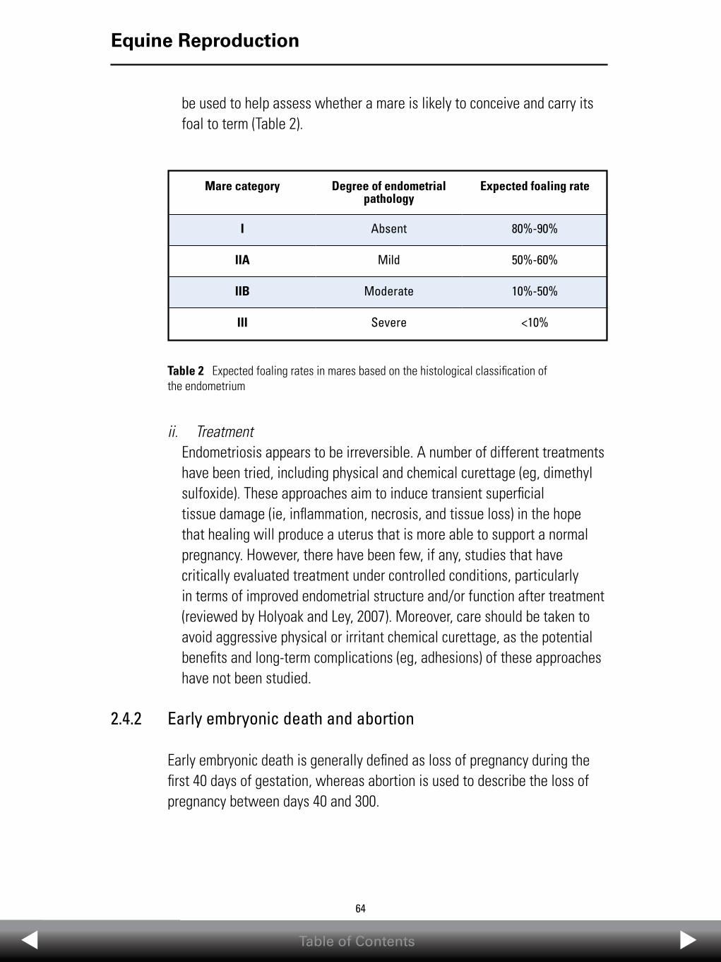
Table of Contents
64
Equine Reproduction
64
be used to help assess whether a mare is likely to conceive and carry its foal to term (Table 2).
ii. Treatment Endometriosis appears to be irreversible. A number of different treatments have been tried, including physical and chemical curettage (eg, dimethyl sulfoxide). These approaches aim to induce transient superficial tissue damage (ie, inflammation, necrosis, and tissue loss) in the hope that healing will produce a uterus that is more able to support a normal pregnancy. However, there have been few, if any, studies that have critically evaluated treatment under controlled conditions, particularly in terms of improved endometrial structure and/or function after treatment (reviewed by Holyoak and Ley, 2007). Moreover, care should be taken to avoid aggressive physical or irritant chemical curettage, as the potential benefits and long-term complications (eg, adhesions) of these approaches have not been studied.
2.4.2 Early embryonic death and abortion
Early embryonic death is generally defined as loss of pregnancy during the first 40 days of gestation, whereas abortion is used to describe the loss of pregnancy between days 40 and 300.
Table 2 Expected foaling rates in mares based on the histological classification of the endometrium
Mare category Degree of endometrial pathology
Expected foaling rate
I Absent 80%-90%
IIA Mild 50%-60%
IIB Moderate 10%-50%
III Severe <10%

Table of Contents
65
Equine Reproduction
65
a. Early embryonic death Early embryonic death, generally ranging in frequency from 5% to 15%, is assessed by measuring losses occurring between the first diagnosis of pregnancy and follow-up examination at around day 40. This has been confirmed in large-scale studies, such as a study in 3,740 mares where the overall embryonic mortality, based on examinations between days 22 and 44, was 8.9% (Chevalier-Clément, 1989). Another large study (in a group of 1,393 mares monitored closely throughout pregnancy) showed that 63% of all pregnancy losses occurred between days 15 and 45 (Morris and Allen, 2001). A much higher incidence was seen in some specific categories of mares (24.4% in mares with endometrial cysts and 34.8% in mares with an abnormal conceptus) (Chevalier-Clément, 1989).
The timing of breeding in relation to ovulation is important for the prevention of early embryonic death (see Section 2.2.3).
b. Luteal insufficiency Adequate progesterone concentrations are essential for the maintenance of pregnancy to term. There is no clear evidence that primary luteal insufficiency causes early embryonic death and pregnancy loss prior to day 25 in the mare, although there is evidence in other species. Endometrial oxytocin receptors reappear around day 18 (Stout and Allen, 2001). There is no luteotropic support for the corpus luteum from day 18 until the beginning of PMSG secretion at day 38-40, and the corpus luteum is susceptible to luteolysis during this period, which is also when many equine pregnancies fail.
In experimental paradigms of luteal inadequacy, triggered by PG release (eg, associated with endotoxaemia in colic) during the first 40 days of pregnancy, reduced progesterone concentrations lead to luteolysis and pregnancy loss (Daels et al., 1987).
i. Treatment Despite the lack of evidence for early luteal insufficiency in mares, attempts to support luteal function through either progestin supplementation or the induction of additional corpora lutea are common In fact, progestins (altrenogest) are used more widely in an attempt to maintain pregnancy in mares than in any other species (Allen, 2001b; Canisso et al., 2013a).

Table of Contents
66
Equine Reproduction
66
A GnRH analogue can be administered 11-12 days after breeding in an attempt to induce additional corpora lutea. A single intramuscular administration of buserelin (0.02-0.04 mg), a synthetic GnRH analog, during the late luteal phase (8-12 days after breeding) has been shown to improve pregnancy rates by around 10% at day 28-30 in mares (Newcombe et al., 2000). Buserelin, administered 9-10 days after detection of ovulation, has also been shown to increase pregnancy rates at 12-13 days compared to untreated controls without differences in the progesterone concentration (Jackson et al., 1986; Newcombe and Peters, 2014). Improvement in pregnancy rate was the same whether buserelin was administered intramuscularly on day 10 or 11 or subcutaneously on day 8 (Pycock and Newcombe, 1996b).
Treatment with GnRH in late dioestrus, before the luteolytic signal is triggered, may perhaps prevent luteal regression in mares in which the embryo alone is incapable of eliciting the proper signal for the maternal recognition of pregnancy.
c. Mare Reproductive Loss Syndrome Mare Reproductive Loss Syndrome (MRLS) was first observed and described in the spring of 2001, following the loss of more than 4,500 foals due to early foetal loss, late-term abortions, stillbirths, and neonatal deaths on central Kentucky horse farms (reviewed by Sebastian et al., 2008). Similar syndromes were described in Australia in 2004 and in Florida and New Jersey in 2006. Both the early and late foetal losses were characterised by the absence of specific clinical signs in aborting mares. It is also now generally accepted that ophthalmic and cardiac disease also form part of this syndrome (reviewed by Gwaltney-Brant, 2012).
The pathogenesis of MRLS is still unclear. In the United States, there appears to be a temporal correlation between MRLS and the presence of Eastern tent caterpillars (Malacosoma americanum), wild black cherry trees (Prunus serotina), and waterfowl and the practice of feeding hay off the ground (reviewed by Gwaltney-Brant, 2012; McDowell et al., 2010). In Australia, this appears to have been linked to processionary caterpillars (Ochragasterlunifer species). Affected mares have decreased concentrations of conjugated oestrogens, suggesting that the chorionic portion of the placenta is the target, supported by the fact that pregnancies of less than 35

Table of Contents
67
Equine Reproduction
67
days (prior to placental development) were largely unaffected (Volkmann et al., 2008).
d. Abortion Loss of pregnancy can be due to infectious or noninfectious (eg, twinning) causes or of unknown origin. Viral, bacterial, and parasitic infections of the equine reproductive tract are a common cause of infertility, abortion, and premature births. Some of the most common infectious causes of abortion and infertility in mares are listed in Table 3.
Other bacteria associated with abortion in mares include Actinobacillus equuli, Escherichia coli, and Pseudomonas, Klebsiella, Enterobacter, Leptospira, and Chlamydia spp. Fungal infection can also cause abortion and infertility in mares. Abortion rates between days 44 and 310 of pregnancy are generally around 9%.
i. Viral •Equineherpesvirus Equine rhinopneumonitis is the collective term used to describe highly contagious clinical disease in horses due to infection by equid herpesvirus-1 (EHV-1) or EHV-4, two closely related alphaherpesviruses of the horse. EHV-1 and EHV-4 are enzootic in all countries where there are large populations of horses. Equine rhinopneumonitis is highly contagious among susceptible horses, with transmission occurring by inhalation of an aerosol of virus-laden respiratory secretions. The viruses infect and multiply in epithelial cells of the respiratory mucosa. Clinical signs become apparent 2-8 days after exposure and are characterised by fever, inappetence, depression, and nasal discharge. The severity of respiratory disease varies with the age
Table 3 Infectious causes of infertility and abortion in horses (not luteal dependent during middle and late foetal development) (Adapted from Givens and Marley, 2008)
Viral Bacterial Parasitic
Equine herpesvirus-1Equine viral arteritis virus
Streptococcus equi subsp. zooepidemicusTaylorella equigenitalis
Trypanosoma equiperdum

Table of Contents
68
Equine Reproduction
68
and immunological status (eg, vaccination, previous infection) of the horse. Generally, recovery is uncomplicated and occurs within 1-2 weeks. EHV-1 can spread beyond the respiratory tract and is the most important viral cause of abortion in horses, often at more than 7 months of gestation and without any previous clinical signs. The placenta may be oedematous or normal, while the aborted foetus may exhibit subcutaneous oedema, jaundice, increased thoracic fluid volume, and/or an enlarged liver with yellow-white lesions approximately 1 mm in diameter. Histologically, there are areas of necrosis and characteristic intranuclear inclusions. Necrotising bronchiolitis is also a common finding. EHV-1 can also cause perinatal foal death and/or neurological dysfunction (myeloencephalopathy). Recovered animals can remain latently infected. Diagnosis is performed using direct immunofluorescent detection of viral antigen or by virus isolation, eg, on tissues from an aborted foetus. Polymerase chain reaction (PCR) and immunoperoxidase staining methods are also available. Prevention of equine rhinopneumonitis is based on biosecurity and vaccination. For a review of equine rhinopneumonitis, see the World Organisation of Animal Health (OIE) (2008).
•Equineviralarteritis Equine viral arteritis (EVA) is an acute, contagious, viral disease caused by equine arteritis virus (EAV), an RNA virus belonging to the genus Arterivirus, family Arteriviridae. EAV is present in the horse population of many countries worldwide (Timoney and McCollum, 1993). Transmission of EAV is by respiratory, venereal, and congenital routes. Infection is often subclinical. When present, clinical signs can include fever, depression, anorexia, dependent oedema, conjunctivitis, an urticarial-type skin reaction, and abortion. Apart from mortality in young foals, particularly those congenitally infected, the case-fatality rate in outbreaks of EVA is very low (Timoney and McCollum, 1993; Vaala et al., 1992). EVA cannot be differentiated clinically from other viruses causing respiratory and systemic disease in horses. Diagnosis of EAV infection is based on virus isolation, detection of nucleic acid (eg, by reverse transcriptase PCR [RT-PCR]) or viral antigen, or demonstration of a specific antibody response. A variety of serological tests have been used for the detection of antibody to EAV. Currently, complement enhanced virus neutralisation (VN) and enzyme-linked

Table of Contents
69
Equine Reproduction
69
immunosorbent assay (ELISA) are the most widely used, with VN being the most sensitive and specific. EVA control programs employ appropriate biosecurity measures along with, in countries where this is permissible and available, targeted vaccination to prevent the spread of EAV in breeding populations, thus minimising the risk of abortion outbreaks, death of young foals, and the establishment of carrier colts and stallions (Timoney and McCollum, 1993). For a review of equine viral arteritis, see the World Organisation of Animal Health (2013a).
ii. Bacterial Bacterial placentitis is reported to be the cause of 9.8%-33.5% of abortions, stillbirths, and perinatal losses in horses (reviewed by Lyle, 2014). The majority of placentitis (53%), a condition that occurs in late pregnancy, is caused by ascending bacterial infections. Studies in an experimental model of ascending placentitis in pony mares appear to suggest that it is secondary inflammation of the chorion rather than infection that may lead to premature parturition (reviewed by Macpherson and Bailey, 2008). It appears that there is a complex interplay between the inflammatory response to the pathogen, loss of myometrial quiescence, and activation of the foetal hypothalamic-pituitary-adrenal axis that leads to abortion.
•Streptococcusequisubspecies zooepidemicus Streptococcus equi subsp. zooepidemicus (Strep. zooepidemicus) is a Gram-positive bacterium that belongs to the commensal flora of the caudal reproductive tract of mares. It is an opportunistic pathogen and the pathogen most commonly isolated from the uterus of mares. Strep. zooepidemicus can cause abortion following ascending infection. It is isolated from around 28% of cases of placentitis. Other bacteria isolated frequently include Escherichia coli and Leptospira, Pseudomonas, Enterobacter, and Klebsiella spp., other streptococci, staphylococci, and Actinobacillus spp. (Giles et al., 1993; reviewed by Lyle, 2014). The placenta can also become infected by viruses and fungi, but this is more commonly associated with abortion in early gestation. Mares with placentitis often present in late pregnancy with signs of premature udder development and premature lactation with or without vulvar discharge (reviewed by Macpherson et al.,2008).

Table of Contents
70
Equine Reproduction
70
Early detection and treatment of this condition is vital for ensuring the production of a viable foal (reviewed by Cummins et al., 2008). Transabdominal or transrectal ultrasonography is used commonly in the diagnosis of placentitis. Mares with placental infection or inflammation exhibit an increase in the combined thickness of the uterus and placenta (CTUP) (reviewed by Macpherson and Bailey, 2008). Thickening of the amnion and/or separation of the membranes from the endometrium can also be observed. Purulent hyperechoic material may be seen in pockets between the chorioallantois and the endometrium. Definitive diagnosis is based on histopathology of the chorioallantoic membrane. Management strategies are aimed at treating the infection and maintaining the pregnancy to as close to term as possible (reviewed by Cummings et al., 2008). Treatment generally consists of antimicrobial therapy, based on bacterial culture and susceptibility testing, anti-inflammatories, and hormonal support (Cummins et al., 2008; Macpherson, 2005; Macpherson and Bailey, 2008).
•Taylorellaequigenitalis Contagious equine metritis (CEM) is an inflammatory disease of the proximal and distal reproductive tract of the mare caused by Taylorella equigenitalis, a Gram-negative, nonmotile, bacillus or coccobacillus. Mares with CEM may present with signs of endometritis, cervicitis, and vaginitis of varying severity and slight to copious mucopurulent vaginal discharge. Prolonged asymptomatic or symptomatic carriage is established in a proportion of infected mares. Stallions can become asymptomatic carriers. The highest risk of transmission is by direct venereal contact from a contaminated stallion or an infected mare. However, venereal transmission can also take place at AI if fresh, cooled or possibly even frozen, thawed semen is used (Schulman et al., 2013). Indirect transmission can also occur (eg, fomites, inadequate biosecurity at time of breeding and/or semen collection, processing, and transport) (Schulman et al., 2013). The level of risk associated with cryopreserved semen and embryos needs further investigation (Schulman et al., 2013). Diagnosis of CEM and detection of carriers is based on bacterial culture of T. equigenitalis from swabs taken from the genital tract, particularly the clitoral fossa and recesses of the clitoral sinuses (Platt et al., 1978). This methodology, despite

Table of Contents
71
Equine Reproduction
71
some limitations, is the gold standard used for all international horse trade and movement protocols (Schulman et al., 2013). Serology is not a reliable means of detecting T. equigenitalis infection. However, serum antibody to T. equigenitalis can be used to help detect recent (3-7 weeks previously) infection. PCR can also be used to detect T. equigenitalis and is rapid, cost effective, and offers high throughput and improved sensitivity and specificity (Schulman et al., 2013). However, a robust, validated PCR assay is needed before regulatory authorities worldwide could consider this as a potential replacement for bacterial culture (Schulman et al., 2013). For a review of CEM, see the World Organisation of Animal Health (2012).
iii. Parasitic •Trypanosomaequiperdum Trypanosoma equiperdum is a protozoan parasite that causes Dourine, a potentially fatal, acute or chronic disease that is transmitted during coitus (venereal transmission). It is the only trypanosome that is not transmitted by an invertebrate vector and is found in Africa and South America. Trypanosoma equiperdum is unusual in that it is primarily a tissue parasite and is rarely detected in the blood. There is no known natural reservoir of the parasite other than infected equids. It is present in the genital secretions of both infected males and females. The incubation period, severity, and duration of the disease vary considerably. Clinical signs include a fluctuating course of fever, oedema of the genitalia and mammary glands, cutaneous eruptions and plaques (pathognomonic), facial or lip paralysis, incoordination, anaemia, and emaciation, leading to death. Diagnosis is usually based on clinical signs and serology (complement fixation test). The only effective control is through strict quarantine and identification and removal of infected animals. Good hygiene is essential during assisted breeding because infection may be transmitted through contaminated fomites. For a detailed review of Dourine, see the World Organisation of Animal Health (2013b).
2.4.3 Retained foetal membranes
In mares, the placenta is normally expelled within 90 minutes after parturition (Vandeplassche et al., 1971). There appears to be consensus

Table of Contents
72
Equine Reproduction
72
that retained foetal membranes (RFM) in mares can be defined as a failure to expel some or all of the chorioallantois spontaneously within 3 hours of foaling (reviewed by Canisso et al., 2013b). The incidence of RFM ranges from 2% to 10% of foalings in light breed-type mares and has been reported to be as high as 30%-54% in Friesian mares. The incidence of RFM is increased by dystocia, late-term abortion, prolonged gestation, hydrops, induction of parturition, and Caesarean section. Complications of retained foetal membranes include metritis, septicaemia, toxaemia, laminitis, and even death. In one study, 25% of mares with RFM developed signs of laminitis within 24 hours of foaling. RFM may also result in the delayed involution of the uterus and impaired fertility at the foal heat. a. Treatment RFM is generally treated in a stepwise fashion, starting with the administration of 10-20 IU of oxytocin intravenously or intramuscularly every few hours from 3 hours after foaling (reviewed by McCue and Ferris, 2015). Larger doses of oxytocin can lead to intense spasmodic uterine contractions that may cause considerable distress to the mare. Oxytocin (50-80 IU) can also be administered as an intravenous infusion in 1-2 L of sterile saline or glucose solution (5%) (Hospes and Huchzermeyer, 2004). The placenta is normally expelled within 1-2 hours after administration (Blanchard and Varner, 1993). Gentle traction can also be applied to the placenta, but care should be taken to avoid damaging the uterus, tearing the placenta, or causing uterine prolapse. Oxytocin treatment can be combined with uterine lavage, which results in a more complete separation of the chorionic villi and removes small pieces of the placenta and debris. Intrauterine and/or systemic antimicrobial treatment can prevent the development of septicaemia. Nonsteroidal anti- inflammatory drugs (NSAID) are indicated should signs of toxaemia occur (Blanchard and Varner, 1993). The prognosis is favourable if treatment is initiated promptly. The long-term prognosis for survival is poor to moderate for mares that develop severe laminitis, endotoxaemia, and/or metritis (reviewed by Canisso et al., 2013b).
2.5 The stallion
Fertility in stallions is assessed by clinical examination, semen evaluation, and observation of sexual behaviour.

Table of Contents
73
Equine Reproduction
73
2.5.1 Reproductive performance evaluation
A stallion’s reproductive evaluation begins with a clinical examination, focusing on the external genitalia and the hind legs and back (to confirm its ability to mount a mare). The testicles should be palpated for consistency and position within the scrotum and their circumference measured. Libido is then evaluated—in particular, the reaction time from presentation of the mare to the time of covering. Deficiencies in libido, excessive aggressiveness toward the mare or handler, and other abnormalities of behaviour should all be recorded.
a. Semen collection If the examination takes place before the breeding season, semen should be collected three times 24 hours apart to eliminate existing semen reserves. During the season, the stallion should be rested (from sexual activity) for 3 days prior to semen collection. For testing, semen is collected on two occasions, 1 hour apart, and evaluated for gel-free volume, total number of spermatozoa, percentage of progressive motile sperm (PMS), morphology, and pH. Assessment of the number of PMS of a particular stallion means that it can be managed better: •Fornaturalmating,astallionwillusuallybeusedtwiceaday,sixtimes per week •ForAI,spermqualityandquantitywilldeterminehowmanymarescan be inseminated from each ejaculate, and the semen will usually be collected three times a week
b. Semen transportation The widespread acceptance of the transport of fresh, cooled semen in many parts of the equine industry signifies a major change. Semen transportation offers the advantage that mares do not need to be transported for breeding, reducing the possibility of injury or disease to the mare or foal, and increases access to desirable stallions. The number of mares bred using fresh, cooled semen is increasing annually. Semen is diluted 1:3 with semen extenders to provide an energy source and protect it from cold shock. It is then cooled from 37°C to 5°C, at a rate of less than 0.05°C per minute from 18°C to 8°C, and maintained at a low temperature (3°C-6°C) for up to 36 hours. The semen is placed in an airtight polystyrene container with a separate cooling

Table of Contents
74
Equine Reproduction
74
system and shipped by express courier. Spermatozoa should not come into contact with the rubber plunger of a syringe or the cooling system.
c. Semen conservation at low temperatures There are certain limitations to the conservation of equine semen at low temperatures, mainly associated with variability in the ability of sperm from different stallions to tolerate freezing and thawing. It is thought that frozen, thawed semen from only 25% of stallions will produce pregnancy rates comparable to that of fresh, cooled semen or natural mating (Vidament et al., 1997). The majority of equine semen is frozen in 0.5-mL straws at a concentration of 200 million-400 million sperm per mL. Typically, cooling rates in the range of 10°C-50°C/min are employed, with relatively low concentrations of cryoprotectants (reviewed by Squires, 2005).
d. Use of sexed semen Although flow cytometry has proven to be an accurate method for separating X- and Y-chromosome-bearing spermatozoa, it is still not in widespread use in the equine industry. It has been tested in a large-scale field study with promising results (Panarace et al., 2014). Factors limiting the use of sex-sorted semen include the greater cost of the equipment as well as the license needed to use it. Furthermore, the fertility of sex-sorted spermatozoa is highly stallion dependent, and the logistics of having the mare, stallion, and equipment in the same place can be problematic (Mari et al., 2010).
2.5.2 Cryptorchidism
Cryptorchidism is the condition where one or both testes fail to descend normally into the scrotum. Cryptorchidism is common in male horses (2%-8%), and is generally accepted to be a hereditary condition (reviewed by Edwards, 2008). It is often unilateral, occurring with equal frequency on the left or right, but occurs bilaterally in 10%-15% of cases. In slightly more than one-half of cases the testis (or testes) is (are) abdominal rather than inguinal.
When a cryptorchid stallion is hemicastrated, leaving one testicle in the inguinal canal or abdominal cavity, it will continue to exhibit male characteristics, very often including aggressive and dangerous behaviour. In addition, it is possible that the retained testicle may undergo neoplastic

Table of Contents
75
Equine Reproduction
75
change. If the testicle can be palpated in the inguinal canal, diagnosis is easy; when the testicle is within the abdominal cavity, it is more difficult.
a. Diagnosis Cryptorchidism, or failure of testicular descent, can be a challenging clinical diagnosis, particularly in horses with an unknown history (reviewed by Lu, 2005). Diagnosis should include behaviour assessment, physical examination, and hormonal assessment.
The testes of the horse produce testosterone and oestrogens. Hormonal assessment can include baseline hormone concentrations and stimulation tests to examine the function of the hypothalamic-pituitary-testicular axis. Cryptorchids have high basal testosterone compared to geldings, but baseline testosterone concentrations in unilateral cryptorchids may not differ from those found in stallions (reviewed by Lu, 2005). Equivocal results are obtained in around 14% of cases if a single baseline sample is taken for testosterone (Cox et al., 1986). Bilateral cryptorchids have higher oestrogen concentrations than stallions, geldings, or unilateral cryptorchids (Ganjam and Kenney, 1975). The concentration of oestradiol-17β has been shown to be lower in unilateral cryptorchids than in stallions (Coryn et al., 1981).
An hCG (6,000-12,000 IU intravenously) stimulation test has long been used for the diagnosis of cryptorchidism in horses (reviewed by Lu, 2005). An increase in testosterone concentrations following administration reflects the presence of functional Leydig cells. Two blood samples need to be taken, the first prior to hCG administration and the second, depending on the test protocol used, 30 minutes-2 hours later. In one study the accuracy was 94.6% following administration of the higher dose of hCG (12,000 IU), with sampling prior to and 30 minutes after treatment (Cox et al., 1986). This also decreased the number of equivocal results to 6.7%.
Baseline oestrogen (oestrone sulphate) concentrations may be helpful in the diagnosis of cryptorchidism. However, oestrogen concentrations following hCG administration can be difficult to interpret (reviewed by Lu, 2005). Following a single intravenous administration of hCG (10,000 IU) to stallions and cryptorchids, there was a gradual increase in unconjugated

Table of Contents
76
Equine Reproduction
76
androgen concentrations (testosterone), peaking after 2 days, with higher concentrations reached in stallions than in cryptorchids (Silberzahn et al., 1989).
b. Treatment Given the likely heritable nature of cryptorchidism, medical treatment of this condition is not advised. In addition, no controlled studies have been conducted to support anecdotal reports on the use of GnRH or hCG in the treatment of cryptorchidism. It is not known how often testicular descent has been achieved and whether the testis is subsequently able to function normally. Cryptorchidism in horses should be treated surgically.
2.5.3 Sexual behavioural disorders
Very few centers specialise in the diagnosis and treatment of sexual disorders of stallions. Sexual behaviour in the stallion is influenced by many factors, including season, hormone concentrations, psychology, and the skills of the handler. Suboptimal libido or poor mating ability appear to be among the more common reproductive complaints in the breeding stallion. Common problems include overuse, illness, or pain (often musculoskeletal) or, in the case of a stallion used for AI, an inadequately prepared artificial vagina (eg, not warm enough, too little pressure) and unsympathetic handling. More research is needed to fully understand the complexities of stallion sexual behaviour.
a. Deficient libido Pharmacological treatment to stimulate libido or mating ability is a last resort, to be attempted only when clinical examination, careful management and handling, and patient attempts to train and encourage the stallion have failed. Medical intervention is usually only required once in novice stallions that are anxious (diazepam 0.05 mg/kg by slow intravenous injection 5-7 minutes before breeding) or have low libido (GnRH 0.05 mg subcutaneously 2 hours and 1 hour before breeding), because ejaculation is a powerful reinforcing stimulus (McDonnell, 1999). The administration of testosterone to boost libido is not recommended, because too high a dose can suppress spermatogenesis and stimulate aggressive behaviour (Stout et al., 2005).

Table of Contents
77
Equine Reproduction
77
2.5.4 Testicular degeneration
Numerous factors, including age, trauma, and infection, including parasitism, can lead to testicular degeneration. Inflammation or oedema of the scrotum can interfere with heat dissipation, resulting in an increase in scrotal and testicular temperature, severely affecting fertility. An increase in the temperature of as little as 2°C for 24 hours, if not promptly addressed, sterilises the stallion until new spermatozoa are formed (around 57 days).
Testicular degeneration is difficult to diagnose if there are no results and measurements (testicular size and consistency) from previous examinations for comparison. Histological examination of a testicular biopsy is possible but can lead to rupture of the blood-testicle barrier, leading to the formation of antibodies to spermatozoa, which can hamper reproductive performance, and severe hemorrhage. Ultrasonography is noninvasive and allows investigation of the testicular stroma.
2.5.5 Hemospermia and urospermia
The presence of blood or urine in the ejaculate reduces fertility.
a. Hemospermia Hemospermia is the contamination of semen with blood. The underlying cause of hemospermia is not always known (Varner et al., 2000). However, blood can enter the semen following infection, trauma, neoplasia, or habronemiasis. It seems that the presence of red blood cells, as much as 20% of whole blood, is the key factor in reducing fertility. Immediate dilution with semen extenders lessens the negative effects of contamination with blood. Treatment involves identification and treatment of the underlying cause and resting from sexual activity for up to 3 months.
b. Urospermia Urospermia is a sporadic condition that is more difficult to diagnose because the clinical signs can be subtle. The underlying cause is unknown. Although affected stallions may not always urinate during ejaculation (eg, only 30% of the time), it takes very little urine (pH, osmolality) to affect fertility (Griggers et al., 2001). There appear to be no best-practice

Table of Contents
78
Equine Reproduction
78
approaches to treatment of this sporadic condition, and the results of empirical treatment are often inconclusive.
2.6 ReferencesAllen WR. Fetomaternal interactions and influences during equine pregnancy. Reproduction. 2001a;121:513–527.
Allen WR. Luteal insufficiency and embryo mortality in the mare. Reprod Domest Anim. 2001b;36:121–131.
Allen WR. The development and application of the modern reproductive technologies to horse breeding. Reprod Domest Anim. 2005;40:310–329.
Allen WR, Urwin V, Simpson DJ, et al. Preliminary studies on the use of an oral progestogen to induce oestrus and ovulation in seasonally anoestrous Thoroughbred mares. Equine Vet J. 1980;12:141–145.
Ataman MB, Gunay A, Gunay U, Baran A, Suman M. Oestrus synchronization with progesterone-impregnated device and prostaglandin F2α combined with human chorionic gonadotrophin in transitional mares. Rev Med Vet. 2000;151:1031–1034.
Barbacini S, Gulden P, Marchi V, Zavaglia G. Incidence of embryonic loss in mares inseminated before or after ovulation. Equine Vet Educ. 1999;1:108–112.
Barbacini S, Zavaglia G, Gulden P, Marchi V, Necchi D. Retrospective study on the efficacy of hCG in an equine artificial insemination programme using frozen semen. Equine Vet Educ. 2000;2:404–408.
Barrier-Battut I, Le Poutre N, Trocherie E, et al. Use of buserelin to induce ovulation in the cyclic mare. Theriogenology. 2001;55:1679–1695.
Becker SE, Johnson AL. Effects of gonadotrophin-releasing hormone infused in a pulsatile or continuous fashion on serum gonadotropin concentrations and ovulation in the mare. J Anim Sci. 1992;70:1208–1215.
Berezowski CJ, Stitch KL, Wendt KM, Vest DJ. Clinical comparison of 3 products available to hasten ovulation in cyclic mares. J Equine Vet Sci. 2004;24:231–233.
Besognet B, Hansen BS, Daels PF. Induction of reproductive function in anestrous mares using a dopamine antagonist. Theriogenology. 1997;47:467–480.
Blanchard TL, Varner DD. Therapy for retained placenta in the mare. Vet Med. 1993;1:55–59.
Bott RM, Shambley MO, Bailey MT. Induction of ovulation in the mare with the synthetic GnRH analogue leuprolide. Equine Practice. 1996;18:30–33.
Brendemuehl JP. Effect of oxytocin and cloprostenol on luteal formation, function and pregnancy rates in mares. Theriogenology. 2002;58:623–626.
Brinsko SP, Blanchard TL, Varner DD, et al. In: Manual of Equine Reproduction, 3rd ed. Maryland Heights, MO: Mosby Elsevier; 2011.
Bucca S, Fogarty U, Collins A, Small V. Assessment of feto-placental wellbeing in the mare from mid gestation to term: transrectal and transabdominal ultrasonographic features. Theriogenology. 2005;64:542–557.
Bucca S, Carli A, Buckley T, Dolci G, Fogarty U. The use of dexamethasone administered to mares at breeding time in the modulation of persistent mating induced endometritis. Theriogenology. 2008;70:1093–1100.
Camillo F, Pacini M, Panzani D, Vannozzi I, Rota A, Aria G. Clinical use of twice daily injections of buserelin acetate to induce ovulation in the mare. Vet Res Commun. 2004;28:169–172.
Camillo F, Marmorini P, Romagnoli S, Cela M, Duchamp G, Palmer E. Clinical studies on daily low dose oxytocin in mares at term. Equine Vet J. 2000;32:307–310.

Table of Contents
79
Equine Reproduction
79
Canisso IF, Beltaire KA, Bedford-Guaus SJ. Premature luteal regression in a pregnant mare and subsequent pregnancy maintenance with the use of oral altrenogest. Equine Vet J. 2013a;45:97–100.
Canisso IF, Rodriguez JS, Sanz MG, Coutinho da Silva MA. A clinical approach to the diagnosis and treatment of retained fetal membranes with an emphasis placed on the critically ill mare. Equine Vet Sci. 2013b;33:570–579.
Card C. Post-breeding inflammation and endometrial cytology in mares. Theriogenology. 2005;64:580–588.
Carnevale EM. Vitrification of equine embryos. Vet Clin North Am Equine Pract. 2006; 22:831–841.
Checura CM, Beg MA, Parrish JJ, Ginther OJ. Functional relationships among insulin like growth factor 1, circulatory gonadotropins and development of the dominant follicle in mares. Anim Reprod Sci. 2010;118:270–278.
Chevalier-Clément F. Pregnancy loss in the mare. Anim Reprod Sci. 1989;20:231–244.
Coryn M, De Morr A, Bouters R, Vandeplassche M. Clinical, morphological and endocrinological aspects of cryptorchidism in the horse. Theriogenology. 1981;16:489–496.
Cox JE, Redhead PH, Dawson FE. Comparison of the measurement of plasma testosterone and plasma oestrogens for the diagnosis of cryptorchidism in the horse. Equine Vet J. 1986;18:179–182.
Cuervo-Arango J, Newcombe JR. Repeatability of preovulatory follicular diameter and uterine edema pattern in two consecutive cycles in the mare and how they are influenced by ovulation inductors. Theriogenology. 2008;69:681–687.
Cummins C, Carrington S, Fitzpatrick E, Duggan V. Ascending placentitis in the mare: A review. Irish Vet J. 2008;61:307–313.
Daels PF, Fatone S, Hansen B, Concannon PW. Dopamine antagonist induced reproductive activity in anoestrous mares: gonadotrophin secretion and the effects of environmental cues. J Reprod Fertil Suppl. 2000;56:173–183.
Daels P, Starr M, Kindahl H, Fredriksson G, Hughes JP, Stabenfeldt GH. Effect of Salmonella typhimurium endotoxin on PGF2α release and foetal death in the mare. J Reprod Fertil Suppl. 1987;35:485–492.
Davies Morel MCG, Newcombe JR, Hayward K. Factors affecting preovulatory diameter in the mare: the effect of mare age, season and presence of other ovulatory follicles. Theriogenology. 2010;74:1241–1247.
Decourt C, Caraty A, Briant C, et al. Acute injection and chronic perfusion of kisspeptin elicit gonadotropin release but fail to trigger ovulation in the mare. Biol Reprod. 2014;90:1–12.
Donadeu FX, Ginther OJ. Follicular waves and circulating concentrations of gonadotrophins, inhibin and oestradiol during the anovulatory season in mares. Reproduction. 2002;124:875–885.
Donadeu FX, Watson ED. Seasonal changes in ovarian activity: lessons learnt from the horse. Anim Reprod Sci. 2007;1000:225–242.
Driancourt MA, Palmer E. Seasonal and individual effects on ovarian and endocrine responses of mares to a synchronization treatment with progestagen impregnated sponges. J Reprod Fertil Suppl. 1982;32:283–291.
Edwards JF. Pathologic conditions of the stallion reproductive tract. Anim Reprod Sci. 2008;107:197–207.
Evans MJ, Alexander SL, Irvine CHG, Livesey JH, Donald RA. In vitro and in vivo studies of equine prolactin secretion throughout the year. J Reprod Fertil Suppl. 1991;44:27–35.
Fay JE, Douglas RH. Changes in thecal and granulosa cell LH and FSH receptor content associated with follicular fluid and peripheral plasma gonadotropin in preovulatory follicles of mares. J Reprod Fertil Suppl. 1987;35:169–181.

Table of Contents
80
Equine Reproduction
80
Ganjam VK, Kenney RM. Androgens and oestrogens in normal and cryptorchid stallions. J Reprod Fertil Suppl. 1975;23:67–73.
Giles RC, Donahue JM, Hong CB, et al. Causes of abortion, stillbirth and perinatal death in horses: 3,527 cases (1986-1991). J Am Vet Med Assoc. 1993;203:1170–1175.
Ginther OJ. Embryonic loss in mares: incidence, time of occurrence and hormonal involvement. Theriogenology. 1985;23:77–89.
Ginther OJ, Gastal EL, Gastal MO, Beg MA. Dynamics of the equine preovulatory follicle and periovulatory hormones: What’s new? Review. J Equine Vet Sci. 2008;28:454–460.
Ginther OJ, Woods BG, Meira C, Beg MA, Bergfelt DR. Hormonal mechanism of follicle deviation as indicated by major versus minor follicular waves during the transition into the anovulatory season in mares. Reproduction. 2003;126:653–660.
Ginther OJ, Utt MD, Bergfelt DR, Beg MA.Controlling interrelationships of progesterone/LH and estradiol/LH in mares. Anim Reprod Sci. 2006;95:144–150.
Givens MD, Marley MSD. Infectious causes of embryonic and fetal mortality. Theriogenology. 2008;70:270–285.
Griggers S, Paccamonti DL, Thompson RA, Eilts BE. The effects of pH, osmolarity and urine contamination on equine spermatozoal motility. Theriogenology. 2001;56:613–622.
Grimmett JB, Perkins NR. Human chorionic gonadotrophin (hCG): the effect of dose on ovulation and pregnancy rate in thoroughbred mares experiencing their first ovulation of the breeding season. N Z Vet J. 2001;49:88–93.
Gwaltney-Brant SM. Mare reproductive loss syndrome. In: Gupta RC, ed. Veterinary Toxicology, 2nd ed. Ames: Elsevier; 2012.
Handler J, Schonlieb S, Hoppen HO, Aurich C. Seasonal effects on attempts to synchronize estrus and ovulation by intravaginal application of progesterone-releasing device (PRID) in mares. Theriogenology. 2006;65:1145–1158.
Hoffmann C, Ellenberger C, Costa Mattos R, et al. The equine endometrosis: New insights into the pathogenesis. Anim Reprod Sci. 2009;111:261–278.
Holyoak GR, Ley WB. Management regimens for uterine cysts. In: Samper JC, Pycock JF, McKinnon AO, eds. Current Therapy in Equine Reproduction. St. Louis, MO: Saunders Elsevier; 2007.
Horteloup MP, Threlfall WR, Funk JA. The early conception factor (ECFTM) lateral flow assay for non-pregnancy determination in the mare. Theriogenology. 2005;64:1061–1071.
Hospes R, Huchzermeyer S. Behandlung der retentio secundinarum bei der stute nach einem 4-stufen-program m - Ein rückblick auf 36 fälle | [Treatment of retained placenta in broodmares in a 4-step-routine - Review of 36 cases]. Pferdeheilkunde. 2004;20:498–504.
Hurtgen JP. Pathogenesis and treatment of endometritis in the mare: A review. Theriogenology. 2006;66:560–566.
Jackson SA, Squires EL, Nett TM. The effect of exogenous progestins on endogenous progesterone secretion in pregnant mares. Theriogenology. 1986;25:275–279.
Johnson AL. Pulsatile administration of gonadotrophin-releasing hormone advances ovulation in cyclic mares. Biol Reprod. 1986;35:1123–1130.
Kenney RM, Doig PA. Equine endometrial biopsy. In: Morrow DA, ed. Current Therapy in Theriogenology. Philadelphia, PA: WB Saunders; 1986.
Kindahl H, Odensvik K, Hansen B, Daels PF. Changes in PGF2alpha secretion during prolonged luteal phase in mares. J Reprod Fertil Suppl. 2000;56:305–315.
King SS, Roser JF, Jones KL. Follicular fluid prolactin and the periovulatory prolactin surge in the mare. J Equine Vet Sci. 2008;28:468–472.
Klug E, Jöchle W. Advances in synchronizing estrus and ovulation in the mare: a mini review. J Equine Vet Sci. 2001;21:474–479.

Table of Contents
81
Equine Reproduction
81
LeBlanc MM, Causey RC. Clinical and subclinical endometritis in the mare: both threats to fertility. Reprod Domest Anim. 2009;44 Suppl 3:10–22.
Levy I, Duchamp G. A single subcutaneous administration of buserelin induces ovulation in the mare: field data. Reprod Domest Anim. 2007;42:550–554.
Logan NL, McCue PM, Alonso MA, Squires EL. Evaluation of three equine FSH superovulation protocols in mares. Anim Reprod Sci. 2007;102:48–55.
Loomis PR. The equine frozen semen industry. Anim Reprod Sci. 2001;68:191–200.
Lowis TC, Hyland JH. The effect of an extended artificial photoperiod and gonadotrophin-releasing hormone infusions in inducing fertile oestrus in anoestrous mares. Aust Vet J. 1991;68:400–402.
Loy RG, Buell JR, Stevenson W, Hamm D. Sources of variation in response intervals after prostaglandin treatment in mares with functional corpora lutea. J Reprod Fertil Suppl. 1979;27:229–235.
Lu KG. Clinical diagnosis of the cryptorchid stallion. Equine Pract. 2005;4:250–256.
Lyle SK. Immunology of infective preterm delivery in the mare. Equine Vet J. 2014;46:661–668.
Macpherson ML. Treatment strategies for mares with placentitis. Theriogenology. 2005;64:528–534.
Macpherson ML, Bailey CS. A clinical approach to managing the mare with placentitis. Theriogenology. 2008;70:435–440.
Macpherson ML, Blanchard TL. Breeding mares on foal heat. Equine Vet Educ. 2005; 17:44–52.
Magee C, Bruemmer JE, Nett TM, Squires EL, Clay CM. Kisspeptide in the estrous mare: is it an appropriate ovulation inducing agent? Theriogenology. 2012;78:1987–1996.
Malpaux B, Thiery JC, Chemineau P. Melatonin and the seasonal control of reproduction. Reprod Nutr Dev. 1999;39:355–366.
Mari G, Rizzato G, Merlo B, et al. Quality and fertilizing ability in vivo of sex-sorted stallion spermatozoa. Reprod Domest Anim. 2010;45:331–335.
Marino E, Threlfall WR, Schwarze RA. Early conception factor lateral flow assays for pregnancy in the mare. Theriogenology. 2009;71:877–883.
McCue PM, Ferris RA. Hormone therapy in equine reproduction. In: Sprayberry KA, Robinson NE, eds. Robinson’s Current Therapy in Equine Medicine, 7th ed. St. Louis, MO: Saunders; 2015.
McDonnell AM, Watson ED. The effect of transcervical uterine manipulations on establishment of uterine infection in mares under the influence of progesterone. Theriogenology. 1992;38:945–950.
McDonnell SM. Sexual behavior dysfunction in stallions. In: Robinson NE, ed. Current Therapy in Equine Medicine. Philadelphia, PA: WB Saunders Company; 1992.
McDowell KJ, Webb BA, Williams NM, et al. Invited review: the role of caterpillars in mare reproductive loss syndrome: a model for environmental causes of abortion. J Anim Sci. 2010;88:1379–1387.
McKinnon AO, Nobelius AM, del Marmol Figueroa ST, Skidmore J, Vasey JR, Trigg TE. Predictable ovulation in mares treated with an implant of the GnRH analogue deslorelin. Equine Vet J. 1993;25:321–323.
McKinnon AO, Vasey JR, Lescun TB, Trigg TE. Repeated use of a GnRH analogue deslorelin (Ovuplant) for hastening ovulation in the transitional mare. Equine Vet J. 1997;29:153–155.
Meyers PJ. Using hormones to control cyclicity and ovulation in the broodmare. Vet Med. 1991;11:1106–1111.
Meyers PJ, Bowman T, Blodgett G, et al. Use of the GnRH analogue deslorelin in a slow-release implant to accelerate ovulation in oestrous mares. Vet Rec. 1997;140:249–252.

Table of Contents
82
Equine Reproduction
82
Meyers SA, LeBlanc MM. Induction of parturition: Clinical considerations for successful foalings. Vet Med. 1991;86:1117–1121.
Meyers-Brown G, Bidstrup LA, Famula TR, Colgin M, Roser JF. Treatment with recombinant FSH followed by recombinant LH increases embryo recovery in superovulated mares. Anim Reprod Sci. 2011;128:52–59.
Morris LHA, Allen WR. Reproductive efficiency in the Thoroughbred mares in Newmarket. Equine Vet J. 2002;34:51–60.
Nagy P, Guillaume D, Daels P. Seasonality in mares. Anim Reprod Sci. 2000;60-61:245–262.
Newcombe JR, Martinez TA, Peters AR. The effect of the gonadotrophin releasing hormone analogue, buserelin, on pregnancy rates in horse and pony mares. Theriogenology. 2000;55:1619–1631.
Newcombe JR, Peters AR. The buserelin enigma; how does treatment with this GnRH analogue decrease embryo mortality? J Vet Sci Technol. 2014;5:151.
Oxender WD, Noden PA, Hafs HD. Oestrus, ovulation and plasma hormones after prostaglandin F2alpha in mares. J Reprod Fertil Suppl. 1975;23:251–255.
Palmer E. Control of the oestrous cycle of the mare. J Reprod Fertil. 1978;54:495–505.
Palmer E, Driancourt MA. Use of ultrasonic echography in equine gynecology. Theriogenology. 1980;13:203–216.
Palmer E, Driancourt MA. Some interactions of season of foaling, photoperiod and ovarian activity in the equine. Livest Prod Sci. 1983;10:197–210.
Panarace M, Pellegrini RO, Basualdo MO, et al. First field results on the use of stallion sex-sorted semen in a large-scale embryo transfer program. Theriogenology. 2014;81:520–525.
Platt H, Atherton JG, Simpson DJ. The experimental infection of ponies with contagious equine metritis. Equine Vet J. 1978;10:153–159.
Pycock JF, Newcombe JR. Assessment of three treatments to remove intrauterine fluid on pregnancy rate in the mare. Vet Rec. 1996a;138:320–323.
Pycock JF, Newcombe JR. The effect of the gonadotrophin-releasing hormone analog, buserelin, administered in diestrus on pregnancy rates and pregnancy failure in mares. Theriogenology. 1996b;46:1097–1101.
Rickets SW, Alonso S. Assessment of the breeding prognosis of mares using paired endometrial biopsy techniques. Equine Vet J. 1991;23:185–188.
Rogan D, Fumuso E, Rodríguez E, Wade J, Sánchez Bruni SF. Use of a mycobacterial cell wall extract (MCWE) in susceptible mares to clear experimentally induced endometritis with Streptococcus zooepidemicus. J Equine Vet Sci. 2007;27:112–117.
Roser JF, Kiefer BL, Evans JW, Neely DP, Pacheco CA. The development of antibodies to hCG following its repeated injection in the cyclic mare. J Reprod Fertil Suppl. 1979;27:173–179.
Rutten DR, Chaffaux S, Valon M, Deletang F, de Haas V. Progesterone therapies in mares with abnormal oestrous cycles. Vet Rec. 1986;119:569–571.
Samper JC. Management and fertility of mares bred with frozen semen. Anim Reprod Sci. 2001;68:219–228.
Samper JC. Induction of estrus and ovulation: Why some mares respond and others do not? Theriogenology. 2008;70:445–447.
Samper JC, Morris CA. Current methodology for stallion semen cryopreservation: an international survey. Theriogenology. 1998;49:895–904.
Schoon HA, Schoon D, Klug E. Uterusbiopsien als Hilfsmittel fur Diagnose und Prognose von Fertilitatsstorungen der Stute. Pferdeheilkunde. 1992;8:355–362.
Schulman ML, May CE, Keys B, Guthrie AJ. Contagious equine metritis: Artificial reproduction changes the epidemiologic paradigm. Vet Microbiol. 2013;167:2–8.
Table of Contents
83
Equine Reproduction
83
Sebastian MM, Bernard WV, Riddle TW, Latimer CR, Fitzgerald TD, Harrison LR. Mare reproductive loss syndrome. Vet Pathol. 2008;45:710–722.
Sieme H, Schafer T, Stout TAE, Klug E, Waberski D. The effects of different insemination regimes on fertility in mares. Theriogenology. 2003;60:1153–1164.
Silberzahn P, Pouret EJ-M, Zwain I. Androgen and oestrogen response to a single injection of hCG in cryptorchid horses. Equine Vet J. 1989;21:126–129.
Squires EL. Use of progestins in open and pregnant mares. Anim Reprod Sci. 1993; 33:183–193.
Squires EL. Integration of future biotechnologies into the equine industry. Anim Reprod Sci. 2005;89:187–198.
Squires EL. Hormonal manipulation of the mare: a review. J Equine Vet Sci. 2008;28:627–634.
Squires EL, Carnevale EM, McCue PM, Bruemmer JE. Embryo technologies in the horse. Theriogenology. 2003;59:151–170.
Squires EL, Heesemann CP, Webel SK, Shideler RK, Voss JL. Relationship of altrenogest to ovarian activity, hormone concentrations and fertility of mares. J Anim Sci. 1983; 56:901–910.
Squires EL, McCue PM. Superovulation in mares. Anim Reprod Sci. 2007;99:1–8.
Squires EL, Stevens WB, McGlothlin DE, Pickett BW. Use of an oral progestin for estrus synchronization in mares. Theriogenology. 1979;11:110.
Stout TAE. Modulating reproductive activity in stallions: a review. Anim Reprod Sci. 2005;89:93–103.
Stout TAE, Allen WR. Role of prostaglandins in intrauterine migration of the equine conceptus. Reproduction. 2001;121:771–775.
Stout TAE, Lamming GE, Allen WR. The uterus as a source of oxytocin in cyclic mares. J Reprod Fertil Suppl. 2000;56:281–287.
Thorson JF, Allen CC, Amstalden M, Williams GL. Pharmacologic application of native GnRH in the winter anovulatory mare, I: Frequency of reversion to the anovulatory state following ovulation induction and cessation of treatment. Theriogenology. 2014a;81:579–586.
Thorson JF, Prezotto LD, Cardoso RC, et al. Pharmacologic application of native GnRH in the winter anovulatory mare, II: Accelerating the timing of pregnancy. Theriogenology. 2014b;81:625–631.
Timoney PJ, McCollum WH. Equine viral ateritis. Vet Clin North Am Equine Pract. 1993;9:295–309.
Troedsson MH, Ababneh MM, Ohlgren AF, Madill S, Vetscher N, Gregas M. Effect of periovulatory prostaglandin F2alpha on pregnancy rates and luteal function in the mare. Theriogenology. 2001;55:1897–1899.
Vaala WE, Hamir AN, Dubovi EJ, Timoney PJ, Ruiz B. Fatal congenitally acquired equine arteritis virus infection in a neonatal foal. Equine Vet J. 1992;24:155–158.
Vandeplassche M, Spincemaille J, Bouters R. Aetiology, pathogenesis and treatment of retained placenta in the mare. Equine Vet J. 1971;3:144–147.
Vanderwall DK, Juergens TD, Woods GL. Reproductive performance of commercial broodmares after induction of ovulation with hCG or Ovuplant (Deslorelin). J Eq Vet Sci. 2001;21:539–542.
Varner DD, Blanchard TL, Brinsko SP, Love CC, Taylor TS, Johnson L. Techniques for evaluating selected reproductive disorders of stallions. Anim Reprod Sci. 2000;60-61: 493–509.
Vidament M, Dupere AM, Julienne P, Evain A, Noue P, Palmer E. Equine frozen semen freezability and fertility results. Theriogenology. 1997;48,907–917.
Volkmann D, Zent W, Little T, et al. Hormone profiles of mares affected by the Mare Reproductive Loss Syndrome. Reprod Domest Anim. 2008;43:578–583.

Table of Contents
84
Equine Reproduction
84
Watson ED. Post-breeding endometritis in the mare. Anim Reprod Sci. 2000;60-61:221–232.
Webel SK, Squires EL. Control of the oestrous cycle in mares with altrenogest. J Reprod Fertil Suppl. 1982;32:193–198.
Woods GL, Baker CB, Bladwin JL, et al. Early pregnancy loss in mares. J Reprod Fertil Suppl. 1987;35:455–459.
Woods J, Bergfelt, DR, Ginther OJ. Effects of time of insemination relative to ovulation on pregnancy rate and embryonic-loss rate in mares. Equine Vet J. 1990;22:41–45.
Woodward EM, Troedsson MH. Equine breeding-induced endometritis: a review. J Eq Vet Sci. 2013;33:673–682.
World Organisation of Animal Health. Equine rhinopneumonitis. OIE Terrestrial Manual. http://www.oie.int/fileadmin/Home/eng/Health_standards/tahm/2.05.09_EQUINE_RHINO.pdf. 2008.
World Organisation of Animal Health. Contagious equine metritis. OIE Terrestrial Manual. http://www.oie.int/fileadmin/Home/eng/Health_standards/tahm/2.05.02_CEM.pdf. 2012.
World Organisation of Animal Health. Equine viral arteritis. OIE Terrestrial Manual. http://www.oie.int/fileadmin/Home/eng/Health_standards/tahm/2.05.10_EVA.pdf. 2013a.
World Organisation of Animal Health. Dourine. OIE Terrestrial Manual. http://www.oie.int/fileadmin/Home/fr/Health_standards/tahm/2.05.03_DOURINE.pdf. 2013b.
Yoon MJ, Boime I, Colgin M, et al. The efficacy of a single chain recombinant equine luteinizing hormone in mares: Induction of ovulation, hormones profiles and inter-ovulatory intervals. Domest Anim Endocr. 2007;33:470–479.

MERCK® is a registered trademark of Merck Sharp & Dohme Corp.© 2020 Intervet Canada Corp. All rights reserved.CA-REG-200600001 V1.0