Ultrastructure and functional versatility of hirudinean botryoidal tissue
Upload
khangminh22Category
view
2download
0
Fasciola gigantica:Ultrastructure of the Adult Tegument
P Sobhona,*, T Dangprasertc, S Chuanchaiyakuld, A Meepoola, W Khawsuka,C Wanichanona, V Viyanantb and ES Upathamb
a Department of Anatomy , Faculty of Science, Mahidol University, Bangkok 10400, Thailand.b Department of Biology2, Faculty of Science, Mahidol University, Bangkok 10400, Thailand.c Department of Anatomy, Pramongkutklao College of Medicine, Bangkok 10400, Thailand.d Department of Anatomy, Faculty of Medicine, Srinakarinviroj University, Bangkok 10110, Thailand.* Corresponding author, E-mail: [email protected]
Received 27 Apr 2000Accepted 6 Jun 2000
ABSTRACT The tegument of adult Fasciola gigantica can be divided into four layers based on ultra-structural characteristics. The first layer includes ridges and pits which are covered by a trilaminatemembrane about 8 nm thick, underlined by a dense lamina about 15 nm thick. The membrane is coatedexternally by the glycocalyx which consists of two layers: the inner dense homogeneous layer about 10-15 nm, and the outer fibrillar layer about 100-300 nm thick which is intensely stained with rutheniumred. The cytoplasm is composed of densely-packed microtrabecular network, and contains many ovoidgranules (G1) whose size is about 90 x 180 nm, and numerous discoid granules (G2) whose size is about40 x 250 nm. G1 contain dense ruthenium red-positive matrix while G2 contain translucent matrix, andboth are surrounded by a trilaminate membrane. G1 close to the surface invariably exocytose theircontent into bottoms of the pits, while some G2 are fused and have their membrane joined up with thesurface membrane. It is, therefore, suggested that G1 contribute to the formation of glycocalyx while G2
are the main contributor to the surface membrane. The second layer of the tegument is a narrow zone ofcytoplasm that contains high concentrations of G1, G2 granules and lysosomes. The third layer is thewidest middle portion of the tegument which contains numerous and evenly distributed mitochondria.Both G1 and G2 granules are present but in much fewer number than in the first and second layers. Thefourth layer is the innermost zone that rests on and couples with the 120-140 nm thick basal lamina. Itscytoplasm is loosely packed and contains numerous infoldings of the basal plasma membrane whichhave mitochondria in close association. It contains fairly large numbers of G1 and G2 granules which areproduced and transported to the tegument by one type of tegumental cells lying in rows underneath themuscular layers. Spines in the tegument are numerous, each is a wedge-shaped crystalline structurewith the lattice spacing about 4 nm, and its rootlets are firmly implanted in the basal lamina.
KEYWORDS: Fasciola gigantica, tegument, ultrastructure, transmission electron microscopy.
INTRODUCTION
Fasciolosis due to Fasciola gigantica causessignificant economic loss in animal production inthe tropics. The disease can also infect humans,and there are reports of increasing incidenceworldwide.1 The prevalence of infection of animalare as high as 30-90% in Africa, 25-90% inIndonesia.2-4 In Thailand the prevalence of infectionin cattle and buffaloes are 4-24%, with the highestincidence in the North and Northeast, and thelowest in the South.5,6 Hence, the disease is one ofthe major impediments to economic progress indeveloping countries. Fasciolosis could be partiallycontrolled by periodic treatment of animals inendemic areas with drugs, among whichtriclabendazole was reported to be highly effective,even though resistance has been observed.7 In view
of the cost and possible emergence of drugresistance, a better preventive measure would bethe development of vaccines which could eithercompletely prevent the infection or arrest thedevelopment of the parasites at certain stages oftheir life cycle.
The tegument of the parasite is one of the majortargets for vaccines since it produces and releases anumber of antigens that can stimulate the immuneresponses in hosts.6,8 Furthermore, the tegumentplays roles in maintaining the parasites’ homeostasis,such as the absorption and exchange of nutritive andwaste molecules, and the regulation of ionic equilibriumbetween the interior of the parasites and thesurrounding host fluid.9 A complete understanding ofthe structural organization of the tegument is hencecrucial in developing any rational drugs or vaccinesthat can damage the parasites through their actions
ScienceAsia 26 (2000) : 137-148ESEARCH ARTICLER

138 ScienceAsia 26 (2000)
on the tegument. Up to now, all work on thetegument ultrastructure have been carried out inFasciola hepatica.10-13 Though the basic structure ofthe tegument of F. gigantica is expected to be similarto that of F. hepatica, there is evidence that the twospecies exhibit differences in their resistance to drugsand potential vaccine candidates.14 Hence there couldbe variations in the tegument ultrastructure that havenot yet been observed. In this study, we report thetegument ultrastructure of adult F. gigantica.
MATERIALS AND METHODS
Specimen CollectionAdult F. gigantica were collected from the bile
ducts and gallbladders of cattle and water buffaloeskilled at the abattoirs. The flukes were washedseveral times in 0.85% normal saline before beingtransferred into Minimum Essential Medium (MEM)with three changes. The flukes were kept in MEMsupplemented with 10mg/ml penicillin and 100 unit/ml streptomycin until prepared for TEM.
Conventional Transmission Electron Microscopy(TEM)
The adult flukes were sliced into thin strips whilebeing fixed in 2.5 % glutaraldehyde in 0.1 M sodiumcacodylate buffer containing 0.1 M calcium acetate,pH 7.2, at 4°C for 2 h. Then they were washed threetimes with the same buffer, post-fixed in 1 % osmuimtetoxide in 0.1 M sodium cacodylate buffer, pH 7.2,at 4°C for 3 h, and washed in distilled water. Finally,the specimens were fixed in 0.5% aqueous solutionof uranyl acetate, pH5, containing 45mg/ml sucrose,for 30 min, at 4°C. The specimens were washed threetimes in distilled water, then dehydrated in gradedseries of ethanol (50-100%), for 20 min at each step.Subsequently, they were infiltrated twice inpropylene oxide for 20 min each; and the solutionwas later sequentially replaced with the mixture ofpropylene oxide and Aradite-502 at the ratio of 2:1for 1 h and 1:2 overnight at room temperature.Finally, the specimens were infiltrated with pureAradite for at least 12 h at room temperature, andthen polymerized at 45°C and 60°C for 2 days each.Thin section were cut and collected either on nakedor formvar-coated 200-mesh copper grids, andstained with methanolic 1.0% uranyl acetate and leadcitrate, for 30 min each. The sections were viewedin a Hitachi H-300 TEM, operating at 75 kV.
Ruthenium Red StainingRuthenium red is a low-mol.wt polycationic
compound which binds electrostatically to polyanionssuch as proteoglycans and sialoglycoproteins.15 Thismethod is used to exhibit glycocalyx and thestructure that contains negatively-charged acidiccarbohydrates.16,17 Another group of parasitespecimens was thus stained with ruthenium red.15
Briefly, the specimens were fixed and stained for 1 hat room temperature in a solution containing equalproportions of 4% glutaraldehyde in 0.2 M cacodylatebuffer pH 7.2, and 1500 ppm aqueous solution ofruthenium red (Polysciences Co). After washingthree times with the same buffer, the flukes werepost-fixed for 3 h at room temperature in a solutioncontaining equal proportions of 2 % osmiumtetroxide in 0.2 M cacodylate buffer pH 7.2, and anaqueous solution of 1500 ppm ruthenium red. Thespecimens were rinsed briefly with the same buffer,and then dehydrated and processed for TEM as alreadydescribed. Both unstained and counterstainedsections with uranyl acetate and lead citrate wereexamined in TEM.
RESULTS
Ultrastructure of the TegumentWhen examined in cross section, the tegument
can be divided into four layers based on the presenceof various organelles and the density of thetegumental cytoskeleton (Fig 1A-C; 2A, B). The firstand outer most layer is the microvillus-like zonewhich actually represents cross sections of ridges ormicrofolds intervened by oblong pits as visualizedin SEM.6 The crevices or grooves between major foldsmay run deep down in the tegument, such that inthese areas the surface membrane of the ridges arecompressed together (Fig 2B). The cytoplasm of thefirst layer consists of tightly packed microtrabraculaeof very thin filaments (Fig 2C, D), and containsmoderate number of granules. In conventional TEMpreparation the surface membrane appearstrilaminate with 8 nm in thickness, and coated onthe exterior by a thin layer of homogeneousglycocalyx about 10-15 nm in width (Fig 2D; 3C).By contrast, in ruthenium red-stained sections,glycocalyx appears as a fibrillar layer with thicknessas much as 100-300nm (Fig 4D, E). On the cyto-plasmic side, the surface membrane is underlinedby a lamina of condensed cytoplasm about 15 nmthick (Fig 2D).
The second layer is a narrow zone of cytoplasmunder the first layer, which is characterized by thepresence of a high concentration of tegumentalgranules and lysosome-like bodies (Fig 1A; 2B, C).

ScienceAsia 26 (2000) 139
Fig 1. A, B) Low power conventional TEM micrographs illustrating the division across the width of the adult F. gigantica tegumentinto 4 zones (1, 2, 3, 4). Notice the cross sections of ridge (Ri) and pits (Pi) in zone 1, high concentration of granules (Gr) andlysosomes (Ly) in zone 2, mitochondria (Mi) in zone 3, basal plasma membrane infoldings (In) in zone 4.C) A TEM micrograph showing basal (Ba), reticular (Rl) laminae and muscle (Mu) of the body wall traversed by tegumentalcells’ processes (Pc).

140 ScienceAsia 26 (2000)
Fig 2. A, B) Zone 1 and 2 of the tegument, exhibiting cross sections of ridges (Ri) and pits (Pi) and deep crevices (stars). Zone 2 hashigh concentrations of tegumental granules (rectangle) and lysosomes (Ly).C, D) Medium and high magnifications of zone 1 exhibiting trilaminate surface membrane (Sm) underlined by dense lamina ofcytoplasm (Dl), and covered by thin glycocalyx (Gy). A large member of G2 granules are present in this zone while there arevery few G1 granules.

ScienceAsia 26 (2000) 141
Fig 3. A, B) High magnifications of zone 2, (taken from boxed area in Fig. 2B) illustrating a high concentration of G2 granules and lessnumerous G1 granules. Most G1 contain homogeneously dense matrix, while some (G1') contain dark fibrous matrix. G2 havedumbbell shape and contain pale matrix, and their membrane is surrounded by dense lamina (Dl).C) A high magnification micrograph of a ridge showing the fusion of some G2 with the surface membrane (arrow).
142 ScienceAsia 26 (2000)
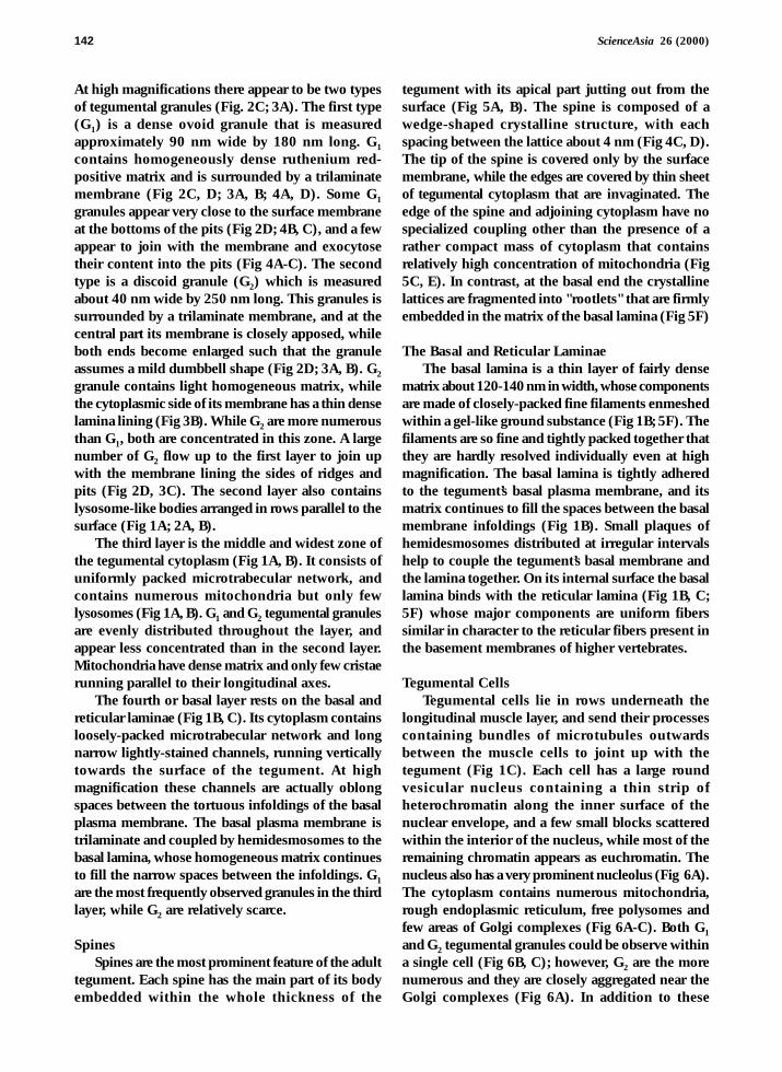
At high magnifications there appear to be two typesof tegumental granules (Fig. 2C; 3A). The first type(G1) is a dense ovoid granule that is measuredapproximately 90 nm wide by 180 nm long. G1
contains homogeneously dense ruthenium red-positive matrix and is surrounded by a trilaminatemembrane (Fig 2C, D; 3A, B; 4A, D). Some G1
granules appear very close to the surface membraneat the bottoms of the pits (Fig 2D; 4B, C), and a fewappear to join with the membrane and exocytosetheir content into the pits (Fig 4A-C). The secondtype is a discoid granule (G2) which is measuredabout 40 nm wide by 250 nm long. This granules issurrounded by a trilaminate membrane, and at thecentral part its membrane is closely apposed, whileboth ends become enlarged such that the granuleassumes a mild dumbbell shape (Fig 2D; 3A, B). G2
granule contains light homogeneous matrix, whilethe cytoplasmic side of its membrane has a thin denselamina lining (Fig 3B). While G2 are more numerousthan G1, both are concentrated in this zone. A largenumber of G2 flow up to the first layer to join upwith the membrane lining the sides of ridges andpits (Fig 2D, 3C). The second layer also containslysosome-like bodies arranged in rows parallel to thesurface (Fig 1A; 2A, B).
The third layer is the middle and widest zone ofthe tegumental cytoplasm (Fig 1A, B). It consists ofuniformly packed microtrabecular network, andcontains numerous mitochondria but only fewlysosomes (Fig 1A, B). G1 and G2 tegumental granulesare evenly distributed throughout the layer, andappear less concentrated than in the second layer.Mitochondria have dense matrix and only few cristaerunning parallel to their longitudinal axes.
The fourth or basal layer rests on the basal andreticular laminae (Fig 1B, C). Its cytoplasm containsloosely-packed microtrabecular network and longnarrow lightly-stained channels, running verticallytowards the surface of the tegument. At highmagnification these channels are actually oblongspaces between the tortuous infoldings of the basalplasma membrane. The basal plasma membrane istrilaminate and coupled by hemidesmosomes to thebasal lamina, whose homogeneous matrix continuesto fill the narrow spaces between the infoldings. G1
are the most frequently observed granules in the thirdlayer, while G2 are relatively scarce.
SpinesSpines are the most prominent feature of the adult
tegument. Each spine has the main part of its bodyembedded within the whole thickness of the
tegument with its apical part jutting out from thesurface (Fig 5A, B). The spine is composed of awedge-shaped crystalline structure, with eachspacing between the lattice about 4 nm (Fig 4C, D).The tip of the spine is covered only by the surfacemembrane, while the edges are covered by thin sheetof tegumental cytoplasm that are invaginated. Theedge of the spine and adjoining cytoplasm have nospecialized coupling other than the presence of arather compact mass of cytoplasm that containsrelatively high concentration of mitochondria (Fig5C, E). In contrast, at the basal end the crystallinelattices are fragmented into "rootlets" that are firmlyembedded in the matrix of the basal lamina (Fig 5F)
The Basal and Reticular LaminaeThe basal lamina is a thin layer of fairly dense
matrix about 120-140 nm in width, whose componentsare made of closely-packed fine filaments enmeshedwithin a gel-like ground substance (Fig 1B; 5F). Thefilaments are so fine and tightly packed together thatthey are hardly resolved individually even at highmagnification. The basal lamina is tightly adheredto the tegument’s basal plasma membrane, and itsmatrix continues to fill the spaces between the basalmembrane infoldings (Fig 1B). Small plaques ofhemidesmosomes distributed at irregular intervalshelp to couple the tegument’s basal membrane andthe lamina together. On its internal surface the basallamina binds with the reticular lamina (Fig 1B, C;5F) whose major components are uniform fiberssimilar in character to the reticular fibers present inthe basement membranes of higher vertebrates.
Tegumental CellsTegumental cells lie in rows underneath the
longitudinal muscle layer, and send their processescontaining bundles of microtubules outwardsbetween the muscle cells to joint up with thetegument (Fig 1C). Each cell has a large roundvesicular nucleus containing a thin strip ofheterochromatin along the inner surface of thenuclear envelope, and a few small blocks scatteredwithin the interior of the nucleus, while most of theremaining chromatin appears as euchromatin. Thenucleus also has a very prominent nucleolus (Fig 6A).The cytoplasm contains numerous mitochondria,rough endoplasmic reticulum, free polysomes andfew areas of Golgi complexes (Fig 6A-C). Both G1
and G2 tegumental granules could be observe withina single cell (Fig 6B, C); however, G2 are the morenumerous and they are closely aggregated near theGolgi complexes (Fig 6A). In addition to these

ScienceAsia 26 (2000) 143
Fig 4. A, B, C) Micrographs of ruthenium red-fixed sections, uncounterstained (A) and counterstained with lead citrate and uranylacetate (B, C), illustrating intense staining of ruthenium red on the glycocalyx (Gy) lining the pits (Pi) and the ridges (Ri), andG1 granules closed to the surface. B, C indicate the exocytosis and release of matrix (Em) of G1 granules into the pit.D, E) The counterstained ruthenium-fixed sections illustrating thick (100-300nm) non uniform glycocalyx on the surface ofa ridge, and exocytosed matrix of G1 in the pit (Em). The surface membrane (Sm) and underlining dense lamina (Dl) areclearly seen.

144 ScienceAsia 26 (2000)
Fig 5. A, B) Micrographs of the spines (Sp) whose tips are covered by the surface membrane and whose edges by tegument cytoplasmthat are invaginated (arrow).C, D, E) The middle of the spines showing crystalline structure with lattice space about 4nm apart (boxed area in C, D). Theside of the spine is covered by dense cytoplasm containing numerous mitochondria (Mi).F) The base of a spine (Sp) which is fragmented into rootlets (arrow) that are embedded in matrix of the basal lamina (Ba).

ScienceAsia 26 (2000) 145
Fig 6. A, B, C) Micrographs of tegumental cells showing the nucleus (Nu) containing mostly euchromatin and prominent nucleolus(No). The Golgi complex areas (Gc) contain high concentration of G2 granules. In the cytoplasm (in B, C) both G1 and G2
granules are present within the same cell, with G2 predominate in number.

146 ScienceAsia 26 (2000)
tegumental granules, there are spherical-shapedgranules whose content are very lightly stained, andsome may actually appear empty (Fig 6B, C). Thesegranules could be the precursor of G1 whose matrixhas not yet been highly concentrated.
DISCUSSION
Ultrastructure of the TegumentTrematode parasites that live in the mammalian
hosts’ circulation or biliary system need to absorbnutrient molecules from either the blood or bile, asdemonstrated in schistosomes’ tegument which canabsorb substantial amount of small nutrientmolecules such as glucose, fructose and aminoacids.18,19 Simultaneously, the tegument of theseparasites can also protect them from hosts’ immuneattacks by evolving evasion mechanisms that includethe rapid turn over of the surface membrane toprevent the attachment of immune effector cells, andby immune mimickry or disguise through theadsorption of hosts’ antigens onto the parasites’surfaces. In addition, the fluid environment in thehost body, especially the bile, is vastly different interms of ionic composition from that of the parasites’bodies. Hence another important role of the tegumentis the regulation of ions and fluid balance, whichwill keep the homeostatic equilibrium within theparasites’ bodies. Adult F. gigantica tegument, asreported in the present study, exhibits all the ultra-structural features to subserve these critical functions.
The First LayerBased on their ultrastructural features the tegument
of adult F. gigantica could be divided into four layersof specialized functions. The first and outermostregion of the tegument contains highly folded parts,namely, ridges and pits. The membrane coveringthese structures is trilaminate and coated with asubstantial layer of glycocalyx, a typical feature foundin all bile and lumen-dwelling trematodes, including,F. hepatica20-22 Clonorchis sinensis23 and Oposthorchisviverini.24 The vast amount of membrane coveringthe ridges and pits helps to increase the surface areafor absorption and exchanging of materials.Glycocalyx may provide the first line of defensebecause of its substantial thickness and insolubility.In conventional TEM, glycocalyx appears only as athin layer (about 10-15 nm in thickness) which maybe due to the failure of the fixative to preserve theglycocalyx in its entirety. By contrast, when the parasiteswere simultaneously treated with ruthenium redwhich acts as both stain and fixative15-17 the
glycocalyx appears as thick as 100-300 nm, andconsists of two definite layers, ie, the thin innerhomogeneous layer apposed to the surfacemembrane and the thick outer fibrillar layer. The lattermay be more labile and could be preserved only byextra treatment with ruthenium red. Glycocalyx maybe quite resistant to the emulsifying action of thebile and, therefore, able to confer a certain degree ofprotection to F. gigantica. High affinity to rutheniumred is indicative that glycocalyx of adult F. giganticais highly negatively charged. The anions present maybe contributed mainly by sialic acids, as it has beenshown that ruthenium red can bind strongly to thissugar.17 The presence of abundant negative chargeson the surface could be another factor that helps todefend the parasites against hosts’ immune attacksby repelling the attachment of the hosts’ immuneeffector cells, which also bear high electronegativityon their own surfaces.11,25 Moreover, the presence ofa large quantity of large negatively-charged molecules,like glycoproteins, may be instrumental in retainingand concentrating small molecules including sugarsand amino acids and various ions. Thus, theglycocalyx may be viewed as a hydrated shell aroundthe parasites’ bodies that helps to protect as well asconcentrate nutrient molecules, in order to make themreadily available for the absorption by the tegument.
Glycocalyx is probably derived from G1 granulespresent in abundance within the second layer of thetegument. Ruthenium red stain shows similarbinding to the glycocalyx as well as the matrix of G1
granules lying close to the surface. These granulesinvariably join up with the surface membrane at thebottom of the pits, and exocytose their matrix intothe lumen of pits. This material could be laterincorporated into the surface membrane, thusforming part of the new glycocalyx. In F. hepatica,similar granules which were termed T0 by Hanna,26
were also thought to be contributing to the formationof glycocalyx in metacercaria and juvenile parasites,particularly during their invasive migration throughthe host’s tissues. Once F. hepatica juveniles reachtheir final destination and take up permanentresidence in bile ducts of the liver T0 granules werereplaced by a similar set of adult-type granules (T1).In comparison to T0, the number of T1 in adulttegument is drastically decreased, while another setof so call T2 granules became the majority oftegumental granules in adult. Each T2 granule issurrounded by a trilaminate membrane andassociated thin layer of glycocalyx that express adult-type antigens on the surface.26-28 These adult antigensare much less immunogenic than juvenile

ScienceAsia 26 (2000) 147
antigens.26,28 In contrast to F. hepatica, we have shownthat in the tegument of adult F. gigantica G1 granules,which are probably equivalent to T1 granules, arestill quite numerous; and the ruthenium red stainingimplicates that the secretion of these granules intothe pits may form the major part of glycocalyx.
The surface membrane of an adult F. gigantica istrilaminate and supported internally by a thin laminaof dense cytoplasm, while the cytoplasm underneaththe lamina is composed of tightly packedmicrotrabeculae and bundles of larger fibrils. Withinthis interstice there are numerous G2 granules thatmay be equivalent to T2 granules of F. hepaticategument. In fortuitous sections several G2 granuleswere seen joined up with the surface membrane, andevert their cisternal side outwards, thus turning intothe exterior surface of the tegument’s surfacemembrane, which may later become coated with theglycocalyx material released from G1 granules. Thematrix inside the cisternae of G2 granules couldbecome part of the thin homogenous layer ofglycocalyx that may be coupled with the morefibrillar components derived from the secretion ofG1 granules. On the cytoplasmic side, which is nowturned into the cytoplasmic face of the surfacemembrane, the dense cytoplasm covering themembrane of the G2 granules could coalesce andform the dense lamina underlining the surfacemembrane. The surface membrane and associatedglycocalyx of the adult F. gigantica tegument probablymaintain a high turnover rate, since there are alwaysa vast amount of G2 granules accumulated in thefirst and the second layers of the tegument, as wellas a still relatively large number of G1 granules inthe second layer. Such rapid turnover and renewalof the surface membrane could be a part of themechanism for self defense against the detergenicaction of bile and the host’s immune attacks, as ithas been shown that bile also contains copiousamount of antibodies, especially IgA.29
The Second LayerThe most prominent characteristic of this
layer is the presence of a very high concentration ofG1 and G2 granules, particularly G2 granules.Therefore, this layer could be viewed as the storagearea for the new membrane (G2) and glycocalyx (G1)material. Another prominent feature of this layer isthe presence of a large number of lysosomal granules,which implies that there may be absorption of largemolecules via the endocytotic pathway within thetegument. In addition to nutritive substances, someof these large molecules could be antibodies forming
immune complexes with the surface antigens. Onceattached to the surface membrane these largemolecules or immune complexes could beinternalized and broken down by the fusion withlysosomes, which could be another part of parasites’defense mechanism against the hosts’ immune attacks.
The Third LayerThis is the widest layer of the tegument. Its
cytoskeleton is less tightly packed, and it has thehighest concentration of mitochondria which areevenly distributed throughout the layer. G1 and G2
granules are present but appear to be much fewerthan in the second layer. This layer may, therefore,be involved mainly in supplying energy to otherlayers. The concentration of mitochondria and theirpositioning within the tegument of this parasite arequite different from that of schistosomes and ahuman liver fluke, O. viverrini. In schistosomes mostof mitochondria are concentrated in the basal layer,30
while in O. viverrini they are localized close to thesurface plasma membrane.24 The distribution ofenergy for various metabolic processes in thetegument of these two species could be carried outby these eccentrically-located mitochondria becauseof the relative thinness of their tegument. In contrast,F. gigantica has a much thicker tegument because ofthe worms’ very large size. The increased energyrequirement dictates that the parasites’ tegument mustpossess a very large number of mitochondria that arepositioned strategically in the middle zone, so thatenergy could be sufficiently distributed to other layers.
The Fourth LayerThis layer exhibits the most unique features in
having highly convoluted infoldings of the basalplasma membrane, with closely-associatedmitochondria. These features resemble the basalcytoplasm of ion-transport epithelium in mammals,such as, the kidney proximal and distal tubules.Based on these similarities the fourth layer could beinvolved in the transport of ions which will help tomaintain the ionic equilibrium within the parasites’bodies with regards to the bile.
Tegumental CellsIn F. hepatica, Hanna26 and Burden et al13 reported
that there is one type of cell which synthesize T0
granules in metacercaria and in the very earlyjuvenile stage, while the parasites are migratingthrough the hosts’ abdominal cavities. T0 cellstransform into T1 cells which produce granules withsimilar feature but with greater density when the

148 ScienceAsia 26 (2000)
parasites reach the liver. Both T0 and T1 granuleswere thought to give rise to the juveniles’, and lateradults’ glycocalyx, that enables the parasites to evadethe killing by hosts’ immune responses. Whenparasites become established in the bile duct at about12 weeks there is another type of tegumental cellswhich produce T2 granules, which are the maincontributor to the formation of the adult parasites’surface membrane and glycocalyx. These T2 cellssupersede T1 cells, and as a result the number of T1
granules in the tegument of the adult parasites isdrastically decreased. In F. gigantica the number ofG1 granules in the adult tegument is still relativelyhigh in comparison with the T1 granules of F.hepatica. In contrast to the two types of tegumentcells reported in the adult F. hepatica we observedonly one type of cells in adult F. gigantica. Thesecells produce both G1 and G2 granules, even thoughthe productions of G2 predominate in the adult stage.Both types of granules are found in close associationwith microtubules within the processes of the cellsthat extend outwards to join up with the tegument.Hence, we believe that these granules aretranslocated form cell soma to the tegument by thesliding action of microtubules.
ACKNOWLEDGEMENTS
This research was supported financially by theThailand Research Fund (Senior Research ScholarFellowship to P Sobhon).
REFERENCES
1. Maurice J (1994) Is something lurking in your liver? NewScientist 1917, 26-31.
2. Edney JM, Muchlis A (1962) Fascioliasis in Indonesianlivestock. Communication Veterinarae 6, 49-52.
3. Soesetya RHB (1975) The prevalence of Fasciola giganticainfection in cattle in East Java, Indonesia. Malaysian Vet J 6, 5-8.
4. Fabiyi JP (1987) Production losses and control of helminthesin ruminants of tropical regions. Int J Parasitol 17, 435-42.
5. Sukhapesna V, Tantasuvan D, Sarataphan N, Imsup K (1994)Economic impact of fascioliasis in buffalo production. J ThaiVet Med Assoc 45, 45-52.
6. Sobhon P, Anantavara S, Dangprasert T, Viyanant V, Krailas D,Upatham ES, Wanichanon C, Kusamran T (1998) Fasciolagigantica: studies of the tegument as a basis for thedevelopments of immunodiagnosis and vaccine. SoutheastAsian J Trop Med and Pub Hlth 29, 387-400.
7. Overend DJ, Bower FL (1995) Resistance of Fasciola hepaticato triclabendazole. Aus Vet J 72, 275-6.
8. Sobhon P, Anantavara S, Dangprasert T, Meepool A,Wanichanon C, Viyanant V, Upatham ES, Kusamran T,Chompoochan T, Thammasart S, Prasitirat P (1996) Fasciolagigantica: Identification of adult antigens, their tissue sourcesand possible origin. J Science Soc Thai 27, 312-7.
9. Fairweather I, Lawence T, Threadgold LT, Hanna REB (1999)Development of Fasciola hepatica in the mammalian hosts.
In: Fasciolosis (Edited by Dalton JP), pp. 47-111, CABIPublising, Wallingford, UK.
10.Bennett CE, Threadgold LT (1975) Fasciola hepatica:Development of tegument during migration in mouse. ExpParasitol 38, 38-55.
11.Threagold LT (1976) Fasciola hepatica: Ultrastructure andhistochemistry of the glycocalyx of the tegument. Exp Parasitol39, 119-34.
12.Threagold LT, Brennan G (1978) Fasciola hepatica: Basalinfolds and associated vacuoles of the tegument. Exp Parasitol46, 300-16.
13.Burden DJ, Bland AP, Hughes DL, Hammet NC (1982) Fasciolahepatica: development of the tegument of normal and γ-irradiatedflukes during infection in rats and mice. Parasitol 86, 137-45.
14.Estuningsith SE, Smooker PM, Wiedosari E, Widjajanti S,Vaiano S, Partoutomo S, Spithill TW (1997) Evaluation ofantigens of Fasciola gigantica as vaccines against tropicalfasciolosis in cattle. Int J Parasitol 27, 1419-28.
15.Luft JH (1971) Ruthenium Red and Violet I. Chemistry,purification, methods of use for electron microscopy andmechanism of action. Anat Rec 171, 347-68.
16.Luft JH (1971) Ruthenium Red and Violet II. Fine structurelocalization in animal tissues. Anat Rec 171, 369-416.
17.Rambourg A (1971) Morphological and histochemical aspectsof glycoproteins at the surface of animal cells. Int Rev Cytol31, 57-114.
18.Fripp PJ (1967) The sites of (C14) glucose assimilation inSchistosoma haematobium. Comp Biochem Physiol 23, 893-8.
19.Senft AW (1968) Studies in proline metabolism by Schistosomamansoni. I. Radioautopraphy following in vitro exposure toradioproline C14. Comp Biochem Physiol 27, 251-61.
20.Threadgold LT (1967) Electron-microscope studies of Fasciolahepatica III. Further observations on the tegument. ExpParasitol 46, 300-16.
21.Theadgold LT (1984) Parasitic platyhelminths. In : Biology ofthe tegument (Edited by Bereiter-Hahn J, Matoltsy AG, RichardsKS), pp 132-85, Springer-Verlag, Berlin.
22.Hanna REB (1980) Fasciola hepatica: Glycocalyx replacementin the juvenile as a possible mechanism for protection againsthost immunity. Exp Parasitol 50, 103-44.
23.Fujino T, Ishii Y, Choi DW (1979) Surface ultrastructure ofthe tegument of Clonorchis sinensis newly excysted juvenileand adult worms. J Parasitol 65, 579-90.
24.Apinhasmit W, Sobhon P, Saitongdee P, Menayotin S, UpathamES (1994) Opisthorchis viverini: ultrastructure of the tegumentof the first-week juveniles and adult flukes. Int J Parasitol 24,613-21.
25.Lumsden RD (1972) Cytological studies on the absorptivesurface of cestodes. VI. Cytochemical evaluation ofelectrostatic charge. J Parasitol 58, 229-34.
26.Hanna REB (1980) Fasciola hepatica: An immunofluorescentstudy of antigenic changes in the tegument duringdevelopment in the rat and sheep. Exp Parasitol 50, 155-70.
27.Hanna REB (1980) Fasciola hepatica: Autoradiography ofprotein synthesis, transport and secretion by the tegument.Exp Parasitol 50, 297-304.
28.Hanna REB, Trudgett AG (1983) Fasciola hepatica:development of monoclonal antibodies and their use tocharacterize a glycocalyx antigen in migrating flukes. ParasiteImmunol 5, 409-425.
29.Hughes DL, Hanna REB, Symonds HW (1981) Fasciolahepatica: IgG and IgA levels in the serum and bile of infectedcattle. Exp Parasitol 52, 271-9.
30.Sobhon P, Upatham ES (1990) The tegument of adult parasites.In : Snail hosts, life-cycle, and tegumental structure of orientalschistosomes, pp57-107. UNDP/World Bank/WHO SpecialPrograms for Research and Training in Tropical Diseases,Geneva.
Copyright © 2022 FDOKUMEN