Reconstructing the evolution of agarics from nuclear gene sequences and basidiospore ultrastructure
11
Reconstructing the evolution of agarics from nuclear gene sequences and basidiospore ultrastructure Sigisfredo GARNICA a, * ,y , Michael WEISS a,y , Grit WALTHER b , Franz OBERWINKLER a a Eberhard-Karls-Universita ¨t Tu ¨ bingen, Botanisches Institut, Lehrstuhl fu ¨ r Spezielle Botanik und Mykologie, Auf der Morgenstelle 1, D-72076 Tu ¨ bingen, Germany b Centraalbureau voor Schimmelcultures, P.O. Box 85167, 3508 AD Utrecht, The Netherlands article info Article history: Received in revised form 13 March 2007 Accepted 26 March 2007 Published online 6 April 2007 Corresponding Editor: David L. Hawksworth Keywords: Agaricales Basidiomycota Euagarics clade Homobasidiomycetes Molecular phylogeny abstract Traditional classifications of agaric fungi involve gross morphology of their fruit bodies and meiospore print-colour. However, the phylogeny of these fungi and the evolution of their morphological and ecological traits are poorly understood. Phylogenetic analyses have already demonstrated that characters used in traditional classifications of basidiomycetes may be heavily affected by homoplasy, and that non-gilled taxa evolved within the agarics several times. By integrating molecular phylogenetic analyses including domains D1–D3 and D7–D8 of nucLSU rDNA and domains A–C of the RPB1 gene with morphological and chemical data from representative species of 88 genera, we were able to resolve higher groups of agarics. We found that the species with thick-walled and pigmented basidio- spores constitute a derived group, and hypothesize that this specific combination of char- acters represents an evolutionary advantage by increasing the tolerance of the basidiospores to dehydration and solar radiation and so opened up new ecological niches, e.g. the colonization of dung substrates by enabling basidiospores to survive gut passages through herbivores. Our results confirm the validity of basidiospore morphology as a phy- logenetic marker in the agarics. ª 2007 The British Mycological Society. Published by Elsevier Ltd. All rights reserved. Introduction Agarics comprise the most familiar fungal forms, and fossils exhibiting agaricoid features date back to the mid-Cretaceous (Hibbett et al. 1997a). Agarics have undergone an enormous adaptive radiation, with an extant described 8400 species, in the course of which they have occupied a broad range of eco- logical niches, including ectomycorrhizas, mutualisms with ants and termites, as decomposers of wood and other organic substrates, and as plant pathogens. Molecular studies (e.g. Hibbett et al. 1997b; Larsson et al. 2004; Moncalvo et al. 2002; see also Hibbett 2004, 2007) have shown that: (1) the core group of agarics (‘euagarics’; ‘agarics’ is used in this sense in this paper) does not contain all species of gilled mushrooms; and (2) this core group also includes non-gilled species. Thus, not only are there morphs represent- ing gilled mushrooms, but also puffballs, bird’s nest fungi, polypores, coral-shaped, cup-shaped, and crust-like agarics (Binder et al. 2005; Bodensteiner et al. 2004; Hibbett 2007; Hibbett et al. 1997b; Larsson et al. 2004; Moncalvo et al. 2002). The traditional classifications of agarics characterize several higher groups, mostly families, on the basis of morphological traits (Singer 1986). These include the type of attachment of the gills to the stipe, and the colour of spore * Corresponding author. E-mail address: [email protected] y These authors contributed equally to this work. journal homepage: www.elsevier.com/locate/mycres mycological research 111 (2007) 1019–1029 0953-7562/$ – see front matter ª 2007 The British Mycological Society. Published by Elsevier Ltd. All rights reserved. doi:10.1016/j.mycres.2007.03.019
-
Upload
uni-tuebingen1 -
Category
Documents
-
view
3 -
download
0
Transcript of Reconstructing the evolution of agarics from nuclear gene sequences and basidiospore ultrastructure
journa l homepage : www.e l sev i er . com/ loca te /mycres
m y c o l o g i c a l r e s e a r c h 1 1 1 ( 2 0 0 7 ) 1 0 1 9 – 1 0 2 9
Reconstructing the evolution of agarics from nuclear genesequences and basidiospore ultrastructure
Sigisfredo GARNICAa,*,y, Michael WEISSa,y, Grit WALTHERb, Franz OBERWINKLERa
aEberhard-Karls-Universitat Tubingen, Botanisches Institut, Lehrstuhl fur Spezielle Botanik und Mykologie,
Auf der Morgenstelle 1, D-72076 Tubingen, GermanybCentraalbureau voor Schimmelcultures, P.O. Box 85167, 3508 AD Utrecht, The Netherlands
a r t i c l e i n f o
Article history:
Receivedinrevisedform13March2007
Accepted 26 March 2007
Published online 6 April 2007
Corresponding Editor:
David L. Hawksworth
Keywords:
Agaricales
Basidiomycota
Euagarics clade
Homobasidiomycetes
Molecular phylogeny
a b s t r a c t
Traditional classifications of agaric fungi involve gross morphology of their fruit bodies and
meiospore print-colour. However, the phylogeny of these fungi and the evolution of their
morphological and ecological traits are poorly understood. Phylogenetic analyses have
already demonstrated that characters used in traditional classifications of basidiomycetes
may be heavily affected by homoplasy, and that non-gilled taxa evolved within the agarics
several times. By integrating molecular phylogenetic analyses including domains D1–D3
and D7–D8 of nucLSU rDNA and domains A–C of the RPB1 gene with morphological and
chemical data from representative species of 88 genera, we were able to resolve higher
groups of agarics. We found that the species with thick-walled and pigmented basidio-
spores constitute a derived group, and hypothesize that this specific combination of char-
acters represents an evolutionary advantage by increasing the tolerance of the
basidiospores to dehydration and solar radiation and so opened up new ecological niches,
e.g. the colonization of dung substrates by enabling basidiospores to survive gut passages
through herbivores. Our results confirm the validity of basidiospore morphology as a phy-
logenetic marker in the agarics.
ª 2007 The British Mycological Society. Published by Elsevier Ltd. All rights reserved.
Introduction
Agarics comprise the most familiar fungal forms, and fossils
exhibiting agaricoid features date back to the mid-Cretaceous
(Hibbett et al. 1997a). Agarics have undergone an enormous
adaptive radiation, with an extant described 8400 species, in
the course of which they have occupied a broad range of eco-
logical niches, including ectomycorrhizas, mutualisms with
ants and termites, as decomposers of wood and other organic
substrates, and as plant pathogens.
Molecular studies (e.g. Hibbett et al. 1997b; Larsson et al.
2004; Moncalvo et al. 2002; see also Hibbett 2004, 2007) have
shown that: (1) the core group of agarics (‘euagarics’; ‘agarics’
is used in this sense in this paper) does not contain all species
of gilled mushrooms; and (2) this core group also includes
non-gilled species. Thus, not only are there morphs represent-
ing gilled mushrooms, but also puffballs, bird’s nest fungi,
polypores, coral-shaped, cup-shaped, and crust-like agarics
(Binder et al. 2005; Bodensteiner et al. 2004; Hibbett 2007;
Hibbett et al. 1997b; Larsson et al. 2004; Moncalvo et al. 2002).
The traditional classifications of agarics characterize
several higher groups, mostly families, on the basis of
morphological traits (Singer 1986). These include the type of
attachment of the gills to the stipe, and the colour of spore
* Corresponding author.E-mail address: [email protected]
y These authors contributed equally to this work.0953-7562/$ – see front matter ª 2007 The British Mycological Society. Published by Elsevier Ltd. All rights reserved.doi:10.1016/j.mycres.2007.03.019
1020 S. Garnica et al.
prints (Fig 1). However, although considerable progress has
been made in recent years, there is still no consensus as to
the natural delimitations within such groupings. We now
know that fruit body gross architectures, as well as other mor-
phological traits, evolved more than once in the agarics, but
the often relatively uniform anatomy of the fruit bodies and
also the scanty fossil record have hampered attempts to re-
construct the agaricalean branch of the tree of life with confi-
dence. Molecular phylogenetic analyses, mostly based on
partial sequences of the nucLSU rDNA gene, have revealed
several monophyletic groups within the agarics (Moncalvo
et al. 2000, 2002). Some of these are congruent with traditional
groupings, whereas others are surprisingly different from
morphologically-based classifications. However, these analy-
ses could not resolve the deeper nodes of the agaricalean
phylogeny.
The purpose of the present study was to derive sup-
ported hypotheses about phylogenetic relationships and
trends in character evolution within the euagarics by se-
quencing nuclear genes and comparing macro- and micro-
morphology of the basidiomes as well as the ultrastructure
of the basidiospores. In designing our molecular and mor-
phological analyses, the spectrum of included genera was
made as broad as possible. Each specimen used in the mo-
lecular analysis was also thoroughly studied using light
microscopy.
Materials and methods
Taxon sampling
Ninety taxa, including representative species of 88 euagaric
genera, were used in the molecular and morphological analy-
ses to ensure a broad sampling of species covering the mor-
phological variation and taxonomic diversity of this fungal
group, both according to traditional treatments (e.g. Kuhner
1980; Singer 1986) and to previous molecular studies (Hibbett
et al. 1997b; Moncalvo et al. 2002). Boletus edulis and Tapinella
panuoides (bolete clade) were used as outgroup species. Loca-
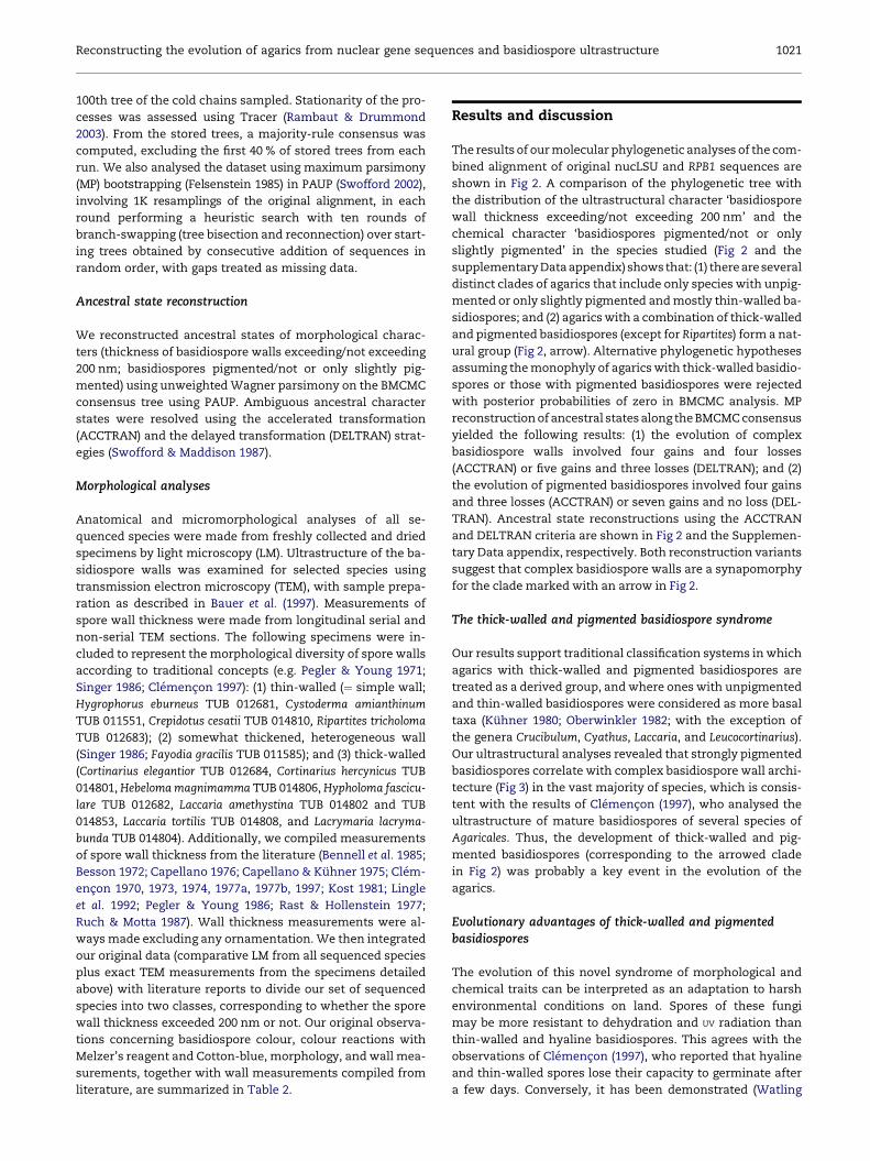
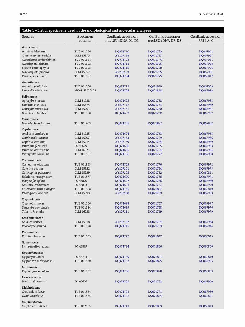
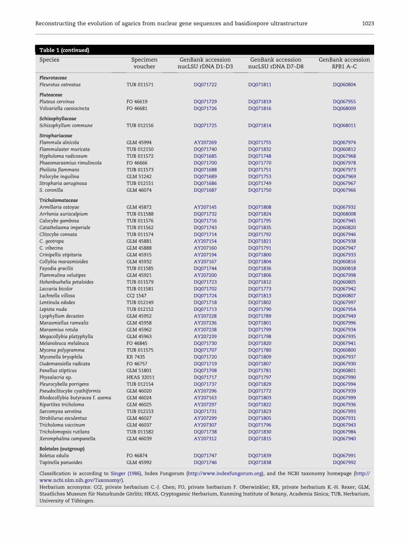
tions of the voucher specimens and GenBank accession num-
bers for the DNA sequences obtained for this study are listed
in Table 1. Representative basidiome types of agarics are
shown in Fig 2.
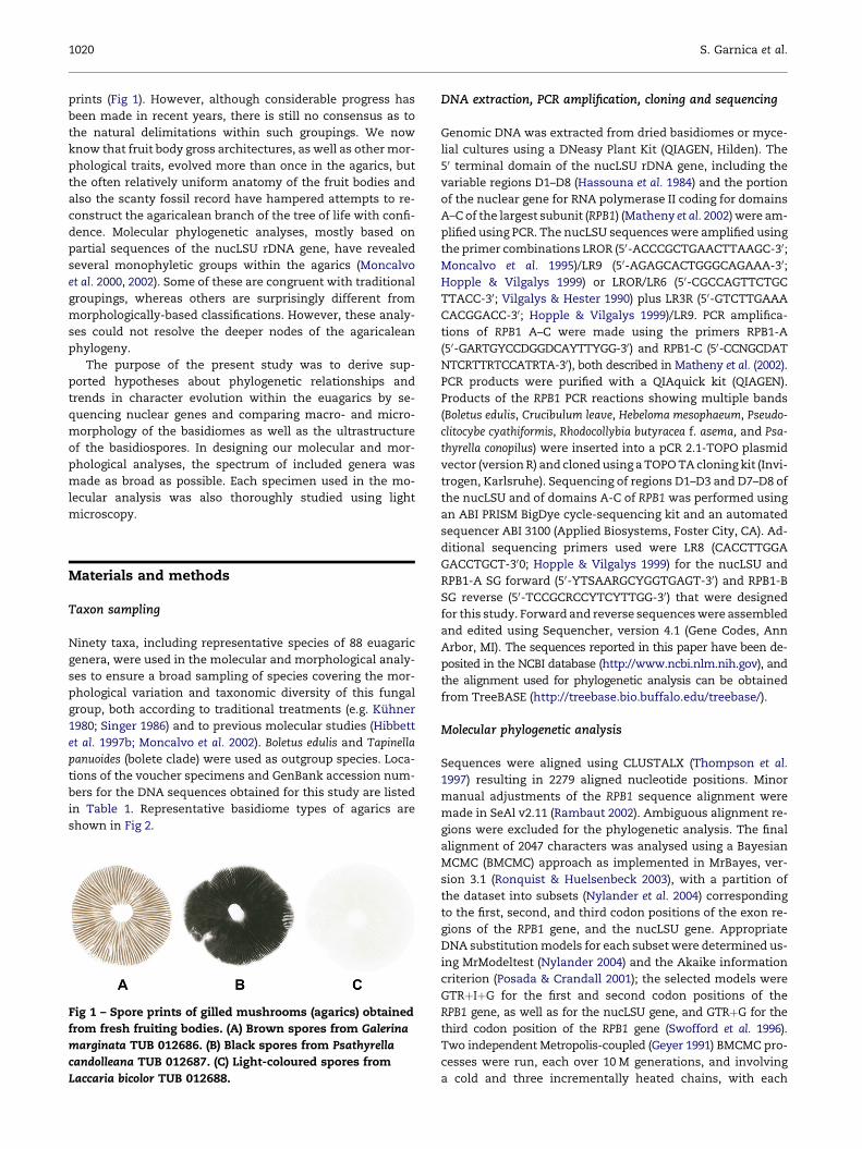
Fig 1 – Spore prints of gilled mushrooms (agarics) obtained
from fresh fruiting bodies. (A) Brown spores from Galerina
marginata TUB 012686. (B) Black spores from Psathyrella
candolleana TUB 012687. (C) Light-coloured spores from
Laccaria bicolor TUB 012688.
DNA extraction, PCR amplification, cloning and sequencing
Genomic DNA was extracted from dried basidiomes or myce-
lial cultures using a DNeasy Plant Kit (QIAGEN, Hilden). The
50 terminal domain of the nucLSU rDNA gene, including the
variable regions D1–D8 (Hassouna et al. 1984) and the portion
of the nuclear gene for RNA polymerase II coding for domains
A–C of the largest subunit (RPB1) (Matheny et al. 2002) were am-
plified using PCR. The nucLSU sequences were amplified using
the primer combinations LROR (50-ACCCGCTGAACTTAAGC-30;
Moncalvo et al. 1995)/LR9 (50-AGAGCACTGGGCAGAAA-30;
Hopple & Vilgalys 1999) or LROR/LR6 (50-CGCCAGTTCTGC
TTACC-30; Vilgalys & Hester 1990) plus LR3R (50-GTCTTGAAA
CACGGACC-30; Hopple & Vilgalys 1999)/LR9. PCR amplifica-
tions of RPB1 A–C were made using the primers RPB1-A
(50-GARTGYCCDGGDCAYTTYGG-30) and RPB1-C (50-CCNGCDAT
NTCRTTRTCCATRTA-30), both described in Matheny et al. (2002).
PCR products were purified with a QIAquick kit (QIAGEN).
Products of the RPB1 PCR reactions showing multiple bands
(Boletus edulis, Crucibulum leave, Hebeloma mesophaeum, Pseudo-
clitocybe cyathiformis, Rhodocollybia butyracea f. asema, and Psa-
thyrella conopilus) were inserted into a pCR 2.1-TOPO plasmid
vector (version R) and cloned using a TOPO TA cloning kit (Invi-
trogen, Karlsruhe). Sequencing of regions D1–D3 and D7–D8 of
the nucLSU and of domains A-C of RPB1 was performed using
an ABI PRISM BigDye cycle-sequencing kit and an automated
sequencer ABI 3100 (Applied Biosystems, Foster City, CA). Ad-
ditional sequencing primers used were LR8 (CACCTTGGA
GACCTGCT-300; Hopple & Vilgalys 1999) for the nucLSU and
RPB1-A SG forward (50-YTSAARGCYGGTGAGT-30) and RPB1-B
SG reverse (50-TCCGCRCCYTCYTTGG-30) that were designed
for this study. Forward and reverse sequences were assembled
and edited using Sequencher, version 4.1 (Gene Codes, Ann
Arbor, MI). The sequences reported in this paper have been de-
posited in the NCBI database (http://www.ncbi.nlm.nih.gov), and
the alignment used for phylogenetic analysis can be obtained
from TreeBASE (http://treebase.bio.buffalo.edu/treebase/).
Molecular phylogenetic analysis
Sequences were aligned using CLUSTALX (Thompson et al.
1997) resulting in 2279 aligned nucleotide positions. Minor
manual adjustments of the RPB1 sequence alignment were
made in SeAl v2.11 (Rambaut 2002). Ambiguous alignment re-
gions were excluded for the phylogenetic analysis. The final
alignment of 2047 characters was analysed using a Bayesian
MCMC (BMCMC) approach as implemented in MrBayes, ver-
sion 3.1 (Ronquist & Huelsenbeck 2003), with a partition of
the dataset into subsets (Nylander et al. 2004) corresponding
to the first, second, and third codon positions of the exon re-
gions of the RPB1 gene, and the nucLSU gene. Appropriate
DNA substitution models for each subset were determined us-
ing MrModeltest (Nylander 2004) and the Akaike information
criterion (Posada & Crandall 2001); the selected models were
GTRþIþG for the first and second codon positions of the
RPB1 gene, as well as for the nucLSU gene, and GTRþG for the
third codon position of the RPB1 gene (Swofford et al. 1996).
Two independent Metropolis-coupled (Geyer 1991) BMCMC pro-
cesses were run, each over 10 M generations, and involving
a cold and three incrementally heated chains, with each
Reconstructing the evolution of agarics from nuclear gene sequences and basidiospore ultrastructure 1021
100th tree of the cold chains sampled. Stationarity of the pro-
cesses was assessed using Tracer (Rambaut & Drummond
2003). From the stored trees, a majority-rule consensus was
computed, excluding the first 40 % of stored trees from each
run. We also analysed the dataset using maximum parsimony
(MP) bootstrapping (Felsenstein 1985) in PAUP (Swofford 2002),
involving 1K resamplings of the original alignment, in each
round performing a heuristic search with ten rounds of
branch-swapping (tree bisection and reconnection) over start-
ing trees obtained by consecutive addition of sequences in
random order, with gaps treated as missing data.
Ancestral state reconstruction
We reconstructed ancestral states of morphological charac-
ters (thickness of basidiospore walls exceeding/not exceeding
200 nm; basidiospores pigmented/not or only slightly pig-
mented) using unweighted Wagner parsimony on the BMCMC
consensus tree using PAUP. Ambiguous ancestral character
states were resolved using the accelerated transformation
(ACCTRAN) and the delayed transformation (DELTRAN) strat-
egies (Swofford & Maddison 1987).
Morphological analyses
Anatomical and micromorphological analyses of all se-
quenced species were made from freshly collected and dried
specimens by light microscopy (LM). Ultrastructure of the ba-
sidiospore walls was examined for selected species using
transmission electron microscopy (TEM), with sample prepa-
ration as described in Bauer et al. (1997). Measurements of
spore wall thickness were made from longitudinal serial and
non-serial TEM sections. The following specimens were in-
cluded to represent the morphological diversity of spore walls
according to traditional concepts (e.g. Pegler & Young 1971;
Singer 1986; Clemencon 1997): (1) thin-walled (¼ simple wall;
Hygrophorus eburneus TUB 012681, Cystoderma amianthinum
TUB 011551, Crepidotus cesatii TUB 014810, Ripartites tricholoma
TUB 012683); (2) somewhat thickened, heterogeneous wall
(Singer 1986; Fayodia gracilis TUB 011585); and (3) thick-walled
(Cortinarius elegantior TUB 012684, Cortinarius hercynicus TUB
014801, Hebeloma magnimamma TUB 014806, Hypholoma fascicu-
lare TUB 012682, Laccaria amethystina TUB 014802 and TUB
014853, Laccaria tortilis TUB 014808, and Lacrymaria lacryma-
bunda TUB 014804). Additionally, we compiled measurements
of spore wall thickness from the literature (Bennell et al. 1985;
Besson 1972; Capellano 1976; Capellano & Kuhner 1975; Clem-
encon 1970, 1973, 1974, 1977a, 1977b, 1997; Kost 1981; Lingle
et al. 1992; Pegler & Young 1986; Rast & Hollenstein 1977;
Ruch & Motta 1987). Wall thickness measurements were al-
ways made excluding any ornamentation. We then integrated
our original data (comparative LM from all sequenced species
plus exact TEM measurements from the specimens detailed
above) with literature reports to divide our set of sequenced
species into two classes, corresponding to whether the spore
wall thickness exceeded 200 nm or not. Our original observa-
tions concerning basidiospore colour, colour reactions with
Melzer’s reagent and Cotton-blue, morphology, and wall mea-
surements, together with wall measurements compiled from
literature, are summarized in Table 2.
Results and discussion
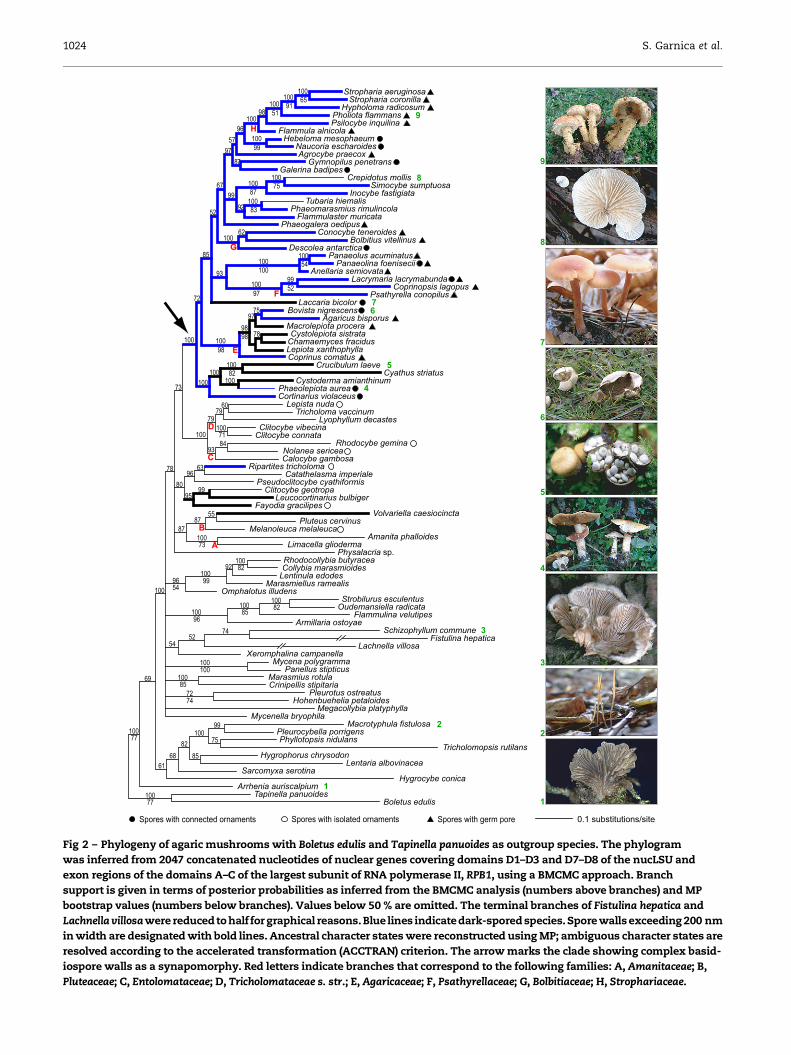
The results of our molecular phylogenetic analyses of the com-
bined alignment of original nucLSU and RPB1 sequences are
shown in Fig 2. A comparison of the phylogenetic tree with
the distribution of the ultrastructural character ‘basidiospore
wall thickness exceeding/not exceeding 200 nm’ and the
chemical character ‘basidiospores pigmented/not or only
slightly pigmented’ in the species studied (Fig 2 and the
supplementary Data appendix) shows that: (1) there are several
distinct clades of agarics that include only species with unpig-
mented or only slightly pigmented and mostly thin-walled ba-
sidiospores; and (2) agarics with a combination of thick-walled
and pigmented basidiospores (except for Ripartites) form a nat-
ural group (Fig 2, arrow). Alternative phylogenetic hypotheses
assuming the monophyly of agarics with thick-walled basidio-
spores or those with pigmented basidiospores were rejected
with posterior probabilities of zero in BMCMC analysis. MP
reconstruction of ancestral states along the BMCMC consensus
yielded the following results: (1) the evolution of complex
basidiospore walls involved four gains and four losses
(ACCTRAN) or five gains and three losses (DELTRAN); and (2)
the evolution of pigmented basidiospores involved four gains
and three losses (ACCTRAN) or seven gains and no loss (DEL-
TRAN). Ancestral state reconstructions using the ACCTRAN
and DELTRAN criteria are shown in Fig 2 and the Supplemen-
tary Data appendix, respectively. Both reconstruction variants
suggest that complex basidiospore walls are a synapomorphy
for the clade marked with an arrow in Fig 2.
The thick-walled and pigmented basidiospore syndrome
Our results support traditional classification systems in which
agarics with thick-walled and pigmented basidiospores are
treated as a derived group, and where ones with unpigmented
and thin-walled basidiospores were considered as more basal
taxa (Kuhner 1980; Oberwinkler 1982; with the exception of
the genera Crucibulum, Cyathus, Laccaria, and Leucocortinarius).
Our ultrastructural analyses revealed that strongly pigmented
basidiospores correlate with complex basidiospore wall archi-
tecture (Fig 3) in the vast majority of species, which is consis-
tent with the results of Clemencon (1997), who analysed the
ultrastructure of mature basidiospores of several species of
Agaricales. Thus, the development of thick-walled and pig-
mented basidiospores (corresponding to the arrowed clade
in Fig 2) was probably a key event in the evolution of the
agarics.
Evolutionary advantages of thick-walled and pigmentedbasidiospores
The evolution of this novel syndrome of morphological and
chemical traits can be interpreted as an adaptation to harsh
environmental conditions on land. Spores of these fungi
may be more resistant to dehydration and UV radiation than
thin-walled and hyaline basidiospores. This agrees with the
observations of Clemencon (1997), who reported that hyaline
and thin-walled spores lose their capacity to germinate after
a few days. Conversely, it has been demonstrated (Watling
1022 S. Garnica et al.
Table 1 – List of specimens used in the morphological and molecular analyses
Species Specimenvoucher
GenBank accessionnucLSU rDNA D1–D3
GenBank accessionnucLSU rDNA D7–D8
GenBank accessionRPB1 A–C
Agaricaceae
Agaricus bisporus TUB 011586 DQ071710 DQ071783 DQ067962
Chamaemyces fracidus GLM 45875 AY207148 DQ071787 DQ067957
Cystoderma amianthinum TUB 011551 DQ071703 DQ071774 DQ067951
Cystolepiota sistrata TUB 011552 DQ071711 DQ071786 DQ067958
Lepiota xanthophylla TUB 011553 DQ071712 DQ071788 DQ067956
Macrolepiota procera GLM 45957 AY207233 DQ071785 DQ067961
Phaeolepiota aurea TUB 011557 DQ071704 DQ071775 DQ060817
Amanitaceae
Amanita phalloides TUB 011556 DQ071721 DQ071810 DQ067953
Limacella glioderma HKAS ZLY D 72 DQ071728 DQ071818 DQ067952
Bolbitiaceae
Agrocybe praecox GLM 51238 DQ071692 DQ071758 DQ067985
Bolbitius vitellinus GLM 45874 AY207147 DQ071761 DQ067989
Conocybe teneroides GLM 45901 AY207171 DQ071760 DQ067981
Descolea antarctica TUB 011558 DQ071693 DQ071762 DQ067982
Clavariaceae
Macrotyphula fistulosa TUB 011469 DQ071735 DQ071827 DQ067802
Coprinaceae
Anellaria semiovata GLM 51235 DQ071694 DQ071763 DQ067965
Coprinopsis lagopus GLM 45907 AY207183 DQ071779 DQ067986
Coprinus comatus GLM 45914 AY207179 DQ071784 DQ067959
Paneolina foenisecii FO 46609 DQ071696 DQ071765 DQ067963
Paneolus acuminatus GLM 46071 DQ071695 DQ071764 DQ067964
Psathyrella conopilus TUB 011587 DQ071706 DQ071777 DQ067988
Cortinariaceae
Cortinarius violaceus TUB 011825 DQ071705 DQ071776 DQ067972
Galerina badipes GLM 45922 AY207201 DQ071754 DQ067975
Gymnopilus penetrans GLM 45929 AY207208 DQ071752 DQ060814
Hebeloma mesophaeum TUB 011577 DQ071690 DQ071756 DQ067971
Inocybe fastigiata FO 46800 DQ071697 DQ071766 DQ067980
Naucoria escharoides FO 46893 DQ071691 DQ071757 DQ067970
Leucocortinarius bulbiger TUB 011568 DQ071745 DQ071837 DQ060819
Phaeogalera oedipus GLM 45993 AY207268 DQ071759 DQ067983
Crepidotaceae
Crepidotus mollis TUB 011566 DQ071698 DQ071767 DQ067977
Simocybe sumptuosa TUB 011584 DQ071699 DQ071768 DQ067976
Tubaria hiemalis GLM 46038 AY207311 DQ071769 DQ067979
Entolomataceae
Nolanea sericea GLM 45918 AY207197 DQ071794 DQ067948
Rhodocybe gemina TUB 011578 DQ071715 DQ071793 DQ067944
Fistulinaceae
Fistulina hepatica TUB 011583 DQ071727 DQ071817 DQ060815
Gomphaceae
Lentaria albovinacea FO 46869 DQ071734 DQ071826 DQ060806
Hygrophoraceae
Hygrocybe conica FO 46714 DQ071739 DQ071831 DQ060810
Hygrophorus chrysodon TUB 011570 DQ071733 DQ071825 DQ067995
Lentinaceae
Phyllotopsis nidulans TUB 011567 DQ071736 DQ071828 DQ060803
Lycoperdaceae
Bovista nigrescens FO 46606 DQ071709 DQ071782 DQ067960
Nidulariaceae
Crucibulum laeve TUB 011564 DQ071701 DQ071771 DQ067950
Cyathus striatus TUB 011565 DQ071742 DQ071834 DQ060821
Omphalotaceae
Omphalotus illudens TUB 012155 DQ071741 DQ071833 DQ060813
Reconstructing the evolution of agarics from nuclear gene sequences and basidiospore ultrastructure 1023
Table 1 (continued)
Species Specimenvoucher
GenBank accessionnucLSU rDNA D1–D3
GenBank accessionnucLSU rDNA D7–D8
GenBank accessionRPB1 A–C
Pleurotaceae
Pleurotus ostreatus TUB 011571 DQ071722 DQ071811 DQ060804
Pluteaceae
Pluteus cervinus FO 46619 DQ071729 DQ071819 DQ067955
Volvariella caesiocincta FO 46681 DQ071726 DQ071816 DQ068009
Schizophyllaceae
Schizophyllum commune TUB 012156 DQ071725 DQ071814 DQ068011
Strophariaceae
Flammula alnicola GLM 45994 AY207269 DQ071755 DQ067974
Flammulaster muricata TUB 012150 DQ071740 DQ071832 DQ060812
Hypholoma radicosum TUB 011572 DQ071685 DQ071748 DQ067968
Phaeomarasmius rimulincola FO 46666 DQ071700 DQ071770 DQ067978
Pholiota flammans TUB 011573 DQ071688 DQ071751 DQ067973
Psilocybe inquilina GLM 51242 DQ071689 DQ071753 DQ067969
Stropharia aeruginosa TUB 012151 DQ071686 DQ071749 DQ067967
S. coronilla GLM 46074 DQ071687 DQ071750 DQ067966
Tricholomataceae
Armillaria ostoyae GLM 45872 AY207145 DQ071808 DQ067932
Arrhenia auriscalpium TUB 011588 DQ071732 DQ071824 DQ068008
Calocybe gambosa TUB 011576 DQ071716 DQ071795 DQ067945
Catathelasma imperiale TUB 011562 DQ071743 DQ071835 DQ060820
Clitocybe connata TUB 011574 DQ071714 DQ071792 DQ067946
C. geotropa GLM 45881 AY207154 DQ071821 DQ067938
C. vibecina GLM 45888 AY207160 DQ071791 DQ067947
Crinipellis stipitaria GLM 45915 AY207194 DQ071800 DQ067933
Collybia marasmioides GLM 45932 AY207167 DQ071804 DQ060816
Fayodia gracilis TUB 011585 DQ071744 DQ071836 DQ060818
Flammulina velutipes GLM 45921 AY207200 DQ071806 DQ067998
Hohenbuehelia petaloides TUB 011579 DQ071723 DQ071812 DQ060805
Laccaria bicolor TUB 011581 DQ071702 DQ071773 DQ067942
Lachnella villosa CCJ 1547 DQ071724 DQ071813 DQ060807
Lentinula edodes TUB 012149 DQ071718 DQ071802 DQ067997
Lepista nuda TUB 012152 DQ071713 DQ071790 DQ067954
Lyophyllum decastes GLM 45952 AY207228 DQ071789 DQ067949
Marasmiellus ramealis GLM 45958 AY207236 DQ071801 DQ067996
Marasmius rotula GLM 45962 AY207238 DQ071799 DQ067934
Megacollybia platyphylla GLM 45963 AY207239 DQ071798 DQ067935
Melanoleuca melaleuca FO 46845 DQ071730 DQ071820 DQ067941
Mycena polygramma TUB 011575 DQ071707 DQ071780 DQ060800
Mycenella bryophila KR 7435 DQ071720 DQ071809 DQ067937
Oudemansiella radicata FO 46757 DQ071719 DQ071807 DQ067930
Panellus stipticus GLM 51801 DQ071708 DQ071781 DQ060801
Physalacria sp. HKAS 32011 DQ071717 DQ071797 DQ067990
Pleurocybella porrigens TUB 012154 DQ071737 DQ071829 DQ067994
Pseudoclitocybe cyathiformis GLM 46020 AY207296 DQ071772 DQ067939
Rhodocollybia butyracea f. asema GLM 46024 AY207163 DQ071803 DQ067999
Ripartites tricholoma GLM 46025 AY207297 DQ071822 DQ067936
Sarcomyxa serotina TUB 012153 DQ071731 DQ071823 DQ067993
Strobilurus esculentus GLM 46027 AY207299 DQ071805 DQ067931
Tricholoma vaccinum GLM 46037 AY207307 DQ071796 DQ067943
Tricholomopsis rutilans TUB 011582 DQ071738 DQ071830 DQ067984
Xeromphalina campanella GLM 46039 AY207312 DQ071815 DQ067940
Boletales (outgroup)
Boletus edulis FO 46874 DQ071747 DQ071839 DQ067991
Tapinella panuoides GLM 45992 DQ071746 DQ071838 DQ067992
Classification is according to Singer (1986), Index Fungorum (http://www.indexfungorum.org), and the NCBI taxonomy homepage (http://
www.ncbi.nlm.nih.gov/Taxonomy/).
Herbarium acronyms: CCJ, private herbarium C.-J. Chen; FO, private herbarium F. Oberwinkler; KR, private herbarium K.-H. Rexer; GLM,
Staatliches Museum fur Naturkunde Gorlitz; HKAS, Cryptogamic Herbarium, Kunming Institute of Botany, Academia Sinica; TUB, Herbarium,
University of Tubingen.
1024 S. Garnica et al.
0.1 substitutions/site
Fistulina hepatica
Stropharia aeruginosa
Stropharia coronilla
Hypholoma radicosum
Pholiota flammans
Psilocybe inquilina
Flammula alnicola
Hebeloma mesophaeum
Naucoria escharoides
Agrocybe praecox
Gymnopilus penetrans
Galerina badipes
Crepidotus mollis
Simocybe sumptuosa
Inocybe fastigiata
Tubaria hiemalis
Phaeomarasmius rimulincola
Flammulaster muricata
Phaeogalera oedipus
Conocybe teneroides
Bolbitius vitellinus
Descolea antarctica
Panaeolus acuminatus
Panaeolina foenisecii
Anellaria semiovata
Lacrymaria lacrymabunda
Coprinopsis lagopus
Psathyrella conopilus
Laccaria bicolor
Bovista nigrescens
Agaricus bisporus
Macrolepiota procera
Cystolepiota sistrata
Chamaemyces fracidus
Lepiota xanthophylla
Coprinus comatus
Crucibulum laeve
Cyathus striatus
Cystoderma amianthinum
Phaeolepiota aurea
Cortinarius violaceus
Lepista nuda
Tricholoma vaccinum
Lyophyllum decastes
Clitocybe vibecina
Clitocybe connata
Rhodocybe gemina
Nolanea sericea
Calocybe gambosa
Ripartites tricholoma
Catathelasma imperiale
Pseudoclitocybe cyathiformis
Clitocybe geotropa
Leucocortinarius bulbiger
Fayodia gracilipes
Volvariella caesiocincta
Pluteus cervinus
Melanoleuca melaleuca
Amanita phalloides
Limacella glioderma
Physalacria sp.Rhodocollybia butyracea
Collybia marasmioides
Lentinula edodes
Marasmiellus ramealis
Omphalotus illudens
Strobilurus esculentus
Oudemansiella radicata
Flammulina velutipes
Armillaria ostoyae
Schizophyllum commune
Lachnella villosa
Xeromphalina campanella
Mycena polygramma
Panellus stipticus
Marasmius rotula
Crinipellis stipitaria
Pleurotus ostreatus
Hohenbuehelia petaloides
Megacollybia platyphylla
Mycenella bryophila
Macrotyphula fistulosa
Pleurocybella porrigens
Phyllotopsis nidulans
Tricholomopsis rutilans
Hygrophorus chrysodon
Lentaria albovinacea
Sarcomyxa serotina
Hygrocybe conica
Arrhenia auriscalpium
Tapinella panuoides
Boletus edulis
F
G
H
E
D
C
A
B
9
8
7
6
5
4
3
2
1
Spores with germ poreSpores with connected ornaments Spores with isolated ornaments
1
2
3
4
5
6
7
8
9
Fig 2 – Phylogeny of agaric mushrooms with Boletus edulis and Tapinella panuoides as outgroup species. The phylogram
was inferred from 2047 concatenated nucleotides of nuclear genes covering domains D1–D3 and D7–D8 of the nucLSU and
exon regions of the domains A–C of the largest subunit of RNA polymerase II, RPB1, using a BMCMC approach. Branch
support is given in terms of posterior probabilities as inferred from the BMCMC analysis (numbers above branches) and MP
bootstrap values (numbers below branches). Values below 50 % are omitted. The terminal branches of Fistulina hepatica and
Lachnella villosawere reducedto half for graphical reasons. Blue lines indicatedark-spored species. Spore wallsexceeding 200 nm
in width are designated with bold lines. Ancestral character states were reconstructed using MP; ambiguous character states are
resolved according to the accelerated transformation (ACCTRAN) criterion. The arrow marks the clade showing complex basid-
iospore walls as a synapomorphy. Red letters indicate branches that correspond to the following families: A, Amanitaceae; B,
Pluteaceae; C, Entolomataceae; D, Tricholomataceae s. str.; E, Agaricaceae; F, Psathyrellaceae; G, Bolbitiaceae; H, Strophariaceae.
Reconstructing the evolution of agarics from nuclear gene sequences and basidiospore ultrastructure 1025
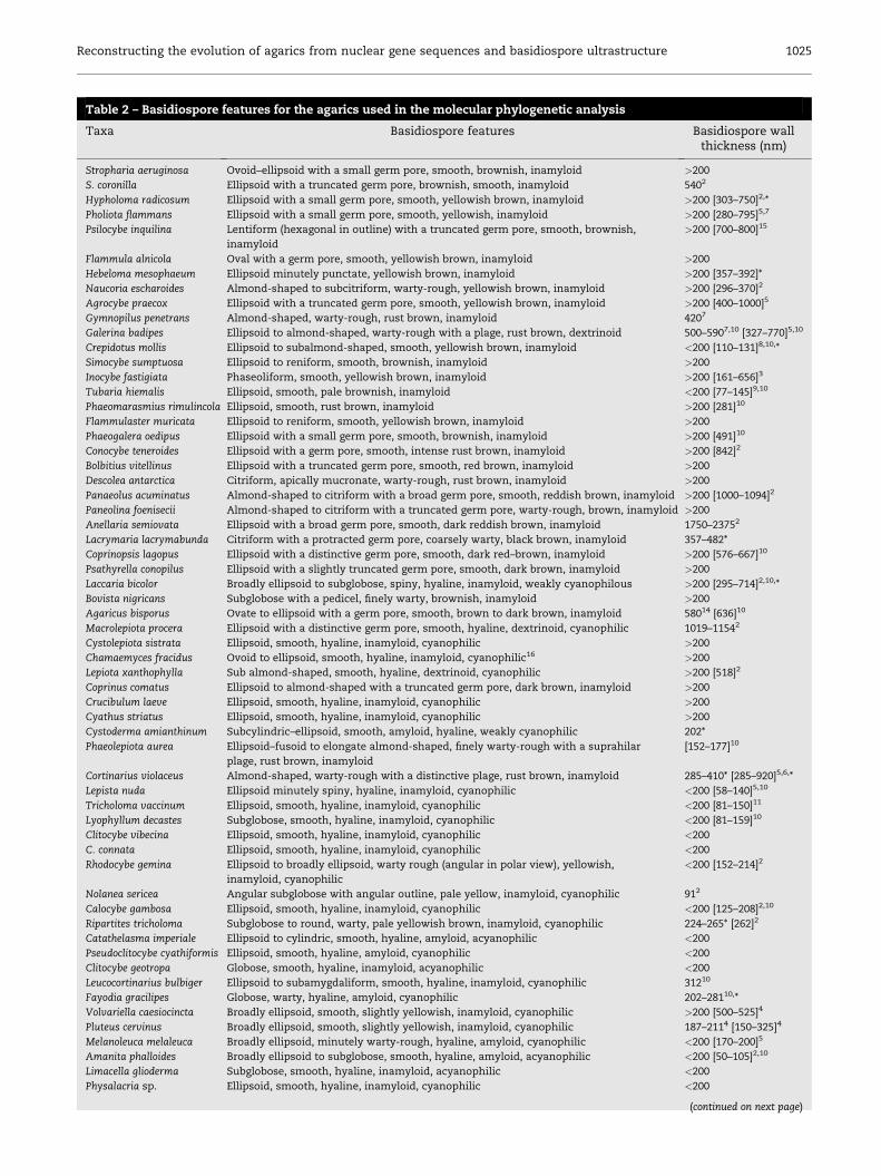
Table 2 – Basidiospore features for the agarics used in the molecular phylogenetic analysis
Taxa Basidiospore features Basidiospore wallthickness (nm)
Stropharia aeruginosa Ovoid–ellipsoid with a small germ pore, smooth, brownish, inamyloid >200
S. coronilla Ellipsoid with a truncated germ pore, brownish, smooth, inamyloid 5402
Hypholoma radicosum Ellipsoid with a small germ pore, smooth, yellowish brown, inamyloid >200 [303–750]2,*
Pholiota flammans Ellipsoid with a small germ pore, smooth, yellowish, inamyloid >200 [280–795]5,7
Psilocybe inquilina Lentiform (hexagonal in outline) with a truncated germ pore, smooth, brownish,
inamyloid
>200 [700–800]15
Flammula alnicola Oval with a germ pore, smooth, yellowish brown, inamyloid >200
Hebeloma mesophaeum Ellipsoid minutely punctate, yellowish brown, inamyloid >200 [357–392]*
Naucoria escharoides Almond-shaped to subcitriform, warty-rough, yellowish brown, inamyloid >200 [296–370]2
Agrocybe praecox Ellipsoid with a truncated germ pore, smooth, yellowish brown, inamyloid >200 [400–1000]5
Gymnopilus penetrans Almond-shaped, warty-rough, rust brown, inamyloid 4207
Galerina badipes Ellipsoid to almond-shaped, warty-rough with a plage, rust brown, dextrinoid 500–5907,10 [327–770]5,10
Crepidotus mollis Ellipsoid to subalmond-shaped, smooth, yellowish brown, inamyloid <200 [110–131]8,10,*
Simocybe sumptuosa Ellipsoid to reniform, smooth, brownish, inamyloid >200
Inocybe fastigiata Phaseoliform, smooth, yellowish brown, inamyloid >200 [161–656]3
Tubaria hiemalis Ellipsoid, smooth, pale brownish, inamyloid <200 [77–145]9,10
Phaeomarasmius rimulincola Ellipsoid, smooth, rust brown, inamyloid >200 [281]10
Flammulaster muricata Ellipsoid to reniform, smooth, yellowish brown, inamyloid >200
Phaeogalera oedipus Ellipsoid with a small germ pore, smooth, brownish, inamyloid >200 [491]10
Conocybe teneroides Ellipsoid with a germ pore, smooth, intense rust brown, inamyloid >200 [842]2
Bolbitius vitellinus Ellipsoid with a truncated germ pore, smooth, red brown, inamyloid >200
Descolea antarctica Citriform, apically mucronate, warty-rough, rust brown, inamyloid >200
Panaeolus acuminatus Almond-shaped to citriform with a broad germ pore, smooth, reddish brown, inamyloid >200 [1000–1094]2
Paneolina foenisecii Almond-shaped to citriform with a truncated germ pore, warty-rough, brown, inamyloid >200
Anellaria semiovata Ellipsoid with a broad germ pore, smooth, dark reddish brown, inamyloid 1750–23752
Lacrymaria lacrymabunda Citriform with a protracted germ pore, coarsely warty, black brown, inamyloid 357–482*
Coprinopsis lagopus Ellipsoid with a distinctive germ pore, smooth, dark red–brown, inamyloid >200 [576–667]10
Psathyrella conopilus Ellipsoid with a slightly truncated germ pore, smooth, dark brown, inamyloid >200
Laccaria bicolor Broadly ellipsoid to subglobose, spiny, hyaline, inamyloid, weakly cyanophilous >200 [295–714]2,10,*
Bovista nigricans Subglobose with a pedicel, finely warty, brownish, inamyloid >200
Agaricus bisporus Ovate to ellipsoid with a germ pore, smooth, brown to dark brown, inamyloid 58014 [636]10
Macrolepiota procera Ellipsoid with a distinctive germ pore, smooth, hyaline, dextrinoid, cyanophilic 1019–11542
Cystolepiota sistrata Ellipsoid, smooth, hyaline, inamyloid, cyanophilic >200
Chamaemyces fracidus Ovoid to ellipsoid, smooth, hyaline, inamyloid, cyanophilic16 >200
Lepiota xanthophylla Sub almond-shaped, smooth, hyaline, dextrinoid, cyanophilic >200 [518]2
Coprinus comatus Ellipsoid to almond-shaped with a truncated germ pore, dark brown, inamyloid >200
Crucibulum laeve Ellipsoid, smooth, hyaline, inamyloid, cyanophilic >200
Cyathus striatus Ellipsoid, smooth, hyaline, inamyloid, cyanophilic >200
Cystoderma amianthinum Subcylindric–ellipsoid, smooth, amyloid, hyaline, weakly cyanophilic 202*
Phaeolepiota aurea Ellipsoid–fusoid to elongate almond-shaped, finely warty-rough with a suprahilar
plage, rust brown, inamyloid
[152–177]10
Cortinarius violaceus Almond-shaped, warty-rough with a distinctive plage, rust brown, inamyloid 285–410* [285–920]5,6,*
Lepista nuda Ellipsoid minutely spiny, hyaline, inamyloid, cyanophilic <200 [58–140]5,10
Tricholoma vaccinum Ellipsoid, smooth, hyaline, inamyloid, cyanophilic <200 [81–150]11
Lyophyllum decastes Subglobose, smooth, hyaline, inamyloid, cyanophilic <200 [81–159]10
Clitocybe vibecina Ellipsoid, smooth, hyaline, inamyloid, cyanophilic <200
C. connata Ellipsoid, smooth, hyaline, inamyloid, cyanophilic <200
Rhodocybe gemina Ellipsoid to broadly ellipsoid, warty rough (angular in polar view), yellowish,
inamyloid, cyanophilic
<200 [152–214]2
Nolanea sericea Angular subglobose with angular outline, pale yellow, inamyloid, cyanophilic 912
Calocybe gambosa Ellipsoid, smooth, hyaline, inamyloid, cyanophilic <200 [125–208]2,10
Ripartites tricholoma Subglobose to round, warty, pale yellowish brown, inamyloid, cyanophilic 224–265* [262]2
Catathelasma imperiale Ellipsoid to cylindric, smooth, hyaline, amyloid, acyanophilic <200
Pseudoclitocybe cyathiformis Ellipsoid, smooth, hyaline, amyloid, cyanophilic <200
Clitocybe geotropa Globose, smooth, hyaline, inamyloid, acyanophilic <200
Leucocortinarius bulbiger Ellipsoid to subamygdaliform, smooth, hyaline, inamyloid, cyanophilic 31210
Fayodia gracilipes Globose, warty, hyaline, amyloid, cyanophilic 202–28110,*
Volvariella caesiocincta Broadly ellipsoid, smooth, slightly yellowish, inamyloid, cyanophilic >200 [500–525]4
Pluteus cervinus Broadly ellipsoid, smooth, slightly yellowish, inamyloid, cyanophilic 187–2114 [150–325]4
Melanoleuca melaleuca Broadly ellipsoid, minutely warty-rough, hyaline, amyloid, cyanophilic <200 [170–200]5
Amanita phalloides Broadly ellipsoid to subglobose, smooth, hyaline, amyloid, acyanophilic <200 [50–105]2,10
Limacella glioderma Subglobose, smooth, hyaline, inamyloid, acyanophilic <200
Physalacria sp. Ellipsoid, smooth, hyaline, inamyloid, cyanophilic <200
(continued on next page)
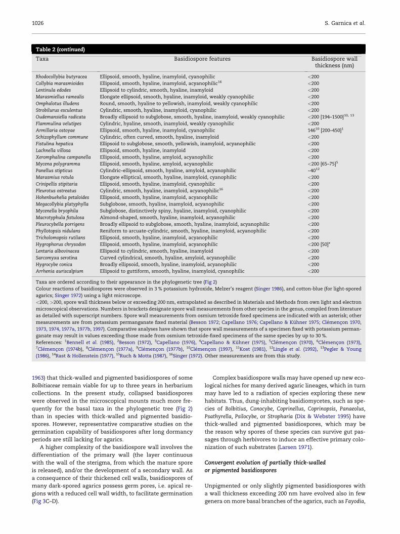
1026 S. Garnica et al.
Table 2 (continued)
Taxa Basidiospore features Basidiospore wallthickness (nm)
Rhodocollybia butyracea Ellipsoid, smooth, hyaline, inamyloid, cyanophilic <200
Collybia marasmioides Ellipsoid, smooth, hyaline, inamyloid, acyanophilic16 <200
Lentinula edodes Ellipsoid to cylindric, smooth, hyaline, inamyloid <200
Marasmiellus ramealis Elongate ellipsoid, smooth, hyaline, inamyloid, weakly cyanophilic <200
Omphalotus illudens Round, smooth, hyaline to yellowish, inamyloid, weakly cyanophilic <200
Strobilurus esculentus Cylindric, smooth, hyaline, inamyloid, cyanophilic <200
Oudemansiella radicata Broadly ellipsoid to subglobose, smooth, hyaline, inamyloid, weakly cyanophilic <200 [194–1500]10, 13
Flammulina velutipes Cylindric, hyaline, smooth, inamyloid, weakly cyanophilic <200
Armillaria ostoyae Ellipsoid, smooth, hyaline, inamyloid, cyanophilic 14610 [200–450]1
Schizophyllum commune Cylindric, often curved, smooth, hyaline, inamyloid <200
Fistulina hepatica Ellipsoid to subglobose, smooth, yellowish, inamyloid, acyanophilic <200
Lachnella villosa Ellipsoid, smooth, hyaline, inamyloid <200
Xeromphalina campanella Ellipsoid, smooth, hyaline, amyloid, acyanophilic <200
Mycena polygramma Ellipsoid, smooth, hyaline, amyloid, acyanophilic <200 [65–75]5
Panellus stipticus Cylindric–ellipsoid, smooth, hyaline, amyloid, acyanophilic ~4012
Marasmius rotula Elongate elliptical, smooth, hyaline, inamyloid, cyanophilic <200
Crinipellis stipitaria Ellipsoid, smooth, hyaline, inamyloid, cyanophilic <200
Pleurotus ostreatus Cylindric, smooth, hyaline, inamyloid, acyanophilic16 <200
Hohenbuehelia petaloides Ellipsoid, smooth, hyaline, inamyloid, acyanophilic <200
Megacollybia platyphylla Subglobose, smooth, hyaline, inamyloid, acyanophilic <200
Mycenella bryophila Subglobose, distinctively spiny, hyaline, inamyloid, cyanophilic <200
Macrotyphula fistulosa Almond-shaped, smooth, hyaline, inamyloid, acyanophilic <200
Pleurocybella porrigens Broadly ellipsoid to subglobose, smooth, hyaline, inamyloid, acyanophilic <200
Phyllotopsis nidulans Reniform to arcuate-cylindric, smooth, hyaline, inamyloid, acyanophilic <200
Tricholomopsis rutilans Ellipsoid, smooth, hyaline, inamyloid, acyanophilic <200
Hygrophorus chrysodon Ellipsoid, smooth, hyaline, inamyloid, acyanophilic <200 [50]*
Lentaria albovinacea Ellipsoid to cylindric, smooth, hyaline, inamyloid <200
Sarcomyxa serotina Curved cylindrical, smooth, hyaline, amyloid, acyanophilic <200
Hygrocybe conica Broadly ellipsoid, smooth, hyaline, inamyloid, acyanophilic <200
Arrhenia auriscalpium Ellipsoid to guttiform, smooth, hyaline, inamyloid, cyanophilic <200
Taxa are ordered according to their appearance in the phylogenetic tree (Fig 2)
Colour reactions of basidiospores were observed in 3 % potassium hydroxide, Melzer’s reagent (Singer 1986), and cotton-blue (for light-spored
agarics; Singer 1972) using a light microscope.
<200, >200, spore wall thickness below or exceeding 200 nm, extrapolated as described in Materials and Methods from own light and electron
microscopical observations. Numbers in brackets designate spore wall measurements from other species in the genus, compiled from literature
as detailed with superscript numbers. Spore wall measurements from osmium tetroxide fixed specimens are indicated with an asterisk; other
measurements are from potassium permanganate fixed material (Besson 1972; Capellano 1976; Capellano & Kuhner 1975; Clemencon 1970,
1973, 1974, 1977a, 1977b, 1997). Comparative analyses have shown that spore wall measurements of a specimen fixed with potassium perman-
ganate may result in values exceeding those made from osmium tetroxide-fixed specimens of the same species by up to 30 %.
References: 1Bennell et al. (1985), 2Besson (1972), 3Capellano (1976), 4Capellano & Kuhner (1975), 5Clemencon (1970), 6Clemencon (1973),7Clemencon (1974b), 8Clemencon (1977a), 9Clemencon (1977b), 10Clemencon (1997), 11Kost (1981), 12Lingle et al. (1992), 13Pegler & Young
(1986), 14Rast & Hollenstein (1977), 15Ruch & Motta (1987), 16Singer (1972). Other measurements are from this study.
1963) that thick-walled and pigmented basidiospores of some
Bolbitiaceae remain viable for up to three years in herbarium
collections. In the present study, collapsed basidiospores
were observed in the microscopical mounts much more fre-
quently for the basal taxa in the phylogenetic tree (Fig 2)
than in species with thick-walled and pigmented basidio-
spores. However, representative comparative studies on the
germination capability of basidiospores after long dormancy
periods are still lacking for agarics.
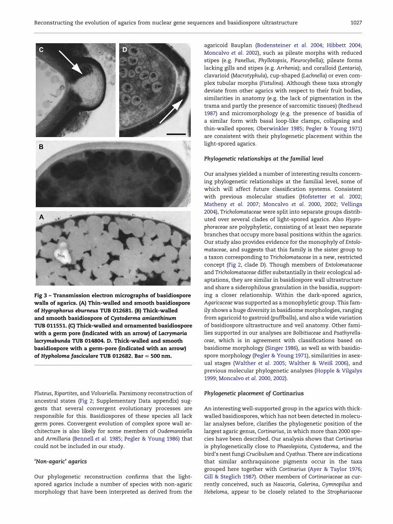
A higher complexity of the basidiospore wall involves the
differentiation of the primary wall (the layer continuous
with the wall of the sterigma, from which the mature spore
is released), and/or the development of a secondary wall. As
a consequence of their thickened cell walls, basidiospores of
many dark-spored agarics possess germ pores, i.e. apical re-
gions with a reduced cell wall width, to facilitate germination
(Fig 3C–D).
Complex basidiospore walls may have opened up new eco-
logical niches for many derived agaric lineages, which in turn
may have led to a radiation of species exploring these new
habitats. Thus, dung-inhabiting basidiomycetes, such as spe-
cies of Bolbitius, Conocybe, Coprinellus, Coprinopsis, Panaeolus,
Psathyrella, Psilocybe, or Stropharia (Dix & Webster 1995) have
thick-walled and pigmented basidiospores, which may be
the reason why spores of these species can survive gut pas-
sages through herbivores to induce an effective primary colo-
nization of such substrates (Larsen 1971).
Convergent evolution of partially thick-walledor pigmented basidiospores
Unpigmented or only slightly pigmented basidiospores with
a wall thickness exceeding 200 nm have evolved also in few
genera on more basal branches of the agarics, such as Fayodia,
Reconstructing the evolution of agarics from nuclear gene sequences and basidiospore ultrastructure 1027
Pluteus, Ripartites, and Volvariella. Parsimony reconstruction of
ancestral states (Fig 2; Supplementary Data appendix) sug-
gests that several convergent evolutionary processes are
responsible for this. Basidiospores of these species all lack
germ pores. Convergent evolution of complex spore wall ar-
chitecture is also likely for some members of Oudemansiella
and Armillaria (Bennell et al. 1985; Pegler & Young 1986) that
could not be included in our study.
‘Non-agaric’ agarics
Our phylogenetic reconstruction confirms that the light-
spored agarics include a number of species with non-agaric
morphology that have been interpreted as derived from the
Fig 3 – Transmission electron micrographs of basidiospore
walls of agarics. (A) Thin-walled and smooth basidiospore
of Hygrophorus eburneus TUB 012681. (B) Thick-walled
and smooth basidiospore of Cystoderma amianthinum
TUB 011551. (C) Thick-walled and ornamented basidiospore
with a germ pore (indicated with an arrow) of Lacrymaria
lacrymabunda TUB 014804. D. Thick-walled and smooth
basidiospore with a germ-pore (indicated with an arrow)
of Hypholoma fasciculare TUB 012682. Bar [ 500 nm.
agaricoid Bauplan (Bodensteiner et al. 2004; Hibbett 2004;
Moncalvo et al. 2002), such as pileate morphs with reduced
stipes (e.g. Panellus, Phyllotopsis, Pleurocybella); pileate forms
lacking gills and stipes (e.g. Arrhenia); and coralloid (Lentaria),
clavarioid (Macrotyphula), cup-shaped (Lachnella) or even com-
plex tubular morphs (Fistulina). Although these taxa strongly
deviate from other agarics with respect to their fruit bodies,
similarities in anatomy (e.g. the lack of pigmentation in the
trama and partly the presence of sarcomitic tissues) (Redhead
1987) and micromorphology (e.g. the presence of basidia of
a similar form with basal loop-like clamps, collapsing and
thin-walled spores; Oberwinkler 1985; Pegler & Young 1971)
are consistent with their phylogenetic placement within the
light-spored agarics.
Phylogenetic relationships at the familial level
Our analyses yielded a number of interesting results concern-
ing phylogenetic relationships at the familial level, some of
which will affect future classification systems. Consistent
with previous molecular studies (Hofstetter et al. 2002;
Matheny et al. 2007; Moncalvo et al. 2000, 2002; Vellinga
2004), Tricholomataceae were split into separate groups distrib-
uted over several clades of light-spored agarics. Also Hygro-
phoraceae are polyphyletic, consisting of at least two separate
branches that occupy more basal positions within the agarics.
Our study also provides evidence for the monophyly of Entolo-
mataceae, and suggests that this family is the sister group to
a taxon corresponding to Tricholomataceae in a new, restricted
concept (Fig 2, clade D). Though members of Entolomataceae
and Tricholomataceae differ substantially in their ecological ad-
aptations, they are similar in basidiospore wall ultrastructure
and share a siderophilous granulation in the basidia, support-
ing a closer relationship. Within the dark-spored agarics,
Agaricaceae was supported as a monophyletic group. This fam-
ily shows a huge diversity in basidiome morphologies, ranging
from agaricoid to gastroid (puffballs), and also a wide variation
of basidiospore ultrastructure and veil anatomy. Other fami-
lies supported in our analyses are Bolbitiaceae and Psathyrella-
ceae, which is in agreement with classifications based on
basidiome morphology (Singer 1986), as well as with basidio-
spore morphology (Pegler & Young 1971), similarities in asex-
ual stages (Walther et al. 2005; Walther & Weiß 2006), and
previous molecular phylogenetic analyses (Hopple & Vilgalys
1999; Moncalvo et al. 2000, 2002).
Phylogenetic placement of Cortinarius
An interesting well-supported group in the agarics with thick-
walled basidiospores, which has not been detected in molecu-
lar analyses before, clarifies the phylogenetic position of the
largest agaric genus, Cortinarius, in which more than 2000 spe-
cies have been described. Our analysis shows that Cortinarius
is phylogenetically close to Phaeolepiota, Cystoderma, and the
bird’s nest fungi Crucibulum and Cyathus. There are indications
that similar anthraquinone pigments occur in the taxa
grouped here together with Cortinarius (Ayer & Taylor 1976;
Gill & Steglich 1987). Other members of Cortinariaceae as cur-
rently conceived, such as Naucoria, Galerina, Gymnopilus and
Hebeloma, appear to be closely related to the Strophariaceae
1028 S. Garnica et al.
(excluding Phaeomarasmius and Flammulaster), a relationship
supported by shared styrylpyrone pigments of the fruit bodies
(Gill & Steglich 1987), as well as by molecular phylogenetic
analyses (Gulden et al. 2005). This analysis is the first to signif-
icantly support a close relationship between Phaeolepiota, Cys-
toderma, Crucibulum, and Cyathus. We suspect that similarities
in pigments located in the velar hyphae might further support
this grouping. Phaeolepiota–Cystoderma and Crucibulum–
Cyathus, respectively, appear as sister taxa in our phylogenetic
tree with 100 % BMCMC support. Members of Phaeolepiota and
Cystoderma share features of veil anatomy, whereas Crucibu-
lum and Cyathus are similar in hymenial organization and
basidiome shape.
Laccaria is closely related to dark-spored agarics
This is the first study that demonstrates that the light-spored
genus Laccaria evolved within the dark-spored agarics (Fig 2).
In previous molecular studies (Hibbett & Binder 2002) based
on nu- and mt-rDNA sequences, Laccaria species appeared
close to members of the dark-spored genus Cortinarius, but
without significant support. Laccaria species, which are
currently classified in Tricholomataceae, have thick-walled
basidiospores bearing spectacular ornamentation. The orna-
mentation is connected by a continuous cell wall layer, and
this architecture was also found in dark-spored agarics with
thick-walled basidiospores. In contrast, spore ornamentation in
light-spored agarics is isolated, except for species of Laccaria.
This is another example for the importance of basidio-
spore-related characters in a natural classification of the
agarics.
In conclusion, our study demonstrates that higher-level
phylogenetic relationships in agarics can be resolved using se-
quence analysis of appropriate genetic markers, and forms
the basis for a more natural classification. We conclude from
new molecular and morphological data that the development
of complex architecture of the basidiospore walls was a key
event in the evolution of the euagarics, and suggest that this
probably increased ability to survive in harsh ecological condi-
tions and enabled subsequent adaptive radiations to new eco-
logical niches. Future molecular analyses involving more
genes and an extended sampling of species, together with
careful morphological and chemical (re-)examinations, will
provide more insight into the evolution of the fascinating aga-
ric branch of the fungal tree of life.
Acknowledgements
We thank Doris Laber, Bernhard Oertel, Karl-Heinz Rexer,
Gunter Saar, Eduardo Valenzuela, and Zhu-Liang Yang for pro-
viding specimens, Ruth Fleischmann, David L. Hawksworth,
David Hibbett, Mika Tarkka, Rainer Wurgau, and two anony-
mous reviewers for their helpful comments on drafts of the
manuscript, and Magda Wagner-Eha and Christiane Karasch-
Wittmann for technical assistance in the transmission elec-
tron microscopy. This study was supported by grant OB24/
27-1, 2 from the Deutsche Forschungsgemeinschaft (German
Research Association; DFG) to F.O. and M.W.
Supplementary data
Supplementary data associated with this article can be found,
in the online version, at 10.1016/j.mycres.2007.03.019
r e f e r e n c e s
Ayer WA, Taylor DR, 1976. Metabolites of bird’s nest fungi 5. Theisolation of 1-hydroxy-6-methyl-8-hydroxymethylxanthone,a new xanthone, from Cyathus intermedius. Synthesis viaphotoenolisation. Canadian Journal of Chemistry 54: 1703.
Bauer R, Oberwinkler F, Vanky K, 1997. Ultrastructural markersand systematics in smut fungi and allied taxa. Canadian Journalof Botany 75: 1273–1314.
Bennell AP, Watling R, Kile G, 1985. Spore ornamentation inArmillaria (Agaricales). Transactions of the British MycologicalSociety 84: 447–455.
Besson MA, 1972. Contribution a la connaissance de I’infrastructurede la paroi sporique des Hymenomycetes. PhD thesis, Universityof Claude Bernard, Lyon.
Binder M, Hibbett DS, Larsson K-H, Larsson E, Langer E, Langer G,2005. The phylogenetic distribution of resupinate forms acrossthe major clades of mushroom-forming fungi (Homobasidio-mycetes). Systematics and Biodiversity 3: 113–157.
Bodensteiner P, Binder M, Moncalvo JM, Agerer R, Hibbett DS,2004. Phylogenetic relationships of cyphelloid Homobasidio-mycetes. Molecular Phylogenetics and Evolution 33: 501–515.
Capellano A, 1976. Architecture de la paroi sporique des Inocybe(basidiomycetes – Agaricales) en microscopie electronique partransmission. Comptes rendus hebdomadaires des seances del’Academie des sciences, Serie D: Sciences naturelles (Paris) 282:847–849.
Capellano A, Kuhner R, 1975. Architecture de la paroi sporiquedes volvariacees (basidiomycetes – Agaricales) en microscopephotonique et electronique. Bulletin de la Societe Linneenne deLyon 44: 4–21.
Clemencon H, 1970. Bau der Wande der Basidiosporen und einVorschlag zur Benennung ihrer Schichten. Zeitschrift furPilzkunde 36: 113–133.
Clemencon H, 1973. Die Wandstrukturen der Basidiosporen III.Cortinarius und Dermocybe. Zeitschrift fur Pilzkunde 39: 121–144.
Clemencon H, 1974. Die Wandstrukturen der Basidiosporen V.Pholiota und Kuehneromyces, verglichen mit Galerina und Gym-nopilus. Zeitschrift fur Pilzkunde 40: 105–126.
Clemencon H, 1977a. Die Wandstrukturen der Basidiosporen VI.Crepidotus sphaerosporus und verwandte Arten. Zeitschrift furPilzkunde 43: 269–282.
Clemencon H, 1977b. Die Wandstrukturen der Basidiosporen VII.Tubaria. Zeitschrift fur Pilzkunde 43: 283–289.
Clemencon H, 1997. Anatomie der Hymenomyceten. Universite deLausanne, Lausanne.
Dix NJ, Webster J, 1995. Fungal Ecology. Chapman & Hall, London.Geyer CJ, 1991. Markov chain Monte Carlo maximum likelihood.
In: Keramidas EM (ed), Computing Science and Statistics: Pro-ceedings of the 23rd Symposium on the Interface. Interface Foun-dation, Fairfax Station, pp. 156–163.
Gill M, Steglich W, 1987. Pigments of fungi (Macromycetes). Progressin the Chemistry of Organic Natural Products 51: 1–317.
Gulden G, Stensrud Ø, Shalchian-Tabrizi K, Kauserud H, 2005.Galerina Earle: a polyphyletic genus in the consortium of dark-spored agarics. Mycologia 97: 823–837.
Hassouna N, Michot B, Bachellerie JP, 1984. The completenucleotide sequence of mouse 28S rRNA gene. Implicationsfor the process of size increase of the large subunit rRNA inhigher eucaryotes. Nucleic Acids Research 12: 3563–3583.
Reconstructing the evolution of agarics from nuclear gene sequences and basidiospore ultrastructure 1029
Hibbett D, 2004. Trends in morphological evolution in Homobasi-diomycetes inferred using maximum likelihood: a comparisonof binary and multistate approaches. Systematic Biology 53:889–903.
Hibbett DS, 2007. After the gold rush, or before the flood?Evolutionary morphology of mushroom-forming fungi (agarico-mycetes) in the early 21st century. Mycological Research 111: 1003–1020.
Hibbett D, Binder M, 2002. Evolution of complex fruiting bodymorphologies in Homobasidiomycetes. Proceedings of the RoyalSociety of London B 269: 1963–1969.
Hibbett DS, Grimaldi D, Donoghue MJ, 1997a. Fossil mushroomsfrom Miocene and Cretaceous ambers and the evolution ofHomobasidiomycetes. American Journal of Botany 84: 981–991.
Hibbett DS, Pine EM, Langer E, Langer G, Donoghue MJ, 1997b.Evolution of gilled mushrooms and puffballs inferred fromribosomal DNA sequences. Proceedings of the National Academyof Sciences USA 94: 12002–12006.
Hofstetter V, Clemencon H, Vilgalys R, Moncalvo JM, 2002. Phy-logenetic analyses of the Lyophylleae (Agaricales, Basidiomycota)based on nuclear and mitochondrial rDNA sequences. Myco-logical Research 106: 1043–1059.
Hopple JS, Vilgalys R, 1999. Phylogenetic relationships in themushroom genus Coprinus and dark-spored allies based onsequence data from the nuclear gene coding for the largeribosomal subunit RNA: divergent domains, outgroups,and monophyly. Molecular Phylogenetics and Evolution 13:1–19.
Kost G, 1981. Vergleichende morphologische, anatomische und fein-strukturelle Merkmalsstudien an Arten der Gattung Tricholoma(Fr.) Staude, Sektion Genuina (Fr.) Sacc. PhD thesis, Eberhard-Karls-Universitat, Tubingen.
Kuhner R, 1980. Les Hymenomycetes agaricoıdes (Agaricales,Tricholomatales, Pluteales, Russulales). Bulletin Societe LinneenneLyon, Numero Special 49: 1027.
Larsen K, 1971. Danish endocoprophilous fungi and theirsequence of occurrence. Botanisk Tidsskrift 65: 1–32.
Larsson K-H, Larsson E, Koljalg U, 2004. High phylogenetic-diversity among corticioid Homobasidiomycetes. MycologicalResearch 108: 983–1002.
Lingle WL, Clay RP, Porter D, 1992. Ultrastructural analysis ofbasidiosporogenesis in Panellus stypticus. Canadian Journal ofBotany 70: 2017–2027.
Matheny PB, Liu Y, Ammirati J, Hall BD, 2002. Using RPB1 sequencesto improve phylogenetic inference among mushrooms (Inocybe,Agaricales). American Journal of Botany 89: 688–698.
Matheny PB, Wang Z, Binder M, Curtis JM, Lim YW, Hilsson RH,Hughes KW, Hofstetter V, Ammirati JF, Schoch C, Langer E,Langer G, McLaughlin DJ, Wilson AW, Frøslev T, Ge ZW,Kerrigan RW, Slot JC, Yang ZL, Baroni TJ, Fischer M, Hosaka K,Matsuura K, Seidl MT, Vauras J, Hibbett DS, 2007. Contribu-tions of rpb2 and tef1 to the phylogeny of mushrooms andallies (Basidiomycota, Fungi). Molecular Phylogenetics and Evolu-tion 43: 430–451.
Moncalvo JM, Wang H-H, Hseu R-S,1995. Phylogenetic relationshipsin Ganoderma inferred from the internal transcribed spacers and25S ribosomal DNA sequences. Mycologia 87: 223–238.
Moncalvo JM, Lutzoni FM, Rehner SA, Johnson J, Vilgalys R, 2000.Phylogenetic relationships of agaric fungi based on nuclearlarge subunit ribosomal DNA sequences. Systematic Biology 49:278–305.
Moncalvo JM, Vilgalys R, Redhead SA, Johnson JE, James TY,Aime MC, Hofstetter V, Verduin SJW, Larsson E, Baroni TJ,Thorn RG, Jacobsson S, Clemencon H, Miller jr OK, 2002. One
hundred and seventeen clades of euagarics. Molecular Phylo-genetics and Evolution 23: 357–400.
Nylander JAA, 2004. MrModeltest. Version 2. Evolutionary BiologyCentre, Uppsala University.
Nylander JAA, Ronquist F, Huelsenbeck JP, Nieves-Aldrey JL, 2004.Bayesian phylogenetic analysis of combined data. SystematicBiology 53: 47–67.
Oberwinkler F, 1982. The significance of the morphology of thebasidium in the phylogeny of basidiomycetes. In: Wells K,Wells EK (eds), Basidium and Basidiocarp. Springer-Verlag, NewYork, pp. 9–35.
Oberwinkler F, 1985. Anmerkungen zur Evolution und Systematikder Basidiomyceten. Botanische Jahrbucher 107: 541–580.
Pegler DN, Young TWK, 1971. Basidiospore morphology in the.Agaricales. Beihefte zur Nova Hedwigia 35: 1–210.
Pegler DN, Young TWK, 1986. Classification of Oudemansiella(Basidiomycotina: Tricholomataceae), with special reference tospore structure. Transactions of the British Mycological Society87: 583–602.
Posada D, Crandall KA, 2001. Selecting the best-fit model ofnucleotide substitution. Systematic Biology 50: 580–601.
Rambaut A, 2002. Se-Al. Sequence Alignment Editor Version 2.0a11.University of Oxford.
Rambaut A, Drummond A, 2003. Tracer. MCMC Trace Analysis ToolVersion 1.2.1. University of Oxford, Oxford.
Rast D, Hollenstein GO, 1977. Architecture of the Agaricus bisporusspore wall. Canadian Journal of Botany 55: 2251–2262.
Redhead SA, 1987. The Xerulaceae (Basidiomycetes), a family withsarcodimitic tissues. Canadian Journal of Botany 65: 1551–1562.
Ronquist F, Huelsenbeck JP, 2003. MrBayes 3: Bayesian phyloge-netic inference under mixed models. Bioinformatics 19: 1572–1574.
Ruch DG, Motta JJ, 1987. Ultrastructure and cytochemistry ofdormant basidiospores of Psilocybe cubensis. Mycologia 79:387–398.
Singer R, 1972. Cyanophilous spore walls in the Agaricales andagaricoid Basidiomycetes. Mycologia 64: 822–829.
Singer R, 1986. The Agaricales in Modern Taxonomy, 4th edn. KoeltzScientific Books, Koenigstein.
Swofford DL, Maddison WP, 1987. Reconstructing ancestralcharacter states under Wagner parsimony. MathematicalBiosciences 87: 199–229.
Swofford DL, 2002. PAUP*: Phylogenetic Analysis Using Parsimony(*and Other Methods). Sinauer Associates, Sunderland, MA.
Swofford DL, Olsen GJ, Waddell PJ, Hillis DM, 1996. PhylogeneticInference. In: Hillis DM, Moritz C, Mable BK (eds), MolecularSystematics. Sinauer Associates, Sunderland MA, pp. 407–514.
Thompson JD, Gibson TJ, Plewniak F, Jeanmougin F, Higgins DG,1997. The ClustalX windows interface: flexible strategies formultiple sequence alignment aided by quality analysis tools.Nucleic Acids Research 24: 4876–4882.
Vellinga EC, 2004. Genera in the family Agaricaceae: evidence fromnrITS and nrLSU sequences. Mycological Research 108: 354–377.
Vilgalys R, Hester M, 1990. Rapid genetic identification and map-ping of enzymatically amplified ribosomal DNA from severalCryptococcus species. Journal of Bacteriology 172: 4238–4246.
Walther G, Garnica S, Weiß M, 2005. The systematic relevance ofconidiogenesis modes in the gilled Agaricales. MycologicalResearch 109: 525–544.
Walther G, Weiß M, 2006. Anamorphs of the Bolbitiaceae (Basidio-mycota, Agaricales). Mycologia 98: 792–800.
Watling R, 1963. Germination of basidiospores and production offructifications of members of the agaric family Bolbitiaceaeusing herbarium material. Nature 197: 717–718.