Digital Three-Dimensional Reconstruction and Ultrastructure ...

Acta Palaeobotanica 46(2): 137–155, 2006
Pollen genus Eucommiidites: ultrastructureand affi nities
MARIA V. TEKLEVA1, VALENTIN A. KRASSILOV 1,2, JIŘÍ KVAČEK 3,and JOHANNA H.A. van KONIJNENBURG-van CITTERT 4
1 Paleontological Institute of the Russian Academy of Sciences, Profsoyuznaya 123, Moscow, 117647 Russia; e-mail: [email protected]
2 Institute of Evolution, University of Haifa, Mount Carmel, Haifa 31905, Israel;e-mail: [email protected]
3 National Museum, Prague, Vaclavske nam. 68, 115 79, Praha 1, Czech Republic; e-mail: [email protected] Laboratory of Palaeobotany and Palynology, Budapestlaan 4, 3584 CD Utrecht, and National Natural History Museum Naturalis, P.O. Box 9517, 2300 RA Leiden, The Netherlands; e-mail: [email protected]
Received 13 June 2006; accepted for publication 11 December 2006
ABSTRACT. Pollen grains of Eucommiidites type were produced by Mesozoic gymnosperms of supposedly gnetophytic affi nities. Microsporangiate organs and seeds with Eucommiidites pollen in situ were recently assigned to an extinct family Erdtmanithecaceae of the order Erdtmanithecales. We studied micromorphol-ogy and ultrastructure (SEM, TEM) of in situ Eucommiidites-type pollen grains from pollen cones Bayeritheca hughesii, recently described from the Late Cretaceous of Bohemia, a putative member of the Erdtmanithecales, and Hastystrobus muirii from the Middle Jurassic of Yorkshire), which perhaps has a more conventional cyca-dophytic microsporophyll morphology. Pollen from both taxa show differences in confi guration of lateral furrows and arrangement of infratectal granules. The pollen cones of Aegianthus (mid-Jurassic of Siberia) and Lorican-thus (Early Cretaceous of Transbaikalia), macromorphologically similar to Erdtmanitheca, produced monosul-cate pollen grains ultrastructurally similar to Eucommiidites, but lacking additional furrows, whereas in the trisulcate Cryptosacciferites and Zolerella, the ultrastructures are alveolate rather than granular, attesting to parallel development of Eucommiidites morphology in different groups of Mesozoic gymnosperms.
KEY WORDS: Eucommiidites, ultrastructure, pollen, gnetophytes, Mesozoic
INTRODUCTION
The genus Eucommiidites Erdman is based on dispersed Mesozoic pollen, and was fi rst described as angiospermous (Erdtman 1948), but actually of a gymnospermous heteropolar morphology (Kuyl et al. 1955, Couper 1956). It has been found in situ in the pollen organs of Erdtmanitheca Pedersen et al. (1989), Eucom-miitheca Friis and Pedersen (1996) and Bay-eritheca Kvaček & Pacltová 2001). Addition-ally, it has been found repeatedly in the pollen chambers and micropyles of detached ovules (Hughes 1961, Brenner 1963, 1967, Reyma-nówna 1968, Pedersen et al 1989), which proves a gymnospermous mode of pollination
in this plants. Eucommiidites is characterized by a median sulcus and additional sulci (fur-rows) parallel to it or joined into an encircling furrow. This apertural confi guration has no close morphological analogues among extant pollen types, thereby conceivably is represent-ing an extinct plant group (Friis & Pedersen 1996, Pedersen et al. 1989).
Based on morphology of the reproductive organs and the in situ Eucommiidites pollen Friis and Pedersen (1996) established a new family and order of seed plants, Erdtmanithe-caceae – Erdtmanithecales, characterized by male organs with closely spaced peltate

138
and stalked microsporangiate units bearing numerous radially arranged sporangia produc-ing pollen grains of Eucommiidites type.
However, a wide range of variation in pol-len ultrastructural characters (e.g.: Osborn 2000), may suggest that the group of Eucom-miidites pollen is more diversifi ed or even polyphyletic.
In this paper we describe the ultrastruc-ture of in situ Eucommiidites pollen from two pollen organs Hastystrobus from the Middle Jurassic of Yorkshire and Bayeritheca from the Late Cretaceous of the Czech Republic. Pollen from these organs is compared with Cryptosacciferites, a pollen morphotype with aperture confi guration similar to that of Eucommiidites from the gut compression of a Cretaceous insect, as well as with the in situ pollen, distinct from Eucommiidites, but found in pollen organs of Aegianthus sibiricus and Loricanthus resinifer that are similar to those of Eucommiidites-producing plants (Erdtma-nitheca). We attempt to assess morphological variability of the Eucommiidites morphotype and its taxonomic signifi cance.
MATERIAL AND METHODS
Pollen grains of Hastystrobus muirii from the Middle Jurassic of Yorkshire, UK, and Bayeritheca hughesii from the Late Cretaceous, Cenomanian of Bohemia, Czech Republic, were extracted from frag-ments of respective pollen organs from the type mate-rial studied by van Konijnenburg-van Cittert (1971), and Kvaček and Pacltová (2001). We sectioned also in situ pollen grains of Aegianthus sibiricus from the mid-Jurassic of Ust’-Baley, Angara River, East Siberia (Krassilov & Bugdaeva 1988). For comparison, we uti-lized our ultrastructural data for in situ pollen grains of Loricanthus resinifer from the Early Cretaceous of Transbaikalia (Tekleva & Krassilov 2004) and Cryp-tosacciferites pabularis from gut compression of an Early Cretaceous sawfl y Ceroxyela dolichocera Rasnit-syn (Krassilov et al. 2003).
Pollen of Gnetum species was obtained from her-barium specimens of Botanical Institute, St.-Peters-burg, Russia (G. africanum and G. funiculare) and Chiang Mai University, Thailand (G. leptostachyum).
Pollen grains selected for transmission electron microscopy (TEM) were fi xed with 1% OsO4, dehy-drated in a graded ethanol series, stained with ura-nyl acetate, dehydrated in acetone, and embedded in Epon mixture (Meyer-Melikian et al. 2004). Ultrathin sections were made with a diamond knife on an ultra-microtome LKB-3. Some sections were post-stained with lead citrate for 15–20 minutes. Both stained and unstained sections were examined under Jeol 100B transmission electron microscope.
DESCRIPTION OF POLLEN WALL ULTRASTRUCTURE
IN EUCOMMIIDITES AND RELATED FORMS
Hastystrobus muiriivan Konijnenburg-van Cittert 1971
Pl. 1, fi gs 1–8
1971 Hastystrobus muirii sp. n., van Konijnenburg-van Cittert, p. 30–33, pl. 9, fi gs. 2, 4; text-fi g. 5,
1972 Hastystrobus muirii van Konijnenburg-van Cit-tert; p. 95, pl.1, fi gs. 1, 2
The pollen cone was described (van Koni-jnenburg-van Cittert 1971) as cylindrical, with spirally arranged microsporophylls prob-ably abaxially covered with sporangia. Based on LM studies the in situ pollen grains, were described as “tricolpate”, with one colpus more developed than the other two (van Konijnen-burg-van Cittert 1971). The pollen are ellipti-cal with longest axis 29–36 (av. 33) μm, asym-metrical, fl attened on the apertural face. The median colpus extends for almost the whole length of pollen grain, obtuse on ends; the other two on either face of it are much shorter, slit-like. In optical section the exine clearly consists of two layers, the sexine 0.5–1.5 μm thick and nexine 0.5–1.0 μm thick.
In SEM the median aperture extends the whole length of the grain, gaping or only slightly so in the middle. The lateral sulci are considerably shorter than the median, slightly arcuate, converging, but never joining on ends, often, but not always, expanded at one end. The pollen surface is psilate or, locally, minutely scabrate, occasionally showing an indistinctly foveolate sculpture, probably caused by preservation (Pl. 1, fi gs 1, 2).
U l t r a s t r u c t u r e. In TEM, the sporoderm is of a different thickness on the opposite sides, about 0.4 μm on the apertural side, 0.7 μm on the non-apertural side (Pl. 1, fi gs 3, 4, 7, 8). The tectum is about 0.16 μm thick through-out, imperforate. The infratectum is granular, of a somewhat different structure with gran-ules about 0.05 μm, arranged in 1–2 tiers and partly merging into columella-like elements on the apertural side (Pl. 1, fi g. 8), and in 2–4 tiers on the non-apertural side, attached at both ends and giving the infratectum a spongy appearance (Pl. 1, fi gs 4, 7). A thin foot layer is sometimes discernible (Pl. 1, fi g. 6). Under the

139
sexine, there is an electronically dense homo-geneous layer, about 0.21 μm thick interpreted as the nexine.
Towards both the median sulcus and lateral furrows, the ectexine is gradually reduced, and over the apertures the pollen wall consists solely of the supposed endexine (Pl. 1, fi g. 5). There are no appreciable ultrastructural differences between the median and lateral apertures.
Bayeritheca hughesii J.Kvaček& Pacltová 2001
Pl. 2, fi gs 1–5
2001 Bayeritheca hughesii sp.n., J. Kvaček & Pacltová, p. 696, fi gs. 1–6
The pollen cone was described by Kvaček and Pacltová (2001) as elongate, consisting of numerous closely spaced and whorled peltate microsporophylls bearing radially arranged sporangia/synangia attached at the stalk insertion.
The pollen grains are circular or subcircu-lar in polar view, elongate-ovate in equatorial view, length 14–16 (av. 15.2) μm, width 10–13 (av. 11.2) μm. Three furrows are present. The median is straight, scarcely reaching the equa-tor, rounded on ends. The other two are curved, approaching each other or meeting before the equator to form a circular furrow. The non-apertural face is convex, the apertural fl at-tened. The exine is 1.0 μm thick; the ectexine thinner than the endexine. In SEM, the exine is fi nely granular, occasionally showing globu-lar protrusions at the ends of one furrow (Pl. 2, fi g. 1, Kvaček & Pacltová 2001).
U l t r a s t r u c t u r e. The ectexine of non-aper-tural region is 0.35–0.45 μm thick. The tectum is about 0.2 μm thick throughout, imperforate. The infratectum is 0.02–0.06 (av. 0.03) μm thick, granular, with granules in a single tier, occasionally attached to the tectum and/or resting on the foot layer. The foot layer is distinct, uniform, about 0.11 μm thick (Pl. 2, fi gs 3–5).
Under the ectexine, there is a less elec-tronically dense homogeneous layer, which probably represents the endexine.
Towards the median sulcus and lateral fur-rows the ectexine rapidly decreases; fi rst the tectum and then the foot layer are wedging out. In the apertural region, only the supposed endexine, is left, with few scattered granules of sporopollenin on it (Pl. 2, fi g. 2).
The occasionally preserved orbicules are about 0.5 μm in diameter, with a dense core about 0.08 μm in diameter.
Aegianthus sibiricus (Heer) Krassilov 1988Pl. 3, fi gs 1–5
1876 Kaidacarpum sibiricum Heer, p. 84, pl. 15, fi gs 9–16
1988 Aegianthus sibiricus sp.n., Krassilov & Bugda-eva, p. 369–374, pls 7–9
The pollen organs are large loose strobili of peltate microsporophylls with hexagonal thickly cutinized peltae bearing numerous pendant elon-gate sporangia. Krassilov and Bugdaeva (1988) described the in situ pollen grains as bilaterally symmetrical, monosulcate, elliptical in polar view (Pl. 3, fi g. 1), frequently folded, sometimes split into halves which may indicate equatorial zone of weakness. The longest axis is 30–42.5 (av. 40) μm, transverse equatorial axis is 20–26 (av. 24) μm. Sulcus extends the entire length of the grain, straight or slightly curved, mostly gaping in the middle or at one or both ends. Borders of the sulcus are strongly folded. Exinal folds parallel to the sulcus may be present, but they do not seem to have been a constant fea-ture. The exine is about 1.5 μm thick, smooth in LM, minutely pitted in SEM. Pits are discern-ible with magnifi cation ca. 10.000 ×.
U l t r a s t r u c t u r e. Stratifi cation of ectexine is discernible in the non-apertural region alone (Pl. 3, fi gs 2, 4). The tectum is about 0.26 μm thick, undulate, imperforate (Pl. 3, fi g. 4). The infratectum consists of large granules, disposed in 2 distinct tiers (Pl. 3, fi g. 5) or confl uent, forming columella-like elements 0.2–0.6 μm high, 0.2–0.25 μm thick. A thin lamella at the base of the granules may represent foot layer, but it is only locally distinct.
Under the ectexine, there is an electroni-cally less dense homogeneous layer about 0.06 μm thick, probably representing the endexine (Pl. 3, fi g. 3).
Towards the apertures, the ectexine decreases rather abruptly and only the sup-posed endexine, remains (Pl. 3, fi g. 3).
Loricanthus resinifer Krassilov& Bugdaeva 1999
Pl. 3, fi gs 6–7
1999 Loricanthus resinifer sp.n., Krassilov & Bugda-eva, p. 115–116, fi g. 2, pl. 2, fi gs 1–8, pl. 3, fi gs 1–5,

140
2004 Loricanthus resinifer Krassilov & Bugdaeva; Tekleva & Krassilov, p. 98–100, fi g. 1a, pl. 10, fi gs 1–3
Large strobili with peltate sporangiophores, tightly adpressed early in development, becoming more open with maturation. The sporangia are free, pendent, clustered on the adaxial side of the shields. The pollen grains are about several hundreds per sporangium, about 17–19.5 μm in diameter, ellipsoidal or nearly spherical, slightly fl attened on the apertural face. In SEM, the aperture is seen as an irregularly elliptical thin area, occasion-ally crossed with a longitudinal fold and then appearing as two parallel furrows (Pl. 3, fi g. 6, Krassilov & Bugdaeva 1999).
U l t r a s t r u c t u r e. The non-apertural exine is uniformly 1.25–1.4 μm thick, divided into four structural layers. The tectum is electroni-cally dense, weakly undulate, with occasional narrow perforations, about 0.2 μm thick, abruptly tapering towards the apertural area and disappearing over it. Tapetal remains of variable electronic density are occasionally present. The infratectum is likewise tapering towards the aperture, consisting of large gran-ules 0.2 μm in diameter and columella-like elements 0.4–0.5 μm long, 0.2–0.3 μm thick, attached to the tectum and pending or rest-ing on the foot layer. The latter is thin, about 0.04–0.07 μm, locally up to 0.17 μm, homo-geneous and uniform in both apertural and non-apertural regions. The foot layer is either separated from the innermost layer, appear-ing as a solitary undulate lamella, on which the infratectal elements are resting, or it is coherent with the innermost layer, being only slightly thicker and denser than the lamel-lae of the latter, which are distinguishable in oblique sections (in Tekleva & Krassilov 2004, pl. 10, fi g. 3).
The endexine is slightly less electronically dense than the ectexinal layers, uniformly thick throughout, including the apertural region, lamellate. It comprises fi ve lamellae 0.034 μm thick, divided by irregular slits (Pl. 3, fi g. 7; and in Tekleva & Krassilov 2004, pl. 10 fi gs 2, 3).
Cryptosacciferites pabularis Krassilov & Tekleva 2003
Pl. 4, fi gs 1–3
2003 Cryptosacciferites pabularis sp.n., Krassilov et al., p. 150–154, fi gs 3–10
Trisulcate pollen grains were extracted from the gut compression of a fossil insect Ceroxyela dolihocera Rasnitsyn. They are much larger than the size range of the typical Eucommiidites and differ also in rudimentary air bladders discernible in TEM (Pl. 4, fi g. 1, Krassilov et al. 2003).
U l t r a s t r u c t u r e. The exine is about 1.0 μm thick over the central area increasing to 2.0 μm over the marginal fl ange (in Krassi-lov et al. 2003, fi g.10A). On the non-apertural side, the tectum is relatively massive, up to 0.53 μm thick, irregular, undulating, with nar-row perforations. The infratectum is about the same maximum thickness as the tectum, with alveoli in a single or two, occasionally three, tiers. In the marginal fl ange, the infratectum is expanded, with the alveoli radially stretched and resembling brochi of protosaccate bladders (Pl. 4, fi g. 2). The foot layer is dense, uniformly about 0.1 μm thick. On the apertural side, the tectum is less than 0.26 μm thick, the infratec-tum is irregularly alveolate vanishing over the furrows (Pl. 4, fi g. 3). The foot layer appears as a discontinuous (crumpled) undulate lamella 0.05 μm thick, separated from the somewhat less electronically dense endexine of 3–5 lamellae (in Krassilov et al. 2003, fi g. 10B).
DISCUSSION
Sharing the peculiar apertural confi gura-tion, Eucommiidites-type pollen grains show a considerable diversity of their sporoderm ultrastructure (Osborn 2000 and this study). However, not all the species are adequately studied. Ultrastructural characters have been reported for the following species:
(1) Dispersed pollen grains Eucommiidites troedsonii (Scheuring 1978, Batten & Dutta 1997), Eucommiidites sp. (Doyle et al. 1975), Eucommiidites sp.1 and sp.2 (Trevisan 1980), and Eucommiidites sp. (Zavada 1984).
(2) Eucommiidites-type pollen grains, found in the micropyles and pollen chambers of ovules Erdtmanispermum balticum (Pedersen et al. 1989);
(3) In situ Eucommiidites-type pollen grains of Hastystrobus muirii van Konijnenburg-van Cittert, (this study), Erdtmanitheca texensis (Pedersen et al. 1989), Eucommiitheca hir-suta (Friis & Pedersen 1996), and Bayeritheca hughesii (this study).

141
We also discuss in situ pollen grains of Aegianthus sibiricus and Loricanthus resini-fer, two pollen cones resembling the Eucom-miidites-producing organ Erdtmanitheca. Further, we discuss the trisulcate Cryptosac-ciferites pabularis from the gut compression of Ceroxyela dolichocera.
Variation. Eucommiidites-type pollen grains vary in their dimensions, in the confi guration and relative development of the apertures, as well as in the sculptural and ultrastructural characters. Consistency of the genus has to be assessed in respect to the ranges of variation and correlation of these characters.
Dimensions. The smallest pollen grains come from Eucommiitheca hirsuta (Friis & Ped-ersen 1996), 10–12 × 15–20 μm, and Bayeri-theca hughesii (Kvaček & Pacltova 2001), 10–13 × 14–16 μm. Pollen grains from Erdt-manitheca texensis, and Erdtmanispermum balticum (Pedersen et al. 1989) are about 15 × 25 μm, those of Hastystrobus muirii (van Konijnenburg-van Cittert 1971) and dispersed Eucommiidites troedsonii (Scheuring 1978, Batten & Dutta 1997), Eucommiidites sp. (Doyle et al. 1975), Eucomjmidiites sp. 1 and sp. 2 (Trevisan 1980) are about 25 × 36 μm. Eucommiidites sp. (Zavada 1984) is rather large, about 40 × 45 μm, and the lateral fur-rows are scarcely discernible in the published LM image, which casts some doubt on the generic assignment (Pedersen et al. 1989, Bat-ten & Dutta 1997, Osborn 2000). However, the lateral furrows are distinguishable in the TEM sections.
Apertures. The additional apertures are either linear, roughly parallel to the median sulcus, or curved, converging and joined into an encircling subequatorial furrow. The latter condition was described by Hughes (1961) for the type species Eucommiidites troedsonii, but doubted by other researchers (van Konij-nenburg-van Cittert 1971, Friis & Pedersen 1996). However, it has been confi rmed for dis-persed Eucommiidites sp. (Doyle et al. 1975) and recently for Bayeritheca hughesii (Kvaček & Pacltová 2001).
Typically, in the forms with three separate furrows, the median one is relatively broad, with obtuse or rounded ends, whereas the lateral ones are slit-like, with pointed ends, but in the pollen from Erdtmanispermum bal-
ticum (Pedersen et al. 1989) the furrows are only slightly unequal. The median aperture is longer than the lateral ones in Hastystrobus muirii and Erdtmanitheca texensis, nearly of the same length in Eucommiidites sp.1 and sp.2 (Trevisan 1980) and Eucommiitheca hirsuta. For Hastystrobus muirii van Koni-jnenburg-van Cittert (1971) noted aberrant grains lacking lateral furrows, or with only one developed.
Sculpture. The pollen surface is typically psi-late or indistinctly scabrate, sometimes with irregular granules or pits, except in Erdtma-nispermum balticum (Pedersen et al. 1989), where it is described as regularly foveolate. The apertural membrane is granulate in Baye-ritheca hughesii (Kvaček & Pacltová 2001), but apparently smooth in Erdtmanitheca texensis (Pedersen et al. 1989), Erdtmanispermum bal-ticum (Pedersen et al. 1989), Eucommiidites sp.1 and sp.2 (Trevisan 1980).
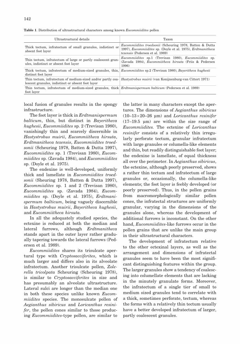
Ultrastructure. Ectexine stratifi cation is well-developed in all Eucommiidites and allied pol-len grains, whereas the presence of endexine is problematic in some of them. The distribution of ultrastructural characters among the consi-dered taxa is presented in Table 1. The tectum is thick, coarsely perforate in E. troedsonii as described in Scheuring (1978) and Batten and Dutta (1997) and Eucommiidites sp. (Doyle et al. 1975), thick, but imperforate in Bayeritheca hughesii and Erdtmanitheca texensis, as well as in dispersed Eucommiidites sp. 2 (Trevisan 1980). In contrast, in Hastystrobus muirii, Eucommiidites sp. 1 (Trevisan 1980), Eucom-miidites sp. (Zavada 1984), Erdtmanispermum balticum and Eucommiitheca hirsuta the tec-tum is relatively thin, no more than half of ectexine, imperforate.
The infratectum varies in thickness from a narrow slit-like layer to about half of ect-exine, consisting of small granules in Erdt-manitheca texensis, dispersed E. troedsonii (Scheuring 1978, Batten & Dutta 1997), and Eucommiidites sp. (Doyle et al. 1975), of mid-dle-sized granules in Hastystrobus muirii, Bayeritheca hughesii, Erdtmanispermum balticum and in dispersed Eucommiidites sp. 2 (Trevisan 1980), but of large partly coales-cent granules forming columella-like elements in ?Eucommiitheca hirsuta, Eucommiidites sp. 1 (Trevisan 1980) and Eucommiidites sp. (Zavada 1984). In Hastystrobus muirii,
142
local fusion of granules results in the spongy infrastructure.
The foot layer is thick in Erdtmanispermum balticum, thin, but distinct in Bayeritheca hughesii, Eucommiidites sp. 2 (Trevisan 1980), vanishingly thin and scarcely discernible in Hastystrobus muirii, Eucommiitheca hirsuta, Erdtmanitheca texensis, Eucommiidites troed-sonii (Scheuring 1978, Batten & Dutta 1997), Eucommiidites sp. 1 (Trevisan 1980), Eucom-miidites sp. (Zavada 1984), and Eucommiidites sp. (Doyle et al. 1975).
The endexine is well-developed, uniformly thick and lamellate in Eucommiidites troed-sonii (Sheuring 1978, Batten & Dutta 1997), Eucommiidites sp. 1 and 2 (Trevisan 1980), Eucommiidites sp. (Zavada 1984), Eucom-miidites sp. (Doyle et al. 1975), Erdtmani-spermum balticum, being vaguely discernible in Hastystrobus muirii, Bayeritheca hughesii, and Eucommiitheca hirsuta.
In all the adequately studied species, the ectexine is reduced at both the median and lateral furrows, although Erdtmanitheca stands apart in the outer layer rather gradu-ally tapering towards the lateral furrows (Ped-ersen et al. 1989).
Eucommiidites shares its trisulcate aper-tural type with Cryptosacciferites, which is much larger and differs also in its alveolate infratectum. Another trisulcate pollen, Zole-rella tricolpata Scheuring (Scheuring 1978), is similar to Cryptosacciferites in size and has presumably an alveolate ultrastructure. Lateral sulci are longer than the median one in both these species unlike known Eucom-miidites species. The monosulcate pollen of Aegianthus sibiricus and Loricanthus resini-fer, the pollen cones similar to those produc-ing Eucommiidites-type pollen, are similar to
the latter in many characters except the aper-tures. The dimensions of Aegianthus sibiricus (10–13 × 20–26 μm) and Loricanthus resinifer (17–19.5 μm) are within the size range of Eucommiidites. The ectexine of Loricanthus resinifer consists of a relatively thin irregu-larly perforate tectum, granular infratectum with large granules or columella-like elements and thin, but readily distinguishable foot layer; the endexine is lamellate, of equal thickness all over the perimeter. In Aegianthus sibiricus, the ectexine, although poorly preserved, shows a rather thin tectum and infratectum of large granules or, occasionally, the columella-like elements; the foot layer is feebly developed (or poorly preserved). Thus, in the pollen grains from macromorphologically similar pollen cones, the infratectal structures are uniformly granular, varying in the dimensions of the granules alone, whereas the development of additional furrows is inconstant. On the other hand, Eucommiidites-like furrows occur in the pollen grains that are unlike the main group in their ultrastructural characters.
The development of infratectum relative to the other ectexinal layers, as well as the arrangement and dimensions of infratectal granules seem to have been the most signifi -cant distinguishing features within the group. The larger granules show a tendency of coalesc-ing into columellate elements that are lacking in the minutely granulate forms. Moreover, the infratectum of a single tier of small to medium sized granules tend to correlate with a thick, sometimes perforate, tectum, whereas the forms with a relatively thin tectum usually have a better developed infratectum of larger, partly coalescent granules.
Table 1. Distribution of ultrastructural characters among known Eucommiidites pollen
Ultrastructural details Taxon
Thick tectum, infratectum of small granules, indistinct or absent foot layer
Eucommiidites troedsonii (Scheuring 1978, Batten & Dutta 1997), Eucommiidites sp. (Doyle et al. 1975), Erdtmanitheca texensis (Pedersen et al. 1989)
Thin tectum, infratectum of large or partly coalescent gran-ules, indistinct or absent foot layer
Eucommiidites sp.1 (Trevisan 1980), Eucommiidites sp. (Zavada 1984), Eucommiitheca hirsuta (Friis & Pedersen 1996)
Thick tectum, infratectum of medium-sized granules, thin, distinct foot layer
Eucommiidites sp.2 (Trevisan 1980), Bayeritheca hughesii
Thin tectum, infratectum of medium-sized and/or partly coa-lescent granules, indistinct or absent foot layer
Hastystrobus muirii (van Konijnenburg-van Cittert 1971)
Thin tectum, infratectum of medium-sized granules, thick foot layer
Erdtmanispermum balticum (Pedersen et al. 1989)

143
OUTGROUP COMPARISONS
In Hastystrobus muirii, the infratectum of the supposed proximal side has a spongy appearance, like in Monosulcites sp. (Trevisan 1980). Bayeritheca hughesii and Eucommi-idites sp. 2 (Trevisan 1980), the most closely related members of the group, share their ectexine characters (thick tectum, densely packed granules) with the monosulcate pollen of Leguminanthus siliquosus, a pollen organ of the Bennettitales (Ward et al. 1989).
Forms with infratectum of small granules, such as Eucommiidites troedsonii (Scheuring 1978; Batten & Dutta 1997), Erdtmanitheca texensis, and Eucommiidites sp. (Doyle et al. 1975), especially the latter, are comparable with Equisetosporites sp. (Osborn et al. 1993), Ephedripites sp. (Trevisan 1980), Cycadeoidea dacotensis (Osborn & Taylor 1995) and Letho-masites fossulatus (Ward et al. 1989), a ques-tionable angiosperm pollen showing certain bennettitalean characters (Crepet & Nixon 1994). Ephedripites sp. and Cycadeoidea daco-tensis are polyplicate, assigned to the Gne-tales, Lethomasites fossulatus is monosulcate, belonging to the Bennettitales, and the last one is very large (90 μm) foveolate, supposedly angiospermous.
The forms with large and partly coalescent granules (?Eucommiitheca hirsuta, Eucom-miidites sp. 1 (Trevisan 1980), Eucommiidites sp. (Zavada 1984), Aegianthus sibiricus, Lori-canthus resinifer are similar in this character to Sahnia laxiphora, an Early Cretaceous member of the Pentoxylales. (Osborn et al. 1993) and to Monosulcites sp. recovered from Jurassic of Afghanistan (Zavada 2004).
Among the rimulate pollen morphotypes of the Circumpollis group, commonly assigned to conifers (the Cheirolepidiaceae), but placed among gnetophytes by Krassilov (1982), Clas-sopollis (Pettitt & Chaloner 1964, Taylor & Alvin 1984, Rowley & Srivastava 1986, Pocock et al. 1990) and Circulina (Médus 1977), the infratectum is formed of large gran-ules and/or columellate elements, whereas in Duplicisporites it consists of profuse, closely packed small and medium-sized granules (Zavialova & Roghi 2005) as in Hastystrobus muirii, Bayeritheca hughesii (above) and Euco-mmiidites sp. 2 (Trevisan 1980).
Affi nities. The taxonomic affi nities of Eucom-miidites type pollen grains were discussed by
Friis and Pedersen (1996), who established order Erdtmanithecales based on several intact pollen organs and seeds with in situ Eucommiidites pollen. Our deduction has two sources of inference: (1) the intrinsic features of pollen grains themselves and (2) the mor-phology of macrofossils from which they were extracted. Hughes (1961) suggested a gnet-alean affi nity for Eucommiidites plant and was followed in this by the majority of subsequent researchers. The above comparisons included gnetalean, bennettitalean and pentoxylean forms as possibly related on account of similar infratectal structures.
In situ Eucommiidites-type pollen grains have been fi rst described in Hastystrobus muirii (van Konijnenburg-van Cittert 1971) from the Middle Jurassic of Yorkshire. Their attribution to Eucommiidites was sometimes doubted (Pedersen et al. 1989), but our SEM study of the type material fully confi rms the slit-like lateral furrows, typical of Eucommi-idites. Van Konijnenburg-van Cittert (1971) compared Hastystrobus to Androstrobus imply-ing a cycadalean affi nity because of the micro-sporangia covering the abaxial surface of the microsporophylls. However, unlike Hastystro-bus, pollen of extant and fossil cycads has an alveolar, rather than granular, infratectum.
Erdtmanitheca, a Cretaceous pollen organ similar to Aegianthus, was placed in the extinct family Erdtmanithecaceae of the order Erdtmanithecales (Pedersen et al. 1989, Friis & Pedersen 1996), including also Eucommi-itheca, Spermatites pattensis Hughes (1961), Spermatites patuxensis Brenner (1963), Alli-cospermum retemirum Harris (Reymanówna 1968), Erdtmanispermum balticum (Peder-sen et al. (1989). Another Cretaceous pollen cone, Bayeritheca J. Kvaček & Pacltová, was assigned to Erdtmanithecales primarily on account of Eucommiidites pollen and pollen cone morphology.
Possible relatives: Loricanthus resinifer (Krassilov & Bugdaeva 1999) belongs to the same pollen cone morphotype as members of Erdtmanithecales, but its pollen grains, though similar to Eucommiidites in shape, dimensions, sculpture and ultrastructure, appear mono- or disulcate.
Large pollen cones with hexagonal shields of peltate sporangiophores were described from the mid-Jurassic Ust’ Baley locality, East Siberia, as Kaidocarpum sibiricum Heer

144
(Heer 1876), re-assigned to Aegianthus sibiri-cus by Krassilov and Bugdaeva (1988). Pollen grains from these cones do not show additional furrows, at least not as a constant feature. The gnetalean affi nities of Aegianthus were inferred from its association with samaroid seed-scale structures resembling the samaras of extant Welwitschia (Krassilov & Bugdaeva 1988).
Eucommiidites pollen morphology was con-sidered as a major diagnostic feature of the Erdtmanithecales, the extinct order based on pollen cones and dispersed seeds of a rather generalized gymnospermous morphology. The Erdtmanithecales is placed close to the Gnet-ales (Friis & Pedersen 1996), three extant gen-era of which, Welwitschia, Gnetum and Ephe-dra, produce psilate polyplicate (Welwitschia, Ephedra) or spinulose (Gnetum) pollen of dif-ferent apertural types, monosulcate or inaper-turate, but comparable to Eucommiidites on account of granular infratectal ultrastucture, commonly of small granules (Ueno 1960, Gul-lvåg 1966, Bernard & Meyer 1972, Van Campo & Lugardon 1973, Kedves 1987, Kurmann 1992, El-Ghazaly & Rowley 1997, El-Ghazaly et al. 1998, Zavada & Gabaraeva 1991, Hesse et al. 2000, Yao et al. 2004, Gnetum lepto-stachyum Plume, Pl. 4, fi g. 4), but of large granules in G. africanum Welwitsch (Oryol et al. 1986, Pl. 4, fi g. 5) and extremely small ones in G. funiculare Wight (Pl. 4, fi g. 6).
CONCLUSIONS
Our ultrastructural study of Eucommiidites and allied pollen grains of Hastystrobus muirii, Bayeritheca hughesii, Aegianthus sibiricus, Loricanthus resinifer, Cryptosacciferites pabu-laris shows that:
(a) In all ultrastructurally studied Eucom-miidites, the median and additional furrows are cutting through all the layers of ectexine; the infratectum is granular or granular-colu-mella-like;
(b) At the same time, the pollen genus Eucommiidites shows high variability in terms of apertural confi gurations (subparallel sulci or a median sulcus and encircling furrow) and in such ultrastructural features as a thick or thin, perforate or imperforate tectum; a broad or constricted infratectum of large partly coa-lescent granules or of small to medium-sized
granules arranged in a single or two – three tiers; a well-developed or obscure foot-layer; as well as in the distinct or inconspicuous lamel-lation of endexine;
(c) The pollen cones of typical Ertdma-nithecaceae produced Eucommiidites-type pollen grains with subparallel sulci and with an infratectum of large or small tiered gran-ules. Pollen grains from the pollen chamber of Erdtmanispermum balticum differ from all the conventional Eucommiidites in the almost equally developed median and lateral sulci, massive exine, foveolate sculpture and a rela-tively thick foot layer.
(d) A trisulcate apertural type occurs in the pollen grains Cryptosacciferites and Zol-erella with an alveolate, rather than granular, infratectum, evidently representing a distinct group of Early Cretaceous gymnosperms.
(e) Hastystrobus from the Middle Jurassic of Yorkshire, not included in the Ertdmanithe-caceae, produced pollen of Eucommiidites type with converging lateral sulci and a granulate-columella-like infratectum;
(f) The pollen cones Aegianthus from the mid-Jurassic of Siberia and Loricanthus from the Early Cretaceous of Transbaikalia, are similar to Erdtmanitheca in general habit, peltate sporangiophores with polygonal apo-physes, and sporangial clusters, but their pollen grains are not assignable to, although ultrastructurally comparable with, Eucommi-idites. In this morphological group, the gnet-alean affi nities are so far substantiated for Aegianthus alone on account of its association with Welwitschia-like seed scales Heerala.
ACKNOWLEDGMENTS
We thank Prof. E. Turnau and Prof. E.M. Friis for valuable comments on the manuscript. We are grateful to Dr. J. F. Maxwell (Chiang Mai Univer-sity, Thailand) for providing the material of Gne-tum leptostachyum. The study was supported by the Russian Foundation for Basic Research, grants 06–04–48534 and 06–04–49577, and Sepkoski grant RG0-1337(2)-XX-18. J. Kvaček acknowledges a grant by the Ministry of Culture of the Czech Republic (MK 00002327201).
REFERENCES
BATTEN D.J. & DUTTA R.J. 1997.Ultrastructure of exine of gymnospermous pollen grains from Jurassic and basal Cretaceous deposits in North-

145
west Europe and implications for botanical rela-tionships. Rev. Palaeobot. Palynol., 99: 25–54.
BERNARD V.V. & MEYER N.R. 1972. Pyl’cevye zyorna Ephedra, Welwitschia i Gnetum (sum-mary: Pollen grains of Ephedra, Welwitschia and Gnetum). Vest. Moskov. Univers. Ser. 6. Biologiya, Pochvovedenie, 27: 86–88.
BRENNER G.J. 1963. The spores and pollen of the Potomac Group of Maryland. Maryl. Dep. Geol. Mines and Water Res., Bull., 27: 1–215.
BRENNER G.J. 1967. The gymnospermous affinity of Eucommiidites Erdtman, 1948. Rev. Palaeobot. Palynol., 5: 123–127.
van CAMPO M. & LUGARDON B. 1973.Structure grenue infratectale de l’ectexine des pollens de quelques gymnosperms et angiosperms. Pollen et Spores, 15: 171–187.
COUPER R.A. 1956. Evidence of a possible gymno-spermous affi nity for Tricolpites troedssonii Erdt-man. New phytologist, 55: 280–285.
CREPET W.L. & NIXON K.C. 1994. Flowers of Turo-nian Magnoliidae and their implications. In: Friis E.M. & Endress P.K. (eds) The early fossil record of angiosperm fl owers. Pl. Syst. Evol., Suppl. 8: 73–91.
DOYLE J.A., van CAMPO M. & LUGARDON B. 1975. Observations on exine structure of Eucommiidites and Lower Cretaceous angiosperm pollen. Pollen et Spores, 17: 429–486.
EL-GHAZALY G. & ROWLEY J.R. 1997. Pollen wall of Ephedra foliata. Palynology, 21: 7–18.
EL-GHAZALY G., ROWLEY J.R. & HESSE M. 1998. Polarity, aperture condition and germination in pollen grains of Ephedra (Gnetales). Plant Syst. Evol., 213: 217–231.
ERDTMAN G. 1948. Did dicotyledonous plants exist in Early Jurassic time? Förhandl. Geol. Fören. Stockholm, 70: 265–271.
FRIIS E.M. & PEDERSEN K.R. 1996. Eucommiitheca hirsuta, a new pollen organ with Eucommiidites pollen from the Early Cretaceous of Portugal. Grana, 35: 104–112.
GULLVÅG B. 1966. The fine structure of some gym-nosperm pollen walls. Grana Palynologica, 6: 435–475.
HEER O. 1876. Beiträge zur Jura-Flora Ostsibiriens und des Amurlandes. Acad. Imp. Sci. St. Péters-bourg Mém., 22 (12): 1–122.
HESSE M., WEBER M. & HALBRITTER, H. 2000. A comparative study of the polyplicate pollen types in Arales, Laurales, Zingiberales and Gnet-ales: 227–239. In: Harley M.M., Morton C.M. & Blackmore S. (eds) Pollen and spores: morphol-ogy and biology. Royal Botanic Gardens, Kew.
HUGHES N.F. 1961. Further interpretation of Euco-mmiidites Erdtman 1948. Palaeontology, 4 (2): 292–299.
KEDVES M. 1987. LM and EM studies on pollen
grains of recent Welwitschia mirabilis Hook. and Ephedra species. Acta Bot. Hung., 33: 81–103.
van KONIJNENBURG- van CITTERT J.H.A. 1971. In situ gymnosperm pollen from the Middle Jurassic of Yorkshire. Acta Bot. Neerl., 20 (1): 1–96.
van KONIJNENBURG-van CITTERT J.H.A. 1972. Some additional notes on male gymnosperm fruc-tifi cations from the Jurassic fl ora of Yorkshire. Acta Bot. Neerl., 21: 95–98.
KRASSILOV V.A. 1982. On the ovuliferous organ of Hirmerella. Phyta, Studies on Living & Fossil Plants, Plant Commemorate Volume: 141–144.
KRASSILOV V.A., BUGDAEVA E.V. 1988. Gnetalean plants from the Jurassic of Ust-Balej, east Sibe-ria. Rev. Palaeobot. Palynol., 53: 359–374.
KRASSILOV V.A., BUGDAEVA E.V. 1999. An angio-sperm cradle community and new proangiosperm taxa. Acta Palaeobot. Suppl., 2: 111–127.
KRASSILOV V.A., TEKLEVA M., MEYER-MELIKYAN N. & RASNITSYN A. 2003. New pollen morphotype from gut compression of a Cre-taceous insect, and its bearing on palynomorpho-logical evolution and palaeoecology. Cretaceous Research, 24: 149–156.
KURMANN M.H. 1992. Exine stratification in extant gymnosperms: a review of published transmission electron micrographs. Kew Bulletin, 47: 25–39.
KUYL O.S., MULLER J. & WATERBOLK H.T. 1955. The application of palynology to oil geology, with special reference to western Venezuela. Geologie en Mijnbouw, New Series 17 (3): 49–76.
KVAČEK J. & PACLTOVÁ B. 2001. Bayeritheca hughesii gen. et sp. nov., a new Eucommiidites-bearing pollen organ from the Cenomanian of Bohemia. Cretaceous Research, 22: 695–704.
MÉDUS J. 1977. The ultrastructure of some Circum-polles. Grana, 16: 23–28.
MEYER-MELIKIAN N.R., BOVINA I.YU., KOSENKO YA.V., POLEVOVA S.V., SEVEROVA E.E., TEKLEVA M.V. & TOKAREV P.I. 2004. Atlas pyl’cevykh zeren astrovykh (Asteraceae): pali-nomorfologiya i razvitie sporodermy. Moscow, Tovarischestvo nauchnyh izdaniy KMK: 1–236. (in Russian).
ORYOL L.I., KUPRIJANOVA L.A. & GOLUBEVA E.A. 1986. Ul’trastruktura acetolizoustoychivykh obolochek tapetal’nykh kletok i pyl’cevykh zeren u Gnetum africanum –Gnetaceae (summary: Ultrastructure of acetolysis-resistant wall of tap-etal cells and pollen grains in Gnetum africanum (Gnetaceae)). Bot. Zhur., 71: 750–754.
OSBORN J.M. 2000. Pollen morphology and ultrastructure of gymnospermous anthophytes: 163–185. In: Harley M.M., Morton C.M. & Black-more S. (eds) Pollen and Spores: morphology and biology. Royal Botanic Gardens, Kew.
OSBORN J.M., TAYLOR T.N. 1995. Pollen morphol-ogy and ultrastructure of the Bennettitales: in situ pollen of Cycadeoidea. Amer. J. Bot., 82 (8): 1074–1081.

146
OSBORN J.M., TAYLOR T.N. & DE LIMA M.R. 1993.The ultrastructure of fossil ephedroid pol-len with gnetalean affi nities from the Lower Cretaceous of Brazil. Rev. Palaeobot. Palynol., 77: 171–184.
PEDERSEN K.R., CRANE P.R. & FRIIS E.M. 1989. Pollen organs and seeds with Eucommiidites pol-len. Grana, 28: 279–294.
PETTITT J.M. & CHALONER W.G. 1964. The ultras-trusture of the Mesozoic pollen Classopollis. Pol-len et Spores, 6: 611–620.
POCOCK S.A.J., VASANTHY G. & VENKATACHALA B.S. 1990. Pollen of Circumpolles – an enigma or morphotrends showing evolutionary adaptation. Rev. Palaeobot. Palynol., 63: 179–193.
REYMANÓWNA M. 1968. On seeds containing Euco-mmiidites troedssonii pollen from the Jurassic of Grójec, Poland. Bot. J. Linn. Soc., 61: 147–152.
ROWLEY J.R. & SRIVASTAVA S.K. 1986. Fine structure of Classopollis exine. Can. J. Bot., 64: 3059–3074.
SCHEURING B.W. 1978. Mikrofloren aus den Meridekalken des Mts. San Giorgio (Kanton Terrsin). Schweizerische Paläontologische Abhan-dlungen, 88: 1–119.
TAYLOR T.N. & ALVIN K.L. 1984. Ultrastructure and development of Mesozoic pollen: Classopollis. Amer. J. Bot., 71: 575–587.
TEKLEVA M.V. & KRASSILOV V.A. 2004. Sporo-derm ultrastructure in Early Cretaceous proan-giosperms. Paleont. Jour., 38 (1): 97–102.
TREVISAN L. 1980. Ultrastructural notes and con-siderations of Ephedripites, Eucommiidites and Monosulcites pollen grains from Lower Creta-ceous sediments of southern Tuscany (Italy). Pol-len et Spores, 22: 85–132.
UENO J. 1960. On the fine structure of the cell walls of some gymnosperm pollen. Biological Journal of Nara Women’s University, 10: 19–25.
WARD J.V., DOYLE J.A. & HOTTON C.L. 1989. Probable granular magnoliid angiosperm pollen from the Early Cretaceous. Pollen et Spores, 31: 113–132.
YAO Y.-F., XI Y.-Z., GENG B.-Y. & LI C.-S. 2004. The exine ultrastructure of pollen grains in Gnetum (Gnetaceae) from China and its bearing on the relationship with the ANITA Group. Bot. J. Linn. Soc., 146: 415–425.
ZAVADA M.S. 1984. Angiosperm origins and evolu-tion based on dispersed fossil gymnosperm pol-len ultrastructure. Ann. Miss. Bot. Gard., 71: 444–463.
ZAVADA M.S. 2004. The ultrastructure of Upper Pal-aeozoic and Mesozoic pollen from southern Africa and Asia. Palaeont. Afr., 40: 59–68.
ZAVADA M.S. & GABARAEVA N.I. 1991. Com-parative pollen wall development of Welwitschia mirabilis and selected primitive angiosperms. Bull.Torr. Bot. Club, 118: 292–302.
ZAVIALOVA N.E. & ROGHI G. 2005. Exine morphol-ogy and ultrastructure of Duplicisporites from the Triassic of Italy. Grana, 44: 337–342.
P L A T E S

148
Plate 1
Hastystrobus muirii van Konijnenburg-van Cittert, SEM, TEM
1. Group of pollen grains, SEM. Scale bar = 10 μm2. Single pollen grain, distal view, SEM. Scale bar = 10 μm3. TEM image of the whole pollen grain, arrows indicate three apertures. Scale bar = 1 μm4. TEM image of pollen wall, proximal side. Scale bar = 1 μm5. TEM image of pollen wall, towards apertural region. Scale bar = 0.5 μm6. TEM image of pollen wall, apertural side, arrow indicates foot layer. Scale bar = 0.25 μm7. TEM image of pollen wall, proximal side. Scale bar = 0.5 μm8. TEM image of pollen wall, apertural side. Scale bar = 1 μm

149
M.V. Tekleva et al.Acta Palaeobot. 46(2)
Plate 1

150
Plate 2
Bayeritheca hughesii J.Kvaček & Pacltová, SEM, TEM
1. Single pollen grain, SEM. Scale bar = 1 μm 2. TEM image of pollen wall, apertural region. Arrow heads show inner border of foot layer. Scale bar = 0.5 μm. 3. TEM image of the whole pollen grain, arrows indicate three apertures. Scale bar = 0.5 μm 4–5. TEM image of pollen wall, non-apertural region. Arrows show inner border of foot layer. Scale bar = 0.5 μm

151
M.V. Tekleva et al.Acta Palaeobot. 46(2)
Plate 2

152
Plate 3
Aegianthus sibiricus (Heer) Krassilov, Loricanthus resinifer Krassilov & Bugdaeva, SEM, TEM
1–5. Aegianthus sibiricus (Heer) Krassilov1. Single pollen grain, SEM. Scale bar = 6 μm2. TEM image of the whole pollen grain, arrow indicates apertural region. Scale bar = 2 μm3. TEM image of pollen wall; arrow head indicates apertural region, arrow points at a layer, which probably
represents endexine. Scale bar = 0.5 μm4. TEM image of pollen wall, non-apertural region. Scale bar = 2 μm5. TEM image of pollen wall, non-apertural region, infratectum consists of large granules. Scale bar = 0.5
μm6–7. Loricanthus resinifer Krassilov & Bugdaeva6. Group of pollen grains, SEM. Scale bar = 10 μm7. TEM image of pollen wall, non-apertural region, arrow indicates foot layer. Scale bar = 0.5 μm

153
M.V. Tekleva et al.Acta Palaeobot. 46(2)
Plate 3

154
Plate 4
Cryptosacciferites pabularis Krassilov & Tekleva, SEM, TEM, Gnetum leptostachyum Plume, Gnetum africanum Welwitsch, Gnetum funiculare Wight, TEM.
1–3. Cryptosacciferites pabularis Krassilov & Tekleva1. Single pollen grain, SEM. Scale bar = 10 μm2. TEM image of pollen wall, marginal fl ange, where infratectum is expanded. Scale bar = 1 μm3. TEM image of pollen wall, apertural region; arrow indicates foot layer. Scale bar = 1.84 μm4. Gnetum leptostachyum Plume, TEM image of pollen wall, plicate region; arrow points at infratectal gran-
ules. Scale bar = 0.5 μm5. Gnetum africanum Welwitsch, TEM image of pollen wall, plicate region; arrow points at infratectal gran-
ule. Scale bar = 0.5 μm6. Gnetum funiculare Wight, TEM image of pollen wall, plicate region; arrow points at infratectal granules.
Scale bar = 0.4 μm

155
M.V. Tekleva et al.Acta Palaeobot. 46(2)
Plate 4
Copyright © 2022 FDOKUMEN