Predation resistance and nematocyst scaling for Metridium senile and M. farcimen
Upload
independentCategory
view
0download
0
Fax +41 61 306 12 34E-Mail [email protected]
Skin Pharmacol Physiol 2006;19:95–100 DOI: 10.1159/000091976
Skin Ultrastructure in Senile Lentigo
E. Noblesse
a C. Nizard
a M. Cario-André
c S. Lepreux
d C. Pain
c S. Schnebert
a A. Taïeb
b, c R. Kurfurst
a
a LVMH Recherche, Branche Parfums et Cosmétiques, Saint Jean de Braye , b
Department of Dermatology, Hôpital Saint André, University Hospitals of Bordeaux, c
Inserm E 217, Université Victor Segalen, and d
Department of Pathology, Hôpital Pellegrin, University Hospitals of Bordeaux, Bordeaux , France
which could refl ect intense protein synthesis. In basal keratinocytes from lesional skin, we observed numer-ous melanosome complexes called polymelanosomes, which formed massive caps on the nuclei. Observations in colored semi-thin sections also revealed perturbed structures in the basal layer region, which could explain the skin perturbation. Indeed, we observed keratinocytes that presented important microinvaginations and pen-dulum melanocytes, which sank into the dermis, be-neath the basal layer of keratinocytes. These cell modi-fi cations seemed to be due to a perturbation of the dermal-epidermal junction, which appeared disorga-nized and disrupted and could directly disturb the basal support of the cells.
Copyright © 2006 S. Karger AG, Basel
Introduction
Senile lentigo (SL; lentigo senilis, aging spot, liver spot, solar lentigine) is a common component of photoaged skin. It is characterized by hyperpigmented macules, which affect mostly face (forehead and cheeks), scalp (in bald patients), forearms, and the back of the hands after the age of 50. Hodgson [1] showed in a comprehensive study among residents of a home for the aged that the number of spots is clearly related to age and associated features of skin aging. Macules vary in color from yellow-light brown to black, the medium hue being dark brown, depending probably on the underlying skin phototype.
Key Words Senile lentigo � Pigmentation � Dermal-epidermal junction
Abstract Senile lentigo is a common component of photoaged skin. It is characterized by hyperpigmented macules which affect chronically irradiated skin mostly after the age of 50. This study was undertaken to assess the mor-phology of senile lentigo on the dorsum of the hands. A systematic comparison between lesional and perilesion-al skin using histology and transmission electron micros-copy was done to determine whether melanocytes or keratinocytes are affected in the evolution of lesions and which tissue structure is modifi ed. The histology study showed that lesional skin is characterized by a hyperpig-mented basal layer and an elongation of the rete ridges, which seem to drive deeply into the dermis. The epider-mis contained clusters of keratinocytes, which retained and accumulated the melanin pigment. Electron micros-copy studies showed important modifi cations in the le-sional skin ultrastructure in comparison with perilesion-al skin. In melanocytes from perilesional and lesional skin, we observed normal size melanosomes at all stag-es of maturation in the cytoplasm and in migration with-in dendrites. No pigment accumulation was observed. However, the morphology of melanocytes in lesional skin revealed an activated status with numerous mito-chondria and a well-developed endoplasmic reticulum,
Received: September 6, 2005 Accepted after revision: February 3, 2006 Published online: May 9, 2006
E. Noblesse, MDLVMH RechercheBranche Parfums et CosmétiquesFR–45804 Saint Jean de Braye cedex (France)Tel. +33 2 38 60 37 86, Fax +33 2 38 60 31 76, E-Mail [email protected]
© 2006 S. Karger AG, Basel1660–5527/06/0192–0095$23.50/0
Accessible online at:www.karger.com/spp
Dow
nloa
ded
by:
INS
ER
M D
ISC
IST
193.
54.1
10.3
3 -
2/6/
2015
10:
49:2
4 A
M

Noblesse /Nizard /Cario-André /Lepreux /Pain /Schnebert /Taïeb /Kurfurst
Skin Pharmacol Physiol 2006;19:95–100 96
Histopathology has shown that lesional skin is charac-terized by a hyperpigmented basal layer and an elongation of the rete ridges, with an increased number of epidermal melanocytes in the epidermal basal layer [1–6] . Only 50% of the lesions labeled ‘solar lentigo’ on the face have these typical features, posing the problem of the exact nature of these lesions [3] . Limited ultrastructural data are avail-able [2, 6] . An unusual accumulation of melanosomes in keratinocytes was fi rst noted by Braun-Falco and Schoefi n-ius [2] , and Nakagawa et al. [7] have insisted on the pres-ence of ‘metabolically active’ melanocytes mixed with ‘inactive’ melanocytes without cytological atypia.
Because of the overall poor understanding of the patho-physiology of SL, and of some uncertainties in diagnostic criteria, we chose to fi rst make a basic morphology study in a typical location of lesions (dorsum of the hands), and to systematically compare lesional vs. perilesional skin biopsies using immunohistochemistry and electron mi-croscopy.
Materials and Methods
Patients The study was approved by the University of Bordeaux Hospi-
tal’s internal review board. Twelve patients recruited in the out- and in-patient clinics volunteered to participate, after giving in-formed consent. Seven females and 5 males aged 58–92 years were included in the study.
Biopsy Procedure and Sample Preparation After local anesthesia with lidocaine, a 4-mm punch biopsy was
done at the center of a typical lesion on the dorsum of one hand and at a control site situated immediately at the border from the lesion in clinically nonpigmented skin. Half of the biopsy was fi xed in 4% formaldehyde and the rest was fi xed in a 6.25% glutaralde-hyde, 0.1 M cacodylate (pH 7.4) solution.
Histopathologic Studies Biopsies were embedded in paraffi n, cut in 5- � m sections, and
stained using the hematoxylin-eosin and Fontana-Masson tech-niques to assess the general morphology of the epidermis and mel-anin distribution. An anti-melan-A antibody (Dako, Trappes, France) was used to stain melanocytes. As secondary antibody, we used the envision kit HRP+ (Dako, Trappes, France) which was revealed with amino-ethylcarbamide. Amino-ethylcarbamide was chosen because red staining facilitates the count of melanocytes in the pigmented basal layer.
Melanocyte Counts and Statistical Analyses Red-stained cells were counted over the complete length of the
section stained with anti-melan-A and reported to the correspond-ing length of stratum corneum (four sections examined for each lesion). Statistical differences were evaluated using the paired Stu-dent’s t test. Data are presented as the mean of 12 biopsies.
Electron Microscopy Biopsies were embedded in Epon and cut in 60-nm sections.
Sections were examined after postfi xation in osmium tetroxyde with a CM 10 (Philips, FEI Compagny, Eindhoven, The Nether-lands) electron microscope.
Results
Epidermal Morphology of Lesional vs. Perilesional Skin Conventional Microscopy By conventional histological stainings such as hema-
toxylin-eosin and Fontana Masson, we observed impor-tant histological differences between the lesional zone and the perilesional zone ( fi g. 1 ). Lesional skin is charac-terized by a hyperpigmented basal layer and an elonga-tion of epidermis rete ridges which seem to drive deep-ly into the dermis. By image analysis, we showed that the full epidermis thickness was increased (mean: +55 � m) and that the rete ridges were elongated (mean: +23.35 � m) in lesional skin in comparison with perile-sional skin ( table 1 ). Although we did not examine a large number of samples, this phenomenon increased with the presumed age of the lesion, determined by the age of the patient, and SL was classifi ed histopathologically in three grades of severity: stage 1 ( fi g. 1 d), stage 2 ( fi g. 1 e) and stage 3 ( fi g. 1 f), corresponding to a progressive accumula-tion of pigment at the tip of epidermal rete ridges which increase in length. Melanin pigment is stored at a very high level in the basal and suprabasal layers of the epider-mis. When comparing lesional skin with perilesional skin, a signifi cant increase was detected in the number of me-lanocytes per mm of the stratum corneum with anti-melan-A antibody (8.51 8 1.33 for perilesional skin and 18.19 8 2.12 for lesional skin; fi g. 2 ). Melanocytes did not accumulate in the ridges, a fi nding which was con-fi rmed by electron microscopy.
Electron Microscopy In melanocytes from perilesional and lesional skin, we
observed melanosomes in the cytoplasm at normal size (mean: 0.037 � m 2 8 0.015) and at all stages of matura-tion ( fi g. 3 c, d). In lesional skin, many melanocytes showed an activated phenotype, with numerous mitochondria in the cytoplasm, a very developed endoplasmic reticulum and melanosomes in migration within dendrites. In basal keratinocytes from lesional skin, we observed massive pigmented caps on the nuclei formed by melanosomial complexes called polymelanosomes ( fi g. 3 e). Some of them had an unusual size (mean: 0.13 8 0.006 � m 2 ) and
Dow
nloa
ded
by:
INS
ER
M D
ISC
IST
193.
54.1
10.3
3 -
2/6/
2015
10:
49:2
4 A
M

Skin Ultrastructure in Senile Lentigo Skin Pharmacol Physiol 2006;19:95–100 97
Fig. 1. Histological evolution of SL. a , b Hematoxylin-eosin staining. c–f Fontana Masson staining. g Semi-thin section stain-ing. h Anti-melan-A staining. b , d–h Le-sional skin with clusters of keratinocytes retaining pigment capping the nucleus ( d–g ) and in melanocytes ( h ). Lesional skin, stage 1 ( d ), stage 2 ( e ) and stage 3 ( f ), cor-responding to a progressive accumulation of pigment at the tip of epidermal rete ridg-es of lesser thickness and increased length.
Table 1. Measurement of epidermis thickness and rete ridge elongation (image analysis on hematoxylin-eosin staining)
Patient Lesional skin, �m Nonlesional skin, �m Increase in epidermisthickness in lesionalskin, �m
Increase in reteridge length inlesional skin, �mepidermis
thicknessrete ridgelength
epidermisthickness
rete ridgelength
1 92.23 41.96 47.36 4.23 94.76824.62 37.73815.562 74.18 23.35 56.88 11.42 30.4180.02 11.9386.773 57.43 25.13 47.76 6.35 20.2489.18 18.77811.224 74.51 31.98 53.04 5.80 40.4980.27 26.1783.095 103.61 65.03 39.87 2.62 159.8686.17 62.484.096 54.39 10.78 46.44 6.13 17.1381.75 4.6580.707 101.57 28.04 64.79 10.32 56.76819.99 17.71812.808 48.63 14.07 39.63 6.59 22.7186.46 7.4887.23
Mean +55.29 +23.35
Dow
nloa
ded
by:
INS
ER
M D
ISC
IST
193.
54.1
10.3
3 -
2/6/
2015
10:
49:2
4 A
M

Noblesse /Nizard /Cario-André /Lepreux /Pain /Schnebert /Taïeb /Kurfurst
Skin Pharmacol Physiol 2006;19:95–100 98
seemed to be composed by the association of 2 or 4 me-lanosomes ( fi g. 3 e, enlargement).
Dermal-Epidermal Junction Morphology of Lesional vs. Perilesional Skin Observations of semi-thin sections revealed important
modifi cations in the structure of the basal layer of the epidermis ( fi g. 3 a, b). Cells from lesional skin, and espe-cially keratinocytes, presented several microinvagina-tions in the basal pole of the cell, which is directly in con-tact with the dermal-epidermal junction ( fi g. 3 a, b; 4 a). This observation is particularly important at the level of the epidermis ridges.
In lesional skin, the majority of melanocytes present in the basal layer of the epidermis are ‘hanging down’ in the dermis and are called ‘pendulum melanocytes’ ( fi g. 3 b). In perilesional skin, this is only observed occa-sionally.
0
5
10
15
20
25
Perilesional skin Lesional skin
Mel
ano
cyte
s/m
m s
trat
um
co
rneu
m
Fig. 2. Melanocyte count after detection by anti-melan-A. A sig-nifi cant increase was detected in the number of melanocytes per mm of the stratum corneum in lesional skin in comparison with perilesional skin.
Fig. 3. Observation of lesional skin by optical and transmission electron microscopy. a , b Semi-thin sections of lesional epidermis: enhancement of basal keratinocytes microinvagination and presence of pendulum melanocytes which sink into the dermis ( b ). Melanocytes ( c , d ) and keratinocytes ( e ) in lesional basal layer. c , d In melano-cytes, normal-size melanosomes are present in the cytoplasm and within dendrites and numerous mitochondria (arrowheads on c ) can be observed. e In keratinocytes, melanosomial complexes form a massive cap on the nuclei. M = Melanocyte; k = keratinocyte; d = dendrite.
Dow
nloa
ded
by:
INS
ER
M D
ISC
IST
193.
54.1
10.3
3 -
2/6/
2015
10:
49:2
4 A
M

Skin Ultrastructure in Senile Lentigo Skin Pharmacol Physiol 2006;19:95–100 99
When we observed the dermal-epidermal junction ul-trastructure under these basal cells and particularly under melanocytes, we observed that in contrast to the normal appearance of the basement membrane in perilesional skin, the basement membrane in lesional skin was se-verely abnormal. The lesional dermal-epidermal junction appeared disorganized and disrupted. We also observed in lesional skin that, in comparison with perilesional skin, the lamina densa became thinner or presented abnormal deposition of basement membrane components, of which the most important molecule is type IV collagen.
Discussion
In a systematic comparison of lesional and perilesion-al histopathologic changes, we confi rmed the typical fea-tures of SL already noted by others [1, 4, 5] . Our investi-gation confi rms the opinion of Montagna et al. [6] that ‘the complex and distinctive architecture of SL is prob-ably the result of concurrent proliferation of melanocytes and keratinocytes’, but is also associated with a weaken-ing of the dermal-epidermal junction ultrastructure. This could be due to disorganization in the lamina densa com-ponents such as type IV collagen. Indeed, we counted a greater number of melanocytes in lesional skin, but the
Fig. 4. Ultrastructural observations of dermal-epidermal junction. a Basal keratinocyte with polymelano-somes and microinvaginations. b , c Pendulum melanocytes with a very developed endoplasmic reticulum ( b ) and perturbed dermal-epidermal junction ( c ). d Perilesional dermal-epidermal junction. e Lesional dermal-epidermal junction with disruption (arrowheads: dermal-epidermal junction). M = Melanocyte;k = keratinocyte.
Dow
nloa
ded
by:
INS
ER
M D
ISC
IST
193.
54.1
10.3
3 -
2/6/
2015
10:
49:2
4 A
M
Noblesse /Nizard /Cario-André /Lepreux /Pain /Schnebert /Taïeb /Kurfurst
Skin Pharmacol Physiol 2006;19:95–100 100
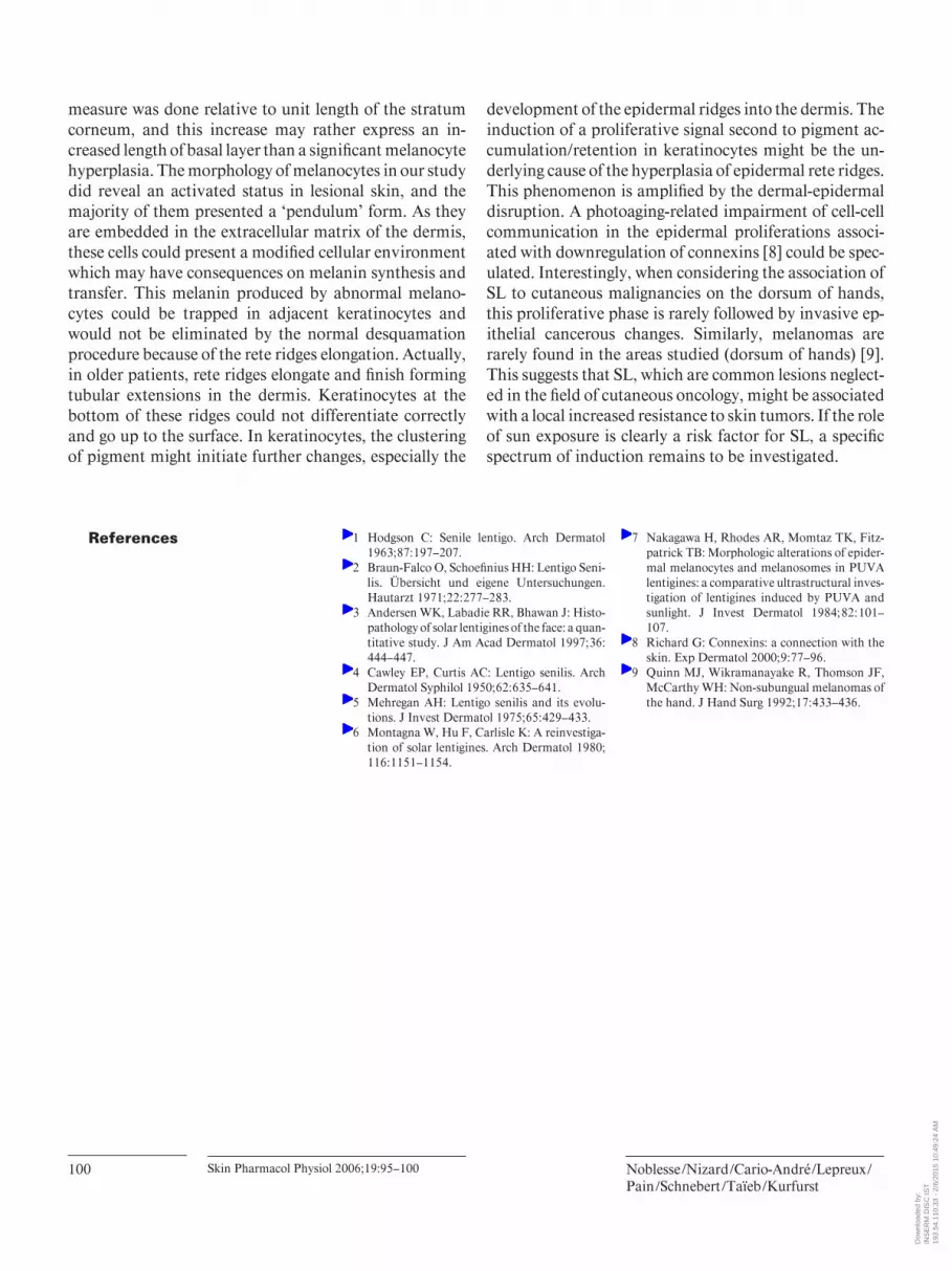
References 1 Hodgson C: Senile lentigo. Arch Dermatol 1963; 87: 197–207.
2 Braun-Falco O, Schoefi nius HH: Lentigo Seni-lis. Übersicht und eigene Untersuchungen. Hautarzt 1971; 22: 277–283.
3 Andersen WK, Labadie RR, Bhawan J: Histo-pathology of solar lentigines of the face: a quan-titative study. J Am Acad Dermatol 1997; 36:
444–447. 4 Cawley EP, Curtis AC: Lentigo senilis. Arch
Dermatol Syphilol 1950; 62: 635–641. 5 Mehregan AH: Lentigo senilis and its evolu-
tions. J Invest Dermatol 1975; 65: 429–433. 6 Montagna W, Hu F, Carlisle K: A reinvestiga-
tion of solar lentigines. Arch Dermatol 1980;
116: 1151–1154.
7 Nakagawa H, Rhodes AR, Momtaz TK, Fitz-patrick TB: Morphologic alterations of epider-mal melanocytes and melanosomes in PUVA lentigines: a comparative ultrastructural inves-tigation of lentigines induced by PUVA and sunlight. J Invest Dermatol 1984; 82: 101–107.
8 Richard G: Connexins: a connection with the skin. Exp Dermatol 2000; 9: 77–96.
9 Quinn MJ, Wikramanayake R, Thomson JF, McCarthy WH: Non-subungual melanomas of the hand. J Hand Surg 1992; 17: 433–436.
measure was done relative to unit length of the stratum corneum, and this increase may rather express an in-creased length of basal layer than a signifi cant melanocyte hyperplasia. The morphology of melanocytes in our study did reveal an activated status in lesional skin, and the majority of them presented a ‘pendulum’ form. As they are embedded in the extracellular matrix of the dermis, these cells could present a modifi ed cellular environment which may have consequences on melanin synthesis and transfer. This melanin produced by abnormal melano-cytes could be trapped in adjacent keratinocytes and would not be eliminated by the normal desquamation procedure because of the rete ridges elongation. Actually, in older patients, rete ridges elongate and fi nish forming tubular extensions in the dermis. Keratinocytes at the bottom of these ridges could not differentiate correctly and go up to the surface. In keratinocytes, the clustering of pigment might initiate further changes, especially the
development of the epidermal ridges into the dermis. The induction of a proliferative signal second to pigment ac-cumulation/retention in keratinocytes might be the un-derlying cause of the hyperplasia of epidermal rete ridges. This phenomenon is amplifi ed by the dermal-epidermal disruption. A photoaging-related impairment of cell-cell communication in the epidermal proliferations associ-ated with downregulation of connexins [8] could be spec-ulated. Interestingly, when considering the association of SL to cutaneous malignancies on the dorsum of hands, this proliferative phase is rarely followed by invasive ep-ithelial cancerous changes. Similarly, melanomas are rarely found in the areas studied (dorsum of hands) [9] . This suggests that SL, which are common lesions neglect-ed in the fi eld of cutaneous oncology, might be associated with a local increased resistance to skin tumors. If the role of sun exposure is clearly a risk factor for SL, a specifi c spectrum of induction remains to be investigated.
Dow
nloa
ded
by:
INS
ER
M D
ISC
IST
193.
54.1
10.3
3 -
2/6/
2015
10:
49:2
4 A
M
Copyright © 2022 FDOKUMEN