A recombinant thioredoxin-glutathione reductase from Fasciola hepatica induces a protective response...
8
A recombinant thioredoxin-glutathione reductase from Fasciola hepatica induces a protective response in rabbits Gabriela Maggioli a,b , Fernando Silveira b , José M. Martín-Alonso a , Gustavo Salinas c , Carlos Carmona b , Francisco Parra a,⇑ a Departamento de Bioquímica y Biología Molecular, Instituto Universitario de Biotecnología de Asturias, Universidad de Oviedo, 33006 Oviedo, Spain b Unidad de Biología Parasitaria, Facultad de Ciencias, Instituto de Higiene, Av. A. Navarro 3051, CP 11600 Montevideo, Uruguay c Cátedra de Inmunología, Facultad de Química, Instituto de Higiene, Av. A. Navarro 3051, CP 11600 Montevideo, Uruguay article info Article history: Received 13 October 2010 Received in revised form 20 September 2011 Accepted 22 September 2011 Available online 1 October 2011 Keywords: Fasciola hepatica Thioredoxin Glutathione Glutaredoxin Antioxidants Selenocysteine abstract Antioxidant systems are fundamental components of host–parasite interactions, and often play a key role in parasite survival. Here, we report the cloning, heterologous expression, and characterization of a thi- oredoxin glutathione reductase (TGR) from Fasciola hepatica. The deduced polypeptide sequence of the cloned open reading frame (ORF) confirmed the experimental N-terminus previously determined for a native F. hepatica TGR showing thioredoxin reductase (TR) activity. The sequence revealed the presence of a fusion between a glutaredoxin (Grx) and a TR domain, similar to that previously reported in Schisto- soma mansoni and Echinococcus granulosus. The F. hepatica TGR sequence included an additional redox active center (ACUG; U being selenocysteine) located at the C-terminus. The addition of a recombinant selenocysteine insertion sequence (SECIS) element in the Escherichia coli expression vector, or the substi- tution of the native selenocysteine by a cysteine, indicated the relevance of this unusual amino acid res- idue for the activity of F. hepatica TGR. Rabbit vaccination with recombinant F. hepatica TGR reduced the worm burden by 96.7% following experimental infection, further supporting the relevance of TGR as a promising target for anti Fasciola treatments. Ó 2011 Elsevier Inc. All rights reserved. 1. Introduction A crucial survival mechanism for parasites inside their host is the evasion of the mammalian immune response. Among immune-effector mechanisms, reactive oxygen and nitrogen inter- mediates produced by inflammatory cells have been associated with toxic effects in metazoan parasites (McGonigle et al., 1998; Selkirk et al., 1998). Accordingly, parasite antioxidant proteins are considered to be first-line actors in host–parasite interactions that tilt the balance towards the parasite (Bogdan et al., 2000; Callahan et al., 1988). In this context, parasite reduced glutathione (GSH) and thioredoxin (Trx) systems are considered to be major thiol-dependent redox pathways involved in the control of cellular redox balance. Both systems act by transferring the reducing equivalents of NADPH to different substrates and substrate reduc- tases, through reversible thiol oxidoreduction (Holmgren, 1989). The Trx system consists of the small protein Trx and thioredoxin reductase (TR). Trx is a powerful thiol-disulfide oxidoreductase, which reacts with numerous cellular proteins, providing electrons to enzymes such as thioredoxin peroxidase and ribonucleotide reductase (Nordberg and Arner, 2001). It also participates in the control of vital cellular processes, such as gene expression and sig- nal transduction, through the redox regulation of kinases, phos- phatases, and transcription factors (Arner and Holmgren, 2000). Trx is maintained in the reduced state by NADPH equivalents transferred by the flavoprotein TR, a member of the pyridine- nucleotide disulfide oxidoreductase family. The other major thiol system, the GSH pathway, comprises glu- tathione reductase (GR), GSH, and glutaredoxin (Grx). In this sys- tem, GR transfers reducing equivalents from NADPH to oxidized GSH (GSSG). GSH in turns acts by recycling Grx and glutathione peroxidase to their reduced state. Grx, a thiol-disulfide oxidoreduc- tase member of the Trx family, contains a glutathione binding site, and in turn transfers reducing equivalents to different substrates and substrate reductases. Recently, a new member of the thioredoxin reductase (TR) family, named thioredoxin glutathione reductase (TGR), has been 0014-4894/$ - see front matter Ó 2011 Elsevier Inc. All rights reserved. doi:10.1016/j.exppara.2011.09.013 Abbreviations: DTNB, 5,5 0 -dithiobis(2-nitrobenzoic acid); EST, expressed sequence tag; FAD, flavin adenine dinucleotide; GR, glutathione reductase; Grx, glutaredoxin; GSH, reduced glutathione; GSSG, oxidized glutathione; HED, b-hydroxyethyl disulfide; IPTG, isopropyl b-D-thiogalactopyranoside; ORF, open reading frame; PBS, phosphate- buffered saline; SECIS, selenocysteine insertion sequence; TGR, thioredoxin glutathione reductase; TR, thioredoxin reductase; Trx, thioredoxin. ⇑ Corresponding author. Address: Edificio Santiago Gascón Campus El Cristo, Universidad de Oviedo, 33006 Oviedo, Spain. Fax: +34 985103157. E-mail address: [email protected] (F. Parra). Experimental Parasitology 129 (2011) 323–330 Contents lists available at SciVerse ScienceDirect Experimental Parasitology journal homepage: www.elsevier.com/locate/yexpr
-
Upload
independent -
Category
Documents
-
view
0 -
download
0
Transcript of A recombinant thioredoxin-glutathione reductase from Fasciola hepatica induces a protective response...

Experimental Parasitology 129 (2011) 323–330
Contents lists available at SciVerse ScienceDirect
Experimental Parasitology
journal homepage: www.elsevier .com/locate /yexpr
A recombinant thioredoxin-glutathione reductase from Fasciola hepatica inducesa protective response in rabbits
Gabriela Maggioli a,b, Fernando Silveira b, José M. Martín-Alonso a, Gustavo Salinas c, Carlos Carmona b,Francisco Parra a,⇑a Departamento de Bioquímica y Biología Molecular, Instituto Universitario de Biotecnología de Asturias, Universidad de Oviedo, 33006 Oviedo, Spainb Unidad de Biología Parasitaria, Facultad de Ciencias, Instituto de Higiene, Av. A. Navarro 3051, CP 11600 Montevideo, Uruguayc Cátedra de Inmunología, Facultad de Química, Instituto de Higiene, Av. A. Navarro 3051, CP 11600 Montevideo, Uruguay
a r t i c l e i n f o
Article history:Received 13 October 2010Received in revised form 20 September2011Accepted 22 September 2011Available online 1 October 2011
Keywords:Fasciola hepaticaThioredoxinGlutathioneGlutaredoxinAntioxidantsSelenocysteine
0014-4894/$ - see front matter � 2011 Elsevier Inc. Adoi:10.1016/j.exppara.2011.09.013
Abbreviations: DTNB, 5,50-dithiobis(2-nitrobenzoictag; FAD, flavin adenine dinucleotide; GR, glutathioneGSH, reduced glutathione; GSSG, oxidized glutathione; HIPTG, isopropyl b-D-thiogalactopyranoside; ORF, open rebuffered saline; SECIS, selenocysteine insertion sequencereductase; TR, thioredoxin reductase; Trx, thioredoxin.⇑ Corresponding author. Address: Edificio Santiag
Universidad de Oviedo, 33006 Oviedo, Spain. Fax: +34E-mail address: [email protected] (F. Parra).
a b s t r a c t
Antioxidant systems are fundamental components of host–parasite interactions, and often play a key rolein parasite survival. Here, we report the cloning, heterologous expression, and characterization of a thi-oredoxin glutathione reductase (TGR) from Fasciola hepatica. The deduced polypeptide sequence of thecloned open reading frame (ORF) confirmed the experimental N-terminus previously determined for anative F. hepatica TGR showing thioredoxin reductase (TR) activity. The sequence revealed the presenceof a fusion between a glutaredoxin (Grx) and a TR domain, similar to that previously reported in Schisto-soma mansoni and Echinococcus granulosus. The F. hepatica TGR sequence included an additional redoxactive center (ACUG; U being selenocysteine) located at the C-terminus. The addition of a recombinantselenocysteine insertion sequence (SECIS) element in the Escherichia coli expression vector, or the substi-tution of the native selenocysteine by a cysteine, indicated the relevance of this unusual amino acid res-idue for the activity of F. hepatica TGR. Rabbit vaccination with recombinant F. hepatica TGR reduced theworm burden by 96.7% following experimental infection, further supporting the relevance of TGR as apromising target for anti Fasciola treatments.
� 2011 Elsevier Inc. All rights reserved.
1. Introduction equivalents of NADPH to different substrates and substrate reduc-
A crucial survival mechanism for parasites inside their host isthe evasion of the mammalian immune response. Amongimmune-effector mechanisms, reactive oxygen and nitrogen inter-mediates produced by inflammatory cells have been associatedwith toxic effects in metazoan parasites (McGonigle et al., 1998;Selkirk et al., 1998). Accordingly, parasite antioxidant proteinsare considered to be first-line actors in host–parasite interactionsthat tilt the balance towards the parasite (Bogdan et al., 2000;Callahan et al., 1988). In this context, parasite reduced glutathione(GSH) and thioredoxin (Trx) systems are considered to be majorthiol-dependent redox pathways involved in the control of cellularredox balance. Both systems act by transferring the reducing
ll rights reserved.
acid); EST, expressed sequencereductase; Grx, glutaredoxin;ED, b-hydroxyethyl disulfide;ading frame; PBS, phosphate-; TGR, thioredoxin glutathione
o Gascón Campus El Cristo,985103157.
tases, through reversible thiol oxidoreduction (Holmgren, 1989).The Trx system consists of the small protein Trx and thioredoxinreductase (TR). Trx is a powerful thiol-disulfide oxidoreductase,which reacts with numerous cellular proteins, providing electronsto enzymes such as thioredoxin peroxidase and ribonucleotidereductase (Nordberg and Arner, 2001). It also participates in thecontrol of vital cellular processes, such as gene expression and sig-nal transduction, through the redox regulation of kinases, phos-phatases, and transcription factors (Arner and Holmgren, 2000).Trx is maintained in the reduced state by NADPH equivalentstransferred by the flavoprotein TR, a member of the pyridine-nucleotide disulfide oxidoreductase family.
The other major thiol system, the GSH pathway, comprises glu-tathione reductase (GR), GSH, and glutaredoxin (Grx). In this sys-tem, GR transfers reducing equivalents from NADPH to oxidizedGSH (GSSG). GSH in turns acts by recycling Grx and glutathioneperoxidase to their reduced state. Grx, a thiol-disulfide oxidoreduc-tase member of the Trx family, contains a glutathione binding site,and in turn transfers reducing equivalents to different substratesand substrate reductases.
Recently, a new member of the thioredoxin reductase (TR)family, named thioredoxin glutathione reductase (TGR), has been
324 G. Maggioli et al. / Experimental Parasitology 129 (2011) 323–330
described (Sun et al., 1999; 2001). This protein is an oxidoreductaseshowing TR, GR, and Grx activities, which gained this broad sub-strate specificity by a fusion of TR and Grx domains. Originally, itwas described in mammals showing low levels of expression in var-ious tissues and accumulation in the testicles after puberty. Addi-tionally, it has been reported that TGR catalyzes the isomerizationof protein disulfide bridges and internal proteins during the processof spermatid maturation (Su et al., 2005). TR is also present in Dro-sophila melanogaster, in which no GR has been detected. It has beenpostulated that, in this insect, the reduction of GSSG is carried out bythe thioredoxin system (Kanzok et al., 2001). More recently, TGR hasbeen identified in the platyhelminth parasites Schistosoma mansoni,Echinococcus granulosus, and Taenia crassiceps (Agorio et al., 2003;Alger and Williams, 2002; Rendon et al., 2004). In these organismsTGR appears to be the sole protein responsible for recycling Trxand also GSH. This hypothesis agrees with the results of inhibitionstudies of enzyme activity in parasite extracts, and the analysis ofexpressed sequence tags (ESTs). These findings suggest that platy-helminth parasites lack GR and TR, and that TGR provides the linkbetween Trx and glutathione systems (Salinas et al., 2004).
The trematode Fasciola hepatica is the causative agent of fasciolo-sis, a zoonosis that affects mainly ruminants and causes importantlosses in agricultural-based economies. Using b-hydroxyethyl disul-fide (HED) as a substrate, and bound GSH–Sepharose, we previouslyisolated and characterized the TR activity of a detergent-soluble ex-tract of F. hepatica (Maggioli et al., 2004), which also showed Grxactivity. These properties suggested that the purified protein mightin fact be a TGR showing glutathione and thioredoxin specificities.Here, we report the cloning of a TGR from adult F. hepatica cDNA,its heterologous expression in Escherichia coli, and its use as a vac-cine against metacercarial challenge in rabbits.
2. Materials and methods
2.1. Bacteria and plasmids
E. coli strains Y1088 and Y1090 were used for cloning and expres-sion, using kgt11. The strains XL1-Blue and BL21 (DE3) were used inroutine plasmid construction and expression experiments, respec-tively. The vectors pBluescript SK+(pSK+) (Stratagene) and pGEM-T (Promega) were used for cloning and sequencing purposes. Theexpression vectors used were pGEX-2T (Pharmacia Biotech) andpET28a(+) (Novagene). DNA manipulations, transformation, andE. coli cultures were performed according to standard protocols(Fritsch et al., 1989).
2.2. Purification of native TGR and antiserum production
The putative native TGR was purified to homogeneity from adetergent-soluble extract of adult F. hepatica, using a combinationof ammonium sulfate fractionation, anion exchange, and affinitychromatography on 20,50-ADP-Sepharose as previously described(Maggioli et al., 2004). A rabbit immune serum was produced bysubcutaneous immunization with the purified native enzyme.The protocol consisted of a priming immunization with 100 lg ofnative protein in Freund’s complete adjuvant (Sigma), and twosubsequent intramuscular immunizations on weeks 4 and 6 with50 lg of the purified enzyme in Freund’s incomplete adjuvant (Sig-ma). Serum was collected 10 d after the second booster, and storedat �20 �C prior to use.
2.3. Determination of the N-terminus sequence
Following SDS–PAGE, the putative native TGR band was electro-transferred onto a polyvinylidene difluoride (PVDF) membrane and
stained with Coomassie Blue. The band was excised and subjectedto automated amino acid sequence analysis at the proteomic ser-vice of Centro Nacional de Biotecnología-CSIC (Spain).
2.4. Cloning and sequencing of the TGR cDNA
We screened 7 � 103 independent recombinants of an unampli-fied cDNA expression library from adult F. hepatica, using the anti-TGR rabbit serum and standard procedures (Marin et al., 1992).After three rounds of screening, several positive clones were ampli-fied, and the purified phage DNA was digested using restrictionendonuclease NotI. The cDNA inserts were then subcloned into aNotI-digested, phosphatase-treated pSK + vector. The cDNAs weresequenced in both directions by the dideoxy chain-terminationmethod (Sanger et al., 1977), using a Sequenase (U.S.B.) sequencingkit. We used T7 and T3 oligonucleotide primers (Promega), andalso three specific primers, i.e., TRR5r, TRR5f, and TRR3r, for DNAsequencing (Table 1). The TGR coding cDNA was selected for fur-ther study, based on the observed sequence homology.
2.5. Cloning, expression, and purification of recombinant FhTGR intopGEX-2T
The full-length TGR cDNA from positive clones was amplified byPCR using forward primer 5TGRBgl (Table 1) and reverse primer3TGREcoSecis. The latter encoded a minimal selenocysteine inser-tion sequence (SECIS) element for Sec incorporation, similar to theone present in E. coli formate dehydrogenase H (Li et al., 2000). Twoother amplifications were made using forward primer 5TGRBgl andtwo alternative reverse primers: 3TGREcoCys, to produce a Sec/Cysmutation; and 3TGREcoStop, which contained the native F. hepat-ica TGA codon without an added SECIS element. The forward andreverse primers included BglII and EcoRI restriction sites, respec-tively, for cloning purposes. The PCR products were gel-purifiedand subcloned into the pGEM-T easy vector (Promega). Theexpression plasmids pGEX-FhTGRsecys, pGEX-FhTGRcys, andpGEX-FhTGRstop were made by insertion of the correspondingBglII–EcoRI fragments from pGEM-T easy derivatives into thepGEX-2T vector (GE Healthcare), digested using BamHI and EcoRI.
Overnight cultures from single colonies of E. coli (BL21), trans-formed with the plasmids pGEX-FhTGRsecys, pGEX-FhTGRcys, orpGEX-FhTGRstop, were grown at 28 �C in 100 mL of Luria brothmedium containing 100 lg/mL ampicillin. When the culturesreached an OD600 of 0.6–0.8, the recombinant gene expressionwas induced with 0.2 mM isopropyl b-D-thiogalactopyranoside(IPTG) for 3 h at 28 �C in the presence of 1 lM Na2SeO3 and a vita-min mixture (riboflavin, niacin, and pyridoxine, 20 lg/mL each).After induction, bacteria were harvested by centrifugation. The cellpellets were suspended in 10 mL of ice-cold PBS, and cell extractswere made by sonication. Cell debris and insoluble proteins wereremoved by centrifugation at 16,000g for 20 min at 4 �C. The solu-ble cell-free extracts were used for purification of GST-fusions byaffinity chromatography by using a GSH–Sepharose 4B matrix(Amersham-Pharmacia), following the guidelines specified by themanufacturer. The FhTGR-moieties were released from the gel-ma-trix by proteolysis using thrombin (Sigma–Aldrich). After purifica-tion, the recombinant proteins were kept at �20 �C prior to use.
2.6. Cloning, expression, and purification of recombinant FhTGR intopET28a
The expression plasmid pET-FhTGR was made by insertion of thecorresponding BglII–EcoRI fragment from pGEM-T easy derivativeinto the pET28a vector (Novagene) digested using BamHI and EcoRI.The pET-FhTGR was used to transform E. coli strain BL21 (DE3) in thepresence of pSUABC, a plasmid that supports high-level expression
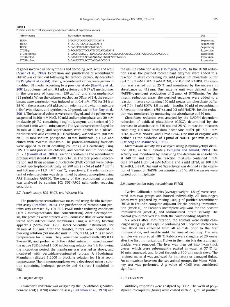
Table 1Primers used for TGR sequencing and construction of expression vectors.
Primer name Nucleotide sequence Use
TRR5r 5-TATCCTGGGGCGTCGGGAC-3 SequencingTRR5f 5-GGTGGTCGATCCGCATAC-3 SequencingTRR3r 5-CAGCCTTCATCGCTACGG-3 Sequencing5TGRBgl 5-AGATCTGCTCCAATTCCCGATGATAC-3 Expression3TGREcoSecis 5-GAATTCATAGGTTAAGCCCCGGTGCAGACCTGCAACCGGGGCTTAACCTCAGCAAGCGG-3 Expression3TGREcoCys 5-GAATTCTTAACCACAGCAAGCGGTCACCTTAGC-3 Expression3TGREcoStop 5-GAATTCTTAACCTCAGCAAGCGG-3 Expression
G. Maggioli et al. / Experimental Parasitology 129 (2011) 323–330 325
of genes involved in Sec synthesis and decoding (selA, selB, and selC)(Arner et al., 1999). Expression and purification of recombinantFhTGR was carried out following the protocol previously describedby Rengby et al. (2004). Briefly, recombinant clones were grown inmodified LB media according to a previous study (Bar-Noy et al.,2001), supplemented with 0.1 g/L cysteine and 0.37 g/L methionine,in the presence of kanamycin (50 lg/mL) and chloramphenicol(33 lg/mL). When the cultures reached an OD600 of 2.4, the recom-binant gene expression was induced with 0.4 mM IPTG for 24 h at25 �C in the presence of 5 lM sodium selenite and a vitamin mixture(riboflavin, niacin, and pyridoxine, 20 lg/mL each) (Bar-Noy et al.,2001). The bacterial cultures were centrifuged, and the pellets weresuspended in 300 mM NaCl, 50 mM sodium phosphate, and 20 mMimidazole, pH 7.2, containing 1 mg/mL lysozyme, and sonicated (10pulses of 1 min with 1-min pauses). The lysates were centrifuged for30 min at 20,000g, and supernatants were applied to a nickel–nitrilotriacetic acid column (GE Healthcare), washed with 300 mMNaCl, 50 mM sodium phosphate, 30 mM imidazole, pH 7.2, andeluted with 250 mM imidazole. The protein-containing fractionswere applied to PD10 desalting columns (GE Healthcare) usingPBS, 150 mM potassium chloride, and 50 mM sodium phosphate,pH 7.2 (Bonilla et al., 2008). Fractions containing the recombinantproteins were stored at�80 �C prior to use. The total protein concen-tration and flavin adenine dinucleotide (FAD) content were deter-mined spectrophotometrically at 280 nm (e = 54.24 mM�1 cm�1)and 460 nm (e = 11.3 mM�1 cm�1), respectively. The selenium con-tent of selenoproteins was determined by atomic absorption usingAAS Shimadzu AA6800. The purity of the recombinant proteinswas analyzed by running 10% SDS–PAGE gels, under reducingconditions.
2.7. Protein assay, SDS–PAGE, and Western blot
The protein concentration was measured using the Bio-Rad pro-tein assay (Bradford, 1976). The purification of recombinant pro-teins was assessed by 10% SDS–PAGE under reducing conditions(10% 2-mercaptoethanol final concentration). After electrophore-sis, the proteins were stained with Coomassie Blue or were trans-ferred onto nitrocellulose membranes using a semidry blottingapparatus (Semi-Phor TE70; Hoefer Scientific Instruments) for30 min at 100 mA. After the transfer, filters were incubated inblocking solution (5% non-fat milk in PBS, 0.1 M, pH 7.3) at roomtemperature for 30 min. They were then washed with PBS 0.1%Tween-20, and probed with the rabbit antiserum raised againstthe native TGR diluted 1:500 in blocking solution for 1 h. Followingthe incubation period, the membrane was washed as above, andincubated with peroxidase-conjugated protein A (BoehringerMannheim) diluted 1:2000 in blocking solution for 1 h at roomtemperature. The immunocomplexes were developed using a solu-tion containing hydrogen peroxide and 4-chloro-1-naphthol inPBS.
2.8. Enzyme assays
Thioredoxin reductase was assayed by the 5,50-dithiobis(2-nitro-benzoic acid) (DTNB) reduction assay (Luthman et al., 1979) and
the insulin reduction assay (Holmgren, 1979). In the DTNB reduc-tion assay, the purified recombinant enzymes were added to areaction mixture containing 200 mM potassium phosphate buffer(pH 7.6), 1 mM EDTA, 1 mM DTNB, and 0.2 mM NADPH. The reac-tion was carried out at 25 �C and monitored by the increase inabsorbance at 412 nm. One enzyme unit was defined as theNADPH-dependent production of 2 lmol of DTNB/min. For theinsulin reduction assay, the purified enzymes were added to areaction mixture containing 100 mM potassium phosphate buffer(pH 7.0), 1 mM EDTA, 1.0 mg mL�1 insulin, 20 lM of recombinantF. hepatica thioredoxin (FhTrx), and 0.2 mM NADPH. Insulin reduc-tion was monitored by measuring the absorbance at 650 nm.
Glutathione reductase was assayed by the NADPH-dependentreduction of oxidized glutathione (GSSG), determined by thedecrease in absorbance at 340 nm and 25 �C, in reaction mixturescontaining 100 mM potassium phosphate buffer pH 7.0, 1 mMEDTA, 0.2 mM NADPH, and 1 mM GSSG. One unit of enzyme wasdefined as the oxidation of 1 lmol of NADPH per min at 25 �C(Carlberg and Mannervik, 1985).
Glutaredoxin activity was assayed using b-hydroxyethyl disul-fide (HED) as the substrate (Holmgren and Aslund, 1995). Theactivity was monitored by measuring the decrease in absorbanceat 340 nm and 25 �C. The reaction mixtures contained 1 mMGSH, 0.7 mM HED, 0.4 mM NADPH, and 2 mM EDTA, in 100 mMTris–HCl, pH 7.0. One unit of Grx activity was defined as the oxida-tion of 1 lmol of NADPH per minute at 25 �C. All the assays werecarried out in triplicate.
2.9. Immunization using recombinant FhTGR
Twelve Californian rabbits (average weight, 1.5 kg) were sepa-rated into two groups and housed individually. All inmunogendoses were prepared by mixing 100 lg of purified recombinantFhTGR in Freund’s complete adjuvant for the priming immuniza-tion (week 0), or Freund’s incomplete adjuvant for the boosterimmunization (week 4), and administered intramuscularly. Thecontrol group received PBS with the corresponding adjuvant.
Six weeks after immunization, the animals were orally chal-lenged using a gelatin capsule containing 50 F. hepatica metacerca-riae. Blood was collected from all animals prior to the firstimmunization, and weekly until the time of necropsy. The serasamples were stored at �80 �C. Rabbits were slaughtered 20 weeksafter the first immunization. Flukes in the main bile ducts and gallbladder were removed. The liver was then cut into 1-cm thickpieces, which were subsequently soaked in water at 37 �C for30 min, squeezed, and forced through a 300-lm mesh sieve. Theretained material was analyzed for immature or damaged flukes.For comparison between the two animal groups, the Mann–Whit-ney test was performed. A p value of <0.05 was consideredsignificant.
2.10. ELISA tests
Antibody responses were analyzed by ELISA. The wells of poly-styrene microplates (Nunc) were coated with 2 lg/mL of purified
326 G. Maggioli et al. / Experimental Parasitology 129 (2011) 323–330
recombinant FhTGR in PBS and incubated overnight at 4 �C. Afterwashing with PBS/Tween 20 (0.05%), the plates were blocked withPBS/Tween 20 (0.2%) overnight at 4 �C. Next, serially diluted serafrom individual rabbits were added and incubated for 1 h at37 �C. The plates were washed and incubated for 1 h at 37 �C withHRP-conjugated anti-rabbit IgG (Sigma–Aldrich). After washing,the substrate solution (o-phenylenediamine and H2O2; Sigma)was added, the enzyme reaction was stopped after 15 min by addi-tion of 1 M HCl, and the absorbance at 492 nm was measured usingan ELISA reader. Serial dilutions of a pool serum were included ineach plate in order to have a calibration curve for conversion ofOD readings into antibody levels expressed in arbitrary units (AU).
3. Results
3.1. Cloning and nucleotide sequence analysis of adult F. hepatica TGRcDNA
A kgt11 cDNA library from adult F. hepatica was screened with arabbit antiserum raised against native FhTGR. One of the clonesshowing strong reactivity was chosen for further characterization.The sequence of the 2287-bp cDNA insert (accession numberAM709787) contained a 1.8-kb open reading frame (ORF) starting
Fig. 1. Alignment of TGR amino acid sequences. F. hepatica (A8E0R8); S. japonicum (AAW2mitochondrial (AAN63051); M. musculus TR (AAK31172). Experimentally determined N-te(Grx); pyridine-oxidoreductases redox active center (RAC); selenocysteine (U) residue (
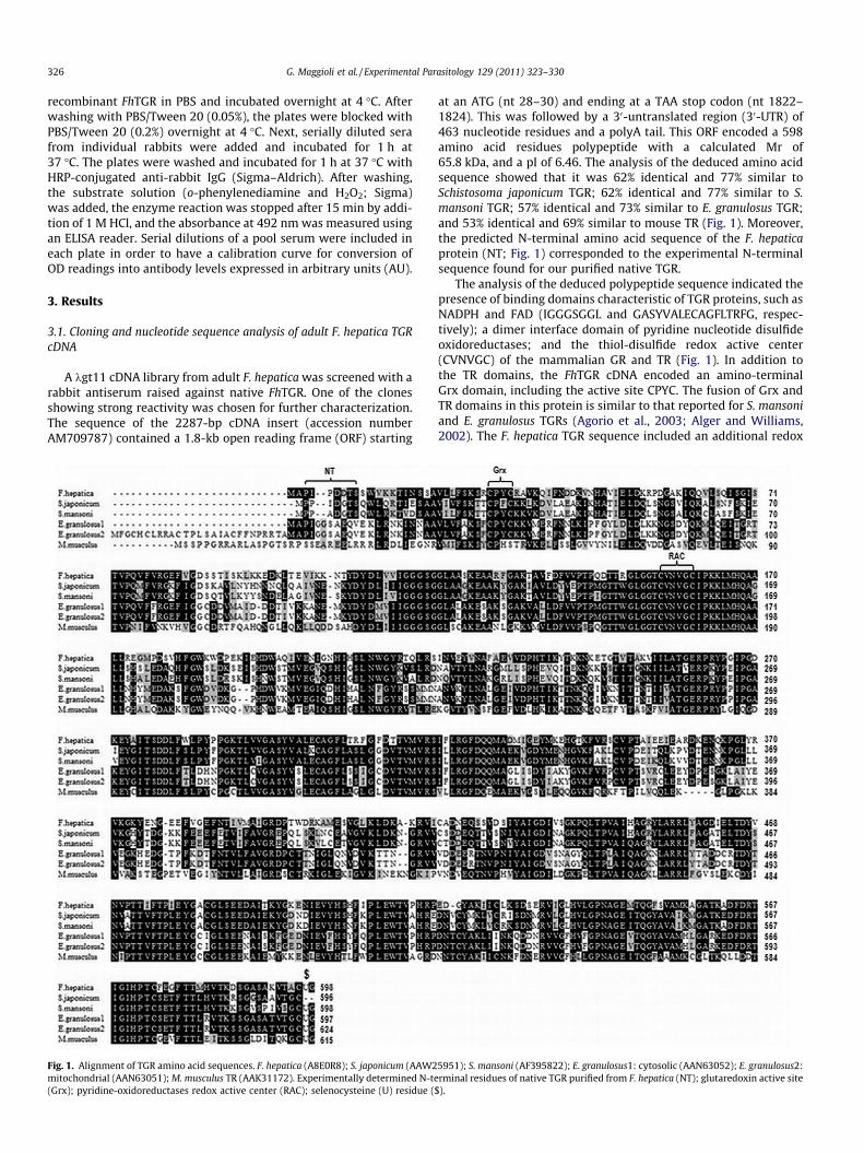
at an ATG (nt 28–30) and ending at a TAA stop codon (nt 1822–1824). This was followed by a 30-untranslated region (30-UTR) of463 nucleotide residues and a polyA tail. This ORF encoded a 598amino acid residues polypeptide with a calculated Mr of65.8 kDa, and a pI of 6.46. The analysis of the deduced amino acidsequence showed that it was 62% identical and 77% similar toSchistosoma japonicum TGR; 62% identical and 77% similar to S.mansoni TGR; 57% identical and 73% similar to E. granulosus TGR;and 53% identical and 69% similar to mouse TR (Fig. 1). Moreover,the predicted N-terminal amino acid sequence of the F. hepaticaprotein (NT; Fig. 1) corresponded to the experimental N-terminalsequence found for our purified native TGR.
The analysis of the deduced polypeptide sequence indicated thepresence of binding domains characteristic of TGR proteins, such asNADPH and FAD (IGGGSGGL and GASYVALECAGFLTRFG, respec-tively); a dimer interface domain of pyridine nucleotide disulfideoxidoreductases; and the thiol-disulfide redox active center(CVNVGC) of the mammalian GR and TR (Fig. 1). In addition tothe TR domains, the FhTGR cDNA encoded an amino-terminalGrx domain, including the active site CPYC. The fusion of Grx andTR domains in this protein is similar to that reported for S. mansoniand E. granulosus TGRs (Agorio et al., 2003; Alger and Williams,2002). The F. hepatica TGR sequence included an additional redox
5951); S. mansoni (AF395822); E. granulosus1: cytosolic (AAN63052); E. granulosus2:rminal residues of native TGR purified from F. hepatica (NT); glutaredoxin active site
$).

G. Maggioli et al. / Experimental Parasitology 129 (2011) 323–330 327
active center (ACUG; U being selenocysteine), located at the C-ter-minal end (Fig. 1). Interestingly, the G in the highly conservedGCUA active redox center is changed by A in FhTGR, a substitutionconfirmed by the analysis of F. hepatica ESTs datasets from the San-ger Institute and Gasser’s Lab.
On the other hand, the Sec residue is cotranslationally insertedat an in-frame UGA codon, usually considered a stop codon, by aselenocysteine tRNA. Decoding the UGA codon in selenoproteinsrequires a SECIS element located at the mRNA 30-UTR. A consensusstem–loop SECIS is present in F. hepatica cDNA between nucleo-tides 2014 and 2068, including a core with the essential non-Wat-son–Crick base pairs, lower stem, adenosine bulge, upper stem, andupper loop (Fig. 2a). Taking into account the requirements of themodel SECIS elements described previously (Low and Berry,1996), this F. hepatica structure could be considered a type II SECIS.It is worth noting that the AAGA sequence at the F. hepatica TGR
Fig. 2. Predicted secondary structure of SECIS elements. (a) F. hepatica TGR SECIS presentthe GenBank AM709787 sequence are indicated. (⁄) U/A change in FhTGR mRNA with reeukaryotic SECIS elements are shown in boldface. (b) Minimal E. coli formate dehydroselenocysteine incorporation. (c) SECIS element included in expression vector pGEX-Selenocysteine codons (UGA) are underlined.
Fig. 3. Analysis of the E. coli expression and purification of recombinant FhTGRsecis. (a)Soluble fraction from transformed cell extract; 3: Proteins not retained in the GSH–Sepha5–8: eluted fractions after thrombin treatment; 9: Proteins retained onto the column afterfrom pGEX-FhTGRsecys transformed cell extract; 2: Proteins retained onto the column bProteins retained onto the column after elution.
SECIS core included a U/A change with respect to the sequenceAUGA sequence described in other organisms.
3.2. Expression and purification of the recombinant pGEX-FhTGR
The recombinant FhTGR, putatively including a Sec residue at theC-terminus, was first produced in E. coli as a glutathione-S-transfer-ase fusion protein (GST-FhTGR), using the recombinant expressionvector pGEX-FhTGRsecys. This plasmid contained a minimal SECISelement for Sec incorporation (Fig. 2c), similar to the one presentin E. coli formate dehydrogenase H (Fig. 2b). The cell extract fromE. coli transformed with pGEX-FhTGRsecys, induced with IPTG,showed a prominent protein band with an apparent molecularweight of 92 kDa in SDS–PAGE and Western blot analyses (Fig. 3aand b). This was in good agreement with the predicted molecularweight for the fusion protein. The soluble cell-free extract
in the 30-end untranslated region of FhTGR mRNA. Nucleotide numbers referring tospect to the AUGA sequence described in other organisms. Conserved sequences ingenase-H SECIS (Li et al., 2000) required for SelB binding (boxed stem loop) andFhTGRsecys using oligonucleotide 3TGREcoSecis. (�) Watson Crick base-pairing.
SDS–PAGE 1: insoluble fraction from pGEX-FhTGRsecys transformed cell extract; 2:rose column; 4: Proteins retained onto the column before incubation with thrombin;
elution. (b) Western blot analysis using F. hepatica anti-TGR sera. 1: Soluble fractionefore incubation with thrombin; 3–5: eluted fractions after thrombin treatment; 6:

328 G. Maggioli et al. / Experimental Parasitology 129 (2011) 323–330
containing the fusion protein was loaded onto a GSH–Sepharose 4Bmatrix. The recombinant FhTGR moiety, with an apparent molecularweight of 66 kDa, was released from the gel-matrix by proteolysisusing thrombin (Fig. 3). Some of the fractions along the purificationprotocol of recombinant FhTGR were then electrophoretically trans-ferred onto nitrocellulose membranes, and probed with a rabbit ser-um raised against the native protein initially described as TR. Theseanalyses (Fig. 3b) showed a positive identification of GST-FhTGR andrecombinant FhTGR proteins bands.
Two other recombinant versions of FhTGR were also producedand purified using expression vectors pGEX-FhTGRcys and pGEX-FhTGRstop. The first was designed for Sec/Cys substitution, andthe second, lacking the SECIS element, most probably produced aC-terminus truncated TGR. Recombinant FhTGRcys and FhTGRstopwere purified as described above for FhTGR. SDS–PAGE analysesyielded similar protein patterns to those shown in Fig. 3a (datanot shown). The three FhTGR variants were further used in theenzymatic activity assays. The expression and purification proce-dures described above yielded relatively low amounts (approxi-mately 900 lg/L) of the recombinant proteins.
3.3. Functional analysis of FhTGR activity
As a preliminary approach to biochemical characterization, weinvestigated the TR, Grx, and GR activities of the three F. hepaticarecombinant TGR proteins produced in E. coli using DTNB, HED,and GSSG reduction assays, respectively. The specific activities ofthe FhTGRsecys and FhTGRcys recombinant proteins were similarusing DTNB and HED reduction assays (0.45 and 0.40; and 0.46and 0.48 U/mg, respectively), possibly indicating low Sec incorpo-ration levels into the FhTGRsecis recombinant protein. The purifiedFhTGRstop construct was approximately three times less activethan the recombinant FhTGRsecis or FhTGRcys, using DTNB orHED assays (0.14 and 0.16 U/mg, respectively). GR activity assaysindicated very low levels of GSSG reduction using the FhTGRsecisand FhTGRcys recombinant proteins (0.22 and 0.14 U/mg, respec-tively), and undetectable levels using FhTGRstop recombinantprotein.
Using the insulin-reduction method, we investigated whetherthe recombinant FhTGRs were capable of reducing recombinantTrx from F. hepatica. We observed that the FhTGRsecis and FhTGR-cys recombinant proteins actively reduced insulin in the presenceof recombinant FhTrx. In contrast, FhTGRstop yielded no detectableTrx-mediated insulin reduction (Fig. 4).
We carried out a new construction using the expression vectorpET28a, because of the lack of activity and low production yield ofthe recombinant protein using pGEX vectors. The new recombinantFhTGR was produced in E. coli BL21 (DE3) cells, using the recombi-
Fig. 4. Insulin-reduction activity mediated by recombinant Trx from F. hepatica. (j)FhTGRsecys; (d) FhTGRcys; (N) FhTGRstop.
nant expression vector pET-FhTGR. This plasmid contained a min-imal SECIS element for Sec incorporation (Fig. 2c). The cell extractfrom E. coli transformed with pET-FhTGR, induced with IPTG, con-tained a prominent protein band with an apparent molecularweight of 66 kDa in SDS–PAGE (not shown). This was in agreementwith the predicted molecular weight for the fusion protein by His-tag. The soluble cell-free extract containing the recombinant pro-tein was loaded onto an NTA-Sepharose column (GE Healthcare).The recombinant FhTGR was eluted with 250 mM imidazole. Thepurity of the recombinant protein by 10% SDS–PAGE analysis wasapproximately 90%. Using this expression method, we obtained30 mg/L of recombinant TGR.
The FAD content of the recombinant FhTGR was approximately25% and the Sec content was 20–25%, similar to that published datafor other recombinant TGRs. We investigated the TR, Grx, and GRactivities of the new recombinant FhTGR using DTNB, HED, andGSSG reduction assays. The specific activities were 1.13; 0.21,and 0.48 U/mg, respectively. These specific activities were cor-rected according to the selenium and FAD contents of the new re-combinant FhTGR. In order to prevent the hysteretic behaviordisplayed by FhTGR at a high GSSG concentration, as describedby Guevara-Flores et al. (2011), the GR assay included the additionof 1 mM GSH.
3.4. Vaccination experiment
In order to investigate the immunoprophylactic potential of therecombinant FhTGR, we performed a pilot immunization trial inrabbits. Specific IgG antibodies against FhTGR were detectable inthe rabbit sera within 4 weeks of the initial immunization, and in-creased sharply following the challenge infection at week 6. Theantibody levels gradually decreased after 11 weeks. A minimal in-crease in antibody titter was detected in the control group follow-ing the challenge (Fig. 5).
We further assessed the effect of FhTGR immunization, byworm counting in immunized and control rabbits (Table 2). We ob-served that only 18% of the original 50 metacercariae gave rise toproductive infections, allowing the recovery of a mean number of9 adult F. hepatica worms per animal in the control group. In clearcontrast, only one of the TGR-vaccinated rabbits harbored liverflukes (Table 2). Our results indicate an overall worm burdenreduction of 96.7% (p < 0.00001) compared to the control group.
4. Discussion
Most platyhelminth parasites have a developmental stage thatlives in an aerobic environment. Thus, they must possess effectivemechanisms to maintain cellular redox homeostasis. These mech-
Fig. 5. Antibody responses in recombinant FhTGR-immunized rabbits. The barsindicated standard deviation.
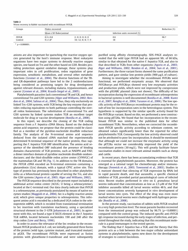
Table 2Worm recovery in Rabbit vaccinated with recombinant FhTGR.
Groups Flukes recovered Reduction (%)
Mean ± SD flukes/animal Infection units recovered (%)
FhTGR 0.3 ± 0.8 2; 0; 0; 0; 0; 0 0.6 96.7Control 9 ± 1.8a 10; 12; 8; 8; 7; 9 18 —
a p < 0.000001.
G. Maggioli et al. / Experimental Parasitology 129 (2011) 323–330 329
anisms are also important for quenching the reactive oxygen spe-cies generated by the host’s immune response. Most eukaryoteorganisms have two major systems to detoxify reactive oxygenspecies, one based on Trx and the other based on GSH. Besides pro-viding protection against oxidative damage, these systems playimportant roles in cell proliferation, redox regulation of geneexpression, xenobiotic metabolism, and several other metabolicfunctions (Gromer et al., 2004). The diverse functions of the TR-and GR-dependent pathways have led to the 2 oxidoreductasesbeing considered as promising targets for drug developmentagainst relevant diseases, including malaria, trypanosomiasis, andcancer (Gromer et al., 2004; Krauth-Siegel et al., 2005).
Platyhelminth parasites lack conventional TR and GR, and henceconventional Trx and GSH systems (Alger and Williams, 2002; Ren-don et al., 2004; Salinas et al., 2004). Thus, they rely exclusively onlinked Trx–GSH systems, with TGR being the key enzyme that pro-vides reducing equivalents to both pathways controlling the over-all redox homeostasis. The substitution of conventional Trx andGSH systems by linked systems makes TGR an excellent targetmolecule for drug or vaccine development (Bonilla et al., 2008).
In this report, we describe the cloning of the TGR codingsequence from a F. hepatica cDNA library. Based on the deducedamino acid sequence, we propose that the protein could be classi-fied as a member of the pyridine-nucleotide disulfide reductasefamily. The analysis of the N-terminal amino acid sequencededuced from the isolated cDNA matched the experimentallydetermined N-terminal sequence of the native protein, thus sup-porting the F. hepatica TGR ORF identification. The amino acid se-quence of the identified ORF indicated the presence of bindingdomains characteristic of TGR proteins, such as NADPH and FAD;a dimer interface domain of pyridine nucleotide disulfide oxidore-ductases; and the thiol-disulfide redox active center (CVNVGC) ofthe mammalian GR and TR (Fig. 1). In addition to the TR domains,the FhTGR cDNA encoded an N-terminal extension containing aglutaredoxin (Grx) domain, with a typical CPYC active site. Thistype of protein has previously been described in other platyhelm-inths as a bifunctional protein capable of serving the Trx, and alsothe GSH systems (Agorio et al., 2003; Alger and Williams, 2002).
The FhTGR sequence described in the present report included anadditional active redox center (ACUG; U being a selenocysteine),located at the C-terminal end. Our data clearly indicate that FhTGRis a selenoenzyme, as previously postulated by means of native en-zymes studies (Maggioli et al., 2004). Sec is a highly reactive aminoacid, which gives unique properties to selenoproteins. This infre-quent amino acid is encoded by a dedicated UGA codon in the sele-noprotein mRNA, which is recoded from translational terminationto Sec insertion with translation machinery, using a specializedstructural element in the 30-UTR (Johansson et al., 2005). In agree-ment with this, we found a type II SECIS element in the F. hepaticaTGR mRNA, located between nucleotides 190 and 240 after thestop codon (Low and Berry, 1996).
In order to assess the relevance of the Sec residue in the recom-binant FhTGR produced in E. coli, we initially generated three formsof the protein (wild type, cysteine mutant, and truncated mutant)in pGEX. The recombinant FhTGRs were expressed as fusionproteins with glutathione-S-transferase and were subsequently
purified using affinity chromatography. SDS–PAGE analyses re-vealed that the wild type FhTGR had an apparent Mr. of 66 kDa,similar to that obtained for the native F. hepatica TGR, and also tothat described in TGRs from other organisms (Agorio et al., 2003;Alger and Williams, 2002; Rendon et al., 2004; Sun et al., 2001).Furthermore, the three variant forms showed the same purificationpattern, and gave similar low protein yields (900 lg/L of culture).
Aiming to investigate whether the recombinant FhTGRs werefunctional, we performed enzymatic assays. We observed thatFhTGRsecys and FhTGRcys showed very low enzymatic activitiesand production yields, which were not improved by coexpressionwith the pSUABC plasmid (data not shown). The difficulty of Secincorporation during the expression of recombinant selenoproteinsin bacterial systems is well documented (Bonilla et al., 2008; Kuntzet al., 2007; Rengby et al., 2004; Turanov et al., 2006). The low spe-cific activity of the FhTGRsecys recombinant protein may be the re-sult of low Sec incorporation rates in the heterologous system. Thishypothesis is supported by the similar specific activity found forthe FhTGRcys variant. Based on this, we performed a new construc-tion using pET28a. We found that Sec incorporation in the recom-binant FhTGR was similar to the published data for otherrecombinant TGRs. Nevertheless, when we corrected the recombi-nant FhTGR specific activity according to selenium content, weobtained values significantly lower than the reported for otherplatyhelminths TGR. Consequently the low activity observed couldnot be attributed to poor Sec incorporation. It should be mentionedthat although we could not increase the enzymatic activity usingthe pET28a vector we considerably improved the yield of therecombinant protein (30 mg/L). This will greatly facilitate futurevaccination studies in more relevant animal models such as sheepand cattle.
In recent years, there has been accumulating evidence that TGRis essential for platyhelminth parasites. Moreover, the protein hasemerged as a rational target for chemotherapy and/or immuno-therapy (Fenwick et al., 2003; Townsend et al., 2003). Studies inS. mansoni showed that silencing of TGR expression by RNAi ledto rapid parasite death, and that auranofin, a specific chemicalinhibitor of TGR, provided partial parasitological cures of infectedmice. Recent studies performed in vitro on E. granulosus larvalworms indicated that concentrations as low as 2.5 lM of the TGRinhibitor auranofin killed all larval worms within 48 h, and thatlower concentrations severely hampered in vitro development oflarval worms into cysts. The effect of the drug was even moremarked when larval worms were challenged with hydrogen perox-ide (Bonilla et al., 2008).
In the present study, vaccination of rabbits with FhTGR resultedin a very high protection level. The mean parasite burden recoveredfrom the immunized group was reduced by 96.7% (p < 0.00001)compared with the control group. The induced specific anti-FhTGRIgG response increased during the early stages of infection, and per-sisted throughout the study, indicating that this recombinant mole-cule is a good immunogen in the tested species.
The finding that F. hepatica has a TGR, and the theory that thisprotein acts as a link between the two major cellular antioxidantsystems in platyhelminths, opens new doors for the developmentof strategies to control fasciolosis.

330 G. Maggioli et al. / Experimental Parasitology 129 (2011) 323–330
Acknowledgments
We thank Daniel Acosta and DILAVE ‘‘Miguel C. Rubino’’ for per-forming the rabbit immunization assay. We also thank Dr EliasArnér for providing the pSUABC. G. Maggioli was funded by theMutis program of the AECID (Agencia Española de CooperaciónInternacional para el Desarrollo).
References
Agorio, A., Chalar, C., Cardozo, S., Salinas, G., 2003. Alternative mRNAs arising fromtrans-splicing code for mitochondrial and cytosolic variants of Echinococcusgranulosus thioredoxin Glutathione reductase. The Journal of BiologicalChemistry 278, 12920–12928.
Alger, H.M., Williams, D.L., 2002. The disulfide redox system of Schistosoma mansoniand the importance of a multifunctional enzyme, thioredoxin glutathionereductase. Molecular and Biochemical Parasitology 121, 129–139.
Arner, E.S., Sarioglu, H., Lottspeich, F., Holmgren, A., Böck, A., 1999. High-levelexpression in Escherichia coli of selenocysteine-containing rat thioredoxinreductase utilizing gene fusions with engineered bacterial-type SECISelements and co-expression with the selA, selB and selC genes. Journal ofMolecular Biology 292, 1003–1016.
Arner, E.S., Holmgren, A., 2000. Physiological functions of thioredoxin andthioredoxin reductase. European Journal of Biochemistry 267, 6102–6109.
Bar-Noy, S., Corlatov, S.N., Stadtman, T.C., 2001. Overexpression of wild type andSecCys/Cys mutant of human thioredoxin reductase in E. coli: the role ofselenocysteine in the catalytic activity.. Free Radical Biology and Medicine 30,51–61.
Bogdan, C., Rollinghoff, M., Diefenbach, A., 2000. Reactive oxygen and reactivenitrogen intermediates in innate and specific immunity. Current Opinion inImmunology 12, 64–76.
Bonilla, M., Denicola, A., Novoselov, S.V., Turanov, A.A., Protasio, A., Izmendi, D.,Gladyshev, V.N., Salinas, G., 2008. Platyhelminth mitochondrial and cytosolicredox homeostasis is controlled by a single thioredoxin glutathione reductaseand dependent on selenium and glutathione. The Journal of BiologicalChemistry 283, 17898–17907.
Bradford, M.M., 1976. A rapid and sensitive method for the quantitation ofmicrogram quantities of protein utilizing the principle of protein-dye binding.Analytical Biochemistry 72, 248–254.
Callahan, H.L., Crouch, R.K., James, E.R., 1988. Helminth anti-oxidant enzymes: aprotective mechanism against host oxidants? Parasitology Today 4, 218–225.
Carlberg, I., Mannervik, B., 1985. Glutathione reductase. Methods in Enzymology113, 484–490.
Fritsch, E.F., Sambrook, J. and Maniatis, T., 1989. Molecular cloning. A laboratorymanual. 2,
Fenwick, A., Savioli, L., Engels, D., Robert, B.N., Todd, M.H., 2003. Drugs for thecontrol of parasitic diseases: current status and development inschistosomiasis. Trends in Parasitology 19, 509–515.
Gromer, S., Urig, S., Becker, K., 2004. The thioredoxin system—from science to clinic.Medicinal Research Reviews 24, 40–89.
Guevara-Flores, A., Pardo, J.P., Rendón, J.L., 2011. Hysteresis in thioredoxin-glutathione reductase (TGR) from the adult stage of the liver fluke Fasciolahepatica. Parasitology International 60, 156–160.
Holmgren, A., 1979. Thioredoxin catalyzes the reduction of insulin disulfides bydithiothreitol and dihydrolipoamide. The Journal of Biological Chemistry 254,9627–9632.
Holmgren, A., 1989. Thioredoxin and glutaredoxin systems. The Journal of BiologicalChemistry 264, 13963–13966.
Holmgren, A., Aslund, F., 1995. Glutaredoxin. Methods in Enzymology 252, 283–292.
Johansson, L., Gafvelin, G., Arner, E.S., 2005. Selenocysteine in proteins-propertiesand biotechnological use. Biochimica et Biophysica Acta 1726, 1–13.
Kanzok, S.M., Fechner, A., Bauer, H., Ulschmid, J.K., Muller, H.M., Botella-Munoz, J.,Schneuwly, S., Schirmer, R., Becker, K., 2001. Substitution of the thioredoxinsystem for glutathione reductase in Drosophila melanogaster. Science 291, 643–646.
Krauth-Siegel, R.L., Bauer, H., Schirmer, R.H., 2005. Dithiol proteins as guardians ofthe intracellular redox milieu in parasites: old and new drug targets intrypanosomes and malaria-causing plasmodia. Angewandte ChemieInternational Edition 44, 690–715.
Kuntz, A.N., Vioud-Charvet, E., Sayed, A.A., Califf, L.L., Dessolin, J., Arner, E.S.,Williams, D.L., 2007. Thioredoxin glutathione reductase from Schistosomamansoni: an essential parasite enzyme and a key drug target. PLoS Medicine4, e206.
Li, C., Reches, M., Engelberg-Kulka, H., 2000. The bulged nucleotide in the Escherichiacoli minimal selenocysteine insertion sequence participates in interaction withSelB: a genetic approach. Journal of Bacteriology 182, 6302–6307.
Low, S.C., Berry, M.J., 1996. Knowing when not to stop: selenocysteine incorporationin eukaryotes. Trends in Biochemical Sciences 21, 203–208.
Luthman, M., Eriksson, S., Holmgren, A., Thelander, L., 1979. Glutathione-dependenthydrogen donor system for calf thymus ribonucleoside-diphosphate reductase.Proceedings of the National Academy of Sciences USA 76, 2158–2162.
Maggioli, G., Piacenza, L., Carambula, B., Carmona, C., 2004. Purification,characterization, and immunolocalization of a thioredoxin reductase fromadult Fasciola hepatica. The Journal of Parasitology 90, 205–211.
Marin, M.S., Prieto, M., Martin, J.M., Casais, R., Boga, J.A., Parra, F., 1992.Identification and expression of a Fasciola hepatica gene encoding a gutantigen protein bearing repetitive sequences. Molecular and BiochemicalParasitology 55, 155–165.
McGonigle, S., Dalton, J.P., James, E.R., 1998. Peroxidoxins: a new antioxidant family.Parasitology Today 14, 139–145.
Nordberg, J., Arner, E.S., 2001. Reactive oxygen species, antioxidants, and themammalian thioredoxin system. Free Radical Biology and Medicine 31, 1287–1312.
Rendon, J.L., Del, A.I., Guevara-Flores, A., Uribe, A., Plancarte, A., Mendoza-Hernandez, G., 2004. Purification, characterization and kinetic properties ofthe multifunctional thioredoxin-glutathione reductase from Taenia crassicepsmetacestode (cysticerci). Molecular and Biochemical Parasitology 133, 61–69.
Rengby, O., Johansson, L., Carlson, L.A., Serini, E., Vlamis-Gardikas, A., Karsnas, P.,Arner, E.S., 2004. Assessment of production conditions for efficient use ofEscherichia coli in high-yield heterologous recombinant selenoprotein synthesis.Applied and Environmental Microbiology 70, 5159–5167.
Salinas, G., Selkirk, M.E., Chalar, C., Maizels, R.M., Fernandez, C., 2004. Linkedthioredoxin-glutathione systems in platyhelminths. Trends in Parasitology 20,340–346.
Sanger, F., Nicklen, S., Coulson, A.R., 1977. DNA sequencing with chain-terminatinginhibitors. Proceedings of the National Academy of Sciences USA 74, 5463–5467.
Selkirk, M.E., Smith, V.P., Thomas, G.R., Gounaris, K., 1998. Resistance of filarialnematode parasites to oxidative stress. International Journal for Parasitology28, 1315–1332.
Su, D., Novoselov, S.V., Sun, Q.A., Moustafa, M.E., Zhou, Y., Oko, R., Hatfield, D.L.,Gladyshev, V.N., 2005. Mammalian selenoprotein thioredoxin-glutathionereductase. Roles in disulfide bond formation and sperm maturation. TheJournal of Biological Chemistry 280, 26491–26498.
Sun, Q.A., Kirnarsky, L., Sherman, S., Gladyshev, V.N., 2001. Selenoproteinoxidoreductase with specificity for thioredoxin and glutathione systems.Proceedings of the National Academy of Sciences USA 98, 3673–3678.
Sun, Q.A., Wu, Y., Zappacosta, F., Jeang, K.T., Lee, B.J., Hatfield, D.L., Gladyshev, V.N.,1999. Redox regulation of cell signaling by selenocysteine in mammalianthioredoxin reductases. The Journal of Biological Chemistry 274, 24522–24530.
Townsend, D.M., Tew, K.D., Tapiero, H., 2003. The importance of glutathione inhuman disease. Biomedicine & Pharmacotherapy 57, 145–155.
Turanov, A.A., Su, D., Gladyshev, V.N., 2006. Characterization of alternative cytosolicforms and cellular targets of mouse mitochondrial thioredoxin reductase. TheJournal of Biological Chemistry 281, 22953–22963.