Exploring the antigenic features of Fasciola hepatica rediae (Trematoda: Digenea) through the...
9
ORIGINAL PAPER Exploring the antigenic features of Fasciola hepatica rediae (Trematoda: Digenea) through the evaluation of different antigenic candidates for further monoclonal antibody generation Annia Alba & Hilda M. Hernández & Ricardo Marcet & Alejandro L. Gil & Antonio A. Vázquez & Mabel Figueredo & Jorge Sánchez & Hilda E. Garay & Jorge Sarracent Received: 28 December 2013 /Accepted: 27 May 2014 # Springer-Verlag Berlin Heidelberg 2014 Abstract The control of fasciolosis, as that of other vector- borne diseases, must be related to the control of the lymnaeid snails, the intermediate hosts of the parasite. Thus, an accurate epidemiological surveillance of the transmission foci where the infected mollusks occur is essential. For this purpose, immunoassays could be a useful tool. However, information regarding specific proteins of intramolluscan larvae and pre- vious studies concerning monoclonal antibody generation against asexual stages of trematodes are scarce. Therefore, we explored the antigenic features of intramolluscan rediae of Fasciola hepatica to evaluate three antigenic preparations in order to use the most promising one for developing specific monoclonal antibodies. Mouse antiserum was generated against each antigen for assessing the polyclonal antibody response against the crude extract of rediae and the cross- reactivity against lymnaeids. The specific C-terminal of F. hepatica cytochrome c oxidase subunit I (first antigen), selected by in silico analyses, might not be the appropriate target for immunoassay detection of infected snails, due to its low representation in the total extract of rediae. The majori- tarian mixture of low-molecular-weight proteins (<30 kDa) from the rediae homogenate (second antigen) revealed a sig- nificant cross-reactivity with lymnaeids. Evidence of the ex- istence of mimetic immunogenic epitopes in this fraction of F. hepatica rediae was achieved. High immunogenicity of the crude extract of rediae (third antigen), mainly related to para- site’ s specific epitopes, was regarded. Therefore, the rediae homogenate is stated as the most promising antigen from those evaluated, for monoclonal antibody development with potentialities for detecting F. hepatica-infected snails. Keywords Fasciola hepatica rediae . Monoclonal antibody . Molecular mimicry . Antigenic variation . Immune evasion Introduction Fasciolosis is an important worldwide distributed disease that affects a wide range of livestock and causes economic losses of large proportions. This infection also stands among the list of reemergent diseases in humans, with estimates ranging from 2.4 to 17 million persons infected (Mas-Coma et al. 2009). The main worldwide etiological agent of this disease is the trematode Fasciola hepatica (Digenea: Fasciolidae), a parasite that requires a freshwater snail as the intermediate host, for cercariae development. The infecting form for mam- mals (e.g., humans, sheep and cattle) as definitive hosts is encysted metacercariae mainly attached to aquatic plants (Andrews 1999). Electronic supplementary material The online version of this article (doi:10.1007/s00436-014-3981-y) contains supplementary material, which is available to authorized users. A. Alba (*) : H. M. Hernández : R. Marcet : M. Figueredo : J. Sarracent Laboratorio de Anticuerpos Monoclonales, Departamento de Parasitología, Instituto de Medicina Tropical “Pedro Kourí”, Ave Novia del Mediodía km 6 ½, AP 601 Havana, Cuba e-mail: [email protected] A. L. Gil Centro de Estudios de Proteínas, Facultad de Biología, Universidad de La Habana, Calle 25 e/ J e I, Havana, Cuba A. A. Vázquez : J. Sánchez Laboratorio de Malacología, Departamento de Control de Vectores, Instituto de Medicina Tropical “Pedro Kourí”, Ave Novia del Mediodía km 6 ½, AP 601 Havana, Cuba H. E. Garay Grupo de Síntesis Química, Departamento de Física-Química, Centro de Ingeniería Genética y Biotecnología, Ave 31 e/ 158 y 190, AP 6162 Havana, Cuba Parasitol Res DOI 10.1007/s00436-014-3981-y
Transcript of Exploring the antigenic features of Fasciola hepatica rediae (Trematoda: Digenea) through the...
ORIGINAL PAPER
Exploring the antigenic features of Fasciola hepatica rediae(Trematoda: Digenea) through the evaluation of differentantigenic candidates for further monoclonal antibody generation
Annia Alba & Hilda M. Hernández & Ricardo Marcet &Alejandro L. Gil & Antonio A. Vázquez & Mabel Figueredo &
Jorge Sánchez & Hilda E. Garay & Jorge Sarracent
Received: 28 December 2013 /Accepted: 27 May 2014# Springer-Verlag Berlin Heidelberg 2014
Abstract The control of fasciolosis, as that of other vector-borne diseases, must be related to the control of the lymnaeidsnails, the intermediate hosts of the parasite. Thus, an accurateepidemiological surveillance of the transmission foci wherethe infected mollusks occur is essential. For this purpose,immunoassays could be a useful tool. However, informationregarding specific proteins of intramolluscan larvae and pre-vious studies concerning monoclonal antibody generationagainst asexual stages of trematodes are scarce. Therefore,we explored the antigenic features of intramolluscan rediaeof Fasciola hepatica to evaluate three antigenic preparationsin order to use the most promising one for developing specificmonoclonal antibodies. Mouse antiserum was generatedagainst each antigen for assessing the polyclonal antibodyresponse against the crude extract of rediae and the cross-
reactivity against lymnaeids. The specific C-terminal ofF. hepatica cytochrome c oxidase subunit I (first antigen),selected by in silico analyses, might not be the appropriatetarget for immunoassay detection of infected snails, due to itslow representation in the total extract of rediae. The majori-tarian mixture of low-molecular-weight proteins (<30 kDa)from the rediae homogenate (second antigen) revealed a sig-nificant cross-reactivity with lymnaeids. Evidence of the ex-istence of mimetic immunogenic epitopes in this fraction ofF. hepatica rediae was achieved. High immunogenicity of thecrude extract of rediae (third antigen), mainly related to para-site’s specific epitopes, was regarded. Therefore, the rediaehomogenate is stated as the most promising antigen fromthose evaluated, for monoclonal antibody development withpotentialities for detecting F. hepatica-infected snails.
Keywords Fasciola hepatica rediae .Monoclonal antibody .
Molecular mimicry . Antigenic variation . Immune evasion
Introduction
Fasciolosis is an important worldwide distributed disease thataffects a wide range of livestock and causes economic lossesof large proportions. This infection also stands among the listof reemergent diseases in humans, with estimates rangingfrom 2.4 to 17 million persons infected (Mas-Coma et al.2009). The main worldwide etiological agent of this diseaseis the trematode Fasciola hepatica (Digenea: Fasciolidae), aparasite that requires a freshwater snail as the intermediatehost, for cercariae development. The infecting form for mam-mals (e.g., humans, sheep and cattle) as definitive hosts isencysted metacercariae mainly attached to aquatic plants(Andrews 1999).
Electronic supplementary material The online version of this article(doi:10.1007/s00436-014-3981-y) contains supplementary material,which is available to authorized users.
A. Alba (*) :H. M. Hernández :R. Marcet :M. Figueredo :J. SarracentLaboratorio de Anticuerpos Monoclonales, Departamento deParasitología, Instituto de Medicina Tropical “Pedro Kourí”,Ave Novia del Mediodía km 6 ½, AP 601 Havana, Cubae-mail: [email protected]
A. L. GilCentro de Estudios de Proteínas, Facultad de Biología, Universidadde La Habana, Calle 25 e/ J e I, Havana, Cuba
A. A. Vázquez : J. SánchezLaboratorio de Malacología, Departamento de Control de Vectores,Instituto de Medicina Tropical “Pedro Kourí”, Ave Novia delMediodía km 6 ½, AP 601 Havana, Cuba
H. E. GarayGrupo de Síntesis Química, Departamento de Física-Química, Centrode Ingeniería Genética y Biotecnología, Ave 31 e/ 158 y 190,AP 6162 Havana, Cuba
Parasitol ResDOI 10.1007/s00436-014-3981-y
The transmission of F. hepatica strictly depends on thepresence of the intermediate host, a freshwater snail of theLymnaeidae family. Like other vector-borne diseases, its con-trol must be based upon controlling the transmission fociwhere the infected mollusks occur (Kaplan et al. 1997;Caron et al. 2011). The epidemiological surveillance oflymnaeid populations is an effective alternative to monitorand manage endemic areas and eventually control the trans-mission of fasciolosis. However, the methods currently avail-able for detecting the parasite within the snails are not sensi-tive and specific enough (parasitological examination) or theyare too expensive (molecular biology techniques) to accom-plish this goal on a larger scale (see Caron et al. (2008)). In thisscenario, the development of immunoassays based on mono-clonal antibody (Mab) with high sensitivity and specificityagainst the long-lasting intramolluscan larval stage of theparasite, the rediae, could be useful for identifying transmis-sion foci of fasciolosis. This could allow restricting the controlefforts to a particular area. Since immunoenzymatic assays(ELISAs) to detect F. hepatica infection in the definitive hostsare being currently used in both developed and developingcountries (Espinoza et al. 2007; Ubeira et al. 2009; Rojas et al.2010), a novel ELISA to be used at the epidemiologicalsurveillance of lymnaeid snails could be easily incorporated,especially in low-income nations.
However, there are some concerns about the proper meth-odology for obtaining specific anti-F. hepatica rediae immu-nological reagents. Among these are (1) the nonexistence ofimmunoassays currently available to detect F. hepatica norother helminthes in mollusks; (2) the existence of mechanismsof molecular mimicry and masking described in otherdigenean species, which help the parasite to evade the molluskimmunity (Yoshino and Bayne 1983; Adema and Loker 1997;Yoshino et al. 2013); and (3) limited molecular informationregarding the specific proteins associated with theintramolluscan stages of Fasciola spp. Regarding this lastissue, Humiczewska (1975) detected oxidase and dehydroge-nase enzymatic activities in F. hepatica rediae. Specific cyto-chrome oxidase activity was also discovered in later rediae,ascertaining that this larval stage uses different pathwaysof energy release. In another study, two enzymes, Cu/Zn-superoxide dismutase (SOD) and thioredoxin (TRX)(previously characterized on the excretory-secretory anti-gens of F. hepatica adults), were identified on theexcretory-secretory products of F. hepatica sporocysts(Gourbal et al. 2008).
Therefore, in order to develop specific anti-F. hepaticarediae Mabs for analytical purposes, we selected and evaluat-ed three different antigenic preparations using mouse hyper-immune sera to assess the antibody response generated againsteach candidate. In this sense, a high recognition of the crudeextract of rediae by mouse antisera together with none orreduced cross-reactivity against snails’ antigens was an issue
of major importance for selecting the most promising antigen-ic candidate. Two rabbit anti-rediae polyclonal antibodies(Pabs) were also used to thoroughly explore some featuresof the antigenic composition of rediae.
Material and methods
Computational sequence and structure analysis
In order to identify promising peptides for the development ofspecific anti-F. hepatica rediae Mabs (non-cross-reactive withsnails nor with other flukes), we compared the protein sequencesof F. hepatica cytochrome c oxidase subunit I (COX I)(Q9B8Y2), SOD (Q9XY94), and TRX (Q9U1G7), with theircorresponding homologues of other related digenean pathogenicparasites and of the taxon Lymnaeidae, the intermediate hostspecies of F. hepatica. For these analyses, the following webservers were used: UniProt (www.uniprot.org) for retrieving thesequences of the target proteins; PSI-BLAST (www.ncbi.nlm.nih.gov/BLAST) for similarity searches in the nonredundantNCBI protein database (NCBInr); M-Coffee (tcoffee.crg.cat/apps/tcoffee/do:mcoffee) for multiple sequence align-ment; Jalview (www.jalview.org) for alignment editing,visualization, and analysis; and the Protein Model Portal(www.proteinmodelportal.org) for obtaining the proteinhomology-modeling structures when experimental structureswere not available.
Peptide synthesis
The selected fragment derived from the structure analysis wassynthesized at the Institute of Genetic Engineering andBiotechnology of Havana by the solid-phase method using9-fluorenyl-methoxycarbonyl chemistry (Merrifield 1986). Itwas purified by reverse phase high-performance liquid chro-matography to >98 % purity on an acetonitrile/H2O-trifluoracetic acid gradient and confirmed by ion-spray massspectrometry (Micromass, UK). A cysteine residue was addedto the N-terminal in order to conjugate a portion of thesynthetic peptide to a carrier, bovine thyroglobulin(Pept-BTg), and used for mice immunization, as the firstantigenic candidate. The nonconjugated peptide (Pept) wasused for titer evaluation of mouse hyperimmune sera.
F. hepatica adults, host snails, and experimental miceand rabbits
F. hepatica adults were obtained in cattle slaughterhouses andplaced in a solution of 0.85 % NaCl (saline solution) and 5 %glucose (Sigma, USA), for 5 h, and observed for egg release.Eggs were preserved at 4 °C in complete darkness, in a salinesolution supplemented with gentamicin (50 μg/mL) (Sigma),
Parasitol Res
until use. Populations of the Cuban intermediate hosts, Galbacubensis and Pseudosuccinea columella reared in theLaboratory of Malacology of the Institute of TropicalMedicine “Pedro Kourí”, were used in this study.Experimental BALB/c mice and chinchilla rabbits were sup-plied by the Centre for Laboratory Animal Production ofCuba. Animal care and maintenance were in agreement toinstitutional bioethical guidelines. All protocols involvingexperimental animals were approved by the Committee ofAnimal Care and Use of the institute.
Miracidia and rediae of F. hepatica: experimental infectionin snails
Eggs of F. hepatica were incubated in distilled water, in totaldarkness, at 28 °C, during 15 days for complete maturation. Atday 15, miracidia were obtained after egg hatching induced bydirect light exposure. Each of snails was infected with fivefreshly hatched miracidia, according to the methodology de-scribed by Vázquez et al. (2013). F. hepatica rediae wereobtained by dissecting experimentally infected snails at day30 postinfection (in our conditions, cercarial sheddingnormally begins between 35 and 40 days postinfection inG. cubensis and lately (day 40) in P. columella). Rediaewere thoroughly washed with phosphate buffered saline(pH 7.2, PBS) after collection, in order to minimize larvaecontamination with snail material.
Preparation of crude extracts
Protein homogenates of F. hepatica rediae were obtained after10 sonication cycles with 10-s intervals between each cycle, inPBS supplemented with a cocktail of protease inhibitors(Complete, Roche, USA). Crude extracts of miracidia andrediae were also obtained by the same sonication procedurein the presence of 0.01 % Triton X-100 (Sigma) as detergent,for COX solubilization (Musatov et al. 2000). Crude extractsof lymnaeid snailsG. cubensis and P. columellawere preparedusing healthy (nonexperimentally infected) laboratory rearedindividuals. The mollusks were homogenized in PBS supple-mented with protease inhibitors, in a Potter homogenizer.Triton X-100 was added to the homogenates of the snails usedfor evaluation of the serum anti-Pept. Protein quantificationwas performed by the bicinchoninic acid reaction (Smith et al.1986) using bovine serum albumin (Sigma) as the standard.
Protein electrophoresis
The majoritarian antigens of F. hepatica rediae were separatedby sodium dodecyl sulfate polyacrylamide gel electrophoresis(SDS-PAGE) on 12 % polyacrylamide gels (Laemli 1970).The samples were diluted without any reducing reagent, and0.1%Coomassie Brilliant Blue R-250 (Merck, Germany) was
used for staining. Homogenates of G. cubensis andP. columella were also included in the electrophoresis.Coomassie-stained gels of resolved proteins were quantifiedby scanning densitometry using ImageJ software 1.41. Themolecular weights of the most representative proteins of redi-ae were estimated using a commercial standard of 78 to12 kDa (BDH, UK).
Fractioning of the crude extract of F. hepatica rediae
Crude extract of rediae was separated into low- (<30 kDa) andhigh-molecular-weight fractions (>30 kDa) using an AmiconUltra-15 (30 kDa) centrifugal device (Millipore, USA). Thelow-molecular-weight fraction (LMWF) was lyophilized andthen diluted in saline solution for mouse immunization (sec-ond antigenic preparation). In parallel, crude extract of rediaewas also used for mice immunization as the third antigeniccandidate.
Mice immunization
Three groups, each with five 6–8-week-old female BALB/cmice, were immunized intraperitoneally with 40 μg of eachantigen (group 1: Pept-BTg, group 2: LMWF, and group 3:crude extract of rediae) in 1:1 (v/v) emulsion of completeFreund’s adjuvant (Sigma) and saline solution. Subsequentdoses with 30 μg of the antigens were administered subcuta-neously, at day 15 and day 30 after initial immunization, in 1:1(v/v) emulsion of incomplete Freund’s adjuvant (Sigma) andsaline solution. The last dose of 20 μg of the antigens wasadministered by the subcutaneous route at day 45 postinitialimmunization in 1:1 (v/v) emulsion of incomplete Freund’sadjuvant and saline solution. Before and 10 days after the lastimmunization, blood from each mouse was obtained. Duringimmunization and bleeding procedures, the mice were anes-thetized with thiopental (25 mg/kg, from Sigma).
Indirect immunoenzymatic assays
Mouse sera were titered against the antigenic candidate usedfor immunization, and each of the serum with the higher titerper immunized group was further analyzed for antibodiesreacting to the crude extract of F. hepatica rediae and to thehomogenates of lymnaeid snails using indirect ELISAs.Briefly, the sera titer from immunized mice from group 1was assessed using CovaLink™ plates (NUNC) (in order toassure an accurate binding of the peptide to the plate with aproper orientation) coated with a solution containing 10 μg/mL with the Pept in PBS (100 μL/well) and incubated over-night, at 4 °C, with 10 μL of disuccinimidyl suberate (2 mg/mL) in dimethyl sulfoxide (Sigma). Instead, MaxiSorp(NUNC, Denmark) plates were coated with 100 μL/well ofa solution of carbonate-bicarbonate buffer (pH 9.6)
Parasitol Res
containing 10 μg/mL of (1) the LMWF or (2) the crudeextract of F. hepatica rediae, for titer evaluation of mouse serafrom groups 2 and 3, respectively. To assess the anti-F. hepatica rediae activity and the cross-reactivity with snailsof the mouse serum with the higher titer per group of antigen,plates were sensitized with 10 μg/mL of the crude extracts of(3) F. hepatica rediae, (4) G. cubensis, and (5) P. columelladiluted in carbonate-bicarbonate buffer (pH 9.6, 100 μL/well).A solution of 10 μg/mL of (6) crude extract of F. hepaticamiracidia in carbonate-bicarbonate buffer (pH 9.6) was alsoincluded for the evaluation of hyperimmune serum anti-Pept-BTg. The plates were incubated at 4 °C, for 16 h, and wellswere blocked to prevent nonspecific binding using 5 % bovineserum albumin (Sigma) in carbonate-bicarbonate buffer(pH 9.6), for 1 h. Sera were serially diluted in PBS, added towells of the antigen-coated plates, and incubated for 1 h, at37 °C. Then, a peroxidase-conjugated goat anti-mouse (Dako,Denmark) was added to the wells and incubated for 1 h.Antibody binding was detected by adding o-phenylenediaminedihydrochloride and hydrogen peroxide in phosphate-citratebuffer (pH 5.0) and by the measurement of optical density at492 nm, after 15 min of reaction previously stopped with12.5 % H2SO4. The plates were thoroughly washed with0.05 % Tween 20 in PBS (PBS-T), after each incubation step.
The most promising antigenic preparation for the develop-ment of anti-F. hepatica rediae Mabs (with potential forimmunoassays) was defined by balancing the anti-rediaeactivity achieved with none or reduced snail cross-reactivity. The serum prior to immunizations was usedas a negative control for each determination at the samedilutions of the hyperimmune sera.
Development of rabbit polyclonal antibodies
Anti-F. hepatica rediae Pab was obtained by immunizing 10-week-old-female chinchilla rabbits with the crude extract ofrediae. The immunization protocol involved four differentdoses of 350, 300, 250, and 150 μg of the antigen per rabbit.The first dose of the antigen, mixed with Freund’s completeadjuvant in 1:1 (v/v) and saline solution, was inoculated intra-peritoneally. Additional doses were applied subcutaneously inFreund’s incomplete adjuvant in 1:1 (v/v) and saline solution,with 2 weeks between each immunization. Before the firstdose of antigen and 10 days after the last one, sera wereobtained and titered against the crude extract of rediae bythe indirect ELISA described before. At this point, animalswere bled and serum was precipitated with 50 % (NH4)2SO4,dialyzed against PBS, and then fractionated on a matrix ofProtein A Sepharose 4 Fast Flow (Amersham PharmaciaBiotech; GE Healthcare, USA).
In order to eliminate possible cross-reactivity of the anti-F. hepatica rediae Pab with the intermediate host snails, totalantigens of G. cubensis and P. columella were coupled to a
matrix of ω-Aminohexyl-Sepharose® 4B (Sigma) accordingto the manufacturer’s guidelines. Then, the purified Pab(total Pab) was fractionated by an affinity chromatographyon the matrix of ω-Aminohexyl-Sepharose 4B®-total anti-gens of lymnaeid snails. Cross-reactive IgG to snails’ antigenswas eliminated after interacting with the matrix, and therecovered specific fraction of the anti-rediae Pab (rediae-specific Pab) was quantified and then evaluated by an indirectELISA against the crude extract of F. hepatica rediae and thehomogenates of G. cubensis and P. columella. The total Pabwas also included in the ELISA, and antigens-rabbit antibod-ies interaction was revealed by a peroxidase-conjugated goatanti-rabbit IgG (Sigma).
Immunoblotting
Resolved proteins of F. hepatica rediae and of G. cubensisand P. columella, previously separated on a 12 % SDS-PAGE gel, were transferred onto nitrocellulose mem-branes. Immunodetection was carried with the total Paband with the rediae-specific Pab. Peroxidase-conjugatedgoat anti-rabbit IgG, at 1:1,000 dilution, was used todetect recognized proteins.
Statistical analysis
Mean and standard deviation for triplicate values, as plottingpoints in graphics, and the mean of the dilution of pre-immunized mice sera plus twice the standard deviation, asanalytical limit criteria for mice titer assessment, were used.Data were analyzed byKolmogorov-Smirnov statistical test toverify normal distribution and with the Levene test to checkfor homogeneity of variance. One-way ANOVA and Tukey’stest were used to analyze the differences among means ofserum anti-Pept reacting with the crude extracts of miracidiaand redia of F. hepatica and with homogenates of snails.
Results
Selection of the antigenic candidates
Computational analysis of target proteins
In order to detect specific short regions on the F. hepaticaCOX I, SOD, and TRX protein sequences suitable for anti-F. hepatica rediae Mab production, we performed two sepa-rate multiple sequence alignment (MSA) of each protein withtheir corresponding homologous proteins in specific groups oftrematodes and mollusks. Unfortunately, the number of se-quences of SOD and TRX from Digenea is very small (onlyfive for SOD and three for TRX) and there are almost no SOD
Parasitol Res
and TRX sequences of snails on databases (only one for SODand none for TRX). This limited the statistical relevance of theresults obtained from the sequence analysis of these proteins.Therefore, SOD and TRX were set aside of further analyses.
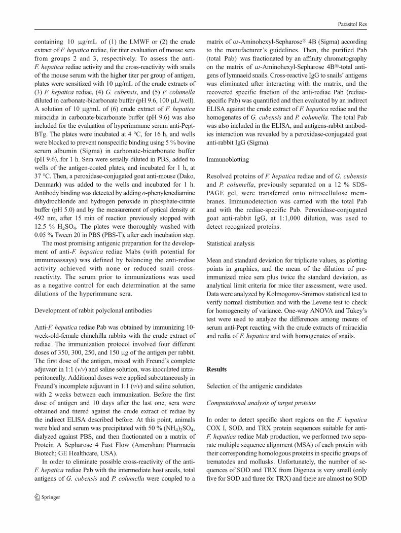
The case of COX I was different as 44 sequences ofDigenea and 19 sequences of Lymnaeidae were available foranalysis. The similarity among the COX I sequences of flukeswas high (ranging from 72 to 92 %), as expected for a well-conserved protein, although not all the sequences analyzedwere fully annotated (with most fragments missing at thebeginning and at the end) (see Supplementary Material 1.1for detail). Four segments of the F. hepatica sequence withlow identity percent among the flukes were identified (namedB1–B4) and are shown in Fig. 1c, d.
The percentage of sequence identity of COX I with theproteins from mollusks ranged from 50 to 56 %, due to theincompleteness of the snail sequences, as only the first half ofthe sequences (from 1 to around 224 residues) was available forcomparison (except for the Galba pervia species). Therefore,we decided to take into account complete sequences of COX Ifrom the taxonomic clade Panpulmonata, which includes snailsand slugs. Those new sequences (a total of 25) were realigned
to F. hepatica and G. pervia. As expected, the first half of theMSA was very similar to the one constructed with theLymnaeidae orthologues. On the MSA of F. hepatica-molluskthe nine segments that shared a low identity percent with therest of the proteins were identified (named A1–A9) (seeSupplementary Material 1.2 for details of the MSA).
The four segments identified as specific of F. hepatica onthe MSA of flukes were also included (and denoted) on thoseidentified from the MSA of mollusks (A2/B1, A4/B2, A5/B3,A9/B4) and are shown in Fig. 1c, d. The A2/B1 segment islocated within a highly hydrophobic α-helix (see Fig. 1a, b forhydrophobicity analysis) and therefore is not suitable forchemical synthesis (Angeletti 1999). In designing potentialantigenic epitopes, it is important to select a hydrophilic andsurface oriented-segment of 12-to-16-residue length. Thesefeatures are often present in the N- and C-termini of proteinsthat are often exposed and have a high degree of flexibility(Angeletti 1999). Therefore, we selected a sequence of 14residues (QHNSYMNGVGRWVF) of theA9/B4 segment thatis part of the C-terminal of the COX I (Fig. 1d). This sequenceformed a variable and exposed segment of random conforma-tion within a well-conserved transmembrane protein. Thesecharacteristics make the selected fragment quite suitable forchemical synthesis and further evaluation as a pertinent candi-date for specific anti-F. hepatica Mab development.
Analysis of the antigenic composition of F. hepatica rediaeby protein electrophoresis
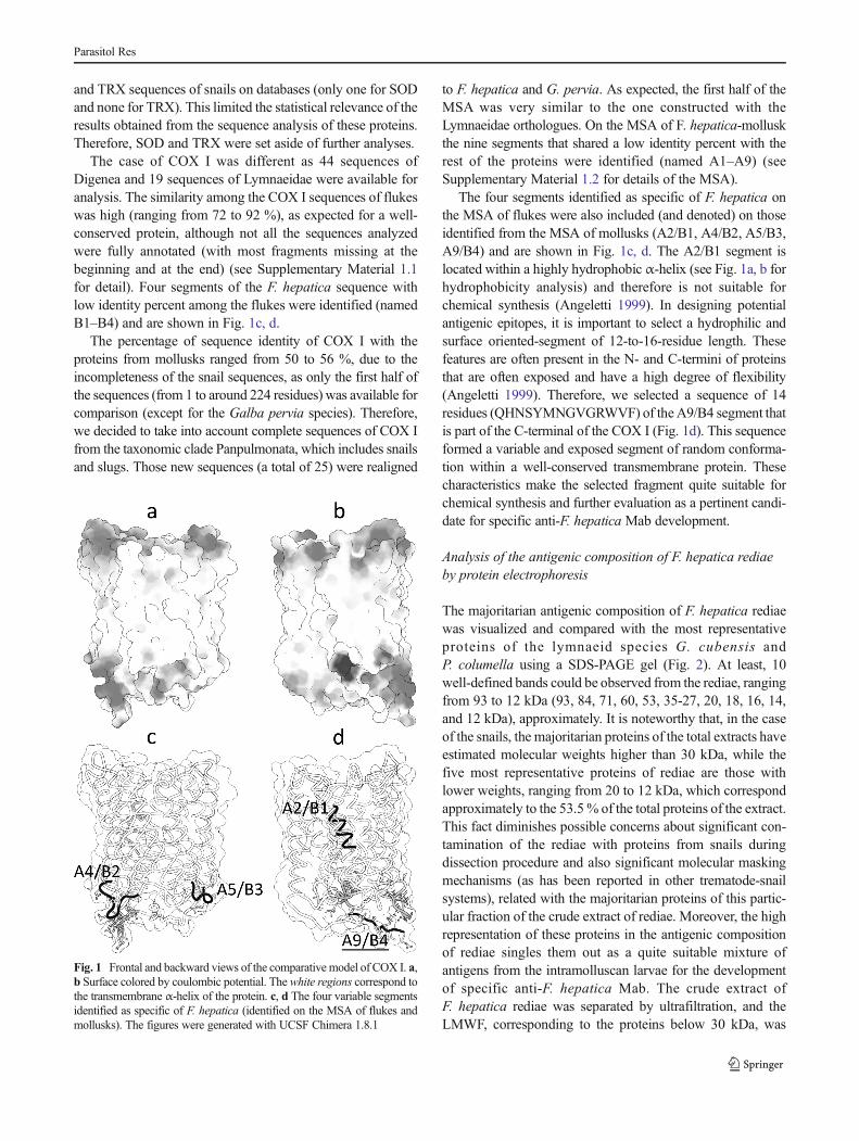
The majoritarian antigenic composition of F. hepatica rediaewas visualized and compared with the most representativeproteins of the lymnaeid species G. cubensis andP. columella using a SDS-PAGE gel (Fig. 2). At least, 10well-defined bands could be observed from the rediae, rangingfrom 93 to 12 kDa (93, 84, 71, 60, 53, 35-27, 20, 18, 16, 14,and 12 kDa), approximately. It is noteworthy that, in the caseof the snails, the majoritarian proteins of the total extracts haveestimated molecular weights higher than 30 kDa, while thefive most representative proteins of rediae are those withlower weights, ranging from 20 to 12 kDa, which correspondapproximately to the 53.5% of the total proteins of the extract.This fact diminishes possible concerns about significant con-tamination of the rediae with proteins from snails duringdissection procedure and also significant molecular maskingmechanisms (as has been reported in other trematode-snailsystems), related with the majoritarian proteins of this partic-ular fraction of the crude extract of rediae. Moreover, the highrepresentation of these proteins in the antigenic compositionof rediae singles them out as a quite suitable mixture ofantigens from the intramolluscan larvae for the developmentof specific anti-F. hepatica Mab. The crude extract ofF. hepatica rediae was separated by ultrafiltration, and theLMWF, corresponding to the proteins below 30 kDa, was
Fig. 1 Frontal and backward views of the comparative model of COX I. a,b Surface colored by coulombic potential. The white regions correspond tothe transmembrane α-helix of the protein. c, d The four variable segmentsidentified as specific of F. hepatica (identified on the MSA of flukes andmollusks). The figures were generated with UCSF Chimera 1.8.1
Parasitol Res
used for mouse immunization along with the other twoantigenic preparations (synthetic C-terminal of the COXI (Pept-BTg) and the crude extract of rediae).
Evaluation of mice hyperimmune sera
The titer of mouse hyperimmune sera anti- each one of thethree antigenic candidates was evaluated by indirect ELISAsusing plates coated with each antigen. The synthetic C-terminal COX I peptide and the LMWF of the homogenateof F. hepatica rediae induced an effective stimulation of theimmune system ofmicewith antibody titer of 1/20,000 in bothcases. The higher titer (1/40,000) was achieved in mice im-munized with the crude extract of F. hepatica rediae.
The mouse serum with the higher titer per group wasevaluated against the crude extract of F. hepatica rediae inorder to determine parasite recognition. In the same way, eachserum was also evaluated against the homogenates ofG. cubensis and P. columella to estimate possible cross-reactivity with the intermediate hosts. In Fig. 3, the results ofthe assay for the anti-Pept COX I serum diluted 1/4,000 areshown. In this case, the crude extract of F. hepatica miracidiawas also included and there are significant differences be-tween the recognition of rediae and miracidia. Even when nocross-reactivity with lymnaeid snails was attained with thisserum (as was expected from the in silico analysis), thepolyclonal reaction against the crude extract of rediae wasdiscrete (Fig. 3). No significant recognition of this larval stagewas achieved at serum dilution higher than 1/4,000.Therefore, COX I might not be an appropriate antigen forthe development of anti-F. hepatica rediae Mabs aimed to
detect the parasite within the snails due to its low representa-tion on the total extract of this particular stage.
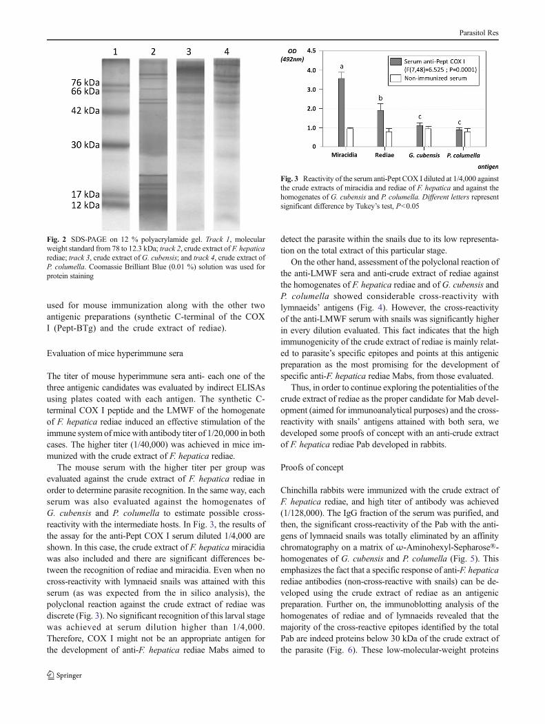
On the other hand, assessment of the polyclonal reaction ofthe anti-LMWF sera and anti-crude extract of rediae againstthe homogenates of F. hepatica rediae and of G. cubensis andP. columella showed considerable cross-reactivity withlymnaeids’ antigens (Fig. 4). However, the cross-reactivityof the anti-LMWF serum with snails was significantly higherin every dilution evaluated. This fact indicates that the highimmunogenicity of the crude extract of rediae is mainly relat-ed to parasite’s specific epitopes and points at this antigenicpreparation as the most promising for the development ofspecific anti-F. hepatica rediae Mabs, from those evaluated.
Thus, in order to continue exploring the potentialities of thecrude extract of rediae as the proper candidate for Mab devel-opment (aimed for immunoanalytical purposes) and the cross-reactivity with snails’ antigens attained with both sera, wedeveloped some proofs of concept with an anti-crude extractof F. hepatica rediae Pab developed in rabbits.
Proofs of concept
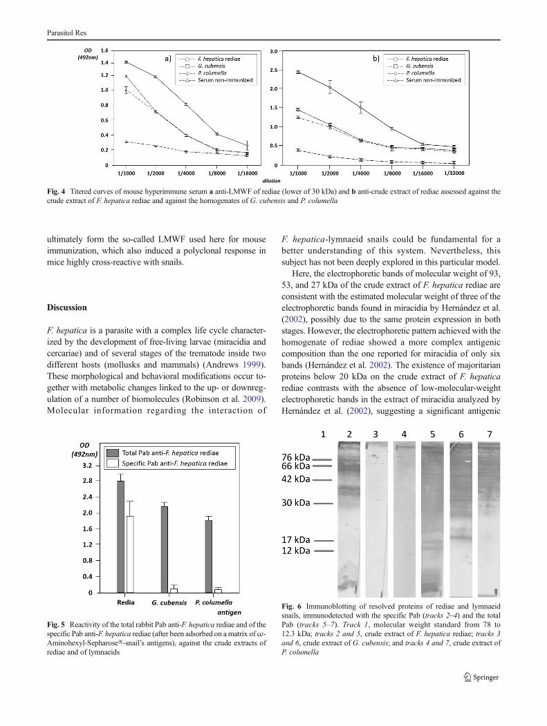
Chinchilla rabbits were immunized with the crude extract ofF. hepatica rediae, and high titer of antibody was achieved(1/128,000). The IgG fraction of the serum was purified, andthen, the significant cross-reactivity of the Pab with the anti-gens of lymnaeid snails was totally eliminated by an affinitychromatography on a matrix of ω-Aminohexyl-Sepharose®-homogenates of G. cubensis and P. columella (Fig. 5). Thisemphasizes the fact that a specific response of anti-F. hepaticarediae antibodies (non-cross-reactive with snails) can be de-veloped using the crude extract of rediae as an antigenicpreparation. Further on, the immunoblotting analysis of thehomogenates of rediae and of lymnaeids revealed that themajority of the cross-reactive epitopes identified by the totalPab are indeed proteins below 30 kDa of the crude extract ofthe parasite (Fig. 6). These low-molecular-weight proteins
Fig. 3 Reactivity of the serum anti-Pept COX I diluted at 1/4,000 againstthe crude extracts of miracidia and rediae of F. hepatica and against thehomogenates of G. cubensis and P. columella. Different letters representsignificant difference by Tukey’s test, P<0.05
Fig. 2 SDS-PAGE on 12 % polyacrylamide gel. Track 1, molecularweight standard from 78 to 12.3 kDa; track 2, crude extract of F. hepaticarediae; track 3, crude extract of G. cubensis; and track 4, crude extract ofP. columella. Coomassie Brilliant Blue (0.01 %) solution was used forprotein staining
Parasitol Res
ultimately form the so-called LMWF used here for mouseimmunization, which also induced a polyclonal response inmice highly cross-reactive with snails.
Discussion
F. hepatica is a parasite with a complex life cycle character-ized by the development of free-living larvae (miracidia andcercariae) and of several stages of the trematode inside twodifferent hosts (mollusks and mammals) (Andrews 1999).These morphological and behavioral modifications occur to-gether with metabolic changes linked to the up- or downreg-ulation of a number of biomolecules (Robinson et al. 2009).Molecular information regarding the interaction of
F. hepatica-lymnaeid snails could be fundamental for abetter understanding of this system. Nevertheless, thissubject has not been deeply explored in this particular model.
Here, the electrophoretic bands of molecular weight of 93,53, and 27 kDa of the crude extract of F. hepatica rediae areconsistent with the estimated molecular weight of three of theelectrophoretic bands found in miracidia by Hernández et al.(2002), possibly due to the same protein expression in bothstages. However, the electrophoretic pattern achieved with thehomogenate of rediae showed a more complex antigeniccomposition than the one reported for miracidia of only sixbands (Hernández et al. 2002). The existence of majoritarianproteins below 20 kDa on the crude extract of F. hepaticarediae contrasts with the absence of low-molecular-weightelectrophoretic bands in the extract of miracidia analyzed byHernández et al. (2002), suggesting a significant antigenic
Fig. 4 Titered curves of mouse hyperimmune serum a anti-LMWF of rediae (lower of 30 kDa) and b anti-crude extract of rediae assessed against thecrude extract of F. hepatica rediae and against the homogenates of G. cubensis and P. columella
Fig. 5 Reactivity of the total rabbit Pab anti-F. hepatica rediae and of thespecific Pab anti-F. hepatica rediae (after been adsorbed on a matrix ofω-Aminohexyl-Sepharose®-snail’s antigens), against the crude extracts ofrediae and of lymnaeids
Fig. 6 Immunoblotting of resolved proteins of rediae and lymnaeidsnails, immunodetected with the specific Pab (tracks 2–4) and the totalPab (tracks 5–7). Track 1, molecular weight standard from 78 to12.3 kDa; tracks 2 and 5, crude extract of F. hepatica rediae; tracks 3and 6, crude extract of G. cubensis; and tracks 4 and 7, crude extract ofP. columella
Parasitol Res
variation between the two larval stages. This antigenic varia-tion could be related to the lower recognition of the crudeextract of rediae by the anti-Pept serum compared to thepolyclonal reaction assessed against the homogenate of mira-cidia. Even when in later aerobic metabolism of F. hepaticarediae is possible (Humiczewska 1975), oxygen availabilityfor the parasite larvae inside the snails is lower compared tothe natural environment where the free-living miracidia oc-curs. This might affect the level of expression of aerobicproteins like the COX I between these stages. In fact, differ-ential expression of COX complex among different develop-mental stages of species from other phyla has been reported(Liénard et al. 2006). On the other hand, transitions in energymetabolism have been described and characterized on thefully aerobic functioning of F. hepatica juvenile to the almostcompletely anaerobic metabolism of the adult parasites(extensively reviewed by Tielens (1999)). The results alreadydiscussed could be an interesting starting point to furtherexplore the regulation of the energy pathways in miracidia-sporocyst-rediae switching larvae of F. hepatica.
A complex fraction of low-molecular-weight proteins is animportant component of F. hepatica sporocyst, particularly ofthe excretory-secretory antigens (Gourbal et al. 2008), as itturned out to be for the crude extract of rediae. Gourbal et al.(2008) identified the antioxidative enzymes SOD and TRX asmolecular weight dots of 16 and 12 kDa, respectively, on abidimensional electrophoresis. Regarding the important func-tion of these proteins in the oxidative stress that occurs duringthe immune response of the snail Biomphalaria glabrataagainst Schistosoma mansoni sporocyst (Mourão et al. 2009)and in mammalian immunity against F. hepatica adults(Salazar-Calderón et al. 2001; Piedrafita et al. 2007), it isreasonable to infer that SOD and TRX could also be part ofthe antigenic composition of rediae (electrophoretic bands of16 and 12 kDa identified in this study). Unfortunately, thescarce number of sequences of SOD and TRX of flukes andmollusks on databases limited the statistical relevance of theresults from the in silico analyses.
The existence of a polyclonal reaction against snails’ anti-gens developed in mice and rabbits immunized with the crudeextract of F. hepatica rediae, and especially in mice immu-nized with the LMWF, supports the idea of possible mecha-nisms of host immune evasion through parasite molecularmasking and/or mimicry. These evasion mechanisms havebeen reported for other trematode-snail systems (Yoshinoand Bayne 1983; Adema and Loker 1997; Yoshino et al.2013), but none previously studied on the F. hepatica-lymnaeid model. The immunoblotting assay with the totalanti-rediae Pab showed a marked recognition of the antigensfrom both parasite and mollusks, while the specific Pabreacted only with the proteins of the rediae of molecularweights higher than 30 kDa. This constitutes the first evidenceof the possible existence of immunogenic mimetic epitopes in
intramolluscan stages of F. hepatica and suggests that themarked cross-reactivity with snails achieved with mouseanti-LMWF serum was due to a molecular mimicry phenom-enon. The high representation of this particular low-molecular-weight fraction on the crude extract of F. hepaticarediae (and the nonexistence of these electrophoretic bandsreported in miracidia), as well as the evidence of signifyingmimicry between antigens of parasite and snails, leads us tothink of important roles for these proteins during theintramolluscan development of the parasite.
Parasites like F. hepatica are masters of a variety of strat-egies for survival, and new preliminary insights into themolecular features of F. hepatica rediae have been discussedin order to extend the knowledge about parasite-snail interac-tion. Antigenic variation among different stages allows theparasite to overcome an amazing diversity of hostile environ-ments from free-living nonfeeding larvae to snails and mam-malian hosts, ensured by accurate regulation routes. Also,parasite evasion mechanisms of host immunity like molecularmimicry elude the self-nonself-recognition, thus compromis-ing the effective activation of defense strategies. Further stud-ies are required to achieve a more comprehensive overview ofthe development of intramolluscan stages of F. hepatica.
In terms of Mab generation for detecting ongoingF. hepatica infection in snails, the redia rises as the mostpromising larval stage not only because it is the long-lastingintramolluscan larva (Rondelaud et al. 2009) but also for thediscussed evidences of antigenic variations between differentstages of the parasite that occurred within the snails.Moreover, the existence of specific immunodominant epi-topes in the crude extract of this stage, as was assessed withthe specific rabbit anti-rediae Pab, strongly supports this view.These results provide a feasible initiation point for futureinvestigations into immunoassay development aimed at theepidemiological surveillance of snail populations of interme-diate hosts of F. hepatica.
Acknowledgments The authors would like to thank Dr. Osvaldo Reyesfrom the Havana Center of Genetic Engineering and Biotechnology forthe assistance on the peptide synthesis and conjugation. We would alsolike to thank an anonymous reviewer for the proper comments andsuggestions. In addition, we would like to express our thanks to the editorwho smoothed the English.
References
Adema CM, Loker ES (1997) Specificity and immunobiology of larvaldigenean-snail associations. In: Fried B, Graczyk TK (eds) Advancesin trematode biology. CRC, Boca Raton, USA, pp 229–263
Andrews SJ (1999) The life cycle of Fasciola hepatica. In: Dalton JP (ed)Fasciolosis. CAB International, Wallingford, UK, pp 1–30
Angeletti RH (1999) Design of useful peptide antigens. J Biomol Tech10(1):2–10
Parasitol Res
CaronY, RondelaudD, RossonB (2008) The detection and quantificationof a digenean infection in the snail host with emphasis in Fasciolasp. Parasitol Res 103:735–744
Caron Y, Righi S, Lempereur L, Saegerman C, Losson B (2011)An optimized DNA extraction and multiplex PCR for thedetection of Fasciola sp. in lymnaeid snails. Vet Parasitol178(1–2):93–99
Espinoza JR, Maco V, Marcos L, Saez S, Neyra V, Terashima A,Salmavides F, Gotuzzo E, Chavarry E, Huaman MC, BarguesMD, Valero MA, Mas-Coma S (2007) Evaluation of Fas2-ELISAfor the serological detection of Fasciola hepatica infection inhumans. Am J Trop Med Hyg 76(5):977–982
Gourbal BEF, Guillou F, Mitta G, Sibille P, Thèron A, Pointier JP,Coustau C (2008) Excretory–secretory products of larval Fasciolahepatica investigated using a two-dimensional proteomic approach.Mol Biochem Parasitol 161:63–66
Hernández H, Marcet R, Martínes AR, Gutiérrez A, Sánchez J, SarracentJ (2002) Obtención y caracterización de anticuerpos monoclonalescontra antígenos de miracidios de Fasciola hepatica. J Med ApplMalacol 11:35–40
Humiczewska M (1975) Oxidative enzymes in the development ofFasciola hepatica L. IV. The activity of oxidases and dehydroge-nases in redia. Folia Histochem Cytochem (Krakow) 13(3–4):161–174
Kaplan RM, Dame JB, Reddy GR, Courtney CH (1997) The prevalenceof Fasciola hepatica in its snail intermediate host determined byDNA probe assay. Int J Parasitol 27(12):1585–1593
Laemli U (1970) Clevage of structural proteins during the assembling ofthe head of bacteriophage T4. Nature 227:680–685
Liénard MA, Lassance JM, Paulmier I, Picimbon JF, Löfstedt C (2006)Differential expression of cytochrome c oxidase subunit III gene incastes of the termite Reticulitermes santonensis. J Insect Physiol52(6):551–557
Mas-Coma S, Bargues MD, Valero MA (2009) Chapter 2.Fasciola, lymnaeids and human fascioliasis, with a globaloverview on disease transmission, epidemiology, evolutionarygenetics, molecular epidemiology and control. Adv Parasitol69:41–146
Merrifield B (1986) Solid phase synthesis. Science 232:341–347Mourão MM, Dinguirard N, Franco GR, Yoshino TP (2009) Role of the
endogenous antioxidant system in the protection of Schistosomamansoni primary sporocysts against exogenous oxidative stress.PLoS Negl Trop Dis 3(11):e550. doi:10.1371/journal.pntd.0000550
Musatov A, Ortega-Lopez J, Robinson NC (2000) Detergent-solubilizedbovine cytochrome c oxidase: dimerization depends on the amphi-philic environment. Biochemistry 39(42):12996–13004
Piedrafita D, Estuningsih E, Pleasance J, Prowse R, Raadsma HW,Meeusen EN, Spithill TW (2007) Peritoneal lavage cells ofIndonesian thin-tail sheep mediate antibody-dependent superoxideradical cytotoxicity in vitro against newly excysted juvenileFasciola gigantica but not juvenile Fasciola hepatica. InfectImmun 75(4):1954–1963
Robinson MW, Menon R, Donnelly SM, Dalton JP, Ranganathan S(2009) An integrated transcriptomics and proteomics analysis ofthe secretome of the helminth pathogen Fasciola hepatica: proteinsassociated with invasion and infection of the mammalian host. MolCell Proteomics 8(8):1891–1907
Rojas L, Vázquez AA, Domenech I, Robertson L (2010) Fascioliasis: canCuba conquer this emerging parasitosis? Trends Parasitol26(1):26–34
Rondelaud D, Belfaiza M, Vignoles P, Moncef M, Dreyfuss G (2009)Redial generations of Fasciola hepatica: a review. J Helminthol 83:245–254
Salazar-Calderón, Martín-Alonso JM, Ruiz de Eguino AD, Parra F(2001) Heterologous expression and functional characterization ofthioredoxin from Fasciola hepatica. Parasitol Res 87(5):390–395
Smith PK, Krohn RI, Hermanson GT (1986) Measurement of proteinusing bicinchoninic acid. Anal Biochem 150:76–85
Tielens AGM (1999) Metabolism. In: Dalton JP (ed) Fasciolosis. CABInternational, UK, pp 277–306
Ubeira FM, Muiño L, ValeroMA, PeriagoMV, Pérez-Crespo I, MezoM,González-Warleta M, Romarís F, Paniagua E, Cortizo S, Llovo J,Más-Coma S (2009) MM3-ELISA detection of Fasciola hepaticacoproantigens in preserved human stool samples. Am J Trop MedHyg 81:156–162
Vázquez AA, Sánchez J, Pointier JP, Théron A, Hurtrez-Boussès S (2013)Fasciola hepatica in Cuba: compatibility of different isolates with twointermediate intermediate hosts, Galba cubensis and Pseudosuccineacolumella. J Helminthol. doi:10.1017/S0022149X13000382
Yoshino TP, Bayne CJ (1983)Mimicry of snail host antigens bymiracidiaand primary sporocysts of Schistosoma mansoni. Parasite Immunol5:317–328
Yoshino TP, Wu XJ, Gonzalez LA, Hokke CH (2013) CirculatingBiomphalaria glabrata hemocyte subpopulations possess sharedschistosome glycans and receptors capable of binding larvalglycoconjugates. Exp Parasitol 133(1):28–36
Parasitol Res