Mobile Regulatory Cassettes Mediate Modular Shuffling in T4-Type Phage Genomes
Upload
independentCategory
view
1download
0
Archives of Biochemistry and Biophysics 425 (2004) 14–24
ABBwww.elsevier.com/locate/yabbi
Glutathione and thioredoxin peroxidases mediate susceptibilityof yeast mitochondria to Ca2þ-induced damage
Gisele Monteiro,a Alicia J. Kowaltowski,b Mario H. Barros,b and Luis E.S. Nettoa,*
a Departamento de Biologia—Gen�etica, Instituto de Biociencias, Universidade de S~ao Paulo, Rua do Mat~ao 277, CEP05508-900, S~ao Paulo, SP, Brazilb Departamento de Bioqu�ımica, Instituto de Qu�ımica, Universidade de S~ao Paulo, Brazil
Received 29 December 2003, and in revised form 27 February 2004
Abstract
The effect of thioredoxin peroxidases on the protection of Ca2þ-induced inner mitochondrial membrane permeabilization was
studied in the yeast Saccharomyces cerevisiae using null mutants for these genes. Since deletion of a gene can promote several other
effects besides the absence of the respective protein, characterizations of the redox state of the mutant strains were performed. Whole
cellular extracts from all the mutants presented lower capacity to decompose H2O2 and lower GSH/GSSG ratios, as expected for
strains deficient for peroxide-removing enzymes. Interestingly, when glutathione contents in mitochondrial pools were analyzed, all
mutants presented lower GSH/GSSG ratios than wild-type cells, with the exception of DcTPxI strain (cells in which cytosolic
thioredoxin peroxidase I gene was disrupted) that presented higher GSH/GSSG ratio. Low GSH/GSSG ratios in mitochondria
increased the susceptibility of yeast to damage induced by Ca2þ as determined by membrane potential and oxygen consumption
experiments. However, H2O2 removal activity appears also to be important for mitochondria protection against permeabilization
because exogenously added catalase strongly inhibited loss of mitochondrial potential. Moreover, exogenously added recombinant
peroxiredoxins prevented inner mitochondrial membrane permeabilization. GSH/GSSG ratios decreased after Ca2þ addition,
suggesting that reactive oxygen species (ROS) probably mediate this process. Taken together our results indicate that both mito-
chondrial glutathione pools and peroxide-removing enzymes are key components for the protection of yeast mitochondria against
Ca2þ-induced damage.
� 2004 Elsevier Inc. All rights reserved.
Keywords: Calcium; Oxidative stress; Mitochondria; Thioredoxin peroxidase; Yeast; Thiol; Glutathione
Sulfhydryl groups are important components of cel-lular defense against oxidative stress and for the main-
tenance of the redox homeostasis of cells (review in [1]).
In eukaryotes, the major thiol compound is the peptide
glutathione (GSH)1 [2], whereas thioredoxin and glut-
aredoxin are important protein sources of sulfhydryl
* Corresponding author. Fax: +55-11-30917553.
E-mail address: [email protected] (L.E.S. Netto).1 Abbreviations used: cTPxI, cytosolic thioredoxin peroxidase I
protein also known as Tsa1p; DTNB, 5-50-dithiobis(2-nitrobenzoic
acid); TSA1, cytosolic thioredoxin peroxidase I gene (ORF:
YML028w); cTPxII, cytosolic thioredoxin peroxidase II protein also
known as Tsa2p; TSA2, cytosolic thioredoxin peroxidase II gene
(ORF: YDR453c); cTPxIII, cytosolic thioredoxin peroxidase III also
known as Ahp1, type II TPx or TSA II (ORF: YLR109w); mTPxI
mitochondrial thioredoxin peroxidase I also known as Prx1 (ORF:
YBL064c); nTPxI, nuclear thioredoxin peroxidase I protein also known
0003-9861/$ - see front matter � 2004 Elsevier Inc. All rights reserved.
doi:10.1016/j.abb.2004.03.005
groups in the cell. Both glutathione and thioredoxin canfunction as electron donors for thiol-dependent peroxi-
dases. In the case of Saccharomyces cerevisiae, at least
three glutathione phospholipid peroxidases and five
thioredoxin peroxidase isoforms are expressed [3–5].
Glutathione and thioredoxin systems are closely linked
[1]. In fact, disruption of CTPXI gene (also named
TSA1) leads to increase in de novo synthesis and recy-
cling of glutathione [3].
as DOT5 (Disruptor Of Telomeric silencing) (ORF: YIL010w), GSH,
glutathione; GSSG, oxidized glutathione; GSH/GSSG, ratio between
reduced and oxidized form of glutathione; RNS, reactive nitrogen
species; ROS, reactive oxygen species; TNB, 2-nitro-5-thiobenzoic-
acid; TRR1, thioredoxin reductase I gene (ORF: YDR353w); Trr1,
thioredoxin reductase I protein; TRX2, thioredoxin 2 gene (ORF:
YGR209c); Trx2, thioredoxin 2 protein.

G. Monteiro et al. / Archives of Biochemistry and Biophysics 425 (2004) 14–24 15
Among the five yeast thioredoxin peroxidases, cyto-solic thioredoxin peroxidase I (cTPxI) is the most
studied. It is very abundant, even under basal conditions
[6]. As expected from its enzymatic activity, deletion of
the TSA1 gene (that codifies for cTPxI protein) slightly
increases the sensitivity of cells to peroxides [5,7], espe-
cially when mitochondria are not functional [8]. Inter-
estingly, besides its antioxidant role, cTPxI is also
involved in the regulation of the expression and activityof several other genes such as GSH1 (c-glutamylcysteine
synthetase), GPX2 (glutathione peroxidase), GLR1
(glutathione reductase), TRX2 (thioredoxin), TRR1
(thioredoxin reductase), and YAP1 (transcriptional
regulator) [3,9].
cTPxI belongs to a family of proteins named perox-
iredoxins [10]. The other yeast peroxiredoxins are less
studied. Cytosolic thioredoxin peroxidase II (cTPxII)shares 86% of identity with cTPxI and therefore they are
expected to behave similarly. However, the peroxidase
activity of cTPxI is about sixfold higher than the activity
of cTPxII [5]. On the other hand, in vivo studies indi-
cated that cTPxI and cTPxII are equally important in
the defense of yeast against reactive oxygen species
(ROS) and reactive nitrogen species (RNS) [11]. Con-
trary to cTPxI, cTPxII expression level is undetectableunder basal conditions, but is highly inducible by per-
oxides [12]. The only mitochondrial peroxiredoxin in
yeast, named mitochondrial thioredoxin peroxidase I
(mTPxI), shares about 30% of identity with the other
two peroxidases and has only one cysteine involved in
the catalytic cycle [13]. The expression level of mTPxI is
very low in fermentative supporting media and increases
about tenfold in respiratory supporting media [14].There are two other peroxiredoxin proteins in S. ce-
revisiae: cytosolic thioredoxin peroxidase III (cTPxIII)
and nuclear thioredoxin peroxidase I (nTPxI). These
two proteins also possess thioredoxin-dependent per-
oxidase activity, but they have very low similarity with
cTPxI, cTPxII, and mTPxI [5]. cTPxIII is abundant as
cTPxI, but it is at least one order of magnitude more
efficient in the removal of organic peroxide than in thedecomposition of H2O2 (because of this, cTPxIII is also
named alkyl hydroperoxidase—AhpI) [15]. nTPxI also
has higher peroxidase activity towards alkyl peroxides
and is preferentially expressed during the stationary
phase [16].
Besides the levels of antioxidant proteins, mitochon-
drial activity is also important for the maintenance of
redox balance of eukaryotic cells since this organelle is aconstant source of free radicals. Mitochondrial activity
can be impaired by processes such as oxidative stress
and increases in the content of cytosolic Ca2þ. In
mammalian mitochondria, Ca2þ induces permeabiliza-
tion of the inner mitochondrial membrane through a
process known as mitochondrial permeability transition
(MPT) [17,18], a phenomenon that has been implicated
in processes such as cell injury and death [19]. MPTpromotes swelling, a loss of the inner mitochondrial
membrane potential, and the ability to produce ATP
among other factors. Another important aspect related
to MPT is that oxidation of thiol groups in proteins
present in the inner mitochondrial membrane has been
implicated as a major cause of this permeabilization
[20,21]. Thiol oxidation causes protein cross-linkage,
formation of large protein aggregates, and opening of anon-selective pore in the inner mitochondrial membrane
[17,22,23].
Because thiols are important in both peroxiredoxin
enzymatic activity and mitochondrial integrity, we are
interested in the role of these proteins in the physiology
of this organelle. We have shown before that yeast
cTPxI and catalase cooperate in the protection of
mammalian mitochondria against MPT induced byCa2þ and oxidant agents [18]. Interestingly, cTPxI seems
to use mitochondrial thiols as electron donors to de-
compose peroxides [18]. Similarly, endogenous cTPxI
and catalase also protect yeast mitochondria from Ca2þ-promoted membrane permeabilization [24]. The experi-
ments reported in this paper were designed to determine
the relative importance of different thioredoxin peroxi-
dase isoforms in the maintenance of mitochondrial in-tegrity and function. Emphasis was given to cTPxII and
mTPxI since these proteins share high similarity with
cTPxI. The possible effect of cTPxIII was also investi-
gated, but because mitochondria from the DcTPxIIIstrain were not more sensitive to Ca 2þ-induced injury
than wild-type mitochondria, cTPxIII was not further
considered. nTPxI was not studied here because it is
located farther away from mitochondria (separated bytwo membranes: nuclear and outer mitochondrial) and
because this protein has low specific activity [5].
Interestingly, we also show in this report that deletion
of genes encoding peroxiredoxins altered the metabo-
lism of glutathione, which is in agreement with the close
relationship between glutathione and thioredoxin sys-
tems [1,3].
Experimental procedures
Yeast strains and growth conditions
Yeast strains used here were obtained from EURO-
SCARF:
Wild type (BY4741; Mata; his3D1; leu2D0; met15D0;ura3D0); DmTPxI (BY4741; Mata; his3D1; leu2D0;met15D0; ura3D0;YBL064c::kanMX4);DcTPxI (BY4742;Mata; his3D1; leu2D0; lys2D0; ura3D0; YML028w::
kanMX4); DcTPxII (BY4741; Mata; his3D1; leu2D0;met15D0; ura3D0; YDR453c::kanMX4); and DcTPxIII(BY4741; Mata; his3D1; leu2D0; met15D0; ura3D0;YLR109w::kanMX4).

16 G. Monteiro et al. / Archives of Biochemistry and Biophysics 425 (2004) 14–24
Yeast was pre-inoculated in 10ml YPGAL (2% ga-lactose, 2% peptone, and 1% yeast extract) for 24 h, at
30 �C, with 250 rpm shaking. One milliliter of the culture
was then inoculated in 150ml of fresh media and incu-
bated overnight under the same conditions. When cells
were grown in YEPG (2% glycerol, 2% ethanol, 2%
peptone, and 1% yeast extract), the pre-inoculated cul-
ture was incubated at 30 �C, 250 rpm for 48 h and the
inoculated culture was incubated for 36 h. Yeast catalaseT (cytosolic) and/or catalase A (peroxisomal) were spe-
cifically inhibited in the wild-type strain by addition of
3-amino-1,2,4-triazole (ATZ—5mM) in the pre-inocu-
lation and inoculation media. After this procedure, cells
reached OD600 nm around 6.0, therefore they were con-
sidered to be in stationary growth phase.
Determination of protein concentration
Protein concentration was determined with the
Bradford reagent from BIORAD, using bovine serum
albumin as a standard.
H2O2 removal activity
Total protein extracts from yeast cells were obtainedby lysing spheroblasts previously digested with lyticase
(Sigma—Lyticase from Arthrobacter luteus—L4025),
with a buffer containing 10mM Tris–HCl, pH 7.5, and
0.5mM EDTA (TE buffer). Fluorimetric assays were
conducted using the spheroblast samples to determine
total H2O2 removal activity. After incubating 0.5mg of
recently (<3 h) lysed spheroblast protein in 100 ll TEbuffer for 3min with 2mM H2O2, a 1 ll aliquot wastaken and diluted in 2ml of suspension buffer with
50 lM Amplex Red and 1.0U/ml horseradish peroxi-
dase [25]. A single reading was taken at 563 nm excita-
tion and 587 nm emission.
Preparation of mitochondrially-enriched fractions
Yeast cultures were harvested and washed with 30mlof 1.2M sorbitol. Cells were then incubated with 3ml/g
of digestion buffer (1.2M sorbitol, 75mM sodium
phosphate, 1mM EDTA, 0.01 volume b-mercap-
toethanol, and 0.4mg/ml lyticase) and after 2 h of in-
cubation at 37 �C, the obtained spheroblasts were
harvested and washed twice with 30ml of 1.2M sorbitol.
Pellets were then suspended in 3ml/g lyses buffer (0.5M
sorbitol, 10mM Tris–HCl, pH 7.5, and 1mM EDTA)and homogenized with a potter. The suspension was
harvested twice (3500g, 10min, 4 �C) to precipitate the
cellular debris and the supernatant was then harvested
(14,000 g, 10min, 4 �C) to obtain the mitochondrially
enriched fraction. This fraction was washed once with
10ml of lyses buffer and the final fraction was suspended
in 10mg/ml protein in lyses buffer. The procedure de-
scribed here is the same as that developed by Faye et al.[26], except for the fact that glusulase was replaced by
lyticase. Quality of our mitochondrial preparations was
verified by determinations of respiratory control ratio.
Besides, the integrity of our mitochondrial preparations
could also be demonstrated by the fact that they can
mount a potential in the inner membrane and because
they consume oxygen.
Measurement of mitochondrial matrix Ca2þ content
The procedure was carried out as described by Ko-
waltowski et al. [27], with some modifications. Mitoc-
hondrially enriched fractions (650 lg/ml) were loaded
with the fluorescent Ca2þ indicator fura-2/AM (10 lM—
Sigma) in buffer containing 240mM sucrose, 10mM
Hepes buffer, pH 7.4, 0.1mM EGTA, and 1mg/ml bo-vine serum albumin, for 30min at 37 �C. The suspensionwas diluted 10-fold and washed twice at 4 �C to elimi-
nate free extramitochondrial fura-2/AM. Fluorescence
emission at 510 nm was monitored with excitations be-
tween 300 and 400 nm, and Ca2þ uptake was determined
ratiometrically as indicated by the supplier. Maximal
and minimal intramitochondrial levels were measured in
each cell type by treating samples with 10 lM ionomycinplus Ca2þ and with 1mM EGTA.
Measurement of thiol content
Fresh mitochondrially enriched fractions were solu-
bilized with 1% SDS in a buffer containing 87mM Tris
and 4mM EDTA, pH 8.1, in the presence of 520 lM 5-
50-dithiobis(2-nitrobenzoic acid), (DTNB—also knownas Ellman�s reagent). These suspensions were homoge-
nized and incubated for 30min at room temperature in
the dark. Thiol content was determined spectrophoto-
metrically at 412 nm and calculated taking into account
that 2-nitro-5-thiobenzoic-acid (TNB) possesses
e412 nm ¼ 13,600M�1 cm�1 [28].
Determination of GSH and GSSG
Yeast cell or mitochondrially-enriched fractions were
disrupted in 1 volume of glass beads and 2 volumes of
3.5% sulfosalicylic acid. The suspension was vortexed
for 20min at 4 �C in a multifold vortex and harvested at
13,000 rpm. This procedure was repeated twice and the
supernatants were combined. Total glutathione as well
as GSSG were assayed as previously described [29].Briefly, total glutathione was determined by reaction
with DTNB (76 lM) in the presence of glutathione re-
ductase (0.12U/ml) and NADPH (0.27mM). For GSSG
determination, samples were incubated for 1 h with N-
ethylmaleimide (NEM—5mM) after adjusting the pH to
7 with NaOH. GSH concentration was determined by
the difference between total glutathione and GSSG.

G. Monteiro et al. / Archives of Biochemistry and Biophysics 425 (2004) 14–24 17
Samples were normalized by mitochondrial proteinconcentration (for analyses of the mitochondrially en-
riched fractions) or by weight of cell pellet (for analyses
of the whole cell).
Mitochondrial membrane potential (DW) measurements
Mitochondrial DW was determined through fluores-
cence changes of safranin O (5 lM), recorded on aHitachi F-4010 fluorescence spectrophotometer (Hit-
achi, Tokyo, Japan) operating at excitation and emis-
sion wavelengths of 495 and 586 nm, with a slit width
of 5 nm [30]. The fluorescence data were transformed
into DW using a Kþ distribution curve exactly as de-
scribed by Kowaltowski et al. [31]. Mitochondrially-
enriched fractions were incubated in buffer 1 (0.25M
sucrose, 10mM Hepes, pH 7.5, and 2mM Pi) con-taining 2.5mM ethanol, 5mM glutamate, and 2.5mM
succinate. DW was continuously monitored at 30 �Cwith stirring. In the experiments where exogenous
catalase was added, the enzyme was obtained from
Sigma (C9322).
Western and Northern blot analyses
Immunoblot analysis was performed using rabbit
polyclonal antibodies against cTPxI. Transfer of protein
from 12% SDS–PAGE gels to nitrocellulose and pro-
cessing of nitrocellulose blots were carried out in the
NOVEX system. The HRP-luminol based system from
ECL (RPN 2108—Amersham–Pharmacia Biotech) was
used to detect cTPxI bands. Northern blot studies were
carried out exactly as described by Monteiro et al. [14].The GSH1 fragment was obtained from yeast genomic
DNA using the specific primers forward (50-CGCGGATCCCATATGGGACTCTTAGCTTTGGG-30)and reverse (50-CGCAAGCTTGGATCCTTAACATT
TGCTTTCTATTGA-30). The PCR product was puri-
fied from agarose gel (0.7%) and sequenced.
Oxygen consumption measurements
Oxygen concentration was measured using a Clarke-
type electrode in a glass cuvette equipped with magnetic
stirring. The mitochondrially enriched fractions were
incubated in buffer 2 in the presence of 2.5mM ethanol,
5mM glutamate, and 2.5mM succinate and in the ab-
sence or in the presence of 500 lM Ca2þ.
Expression and purification of recombinant thioredoxin
peroxidases
Recombinant cTPxI, which possesses a N-terminal
His-tag, was expressed in Escherichia coli BL21(DE3)
strain transformed with pET15b-cTPxI plasmid (CTPXI
gene was cloned into NdeI and BamHI restriction sites
of expression vector pET15b from Novagen). Re-combinant mTPxI was expressed in the same E. coli
BL21(DE3) strain from a construct kindly provided by
Spyrou and co-workers [13]. In summary, MTPXI gene
without the first 20 codons (supposedly the mitochon-
drial target peptide) and with a substitution of codon
corresponding to Cys-38 for a codon encoding for Ser-
38 was cloned into NdeI and BamHI restriction sites of
pET-15b (Novagen). Therefore, the protein expressed inthis way also possesses a N-terminal His-tag. Finally,
the entire CTPXII gene was cloned into NdeI and
BamHI restriction sites of expression vector pPROEX
(Gibco) E. coli DH5a strain was utilized as the host for
expression.
Cells were cultured (50ml) overnight in LB+ampi-
cillin (100 lg/ml) medium, transferred to 1L of fresh
LB+ampicillin medium, and cultured further until theOD600 reached 0.6–0.8. IPTG was then added to a final
concentration of 1mM. After 3 h of incubation, cells
were harvested by centrifugation. The pellet was washed
and suspended in the start buffer: 20mM phosphate
buffer, pH 7.4 (for cTPxI and cTPxII) or 20mM Tris–
HCl buffer, pH 8.0 (for mTPxI). Two cycles of 15 s of
sonication (25% amplitude) following 30 s over ice were
applied to cell suspension. Cell extracts were kept on iceduring 1% streptomycin sulfate treatment (20min) and
the suspension was centrifuged at 31,500g for 30min to
remove nucleic acid precipitates. Finally, extracts were
applied to a nickel-affinity column (Hi-trap from
Amersham–Pharmacia Biotech). The conditions for
protein purification were optimized using the gradient
procedure for imidazole concentration described by the
manufacturer.
Results
Redox state of yeast mutants for thioredoxin peroxidase
genes
Phenotypic analysis of null mutants is sometimesdifficult because compensatory effects may occur. As an
example, the deletion of CTPXI gene in S. cerevisiae
leads to an increase in the levels of glutathione-depen-
dent proteins [3]. Therefore, a characterization of the
redox state of yeast mutants for thioredoxin peroxidase
was performed. Initially, the total H2O2 removal activity
of yeast cells was analyzed. In all cases, the consumption
of H2O2 dropped significantly in the mutant cells rela-tive to control, and cTPxI mutant cells (DcTPxI cells)
showed the most pronounced effect (Fig. 1A). These
results were expected since cTPxI is very abundant,
representing almost 1% of total cytosolic protein [6].
H2O2 removal in yeast extracts in which catalase activity
was inhibited by 3-amino-1,2,4-triazole (ATZ) also
dropped significantly (Fig. 1A).
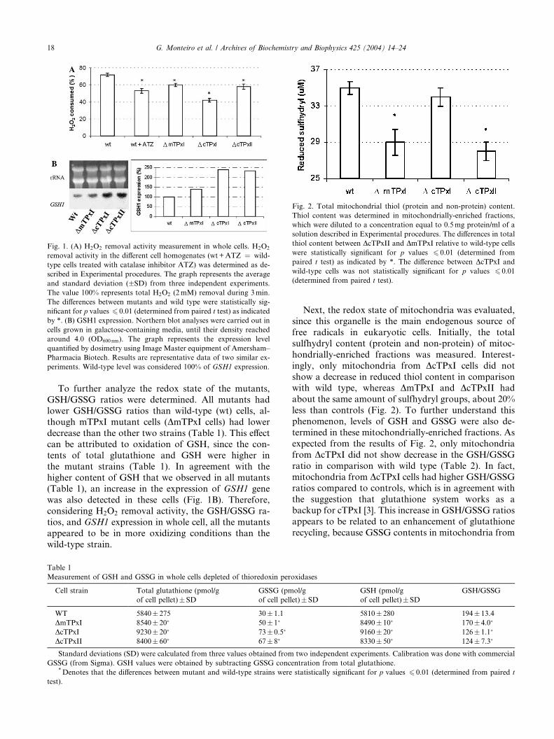
Fig. 1. (A) H2O2 removal activity measurement in whole cells. H2O2
removal activity in the different cell homogenates (wt +ATZ ¼ wild-
type cells treated with catalase inhibitor ATZ) was determined as de-
scribed in Experimental procedures. The graph represents the average
and standard deviation (�SD) from three independent experiments.
The value 100% represents total H2O2 (2mM) removal during 3min.
The differences between mutants and wild type were statistically sig-
nificant for p values 6 0.01 (determined from paired t test) as indicatedby *. (B) GSH1 expression. Northern blot analyses were carried out in
cells grown in galactose-containing media, until their density reached
around 4.0 (OD600nm). The graph represents the expression level
quantified by dosimetry using Image Master equipment of Amersham–
Pharmacia Biotech. Results are representative data of two similar ex-
periments. Wild-type level was considered 100% of GSH1 expression.
Fig. 2. Total mitochondrial thiol (protein and non-protein) content.
Thiol content was determined in mitochondrially-enriched fractions,
which were diluted to a concentration equal to 0.5mg protein/ml of a
solution described in Experimental procedures. The differences in total
thiol content between DcTPxII and DmTPxI relative to wild-type cells
were statistically significant for p values 6 0.01 (determined from
paired t test) as indicated by *. The difference between DcTPxI and
wild-type cells was not statistically significant for p values 6 0.01
(determined from paired t test).
18 G. Monteiro et al. / Archives of Biochemistry and Biophysics 425 (2004) 14–24
To further analyze the redox state of the mutants,GSH/GSSG ratios were determined. All mutants had
lower GSH/GSSG ratios than wild-type (wt) cells, al-
though mTPxI mutant cells (DmTPxI cells) had lower
decrease than the other two strains (Table 1). This effect
can be attributed to oxidation of GSH, since the con-
tents of total glutathione and GSH were higher in
the mutant strains (Table 1). In agreement with the
higher content of GSH that we observed in all mutants(Table 1), an increase in the expression of GSH1 gene
was also detected in these cells (Fig. 1B). Therefore,
considering H2O2 removal activity, the GSH/GSSG ra-
tios, and GSH1 expression in whole cell, all the mutants
appeared to be in more oxidizing conditions than the
wild-type strain.
Table 1
Measurement of GSH and GSSG in whole cells depleted of thioredoxin per
Cell strain Total glutathione (pmol/g
of cell pellet)�SD
GSSG (pm
of cell pel
WT 5840� 275 30� 1.1
DmTPxI 8540� 20� 50� 1�
DcTPxI 9230� 20� 73� 0.5�
DcTPxII 8400� 60� 67� 8�
Standard deviations (SD) were calculated from three values obtained from
GSSG (from Sigma). GSH values were obtained by subtracting GSSG conc*Denotes that the differences between mutant and wild-type strains were
test).
Next, the redox state of mitochondria was evaluated,
since this organelle is the main endogenous source of
free radicals in eukaryotic cells. Initially, the total
sulfhydryl content (protein and non-protein) of mitoc-
hondrially-enriched fractions was measured. Interest-
ingly, only mitochondria from DcTPxI cells did not
show a decrease in reduced thiol content in comparisonwith wild type, whereas DmTPxI and DcTPxII had
about the same amount of sulfhydryl groups, about 20%
less than controls (Fig. 2). To further understand this
phenomenon, levels of GSH and GSSG were also de-
termined in these mitochondrially-enriched fractions. As
expected from the results of Fig. 2, only mitochondria
from DcTPxI did not show decrease in the GSH/GSSG
ratio in comparison with wild type (Table 2). In fact,mitochondria from DcTPxI cells had higher GSH/GSSG
ratios compared to controls, which is in agreement with
the suggestion that glutathione system works as a
backup for cTPxI [3]. This increase in GSH/GSSG ratios
appears to be related to an enhancement of glutathione
recycling, because GSSG contents in mitochondria from
oxidases
ol/g
let)� SD
GSH (pmol/g
of cell pellet)�SD
GSH/GSSG
5810� 280 194� 13.4
8490� 10� 170� 4.0�
9160� 20� 126� 1.1�
8330� 50� 124� 7.3�
two independent experiments. Calibration was done with commercial
entration from total glutathione.
statistically significant for p values 6 0.01 (determined from paired t

Table 2
Measurement of mitochondrial pools of GSH and GSSG
Mitochondrial
samples
Total glutathione (pmol/mg
of protein)�SD
GSSG (pmol/mg
of protein)�SD
GSH (pmol/mg
of protein)�SD
GSH/GSSG
WT 870� 24 20� 3 850� 30 43� 7.2
DmTPxI 1140� 34� 56� 8� 1084� 42� 19� 3.32�
DcTPxI 870� 26 12� 2� 858� 28 72� 1.42�
DcTPxII 660� 12� 46� 3� 614� 15� 13� 1.06�
Standard deviations (SD) were calculated from three values obtained from two independent experiments. Calibration was done with commercial
GSSG (from Sigma). GSH values were obtained by subtracting GSSG concentration from total glutathione.*Denotes that the differences between mutant and wild-type strains were statistically significant for p values 6 0.01 (determined from paired
t test).
G. Monteiro et al. / Archives of Biochemistry and Biophysics 425 (2004) 14–24 19
DcTPxI cells were approximately half the GSSG content
in mitochondria from wild-type (Table 2).
Contrary to DcTPxI, mitochondria from DcTPxIIcells presented very low GSH/GSSG ratios (Table 2),
which was due to both low levels of total glutathione
and high amounts of GSSG. These results can account,at least in part, for the reduced amount of total sulf-
hydryl groups in their mitochondria (Fig. 2). In DmTPxI
mitochondria, the amounts of total glutathione and
GSH measured were slightly increased in comparison
with wild-type (Table 2). Therefore, the reduced amount
of sulfhydryl groups observed in mitochondria from
these mutants (Fig. 2) must reflect a lower content of
thiols from proteins. DmTPxI mitochondria also showedan increase in the levels of GSSG, indicating that the
recycling of glutathione is not functioning properly in
these organelle (Table 2). In any case, the very low GSH/
GSSG ratios observed for DcTPxII and DmTPxI indi-
cated that the mitochondria of these strains were under
more oxidizing conditions than DcTPxI and wild-type
mitochondria. It is important to note that although
GSSG values were always very low in comparison withtotal glutathione, we confirmed the results through three
independent experiments performed in triplicate.
Moreover, the differences between wild-type and mu-
tants were statistically significant with p value 6 0.01.
In summary, our data indicated that DcTPxII and
DmTPxI cells are under a more oxidizing condition than
wild-type cells. In the case of DcTPxI, whole cells ap-
peared to be in a more oxidizing condition, whereas theirmitochondria are at least equally reduced as wild-type
mitochondria. As described below, the amount of thiols
measured in these mitochondrially-enriched fractions
and peroxidase activity seem to be important factors
involved in yeast susceptibility to Ca2þ-induced damage.
Mitochondrial susceptibility to stress induced by calcium
After a characterization of the redox state of whole
cells and their mitochondria, the sensitivity of this or-
ganelle to inner membrane permeabilization by Ca2þ
was studied. We have shown before that cells deficient in
cTPxI and catalase are more prone to loss of mito-
chondrial function by Ca2þ [24]. Initially, the ability of
mitochondria from wild-type, ATZ-treated wild-type,
and mutant cells to keep inner membrane potential was
analyzed from mitochondrially-enriched fractions of
cells grown in galactose-containing media. As described
previously, mitochondria from wild-type cells were re-sistant to Ca2þ-induction of inner membrane permea-
bilization [24,32,33] whereas mitochondria from DcTPxIcells and from ATZ-treated cells did suffer loss of po-
tential [24]. Once again, the loss of potential was more
pronounced in cells depleted of catalase than in DcTPxIcells (Fig. 3). Now, we describe for the first time that
cells deficient in mTPxI and cTPxII were also susceptible
to Ca2þ-induced permeabilization (Fig. 3). As a matterof fact, DmTPxI and DcTPxII cells are even more sen-
sitive to loss of potential than DcTPxI cells, which ap-
pears to be related to the low content of thiol groups in
the mitochondria of these cells (Fig. 2, Table 2). Inter-
estingly, cells with disruption in the gene for cytosolic
thioredoxin peroxidase III (DcTPxIII cells) did not
present a Ca2þ-induced drop in DW and behaved similar
to wild-type mitochondria (data not shown). It is im-portant to note that mitochondrially-enriched fractions
obtained from all yeast strains analyzed here were ca-
pable to uptake the same amount of Ca2þ, reaching
intramitochondrial concentrations of approximately
400 lM (as measured using mitochondrial loaded with
fura-2/AM [27], data not shown).
As described before, the experiments shown in Fig. 3
were performed in mitochondrially-enriched fractionsobtained from cells grown in galactose. Galactose, like
glucose, is a fermentative supporting carbon source in-
volved in the signaling of several biochemical pathways
[34,35] and was used in most of the experiments of this
work. When the yeast S. cerevisiae is grown in the
presence of high levels of glucose, mitochondrial bio-
genesis and the expression of several antioxidant pro-
teins, including some isoforms of thioredoxinperoxidases, is inhibited [8,14,34]. These processes are
collectively called catabolite repression and involve
several signaling pathways [35,36]. Galactose also re-
presses several processes, but not mitochondrial prolif-
eration [34,37].

Fig. 3. Effect of thioredoxin peroxidase and catalase depletion on
mitochondrial membrane potential (DW) decrease promoted by Ca2þ.DW was measured in mitochondrial-enriched fractions whose protein
concentration was equal to 0.5mg/ml. (A) A representative DW curve
obtained from wild type and mTPxI null strain in the presence of Ca2þ
(500 lM), whose addition is indicated. (B) Loss of potential in wild
type (wt), in the null strains for mTPxI, cTPxI, cTPxII or in catalase-
depleted cells (treated with ATZ). The values presented are averages
and standard deviation from three independent experiments and rep-
resent the loss in DW caused by Ca2þ addition (500 lM). The values
were obtained by subtracting DW in the absence of Ca2þ from the final
DW in the presence of Ca2þ. The differences between mutants and wild-
type cells were statistically significant for p values 6 0.01 (determined
from paired t test) as indicated by *.
Fig. 4. Effect of thioredoxin peroxidase depletion on DW from cells
grown in respiratory conditions. The conditions in this experiment
were the same as those described for Fig. 3, except that mitochondrial-
enriched fractions were extracted from cells grown in glycerol/ethanol-
containing media. The differences between mutants and wild type were
statistically significant for p values 6 0.01 (1) (determined from paired
t test) as indicated by *.
20 G. Monteiro et al. / Archives of Biochemistry and Biophysics 425 (2004) 14–24
Catalase T (cytosolic) and/or catalase A (peroxi-somal) protected yeast mitochondria from Ca2þ-inducedinjury (Fig. 3 and [24]), in spite of the fact that they
suffer catabolite repression [38]. It is important to ob-
serve, however, that yeast have catalase activity in cells
cultured in media containing high levels of glucose [39],
indicating that catabolite repression is not total in fer-
mentative conditions.
In the specific case of mTPxI expression, galactosepossesses a repressing effect that is similar to that ex-
erted by glucose [14]. Therefore, the effect of thioredoxin
peroxidase gene disruptions when cells were cultured in
glycerol/ethanol media (which does not support fer-
mentation and has no repressor effect) was also ana-
lyzed. In this case, the loss of potential was more intense
in DmTPxI cells (Fig. 4), which can be related with the
cellular location and abundance of mTPxI in respiratoryadapted yeast [14].
The loss of membrane potential for DcTPxI mito-
chondria was also more intense in glycerol/ethanol than
in galactose containing media (Figs. 3 and 4), which
again is correlated with the fact that cTPxI is slightly
more abundant in respiratory conditions [8]. It is
important to note that cTPxI was present in the mitoc-
hondrially-enriched fractions analyzed here, as deter-mined by Western blot analysis of cTPxI in different cell
fractions (data not shown). Finally, in the case of
DcTPxII cells, the loss of potential was about the same
in both growth conditions (galactose or glycerol/ethanol
as carbon sources) (Figs. 3 and 4). In this case, loss of
membrane potential is probably related to other factors
than cTPxII abundance because this protein has very
low expression levels under basal conditions [12].
Involvement of ROS in yeast inner membrane mitochon-
drial permeability
ROS are involved in the MPT process in mammalian
mitochondria [18] and in the Ca2þ-induced permeabili-
zation of yeast mitochondria from cells deficient in cTPxI
and catalase [24]. In agreement with these previousobservations, addition of catalase to themitochondrially-
enriched fractions strongly inhibited the loss of mem-
brane potential induced by Ca2þ (Fig. 5), indicating that
in all cases, H2O2 is involved in the loss of mitochondrial
function. Moreover, mitochondrial GSH/GSSG ratios
decreased sharply after Ca2þ exposure, which indicates
that these mitochondria suffered oxidative stress after the
addition of this cation (Fig. 6). In wild-type mitochond-rially-enriched fractions, the GSH/GSSG ratio was also
reduced during Ca2þ exposure, but to a level that was
significantly higher than GSH/GSSG ratios of mutant
cells. Therefore, the drop in GSH/GSSG ratio in wild-
type mitochondria appears to be not enough to promote
inner membrane permeabilization (Fig. 3).
As shown in Table 2, mitochondria from DmTPxI
and DcTPxII presented low basal GSH/GSSG ratios in

Fig. 5. Inhibitory effect of exogenously added catalase on the loss of
DW promoted by Ca2þ. Bovine catalase was added to the reaction
medium at final concentration of 2lM (gray bars). Empty bars rep-
resent samples with no catalase addition. Mitochondrially-enriched
fractions were obtained from yeast that were grown on galactose-
containing media. The conditions of this experiment were the same as
those described for Fig. 3. Results are representative of series of three
similar experiments.
G. Monteiro et al. / Archives of Biochemistry and Biophysics 425 (2004) 14–24 21
the absence of Ca2þ, indicating that they are in a chronicoxidative state and decreased even more after Ca2þ
addition. These data suggest that redox status of yeast
mitochondria seems to be a very important factor for
the sensitivity of yeast mitochondria to Ca2þ, althoughother systems, such as peroxide-removing enzymes or
other GSH pools, should cooperate in this process.
Besides loss of membrane potential and decrease in
GSH/GSSG ratios, mitochondria from DmTPxI and
Fig. 6. Mitochondrial GSH/GSSG ratios after Ca2þ treatment. Mito-
chondrial-enriched fractions were incubated for 15min at 30 �C with
controlled stirring in buffer 2 (with respiratory substrates) in the ab-
sence (empty bars) or in the presence of 500lMCa2þ (gray bars). GSH
and GSSG were determined as described in Experimental procedures.
GSH/GSSG ratios represent averages of three independent experi-
ments done in triplicate and were normalized by protein concentra-
tions equal to 1mg/ml. The differences between mutants and wild type
(*) and the differences between mitochondrially enriched fractions
treated and non-treated with Ca2þ (X) were statistically significant for
p values 6 0.01 (determined from paired t test).
DcTPxII, but not from DcTPxI and wild-type cells,suffered a decrease in oxygen consumption after Ca2þ
treatment (data not shown), probably because of cyto-
chrome c release secondary to non-selective inner
membrane permeabilization promoted by Ca2þ [24].
Once again, the sensitivity of mitochondrial function to
Ca2þ treatment correlates very well with the amount
of thiol groups that this organelle possessed (Fig. 2,
Table 2). In DcTPxI mitochondria, the loss of mem-brane potential (Fig. 3) and the decrease in GSH/GSSG
ratios (Fig. 6) after Ca2þ were smaller, and respiratory
rate differences were non-significant.
Not only GSH/GSSG ratios but also the lack of
peroxiredoxins in mutant cells should be involved in
the sensitivity of mitochondria to Ca2þ-induced per-
meabilization. Therefore, we decided to check if dif-
ferent recombinant peroxiredoxins could also preventyeast mitochondria from membrane potential loss.
These recombinant proteins were expressed and puri-
fied from E. coli and added to DmTPxI mitochondri-
ally-enriched fractions (that suffered large membrane
potential drop and do not possess any TPxI isoform to
interfere with the assay). All of the thioredoxin per-
oxidase isoforms studied protected mitochondria from
permeabilization induced by Ca2þ. mTPxI was themost protective and cTPxI the least efficient enzyme
(Fig. 7). Therefore, our results indicated that mito-
chondrial susceptibility to loss of function induced by
Ca2þ is very dependent on both thiol contents and on
peroxide-removing enzymes.
Discussion
Eukaryotic cells possess several pathways to decom-
pose peroxides. As an example, yeasts have two catalases,
Fig. 7. Inhibitory effect of exogenously added TPxs on the loss of DWpromoted by Ca2þ. Conditions of this experiment were the same as
described for Fig. 3. Recombinant thioredoxin peroxidases were added
to the reaction medium at final concentration of 5lM. Mitochondri-
ally-enriched fractions were obtained from DmTPxI cells that were
grown on galactose-containing media. These results are representative
of three similar experiments performed in similar conditions.

Fig. 8. Effects of deletions of TPx genes on yeast cells. Schematic
representation of alterations in yeast redox status that appear to be
involved in the sensitivity of strains to Ca2þ promoted damage in
mitochondria. The signal (+) denotes the intensity of the phenomena
observed. The symbols ! and a indicate induction and inhibition,
respectively.
22 G. Monteiro et al. / Archives of Biochemistry and Biophysics 425 (2004) 14–24
five peroxiredoxins, three phospholipid glutathioneperoxidases, and one cytochrome c peroxidase [40]. It is
reasonable to think that each of these enzymes may have
a particular role in distinct cell compartments or under
different conditions. Previously, cTPxI as well as per-
oxisomal and/or cytosolic catalases were implicated in
the defense of yeast mitochondria against Ca2þ-pro-moted mitochondrial membrane permeabilization [24].
Here, we compared the protection of yeast mitochondriafrom Ca2þ-induced damage by catalase and by other
peroxiredoxin isoforms.
Before the beginning of the analysis on mitochondrial
function, a characterization of cellular and mitochon-
drial redox state was performed. The reason for this
strategy is that several processes are affected in null
mutants besides the absence of the gene. As a matter of
fact, deletion of CTPXI gene promotes: (1) an inductionof the GSH1 gene (Fig. 1B), [3]; (2) an increase in glu-
tathione reductase and glutathione peroxidase activities
[3]; (3) an increase in the ability of yeast to induce cy-
tosolic catalase expression by H2O2 [9]; and (4) a de-
crease in the ability to induce the expression of
thioredoxin (TRX2) and cytosolic thioredoxin reductase
by H2O2 exposure [9].
Our data showed that all TPxs mutant presentedlower capacity to decompose H2O2 (Fig. 1A), which
could increase oxidation of glutathione and conse-
quently lead to lower GSH/GSSG ratios (Table 1). Be-
sides, in all mutants studied here we observed an
increase in the expression of the GSH1 gene (that en-
codes c-glutamylcysteine synthetase, the enzyme that
catalyzes the rate-limiting step of glutathione synthesis),
which could represent an attempt of mutant strains tocope with the absence of TPx isoforms. Taken together,
our results indicated that mutants are in chronic oxi-
dative stress (Fig. 1, Table 1).
After the characterization of the whole yeast cells, an
analysis of the redox state of mitochondria from mutant
strains was also made. In parallel to the cellular redox
state, mitochondria from DmTPxI and DcTPxII cells
were more oxidized than wild-type countertypes. Incontrast, mitochondria from DcTPxI cells were at least
equally reduced compared to wild-type mitochondria,
since they showed the same level of total sulfhydryl
groups (Fig. 2) and higher GSH/GSSG ratios (Table 2).
Our data indicated that the higher level of sulfhydryl in
mitochondria of DcTPxI cells could be attributed at
least in part to the high capacity of these cells to recycle
glutathione (Table 2). In fact, DcTPxI cells have higherGSSG reductase activities than the parental strain [3]
and part of glutathione reductase protein (GLR1) is also
located inside mitochondria [41]. An alternative expla-
nation for the high GSH/GSSG ratio observed in
DcTPxI cells (Table 2) could be related to the presence
of a GSH transporter from cytosol to mitochondria,
according to studies using mice [42].
The sensitivity of yeast mitochondria to membranepotential loss induced by Ca2þ appeared to be the result
of the interplay of several factors. Our results identified
two of them: (1) level of peroxide removing enzymes and
(2) sulfhydryl levels in mitochondria. All the mutants
analyzed here (and also catalase-depleted cells) were
more sensitive than wild-type cells to Ca2þ-induced in-
ner membrane permeabilization (Figs. 3 and 4). Inter-
estingly, our results indicated that the sensitivities of theperoxiredoxin mutants appeared to be a consequence of
different mechanisms (Fig. 8) as is discussed below. The
biochemical pathways responsible for the differential
effects of peroxiredoxin genes deletions on thiol redox
status and total H2O2 removing activity are unknown.
In any case these results indicate that peroxiredoxins are
not totally redundant among themselves. In this regard,
it is noteworthy to point out that deletion of CTPXIII(cytosolic thioredoxin peroxidase III gene) did not alter
the sensitivity of yeast to Ca2þ (data not shown). In spite
of these variations in mitochondrial redox status, in all
cases Ca2þ-induced inner mitochondrial membrane
permeabilization appeared to be mediated by ROS,
since catalase and TPxs strongly inhibited damage to

G. Monteiro et al. / Archives of Biochemistry and Biophysics 425 (2004) 14–24 23
this organelle (Figs. 5 and 7) and GSH/GSSG ratiodecreased sharply after Ca2þ addition (Fig. 6).
In the case of DcTPxI mutant cells, the levels of mi-
tochondrial GSH were about the same as the wild-type
cells (Table 2) and, therefore, should not contribute
significantly to the susceptibility of this strain to loss of
membrane potential induced by Ca2þ (Figs. 3 and 4). In
all cases, a large decrease of GSH/GSSG ratio occurred
after Ca2þ treatment (Fig. 6). Since whole cellular GSH/GSSG ratios were decreased in DcTPxI (Table 1), we
cannot exclude the possibility that other pools of GSH
are also important for the maintenance of mitochondrial
integrity. It is noteworthy to observe that DcTPxI cells
possessed the lowest peroxidase activity of the all strains
analyzed (Fig. 1A), indicating that the absence of the
very abundant cTPxI protein should be a very important
factor for the increased sensitivity to mitochondrialpermeabilization in DcTPxI cells (Fig. 8).
Deletion of the MTPXI gene promotes different ef-
fects compared to CTPXI deletion. In this case, both
mitochondrial and cytosolic GSH/GSSH ratios, as well
as the total sulfhydryl content, decreased sharply
(Tables 1 and 2 and Fig. 2), indicating that DmTPxI cells
are under chronic oxidative stress (Fig. 8). Since mito-
chondria are highly dependent on GSH for the preven-tion of oxidative damage [42], the low levels of this thiol
in DmTPxI mitochondria should be a crucial factor for
the high susceptibility of these cells to Ca2þ. However, it
is important to note that the total H2O2 removal activity
is also decreased in DmTPxI cells (Fig. 1A). Moreover,
the drop in inner membrane potential is higher in mi-
tochondria derived from cells grown in glycerol/ethanol
than in cells grown in galactose-containing media (Figs.3 and 4), which correlates with the expression levels of
MTPXI gene [14]. Therefore, our data indicate that both
low sulfhydryl levels and mTPxI absence contribute
significantly to the high sensitivity of DmTPxI cells to
Ca2þ-induced loss of mitochondrial function (Fig. 8).
As in DmTPxI cells, the levels of sulfhydryl com-
pounds in DcTPxII were very low in both mitochondria
and in whole cells (Tables 1 and 2 and Fig. 2), whichagain should be related to the high sensitivity of this
strain to Ca2þ treatment (Figs. 3 and 4). The lack of
cTPxII protein would not be expected to be important in
this process, since this protein is present in undetectable
amounts under basal conditions [12]. In any case, it is
interesting to note that cTPxII cells presented lower
capacity to decompose H2O2 (Figs. 1A and 8). This
result could directly reflect the absence of DcTPxIIprotein or the down regulation of other antioxidant
enzymes. In this regard, it is important to observe that
deletion of other peroxiredoxin gene, namely CTPXI,
promotes a down regulation of TRX2 and TRR1 that
encode other antioxidant proteins [9]. Besides, expres-
sion of GSH1 gene in DcTPxII is increased in compar-
ison to levels in wild-type cells (Fig. 1B).
As shown above, the levels of thiols in the mito-chondria of peroxiredoxin mutants are an important
factor in the sensitivity of yeast strains to Ca2þ-induceddamage. In fact, several reports have highlighted the
importance of depletion of mitochondrial rather than
cytosolic pools of GSH in pathological processes [42–
44]. Under normal conditions, the concentration of
GSH within the organelle is the same as in cytoplasm,
but under oxidative stress conditions, the level in themitochondria is preserved although the cytoplasmic le-
vel is decreased [43]. Interestingly, this was the case for
DcTPxI cells (Tables 1 and 2). Besides, GSH efflux from
mitochondria is very slow even under conditions where
cytoplasmic GSH contents are depleted [42]. This means
that GSH may be essential for mitochondrial function
and its concentration is strongly controlled in the cell
[42–44].The maintenance of mitochondrial GSH pool in
DcTPxI cells seems to be related to the fact that their
mitochondria did not lose the ability to respire, although
it suffered drop of potential (Fig. 3) and had lower GSH/
GSSG ratios after Ca2þ treatment (Fig. 6). It is impor-
tant to emphasize here that the decrease of potential in
DcTPxI cells was significantly lower than in the other
two mutants (Fig. 3). Probably the loss of potential inDcTPxI mitochondria was not sufficient to alter oxygen
consumption.
Although GSH levels are important for the mainte-
nance of mitochondrial integrity, they are not the only
factor. In fact, the importance of thiol-dependent per-
oxidase activity on mitochondrial protection against
Ca2þ-induced permeabilization can be demonstrated by
the exogenous addition of recombinant peroxiredoxinsto mitochondrially-enriched fractions (Fig. 7). In all
cases, significant protection was observed. Interestingly,
all peroxiredoxins protected mitochondria, but not
equally. mTPxI was the most effective, which can be
related with its high enzymatic activity [5,13]. Accord-
ingly to Park et al. [5] cTPxI is more active than cTPxII,
but our results indicated that other properties rather
than their enzymatic activities interfere with their pro-tective effects. A possible hypothesis could be the dif-
ferential capability to use mitochondrial thiols as
electrons donors.
Thioredoxin could be another important compo-
nent to maintain mitochondrial activity intact. This is
because it is known that oxidative stress and de-
creases in glutathione content lead to the oxidation
of thioredoxin in vivo [45,46]. In the absence ofthioredoxin peroxidases, it is expected that thiore-
doxin would be oxidized slower and this could have
a consequence in cell signaling. In this regard, it is
important to mention that thioredoxin, but not glu-
tathione, is involved in regulation of yAP1p (a key
transcription regulator of yeast response to oxidative
stress) [3,9,45,47].

24 G. Monteiro et al. / Archives of Biochemistry and Biophysics 425 (2004) 14–24
Ca2þ-induced inner mitochondrial membrane per-meabilization in yeast possesses several similarities with
MPT in mammalian mitochondria [24]. Therefore, the
studies presented here may have implications in the
regulation of processes that occur in higher eukaryotes,
such as cell death. In fact, it has been shown before that
mammalian thioredoxin, which is the substrate of TPxs,
is involved in the regulation of mitochondrial membrane
potential and apoptosis [48,49]. Moreover, yeast hasbeen considered a good model for studies of human
mitochondrial disorders [50]. Our results presented here
indicated the existence of unknown signaling pathways
that coordinate the levels of thiol compounds and per-
oxidases that may also be relevant to higher eukaryotes.
Acknowledgments
We thank Marilene Demasi and Gustavo Monteiro
Silva for helping us with glutathione measurements,
Simone Vidigal Alves for providing us with pure thio-
redoxin peroxidase proteins, and Victor Genu for re-
vising the paper. We especially thank Giannis Spyrou
for kindly providing us with the Ser38-mPrx1p construct.
We also thank Fundac�~ao de Amparo �a Pesquisa do
Estado de S~ao Paulo (FAPESP) and Conselho Nacionalde Desenvolvimento Cient�ıfico e Tecnol�ogico (CNPq)
for financial support.
References
[1] C.M. Grant, Mol. Microbiol. 39 (2001) 533–541.
[2] M. Penninckx, Enzyme Microb. Technol. 26 (2000) 737–742.
[3] Y. Inoue, T. Matsuda, K. Sugiyama, S. Izawa, A. Kimura, J. Biol.
Chem. 274 (1999) 27002–27009.
[4] A.M. Avery, S.V. Avery, J. Biol. Chem. 276 (2001) 33730–33735.
[5] S.G. Park, M-K. Cha, W. Jeong, I-H. Kim, J. Biol. Chem. 275
(2000) 5723–5732.
[6] I.H. Kim, K. Kim, S.G. Rhee, Proc. Natl. Acad. Sci. USA 86
(1989) 6018–6022.
[7] H.Z Chae, I.H. Kim, K. Kim, S.G. Rhee, J. Biol. Chem. 268
(1993) 16815–16821.
[8] A.P. Demasi, G.A.G. Pereira, L.E.S. Netto, FEBS Lett. 509
(2001) 430–434.
[9] S.J. Ross, V.J. Findlay, P. Malakasi, B.A. Morgan, Mol. Biol.
Cell 11 (2000) 2631–2642.
[10] S.G. Rhee, S.W. Kang, L.E.S. Netto, M.S. Seo, E.R. Stadtman,
Biofactors 10 (1999) 207–209.
[11] C.M. Wong, Y. Zhou, R.W.M. Ng, H.F. Kung, D.Y. Jin, J. Biol.
Chem. 277 (2002) 5385–5394.
[12] S.K. Hong, M.K. Cha, Y.S. Choi, W.C. Kim, I.H. Kim, J. Biol.
Chem. 277 (2002) 12109–12117.
[13] J.R. Pedrajas, A. Miranda-Vizuete, N. Javanmardy, J.A. Gu-
stafsson, G. Spyrou, J. Biol. Chem. 275 (2000) 16296–16301.
[14] G. Monteiro, G.A.G. Pereira, L.E.S. Netto, Free Rad. Biol. Med.
32 (2002) 278–288.
[15] J.S. Jeong, S.J. Kwon, S.W. Kang, S.G. Rhee, K. Kim,
Biochemistry 38 (1999) 776–783.
[16] M.K. Cha, Y.S. Choi, S.K. Hong, W.C. Kim, K.T. No, I.H. Kim,
J. Biol. Chem. 278 (2003) 24636–24643.
[17] M. Zoratti, I. Szab�o, Biochim. Biophys. Acta 1241 (1995) 139–
176.
[18] A.J. Kowaltowski, L.E.S. Netto, A.E. Vercesi, J. Biol. Chem. 273
(1998) 12766–12769.
[19] D.G. Nicholls, S.L. Budd, Physiol. Rev. 80 (2000) 315–360.
[20] V. Petronilli, P. Costantini, L. Scorrano, R. Colonna, S. Passa-
monti, P. Bernardi, J. Biol. Chem. 269 (1994) 16638–16642.
[21] R.F. Castilho, A.J. Kowaltowski, A.R. Meinicke, E.J.H. Bechara,
A.E. Vercesi, Free Rad. Biol. Med. 18 (1995) 479–486.
[22] M.M. Fagian, L. Pereira-da-Silva, I.S. Martins, A.E. Vercesi, J.
Biol. Chem. 265 (1990) 19955–19960.
[23] D.R. Green, J.C. Reed, Science 281 (1998) 1309–1312.
[24] A.J. Kowaltowski, A.E. Vercesi, S.G. Rhee, L.E.S. Netto, FEBS
Lett. 473 (2000) 177–182.
[25] M. Zhou, Z. Diwu, N. Panchuk-Voloshina, R.P. Haugland, Anal.
Biochem. 253 (1997) 162–168.
[26] G. Faye, C. Kujawa, H. Fukuhara, J. Mol. Biol. 88 (1974) 185–
203.
[27] A.J. Kowaltowski, R.F. Castilho, Biochim. Biophys. Acta 1322
(1997) 221–229.
[28] P.W. Riddles, R.L. Blakeley, B. Zerner, Methods Enzymol. 91
(1983) 49–60.
[29] M. Demasi, R. Shingarpure, K.J.A. Davies, Arch. Biochem.
Biophys. 389 (2001) 254–263.
[30] K.E. Akerman, M.K. Wikstrom, FEBS Lett. 68 (1976) 191–197.
[31] A.J. Kowaltowski, R.G. Cosso, C.B. Campos, G. Fiskum, J. Biol.
Chem. 277 (2002) 42802–42807.
[32] D.W. Jung, P.C. Bradshaw, D.R. Pfeifer, J. Biol. Chem. 272
(1997) 21104–21112.
[33] P.C. Bradshaw, D.W. Jung, D.R. Pfeiffer, J. Biol. Chem. 276
(2001) 40502–40509.
[34] A.D. Panek, J.R. Mattoon, Arch. Biochem. Biophys. 183 (1977)
306–316.
[35] J.M. Thevelein, Yeast 10 (1994) 1753–1790.
[36] J.M. Gancedo, Microbiol. Mol. Biol. Rev. 62 (1998) 334–361.
[37] D. Lohr, J. Zlatanova, FASEB J. 9 (1995) 777–787.
[38] H. H€ortner, G. Ammerer, E. Hartter, B. Hamilton, J. Rytka, T.
Bilinski, H. Ruis, Eur. J. Biochem. 128 (1982) 179–184.
[39] S. Izawa, I. Inoue, A. Kimura, Biochem. J. 320 (1996) 61–67.
[40] D.J. Jamieson, Yeast 14 (1998) 1511–1527.
[41] W.K. Huh, J.V. Falvo, L.C. Gerke, A.S. Carroll, R.W. Howson,
J.S. Weissman, E.K. O�Shea, Nature 425 (2003) 686–691.
[42] O.W. Griffith, A. Meister, Proc. Natl. Acad. Sci. USA 82 (1985)
4668–4672.
[43] A.G. Hall, Eur. J. Clin. Invest. 29 (1999) 238–245.
[44] D.J. O�Donovan, C.J. Fernandes, Mol. Genet. Metab. 71 (2000)
352–358.
[45] S. Kuge, M. Arita, A. Murayama, K. Maeta, S. Izawa, Y. Inoue,
A. Nomoto, Mol. Cell. Biol. 21 (2001) 6139–6150.
[46] E.W. Trotter, C.M. Grant, EMBO Rep. 4 (2003) 184–188.
[47] A. Delaunay, A-D. Isnard, M.B. Toledano, EMBO J. 19 (2000)
5157–5166.
[48] A.E. Damdimopoulos, A. Miranda-Vizuete, M. Pelto-Huikko,
J.A. Gustafsson, G. Spyrou, J. Biol. Chem. 277 (2002) 33249–
33257.
[49] T. Tanaka, F. Hosoi, Y. Yamaguchi-Iwai, H. Nakamura, H.
Masutani, S. Ueda, A. Nishiyama, S. Takeda, H. Wada, G.
Spyrou, J. Yodoi, EMBO J. 21 (2002) 1695–1703.
[50] A. Barrientos, IUBMB Life 55 (2003) 83–95.
Copyright © 2022 FDOKUMEN