Class III peroxidases in plant defence reactions
14
Journal of Experimental Botany, Vol. 60, No. 2, pp. 377–390, 2009 doi:10.1093/jxb/ern277 Advance Access publication 10 December, 2008 REVIEW PAPER Class III peroxidases in plant defence reactions L. Almagro 1, *, L. V. Go ´ mez Ros 1, *, S. Belchi-Navarro 1 , R. Bru 2 , A. Ros Barcelo ´ 1 and M. A. Pedren ˜o 1,† 1 Department of Plant Biology, Faculty of Biology, University of Murcia, Campus de Espinardo, E-30100 Murcia, Spain 2 Departamento de Agroquimica y Bioquimica, Universidad de Alicante, Campus de San Vicente del Raspeig, E-03080 Alicante, Spain Received 12 August 2008; Revised 8 October 2008; Accepted 14 October 2008 Abstract When plants are attacked by pathogens, they defend themselves with an arsenal of defence mechanisms, both passive and active. The active defence responses, which require de novo protein synthesis, are regulated through a complex and interconnected network of signalling pathways that mainly involve three molecules, salicylic acid (SA), jasmonic acid (JA), and ethylene (ET), and which results in the synthesis of pathogenesis-related (PR) proteins. Microbe or elicitor-induced signal transduction pathways lead to (i) the reinforcement of cell walls and lignification, (ii) the production of antimicrobial metabolites (phytoalexins), and (iii) the production of reactive oxygen species (ROS) and reactive nitrogen species (RNS). Among the proteins induced during the host plant defence, class III plant peroxidases (EC 1.11.1.7; hydrogen donor: H 2 O 2 oxidoreductase, Prxs) are well known. They belong to a large multigene family, and participate in a broad range of physiological processes, such as lignin and suberin formation, cross-linking of cell wall components, and synthesis of phytoalexins, or participate in the metabolism of ROS and RNS, both switching on the hypersensitive response (HR), a form of programmed host cell death at the infection site associated with limited pathogen development. The present review focuses on these plant defence reactions in which Prxs are directly or indirectly involved, and ends with the signalling pathways, which regulate Prx gene expression during plant defence. How they are integrated within the complex network of defence responses of any host plant cell will be the cornerstone of future research. Key words: Ethylene, jasmonic acid, lignification, peroxidases, phytoalexin, reactive nitrogen species, reactive oxygen species, salicylic acid. Introduction When plants are attacked by pathogens, they defend themselves against such invasion with an arsenal of defence mechanisms, both passive and active. The passive or pre- existing defence mechanisms involve structural barriers or strategically positioned reservoirs of antimicrobial com- pounds which prevent colonization of the tissue. The active or induced defence responses include the hypersensitive response (HR), the production of phytoalexins and patho- genesis-related (PR) proteins, ion fluxes across the plasma membrane, the production of reactive oxygen species (ROS) and reactive nitrogen species (RNS) (oxidative bursts), lignification, and the reinforcement of the cell wall through both the cross-linking of cell wall structural proteins and the deposition of callose. The efficacy of these defence responses often determines whether plants are susceptible or resistant to pathogenic infection. In many plants, resistance to diseases or to avirulence determinants is known to be genetically controlled by plant resistance genes which confer resistance to pathogens with a matching avirulent (Avr) gene by specific recognition events (Zhao et al., 2005). * These authors contributed equally to this review. y To whom correspondence should be addressed: E-mail: [email protected] Abbreviations: Ara, arabinose; Avr, avirulent; CI, compound I; CII, compound II; CIII, compound III; DPI, diphenylene iodonium; EPR, electron paramagnetic resonance; ESR, electron spin resonance; ET, ethylene; HR, hypersensitive response; HRGPs, hydroxyl-proline (Hyp)-rich; JA, jasmonic acid; MeJA, methyljasmonate; NOS, NO synthase; PCD, programmed cell death; PPD, poly(phenolic) domain; PR, pathogenesis-related; Prx, class III plant peroxidase; RNS, reactive nitrogen species; ROS, reactive oxygen species; SA, salicylic acid; SAR, systemic acquired resistance. ª The Author [2008]. Published by Oxford University Press [on behalf of the Society for Experimental Biology]. All rights reserved. For Permissions, please e-mail: [email protected] by guest on October 6, 2014 http://jxb.oxfordjournals.org/ Downloaded from
-
Upload
independent -
Category
Documents
-
view
0 -
download
0
Transcript of Class III peroxidases in plant defence reactions
Journal of Experimental Botany, Vol. 60, No. 2, pp. 377–390, 2009doi:10.1093/jxb/ern277 Advance Access publication 10 December, 2008
REVIEW PAPER
Class III peroxidases in plant defence reactions
L. Almagro1,*, L. V. Gomez Ros1,*, S. Belchi-Navarro1, R. Bru2, A. Ros Barcelo1 and M. A. Pedreno1,†
1 Department of Plant Biology, Faculty of Biology, University of Murcia, Campus de Espinardo, E-30100 Murcia, Spain2 Departamento de Agroquimica y Bioquimica, Universidad de Alicante, Campus de San Vicente del Raspeig, E-03080 Alicante, Spain
Received 12 August 2008; Revised 8 October 2008; Accepted 14 October 2008
Abstract
When plants are attacked by pathogens, they defend themselves with an arsenal of defence mechanisms, both
passive and active. The active defence responses, which require de novo protein synthesis, are regulated through
a complex and interconnected network of signalling pathways that mainly involve three molecules, salicylic acid
(SA), jasmonic acid (JA), and ethylene (ET), and which results in the synthesis of pathogenesis-related (PR) proteins.
Microbe or elicitor-induced signal transduction pathways lead to (i) the reinforcement of cell walls and lignification,
(ii) the production of antimicrobial metabolites (phytoalexins), and (iii) the production of reactive oxygen species
(ROS) and reactive nitrogen species (RNS). Among the proteins induced during the host plant defence, class III plantperoxidases (EC 1.11.1.7; hydrogen donor: H2O2 oxidoreductase, Prxs) are well known. They belong to a large
multigene family, and participate in a broad range of physiological processes, such as lignin and suberin formation,
cross-linking of cell wall components, and synthesis of phytoalexins, or participate in the metabolism of ROS and
RNS, both switching on the hypersensitive response (HR), a form of programmed host cell death at the infection site
associated with limited pathogen development. The present review focuses on these plant defence reactions in
which Prxs are directly or indirectly involved, and ends with the signalling pathways, which regulate Prx gene
expression during plant defence. How they are integrated within the complex network of defence responses of any
host plant cell will be the cornerstone of future research.
Key words: Ethylene, jasmonic acid, lignification, peroxidases, phytoalexin, reactive nitrogen species, reactive oxygen species,
salicylic acid.
Introduction
When plants are attacked by pathogens, they defend
themselves against such invasion with an arsenal of defence
mechanisms, both passive and active. The passive or pre-
existing defence mechanisms involve structural barriers or
strategically positioned reservoirs of antimicrobial com-
pounds which prevent colonization of the tissue. The active
or induced defence responses include the hypersensitiveresponse (HR), the production of phytoalexins and patho-
genesis-related (PR) proteins, ion fluxes across the plasma
membrane, the production of reactive oxygen species (ROS)
and reactive nitrogen species (RNS) (oxidative bursts),
lignification, and the reinforcement of the cell wall through
both the cross-linking of cell wall structural proteins and
the deposition of callose. The efficacy of these defence
responses often determines whether plants are susceptible or
resistant to pathogenic infection. In many plants, resistance
to diseases or to avirulence determinants is known to begenetically controlled by plant resistance genes which confer
resistance to pathogens with a matching avirulent (Avr)
gene by specific recognition events (Zhao et al., 2005).
* These authors contributed equally to this review.y To whom correspondence should be addressed: E-mail: [email protected]: Ara, arabinose; Avr, avirulent; CI, compound I; CII, compound II; CIII, compound III; DPI, diphenylene iodonium; EPR, electron paramagneticresonance; ESR, electron spin resonance; ET, ethylene; HR, hypersensitive response; HRGPs, hydroxyl-proline (Hyp)-rich; JA, jasmonic acid; MeJA,methyljasmonate; NOS, NO synthase; PCD, programmed cell death; PPD, poly(phenolic) domain; PR, pathogenesis-related; Prx, class III plant peroxidase; RNS,reactive nitrogen species; ROS, reactive oxygen species; SA, salicylic acid; SAR, systemic acquired resistance.ª The Author [2008]. Published by Oxford University Press [on behalf of the Society for Experimental Biology]. All rights reserved.For Permissions, please e-mail: [email protected]
by guest on October 6, 2014
http://jxb.oxfordjournals.org/D
ownloaded from
However, triggering resistance is not always due to specific
Avr products, which activate defence responses in cultivars
possessing the matching resistance genes but, instead, pro-
ceeds from the action of general elicitors able to activate
defences in different cultivars of one or many species
(Garcia-Brugger et al., 2006).
Early signal transduction pathway studies with elicitors
revealed striking similarities between plants and animals inmolecules which are used to perceive and transmit signals
associated with invaders. These observations highlight the
conservation of a defence-related signalling system in the
different living kingdoms throughout evolution (Nurnberger
et al., 2004). Early events also mobilize or generate, directly
or indirectly, diverse signalling molecules and regulate many
processes, interconnecting branch pathways that amplify and
specify the physiological response through transcriptionaland metabolic changes (Zhao et al., 2005). Studies with
different plant–pathogen systems have shown that plants can
activate different defence pathways involving different regu-
lators, depending on the types of infection (Ton et al., 2002).
The ethylene (ET)- and jasmonic acid (JA)-dependent
defence responses seem to be activated by necrotrophic
pathogens (Lecourieux-Ouaked et al., 2000), whereas the
SA-dependent response is triggered by biotrophic pathogens(Thomma et al., 2001). Some studies indicate that ET or JA
and salicylic acid (SA) responses inhibit each other,
suggesting that cross-talk exists between the pathways,
enabling the plant to adapt the response depending on the
type of pathogen (Spoel et al., 2003). Genetic studies in
Arabidopsis have made it possible to identify numerous
genes involved in both pathways and how they may be
interconnected (Lorrain et al., 2003). Prxs are frequentlyresponsive to SA, JA or ET (El-Sayed and Verpoorte,
2004), and it is widely known that Prxs play a central role in
host plant defences against necrotrophic or biotrophic
pathogens (van Loon et al., 2006). The present review
focuses on the orchestrated plant defence reactions in which
Prxs are directly or indirectly involved, and ends with the
signalling pathways, which regulate Prx gene expression
during host plant defence.
Class III plant peroxidases: an overview
Among the proteins induced during plant defence and playinga key role in several metabolic responses, class III plant
peroxidases (EC 1.11.1.7) are well known. In the literature,
various abbreviations are used for class III plant peroxidases
(POD, POX, Prx, Px, and PER) but, in accordance with gene
annotations, the use of Prxs appears to be the most common
choice. They are members of a large multigenic family, with
138 members in rice (Passardi et al., 2004a) and 73 members
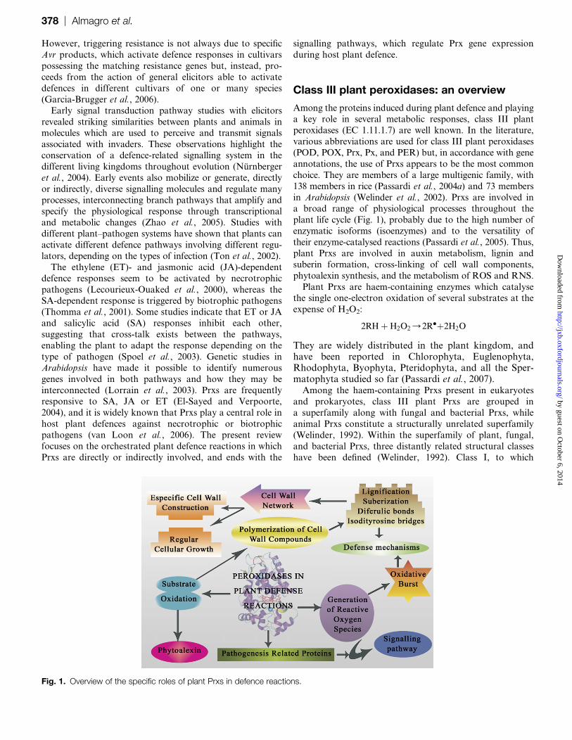
in Arabidopsis (Welinder et al., 2002). Prxs are involved ina broad range of physiological processes throughout the
plant life cycle (Fig. 1), probably due to the high number of
enzymatic isoforms (isoenzymes) and to the versatility of
their enzyme-catalysed reactions (Passardi et al., 2005). Thus,
plant Prxs are involved in auxin metabolism, lignin and
suberin formation, cross-linking of cell wall components,
phytoalexin synthesis, and the metabolism of ROS and RNS.
Plant Prxs are haem-containing enzymes which catalysethe single one-electron oxidation of several substrates at the
expense of H2O2:
2RHþ H2O2/2Rdþ2H2O
They are widely distributed in the plant kingdom, andhave been reported in Chlorophyta, Euglenophyta,Rhodophyta, Byophyta, Pteridophyta, and all the Sper-matophyta studied so far (Passardi et al., 2007).Among the haem-containing Prxs present in eukaryotes
and prokaryotes, class III plant Prxs are grouped in
a superfamily along with fungal and bacterial Prxs, while
animal Prxs constitute a structurally unrelated superfamily
(Welinder, 1992). Within the superfamily of plant, fungal,
and bacterial Prxs, three distantly related structural classeshave been defined (Welinder, 1992). Class I, to which
Fig. 1. Overview of the specific roles of plant Prxs in defence reactions.
378 | Almagro et al. by guest on O
ctober 6, 2014http://jxb.oxfordjournals.org/
Dow
nloaded from
chloroplast and cytosol ascorbate Prxs from higher plants
and bacterial Prx also belong is composed of mitochondrial
yeast cytochrome c Prx. Within class II are grouped all
secretory fungal (manganese) Prxs, while class III contains
all the secretory plant Prxs which show distinctive features
from other plant Prxs, such as ascorbate Prxs.
Prxs (class III) are of a glycoprotein nature, and are
located in vacuoles and cell walls (Passardi et al., 2005).They show a broad range in their substrate requirements
with a moderate but noticeable substrate specificity for
phenols, especially for coniferyl alcohol (Morales and Ros
Barcelo, 1997), and an unusual degree of thermal stability;
all of which distinguishes them from plant ascorbate Prxs
(class I), which are not glycoproteins, and are located in
chloroplasts, mitochondria, peroxysomes, and the cytosol,
where they show moderate substrate specificity for ascorbicacid (Jimenez et al., 1998).
Secretory plant Prxs usually show a molecular mass in the
30–45 kDa range, and contain protohaemin IX (haem b) as
the prosthetic group, as well as two structural Ca2+ ions. In
their resting state (FeIII), an iron ion is present in the
oxidation state of +3. The iron is five co-ordinated to the
four pyrrole nitrogens of the haem and to nitrogen from an
axial (proximal) histidine. The sixth co-ordination positionis free, thus determining a high spin state for the iron
(Banci, 1997). Crystallographic analysis and modelling
studies (Ros Barcelo et al., 2007) reveal that class III plant
Prxs usually contain 10–12 conserved a-helices embedding
the prosthetic group, 2 short b-strands, and four conserved
disulphide bridges.
Prxs as pathogenesis-related (PR) proteins
PR proteins are coded by host plants as a response to
pathological or related situations, and normally accumulate
not only locally in the place of infection, but are also
formed systemically following any kind of infection (Schereret al., 2005). The term PRs is a collective term for all
microbe-induced proteins and their homologues to the
extent that enzymes such as phenylalanine ammonia-lyase,
Prxs, and polyphenoloxidase, which are generally present
constitutively and only increase during most infections, are
often also referred to as PRs (van Loon et al., 2006).
The PR protein family includes all pathogen-induced
proteins and their homologues, and are routinely classifiedinto 17 subfamilies based on their biochemical and molec-
ular biological properties (van Loon et al., 2006). Most of
the PR proteins are induced through the action of several
endogenous growth hormones, including SA, JA, and ET,
whose levels are also increased in infected tissues (Durrant
and Dong, 2004). In Arabidopsis, it has been shown that SA
and JA activate distinct sets of PR genes in an antagonistic
pattern (Thomma et al., 1998). In addition, PR genes aredifferentially regulated by plant growth hormones, includ-
ing SA, ABA, JA, ET, and brassinosteroids, and by diverse
abiotic stresses, supporting the contention that the PR
proteins play a role in plant developmental processes other
than disease resistance responses (Seo et al., 2008).
Prxs are a well-known class of PR proteins, and so they
are induced in host plant tissues by pathogen infection.
They belong to the PR-protein 9 subfamily (van Loon et al.,
2006), and are expressed to limit cellular spreading of the
infection through the establishment of structural barriers or
the generation of highly toxic environments by massively
producing ROS and RNS (Passardi et al., 2005). Prxs
activity or Prxs gene expression in higher plants is, indeed,induced by fungi (Sasaki et al., 2004), bacteria (Young
et al., 1995; Lavania et al., 2006), viruses (Lagrimini and
Rothstein, 1987; Hiraga et al., 2000; Dıaz-Vivancos et al.,
2006), and viroids (Vera et al., 1993). The biotic or abiotic
stress-induced expression of Prxs is conferred by the nature
of the 5’ flanking regions of the genes that contain many
kinds of potential stress-responsive cis-elements (Sasaki
et al., 2007).
Prxs and the reinforcement of the cell walls
Prxs can create a physical barrier to limit pathogen invasion
in host tissues by catalysing the cross-linking of cell wall
components in response to different stimuli such as wound-
ing and pathogen interactions and thus, cell wall rigid-
ification is, in most cases, the result of the Prxs-mediated
H2O2-dependent cross-linking of cell wall components.The irreversible process of cell wall stiffening may either
be dependent or independent of the cell type. Thus, extensin
and ferulic acid cross-linking may constitute temporal and
transitory events within the general programme of de-
velopment of any host plant cell, while lignification and
suberization are terminal processes of determinate and
highly differentiated plant cells capable of forming second-
ary cell walls. In the case of lignification, this process isrestricted to water-conducting vascular cells and their
neighbouring fibres (Ros Barcelo, 1997), while suberization
is restricted to the cell wall of epidermal tissues of un-
derground plant parts (roots, stolons, and tubers), the
endodermis, and the cells of bark tissues (cork and
periderm) (Bernards et al., 2004). In either case, lignification
and suberization have the hallmark of being highly tissue/
cell specific. Indeed, cells that finally become suberized orlignified either derive from specific progenitor cells in
meristems or arise from the vascular cambium, respectively.
Extensin cross-linking
Extensins are the most studied family of hydroxy-proline
(Hyp)-rich proteins (HRGPs). The polypeptide backbone of
extensins contains many repeats of the structural Ser(Hyp)4–
6 motif. These structural motifs are often flanked by short
sequences rich in Tyr, Lys, Val, and His, the Val-Tyr-Lys
motifs being the sites for extensin cross-linking (Fry, 2004b).
Extensins are secreted into the apoplast as soluble mono-mers where the positively charged Lys and the protonated
His residues interact ionically with the negatively charged
uronic acids of pectins. The formation of the insoluble
extensin network is a well-characterized Prxs-mediated and
H2O2-dependent process, which, it has been proposed (Fry,
Peroxidases in plant defence reactions | 379 by guest on O
ctober 6, 2014http://jxb.oxfordjournals.org/
Dow
nloaded from

2004b), involves the coupling of extensin Tyr residues to
form isodityrosine linkages and larger Tyr oligomers such
as di-isodityrosine or pulcherosine. The interaction of Prxs
with extensins also has a defence function since it makes the
cell wall harder to penetrate. The common relationship
between extensins and Prxs could be the pectin layer.
Indeed, some Prxs can bind to calcium–pectate complexes
(Shah et al., 2004), and there is growing evidence thatcovalent bonds exist between pectins and extensins. Pectins
would act as an anchor for Prxs, which would then cross-
link extensins to create a dense and solid network in the
host plant cell wall, with the aim of limiting pathogen
colonization (Passardi et al., 2004b).
Ferulic acid cross-linking
Ferulic acid is ester-bonded to cell wall polysaccharides
(Ralph et al., 2004a). In dicots, non-reducing terminalarabinose (Ara) and galactose residues of pectic polysac-
charides are feruloylated, while in monocots feruloylation is
mainly restricted to the O-5 position of some Ara residues
of arabinoxylans (Fry, 2004a). Feruloyl residues can form
covalent bonds with each other by oxidative coupling. This
process only occurs in the presence of Prx and H2O2 and it
has been speculated (Ralph et al., 2004a) that the resulting
dehydrodiferuloyl residue (5,5#-dehydrodiferulate) formsa cross-link between the polysaccharides to which it is
esterified.
In addition to the first-discovered dimer, 5,5#-dehydrodi-ferulate, several of its isomers have been obtained by alkaline
hydrolysis of plant cell wall polysaccharides; and several
specific trimers and tetramers have now been characterized
(Ralph et al., 2004a). Depending on the linkage formed
(intra- or inter-polysaccharide), up to four polysaccharidechains could potentially be cross-linked by a tetramer of
ferulic acid, which suggests that these Prx-mediated oxida-
tively generated oligomers of ferulate may play an important
role in determining cell wall assembly and cell wall suscepti-
bility to digestion (Grabber et al., 1998).
Lignification
Lignins are three-dimensional, amorphous, heteropolymers
which result from the oxidative coupling of three p-
hydroxycinnamyl alcohols (monolignols): p-coumaryl, con-
iferyl, and sinapyl alcohols, in a H2O2-dependent reactionmediated by Prxs, enzymes which generate the corresponding
free radicals from the three monolignols (Ros Barcelo, 1997).
The regio- and stereospecificity of the cross-coupling reaction
of monolignol radicals produces a hydrophobic heteropoly-
mer composed of p-hydroxyphenyl, guaiacyl, and syringyl
units, respectively. Both the chemistry and biochemistry of
the lignification have recently been revised (Ralph et al.,
2004b), as well as the nature of the Prx responsible for thisprocess (Ros Barcelo et al., 2007). Although lignification is
a structural barrier restricted to vascular tissues, xylem cell
wall lignification may be especially important during the
defence of plants against soil-borne pathogens which cause
vascular wilts (Pomar et al., 2004).
The cross-linking of the phenolic monomers in the
oxidative coupling of lignin subunits has been associated
with Prxs using H2O2 as the oxidant. Acidic and basic Prxs
are capable of oxidizing p-coumaryl and coniferyl alcohol.
However, this situation is not so clear in the case of sinapyl
alcohol, which possesses a syringyl moiety; for this reason
typical acidic Prxs, with some exceptions (Christensen et al.,
1998), are generally regarded as poor catalysts (Bernardset al., 1999). This observation constitutes a central key for
the unravelling specificity of lignin assembly since sinapyl
alcohol is more prone to oxidation than either coniferyl
alcohol or p-coumaryl alcohol (Kobayashi et al., 2005), and
suggests that, although Prx-catalysed reactions are driven
by redox thermodynamic forces (Ros Barcelo et al., 2004),
substrate accommodation in the catalytic centre of the
enzyme determines the real role played by each Prxisoenzyme in lignin biosynthesis (Kobayashi et al., 2005;
Ros Barcelo et al., 2007).
Lignification is an H2O2-dependent Prx-mediated process
(Czaninski et al., 1993; Olson and Varner, 1993; Ros
Barcelo, 1998a; Weir et al., 2005), in which the H2O2
necessary for the peroxidative oxidation of monolignols
comes from a diphenylene iodonium (DPI)-sensitive
NADPH oxidase-like enzyme (Ogawa et al., 1997; RosBarcelo, 1998b, 1999; Karlsson et al., 2005), which gener-
ates Od
2 . This Od
2 then dismutates to H2O2 by a tissue-
specific CuZn-SOD (Ogawa et al., 1997; Karlsson et al.,
2005; Srivastava et al., 2007) or, alternatively, by an Mn-
SOD (Corpas et al., 2006).
Suberization
Both lignification and suberization involve the formation of
a three-dimensional poly(phenolic) matrix initially within
the carbohydrate matrix of the primary cell wall (Keren-
Keiserman et al., 2004). In addition to this phenolic
component, suberized cells develop a distinct poly(aliphatic)layer described (Bernards et al., 2004) as a polyester
connected through primary ester bonds between the major
aliphatic components (a,x-dioic acids and x-hydroxyalka-noic acids), in close association with p-hydroxycinnamic
acids, such as p-coumaric, caffeic, and ferulic acids, which
constitute the cell wall-bound poly(phenolic) domain
(PPD). The macromolecular assembly of potato PPD
occurs via a H2O2-dependent Prx-mediated free radicalcoupling process (Bernards et al., 2004), analogous to the
process of lignin formation from monolignols, but this
enzyme shows a marked substrate preference for p-hydroxy-
cinnamates (i.e. ferulic acid and derivatives).
Suberization is an H2O2-dependent Prx-mediated process,
in which the H2O2 necessary for the peroxidative oxidation
of phenolics also comes from a DPI-sensitive NADPH
oxidase-like enzyme (Razem and Bernards, 2003; Bernardset al., 2004), similar to that occurring during lignification
(Ros Barcelo, 1998b). In fact, a potato NADPH-oxidase
homologue (StrbohA) has recently been implicated
(Kumar et al., 2007) in the generation of H2O2 during
suberization.
380 | Almagro et al. by guest on O
ctober 6, 2014http://jxb.oxfordjournals.org/
Dow
nloaded from

Prxs and the metabolism of ROS and RNS
ROS in plant defence responses
One significant event in plant defence reactions is the
oxidative burst, a common early response of host plant cells
to pathogen attack and elicitor treatment. ROS (Od
2 , H2O2,
and OHd
), predominantly Od
2 and H2O2, are toxic intermedi-
ates resulting from the successive one-step reduction of O2 to
H2O. Four possible mechanisms have been proposed to
explain how ROS are produced in host plant cells: one is
located at the level of the external face of the plasma
membrane, and is mediated by NADPH oxidases (Desikan
et al., 1996), and three are located at the level of the cell wall
matrix, and which would involve the action of Prxs (Bolwell
et al., 2002; Kawano, 2003; Choi et al., 2007), poly(di)amine
oxidases (Angelini and Federico, 1989) and oxalate oxidases
(Lane, 1994). Unlike poly(di)amine oxidases and oxalate
oxidases, which directly generate H2O2, both NADPH
oxidases and Prxs catalyse the initial formation of Od
2 , which
later dismutates to H2O2. The Prx-mediated Od
2 production
may be distinguished from that catalysed by NADPH-
oxidase by the different Km values for O2 and the different
sensitivities of the two enzymes to inhibitors such as cyanide,
azide, and DPI (Bolwell et al., 1998). However, caution
should be exercised in the use of these inhibitors (Frahry
and Schopfer, 1998; Ros Barcelo, 1998b; Ros Barcelo and
Ferrer, 1999).
Recent studies have identified plant respiratory burst
oxidase homologues (rboh) as the main source of ROS
during the apoplastic oxidative burst (Overmyer et al.,
2003), although a role for Prxs has been also proposed
(Bolwell et al., 2002). The relative importance of both
systems can be clearly seen from the following example.
Tobacco cells that had been transformed with an antisense
construct of NtrbohD no longer produced ROS (Simon-Plas
et al., 2002) after treatment with cryptogein, an elicitor of
defence responses. Likewise, Ntrboh-silenced tobacco plants
had a reduced oxidative burst and a reduced disease
resistance to Phytophthora infestans (Yoshioka et al., 2003).
Whatever the source of ROS, H2O2 acts as a signal
molecule to induce an array of molecular, biochemical, and
physiological responses in plant cells (Neill et al., 2002).
Certainly, H2O2 is able to initiate the octadecanoid pathway
leading to the biosynthesis of JA, JA-related compounds,
and other oxylipins which have been reported to function as
inducers of plant secondary metabolites biosynthesis
(Thomma et al., 2001). Given that H2O2 is produced in
response to a wide variety of abiotic and biotic stimuli, it is
likely that H2O2 mediates the cross-talk between signalling
pathways and is a signalling molecule contributing to cross-
tolerance; that is, the exposure of plants to one stress
confers protection towards others (Bowler and Fluhr, 2000).
Moreover, it may be that cellular responses to H2O2 differ
according to their site of synthesis or perception (Neill
et al., 2002). Kacperska (2004) has suggested that the role of
ROS and H2O2 in the mediation of stress responses may
depend on the severity of the stressor.
This implies that, rather than sensor type, the quantita-
tive effects of the sensor-initiated modifications in the
oxidant-antioxidant activities in different cell compartments
may be responsible for the different effects of a particular
stressor. This suggestion is in line with observations that
small increases of H2O2 allow the general enhancement of
stress tolerance; whereas large increases in H2O2 trigger
local responses that unavoidably lead to programmed celldeath (PCD). In fact, H2O2 has been shown to be
a diffusible signal mediating localized PCD during HR
(Levine et al., 1996), as well as being involved in a systemic
signalling network (Alvarez et al., 1998). Mittler et al.
(1999) using transgenic catalase/Prx-deficient tobacco plants
showed that these were hyper-responsive to pathogen
challenge, thus providing direct evidence for a role for
H2O2 in HR cell death. Within this context, Neill et al.
(2002), using Arabidopsis suspension cell cultures to eluci-
date the role of H2O2 as a signalling molecule, showed that
H2O2 is generated following elicitor and pathogen challenge
and that this H2O2 acts as a signal to induce PCD and
defence gene expression (Desikan et al., 2000). PCD in-
duced by H2O2 during the HR in Arabidopsis (Desikan
et al., 1998) and soybean (Solomon et al., 1999) requires
transcription and translation, and several studies havedemonstrated that H2O2 modulates gene expression during
defence responses (Levine et al., 1994; Desikan et al., 1998).
Prxs may transitorily deliver ROS
Although the key function of plant Prxs is to oxidize
phenolic substrates at the expense of H2O2, Prxs may,
paradoxically and transitorily, generate Od
2 /H2O2 through
two mechanisms. The first mechanism is restricted to the
Od
2 /H2O2 generating step of plant Prxs during their
catalytic cycle, which is represented by the decay of
Compound III (CIII) into FeIII (Fig. 2A). This reaction
most likely involves the dissociation of a (FeIII)- Od
2
complex and yields the FeIII form of the enzyme and Od
2 ,
which may further dismutate to H2O2, either chemically
or enzymatically. However, the kdecay of CIII into FeIII is
very slow, and it is unlikely to be responsible for noticeable
Od
2 /H2O2 production such as that observed during the
oxidative burst. Thus, early kinetic studies (Yamazaki and
Yokota, 1973) suggested even that the main way for CIII
decay is its auto-decomposition and that this elapses with nonet Od
2 /H2O2 production (Fig. 2B).
The second mechanism involves the oxidation through
a Prx cycle of a suitable reductor (i.e., a thiol [RSH]) to its
corresponding radical ([RSSR]d�) (Fig. 3), which further
reacts with O2 through a reaction of mediation redox, to
give Od
2 (Burner and Obinger, 1997). However, caution
should be exercised when considering Prx as a possible
enzyme responsible for Od
2 /H2O2 production since theoverall balance of these oxidase/Prx reactions in a closed
system is non-net Od
2 /H2O2 production (Fig. 3).
The overall balance of the reactions depicted in Fig. 3,
however, does not exclude the possibility that, in these
enzymatic reactions, (i) both Od
2 and H2O2 can be
Peroxidases in plant defence reactions | 381 by guest on O
ctober 6, 2014http://jxb.oxfordjournals.org/
Dow
nloaded from

transitorily formed while [RSSR]d�
is not totally consumed,
(ii) Od
2 and H2O2 can be detected by fast trapping agents,
and (iii) the overall reaction is inhibited by both superoxide
dismutase and catalase.
Furthermore, due to the fact that compound I (CI) and
compound II (CII) are continuously generated and broken
during these oxidase/Prx cycles (Figs 2, 3), the introduction
in the bulk of the reactions of a phenolic compound, whichmay act as substrate for CI and CII, would result in the
oxidation of the phenolic to its corresponding radical,
which would then undergo polymerization reactions. This
is what occurs when either a lignin precursor (e.g. coniferyl
alcohol) (Ferrer et al., 1990), or soluble cell wall proteins
(Wojtaszek et al., 1997), are introduced in the bulk of the
oxidase/Prx reaction. All these contraints mean that Prxs
are mainly considered to be merely ROS-detoxifying
enzymes (Zhao et al., 2005).
However, not all Prx-mediated ROS-production reactions
deliver Od
2 /H2O2. It has been proposed (Fry, 1998) that
plant cell wall polysaccharides may be subjected in vivo to
non-enzymatic scission mediated by hydroxyl radicals
(OHd
), and it is probable that microbial polysaccharides arealso targets of OH
d
, and so the OHd
produced by host plant
cells may provoke microbial cell wall polysaccharide
scission as a fine defence mechanism against pathogens.
OHd
can be generated from H2O2 in the host plant cell wall
by Prxs (Liszkay et al., 2003) from Od
2 and H2O2 through
a Haber–Weiss-type reaction:
Od
2 þ FeIII/O2þFeII
2Od
2 þ2Hþ/H2O2þO2
FeIIþ H2O2/FeIIIþ OHdþOH�
Although OHd
is exceedingly reactive, if its production is
controlled, for example, by the correct siting of Prx relative
to microbial polysaccharide molecules, OHd
could be very
precisely targeted to cause microbial polysaccharide scission.
How this might be achieved has yet to be determined.
Evidence for OHd
-induced polysaccharide splitting in plant
cell walls has been obtained by finding the predicted productsof OH
d
action on polysaccharides (Fry, 1998); however,
evidence for such products has not been found from either
plant cell walls or microbial cell walls.
All these results suggest that host plant cells, by means of
Prxs, may generate ROS. However, the dichotomy of the
Prx-mediated ROS production, especially as regards the fate
of H2O2, Od
2 , and OHd
in plant defence reactions, is
apparently far from being completely understood (Passardiet al., 2004b).
RNS in plant defence responses
NOd
is a relatively stable paramagnetic free-radical mole-
cule involved in many physiological processes in plants,
where it serves as a synchronizing chemical messenger
involved in cytotoxicity and PCD (Van Camp et al., 1998;
Durner and Klessig, 1999; Neill et al., 2003). In animal cells,most of the biological regulatory properties of NO
d
have
been explained on the basis of its capacity to act as an iron
ligand in haemproteins (Tsai, 1994). Dual (activating or
inhibitory) effects of NOd
on haemproteins have been
described (Tsai, 1994), and the nature of the effect seems to
depend to a great extent on the resting state (oxidation
state) of the haemprotein, which conditions the ligand
properties and the electronic configuration of the haem iron(Tsai, 1994).
It is now clear that NOd
and, in general, most of the RNS
(NOd
, NO+, NO–, NOd
2, and ONOO–), are major signalling
molecules in plants (Durner and Klessig, 1999) which can be
synthesized during stress responses at the same time as H2O2.
Fig. 3. Catalytic cycle of Prxs in the presence of thiols, showing
the inter-conversion between the resting state (FeIII), and the
activated forms, compound I (CoI) and compound II (CoII). Kinetic
constants are taken from Yokota and Yamazaki (1973) and Burner
and Obinger (1997). The balance of the reaction is put in a frame.
Fig. 2. Pathways of compound III (CIII) formation (A) and CIII auto-
decomposition (B) of Prxs, showing the resting state of the enzyme
(FeIII), its reduced form (FeII), and the activated forms, compound I
(CI) and compound II (CII). Kinetic constants are taken from Yokota
and Yamazaki (1973). The balance of the reaction is put in a frame.
382 | Almagro et al. by guest on O
ctober 6, 2014http://jxb.oxfordjournals.org/
Dow
nloaded from

Two landmark publications have demonstrated the role of
NOd
during the HR to infection by bacteria and viruses
(Delledonne et al., 1998; Durner et al., 1998), NO being part
of the intracellular signalling cascade is activated in plant
cells in response to pathogens or elicitors (Garcıa-Brugger
et al., 2006). At the transcriptional level, microarray and
cDNA-AFLP data obtained from NOd
donor-treated Arabi-
dopsis cells indicate that NOd
modulates the expression ofseveral defence genes, including genes encoding PR proteins
and proteins related to secondary metabolism (Polverari
et al., 2003; Parani et al., 2004).
RNS, generated together with H2O2 in response to pathogen
attack, was found to mediate defence responses similar to
those seen following H2O2 generation. Thus, stress responses
may reflect responses to both H2O2 and RNS. In fact,
bacteria-induced PCD has been reported to involve both ofthese signals in soybean (Delledonne et al., 1998) and in
Arabidopsis (Clarke et al., 2000), although the effects of RNS
and H2O2 were synergistic in the former, while in the latter,
they were additive. As discussed above, H2O2 formation may
transit via an Od
2 intermediate. It is then possible that NOd
,
a free radical itself, may react with Od
2 to form the highly
reactive peroxynitrite anion, ONOO–, and subsequent cellular
effects may then be induced by ONOO–. In this context, it isnoteworthy that NO
d
and H2O2 might control each other’s
synthesis. Thus, exogenously applied H2O2 has been found to
trigger NO production in mung bean through a Ca2+ influx-
dependent process (Lum et al., 2002), and it has recently been
reported that NOd
is required for the activation of plasma
membrane NADPH oxidases (Vandelle et al., 2006). There are
also some controversial reports on NOd
cross-talk with ROS
in host plant defence responses: for example, it appears thatRNS works collaboratively with ROS in many cases (Delle-
donne et al., 1998), while in other cases RNS acts antagonis-
tically (Neill et al., 2002).
Prxs may participate in the metabolism of RNS
It has long been known that NOd
reversibly binds to the haem
prosthetic group of plant Prxs (Yonetani et al., 1972; Ascenzi
et al., 1989). Optical and electron paramagnetic resonance(EPR) studies suggest that, once the complex is formed,
a one electron transfer between NOd
and the protoporphyrin
IX prosthetic group of Prxs takes place, which results in the
formation of spin-paired complexes (Yonetani et al., 1972).
The binding of NOd
to plant Prxs results in spectrally distinct
complexes (Yonetani et al., 1972), in which the Soret peak at
405 nm shifted to 419 nm, and the a and b-absorption bands
at 495 nm and 636 nm shifted to 533 nm and 568 nm,respectively. The shapes and positions of these bands are
typical of low spin ferrous [Fe(II)] Prxs. Kinetic analyses
using flash photolysis and stopped-flow methods (Kobayashi
et al., 1982) have revealed a reaction constant (k) of 1.93105
M�1 s�1. The formation of these ferrous nitrosyl complexes
[Fe(II)NO+] removes enzyme turnover intermediates such as
the resting form, Fe(III):
FeðIIIÞ þ NOd/FeðIIIÞNO4FeðIIÞNOþ
so that NOd is capable of inhibiting Prx-catalysedreactions with Ki in the lM range (Ischiropoulos et al.,1996; Ferrer and Ros Barcelo, 1999), especially when Prxis working with physiological substrates, such as con-iferyl alcohol.
NOd
may also be regarded as a Prx substrate (Glover et al.,
1999), and therefore Prx could play any role in NO
detoxification in plant tissues. In fact, NO reacts with CI
with a rate constant (k2) of 7.03105 M�1 s�1 yielding thenitrosyl species, NO+. NO
d
also reacts with CII with a rate
constant (k3) of 1.33106 M�1 s�1 to lead probably to the
nitrite anion, NO2. Interestingly, the reaction of CII with
NOd
is unusually high relative to that of CI, which is usually
the faster reaction. This means that, in the presence of poor
electron donors of CII, such as catechols (Nappi and Vass,
2001) or guaiacol (Uchida et al., 2002; Hung et al., 2002),
which show k3 values ;102 M�1 s�1 and ;105 M�1 s�1,respectively (Yamazaki and Yokota, 1973), NO
d
may
enhance Prx activity by promoting the formation of the
native enzyme, FeIII, from CII. Thus, both activating and
inhibitory effects of NOd
on Prx could be observed, depend-
ing on the environment in which the enzyme is located.
The product of NOd
oxidation by CII of Prx is apparently
NO2, which can be further oxidized by plant Prxs to lead to
the most likely form, NOd
2 (Shibata et al., 1995; van derVliet et al., 1997). Plant Prxs are also capable of oxidizing
the Tyr contained in proteins to Tyr radicals (Tyrd
), which
combine with another Tyrd
to form dityrosine (Michon
et al., 1997). When the oxidation of Tyr by plant Prxs is
carried out in the presence of NO2, a mixture of Tyrd
and
NOd
2 is formed in the reaction medium, species that couple
to yield 3-nitro-tyrosine (van der Vliet et al., 1997). 3-Nitro-
tyrosine is a well-established marker of oxidative proteindamage in mammals (van der Vliet et al., 1997), and
evidence for this has recently been reported in plant tissues
(Romero-Puertas et al., 2007).
As discussed above, during pathogen attack, host plant
tissues are capable of sustaining both NOd
and Od
2 pro-
duction. Therefore, it is likely that in diseased tissues both
NO and Od
2 will react to lead to ONOO–:
NOd þ Od
2 /ONOO
ONOO– is formed at a near difusion controlled rate
(k¼6.73109 M�1 s�1), and it has been postulated that it
plays a major role in cytotoxicity (Gabaldon et al., 2005),
including PCD (Delledonne et al., 2001; Wendehenne et al.,2001), although its real role in living cells remains unclear
(Fukuto and Ignarro, 1997). Independently of their real role
in plant cells, which remains to be clearly established,
ONOO– may be regarded as a substrate of plant Prxs (Floris
et al., 1993; Gebicka and Gebicki, 2000). In fact, ONOO–
reacts (k¼33106 M�1 s�1) with the resting form of the
enzyme to lead to CII (Floris et al., 1993):
FeðIIIÞ þ ONOO /CIIþ NOd
2
yielding the nitrosylating species, NOd
2. CII is catalyticallyinactive towards ONOO–, and the decay of CII to the
Peroxidases in plant defence reactions | 383 by guest on O
ctober 6, 2014http://jxb.oxfordjournals.org/
Dow
nloaded from
native enzyme, Fe(III), only takes place in the presence ofan external electron donor, such as a phenol. In sucha scenario, a role in the scavenging of ONOO– has beenproposed for the chlorogenic acid/Prx system (Grace et al.,1998), which may be functional with any other phenol.
Prx is not only involved in the detoxification of NO
species and relatives, but it may also participate in bio-mimetic NO-synthesizing pathways. One of the possible
enzymes involved in NO synthesis in plant cells is NO
synthase (NOS) (Wendehenne et al., 2001). NOS catalyses
the conversion of L-arginine in L-citrulline yielding NOd
.
In this reaction, N-hydroxy-L-arginine, an N-hydroxy-
guanidine, acts as key intermediate. N-hydroxyguanidines,
including N-hydroxy-L-arginine, may be oxidized by plant
Prxs to release NOd
, yielding the same products as thoseobtained with NOS (Xian et al., 2001; Cai et al., 2002). The
ability of plant Prxs to synthesize NOd
is not restricted to
the course of the oxidation of N-hydroxyguanidines, since
the oxidation of N-hydroxy-N-nitrosamines (Alston et al.,
1985), such as cupferron, a xenobiotic, or alanosine (a
natural antineoplastic drug closely related to aspartic acid),
also yields NOd
as a collateral product of the reaction.
Prxs and the production of anti-microbialmetabolites
Plant Prxs are able to catalyse the synthesis of bioactive
plant products (Ros Barcelo and Pomar, 2002), andtherefore a role in plant defence through their involvement
in the synthesis of phytoalexins has been proposed for these
enzymes. Thus, viniferins (stilbene phytoalexins in Vitis
spp.), hordatines (dimers of p-coumaryl-agmatine and p-
coumaryl-hydroxy-agmatine in Hordeum spp.), and certain
lignans/neo-lignans (dimers and oligomers of monolignols
and p-hydroxy-cinnamic acids) are well known bioactive
(anti-fungal) products resulting from Prx-mediated reac-tions, and their significance in vivo has been addressed
several times (Langcake and Pryce, 1977a, b;Langcake,
1981; Waffo-Teguo et al., 2001; Ros Barcelo and Pomar,
2002).
The list of bioactive (anti-fungal and anti-bacterial)
products resulting from Prx-mediated reactions is continu-
ously growing, as is illustrated by the following examples.
After infection with pathogenic fungi, oat (Avena sativa)leaves produce the phenolic phytoalexins, avenanthramides,
which are a series of p-hydroxycinnamic amides of
p-hydroxyanthranilates. Okazaki et al. (2004), investigating
the biosynthesis of avenanthramides, identified a dimeric
compound of avenanthramide B in elicited oat leaves. In
a recent study, Okazaki et al. (2007) reported the structure
of five novel dimers, named bisavenanthramides B1–B6,
which exclusively result from Prx-mediated reactions.The involvement of Prx in the metabolism of other
phytoalexins has also been inferred through accumulated
evidence. In Medicago truncatula cell suspension cultures,
the isoflavonoid-derived daidzein dimer was found to
accumulate extracellularly exclusively in response to a yeast
elicitor (Farag et al., 2008), and it is well-known that
isoflavonoid dimers lacking o-catechol substructures may
only arise from Prx-catalysed oxidations (Ros Barcelo and
Pomar, 2002). In white lupin (Lupinus albus), lupinalbisone
A and B, two biflavonoids derived from 2#-hydroxygenistein,are well-known products of the Prx-catalysed oxidation of
2#-hydroxygenistein, which show increased antifungal activ-
ity (Sakasai et al., 2000).Prxs are also involved in the biosynthesis of secondary
metabolites with not well-understood functions in plants,
but instead have recognized medicinal properties. This is the
case of the terpenoid indole alkaloids of Catharanthus
roseus (Sottomayor et al., 2004). In this plant, a basic Prx
was shown to be responsible for the dimerization reaction
between catharantine and vindoline to produce a-3#,4#-anhydrovinblastine (Costa et al., 2008), the major alkaloidpresent in C. roseus leaves, and the precursor of the natural
antitumoral products, vinblastine and vincristine.
Signalling pathways in the regulation of Prxs
SA-mediated defences
SA has long been known to play a central role in plant
defence reactions. SA levels increase in plant tissue follow-
ing pathogen infection, and exogenous application of SA
results in enhanced resistance to a broad range of pathogens
(Ryals et al., 1996). Genetic studies have shown that SA isrequired for the rapid activation of defence responses that
are mediated by several resistance genes, for the induction
of local defences that contain the growth of virulent
pathogens, and for the establishment of systemic acquired
resistance (SAR) (Chen et al., 1993; Rao et al., 1997). SAR
is a state of heightened defence that is activated throughout
the plant following primary infection by pathogens that
elicit tissue damage at the site of infection (Ryals et al.,1996). Several PR genes whose expression is SA-dependent
are commonly used as reporters of SA-dependent defences.
In this context, a great number of class III plant Prxs are
generally induced by SA (Rasmussen et al., 1995; Rao et al.,
1997; Martınez et al., 2000; Fernandes et al., 2006), but this
behaviour is not universal since some Prxs are not re-
sponsive to SA (Ward et al., 1991; Hiraga et al., 2000). This
SA-sensitivity probably marks the frontier line between Prxsinvolved in plant defence reactions and Prxs involved in
other aspects of the plant cell metabolism.
The interaction between SA and Prxs not only covers
those aspects related to gene regulation, but SA may
also interfere positively or negatively in Prx-mediated
metabolic pathways. It has been proposed that the SA
signal transduction pathway leading to SAR may be
mediated by increased ROS levels, since SA binds andinhibits catalase as well as Prxs (Chen et al., 1993; Ruffer
et al., 1995). The inhibition of Prxs by SA is probably due
to the fact that SA may act as a kinetic inhibitor of the
enzyme since, on the one hand, SA is a substrate, although
a weak one, of CII (Kawano et al., 2002a) and, on the other
384 | Almagro et al. by guest on O
ctober 6, 2014http://jxb.oxfordjournals.org/
Dow
nloaded from

hand, SA favours the irreversible inactivation of the enzyme
(Kawano et al., 2002b), blocking all those Prx-mediated
reactions which consume H2O2. The mechanism of SA
action on Prxs, however, is apparently more complex.
Kawano et al. (2004) have suggested a series of reactions
for the generation of Od
2 during the Prx catalytic cycle, in
which SA could act as e– donor for Prx, generating SA
radicals (SAd
). SAd
could thus generate Od
2 by means ofa reaction of mediation redox, such as that described in Fig.
3 for [RSSR]d�. Evidence supporting the production of SA
d
by Prxs has been obtained by electron spin resonance (ESR)
studies (Kawano and Muto, 2000). In such a scenario, it has
been proposed that the mechanism for the SA-dependent
early ROS production solely depends on the interaction of
SA with Prx (Kawano and Muto, 2000), whereas the
involvement of NADPH oxidase in the later stages of SAaction acquires a major role since this requires the SA-
induced NADPH oxidase gene expression (Yoshioka et al.,
2001, 2003).
JA-dependent and ET-dependent defences
JA, and its more permeable derivative, methyljasmonate
(MeJA), have been proposed as key compounds of the
signal transduction pathway involved in the elicitation of
plant defence reactions (Farmer et al., 2003). Consistent
with the notion of Prxs participating in plant defence
responses, JA and MeJA positively regulate Prx gene
expression (Repka et al., 2004; Ali et al., 2006; Kumariet al., 2006), but cross-talks between JA and SA in the
regulation of Prxs have not been established.
Unlike SA and JA, ET is a phytohormone that regulates
a wide range of plant processes from growth and de-
velopment to defence responses. ET production can be
induced by various stresses such as wounding, microbial
pathogen and insect attack, as well as small elicitors.
However, the role of ET in plant defence is controversial asit contributes to resistance in some interactions (Norman-
Setterblad et al., 2000; Thomma et al., 1999), but promotes
disease in others (Bent et al., 1992; Lund et al., 1998;
Hoffman et al., 1999). The behaviour of Prx in these
systems has not been studied. However, in others, ET
promotes Prx activity and Prx gene expression, with the de
novo expression, although not always of novel Prx iso-
enzymes (Abeles et al., 1989; Morgens et al., 1990; Ishigeet al., 1993).
The interaction of JA and ET signalling pathways is one
of the most significant interactions known, and it influences
many aspects of plant development and defence responses.
JA signalling can lead to ET production in host plants,
while ET apparently stimulates JA production (Xu et al.,
1994; Mirjalili and Linden, 1996; Zhao et al., 2004). In
many cases, JA and ET co-operatively regulate defenceresponses. Evidence that JA and ET co-ordinately regulate
defence-related genes has been obtained in A. thaliana from
microarray experiments that monitored gene expression in
response to various defence-related stimuli (Schenk et al.,
2000). These authors reported that nearly half of the genes
that were induced by ET were also induced by JA
treatment. Consistent with this notion, Prxs that apparently
participate in plant defence responses are activated by both
JA and ET (Buzi et al., 2004; Bailey et al., 2005).
The above-described results point to the possible signal-
ling pathways that regulate Prx gene expression during
plant defence. How certain Prxs have suffered the natural
selection to be responsive to biotic factors, and how thesePrxs are really integrated within the complex network of
defence responses of any host plant cell, will be the
cornerstone of future research.
Acknowledgements
L Almagro and S Belchı-Navarro hold grants from the
Fundacion Seneca. We thank Estefanıa Pedreno for revising
the English manuscript. This work has been partially
supported by the MEC and FEDER (BIO2005-00332,BFU2006-11577) and by the Consejerıa de Educacion,
Ciencia e Investigacion de la Region de Murcia (BIO BVA
07 01 0003).
References
Abeles FB, Hershberger WL, Dunn LJ. 1989. Hormonal regulation,
and intracellular localization of a 33-kD cationic peroxidase in excised
cucumber cotyledons. Plant Physiology 89, 664–668.
Ali MB, Yu KW, Hahn EJ, Paek KY. 2006. Methyl jasmonate and
salicylic acid elicitation induces ginsenosides accumulation, enzymatic
and non-enzymatic antioxidant in suspension culture Panax ginseng
roots in bioreactors. Plant Cell Reports 25, 613–620.
Alston TA, Porter DJT, Bright HJ. 1985. Generation of nitric oxide
by enzymatic oxidation of N-hydroxy-N-nitrosamines. Journal of
Biological Chemistry 260, 4069–4074.
Alvarez ME, Pennell RI, Meijer P-J, Ishikawa A, Dixon RA,
Lamb C. 1998. Reactive oxygen intermediates mediate a systemic
signal network in the establishment of plant immunity. Cell 92,
773–784.
Angelini R, Federico R. 1989. Histochemical evidence of polyamine
oxidation and hydrogen peroxide generation in the cell wall. Journal of
Plant Physiology 135, 212–217.
Ascenzi P, Brunori M, Coletta M, Desideri A. 1989. pH effects on
the haem iron co-ordination state in the nitric oxide and deoxy
derivatives of ferrrous horseradish peroxidase and cytochrome c
peroxidase. Biochemical Journal 258, 473–478.
Bailey BA, Strem MD, Bae H, Antunez de Mayolo G,
Guiltinan MJ. 2005. Gene expression in leaves of Theobroma cacao
in response to mechanical wounding, ethylene, and/or methyl
jasmonate. Plant Science 168, 1247–1258.
Banci L. 1997. Structural properties of peroxidases. Journal of
Biotechnology 53, 253–263.
Bent AF, Innes RW, Ecker JR, Staskawicz BJ. 1992. Disease
development in ethylene-insensitive Arabidopsis thaliana infected with
virulent and avirulent Pseudomonas and Xanthamonas pathogens.
Molecular Plant–Microbe Interactions 5, 372–378.
Peroxidases in plant defence reactions | 385 by guest on O
ctober 6, 2014http://jxb.oxfordjournals.org/
Dow
nloaded from
Bernards MA, Fleming WD, Llewellyn DB, Priefer R, Yang X,
Sabatino A, Plourde GL. 1999. Biochemical characterization of the
suberization-associated anionic peroxidase of potato. Plant Physiology
121, 135–145.
Bernards MA, Summerhurst DK, Razem FA. 2004. Oxidases,
peroxidases and hydrogen peroxide: the suberin connection. Phyto-
chemistry Reviews 3, 113–126.
Bolwell GP, Bindschedler LV, Blee KA, Butt VS, Davies DR,
Gardner SL, Gerrish C, Minibayeva F. 2002. The apoplastic
oxidative burst in response to biotic stress in plants: a three-
component system. Journal of Experimental Botany 53, 1367–1376.
Bolwell GP, Davies DR, Gerrish C, Auh C-K, Murphy TM. 1998.
Comparative biochemistry of the oxidative burst produced by rose and
French bean cells reveals two distinct mechanisms. Plant Physiology
116, 1379–1385.
Bowler C, Fluhr R. 2000. The role of calcium and activated oxygen
as signals for controlling cross-tolerance. Trends in Plant Science 5,
241–245.
Burner U, Obinger C. 1997. Transient state and steady-state kinetics
of the oxidation of aliphatic and aromatic thiols by horseradish
peroxidase. FEBS Letters 411, 269–274.
Buzi A, Chilosi G, Magro P. 2004. Induction of resistance in melon
seedlings against soil-borne fungal pathogens by gaseous treatments
with methyl jasmonate and ethylene. Journal of Phytopathology 152,
491–497.
Cai T, Xian M, Wang PG. 2002. Electrochemical and peroxidase
oxidation study of N#-hydroxyguanidine derivatives as NO donors.
Bioorganic and Medicinal Chemistry Letters 12, 1507–1510.
Chen Z, Silva H, Klessig DF. 1993. Active oxygen species in the
induction of plant systemic acquired resistance induced by salicylic
acid. Science 262, 1883–1886.
Choi HW, Kim YJ, Lee SC, Hong JK, Hwang BK. 2007.
Hydrogen peroxide generation by the pepper extracellular
peroxidase CaPO2 activates local and systemic cell death and
defence response to bacterial pathogens. Plant Physiology 145,
890–904.
Christensen JH, Bauw G, Welinder KG, Montagu MV, Boerjan W.
1998. Purification and characterization of peroxidases correlated with
lignification in poplar xylem. Plant Physiology 118, 125–135.
Clarke A, Desikan R, Hurst RD, Hancock JT, Neill SJ. 2000. NO
way back: nitric oxide and programmed cell death in Arabidopsis
thaliana suspension cultures. The Plant Journal 24, 667–677.
Corpas FJ, Fernandez-Ocana A, Carreras A, et al. 2006. The
expression of different superoxide dismutase forms is cell-type de-
pendent in olive (Olea europaea L.) leaves. Plant and Cell Physiology
47, 984–994.
Costa MMR, Hilliou F, Duarte P, Pereira LG, Almeida I, Leech M,
Memelink J, Ros Barcelo A, Sottomayor M. 2008. Molecular
cloning and characterization of a vacuolar class III peroxidase involved
in the metabolism of anticancer alkaloids in Catharanthus roseus.
Plant Physiology 146, 403–417.
Czaninski Y, Sachot RM, Catesson AM. 1993. Cytochemical
localization of hydrogen peroxide in lignifying cell walls. Annals of
Botany 72, 547–550.
Delledonne M, Xia Y, Dixon RA, Lamb C. 1998. Nitric oxide
functions as a signal in plant disease resistance. Nature 394,
585–588.
Delledonne M, Zeier J, Marocco A, Lamb C. 2001. Signal
interactions between nitric oxide and reactive oxygen intermediates in
the plant hypersensitive disease resistance response. Proceedings of
the National Academy of Sciences, USA 98, 13454–13459.
Desikan R, Hancock JT, Coffey MJ, Neill SJ. 1996. Generation of
active oxygen in elicited cells of Arabidopsis thaliana is mediated by
a NADPH oxidase-like enzyme. FEBS Letters 382, 213–217.
Desikan R, Neill SJ, Hancock JT. 2000. Hydrogen peroxide
induced gene expression in Arabidopsis thaliana. Free Radical Biology
and Medicine 28, 773–778.
Desikan R, Reynolds A, Hancock JT, Neill SJ. 1998. Harpin and
hydrogen peroxide both initiate programmed cell death but have
differential effects on gene expression in Arabidopsis suspension
cultures. Biochemical Journal 330, 115–120.
Dıaz-Vivancos P, Rubio M, Mesonero V, Periago PM, Ros
Barcelo A, Martınez-Gomez P, Hernandez JA. 2006. The
apoplastic antioxidant system in Prunus: response to long-term
plum pox virus infection. Journal of Experimental Botany 57,
3813–3824.
Durner J, Klessig DF. 1999. Nitric oxide as a signal in plants. Current
Opinion in Plant Biology 2, 369–374.
Durner J, Wendehenne D, Klessig DF. 1998. Defence gene
induction in tobacco by nitric oxide, cyclic GMP, and cyclic ADP
ribose, 95. Proceedings of the National Academy of Sciences, USA,
10328–10333.
Durrant WE, Dong X. 2004. Systemic acquired resistance. Annual
Review of Phytopathology 42, 185–209.
El-Sayed M, Verpoorte R. 2004. Growth, metabolic profiling and
enzymes activities of Catharanthus roseus seedlings treated with plant
growth regulators. Plant Growth Regulation 44, 53–58.
Farag MA, Huhman DV, Dixon RA, Sumner LW. 2008. Metabolo-
mics reveals novel pathways and differential mechanistic and elicitor-
specific responses in phenylpropanoid and isoflavonoid biosynthesis in
Medicago truncatula cell cultures. Plant Physiology 146, 387–402.
Farmer EE, Almeras E, Krishnamurthy V. 2003. Jasmonates and
related oxylipins in plant responses to pathogenesis and herbivory.
Current Opinion in Plant Biology 6, 372–378.
Fernandes CF, Moraes VCP, Vasconcelos IM, Silveira JAG,
Oliveira JTA. 2006. Induction of an anionic peroxidase in cowpea
leaves by exogenous salicylic acid. Journal of Plant Physiology 163,
1040–1048.
Ferrer MA, Pedreno MA, Munoz R, Ros Barcelo A. 1990. Oxidation
of coniferyl alcohol by cell wall peroxidases at the expense of indole-3-
acetic acid and O2. FEBS Letters 276, 127–130.
Ferrer MA, Ros Barcelo A. 1999. Differential effects of nitric oxide
on peroxidase and H2O2 production by the xylem of Zinnia elegans.
Plant, Cell and Environment 22, 891–897.
Floris R, Piersma SR, Yang G, Jones P, Wever R. 1993.
Interaction of myeloperoxidase with peroxynitrite. A comparison with
lactoperoxidase, horseradish peroxidase and catalase. European
Journal of Biochemistry 215, 767–775.
386 | Almagro et al. by guest on O
ctober 6, 2014http://jxb.oxfordjournals.org/
Dow
nloaded from
Frahry G, Schopfer P. 1998. Inhibition of O2-reducing activity of
horseradish peroxidase by diphenyleniodonium. Phytochemistry 48,
223–227.
Fry SC. 1998. Oxidative scission of plant cell wall polysaccharides
by ascorbate-induced hydroxyl radicals. Biochemical Journal 332,
507–515.
Fry SC. 2004a. Primary cell wall metabolism: tracking the careers of
wall polymers in living plant cells. New Phytologist 161, 641–675.
Fry SC. 2004b. Oxidative coupling of tyrosine and ferulic acid
residues: Intra- and extra-protoplasmic occurrence, predominance of
trimers and larger products, and possible role in inter-polymeric cross-
linking. Phytochemistry Reviews 3, 97–111.
Fukuto JM, Ignarro LJ. 1997. In vivo aspects of nitric oxide (NO)
chemistry: does peroxynitrite (–OONO) play a major role in cytotoxicity?
Accounts of Chemical Research 30, 149–152.
Gabaldon C, Gomez Ros LV, Pedreno MA, Ros Barcelo A. 2005.
Nitric oxide production by the differentiating xylem of Zinnia elegans.
New Phytologist 165, 121–130.
Garcıa-Brugger A, Lamotte O, Vandelle E, Bourque S,
Lecourieux D, Poinssot B, Wendehenne D, Pugin A. 2006. Early
signaling events induced by elicitors of plant defences. Molecular
Plant–Microbe Interactions 7, 711–724.
Gebicka L, Gebicki JL. 2000. Reactions of haem peroxidases with
peroxynitrite. IUBMB Life 49, 11–15.
Glover R, Koshkin V, Dunford HB, Mason RP. 1999. The reaction
rates of NO with horseradish peroxidase compounds I and II. Nitric
oxide: Biology and Chemistry 3, 439–444.
Grabber JH, Hatfield RD, Ralph J. 1998. Diferulate cross-links
impede the enzymatic degradation of non-lignified maize walls. Journal
of Science and Food Agriculture 77, 193–200.
Grace SC, Salgo MG, Prior WA. 1998. Scavenging of peroxynitrite
by a phenolic/peroxidase system prevents oxidative damage to DNA.
FEBS Letters 426, 24–28.
Hiraga S, Ito H, Yamakawa H, Ohtsubo N, Seo S, Mitsuhara I,
Matsui H, Honma M, Ohashi Y. 2000. An HR-induced tobacco
peroxidase gene is responsive to spermine, but not to salicylate,
methyl jasmonate, and ethephon. Molecular Plant–Microbe Interac-
tions 13, 210–216.
Hoffman T, Schmidt JS, Zheng X, Bent AF. 1999. Isolation of
ethylene-insensitive soybean mutants that are altered in pathogen
susceptibility and gene-for-gene disease resistance. Plant Physiology
119, 935–949.
Hung KT, Chang CJ, Kao CH. 2002. Paraquat toxicity is reduced by
nitric oxide in rice leaves. Journal of Plant Physiology 159, 159–166.
Ischiropoulos H, Nelson J, Duran D, Al-Mehdi A. 1996. Reactions
of nitric oxide and peroxynitrite with organic molecules and ferrihorser-
adish peroxidase: interference with the determination of hydrogen
peroxide. Free Radical Biology and Medicine 20, 373–381.
Ishige F, Mori H, Yamazaki K, Imaseki H. 1993. Identification of
a basic glycoprotein induced by ethylene in primary leaves of Azuki bean
as a cationic peroxidase. Plant Physiology 101, 193–199.
Jimenez A, Hernandez JA, Ros Barcelo A, Sandalio LM, del
Rıo LA, Sevilla F. 1998. Mitochondrial and peroxisomal ascorbate
peroxidase of pea leaves. Physiologia Plantarum 104, 687–692.
Kacperska A. 2004. Sensor types in signal transduction pathways in
plant cells responding to abiotic stressors: do they depend on stress
intensity? Physiologia Plantarum 122, 159–168.
Karlsson M, Melzer M, Prokhorenko I, Johansson T, Wingsle G.
2005. Hydrogen peroxide and expression of hipI-superoxide
dismutase are associated with the development of secondary
cell walls in Zinnia elegans. Journal of Experimental Botany 56,
2085–2093.
Kawano T. 2003. Role of the reactive oxygen species generating
peroxidase reactions in plant defence and growth induction. Plant Cell
Reports 21, 829–837.
Kawano T, Furuichi T, Muto S. 2004. Controlled salicylic acid levels
and corresponding signaling mechanisms in plants. Plant Biotechnol-
ogy 21, 319–335.
Kawano T, Kawano N, Lapeyrie F. 2002a. A fungal auxin antagonist,
hypaphorine prevents the indole-3-acetic acid-dependent irreversible
inactivation of horseradish peroxidase: inhibition of Compound III-
mediated formation of P-670. Biochemical and Biophysical Research
Communications 294, 553–559.
Kawano T, Kawano N, Muto S, Lapeyrie F. 2002b. Retardation
and inhibition of the cation-induced superoxide generation in BY-2
tobacco cell suspension culture by Zn2+ and Mn2+. Physiologia
Plantarum 114, 395–404.
Kawano T, Muto S. 2000. Mechanism of peroxidase actions for
salicylic acid-induced generation of active oxygen species and an
increase in cytosolic calcium in tobacco suspension culture. Journal of
Experimental Botany 51, 685–693.
Keren-Keiserman A, Tanami Z, Shoseyov O, Ginzberg I. 2004.
Peroxidase activity associated with suberization processes of the
muskmelon (Cucumis melo) rind. Physiologia Plantantarum 121,
141–148.
Kobayashi K, Tamura M, Hayashi K. 1982. Kinetic analyses of the
recombination of NO with ferrihemoproteins by the flash photolysis
method. Biochemistry 21, 729–732.
Kobayashi T, Taguchi H, Shigematsu M, Tanahashi M. 2005.
Substituent effects of 3,5-disubstituted p-coumaryl alcohols on their
oxidation using horseradish peroxidase-H2O2 as the oxidant. Journal
of Wood Science 51, 607–614.
Kumar GNM, Iyer S, Knowles NR. 2007. Strboh A homologue of
NADPH oxidase regulates wound-induced oxidative burst and facili-
tates wound-healing in potato tubers. Planta 227, 25–36.
Kumari GJ, Reddy AM, Naik ST, Kumar SG, Prasanthi J,
Sriranganayakulu G, Reddy PC, Sudhakar C. 2006. Jasmonic acid
induced changes in protein pattern, antioxidative enzyme activities and
peroxidase isoenzymes in peanut seedlings. Biologia Plantarum 50,
219–226.
Lagrimini LM, Rothstein S. 1987. Tissue specificity of tobacco
peroxidase isozymes and their induction by wounding and tobacco
mosaic virus infection. Plant Physiology 84, 438–442.
Lane BG. 1994. Oxalate, germin, and the extracellular matrix of higher
plants. FASEB Journal 8, 294–301.
Langcake P. 1981. Disease resistance of Vitis spp. and the production
of the stress metabolites resveratrol, e-viniferin, a-viniferin and pterostil-
bene. Physiological Plant Pathology 18, 213–226.
Peroxidases in plant defence reactions | 387 by guest on O
ctober 6, 2014http://jxb.oxfordjournals.org/
Dow
nloaded from

Langcake P, Pryce RJ. 1977a. A new class of phytoalexins from
grapevines. Experientia 33, 151–152.
Langcake P, Pryce RJ. 1977b. The production of resveratrol and the
viniferins by grapevines in response to ultraviolet irradiations. Phyto-
chemistry 16, 1193–1196.
Lavania M, Chauhan PS, Chauhan SVS, Singh HB, Nautiyal CS.
2006. Induction of plant defence enzymes and phenolics by treatment
with plant growth-promoting rhizobacteria Serratia marcescens
NBRI1213. Current Microbiology 52, 363–368.
Lecourieux-Ouaked F, Pugin A, Lebrun-Garcia A. 2000. Phos-
phoproteins involved in the signal transduction of cryptogein, an
elicitor of defence reaction in tobacco. Molecular Plant–Microbe
Interactions 13, 821–829.
Levine A, Pennell RI, Alvarez ME, Palmer R, Lamb C. 1996.
Calcium-mediated apoptosis in a plant hypersensitive disease re-
sistance response. Current Progress in Biology 6, 427–437.
Levine A, Tenhaken R, Dixon R, Lamb C. 1994. H2O2 from the
oxidative burst orchestrates the plant hypersensitive disease resis-
tance response. Cell 79, 583–593.
Liszkay A, Kenk B, Schopfer P. 2003. Evidence for the involvement
of cell wall peroxidase in the generation of hydroxyl radicals mediating
extension growth. Planta 217, 658–667.
Lorrain S, Vailleau F, Balague C, Roby D. 2003. Lesion mimic
mutants: key for deciphering cell death and defence pathways in
plants? Trends in Plant Science 8, 263–271.
Lum HK, Butt YK, Lo SC. 2002. Hydrogen peroxide induces a rapid
production of nitric oxide in mung bean (Phaseolus aureus). Nitric
Oxide 6, 205–213.
Lund ST, Stall RE, Klee HJ. 1998. Ethylene regulates the suscepti-
ble response to pathogen infection in tomato. The Plant Cell 10,
371–382.
Martinez C, Baccou JC, Bresson E, Baissac Y, Daniel JF,
Jalloul A, Montillet JL, Geiger JP, Assigbetse K, Nicole M. 2000.
Salicylic acid mediated by the oxidative burst is a key molecule in local
and systemic responses of cotton challenged by an avirulent race of
Xanthomonas campestris pv. malvacearum. Plant Physiology 122,
757–766.
Michon T, Chenu M, Kellershon N, Desmadril M, Gueguen J.
1997. Horseradish peroxidase oxidation of tyrosine-containing pep-
tides and their subsequent polymerization: a kinetic study. Biochem-
istry 36, 8505–8513.
Mirjalili N, Linden JC. 1996. Methyl jasmonate induced production
of taxol in suspension cultures of Taxus cuspidata: ethylene interaction
and induction models. Biotechnology Progress 12, 110–118.
Mittler R, Herr EH, Orvar BL, Van Camp W, Willekens H, Inze D,
Ellis BE. 1999. Transgenic tobacco plants with reduced capability to
detoxify reactive oxygen intermediates are hyperresponsive to patho-
gen infection. Proceedings of the National Academy of Sciences, USA
96, 14165–14170.
Morales M, Ros Barcelo A. 1997. A basic peroxidase isoenzyme
from vacuoles and cell walls of Vitis vinifera. Phytochemistry 45,
229–232.
Morgens PH, Callahan AM, Dunn LJ, Abeles FB. 1990. Isolation
and sequencing of cDNA clones encoding ethylene-induced putative
peroxidases from cucumber cotyledons. Plant Molecular Biology 14,
715–725.
Nappi AJ, Vass E. 2001. The effects of nitric oxide on the oxidations
of L-Dopa and dopamine mediated by tyrosinase and peroxidase.
Journal of Biological Chemistry 276, 11214–11222.
Neill SJ, Desikan R, Clarke A, Hurst RD, Hancock JT. 2002.
Hydrogen peroxide and nitric oxide as signalling molecules in plants.
Journal of Experimental Botany 53, 1237–1247.
Neill SJ, Desikan R, Hancock JT. 2003. Nitric oxide signaling in
plants. New Phytologist 159, 11–35.
Norman-Setterblad C, Vidal S, Palva ET. 2000. Interacting signal
pathways control defence gene expression in Arabidopsis in response
to cell wall-degrading enzymes from Erwinia carotovora. Molecular
Plant–Microbe Interactions 13, 430–438.
Nurnberger T, Brunner F, Kemmerling B, Piater L. 2004. Innate
immunity in plants and animals: striking similarities and obvious
differences. Immunology Reviews 198, 249–266.
Ogawa K, Kanematsu S, Asada K. 1997. Generation of superoxide
anion and localization of CuZn-superoxide dismutase in the vascular
tissue of spinach hypocotyls: their association with lignification. Plant
and Cell Physiology 38, 1118–1126.
Okazaki Y, Ishihara A, Nishioka T, Iwamura H. 2004a. Identifica-
tion of a dehydrodimer of avenanthramide phytoalexin in oats.
Tetrahedron 60, 4765–4771.
Okazaki Y, Ishizuka A, Ishihara A, Nishioka T, Iwamura H. 2007.
New dimeric compounds of avenanthramide phytoalexin in oats.
Journal of Organic Chemistry 72, 3830–3839.
Olson PD, Varner JE. 1993. Hydrogen peroxide and lignification.
The Plant Journal 4, 887–892.
Overmyer M, Brosche M, Kangasjarvi J. 2003. Reactive oxygen
species and hormonal control of cell death. Trends in Plant Science 8,
335–342.
Parani M, Rudrabhatla S, Myers R, Weirich H, Smith B,
Leaman DW, Goldman SL. 2004. Microarray analysis of nitric oxide
responsive transcripts in. Arabidopsis. Plant Biotechnology Journal 2,
359–366.
Passardi F, Cosio C, Penel C, Dunand C. 2005. Peroxidases have
more functions than a Swiss army knife. Plant Cell Reports 24, 255–265.
Passardi F, Longet D, Penel C, Dunand C. 2004a. The class III
peroxidase multigenic family in rice and its evolution in land plants.
Phytochemistry 65, 1879–1893.
Passardi F, Penel C, Dunand C. 2004b. Performing the paradoxical:
how plant peroxidasas modify the cell wall. Trends in Plant Science 9,
534–540.
Passardi F, Zamocky M, Favet J, Jakopitsch C, Penel C, Obinger C,
Dunand C. 2007. Phylogenetic distribution of catalase-peroxidases: are
these patches of order in chaos? Gene 397, 101–113.
Polverari A, Molesini B, Pezzotti M, Buonaurio R, Marte M,
Delledonne M. 2003. Nitric oxide-mediated transcriptional changes
in Arabidopsis thaliana. Molecular Plant–Microbe Interactions 16,
1094–1105.
Pomar F, Novo M, Bernal MA, Merino F, Ros Barcelo A. 2004.
Changes in stem lignins (monomer composition and crosslinking) and
388 | Almagro et al. by guest on O
ctober 6, 2014http://jxb.oxfordjournals.org/
Dow
nloaded from

peroxidase are related with the maintenance of leaf photosynthetic
integrity during Verticillium wilt in Capsicum annuum. New Phytologist
163, 111–123.
Ralph J, Bunzel M, Marita MJ, Hatfield RD, Lu F, Kim H,
Schatz PF, Grabber JH, Steinhart H. 2004a. Peroxidase-dependent
cross-linking reactions of p-hydroxycinnamates in plant cell walls.
Phytochemistry Reviews 3, 79–96.
Ralph J, Lundquist K, Brunow G, et al. 2004b. Lignins: natural
polymers from oxidative coupling of 4-hydroxyphenylpropanoids.
Phytochemistry Reviews 3, 29–60.
Rao MV, Pliyath C, Ormrod DP, Murr DP, Watkins CB. 1997.
Influence of salicylic acid on H2O2 production, oxidative stress, and
H2O2-metabolizing enzymes. Plant Physiology 115, 137–149.
Rasmussen JB, Smith JA, Williams S, Burkhart W, Ward E,
Somerville SC, Ryals J, Hammerschmidt R. 1995. cDNA cloning
and systemic expression of acidic peroxidases associated with
systemic acquired resistance to disease in cucumber. Physiologycal
and Molecular Plant Pathology 46, 389–400.
Razem FA, Bernards MA. 2003. Reactive oxygen species production
in association with suberization: evidence for an NADPH-dependent
oxidase. Journal of Experimental Botany 54, 935–941.
Repka V, Fischerova I, Silharova K. 2004. Methyl jasmonate is
a potent elicitor of multiple defence responses in grapevine leaves and
cell-suspension cultures. Biologia Plantarum 48, 273–283.
Romero-Puertas MC, Laxa M, Matte A, Zaninotto F,
Finkemeier I, Jones AME, Perazzolli M, Vandelle E, Dietz KJ,
Delledonne M. 2007. S-nitrosylation of peroxiredoxin II E promotes
peroxynitrite-mediated tyrosine nitration. The Plant Cell 19, 4120–4130.
Ros Barcelo A. 1997. Lignification in plant cell walls. International
Review of Cytology 176, 87–132.
Ros Barcelo A. 1998a. Hydrogen peroxide production is a general
property of the lignifying xylem from vascular plants. Annals of Botany
82, 97–103.
Ros Barcelo A. 1998b. The generation of H2O2 in the xylem of Zinnia
elegans is mediated by an NADPH-oxidase-like enzyme. Planta 207,
207–216.
Ros Barcelo A. 1999. Some properties of the H2O2=O2 generating
system from the lignifying xylem of Zinnia elegans. Free Radical Research
31, 147–154.
Ros Barcelo A, Ferrer MA. 1999. Does diphenylene iodonium
chloride have any effect on the O2 generating step of plant
peroxidases? FEBS Letters 462, 254–256.
Ros Barcelo A, Gomez Ros LV, Esteban Carrasco A. 2007.
Looking for syringyl peroxidases. Trends in Plant Science 12,
486–491.
Ros Barcelo A, Gomez Ros LV, Gabaldon C, Lopez-Serrano M,
Pomar F, Carrion JS, Pedreno MA. 2004. Basic peroxidases: the
gateway for lignin evolution? Phytochemistry Reviews 3, 61–78.
Ros Barcelo A, Pomar F. 2002. Plant peroxidases: versatile catalysts
in the synthesis of bioactive natural products. In: Atta-ur-Rahman, ed.
Studies in natural products chemistry, Vol. 27. The Netherlands:
Elsevier, 735–791.
Ruffer M, Steipe B, Zenk MH. 1995. Evidence against specific
binding of salicylic acid to plant catalase. FEBS Letters 377, 175–180.
Ryals JA, Neuenschwander UH, Willits MG, Molina A, Steiner H,
Hunt MD. 1996. Systemic acquired resistance. The Plant Cell 8,
1809–1819.
Sakasai M, Fukui H, Yamane H, Kyaw AN, Tahara S. 2000. A new
class of biflavonoids: 2#-hydroxygenistein dimers from the roots of
white lupin. Zeitschrift fur Naturforschung C-A Journal of Biosciences
55, 165–174.
Sasaki K, Iwai T, Hiraga S, et al. 2004. Ten rice peroxidases
redundantly respond to multiple stresses including infection with rice
blast fungus. Plant and Cell Physiology 45, 1442–1452.
Sasaki K, Yuichi O, Hiraga S, Gotoh Y, Seo S, Mitsuhara I, Ito H,
Matsui H, Ohashi Y. 2007. Characterization of two rice peroxidase
promoters that respond to blast fungus infection. Molecular Genetics
and Genomics 278, 709–722.
Schenk PM, Kazan K, Wilson I, Anderson JP, Richmond T,
Somerville SC, Manners JM. 2000. Co-ordinated plant defence
responses in Arabidopsis revealed by microarray analysis.
Proceedings of the National Academy of Sciences, USA 97,
11655–11660.
Scherer NM, Thompson CE, Freitas LB, Bonatto SL,
Salzano FM. 2005. Patterns of molecular evolution in pathogenesis-
related proteins. Genetics and Molecular Biology 28, 645–653.
Seo PJ, Lee AK, Xiang F, Park CM. 2008. Molecular and functional
profiling of Arabidopsis pathogenesis-related genes: insights into their
roles in salt response of seed germination. Plant and Cell Physiology
49, 334–344.
Shah K, Penel C, Gagnon J, Dunand C. 2004. Purification and
identification of a Ca2+-pectate binding peroxidase from Arabidopsis
leaves. Phytochemistry 65, 307–312.
Shibata H, Kono Y, Yamashita S, Sawa Y, Ochiai H, Tanaska K.
1995. Degradation of chlorophyll by nitrogen dioxide generated from
nitrite by the peroxidase reaction. Biochimica et Biophysica Acta 1230,
45–50.
Simon-Plas F, Elmayan T, Blein JP. 2002. The plasma membrane
oxidase NtrbohD is responsible for AOS production in elicited tobacco
cells. The Plant Journal 31, 137–147.
Solomon M, Belenghi B, Delledonne M, Menachem E, Levine A.
1999. The involvement of cysteine proteases and protease inhibitor
genes in the regulation of programmed cell death in plants. The Plant
Cell 11, 431–443.
Sottomayor M, Lopes Cardoso I, Pereira LG, Ros Barcelo A.
2004. Peroxidase and the biosynthesis of terpenoid indole alkaloids in
the medicinal plant Catharanthus roseus (L.) G. Don. Phytochemistry
Reviews 3, 159–171.
Spoel SH, Koornneef A, Claessens SMC, Korzelius JP, Van
Pelt JA. 2003. NPR1 modulates cross-talk between salicylate- and
jasmonate-dependent defence pathways through a novel function in
the cytosol. The Plant Cell 15, 760–70.
Srivastava V, Schinkel H, Witzell J, Hertzberg M, Torp M,
Srivastava MK, Karpinska B, Melzer M, Wingsle G. 2007. Down-
regulation of high-isoelectric-point extracellular superoxide dismutase
mediates alterations in the metabolism of reactive oxygen species and
developmental disturbances in hybrid aspen. The Plant Journal 49,
135–148.
Peroxidases in plant defence reactions | 389 by guest on O
ctober 6, 2014http://jxb.oxfordjournals.org/
Dow
nloaded from
Thomma BP, Eggermont K, Penninckx IAMA, Mauch-Mani B,
Vogelsang R, Cammue BPA, Broekaert WF. 1998. Separate
jasmonate-dependent and salicylate-dependent defence-response
pathways in Arabidopsis are essential for resistance to distinct
microbial pathogens. Proceedings of the National Academy of
Sciences, USA 95, 15107–15111.
Thomma BP, Eggermont K, Koenraad FM, Tierens KF,
Broekaert WF. 1999. Requirement of functional ethylene-insensitive 2
gene for efficient resistance of Arabidopsis to infection by Botrytis
cinerea. Plant Physiology 121, 1093–1102.
Thomma BP, Penninckx IA, Broekaert WF, Cammue BP. 2001.
The complexity of disease signaling in Arabidopsis. Current Opinion in
Immunology 13, 63–68.
Ton J, Van Pelt JA, Van Loon LC, Pieterse CMJ. 2002. Differential
effectiveness of salicylate-dependent and jasmonate/ethylene-depen-
dent induced resistance in Arabidopsis. Molecular Plant–Microbe
Interactions 15, 27–34.
Tsai AL. 1994. How does NO activate haemproteins? FEBS Letters
341, 141–145.
Uchida A, Jagendorf AT, Hibino T, Takabe T, Takabe T. 2002.
Effects of hydrogen peroxide and nitric oxide on both salt and heat
stress tolerance in rice. Plant Science 163, 515–523.
Van Camp W, Van Montagu M, Inze D. 1998. H2O2 and NO: redox
signals in disease resistance. Trends in Plant Science 3, 330–334.
Van der Vliet A, Eiserich JP, Halliwell B, Cross CE. 1997. Formation
of reactive nitrogen species during peroxidase-catalized oxidation of nitrite.
Journal of Biological Chemistry 272, 7617–7625.
Van Loon LC, Rep M, Pieterse CMJ. 2006. Significance of inducible
defence-related proteins in infected plants. Annual Review of Phyto-
pathology 44, 135–162.
Vandelle E, Poinssot B, Wendehenne D, Bentejac M, Pugin A.
2006. Integrated signaling network involving calcium, nitric oxide,
active oxygen species but not mitogen-activated protein kinases in
BcPG1-elicited grapevine defences. Molecular Plant–Microbe Interac-
tions 19, 429–440.
Vera P, Tornero P, Conejero V. 1993. Cloning and expression
analysis of a viroid-induced peroxidase from tomato plants. Molecular
Plant–Microbe Interactions 6, 790–794.
Waffo-Teguo P, Lee D, Cuendet M, Merillon JM, Pezzuto JM,
Kinghorn AD. 2001. Two new stilbene dimer glucosides from grape
(Vitis vinifera) cell cultures. Journal of Natural Products 64, 136–138.
Ward ER, Uknes SJ, Williams SC, Dincher SS, Wiederhold DL,
Alexander DC, Ahl-Goy P, Metraux JP, Ryals JA. 1991. Co-
ordinate gene activity in response to agents that induce systemic
acquired resistance. The Plant Cell 3, 1085–1094.
Weir IE, Maddumage R, Allan AC, Ferguson IB. 2005. Flow
cytometric analysis of tracheary element differentiation in Zinnia
elegans cells. Cytometry 68, 81–91.
Welinder KG, Justesen AF, Kjaersgard IV, Jensen RB,
Rasmussen SK, Jespersen HM, Duroux L. 2002. Structural
diversity and transcription of class III peroxidases from
Arabidopsis thaliana. European Journal of Biochemistry 269,
6063–6081.
Welinder KG. 1992. Superfamily of plant, fungal and bacterial
peroxidases. Current Opinion in Structural Biology 2, 388–393.
Wendehenne D, Pugin A, Klessig DF, Durner J. 2001. Nitric oxide:
comparative synthesis and signaling in animal and plant cells. Trends
in Plant Science 6, 177–183.
Wojtaszek P, Trethowan J, Bolwell P. 1997. Reconstitution in vitro
of the components and conditions required for the oxidative cross-
linking of extracellular proteins in French bean (Phaseolus vulgaris L.).
FEBS Letters 405, 95–98.
Xian M, Li X, Tang X, Chen X, Zheng Z, Galligan JJ, Kreulen DL,
Wang PG. 2001. N-hydroxy-derivatives of guanidine based drugs as
enzymatic NO donors. Bioorganic and Medicinal Chemistry Letters 11,
2377–2380.
Xu Y, Chang PLC, Liu D, Narasimhan ML, Kashchandra GR,
Hasegawa PM, Bressan RA. 1994. Plant defence genes are
synergistically induced by ethylene and methyl jasmonate. The Plant
Cell 6, 1077–1085.
Yamazaki I, Yokota KN. 1973. Oxidation states of peroxidase.
Molecular Cell Biochemistry 2, 39–52.
Yonetani T, Yamamoto H, Erman JE, Leigh Jr JS, Reed GH.
1972. Electromagnetic properties of hemoproteins. Journal of Bi-
ological Chemistry 247, 2447–2455.
Yoshioka H, Numata N, Nakajima K, Katou S, Kawakita K,
Rowland O, Jones JDG, Doke N. 2003. Nicotiana benthamiana
gp91(phox) homologs NbrbohA and NbrbohB participate in H2O2
accumulation and resistance to Phytophthora infestans. The Plant Cell
15, 706–718.
Yoshioka H, Sugie K, Park HJ, Maeda H, Tsuda N, Kawakita K,
Doke N. 2001. Induction of plant gp91 phox homolog by fungal cell
wall, arachidonic acid, and salicylic acid in potato. Molecular Plant–
Microbe Interactions 14, 725–736.
Young SA, Guo A, Guikema JA, White FF, Leach JE. 1995. Rice
cationic peroxidase accumulates in xylem vessels during incompatible
interactions with Xanthomonas oryzae pv. oryzae. Plant Physiology
107, 1333–1341.
Zhao J, Davis LC, Verpoorte R. 2005. Elicitor signal transduction
leading to production of plant secondary metabolites. Biotechnology
Advances 23, 283–333.
Zhao J, Zheng SH, Fujita K, Sakai K. 2004. Jasmonate and
ethylene signaling pathways and their interaction are integral parts of
the elicitor signal transduction leading to phytoalexin biosynthesis in
Cupressus lusitanica cell culture. Journal of Experimental Botany 55,
1003–1012.
390 | Almagro et al. by guest on O
ctober 6, 2014http://jxb.oxfordjournals.org/
Dow
nloaded from