Protocadherins mediate dendritic self-avoidance in the mammalian nervous system
7
LETTER doi:10.1038/nature11305 Protocadherins mediate dendritic self-avoidance in the mammalian nervous system Julie L. Lefebvre 1 , Dimitar Kostadinov 1 , Weisheng V. Chen 2 , Tom Maniatis 2 & Joshua R. Sanes 1 Dendritic arborizations of many neurons are patterned by a process called self-avoidance, in which branches arising from a single neuron repel each other 1–7 . By minimizing gaps and overlaps within the arborization, self-avoidance facilitates complete coverage of a neuron’s territory by its neurites 1–3 . Remarkably, some neurons that display self-avoidance interact freely with other neurons of the same subtype, implying that they discriminate self from non-self. Here we demonstrate roles for the clustered protocadherins (Pcdhs) in dendritic self-avoidance and self/non-self discrimination. The Pcdh locus encodes 58 related cadherin-like transmembrane proteins, at least some of which exhibit isoform-specific homophilic adhesion in heterologous cells and are expressed stochastically and combinatorially in single neurons 7–11 . Deletion of all 22 Pcdh genes in the mouse c-subcluster (Pcdhg genes) disrupts self-avoidance of dendrites in retinal starburst amacrine cells (SACs) and cerebellar Purkinje cells. Further genetic analysis of SACs showed that Pcdhg proteins act cell-autonomously during development, and that replacement of the 22 Pcdhg proteins with a single isoform restores self-avoidance. Moreover, expression of the same single isoform in all SACs decreases interactions among dendrites of neighbouring SACs (heteroneuronal interactions). These results suggest that homophilic Pcdhg interactions between sibling neurites (isoneuronal interactions) generate a repulsive signal that leads to self-avoidance. In this model, heteroneuronal interactions are normally permitted because dendrites seldom encounter a matched set of Pcdhg proteins unless they emanate from the same soma. In many respects, our results mirror those reported for Dscam1 (Down syndrome cell adhesion molecule) in Drosophila: this complex gene encodes thousands of recognition molecules that exhibit stochastic expression and isoform-specific interactions, and mediate both self-avoidance and self/non-self discrimina- tion 4–7,12–15 . Thus, although insect Dscam and vertebrate Pcdh proteins share no sequence homology, they seem to underlie similar strategies for endowing neurons with distinct molecular identities and patterning their arborizations. The 58 genes of the mouse Pcdh locus are tandemly arranged in a-, b- and c-subclusters, called Pcdha, Pcdhb and Pcdhg, which encode 14, 22 and 22 cadherin-like proteins, respectively 8 (Fig. 1a). In the Pcdha and Pcdhg subclusters, single variable exons encoding extracellular, trans- membrane and juxtamembrane domains are spliced to three constant exons, generating proteins with unique extracellular but common intra- cellular domains 8 . The complexity of this locus is reminiscent of that of Dscam1, which mediates self-avoidance in Drosophila 4–7,15 . Moreover, Pcdh genes, like Dscam1, exhibit stochastic expression, and both Pcdhg and Dscam proteins exhibit isoform-specific homotypic recognition 13,14 . In contrast, the two vertebrate Dscams are not complex genes, so although they mediate both repulsive and attractive interactions among neurons 16–19 , they are unlikely to underlie self/non-self discrimination. We therefore investigated roles of Pcdh genes in these processes. Previous studies of mouse mutants lacking all 22 Pcdhg genes revealed that they are required for survival of multiple neuronal types 20–23 . To seek roles of Pcdhgs in self-avoidance, we focused on a retinal interneuron, the SAC, which expresses Pcdhg genes 22 and exhibits marked dendritic self-avoidance 24 . Radially symmetric SAC dendritic arborizations are confined to narrow planes within the inner plexiform (synaptic) layer; SACs have no axons. Dendrites of a single SAC seldom cross one another, yet dendrites of neighbouring SACs cross freely (Fig. 1b, c; Supplementary Fig. 1) and even form synapses with each other 24,25 , suggesting that they can distinguish ‘self’ from ‘non-self’. We used a conditional mutant (Pcdhg fcon3 ) 22 to bypass the neonatal lethality of constitutive Pcdhg mutants and employed Cre drivers that delete Pcdhg genes from all or subsets of retinal cells. We visualized individual neurons by infection with recombinant adeno-associated virus (rAAV) expressing a fluorescent protein (XFP; Fig. 1d, e), biolistic delivery of DNA encoding XFP, or intracellular injection of a fluorescent dye. We identified SACs, the sole cholinergic neurons in retina, with antibodies to choline acetytransferase (ChAT), which also demonstrated the association of XFP-positive SAC dendrites with dendrites from other (XFP-negative) SACs (Supplementary Figs 1 and 2). SAC morphology was profoundly altered in Pcdhg mutant retinas (Pcdhg fcon3/fcon3 ; retina-cre, called Pcdhg rko/rko here; see Methods for genotypes). Dendrites arising from a single SAC frequently crossed each other and sometimes formed loose bundles (Fig. 1f–i and Supplementary Fig. 1). Crossing frequency was increased several-fold in both proximal and distal regions of the arborization (Fig. 1j). These defects were highly specific, in that the diameter of SAC arborizations, the number of dendritic termini, the laminar targeting of SAC dendrites, and the mosaic arrangement of SAC bodies were all unaffected in Pcdhg rko/rko mutants (Fig. 1k, l and Supplementary Figs 1 and 2). Thus, Pcdhgs are dispensable for many aspects of SAC morphogenesis but are required for their self-avoidance. In the absence of Pcdhg genes, neurons of many types die in elevated numbers during the period of naturally occurring cell death 20–23 . Although SACs are largely spared in Pcdhg mutants 22 , their dendritic defects might be secondary to loss of other neurites with which they ordinarily interact. To test this possibility, we blocked apoptosis by deleting the Bax gene, which is required for naturally occurring and Pcdhg-dependent neuronal death 22,23,26 . SAC morphology was normal in Bax 2/2 mice, but self-avoidance defects persisted in Bax 2/2 ; Pcdhg rko/rko double mutants (Supplementary Fig. 3). We next asked whether Pcdhgs are required for the development of SAC arborizations, or only for their maintenance. In wild-type neonates, SACs extended dendrites that branched profusely and contacted each other (Fig. 2a–c). By postnatal day (P)12, however, excess neurites and isoneuronal contacts were eliminated, resulting in a radial arborization with evenly spaced branches (Fig. 2d, and see ref. 24). Thus, self-avoidance arises rapidly following a short period of isoneuronal ‘sampling’. In Pcdhg rko/rko mice, SACs were clearly aberrant by P3, exhibiting excessive crossing and tangling of neurites (Fig. 2e–g). Excess branches were subsequently eliminated, but 1 Center for Brain Science and Department of Molecular and Cellular Biology, Harvard University, 52 Oxford Street, Cambridge, Massachusetts 02138, USA. 2 Department of Biochemistry and Molecular Biophysics, Columbia University Medical Center, 701 West 168th Street, New York, New York 10032, USA. 23 AUGUST 2012 | VOL 488 | NATURE | 517 Macmillan Publishers Limited. All rights reserved ©2012
Transcript of Protocadherins mediate dendritic self-avoidance in the mammalian nervous system
LETTERdoi:10.1038/nature11305
Protocadherins mediate dendritic self-avoidance inthe mammalian nervous systemJulie L. Lefebvre1, Dimitar Kostadinov1, Weisheng V. Chen2, Tom Maniatis2 & Joshua R. Sanes1
Dendritic arborizations of many neurons are patterned by aprocess called self-avoidance, in which branches arising from asingle neuron repel each other1–7. By minimizing gaps and overlapswithin the arborization, self-avoidance facilitates complete coverageof a neuron’s territory by its neurites1–3. Remarkably, some neuronsthat display self-avoidance interact freely with other neurons of thesame subtype, implying that they discriminate self from non-self.Here we demonstrate roles for the clustered protocadherins (Pcdhs)in dendritic self-avoidance and self/non-self discrimination. ThePcdh locus encodes 58 related cadherin-like transmembraneproteins, at least someofwhich exhibit isoform-specific homophilicadhesion in heterologous cells and are expressed stochastically andcombinatorially in single neurons7–11. Deletion of all 22 Pcdh genesin the mouse c-subcluster (Pcdhg genes) disrupts self-avoidance ofdendrites in retinal starburst amacrine cells (SACs) and cerebellarPurkinje cells. Further genetic analysis of SACs showed thatPcdhg proteins act cell-autonomously during development, andthat replacement of the 22 Pcdhg proteins with a single isoformrestores self-avoidance. Moreover, expression of the same singleisoform in all SACs decreases interactions among dendrites ofneighbouring SACs (heteroneuronal interactions). These resultssuggest that homophilic Pcdhg interactions between siblingneurites (isoneuronal interactions) generate a repulsive signal thatleads to self-avoidance. In this model, heteroneuronal interactionsare normally permitted because dendrites seldom encounter amatched set of Pcdhg proteins unless they emanate from the samesoma. In many respects, our results mirror those reported forDscam1 (Down syndrome cell adhesion molecule) in Drosophila:this complex gene encodes thousands of recognition moleculesthat exhibit stochastic expression and isoform-specific interactions,and mediate both self-avoidance and self/non-self discrimina-tion4–7,12–15. Thus, although insect Dscam and vertebrate Pcdhproteins share no sequence homology, they seem to underlie similarstrategies for endowing neurons with distinct molecular identitiesand patterning their arborizations.The 58 genes of themousePcdh locus are tandemly arranged ina-,b-
and c-subclusters, called Pcdha, Pcdhb and Pcdhg, which encode 14, 22and 22 cadherin-like proteins, respectively8 (Fig. 1a). In the Pcdha andPcdhg subclusters, single variable exons encoding extracellular, trans-membrane and juxtamembrane domains are spliced to three constantexons, generating proteins with unique extracellular but common intra-cellular domains8. The complexity of this locus is reminiscent of that ofDscam1, which mediates self-avoidance in Drosophila4–7,15. Moreover,Pcdh genes, like Dscam1, exhibit stochastic expression, and both PcdhgandDscamproteins exhibit isoform-specific homotypic recognition13,14.In contrast, the two vertebrate Dscams are not complex genes, soalthough they mediate both repulsive and attractive interactions amongneurons16–19, they are unlikely to underlie self/non-self discrimination.We therefore investigated roles of Pcdh genes in these processes.Previous studies of mouse mutants lacking all 22 Pcdhg genes
revealed that they are required for survival of multiple neuronal
types20–23. To seek roles of Pcdhgs in self-avoidance, we focused on aretinal interneuron, the SAC, which expresses Pcdhg genes22 andexhibits marked dendritic self-avoidance24. Radially symmetric SACdendritic arborizations are confined to narrow planes within the innerplexiform (synaptic) layer; SACs have no axons. Dendrites of a singleSAC seldom cross one another, yet dendrites of neighbouring SACscross freely (Fig. 1b, c; Supplementary Fig. 1) and even form synapseswith each other24,25, suggesting that they can distinguish ‘self’ from‘non-self’.We used a conditional mutant (Pcdhgfcon3)22 to bypass the neonatal
lethality of constitutive Pcdhgmutants and employed Cre drivers thatdelete Pcdhg genes from all or subsets of retinal cells. We visualizedindividual neurons by infection with recombinant adeno-associatedvirus (rAAV) expressing a fluorescent protein (XFP; Fig. 1d, e),biolistic delivery of DNA encoding XFP, or intracellular injection ofa fluorescent dye. We identified SACs, the sole cholinergic neurons inretina, with antibodies to choline acetytransferase (ChAT), which alsodemonstrated the association of XFP-positive SAC dendrites withdendrites from other (XFP-negative) SACs (Supplementary Figs 1and 2).SAC morphology was profoundly altered in Pcdhg mutant retinas
(Pcdhgfcon3/fcon3; retina-cre, called Pcdhgrko/rko here; see Methods forgenotypes). Dendrites arising from a single SAC frequently crossedeach other and sometimes formed loose bundles (Fig. 1f–i andSupplementary Fig. 1). Crossing frequency was increased several-foldin both proximal and distal regions of the arborization (Fig. 1j). Thesedefects were highly specific, in that the diameter of SAC arborizations,the number of dendritic termini, the laminar targeting of SACdendrites, and the mosaic arrangement of SAC bodies were allunaffected in Pcdhgrko/rko mutants (Fig. 1k, l and SupplementaryFigs 1 and 2). Thus, Pcdhgs are dispensable for many aspects ofSAC morphogenesis but are required for their self-avoidance.In the absence of Pcdhg genes, neurons ofmany types die in elevated
numbers during the period of naturally occurring cell death20–23.Although SACs are largely spared in Pcdhg mutants22, their dendriticdefects might be secondary to loss of other neurites with which theyordinarily interact. To test this possibility, we blocked apoptosis bydeleting the Bax gene, which is required for naturally occurring andPcdhg-dependent neuronal death22,23,26. SAC morphology was normalin Bax2/2 mice, but self-avoidance defects persisted in Bax2/2;Pcdhgrko/rko double mutants (Supplementary Fig. 3).We next asked whether Pcdhgs are required for the development
of SAC arborizations, or only for their maintenance. In wild-typeneonates, SACs extended dendrites that branched profusely andcontacted each other (Fig. 2a–c). By postnatal day (P)12, however,excess neurites and isoneuronal contacts were eliminated, resultingin a radial arborization with evenly spaced branches (Fig. 2d, andsee ref. 24). Thus, self-avoidance arises rapidly following a short periodof isoneuronal ‘sampling’. In Pcdhgrko/rko mice, SACs were clearlyaberrant by P3, exhibiting excessive crossing and tangling of neurites(Fig. 2e–g). Excess branches were subsequently eliminated, but
1Center for Brain Science and Department of Molecular and Cellular Biology, Harvard University, 52 Oxford Street, Cambridge, Massachusetts 02138, USA. 2Department of Biochemistry and Molecular
Biophysics, Columbia University Medical Center, 701 West 168th Street, New York, New York 10032, USA.
2 3 A U G U S T 2 0 1 2 | V O L 4 8 8 | N A T U R E | 5 1 7
Macmillan Publishers Limited. All rights reserved©2012
whereas most crossing branches were eliminated in controls, manypersisted inmutants (Fig. 2h). Thus, Pcdhgsmay lead to self-avoidanceby mediating repulsive interactions that bias the rearrangement pro-cess to selectively eliminate contacts among isoneuronal branches.To initiate analysis of themechanism bywhich Pcdhgsmediate self-
avoidance, we next asked whether they act cell-autonomously. Weselectively removed Pcdhg genes from SACs using a ChAT-Cre line.In this case, Pcdhg-negative SACs were surrounded by Pcdhg-positiveneurons of other types. We also deleted Pcdhg genes from individualSACs using a transgenic line that expressed tamoxifen-activated Crerecombinase in SACs; we activated Cre with a low dose of tamoxifenand introduced a Cre-dependent reporter to mark mutant SACs. Inthis case, Pcdhg-negative SACs were surrounded by Pcdhg-positiveSACs. In both cases, SACs lacking Pcdhg genes exhibited strikingself-avoidance defects (Supplementary Fig. 4). To test whetherPcdhgs can act in completely isolated SACs, we used fluorescence-activated cell sorting to purify SACs from a transgenic line in which
0 5 10 15 20 25 30
5′
4′
3′
2′
1′
Variable exons (22)
pcdha pcdhb pcdhg
0
20
40
60
80
100
0
100
200
300
B
ran
ch
ord
er
Dend
ritic i
eld
dia
mete
r
(μ
m)
Term
inal b
ranch
nu
mb
er
a b
f
j
ICD
Dendrite self-crossings per SAC
Variable
**
**
**
**
Pcdhgrko/rko
Isoneuronal
Heteroneuronal
Cross-section c
k l
Pcdhg+/rko Pcdhgrko/rko Pcdhgrko/rko
d
e g
h
i
Wild-type
Variable ECD
INL
IPL
GCL
en faceCons-
tantCons-
tant
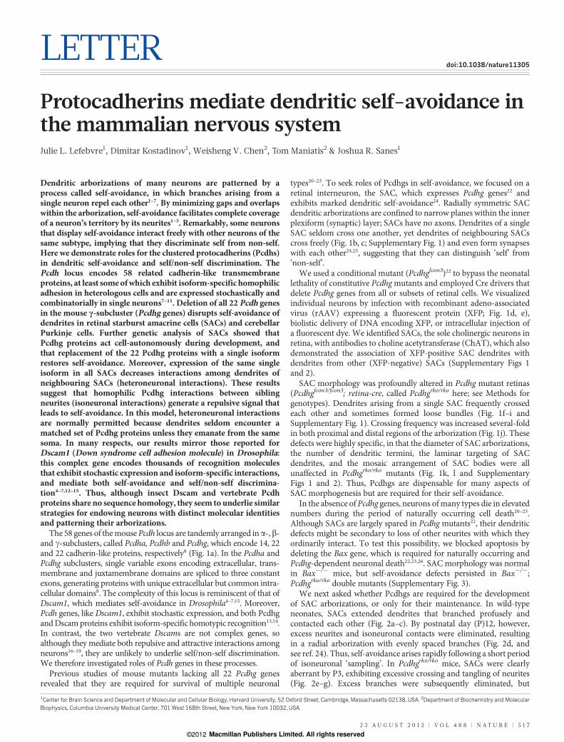
Figure 1 | Pcdhgs are required for self-avoidance of SAC dendrites. a, Pcdhlocus comprises Pcdha, Pcdhb and Pcdhg subclusters. Pcdha and Pcdhg isoformsare assembled by splicing of one variable exon encoding extracellular (ECD) andtransmembrane domains to three constant exons encoding the intracellulardomain (ICD). b, SACs are present in both the inner nuclear layer (INL) and theganglion cell layer (GCL) and extend dendrites that form radially symmetricalarborizations confined to thin sublaminae in the inner plexiform layer (IPL).c, SAC dendrites avoid isoneuronal dendrites (red) but interact heteroneuronallywith other SACs (blue), forming synapses on their dendrites. d–i, Morphology ofa single SAC, labelled with membrane-Cherry, in the GCL in control and Pcdhg
mutant retinas. Wild-type SAC dendrites self-avoid. In Pcdhgmutants, self-avoidance defects include self-crossing and bundling of dendrites. Crossings aredetected at 0.2mm x–y resolution in single 0.8-mm optical sections (e, g, i showmagnified views of boxed areas in d, f, h). Images with 0.2mm z resolution areshown in Supplementary Fig. 1. j, SAC dendritic self-crossings in first–fifth orderbranches per SAC. Graph underestimates difference between genotypes becausethe most severely affected mutant SACs could not be scored. **P, 0.01.k, l, Numberof terminal branches (k) anddendritic field diameter (l) donotdifferbetween wild-type andmutant SACs. Panels j–l showmeans6 s.e.m.; n5 8 cellsfrom 5–6 animals per genotype. Scale bars, 50mm (d, f, h) and 10 mm (e, g, i).
h
f
P8 c
P3a
b
Pcdhgrko/rkoWild-type
e
d
1.40
1.45
1.50
1.55
1.60
1.65
0
5
10
15
1.44 1.48 1.52 1.56 1.60 1.64
g WTPcdhgrko/rko
Fre
quency
Fractal dimension, Df
Mean D
f
8DIV
in vitro
P5–6
in vivo
adult
in vivo
***
***
***
P5
P12
8DIV
8DIV
k
l
Pcd
hg
rko
/rko
j
i
Wild
-typ
e
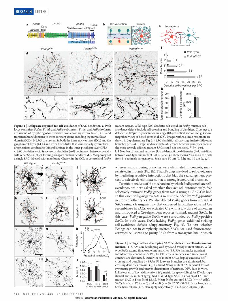
Figure 2 | Pcdhgs pattern developing SAC dendrites in a cell-autonomousmanner. a–h, SACs in developing wild-type and Pcdhgmutant retinas. Wild-type SACs extend fine, exuberant branches (P3, P5) that make transientintradendritic contacts (P5, P8); by P12, excess branches and isoneuronalcontacts are eliminated. Dendrites of mutant SACs display excessive self-crossing and bundling by P3; by P12, excess branches are eliminated, butcrossing dendrites remain. i, j, Cultured Pcdhg mutant SACs exhibit loss ofsymmetric growth and uneven distribution of neurites. DIV, days in vitro.k, Histogramof fractal dimensions (Df,metric for space-filling) for 47wild-type(black) and 47 mutant (grey) SACs. Wild-type SAC in i has Df of 1.61 andmutant SAC in j has Df of 1.53. l, Mean Df for cultured SACs (n5 47 cells),SACs in vivo at P5 (n5 6) and adult (n5 9). ***P, 0.001. Error bars, s.e.m.Scale bars, 50mm (a–d, also apply respectively to e–h) and 20mm (i, j).
RESEARCH LETTER
5 1 8 | N A T U R E | V O L 4 8 8 | 2 3 A U G U S T 2 0 1 2
Macmillan Publishers Limited. All rights reserved©2012
they are selectively labelled by an orange fluorescent protein (Thy1-OFP3) and cultured them at low density. Isolated SACs extendeddendrites that formed radial, web-like arborizations (Fig. 2i),reminiscent of those observed at ,P5 in vivo (Fig. 2b). In contrast,SACs from Pcdhgrko/rko; Thy1-OFP3 mice exhibited less symmetricaland unevenly spaced arborizations, reminiscent of those observed inPcdhgrko/rko retinas at P5 (Fig. 2j and Supplementary Fig. 5). Analysis ofthe space-filling capacity of dendritic arborizations2,27 (see Methods)revealed that defects in vitrowere similar inmagnitude to those in vivo(Fig. 2k, l). Thus, Pcdhgs do not depend on intercellular interactions topromote self-avoidance.We next assessed the requirement for isoform diversity in Pcdhg-
dependent self-avoidance. We used RT–PCR (PCR with reverse tran-scription) to survey expression of Pcdhg isoforms in whole retina, inamacrines generally and in SACs specifically. All 22 Pcdhg variantswere expressed in each preparation, with no indication of decreaseddiversity in purified subpopulations (Supplementary Fig. 6). Wethen analysed a targeted mouse mutant, Pcdhgtcko, in which three
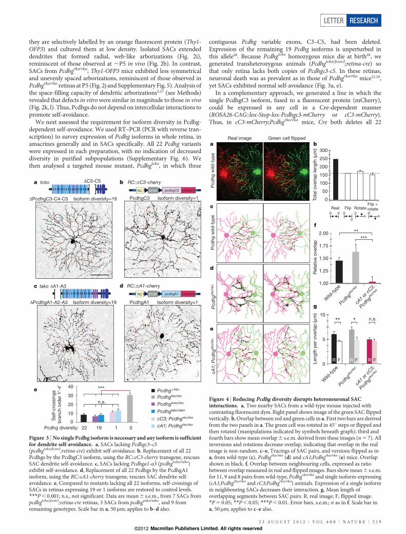
contiguous Pcdhg variable exons, C3–C5, had been deleted.Expression of the remaining 19 Pcdhg isoforms is unperturbed inthis allele28. Because Pcdhgtcko homozygous mice die at birth28, wegenerated transheterozygous animals (Pcdhgtcko/fcon3;retina-cre) sothat only retina lacks both copies of Pcdhgc3-c5. In these retinas,neuronal death was as prevalent as in those of Pcdhgrko/rko mice22,28,yet SACs exhibited normal self-avoidance (Fig. 3a, e).In a complementary approach, we generated a line in which the
single PcdhgC3 isoform, fused to a fluorescent protein (mCherry),could be expressed in any cell in a Cre-dependent manner(ROSA26-CAG::lox-Stop-lox-Pcdhgc3-mCherry or cC3-mCherry).Thus, in cC3-mCherry;Pcdhgrko/rko mice, Cre both deletes all 22
a
dc
tcko ΔC3-C5
ΔPcdhgC3-C4-C5 Isoform diversity=19
b RC::cC3-cherry
tako ΔA1-A3 RC::cA1-cherry
e Pcdhg+/rko
Pcdhgrko/rko
Pcdhgtcko/rko
Pcdhgtako/tako
cC3; Pcdhgrko/rko
cA1; Pcdhgrko/rko S
elf-c
rossin
gs
bra
nch
ord
er
1′-
4′
0
10
20
30
40
n.s.
22 19 1 0
***
STOPRC pcdhgC3 mCherry
STOPRC pcdhgA1 mCherry
PcdhgC3 Isoform diversity=1
ΔPcdhgA1-A2-A3 Isoform diversity=19 PcdhgA1 Isoform diversity=1
Pcdhg diversity:
Figure 3 | No single Pcdhg isoform is necessary and any isoform is sufficientfor dendrite self-avoidance. a, SACs lacking Pcdhgc3–c5(pcdhgtcko/fcon3;retina-cre) exhibit self-avoidance. b, Replacement of all 22Pcdhgs by the PcdhgC3 isoform, using the RC::cC3-cherry transgene, rescuesSAC dendrite self-avoidance. c, SACs lacking Pcdhga1-a3 (pcdhgtako/tako)exhibit self-avoidance. d, Replacement of all 22 Pcdhgs by the PcdhgA1isoform, using the RC::cA1-cherry transgene, rescues SAC dendrite self-avoidance. e, Compared to mutants lacking all 22 isoforms, self-crossings ofSACs in retinas expressing 19 or 1 isoforms are restored to control levels.***P, 0.001; n.s., not significant. Data are mean6 s.e.m., from 7 SACs frompcdhgtcko/fcon3;retina-cre retinas, 3 SACs from pcdhgtako/tako, and 9 fromremaining genotypes. Scale bar in a, 50mm; applies to b–d also.
e
a
Pcd
hg
wild
-typ
eP
cd
hg
rko
/rko
Real image Green cell �ipped
c
cA
1; P
cd
hg
rko
/rko
Pcd
hg
wild
-typ
e
d
Tota
l o
verlap
len
gth
(μ
m)
Real Flip RotateFlip +
rotate
0
50
100
150
200
250
300
b
1.00
1.25
1.50
1.75
2.00
f
Rela
tive o
verlap
Wild
-typ
e
Pcdhg
rko/
rko
cA1
or c
C3;
Pcd
hgrk
o/rk
o
g
**
0
5
10Leng
th p
er
overlap
(μ
m)
R F R FR F
Wild
-typ
e
Pcdhg
rko/
rko
cA1
or c
C3;
Pcd
hgrk
o/rk
o
** n.s. *
***
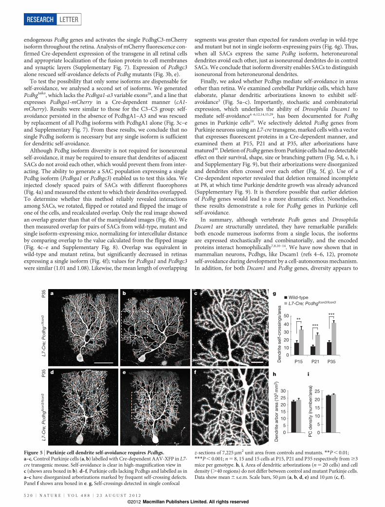
Figure 4 | Reducing Pcdhg diversity disrupts heteroneuronal SACinteractions. a, Two nearby SACs from a wild-type mouse injected withcontrasting fluorescent dyes. Right panel shows image of the green SAC flippedvertically. b, Overlap between red and green cells in a. First two bars are derivedfrom the two panels in a. The green cell was rotated in 45u steps or flipped andthen rotated (manipulations indicated by symbols beneath graph); third andfourth bars showmean overlap6 s.e.m. derived from these images (n5 7). Allinversions and rotations decrease overlap, indicating that overlap in the realimage is non-random. c–e, Tracings of SAC pairs, and versions flipped as ina, from wild-type (c), Pcdhgrko/rko (d) and cA1;Pcdhgrko/rko (e) mice. Overlapshown in black. f, Overlap between neighbouring cells, expressed as ratiobetween overlapmeasured in real and flipped images. Bars showmean6 s.e.m.for 11, 9 and 8 pairs from wild-type, Pcdhgrko/rko and single isoform-expressing(cA1;Pcdhgrko/rko and cC3;Pcdhgrko/rko) animals. Expression of a single isoformin neighbouring SACs decreases their interaction. g, Mean length ofoverlapping segments between SAC pairs. R, real image; F, flipped image.*P5 0.05; **P, 0.05; ***P, 0.01. Error bars, s.e.m.; n as in f. Scale bar ina, 50mm; applies to c–e also.
LETTER RESEARCH
2 3 A U G U S T 2 0 1 2 | V O L 4 8 8 | N A T U R E | 5 1 9
Macmillan Publishers Limited. All rights reserved©2012
endogenous Pcdhg genes and activates the single PcdhgC3-mCherryisoform throughout the retina. Analysis of mCherry fluorescence con-firmed Cre-dependent expression of the transgene in all retinal cellsand appropriate localization of the fusion protein to cell membranesand synaptic layers (Supplementary Fig. 7). Expression of Pcdhgc3alone rescued self-avoidance defects of Pcdhg mutants (Fig. 3b, e).To test the possibility that only some isoforms are dispensable for
self-avoidance, we analysed a second set of isoforms. We generatedPcdhgtako, which lacks the Pcdhga1-a3 variable exons28, and a line thatexpresses Pcdhga1-mCherry in a Cre-dependent manner (cA1-mCherry). Results were similar to those for the C3–C5 group: self-avoidance persisted in the absence of PcdhgA1–A3 and was rescuedby replacement of all Pcdhg isoforms with PcdhgA1 alone (Fig. 3c–eand Supplementary Fig. 7). From these results, we conclude that nosingle Pcdhg isoform is necessary but any single isoform is sufficientfor dendritic self-avoidance.Although Pcdhg isoform diversity is not required for isoneuronal
self-avoidance, it may be required to ensure that dendrites of adjacentSACs do not avoid each other, which would prevent them from inter-acting. The ability to generate a SAC population expressing a singlePcdhg isoform (Pcdhga1 or Pcdhgc3) enabled us to test this idea. Weinjected closely spaced pairs of SACs with different fluorophores(Fig. 4a) andmeasured the extent to which their dendrites overlapped.To determine whether this method reliably revealed interactionsamong SACs, we rotated, flipped or rotated and flipped the image ofone of the cells, and recalculated overlap. Only the real image showedan overlap greater than that of the manipulated images (Fig. 4b). Wethen measured overlap for pairs of SACs from wild-type, mutant andsingle isoform-expressing mice, normalizing for intercellular distanceby comparing overlap to the value calculated from the flipped image(Fig. 4c–e and Supplementary Fig. 8). Overlap was equivalent inwild-type and mutant retina, but significantly decreased in retinasexpressing a single isoform (Fig. 4f); values for Pcdhga1 and Pcdhgc3were similar (1.01 and 1.08). Likewise, the mean length of overlapping
segments was greater than expected for random overlap in wild-typeand mutant but not in single isoform-expressing pairs (Fig. 4g). Thus,when all SACs express the same Pcdhg isoform, heteroneuronaldendrites avoid each other, just as isoneuronal dendrites do in controlSACs.We conclude that isoform diversity enables SACs to distinguishisoneuronal from heteroneuronal dendrites.Finally, we asked whether Pcdhgs mediate self-avoidance in areas
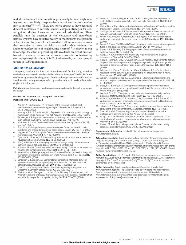
other than retina. We examined cerebellar Purkinje cells, which haveelaborate, planar dendritic arborizations known to exhibit self-avoidance3 (Fig. 5a–c). Importantly, stochastic and combinatorialexpression, which underlies the ability of Drosophila Dscam1 tomediate self-avoidance4–6,12,14,15,29, has been documented for Pcdhggenes in Purkinje cells10. We selectively deleted Pcdhg genes fromPurkinje neurons using an L7-cre transgene, marked cells with a vectorthat expresses fluorescent proteins in a Cre-dependent manner, andexamined them at P15, P21 and at P35, after arborizations havematured30. DeletionofPcdhg genes fromPurkinje cells hadnodetectableeffect on their survival, shape, size or branching pattern (Fig. 5d, e, h, iand Supplementary Fig. 9), but their arborizations were disorganizedand dendrites often crossed over each other (Fig. 5f, g). Use of aCre-dependent reporter revealed that deletion remained incompleteat P8, at which time Purkinje dendrite growth was already advanced(Supplementary Fig. 9). It is therefore possible that earlier deletionof Pcdhg genes would lead to a more dramatic effect. Nonetheless,these results demonstrate a role for Pcdhg genes in Purkinje cellself-avoidance.In summary, although vertebrate Pcdh genes and Drosophila
Dscam1 are structurally unrelated, they have remarkable parallels:both encode numerous isoforms from a single locus, the isoformsare expressed stochastically and combinatorially, and the encodedproteins interact homophilically7,8,10–14. We have now shown that inmammalian neurons, Pcdhgs, like Dscam1 (refs 4–6, 12), promoteself-avoidance during development by a cell-autonomousmechanism.In addition, for both Dscam1 and Pcdhg genes, diversity appears to
L7-C
re; P
cd
hg
+/f
co
n3
P35
L7-C
re; P
cd
hg
fco
n3
/fco
n3
P
35
a b
ed
c
f
g
D
end
rite
self-c
rossin
gs/a
rea
h
0
10
20
30
40
50 **
***
P35
0
5
10
15
20
25
!"PC
den
sity (num
ber/
are
a)
i
***
P15 P21
0
5
10
15
20
25
30
Den
drite
arb
or
are
a (10
3 m
m2)
Wild-type
L7-Cre; Pcdhgfcon3/fcon3
Figure 5 | Purkinje cell dendrite self-avoidance requires Pcdhgs.a–c, Control Purkinje cells (a, b) labelled with Cre-dependent AAV-XFP in L7-cre transgenic mouse. Self-avoidance is clear in high-magnification view inc (shows area boxed in b). d–f, Purkinje cells lacking Pcdhgs and labelled as ina–c have disorganized arborizations marked by frequent self-crossing defects.Panel f shows area boxed in e. g, Self-crossings detected in single confocal
z-sections of 7,225mm2 unit area from controls and mutants. **P, 0.01;***P, 0.001; n5 8, 15 and 15 cells at P15, P21 and P35 respectively from$3mice per genotype. h, i, Area of dendritic arborizations (n5 20 cells) and celldensity (.40 regions) do not differ between control andmutant Purkinje cells.Data show mean6 s.e.m. Scale bars, 50mm (a, b, d, e) and 10mm (c, f).
RESEARCH LETTER
5 2 0 | N A T U R E | V O L 4 8 8 | 2 3 A U G U S T 2 0 1 2
Macmillan Publishers Limited. All rights reserved©2012
underlie self/non-self discrimination, presumably because neighbour-ing neurons are unlikely to express the same isoforms and are thereforefree to interact7,12,14,15,29. Thus, two phyla appear to have recruiteddifferent molecules to mediate similar, complex strategies for self-recognition during formation of neuronal arborizations. Theseparallels raise the question of why vertebrate and invertebratenervous systems have invested heavily in mechanisms that promoteself-avoidance. In principle, self-avoidance allows neurons to covertheir receptive or projective fields maximally while retaining theability to overlap those of neighbouring neurons1–3. However, to ourknowledge, the effect of perturbing self-avoidance on circuit functionhas yet to be assessed in any system. We can now address this issue byelectrophysiological analysis of SACs, Purkinje cells, and their synaptictargets in Pcdhg mutant mice.
METHODS SUMMARYTransgenic, knockout and knock-in mouse lines used for this study, as well asmethods for marking cells are described inMethods. Identity of labelled SACs wasconfirmed by immunolabelling retinas for the cholinergic neuron-specificmarker.Dendrite self-crossings were quantified by number of branch overlaps detected insingle confocal planes.
Full Methods and any associated references are available in the online version ofthe paper.
Received 19 December 2011; accepted 7 June 2012.
Published online 29 July 2012.
1. Kramer, A. P. & Kuwada, J. Y. Formation of the receptive fields of leechmechanosensory neurons during embryonic development. J. Neurosci. 3,2474–2486 (1983).
2. Montague, P. R. & Friedlander, M. J. Expression of an intrinsic growth strategy bymammalian retinal neurons. Proc. Natl Acad. Sci. USA 86, 7223–7227 (1989).
3. Grueber,W.B.&Sagasti, A. Self-avoidance and tiling:mechanismsof dendrite andaxon spacing. Cold Spring Harb. Perspect. Biol. 2, a001750 (2010).
4. Matthews, B. J. et al. Dendrite self-avoidance is controlled by Dscam. Cell 129,593–604 (2007).
5. Soba, P. et al. Drosophila sensory neurons require Dscam for dendritic self-avoidance and proper dendritic field organization. Neuron 54, 403–416 (2007).
6. Hughes, M. E. et al. Homophilic Dscam interactions control complex dendritemorphogenesis. Neuron 54, 417–427 (2007).
7. Zipursky, S. L. & Sanes, J. R. Chemoaffinity revisited: dscams, protocadherins, andneural circuit assembly. Cell 143, 343–353 (2010).
8. Wu, Q. & Maniatis, T. A striking organization of a large family of human neuralcadherin-like cell adhesion genes. Cell 97, 779–790 (1999).
9. Kohmura, N. et al. Diversity revealed by a novel family of cadherins expressed inneurons at a synaptic complex. Neuron 20, 1137–1151 (1998).
10. Kaneko,R.et al.Allelicgene regulationofPcdh-aandPcdh-cclusters involvingbothmonoallelic and biallelic expression in single Purkinje cells. J. Biol. Chem. 281,30551–30560 (2006).
11. Schreiner, D. & Weiner, J. A. Combinatorial homophilic interaction betweenc-protocadherin multimers greatly expands the molecular diversity of celladhesion. Proc. Natl Acad. Sci. USA 107, 14893–14898 (2010).
12. Zhan, X. L. et al. Analysis of Dscam diversity in regulating axon guidance inDrosophilamushroom bodies. Neuron 43, 673–686 (2004).
13. Wojtowicz, W. M., Flanagan, J. J., Millard, S. S., Zipursky, S. L. & Clemens, J. C.Alternative splicing of Drosophila Dscam generates axon guidance receptors thatexhibit isoform-specific homophilic binding. Cell 118, 619–633 (2004).
14. Neves, G., Zucker, J., Daly, M. & Chess, A. Stochastic yet biased expression ofmultiple Dscam splice variants by individual cells. Nature Genet. 36, 240–246(2004).
15. Hattori, D. et al. Robust discrimination between self and non-self neurites requiresthousands of Dscam1 isoforms. Nature 461, 644–648 (2009).
16. Yamagata, M. & Sanes, J. R. Dscam and Sidekick proteins direct lamina-specificsynaptic connections in vertebrate retina. Nature 451, 465–469 (2008).
17. Fuerst, P. G., Koizumi, A., Masland, R. H. & Burgess, R. W. Neurite arborizationandmosaic spacing in the mouse retina require DSCAM. Nature 451, 470–474(2008).
18. Fuerst, P. G. et al.DSCAMandDSCAML1 function in self-avoidance inmultiple celltypes in the developing mouse retina. Neuron 64, 484–497 (2009).
19. Sanes, J. R. & Zipursky, S. L. Design principles of insect and vertebrate visualsystems. Neuron 66, 15–36 (2010).
20. Wang, X. et al. Gamma protocadherins are required for survival of spinalinterneurons. Neuron 36, 843–854 (2002).
21. Prasad, T., Wang, X., Gray, P. A. &Weiner, J. A. A differential developmental patternof spinal interneuron apoptosis during synaptogenesis: insights from geneticanalyses of the protocadherin-c gene cluster. Development 135, 4153–4164(2008).
22. Lefebvre, J. L., Zhang, Y., Meister, M., Wang, X. & Sanes, J. R. c-Protocadherinsregulate neuronal survival but are dispensable for circuit formation in retina.Development 135, 4141–4151 (2008).
23. Weiner, J. A., Wang, X., Tapia, J. C. & Sanes, J. R. Gamma protocadherins arerequired for synapticdevelopment in the spinal cord.Proc.Natl Acad. Sci.USA102,8–14 (2005).
24. Stacy, R. C. & Wong, R. O. Developmental relationship between cholinergicamacrine cell processes and ganglion cell dendrites of the mouse retina. J. Comp.Neurol. 456, 154–166 (2003).
25. Lee, S. & Zhou, Z. J. The synaptic mechanism of direction selectivity in distalprocesses of starburst amacrine cells. Neuron 51, 787–799 (2006).
26. White, F. A., Keller-Peck, C. R., Knudson, C. M., Korsmeyer, S. J. & Snider, W. D.Widespread elimination of naturally occurring neuronal death in Bax-deficientmice. J. Neurosci. 18, 1428–1439 (1998).
27. Jelinek, H. F. & Fernandez, E. Neurons and fractals: how reliable and useful arecalculations of fractal dimensions? J. Neurosci. Methods 81, 9–18 (1998).
28. Chen, W. V. et al. Functional significance of isoform diversification in theprotocadherin gamma gene cluster. Neuron. (in the press).
29. Wang, J. et al. Transmembrane/juxtamembrane domain-dependent Dscamdistribution and function during mushroom body neuronal morphogenesis.Neuron 43, 663–672 (2004).
30. Kaneko, M. et al. Remodeling of monoplanar Purkinje cell dendrites duringcerebellar circuit formation. PLoS ONE 6, e20108 (2011).
Supplementary Information is linked to the online version of the paper atwww.nature.com/nature.
AcknowledgementsWe thank members of our laboratory for providing advice andreagents, including D. Cai and K. Cohen (rAAV), I.-J. Kim (fstl4-line 1 mice) andM. Yamagata for modified Rosa-CAG targeting vector. We also thank B. Stevens(Children’sHospital) for advice onculturemethods. ThisworkwassupportedbygrantsfromNIH to J.R.S. (R01NS029169 andR01EY022073) and T.M. (R01NS043915) andNARSAD Young Investigator Award to J.L.L.
Author Contributions J.L.L., D.K. and J.R.S. designed experiments and prepared themanuscript. J.L.L. andD.K. performed experiments and data analysis. J.R.S. supervisedthe project. W.V.C. and T.M. generated Pcdhgtako and Pcdhgtcko mice. All authorscommented on the manuscript.
Author Information Reprints and permissions information is available atwww.nature.com/reprints. The authors declare no competing financial interests.Readers are welcome to comment on the online version of this article atwww.nature.com/nature. Correspondence and requests for materials should beaddressed to J.R.S. ([email protected]).
LETTER RESEARCH
2 3 A U G U S T 2 0 1 2 | V O L 4 8 8 | N A T U R E | 5 2 1
Macmillan Publishers Limited. All rights reserved©2012
METHODSMouse strains. The Pcdhgfcon3 conditional mutant allele, in which the third con-stant exon is flanked by loxP sequences and which generates a functionally nullallele following Cre recombination, was described previously21,22. Retina-specific Chx10-cre31 and Six3-cre transgenic mice32 were provided by C. Cepko(Harvard) and W. Klein (M.D. Anderson Cancer Center), respectively. Bax2/2
mutants33, Purkinje-specific L7Bac-cre transgenic mice34, Chat-cre, in which theCre recombinase gene was targeted to the endogenous ChAT gene35, and Rosa-CAG-LoxP-STOP-LoxP-tdTomato-WPRE reporter mice36 were obtained fromJackson Laboratories. A line of BAC transgenic mice in which regulatory elementsfrom the fstl4 gene drive expression of CreERwas generated as described in ref. 37.In this line, called line 1 to distinguish it from the line called ‘BD’ in ref. 37, CreERwas expressed in SACs, as well as sparse other amacrine cells. We believe thatexpression reflects influences at the site of transgene integration rather thanexpression of fstl4. Thy1-OFP3 transgenic mice, in which Thy1 promoter andregulatory elements direct expression of Kusabira Orange (OFP) in SACs andsubsets of retinal ganglion cells (RGCs), were described previously37. Pcdhgtcko
and Pcdhgtako mice were generated using standard gene targeting techniques28.Miceweremaintained on aC57/B6J background.All experiments were carried outin accordance with protocols approved by the Harvard University StandingCommittee on the Use of Animals in Research and Teaching.Generation of single Pcdhg isoform conditional knock-in mice. Pcdhga1 andpcdhgc3 full-length cDNAs were amplified from RNA isolated from P21 C57/BL6mouse brain, and cloned in frame into pCMV-mCherry-N1 (Clontech). Linkersequence residing between the third constant exon and gfp in Pcdhgfusg knock-inmice and shown to produce functional Pcdhg-GFP fusion proteins in vivo20 wassubcloned into pCMV-pcdhga1/c3-mCherry-N1. Targeting vector pRosa26-PAS38 was modified as described in ref. 39 to include a CAG cassette (chickenb-actin promoter and CMV immediate-early enhancer), a Gateway RfA destina-tion site (Invitrogen), aWPRE fragment (woodchuckhepatitis virus posttranscrip-tional element), and a STOP sequencewas cloned frompBS302 (Addgene plasmid11925). LoxP-STOP-loxP-Pcdhga1/c3-mCherry was recombined into pROSA26-CAG-Rfa-WPRE-FNF-iSceI, creating pROSA26-CAG-loxP-STOP-loxP-Pcdhga1/c3-mCherry-WPRE-FNF-iSceI targeting vectors. The iSceI-linearizedvectors were electroporated into 129/B6 F1 hybrid ES cell line V6.5. G418-resistant, targeted ES clones were identified by PCR: 1.7 kb fragment amplifiedby 59-Rosa-F: GGCGGACTGGCGGGACTA and 59-CAG-R: CCAGGCGGGCCATTTACCGTAAG; and 8.2 kb fragment amplified by 39-CherryF: CTCCCACAACGAGGACTACACCATCand39-RosaR:GCATTTTAAAAGCATGAAACTACAAC. ES cell transfections and blastocyst injections were performed by theGenome Modification Facility, Harvard University. Following germ-line trans-mission, the FRT-neo-FRT cassette was excised by crossing to mice that expressFlp recombinase ubiquitously40.Gt(ROSA)26Sor::CAG-loxP-STOP-loxP-Pcdhga1/c3-mCherry conditional knock-inmice are called cA1-mCherry and cC3-mCherry.Labelling of neurons. Plasmid encoding pAAV2/2-CAG-palmitoylation tag-mCherry-WPRE was used to generate recombinant AAV2/2 expressingmembrane-tagged Cherry. To label SACs in retina expressing cC3-mCherry orcA1-mCherry, we used rAAV2/2-CBA-YC3.6-WPRE expressing a calciumsensor that includes cytosolic YFP and used here for visualization of neuronalmorphology41. Recombinant AAV2/2-CAG-memb-mCherry and rAAV2/2-YC3.6 were prepared at the Harvard Gene Therapy Institute ((1–2)3 1012
genome copies per ml). Optimal titres of (1–2)3 109 viral genome particlesper ml for AAV2/2-CAG-memb-mCherry and 23 1010 viral genome particlesper ml for rAAV2/2- YC3.6 were prepared in phosphate-buffered saline (PBS,pH5 7.4). rAAV2/9 expressing GFP and mCherry were generated and providedby D. Cai and K. Cohen in our laboratory; high titre virus was produced at theUniversity of Pennsylvania Vector Core.To inject virus into eyes, adult mice were anaesthetized with ketamine/xylazine
by intraperitoneal injection. A 30KG needle was used to make a small hole in thetemporal eye, below the cornea, and 1.5ml of rAAV virus was injected into thevitreous humour with a Hamilton syringe and 33G blunt-ended needle. Animalswere killed and retinas were dissected 4–6weeks following injection. For cerebellarvirus infection, P1–P2 mice were anaesthetized with ice and a small puncture wasmade into the caudal-medial position of one cortical lobe; 1.5ml of rAAV2/9-GFP;mCherry virus was injected with a Hamilton syringe and 33G blunt-ended needle.Mice were analysed 12–35 days after infection.For biolistic transfection of SACs, gold particles (1.0mm diameter, Bio-Rad)
were coated with plasmids encoding tdTomato driven by CMV promoter24. Liveretinas were dissected, transected with four radial incisions, flattened withphotoreceptor side down, and mounted onto a nitrocellulose filter (Millipore).Gold particles were delivered using a Biolistics Helios Gene gun device (Bio-Rad),and retinas were cultured in Ames medium (Sigma) in an oxygenated incubatorheated to 37 uC for 12–16 h.
To assess interactions between dendrites of neighbouring SACs, we injectedpairs of cells with fluorescent dyes. Retinas from mice expressing OFP in SACs(Thy1-OFP3) were mounted RGC side up and perfused with Ames mediumbubbled with 95%O2/5%CO2 at 25 uC. OFP
1 SACs were visualized with epifluor-escence, and impaled with high resistance electrodes (50MV) filled with a K1
based intracellular recording solution supplemented with 50mMAlexa Fluor 568(for targeting) and 200mM of Alexa Fluor 488 or 647 (for filling, Invitrogen).Square voltage pulses of ,3V were applied to SACs at 50Hz using a BKPrecision Model 3011B function generator. After filling one SAC, the electrodewas replaced with a second containing the contrasting dye and the second cell wasfilled. Images of labelled SAC pairs in live retinas were acquired at 403 on a ZeissLSM 510 confocal microscope.
Tissue preparation and immunohistochemistry.Micewere killedwith intraper-itoneal injection ofNembutal, and either enucleated immediately or transcardiallyperfused with Ringer’s solution followed by 4% paraformaldehyde (PFA) in PBS.Eye cups were removed and fixed in 4% PFA on ice for 1 h, followed by dissectionand post-fixation of retinas for an additional 30min, then rinsed with PBS. Brainswere post-fixed in 4% PFA at 4 uC overnight. Animal procedures were in compli-ance with the US National Institutes of Health Guide for the Care and Use ofLaboratory Animals and approved by the Animal and Care and Use Program atHarvard University.
Whole-mount preparations and cryosections of retinas were performed asdescribed22,42. Briefly, whole retinas were incubated for 1–2 h in blocking buffer(0.4% Triton-X, 4% normal donkey serum in PBS), then incubated for 6 days at4 uCwith primary antibodies. Sagittal 80mm sections of cerebellumwere obtainedwith a vibratome (Leica), incubated in blocking buffer, andwith primary antibodiesfor 2 days at 4 uC. Following washing, retinas and brain sections were incubated for3 h at room temperature with Alexa-conjugated secondary antibodies (Invitrogenor Jackson ImmunoResearch). Whole retinas were flattened with photoreceptorside down onto nitrocellulose filters. Retina flat-mounts and brain sections weremounted onto glass slides, covered with Vectashield (Vector) or Fluoromount G(Southern Biotech), and imaged on anOlympus FV1000 scanning confocal micro-scope. Antibodies used were as follows: chick and rabbit anti-GFP (Aves andMillipore); rabbit anti-DsRed (Clontech); goat anti-choline acetyltransferase(Millipore); guinea pig anti-vGluT3 (Millipore); rabbit anti-Calbindin (Swant);mouse anti-syntaxin HPC1 clone (Sigma); rabbit anti-cleaved caspase3 (CellSignaling Technology). Nuclei were labelled with DAPI, Po-pro1, or NeuroTraceNissl 435/455 (Invitrogen).
SACpurification and culture.To isolate and culturewild-type andPcdhgmutantSACs in vitro, we crossed the Thy1-OFP3 transgene, which selectively directsexpression of Kusabira Orange (OFP) in SACs and subset of RGCs37, intoPcdhgfcon3; Six3-cre mice. Retinas from genotyped Pcdhgfcon3/fcon3; Six3-cre;Thy1-OFP3 mutant and control P2 mice were dissociated using papain22. OFP1
SACs were isolated by fluorescence activated cell sorting (FACS, MoFlo), platedonto poly-L-lysine-coated glass coverslips (Warner) and cultured for 7–9 days inRGC growth media modified from Meyer-Franke43 in the following ways: (1)substitution of NS2144 for B27, (2) substitution of N2 (Invitrogen) for Sato stock,(3) addition of TGF-b1 and TGF-b2 (2.5 ngml21; Peprotech), and (4) addition ofmouse glia-conditioned medium (15%). One-third of media was exchanged withfresh media every three days. Cells were fixed with cold 4%PFA/4% sucrose for15min, and immunostained for syntaxin and calbindin to confirm SAC identity,and for GFP to confirm Pcdhg2/2; GFP2 SACs from unrecombined Pcdhg-GFP1
SACs due to variegated Six3-Cre activity in retina.
Image analysis. For best reproduction and clarity of SAC arborizations,maximized projections of confocal images were inverted and contrast-enhancedusing Photoshop (Adobe Systems). For morphometric analysis of SACs, we usedFiji software and selected confocal image series of wild-type and Pcdhg mutantSACs situated in comparable retinal eccentricities. Self-crossings per dendriticbranch order were quantified as number of branch overlaps detected in singleconfocal planes; crossings occurring distal to fifth branch order could not bequantified accurately owing to severity of defects in mutants. Dendritic fielddiameter was measured as the longest axis of arborization. In some cases, arbor-izations were re-imaged by oversampling using a 603 1.45NA objective at x,y,zresolution of 473 473 131mm and then subjected to deconvolution usingHuygens software (http://www.svi.nl/HuygensProfessional).
For analysis of SAC density and mosaic regularity, confocal z-stacks of ChAT-labelled SACs through the GCL and INL were acquired at similar locations incentral retina. Sample sizes were 4–5 areas (0.099mm2) per animal, 2–4 animalsper genotype. For each field, x–y coordinates of SAC arrays were obtained bymanually marking centres of cells using Fiji and used to compute SAC density(number per mm2), packing factor45, and density recovery profiles (DRP)46 withWinDRP software (http://www.mpimf-heidelberg.mpg.de/,teuler/WinDRP/ReadMe.htm).
RESEARCH LETTER
Macmillan Publishers Limited. All rights reserved©2012
To compare the space-filling and complexity of control andmutant SAC arbor-izations, we computed fractal dimensions, Df, which provide a measure of howcompletely dendrites fill its area2,27,47,48. To calculate Df, we applied the box-counting method as implemented in the FracLac 2.5 plug-in for ImageJ software(http://rsb.info.nih.gov/ij/plugins/fraclac/FLHelp/Introduction.htm; NIH). Confocalimages of cultured mutant and control SACs were obtained at equivalent laserscanning parameters with a 603 oil immersion lens, and maximum projectionsand thresholded, binary images were processed using Image J. Box counts using aseries of progressively smaller box sizes (d) were scanned in a region of interestcovering the SAC arborization, and the number of boxes intersected by pixels[k(d)] were analysed; this computes Df, which represents an inverse linear regres-sion between log[k(d)] and log(d).Df ranges from 1.0 (straight line with a dimen-sion of 1) to 2.0 (plane with a dimension of 2); a difference of 0.1 represents adoubling of complexity27.For analysis of dendrite overlap between arborizations of neighbouring SACs,
pairs with somata separated by 80–160mm were selected because their dendritesare known to interact25. Images were processed using Fiji or Photoshop software.To estimate the amount of dendritic overlap that would occur by chance if twoSAC arborizations occupy the same territory, we flipped or rotated the image ofone SAC, realigned cell body position, and merged images. This method wasinspired by work on tiling of RGC dendrites49. We measured total overlappingpixels in real and flipped images, interpreting ratios of .1 (real/flipped) as indi-cating non-random interactions between SACs.Purkinje cell dendrite self-crossings detected in single confocal planes were
counted in a 7,225mm2 region of interest assigned to middle of arborization.Purkinje arborization areas were measured using the convex-hull selection inFiji. Calbindin-labelled Purkinje somata residing along a 635mm segment inlobules III–VI in single confocal planes were counted to measure Purkinje celldensity.Means were compared using the two-tailed Student’s t test on condition of
equivalent variances determined by F-test, or with the Mann–Whitney non-parametric test. Means of multiple samples were compared using ANOVA andposthoc Tukey test.RT–PCR of dissociated retina cells. We used FACS to sort live cells from dis-sociated P7 whole retina, VC1.11 amacrine cells, and OFP1;Thy1.22 SACs cells,as described previously37,50. Amacrine cells were sorted from a live cell suspensionof dissociated retinal cells usingmonoclonalVC1.1 antibody (200mgml21, Sigma)and an anti-IgM secondary conjugated to phycoerythrin-Cy7 (Southern). OFP1
SACs were sorted from OFP1 RGCs by negative selection of Thy1.2-PE-Cy7labelled RGCs. In each condition, 2,000 cells were sorted directly into RNA lysisbuffer (Qiagen); RNA was purified and first strand cDNAs were generated withSuperscript RT III (Invitrogen). Primers that uniquely detect the 22 Pcdhg variableexon-constant exon spliced transcripts were adapted from ref. 21, with modifica-tions to avoid cross-hybridization. These primers, and others used to assess purityof the sorted population, are listed in Supplementary Table 1. PCR program used
is: 94 uC for 2min; 30 cycles of 94 uC for 20 s, 56 uC for 30 s, 72 uC for 1min; 72 uCfor 7min.
31. Rowan, S. &Cepko, C. L. Genetic analysis of the homeodomain transcription factorChx10 in the retina using a novel multifunctional BAC transgenicmouse reporter.Dev. Biol. 271, 388–402 (2004).
32. Furuta, Y., Lagutin, O., Hogan, B. L. & Oliver, G. C. Retina- and ventral forebrain-specific Cre recombinaseactivity in transgenicmice.Genesis26,130–132 (2000).
33. Knudson, C. M., Tung, K. S., Tourtellotte, W. G., Brown, G. A. & Korsmeyer, S. J. Bax-deficient mice with lymphoid hyperplasia andmale germ cell death. Science 270,96–99 (1995).
34. Zhang, X. M. et al. Highly restricted expression of Cre recombinase in cerebellarPurkinje cells. Genesis 40, 45–51 (2004).
35. Rossi, J. et al.Melanocortin-4 receptors expressedby cholinergic neurons regulateenergy balance and glucose homeostasis. Cell Metab. 13, 195–204 (2011).
36. Madisen, L. et al.A robust and high-throughput Cre reporting and characterizationsystem for the whole mouse brain. Nature Neurosci. 13, 133–140 (2010).
37. Kay, J. N. et al. Retinal ganglion cells with distinct directional preferences differ inmolecular identity, structure, and central projections. J. Neurosci. 31, 7753–7762(2011).
38. Srinivas, S. et al. Expression of green fluorescent protein in the ureteric bud oftransgenic mice: a new tool for the analysis of ureteric bud morphogenesis. Dev.Genet. 24, 241–251 (1999).
39. Yamagata, M&. Sanes, J. R. Transgenic strategy for identifying synapticconnections in mice by fluorescence complementation (GRASP). Front. Mol.Neurosci. 5, 18 (2012).
40. Farley, F. W., Soriano, P., Steffen, L. S. & Dymecki, S. M. Widespread recombinaseexpression using FLPeR (flipper) mice. Genesis 28, 106–110 (2000).
41. Kuchibhotla, K. V. et al. Ab plaques lead to aberrant regulation of calciumhomeostasis in vivo resulting in structural and functional disruption of neuronalnetworks. Neuron 59, 214–225 (2008).
42. Hong, Y. K., Kim, I. J. &Sanes, J. R. Stereotypedaxonal arbors of retinal ganglion cellsubsets in the mouse superior colliculus. J. Comp. Neurol. 519, 1691–1711(2011).
43. Meyer-Franke,A., Kaplan,M.R., Pfrieger, F.W.&Barres,B.A.Characterizationof thesignaling interactions that promote the survival and growth of developing retinalganglion cells in culture. Neuron 15, 805–819 (1995).
44. Chen, Y. et al. NS21: re-defined and modified supplement B27 for neuronalcultures. J. Neurosci. Methods 171, 239–247 (2008).
45. Whitney, I. E., Keeley, P. W., Raven, M. A. & Reese, B. E. Spatial patterning ofcholinergic amacrine cells in themouse retina. J. Comp. Neurol. 508,1–12 (2008).
46. Rodieck, R. W. The density recovery profile: a method for the analysis of points inthe plane applicable to retinal studies. Vis. Neurosci. 6, 95–111 (1991).
47. Montague, P. R. & Friedlander, M. J. Morphogenesis and territorial coverage byisolated mammalian retinal ganglion cells. J. Neurosci. 11, 1440–1457 (1991).
48. Smith, T. G. Jr, Lange, G. D. & Marks, W. B. Fractal methods and results in cellularmorphology—dimensions, lacunarity and multifractals. J. Neurosci. Methods 69,123–136 (1996).
49. Wassle, H., Peichl, L. & Boycott, B. B. Dendritic territories of cat retinal ganglioncells. Nature 292, 344–345 (1981).
50. Kay, J. N., Voinescu, P. E., Chu,M.W.&Sanes, J. R.Neurod6expressiondefinesnewretinal amacrine cell subtypes and regulates their fate. Nature Neurosci. 14,965–972 (2011).
LETTER RESEARCH
Macmillan Publishers Limited. All rights reserved©2012