Treatment of neurocysticercosis: Current status and future research needs
Upload
independentCategory
view
3download
0
INFECTION AND IMMUNITY, May 2003, p. 2634–2642 Vol. 71, No. 50019-9567/03/$08.00�0 DOI: 10.1128/IAI.71.5.2634–2642.2003Copyright © 2003, American Society for Microbiology. All Rights Reserved.
CC Chemokines Mediate Leukocyte Trafficking into the CentralNervous System during Murine Neurocysticercosis: Role of ��
T Cells in Amplification of the Host Immune ResponseAstrid E. Cardona, Paula A. Gonzalez, and Judy M. Teale*
Department of Microbiology, The University of Texas Health Science Center at San Antonio, San Antonio, Texas 78229
Received 22 May 2002/Returned for modification 18 July 2002/Accepted 14 February 2003
According to a previous report, the degree of the host immune response highly correlates with severity of thedisease in the murine model for neurocysticercosis. In wild-type mice, Mesocestoides corti infection induced arapid and extensive accumulation of �� T cells and macrophages in the brain. NK cells, dendritic cells, �� Tcells, and B cells were also recruited to the brain but at lower levels. In contrast, �� T-cell-deficient miceexhibited decreased cellular infiltration and reduced central nervous system (CNS) pathology. To understandthe mechanisms of leukocyte recruitment into the CNS, chemokine expression was analyzed in infected brainsin the present study. MCP-1 (CCL2), MIP-1� (CCL3), and MIP-1� (CCL4) were up-regulated within 2 daysafter M. corti infection. Protein expression of RANTES (CCL5), eotaxin (CCL11), and MIP-2 was detectedlater, at 1 week postinfection. Correlating with the decreased cellular infiltration, delta chain T-cell receptor-deficient (TCR��/�) mice exhibited substantially reduced levels of most of the chemokines analyzed (with theexception of eotaxin). The results suggest that �� T cells play an important role in the CNS immune responseby producing chemokines such as MCP-1 and MIP-1�, enhancing leukocyte trafficking into the brain duringmurine neurocysticercosis.
Chemokines represent members of a class of chemotacticcytokines that mediate their function by signaling throughseven transmembrane G-protein-coupled receptors (reviewedin reference 46). Chemokines were initially defined as modu-lators of leukocyte trafficking and positioning within tissues,which are fundamental requirements for effective immunity.More recently it has been discovered that chemokines areinvolved in inflammatory responses including leukocyte de-granulation and mediator release as well as angiogenesis orangiostasis (46). There are approximately 40 to 50 chemokinesthat have been described and classified into four families (CC,CXC, C, and CX3C4) on the basis of spacing of cysteine resi-dues at the amino terminus (5, 6, 46, 58). The CXC chemo-kines predominantly target neutrophils and subsets of T cells,whereas the CC chemokines target a variety of cell types,including T cells, macrophages, eosinophils, and basophils (46,58).
Chemokine expression has been demonstrated to correlatewith inflammatory pathology in neurological diseases (54), au-toimmune diseases (28, 41), and infectious diseases (15, 26, 37,38, 50, 59). Neurocysticercosis (NCC) is the most commonparasitic disease of the human central nervous system (CNS)and is caused by the presence of Taenia solium metacestodes inthe brain (19, 57, 72, 74). Seizures are the most commonclinical manifestation associated with NCC (20, 73), and lesscommon symptoms include headache, increased intracranialpressure, and altered mental state (18, 20, 60, 65). CNS infec-tion with Mesocestoides corti has been used as a model for NCC(12). The CNS immune response in mice was characterized by
the induction of severe CNS pathology and a massive recruit-ment of �� T cells and macrophages (12, 13). It was demon-strated that �� T cells regulate the development of the inflam-matory response in the brain by producing type 1 cytokines(13). Furthermore, �� T-cell-deficient mice exhibited de-creased cellular infiltration and reduced CNS pathology.Therefore, �� T cells appear to play a crucial role in theimmunopathogenesis of murine NCC.
To understand the mechanisms involved in leukocyte re-cruitment in the brain, we performed a kinetic study to deter-mine the chemokines induced in infected wild-type and deltachain T-cell receptor-deficient (TCR��/�) mice. The resultssuggest that CC chemokines are key players in leukocyte infil-tration into the CNS and that �� T cells can contribute byproducing requisite chemokines.
MATERIALS AND METHODS
Mice. Female 3- to 5-week-old C57BL/6 and TCR��/� mice on the C57BL/6background were purchased from the Jackson Laboratory (Bar Harbor, Maine).Animal experiments were conducted under the guidelines of the University ofTexas System, the U.S. Department of Agriculture, and the National Institutes ofHealth.
Parasites and inoculations. M. corti metacestodes were maintained by serialintraperitoneal (i.p.) inoculations. Intracranial inoculations were performed asdescribed previously (12).
Tissue processing. The brain was immediately removed from perfused ani-mals, embedded in optimal cutting temperature medium (O.C.T.), and snap-frozen as described previously (12, 13). Serial horizontal cryosections 10 �m inthickness were placed on silane prep slides (Sigma Biosciences, St. Louis, Mo.).One in every four slides was fixed in formalin for 12 min at room temperatureand stained with hematoxylin and eosin. The remainder of the slides were airdried over night and fixed in fresh acetone for 20 s at room temperature.Acetone-fixed sections were wrapped in aluminum foil and stored at �80°C orprocessed immediately for immunohistochemistry or immunofluorescence.
Antibodies. Anti-mouse biotinylated antibodies included GL3 (pan anti-��),4E2/MCP (anti-MCP-1) purchased from Pharmingen (San Diego, Calif.), andanti-mouse MIP-2 and CRG-2 obtained from R&D Systems. Polyclonal anti-
* Corresponding author. Mailing address: Department of Microbi-ology, The University of Texas Health Science Center at San Antonio,7703 Floyd Curl Dr., San Antonio, TX 78229. Phone: (210) 657-3959.Fax: (210) 567-6612. E-mail: [email protected].
2634
on March 19, 2015 by guest
http://iai.asm.org/
Dow
nloaded from
bodies against mouse MIP-1�, MIP-1�, and RANTES were also purchased fromR&D Systems. The anti-mouse polyclonal antibody against eotaxin was pur-chased from Serotec. All antibodies were titrated in spleen sections from miceinfected i.p. to determine optimal dilutions. Spleen sections were used as positivecontrols in all experiments. Negative controls consisted of sections from mock-infected mice that were injected intracerebrally with Hanks balanced salt solu-tion (HBSS) (the buffer used in the parasite inoculum). In addition, negativestaining controls from which the primary antibody was excluded were alwaysincluded.
Immunohistochemistry. Staining was performed as described previously (12,13). For the purified antibodies, indirect immunohistochemistry was performedusing specific biotinylated secondary antibodies obtained from Kirkegaard &Perry Laboratories (Gaithersburg, Md.). Positive cells were counted at a mag-nification of �600, and results were scored as 1 to 100, 100 to 300, 300 to 500, and500 positive cells per section.
Double immunofluorescence. Double-immunofluorescence assays were per-formed in brain cryosections to detect chemokines produced by �� T cells. Allincubations were carried out at room temperature, and the slides were washed sixtimes for 5 min each time. Sections were incubated first with the phycoerythrin-conjugated red fluorescent GL3 antibody (Pharmingen) for 1 h and then washedand incubated with biotinylated anti-MCP-1 or purified anti-MIP-1� antibody.The biotinylated antibody treatment was followed by incubation with streptavidinAlexa Fluor 488 conjugate (green fluorescence) (Molecular Probes) for 30 min.The purified anti-MIP-1� antibody was detected using green fluorescent anti-goat immunoglobulin G-Cy2 conjugate (Jackson Laboratories, West Grove, Pa.).Sections were then washed in phosphate-buffered saline (PBS) and mountedusing FluorSave reagent (Calbiochem, La Jolla, Calif.) containing 0.3 �M 4,6-diamidino-2-phenylindole (DAPI)-dilactate (Molecular Probes).
Lymphocyte preparation. Brain lymphocytes were isolated from C57BL/6 andTCR��/� mice 3 weeks after M. corti infection. The brains were removed asdescribed previously. Each perfused brain was minced gently through a fine-mesh nylon screen using a syringe plunger and collected into 10 ml of HBSScontaining 0.05% collagenase D (Boehringer Mannheim), 0.1 �g of TLCK (N�-p-tosyl-L-lysine chloromethyl ketone) (Sigma)/ml, 10 �g of DNase I (Sigma)/ml,and 10 mM HEPES buffer (pH 7.4) (Invitrogen, Carlsbad, Calif.). The mixturewas rocked at room temperature for 45 min and then allowed to settle for 20 min.The supernatant was collected and pelleted at 200 � g for 5 min and resuspendedin 5 ml of HBSS without Ca2� and Mg2�. The suspension was layered onto 10ml of a density gradient containing three parts of Ficoll-Hypaque (AmershamPharmacia Biotech, Piscataway, N.J.) and one part of RPMI medium (Invitro-gen) supplemented with 10% fetal bovine serum (Sigma), 10 mM HEPES (In-vitrogen), and 50 �g gentamicin (Invitrogen)/ml. Each gradient was centrifugedat 300 � g for 30 min at room temperature. The overlaying medium and interfaceof tissue debris were removed. The entire 10 ml of gradient medium was thendiluted fivefold with HBSS and centrifuged at 300 � g for 10 min. Cells were thenwashed three times in 1 ml of 0.1% bovine serum albumin in PBS and counted.
Peritoneal lymphocytes were isolated from C57BL/6 mice infected i.p. at 3 to5 weeks postinfection (p.i.). The peritoneal cavity was washed with 15 ml ofHBSS, and the cellular preparation was filtered through a 150-�m-pore-sizenylon screen to remove parasites. Cells were centrifuged at 300 � g for 10 min,washed three times in 0.3% bovine serum albumin in PBS, and counted. Forpositive selection of �� T lymphocytes, cell suspensions were incubated with 10�g of Dynabeads M-280 streptavidin (Dynal, Lake Success, N.J.)/ml previouslycoated with 500 �g of biotinylated GL3 antibody (Pharmingen)/ml for 20 min at4°C. Labeled cells were then selected using the Dynal magnetic particle concen-trator and washed eight times according to the manufacturer’s instructions.Purified populations of �� T cells were found to be 95 to 97% pure on the basisof subsequent immunocytochemistry analyses.
RNase protection assay (RPA). Using Trizol reagent (Invitrogen) according tothe manufacturer’s instructions, total RNA was extracted from brain lympho-cytes and from positively selected �� T cells. [�-33P]UTP-labeled antisense RNAtranscripts for the chemokines were generated using the template set mCK-5(Pharmingen). Total RNA (5 to 10 mg) was allowed to hybridize to the labeledprobe for 12 to 16 h at 56°C. An RNase T1 mixture was used to digest single-stranded RNA, and the hybrids were analyzed on denaturing urea-polyacrylam-ide gels. Protected fragments were visualized by autoradiography and analyzedby densitometry. The results were analyzed by normalizing the values of eachchemokine to the GAPDH (glyceraldehyde-3-phosphate dehydrogenase) gene.The ratio of infected cells/HBSS control cells was obtained, and the data arepresented as severalfold increase for each chemokine.
RESULTS
M. corti infection of the CNS. As reported previously (12,13), infection of the CNS by M. corti leads to an infiltration ofmononuclear cells by day 2 p.i. The parasites initially invadethe extraparenchymal spaces of the brain such as the ventriclesand subarachnoid spaces. However, by 3 weeks p.i. there arenearly equal numbers of parasites in the extraparenchymal andparenchymal regions of the brain. The cellular infiltrate eithersurrounds the parasite (Fig. 1A) or appears to be movingtowards the parasite (12). Importantly, it has been shown that�� T cells appear to be the dominant T-cell population in thisinfection and appear by day 2 to 3 p.i. (12). Figure 1B exhibitsthe results of an immunocytochemistry assay, showing an ac-cumulation of �� T cells into an extraparenchymal area of thebrain.
Up-regulation of CC chemokines in the brain after M. cortiinfection. The expression of several chemokines was analyzedusing a RPA. Normal mouse brains and HBSS-inoculated con-trols revealed the presence of low levels of eotaxin, MIP-1�,and MIP-1� (Fig. 2A). In wild-type mice, MIP-1�, MIP-1�,and eotaxin were the predominant transcripts at 2 days p.i.(Fig. 2B). Of the chemokines that are substantially up-regu-lated at 2 days p.i. in TCR��/� mice, mRNAs for MIP-1� andMIP-1� were found in levels similar to those for wild-type mice(Fig. 2B). By 1 week p.i., wild-type mice exhibited an increasein the levels of RANTES, in addition to the sustained predom-inance of MIP-1�, MIP-1�, and eotaxin (Fig. 2C). In TCR��/�
mice, in contrast, eotaxin represented the predominant tran-script at 1 week p.i. These results indicate that in wild-typemice, the CC chemokines RANTES, MIP-1�, MIP-1�, andeotaxin were the predominant mRNA species present in brainlymphocytes 1 week after M. corti infection. mRNAs forMCP-1, MIP-2, CRG-2, and lymphotactin (LTN) were de-tected at all of the early time points analyzed, although at lesssignificant levels (severalfold increases of less than 1).
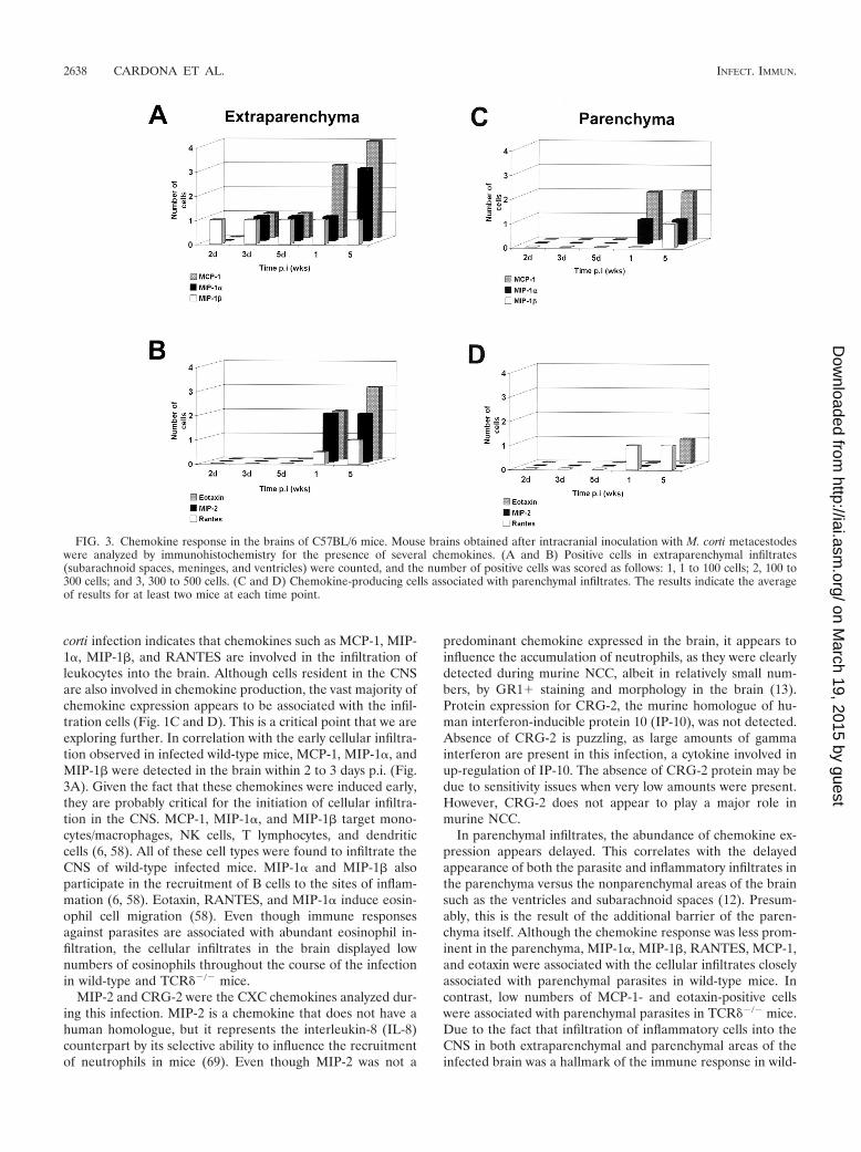
Predominant expression of CC chemokines in the CNS. Tofurther examine the chemokine profile present after M. cortiinfection, in situ immunohistochemistry was performed inbrain cryosections. Specific antibodies against MIP-2, CRG-2,RANTES, MIP-1�, MIP-1�, MCP-1, and eotaxin were used.Examples are shown in Fig. 1C (eotaxin) and Fig. 1D (MCP-1);it is interesting that most of the staining is in the infiltrate andnot the surrounding brain tissue. At the protein level, all ofthese chemokines, except CRG-2, were up-regulated in thebrain after M. corti infection. CRG-2 protein was undetectableat all of the time points analyzed. In wild-type mice, MIP-1�-,MIP-1�-, and MCP-1-positive cells were detected within 2 to 3days p.i. in extraparenchymal areas of the brain, and theamount of MIP-1� and MCP-1 increased dramatically after 1week p.i. (Fig. 3A). Eotaxin, MIP-2, and RANTES were de-tected after 1 week p.i. (Fig. 3B), and MIP-1�, RANTES, andMIP-2 were consistently present in extraparenchymal areas inall of the wild-type mice analyzed during the course of theinfection but at low levels.
In parenchymal infiltrates, chemokine-producing cells werefound with a predominance of MCP-1 expression after 1 weekp.i. (Fig. 3C). MIP-1� and RANTES were also detected in theareas of cellular infiltration, and gradients of positive stainingfor MIP-1� were found in regions not directly associated with
VOL. 71, 2003 CHEMOKINES IN MURINE NEUROCYSTICERCOSIS 2635
on March 19, 2015 by guest
http://iai.asm.org/
Dow
nloaded from
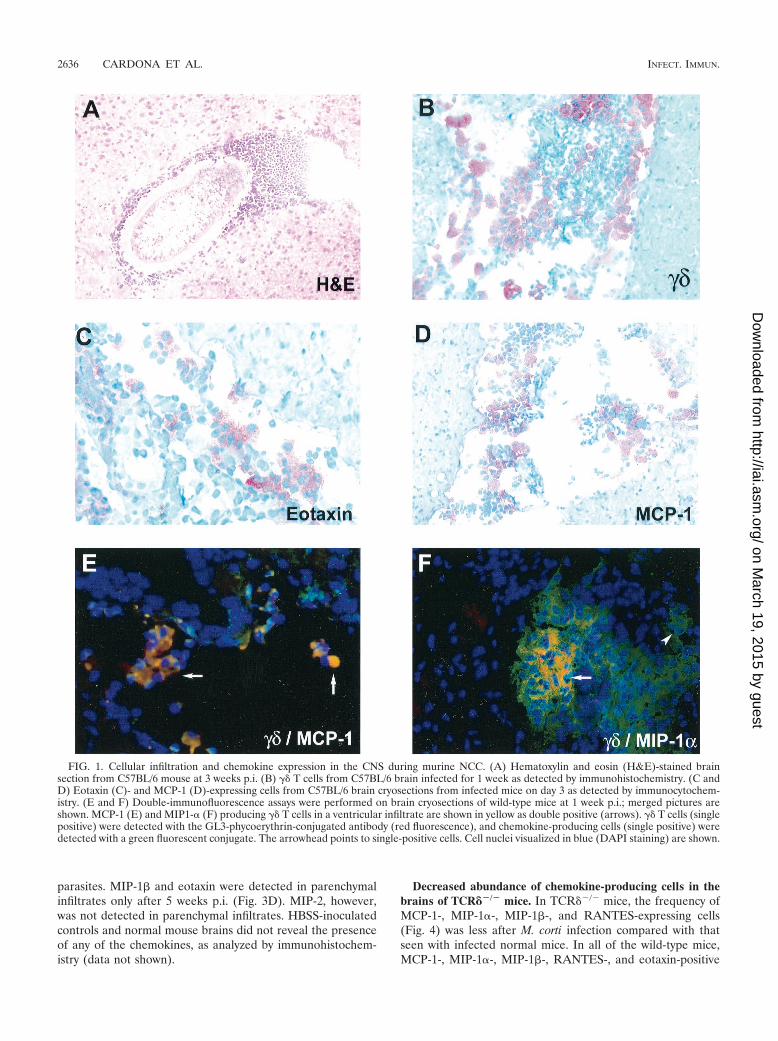
parasites. MIP-1� and eotaxin were detected in parenchymalinfiltrates only after 5 weeks p.i. (Fig. 3D). MIP-2, however,was not detected in parenchymal infiltrates. HBSS-inoculatedcontrols and normal mouse brains did not reveal the presenceof any of the chemokines, as analyzed by immunohistochem-istry (data not shown).
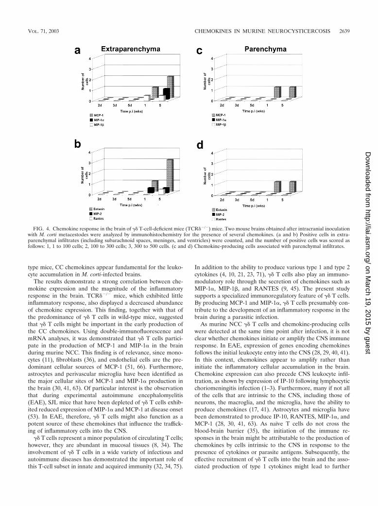
Decreased abundance of chemokine-producing cells in thebrains of TCR��/� mice. In TCR��/� mice, the frequency ofMCP-1-, MIP-1�-, MIP-1�-, and RANTES-expressing cells(Fig. 4) was less after M. corti infection compared with thatseen with infected normal mice. In all of the wild-type mice,MCP-1-, MIP-1�-, MIP-1�-, RANTES-, and eotaxin-positive
FIG. 1. Cellular infiltration and chemokine expression in the CNS during murine NCC. (A) Hematoxylin and eosin (H&E)-stained brainsection from C57BL/6 mouse at 3 weeks p.i. (B) �� T cells from C57BL/6 brain infected for 1 week as detected by immunohistochemistry. (C andD) Eotaxin (C)- and MCP-1 (D)-expressing cells from C57BL/6 brain cryosections from infected mice on day 3 as detected by immunocytochem-istry. (E and F) Double-immunofluorescence assays were performed on brain cryosections of wild-type mice at 1 week p.i.; merged pictures areshown. MCP-1 (E) and MIP1-� (F) producing �� T cells in a ventricular infiltrate are shown in yellow as double positive (arrows). �� T cells (singlepositive) were detected with the GL3-phycoerythrin-conjugated antibody (red fluorescence), and chemokine-producing cells (single positive) weredetected with a green fluorescent conjugate. The arrowhead points to single-positive cells. Cell nuclei visualized in blue (DAPI staining) are shown.
2636 CARDONA ET AL. INFECT. IMMUN.
on March 19, 2015 by guest
http://iai.asm.org/
Dow
nloaded from
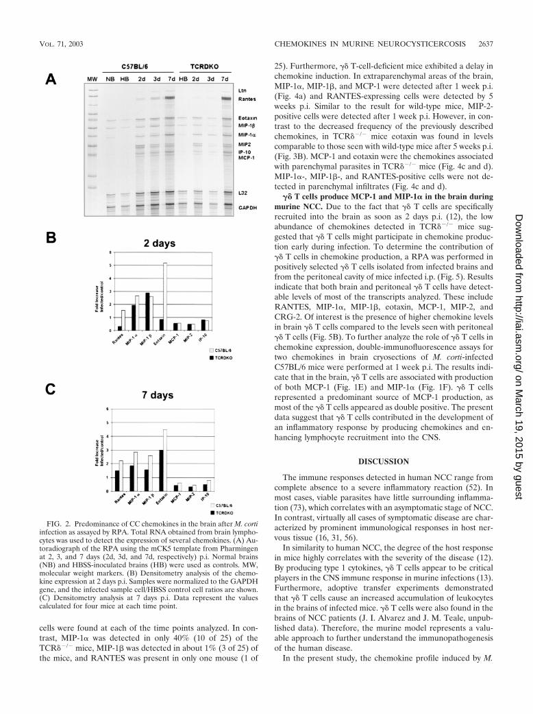
cells were found at each of the time points analyzed. In con-trast, MIP-1� was detected in only 40% (10 of 25) of theTCR��/� mice, MIP-1� was detected in about 1% (3 of 25) ofthe mice, and RANTES was present in only one mouse (1 of
25). Furthermore, �� T-cell-deficient mice exhibited a delay inchemokine induction. In extraparenchymal areas of the brain,MIP-1�, MIP-1�, and MCP-1 were detected after 1 week p.i.(Fig. 4a) and RANTES-expressing cells were detected by 5weeks p.i. Similar to the result for wild-type mice, MIP-2-positive cells were detected after 1 week p.i. However, in con-trast to the decreased frequency of the previously describedchemokines, in TCR��/� mice eotaxin was found in levelscomparable to those seen with wild-type mice after 5 weeks p.i.(Fig. 3B). MCP-1 and eotaxin were the chemokines associatedwith parenchymal parasites in TCR��/� mice (Fig. 4c and d).MIP-1�-, MIP-1�-, and RANTES-positive cells were not de-tected in parenchymal infiltrates (Fig. 4c and d).
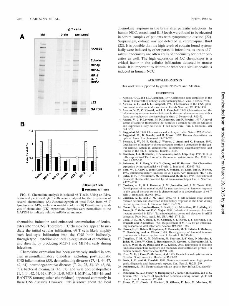
�� T cells produce MCP-1 and MIP-1� in the brain duringmurine NCC. Due to the fact that �� T cells are specificallyrecruited into the brain as soon as 2 days p.i. (12), the lowabundance of chemokines detected in TCR��/� mice sug-gested that �� T cells might participate in chemokine produc-tion early during infection. To determine the contribution of�� T cells in chemokine production, a RPA was performed inpositively selected �� T cells isolated from infected brains andfrom the peritoneal cavity of mice infected i.p. (Fig. 5). Resultsindicate that both brain and peritoneal �� T cells have detect-able levels of most of the transcripts analyzed. These includeRANTES, MIP-1�, MIP-1�, eotaxin, MCP-1, MIP-2, andCRG-2. Of interest is the presence of higher chemokine levelsin brain �� T cells compared to the levels seen with peritoneal�� T cells (Fig. 5B). To further analyze the role of �� T cells inchemokine expression, double-immunofluorescence assays fortwo chemokines in brain cryosections of M. corti-infectedC57BL/6 mice were performed at 1 week p.i. The results indi-cate that in the brain, �� T cells are associated with productionof both MCP-1 (Fig. 1E) and MIP-1� (Fig. 1F). �� T cellsrepresented a predominant source of MCP-1 production, asmost of the �� T cells appeared as double positive. The presentdata suggest that �� T cells contributed in the development ofan inflammatory response by producing chemokines and en-hancing lymphocyte recruitment into the CNS.
DISCUSSION
The immune responses detected in human NCC range fromcomplete absence to a severe inflammatory reaction (52). Inmost cases, viable parasites have little surrounding inflamma-tion (73), which correlates with an asymptomatic stage of NCC.In contrast, virtually all cases of symptomatic disease are char-acterized by prominent immunological responses in host ner-vous tissue (16, 31, 56).
In similarity to human NCC, the degree of the host responsein mice highly correlates with the severity of the disease (12).By producing type 1 cytokines, �� T cells appear to be criticalplayers in the CNS immune response in murine infections (13).Furthermore, adoptive transfer experiments demonstratedthat �� T cells cause an increased accumulation of leukocytesin the brains of infected mice. �� T cells were also found in thebrains of NCC patients (J. I. Alvarez and J. M. Teale, unpub-lished data). Therefore, the murine model represents a valu-able approach to further understand the immunopathogenesisof the human disease.
In the present study, the chemokine profile induced by M.
FIG. 2. Predominance of CC chemokines in the brain after M. cortiinfection as assayed by RPA. Total RNA obtained from brain lympho-cytes was used to detect the expression of several chemokines. (A) Au-toradiograph of the RPA using the mCK5 template from Pharmingenat 2, 3, and 7 days (2d, 3d, and 7d, respectively) p.i. Normal brains(NB) and HBSS-inoculated brains (HB) were used as controls. MW,molecular weight markers. (B) Densitometry analysis of the chemo-kine expression at 2 days p.i. Samples were normalized to the GAPDHgene, and the infected sample cell/HBSS control cell ratios are shown.(C) Densitometry analysis at 7 days p.i. Data represent the valuescalculated for four mice at each time point.
VOL. 71, 2003 CHEMOKINES IN MURINE NEUROCYSTICERCOSIS 2637
on March 19, 2015 by guest
http://iai.asm.org/
Dow
nloaded from
corti infection indicates that chemokines such as MCP-1, MIP-1�, MIP-1�, and RANTES are involved in the infiltration ofleukocytes into the brain. Although cells resident in the CNSare also involved in chemokine production, the vast majority ofchemokine expression appears to be associated with the infil-tration cells (Fig. 1C and D). This is a critical point that we areexploring further. In correlation with the early cellular infiltra-tion observed in infected wild-type mice, MCP-1, MIP-1�, andMIP-1� were detected in the brain within 2 to 3 days p.i. (Fig.3A). Given the fact that these chemokines were induced early,they are probably critical for the initiation of cellular infiltra-tion in the CNS. MCP-1, MIP-1�, and MIP-1� target mono-cytes/macrophages, NK cells, T lymphocytes, and dendriticcells (6, 58). All of these cell types were found to infiltrate theCNS of wild-type infected mice. MIP-1� and MIP-1� alsoparticipate in the recruitment of B cells to the sites of inflam-mation (6, 58). Eotaxin, RANTES, and MIP-1� induce eosin-ophil cell migration (58). Even though immune responsesagainst parasites are associated with abundant eosinophil in-filtration, the cellular infiltrates in the brain displayed lownumbers of eosinophils throughout the course of the infectionin wild-type and TCR��/� mice.
MIP-2 and CRG-2 were the CXC chemokines analyzed dur-ing this infection. MIP-2 is a chemokine that does not have ahuman homologue, but it represents the interleukin-8 (IL-8)counterpart by its selective ability to influence the recruitmentof neutrophils in mice (69). Even though MIP-2 was not a
predominant chemokine expressed in the brain, it appears toinfluence the accumulation of neutrophils, as they were clearlydetected during murine NCC, albeit in relatively small num-bers, by GR1� staining and morphology in the brain (13).Protein expression for CRG-2, the murine homologue of hu-man interferon-inducible protein 10 (IP-10), was not detected.Absence of CRG-2 is puzzling, as large amounts of gammainterferon are present in this infection, a cytokine involved inup-regulation of IP-10. The absence of CRG-2 protein may bedue to sensitivity issues when very low amounts were present.However, CRG-2 does not appear to play a major role inmurine NCC.
In parenchymal infiltrates, the abundance of chemokine ex-pression appears delayed. This correlates with the delayedappearance of both the parasite and inflammatory infiltrates inthe parenchyma versus the nonparenchymal areas of the brainsuch as the ventricles and subarachnoid spaces (12). Presum-ably, this is the result of the additional barrier of the paren-chyma itself. Although the chemokine response was less prom-inent in the parenchyma, MIP-1�, MIP-1�, RANTES, MCP-1,and eotaxin were associated with the cellular infiltrates closelyassociated with parenchymal parasites in wild-type mice. Incontrast, low numbers of MCP-1- and eotaxin-positive cellswere associated with parenchymal parasites in TCR��/� mice.Due to the fact that infiltration of inflammatory cells into theCNS in both extraparenchymal and parenchymal areas of theinfected brain was a hallmark of the immune response in wild-
FIG. 3. Chemokine response in the brains of C57BL/6 mice. Mouse brains obtained after intracranial inoculation with M. corti metacestodeswere analyzed by immunohistochemistry for the presence of several chemokines. (A and B) Positive cells in extraparenchymal infiltrates(subarachnoid spaces, meninges, and ventricles) were counted, and the number of positive cells was scored as follows: 1, 1 to 100 cells; 2, 100 to300 cells; and 3, 300 to 500 cells. (C and D) Chemokine-producing cells associated with parenchymal infiltrates. The results indicate the averageof results for at least two mice at each time point.
2638 CARDONA ET AL. INFECT. IMMUN.
on March 19, 2015 by guest
http://iai.asm.org/
Dow
nloaded from
type mice, CC chemokines appear fundamental for the leuko-cyte accumulation in M. corti-infected brains.
The results demonstrate a strong correlation between che-mokine expression and the magnitude of the inflammatoryresponse in the brain. TCR��/� mice, which exhibited littleinflammatory response, also displayed a decreased abundanceof chemokine expression. This finding, together with that ofthe predominance of �� T cells in wild-type mice, suggestedthat �� T cells might be important in the early production ofthe CC chemokines. Using double-immunofluorescence andmRNA analyses, it was demonstrated that �� T cells partici-pate in the production of MCP-1 and MIP-1� in the brainduring murine NCC. This finding is of relevance, since mono-cytes (11), fibroblasts (36), and endothelial cells are the pre-dominant cellular sources of MCP-1 (51, 66). Furthermore,astrocytes and perivascular microglia have been identified asthe major cellular sites of MCP-1 and MIP-1� production inthe brain (30, 41, 63). Of particular interest is the observationthat during experimental autoimmune encephalomyelitis(EAE), SJL mice that have been depleted of �� T cells exhib-ited reduced expression of MIP-1� and MCP-1 at disease onset(53). In EAE, therefore, �� T cells might also function as apotent source of these chemokines that influence the traffick-ing of inflammatory cells into the CNS.
�� T cells represent a minor population of circulating T cells;however, they are abundant in mucosal tissues (8, 34). Theinvolvement of �� T cells in a wide variety of infectious andautoimmune diseases has demonstrated the important role ofthis T-cell subset in innate and acquired immunity (32, 34, 75).
In addition to the ability to produce various type 1 and type 2cytokines (4, 10, 21, 23, 71), �� T cells also play an immuno-modulatory role through the secretion of chemokines such asMIP-1�, MIP-1�, and RANTES (9, 45). The present studysupports a specialized immunoregulatory feature of �� T cells.By producing MCP-1 and MIP-1�, �� T cells presumably con-tribute to the development of an inflammatory response in thebrain during a parasitic infection.
As murine NCC �� T cells and chemokine-producing cellswere detected at the same time point after infection, it is notclear whether chemokines initiate or amplify the CNS immuneresponse. In EAE, expression of genes encoding chemokinesfollows the initial leukocyte entry into the CNS (28, 29, 40, 41).In this context, chemokines appear to amplify rather thaninitiate the inflammatory cellular accumulation in the brain.Chemokine expression can also precede CNS leukocyte infil-tration, as shown by expression of IP-10 following lymphocyticchoriomeningitis infection (1–3). Furthermore, many if not allof the cells that are intrinsic to the CNS, including those ofneurons, the macroglia, and the microglia, have the ability toproduce chemokines (17, 41). Astrocytes and microglia havebeen demonstrated to produce IP-10, RANTES, MIP-1�, andMCP-1 (28, 30, 41, 63). As naïve T cells do not cross theblood-brain barrier (35), the initiation of the immune re-sponses in the brain might be attributable to the production ofchemokines by cells intrinsic to the CNS in response to thepresence of cytokines or parasite antigens. Subsequently, theeffective recruitment of �� T cells into the brain and the asso-ciated production of type 1 cytokines might lead to further
FIG. 4. Chemokine response in the brain of �� T-cell-deficient mice (TCR��/�) mice. Two mouse brains obtained after intracranial inoculationwith M. corti metacestodes were analyzed by immunohistochemistry for the presence of several chemokines. (a and b) Positive cells in extra-parenchymal infiltrates (including subarachnoid spaces, meninges, and ventricles) were counted, and the number of positive cells was scored asfollows: 1, 1 to 100 cells; 2, 100 to 300 cells; 3, 300 to 500 cells. (c and d) Chemokine-producing cells associated with parenchymal infiltrates.
VOL. 71, 2003 CHEMOKINES IN MURINE NEUROCYSTICERCOSIS 2639
on March 19, 2015 by guest
http://iai.asm.org/
Dow
nloaded from
chemokine induction and enhanced accumulation of leuko-cytes into the CNS. Therefore, CC chemokines appear to me-diate the initial cellular infiltration. �� T cells likely amplifysuch leukocyte infiltration into the CNS both indirectly,through type 1 cytokine-induced up-regulation of chemokines,and directly, by producing MCP-1 and MIP-1� early duringinfections.
Chemokine expression has been extensively studied in sev-eral neuroinflammatory disorders, including posttraumaticCNS inflammation (55), demyelinating diseases (27, 41, 44, 47,49, 64), neurodegenerative diseases (7, 24, 25, 33, 39, 48, 68,70), bacterial meningitis (43, 67), and viral encephalopathies(1, 3, 14, 42, 61, 62). IP-10, IL-8, MCP-1, MIP-1�, MIP-1�, andRANTES (among other chemokines) appeared commonly inthese CNS diseases. However, little is known about the local
chemokine response in the brain after parasitic infections. Inhuman NCC, eotaxin and IL-5 levels were found to be elevatedin serum samples of patients with symptomatic disease (22).Surprisingly, eotaxin was not detected in cerebrospinal fluid(22). It is possible that the high levels of eotaxin found system-ically were induced by other parasitic infections, as areas of T.solium endemicity are often areas of endemicity for other par-asites as well. The high expression of CC chemokines is acritical factor in the cellular infiltration detected in mousebrain. It is important to determine whether a similar profile isinduced in human NCC.
ACKNOWLEDGMENTS
This work was supported by grants NS35974 and AI19896.
REFERENCES
1. Asensio, V. C., and I. L. Campbell. 1997. Chemokine gene expression in thebrains of mice with lymphocytic choriomeningitis. J. Virol. 71:7832–7840.
2. Asensio, V. C., and I. L. Campbell. 1999. Chemokines in the CNS: pluri-functional mediators in diverse states. Trends Neurosci. 22:1453–1458.
3. Asensio, V. C., C. Kincaid, and I. L. Campbell. 1999. Chemokines and theinflammatory response to viral infection in the central nervous system with afocus on lymphocytic choriomeningitis virus. J. Neurovirol. 5:65–75.
4. Azuara, V., J. P. Levraud, M. P. Lembezat, and P. Pereira. 1997. A novelsubset of adult �� thymocytes that secretes a distinct pattern of cytokinesand expresses a very restricted T cell repertoire. Eur. J. Immunol. 27:544–553.
5. Baggiolini, M. 1998. Chemokines and leukocyte traffic. Nature 392:565–568.6. Baggiolini, M., B. Dewald, and B. Moser. 1997. Human chemokines: an
update. Annu. Rev. Immunol. 15:675–705.7. Berman, J. W., M. P. Guida, J. Warren, J. Amat, and C. Brosnan. 1996.
Localization of monocyte chemoattractant peptide-1 expression in the cen-tral nervous system in experimental autoimmune encephalomyelitis andtrauma in the rat. J. Immunol. 156:3017–3023.
8. Bluestone, J. A., R. Khattri, R. Scianmmas, and A. I. Sperling. 1995. TCR ��cells: a specialized T-cell subset in the immune system. Annu. Rev. Cell Dev.Biol. 11:307–353.
9. Boismenu, R., L. Feng, Y. Xia, Y. Chang, and W. Havran. 1996. Chemokineexpression by intraepithelial �� T cells. J. Immunol. 157:985–992.
10. Born, W., C. Cady, J. Jones-Carson, A. Mukasa, M. Lahn, and R. O’Brien.1999. Immunoregulatory functions of �� T cells. Adv. Immunol. 71:77–144.
11. Calvo, C.-F., T. Yoshimura, M. Gelman, and M. Mallat. 1996. Production ofmonocyte chemotactic protein-1 by rat brain macrophages. Eur. J. Neurosci.8:1725–1734.
12. Cardona, A. E., B. I. Restrepo, J. M. Jaramillo, and J. M. Teale. 1999.Development of an animal model for neurocysticercosis: immune responsein the central nervous system is characterized by a predominance of �� Tcells. J. Immunol. 162:995–1002.
13. Cardona, A. E., and J. M. Teale. 2002. �� T cell-deficient mice exhibitreduced severity and decreased inflammatory response in the brain duringmurine cysticercosis. J. Immunol. 169:3163–3171.
14. Conant, K., A. Garzino-Demo, A. Nath, J. C. McArthur, W. Halliday, C.Power, R. C. Gallo, and E. O. Major. 1998. Induction of monocyte chemoat-tractant protein-1 in HIV-1 Tat-stimulated astrocytes and elevation in AIDSdementia. Proc. Natl. Acad. Sci. USA 95:3117–3121.
15. Cook, D. N., M. A. Beck, T. M. Coffman, S. L. Kirby, J. F. Sheridan, I. B.Pragnell, and O. Smithies. 1995. Requirement of MIP-1� for an inflamma-tory response to viral infections. Science 269:1583–1585.
16. Correa, D., D. Dalma, B. Espinoza, A. Plancarte, M. T. Rabiela, I. Madrazo,C. Gorodezky, and A. Flisser. 1985. Heterogeneity of humoral immunecomponents in human cysticercosis. J. Parasitol. 71:535–541.
17. Coughlan, C. M., C. M. McManus, M. Sharron, Z.-Y. Gao, D. Murphy, S.Jaffer, W. Choe, W. Chen, J. Hesselgesser, H. Gaylord, A. Kalyuzhny, M.-Y.Lee, B. Wolf, R. W. Doms, and D. L. Kolson. 2000. Expression of multiplefunctional chemokine receptors and monocyte chemoattractant protein-1 inhuman neurons. Neuroscience 97:591–600.
18. Cruz, M. E., I. Cruz, P.-M. Preux, et al. 1995. Headaches and cysticercosis inEcuador, South America. Headache 35:93–97.
19. Davis, L. E., and M. Kornfeld. 1991. Neurocysticercosis: neurologic, patho-genic, diagnostic and therapeutic aspects. Eur. Neurol. 31:229–240.
20. Del Brutto, S. 1988. Neurocysticercosis: an update. Rev. Infect. Dis. 10:1075–1087.
21. Duhindan, N., A. J. Farley, S. Humphreys, C. Parker, B. Rossiter, and C. G.Brooks. 1997. Patterns of lymphokine secretion among mouse �� T cellclones. Eur. J. Immunol. 27:1704.
22. Evans, C., H. Garcia, A. Hartnell, R. Gilman, P. Jose, M. Martinez, D.
FIG. 5. Chemokine analysis in isolated �� T cells. Using an RPA,brain and peritoneal �� T cells were analyzed for the expression ofseveral chemokines. (A) Autoradiograph of total RNA from �� Tlymphocytes. MW, molecular weight markers. (B) Densitometry anal-ysis of chemokine (CK) expression. Samples were normalized to theGAPDH to indicate relative mRNA abundance.
2640 CARDONA ET AL. INFECT. IMMUN.
on March 19, 2015 by guest
http://iai.asm.org/
Dow
nloaded from
Remick, T. Williams, and J. Friedland. 1998. Elevated concentrations ofeotaxin and interleukin-5 in human neurocysticercosis. Infect. Immun. 66:4522–4525.
23. Ferrick, D., M. Schrenzel, T. Mulvania, B. Hsieh, W. Ferlin, and H. Lepper.1995. Differential production of interferon-� and interleukin-4 in response toTh1- and Th2-stimulating pathogens by �� T cells in vivo. Nature 373:255–257.
24. Fiala, M., L. Zhang, B. Sherry, D. D. Taub, M. C. Graves, S. Hama, D. Way,M. Weinand, M. Witte, D. Lorton, Y. M. Kuo, and A. E. Roher. 1998.Amyloid-beta induces chemokine secretion and monocyte migration across ahuman blood-brain barrier model. Mol. Med. 4:480–489.
25. Gitter, B. D., L. M. Cox, R. E. Rydel, and P. C. May. 1995. Amyloid � peptidepotentiates cytokine secretion by interleukin-1�-activated human astrocy-toma cells. Proc. Natl. Acad. Sci. USA 92:10738–10741.
26. Glabinski, A. R., and R. M. Ransohoff. 1999. Sentries at the gate: chemo-kines and the blood-brain barrier. J. Neurovirol. 5:623–634.
27. Glabinski, A. R., M. Tani, S. Aras, H. Stoler, V. Tuohy, and R. Ransohoff.1995. Regulation and function of central nervous system chemokines. Int. J.Dev. Neurosci. 13:153–165.
28. Glabinski, A. R., M. Tani, R. Strieter, V. Tuohy, and R. Ransohoff. 1997.Synchronous synthesis of alpha- and beta-chemokines by cells of diverselineage in the central nervous system of mice with relapses of chronic ex-perimental autoimmune encephalomyelitis. Am. J. Pathol. 150:617–630.
29. Glabinski, A. R., M. Tani, V. Tuohy, R. Tuthill, and R. Ransohoff. 1995.Central nervous system mRNA accumulation follows initial leukocyte entryat the onset of acute murine experimental autoimmune encephalomyelitis.Brain Behav. Immun. 9:315–330.
30. Gourmala, N. G., M. Buttini, S. Limonta, A. Sauter, and H. Boddeke. 1997.Differential and time-dependent expression of monocyte chemoattractantprotein-1 mRNA by astrocytes and macrophages in rat brain: effects ofischemia and peripheral lipopolysaccharide administration. J. Neuroimmu-nol. 74:35–44.
31. Grewal, J. S., S. Kaur, G. Bhatti, and N. Malla. 2000. Cellular immuneresponses in human neurocysticercosis. Parasitol. Res. 86:500–503.
32. Haas, W., P. Pereira, and S. Tonegawa. 1993. Gamma/delta cells. Annu. Rev.Immunol. 11:637–685.
33. Hausmann, E. H., N. E. Berman, Y. Y. Wang, J. B. Meara, G. W. Wood, andR. M. Klein. 1998. Selective chemokine mRNA expression following braininjury. Brain Res. 788:49–59.
34. Hayday, A. C. 2000. �� T cells: a right time and a right place for a conservedthird way protection. Annu. Rev. Immunol. 18:975–1026.
35. Hickey, W. F., B. L. Hsu, and H. Kimura. 1991. T-lymphocyte entry into thecentral nervous system. J. Neurosci. Res. 28:254–260.
36. Hogaboam, C., N. Luckacs, W. Chensue, R. Strieter, and S. Kunkel. 1998.Monocyte chemoattractant protein-1 synthesis by murine lung fibroblastsmodulates CD4� T cell activation. J. Immunol. 160:4606–4614.
37. Huffnagle, G. B., and L. K. McNeil. 1999. Dissemination of C. neoformans tothe central nervous system: role of chemokines, Th1 immunity and leukocyterecruitment. J. Neurovirol. 5:76–81.
38. Huffnagle, G. B., L. K. McNeil, R. A. McDonald, J. W. Murphy, G. B. Toews,N. Maeda, and W. A. Kuziel. 1999. Cutting edge: the role of CCR5 inorgan-specific and innate immunity to Cryptococcus neoformans. J. Immunol.163:4642–4646.
39. Ishizuka, K. K., I. R. Katsugari, J. Takamatsu, and T. Miyakawa. 1997.Identification of monocyte chemoattractant protein-1 in senile and reactivemicroglia of Alzheimer’s disease. Psychiatry Clin. Neurosci. 51:135–138.
40. Karpus, W., N. Lukacs, B. L. McRae, R. M. Strieter, S. L. Kunkel, and S. D.Miller. 1995. An important role for the chemokine macrophage inflamma-tory protein-1� in the pathogenesis of the T cell-mediated autoimmunedisease experimental autoimmune encephalomyelitis. J. Immunol. 155:5003–5010.
41. Karpus, W. J., and R. M. Ransohoff. 1998. Chemokine regulation of exper-imental autoimmune encephalomyelitis: temporal and spatial expressionpatterns govern disease pathogenesis. J. Immunol. 161:2667–2671.
42. Kelder, W., J. C. McArthur, T. Nance-Sproson, D. McClernon, and D. E.Griffin. 1998. Beta-chemokines MCP-1 and RANTES are selectively in-creased in cerebrospinal fluid of patients with human immunodeficiencyvirus-associated dementia. Ann. Neurol. 44:831–835.
43. Lahrtz, F., L. Piali, K. S. Spanaus, J. Seebach, and A. Fontana. 1998.Chemokine and chemotaxis of leukocytes in infectious meningitis. J. Neu-roimmunol. 85:33–43.
44. Lane, E., V. C. Asensio, N. Yu, A. D. Paoletti, I. L. Campbell, and M. J.Buchmeier. 1998. Dynamic regulation of �- and �-chemokine expression inthe central nervous system during mouse hepatitis virus-induced demyeli-nating disease. J. Immunol. 160:970–978.
45. Lehner, T., E. Mitchell, L. Bergmeier, M. Singh, R. Spallek, M. Cranage, G.Hall, M. Dennis, F. Villinger, and Y. Wang. 2000. The role of �� T cells ingenerating antiviral factors and b-chemokines in protection against mucosalsimian immunodeficiency virus infection. Eur. J. Immunol. 30:2245–2256.
46. Mackay, C. R. 2001. Chemokines: immunology’s high impact factors. Nat.Immunol. 2:95–99.
47. McManus, C., J. W. Berman, F. M. Brett, H. Staunton, M. Farrell, and C.Brosnan. 1998. MCP-1, MCP-2 and MCP-3 expression in multiple sclerosis
lesions: an immunohistochemical and in situ hybridization study. J. Neuro-immunol. 86:20–29.
48. McTigue, D. M., M. Tani, K. Krivacic, A. Chernosky, G. Kelner, D. Ma-ciejewski, R. Maki, R. M. Ransohoff, and B. T. Stokes. 1998. Selectivechemokine mRNA accumulation in the rat spinal cord after contusion injury.J. Neurosci. Res. 53:368–376.
49. Miyagishi, R., S. Kikuchi, T. Fukazawa, and K. Tashiro. 1995. Macrophageinflammatory protein-1� in the cerebrospinal fluid of patients with multiplesclerosis and other inflammatory neurological diseases. J. Neurol. Sci. 129:223–227.
50. Murdoch, C., and A. Finn. 2000. Chemokine receptors and their role ininflammation and infectious diseases. Blood 95:3032–3043.
51. Nickel, R., L. A. Beck, C. Stellato, and R. P. Schleimer. 1999. Molecularmechanisms in allergy and clinical immunology. J. Allergy Clin. Immunol.104:723–742.
52. Ostrosky-Zeichner, P., E. Garcia-Mendoza, C. Rios, and J. Sotelo. 1996.Humoral and cellular immune response within the subarachnoid space ofpatients with neurocysticercosis. Arch. Med. Res. 27:513–517.
53. Rajan, A., V. C. Asensio, I. L. Campbell, and C. Brosnan. 2000. Experimentalautoimmune encephalomyelitis on the SJL mouse: effect of �� T cell deple-tion on chemokine and chemokine receptor expression in the central nervoussystem. J. Immunol. 164:2120–2130.
54. Ransohoff, R. 1997. Chemokines in neurological disease models: correlationbetween chemokine expression patterns and inflammatory pathology. J. Leu-koc. Biol. 62:645–651.
55. Ransohoff, R. M., and M. Tani. 1998. Do chemokines mediate leukocyterecruitment in post-traumatic CNS inflammation? Trends Neurosci. 21:154–159.
56. Restrepo, B. I., P. Llaguno, M. A. Sandoval, J. A. Enciso, and J. M. Teale.1998. Analysis of immune lesions in neurocysticercosis patients: central ner-vous system response to helminth appears Th1-like instead of Th2. J. Neu-roimmunol. 89:64–72.
57. Roman, G., J. Sotelo, O. Del Brutto, A. Flisser, M. Dumas, N. Wadia, D.Botero, M. Cruz, H. H. Garcia, P. de Bittencourt, L. Trelles, P. Lorenzana,T. E. Nash, and A. Spina-Franca. 2000. A proposal to declare neurocystic-ercosis an international reportable disease. Bull. W. H. O. 78:399–406.
58. Rothenberg, M. E., N. Zimmermann, A. Mishira, E. Brandt, L. Birken-berger, S. Hogan, and P. Foster. 1999. Chemokines and chemokine recep-tors: their role in allergic airway disease. J. Clin. Immunol. 19:250–265.
59. Rottman, J. B., K. P. Granley, K. Williams, L. Wu, C. R. Mackay, and D. J.Ringler. 1997. Cellular localization of the chemokine receptor CCR5. Cor-relation to cellular targets of HIV-1 infection. Am. J. Pathol. 151:1341–1351.
60. Rubalcava, M. A., and J. Sotelo. 1995. Differences between ventricular andlumbar cerebrospinal fluid in hydrocephalus secondary to cysticercosis. Neu-rosurgery 37:668–672.
61. Sasseville, V. G., M. M. Smith, C. R. Mackay, D. R. Pauley, K. G. Mansfield,D. J. Ringler, and A. A. Lackner. 1996. Chemokine expression in simianimmunodeficiency virus-induced AIDS encephalitis. Am. J. Pathol. 149:1459–1467.
62. Schmidtmayerova, H., H. S. Nottet, G. Nuovo, T. Raabe, C. R. Flanagan, L.Dubrovsky, H. E. Gendelman, A. Cerami, M. Bukrinsky, and B. Sherry.1996. Human immunodeficiency virus type 1 infection alters chemokine �peptide expression in human monocytes: implications for recruitment ofleukocytes into the brain and lymph nodes. Proc. Natl. Acad. Sci. USA93:700–704.
63. Simpson, J. E., J. Newcombe, M. L. Cuzner, and M. N. Woodroofe. 1998.Expression of monocyte chemoattractant protein-1 and other �-chemokinesby resident glia and inflammatory cells in multiple sclerosis lesions. J. Neu-roimmunol. 84:238–249.
64. Sorensen, T. L., M. Tani, J. Jensen, V. Pierce, C. Lucchinetti, V. Folcik, S.Qin, J. Rottman, F. Sellebjerg, R. Strieter, J. L. Frederiksen, and R. M.Ransohoff. 1999. Expression of specific chemokines and chemokine recep-tors in the central nervous system of multiple sclerosis patients. J. Clin.Investig. 103:807–815.
65. Sotelo, J., and C. Marin. 1987. Hydrocephalus secondary to cysticercosisarachnoiditis. A long-term follow-up review of 92 cases. J. Neurosurg. 66:686–689.
66. Sozzani, S., P. Allavena, P. Proost, J. Damme, and A. Mantovani. 1996.Chemokines as targets for pharmacological intervention. Prog. Drug Res.47:53–80.
67. Sprenger, H., A. Rosler, P. Tonn, H. J. Braune, G. Huffman, and D. Gemsa.1996. Chemokines in the cerebrospinal fluid of patients with meningitis. Clin.Immunol. Immunopathol. 80:155–161.
68. Tarkowski, E., L. Rosengren, C. Blomstrand, C. Wikkelso, C. Jensen, S.Ekholm, and A. Tarkowski. 1997. Intrathecal release of pro- and anti-in-flammatory cytokines during stroke. Clin. Exp. Immunol. 110:492–499.
69. Tekamp-Olson, P., P. Gallegos, C. Bauer, J. McClain, B. Sherry, B. Fabre,M. Van Deventer, and A. Cerami. 1990. Cloning and characterization ofcDNAs for murine macrophage inflammatory protein 2 and its human ho-mologues. J. Exp. Med. 172:911–919.
70. Wang, X., J. A. Ellison, A. L. Siren, P. G. Lysko, T. L. Yue, F. C. Barone, A.Shatzman, and G. Z. Feuerstein. 1998. Prolonged expression of interferon-
VOL. 71, 2003 CHEMOKINES IN MURINE NEUROCYSTICERCOSIS 2641
on March 19, 2015 by guest
http://iai.asm.org/
Dow
nloaded from
inducible protein-10 in ischemic cortex after permanent occlusion of themiddle cerebral artery in rat. J. Neurochem. 71:1194–1204.
71. Wen, L., D. F. Barber, W. Pao, F. S. Wong, M. J. Owen, and A. Hayday. 1998.Primary �� T cell clones can be defined phenotypically and functionally asTh1/Th2 cells and illustrate the association of CD4 with Th2 differentiation.J. Immunol. 160:1965–1974.
72. White, A. C., Jr. 1997. Neurocysticercosis: a major cause of neurologicaldisease worldwide. Clin. Infect. Dis. 24:101–115.
73. White, A. C., Jr. 2000. Neurocysticercosis: updates on epidemiology, patho-genesis, diagnosis, and management. Annu. Rev. Med. 51:187–206.
74. White, A. C., Jr., P. Robinson, and R. Kuhn. 1997. Taenia solium cysticer-cosis: host-parasite interactions and the immune response. Chem. Immunol.66:209–230.
75. Ziegler, H. K., M. J. Skeen, and K. M. Pearce. 1994. Role of �/� T and�/� T cells in innate and acquired immunity. Ann. N. Y. Acad. Sci. 730:53–70.
Editor: W. A. Petri, Jr.
2642 CARDONA ET AL. INFECT. IMMUN.
on March 19, 2015 by guest
http://iai.asm.org/
Dow
nloaded from
Copyright © 2022 FDOKUMEN