Leukocyte mitochondrial DNA copy number is a ... - PLOS
14
RESEARCH ARTICLE Leukocyte mitochondrial DNA copy number is a potential non-invasive biomarker for psoriasis Materah Salem Alwehaidah ID 1 *, Suad AlFadhli 1 , Ghada Al-Kafaji 2 1 Faculty of Allied Health, Department of Medical Laboratory, Kuwait University, State of Kuwait, 2 Department of Molecular Medicine and Al-Jawhara Centre for Molecular Medicine, Genetics, and Inherited Disorders, College of Medicine and Medical Sciences, Arabian Gulf University, Manama, Kingdom of Bahrain * [email protected], [email protected] Abstract Abnormalities in the mitochondria have been linked to psoriasis, a chronic immune-medi- ated inflammatory skin disease. The mitochondrial DNA (mtDNA) is present in thousands of copies per cell and altered mtDNA copy number (mtDNA-CN), a common indicator of mito- chondrial function, has been proposed as a biomarker for several diseases including autoim- mune diseases. In this case–control study, we investigated whether the mtDNA-CN is related to psoriasis, correlates with the disease duration and severity, and can serve as a disease biomarker. Relative mtDNA-CN as compared with nuclear DNA was measured by a quantitative real-time polymerase chain reaction in peripheral blood buffy coat samples from 56 patients with psoriasis and 44 healthy controls. The receiver operating characteristic (ROC) curve analysis was performed to evaluate the value of mtDNA-CN as a biomarker. We found that the mtDNA-CN was significantly decreased in patients with psoriasis com- pared to healthy controls (93.6±5.3 vs. 205±71; P = 0.04). Sub-group analyses with stratifi- cation of patients based on disease duration under or over 10 years and disease severity indicated that the mtDNA-CN was significantly lower in patients with longer disease duration (74±4.3 in disease duration >10 years vs. 79±8.3 in disease duration <10 years, P = 0.009), and higher disease severity (72±4.3 in moderate-to-severe index vs. 88.3 ± 6 in mild index, P = 0.017). Moreover, the mtDNA-CN was negatively correlated with the disease duration and disease severity (r = -0.36, P = 0.006; r = -0.41, P = 0.003 respectively). The ROC anal- ysis of mtDNA-CN showed an area under the curve (AUC) of 0.84 (95% confidence interval: 0.69–0.98; P = 0.002) for differentiating patients from healthy controls. Our study suggests that low mtDNA-CN may be an early abnormality in psoriasis and associates with the dis- ease progression. Our study also suggests that mtDNA-CN may be a novel blood-based biomarker for the early detection of psoriasis. Introduction Psoriasis is a chronic immune-mediated inflammatory skin disease, affecting approximately 2–3% of the worldwide population [1]. Clinical manifestation is characterized by the PLOS ONE PLOS ONE | https://doi.org/10.1371/journal.pone.0270714 June 29, 2022 1 / 14 a1111111111 a1111111111 a1111111111 a1111111111 a1111111111 OPEN ACCESS Citation: Alwehaidah MS, AlFadhli S, Al-Kafaji G (2022) Leukocyte mitochondrial DNA copy number is a potential non-invasive biomarker for psoriasis. PLoS ONE 17(6): e0270714. https://doi.org/ 10.1371/journal.pone.0270714 Editor: Tomoyoshi Komiyama, Tokai University School of Medicine, JAPAN Received: March 11, 2022 Accepted: June 16, 2022 Published: June 29, 2022 Peer Review History: PLOS recognizes the benefits of transparency in the peer review process; therefore, we enable the publication of all of the content of peer review and author responses alongside final, published articles. The editorial history of this article is available here: https://doi.org/10.1371/journal.pone.0270714 Copyright: © 2022 Alwehaidah et al. This is an open access article distributed under the terms of the Creative Commons Attribution License, which permits unrestricted use, distribution, and reproduction in any medium, provided the original author and source are credited. Data Availability Statement: Data cannot be shared publicly because of data contain potentially identifying or sensitive patient information. Data are available from the Health Science Centre Ethics Committee at Kuwait University (hsc.
-
Upload
khangminh22 -
Category
Documents
-
view
3 -
download
0
Transcript of Leukocyte mitochondrial DNA copy number is a ... - PLOS
RESEARCH ARTICLE
Leukocyte mitochondrial DNA copy number is
a potential non-invasive biomarker for
psoriasis
Materah Salem AlwehaidahID1*, Suad AlFadhli1, Ghada Al-Kafaji2
1 Faculty of Allied Health, Department of Medical Laboratory, Kuwait University, State of Kuwait,
2 Department of Molecular Medicine and Al-Jawhara Centre for Molecular Medicine, Genetics, and Inherited
Disorders, College of Medicine and Medical Sciences, Arabian Gulf University, Manama, Kingdom of Bahrain
* [email protected], [email protected]
Abstract
Abnormalities in the mitochondria have been linked to psoriasis, a chronic immune-medi-
ated inflammatory skin disease. The mitochondrial DNA (mtDNA) is present in thousands of
copies per cell and altered mtDNA copy number (mtDNA-CN), a common indicator of mito-
chondrial function, has been proposed as a biomarker for several diseases including autoim-
mune diseases. In this case–control study, we investigated whether the mtDNA-CN is
related to psoriasis, correlates with the disease duration and severity, and can serve as a
disease biomarker. Relative mtDNA-CN as compared with nuclear DNA was measured by a
quantitative real-time polymerase chain reaction in peripheral blood buffy coat samples from
56 patients with psoriasis and 44 healthy controls. The receiver operating characteristic
(ROC) curve analysis was performed to evaluate the value of mtDNA-CN as a biomarker.
We found that the mtDNA-CN was significantly decreased in patients with psoriasis com-
pared to healthy controls (93.6±5.3 vs. 205±71; P = 0.04). Sub-group analyses with stratifi-
cation of patients based on disease duration under or over 10 years and disease severity
indicated that the mtDNA-CN was significantly lower in patients with longer disease duration
(74±4.3 in disease duration >10 years vs. 79±8.3 in disease duration <10 years, P = 0.009),
and higher disease severity (72±4.3 in moderate-to-severe index vs. 88.3 ± 6 in mild index,
P = 0.017). Moreover, the mtDNA-CN was negatively correlated with the disease duration
and disease severity (r = -0.36, P = 0.006; r = -0.41, P = 0.003 respectively). The ROC anal-
ysis of mtDNA-CN showed an area under the curve (AUC) of 0.84 (95% confidence interval:
0.69–0.98; P = 0.002) for differentiating patients from healthy controls. Our study suggests
that low mtDNA-CN may be an early abnormality in psoriasis and associates with the dis-
ease progression. Our study also suggests that mtDNA-CN may be a novel blood-based
biomarker for the early detection of psoriasis.
Introduction
Psoriasis is a chronic immune-mediated inflammatory skin disease, affecting approximately
2–3% of the worldwide population [1]. Clinical manifestation is characterized by the
PLOS ONE
PLOS ONE | https://doi.org/10.1371/journal.pone.0270714 June 29, 2022 1 / 14
a1111111111
a1111111111
a1111111111
a1111111111
a1111111111
OPEN ACCESS
Citation: Alwehaidah MS, AlFadhli S, Al-Kafaji G
(2022) Leukocyte mitochondrial DNA copy number
is a potential non-invasive biomarker for psoriasis.
PLoS ONE 17(6): e0270714. https://doi.org/
10.1371/journal.pone.0270714
Editor: Tomoyoshi Komiyama, Tokai University
School of Medicine, JAPAN
Received: March 11, 2022
Accepted: June 16, 2022
Published: June 29, 2022
Peer Review History: PLOS recognizes the
benefits of transparency in the peer review
process; therefore, we enable the publication of
all of the content of peer review and author
responses alongside final, published articles. The
editorial history of this article is available here:
https://doi.org/10.1371/journal.pone.0270714
Copyright: © 2022 Alwehaidah et al. This is an
open access article distributed under the terms of
the Creative Commons Attribution License, which
permits unrestricted use, distribution, and
reproduction in any medium, provided the original
author and source are credited.
Data Availability Statement: Data cannot be
shared publicly because of data contain potentially
identifying or sensitive patient information. Data
are available from the Health Science Centre Ethics
Committee at Kuwait University (hsc.
appearance of erythematosquamous papules or plaques of various sizes, which are typically
symmetrically distributed over the knees, elbows, genital area, scalp, and body [2–4]. Psoriasis
is associated with several comorbidities such as type 2 diabetes millets, cardiovascular disease,
and hypertension [3,5]. The appearance of comorbidities correlates with the severity of clinical
presentation, which is assessed by the Psoriasis Area severity index (PASI) and is usually
increases with age or disease duration [5,6]. Although the etiology of psoriasis remains ambig-
uous, the disease is considered multifactorial, involving an interplay between genetics and
environmental factors [6–8]. At the genetic level, studies have revealed the association of
genetic variations in the human leukocyte antigen (HLA) genes and non-HLA genes with the
risk of psoriasis [9]. Emerging evidence suggests that the mitochondria are important regula-
tors of keratinocyte development and differentiation [10], which are abnormally regulated in
psoriasis [11]. The primary function of mitochondria is energy production in the form of
adenosine triphosphate (ATP) through the process of oxidative phosphorylation (OXPHOS).
During this process, reactive oxygen species (ROS) are generated as by-products of oxygen
metabolism. The mitochondria are also involved in other essential cell functions such as regu-
lation of calcium homeostasis, insulin secretion, innate immune and inflammatory responses,
and apoptosis [12].
Each mitochondrion carries several copies of its own DNA (mtDNA), a circular double-
stranded DNA molecule of about 16.568 kb. Human mtDNA contains 37 genes encoding 13
protein subunits of the electron transport chain (ETC) complexes that are involved in the
OXPHOS, as well as 22 tRNAs and 2 rRNAs, all of which are important for normal mitochon-
drial function [13]. The number of mtDNA copies is highly dynamic and regulated in a cell-
specific manner by mechanisms that are not fully understood [14]. Estimation of mtDNA
copy number (mtDNA-CN) is often determined by the ratio of mtDNA to nuclear DNA
(nDNA), which indicates the number of mitochondrial genomes per cell. Since mtDNA
encodes most of the subunit genes of the OXPHOS system, mtDNA-CN is associated with
mitochondrial gene stability and mitochondrial biogenesis and is considered as a surrogate
measure of mitochondrial function [14].
The mtDNA is particularly vulnerable to oxidative stress because of inadequate DNA repair
pathways, absence of protective histone, and high ROS exposure [15]. Oxidative damage to
mtDNA can result in mutations and alterations in mtDNA replication and/or transcription
efficiency and mtDNA-CN, which may subsequently lead to a decline in mitochondrial func-
tion with more ROS production [16–19]. Indeed, impaired mitochondrial function and
increased oxidative stress have been implicated in the aging process and various human dis-
eases including psoriasis [17–21].
Evidence also indicates an essential role of the interleukin (IL)-23/IL-17 axis and dendritic
cell-T cell crosstalk in the development of skin inflammation through mitochondrial ROS [22]
suggesting a link between increased oxidative stress and inflammation-induced mitochondrial
impairment [23]. Different other mitochondrial abnormalities and mtDNA defects have been
reported in psoriasis and other skin diseases [24]. For instance, common mtDNA single nucle-
otide polymorphisms (SNPs) which can be broadly categorized into mitochondrial haplogroup
have been linked to psoriasis. In this context, an association between mitochondrial European
haplogroup U and elevated IgE has been reported in children with atopic dermatitis [25]. In
our previous studies, we found that haplogroup M can increase the risk of psoriasis in an Arab
population [26] and that mtDNA variations play a role in the pathogenesis of psoriasis [27].
Moreover, changes in the mtDNA content and expression of mitochondrial regulatory pro-
teins have been described in psoriasis and are implicated in the pathogenesis of the disease
[28]. Alterations in the mtDNA-CN have been also described in several diseases in which oxi-
dative stress plays a significant role. Specifically, decreased mtDNA-CN in peripheral blood
PLOS ONE mtDNA-CN as a biomarker for psoriasis
PLOS ONE | https://doi.org/10.1371/journal.pone.0270714 June 29, 2022 2 / 14
[email protected]) for researchers who
meet the criteria for access to confidential data.
Funding: The author(s) received no specific
funding for this work.
Competing interests: The authors have declared
that no competing interests exist.
was reported in a number of autoimmune diseases such as rheumatoid arthritis and multiple
sclerosis [29,30]. Decreased peripheral blood mtDNA-CN was also associated with cancer
[31], type 2 diabetes [32], metabolic syndrome [33] and correlated with stroke [34], and the
severity of coronary heart disease [35].
In the present study we aimed to 1) investigate changes in the mtDNA-CN in patients with
psoriasis and healthy controls, 2) determine if mtDNA-CN is correlated with the disease dura-
tion or disease severity, and 3) determine the value of mtDNA-CN as a biomarker for psoriasis.
Given that mtDNA-CN is an indicator of mitochondrial function and because mitochondrial
dysfunction is involved in the pathogenesis of psoriasis, we hypothesized that the mtDNA-CN
would be decreased in psoriasis patients and can be utilized as a non-invasive biomarker for
psoriasis.
Methods
Subject
A total of 100 subjects were enrolled in this study, including 56 patients with psoriasis and 44
unrelated healthy control individuals. Patients were recruited from the Suaid Al-Subah Der-
matology Centre in the State of Kuwait and were diagnosed clinically based on the presence of
typical erythematous scaly patches and papules. The severity of the disease was determined by
Psoriasis Area Severity Index (PASI) within a score range of 0–72 [36]. Mild psoriasis patients
had a PASI score of up to 10, moderate to severe psoriasis patients had a PASI score of 10–20,
and severe psoriasis patients had a PASI score above 20. Healthy control subjects were free
from inflammatory dermatoses or autoimmune diseases and were recruited from Central
Blood Bank, State of Kuwait. Clinical data of patients such as disease duration (time from
onset of disease to blood collection), PASI score, and type of medication (topical or systemic
treatment) were collected from their medical records. Demographic data including age and
gender were also reported for patients and controls. Written informed consent was obtained
from each participant under a protocol approved by the Health Science Centre Ethics Com-
mittee at Kuwait University and the Health and Medical Research Committee in the Ministry
of Health in Kuwait (No. 2016/496).
Extraction of genomic DNA
From each participant, 5 ml blood was collected in ethylenediaminetetraacetic acid (EDTA)
tubes. Blood samples were promptly centrifuged at 1000 g for 15 min to separate the buffy coat
for genomic DNA extraction using the QIAamp DNA Mini Kit (Qiagen, Germany) according
to the manufacturer’s instructions. In brief, 200 μl of buffy coat was mixed with 20 μl of prote-
ase. Lysis buffer (200 μl) was added, and the mixture was incubated at 56˚C for 10 min. This
was followed by centrifugation at 20,000 g for 1 min. Then 200 μl of absolute ethanol was
added and the mixture was centrifuged at 6000 g for 1 min. Washing buffer (500 μl) was added
twice and centrifuged at 6000 g for 1 min then at 20,000 g for 3 min. 200 μl of elution buffer
was added to elute the genomic DNA in clean tubes, then incubated at room temperature for 1
min, and centrifugated at 6000 g for 1 min. Each DNA sample was checked for purity using a
NanoDrop 1000 system (Thermo Fisher Scientific) and for concentration using a Qubit 3.0
Fluorometer (Thermo Fisher Scientific).
Determination of mtDNA copy number
The mtDNA copy number (mtDNA-CN) per nuclear genome was determined by quantitative
real-time PCR (qPCR) using Power SYBR1Green PCR Master Mix (Applied Biosystems;
PLOS ONE mtDNA-CN as a biomarker for psoriasis
PLOS ONE | https://doi.org/10.1371/journal.pone.0270714 June 29, 2022 3 / 14
Thermo Fisher Scientific, Inc.). NADH dehydrogenase subunit 2 (ND2) gene was used as the
target sequence for the determination of mtDNA (Forward primer 5’- CAC AGA AGC TGCCAT CAA GTA-3’ and reverse primer 5’- CCG GAG AGT ATA TTG TTG AAG AG-3’).
Beta-2-macroglobulin (β2M) was used as an internal reference gene (Forward primer 5’-CCAGCA GAG AAT GGA AAG TCA A-3’ and reverse primer 5’-TCT CTC TCC ATT CTTCAG TAA GTC AAC T-3’). Genomic DNA (10 ng) was mixed with 1X Power SYBR1 Green
PCR Master Mix, forward and revers primers (50 nM each), and nuclease-free water to a final
volume of 10 μl. qPCR was performed with a 7900HT real-time PCR system (Applied Biosys-
tems; Thermo Fisher Scientific, Inc.) under the following conditions: denaturation at 95˚C for
10 minutes followed by 40 cycles of 10s at 95˚C, 30s at 60˚C, and 30s at 72˚C. Experiments
were done in duplicate and non-template control (with omitted DNA) was included in each
run. Relative quantitation of mtDNA-CN was obtained from the Ct values of ND2 as the target
gene, and the Ct values of β2M as the reference gene. ΔCt (CtND2—Ctβ2M) values were then
obtained for cases and controls and the relative mtDNA-CN was calculated using the 2-ΔΔct
method.
Statistical analysis
Statistical analysis was done with the Statistical Package for the Social Sciences (SPSS, version
20.0; IBM Corp., Armonk, NY, USA). First the normal distribution of the data was assessed by
the Kolmogorov-Smirnov test. The results showed a P value of 0.2, confirming the normal dis-
tribution of data. Accordingly, the equivalent non-parametric Wilcoxon paired t-test, and
Mann–Whitney test were used to compare the variables between cases and controls. Spearman
correlation analysis was performed to determine the correlation between mtDNA-CN and
clinical variables. The receiver operating characteristic (ROC) curve analysis was performed to
evaluate the value of mtDNA-CN as a biomarker. In this analysis, the area under the ROC
curve (AUC) and 95% confidence interval (95% CI) were obtained. A P<0.05 was considered
statistically significant.
Results
Characteristics of psoriasis patients and healthy controls
A total of 56 patients with psoriasis and 44 healthy controls were included in this study. The
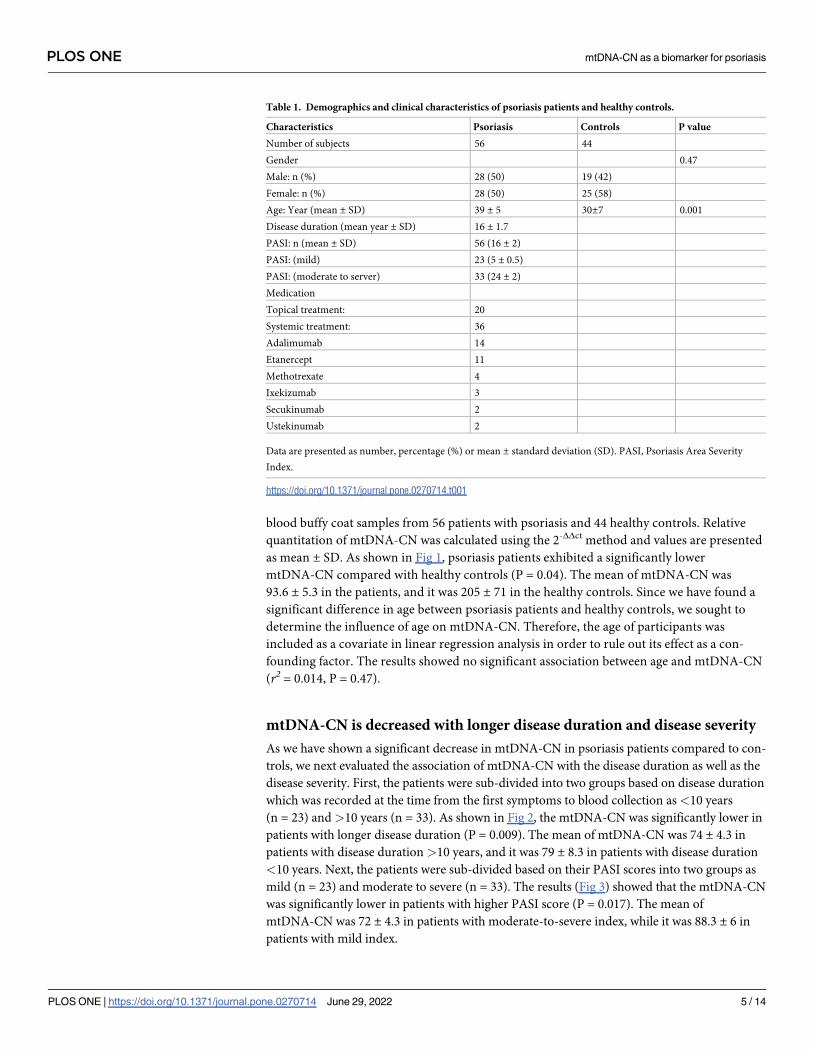
demographics and clinical characteristics of patients and controls are shown in Table 1. Data
are presented as number, percentage (%) or mean ± standard deviation (SD). The patient
group comprised 28 males and 28 females with a group mean age of 39 ± 5. The healthy con-
trol group comprised 19 males and 25 females with a group mean age of 30 ± 7. No significant
difference was observed in the gender distribution between patients and controls (P = 0.47),
while a significant difference was found in the mean age between the two groups (P = 0.001).
The disease duration in the patient group ranged from 1–40 years, with a mean of 16 ± 1.7.
The mean of PASI score for disease severity was 16 ± 2. Twenty-three patients had mild psoria-
sis (PASI score: 5 ± 0.5), and 33 patients had moderate to severe psoriasis (PASI score: 24 ± 2).
The patients were undergoing the following treatment: Topical treatment with corticosteroids
cream (n = 20), or systemic treatment with Adalimumab, Etanercept, Methotrexate, Ixekizu-
mab, Secukinumab and Ustekinumab (n = 36).
mtDNA-CN is decreased in psoriasis patients
The mtDNA-CN was quantified by real-time qPCR as the DNA ratio (mtDNA/nDNA)
between a target mitochondrial gene (ND2) and a reference nuclear gene (β2M) in peripheral
PLOS ONE mtDNA-CN as a biomarker for psoriasis
PLOS ONE | https://doi.org/10.1371/journal.pone.0270714 June 29, 2022 4 / 14
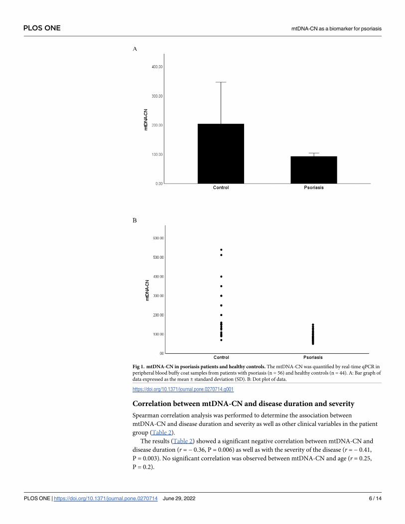
blood buffy coat samples from 56 patients with psoriasis and 44 healthy controls. Relative
quantitation of mtDNA-CN was calculated using the 2-ΔΔct method and values are presented
as mean ± SD. As shown in Fig 1, psoriasis patients exhibited a significantly lower
mtDNA-CN compared with healthy controls (P = 0.04). The mean of mtDNA-CN was
93.6 ± 5.3 in the patients, and it was 205 ± 71 in the healthy controls. Since we have found a
significant difference in age between psoriasis patients and healthy controls, we sought to
determine the influence of age on mtDNA-CN. Therefore, the age of participants was
included as a covariate in linear regression analysis in order to rule out its effect as a con-
founding factor. The results showed no significant association between age and mtDNA-CN
(r2 = 0.014, P = 0.47).
mtDNA-CN is decreased with longer disease duration and disease severity
As we have shown a significant decrease in mtDNA-CN in psoriasis patients compared to con-
trols, we next evaluated the association of mtDNA-CN with the disease duration as well as the
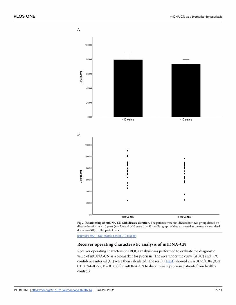
disease severity. First, the patients were sub-divided into two groups based on disease duration
which was recorded at the time from the first symptoms to blood collection as<10 years
(n = 23) and>10 years (n = 33). As shown in Fig 2, the mtDNA-CN was significantly lower in
patients with longer disease duration (P = 0.009). The mean of mtDNA-CN was 74 ± 4.3 in
patients with disease duration >10 years, and it was 79 ± 8.3 in patients with disease duration
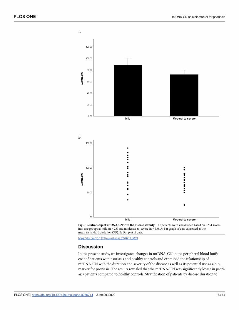
<10 years. Next, the patients were sub-divided based on their PASI scores into two groups as
mild (n = 23) and moderate to severe (n = 33). The results (Fig 3) showed that the mtDNA-CN
was significantly lower in patients with higher PASI score (P = 0.017). The mean of
mtDNA-CN was 72 ± 4.3 in patients with moderate-to-severe index, while it was 88.3 ± 6 in
patients with mild index.
Table 1. Demographics and clinical characteristics of psoriasis patients and healthy controls.
Characteristics Psoriasis Controls P value
Number of subjects 56 44
Gender 0.47
Male: n (%) 28 (50) 19 (42)
Female: n (%) 28 (50) 25 (58)
Age: Year (mean ± SD) 39 ± 5 30±7 0.001
Disease duration (mean year ± SD) 16 ± 1.7
PASI: n (mean ± SD) 56 (16 ± 2)
PASI: (mild) 23 (5 ± 0.5)
PASI: (moderate to server) 33 (24 ± 2)
Medication
Topical treatment: 20
Systemic treatment: 36
Adalimumab 14
Etanercept 11
Methotrexate 4
Ixekizumab 3
Secukinumab 2
Ustekinumab 2
Data are presented as number, percentage (%) or mean ± standard deviation (SD). PASI, Psoriasis Area Severity
Index.
https://doi.org/10.1371/journal.pone.0270714.t001
PLOS ONE mtDNA-CN as a biomarker for psoriasis
PLOS ONE | https://doi.org/10.1371/journal.pone.0270714 June 29, 2022 5 / 14
Correlation between mtDNA-CN and disease duration and severity
Spearman correlation analysis was performed to determine the association between
mtDNA-CN and disease duration and severity as well as other clinical variables in the patient
group (Table 2).
The results (Table 2) showed a significant negative correlation between mtDNA-CN and
disease duration (r = − 0.36, P = 0.006) as well as with the severity of the disease (r = − 0.41,
P = 0.003). No significant correlation was observed between mtDNA-CN and age (r = 0.25,
P = 0.2).
Fig 1. mtDNA-CN in psoriasis patients and healthy controls. The mtDNA-CN was quantified by real-time qPCR in
peripheral blood buffy coat samples from patients with psoriasis (n = 56) and healthy controls (n = 44). A: Bar graph of
data expressed as the mean ± standard deviation (SD). B: Dot plot of data.
https://doi.org/10.1371/journal.pone.0270714.g001
PLOS ONE mtDNA-CN as a biomarker for psoriasis
PLOS ONE | https://doi.org/10.1371/journal.pone.0270714 June 29, 2022 6 / 14
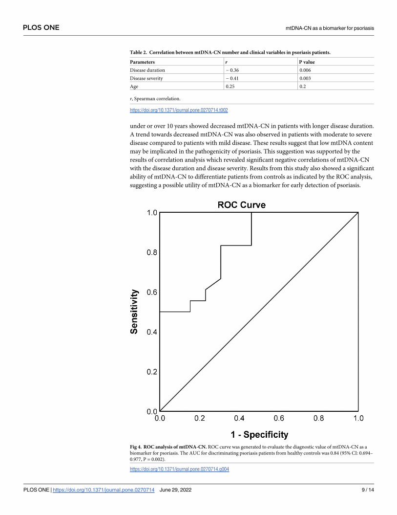
Receiver operating characteristic analysis of mtDNA-CN
Receiver operating characteristic (ROC) analysis was performed to evaluate the diagnostic
value of mtDNA-CN as a biomarker for psoriasis. The area under the curve (AUC) and 95%
confidence interval (CI) were then calculated. The result (Fig 4) showed an AUC of 0.84 (95%
CI: 0.694–0.977, P = 0.002) for mtDNA-CN to discriminate psoriasis patients from healthy
controls.
Fig 2. Relationship of mtDNA-CN with disease duration. The patients were sub-divided into two groups based on
disease duration as<10 years (n = 23) and>10 years (n = 33). A: Bar graph of data expressed as the mean ± standard
deviation (SD). B: Dot plot of data.
https://doi.org/10.1371/journal.pone.0270714.g002
PLOS ONE mtDNA-CN as a biomarker for psoriasis
PLOS ONE | https://doi.org/10.1371/journal.pone.0270714 June 29, 2022 7 / 14
Discussion
In the present study, we investigated changes in mtDNA-CN in the peripheral blood buffy
coat of patients with psoriasis and healthy controls and examined the relationship of
mtDNA-CN with the duration and severity of the disease as well as its potential use as a bio-
marker for psoriasis. The results revealed that the mtDNA-CN was significantly lower in psori-
asis patients compared to healthy controls. Stratification of patients by disease duration to
Fig 3. Relationship of mtDNA-CN with the disease severity. The patients were sub-divided based on PASI scores
into two groups as mild (n = 23) and moderate-to-severe (n = 33). A: Bar graph of data expressed as the
mean ± standard deviation (SD). B: Dot plot of data.
https://doi.org/10.1371/journal.pone.0270714.g003
PLOS ONE mtDNA-CN as a biomarker for psoriasis
PLOS ONE | https://doi.org/10.1371/journal.pone.0270714 June 29, 2022 8 / 14
under or over 10 years showed decreased mtDNA-CN in patients with longer disease duration.
A trend towards decreased mtDNA-CN was also observed in patients with moderate to severe
disease compared to patients with mild disease. These results suggest that low mtDNA content
may be implicated in the pathogenicity of psoriasis. This suggestion was supported by the
results of correlation analysis which revealed significant negative correlations of mtDNA-CN
with the disease duration and disease severity. Results from this study also showed a significant
ability of mtDNA-CN to differentiate patients from controls as indicated by the ROC analysis,
suggesting a possible utility of mtDNA-CN as a biomarker for early detection of psoriasis.
Table 2. Correlation between mtDNA-CN number and clinical variables in psoriasis patients.
Parameters r P value
Disease duration − 0.36 0.006
Disease severity − 0.41 0.003
Age 0.25 0.2
r, Spearman correlation.
https://doi.org/10.1371/journal.pone.0270714.t002
Fig 4. ROC analysis of mtDNA-CN. ROC curve was generated to evaluate the diagnostic value of mtDNA-CN as a
biomarker for psoriasis. The AUC for discriminating psoriasis patients from healthy controls was 0.84 (95% CI: 0.694–
0.977, P = 0.002).
https://doi.org/10.1371/journal.pone.0270714.g004
PLOS ONE mtDNA-CN as a biomarker for psoriasis
PLOS ONE | https://doi.org/10.1371/journal.pone.0270714 June 29, 2022 9 / 14
Mitochondria is the main intracellular source of energy and the major site of reactive oxy-
gen species (ROS) production. Human mitochondria have their own genome (mtDNA),
which encodes essential subunit genes of the electron transport chain (ETC) complexes as well
as tRNAs and rRNAs genes, all of which are important for normal mitochondrial function
[13]. Compared to nuclear DNA (nDNA), the mtDNA is highly susceptible to oxidative stress
and is a target of ROS attack due to its proximity to ROS-generating sites (respiratory chain),
lack of a histone protection and limited DNA repair pathways [15]. Because mtDNA-CN
reflects the abundance and function of mitochondria [14], increased oxidative stress and
mtDNA damage will ultimately lead to impaired mitochondrial function with more ROS pro-
duction [16–19].
Previous studies have revealed an essential role of mitochondrial defects in human aging
and other pathological conditions including psoriasis [17–21]. Recent studies have shown that
low mtDNA-CN is implicated in the pathogenesis of chronic diseases such as autoimmune dis-
eases [29,30], cancer [31], type 2 diabetes [32], metabolic syndrome [33], type 2 diabetes, and
the severity of coronary heart disease [35]. Although the exact mechanism(s) of decreased
mtDNA-CN in diseases remain to be elucidated, oxidative stress and inflammation have been
suggested as highly probable factors [21,22,24,37]. Specifically, increased oxidative stress is an
important contributor to the pathogenesis of autoimmune diseases through enhancing inflam-
mation, apoptotic cell death, and breaking down the immunological tolerance [38]. In psoria-
sis, a link between enhancement ROS production and decreased antioxidant defences has been
suggested as a result of immunological and inflammatory mechanisms which are important in
the etiopathogenesis of the disease [39]. Indeed, the mtDNA content in peripheral blood is
associated with the overall level of oxidative stress [40] and suggested as a biomarker associated
with oxidative stress and inflammation [41]. In most studies to date, mtDNA-CN has been
measured in the buffy coat [29,33,34] or whole blood samples [30–32,35]. In our study, we
observed lower mtDNA-CN in psoriasis patients compared to controls. We extracted DNA
from peripheral blood buffy coat which contains all the white blood cells and platelets. Con-
trary to our results, a study by Therianou, et al [28] reported a significant increase in mtDNA
content in serum from psoriatic patients compared to controls. Since mtDNA present in leu-
kocytes and platelets, the use of serum can affect the accuracy of mtDNA-CN quantification
[42], and this may explain the discrepancy in the two studies.
The reduction of mtDNA-CN in psoriasis patients observed in this study was correlated
with the disease duration and disease severity, suggesting that low mtDNA-CN may be an
early abnormality and associated with the disease progression. It is possible that low
mtDNA-CN is a result of progressive impairment of oxidative phosphorylation and mitochon-
drial function, a speculation which needs to be investigated further. The link between mtDNA
content and severity or outcome of diseases has been reported in several previous studies. For
instance, a reduction in peripheral blood mtDNA-CN was correlated with the development of
multiple sclerosis [30]. Lower peripheral mtDNA-CN was also associated with adverse clinical
outcomes in peritoneal dialysis patients [43] as well as with the severity and inflammation in
bipolar disorder [44]. Moreover, reduced mtDNA-CN has been shown to precede the develop-
ment of type 2 diabetes [45] and observed in the early stages of neurodegenerative disorders
including multiple sclerosis [30], Parkinson’s disease [46] and Alzheimer’s disease [47].
Whereas, low mtDNA-CN predict a poor outcome in hemodialysis patients with end-stage
renal disease [48].
At present, there is no specific or diagnostic blood test for psoriasis. Previously evaluated
markers are universal for inflammation and not specific to psoriasis [49]. Therefore, the devel-
opment of non-invasive diagnostic tests or biomarkers for psoriasis is urgently needed.
mtDNA-CN is an especially attractive biomarker because it can be non-invasively measured in
PLOS ONE mtDNA-CN as a biomarker for psoriasis
PLOS ONE | https://doi.org/10.1371/journal.pone.0270714 June 29, 2022 10 / 14
blood using a relatively easy and cheap method such as real-time PCR, in addition to the
repeatability and reproducibility measurement of blood mtDNA-CN. Moreover, the observa-
tion that mtDNA-CN can differentiate patients from healthy controls, makes it a suitable bio-
marker for several diseases [29–32,50,51]. The present study also evaluated the diagnostic
value of mtDNA-CN in psoriasis. mtDNA-CN was found to have a significant ability to differ-
entiate psoriasis patients from healthy controls as indicated by the ROC analysis with a very
good diagnostic value (AUC: 0.84, 95% CI: 0.694–0.977, P = 0.002). These findings suggest a
potential use of peripheral blood mtDNA-CN as a biomarker for psoriasis.
The present study has certain limitations. First, the sample size was relatively small and
future large-scale validation of the results is recommended, particularly the use of mtDNA-CN
as a biomarker for psoriasis. Second, additional studies on the correlation of mtDNA-CN with
inflammatory and oxidative stress markers can help in understanding the role of mtDNA con-
tent in the pathophysiologic mechanism(s) of psoriasis. Moreover, future studies to examine
other blood indicators related to mitochondrial function and compare them with mtDNA are
required to further clarify the influence of mtDAN on mitochondrial function. Finally, the
possible changes of mtDNA-CN with different treatments should be evaluated in future stud-
ies to investigate the potential use of mtDNA-CN to monitor response to treatment.
Conclusions
In this study we observed the mtDNA-CN in peripheral blood buffy coat was significantly
reduced in patients with psoriasis. We also showed that decreased mtDNA-CN correlated with
the disease duration and severity, suggesting that low mtDNA-CN may be an early abnormal-
ity in psoriasis and associated with the disease progression. Additionally, we showed the feasi-
bility of mtDNA-CN as a non-invasive biomarker for psoriasis. Additional research is needed
to assess whether these results are replicable in the future.
Acknowledgments
We would like to thank Dr. Anantha Kethireddy and her colleagues at the Research Core Facil-
ity in the Health Sciences Center, Faculty of Medicine, Kuwait University for their technical
support during this study.
Author Contributions
Conceptualization: Materah Salem Alwehaidah, Ghada Al-Kafaji.
Data curation: Materah Salem Alwehaidah.
Formal analysis: Materah Salem Alwehaidah, Suad AlFadhli, Ghada Al-Kafaji.
Investigation: Materah Salem Alwehaidah, Ghada Al-Kafaji.
Methodology: Materah Salem Alwehaidah, Suad AlFadhli, Ghada Al-Kafaji.
Project administration: Materah Salem Alwehaidah.
Resources: Materah Salem Alwehaidah.
Supervision: Materah Salem Alwehaidah.
Writing – original draft: Materah Salem Alwehaidah.
Writing – review & editing: Materah Salem Alwehaidah, Ghada Al-Kafaji.
PLOS ONE mtDNA-CN as a biomarker for psoriasis
PLOS ONE | https://doi.org/10.1371/journal.pone.0270714 June 29, 2022 11 / 14
References1. Raychaudhuri SP, Farber EM. The prevalence of psoriasis in the world. J Eur Acad Dermatol Venereol
2001; 15:16–17. https://doi.org/10.1046/j.1468-3083.2001.00192.x PMID: 11451313
2. Langley RG, Krueger GG, Griffiths CE. Psoriasis: epidemiology, clinical features, and quality of life. Ann
Rheum Dis 2005; 64 Suppl 2:ii18–23
3. Mallbris L, Ritchlin CT, Stahle M. Metabolic disorders in patients with psoriasis and psoriatic arthritis.
Curr Rheumatol Rep 2006; 8:355–363. https://doi.org/10.1007/s11926-006-0065-8 PMID: 16973109
4. Boehncke WH, Schon MP. Psoriasis. Lancet 2015; 386:983–994. https://doi.org/10.1016/S0140-6736
(14)61909-7 PMID: 26025581
5. Onumah N, Kircik LH. Psoriasis and its comorbidities. J Drugs Dermatol 2012; 11:s5–10. PMID:
22644770
6. Kim WB, Jerome D, Yeung J. Diagnosis and management of psoriasis. Can Fam Physician 2017;
63:278–285. PMID: 28404701
7. Yang H, Zheng J. Influence of stress on the development of psoriasis. Clin Exp Dermatol 2019; 45:284–
288. https://doi.org/10.1111/ced.14105 PMID: 31592542
8. Snekvik I, Nilsen TIL, Romundstad PR, Saunes M. Metabolic syndrome and risk of incident psoriasis:
prospective data from the HUNT Study, Norway Br J Dermatol 2019; 180:94–99. https://doi.org/10.
1111/bjd.16885 PMID: 29904911
9. Alshobaili HA, Shahzad M, Al-Marshood A, Khalil A, Settin A, Barrimah I. Genetic background of psoria-
sis. Int J Health Sci (Qassim) 2010; 4:23–29. PMID: 21475522
10. Hamanaka RB, Chandel NS. Mitochondrial metabolism as a regulator of keratinocyte differentiation.
Cell Logist 2013; 3:e25456. https://doi.org/10.4161/cl.25456 PMID: 24475371
11. Arul S, Dayalan H, Jegadeesan M, Damodharan P. Induction of differentiation in psoriatic keratinocytes
by propylthiouracil and fructose. BBA Clin 2016; 6:82–86. https://doi.org/10.1016/j.bbacli.2016.06.002
PMID: 27453822
12. Osellame LD, Blacker TS, Duchen MR. Cellular and molecular mechanisms of mitochondrial function.
Best Pract Res Clin Endocrinol Metab 2012; 26:711–723. https://doi.org/10.1016/j.beem.2012.05.003
PMID: 23168274
13. Garcia I, Jones E, Ramos M, Innis-Whitehouse W, Gilkerson R. The little big genome: the organization
of mitochondrial DNA. Front Biosci (Landmark Ed) 2017; 22:710–721. https://doi.org/10.2741/4511
PMID: 27814641
14. Clay Montier LL, Deng JJ, Bai Y. Number matters: control of mammalian mitochondrial DNA copy num-
ber. J Genet Genomics 2009; 36:125–131. https://doi.org/10.1016/S1673-8527(08)60099-5 PMID:
19302968
15. Bohr VA, Stevnsner T, de Souza-Pinto NC. Mitochondrial DNA repair of oxidative damage in mamma-
lian cells. Gene 2002; 286:127–134. https://doi.org/10.1016/s0378-1119(01)00813-7 PMID: 11943468
16. Santos JH, Hunakova L, Chen Y, Bortner C, Van Houten B. Cell sorting experiments link persistent
mitochondrial DNA damage with loss of mitochondrial membrane potential and apoptotic cell death. J
Biol Chem 2003; 278:1728–1734. https://doi.org/10.1074/jbc.M208752200 PMID: 12424245
17. Balaban RS, Nemoto S, Finkel T. Mitochondria, oxidants, and aging. Cell 2005; 120:483–495. https://
doi.org/10.1016/j.cell.2005.02.001 PMID: 15734681
18. Al-Kafaji G, Sabry MA, Bakhiet M. Increased expression of mitochondrial DNA-encoded genes in
human renal mesangial cells in response to high glucose-induced reactive oxygen species. Mol Med
Rep 2016; 13:1774–1780. https://doi.org/10.3892/mmr.2015.4732 PMID: 26719045
19. Al-Kafaji G, Sabry MA, Skrypnyk C. Time-course effect of high-glucose-induced reactive oxygen spe-
cies on mitochondrial biogenesis and function in human renal mesangial cells. Cell Biol Int 2016; 40:36–
48. https://doi.org/10.1002/cbin.10520 PMID: 26251331
20. Lee HC, Wei YH. Mitochondrial biogenesis and mitochondrial DNA maintenance of mammalian cells
under oxidative stress. Int J Biochem Cell Biol 2005; 37:822–834. https://doi.org/10.1016/j.biocel.2004.
09.010 PMID: 15694841
21. Pagano G, Talamanca AA, Castello G, Cordero MD, d’Ischia M, Gadaleta MN, et al. Oxidative stress
and mitochondrial dysfunction across broad-ranging pathologies: toward mitochondria-targeted clinical
strategies. Oxid Med Cell Longev 2014; 2014:541230. https://doi.org/10.1155/2014/541230 PMID:
24876913
22. Mizuguchi S, Gotoh K, Nakashima Y, Setoyama D, Takata Y, Ohga S, et al. Mitochondrial reactive oxy-
gen species are essential for the development of psoriaticinflammation. Front Immunol 2021;
12:714897. https://doi.org/10.3389/fimmu.2021.714897 PMID: 34421919
PLOS ONE mtDNA-CN as a biomarker for psoriasis
PLOS ONE | https://doi.org/10.1371/journal.pone.0270714 June 29, 2022 12 / 14
23. Chawla A, Nguyen KD, Goh YP. Macrophage-mediated inflammation in metabolic disease. Nat Rev
Immunol 2011; 11:738–749. https://doi.org/10.1038/nri3071 PMID: 21984069
24. Feichtinger RG, Sperl W, Bauer JW, Kofler B. Mitochondrial dysfunction: a neglected component of
skin diseases. Exp Dermatol 2014; 23:607–614. https://doi.org/10.1111/exd.12484 PMID: 24980550
25. Raby BA, Klanderman B, Murphy A, Mazza S, Camargo CA Jr, Silverman EK, et al. A common mito-
chondrial haplogroup is associated with elevated total serum IgE levels. J Allergy Clin Immunol 2007;
120:351–358. https://doi.org/10.1016/j.jaci.2007.05.029 PMID: 17666217
26. Alwehaidah MS, Bakhiet M, AlFadhli S. Mitochondrial haplogroup reveals the genetic basis of diabetes
mellitus type 2 comorbidity in psoriasis. Med Princ Pract 2021; 30:62–68. https://doi.org/10.1159/
000509937 PMID: 32629455
27. Alwehaidah MS, Al-Kafaji G, Bakhiet M, Alfadhli S. Next-generation sequencing of the whole mitochon-
drial genome identifies novel and common variants in patients with psoriasis, type 2 diabetes mellitus
and psoriasis with comorbid type 2 diabetes mellitus. Biomed Rep 2021; 14:41. https://doi.org/10.3892/
br.2021.1417 PMID: 33728047
28. Therianou A, Vasiadi M, Delivanis DA, Petrakopoulou T, Katsarou-Katsari A, Antoniou C, et al. Mito-
chondrial dysfunction in affected skin and increased mitochondrial DNA in serum from patients with pso-
riasis. Exp Dermatol 2019; 28:72–75. https://doi.org/10.1111/exd.13831 PMID: 30390357
29. Svendsen AJ, Tan Q, Jakobsen MA, Thyagarajan B, Nygaard M, Christiansen L, et al. White blood cell
mitochondrial DNA copy number is decreased in rheumatoid arthritis and linked with risk factors. A twin
study. J Autoimmun 2019; 96:142–146. https://doi.org/10.1016/j.jaut.2018.09.008 PMID: 30327147
30. Al-Kafaji G, Bakheit HF, Alharbi MA, Farahat AA, Jailani M, Ebrahin BH, et al. Mitochondrial DNA copy
number in peripheral blood as a potential non-invasive biomarker for multiple sclerosis. Neuromolecular
Med 2020; 22:304–313. https://doi.org/10.1007/s12017-019-08588-w PMID: 31902116
31. Xu E, Sun W, Gu J, Chow WH, Ajani JA, Wu X. Association of mitochondrial DNA copy number in
peripheral blood leukocytes with risk of esophageal adenocarcinoma. Carcinogenesis 2013; 34:2521–
2524. https://doi.org/10.1093/carcin/bgt230 PMID: 23803692
32. Al-Kafaji G, Aljadaan A, Kamal A, Bakhiet M. Peripheral blood mitochondrial DNA copy number as a
novel potential biomarker for diabetic nephropathy in type 2 diabetes patients. Exp Ther Med 2018;
16:1483–1492. https://doi.org/10.3892/etm.2018.6319 PMID: 30116398
33. Kim JH, Im JA, Lee DC. The relationship between leukocyte mitochondrial DNA contents and metabolic
syndrome in postmenopausal women. Menopause 2012; 19:582–587. https://doi.org/10.1097/gme.
0b013e31823a3e46 PMID: 22354267
34. Chong MR, Narula S, Morton R, Judge C, Akhabir L, Cawte N, et al. Mitochondrial DNA copy number
as a marker and mediator of stroke prognosis: Observational and mendelian randomization analyses.
Neurology 2022; 98:e470–e482. https://doi.org/10.1212/WNL.0000000000013165 PMID: 34880091
35. Liu LP, Cheng K, Ning MA, Li HH, Wang HC, Li F, et al. Association between peripheral blood cells mito-
chondrial DNA content and severity of coronary heart disease. Atherosclerosis 2017; 261:105–110.
https://doi.org/10.1016/j.atherosclerosis.2017.02.013 PMID: 28242046
36. Gottlieb AB, Chaudhari U, Baker DG, Perate M, Dooley LT. The National Psoriasis Foundation Psoria-
sis Score (NPF-PS) system versus the Psoriasis Area Severity Index (PASI) and Physician’s Global
Assessment (PGA): a comparison. J Drugs Dermatol 2003; 2:260–266. PMID: 12848110
37. Zhan D, Tanavalee A, Tantavisut S, Ngarmukos S, Edwards SW, Honsawek S. Relationships between
blood leukocyte mitochondrial DNA copy number and inflammatory cytokines in knee osteoarthritis J
Zhejiang Univ Sci B 2020; 21:42–52. https://doi.org/10.1631/jzus.B1900352 PMID: 31898441
38. Kumagai S, Jikimoto T, Saegusa J. Pathological roles of oxidative stress in autoimmune diseases. Rin-
sho Byori 2003; 51:126–132. PMID: 12690629
39. Kadam DP, Suryakar AN, Ankush RD, Kadam CY, Deshpande KH. Role of oxidative stress in various
stages of psoriasis. Indian J Clin Biochem 2010; 25:388–392. https://doi.org/10.1007/s12291-010-
0043-9 PMID: 21966111
40. Liu CS, Tsai CS, Kuo CL, Chen HW, Lii CK, Ma YS, et al. Oxidative stress-related alteration of the copy
number of mitochondrial DNA in human leukocytes. Free Radic Res 2003; 37:1307–1317. https://doi.
org/10.1080/10715760310001621342 PMID: 14753755
41. Knez J, Winckelmans E, Plusquin M, Thijs L, Cauwenberghs N, Gu Y, et al. Correlates of peripheral
blood mitochondrial DNA content in a general population. Am J Epidemiol 2016; 183:138–146. https://
doi.org/10.1093/aje/kwv175 PMID: 26702630
42. Hurtado-Roca Y, Ledesma M, Gonzalez-Lazaro M, Moreno-Loshuertos R, Fernandez-Silva P, Enri-
quez JA, et al. Adjusting MtDNA quantification in whole blood for peripheral bood platelet and leukocyte
counts. PLoS One 2016; 11:e0163770. https://doi.org/10.1371/journal.pone.0163770 PMID: 27736919
PLOS ONE mtDNA-CN as a biomarker for psoriasis
PLOS ONE | https://doi.org/10.1371/journal.pone.0270714 June 29, 2022 13 / 14
43. Yoon CY, Park JT, Kee YK, Han SG, Han IM, Kwon YE, et al. Low Mitochondrial DNA copy number is
associated with adverse clinical outcomes in peritoneal dialysispatients. Medicine (Baltimore) 2016; 95:
e2717.
44. Angrand L, Boukouaci W, Lajnef M, Richard JR, Andreazza A, Wu CL, et al. Low peripheral mitochon-
drial DNA copy number during manic episodes of bipolar disorders is associated with disease severity
and inflammation. Brain Behav Immun 2021; 98:349–356. https://doi.org/10.1016/j.bbi.2021.09.003
PMID: 34500035
45. Lee HK, Song JH, Shin CS, Park DJ, Park KS, Lee KU, et al. Decreased mitochondrial DNA content in
peripheral blood precedes the development of non-insulin-dependent diabetes mellitus. Diabetes Res
Clin Pract 1998; 42:161–167. https://doi.org/10.1016/s0168-8227(98)00110-7 PMID: 9925346
46. Pyle A, Brennan R, Kurzawa-Akanbi M, Yarnall A, Thouin A, Mollenhauer B, et al. Reduced cerebrospi-
nal fluid mitochondrial DNA is a biomarker for early-stage Parkinson’s disease. Ann Neurol 2015;
78:1000–1004. https://doi.org/10.1002/ana.24515 PMID: 26343811
47. Podlesniy P, Figueiro-Silva J, Llado A, Antonell A, Sanchez-Valle R, Alcolea D, et al. Low cerebrospinal
fluid concentration of mitochondrial DNA in preclinical Alzheimer disease. Ann Neurol 2013; 74:655–68.
https://doi.org/10.1002/ana.23955 PMID: 23794434
48. Rao M, Li L, Demello C, Guo D, Jaber BL, Pereira BJ, et al. Mitochondrial DNA injury and mortality in
hemodialysis patients. J Am Soc Nephrol 2009; 20:189–196. https://doi.org/10.1681/ASN.2007091031
PMID: 18684894
49. Jiang S, Hinchliffe TE, Wu T. Biomarkers of an autoimmune skin disease-psoriasis. Genomics Proteo-
mics Bioinformatics 2015; 13:224–233. https://doi.org/10.1016/j.gpb.2015.04.002 PMID: 26362816
50. Pyle A, Anugrha H, Kurzawa-Akanbi M, Yarnall A, Burn D, Hudson G. Reduced mitochondrial DNA
copy number is a biomarker of Parkinson’s disease. Neurobiol Aging 2016; 38:216 e217–216 e210.
https://doi.org/10.1016/j.neurobiolaging.2015.10.033 PMID: 26639155
51. Filograna R, Mennuni M, Alsina D, Larsson NG. Mitochondrial DNA copy number in human disease: the
more the better? FEBS Lett 2021; 595:976–1002. https://doi.org/10.1002/1873-3468.14021 PMID:
33314045
PLOS ONE mtDNA-CN as a biomarker for psoriasis
PLOS ONE | https://doi.org/10.1371/journal.pone.0270714 June 29, 2022 14 / 14