LEUKOCYTE 11 G - DTIC
104
Volme44 Nube 4, Ocoer98 - O R ALT.F* OFINL ETN =H o 1A O LEUKOCYTE 11 G Washngto, D Octoer 2-30,198 AlanR. issInc, Neww ork * *o74
-
Upload
khangminh22 -
Category
Documents
-
view
0 -
download
0
Transcript of LEUKOCYTE 11 G - DTIC
Volme44 Nube 4, Ocoer98
- O R ALT.F* OFINL ETN=H o 1A O LEUKOCYTE 11 G
Washngto, DOctoer 2-30,198
AlanR. issInc, Neww ork
* *o74
JOURNAL OF
LEUKO CYTEBIOLOGYAn Official Publication of the Society for Leukocyte Biology,Incorporated as the Reticuloendothellal Society
This journal will consider for publication manuscripts of original investigationsfocusing on the origins, the developmental biology and the functions ofgranulocytes, lymphocytes and mononuclear phagocytes. These reportsinclude full length papers on original research, short communications of newdiscoveries and invited reviews.
EDITOR-IN-CHIEF, CARLETON C. STEWART
Editorial Board Editors Hillel S. KorenLaurence A. Boxer Dolph 0. Adam- Margaret L. KripkeSteven D Doug!as Marco Baggiolini Quentin N. MyrvikMonte S. Meltzer Robert J. Bonney Peter RalphJoost J. Oppenheim Peter Bonventre Richard StanleyWilliam S. Walker Robert A. Clark Robert F. Todd, III
Theodore G. Gabig Dennis Van EppsHoward E. Gendelman Alvin VolkmanAlan M. Kaplan Mark A. Wainberg
Authorization to photocopy items for internal or personal use, or the internal or personal use ofspecific clients, is granted by Alan R. Liss, Inc. for libraries and other users registered with theCopyright Clearance Center (CCC) Transactional Reporting Service, provided that the base feeof $00.50 per copy, plus $00.25 per page is paid directly to CCC, 27 Congress Street, Salem,MA 01970, 0741-5400/88 $00.50 + .25.
Journal of Leukocyte Biology (ISSN 0741-5400) is published monthly by Alan R. Liss, Inc., 41 East11th Street, New York, NY 10003, with editorial offices at the Roswell Park Memorial Institute,Buffalo, New York.Advertising Inquiries should be addressed to: Alan R. Liss, Inc., Att. Advertising Sales Manager,41 East 11 th Street, New York, NY 10003 (212) 475-7700.Subscription Information: For Volumes 43 and 44, 1988, twelve issues: $448.00 in U.S., $487.00outside U.S. All subscriptions outside North America will be sent by air. Payment must be made inU.S dollars drawn on a U.S. bank. Change of Address: Send to publisher six weeks prior to move;enclose present mailing label with address change. For members of the Society for LeukocyteBiology: Please be sure to notify the Society, Dr. Sherwood Reichard, Medic'il College of Georgia,Augusta, GA 30912, as well. to effect address changes for Society mail: . ¢. Claims for MissingIssues: Claims cannot be honored oeyond four months after mailing date. Duplicate copies cannotbe sent to replace issues not delivered because of failure to notify publisher of change of address.Cancellations: Subscription cancellations will not be accepted after the first issue has been mailed.Exclusive agent in Japan: lgaku Shoin. Ltd., Foreign Department, 1-28-36 Hongo, Bunkyo-ku,Tokyo 113. Japan. Price in Japan: V 125,200 for 1988. Air Cargo Service only. Second Class Postagepaid at New York, NY, and at additional mailing offices. Printed in U.S.A. 1988 Alan R. Liss, Inc.Indexed by: Current Contents/Life Sciences-Social Science Citation Index and Current Contents/Internal Medicine e Index Medicus e BIOSIS-Data Base e Excerpta Medica * Cambridge ScientificAbstracts e Chemical AbstractsPOSTMASTER: Send address changes to Journal of Leukocyte Biology, Alan R. Liss, Inc., 41 EastI Ith St, New York, N.Y. 10003
TWENTY-FIFTH MEETING OF THE SOCIETY FOR LEUKOCYTE BIOLOGY
The Twenty-Fifth Meeting of the Society for Leukocyte Biology wasconvened this year at the Washington Hilton and Towers Hotel, Washington
D.C. between October 27-30th. The meeting comprised three plenary
sessions, eight minisymposia and three poster sessions as well as apremeeting discussion workshop on the problems and successes in thequantitative measurement of lipid mediators, wet workshops on
spectrofluorimetry and flow cytometry and club meetings focussing attention
on topics ranging from aging and the immune response to neutrophilproteases in inflammation.
The scientific programme commenced with the Keynote Address thatwas delivered by Dr. Philippa Marrack on the subject of the T-cell
repertoire. Particular attention was directed towards the issues of control ofthe T-cell repertoire by several mechanisms including recombination ofgerm line gene elements, as well as the events that occur in the thymus thatlead to the elimination of self-reactive T-cells, and the selection of T-cells
bearing receptors that can recognize antigen in the context of self-MHC.
The first of the three plenary sessions entitled "Phospholipids and Cell
Activation" focussed on the elicitation of inflammatory cell function. Dr AlanAderem discussed recent findings from his laboratory on the mechanism ofpriming of arachidonic acid release from macrophages by bacterial
lipoppolysaccharide (LPS). Priming by LPS not only enhances the release ofarachidonate in response to challenge with zymosan particles and
tetradecanoyl phorbol acetate (TPA). but also induces a competence state
that subsequently allows latex particles to trigger arachidonate release, afeature that is not associated upon application of this stimulus to unprimed
cells. A prominent feature of the LPS-induced priming response is themyristoylation of a 68 kDa membrane protein. Dr Aderem speculated that
the myristoylation may target the 68 kDa protein to the plasma membrane.The 68 kDa protein is also phosphorylated by protein kinase C. This latter
event may be important in its release back into the cytoplasm. Unlike LPS,
TPA does not induce myristoylation of the 68 kDa protein, but it doespromote its phosphorylation. Importantly, evidence was also presented that
myristoylation also accompanies another priming phenomenon, namely that
induces by IFNy. However, under these conditions, the myristoylated protein
was clearly different from that myristoylated in response to LPS.
Dr Christina Leslie presented recent findings on the properties of an
arachidonyl specific phospholipase A2 enzyme of the mouse macrophage cell
line RAW 264.7. This enzyme has been purified to homogeneity and exhibits
a relatively high molecular weight (60 kDa) compared to other
phospholipase A2 enzymes. The enzyme plays a pivotal role not only in the
liberation of the arachidonic acid that is utilized in the synthesis of
icosanoids, but also in the synthesis of the bioactive ether phospholipid,
platelet-activating factor. The phospholipase is largely located in the
cytoplasmic compartment of resting cells but becomes associated with a
membrane fraction in the presence of Ca 2+ . A curious and unexpected
feature of the phospholipase A2 is its similarity in many respects to other
calcium-phospholipid binding proteins including protein kinase C, such as its
requirement for Ca 2+ and in particular the stimulation of its activity by acidic
phospholipids such as phosphatidyl serine and diacylglycerol.
Other presentations focussed on the generation of the second
messengers that convey information from the cell surface to the various
internal effector mechanisms. 1,4,5-inositol trisphosphate([1,4,5 IP3]) and
diacylglycerol have been ubiquitously recognized as a important second
messengers. Dr Charles Rock discussed the biochemistry of a membrane
associated phospholipase C that exhibits specificity for phosphatidylinositol
4,5-bisphosphate to yield [1,4,5 IP3] and diacylglycerol. Dr Rock suggested
that phosphatidic acid, produced by the phosphorylation of diacylglycerol,
may play an important regulatory role in the control of phospholipase C
activity. This observation may in part explain previous suggestions that
phosphatidic acid could behave as a calcium ionophore. The link between
the cell surface and the phospholipase C is frequently accomplished by G-
proteins. This issue was discussed by Dr. Shamshad Cockcroft with
reference to her studies on the role of G-proteins in ATP and formyl.peptide
induced 1-glucuronidase secretion by neutrophils and differentiated HL-60
cells.
The second plenary session was devoted to a discussion of the
involvement of mononuclear phagocytes in retroviral diseases, with a timely
and appropriate emphasis on interactions between the human
immunodeficiency virus (HIV) and macrophages. Until recently, the
predominent host cell implicated in the replication of HIV was the CD4
positive T-helper cell. Macrophage-HIV interactions and the role of the
macrophage in the replication of the virus has only emerged during the past
9-12 months. Dr Howard Gendelman reviewed the biology of HIV including
its mechanism of interaction with T-cells, and the replication. Using in situ
hybridization with an HIV cDNA probe, combined with
immunocytochemistry with anti-lysozyme antibodies (to identify
.iacrophages), macrophage rich areas of the brain were shown to harbour
abundent amounts of the virus. At the electron microscope level, HIV virionswere clearly demonstrable within macrophages. Immensely significant
were the findings that HIV could infect macrophages in the presence of GM-
CSF, and the fact that when infected T-cells were grown in the presence of
infected macrophages, the virus appeared to gradually adapt to itself to
become competent to infect the cocultivated mononuclear phagocytes. How
HIV is able to infect macrophages is not known, but it appears to take place
in a CD4 independent fashion since soluble CD4, while totally blocing virus
replication in T-cells, was only partially effective at blocking H1V replication
in macrophages.
The third and final plenary session was devoted to recent advances in
the biochemistry and molecular biology of oxidase activation in phagocytic
cells. Dr Bernard Babior discussed the mechanism of translocation of the
oxidase from the cytoplasm to the plasma membrane during neutrophil
activation, as well as the role of protein phosphorylation in the formation of
the active ternary complex that contains the oxidase. The role and structure
of the G-proteins involved in coupling the FMLP receptor to phospholipase C
during the activation of neutrophils was discussed by Dr Gary Bokoch, while
Dr Al Jesaitis reviewed recent work from his laboratory that has led to the
purification and characterization of the subunit structure of the oxidase and
the deduction of the amino acid sequence from gene cloning. The regulation
of cytochrome b gene expression during mononuclear phagocyte
development and differentiation was discussed by Dr. Peter Newburger. As
monocvtes differentiate into macrophages in vitro, the expression of the
heavy chain gene begins to decline. However, the deficit can be restored by
exposure of macrophages to IFNy. Expression of the oxidase light chain gene
however, is not regulated to the same extent as that of the heavy chain gene.
Of immense clinical significance were the observations that treatment of
monocytes from individuals with X-linked chronic granulomatous disease
(CGD) with IFNy not only up-regulated cytochrome b gene expression, but
also functionally restored the ability of monocytes and neutrophils from
these individuals to generate reactive oxygen free radicals. These important
observations have now formed the basis of a clinical trail of the effects of
IFNy in CGD. In one reported case, a single injection of IFNy reconstituted a
respiratory burst with effects that persisted for up to 20-30 days.
Eight minisymposia addressed issues that included the biochemistry
of neutrophil activation and priming, inflammatory mediators and cytokine
networks, mechanisms of gene expression during leukocyte development
and differentiation, and mechanisms of tumor, parasite, and microbial
elimination and destruction. Since four minisymposia were held
concurrently on each of two afternoon, it is not possible to give a
comprehensive summary of each session.
The spectrum and role of cell surface gangliosides as primary low
affinity receptors or secondary binding sites for an array of growth factors,
cytokines and bacterial toxins was comprehensively reviewed by Dr. John
Ryan in the minisymposium on "Inflammatory Mediators". Dr Stephen
Russell, chairing the minsymposium on "Mechanisms of Cellular
Cytotoxicity" initially reviewed recent work conducted in his laboratory on
the regulation of the activated macrophage phenotype by IFNY and triggering
stimuli such as LPS and double-stranded polyribonucleotides, and on the
dynamics of expression of two unique markers of the primed and activated
states. Drawing on other work by Dr. Luigi Varesio, Dr. Russell speculated
that under certain conditions, notably with the C57/Black mouse,
macrophage tumoricidal activity can be elaborated in response to IFNy alone.
A prominent feature of this response is the induction of the enzyme
indoleamine-2,3-dioxygenase which is involved in the formation of picolinic
acid by tryptophan degradation. Picolinic acid itself, when added in
millimolar concentrations to macrophages, induces the cytolytic phenotype.
However, the relationship between picolinic acid and the triggering of
cytolysis by other agents such as LPS is not as yet apparent. A possibility is
that precursor rRNA molecules which are accumulated during macrophage
activation and which, perhaps as a consequence of elements of internal
double-strandedness, may enhance cytolytic activity in much the same way
that polyribonucleotides have been suggested to do so. In the search for
macrophage phenotypic markers, Dr. Paul Johnston described recent
studies from his laboratory on p120, a macrophage protein marker of the
cytolytic phenotype. Using a monospecific antiserum, evidence was
presented that while p120 itself is not implicated in the expression of
cytolytic activity, it is nevertheless a useful phenotypic marker of activated
mouse peritoneal macrophages.
Collectively, papers presented at several minisymposia, focussed
attention on the regulation of cytokine production by mononuclear
phagocytes, or on the effects of cytokines on mononuclear phagocyte
function. With a broader appreciation of the breadth of cytokines that are
produced and act on mononuclear phagocytes, it is becoming clearer
cytokine biology is considerably more complex that perhaps at first
suspected in that many cytokines have overlapping activities, while single
cytokine species frequently express. in a dose dependent fashion, both
agonist and antagonist activities. Several papers discussed the autocrine-
paracrine regulatory functions of macrophage derived molecules such as the
role of 1,25-dihydroxyvitamin D 3 in the differentiation of, and expression of
protooncogenes by, myeloid precursor cells.
The Presidential and Young Investigator Awards Competition have
formed an important element of the Leukocyte Biology Meeting for several
years. Four contenders for each award presented work ranging from the
genetics of aquired immunity to mycobacteria, to the role of neutrophils in
experimental myocardial ischaemia. Molecular biological technology has led
to major advances in our understanding of the mononuclear phagocyte
system and this trend was reflected in the selection of the awardees. The
Presidential Award was presented to Dr. Sarah Sporn, University of North
Carolina. Chapel Hill, for her work on the cloning, sequencing and analysisof adherence specific cDNA's derived by subtractive hybridization using ahuman monocyte cDNA library. The Young Investigator Award went to Dr.Karen MacNaul, Merck. Sharp and Dohme Research Laboratories. Rahway,for investigations into the expression of transcripts for Il-I and TNF bysynoviocytes and monocytes using in situ hybridization as the detectingsystem. The awards were presented to the winners at the Banquet whichtraditionally is the high spot of the social calender of the Meeting. Theprestigious Maria T. Bonazinga Award for outstanding contributions to thefield of Leukocyte Biology was presented to Dr. Marco Baggiolini, Universityof Bern who delighted all those present at the Banquet with a witty dialogue(complete with "slides") of his research experiences spanning threedecades.
All in all, the 25th Meeting of the Society for Leukocyte Biologypresented a Scientific Program that covered new ground (e.g. retroviral-macrophage interactions) as well as ongoing, more traditional areas ofinterest in leukocyte biology. Furthermore, the meeting was well attended,not only by scientists from the U.S.A., but also (perhaps as a consequence ofits east coast location) by many of our colleagues from Europe. I am sure wewill all look forward to the 26th Meeting of the Society for Leukocyte Biologyon Marco Island, Florida, next October.
David W. H. Riches. Ph.D.Department of Pediatrics,National Jewish Center for Immunology
and Respiratory Medicine,1400 Jackson Street,Denver
CO 80122.
WHY NOT THE BEST?7A10675 ACRYLAMIDE 2x CRYST. 1 kg $ 54.50
(Analytical Grade) *3 x 1 kg 141.00
IF 0 B. WESTBURY, NEW YORK TERMS, 2% 10 NET 30 DAYS
v4 CCUR4T Chemical & Scientific Corporation300 SHAMES DRIVE. WESTBURY, NY 11590 USA * TELEX:-4972582
WESTBURY, NY: 516-433-4900 SAN DIEGO, CA: 619-235-9400FAX: 1-516-997-4948
0ORER CAt CLFRI AII nT PFF 1 -- 64-6264 OR 1-800-ALL-WEST' MCONA FIM C And
ACCURATE CHEMICAL & SCIENTIFIC CORPORATIONtO0 SHAMES DRIVE WESTBURY N Y 11590
0l Please send me the complete Accurate Catalog
N AME
- CITY' 'drATE & ZIP
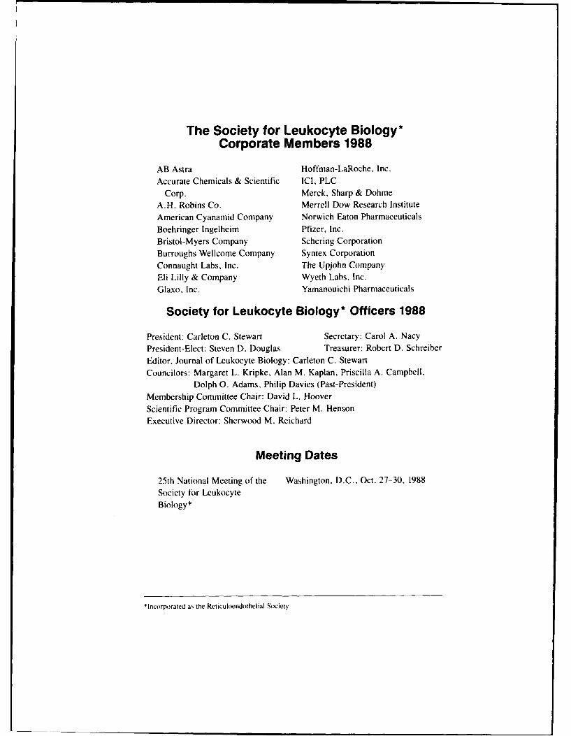
The Society for Leukocyte Biology*Corporate Members 1988
AB Astra Hoffman-LaRoche, Inc.
Accurate Chemicals & Scientific ICI. PLC
Corp. Merck, Sharp & Dohme
A.H. Robins Co. Merrell Dow Research Institute
American Cyanamid Company Norwich Eaton Pharmaceuticals
Boehringer Ingelheim Pfizer. Inc.
Bristol-Myers Company Schering Corporation
Burroughs Wellcome Company Syntex Corporation
Connaught Labs, Inc. The Upjohn Company
Eli Lilly & Company Wyeth Labs. Inc.
Glaxo, Inc. Yamanouichi Pharmaceuticals
Society for Leukocyte Biology* Officers 1988
President: Carleton C. Stewart Secretary: Carol A. Nacy
President-Elect: Steven D. Douglas Treasurer: Robert D. Schreiber
Editor, Journal of Leukocyte Biology: Carleton C. Stewart
Councilors: Margaret L. Kripke, Alan M. Kaplan, Priscilla A. Campbell.
Dolph 0. Adams, Philip Davies (Past-President)
Membership Committee Chair: David L. Hoover
Scientific Program Committee Chair: Peter M. Henson
Executive Director: Sherwood M. Reichard
Meeting Dates
25th National Meeting of the Washington. D.C., Oct. 27-30, 1988
Society for Leukocyte
Biology*
*Incorporated as the Reticuloendothelial Society
Program and Abstracts ofTWENTY-FIFTH NATIONAL MEETING
ofTHE SOCIETY FOR LEUKOCYTE
BIOLOGYWashington HiltonWashington, DC
October 27-30, 1988 '9
Accesion For
NTIS CRA&I
DTIC TAB F]Unannournced E]
Sold by: . Jisfficatw )
Alan R. Liss Inc.41 East l1th Street By CNew York, NY 10003Price: $41.17 DistributIo:" J
Availability Codes
Avail and! o,Dist Sc
i An n n_ ____
SOCIETY FOR LEUKOCYTE BIOLOGY(A Reticuloendothelial Society)
Application Form
(Please type or print)
NAME:(Last) (First) (Middle initial) Degree(s)
DEPARTMEN i AND INSTITUTION:
MAILING ADDRESS:
CITY AND STATE: ZIP CODE:
TELEPHONE: (
PRESENT POSITION:
EDUCATION ANDACADEMIC DEGREES:
PROFESSIONALEXPERIENCE:
*LIST RECENT AND RELEVANT PUBLICATIONS (4 MAXIMUM) (include titles):
APPLICANT'S SIGNATURE: _ DATE:
SPONSOR'S STATEMENT: I support this candidate for membership in the Society. (Sponsormay send additional supporting information to the Society office.)
SPONSOR'S SIGNATURE: DATE:(Please also print name)
APPLICANT: Please return original and 3 copies of application to:Society Officec/o Dr. Sherwood ReichardMedical College of GeorgiaAugusta, Georgia 30912
ANNUAL DUES of $48 U.S., should accompany this application. (Applicants outside NorthAmerica, please add $39 U.S. to cover international postage.)
*STUDENT MEMBERSHIP: . Please mark this box and complete application form. Student
Dues $36 U.S. ior Journal and membership, or $6.00 mem-bership without Journal.
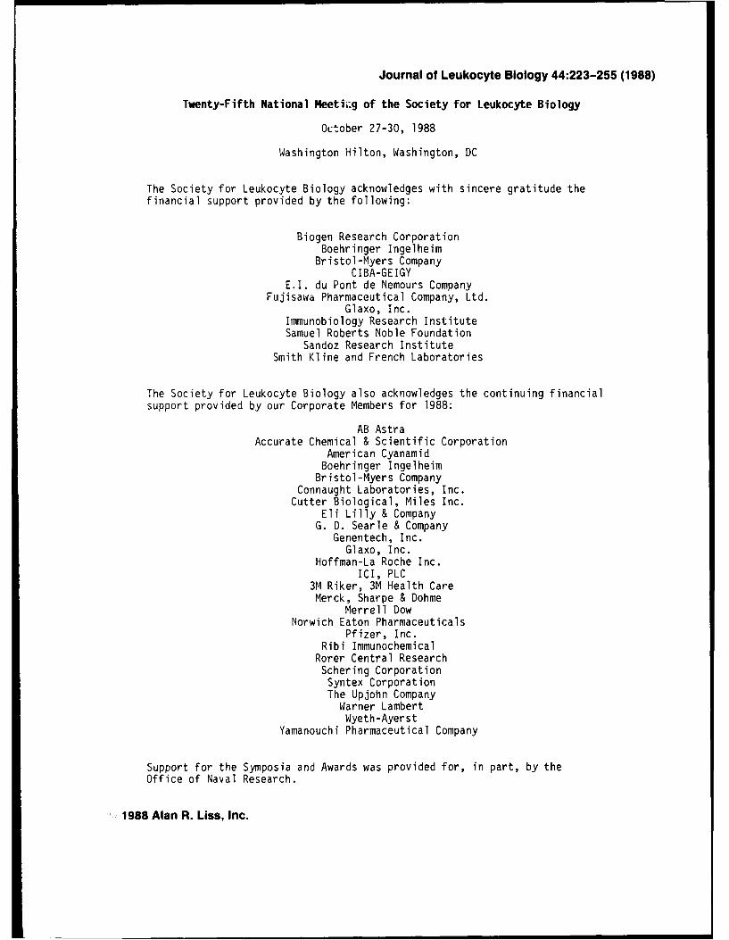
Journal of Leukocyte Biology 44:223-255 (1988)
Twenty-Fifth National Meetii;g of the Society for Leukocyte Biology
October 27-30, 1988
Washington Hilton, Washington, DC
The Society for Leukocyte Biology acknowledges with sincere gratitude thefinancial support provided by the following:
Biogen Research CorporationBoehringer Ingelheim
Bristol-Myers CompanyCIBA-GEIGY
E.I. du Pont de Nemours CompanyFujisawa Pharmaceutical Company, Ltd.
Glaxo, Inc.Immunobiology Research InstituteSamuel Roberts Noble Foundation
Sandoz Research InstituteSmith Kline and French Laboratories
The Society for Leukocyte Biology also acknowledges the continuing financialsupport provided by our Corporate Members for 1988:
AB AstraAccurate Chemical & Scientific Corporation
American CyanamidBoehringer Ingelheim
Bristol-Myers CompanyConnaught Laboratories, Inc.
Cutter Biological, Miles Inc.Eli Lilly & CompanyG. D. Searle & Company
Genentech, Inc.Glaxo, Inc.
Hoffman-La Roche Inc.ICI, PLC
3M Riker, 3M Health CareMerck, Sharpe & Dohme
Merrell DowNorwich Eaton Pharmaceuticals
Pfizer, Inc.Ribi Immunochemical
Rorer Central ResearchSchering CorporationSyntex CorporationThe Upjohn Company
Warner LambertWyeth-Ayerst
Yamanouchi Pharmaceutical Company
Support for the Symposia and Awards was provided for, in part, by theOffice of Naval Research.
1988 Alan R. Liss, Inc.
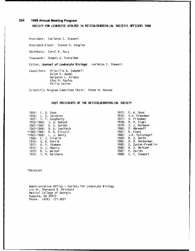
224 1988 Annual Meeting Program
SOCIETY FOR LEUKOCYTE BIOLOGY (A RETICULOENDOTHLIAL SOCIETY) OFFICERS 1988
President: Carleton C. Stewart
President-Elect: Steven D. Douglas
Secretary: Carol A. NaCy
Treasurer: Robert D. Schreiber
Editor, Journal of Leukocyte Biology: Carleton C. Stewart
Councilors: Priscilla A. CampbellDolph 0. AdamsMargaret L. KripkeAlan M. KaplanPhilip Davies
Scientific Program Committee Chair: Peter M. Henson
PAST PRESIDENTS OF THE RETICULOENDOTHELIAL SOCIETY
1955: C. A. Doan 1975: R. A. Good1956: L. 0. Jacobson 1976: H.H. Freedman1957: T. F. Dougherty 1977: H. Friedman1958-1960: J. W. Rebuck 1978: M. M. Sigel1961-1962: A. S. Gordon 1979: S. J. Normann1963-1964: B. W. Zweifach 1980: P. Abramoff
*1965-1966: N. R. DiLuzio 1981: R. Evans*1967-1968: L. J. Berry 1982: J.K. Spitznagel1969: F. J. DiCarlo 1983: R. J. North1970: Q. N. Myrvik 1984: R. B. Herberman1971: G. P. Youmans 1985: D. Zucker-Franklin1972: A. J. Sbarra 1986: M. S. Meltzer1973: R. S. Weiser 1987: P. Davies1974: S. M. Reichard 1988: C. C. Stewart
*Deceased
Administrative Office - Society for Leukocyte Biologyc/o Dr. Sherwood M. ReichardMedical College of GeorgiaAugusta, GA 30912Phone: (404) 721-2601
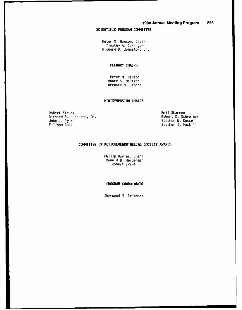
1988 Annual Meeting Program 225SCIENTIFIC PROGRAM COMMITTEE
Peter M. Henson, ChairTimothy A. Springer
Richard B. Johnston, Jr.
PLENARY CHAIRS
Peter M. HensonMonte S. MeltzerBernard M. Babior
MINISYMPOSIU CHAIRS
Robert Strunk Emil SkameneRichard B. Johnston, Jr. Robert D. SchreiberJohn L. Ryan Stephen W. RussellFilippo Rossi Stephen J. Haskill
COMITTEE ON RETICULOENDOTHELIAL SOCIETY AWARDS
Philip Davies, ChairRonald B. Herberman
Robert Evans
PROGRAM COORDINATOR
Sherwood M. Reichard
226 1988 Annual Meeting Program
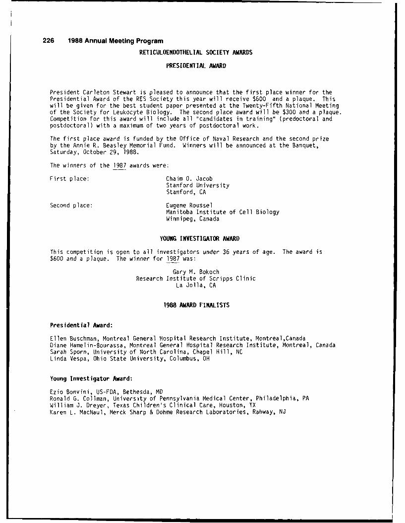
RETICULOENDOTHELIAL SOCIETY AWARDS
PRESIDENTIAL AWARD
President Carleton Stewart is pleased to announce that the first place winner for thePresidential Award of the RES Society this year will receive $600 and a plaque. Thiswill be given for the best student paper presented at the Twenty-Fifth National Meetingof the Society for Leukocyte Biology. The second place award will be $300 and a plaque.Competition for this award will include all "candidates in training" (predoctoral andpostdoctoral) with a maximum of two years of postdoctoral work.
The first place award is funded by the Office of Naval Research and the second prizeby the Annie R. Beasley Memorial Fund. Winners will be announced at the Banquet,Saturday, October 29, 1988.
The winners of the 1987 awards were:
First place: Chaim 0. JacobStanford UniversityStanford, CA
Second place: Eugene RousselManitoba Institute of Cell BiologyWinnipeg, Canada
YOUNG INVESTIGATOR AWARD
This competition is open to all investigators under 36 years of age. The award is$600 and a plaque. The winner for 1987 was:
Gary M. BokochResearch Institute of Scripps Clinic
La Jolla, CA
1988 AWARD FINALISTS
Presidential Award:
Ellen Buschman, Montreal General Hospital Research Institute, Montreal,CanadaDiane Hamelin-Bourassa, Montreal General Hospital Research Institute, Montreal, CanadaSarah Sporn, University of North Carolina, Chapel Hill, NCLinda Vespa, Ohio State University, Columbus, OH
Young Investigator Award:
Ezio Bonvini, US-FDA, Bethesda, MDRonald G. Collman, University of Pennsylvania Medical Center, Philadelphia, PAWilliam J. Dreyer, Texas Children's Clinical Care, Houston, TXKaren L. MacNaul, Merck Sharp & Dohme Research Laboratories, Rahway, NJ
1988 Annual Meeting Program 227
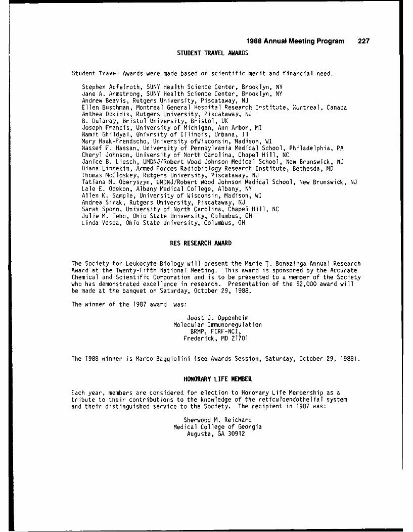
STUDENT TRAVEL AWARDS
Student Travel Awards were made based on scientific merit and financial need.
Stephen Apfelroth, SUNY Health Science Center, Brooklyn, NYJane A. Armstrong, SUNY Health Science Center, Brooklyn, NYAndrew Beavis, Rutgers University, Piscataway, NJEllen Buschman, Montreal General Hospital Research I"stitute, 'luntreal, CanadaAnthea Dokidis, Rutgers University, Piscataway, NJB. Dularay, Bristol University, Bristol, UKJoseph Francis, University of Michigan, Ann Arbor, MINamit Ghildyal, Univrsity of Illinois, Urbana, IlMary Haak-Frendscho, University ofWisconsin, Madison, WINassef F. Hassan, University of Pennsylvania Medical School, Philadelphia, PACheryl Johnson, University of North Carolina, Chapel Hill, NCJanice B. Liesch, UMDNJ/Robert Wood Johnson Medical School, New Brunswick, NJDiana Linnekin, Armed Forces Radiobiology Research Institute, Bethesda, MDThomas McCloskey, Rutgers University, Piscataway, NJTatiana M. Oberyszyn, UMDNJ/Robert Wood Johnson Medical School, New Brunswick, NJLale E. Odekon, Albany Medical College, Albany, NYAllen K. Sample, University of Wisconsin, Madison, WIAndrea Sirak, Rutgers University, Piscataway, NJSarah Sporn, University of North Carolina, Chapel Hill, NCJulie M. Tebo, Ohio State University, Columbus, OHLinda Vespa, Ohio State University, Columbus, OH
RES RESEARCH AWARD
The Society for Leukocyte Biology will present the Marie T. Bonazinga Annual ResearchAward at the Twenty-Fifth National Meeting. This award is sponsored by the AccurateChemical and Scientific Corporation and is to be presented to a member of the Societywho has demonstrated excellence in research. Presentation of the $2,000 award willbe made at the banquet on Saturday, October 29, 1988.
The winner of the 1987 award was:
Joost J. OppenheimMolecular Immunoregulation
BRMP, FCRF-NCI,Frederick, MD 21701
The 1988 winner is Marco Baggiolini (see Awards Session, Saturday, October 29, 1988).
HONORARY LIFE MEMBER
Each year, members are considered for election to Honorary Life Membership as atribute to their contributions to the knowledge of the reticuloendothelial systemand their distinguished service to the Society. The recipient in 1987 was:
Sherwood M. ReichardMedical College of Georgia
Augusta, GA 30912
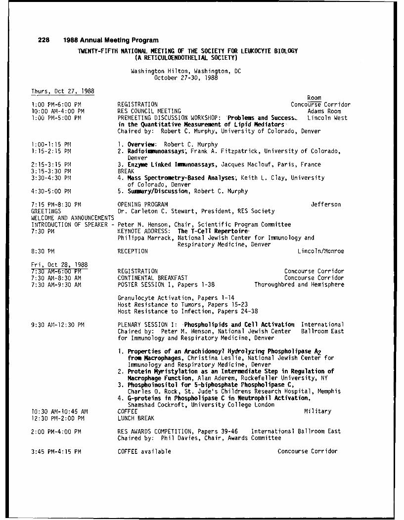
228 1988 Annual Meeting Program
TWENTY-FIFTH NATIONAL MEETING OF THE SOCIETY FOR LEUKOCYTE BIOLOGY(A RETICULOENDOTHELIAL SOCIETY)
Washington Hilton, Washington, DCOctober 27-30, 1988
Thurs, Oct 27, 1988Room
1:00 PM-6:00 PM REGISTRATION Concourse Corridor10:00 AM-4:00 PM RES COUNCIL MEETING Adams Room1:00 PM-5:00 PM PREMEETING DISCUSSION WORKSHOP: Problems and Success, Lincoln West
in the Quantitative Measurement of Lipid Mediators-Chaired by: Robert C. Murphy, University of Colorado, Denver
1:00-1:15 PM 1. Overview: Robert C. Murphy1:15-2:15 PM 2. Radioimmunoassays; Frank A. Fitzpatrick, University of Colorado,
Denver2:15-3:15 PM 3. Enzyme Linked Immunoassays, Jacques Maclouf, Paris, France3:15-3:30 PM BREAK3:30-4:30 PM 4. Mass Spectrometry-Based Analyses; Keith L. Clay, University
of Colorado, Denver4:30-5:00 PM 5. Summary/Discussion, Robert C. Murphy
7:15 PM-8:30 PM OPENING PROGRAM JeffersonGREETINGS Dr. Carleton C. Stewart, President, RES SocietyWELCOME AND ANNOUNCEMENTSINTRODUCTION OF SPEAKER - Peter M. Henson, Chair, Scientific Program Committee7:30 PM KEYNOTE ADDRESS: The T-Cell Repertoire'
Philippa Marrack, National Jewish Center for Immunology andRespiratory Medicine, Denver
8:30 PM RECEPTION Lincoln/Monroe
Fri, Oct 28, 19887:30 AM-6:00 PM REGISTRATION Concourse Corridor7:30 AM-8:30 AM CONTINENTAL BREAKFAST Concourse Corridor7:30 AM-9:30 AM POSTER SESSION I, Papers 1-38 Thoroughbred and Hemisphere
Granulocyte Activation, Papers 1-14Host Resistance to Tumors, Papers 15-23Host Resistance to Infection, Papers 24-38
9:30 AM-12:30 PM PLENARY SESSION I: -Phospholipids and Cell Activation InternationalChaired by: Peter M. Henson, National Jewish Center Ballroom Eastfor Immunology and Respiratory Medicine, Denver
1. Properties of an Arachidonoyl Hydrolyzing Phospholipase A2from Macrophages, Christina Leslie, National Jewish Center forImmunology and Respiratory Medicine, Denver
2. Protein Myristylation as an Intermediate Step in Regulation ofMacrophage Function, Alan Aderem, Rockefeller University, NY
3. Phosphoinositol for 5-biphosphate Phospholipase C,Charles 0. Rock, St. Jude's Childrens Research Hospital, Memphis
4. G-proteins in Phospholipase C in Neutrophil Activation,Shamshad Cockroft, University College London
10:30 AM-1O:45 AM COFFEE Military12:30 PM-2:00 PM LUNCH BREAK
2:00 PM-4:00 PM RES AWARDS COMPETITION, Papers 39-46 International Ballroom East
Chaired by: Phil Davies, Chair, Awards Committee
3:45 PM-4:15 PM COFFEE available Concourse Corridor
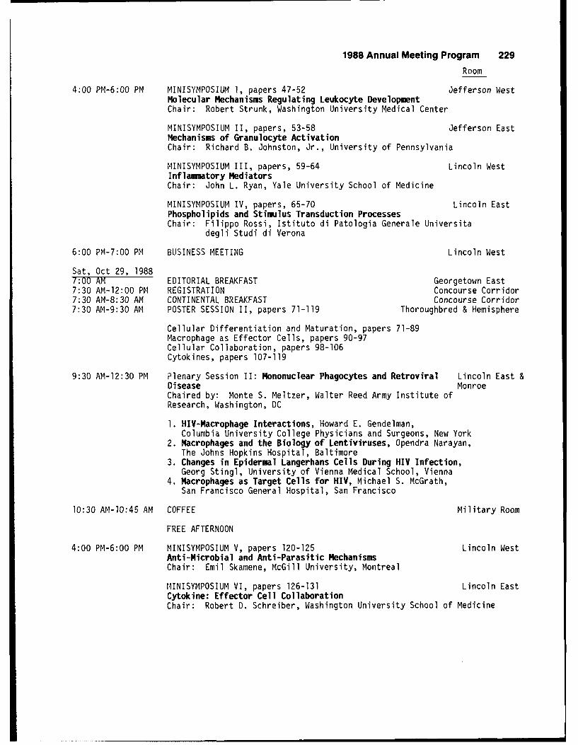
1988 Annual Meeting Program 229
Room
4:00 PM-6:00 PM MINISYMPOSIUM 1, papers 47-52 Jefferson WestMolecular Mechanisms Regulating Leukocyte DevelopmentChair: Robert Strunk, Washington University Medical Center
MINISYMPOSIUM II, papers, 53-58 Jefferson EastMechanisms of Granulocyte ActivationChair: Richard B. Johnston, Jr., University of Pennsylvania
MINISYMPOSIUM III, papers, 59-64 Lincoln WestInflammatory MediatorsChair: John L. Ryan, Yale University School of Medicine
MINISYMPOSIUM IV, papers, 65-70 Lincoln EastPhospholipids and Stimulus Transduction ProcessesChair: Filippo Rossi, Istituto di Patologia Generale Universita
degli Studi di Verona
6:00 PM-7:00 PM BUSINESS MEETING Lincoln West
Sat, Oct 29, 19887:00 AM EDITORIAL BREAKFAST Georgetown East7:30 AM-12:00 PM REGISTRATION Concourse Corridor7:30 AM-8:30 AM CONTINENTAL BREAKFAST Concourse Corridor7:30 AM-9:30 AM POSTER SESSION II, papers 71-119 Thoroughbred & Hemisphere
Cellular Differentiation and Maturation, papers 71-89Macrophage as Effector Cells, papers 90-97Cellular Collaboration, papers 98-106Cytokines, papers 107-119
9:30 AM-12:30 PM Plenary Session II: Mononuclear Phagocytes and Retroviral Lincoln East &Disease MonroeChaired by: Monte S. Meltzer, Walter Reed Army Institute ofResearch, Washington, DC
1. HIV-Macrophage Interactions, Howard E. Gendelman,Columbia University College Physicians and Surgeons, New York
2. Macrophages and the Biology of Lentiviruses, Opendra Narayan,The Johns Hopkins Hospital, Baltimore
3. Changes in Epidermal Langerhans Cells During HIV Infection,Georg Stingl, University of Vienna Medical School, Vienna
4. Macrophages as Target Cells for HIV, Michael S. McGrath,San Francisco General Hospital, San Francisco
10:30 AM-1O:45 AM COFFEE Military Room
FREE AFTERNOON
4:00 PM-6:00 PM MINISYMPOSIUM V, papers 120-125 Lincoln WestAnti-Microbial and Anti-Parasitic MechanismsChair: Emil Skamene, McGill University, Montreal
MINISYMPOSIUM VI, papers 126-131 Lincoln EastCytokine: Effector Cell CollaborationChair: Robert D. Schreiber, Washington University School of Medicine
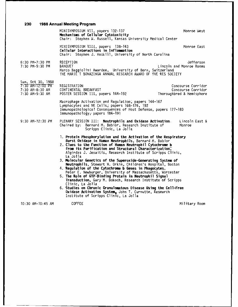
230 1988 Annual Meeting Program
MINISYMPOSIUM VII, papers 132-137 Monroe WestMechanisms of Cellular CytotoxicityChair: Stephen W. Russell, Kansas University Medical Center
MINISYMPOSIUM VIII, papers 138-143 Monroe EastCellular Interactions in InflammationChair: Stephen J. Haskill, University of North Carolina
6:30 PM-7:30 PM RECEPTION Jefferson7:30 PM-9:30 PM BANQUET Lincoln and Monroe Rooms
Marco Baggiolini Awardee, University of Bern, SwitzerlandTHE MARIE T BONAZINGA ANNUAL RESEARCH AWARD OF THE RES SOCIETY
Sun, Oct 30, 19887:30 AM-12:00 PM REGISTRATION Concourse Corridor7:30 AM-8:30 AM CONTINENTAL BREAKFAST Concourse Corridor7:30 AM-9:30 AM POSTER SESSION III, papers 144-192 Thoroughbred & Hemisphere
Macrophage Activation and Regulation, papers 144-167Lymphocytes and NK Cells; papers 168-176, 192Immunopathological Consequences of Host Defense, papers 177-183Immunopathology. papers 184-191
9:30 AM-12:30 PM PLENARY SESSION III: Neutrophils and Oxidase Activation, Lincoln East &Chaired by: Bernard M. Babior, Research Institute of Monroe
Scripps Clinic, La Jolla
1. Protein Phosphorylation and the Activation of the RespiratoryBurst Oxidase in Human Neutrophils, Bernard M. Babior
2. Clues to the Function of Human Neutrophil Cytochrome bfrom its Purification and Structural Characterization7Algirdas J. Jesaitis, Research Institute of Scripps Clinic,La Jolla
3.-Molecular Genetics of the Superoxide-Generating System ofNeutrophils,. Stewart H. Orkin, Children's Hospital, Boston
4. Regulation of the Cytochrome b Genes in Phagocytes,Peter E. Newburger, University of Massachusetts, Worcester
5. The Role of GTP-Binding Protein in Neutrophil SignalTransduction, Gary M. Bokoch, Research Institute of ScrippsClinic, La Jolla
6. Studies on Chronic Granulomatous Disease Using the Cell-FreeOxidase Activation System, John T. Curnutte, ResearchInstitute of Scripps Clinic, La Jolla
10:30 AM-1O:45 AM COFFEE Military Room
1988 Annual Meeting Program 231
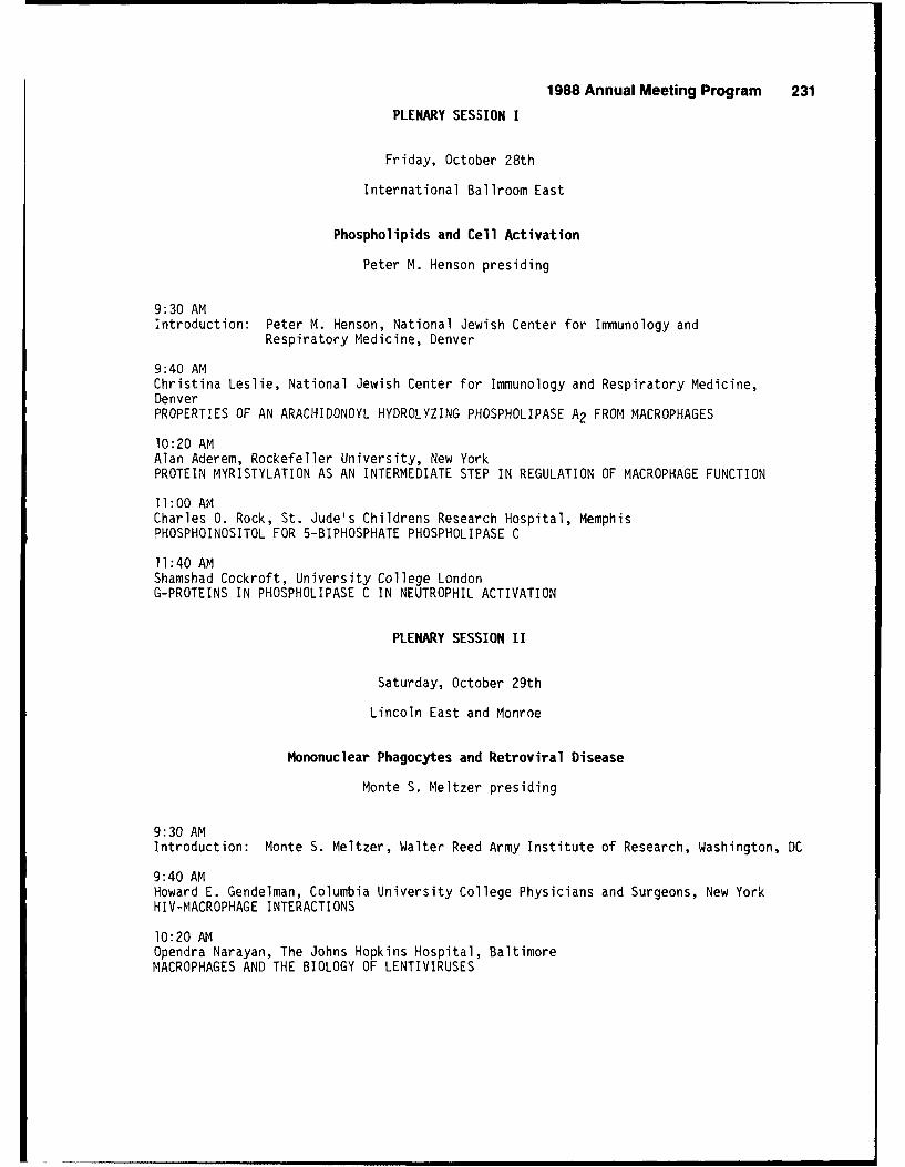
PLENARY SESSION I
Friday, October 28th
International Ballroom East
Phospholipids and Cell Activation
Peter M. Henson presiding
9:30 AMIntroduction: Peter M. Henson, National Jewish Center for Immunology and
Respiratory Medicine, Denver
9:40 AMChristina Leslie, National Jewish Center for Immunology and Respiratory Medicine,DenverPROPERTIES OF AN ARACHIDONOYL HYDROLYZING PHOSPHOLIPASE A2 FROM MACROPHAGES
10:20 AMAlan Aderem, Rockefeller University, New YorkPROTEIN MYRISTYLATION AS AN INTERMEDIATE STEP IN REGULATION OF MACROPHAGE FUNCTION
11:00 AMCharles 0. Rock, St. Jude's Childrens Research Hospital, MemphisPHOSPHOINOSITOL FOR 5-BIPHOSPHATE PHOSPHOLIPASE C
11:40 AMShamshad Cockroft, University College LondonG-PROTEINS IN PHOSPHOLIPASE C IN NEUTROPHIL ACTIVATION
PLENARY SESSION II
Saturday, October 29th
Lincoln East and Monroe
Mononuclear Phagocytes and Retroviral Disease
Monte S. Meltzer presiding
9:30 AMIntroduction: Monte S. Meltzer, Walter Reed Army Institute of Research, Washington, DC
9:40 AMHoward E. Gendelman, Columbia University College Physicians and Surgeons, New YorkHIV-MACROPHAGE INTERACTIONS
10:20 AMOpendra Narayan, The Johns Hopkins Hospital, BaltimoreMACROPHAGES AND THE BIOLOGY OF LENTIVIRUSES
232 1988 Annual Meeting Program
11:00 AMGeorg Stingl, University of Vienna Medical School, ViennaCHANGES IN EPIDERMAL LANGERHANS CELLS DURING HIV INFECTION
11:40 AMMichael S. McGrath, San Francisco General Hospital, San Francisco, CAMACROPHAGES AS TARGET CELLS FOR HIV
PLENARY SESSION III
Sunday, October 30th
Lincoln East and Monroe
Neutrophils and Oxidase Activation
Bernard M. Babior presiding
9:30 AMBernard M. Babior, Research Institute of Scripps Clinic, La JollaPROTEIN PHOSPHORYLATION AND THE ACTIVATION OF THE RESPIRATORY BURST OXIDASE INHUMAN NEUTROPHILS
10:00 AMAlgirdas J. Jesaitis, Research Institute of Scripps Clinic, La JollaCLUES TO THE FUNCTIONS OF HUMAN NEUTROPHIL CYTOCHROME b FROM ITS PURIFICATIONAiD STRUCTURAL CHARACTERIZATION
10:30 AMStewart H. Orkin, Children's Hospital, BostonMOLECULAR GENETICS OF THE SUPEROXIDE-GENERATING SYSTEM OF NEUTROPHILS
11:00 AMPeter E. Newburger, University of Massachusetts, WorcesterREGULATION OF THE CYTOCHROME b GENES IN PHAGOCYTES
11:30Gary M. Bokoch, Research Institute of Scripps Clinic, La JollaTHE ROLE OF GTP-BINDING PROTEIN IN NEUTROPHIL SIGNAL TRANSDUCTION
12:00 PMJohn T. Curnutte, Research Institute of Scripps Clinic, La JollaSTUDIES ON CHRONIC GRANULOMATOUS DISEASE USING THE CELL-FREE OXIDASE ACTIVATIONSYSTEM
1988 Annual Meeting Program 233
RETICULOENDOTHELIAL SOCIETY AWARDS
Friday, October 28th
International Ballroom East
Philip Davies presiding
2:00 PM-3:00 PM THE RES PRESIDENTIAL AWARD COMPETITION, papers 39-42
Ellen Buschman, Montreal General Hospital Research Institute,Montreal, CanadaACQUIRED IMMUNITY TO M. BOVIS AND M. INTRACELLULARE IS INFLUENCEDBY THE Bcg GENE.
Diane Hamelin-Bourassa, Montreal General Hospital ResearchInstitute, Montreal, CanadaSUSCEPTIBILITY TO A MURINE RETROVIRUS-INDUCED IMMUNO-DEFICIENCYSYNDROME IS CONTROLLED BY THE H-2 COMPLEX.
Sarah Sporn, University of North Carolina, Chapel Hill, NCISOLATION OF ADHERENCE SPECIFIC cDNA CLONES FROM A MONOCYTEcDNA LIBRARY.
Linda Vespa, The Ohio State University, Columbus, OHBIOCHEMICAL BASIS OF CONTINUOUS 1-A EXPRESSION BY MACROPHAGESFROM MICE RESISTANT TO MYCOBACTERIUM BOVIS (STRAIN BCG).
3:00 PM-4:00 PM THE RES YOUNG INVESTIGATOR AWARD COMPETITION, papers 43-46
Ezio Bonvini, Laboratory Cell Biology/DBBP CBER/US-FDA, Bethesda, MDTHE NON-HYDROLYSABLE GUANOSINE-5'-TRIPHOSPHATE ANALOG, GUANOSINE-5'-O-(3-THIOTRIPHOSPHATE) (GTPgammaS), ACTIVATES PHOSPHOLIPASEC-MEDIATED HYDROLYSIS OF INOSITOL PHOSPHOLIPIDS IN MURINEHELPER T CELL CLONES.
Ronald G. Collman, University of Pennsylvania Medical Center,Philadelphia, PAMONOCYTE(M)-TROPIC AND T LYMPHOCYTE(T)-TROPIC STRAINS OF HIV;REPLICATION IN CULTURED HUMAN MONOCYTES.
William J. Dreyer, Texas Children's Clinical Care, Houston, TXNEUTROPHIL CHEMOTACTIC ACTIVATION DURING EXPERIMENTAL MYOCARDIALISCHEMIA AND REPERFUSION.
Karen L. MacNaul, Merck Sharp & Dohme Research Laboratories,Rahway, NJANALYSIS OF IL-l AND TNF GENE EXPRESSION IN HUMAN SYNOVIOCYTESAND MONOCYTES BY IN SITU HYBRIDIZATION.
234 1988 Annual Meeting Program
POSTER SESSION I
Friday, October 28th, 7:30 AM-9:3OAM
GRANULOCYTE ACTIVATION
Hemisphere Room
1. SELECTIVE ACTIVATION OF BOVINE NEUTROPHIL FUNCTIONS BY RECOMBINANT BOVINEINTERLEUKIN-l . P. Canning, National Animal Disease Center, Agricultural ResearchService, U.S.D.A.,AmesTA 50010.
2. GENERATION OF SUPEROXIDE ANIONS AND MYELOPEROXIDASE BY PMN IN JOINTS OF RHEUMATOIDARTHRITIS PATIENTS. B. Dularay, C.J. Elson and P.A. Dieppe, Univ. Bristol, BristolBS8 ITD, UK.
3. ONTOGENY OF LEUKOCYTE FUNCTION: SUPEROXIDE ANION PRODUCTION BY FETAL, NEWBORN ANDADULT BOVINE NEUTROPHILS. Charles B. Clifford, D.O. Slauson, N.R. Neilsen, R.D.Zwahlen, and D.H. Schlafer, Inflammation Research Laboratory, Pathology Department,College of Veterinary Medicine, Cornell University, Ithaca, NY 14853.
O 3SERVATION OF ANTIBODY-DEPENDENT LYSIS OF RED BLOOD CELLS BY NEUTROPHILS USING NOVELOPTICAL MICROSCOPIC TECHNIQUES. J.W. Francis, M.J. Zhou, L.A. Boxer, H.R. Petty,Biological Sciences, Wayne State Univ., Detroit, MI 48202 and Dept. of Pediatrics,Univ. of Michigan, Ann Arbor, 48109.
5. RECOMBINANT HUMAN GM-CSF IS A DIRECT STIMULATOR OF GRANULOCYTE RESPIRATORY BURST BUTREQUIRES AN ADDITIONAL STIMULUS FOR INITIATING DEGRANULATION. C. Lam, L. Klein,Sandoz Forschungsinstitut, A-1235 Vienna, Austria.
6. PAF ACTIVATION OF ACETYL-CoA: I-ALKYL-SN-GLYCERO-3-PHOSPHOCHOLINE 02-ACETYLTRANS-FERASE, PAF SYNTHESIS AND DEGRANULATION IN RAT NEUTROPHILS. Thomas W. Doebber andMargaret S. Wu (Spon: Philip Davies), Merck Sharp & Dohme Research Laboratories,Rahway, NJ 07065.
7. EFFECT OF PLATELET ACTIVATING FACTOR AND FMLP ON NEUTROPHIL CRI, CR3 AND FcR RECEPTORSAND ON BINDING AND PHAGOCYTOSIS OF OPSONIZED MICROSPHERES. J. Ogle, G. Noel, C. Ogle,M. Sramkoski, J. Alexander and G. Warden, Cincinnati Sch. Med. and Shriners BurnsInstitute, Cincinnati, OH 45267.
8. ENDOTOXIC INJURY T' EQUINE MICROVASCULAR ENDOTHELIUM IN VITRO IS MEDIATED THROUGHPERIP(ERAL BLOOD NEUTROPHILS. Philip N. Bochsler, D.OT. SThiTon, M.M. Suyemoto, and N.R.Neilsen. Inflammation Research Laboratory, Patholoqy Department, College of Veterinaryffe-d-icine, Cornell University, Ithaca, NY 14853.
9. RECOMBINANT HUMAN GRANULOCYTE-MACROPHAGE COLONY STIMULATING FACTOR (RH-GM-CSF) PRIMESAND RECRUITS RHESUS MONKEY NEUTROPHIL (PMN) RESPONSES TO N-FORMYL-L-METHIONYL-L-LEUCYL-L-PHENYLALANINE (Fi4LP). D.M. Linnekin, R.L. Monroy, G. Murano, R.E. Donahue andT.J. MacVittie (Spon: M.L. Patchen), Armed Forces Radiobiology Research Institute,Bethesda, MD !0814-5145.
10. DEPRESSION OF HUMAN GRANULOCYTE CHEMILUMINESCENCE WITH SCHISTOSOMA MANSONISCHISTOSOMULAE. I. 'Mofleh, A. Mahmoud, S. Al-Khwaitir, M. Alam and A-Tuwaijiri,College of Medici -n-h -ge4TaudUniversity, Riyadh 11461, Saudi Arabia.
11. INHIBITION OF EOSINOPHIL SUPEROXIDE GENERATION BY MAST CELL GRANULES. K.N. Dileepan,K.M. Simpson, S. Lynch, D.J. Stechschulte (Spon: Tsuneo Suzuki), Department of Medicine,U nf-v. of Kansas MedcTc_;Center, Kansas-C-Tty, KS 66103.
1988 Annual Meeting Program 235
12. PROPERTIES OF EOSINOPHIL ADHESION IN VITIRO. P.J. Koker, C.C. Clarke, R. Rothleinand C.D. Wegner (Spon: A.S. RosentIlTT---epartments of Pharmacology and Immunology,Boelringer Ingelheim Pharma, Inc., Ridgefield, CT 06877.
13. MODULATION OF HEMATOPOIESIS IN THE GOLDEN SYRIAN HAMSTER BY THE ENDOCRINE SYSTEM.J.A. Hightower, M.J. Horacek, M.O. Dada and C.A. Blake, Sch. Med., Univ. SouthCarolina, Columbia, SC 29Z08.
14. INVOLVEMENT OF MONOCYTES IN SELECTIVE PRODUCTION OF EOSINOPHIL CHEMOTACTIC LYMPHOKINE.M. Hirashima, Kumamoto Univ. Med. Sch., Kumamoto, Japan.
HOST RESISTANCE TO TL4ORS
Thoroughbred Room
15. FORMALIN-FIXED MACROPHAGES BIND TUMOR TARGETS SIMILARLY TO VIABLE MACROPHAGES.Stephen Keith Chapes, Div. of Biology, Kansas State University, Manhattan, KS 66506.
16. REGULATION OF TUMOR-INDUCED MYELOPOIESIS AND THE ASSOCIATED IMMUNE SUPPRESSOR CELLSIN MICE BEARING METASTATIC LEWIS LUNG CARCINOMAS BY PROSTAGLANDIN E2. M.R. Young,M.E. Young and K. Kim, Research Serv., Hines V.A. Hosp., Hines, IL 60141 and Uept.Pathology, Loyola Univ. Stritch Sch. Med., Maywood, IL 60153.
17. PHENOTYPIC DIFFERENCES BETWEEN NORMAL AND TUMOR-BEARING HOST MACROPHAGES.A.D. Yurochko, R.H. Pyle, and K.D. Elgert, Dept. Biology, Microbiol. & Immunol.Section, and Veterinary Biosciences, Virginia Polytechnic Institute and State Univ.,Blacksburg, VA 24061.
18. INHIBITION OF TUMOR CELL GLUTAMINE UPTAKE AS AN INDICATOR OF BOTH OXIDATIVE ANDNON-OXIDATIVE CYTOTOXICITY CAUSED BY STIMULATED NEUTROPHILS. Douglas B. Learn andEdwin L. Thomas, Dept. Biochemistry, St. Jude Children's Research Hospital,Memphis, TN 38101.
19. A COMPARISON OF LEUKOCYTE INFILTRATION INTO AN IMMUNOGENIC AND A NONIMMUNOGENICMURINE TUMOR. F.A. Sneed, A.P. Stevenson, C.C. Stewart, Cell Biology Group,Los Alamos National Laboratory, Los Alamos, NM 81545.
20. THE EFFECT OF SELENIUM ON THE TUMORCYTOTOXICITY OF MOUSE PERITONEAL MACROPHAGESAND SPLEEN LYMPHOCYTES. L. Kiremidjian-Schumacher, M. Roy, H.I. Wishe, M.W. Cohen,G. Stotzky, New York Univ., College of Dentistry and Graduate School of Arts andScience, New York, NY 10010.
21. DEPRESSED CELL MEDIATED IMMUNITY IN PATIENTS WITH SEVERE INHERITED FORMS OFEPIDERMOLYSIS BULLOSA. V. Chopra l, S. Tyringl, S. Vaidya 2, L. Johnson3, J.D. Fine3,(Spon: K. Mehta), Univ. of Texas Med. Branch, Departments of MicrobiologyI andPathology2, Galveston, TX 77550, and Department Dermatology3, University ofAlabama at Birmingham, AL 35294.
22. THE SIGNIFICANCE OF FREE RADICAL AND FREE RADICAL SCAVENGERS IN L1210 LEUKEMIA.A. Brown and J. Lutton, New York Medical College, Valhalla, NY 10595.
23. ODULATION OF MACROPHAGE-TUMOR CELL CYTOTOXIC INTERACTIONS BY HYPERTHERMIA.J. Klostergaard, M. Barta and S.P. Tomasovic, M.D. Anderson Cancer Center,Houston, TX 77030.
236 1988 Annual Meeting Program
HOST RESISTANCE TO INFECTION
Thoroughbred Room
24. ROLE OF ANTIBODY IN COMPLEMENT-MEDIATED KILLING OF TRITRICHOMONAS FOETUS.M.K. Aydintug, P.R. Widders (Spon: S.M. Taylor). Washington State University,College of Veterinary Medicine, Pullman, WA 99164-7040.
25. CORRELATION OF VIRULENCE IN VIVO, SUSCEPTIBILITY TO KILLING BY MURINE POLYMORPHO-NUCLEAR NEUTROPHILS (PMN)-TN-VTTRO, AND PMN SUPEROXIDE ANION (02-) INDUCTION INBLASTOMYCES DERMATITIDIS (BI)TATES. C.J. Morrison* and D.A. Stevens. Inst.Med. Res., Santa Clara Valley Med. Ctr., San Jose, CA 951Z8 and Stanford U.,Stanford, CA 95304.
26. EFFECT OF IONIZING RADIATION ON THE ABILITY OF MURINE PERITONEAL CELLS TOPHAGOCYTIZE KLEBSIELLA PNEUMONIAE. D.G. McChesney, G.S. Madonna and G.D. Ledney(Spon: M. Patchen). Armed Forces Radiobiology Research Institute, Bethesda, MD20814-5145.
27. MITIGATION OF THE LETHAL EFFECTS OF IONIZING RADIATION BY 6,6, TREHALOSE DIESTERS.G.S. Madonna, M.L. Patchen, and G.D. Ledney. Armed Forces Radiobiology ResearchInstitute, Bethesda, MD 20814-5145.
28. PHAGOCITOSIS OF MYCOBACTERIA BY CULTURED-HUMAN MACROPHAGES. M. Arango, G. Merizalde,L.F. Barrera, L.F. Garcia. Univ. of Antioquia, Medellin, Colombia.
29. THE RESTORATIVE EFFECTS OF GAMMA INTERFERON AND CLOFAZAMINE ON PHAGOCYTE DYSFUNCTIONCAUSED BY A 25 KILODALTON FRACTION FROM MYCOBACTERIUM TUBERCULOSIS. A.A. Wadee,A.R. Rabson and R. Anderson (Spon: J. Metz). Dept. Immunology, School of Pathologyof the South African Institute for Medical Research and the University of theWitwatersrand, Johannesburg, 2000, Republic of South Africa.
30. BOVINE POLYMORPHONUCLEAR NEUTROPHILIC GRANULOCYTE-PRODUCT WITH ANTIVIRAL (INTERFERON-LIKE) ACTIVITY: CHARACTERIZATION OF THE INDUCTION, SECRETION AND ACTIVITY OF"POLYFERON". H. Bielefeldt Ohmann, M. Campos, D. Fitzpatrick, M.J.P. Lawman andL.A. Babiuk. Veterinary Infectious Disease Organization, 124 Veterinary Road,Saskatoon, Saskatchewan S7N OWO, Canada.
31. INTERLEUKIN-2 INCREASES MACROPHAGE ACTIVATION BY INTERFERON-y. A. Misefari,P. Vitale*, E. Jirillo*, S. Antonaci*, and V. Covelli*. Chairs of Immunology,University of Messina Med. School, Messina and Immunology, Clinical Medicine andClinical Neuroanatomy, University of Bari, Med, School, Bari, Italy.
32. SEPARATE AND COMBINED EFFECTS OF rIL-l, rTNF-a AND rFN-y ON ANTIBACTERIALRESISTANCE. R. Kurtz, J. Roll and C.J. Czuprynski. Univ. Wisconsin SchoolMedicine, Madison, WI 53706.
33. A SELF-LIMITING SEMLIKI FOREST VIRUS INFECTION ACTIVATES MURINE MACROPHAGES.L.-X. Wu, K. Suryanarayana, K.-C. Lee, R.G. Marusyk and A.A. Salmi. (Spon:P.S. Morahan). Viral Pathogenesis Research Unit, Dept. of Medical Microbiol. andInfectious Diseases, and Dept. of Immunol., University of Alberta, Edmonton,Alta., Canada T6G 2H7.
34. ISOLATION OF HUMAN IMMUNODEFICIENCY VIRUS (HIV) ON RECOMBINANT HUMAN MACROPHAGECOLONY STIMULATING FACTOR (rM-CSF) TREATED HUMAN MONOCYTES: AN EFFICIENT VIRUSDETECTION SYSTEM. H. Husayni, M.S. Meltzer and H.E. Gendelman. Walter Reed ArmyInst. Res., Washington, DC 20307-5100.
1988 Annual Meeting Program 237
35. INFECTIBILITY BY THE HUMAN IMMUNODEFICIENCY VIRUS (HIV) OF HUMAN BLOOD-BORNMONOCYTES/MACROPHAGES AND CHARACTERIZATION OF INFECTED MONOCYTES/MACROPHAGESC. Meichsner, H. Rubsamen-Waigmann, R. Andreesen, M. Limbert, E. Schrinner,H Suhartono, H. von Briesen. Hoechst AU and Georg-Speyer-Haus, Frankfurt andMedizinische Universitatsklinik Freiburg, Freiburg, FRG.
36. ADMINISTRATION OF HUMAN RECOMBINANT INTERLEUKIN 2 ENHANCES ANTI-LISTERIARESISTANCE. Mary Haak-Frendscho and Charles J. Czuprynski. School of VeterinaryMedicine, University of Wisconsin, Madison, WI 5370b.
37. INTERLEUKIN-1-ALPHA ENHANCES PHAGOCYTOSIS AND KILLING OF M. TUBERCULOSIS ANDM. AVIUM COMPLEX BY HUMAN MACROPHAGES. K. Sankaran, R. Swartz, and H. Yeager, Jr.Georgetown Univ. Medical Centar, Washington, DC 2000T.
38. EFFECT OF GLUCOCORTICOIDS ON MACROPHAGE INSTRINSIC RESISTANCE TO HERPES SIMPLEXVIRUS. C.W. Milligan and W.L. Dempsey. Medical College of Pennsylvania,Philadelphia, PA 19129.
238 1988 Annual Meeting Program
MINISYNPOSIUJ4 I
Friday, October 28, 1988
Jefferson West Room
Molecular Mechanisms Regulating Leukocyte Development
Robert Strunk presiding
4:00 PMGENE EXPRESSION IN MACROPHAGES. Robert Strunk, M.D., Children's Hospital,Washington University Medical Center, St. Louis, MO 63110.
4:30 PM47. SHARED 'EARLY RESPONSE' GENE EXPRESSION IN LPS-STIMULATED MACROPHAGES AND PDGF-
STIMULATED FIBROBLASTS. T.A. Hamilton, C.S. Tannenbaum, and Y. Ohmori. ClevelandClinic Foundation, Cleveland, OH 44195.
4:45 PM48. MACROPHAGE la ANTIGEN EXPRESSION INDUCED BY IFNy AND IL-4. H. Cao, R.M. Crawford,
R.G. Wolff, C.A Nacy, and M.S. Meltzer. Walter Reed Army Inst. Res.,Washington, DC 20307-5100.
5:00 PM49. EXPRESSION OF c-MYC, c-FOS AND c-FMS FOLLOWING ENDOTOXIN STIMULATION OF CSF-l
INDUCED MACROPHAGE (MPH) DIFFERENTIATION. N. Ghildyal, M.J. Myers, J.K. Pullenand L.B. Schook, Lab of Molecular Immunology, Dept. of Animal Sciences, Univ. ofIllinois, Urbana, IL 61820.
5:15 PM50. PROTOONCOGENE EXPRESSION IN TUMOR-ASSOCIATED MACROPHAGES (TAM): A PARACRINE
CIRCUIT IN THE REGULATION OF THE PROLIFERATION OF TAM IN MURINE SARCOMAS.A. Mantovani, E. Erba, F. Fazioli, A. Rambaldi, B. Bottazzi. Istituto di RicercheFarmacologiche "Mario Negri", Via Eritrea 62, 20157 Milan, Italy.
5:30 PM51. TUMOR-INDUCED IMMUNOSUPPRESSION: INHIBITION OF INTERLEUKIN 2 PRODUCTION BY TUMOR
CELL PRODUCTS AND A pl5E-RELATED PEPTIDE. David S. Nelson, Peggy Nelson, GeorgeJ. Cianciolo and Ralph Snyderman. Kolling Institute of Medical Research, RoyalNorth Shore Hospital, St Leonards NSW 2065, Australia, and Genentech, Inc.,South San Francisco, CA 94080.
5:45 PM52. SYNERGISTIC EFFECTS OF CYTOKINES ON HL-60 DIFFERENTIATION AND FUNCTIONAL ACTIVATION.
J.B. Liesch, T.J. Krause, T.M. Oberyszyn, R.S. Greco and F.M. Roberston. UMDNJ/Robert Wood Johnson Medical School, New Brunswick, NJ 08903.
1988 Annual Meeting Program 239
MINISYMPOSIUM II
Friday, October 28, 1988
Jefferson East Room
MECHANISMS OF GRANULOCYTE ACTIVATION
Richard B. Johnston, Jr. presiding
4:00 PMNEUTROPHIL PRIMING. Richard B. Johnston, Jr., M.D., University of Pennsylvania,Children's Hospital, Philadelphia, PA 19104.
4:30 PM53. DEPRESSION OF POLYMORPHONUCLEAR LEUKOCYTE (PMNL) FUNCTION INDUCED BY INFLUENZA
VIRUS HEMAGGLUTININ (HA) AND SIALIC ACID (SA)-BINDING LECTINS. J.S. Abramson,L.F. Cassidy, L.S. Winkler and D.S. Lyles (Spon: D.A. Bass). Bowman Gray Schoolof Medicine of Wake Forest University, Winston-Salem, NC 27103.
4:45 PM54. INFLUENZA A VIRUS (IAV) ALTERS ACTIN DISTRIBUTION IN POLYMORPHONUCLEAR LEUKOCYTES
(PMNL). J.G. Wheeler and J.S. Abramson (Spon: D.A. Bass). Bowman Gray Schoolof Medicine of Wake Forest University, Winston-Salem, NC 27103.
5:00 PM55. DIFFERENTIAL EFFECT OF PENTOXIFYLLINE ON RESPONSE OF NEUTROPHILS TO CHEMOTACTIC
PEPTIDE (fMLP) AND PHORBOL ESTER (PMA). M.S. Currie, K.M.K. Rao, J. Crawford,and H.J. Cohen (Spon: J. Brice Weinberg). Duke and DVAMC, Durham, NC 2710b.
5:15 PM56. INVOLVEMENT OF PROTEIN KINASE C (PKC) IN THE ACTIVATION OF fMET-LEU-PHE (fMLF)-
MEDIATED RESPIRATORY BURST IN HUMAN NEUTROPHILS (PMNs). J. Nath and A. Powledge(Spon: C. Nacy). WRAIR, Washington, DC 20307-5100.
5:30 PM57. DEGRANULATION AND ACTIVATION OF THE RESPIRATORY BURST IN HUMAN NEUTROPHILS.
D.L. Schneider, F.S. Manara and J. Chin. Dartmouth Medical School, Hanover,NH 03756.
5:45 PM58. EFFECTS OF RECOMBINANT BOVINE INTERFERON-ALPHA AND INTERFERON-GAMMA ON BOVINE
NEUTROPHIL FUNCTIONS. Allen K. Sample and Charles J. Czuprynski. Univ. ofWisconsin-Madison, Wisconsin, WI 53106.
240 1988 Annual Meeting Program
MINISYPOSILM III
Friday, October 28, 1988
Lincoln West Room
INFLAMMATORY MEDIATORS
John L. Ryan presiding
4:00 PMGANGLIOSIDES AS MACROPHAGE RECEPTORS. John L. Ryan, M.D., Ph.D., Departmentof Internal Medicine, Yale University School Medicine, VA Medical Center,West Haven, CT 06516.
4:30 PM59. DIFFERENTIAL EFFECTS OF ENDOTOXINS ON THE TERMINAL DIFFERENTIATION AND FUNCTIONAL
ACTIVITY OF HUMAN MONOCYTE/MACROPHAGES. R. Andreesen, W. Brugger, D. Waltersbacher,H. Sawert, L. Kanz, A. Rehm, C. Galanos, R. Engelhardt, U.W. Lohr. MedizinischeKlinik, Hugstetter Strasse 55, D-7800 Freiburg, FRG.
4:45 PM60. PRODUCTION OF TWO NOVEL NEUTROPHIL CHEMOTACTIC PEPTIDES BY LPS-STIMULATED
ENDOTHELIAL CELLS. J.-M. Schroder and E. Christophers. Dept. of Dermatology,Univ. Kiel, 2300 Kiel, -FRG.
5:00 PM61. ROLE OF 1,25-DIHYDROXYCHOLECALCIFEROL AS AN AUTOCRINE MACROPHAGE REGULATOR.
D.R. Katz, A. Brennan, I. Ziegler, D.S. Latchman, M. Hewison, J.L.H. O'Riordan.Univ. Coil./Middlesex Sch. Med, London WIP BAA, U.K.
5:15 PM62. 13-CIS RETINOIC ACID (13cRA) INCREASES MACROPHAGE PRODUCTION IN INTERLEUKIN-3
(IL-3) STIMULATED MOUSE BONE MARROW CULTURES. J.G. Bender, C.C. Stewart, andR.A. Habbersett, Dept. of Pathology, Univ. of New Mexico, Albuquerue, NM, andLANL, Los Alamos, NM.
5:30 PM63. MODULATION OF TUMOR NECROSIS FACTOR (TNF) RELEASE BY RETINOIDS IN MURINE PERITONEAL
MACROPHAGES. Kapil Mehta, Department of Clinical Immunol. and Biological Therapy,UT M.D. Anderson Cancer Center, Houston, TX 77030.
5:45 PM64. INTERLEUKIN-4 (IL-4) EFFECTS ON MONOCYTE PGE2 AND TUMOR NECROSIS FACTOR (TNF).
C. Miller, G. Szabo and T. Takayama. Univ. Massachusetts, Worcester, MA 01655.
1988 Annual Meeting Program 241
HINISYNPOSIUN4 IV
Friday, October 28, 1988
Lincoln East Room
PHOSPHOLIPIDS AND STIMULUS TRANSDUCTION PROCESSES
Filippo Rossi presiding
4:00 PMPHOSPHOLIPID TURNOVER IN NEUTROPHIL ACTIVATION. Prof. Filippo Rossi. Istitutodi Patologia Generale Universita degli Studi di Verona, 37134 Verona, Italy
4:30 PM65. ALTERATIONS IN GTP-BINDING PROTEIN IN HUMAN NEUTROPHILS BY INFLUENZA VIRUS.
Elaine L. Mills, Garry M. Bokoch, Jon S. Abramson. McGill Univ., Montreal, PQ,H3H IP3, Res. Inst. Scripps Clinic, La Jolla, CA 92037, Bowman Gray Sch Med,Winston-Salem, NC 27103.
4:45 PM66. LIPOPOLYSACCHARIDE-INDUCED EXPRESSION OF THE COMPETENCE GENE, KC, IN VASCULAR
ENDOTHELIAL CELLS IS MEDIATED THROUGH PROTEIN KINASE C. X. Shen, T.A. Hamilton,and P.E. DiCorleto. Cleveland Clinic Research Inst., Cleveland, UH 44195.
5:00 PM67. ROLE OF PKC IN THE CELL SURFACE EXPRESSION AND PHOSPHORYLATION OF DIFFERENTIATION
ANTIGENS OF RESTING AND ACTIVATED HUMAN T. CELLS. A. Carrera, L. Cardenas,A. Tugores, M. Cebrian, F. Sanchez-Madrid, M. Lopez-Botet and M.U. de Landazuri.Servicio de Inmunologia, Univ. Autonoma. Hospital de Ia Princesa. c/Diego deLeon, 62- 28006 Madrid, Spain.
5:15 PM68. PROTEIN KINASE C ISOTYPE DISTRIBUTION AND SELECTIVE ISOTYPE TRANSLOCATION WITH
Ca2+ IN HUMAN NEUTROPHILS AND CYTOPLASTS. T. Fujiki, M.W. Rossi, W.A. Phillips,R.B. Johnston, Jr. and H.M. Korchak (Spon: L. Kilpatrick-Smith). Univ. ofPennsylvania, Philadelphia, PA 19104.
5:30 PM69. ROLE OF PROTEIN KINASE C IN THE ACTIVATION OF LIVER MACROPHAGES. D.L. Laskin,
C.R. Gardner, A.M. Pilaro and J.D. Laskin. Rutgers Univ. and UMDNJ-Robert WoodJohnson Medical School, Piscataway, NJ 05854.
5:45 PM70. THE ENHANCEMENT OF RECEPTOR-MEDIATED PHAGOCYTOSIS BY AMPHOTERICIN B MONOMETHYL
ESTER (AME). S. Racis, O.J. Plescia, J.C. Mulloy, and C.P. Schaffner.Waksman Institute of Microbiology @ Rutgers-The State University, New Brunswick,NJ 08855-0759.
242 1988 Annual Meeting ProgramPOSTER SESSION II
Saturday, October 29th, 7:30 AM-9:30 AM
CELLULAR DIFFERENTIATION AND MATURATION
Thoroughbred Room
71. DIFFERENTIATION OF MACROPHAGES (MP) AND NEUTROPHILS (PMN) IS ASSOCIATED WITHCHANGES IN CELLULAR PROTEIN PHOSPHORYLATION. A.A. Sirak, F.H. Mermelstein,J.D. Laskin and D.L. Laskin. Rutgers Univ. and UMDNJ-Robert Wood JohnsonMedical School, Piscataway, NJ 08854.
72. THE IN VITRO PROLIFERATION OF PULMONARY ALVEOLAR MACROPHAGES FROM MICE UNDERMONOrYTUPETITA OR MONOCYTOSIS. Y. Oghiso. Div. Comparative Radiotoxicology,National Institute of Radiological Sciences, Chiba 260, Japan.
73. RESIDENT PERITONEAL MACROPHAGES (MO) ARE MAINTAINED BY LOCAL DIVISION. M.J.Melnicoff, T.C. Schmitt, P.K. Horan, and P.S. Morahan. Medical College oTPennsylvania, Philadelphia, PA 19129; and Smith, Kline and French Laboratories,King of Prussia, PA 19406.
74. IDENTIFICATION OF PULMONARY MACROPHAGE POPULATIONS IN THE MOUSE. R. Crowell,B. Lehnert, C. Mold (Spon: J. Bender). Univ. NM, Albq., NM, Los Alamos Nat. Lab.,Los Alamos, NM.
75. CHARACTERISTICS OF HUMAN CD4 MONOCYTE (MO) SUBSETS. G. Szabo, C. Miller, J. Wuand K. Kodys. Univ. Massachusetts Medical Center, Worcester, MA 01655.
76. HUMAN MONOCYTE HETEROGENEITY DEFINED BY HLA-DR EXPRESSION DOES NOT CORRELATEWITH OXIDATIVE BURST CAPABILITY. G.T. Spear, L.C. Rothberg and A.L. Landay.Rush University, Chicago, IL 60612.
77. BIOACTIVITY OF THE INSULIN RECEPTORS (IR) ON SPLENIC MACROPHAGES (M) IN MICE.A.P. Bautista, D.J. Fletcher and A. Volkman. School of Medicine, East CarolinaUniversity, Greenville, NC 27858.
78. MORPHOLOGICAL CHANGE OF B CELL AND MONOCYTE IN VITRO WITH PMA (46-PHORBOL12-MYRISTATE 13-ACETATE). M. Matsuda, M. Is!W-Tkaw , A. Masunaga, M. Narabayashi,H. Hashimoto and Y. Imai. Yamagata Univ. Sch. Med., Yamagata, 990-23, Japan.
79. EFFECTS OF BONE MARROW SUPPRESSION WITH 4 5Ca ON MONOCYTES AND MACROPHAGES (M0).A. Volkman and Y. Shibata. East Carolina University School of Medicine,Greenville, NC 27858-4354.
80. INDUCTION AND CHARACTERIZATION OF HUMAN MONOCYTE-MACROPHAGE-DERIVED MULTI-NUCLEATED GIANT CELLS IN IN VITRO CULTURE. N. Hassan and S. Douglas. Divisionof Allergy-Immunology-BMTClTidren's Hospital of Philadelphia, Univ. ofPennsylvania Medical School, Philadelphia, PA 19104.
81. DIFFERENTIAL PRODUCTION OF TUMOR NECROSIS FACTOR (TNF), MACROPHAGE COLONYSTIMULATING FACTOR (CSF-l) AND INTERLEUKIN 1 (IL-i) BY HUMAN ALVEOLARMACROPHAGES. Susanne Becker, Robert Devlin and Stephen Haskill. EnvironmentalMonitoring and Services, Inc., U.S. Environmental Protection Agency, Chapel Hill,NC 27516.
1988 Annual Meeting Program 243
82. INDUCTION OF DIFFERENTIATION IN HUMAN U-937 HISTIOCYTIC LEUKEMIC CELLS BY DIBUTYRYLCYCLIC ADENOSINE-3',5'-MONOPHOSPHATE (dBcAMP). A.J. Beavis, J.D. Laskin, A.A.Sirak, S.M. O'Connell and D.L. Laskin. Rutgers University and UMDNJ-RWJ MedicalSchool, Piscataway, NJ 08854.
83. INDUCTION OF MACROPHAGE DIFFERENTIATION OF THE HUMAN PROMYELOCYTIC CELL LINE HL-60AS DETERMINED BY FUNCTION AND IMMUNOCYTOCHEMISTRY. R.H.J. Beelen, I.L.Eestermans*,H.J. Bos*, and G.J. Ossenkoppele. Departments of Haematology and Cell Biology*,Free University Hospital and Medical Faculty*, Amsterdam, The Netherlands.
84. SURVIVAL ENHANCEMENT AND HEMOPOIETIC REGENERATION FOLLOWING RADIATION EXPOSURE:THERAPEUTIC APPROACH USING GLUCAN, A MACROPHAGE-ACTIVATOR, IN COMBINATION WITHGRANULOCYTE-COLONY STIMULATING FACTOR. M.L. Patchen, T.J. MacVittie, B.D. Solberg,L.M. Souza. Armed Forces Radiobiology Research Institute, Bethesda, MD 20814-5145and AMGen, Thousand Oaks, CA.
85. PURIFICATION OF HEMATOPOIETIC PROGENITOR CELLS FROM HUMAN PERIPHERAL BLOOD. P. Law,D. Dooley, P. Alsop and L. Haiber (Spon: M. Patchen). American Red Cross,Rockville, MD 20855.
86. EFFECTS OF INTERLEUKIN-l (IL-i) ON GRANULOCYTE AND MACROPHAGE PROGENITOR CELLS INNORMAL AND IRRADIATED MICE. G.N. Schwartz, M.L. Patchen, and T.J. MacVittie.Armed Forces Rad. Res. Inst., Bethesda, MD 20814 and American Red Cross,Rockville, MD 20855.
87. QUANTITATIVE MODEL OF MACROPHAGE LINEAGE PROLIFERATION IN MICE. J.P. Novak,E. Skamene* and F. Gervais.* Institut de recherche d'Hydro-Quebec, Varennes,Quebec, Canada JOL 2PO and *Montreal General Hospital Research Institute,Montreal, Quebec, Canada H3G IA4.
88. IDENTIFICATION OF THE REGULATORY SIGNALS CONTROLLING THE PROLIFERATION ANDDIFFERENTIATION OF MOUSE HEMATOPOIETIC STEM CELLS. R.L. Brown*, J. Keller*.Quality Biological, Inc., Gaithersburg, MD; Biological Carcinogenesis DevelopmentProgram, Program Resources, Inc., Frederick Cancer Research Facility,Frederick, MD.
89. IMMUNOHISTOCHEMICAL STUDY OF FcER IN LYMPH FOLLICLE AND FOLLICULAR LYMPHOMA.A. Masuda*, T. Kasajima* and M. Kojima**. *Tokyo Women's Medical College,Kawadacho, Shinjuku-ku, Tokyo, Japan, **Mito Saiseikai Hospital, Futabadai,Mito, Japan.
MACROPHAGE AS EFFECTOR CELLS
Thoroughbred Room
90. ENHANCEMENT OF HUMAN MONOCYTE CYTOTOXICITY BY MULTIPLE SPECIES OF INTERFERON-ALPHA. D. Webb, K. Zoon, D. Zur Nedden, and T. Gerrard (Spon: J. Roth).FDA, Bethesda, MD 20892.
91. INTERLEUKIN-4 INDUCES TUMOR CYTOTOXICITY IN THE ABSENCE OF DETECTABLE TUMORNECROSIS FACTOR MESSENGER RNA. R.H.G. Wolff, L.S.D. Anthony, R.M. Crawford,C.A. Nacy and M.S. Meltzer. Walter Reed Army Inst. Res., Washington, DC 20307-5100
92. MACROPHAGE RESISTANCE TO INFECTION WITH LEISHMANIA MAJOR: INDUCTION BY TUMORNECROSIS FACTORx. Miodrag Belosevic and Carol A. Nacy. Univ. of Alberta,Edmonton, Canada and Walter Reed Army Inst. of Res., Washington, DC 20307-5100.
244 1988 Annual Meeting Program
93. PERITONEAL CELLS OF CAPD PATIENTS, AND ESPECIALLY THE NON ADHERENT SUBPOPULATION,ARE GOOD STIMULATORS OF A MHC CLASS-Il ANTIGEN DEPENDENT ALLOGENEIC MIXEDLEUCOCYTE REACTION. H.J. Bos, E. de Lang, J.C. de Veld and R.H.J. Beelen*.Departments of Cell Biology and Haematology*, Medical Faculty and UniversityHospital*, Free University, Amsterdam, The Netherlands.
94. COMPARATIVE TUMORICIDAL ACTIVITY AND CYTOKINE SECRETION OF MACROPHAGES OBTAINEDFROM DIFFERENT ANATOMICAL SITES. Viveca Sulich, Alicia V. Palleroni, RosemaryWright, and Michael J. Brunda. Department of Oncology and Virology, RocheResearch Center, Hoffmann-La Roche, Inc., Nutley, NJ 07110.
95. INVESTIGATION OF THE POSSIBLE ROLE OF MACROPHAGE TISSUE TRANSGLUTAMINASE INFC-RECEPTOR-MEDIATED FUNCTIONS. J.A. Rummage, J. Wiggins, R.W. Leu andP.A. Johnston. The S.R. Noble Foundation, Ardmore, UK 7340Z.
96. GAMMA INTERFERON ENHANCED CYTOTOXICITY BY RAT LIVER MACROPHAGES IS ASSOCIATEDWITH DEPRESSED PHAGOCYTOSIS. C.R. Gardner, T.W. McCloskey, and D.L. Laskin.Rutgers University, Piscataway, NJ 08854.
97. INHIBITION OF BOTH ANTIBODY-DEPENDENT AND ANTIBODY-INDEPENDENT CELLULARCYTOTOXICITY OF MOUSE MACROPHAGES BY INHIBITORS OF CIQ SECRETION. R. Leu andM. Herriott. S.R. Noble Foundation, Biomedical Div., Ardmore, OK 774-.
CELLULAR COLLABORATION
Hemisphere Room
98. CYTOKINE INDUCED IMMUNE ACTIVATION OF HUMAN EPIDERMAL KERATINOCYTES.T.M. Oberyszyn, R.S. Greco and F.M. Robertson. UMDNJ/Robert Woood JohnsonMedical School, New Brunswick, NJ 08903.
99. CYTOKINE MODULATION OF EPIDERMAL THYMOCYTE ACTIVATING FACTOR (ETAF)/INTERLEUKIN-l(IL-l) PRODUCTION BY HUMAN EPIDERMAL KERATINOCYTES. F.M. Robertson, T.M.Oberyszyn and R.S. Greco. UMDNJ/Robert Wood Johnson Medical School,New Brunswick, NJ 08903.
100. HEGEMONIES OF THE RETICULOENDOTHELIAL SYSTEM: BARRIER FORMING SYSTEMS OF ACTIVATEDRETICULAR CELLS. L. Weiss. University Pennsylvania School Veterinary Medicine,Philadelphia, PA 19104.
101. GENETIC REGULATION OF ANTIBODY PRODUCTION TO DIFFERENT ANTIGENS IN THE MOUSE.E. Skamene, F. Gervais and D.H. Bourassa. McGill Centre for Host Resistance,Montreal General Hospital Research Institute, Montreal, Quebec, Canada H3G IA4.
102. PHORBOL MYRISTATE ACETATE (PMA) STIMULATED HUMAN UMBILICAL VEIN ENDOTHELIALCELLS RELEASE CHEMOTACTIC FACTOR(S) FOR HUMAN POLYMORPHONUCLEAR LEUKOCYTES (PMNL).L.E. Odekon, M.B. Weaver, P.J. Del Vecchio, T.M. Saba and P.W. Gudewicz. AlbanyMedical College, Albany, NY 12208.
103. PHORBOL INDUCED ADHESION OF HUMAN LYMPHOCYTES TO VASCULAR ENDOTHELIAL CELLS.L.L. Delehanty and G.M. Hebdon. Department of Chemotherapy, Glaxo ResearchLaboratories, Five Moore Drive, Research Triangle Park, NC 27709.
104. ADHERENCE INDUCTION OF MONOCYTE MEDIATOR GENES IS REGULATED BY EXTRACELLULARMATRICES. D. Eierman, C. Johnson and S. Haskill. Depts. of Microbiology andImmunology, Ub/Gyn, and Lineberger Cancer Research Center, Univ. of North Carolina,Chapel Hill, NC 27599.
1988 Annual Meeting Program 245
105. THE ROLE OF MAC-I IN ADHESION INDUCED MEMBRANE INTERLEUKIN-1 (mIL-1) EXPRESSION.M. Labadia, R.B. Faanes, and R. Rothlein (Spon: D.C. Anderson). BoehringerIngelheim Pharmaceuticals, Inc., Ridgefield, CT 06877 and Baylor College ofMedicine, Houston, TX 77054.
106. FLOW CYTOMETRY CHARACTERIZATION OF MURINE MICROGLIAL CELLS MAINTAINED IN IN VITROCULTURE. N. Hassan, J. Rothmann, S. Rifat and S. Douglas. Div. Allergy-limu-no-logy-BMT, Children's Hospital of Philadelphia, University of Pennsylvania MedicalSchool, Philadelphia, PA 19104.
CYTOKINES
Hemisphere Room
107. ABILITY OF INTERLEUKIN-I TO MINIMIZE CYCLOPHOSPHAMIDE INDUCED HEMATOPOIETIC TOXICITY:EVIDENCE FOR AN EFFECT MEDIATED BY STROMAL CELLS. V.S. Gallicchio, M.J. Messino,B.C. Huelette, T.A. Kar-Mirza, D. Friedman, and M.A. Doukas. Hematology/UncologyDivision, University of Kentucky Medical Center, Lexington, KY 40536.
108. SYNTHESIS OF INTERLEUKIN-l (IL-l) BY HUMAN MONOCYTES CULTURED IN VITRO WITHAMPHOTERICIN B (AmB). D.L. Hoover, J.B. McClain, A.S. Dobek, T.A---0Tlon,C.A. Nacy, and B. Joshi. Walter Reed Army Medical Center and Waltr Reed ArmyInstitute of Research, Washington, DC 20307.
109. INTERLEUKIN 1: A GROWTH FACTOR AND INDUCER OF DIFFERENTIATION FOR K-562 CELLS. A.T.Ichiki, W.D. Edmondson, J.T. Crossno, Jr., D.A. Gerard, D.A. Sugantharaj, E.G. -Bamberger, C.B. Lozzio. Univ. Tennessee Med. Center/Knoxville, Knoxville, T77920.
110. COMPARISON OF IN VIVO EFFECTS OF HUMAN RECOMBINANT IL 1 AND IL 6 IN RADIO-PROTECTIONAND INDUCTION F EA Y AND LATE ACUTE PHASE REACTANTS. R. Neta, S.N. Vogel, G.G.Wong, and R. P. Nordan. AFRRI, USUHS, NIH, Bethesda, MD, and SGI, Boston, MA.
111. HUMAN TONSILLAR LYMPHOCYTES RELEASE LYMPHOKINES THAT ALTER HUMAN IN VITRO LYMPHOCYTEMIGRATION. R.G. McFadden, K. Vickers, L.J. Fraher (Spon: P.Lala)_ Lawson ResearchInstitute and University of Western Ontario, London, Canada N6A 4V2.
112. EFFECT OF CYTOKINES ON POLYMORPHONUCLEAR NEUTROPHIL (PMN) INFILTRATION IN THE MOUSE:INDUCTION OF INFILTRATION BY INTERLEUKIN 1 AND TUMOR NECROSIS FACTOR. T.A. Wiltrout,l
A. Pilaro, 2 and T. Sayers1 (Spon: R. Wiltrout). IBCDP, Program Resources, Inc. and2Laboratory of Experimental Immunology, BRMP, NCI-FCRF, Frederick, MD 21701-1013.
113. REQUIREMENT OF LIPID A-ASSOCIATED PROTEIN (LAP) BY rIFN-y-PRIMED C3H/HeJ (Lpsd)MACROPHAGES (MO)FOR TNF PRODUCTION. M. Michele Hogan and Stefanie N. Vogel.U.S.U.H.S., Bethesda, MD 20814.
114. TUMOR NECROSIS FACTOR (TNF) AND INTERLEUKIN-l (IL-18) mRNA HALF-LIVES AREREGULATED BY A SHORT-LIVED RNase. J. Economou, R. Essner, K. Rhoades,W. McBride, D.L. Morton. Division of Surgical Oncology, Department of RadiationUncology, UCLA Medical Center, Los Angeles, CA 90024.
115. AGE AND SENESCENCE: ROLE OF CACHECTIN/TUMOR NECROSIS FACTOR (TNF). S.F. Bradley,S.L. Kunkel, and C.A. Kauffman. VAMC and Univ. of Michigan, Ann Arbor, MI 48105.
246 1988 Annual Meeting Program
116. MODULATION OF ARACHIDONIC ACID METABOLISM BY BOVINE ALVEOLAR MACROPHAGES EXPOSEDTO INTERFERONS. M.G. O'Sullivan, N.J. MacLachlan, L.N. Fleischer, N.C. Olson,and T.T. Brown, Jr.. College of Veteriary Medicine, North Carolina StateUniversity, Raleigh, NC 27606.
117. REDUCTION OF COLLAGEN BIOSYNTHESIS OF VASCULAR ENDOTHELIAL CELLS BY MONOKINESIN VITRO. B. Voss, J. Rauterberg*, K.-M. Muller. Silikose-Forschungsinstitut undTslt-i-ut fur Pathologie, Universitat Bochum, FRG and Institut fur Arteriosklerose-forschung, Universitat Munster, FRG*.
118. CHARACTERIZATION OF A MONOCLONAL ANTIBODY AGAINST A RECEPTOR PROTEIN FOR MOUSEGAMMA INTERFERON. M. Basu, J.L. Pace, D.M. Pinson and S.W. Russell. UniversityKansas Medical Center, Kansas City, KS 66103.
119. MACROPHAGE FUCOGANGLIOSIDES EXIST AS THREE SEPARATE SPECIES. C.S. Berenson, H.C.Yohe and J.L. Ryan. VAMC and Yale Univ. School of Med., West Haven, CT 06516.
1988 Annual Meeting Program 247
MINISYMPOSIUM V
Saturday, October 29, 1988
Lincoln West Room
Anti-Microbial and Anti-Parasitic Mechanisms
Emil Skamene presiding
4:00 PMGENETIC CONTROL OF HOST DEFENSES. Emile Skamene, M.D., McGill University,Montreal General Hospital, Montreal, Quebec, Canada H3G 1A4.
4:30 PM120. ADMINISTRATION OF PURIFIED MONOCLONAL ANTIBODY TO L3T4 IMPAIRS THE RESISTANCE
OF MICE TO LISTERIA MONOCYTOGENES INFECTION. C. Czuprynski, J. Brown,K. Young, and J. Cooley. Univ. Wi:consin School Veterinary Medicine,Madison, WI 5370b.
4:45 PM121. THE C5-SUFFICIENT A/J CONGENIC MOUSE STRAIN: INFLAMMATORY RESPONSE AND RESISTANCE
TO LISTERIA MONOCYTOGENES. F. Gervais, C. Desforges and E. Skamene. McGillCenter for Host Resistance, Montreal General Hospital Research Inst., Montreal,Quebec, Canada H3G IA4.
5:00 PM122. INTERFERON GAMMA ENHANCES HERPES SIMPLEX TYPE 1 REPLICATION IN HUMAN MONOCYTES.
Janis Lazdins, Kathie Woods-Cook, Enrica Alteri and David Gangemi*.Pharmaceutical Div., Laboratory Tumor-Virology, CIBA-GEIGY Ltd, Basel 4002,Switzerland and *Univ. of South Carolina School of Medicine, Columbia, SC 29208.
5:15 PM123. PHORBOL-INDUCED MONONUCLEAR PHAGOCYTE DIFFERENTIATION ALTERS PERMISSIVENESS TO
INFLUENZA A INFECTION. J.A. Armstrong and M. Nowakowski (Spon: T. Athanassiades).S.U.N.Y. Health Science Center at Brooklyn, Brooklyn, NY 11203.
5:30 PM124. HIV PRODUCTION BY CULTURED MACROPHAGES CAN BE REGULATED BY INTERFERON (IFN),
CYTOKINES, AND BACTERIAL LIPOPOLYSACCHARIDE (LPS). R.S. Kornbluth, P.S. Oh,and D.D. Richman (Spon: S.A. Gregory). Univ. of California San Diego and theVeterans Administration Medical Center, San Diego, CA 92161.
5:45 PM125. INVESTIGATION OF THE BLOCK IN VIRAL mRNA AND PROTEIN SYNTHESIS IN INTRINSIC
RESISTANCE OF MOUSE RESIDENT PERITONEAL MACROPHAGES (PMO) AND KUPFFER CELLS (KC)TO HERPES SIMPLEX VIRUS TYPE 1 (HSV-l). S.T. Mama, F. Anaraki, K. Leary andP.S. Morahan. The Medical College of Pennsylvania, Philadelphia, PA 19129.
248 1988 Annual Meeting Program
MINISYMPOSIUM VI
Saturday, October 29, 1988
Lincoln East Room
Cytokine: Effector Cell Collaboration
Robert D. Schreiber presiding
4:00 PMTHE GAMMA INTERFERON RECEPTOR. Robert D. Schreiber, Ph.D., Washington UniversitySchool of Medicine, St. Louis, MO 3110.
4:30 PM126. TRANSFORMING GROWTH FACTOR Bl INHIBITS MACROPHAGE ACTIVATION FOR TUMOR CELL KILL-
ING. Mary Haak-Frendscho, Charles J. Czuprynski and Donna M. Paulnock. Depts.Medical Microbiology and Pathobiology Sciences, University Wisconsin,Madison, WI 63706.
4:45 PM127. TGFB: DIFFERENTIAL SUPPRESSIVE EFFECTS ON THE ACTIVATION OF MACROPHAGES BY
LK AND IFNy FOR INTRACELLULAR DESTRUCTION OF LEISHMANIA. Barbara J. Nelson,Peter Ralph, and Carol A. Nacy. Walter Reed Army Inst. of Research,Washington, DC 20307-5100 and Cetus Corp., Emeryville, CA 94608.
5:00 PM128. EFFECT OF TRANSFORMING GROWTH FACTOR (TGF) TYPE BETA ON MURINE INFLAMMATORY
MONONUCLEAR PHAGOCYTES: INCREASED FIBRONECTIN PRODUCTION. Gideon Strassmann,James L. Cone, and Jacqueline Herrfeldt. Otsuka Pharmaceutical Co., Ltd.,9900 Medical Center Drive, Rockville, MD 20850.
5:15 PM129. TRANSFORMING GROWTH FACTOR BETA INDUCES LEUKOCYTE INFILTRATION AND INFLAMMATION
IN THE SYNOVIAL JOINT. Janice B. Allen, Larry Ellingsworth, and Sharon M. Wahl(Spon: G. Feldman). NIDR, NIH, Bethesda, MD 20892 and Collagen Corporation,Palo Alto, CA 94303.
5:30 PM130. GROWTH REGULATION IN LYMPHOPOIESIS AND HEMATOPOIESIS BY TRANSFORMING GROWTH
FACTOR-B: REGULATION OF RECEPTOR EXPRESSION. L. Ellingsworth, D. Nakayama, andJ. Dasch. Collagen Corporation, Celtrix Laboratories, 2500 Faber Place,PaTA5To, CA 94303.
5:45 PM131. TUMOR NECROSIS FACTOR (TNF) RECEPTORS ON MACROPHAGES (M) ARE RAPIDLY INTERNALIZED
IN REPONSE TO BACTERIAL LIPOPOLYSACCHARIDE (LPS). A. Ding, E. Sanchez andC.F. Nathan. Cornell Univ. Med. College, New York, NY I0021.
1988 AnnuaI Meeting Program 249MINISYMPOSIU4 VII
Saturday, October 29, 1988
Monroe West Room
Mechanisms of Cellular Cytotoxicity
Stephen W. Russell presiding
4:00 PMMACROPHAGE TUMORICIDAL ACTIVITY. Stephen W. Russell, D.V.M., Ph.D.,c/o Wilkinson Laboratory, Kansas University Medical Center, Kansas City,KS 66103.
4:30 PM132. NOVEL PHAGOCYTIC BEHAVIOR OF HUMAN NEUTROPHILS: SCISSON OF YAC TUMOR CELLS
DURING ADCC. M.J. Zhou, J.W. Francis and H.R. Petty. Dept. of BiologicalSciences, Wayne State University, Detroit, MI 48202.
4:45 PM133. ROLE OF SUPEROXIDE AND ASCORBATE IN THE CYTOTOXICITY OF STIMULATED LEUKOCYTES.
Douglas B. Learn and Edwin L. Thomas. Dept. of Biochemistry, St. Jude Childen'sResearch Hospital, Memphis, TN 38101.
5:00 PM134. FUNCTIONAL CHARACTERIZATION OF p120 A MACROPHAGE PROTEIN WHICH COINCIDES WITH
TUMORICIDAL ACTIVATION. P. Johnston. The S.R. Noble Foundation, Ardmore, OK73402.
5:15 PM135. TYROSINE KINASE ACTIVATION CONFERS TARGET CELL RESISTANCE TO TNF. T.C. Suen,
R.U. Rodriguez, M.-C. Hung, and J. Klostergaard. University of Texas M.D.Anderson Hospital Cancer Center, Houston, TX 17030.
5:30 PM136. NG-MONOMETH"L-L-ARGININE (NMMA) BLOCKS KUPFFER CELL SUPPRESSION OF HEPATOCYTE
PROTEIN SYNTHESIS BUT NOT TNF OR IL 1 RELEASE IN RESPONSE TO LPS. T. Billiar,R. Curran, R. Hoffman, B. Bentz, R. Simmons. Univ. of Pittsburgh, Pittsburgh,PA 15261.
5:45 PM137. CULTURE FLUIDS FROM HIV-INFECTED HUMAN MONOCYTES ARE NEUROTOXIC AND INHIBIT
PROLIFERATION OF MITO-GEN-STIMULATED LYMPHOCYTES. R.M. Crawford, H.E. Gendelmanand M.S. Meltzer. Walter Reed Army Inst. Res., Washington, DC 20307-5100.
250 1988 Annual Meeting Program
MINISYMPOSIUM VIII
Saturday, October 29, 1988
Monroe East Room
Cellular Interactions in Inflammation
Stephen J. Haskill presiding
4:00 PMADHERENCE AS A GENERALIZED STIMULUS FOR MONONUCLEAR PHAGOCYTES.Stephen J. Haskill, Ph.D., University of North Carolina, Chapel Hill, NC27514.
4:30 PM138. MOLECULAR MECHANISMS OF ANTIGEN INDEPENDENT DENDRITIC CELL-T CELL CLUSTERING.
P.D. King and D.R. Katz. Univ. Coll. and Middlesex School Medicine, LondonWIP 8AA, England.
4:45 PM139. LFA-I AND ICAM-l IN NEUTROPHIL ADHERENCE AND TRANSENDOTHELIAL MIGRATION.
C.W. Smith, S.D. Marlin, R. Rothlein, C.J. Toman, H.K. Hawkins, D.C. Anderson.Baylor Coll. of Med., Houston, TX 77054 and Boehringer Ingetheim Pharma. Corp.Ridgefield, CT 06877.
5:00 PM140. IMMOBILIZED MONOCLONAL ANTIBODIES SPECIFIC FOR Mol (CDllb/CD1b) CAN TRIGGER
THE OXIDATIVE BURST OF HUMAN NEUTROPHILS. B.J. Locey, M.D. Adams, C.E. Rogers,and R.F. Todd III. Univ. Michigan Med. Sch., Ann Arbor, MI 48109.
5:15 PM141. MONOCYTE ADHERENCE INDUCES DIFFERENTIAL GENE EXPRESSION IN MONOCYTES, ENDOTHELIAL
CELLS AND STROMAL CELLS. C. Johnson, D. Eierman, S. Haskill, C. Rinehart andC.-J. Edgell. Depts. of Microbiol. and Immunol., Ob/Gyn., Pathol. and LinebergerCRC, University of North Carolina, Chapel Hill, NC 27599.
5:30 PM142. SURFACE CONTACT MODULATION OF INFLAMMATORY MACROPHAGE ARACHIDONIC ACID METABOLISM.
P.W. Gudewicz, M.B. Weaver, D.G. Moon and P.J. Del Vecchio. Dept. of Physiology,Albany Medical College, Albany, NY 12208.
5:45 PM143. EFFECT OF TUMOR NECROSIS FACTOR ON NEUTROPHIL AND MONOCYTE MIGRATION. E. Schell-
Frederick, T. Tepass, M. Kreuel, M. Pfreundschuh, M. Schaadt and V. DiehTl.Medizinische Universitaetsklinik I, D-5000 Cologne 41, FRG.
1988 Annual Meeting Program 251
POSTER SESSION III
Sunday, October 30th, 7:30 AM-9:30 AM
MACROPHAGE ACTIVATION AND REGULATION
Thoroughbred Room
144. CHARACTERIZATION OF THE DEFECTIVE P/J MOUSE MACROPHAGE RESPONSE TO ACTIVATIONSIGNALS. Anne H. Fortier, David S. Finbloom and Carol A. Nac '. Department ofImmunology, Walter Reed Army Institute of Research, Washington, DC 20307-5100.
145. TRANSGLUTAMINASE LEVELS AND IMMUNOLOGIC FUNCTIONS OF BCG-ELICITED MOUSEPERITONEAL MACROPHAGES ISOLATED BY CENTRIFUGAL ELUTRIATION. V. Kera1 andK. Mehta 2. University of Texas Medical Branch1 , Galveston, T7= and-UTM.D. Anderson Hospital 2, Houston, TX 77030.
146. DIFFERENTAIL EFFECT OF RECOMBINANT GRANULOCYTE MACROPHAGE COLONY STIMULATINGFACTOR (GM-CSF) ON HUMAN MONOCYTES AND ALVEOLAR MACROPHAGES. M.J. Thomassen,B.P. Barna, H. Wiedemann, M. Farmer, R. Bukowski and M. Ahmad. ClevelandClinic, Cleveland, OH 44106.
147. DIFFERENTIAL EFFECTS OF LIPOSOME-INCORPORATION ON LIVER MACROPHAGE-ACTIVATINGPOTENCIES OF LPS, LIPID A AND MDP: DIFFERENCES IN SUSCEPTIBILITY TO LYSOSOMALENZYMES. Gerit Scherphof I, Jan Dijkstra 2 and Toos Daemen'. IUniversityGroningen, The Netherlands and 2Veterans Administration Medical Center, WestHaven, CT 06516.
148. INTERLEUKIN 2 AND INTERFERON GAMMA ACT SYNERGISTICALLY TO PRIME AND ACTIVATEKUPFFER CELLS. R. Curran, T. Billiar, B. Bentz, F. Ferrari, R. Simmons.Univ. of Pittsburgh, Pittsburgh, PA 152bl.
149. DECREASED EXPRESSION OF IMMUNE ASSOCIATED (Ia) ANTIGEN AND PRODUCTION OF TUMORNECROSIS FACTOR BY LIPOPOLYSACCHARIDE ACTIVATED LIVER MACROPHAGES. T.W.McCloskey, C.R. Gardner, F.M. Robertson and D.L. Laskin. Rutgers UnTvisity andUMDNJ-Robert Wood Johnson Medical School, Piscataway, NJ 08854.
150. cAMP SELECTIVELY SUPPRESSES LPS-INDUCED MACROPHAGE GENE EXPRESSION. C.S.Tannenbaum and T.A. Hamilton. Cleveland Clinic Foundation, Cleveland-OR 44195.
151. ACTIVATION OF MONONUCLEAR PHAGOCYTES BY BACTERIAL PEPTIDOGLYCANS: POSSIBLE ROLEIN THE PATHOGENESIS OF ARTHRITIS. M. Poy, J. Allen, H. Wong, L. Wahl andS. Wahl (Spon: G. Feldman). NIDR, NIH, Bethesda, MD 20892.
152. THE CONDENSED TANNIN FROM COTTON MILL DUST PROMOTES THE RELEASE OF ARACHIDONICACID FROM RABBIT ALVEOLAR MACROPHAGES. M. Rohrbach, T. Kreofsky, J. Russell(Spon: R. Ritts). Mayo Clinic, Rochester, MN 55905 and SUNY-Buffa o,Buffalo, NY 14218.
153.,%HUMORAL FACTORS IN THE INDUCTION OF PROSTAGLANDIN E2-PRODUCING MACROPHAGESIN VITRO. Y. Shibata. Dept. of Pathology, East Carolina University School ofFlFdicine, Greenville, NC 27858, and Fujisaki Cell Center, Hayashibara BiochemicalLaboratories, Inc., Okayama 702, Japan.
252 1988 Annual Meeting Program
154. ASSESSMENT OF MACROPHAGE INFILTRATION AND MATURATION IN THE GUINEA PIG LUNG INRESPONSE TO AN ACUTE INFLAMMATORY STIMULUS. D.K. Newton-Nash and P. Abramoff.Marquette University, Department of Biology, Milwaukee, WI 53233.
155. COMBINATION OF C-REACTIVE PROTEIN (CRP) AND INTERLEUKIN-2-ENHANCES HUMANMONOCYTE TUMORICIDAL ACTIVITY. B. Barna, M.J. Thomassen, S. Malcolm-Kohn,J. Pettay and S.D. Deodhar. Cleveland Clinic, Cleveland, OH 44195-5131.
156. REGULATION OF MACROPHAGE ACTIVATION BY A COLONY STIMULATING FACTOR. G. Frendland D.I. Beller (Spon: C.L. Miller). Boston University Medical Center,Boston, MA 02118.
157. BRIEF EXPOSURE TO Ca++ CAUSES INCREASED EXPRESSION OF MEMBRANE COMPLEMENTRECEPTORS AND ADHESION MOLECULES BY HUMAN PERIPHERAL BLOOD MOOCYTES ANDLYMPHOCYTES ISOLATED IN THE PRESENCE OF EDTA. S.D. Apfelroth and M. Nowakowski.State University of N.Y. at Brooklyn, NY 11203.
158. DOWN REGULATION OF INTERFERON(IFN)-a AND IFN-8 ON MACROPHAGE ACTIVATION BYIFN-y. P. Vitale*, A. Misefari, E. Jirillo*, V. Covelli* and S. Antonaci*.Chairs of Immunology, University of Messina Med. School, Messina and Immunology,Clinical Neuroanatomy and Clinical Medicine, University of Bari Med. School,Bari, Italy.
159. IRON-LOADING INCREASES IL-i SECRETION IN P388DI CELLS BY DECREASING MEMBRANEASSOCIATED ACTIVITY. S. Shedlofsky, C. McClain, D. Cohen, J. Robinson, andK. Keaton. VA Hospital and University Kentucky, Lexington, KY 40511.
160. DOWN REGULATION OF HUMAN PERIPHERAL MONOCYTE INTERLEUKIN-I BY INTERLEUKIN-4.R. Essner, J.S. Economou, K. Rhoades, W. McBride, and D.L. Morton. DivisionSurgical Oncology, Factor Bldg., UCLA Medical Center, Los Angeles, CA 90024.
161. EXPRESSION OF CHONDROITIN SULFATES IN ACTIVATED RAT LIVER MACROPHAGES.A. Dokidis, C.R. Gardner, F.M. Robertson, D.L. Laskin and J.D. Laskin. UMDNJ-RW Johnson Medical School and Rutgers University, Piscataway, NJ 08854.
162. KINETICS AND REGULATION OF BOVINE ALVEOLAR MACROPHAGE PROCOAGULANT ACTIVITY.Bruce D. Car, D.O. Slauson, M.M. Suyemoto, and N.R. Neilsen. InflammationResearch Laboratory, Pathology Department, College of Veterinary Medicine,Cornell University, Ithaca, NY 14853.
163. ROLE OF EPSTEIN-BARR VIRUS GENE EXPRESSION IN HISTIOCYTIC ACTIVATION.Z. Dreyer, B. Dowell and K. McClain (Spon: D. Anderson). Dept. of Pediatrics,Baylor College of Medicine, Houston, TX 77030.
164. THREE DIMENSIONAL STRUCTURE OF BEIGE MOUSE MACROPHAGE LYSOSOMES. P. Strausbauchand N. Sehgal. East Carolina University, Greenville, NC 27858-4354.
165. SURFACE DISTRIBUTION OF Fc RECEPTORS IN RAT ALVEOLAR MACROPHAGES ACTIVATEDIN VIVO BY POLY I:C. P.J. Bugelski, A.M. Klinkner, A. Kelley, P.C. Meuniera-dTIT. Weiss. Smith Kline & French Laboratories, Philadelphia, PA.
166. RECEPTOR MEDIATED BINDING OF C-REACTIVE PROTEIN (CRP) TO HUMAN MONOCYTES ANDTHE U937 MONOCYTE CELL LINE OCCURS VIA RECEPTORS DISTINCT FROM IgG Fc-RECEPTORS.J.M. Tebo and R.F. Mortensen. The Ohio State University, Columbus, OH 43210.
167. IMMUNOLOGIC MECHANISMS OF A TRAUMA ASSOCIATED GLYCOPEPTIDE. A.N. Ozkan,S. Tompkins, S. Gregory, D.B. Hoyt. Univ. of California Medical Center,San Diego, CA 9ZI03 and Univ. of Chicago Medical Center, Chicago, IL 60637.
1988 Annual Meeting Program 253
LYMPHOCYTES AND NK CELLS
Hemisphere Room
168. CHARACTERIZATION OF MUCOSAL LYMPHOCYTES nF RABBIT GUT-ASSOCIATED LYMPHOID TISSUE.A. Ruiz, M. Roy and M. Gordon (Spon: C. Nacy). Walter Reed Army Institute ofResearch, Washington, DC 20307 and N.I.A.I.D., Bethesda, MD 20892.
169. MIXED RBC-LEUKOCYTE AGGREGATES AND STABLE AUTOLOGOUS E-ROSETTES IN HUMAN BLOODCULTURES STIMULATED BY TYPHUS GROUP RICKETTSIAE. T. Khavkin, M. Carl,M. Kuchler, M.J. Liao and D. Testa. Interferon Science, Inc., New Brunswick, NJ08901 and Naval Med. Res. Inst. Bethesda, MD 20814.
170. CHANGES IN LYMPHOCYTE SUBSETS IN PATIENTS SUFFERING FROM "CUTE THERMAL INJURY.F. Chrest, C. White, Y. Guo, W. Adler, A. Munster and R. vlnchurch. JohnsHopkins Univ. Sch. Med. and Gerontology Res. Ctr. NIA, Baltimore, MD 21224.
171. MODULATION OF ANTIBODY RESPONSE BY IMMOBILIZATION STRESS. W. Roscoe, L. Rossand C.E. Taylor. The Medical College of Pennsylvania, Philadelphia, PA.
172. PRECURSOR DULL Ly-l THYMOCYTES ALSO CONTAIN NK PRECURSORS. B.J. Mathieson,T. Gregorio, J. Wine and L. Mason (Spon: R. Wiltrout). Laboratory of ExperimentalImmunology, BRMP, NCI-FCRF, Frederick, MD 21701-1013.
173. DIFFERENTIAL EFFECTS OF PROTFIN KINASE C ACTIVATORS ON RAT LARGE GRANULARLYMPHOCYTE (LGL) CHEMOTAXIS AND CYTOTOXICITY. A.M. Pilaro, T.J. Sayers* andR.H. Wiltrout. Laboratory of Experimental Immunology, BRMP, and *BCDP, ProgramResources, Inc., NCI-FCRF, Frederick, MD 21701-1303.
174. AUGMENTATION OF MURINE SPLENIC NATURAL KILLER (NK) CELL ACTIVITY FOLLOWINGSINGLE AND MULTIPLE INJECTION REGIMENS OF SOLUBLE GLUCAN. D. Williams, E. Jones,H. Pretus, R. McNamee and W. Browder. Departments of Physiology and Surgery,Tulane University School of Medicine, New Orleans, LA 70112.
175. THE EFFECT OF ACUTE IN VIVO EXPOSURE TO 4 ppm NO2 ON MURINE SPLENIC TLYMPHOCYTE SUBPOPULATTOIT. K.S. Damji and A. Richters. University SouthernCalifornia Sch. of Med., Dept. of Pathology, Los Angeles, CA 90033.
176. ENDOTOXINS WITH DIFFERENT STRUCTURES BIND TO DISTINCT B LYMPHOCYTE GANGLIOSIDES.L. Brown, S. Vukajlovitch, D.C. Morrison, and J.L. Ryan. VAMC and Yale Univ.School of Medicine, West Haven, CT 06516, and Kansas University Sch. of Medicine,Kansas City, KS 66101.
192. LYMPHOKINE - ACTIVATED KILLER (LAK) CELLS DISCRIMINATE BETWEEN SMALL ALLOGENEICAND SYNGENEIC LYMPHOCYTES IN VITRO. B. Rolstad, J.T. Vaage and S. Fossum.Anatomical Institute, Univesity of Oslo, Karl Johans Gate 47, N-0162 Oslo 1,Norway.
IMMUNOPATHOLOGICAL CONSEQUENCES OF HOST DEFENSE
Hemisphere Room
177. SYNOVIAL TISSUE MACROPHAGES IN HUMAN RHEUMATOID ARTHRITIS AND OSTEOARTHRITIS.Shobha R. Chitneni, Karen L. Patton and J. Brice Weinberg. V.A. and DukeUniversity Medical Centers, Durham, NC 27705.
178. THE MECHANISM OF SPECIFIC UNRESPONSIVENESS TO RAT CARDIAC ALLOGRAFTS INDUCEDBY B-LYMPHOCYTE PRETREATMENT. Soji F. Oluwole, Tarik Wasfie, Mark A. Hardy.Columbia University, New York, NY 10032.
254 1988 Annual Meeting Program
179. TRANSFORMED FIBROBLAST, "FIBROHISTIOCYTOID CELL" INDUCTION IN VITRO. M. Takagi,M. Yamakawa, K. Tajima, S. Ohe, T. Osanai and Y. Imai. YamaTa UniversitySchool Medicine, Yamagata, Japan 990-23.
180. MONOCYTE ACTIVATION IN AIDS RELATED COMPLEX (ARC). C. Spillert, F. Tecson-Tumang,J. Lombardo, and E. Lazaro. UMDNJ-New Jersey Medical School, Newark, NJ 07103-2757.
181. EVALUATION OF KUPFFER CELL Fc RECEPTOR FUNCTION IN VIVO AFTER INJURY.D.J. Loegering, F.A. Blumenstock, B.G. Cuddy. D-t7.-"Hysiology, Albany MedicalCollege, Albany, NY 12208.
182. MORPHOLOGICAL, IMMUNOHISTOCHEMICAL AND ENZYME HISTOCHEMICAL STUDY OF INTRA-THYROIDAL LYMPHOID FOLLICLES. Y. Imai, M. Yamakawa, K. Tajima, M. Takagi, S. Ohe,and T. Osanai. Yamagata University Sch. Medicine, Yamagata, Japan, 990-23.
183. FUNCTIONAL SIGNIFICANCE OF DECREASED SERUM ZINC IN HUMAN IMMUNODEFICIENCY VIRUS(HIV) DISEASE. J. Falutz, C.M. Tsoukas, G. Deutsch (Spon: E. Skamene). MontrealGeneral Hospital, McGill University, Montreal, Quebec, Canada, H3G 1A4.
IMMUNOPHARMACOLOGY
Hemisphere Room
184. ANTINFLAMMATORY EFFECTS OF NON-STEROIDAL ANTIINFLAMMATORY DRUGS (NSAIDs)INDEPENDENT OF AN EFFECT ON THE CYCLOOXYGENASE ENZYME. M. Forrest, V. Zammit,and P. Brooks (Spon: G. Koo). Dept. of Rheumatology, Royal North Shore Hospital,St. Leonards, 2065, NSW, Australia.
185. IMMUNIZATION AGAINST TRYPANOSOME CRUZI: ADJUVANTICITY OF GLUCAN. D. Williams,R. Yaeger, W. Browder, R. McNameeT. Jones and H. Pretus. Departments ofPhysiology, Tropical Medicine and Surgery, Tulane University School of Medicineand School of Public Health and Tropical Medicine, New Orleans, LA 70112.
186. L-659,286, A SUBSTITUTED BETA-LACTAM, IS A SELECTIVE INHIBITOR OF EXTRACELLULARHUMAN POLYMORPHONUCLEAR LEUKOCYTE ELASTASE. R.J. Bonney, A. Maycock, P. Dellea,K. Hand, D. Osinga, D. Fletcher, R. Mumford, J. Stolk, P. Davies and J. Doherty.Merck Sharp & Dohme Research Laboratories, Rahway, NJ /07b5.
187. IMMUNE STIMULATION FOLLOWING SINGLE OR MULTIPLE INJECTIONS OF SOLUBLE GLUCAN:TEMPORAL RELATIONSHIP. H. Pretus, R. McNamee, E. Jones, W. Browder andD. Williams. Departments of Physiology and Surgery, Tulane University School ofMedicine, New Orleans, LA 70112.
188. PHARAMACOLOGIC REGULATION OF THE DEVELOPMENT OF STREPTOCOCCAL CELL WALL INDUCEDARTHRITIS IN RATS. Jennifer Swisher, Janice Allen, Gerald Feldman, Larry Wahland Sharon Wahl. NIH, Bethesda, MD 20892.
189. CELLULAR IMMUNITY IN MULTIPLE MYELOMA (MM): MODULATION BY RANITIDINE. H. Nielsen,H.J. Nielsen, K. Klarlund, A. Drivsholm, F. Moesgaard, H. Kehlet. StatensSeruminstitut, Hvidovre Hospital and Rigshospitalet, Copenhagen, Denmark.
190. HETEROGENEITY OF LYMPHOCYTE CYCLOSPORINE A BINDING SITES AND THEIR EFFECT ONMEMBRANE FLUIDITY. C.D. Niebylski and H.R. Petty. Department of BiologicalSciences, Wayne State University, Detroit, MI 48202.
191. ARACHIDONIC ACID METABOLISM IN CAPRINE ALVEOLAR MACROPHAGES. M.D. Englen,S.M. Taylor, W.W. Laegreid, R.M. Silflow, K.L. Banks and k.W. Leid. Dept. ofVeterinary Microbiology and Pathology, Washington State Univ., Pulman, WA 99164-7040.
1988 Annual Meeting Program 255
WORKSHOPS AND CLUBS*
Special Afternoon Workshop on Kinetic Methods in Leukocyte Biology(By Special Registration only)
Organized by: Larry Sklar, Scripps Clinic and Research Foundation, La Jolla, CA
Thursday, October 27, 1988
1:00 - 5:00 PM Introduction to Spectrofluorometric and Flow Cytometric Military RoomMethodsGeneva Omann, University of Michigan, Ann Arbor, MI andBruce Seligmann, Ciba-Geigy Corporation, Summit, NJ
Friday, October 28, 1988
1:00 - 5:00 PM Multiparameter Analysis and Spectrofluorometry Military RoomGeneva Omann and Larry Sklar
Saturday, October 29, 1988
1:00 - 5:00 PM Multiparameter Analysis in Flow Cytometry Military RoomBruce Seligmann and Larry Sklar
CLUBS*
Friday, October 28, 1988 Dupont Room
12:30 - 2:00 PM Aging and the Inune ResponseOrganized by: Andy C. Reese, Medical College of Georgia, Augusta, GA
Carol A. Kauffman, VA Hospital, Ann Arbor, MI
Friday, October 28, 1988 Dupont Room
7:00 - 8:30 PM HIV Virus and MacrophagesOrganized by: Richard S. Kornbluth, VA Medical Center, San Diego, CA
Monte S. Meltzer, Walter Reed Army Institute of Research, Washington, DC
Saturday, October 29, 1988 Dupont Room
12:30 - 2:00 PM Neutrophil Proteases in InflaimiationOrganized by: Philip Davies, Merck Sharp and Dohme Research Labs, Rahway, NJ
Robert J. Bonney, Merck Institute for Therapeutic Research, Rahway, NJ
*Other clubs may be organized by participants at the meeting and will be listedat the Registration Desk.
Journal of Leukocyte Biology 44:257-307 (1988)
ABSTRACTS
1SELECTIVE ACTIVATION OF BOVINE NEUTROPHIL FUNC- anions by some IgG aggregates if the SFTIONS BY RECOMBINANT BOVINEINTERLEUKIN-Ilp. inhibitor levels are low and also to release
I. C:inning. National Animal Disease Center, Agricultural Research myeloperoxidase which may be inactivated inService, U.S.D.A., Ames, IA 50010. the presence of a distinct inhibitor.Preliminary experiments to identify the two
The effects of recombinant bovine interleukin-lo (IL-It) upon in inhibitors have shown that both are presentvitro bovine neutrophil functions were determined. Peripheral blood in normal human and RA serum at high levelsneutrophils exposed to various concentrations of IL- Ip exhibited a and that both are non-dia lysable, heatdose-dependent suppression of their ability to migrate under agarose. stable molecules distict from rheumatoidhncubation of neutrophils with IL-I p alone did not effect their ability factor and albumin.to ingest radiolabelled S. aureus, produce hydrogen peroxide orrelcase elastase. However, pretreatment of phagocytes with IL- IresLulted in a dose-dependent enhancement of opsonized zymosan-induced hydrogen peroxide production. In contrast, IL-lIt had noeffect upon the ability of opsonized zymosan-stimulated neutrophilsto release elastase from primary granules. Evaluation of the timecourse required for IL-lIp to exert its effects indicated that 3preincubation of neutrophils with IL-1it for as little as 15 minutes ONTOGENY OF LEUKOCYTE FUNCTION: SUPEROXIDE ANIONwas sufficient to induce suppression of migration and enhancement PRODUCTION BY FETAL, NEWBORN AND ADULT BOVINEofopsonized zymosan-induced hydrogen peroxide production. NEUTROPHILS. Charles B. Clifford. D.O. Slauson. N.R.These results suggest that IL-It functions as a weak direct activator Neilsen. R.D. Zwahlen. and D.H Schlafer, Inflammationof neutrophils and that IL-lit may prime the phagocyte resulting in an Research Laboratory, Pathology Department, College ofenh:nced oxidative metabolic response to other stimuli. Veterinary Medicine, Cornell University, Ithaca, NY 14853.
Newborn calves, like human infants, are uniquelysusceptible to bacterial infections. This increased susceptibilitymay be related to defects in newborn polymorphonuclear2 leukocyte (N-PMN) defensive functions, it remains unclear
GENERATION OF SUPEROXIDE ANIONS AND whether reported deficits in N-PMN function are an uniqueMYELOPEROXIDASE BY PMN IN JOINTS OF perinatal suppression phenomenon or a PMN maturationalRHEUMATOID ARTHRITIS PATIENTS. B.Dularav. defect. We therefore compared the ability of bovine N-PMNC.J. Elson and P.A. Dieppe Univ. Bristol, (less than 24 hrs old), N-PMN (7 to 10 days of age), fetalBristol BS8 1TD, UK. PMN (F-PMN, 210-220 days gestational age) and adult PMN
Polymorphonuclear leukocytes (PMN) from (A-PMN) to generate superoxide anion (02-) as an indicator ofnormal individuals were stimulated by some respiratory burst activity. Citrated blood was collected and(8/35) rheumatoid arthritis (RA) synovial PMN isolated to greater than 95% purity and 98% viability.fluids (SF) to generate superoxide anions 0 2 - generation was measured as the superoxide dismutase-and by some (8/20) to degranulate as judged inhibitable (50 ug/ml) reduction of ferricytochrome C (2by release of myeloperoxidase and/orbeta-glucuronidase. Some SF that stimulated mg/ml) after stimulation with phorbol myristate acetate (PMA,degranulation did not stimulate production 2 1g/ml). 02- production kinetics were measured (37 0 C,of superoxide anions. Removal of IgG 550nm) for 5 minutes after the initial lag period and the totalaggregates from the SF resulted in loss of nmol O2-generated calculated using the molar extinctionboth superoxide anion generatinq and coefficient for ferricytochrome C. PMN from newborn and 7-degranulative activity. SF that failed to 10 day old calves produced significantly less 02 " (5.7 + 0.8stimulate the oxidative response inhibited nmol 02-/106 cells/5 min, p<0.01) than did A-PMN (n=14) (9.6aggregated IgG induced but not FMLP or PMA + 2.1 nmol /2-106 cells/5 min) or F-PMN (n=4) (10.7 + 0.7induced superoxide anion generation. By nmol 02-/10 cells/5 min). There was no difference incontrast, SF that failed to stimulate measured lag time between N-PMN and A-PMN, but F-PMNmyeloperoxidase release inhibited the had significantly reduced (p<0.01) mean lag time. The dataactivity of myeloperoxidase but not that ofbeta-glucuronidase. Myeloperoxidase indicated that bovine N-PMN have a decreased ability toactivity was found in 41/50 RA SF and those generate 02- in response to PMA stimulation which persistscontaining low or no myeloperoxidase for at least 7 to 10 days, and that this functional decrementactivity exhibited most myeloperoxidase may be a perinatal suppression phenomenon rather than ainhibitory activity. Thus in RA joints, PMN developmental abnormality as F-PMN produced 02 " as well asmay be stimulated to generate superoxide did adult PMN. (Supported by U.S.D.A. Grant Funds)
c-, 1988 Alan R. Liss, Inc.
258 1988 Annual Meeting Abstracts
4 6OBSERVATION OF ANTIBODY-DEPENDENT LYSIS OF RED BLOOD PAF ACTIVATION OF ACETYL-CoA: I-ALKYL-SN-GLYCERO-3-
CELLS BY NEUTROPHILS USING NOVEL OPTICAL MICROSCOPIC PHDSPHOCHOLINE (2-ACETYLTRANSFERASE, PAF SYNTHESIS AND
TECHNIQUES. J.W. Francis, M.J. Zhou, L.A.Boxer, H.R. DEGRANULATION IN RAT NEUTROPHILS. Thomas W. Doebber and
Petty, Biological Sciences, Wayne State Univ., Margaret S. Wu (Spon: Philip Davies). Merck Sharp &
Detroit, MI 48202 and Dept. of Pediatrics, Univ. of Dohme Research Laboratories, Rahway, NJ 07065.Michigan, Ann Arbor, 48109. We previously demonstrated that PAF stimulates the
We have developed methods to directly observe PAF-synthesizing acetyltransferase and PAF synthesis in
antibody-dependent destruction of red blood cells isolated neutrophils (T.W. l9oebber and M.S. Wu,(RBCs) by polymorphonuclear leukocytes (PMN). PMNs Proc. Natl. Acad. Sci. USA 84: 7557, 1987). We have
were incubated with IgC-coated sheep or human RBCs at further characterized tnese neutrophil responses to PAF
370C. Phagocytosis and lysis of RBCs were observed by along with PAF indu:tion of neutropiil degranulation
video-intensified microscopy (VIM). Absorption (secretion of a-glucuronidase and myeloperoxidase). PAF
spectrophotometry showed that exposure of hemoglobin sti'mlation of rat PMNs resulted in activation of the(Hb) to superoxide anions decreases Soret band inten- acetyltransferase and PAF synthesis (incorporation of
sity and shifts it to lower wavelengths. Oxidation of F3H]acetate into [
3H]PAF) that were both substantial by
Hb is viewed within RBCs using VIM in conjunction with 30 seconds and maxinal by 1 min after agonist addition.
illumination at 430nm. Using this method, extracel- By 4 min after agonist addition, little or no [3H]PAF
lularly bound RBCs were observed to be sequentially remained probably due to the action of the neutrophil
oxidized by PMNs. This oxidation was confirmed by the acetylhydrolase. Activation of acetyltransferase was the
finding that PMNs from CGD patients did not influence most sensitive response to PAF occurring at 1-10 nM PAFabsorption of IgG-coated RBCs at 430nm. Oxidation of wyile PAF synthesis and degranulation steadily increased
extracellular RBCs was fast (>1 sec.) compared to the with 30 nM to 3 pM PAF. Acetyltransferdse activation andintracellular oxidation of Hb. To observe events degranulation also differed markedly in their sensitivity
surrounding oxidation, the membrane and cytosol were to inhibition by the very potent PAF antaqonist L-659,q89
labeled with fluorescein (FLRBC) or eosin Y (EORBC), F(.-)-trans-,5-.)is(3,4,5-trimethoxyphenyl) tetrahydro-
respectively. Fluorescein predominantly labels band furan]. At 3 x 10⁻⁷ M PAF, the induction of
3. FLRBC membrane remained visible after Hb oxidation acetyltransferase experienced only 13" inhibition byof bound RBCs. In some cases sheep and hereditary 10-7 M L-659,989 while degranulation was 100." inhibited.
spherocytic RBCs demonstrated clustering of band 3 at However, Scnild plot analyses of L-659,989 dose-response
the effector-target interface. Swelling of ingested inhibition of acetyltransferase activation andFLRBCs was apparent in spherocytes. Ingested FLRBCs degranulation induced by different levels of PAF resulted
eventually appeared as remnants inside swollen Hb- in very similar slopes and almost identical KB values for
filled phagosomes. During cytolysis eosin fluor- L-659,989 of 3 x 10- 9
M. PAF induction of acetyl-escence diffused away from bound EORBCs or filled transferase exhibited no dependence on cytoch~lasin B and
phagosomes. The fluorescent diffusion gradient at the was maximal at room temperature while degranulation
membrane rupture site zuggests that a pore 3Dnm in required cytochalasin 3 (5 mg/ml) and was maximal only atdiameter is formed. The site of pore formation is not 37'C. Both neutrophil responses required extracellalar
found at the target-effector interface. These studies Ca+ 2
. The data is consistent with acetyltransferase
provide the first direct microscopic observations of activation and degranulation involving just ne PAF
RBC oxidation and lysis. receptor type on the neutrophil but requirin, different
levels of PAF receptor occupancy.
5PECOMNIUANT HUMAN GM-CSF IS A DIRECT STIMULATOROF GRANULOCYTE RESPIRATORY BURST BUT REQUIRES AN
ADDITIONAL STIMULUS FOR INITIATING DEGRANULATION.C. Lam*, L. Klein, Sandoz Forschungsinstitut,A-.1235 Vienna, AUSTRIA.
The direct stimulation of human granulocyterespiratory burst and induction of degranulationby recombinant human granulocyte-macrophage colo- 7ny stimulating factor (rH GM-CSF) were investiga-ted. In contrast to earlier reports, the GM-CSF EFFECT OF PLATELET ACTIVATING FACTOR AND FMLP ON
directly stimulated the granulocytes to produce NEUTROPHIL CR1, CR3 AND FcR RECEPTORS AND ON BINDING
o in the absence of an additional stimulus in AND PHAGOCYTOSIS OF OPSONIZED MICROSPHERES. J.Ogie,
a-dose-dependent manner. The total amount of the G.Noel, C.Ogle, M.Sramkoski r J.Alexander and G.
02 induced by the GM-CSF was significantly hig- Warden. Cincinnati Sch. Med. and Shriners Burnsher than that produced by control cells. For Tnstitute, Cincinnati, OH 45267.example, whereas control cells produced 2.5 ± Platelet activating factor, PAF, has various direct1.5 nanomoles 0, /10 cells/10 minutes, the gra- physiological effects on neutrophils and also on in-
nulocvtes stimulated with an optimal amount of direct priming action in which the cells acquire an
G-C'F (ing/ml ) produced on the average of 8.1 ± enhanced response to a stimulus such as the chemotactic0.9 nanomoles of 02 (p50.05). Besides its direct peptide FMLP. We were interested in the effect of in-
effect, the GM-CSF was also a potent priming sti- cubation of neutrophils with PAF alone and followed by
mulus for enhanced production of 02 in response incubation with FMLP on CR1, CR3 and FcR levels and on
to f-met-leu-phe. At the optimal concentration of the binding and phagocytosis of opsonized polystyrene
GM-CSF (Ina/rul), the total 02 prodgction increa- microspheres. In seven experiments, PAF alone causedsed to 24.2 t 0.6 nanomoles 0, /10 cells/10min a 1 to 2-fold increase in CR1, 2 to 4-fold increase in
Op.001) in response to a sec)nd stimulus. Con- CR3 and a 50 to 80% increase in FcR; had little effect
trary to the data on respiratory burst, GM-CSF on binding of C3b-coated microspheres and caused awas not a direct stimulator of degranulation but 35% to 110% increase in phagocytosis of C3b-IgG-coatedprimed the granulocytes in a dose-dependent microspheres. However, whcn neutophils were pretreatedfashion for the f-met-leu-phe-enhanced secretion with PAF (10-1
0-10-
5M) there was a dose dependent
of elastase from azurophil granules. Taken toge- decrease up to 50% in phagocytosis of the microspheres.
ther, the results show that E.coli-derived GM-CSF The rate of increase in CRI with increasing PAF con-can selectively activate granulocyte oxidative centration was slow and approached a maximum at 10-5M
metabolism directly but requires an additional PAF whereas phagocytosis was maximum at 10-7M andstimulus for initiating the degranulation of remained constant with further increasing PAFlysosomal enzymes. concentration.
1988 Annual Meeting Abstracts 259
8 10ENDOTOXIC INJURY TO EQUINE MICROVASCULAR Depression of human granulocyte chemilluminescence withENDOTHELIUM IN VITRO IS MEDIATED THROUGH Schistos a mansoni schistoscnulaePERIPHERAL BLOOD NEUTROPHILS Philio N. Bochsler. D.O.Slauson. M.M. Suvemoto, and N.R. Neilsen. Inflammation I. Mofleh, A. Mahmoud, S. A1-Khwaitir, M. Alam andResearch Laboratory, Pathology Department, College of A. Al-TuwaijiriVeterinary Medicine, Cornell University, Ithaca, NY 14853. College of Medicine, King Saud University
Riyadh 11461, Saudi ArabiaMorphologic damage to the vascular endothelium has been
observed in vivo in some models of endotoxemia, but in most The effect of S. mansoni s7histosomulae or.cases it is uncertain whether this is a direct effect of human polymorphonuclea-rTeiilocyte (PMN) and monocyteendotoxin (LPS) or is mediated through other means. We phagocytosis was studied. S. mansori was isolatedexamined the potential direct cytotoxic effects of LPS to fron a patient at KKUH. Monocyte or PMNs were obtainedcultured endothelium using equine microvascular endothelium from control volunteers and inoculated with S. mansoni(EMVE) as a model because of the marked sensitivity of the schistosomulae. Phagocytosis of P1Ns-idrno-equine species to LPS. We also investigated the role of LPS- cyte was measured using Phorbol Myristate Acetate (2ug/ml)exposed peripheral blood neutrophils (PMN) in mediating injury soluble sti.lant or opsonized zymsosan (2.5mg/ml).to EMVE. Confluent monolayers of 5 1Cr-labeled EMVE were Results showed that S. nansoni schistosomulae hadexposed to 1.0 to 500 ug/mI E. coti 0Ill:B4 or S. abortus equi a profound inhibition on phagocytosis of both PNms or mono-LPS for up to 24 hours. No evidence of cytotoxicity as cyte. Beside, blood obtained from patient chronicallyassessed by phase contrast microscopy and 5 1Cr release was infected with S. mansoni showed a marked inhibition indetected for the EMVE, whereas marked injury occurred to whole blood phagocytosis. These results indicated thebovine aortic endothelial cells included as positive controls. itnportarce of PMNs and monocyte in the pathophysiology ofWhen PMN were added to the milieu, significant EMVE injury schistosomiasis.occurred. Injury to EMVE was not diffuse, but was restrictedto areas of PMN contact; intervening zones of EMVE werenormal. The potential involvement of PMN-derived toxicoxygen intermediates was examined by measuring superoxide 11anion (02-) generation. PMN exposed to LPS for I hourgenerated from 22% to 55% more 02-, depending on the INHIBITION OF EOSINOPHIL SUPEROXIDE GENERATION BY MASTagonist used and concentration of LPS. In this in vitro model CELL GRANULES. K. N. Dileepan, K. M. Simpson, S. Lynch,of endotoxin-mediated damage to microvascular endothelium, D.J. Stechschulte (Sponsor: Tsuneo Suzuki), Department
LPS-induced injury was PMN-dependent, and LPS caused no of Medicine, Univ. of Kansas Medical Center, Kansas City,direct effects. In addition, the mechanism of PMN-dependent KS. 66103
injury involved close PMN-endothelial proximity or contact We have recently reported (FASEB J. 2: A1452, 1988)since endothelial cells not in contact with PMN remained that mast cell granules (MCG) are capable of modulatinguninjured. LPS-induced enhancement of PMN reactive oxygen macrophage superoxide (O-) production. MCG effect on
intermediate generation may be an important mechanism in the macrophage respiratory gurst has been attributed tomediation of the microvascular endothelial cell injur,. scavenge of 02 by the MCG-bound superoxide dismrutase(Supported by a Zweig Memorial Fund Grant) (SOD). In the present study we explored MCG effect on
eosinophils (EOS) 0- production. Peripheral blood was9 collected from a paient with hypereosinophi~ic syndromewhose blood leukocyte count was 24,200/mn , of which
RECOMBINANT HUMAN GRANULOCYTE-MACROPHAGE COLONY STIMULAT- 59% were EOS. EOS were isolated from the leukocytesINC FACTOR (RH-GM-CSF) PRIMES AND RECRUITS RHESUS MONKEY to 90% purity by metrizamide gradient centrifugation.NFUTROPHII. (Pw.,) RESPONSES TO N-FORMYL-L-MFTHTONYL-L- MCG were isolated from rat serosal mast cells by theLEUCYL-L-PHENYLALANINE (FMLP). O.M. Liunekin, R.L. established procedure. EOS 0- production was determined4ohroy, G. Muirano, R.E. Ounahue aud T.J. MacVittie (Spun: by the SOD sensitive reduction of cytochrom c with and
M.L. Patche.). Armed Forces Rad[ohiology Research without phorbol nyristate acetate (PMA) activation.Istltste, Bethesda, MD, 20814-5145. In a five minue assay, PMA-activated E19S produced 7.53
RhGM-CSF has bee, shown to itcrease produttion of nmoles of 02/10 cells vs 1.11 nmoles/10 resting cells.superoxide asio:, eliclted by FMLP it, humai, PMNs. Recent Preincubation of EOS with MCG for 5 minutes prior toreports Ldicate that :, the human, popuLatiot,s of PMNs activation with PMA resulted in a marked decrease in,osrespostslve to FMLP cat he recruited to a FMI.P respos- 0--mediated cytochrome c reduction (92%). Addition ofslv state by rhGM-CSF pretreatmet.t. The purpose of MWG to unactivated EOS also caused inhibition (82%) ofthese studies were to determse the effects of rhGM-CSF 0- production. Electron microscopic evaluation revealedon FMLP lidssced motility and oxidative metabolism is. the te presence of MCG in the vacuoles of EOS that had beenrhests monkey and to d lsner., if rhGM-CSF ethances the exposed to MCG. Analysis of MCG has shown that theseoxidative metabolism of those FMLP respoisive populatois organelles contain substantial amounts of SOD activity(pr[mihg) as well as cotnvert "otresponsive populations t) which remain matrix-bound under physiologic conditions.a resposive state (recruitment). PMNs were obtained by These results suggest that phagocytosis of SOD-rich MCGammoi:,him chloride lysis of peripheral blood drawn from by EOS results in the rapid scavenge of 02 presumablysedated monikeys (ketamlne hydrochbride, 10 mg/kg, f.m.). in a similar fashion noted for macrophages. (SupportedPMN motiiity wat evaluated countlnkg numbers of celts by Carey Arthritis Fund).tiratig to FMLP (.1 uM) through the 5 uM pores ofpolyzarhbotate filters It, a 48 well chemotactic chamber.Ceeratlo, of the respiratory burst was determhsed by 12assessme:,t if fsrtcaza:, depositlot, resultig from sitro-bli tstrazlium (NBT) reductio after 15' exposure to PROPERTIES OF EOSINOPHIL ADHESION IN VITRO. P.J Koker,FMLP (.1 uM). Pretreatme!kt if rhesus monkey PMNs with 10 C.C. Clarke, R. Rothlein and C.D. Wegner (Spon: A.S.a,d 1o U/mi of rhGM-CSF for 5' 1!, vitro Iktreased the Rosenthal). Departments of Pharmacology and Immunology,:,ubers of colls migratitig to FMLP to 83% asid 126% (res- Boehringer Ingelheim Pharma. Inc., Ridgefield, CT 06877.pectlvely) over baseline. The percent of PMNs reducing Eoslnophils have been reported to play an importantNBT i:, respot,se to FMLP iLcreased from 20.5% to 78.71 pathologic role in parasite infections, hypersensitivityafter rhGM-CSF (In U/mi) pretreatment for 120'. The in- diseases and asthma. While eosinophil chemotsxls andtensity of NBT positive cells were scored to determie activation have been studied extensively, eosinophilth,. effect of rhGM-CSF on FMLP responsive PMNs. PMNS adhesion (also required for migration and cytotoxicity)pretreated for 12n' with rhGM-CSF has a mean nte:sity has not. The purpose of this study was to investigatesqcre of 4.17 it, coitrast to the 1.55 score obtalied with the adhesive properties of eosinophils in vitro.,vtreated PMNs. These data demonstrate that rhGM-CSF Cynomolgus monkey (Macaca fascicularis) tissue (lung)rouriftod FMLP sto:,resposlve PMN populatio:,s as well as eosinophils were obtained by bronchoalveolar lavage andeha!.ced the oxidative, bsrst of FMLP respouslve PMNs. purified on a continuous density gradient of Percoli.
260 1988 Annual Meeting Abstracts
14Final purity was > 93%. After washing, the eosinophils INVOLVEMENT OP MONOCYTES IN SELECTIVE PRODUCTIONwere added to 96 well flat bottomed plastic tissue cul- OF EOSINOPHIL CHEMOTACTIC LYMPHOKINE.ture plates (5 x 10³ cells/well). After a 90 minute in- M. Hirashima.cubation at 37
0C, the non-adhered cells were removed by Kumamoto Univ. Med. Sch., Kumamoto, Japan.
an automated plate washer. Adhered cells were quanti- OKT4-positive T cells from patients withtated visually and by performing a colorimetric assay for hypereosinophilic syndrome produce aeosinophil peroxidase (EPO). Eosinophils were found to lymphokine chemotactic for eosinophils (ECL)spontaneously (without the addition of a stimulus) adhere without any additional stimulation, such asto the bottom of non-protein coated wells but not pro- antigen or mitogen. This spontaneous ECLtein-coated wells. Of the stimuli tested, platelet ac- production is induced by a monocyte- ortivating factor (PAF) and solid phase immune complexes macrophage-derived factor (MDF) with molecular(IC) were the most potent stimulators of eosinophil adhe- weight of about 70,000 to 100,000 from thesion to protein coated wells. Interestingly, many re- patients, because T cells from normal donorsported eosinophil chemoattractants (e.g., histamine, become to produce ECL activity when they areLTB 4 , ECF-A) as well as reported enhancers of eosinophil previously treated with MDF. MDF-treated Tactivation (e.g., IL-5) did not stimulate eosinophil cells produce little or no chemotacticadhesion (see Table, * mean + S.E.). activities for neutrophils and macrophages.Coated Stimuli Conc. N EPO units* % Augment In contrast, T cells from patients infectedProtein None --- 5 260 + 12 0 with Schistosoma mansoni produce ECL activity
None None --- 6 736 + 43 180 when they are stimulated with 1 ug/ml ConProtein PAF 10-6 M 6 600 + 39 130 A, although those from normal donors fail toProtein IC --- 6 629 + 88 141 produce ECL activity by the stimulation. NormalProtein Histamine 10-6 M 6 254 + 15 -2 T cells, however, selectively produce ECLProtein LTB4 10-6 M 6 150 T 24 -42 activity (but not chemotactic activities forProtein ECF-A 10-6 N 6 213 T 10 -18 neutrophils and macrophages) by Con A (1 ug)Protein IL-5 100 units 6 262 + 33 1 stimulation if they were pretreated with
We conclude that eosinophil adhesion is regulated in- monocyte supernatants from the patients.dependently of eosinophil chemotaxis and/or activation. Monocyte supernatants from the patients, unlike
MDF, fail to induce ECL production. T cellsrequire simultaneous Con A stimulation toproduce ECL activity. The potentiating activityfor ECL production (ECF-PF) is mainly recoveredfrom molecular range between 45,000 and 70,000.
Present data indicate that there are atleast two different mechanisms for ECLproduction, and that monocyte-derived factors(MDF and ECF-PF) may involve ECL production.Comparative study between two ECL activities,and that between MDF and ECF-PF will bepresented.
13 15MODULATION OF HEMATOPOIESIS IN THE GOLDEN SYRIAN HAMSTERBY THE ENDOCRINE SYSTEM. J.A. Hightower, M.J, Horacek, FORMALIN-FIXED MACROPHAGES BIND TUMOR TARGETS SIMILARLYM.D. Dada and C.A. Blake. Sch. Med., Univ. South TO VIABLE MACROPHAGES. Stephen Keith Chapes Division ofCarolina, Columbia, SC 29208. Biology, Kansas State University, Manhattan, KS 66506.
Complex interactions occur between elements of the Macrophage killing of tumor cells can occur viaimmune system and the endocrine system. While performing several mechanisms. Tumor cells that are not sensitiveexperiments to test the effects of various hormones on to the cytotoxic effects of tumor necrosis factor stilltransplanted pituitary glands, we examined the effects of can be killed by a contact-dependent mechanism. One ofthese hormones on a variety of hematological parameters the most important steps during contact-dependent killingin intact hamsters (9- or 14-wk-old) and in 14-wk-old is the recognition or binding of tumor cells. One way tohamsters either orchidectomized (ORCH) or orchidectomized measure whether recognition occurs is to use bindingand hypophysectomized (ORCH-HYPOX) 5 wks previously, procedures that use 1300 x g centrifugal force to disruptEach ORCH-HYPOX hamster had a pituitary gland macrophage-tumor cell conjugates; those conjugates thatallografted beneath its renal capsule and was treated remain together are believed to be specifically bound.with water, estrogen (E), thyrotropin releasing hormone During studies attempting to better understand macrophage(TRH), somatostatin, insulin-like growth factor I, recognition of tumor cells, we determined that macro-melatonin, or VIP for 16 days. TRH increased the thymus phages fixed with 1% formalin in PBS would bind targetsweight/body weight (BW) and the eosinophil (PME) count, similarly to viable macrophages. Peritoneal macrophagesbut decreased the numbers of circulating large from C3H.OL mice, activated with P. aches were used inmononuclear cells. E also increased the PME count and these experiments. Like binding between viable macro-decreased the hematocrit. The other hormones did not phages and tumor L_ lls, the process was temperatureaffect any of the following hematological parameters: dependent. Targetsonly bound to macrophages at 37*C.numbers of thymocytes, splenocytes, platelets, The process was Ca -dependent. Binding did not occurerythrocytes, reticulocytes, and femoral bone marrow in the presence of EDTA. Fixed macrophages were able tocells: weights of thymus glands and spleens; hematocrits, discriminate targets similarly to viable macrophages.and peripheral blood leukocyte differentials. We noted Targets not bound by viable macrophages, like spleenan age-associated reduction in thymus weight which was cells, F5m tumor cells or VERO cells were not bound byunaccompanied by a reduction in the number of fixed macrophages, whereas other tumor cells (e.g. F5b)thymocytes/mg thymus. Orchidectomy prevented the age- were bound. We also found that target cells could not beassociated reduction in absolute thymus weight and fixed in 1% formalin for binding to occur. These datapartially prevented the age-associated reduction in suggest that the receptor on the macrophages for tumorthymus weight/BW. This study suggests that TRH and E cell recognition is functional in the absence of activemodulate hematopoiesis. Whether these two hormones act physiological processes. In contrast, tumor celldirectly, or indirectly through the pituitary, and processes that are dependent upon target cell viabilitywhether their similar effect on the PME count is related are required for binding. These studies were supportedto their ability to release prolactin, remains to be by National Cancer Institute Grant CA40477 and Bioservedetermined. Supported by NIH grant HD22687. Space Technologies NASA contract #NAGW-1197.
1988 Annual Meeting Abstracts 26116 18REGULATION OF TUMOR-INDUCED MYELOPOIESIS AND THE INHIBITION OF TUMOR CELL CLUTAMINE UPTAKE AS AN INDICATORASSOCIATED IMMUNE SUPPRESSOR CELLS IN MICE BEARING OF BOTH OXIDATIVE AND NON-OXIDATIVE CYTOTOXICITY CAUSED BYMETASTATIC LEWIS LUNG CARCINOMAS BY PROSTAGLANDIN E2. STIMULATED NEUTROPHILS. Doumlas B. Learn and Edwin LM.R.Young, M.E.Young and K.Kim Research Serv., Hines Thomas. Department of Biochemistry, St. Jude Children'sV.A. Hosp., Hines, IL 60141 and Dept. Pathology, Loyola Research Hospital, Memphis, IN 38101Univ. Stritch Sch. Med., Maywood, IL 60153 Inhibition of glutamine uptake by CCRF-CEM tumor cells
The in vivo and in vitro effects of prostaglandin exposed to stimulated neutrophils revealed both an oxida-E (PGE ) and of its stable analog, 16,16-dimethyl-PGE 2 tive and non-oxidative portion of cytotoxicity. When(3mPGE2", on myelopoiesis and immune parameters of mice tumor cells were incubated in suspension with neutrophils,bearing metastatic variant Lewis lung carcinoma (LLC- inhibition of tumor cell glutamine uptake was mediated byC3) tumors were assessed. In vitro studies showed that the neutrophil myeloperoxidase/hydrogen peroxide/chloridePGE reduced the growth of myeloid progenitor cells system. Superoxide dismutase and exogenous myeloperoxi-(CF) and reduced the generation of bone marrow immune dase potentiated the damage, and inhibition was blocked bysuppressor cells in co-cultures with LLC-C3 the addition of catalase, an inhibitor of myeloperoxidase,supernatants. In vivo studies showed that 4 daily or compounds that scavenge hypochlorous acid or chlora-injections of dmPGE 2 into LLC-C3 tumor-bearing mice mines. When cells were incubated as a pellet, a portioncaused some reduction in femoral bone marrow CFU and of the inhibition could not be blocked in this way, indi-had an insignificant effect on bone marrow suppressor cating that a non-oxidative mechanism contributed to inhi-cell activity. In contrast, spleen cells of dmPGE 2- bition under these conditions. This non-oxidative inhibi-treated LLC-C3-bearing mice showed a reduction in tion was not apparent when cells were incubated as a sus-cellularity, CFU, and the level of spontaneous pension. Inhibition was obtained under conditions thatproliferation; a reduction in suppressor cell activity; did not result in release of
51Cr from tumor cells labeled
and an increase in blastogenesis. Thus, short-term with [5 1
Cr]-chromate, indicating that inhibition of gluta-dmPGE 2 treatment of LLC-C3-bearing mice was beneficial, mine uptake measured cytotoxicity rather than cytolysis.particularly with regard to the spleen, as it limited Inhibition of glutamine uptake was rapid and was obtainedthe tumor-induced hematopoietic stimulation and reduced at effector:target cell ratios as low as 0.5:1. CCRF-CEMthe associated immune suppressor cell activity, tumor cell glutamine uptake was characterized and found to(Supported by the Medical Research Service of the V.A. be 64% by way of the sodium-dependent A and ASC systems,and by Grant CA-45080 from the National Institutes of with the remainder being by way of the sodium-independentHealth) L system. The rate of uptake was a stable parameter and
did not respond rapidly to the presence or absence ofenergy sources. The results indicate that stimulatedneutrophils are potent antitumor effector cells when cyto-toxicity rather than cytolysis is the measure of activity.Amino acid and other transport systems may be significanttargets for cytotoxicity caused by stimulated neutrophilsand other effector cells. Because glutamine is required
PIII;NO YrPiClC'FFFtiRN('ES B31 F' lN NORMAl AND for growth of many tumor cells, inhibition of glutamineI I Mf)R-lllARING 11OSIMAICRI'IIA(IS. Al). Yurohko R.l1 uptake may represent a significant tumoristatic or tumori-
Pyle, and K. I ert. Dept. of Btiology,' Microbiology and Immunology cidal effect.Section, and Veterinary Biosciences, Virginia 'oltechnic Institute and Stale('nisersity, Blacksburg. VA 24061.
As a tnmor gross, changes occur in the function and phenoatyPC ofmacrophages (MO). Io chart the nourse of phenolvpic changes during toimorgros h. N10 from tnmor-bearin hosts (11311) were colleclcd at different timepoints (0, 7. 14, and 21 days post tumor cell inoculation). I hese MO wrelabck'd with fluorrsccilly-taggcd monocloal antibodies against the MO surfaceantigens NMac- I. -2, -3. and ia. [low cyltnmelry showed that a tuinor-inducedshift of these antigens Occurred in peritoncal and splenic MO. u)ring tumorgro th. the total number of peritoneal M10 bearing \Iac-2 3, and Ii markersdecreased (21.., I1";,. anti 73.1, respecti% cly), w hile Mac- I ' M0 remainunchanged, Iv aial, ing (lie data oii a two dimensioal histogram aid 19c(inparing the si/e of SO to cell surface antigen expression, we identified(list ict subpopulatins of peritoneal M0. I lie three distinct si/e vcrs"u antigen A COMPARISON OF LEUOCYTE INFILTRATION INTO AN IMMUNOGEICexpression aiibpopula iions were about 10-13, 14-17, and IR- 22 inicrons for the AND A NONIMMUNOGENIC MURINE TUMOR. R.A. Sneed, A.P.smal-. ,ntii-, and large-sited populattns respectiscv. 'l[he number ot Stevenson, C.C. Stewart. Cell Biology Group, Los Alamoslargc-sized M0 bearing the Mac-I and -2 antigen decreased (37% and 3R%), National Laboratory, Los Alamos, N.M. 87545.while the number (if large-si/ed M0 bearing the iac-3 antigen did not decrease Using concomitant immunity as an endpoint, we foundduring tumor growth Meidium-sited MO exprcssing the Mac-I antigen that BALB/c mice injected with 105 EMT6 tumor cells coulddecreased 31% dhuring tlhe tumor grmwth, ishile no differec'-s were seen in the completely reject a secondary challenge by 10 gays. Innumhibcr of medium-si/ed 3MO bearing the Mac- I or2 antigens. (oncomiiant contrast, when given a primary injection of 10 Colon 26with the decrease iii large-sited \10 bearing Mac- I was in increase in tlhe cells, mice could never reject any challenge dose ofnunber of small-sited \10 expressing this antigen. Permtiieal M0 displaiing Colon 26. We excised primary tumors in exponentialla antigen -acre rntly small-sited (4-7 fold increase over live melium- i.icd growth and determined the leukocyte content using;Id inone itn the large-sited population). [heir la antigen was nearly ab cnt multiparameter flow cytometry. Tumors were dispersed,by 21 days uif tumor growth, with - 4% of the cells labeling positive (a 7,t% and cells were stained with Hoechst 33342 (for DNAdrop from normal host MO). In splenic M. the number of cells expressing content) and with FITC-Thyl and phycoerythrin-Macl to
atc-I increased by 116% during tumor growth, while Mac-2 and Mac-3 identify T cells, macrophages, and granulocytes. It wasbearing cells increased 43% and 64%. respectively. Additionally, splenic a' found that approximately two times as many leukocytesMOI() decreased by 36% diring tumor growth. with a concomnitant decrease in wrn tt tumorsias co toe Coon 2uosla antigen expression per cell, I rnlike the peritoneal \1, tile splenic \10 did were in ET6 tumors as compared to the Colon 26 tumors.not how dislinct si/e sersus antigen expression subpopulatins Itimor While no Thyl' EMT6 cells were found, -50% of Colon 26groslli, hossesir, id alter the site raige of the 11111 M0 Normal host %10 tumor cells were Thyl*. In the EMT6 tumor, macrohpagesrauigdc from albuot 9-20 micron%, while TI! MI were more nomogencoits and were in cell cycle, but, in the Colon 26 tumor, T cellsraiged fron alout R- 13 microns ,Ilicse d
(lata suiggest hat %10 friom difrent were in cell cycle. Thus, profound differences wereanatomical site% are phenot)pically different aid that tumor grovth modilales found between the leukocyte infiltrates of the twophcnotvpic alterations in peritoral and splcnic M0 snbpnp, (lations tumors.C'ombined with our previo s work. this suggests a relalionship be-twseenchangc in %10 plitnof pe and tiimor-iduced yIsflinctitn of MO-medialcd This work was performed under the auspices of theimunre actisiv (Research upporled hy an NIl[ Iioumcdical Recarch Department of Energy at the National Flow CytometrySuippoir (rant anti a Sigma Xi (irant resource (NIH grant No. RR001315).
262 1988 Annual Meeting Abstracts
20THE EFFECT OF SELENIUMI. ON THE Ti40RCYTOTOXICITY OF pressed helper/suppressor (T4/T8) ratios in their PBMCsMOUSE PERITONEAL MACROPHAGES AND SPLEEN LYMPHOCYTES. associated with a marked increase in suppressor T cells.L. Kiremidjian-Schumacher, M. Roy, H.I. Wishe, In conclusion, the EB patients showed a decreasedM.W. Cohen, G. Stotzky. New York University, College natural killer cell activity associated with a decreaseof Dentistry and Graduate School of Arts and Science, in monokine and lymphokine production and an increase inNew York, N.Y. 10010 the number of suppressor T cells, reflecting a general
Selenium (Se) has been shown to influence the reduction of cellular immunity.development and expression of various biologicalprocesses. This study examined the ability of Se tomodulate the tumorcytotoxicity of peritoneal exudate 22macrophages (PEC) activated in vivo, and spleenlymphocytes activated in vitro. PEC were collected THE SIGNIFICANCE OF FREE RADICAL AND FREE RADICAL SCAVEN-from 14-week old, male C57B1/6J mice maintained for GERS IN L1210 LEUKEMIA. A. Brown and J. Lutton (Spon.
8 weeks on Se-deficient (0.02 ppm Se), normal J. Lutton), New York Medical College, Valhalla, NY 10595.
(0.2 ppm Se), or Se-supplemented (2 ppm Se) Torula L1210 leukemia is a murine leukemia which is associatedYeast diets. Immune macroghages were stimulated by with anemia and marked neutrophilia. In order to deter-i.p. injection with 5 x 10 P815 cells ten days prior mine the significance of free radicals (FR) in this dis-to PBC col~ection. The non-adherent PEC were removed, order, we determined the presence and localization of freeand 2 iT macrophages were cocultured for 16h with radical scavengers (FRS) and scavenger-like systems in2 x 10 cr-labeled, freshly isolated P815 cells. L1210 leukemia cells obtained in vivo and from in vitroThe results indicated that Se supplementation of the cultures. FR are metabolized & detoxified by certain FRSdiet significantly increases, while dietary Se de- such as glutathione (GSH and GSSG), superoxide dismutaseficiency significantly inhibits the ability of peri- (SOD) and enzymes such as epoxide hydrolase (EH). In alltoneal maccophages to destroy tumor cells specifically, cases specific fractions of L1210 cells, bone marrow andCytotoxic spleen lymphocytes from animals maintained liver were examined for FR/FRS levels. Reduced (GSH) andon normal diet were generated by coculture for 5 days oxidized (GSSG) glutathione were measured fluorometricallywith mitomycin C-treated P815 cells (50:; ratio) in -6 using 0-ophthalaldehyde (OPT). SOD was determined colori-culture medium supplemented with 1 x 10 M to 1 x 10 M metrically utilizing pyrogallol by substrate autolysis in-Se (as sodium selenite). The final endogenous level of hibition, and EH was determined by utilizing [3H]styreneSe in the medium contributed 9 by 5% foetal calf serum oxide as a substrate. Ratios of GSH/GSSG in fractions pre-was determined as 6.33 x 10 M. The ability of the pared from in vivo and in vitro L1210 cells showed a pre-lympTcytes to destroy P815 cells was studied with the dominance ofGSreductie wi-t the highest activity in4-h- C6 release-asgay. Se supplementation, i.e., mitochondria (ratio = 15 vs. 10). Normal liver showed a5 x 10 to 5 x 10 M, resulted in a significant similar pattern, whereas leukemic liver showed altered GSH/reduction in the number of lymphocytes required to [SSG ratios in mitochondria and microsomes. Leukemic bonedestroy a fixed number of target cells. Thus, modulation marrow showed a predominance of GSH-reductase in all frac-of Se in the cellular environment may alter the ability tions. EH activity was highest in microsomal fractions ob-of a host to destroy tumors. (Supported by AICR Grant tained from L1210 cells grown in vitro and found to become#86AO8R87B) increased in both the mitochondFia-T--00i) and microsomal
(200k) fractions when cells were exposed to retinoic acid(RA) in culture. SOD activity in the cytosolic (21.2 USOD/mg) and mitochondrial (12 U SOD/mg) fractions, whereasleukemic liver showed a significant decrease in activity inall fractions compared to normals. SOD was determined infractions taken from L1210 cells in vivo and in vitro. Re-
21 sults from these studies indicate that certain-FRS-systems
are functional in L1210 leukemic animals. Furthermore,DEPRESSED CELL MEDIATED IMMUNITY IN PATIENTS WITH SEVERE variations in the ratios or levels may be of significanceINHERITED FORMS OF.EPIDERNOLYSI BULLOSA. V Chopra , S. in the leukemic and h'natological states.Tyring , S. Vaidya , L. Johnson , J.D. Fine . (Spon. K.Mehta). Univerlity of Texas Mqdical Branch, Departmentsof-Microbiology and Pathologq, Galveston, TX 77550,and Department of Dermatology , University of Alabama atBirmingham, AL 35294. 23
Epidermolysis bullosa (EB) refers to a group ofgenetic diseases characterized by marked skin fragility MODULATION OF MACROPHAGE-TUMOR CELL CYTOTOXICand blister formation. Patients with severe forms of EB INTERACTIONS BY HYPERTHERMIA. J. Klostergaard. M.have a high incidence of skin cancers. Peripheral blood Barfs. and S.P. Tomasovic, M.D. Anderson Cancer Center,mononuclear cells (PBMC) were obtained from twenty-six Houston, TX 77030.EB patients and from an equal number of age matched con- Hyperthermia in the therapeutic ( 420-43'C) and fcbriletrols. The PBMCs were evaluated for histocompatibility (5 39*-40.5'C) ranges modulated the cytotoxic activities ofantigens, surface antigen expression, natural killer monocytes (MO)/macrophages (MO) and their monokincscell activity and lymphokine production. Of the twenty- against tumor cells. These modulatory interactionssix patients examined, eight had EB simplex (EBS), six displayed striking treatment sequence dependencies and inhad junctional EB (JEB), three had dominant dystrophic some sequences markedly enhanced cytotoxic activitiesEB (EBDD), and eight patients had recessive dystrophic could be realized. Both BCG-activatcd MO and humanEB (EBDR). Natural killer cell activity was markedly peripheral blood MO showed better retention or pronounceddepressed in EBDR and EBDD patients and was not signif- augmentation of their ability to secrete the cytotoxicicantly affected in ERS and JEB patients as compared to monokines TNF, respiratory inhibition factor (RIF) andcontrols. There was a decrease in interleukin-2 produc- iron-releasing factor (FeRF) if endotoxin-triggeringtion (assayed by CTLL-bioassay and ELISA) and interleu- preceded 1 hr heating at 421 or 43'C by 1.5 to 4 hr than ifkin-i production (assayed by RIA) in the four groups of the same heating was concomittant with or precededEB patients in comparison to normal controls. In vitro triggering by I to 4 hr. Qualitatively similar observationsproduction cf interferon-y but not interferon-a was were made when 24hr heating at 3 9 ' or 40.5'C preceded ormarkedly decreased in the EB patients. In vitro produc- was concomittant with triggering. Studies of MO and MO intion of tumor necrosis factor (TNF) was also suppressed coculturc with various tumor cells also indicated thatin EBDD, EBDR, and JEB patients, but not in EBS pa- endotoxin-triggering prior to heating retained ortients. Evaluation of histocompatibility antigens enhanced function, whereas heating simultaneous withrevealed HLA-DR4 homozygosity in six of seven patients triggering significantly inhibited effector cell ability towith EBDR. The EBDR patients had significantly de- deliver lethal hits. Profound treatment sequence
1988 Annual Meeting Abstracts 263
dependencies were also seen when addition of monokines to isolate of low virulence (GA-i) was most susceptible totumor cells was varied about the heat treatment, killing by PB-PMN (97.5 ± 1.0 ' dead) or PEC (90.3 ± 7.5.).Sensitization to TNF of L929 and TNF resistant ENIT-6 tunor Highly virulent isolates, V and V40, were significantlycells was up to 10,000-fold if ntonokinc administration less susceptible to in vitro killing by PB-PMN (38.0 tpreceded I ltr ieating at 43'C, Ahereas the reverse halved 14.9', 42.3 ± 19.3,, respectively) or PEC (51.7 ± 12.4%,or elintinated sensitization, respectively. Augttentation or 62.1 ± 16.17%). Low virulence BD isolates, A2 and KL-1, wereretention of RIF and FeRF toxicities, respectively, followed a intermediate in suscept-,bility to PB-PMN (69.1 ± 13.3,similar pattern in EMT-6 cells. Both tumor cell types were 68.4 ± 15.1., respectively) or PEC killing (80.2 t 12.7.1,also sensitized to TNF (but not RIF or FcRF) if treatment 85.6 ± 12.6%). V and V40 were poor inducers of 02- pro-preceded 24 hr heating at 40.5'C. These results support the duction by PB-PMN (V: 1.2 ± 1.6 nmol/10PMN/h; V40: 2.0 -hypothesis that appropriately constructed sequences for 2.0 nmol/1O6 PMN/h) while A2, GA-1, and KL-1 stimulated theNO/MO priming/triggering or monokinc treatient of production of 26.4 ± 9.6, 15.0 ± 5.8, and 11.2 ± 7.2 nmol/tumor cells contbined with ltpertlcrmia can significantly 10
6PMN/h, respectively. Another low virulence isolate (A),
potentiate the c~totoxic interactions between effector cr:s although a good inducer of 02- production in vitro (23.6 ±or exogenously added monokines and their tuntor cull 7.2 nmol/10 PMN/h), was no more susceptible to in vitrotargets. (ACS IM-419 to JK atid USPIIS CA32745 to SPT). killing by PB-PMN (47.7 ± 15.0%) or PEC (58.9 ± 17.31%)
than V or V40. These data indicate that while in vitrokilling by PMN and 02 induction correlates with virulencefor five BD isolates, isolate A provides an exceptionwhich may indicate a unique resistance to killing by PMN-generated 02-.
ROLE OF ANTIBODY IN COMPLEMENT-MEDIATED KILLING OFTRITRICHOMONAS FOETUS. M. K. Aydintug, P. R. Widders(Spon: S.M. Taylor). Washington State Univ., Collegeof Veterinary Medicine, Pullman, WA. 99164-7040.
Tritrichomonas foetus is a venereal pathogen ofcattle, causing infertility, abortion and pyometritis. 26Persistence of the organism in the reproductive tract EFFECT OF IONIZING RADIATION ON THE ABILITY OF MURINEof cows and bulls implies that T. foetus is capable of PERITONEAL CELLS TO PHAGOCYTIZE KLEBSIELLA PNEUJMONIAE.evading host defenses in the bovine genital tract. In 0. G. McChesney, G.S. Madonna and G.D. Ledney (Spun: M.this study, antibody-dependent and -independent atchen. Armed Forces Radiobiology Research Institute,complement (C) killing of T. foetus was measured using Bethesda, MD 20814-5145.an assay of trichomonad viability based on protozoal Exposure of B6D2FI female mice to 60Cobalt ionizinguptake of tritiated adenine. Trichomonads were grown radiation greatly increases their susceptibility toin Diamond's medium, washed and resuspended in Klehsiella pneumoniae infection 4 days after irraliationphosphate buffered saline (PBS) containing 20 mM (non-irradiated LD - 10 , irradiated LD = 10 ). Byglucose (G) and 1% fetal bovine serum (FBS). TO day 4 post radiation the mouse is severely lymphocyto-measure killing, 5x10 trichomonads were incubated at penic and must depend on the nonspecific elements of the37'C for 30 minutes with 150 pl of test serum (in 'mmune system (e.g. radioresistant macrophages) to com-the absence or presence of antibody to T. foetus). At bat infection. One explanation for the increase in sus-the end of the incubation, reaction mixtures were ceptibility of these animals to bacterial challenge ispulsed with tritiated adenine in PBS-G-FBS, incubated that macrophages are no longer able to kill the K.for 60 minutes at 37°C, and the reaction stopped by pneumoniae. When the immunomodulator trehalose dimyco-the addition of formaldehyde. The labeled suspension late (TD), known to be a potent macrophage activator,was harvested onto glass fibre filters and counted in is given i.p. as an oil-in-water emulsion (TD/O), mice,cintillant Killing was measured in the presence of are more resistant to K. pneumoniae infection. Thiscomplement-preserved hypogammaglobulinemic bovine suggests that radiation is affecting the macrophagesserum, using heat-inactivated serum as control. The either directly or indirectly and that TDM/O is capablerole of antibody in promoting complement-mediated of altering this effect. Murine resident peritonealkilling was measured using hyperimmune bovine serum. cells obtain 4, 7, or Il days post 7.0 Gy radiationModerate levels of killing were measured in the absence maintain their ability to phagocytize and kill K.of antibody only with high concentrations of pneumoniae. This killing of K. pneumoniae by this cellcomplement-preserved serum. However, very low concen- population, which is greater than 99% monocytes/macro-trations of hyperimmune serum promoted significant phages, is enhanced over that of peritoneal cells obtainenhancement of complement killing. Treating complement From non-irradiated mice and is complement dependent.with Mg-EGTA did not abolish antibody-dependent or The incubation of TDM/O in vitro with peritoneal cells-independent killing of T. foetus. These results obtained from normal or irradiated mice causes a slightdemonstrate antibody-dependent enhancement Of increase (2-8%) in the ability of the peritoneal cellscomplement killing of T. foetus, and suggest that t, phagcytize and kill K. pneumoniae. The percentkilling is independent of the classical pathway of increase in killing is greater for the cells obtainedcomplement activation. From non-irradiated animals than from irradiated
animals. These results indicate that radiation, in theshort term, does not directly affect the ability of themacrophages to phagocytize and kill K. pneumoniae.
25CORRELATION OF VIRULENCE IN VIVO, SUSCEPTIBILITY TO KILLINGBY MURINE POLYMORPHONUCLEAR NEUTROPHILS (PMN) IN VITRO, ANDPMN SUPEROXIDE ANION (0,") INDUCTION IN BLASTOMYCES DERMA-TiTIDIS (BD) ISOLATES. C.J.Morrison* and D.A.Stevenst . 7t.Med.Res., Santa Clara Vly. Med. Ctr., San Jose, CA 95128 KT-InCATON OF IE LNAL FFFFTWIN OF IONTZING RADIATION BY 6,6' IHAISEand Stanford U., Stanford, CA 95304. t[ESTERS. G.S. Madonna, M.L. Patehen and 0.D. Ledney. Armed Forcem
We previously found a correlation between in vitro Radliohtology Research Irstitite, Rethesda, MD 2 814.fungal killing by PMN and in vivo virulence for two FxTosr to whole body ionizing radiation results in hematopoteticisolates of BD, V and A2, whether PMN were obtained from stem cell depletion, depletion of mature hematopoletic and itrune ceils,the peripheral blood (PB-PMN) of normal mice or from the lmpairnrnt of anti icrobial irmnlitv, and death hi to either acqstiredperitoneal cavity (PEC) of BD immune mice given killed BD Infections or translocatei endogerns intestinal microrganism . Wei.p. 24 h earlier. Four other isolates of varying virulence previously showed that survival of mirs' emxoed to either a lethal dosewere compared for susceptibility to PMN killing and the of radiation or a sublethal dose of radiation plus a lethal bacterialability to induce 02- production by PB-P44 in vitro. A BD challpnr(, Is Increased If they are Injected with an oil-In-water
264 1988 Annual Meeting Abstracts29
eulsion of the mycobacterial cell twmll-dertved immrunrinkyilator THE RESTORATIVE EFFECTS OF GAMMA INTERFERON ANiDtrehalose disycolate (TCH), a potent macrophage activator. This CLOFAZAMINE ON PHACOCYTE DYSFUNCTION CAUSED BY A 25suggested that 1~ and perhops other 6,6' trehalose diesters could be KILODALTON FRACTION FROM MYCJEAC=RM!~ R2LOEpotent stimulators of hematopolesis and nonspecific resistance to A.A. Wadee, A.R. Rabson and R. Anderson (Spon. J. Metz).infection in iwnucomromised heats. In particular, we cOrmpared oil Dept. of Immunology, Sc!1ool Of Patholo0Vy Of the Southmilsions of T[N (TIWO) an saline suspensions of a synthetic trehalose African Institute for Miedical Research and the University
6,6' diester of coryncrticolic acid (synT[iM) for their ability to, i) of the Witwatersrand, Johannesburg, 2000, Republic ofIncrease survival in lethally irradiated mice, 2) increase splenic South Africa.endogencis colony fonming units (E-CFU, ani indlicator of endogernus We have previously demonstrated that a 25-kDa fractionhenatopofetic stern cell proliferation), and 3) increase survival in from '..cobactemr e tuberc o.-ic inhibits the intracellularsublethally irradiated, granulocyrepenic mice challenged with lebsiella killing ability of phagocytes. The present studypneumoniae. T111/0 or synfli enhanced 30 day survival of C31i'ieN or undertook to examir., the intvrrecence or' vairicty, ofR6D2Fi female mice whean they were injected ip 20h~ before or lh after phagocytic functions associated with anti-microbialLethal exposure to ff5Kb radiation. For example, survival of B6D2FI mice activity by this mycobacterial component. Our studiesinjected with 111/0 or synTMl (10m ug/mouse) 20h~ before receivirg 10.25 demonstrate that the 25-koa mycobacterial fractionGy was i100Y and 90/ respectively, whnereas only IM% of untreated mice inhibited the ability of both polymorphonuclear (PMNi)survived. In C3iAleN mice, injection with TU111/ or synlT94 before or leukocytes and cultured monocytes to release lysosyme andafter irradiation significantly Increased spleen weights and E--CWl produce hydrogen peroxide (H2 02). In addition thiscount., (day 12 F-CFU) in a dose dependent fashion. For example, the mycobacrerial fraction reduced hexose monophosphate shuntaverage number of i-CFU per spleen for mice injected with synTlil (800, (HIPS) activity. Because both gammna interferon (IFN-,f)
4Xor 21 ug/mceuse) 1h post irradiation were 17.7, 14.9 and 10.5, and clofazamine are associated with increasing phagocyterespectively; untreated = 1.9. Post radiation injection with Ttll/O or microbicidal activities, the present study undertook tosynflil also significantly increased 30 day survival of B6D2Fi mice examine their role in reversinZ the inhibitory effect ofexposed to sublethal radiation and lethally challenged with K. the 25-kDa fraction. Both IFN-yf and clofazamine at apneumkniae 4 or 14d post radiation (e.g., synTDM treatment = 90% variety of concentrations were capable of partiallysurvival, untreated = M%. Thesse results indicate that in mice reversing the inhibitory effects of the mycobacterialIfmsscprssised by radiation, both TEM1/0 and synTlfl are potent components in all of the above systems. These studies
stimulants of hernatopoisis and nonspecific resistance to infection andi indicate important mechanisms in the understanding of theunderscore their prophylactic, as well as therapeutic potential in the pathogenesis of tuberculosis and suggests that IFN--Y andtreatwsnt 4f infections In varioqis immuncompromised conditions. clofazamine may have a therapeutic role in mycobacterial
diseases.
3028 BOVINE OLYMOMcE111U NE7JRI~3ILtC GANULIXYE-PBLDUC Wrni ANTIVIIRAL
(L'IfEREHNH-I]KE) ACTIVITY: CIIARACPERZATTON OF THE INWCTION, SEFLTtONTh~i-~MA: MCplL2~ AND ACTIVTY OF iPOLFERON". H. Bielefeldt nlm , It D).
-. i-CO L.r. 2 reic~ L "Lizatik iJ.P. lawmsin 5! L.A. Babiuk. Veterinary Inifeccios DiseaseOrgamiton, 124 Veterinary Road, Saskatoon, Saskatchewan S7iN M~,Canada.
-r' 'ua~7a,-hapes, w~e .e tan r 'ze The potential isportance of bovima polynnorphorruclear neturophils- - -*. . . . (PMN) in the antiviral defence against herpestirus infection has been
amply demonstrated. In addition to their phagocytic and cytotoxicur: us'-t-ea ur activities, a key function of the iM4s may be the production of an
-1'--~- ~~-s~t-~ vi" tiercu' -iSs antiviral substance with interferon-like activity. TIhe product, fora ' tar 77z 1Cv~ (BC ). r which the name "polyfertn' (OF) was cotta-i, Is produced within 12-18
!- -,e, e. hr ativ -7 o-, -a "I-ated hours upon direct encounter of the IN with bovine herpesvirus type 1cr !,7 -:te c-ula'& with- (W-i) infected cells, or 'na-branes thereof. Eyxosure to purified
7. -ece tl~ virus does not Induce PP. The intimate interaction between M.~ and theI e. rcr,37,taie s 'fo- sk'1 mnbranes of virus Infected cells was also revealed by DI stuidies.
1o,,'vr -he Bovine cells infected 4ith herpes :aisplex virus type I NiSV-1) could- -~- ~ . . . . t. .ue similarly induce PP-?roductlon by bovine M.~4 whereas cells infected
7 -i ~ A r t =i: Lca with BHV1-2, 9531-2, equine herpeseirs type 1 (E13-), bovine respiratory'vtz-l r si at syncytial sirus (BRSV), bovine viral diarrhoea virus (BVDV) or
(. 7e-~-- parainfluenza type 3 virs (P1-3) were unable to do so. Preliminary!,L a. hs experiments sugested1 that gpIll of 111W-i might be involved in the
. "ac=chameS ,ere induction of PP. However, VpIll transfected snirins L-cells, L-cells-actrla. Aundergoing an, abortive infection with 81W-i1 or porcine kidney cells
'X Iteria h-raf undergoing a productive 8H1- infection could not induce detectable',er,--i onticr. Eem to PP-activity. These results sug~pst that a om~plex comprised of bnt'l
- iu sul or viral product(s) and a host cell factor(s) unique for bovine cells is............ ~ .. s. tacelular required for activation of the M4.
'vio t~ r'-3'rlt Polyferon Ls acid labile and cannot be neutralinal by antibodles toca 7z bovine interferons -alpha I -beta , or -gamma. PPF may nevertheless
*/ ~t ~ belonig to the Interferonfaily ot oteLns as Ind~cated by Its abilityra~
1 l~~ trto induce 2-5A oligoriicleotide synthetase in various cell types,
rulturesponsive to bovine interferons, and by its antiviral spectrum. To-, -lee _ 7,c1 further exclude its similarity to Interferon-gama it was derronstrated
i-si. '-0 Pe'r~-s-'- that PP cannot induce Ia antigen expre.sion on epithelial and-- th endothelial cells. Whether it could have other bioloygical functions In
O~' C' tiaddition to the strietly antiviral activity is currently beingict 107,investigated.
( U ~ t~ f Ptised by CIBA -IY Canada Ltd. and the National Sciences andiEngi neeri ng Research Caxincl I of Canada (1JSM) 1.
1988 Annual Meeting Abstracts 265
31 33- o:. ~ ,.-.. c, ~A SELF-LIMITING SEMLIKI FOREST VIRUS INFECTION ACTIVATES
6 t *i.i l ,. V.OO.t MURINE MACRZOPHAGES. L -X. Wu. K. Suvnr-aa .- a: .. .a o,.y,:,eret; ofLae, P.Q. Marusvk. and A-A. Salmi. (Spon: M,....~or~ahan)
-Y, o .. esin-Viral Pathogenesis Research Unit, Departmen* of Medical.1 .. e.al .~. .L~., Jiniral .euii: .00 .. Iini- Mi_-rciolcgy and Infectious Diseases, and Department of
..r1a U e>~.~ o-:*~.,.ric. . IranlyUirst of Alberta, Edmonton, Alta.,
_t i. oe -rly~r.pt.pe; :::at effectlve pfleg0rytowis nl Cana.da -EG 2H17.
i-_--;~e1L,,r n~ccr~ni;:, rpuir ativ.ttonVirus infection of tronocytes and macrophages maya: r~lll~r:crrf~ns.s equreC~i~tifi change immunological fur~ctions and modulate the
a: c...a y it.:. in-inuuceo lI pt~o~ine6. Olil" the . - re-sulting immune response. We have earlier shown that
~ ac1V~i.: f,, --ts of !nterferon-X' k!F.. is oetter Semliki forest virus (SFV), a single stranded RNA virusrole,,ra ~ -- ~~,.of the family Togaviridae, enhances the autoimmune
ui "e,:e ro ajisrelationsnip wit. ore ii.iphoxi disease, experimental allergic encephalomyelitis (EAE)
30:esoo v:t." reen oriwefowrpot in mice and have shown that the effect is
r"::e -co oin,,n t :n trrlu. i n--)2 IE.i L o a immunologically mediated. We have now shown that SFy
n- -. to !-./..,1) on tne intra~cellular ,,l caused a self-limiting infection in chioglycollate-1.cytgereoelicited peritoneal macrophages (PM) and disturbed the
o- _e-rio, :nctope y .. 2urineproteo.e-peptomp ommunol~gical functions of macrophages in vitro. "itas
ic - or oa -croo i., ;"s .. earopaes tretateu wii RNA synthesis and virus proteins were demonstrated .n- - a.Ye e--el..e -,od oacteriojidal uctivity a ainst L. SFV-infected PMs up to 5 days p.i. by nucleic acid
- ..-,.,... ::e .croprages treateG ,.itui !- alonr. fa hybridization and immunofluorescence, respectively. Asmall amount of infectious virus was also released from
r~coeffrto reetio~uoevertoerat of infected PMs but no virus was detected after 5 days p. i.'l.lor:licrq of, L.,ornocytot~enes 01 lf:.-.itivateo Since an immature fraction of bone marrow derived
-. a. o~nficntl figner 4woer the cel-ls 4ere macrophages CSMDM) was resistant to SFV infection, and"e_ - -2.Jo.-,paraole reut er'. woie n actcvati3n of BMOM with rat ConA supernatant had no
-e rsuts ou~ieGeff c t 30 virus replication, we conclude that SFV.- ~r- treoted reversely witn 1-1i followed oy !L-2 infectia.n depends on the differentiation stage but not
oteo 1 .1a-n,_sLy w it._ otn 1fA anu 1--2. Tne effec t of IL-2 on the activation stage of macrophages. SFV infection
i_. ve:ro: P,; y exo,"nous Prostafloandin :[<a, zi (uo r' o was found to transiently activate PM, and to induce a
P*2 temporal production of IL-l in resting macrophages.nor bas cr ecuoi adiltion0 otl nog.Leous Class II antigen (Ia) expression was also induced in
rita--tnarin ki.. .. ,),indicatin,, tt.at r2G Pat!!way infected, resting macrophages. We propose that the Ta-
-0 vaLveu in tris systet:.. -n- results of these sru- inducing effect may be mediated by receptor-ligand like
~~~~~~~i" prit v,,tru: a. lays an i.-portant role in interaction between virus and macrophages. The results
to ntrce11uar::icroganis7.~oter suggest that increased production of IL-l and enhanced,e :r~a'lot totorurllali :-ccorgncs-.~oter Ia expression in macrophages by a self-limiting virus
Lt-ci~t.a] atnAteits effect :)I inrreasind ,in pre- infection may have a significant potentiating effect on
)f :eionati:.icrouial .- ecratisrrs of macro autoimmunity. This study was supported by grants from
of.. )tri currently une' the Multiple Sclerosis Society of Canada and the Alberta.ii~of t~er i~a.o~r~iebi~ JlO. Her .tage Foundation for Medical Research.
34ISOLATION OF HUMAN IMMUNODEFICIENCY VIRUS (HIV) ONRECOMBINANT HUMAN MACROPHAGE COLONY STIMUILA-TING FACTOR (rM-CSF) TREATED HUMAN MONOCYTES: AN
32 EFFICIENT VIRUS DETECTION SYSTEM. H. Husayni. M. S.SEPARAIE AND COMI(INFD EFFEC1S OF rIL- la, rTNF- a AND Meltzer and H. E. Gendelman Walter Reed Army Inst.rFN -f ON AN) [IACIERIAL RESISIANCE. R. Kurtz, J. Roll Res., Washington, DC 20307-5100and CiJ. Czuprynski. Univ. WI Sch. Med., Madlison. WI 53706 Monocyte/macrophages are major target cells for HIV
Our laboratory has reported previously that in man. Brain macrophages, Langerhans cells of skin,administration of murine rll la substantially enhanced follicular dendritic cells of lymph nodes and blood mon-the resistance of mite to Listeria moovtogenes oye upr iu elcto hogottecusinfection. We have extended these findings and oye upr iu elcto hogottecusdemonstrated that two other cytokines. rTNF-a and of HIV infection despite an often vigorous host immunerFN yr, possesses similar abilities to enhance response. We compared monocyte and lymphoblast HIVanti listeria resistance. We then addressed the isolation techniques in 20 patients with or at risk forpossibility that these cytokines might result in additive teaqie muedfcec ydoe elctor synergistic enhancement of antibacterial resistance. teaqie m uedfcec ydoe elctSimultaneous administration of ntL la *rIFNy aliquots of patient blood mononuclear leukocytes, iso-enhanced anti 1 isteria resistance to a greater extent lated by ficoll-hypaque gradient centrifugation, werethen did either cytokine alone, although the modest cocultivated with allogeneic rMCSF-treated monocytesresults did not imply a synergistic interaction betweenthe two cytokines. Maximal protection was observed when or mitogen-stimulated lymphoblasts. Culture fluidsIL I -IFN y were administered concomi tant with the were analyzed for viral antigens and reverse transcrip-bacteria) challenge and the mice were sacrificed three tase activity. The frequency of cells expressing HIV-days post infection, the time at which the bacterial specific mRNA was quantified by in situ hybridizationburden peaks. More dramatic effects were observed withcombined administration of riNF a *rIl. la. A on blood leukocytes. HIV was recovered in monocytessubstantial synergistic enhancement of antibacterial for 19/20 (95%) and in lymphoblasts for 9/20 (45%)resistance was observed when suboptimal doses of both patients. Virus isolation into rMCSF-treated monocytesrTNF and nIL I were administered together with the was successful in healthy seronegative and seroposi-listeria challenge, In contrast combined administration tv niiul hr ypols saswr eaof rlNF , rIFN y did not lead to enhancement of tv niiul hr ypols saswr eaanti listerla resistance. These data suggest that it may tive. That these isolates were HIV-1 was confirmed bybe Important to assess the potential synergistic in situ hybridization and radioimmunorecipitation as-activities of cytokines. such as 11 1 and TNI - It is says on the infected cells. Isolation of HIV into rMCSF-likely that the greatest protection, with the least treated monocytes is a sensitive recovery system thatamount of deleterious effects may be achieved byadministration of "cocktails" consisting of two or more may have particular utility early in the course of viruscytokines with complementary activities, infection.
266 1988 Annual Meeting Abstracts
35 371 NFECTfIBILITY BIY THE iltMAil IMMUNODIEF ICIENCY VIRUlS INTEI.FwglN-1I-AITHIA F.NHANCES IIIAGOCYTOSIS ANDJ KILlINC OF(HIV) OF HUMAN BLOOD-_BOR9N MONOCYTES/lIACROPHiAGiS M. TrUBERCULOSIS AND H. AVIIIM COMPLEX BY HUMANAND CHARACTRISATION OF INFEC'TEI) MOIIDCYTES/MACROPIIAGES. MACROPHAGE.S. K. Sankaran, H. Swart z, and Hi. Yeager, Jr.
--. - -ecsnr ----- Rusmn-Wal gann, _R.Andreesen,_ Georgetown liniv. Med. Center, Washington, D.C. 20007M. timbert, E._Schrinner, H. Suhartono, H. von liriesen Since motlocytes (P881) from tuberculosis (Mtb)
UTelhAG and e per-aunrtdf pat jetits produce increased amounts of II.- I we studiedMedizinische UniversitAtsklinik Fr-eihurg, Freiburg, whether IL-I has a direct effect on PEIM phagocytosis andall FRG). killing of mycobacteria. [IBM enriched cultures were
rDue to the likely role of monocytes/macrophages in intcubat ed on cover-slips for 18 hir with or withoutthe pathogenesis of ADIS there is an urgent need for autologous serum (AIlS) ix, 24 well plates. Thesuitable in vitro systems to study the hehaviour of 11V supernatant was removed and the cultures washed withinfected monocytes/tnacrophage cultures. Human blood-horn warm Hanks' Balanced Salt Solution (HBSS). EqualImnoocytes/ttacrophadgeS werc, cul tivated on hydrophobic tnumbersoif bacteria were added to the h'BM and incubatedinembranes, as described elsewhere in the presence or witht or without 11.-I at 370 C, 10% CO2 . After 2 ltr,in the absence of lymphocytes (R. Andreesen, J. Itmunol. (overslips were removed, washed in IIBSS, fixed inlethods, 56: 295-304 (1q83)). '.ells were infected in methanol, stained for acid fast bacilli (AFB), andtuthes wi t(Whydrophobic surfaces and were plated out examiiined microscopically for the percentage of PBMinto 24 well plates. Reverse Transcriptase activities contt.itting > I AFBi. The effect of 11.-I on killingin supernatants of these cul tures range from 5 000 to mvcobacteria was studied by adding variousover 2 mtill ion cpt't/ml . Virus production in infected cotncent rat otis of Il.- to the cultuores after ingestioncultures of mnonocytes/macrophages could be demonstrated of mycobacteria. At various t 'mes, the supernatants andby Reverse Transcriptase-aSsays for many weeks. Wie cell lysates were collected, incubated ot M71110 agar,present Reverse Transcriptase data from cultures of HIV attd tite M. avium complex (MAC) colony forming utnitsinfectetd nonocytes/alacrophagles using different HIV counted after 14 days, Mtb after 8 weeks. IL-I alonestrains, different maturation states of ixotocytes/inacro- enftanuci pftagocyrosis of Mtb (50-100 li/mI), but was morephajes; cytokines were tested also in these cultures, effect ive itt the presence of serum (> 10 Il/mI). 11.-I!or system allows to characterize IIIV infected mono- had tto significant effect on the phagocytosis of MAC ittcytes/ilacrophayes as well as di ff-rent strains of virus, the absettre of serum, hut when serum was presprtt., 11.-I
at 501-100 U/mI enhanced MAC phagocytosis . preliminaryThe rloru -Speyer -la u s is supported by the Bundes- resttlts -suggest that 25-50 units of IL-Ilml enhances titeiinisteriusm fur lugend, Familie, Frauen undt Gesundheit mycobactericidal activity against both Mth and M4AC bycand by the Hessisches '4inisterium fur Wissenschaft und nearly 511% more thtant that tf control PB8 at 6 days. lIt
inst. summtary, IL-I itt addition to its other known effects mayitave a role it, directly enhancing macrophage htandling ofmycobax ter ma.
36ADMlNlIlUA(ION Of HUMAN ll.COMi3INANI lNltERlttKIN 2 38INIIANCL ANII tISItIlA RESISTANCE. Mar±1 Haak EFFECT OF GLIJCOCORTIOflIDS ONd MACOPHAGE DiS1'RINSIClrendscho and Charles, J._C7.upr.ynskId Scitoul of Vet. RSISTANCE TO0 HERPES SfrI.D' VIRUJS. C.E. Millician and W.Red., IUniv. Wist.,I Madison, WI 5370b. L.Dmrsy Med. College of PA, Philadelphia, PA 19129
Interleukin 2 is an Important imaunoregulatery Glucocorticoids released during a stresspeptide for I tell activation and proliferatiot. response have been dexmonstrated to exert a nu~nmber ofResistance to ti-steria monocytqes is known to be I effects on lymitxocytes and miacrophages (MD). Becausecell dependent,' therefore, we examined the effects of stress often alters host resistance, the effects ofadministration of human recombinant Interleukin 2 qiucocorticoids on Ml: intrinsic resistancie to herpes(hrIL. ?) on resistance to listeria infection. We simnplex virus type I (HSV-I) was determained. A serum-report here that intravenous injection of hrIl 2 free, chemically defined culture system was chosen forsignificantly enhanced antibacterial resistance in both these assays in order to reduce the effects of horimnes11111 and C3I1/HeJ mice. ]he effect of hrll. 2 was 'Iose contain-ed in fetal bovine serum in the assays. Threedependent; limited protection was observed with 100 sersm-free chemically defined mediia were cotpared forunits per mouse whereas optimum protection occurred at their ability to supprt short tertt (up to 72 hours)1000 units per mouse. Hrll. 2 was equally protective cultures of resident peritoneal MO fran CI)-l cuthranlwhen administered toncomleant with the listerlae or up female mtice. All three media tested [PC-l, H1- (Ventrexto 24 hours prior to infection; it had little effect if Labs, Inc.), andi HB102 (Hana Media, Inc.) maintainedgiven after the bacterial challenge. Kinetic resident peritoneal MO) in culture. {L,-1 was chosen forexperiments indicated that both the peak bacterial the excperiments because of lowxer protein content of thetburden and the time lag before L.. monocytogenes began media, and slightly better maintenance of cell number andto be cleared from the spleen and liver were reduced in ectoexzyte profile of the cultured resident peritoneal Mo.hrll 2 treated mice as compared to control mice. MO were continuously co-cultured with variousHistopatholoqiral examination of spleens and livers concentrations of glucoorticoids. Neither dexansethasonecronfirmed that hrIl. 2 treated listerla infected mice nor methyiprednisolone (10-3 - 10-9 M concentrations)experienced considerably less damage to these organs affected survival in culture or ectoenzyme phenotype ofthan did conttrol mice. In addition, hrL 2 treatmentt the resident peritoneal MD~. ID intrinsic resistance tosignificantly enhanced the DIM response of listeria IiSV-I as measured by recovery of infectious virus from 2-infected mice as compared to control mice. Listeria 72 hourm post infection was also unaffected by anyInfected mice exhibited depressed levels of spleen cell oncentration of dexamethasone or methylpreniiolonemltoqenesis. HrIL 2 treatment of infected mice tested. Hydrocortisone (10 3 - 10-916) had no effect on MOresulted in near normal levels of splenocyte survival or ectoenzyme khenoitype. In adition,proliferative responses. Administration of hrIL 2 hydrocortisone (10-5 - 10-'M) had no) effect on thealone had no effect on spleen cell proliferation in expression of HSV -I early antigen ICP4 in infected MD. Inresponse to listeria antigens or to mitogens. Ihese one experimtent, however, MO cultured with 10-8 M and 10-data indicate that hrll. 2 may be an Important mediator
9M hydrocortisone exibited redluced intrinsic resistancein host defense againt the facultative intracellular to fISV-1. Confinmtory experiments for reduced intrinsicpathogen listeria monocy~tqgenes. Furthermore, they reistance to HSV-I in Mo cultured with hydrocortisone are%::ggest that hrl 2 might prove beneficial in in proress. (Supported by Office of Naval Researct Nprophylactic Itaunotheprapy of ham terial Infections. ot014-87-K-0386)
1988 Annual Meeting Abstracts 26739 41ACQUIRED IMMUNITY TO M. BOVIS AND M. INTRACELLULARE IS Isolation of Adherence Specific cDNA ClonesINFLUENCED BY THE Beg GENE. E. Buschman, Y. Goto* and from a Monocyte cDNA Library. S. Sporn, M.E. Skamene. Montreal General Hospital Research Insti- Ladner. G. Martin and S. Haskill, Depts. oftute, Montreal, Canada H3G 1A4 and *Department of Micro. and Immunol., Ob/Gyn and Lineberger CRC,Cellular Immunology, National Institute of Health, Univ. of North Carolina, Chapel Hill, N.C.Tokyo, Japan. 27599. Cetus Corporation, Emeryville, CA 94608.
Tr.e early growth of Mycobacterium bovis BCG and M. Adherence to endothelium or extracellularintracellulare (Mino) in the mouse is controlled by matrices is likely to play a critical role inthe Beg gene which exists in two allelic forms, Begr triggering monocyte activation in extravascular(resistant) and Begs (susceptible). In the unimmu- sites of infection, chronic inflammatorynized mouse, the Beg gene regulates macrophage priming disorders, tissue damage and neoplastic growth.for activation. In this study, we have asked whether We have shown that within 30 minutes ofcellular immunity arising after infection is also in- monocyte adherence there is initiation of afluenced by the Beg gene. The immune response was complex set of regulatory events as defined bystudied by analyzing in vitro antigen specific lympho- rapid changes in mRNA levels of several genes.cyte proliferation in Beg congenic strains on the In order to analyze these molecular events thatBALB/c background; the Begr C.D2 and the Begs are involved in the regulation of induction ofBALB/c. Following intravenous infection with either yene expression by adherence, we haveBCG or Mino, striking similarities were observed in the constructed a cDNA library from monocytespurified protein derivative (PPD) specific responses. adhered for 30 minutes to plastic in theThere was a strong T cell proliferative response at 3 presence of serum. Following differentialweeks after infection in the resistant Begr C.D2 mice hybridization with cDNA probes directed towardwhich was attributed to the Lyt 1.2+ T helper subset, adhered and non-adhered monocyte RNA, 35 clonesNo T helper cell proliferation was detected in the preferentially expressed in adhered monocytessusceptible Beg
a BALB!c mice within the first three were isolated. Cross-hybridization experiments
weeks of infection, although the B cell response was identified 13 clones, induced greater than ten-significant. Furthermore, cell mixing studies showed fold, which correspond to unique mRNAs. We havethat spleens of Mino-infected BALB/c mice contained T done initial characterization of three clones,suppressor cell activity. The results suggest that C2, C6 and C9. Partial sequence analysisthe Beg gene influenced the regulation of immune re- indicates that these are unique clones with nosponses. The mechanism is unknown at present but it is homology to known sequences. All three clonespossible that Beg - regulated macrophage function are super-induced in monocytes treated with(e.g. antigen presentation) may affect the activation cycloheximide. In addition, these clonesof T cells. It is concluded that the Beg gene not only demonstrate differential regulation underregulates natural resistance but also governs the sub- various conditions including stimulation bysequently induced acquired cellular immune responses. LPS, PMA, FMLP, and calcium ionophore. They
also exhibit separate induction patterns in CMVinfected and PMA treated ML3 cells and inmonocyte adherence to endothelial cells.
40 42SUSCEPTIBILITY TO A MURINE RETROVIRUS-INDUCED IMMUNO- BIOCHEMICAL BASIS OF CONTINUOUS I-A EXPRESSION BYDEFICIENCY SYNDROME IS CONTROLLED BY THE H-2 COMPLEX. MACROPHAGES FROM MICE RESISTANT TO MYCOBACTERIUM BOVISD.H. Bourassa, F. Gervais and E. Skamene. McGill (STRAIN BCG). L. Vespa. J. Nath, W. Lafuse and B.S.Centre for Host Resistance, Montreal General Hospital Zwilling (Spon: B.S, Zwilling). The Ohio StateResearch Institute, Montreal, Quebec, Canada H3G IA4. University, Columbus, OH 43210.
LP-BM5 MuLV, a mixture of ecotropic and murine mink The expression of class II major histocompatibilitycell focus forming type C retrovirus, when injected in- complex (MHC) (Ia) glycoproteins by mononuclearto susceptible mice, induces a disease which resembles phagocytes is important for T cell recognition ofAIDS (MAIDS: murine AIDS). Susceptibility to the antigen. We have previously shown that macrophages fromdisease varies among inbred mouse strains: C57BL/6J strains of mice that are resistant to BCG will(B) mice are susceptible while A/J (A) mice are resis- continuously express Ia while macrophages from BCGtant. Profound impairment of the splenocyte lympho- susceptible mice transiently express Ia. We linkedproliferat[,, response to mitogens is seen at 6 weeks continuous Ia expression to BCG resistance by showingafter the infection of susceptible B mice and they that macrophages from Begs BALB/c mice transientlysuccumb shortly thereafter. By 3 months post-infer- express Ia while macrophages from the BALB/c congeniction, B mice exhibit gross lymphadenopathy, spleno- C.D2Bcgr mice continuously expressed Ia followingmegaly and enlargement of the thymus. These changes do production with high concentrations of interferon (IFN)-not occur in LP-BM5-infected resistant A mice. Genetic gamma. The purpose of this investigation was to explorecontrol of resistance/susceptibility to MAIDS was the biochemical basis of continuous Ia expression.determined using a set of 25 recombinant inbred (RI) Using macrophages from both BALB/c and C.D2Bcgr mice,mouse strains derived from susceptible B and resistant which differ only in a 30 centimorgan segment ofA progenitors (AXB/BXA). Typing of resistance/suscep- chromosome I derived from Bcg
r DBA.2 mice, we found that
tibility to MAIDS was based on two criteria: the magni- macrophages from both strains of mice stoppedtuide of splenocyte mitogenic response at 6 weeks after synthesizing class II glycoprotein within 4 days of inInfection and the gross pathologic evaluation of lym- vitro culture. Despite the lack of synthesis, 80% ofphoid organs at 14 weeks of infection. RI strains the macrophages from C.D2Bcg
r mice continued to express
segregated into two distinct groups. Susceptible mice Ia compared to only 22% of the macrophages from Beg'showed a profound decrease in their lymphoproliferative BALB/c mice. The induction of transient Ia expressionresponse to mftogens and a gross enlargement of lym- by macrophages from both Begr and Bcgo mice wasphoid organs. Resistant strains did not exhibit any of accompanied by the degradation of the glycoprotein. Inthese marked changes. When the strain distribution contrast, the induction of continuous expression of Iapattern (SDP) of resistance/susceptibility to MAIDS was by macrophages from Beg' mice with high levels of rIFN-compared with that of 153 other allelic markers, a gamma prevented degradation. The degradation of thestrong correlation between the susceptibility and the class II glycoprotein by macrophages that transientlyInheritance of H-2b haplotype was found. On the other express Ia could be prevented by the addition ofhand, the Inheritance of H-2a allele made the hosts chloroquine. In the presence of chloroquine, 48% of the'enetically resistant to the disease. We conclude macrophages from the Beg' mice expressed Ia while onlythat genes within H-2 complex are the major regulators 25% of the macrophages expressed Ia in the absence of,f susceptibility to MAIDS. We thank Dr. Herbert C. chloroquine. These studies suggest that one effect ofMorse III (NIAD-NIH) for supplying us with the LP-BM5 the Beg gene is to prevent the degradation of Ia."IuTLV preparation. Supported by AI 22249.
268 1988 Annual Meeting Abstracts
43 45THE NON-HYDROLYSABLE GUANOSINE-5'-TRIPHOSPHATE NEUTROPHIL CHEMOTACTIC ACTIVATION DURING EXPER-ANALOG, GUANOSINE-S'-O-(3-THIOTRIPHOSPHATE) IMENTAL MYOCARDIAL ISCHEMIA AND REPERFUSION.(GTPgammaS), ACTIVATES PHOSPHOLIPASE C-MEDIATED W\.J Dreyer, C.W. Smith, Bi. Huehes. L.H. Michael, R.D, Rossen.
HYDROLYSIS OF INOSITOL PHOSPHOLIPIDS IN MURINE M.L. Entman. and D.C. Anderson. Baylor College of Medicine,
HELPER T CELL CLONES. E. Bonvini. K. E. DeBell. Houston, TX 77054.T. Hoffman. R, J. Hodes and M. S, Taplits. The infiltration of neutrophils (PMNL) has been implicated as aLab. Cell Biology, DBBP, CBER, US - FDA, mechanism of injury in ischemia/reperfusion (I/R). Previous studiesBethesda, MD 20892, and Experimental Immunology suggest that proteins of mitochondrial origin released into canineBranch, DC BD, NCI, NIH, Bethesda, MD 20892. cardiac lymph bind CIQ and may activate complement. Therefore, in a
Several pieces of evidence have suggested canine model of I/R, we examined cardiac lymph (CL) for evidence of
that perturbation of the TcR complex is asso- chemotactic activity, and PMNL isolated from CL for evidence ofciated with the rapid hydrolysis of inositol activation. Healthy mongrel dogs had surgical placement of a balloon
phospholipids (InsPL) by phospholipase C (PLC), cuff occluder and a doppler flow probe (DFP) about the circumflex
leading to the production of diacylglycerol coronary artery. Cannulas were placed in the CL duct and in the
(DAG,' and inositol phosphates (IP's). DAG and right atrium. Forty-eight hrs after surgery, venous blood was drawn
certain IP isomers (i.e. I(l,4,5)P 3 or for isolation of PMNL and pre-ischemic CL samples were obtained.
I(l. 3,4,5) P 4 ) may act as "second messengers" Occlusion, documented by the absence of blood flow past the DFP, was
by activaJing protein kinase C (PKC) or mobi- initiated and maintained for 60 min. During occlusion and reperfusion,
lizing Ca . To gain information on the regu- CL was collected at 30 min intervals up to 6 hrs. PMNL isolated from
larion of PLC activation and its coupling to blood were exposed to CL and examined for shape change (SC)
the TcR, we have studied the role of GTP and indicative of chemotactic activation and for increased adherence to
its nop-hydrolysable analog, CTPgammaS, on the canine endothelial monolayers. PMNL incubated with CL samples as
induction of InsPL breakdown by murine, antigen- well as PMNL isolated from CL samples were stained with fluorescein-spec ific, T "helper" clones, Since guanylyl tagged monoclonal antibodies recognizing CDIlb (LM2.1) and CDI8
nucleotides do not cross the plasma membrane, (TSI/18) subunits of the CDI8 glycoprotein complex. Binding was
cells were permeabilized by treatment with quantitated by flow cytometry. In 5 dogs with documented myocardial
streprolysin 0 (SLO). Exposure of SLO-treated infarction (MI) SC activity markedly increased in CL during the first
cells to CTPgammaS resulted in the rapid gene- 30 mins of reperfusion, peaked at 1-2 hrs and diminished by 4 hrs. In
ration of IP's. GTP was ineffective, presumably contrast, in 3 dogs with no MI no SC activity was demonstrated. In
due to its hydrolysis. Neither nucleotide had the MI group, CDIIb and CDI8 expression on both PMNL incubated
an'' effect i f SLO treatment was omitted The with CL and PMNL obtained from CL increased significantly over pre-
e fect of GTPgammaS was increased by a h2
+ in ischemic values during reperfusion up to 3 hrs (p<0.05). In 3 dogsepca~f range a5-8; higher ncednbyrao n o with MI, incubation of blood PMNL with reperfusion CL (at peak
Ch p crane -8; higher concentrations of chemotactic activity) increased PMNL adherence to canine endothelium
E\ drea fS Otr ab e t in th over pre-ischemic CL samples from 33 + 10% to 55 ± 6% (px0,05).ps ofe SThus, cardiac lymph demonstrates evidence of substantial PMNL
p Ca range 3- 8 did not induce InsPL hydrolysis. activation and enhanced PMNL-endothelial adherence following cardiacThese results suggest that PLC activity of I/R. We speculate that monoclonal antibodies directed againstmurine T lymphocytes may be regulated via a adherence determinants may prove useful in limiting neutrophil-GTP - binding protein. mediated reperfusion injury.
44MONOCfrE .TROPICAND TLYMHOCYE(- ROPICSTRANSOF HIV; REPUCATION IN CULTURED HUMAN MONOCYTESR Collman. N Hassan. S Douqlas. H Friedman. R Daniele and 46N Nathanson. University of Pennsylvania Medical Center,Philadelphia, PA 19104 ANALYSIS OF IL-i AND TNF GENE EXPRESSION IN HUMAN SYNOVIO-
CYTES AND MONOCYTES BY IN SITU HYBRIDIZATION. K.L.
The monocyteisanimportanttargetforhumanimmuno- MacNaul, J. Parsons, N. Hutchinson, E. K. Bayne and M. J.deficiency virus (HIV), and we are studying the variables Tocci. (Spon: P. Davies). Merck Sharp & Dohme Researchwhich determine HIV-monocyte interaction. We have compared Laboratories, Rahway, NJ 07065, USA.the replication of HIV strains AD and DV, putative The expression of IL-1a
-, IL-10 and TNF-c( mRNAs were ex-
monocyte-tropic isolates, with 3B, an established T amined in cultured human blood monocytes and primary humanlymphocyte tropic strain. We utilized lymphocyte-enriched synoviocytes by in situ and Northern blot hybridization.monocyte-depleted nonadherent peripheral blood mononuclear Monocytes from healthy donors and synoviocytes fromcells, the T lymphocytic cell line SUP-T1, the monocytoid patients with rheumatoid (RA) or osteoarthritis (OA) werecell line U937, monocyte-enriched lymphocyte-depleted cultured in media containing LPS (lug/ml) or PBS for 2 toadherent peripheral blood mononuclear cells, and 72 hrs. The mRNAs for each cytokine were detected using instringently purified adherent monocytes. In monocyte- vitro
3 2p- or
3 5S- labeled RNA probes transcribed from
enriched or stringently purified monocytes, AD replicates cloned human cDNAs with SP6 or T7 RNA polymerases. Eachto moderate levels, DV replicates to low levels, and 3B experiment was internally controlled for specificity byfails to replicate and cannot be readily rescued by hybridization with sense and antisense RNA probes. Exami-cocultivation. The monocytoid U937 cell line is very nation of LPS- or vehicle-stimulated synoviocytes bypermissive for both 3B and DV; both viruses replicate more Northern blot analyses indicated that IL-Ip mRNA was theefficiently in U937 clones which are high CD4 expressors most abundant trarscript, followed by TNF-c(. IL-l, was thethan in clones which are low CD4 expressors. 3B and DV least abundant mRNA. The expression of IL-1 mRNA reachedreplicate in nonadherent peripheral mononuclear cells while maximum levels at 8 hrs, while TNF-r( peaked at 4 hrs. InAD fails to replicate to significant levels; both 36 and DV situ hybridizations performed on synoviocytes showed that-replicate in SUP-Ti lymphoid cells. In summary, AD virus ~
3% of the total cells from the OA and 2.5-4% of tise cells
is a monocyte(M)- tropic virus which replicates in adherent from RA patients expressed high levels of IL-1V mRNA. Themonocytes better than in lymphocyte-enriched mononuclear number of IL-lp positive cells was -6X that of unstimulat-cultures; DV is an amphitropic virus which can replicate in ed cultures. IL-l§ mRNA was detected only in cells morpho-T cells as well as to modest levels in monocytes; 3B is a T logically resembling tissue macrophages. TNF-0- and IL-lWcell(T)-tropic virus which replicates very well in CD4+ mRNAs were not detected in synoviocytes. However, mRNAscells but not in monocytes. These observations support the for IL-I
o. and rNF-'c( were detected in monocytes and in CHO
hypothesis that HIV strains exhibit a spectrum of cell cells expressing rTNF-W, respectively. In LPS-stimulatedtropism ranging from T-tropic to amphitropic to M-tropic. monocytes, Northern blot analyses revealed that IL-10 was
the predominant mRNA, reaching highest levels at 4-8hr.(AD virus kindly provided by H Gendelman and DV virus by IL-0( mPNA reached maximum levels at the same time but wasG Reyes. Supported by an AmFAR grant.) present at l0-20X lower ievels. In situ hybridizations
1988 Annual Meeting Abstracts 269
showed that '90% of the monocytes contained high levels of culture. IFNy-induced Ia remained at maximal levelsIL-10 mRNA compared to unstimulated cells. IL-l0 was through 6 days of culture.These results were confirmeddetected in a high percentage of cells, while TNF-4 mRNA
was found in only 9% of the cells. These data indicate by Northern blot analysis of laa-specific macrophagethat a single anocyte is capable of making both IL-l0 mRNA. Decrease in Ia expression with time in IL-4and , and that IL-10 is the predominant cytokine treated macrophages was not due to cell death, lympho-expressed in LPS-stinuilared monocytes and synoviocyes. kine depletion, or inhibitory factors in culture fluids.
Thus, IFNy and IL-4 may act through different mechan-47 isms for induction of Ia.SHARED 'EARLY RESPONSE' GENE EXPRESSION INLPS-STIMULATED MACROPHAGES AND PDGF-STIMULATED FIBROBLASTS. TA. Hamilton. C.S.Tannenbaum. and Y. Ohmori, Cleveland Clinic Founda- 49tion, Cleveland, OH 44195.
Differential screening of a cDNA library constructed EXPRESSION OF c-MYC, c-FOS AND c-FMS FOLLOWINGusing RNA from LPS treated murine peritoneal ENDOTOXIN STIMULATION OF CSF-l INDUCEDmacrophages led to the identification and isolation of MACROPHAGE (MPH) DIFFERENTIATION. N. Ghildval. M.J.
recombinant cDNA clones encoding inducible macrophage Myers, J. K. Pullen and L. B. Shook, Lab. of Molecular Immunology,
gene products ranging in size from 1.5 to 6.5 kb. Northern Dept. of Animal Sciences, Univ. of Illinois, Urbana, IL 61820.
analysis of macrophage RNA showed that four of the The expression of c-fos and c-myc have been associated with the
genes (D3, D5, D8, C7) were transiently expressed activation of peripheral MPH. This study was performed to determine
following stimulation of macrophages with LPS and relationship of these proto-oncogenes and c-fims with the activation
exhibited half-lives of only 2-4 hrs. Furthermore all these of MPH during in vitro differentiation. Bone marrow was cultured in the
genes could be induced in the absence of continuing presence of CSF-I and adherent bone marrow derived MPH (BMDM)
protein synthesis. Three of four genes (D3, D8, C7) were were isolated at 3, 5, 7 and 9 d of culture. BMDM were treated withalsotinduce n macrhare atedfu g e ther3,8,7) wit Imedium or endotoxin for 15, 30, 60, 120, 180 and 1440 min, harvestedgamma, IFN beta, or both. When the plasmid cDNAs and total cytoplasmic RNA and nuclear pellets were isolated. In medium
were used to probe specific mRNA levels in PDGF-stimu- treated BMDM, a steady state level of cytoplasmic RNA was observed
lated Balb/c 3T3 fibroblasts, expression of two genes (D3 for all three proto-oncogenes at 3, 5, 7 and 9 d of culture. During
and D8) was observed in a dose and time dependent differentiation, c-myc mRNA levels were highest at 3d of culture with
fashion reminiscent of expression of other previously lower levels observed at latter periods of differentiation. This activity
defined competence or 'early response' genes (eg., c-fos, c- correlated with levels of CSF-I induced proliferation. In contrast, c-fins
myc, JE, KC). Previous work has clearly documented the cytoplasmic RNA levels were low at 3 d of culture and increased in
similar expression of such fibroblast-derived competence levels till 7 d of culture with lower levels observed at 9 d of culture.
genes in LPS-stimulated macrophages. Thus these two Expression of c-fos did not change during the differentiation of thegenes appear to be members of a group whose expression BMDM. Nuclear run-off assays revealed that the steady state RNA levelsis shared among cells stimulated with diverse signals for these proto-oncogenes was reflective of their transcriptional activity.directing the acquisition cellular competence for diverse Further analysis of BMDM, demonstrated that c-fos and c-myc induction
functions. The rapid and transient nature with which peaked at 60 min following endotoxin stimulation during all periods of
such genes appear implies that at least some of these differentiation; however, the expression of c-fns varied duringgenes may be important in regulating the activation differentiation. Endotoxin treatment of BMDM obtained after 3 and 5 dprocess itself. The commonality of their expression of culture did not affect c-fms levels of RNA whereas treatment of cells
indicates that they may perform functions of general at 7 d resulted in the down-regulation of c-fins within 60 min ofsignificance in .such developmental processes. Supported stimulation. Thus, these results suggest that c-myc is associated with the
in part by USPHS grant CA39621. proliferative nature of the differentiating MPH and the response of c-mycto endotoxin stimulation is similar at all stages of differentiation. Theexpression of c-fos is related to endotoxin induced activation and is
48 independent of the proliferative state of the cell. Regulation of c-fins wasBY IFNy associated with activation at discrete stages of MPH differentiation.
Presently, we are evaluating the regulation of these genes in duringAND IL-4. H. Cao. R. M. Crawford, R. G. Wolff, C. A. Nacy interferon-y stimulation and the association of these genes with antigenand M. S. Meltzer, Walter Reed Army Inst. Res., presentation and tumoricidal activity. (Supported in part by NIH grant
Washington, DC 20307-5100. ES-04348).
Expression of class II antigens on macrophages,necessary for their function as antigen presenting cellsduring the induction of an immune response, is not con-stitutive, but carefully regulated by various secretedproducts. Two T cell-derived lymphokines, interferony 50(IFNy) and interleukin-4 (IL-4), induce Ia antigen ex- PROTOONCOGENE EXPRESSION IN TUMOR-ASSOCIATED MACROPHAGES
(TAM): A PARACRINE CIRCUIT IN THE REGULATION OF THE PROLI-pression on murine macrophages. We compared IFNyand FERATION OF TAM IN 'IURINE SARCOMAS. A. Mantovani, E. Erba,
IL-4 as macrophage Ia antigen inducing factors and F. Fazioli, A. Rambaldi, B. Bottazzi. Istituto di Ricerche
found several fundamental differences in their effects. Farmacologiche "Mario Negri", Via Eritrea 62, 20157 Milan,
On a molar basis, IFNy and IL-4 are equipotent in ability Italy.to augment macrophage Ia expression: both induce maxi- The mechanisms involved in the mainteinance of constant
mal levels of Ia antigen 48 hr after treatment. By levels of macrophages in growing neoplastic tissues are pro-
ELISA, the maximal level induced by IL-4 is 80% that bably complex and involve various factors. Monocytes are
induced by IFNy. However, by alkaline phosphatase im- recruited from the blood compartment in tumors and tumor-
munocytochemistry and microscopic analysis, the fre- derived chemoattractants have been identified. Tumor asso-ciated macrophages (TAM) have been reported to have increa-quency of Ia positive cells in IL-4 treated cultures is dpoieaieatvt n nst rtfrtosed proliferative activity and in situ proliferation may
much lower than predicted by ELISA (80% for IFNy vs contribute to the macrophaRe content of tumor tissues. This
30% for IL-4; 2% for medium alone). Time course analy- previous finding was confirmed in the present study in whichsis showed that the IL-4-induced Ia expression was TAM isolated from 2 murine sarcomas, mFS6 and MN/MCA1, had
short-lived and decreased to background by 4 days of high levels of proliferative activity (7-11% of cells in S
270 1988 Annual Meeting Abstracts
phase) compared to peritoneal macrophages (1-2% of cells in the inorphological changes %ere correlated %kiilt cell cl chS phase) as detected by flow cytometry or
3H-thymidine in- position, tle majority of he cells scre found to reside in (0;/G1
corporation. In an effort to elucidate the mechanisms re- of the cli cycle. Using prolilcraton of the cloned T helpersponsible for the proliferative activity of TAM, expression cell line. DI1(.G4.1 as a Measure of the production of soluble
of c-fms and M-CSF was investigated in TAM and sarcoma (slL-1) or ntettbrane bound (nil- Ii ltcrlcukii-I ac ivity. ic
cells. TAM had high levels of mRNA transcripts of the c-fos foutd that IlN alone atd it cothitatiot with INE itcreaxedhth slL-I and ilL-I ttrcc fold. TNF tad it) significant effect
and c-fms protooncogene, the latter encoding a tyrosine ki- itit either fortt of IL-I. Usitg flow csitoietric anals of
nase probably identical to the M-CSF receptor, but did not antibody binding, we found that glEFN. alone ttnd iuexpress appreciable 11-CSF transcripts. Sarcoma cells had coibintation with TNF. ittduccd a signil'icant ittOu oi IIIA-DRhigh levels of M-CSF mRNA and released M-CSF activity, expression oil the surtface of IlL-f0 cells, wlhile INF alone
These findings outline a paracrine circuit in the regula- ittiltced a profound suppiession of tsttA-DR tx Il -< e1C-xi0uttt'o=lf l IFN-= T7 ;r [NIF= 1
0 it +- I=90'; ). lo corrcttc ftilelion of TAM proliferation, involving M-CSF, secreted by 111.A-DR c1 prcsiot tith ftn.tctitta acti tatJsti ofI IL-Ot cells
sarcoma cells and acttng on c-fm expressing TAM. Since TAM IreCatd with cy tokittc s, sc tid Ilos s cN lotltr antd thefrom these murine sarcomas have tumor growth promoting acti- hydtogc perxide scisisivt dye, dichloroflhorescin diacclale
vity, a "ping pong" reciprocal feeding interaction may (DCF). \V found that -lFN, TNF altne and in conthittationoccur between macrophages and neoplastic cells in these tduccd si-tlicitlt ltccasc ill 1(1 oxitilio.
These obscrvatiois suggest that fic ionihinaoiott of glFNand TNIF act synerg'istically to in duce: tiorphol gic atdfunictional ahierations itt lrointcloct tc IlL-f0 Icleukitia cells
51 tIat ar consistcnt with finctional activation and teritntl51 ~~d iffeIre ntIialtion.
TU.IOR-INDUCED l t>IUNOSUPPRESSION: INHIBITION OF INTER-LEUKIN 2 PRODUCTION BY TUMOR CELL PRODUCTS AND A pl5E- 53RELATED PEPTIDE. David S. Nelson, Peggy Nelson, GeorgeJ. Cianciolo and Ralph Snyderman. Kolling Institute DEPRESSION OF POLYMOPPHONUCLEAR LEUKOCYTE (PMNL) FUNCTIONof Medical Research, Royal North Shore Hospital, St Leon- INDUCED BY INFLUENZA VIRUS HPYAGGLUTININ (HA) AND STALICards NSW 2065, Australia, and Genentech, Inc., South ACID (SA)-BINDING LECTINS. J.S. Abramson, L.F. Cassidy,San Francisco, CA 94080. L.S. Wirkler and D.S. Lyles (Spon: D.A. Bass). Bowman
Gray School of Medicine of Vake Forest University,Tumor cells of all types and species tested have been Winston-Salem, NC 27103.
found to produce, in culture, substances that depress Infection of P14NL with influenza virus (IAV) inhibitsthe expression of cell-mediated immunity (delayed-type PM111. metabolic activities. We undertook studies tohypersensitivity reactions in mouse feet). The factors determine whether the HA of IAV mediates this depression.responsible appear to be related immunologically to the PMNL were incubated with purified HA for varying timeretroviral envelope protein p15E. We have measured the periods, and the oxidative response to souble (phorboleffects of tumor products and conjugates of a synthetic myrsitate acetate) and particulate (opsonized zymosan)
pl.5E-related peptide, CKS-17, on IL2 production by cultur- secondary stimuli was measured using the Itminol-enhanceded, Con A-stimulated EL4 cells; in this system IL2 produc- chemiluminescence assay. The HA, In either liposomes ortion is independent of ILl. The 112 produced was assayed protein aggregates (rosettes), depressed P1-Lon a mouse CTLL cell line. aupernatants of cultures chemiluminescence to hoth secondary stimuli. Depressionof tumors of mouse, human and guinea pig origin inhibited was observed within 2 min of HA incubation with PMNL andE1, ,-uction in a dose-dependent fashion. CKS-17 con- lasted for more than two hours. Additional studiesjugates, but not control conjugates, also inhibited IL2 examined if binding of HA to SA-containing receptors wasproduction. Responses to IL2 (with the particular CTLL adequate to inhibit chemiluminescence or if fusion of HAline used) were less inhibited by tumor products and to PMNL membranes was required (the membrane fusionvery slighly inhibited by CKS-17 conjugates, compared activity of HA requires proteolytic cleavage of HA,with 1L2 production. 1L2 receptor density, assayed by whereas receptor binding activity does not). There wasflow cytometry, was inhibited only in restricted condit- no difference in the ability of virions with cleaved orions. IL2 production was inhibited whether the tumor uncleaved HA to depress PIINL chemilumirescence,products or CKS-17 conjugates were added early or late suggesting that fusion is not required for PMN Lin the course of culture of stimulated EL4 cells. Inhib- dysfunction. Further studies examined if otherition by CKS-17 conjugates was selective in that IL2 SA-specific binding proteins irhibit PMNLproduction was inhibited to a greater degree than general chemiluminespence. SA-specific lectins were incubatedprotein synthesis in EL4 cells, and general protein syn- w'th PMNL for varying times and the chemiluminescencethesis by fibroblasts was unaffected. Fractionation response to secondary stimuli was measured. PMNLof 6 different tumor supernatants on Sephacryl S-300 depression due to lectins was similar to that seen withrevealed a single peak of activity of apparent MW-w15,000. hA or IAV. Thus, IAV attachment to SA-containingInhibition of IL2 production provides a strategically receptors is responsible, at least Ir part, foreffective means of subversion of host defences by tumors. IAV-induced PMNL dysfunction. (This work was supported
1-' NIH grunts AI-20506 and K04 Al-00670.)
52 54SYN R(;ISII[: EF:E(FS 01: CYf)KINE-S ON Ill-fil) DIFFERINTIATION INFLUENZA A VIRUS (IAV) ALTERS ACTIN DISTRIBUTION INAND FUNCTIONAl. ACTIVATIfN. ,I H.LilQ., T.J.Krauic, POLYMORPHONUCLEAR LEUKOCYTES (PMNL). J.G. Wheeler and[iii(flcr, sn, R.S. (Ire:o 311d F\I.Roherlon. l'\l)NJ/liobct J.S. Abramson. (Spon: D.A. Bass). Bowman Gray School ofWkood JoIhnsitit Ncdical School. New llrunsiick. NJ 09903. Medicine of Wake Forest University, Winston-Salem, NC
t.ittg lie prom cloc tic ll.-fil cell line, sc in cs icatcd 27103.lie cjfcli of gillnina interferon IcIFN) and turnor necrosis Previous flow cytometry analysis has demonstrated that.lor (INFt aiie umt ii conihination (()(/tit[. 24-96 ltrs) i IAV leads to increases in NBD-Phallacidin (NBD-Ph)
parameters associated wkith tcrttinal dillercttiation ald fluorescence in PMNL treated with virus compared toclhilar acltivaion. Yltorpfiohigicaily, lil60 cells treated with controls, but 20 see following FMLP stimulation both
glIN and lFN 'TNF becainc liglily tacuolalcd. with a decreased virus and buffer treated cells have equal peak F-actinunclear to cylpiasn ratio. These chacs u crc littlc fluorescence. We propose that alterations in actin
dtpendent, kiiti itnatltti changes oi(urring it 72 lirs. WVheit assembly may explain IAV-induced alterations in PMNL
1988 Annual Meeting Abstracts 271
56lysosome-phagosome fusion and chemotactic responses to INVOLVEMENT OF PROTEIN KINASE C (PKC) IN THE ACTIVATIONformyl-methyl-leucyl-phenylalanine (FSILP). To further OF fMET-LEU-PHE (fMLF)-MEDIATED RESPIRATORY BURST INtest this hypothesis, PMNL were exposed to virus or HUMAN NEUTROPHILS (PMNs). J. Nath and A. Powledgebuff~r fo 15 min at 37*C and then stimulated with FMLP (Spon: C. Nacy). WRAIR, Washington, DC 20307-5100(tO -10 M). At various time points PMNL were fixed and Based on the differential inhibitory effects ofstained with the F-actin probe NBD-Ph. Cells were then protein kinase inhibitors C-I and H-7 on fMLF and PMA-analyzed by flow cytometry or by computer enhanced induced superoxide (02- generation in PMNs, otherfluorescent microscopy. In the latter technique, investigators have proposed a PKC-independent pathwayindividual polarized cells were analyzed to determine for the activation of the NADPH oxidase-mediatedlocal fluorescence intensity in the lamellipodium, uropod respiratory burst in fMLF-stimulated PMNs. Contrary toand body of the cells. In addition, experiments were these reports, we observed a marked ighibition (>70%) ofcprried out to determine the relative amounts of actin fMLF-stimulated 02- production by 10- M C-I or H-7. Therecovered in the detergent insouble (1% Triton) observed inhibition was a highly temperature-sensitivecytoskeleton by SDS-polyacrylamide gel electrophoresis event and occurred only when C-I or H-7 was added to(SDS-PAGE). Five min and 10 min after FMLP stimulation, PMNs at physiologic temperature, i.e. at 37'C. Invirus treated cells had significantly increased F-actin carefully monitored studies, when the temperature atfluorescence (p<.O1, n=5). Ten min after addition of which C-I or H-7 added to PMNs was varied between 16'CFMLP, fluorescence microscopy studies showed significant to 37'C, no significant inhibition of fMLF-stimulatedincreases in the F-actin polarization within PMNL with 02 production by C-I or H-7 was observed even at 35'C.greater lamellipodial to uropod fluorescence ratios in However, when added at 37°C, both the maximal rate andvirus treated cells (p<.Ol, n=3). SDS-PAGE studies of the final extent of fMLF-stimu ated 02 production wereTriton extracts of PMNL (8,000xg precipitates) showed significantly inhibited by 10-M C-I or H-7. Aincreases in associated actin from cells incubated with relatively weaker PKC antagonist, HA-1004, was notvirus both before and 20 sec after FMLP stimulation (n=3) inhibitory under identical experimental conditions. Inin the presence or absence of calcium and magnesium. contra9t, the inhibition of PMA-induced 02- productionThese studies show that IAV alters several aspects of by 10-M C-I or H-7 was not temperature-dependent, andactin function in PMNL. Dysfunction of actin occurred regardless of the temperature at which C-I orpolymerization may therefore mediate a number of altered H-7 was added to PMNs. These results suggest that eitherfunctions in PMNL. the PMN membrane fluidity (at 37C) or some other
temperature-dependent metabolic event, is criticallyinvolved in the observed inhibition of fMLF-induced 02production by C-I or H-7. Furthermore, the presentfindings argue against the existence of a PKC-
55 independent pathway for the activation of theDIFFERENTIAL EFFECT OF PENTOXIFYLLINE ON RESPONSE OF respiratory burst in fMLF-stimulated PMNs, and areNEUTROPHILS TO CHEMOTACTIC PEPTIDE (fMLP) AND consistent with recent reports of inhibition of fMLF-PHORBOL ESTER (PMA) MS Currie, KMK Rao. J Crawford. and HJ induced 02- production by the PKC inhibitors sphinganineCohen (Spon: J Brice Weinbero. Duke and DVAMC, Durham, NC 27705 and sphingosine.
Pentoxifylline (Pf), a xanthine derivative used to treat symptoms ofperipheral vascular disease, increases blood cell deformability, and 57enhances chemotaxis in conditions associated with decreased neutrophil DEGRANULATION AND ACTIVATION OF THE RESPIRATORY BURST INdeformability. We have shown that Pf causes a dose-dependent decrease HUMAN NEUTROPHILS. D.L. Schneider, F.S. Manara and J. Chinin the amount of polymerized actin (F-actin), and inhibits capping of Dartmouth Medical School, Hanover, NH 03756.fluoresceinated Con-A in lymphocytes and neutrophils (Clin Res The relationship between degranulation and activation36:368a), not accounted for by changes in cyclic nucleotides. We here in resting, primed, and activated neutrophils wasreport the differential effect of Pf on the respiratory burst and investigated to determine whether degranulation could be adegranulation responses of neutrophils induced by fMLP and PMA. F- mechanism for activation of the respiratory burst. Ifactin was measured by NBD-phallicidin binding. Superoxide production there is a translocation of intracellular membrane compo-was measured by rate of cytochrome c reduction, and degranulalion was nents to the plasma membrane, there should be an increaseassessed as myeloperoxidase (MPO) and lysozyme activity in supernates in membrane at the cell surface. We show that impermeantof cells stimulated in the prescence of cytochalasin B. Pf produced a agents label 3-4 times more membrane components in thedose-dependent inhibition of fMLP-induced but not PMA-induced activated neutrophil than in the resting cell. Thesesuperoxide generation. Pf inhibited fMLP- and to a lesser extent, PMA- components do not represent more of the same or crypticinduced degranulation. Degranulation, expressed as log enzyme activity, plasma membrane components because there is no increase inwas inversely proportional to Pf concentration from 0-10 mM. labeling with an antibody to a plasma membrane protein
stimulus: A O2 -/min MPO lysozyme marker. Rather, results with an antibody to intracellular10-
7 M fMLP 17 nM 88±2 22±-5 granule components show that there is a 1.5-2.0 fold"+ 10 mM Pf 2 nM 7+2 7+3 increase in granule components at the cell surface. These
16 M PMA 8 nM 47+L16 17±_5 results suggest that translocation of granule componentsincrease in the activated cells over that in the resting
+ 10 mM Pf 8 nM 13+ 1 10+4 cells. If degranulation is involved in activation, thenPf reduced F-actin content even after pertussis toxin (which did not inhibitors of membrane fusion or secretion should inhibitalter baseline F-actin, but abolished the fMLP-induced increase), oxidase activation. We present evidence that increasingDespite reduced baseline F-actin. the dose response to fMLP (10.10 to the osmolarity of the medium inhibits both degranulation10, 6 )
measured as % increase in F-actin in the presence of 10 and 1 and oxidase activation. Furthermore, we also presentmM Pf was maintained or enhanced (respectively). These data suggest evidence that degranulation is temperature-sensitive in athat Pf has effects in neutrophils independent of effects on protein kinase manner parallel to the respiratory burst, and neitherC. pertussis-toxin sensitive G proteins, and cyclic nucleotides, which degranulation nor activation occur below 17'C. However,may account for reduced deformability and enhanced chemotaxis. The if cells are primed, we find that activation below 170differential effect of Pf on superoxide generation induced by fMLP vs cccurs. Thus priming by cytochalasin B and lithiumPMA. along with the preservation of fMLP-induced actin polymerization chloride may be due to degranulation or tranlocation ofat reduced levels of total F-actin suggest an effect on restricted aspects intracellular membrane to the plasma membrane. An involve-of chemolactic peptide-receptor mediated signal transduction, possibly ment of degranulation in the activation of oxidase activityrelated to the activation and/or activity of phospholipase C, its in human neutrophils is suggested.substrates or products. Supported by Grants NIH AII8410 and BRSG S07RR05392.
272 1988 Annual Meeting Abstracts
58 60EFFECIS OF RECOMBINANI BOVINE INIERFERON-ALPHA AND PRODUCTION OF TWO NOVEL NEUTROPHIL CHEMOTACTICINTERFERON GAMMA ON BOVINE NEUIROPHIL FUNCI IONS. Allen PEPTIDES BY LPS-STIMULATED ENDOTHELIAL CELLS.K. Sample and Charles J. Czuprynski. Univ. of J.-M. Schroder and E. Christophers, Dept. ofWisconsin-Madison, Wisconsin, WI 53706. Dermatology, Univ. Kiel, 2300 Kiel, FRG.
Neutrophils play a critical role as a first line of Intradermal injection of lipopolysaccharidesdefense against infectious agents. It has recently been causes a local inflammatory response charac-recognized that neutrophil functions can be enhanced both terized by neutrophil accumulation in thein vivo and in vitro by a number of immunoregulatory tissue. Because principally endothelial cellscytokines, including interferons. We have examined the could mediate leukocyte infiltration we askedin vitro effects of recombinant bovine interferon-alpha whether LPS stimulates umbililical vein en-(IFN-a) and interferon gamma (IFN-y) on the oxidative dothelial cells (HUVEC) to produce neutrophilresponse and functional activities of bovine neutrophils. attractants. Cultured HUVEC were stimulatedIncubation of neutrophils with as much as 104 Units per with LPS (100 ng/ml) for 24 hrs. Thereafterml of IFN-, for two hours had no effect on luminol media were collected and separated by G-75dependent chemiluminescence, superoxide anion generation, chromatography. Neutrophil attractants asnor hydrogen peroxide production following stimulation determined by a Boyden chamber chemotaxis assaywith opsonized zymosan. In contrast, treatment of eluted near 10 kDa. Biological active fractionsneutrophils with as little as 2 Units per ml of IFN-y were further purified by subsequently performedenhanced chemiluminescence and hydrogen peroxide wide pore RP-8-HPLC, TSK-2000-HPLC, narrow poreproduction, but not superoxide anion generation, in RP-18-HPLC, and Poly-F-HPLC. As a result tworesponse to opsonized zymosan. Neither interferon was apparently pure polypeptides, termed a- and A-directly chemuiactic (or bovine neutrophils, nor did endothelial cell derived neutrophil activatingtreatment with either 1FN a or IFN y affect the peptide (ENAP) , were detected demonstrating adirected migration of neutrophils towards zymosan single line upon SDS-PAGE at 15 kDa and 7.5activated serum. The increased oxidative response of kDa, respectively. Both factors elicited halfIFN-y treated neutriphils was not the result of maximum chemotactic responses of PMNL at 30enhanced phagocytosis; IFN y treatment had no effect on ng/ml, and 2 ng/ml, respectively, whereaseither the percentage of neutrophils with ingested release of azurophilic granule 6-glucuronidasezymosan nor the number of zymosan particles per of cytochalasin B pretreated PMNL occured atneutrophil. These results suggest that IFN-y, but not nearly 10 fold higher doses. Cross-desensi-IFN-., prime bovine neutrophils for increased oxidative tization-experiments revealed cross-reac-activity in vitro. tivities between both chemotaxins and recently
purified monocyte (MONAP) and lymphocyte-derived neutrophil attracting peptides (LYNAP),not however with C5a or FMLP. In addition, the7.5 kDa factor (P-ENAP) showed chemotacticpotency comparable to that of MONAP indicatingthat ENAP belongs to the same family of
59 recently detected novel neutrophil attractingcytokines.
DIFFERENTIAL EFFECTS OF ENDOTOXINS ON THE TERMINALDIFFERENTIATION AND FUNCTIONAL ACTIVITY OF HUMANMONOCYTE/MACROPHAGES. R. Andreesen, W. Brugger, D.Waltersbacher, H. Sawert, L. Kanz, A. Rehm, C. Galanos, R.Engelhardt, G.W. Lohr. Medizinische Klinik, Hugstetter 61Strae 55, 0-7800 Freiburg, FRG.
ROLE OF 1.25-DIHYDROXYCHOLECALCIFEROL AS AN AUTOCRINEBlood monocytes (mo) undergo maturation into macrophages MACROPHAGE REGULATOR. D R Katz, A Brennan, I Ziegler,
(MO) upon migration from the capillary bed to tissue sites D S Latchman, M Hewison, J L H O'Riordan. Univ Coll/of inflammation at which they are exposed to environmental Middlesex Sch Red, LONDON W1P 8AA. U.K..signals. Functional competence and phenotype expression The active metabolite of vitamin D3. 1,25-dihydroxy-is the result of both differentiation inducing and cholecalciferol, (1,25DHCC) has been studied extensivelyactivating events. Monocyte to MO maturation can be as an in vitro inhibitor of proliferation for myelomono-followed in vitro by the expression of specific cytic cell lines; and at the same time it has also beenmaturation-associated antigens of the MAX series which regarded as a useful tool to facilitate the study of theare measured by the cell-ELISA technique. Here we report cellular and molecular biology of differentiation alongthat bacterial lipopolysaccharides (LPS) and Lipid A very the pathway which leads towards the peripheral mononuclearefficiently inhibited the serum-induced maturation of phagocyte phenotype. The hormone is itself known to be ahuman mo in vitro. At the same time LPS induced the up- macrophage product. Previously we have demonstrated thatregulation of CD14 antigens and down-regulation of HLA-DO differentiation is associated with increased expression ofantigens. Optimal activity was seen in the nanogram range the nuclear receptor for the metabolite, and that the re-with higher concentrations being less effective. ceptor has a higher affinity for the active form of theInhibition of maturation was not due to secondary LPS hormone than for the less active dihydroxy- compound, 24,induced signals like interleukin-l (IL-1) or tumor- 25DHCC. In recent studies we have examined how these dif-necrosis factor whereas interferon-alpha might be ferent effects are related to the biosynthetic pathway ofinvolved. When tested on cells of defined maturation the hormone itself, and to the functional activity of thestages LPS induced IL-i secretion in mo but not in MO but mononuclear phagocytes. Assays for 1-alpha hydroxylasestimulated the release of high amounts of colony- (1-OHase) and 24-alpha hydroxylase (24-OHase) show that instimulating activity in MO and only to a lesser extent in 1,25 DHCC-induced macrophages the synthetic 1-OHase enzymefreshly isolated mo. Simultaneously, MO tumorcytotoxicity is not detectable, but the inactivating 2
4-OHase enzyme is
and MO secretion of the maturation-associated monokines present. Substrate inhibition of synthesis is not seenalpha-2-macroglobulin, transferrin, and fibronectin did when phorbol esters are used as differentiating agents:not change upon LPS treatment. The results indicate that in the phorbol-treated cells the 1-OHase activity is dem-endotoxins may influence MO biology distinctively and onstrable in parallel with receptor induction. Radio-interfere both with the maturation process as well as labelled antibodies have been used to show that 1,25DHCC-with the functional activity. Endotoxins may thereby differentiated and phorbol di2ferentiated cells expressregulate the levels of endogenous pyrogen as well as increased amounts of the CDhsurface receptor; and bothlematopojetic and immunoregulatory molecules, populations are permissive for viral infection by herpes
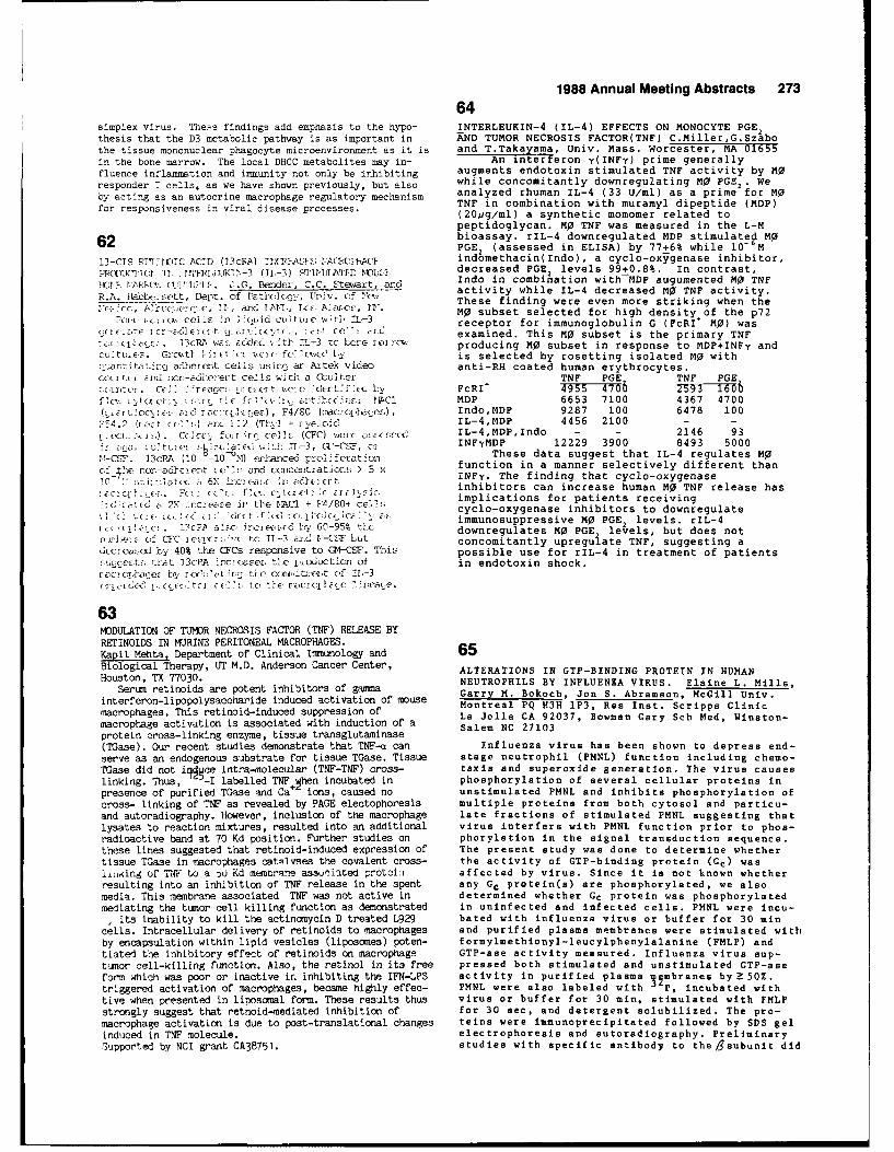
1988 Annual Meeting Abstracts 27364
simplex virus. The. e findings add emphasis to the hypo- INTERLEUKIN-4 (IL-4) EFFECTS ON MONOCYTE PGE2thesis that the D3 metabolic pathway is as important in AkND TUMOR NECROSIS FACTOR(TNF) C.Miller,G.Szabothe tissue mononuclear phagocyte microenvironment as it is an .aaaa Univ. mass. Worcester, MA 01655in the bone marrow. The local DHCC metabolites may in- An interteron y(INFy) prime generally
fluence inflammation and immunity not only be inhibiting augments endotoxin stimulated TNF activity by MOresponder T cells. as we have shown previously, but also while concomitantly downregulating M'o PGE2 .We
by acting as an autocrine macrophage regulatory mechanism analyzed rhuman IL-4 (33 U/ml) as a prime for MOfor espnsienes invirl dseas prceses.TNF in combination with muramyl dipeptide (MDP)for espnsienes invirl dseas prceses.(20/ig/ml) a synthetic momomer related to
peptidoglycan. Mo TNF was measured in the L-Mbioassay. rIL-4 downregulated MOP stimulate MO
62 PGE2 (assessed in ELISA) by 77+6% while 10- M13-CTS PFT * JIiCi ACID (13cR.) 7(1MT,'E1S!; 14A?0HACE, indomethacin(Indo), a cyclo-oxygenase inhibitor,
PPQD1E(TI1 Ii.c,'J'1-1'TVjIAT1r, "'O-USE decreased PGE2 levels 99+0.8%. In contrast,
rnn t4cetctcit~'t. :.G.. Bende., C.C. Stewart, arnd Indo in combination with MOP augumented MO0 TNFHabb ot, Dpt f P~r~cC~c to~. f Cactivity while IL-4 decreased M0 TNF activity.
R.A._.~abe'-setDep. f Pth(Ioy, rl~. ~f euThese finding were even more striking when the~c C, ? i~c~i ITC , u. arc 7. 11T< .. Alarcsc, D'. MO subset selected for high density of the p72Foic iz.,cucels j, i~id ulttewlt, !-3receptor for immunoglobulin G (FcRI' MO) was
yr-t icr c6(i cti . , c.C'f arce examined. This Mo subset is the primary TNFrEc -i .. cRAi uri. FCdncd vti 1 L3 tc bore r '3r producing MO subset in response to MDP+INFy andcuuE-, Grct I~ Cic i jr ft i ,EC 1iC is selected by rosetting isolated MjO withc.uiiat'tau cc aohe rot cells using an Artek video anti-RH coated human erythrocytes.
i~r~ i lid ncr-ad heit cell 1 a dl a Ccul ter TNF PGE TNF PGEccLrt ci . i C(-" 1; t cci _ 0 i'6E t -f le t1Ch- FcRI 'T ~ 7
cx v.,cc ~ xc t ot Cft I ~ 1x tic ~- 17cCl MOP 6653 7100 4367 4700E(ccicr l~~c c.i(ez ), F4/8C (onci~inIndo,MDP 9287 100 6478 100
2,4. liz, -cc I rc: 112 (Tiy1 I rvecC-1 1L-4,MDP 4456 2100 - -
ici. ) .) Cc,,cor Lif ,i " r ce, ' E (CFC) wes c alc~ I L-4,MDP,Indo - - 2146 93r. toa Li-3 tIE, .wS INFyMDP 12229 3900 8493 5000
ICSF. 33cRA (10 10 TA) erhaice] prcdiferat4 )I These data suggest that IL-4 regulates MO
C'f !e nor-adciceut cE 1, arc ccscentiatin > 5 Xfunction in a manner selectively different than10-c crC6 INFy. The finding that cyclo-oxygenase
10 .xi. c.6\ 1C'cl r.inhibitors can increase human MO TNF release hasFcc , rc c' e ,c cc t t :r + FA/i0 implications for patients receiving
(rC cc a 2Y 1cii c-,ithe 2i l F(I/80(tCe' cyclo-oxygenase inhibitors to downregulate60-95 ci ~immunosuppressive M0 PGE2 levels. rIL-4
(11 (t. . 13cF a'C i-c'i& i 1 09" 1' downregulates Mo PGE levels, but does notxuc.of CFC axr> tCc TL-3 zyccl 1-CSF bit concomitantly upregulate TNF, suggesting a
LlcIeIseXI by' 40% the CFCs respoinsive to CI4-CSF. This possible use for rIL-4 in treatment of patientsEcuccert:, that 13c:PA incccaise. th e pihoction of in endotoxin shock.rcc phajc by, Cctatrg tie cctitxN~fit c-f T1-3CriaxE-cC' llc~jv'tc CE, , to 'i,IE racioitiltC ?.Alra'C.
63MODULATION OF TUMOR NECROSIS FACTOR (TNF) RELEASE BYRETINOIDS IN MIJRINE PERITONEAL MACROPHAGES.Kapil Mebta, Department of Clinical Immunology and 65Biological Therapy, UT M.D. Anderson Cancer Center, ALTERATIONS IN GTP-BINDING PROTEIN IN HUMANHouston, TX 77030. NEUTROPHILS BY INFLUENZA VIRUS. Elaine L. Mille,
Seri reinois ae ptentinhbitrs o gasnaGarry M. Bokoch, Jon S. Abramson, McGill Univ.interferon-lipopolysaccharide induced activation of mouse Montreal PQ H3H 1P3, Res Inst. Scripps Clinicmacrophages. This retinoid-induced suppression of La Jolla CA 92037, Bowman Gary Sch Ned, Winston-macrophage activation is associated with induction of a Salem NC 27103protein cross-linking enzyme, tissue transglutaminase(T~ase). Our recent studies demonstrate that TNF-oa can Influenza virus has been shown to depress end-serve as an endogenous substrate for tissue TGase. Tissue stage neutrophil (PMNL) function including chemo-TMase did not iti~ce intra-molecular (TNF-TNF) cross- taxis and superoxide generation. The virus causeslinking. Thus, - -I labelled TNF Pen incubated in phosphorylation of several cellular proteins inpresence of purified TGase and Ca+ ions, caused no unatimulated PMNL and inhibits phosphorylation ofcross- linking of TNF as revealed by PAGE electophoresis multiple proteins from both cytosol and particu-and autoradiography. However, inclusion of the macrophage late fractions of stimulated PMNL suggesting thatlysates to reaction mixtures, resulted into an additional virus interfers with PMNL function prior to phos-radioactive band at 70 Kd position. Further studies on phorylation in the signal transduction sequence.these lines suggested that retinoid-induced expression of The present study was done to determine whethertissue TGase in macrophages catA1vses the covalent cross- the activity of GTP-binding protein (Cc) was
fi.igof TNF Lo a 5u Kd mEmor'ane asscxiflaed protein affec ted by virus. Since it is not known whetherresulting into an inhibition of TNF release in the spent any GC protein(s) are phosphorylated, we alsomedia. This membrane associated TNF was not active in determined whether Gc protein was phosphorylatedmediating the tumor cell killing function as demonstrated in uninfec ted and infected cells. PMNL were incu-
Iits inability to kill the actinonycin D treated L929 bated with influenza virus or buffer for 30 mincells. Intracellular delivery of retinoids to macrophages and purified plasma membranes were stimulated withby encapsulation within lipid vesicles (liposmes) poten- formylmethionyl-leucylphenylalanine (FMLP) andtiated the inhibitory effect of retinoids on macrophage GTP-ase activity measured. Influenza virus sup-tumsor cell-killing function. Also, the retinol in its free pressed both stimulated and unstimulated CTP-aaeform which was poor or inactive ir. inhibiting the IFN-LPS activity In purified p lasma 12mbranes by Z 50%.triggered activation of macrophages, became highly effec- PMNL were also labeled with P, incubated withtive when presented in liposcisal form. These results thus virus or buffer for 30 min, stimulated with FMLPstrongly suggest that retnoid-mediated inhibition of for 30 sec, and detergent solubilized. The pro-macrophage activation is due to post-translational changes teins were immunopreipitated followed by SDS gelinduced in TNF molecule. electrophoresis and autoradiography.- Preliminary
5S1pported by NCI grant CA3B751. studies with specific antibody to the /gsubunit did
274 1988 Annual Meeting Abstracts
not demonstrate protein phosphorylation. Studies phosphorylation patterns of these cell surface antigensin .:ogress are examining phosphorylation of the after activation with PMA. Specific phosphorylations were-Csubunit. These results suggest that influenza observed in the case of CD3, CD4, CD5, CD6 and AIM. Thesevirus nay alter the GTP-ase activity of one or data indicate that PMA induced phosphorylation of specificmore GTP-binding proteins and inhibit protein cell surface differentiation antigens can be associatedphosphorylation add subsequent end-stage PMNL either with down-regulation (CD-4) or with upregulationfunction. (CD5, CD6, AIM) of the cell surface molecules.
66 68PROTEIN KINASE C ISOTYPE DISTRIBUTION AND SELECTIVE
LIPOPOLYSACCHARIDE-INDUCED EXPRESSION OF ISOTYPE TRANSLOCATION WITH Ca2+
IN HUMAN NEUTROPHILS ANDTHE COMPETENCE GENE, KC, IN VASCULAR ENDO- CYTOPLASTS T. Fuiiki, M. W. Rossi, W. A. Phillips, R.THELIAL CELLS IS MEDIATED THROUGH PROTEIN B.Johnston Jr. and H. M. Korchak (Spon: L. Kilpatrick-KINASE C. X. Shen. T.A. Hamilton and P.E. DiCorleto smith), Univ. of Pennsylvania, Philadelphia PA 19104.Cleveland Clinic Research Institute, Cleveland, OH Protein kinase C (PKC;80kDa) has been proposed as a44195. mediator in multiple cell functions, including activationThe KC gene is one of a family of cell cycle-depen- meitrnmuipeclfntos, nldngaivindent competence genes that was originally identified in of 02- production. Translocation of PKC from cytosol toplatelet-derived growth factor stimulated BALB/c-3T3 membranes is thought to be crucial to PKC activation.cells. We have examined the expression of the KC gene in Recently, several isozymes of PKC have been identified by
cultured porcine aortic endothelial cells in response to cloning technology. Polyclonal, antipeptide antibodiesbacterial lipopolysaccharide (LPS) as a first step in defin- that specifically recognized the consensus as well as e,ing the molecular events in endothelial cell "activation. s, and I isozyme epitopes (Makowske et al,JBC 263:3402),LPS markedly stimulated the steady state level of KC were used to study the distribution of PKC in disruptedmRNA in confluent endothelial cells without increasing human neutrophils and cytoplasts. Intact neutrophilsthe growth fraction of the cultures. Maximum induction and cytoplasts (vesicles of cytoplasm enclosed by
of KC occurred in the cells following exposure to 10 ng/ml plasmalemma) produced 02 in response to stimulants suchas FMLP and PMA. PKC was observed in cytosol and membraneLPS for two hours. A series of studies was performed to fractions of resting intact neutrophils and cytoplastsdetermine whether protein kinase C was involved as an with antibody to consensus peptide; the issnunoprecipitinintracellular mediator of this LPS-stimulated response. reatiody t veons ess in the iltun initheEndothelial cells treated with phorbol myristate acetate reaction was qualitatively less in the pellet than in the(PMA) and l-oleoyl-2-acetylglycerol (OAG) exhibited cytosol fraction. The presence of Ca + during disruptioninduction of KC gene expression. A maximum response of cytoplasts resulted in increased imnmunoreactive inwas obtained with 10 nM PMA, the effect decreasing with ka species in the membrane fraction and a reduction inhigher levels of the phorbol ester. The increased expres- the cytosol. PC activity, with histone as the substrate,sion of KC induced by LPS and PMA was inhibited by the corroborated this result. Antibody to 0 PKC immunoblottedpresence of 50 .M 1-(5-isoquinoline-sulfonyl)-2- strongly to a 60kDa polypeptide in cytosol and pellet
methylpiperazine (H7), a protein kinase C inhibitor, but fractions. No apparent differences in the blotting
riot by an H7 analog HA1004 (50 pM), which is a much intensity or attern were ubserved in the Ca -containingless effective inhibitor of C kinase, or by the cyclooxygen- and the Ca
2 -depleted conditions. Antibody to 1 PKC
ase inhibitor acetylsalicylic acid (100 gM). No cytotoxicity immunobloted predominantly to an 8OkDa polypeptide in thedue to the various inhibitors was observed in the endo- cytosol. The imunoprecipitin reaction in the cytosol wasthelial cell cultures. These results demonstrate that the qualitatively less in the Ca +-containing samples than in
KC gene may be induced by LPS in vascular endothelial the Ca2+-depleted samples, suggesting translocation of
cells in a proliferation-independent process. Secondly, this isotype. y PKC antibody immunobloted to an 80kDaunlike LPS-induced KC expression in macrophages and polypeptide in the cytosol. No difference in the distri-PDGF-induced KC expression in 3T3 cells, LPS induction bution of this isotype was observed between Ca
2+ -
of KC in endothelial cells requires the action of protein containing and Ca2+-depleted conditions. These resultskinase C. (Supported in part by HL34727, CA39621 and indicate that I) neutrophils contain a, 5 and y PKCHL01561) isozymes, 2) the isozymes are differentially distributed
and 3) the P isozyme is a translocatable species.
67 69ROLE OF PKC IN THE CELL SURFACE EXPRESSION AND PHOSPHORYLA- ROLE OF PROTEIN KINASE C IN THE ACTIVATION OFTION OF DIFFERENTIATION ANTIGENS OF RESTING AND ACTIVATED LIVER MACROPHAGES. D.L. Laskin, C.R. -Gardn er,HUMAN T CELLS. A. Carrera, L. Cardenas, A. Tugores, M. A.M. P ilaro __and J.. Las ki n. RutgersCebrian, F. Sanchez-Madrid, M. Lopez-Botet and M.O. de University and UMDNJ-Robert Wood JohnsonLandazuri. Servicio de Inmunologia. Univ. Autonoma. Medical School, Piscataway, NJ 08854.Hospital de la Princesa. c/Diego de Leon, 62- 28006 Madrid. Protein kinase C (PKC) has been postulated
We have studied the effect that the activation of to be a critical enzyme in initiating theProtein Kinase C (PKC) has in the cell surface expression biological activity of a variety ofof several human T cell leukocyte differentiation antigens. extracellular signals involved in cellularTreatment of resting T cells with phorbol esters (PMA) activation. To study the role of PKC in liverproduced a dose dependent down-regulation and internaliza- macrophage activation we used synthetiction of CD3 and C04 and upregulation with augmentation in diacylglycerols (DAG) and tumor promoters whichthe expression of CD2, CD5 and CD6. We also studied the are known to bind to and activate PKC. Werole of PKC in the induction of cell surface activation found that treatment of cultured rat liverantigens. Two cell surface activation antigens-4F2 and AIM macrophages with phorbol myristate acetate(a recently described molecule) seems to be very much (TPA), phorbol didecanoate or mezerein produceddependent on PKC activation since they appear very early morphological alterations in the cells(2-5 hours) after PMA treatment and are fully expressed characteristic of activated macrophages. Cellsafter 18-24 hours. We also studied if phorbol esters could became enlarged, highly vacuolated and spreaddown-regulate the expression of activation antigens already on culture dishes. Each of these analogs waspresent on T cells. We observed that the addition of PMA to also found to be potent inducers of macrophageactivated T cells down-regulate the expression of the chemotaxis. Synthetic DAG including l-oleoyl-2-transferrin receptor whereas it upregulates the expression acetylglycerol (OAG) , 1,2-dioctanoylglycerolof IL-2R, 4F2 and AIM. Finally, we analyzed the (diC8) and 1,2-didecanoylglycerol (diClO) were
1988 Annual Meeting Abstracts 275
also found to induce morphological alterations proteins extracted and analyzed for phosphoaminoand chemotaxis in liver macrophages. acid content using thin layer electrophoresisFurthermore, phorbol diacetate (PDA) a following partial hydrolysis in 6N HCl. We foundbiologically inactive analog that blocks TPA that total cellular protein content decreasedreceptor binding inhibited chemotaxis induced during differentiation of the cells into PMN, butby TPA, OAG as well as the complement fragment not MP. In addition, the relative amount of 32pC5a. Both DAG and tumor promoters also incorporated into cellular protein was less instimulated phagocytosis of sheep red blood both PMN and MP. We detected a decrease in thecells by the macrophages. In contrast to the phosphoserine, phosphothreonine andtumor promoters, DAG did not stimulate the phosphotyrosine content of the cellularrespiratory burst as measured by flow cytometry phosphoproteins when compared to untreatedand cytochrome C reduction. These data suggest controls. MP differentiation resulted inthat some, but not all macrophage functions significantly greater reduction inassociated with activation are mediated by PKC. phosphorylation of all three amino acids inSupported by NIH grant GM34310. proteins than did maturation into PMN. With all
inducers, phosphorylation of tyrosine residueswas decreased to a greater extent than wasphosphorylation of serine or threonine residues.
70 These data suggest that PMN and MP
THE ENHANCEMENT OF RECEPTOR-MEDIATED PHAGOCYTOSIS BY differentiation is associated with significantAMPHOTERICIN B MONOMETHYL ESTER (AME).S. . changes in cellular protein kinase and/orT' L i cy. J. Ln_, C'_._ Schatfne-r W3k!3man ln.tltute phosphatase activity. These data further indicate,t :r1oty @ .U'?3Rf-fh, £tate niversity, New a role for protein tyrosine phosphorylation in
N '5-jjis regulating HL-60 cell differentiation. SupportedAinzt.,ricin b AM .) and AME, polyene macroiice by NIH Grant A120183.
rnt-: .oiit3, possess immunomodulatiniart iLti s-. ioeenkty, they have attracted attention tor
'- Lviria i, tivity against -lipid-enveloped viruses.".F, in piticular, an protect H-a celis (human 72
pte p : :neJ from q!V-' (AIDS virus). Also,4 F" i, at leastt '0-rid 1est, toxic than AM, for many THE IN VITRO PROLIFERATION OF PULMONARY ALVEOLARMACROPHAGES7if; ' nt -11 tyPes. Sin,,e polyene-mediatec effects
: a re- t".- re a t of memran,-steroi tnteractlcns, FROM MICE UNDER MONOCYTOPENIA OR MONOCYTOSIS. Y. 0sw , .ve2 variou- pnyFr:a membrane parameters and Division of Comparative Radiotoxicology, Nationa Institute
t-.e immunoreieptors. iding o_- "i1C of Radiological Sciences, Chiba 260, Japan
, , t 7 .L ind ceis was insensitive to Pulmonary alveolar macrophages(PAM) obtained by lung lav-A" : A it - 1u /qmi), as was the bindirQ of age from the normal mice can proliferate in vitro with co-:r(,fiy , ho 3nee[ tel oioed cells (s-BC and the lony stimulating factors(CSFs) including F TST -or GM-CSF.
1 ., ,t, ot ,ompement. Both polyene, have The present work was done to investigate whether or not suchjn-.v.i', act v ty in this range. Although AMB and AME-, f,, -, t tmeLrants of ceili essentially equally, a growth kinetics of PAM in vitro would change either in a
Acnt incre.ised the membrane fei'dVty, whereas monocytopenic condition or und era monocytosis, compared toAll? n!,t . In -, ,, ntn witn T iympnocyten Ind FITC- the normal steady state. The uptake of
5H-TdR and colony
n. ode.;, Ay.6 olorked both patch and cap formation by mouse PAM in a normal culture condition weren w -i"-e - AMF bi-Ked only capping. This not found during the incubation period of 14 days, whereas
! 1 .3 Z p, yenes car, :nhibit celLuLar they were enhanced by addition of CSFs to the cultures. The,, r , f ymphocytir mmbrand iand-recepto expression of surface antigens on PAM, including Ia, Mac-l,-i a hrert tuman Ph[. with AME, but not AIB, caused a and F4/80, was not, however, seen during the incubation in
-ose-pend-.nt increase in phagocytosis ot the presence of CSFs. Such in vitro proliferation of PAMwas, vrt? -eit i . Pietreatment of the sensitized next examined in monocyte-depled mice. During the period
in sncreased phagocytosis, but not as of 56 days after injection of a bone-seeking 89
Sr radioiso-,nd. A : the capacity to enhance Fc ieceptor- tope, bone marrow stem cells and circulating monocytes were
, S qte r, otytosls at concentrations which are severely depleted, while splenic colony forming stem cells.t' rv; ! foi .- V-L. AIso, ufter leating no.-mal PbL withA"- pcr o in't:.,n 'timulaton, we found that increased with an extramedullary hemopoiesis. PAM underthis
i, ' f ind P"F was not atfecred. Similarly, condition, however, showed almost equivalent 3 H-TdR uptake
i. eptir-meditd .olleaton of and colony fomation in vitro by CSFs to the normal condit-
,I-de1 C_ iLL(L-2 ,nd BiL respectively) was ion. On the other hand, when mice were implanted subcutane-it ',"n. -. .e ts Al.. e= was the receptor-meciated cytocidal ously with NFSA fibrosarcoma cells, which have been found to
iti-, -,f Tsmo. Ne-ro:,:; Factor. In this concer.tratis enhance both of bone marrow and splenic hemopoiesis by pro-tsr ,', AM s eiiher cyto-oxic nor does. it adversely ducing GM-CSF, circulating granulocytes and monocytes in-
,ther e-:toi -mediated immune functions. TheIet.d men hacm ,f AME action needs to be elucidated to creased in number. Any metastasis of tumors into the lung
, polyen,: is thtuapeutic/prophylactic druq, was not detected. In this condition, the recovery of PAM andtheir in vitro proliferative responses were not altered,compared to the normal or monocytopenic state. These find-
71 ings indicate that there may exist precursor stem cells in
DIFFERENTIATION OF MACROPHAGES (MP) AND the resident PAM population which can respond to the growth
NEUTROPHILS (PMN) IS ASSOCIATED WITH CHANGES IN factors independently upon the level of circulating mono-CELLULAR PROTEIN PHOSPHORYLATION. A.A. Sirak, cytes. We acknowledge a courtesy of Dr.R.K.Shadduck,Univer-F.H. Mermelstein, J.D. Laskin and D.L. -Laskin. sity of Pittsburgh, for his gift of highly-purified M-CSF.Rutgers University and UMDNJ-Robert Wood Johnson This work was partly supported by a Special Cordination FundMedical School, Piscataway, New Jersey 08854 for Promoting Science and Technology from Science and Tech-
Protein kinases are thought to play an nology Agency, Japan.
important regulatory role in cell growth anddifferentiation. In the present studies, we 73measured changes in protein phosphorylation RESIDENT PERITONEAL MACROPHAGES (Me) ARE MAINTAINED BYduring differentiation of HL-60 promyelocytic LOCAL DIVISION. M.J. Melnicoff, T.C, Schmitt. P.K.leukemia cells. HL-60 cells were induced to I}oran, and P. S. Morahan. Medical College ofdifferentiate into MP by 48 hr treatment with Pennsylvania, Phila., PA 19129; and Smith, Kline andphorbol myristate acetate (170 nM) or into PMN French Laboratories, King of Prussia, PA 19406.with dibutyryl cyclic AMP (500 uM) ordimethylsulfoxide (1.3%). Cells were labeled for Resident peritoneal MO of Balb/c mice were labeled3 hr with
3 2P (250 uCi/ml) and phosphorylated in vivo by intraperitoneal injection of the green
276 1988 Annual Meeting Abstracts75
fluorescent cell tracking dye, PKH-I. After CHARACTERISTICS OF HUMAN CD4 MON0CYTE(MO) SUBSETSimmunofluorescence labeling with MS specific monoclonal G.Szabo,C.Miller,J.u&KKdsUiv as eiaantibodies (Mabs), resident MO were double labeled by Center, Worceste, MA 0165both the green dye and the red (phycoeryrhrin) Mab tag. Although human T lymphocytes and W bear the sameIn the absence of induced inflammation, the number of Leu3 (T4,CD4) antigen, the function of CD4 expressionPKH1-1 labeled resident MO did not decrease for at least on 11O in ?W-T cell interactions or inflammatory immune28 days in vivo; therefore, these cells were saintained responses is uncharacterized. In this study, normal MOwithout replacement by recruited monocytes. depleted of T cells by E rosetting and separated into
To determine whether the resident MS were dividing CD4 positive (26.87+ 5.7%) and negative (73.12+5.7%) W~
in vivo, a pulse of 3H-thysldine was injected i.p. at 4-7 sbesb ann ;t niC4a eeasse odaysaftr aminstrtio of he EN- lael. The presentation of tetanus toxoid. Antigen presenting
aias wfere acificedatone ouraf the -thymel dine capacity (APC) was expressed in cpm over control. CD4*animls ere acrfice on hou afer te 3-thyidie M subset's APC activity was increased concomitant topulse, and the peritoneal MS were labeled with the Mab decreased production of prpstaglandin E2 (PGE asMac-I. The resident MS (Mab and dye labeled) were sorted measured by ELISA in ng/l0 MP) when Compared to theon an EPICS V flow cytometer/sorter, and the fraction of D4_ MP0 subset. The reduced APC function of CD4_ M0 may3H-thymidine labeled MS in each population was evaluated not be secondary to their greater PGE2 production via
by autoradiography. possible downregulation of Class ii antig~ns by PGE.The unsorted peritoneal cells contained 29.6 + 11.8% Even in the presence of indosethacin (10- M) CD450WAPC
MS (by differential cell counts), with a 3H-thymidine was reduced from that of CDC W0.
labeling index of 3.3 ± 1.4% (N-6). Our present data CD4 + CD'F CDC CD4_show that the resident MS fraction collected from the PGE2 5.6 10.0 6.4 8.2same animals after sorting contained 81.1 + 8.2% MO, of APC 17,321 8,357 44,886 35,411which 2.5 + 1.9% were labeled by
3H-thymidine. Three of APC+indo 30,891 15,891 55,957 48,512
the animals had detectable populations of recruited Plasminogen activator capacity of the subsets wasmonocytes/4S (Mab labeled only). The MS labeling index similar (CD4*:26.8,CD4>.29.2% specific fibrinolysis).of these recruited cells was 2.7 + 2.3%. These data show Secretion of the inflammatory monokine tumor necrosis
that resident peritoneal MS in the hesatopoietically factor (TNF, ng/l0 MO in L-M bioassay) andintact animal are dividing in vivo. (Supported by NCI interleukin-l (IL-l stimulation index in D.10 bsioassay)
CA356 an NIHA12551.)were also similar in the subsets after 100U/106 50 IFNYCA356 an NIHA12551.)plus 20vg/ml suramyl dipeptide (MOP) stimulation.
CDC CD4_ CDC CD4_TN'F unstim. 1.74 1.44 0 0
IFN+MDp 6.47 7.46 4.87 3.29IL-l unstim. 5.64 7.04 2.05 2.05
IF14+MDP 29.90 26.60 6.90 7.0274 These data suggest, that the presence of CD4 antigen on
IbI-2TIFICAT1ON OF RUIONARY PBPI-RIAGE POPULATIOBS II 7i1 MO could be related to the better APC of CD4+ MO subset11DUSE. R. Crowell, B. Lehnert, C. MuQ (Sien J. er) to T cells. Since it is the CD4+4B4+ T cells thatUniv. W,, Ab., 2,, fi Aliu, '!t La. Lo AllcN primarily proliferate to soluble antigens presented by
Puitenar)y' acrc1_,1=he3 (PB-) oxi,,t in at least 2 ccnipart- M0, CD4 ag complex may have a role in l'o activation ofments, the alveolax. space sria interstitiuo. Wec evaluateu eprclstt.e Conti: ibutionoL of eacii cu~partrnent to the s:otal PM. Al-v acoi acruphages (Po4) were recuvered by bronchoalveolar.avaige (BAL) al-ki interLstitial rsacruphage, (114) by lung
E,4nc -ng and uiiestion by collagcenaLse. R! were identifieu;crpwulongically sa .3y tiaTir abilit to pliagocytizo IgG 7opsonized shes rea blee., cells (EA) . The puilonary circu- HUA NONOCYTE HETEROGENEITY DEFINED BY ELK.-DRlation was ier fuseu with PBS until the lungys turneu white EXPRESSION DOES NOT CORRELATE WITH OXIDATIVEto Clii71nate blooi iannocytes. The lungs and trachea were BURST CPAB)ILITY. G.T. Svear. L.C. Rothbera
ec.eart.. gAL i-erfonsnid until retii was cEss t! kan and A.L. Landay. Rush University, Chicago, IL0.1r1i. 23x10 colls were recoverhe by BAL (83% vialil- 60612.ity) of w-hich 87% wc.e macrophages. 91.5% of &4 phiace Monocytes from any one blood donor exhibit acyt. zea EA and 61% cantaind,7>4 EA/Al'i. Lung aigestion by wide range of HLA-DR expression with theeel layeniase recovcree 2.7x10 cells (85.6% viability), difference between low and high HLA-DRKnen, lungjs were instilled intratrachcwlly with E7. zicr to expressing cells being as great as 10-fold.BAL ame lung di. otn, 4.4% of thec lung digest; cells; Con- This study determined if the variability intained 11A stzjgestingj these celis were P14 not lkirvesited by HLA-DR expression on monocytes correlated withBAL. 8% of the cells (2.lxlO Cells) in the wung diges;t oxidative burst capability by cells stimulated
hadth srplloicapparnc o bla~irnocte, ut by heat-aggregated IgG (HagIgG), phorbol.if.iled to )iiawocytize EIA. W1 1le 33.9% of th~e lung digest myristate acetate (P1(A) and calcium ionophoreceil ' 9.2x10 cells) roilpholegically resamruec ?,', onily A23187 (ionophore).26.6% of thes;e cel o or 12% of lung diijeot cel]o 4shico Freshly obtained cells were loaded withc~tized EA (2.5xlO cells) . Tl, deters inc i:f collagenase dichlorofluorescin diacetate, stimulated, andaltered EA phagocytoLsis, BAL cellsL were incubated with then stained with phycoerythrin-labeled anti-olagenase unuef the conditions used for lung digestion. LeuM3, anti-HLA-DR or control monoclonal
Only 71.216 of cullagoniase-troateu AM- phagocytized EA, and antibodies (mAb). Two-color flow cytometry was40.2% containeu >4 EA/A1. T'is decrease .;. insufficient to used to determine changes in green fluorescenceaccounit for the d iscrepiancy between cells idenitifiec; mor- corresponding to oxidative product formation by_oGiol caily rK. functionally as P11 in the lung digest. We cells, which stained only with the red mAbconcluoc tihat a subpcopulation of tronunuclear 1phagocytic (monocytes). Forward and 90* light scattercells resaffblinet PB can be identified after: exhautive SAL were also used to ensure that only monocytesarne lung diucotluon. A L;itnificant Wortion (25%) of these were detected.cells- are alveolar in origin. Accounting for these con- The oxidative capacity of total Leu-M3 andtanirints, the rca,-ining cells. ccepeise a larger III stir-- total DR-stained cells was identical,Eart.-ocnt thkan prccviously reurted in mice. The majority of indicating that these markers measured similarcelo di lung digest which rorpholngically resemble W1 functional populations. while monocytesfall to ingest EA. expressing high levels of HLA-DR and low levels
1988 Annual Meeting Abstracts 277
differed substantially, (average difference = cells of peripheral B cell, TA and 154, in the contrast65 channels), changes in oxidative capability with shorter and wider ones in YS cells. As the cellwere only 2 channels for HagIgG, 9 for PMA and markers of this period, C20 and CD21 were positive in14 for ionophore. Other myeloid markers each cells other than YS. All cells of examined cellexhibiting less heterogeneity on normal lines were positive for HLA-DR. There were positive cellsmonocytes than HLA-DR gave similar results. for CDl and LeuM3 partly in YS and IL2-R in all cellsThus, monocytes which are very different in except for YS. Positive reaction of ORC-l(anti-follicularHLA-DR expression are very similar in oxidative dendritic cell) was seen in TA and 154 cells. Some of YSburst capability, cells showed characteristically positive for lysozyme,
S100 protein and Lag (anti-Langerhans cell).These results may suggest that B cells altered their
morphology to dendritic form with PMA still reserving the77 cell markers of B cell characters. On the other hand,
monocytoid cell of YS was capable of transforming intoBIOACTIVITY OF THE INSULIN RECEPTORS (IR) ON SPLENIC IDC on the viewpoint of its morphology and cell markersMACROPHAGES (MO) IN MICE. A.P. Bautista, D.J. Fletcher as well.& A.Volkman. School of Medicine, East CarolinaUniversity, Greenville NC 27858.
The presence of IR on splenic Mo suggests that 79insulin may be important for the regulation of the 45biological activities of these cells. The bioactivity EFFECTS OF BONE MARROW SUPPRESSION WITH Ca ON MONOCYTESof IR in C. parvum (CP) and 89Sr-monocytopenic mice, AND MACROPHAGES (M). A.Volkman and Y. Shibata. Eastin which there is increased proliferation of splenic Carolina University School of Medicine, Greenville, NCMO progenitors was studied. CBA/J mice received 4 27858-4354.uCi/g bw 89SrCl2 IV. On day 3, CP was given IP into Responses of mice to P. acnes vaccine (PA) ip includeeach mouse at a dose of 60 mg/Kg bw. Control mice the elicitation of monocytes into the peritoneal sac andreceived non-radioactive 88SrCl2 and 0.15 M sterile the induction of PGE2-secreting suppressor MO (PGSM) in theNaCl. Insulin binding assays were performed using spleen. Bone marrow ablation with the bone-seeking isotope,125-1-insulin as tracer.H4 colony forming cells(M-CFC) 89Sr (TI/2-50 days, Ea-.56Mev). in mice results in profoundwere determined using a 2-layer agar technique.Results monocytopenia and a lOx increase in splenic M-CFC but lossshow progressive elevation of specific 125-I-insulin of the monocyte elicitation and PGSM responses to PA. Thisbinding from days 2-10, when it was significantly and other observations suggested bone marrow dependence ofgreater (P<O.0i) in CP-mice (12.5 ± 1.18 pg/I x 106 the PGSM response but a dissociation of elicitation andMO) than in the parallel controls (6.25 ± 0.8 pg). The PGSM induction was found in congenitally monocytopenicincrease in 125-I-insuiin specific binding was due to SI/SLd mice (Shibata, Y. and Volkman, A. JI, 135, 3905,an elevation of both high and low affinity sites 1985). To pursue this point,
45CaC12 (Tl/2-164 days,
without any change in binding affinities, and with no Ea-.077) was given iv to 8-12 wk old CBA/J mice, 4uCi/g bw;significant alterations in plasma insulin. In 40
CaC1 2 was used for controls. At 14 days blood monocytesmonocytopenic mice, 125-1-insulin binding to splenic were 14% control in unstimulated mice, 33% in PAM was greater (10 ± 0.9 pg;P<0.05) than the control, (56 mg/kg, ip day 7) mice, and 29% in thioglycollate (TG)and was further increased (P<0.05) to 14 ± 1.2 pg (4 day) mice. Monocyte counts were less depressed in allafter the administration of CP. Although insulin alone groups on day 28 but still about 50% below controls on daycould not induce the formation of M-CFC by splenic 44. Neither PA nor TG was able to elicit monocyte-MO incells, it was associated with a dose-dependent day 14 mice but progressive return was seen in 28 and 44modulation of M-CFC in control (P?0.05) and CP mice day samples. M-CFC day 14: marrow, 25% (no PA) and 12%(P<0.01). Insulin at more than I uM was associated (PA) of controls; spleen, 6x (no PA) and 3x (PA) controls.with reduced M-CFC in control and CP groups. M-CFC Unlike
89Sr mice, however,
45Ca treatment did not impair
(4.57 ± 0.97/ 20,000 nucleated cells) in the spleens release of PGE2 from PA-induced PGSM whether spontaneousof control mice were significantly Increased in the or enhanced with zymosan, Ca
2+ ionophore A23187 or phorbol
presence of I nM insulin, than without (3.0 ± 0.86 ester. The observed dissociation between monocytopeniaP<O.Oi). In the CP-treated group similar responses and failed elicitation on the one hand and unimpaired PGSMwere observed ; with insulin, M-CFC was 169.4 ± 38 and activity on the other suggests that blood monocytes arewithout I13 ± 27 (P<O.01). These observations thus not precursors of PGSM. Since splenic M-CFC are elevatedsuggest that insulin at optimum concentrations may in PGSM-impaired
89Sr-mice and in PGSM-unimpaired
45Ca-mice
augment the proliferative capacity of splenic M-CFC. their role in this function remains uncertain. The 0iffer-ent effects of
89Sr and
45Ca on PGSM may be due to differ-
ences in El suggesting that monocytopoiesis is a more78 radiosensitive path than PGSM formation. (Supported in
MORPHOLOGICAL CHANGE OF B CELL AND MONOCYTE IN VITRO WITH part by NIH grant AI17162.)
PMA ( 4i-PHORBOL 12-MYRISTATE 13-ACETATE ).M.Matsuda, M.Ishikawa, A.Masunaga, M.Narabayashi, H.Hashimoto and Y.
Imai. soYamagata Univ.Sch.Med.,Yamagata,990-23,Japan.
There are still some debates on the nature of various INDUCTION AND CHARACTERIZATION OF HUMAN MONOCYTE-accessory cells concerning the immune reactions. Recently, MACROPHAGE-DERIVED MULTINUCLEATED GIANT CELLS IN IN VITROperipheral B lymphocyte has been reported to transform CULTURE. N. Hassan and S. Douglas. Division of Allergy-into interdigitating cell (IDC) mainly by morphological Immunology-BMT, Children's Hospital of Philadelphia, Univ.observation. In this paper, the changes of morphology and of Pennsylvania Medical School, Phila., PA 19104.some cell markers of S cell and monocyte were studied in Human blood monocyte-macrophage-derived multinucleatedvitro culture in the medium (RPMI1640, 20%FCS) containing giant cells are observed in several pathological conditions10 ng/ml of PMA. Two B cell lines ( TA : derived from re- including inflammatory diseases, foreign body reactions,active lymph node and 154 : from EBV infected 8 cell) as cancer, and also in viral infections such as the acquiredwell as peripheral B lymphocyte and one monocyte line (YS immunodeficiency syndrome. Although multinucleated giant:from AMMoL ) were examined. Each cell showed an adherent cells have been described for a century (Metchnikoff,character and became to reveal cytoplastic processes on 1888) the mechanism of their formation and functions are2nd culture day. They extended their processes.thereafter, unknown. Several studies have described the induction ofmaking a "dendritic form". This morphologic alteration fusion of monocytes and macrophages using gamma interferonbecame to be most conspicuous on 4-5th culture day. Thin (Weinberg et al., Proc. Natl. Acad. Sci. USA 81:4554,and elongated dendritic processes were remarkable in the 1984), 1,25-dihydroxvvitamin D3 (Abe et al., Proc. Natl.
278 1988 Annual Meeting Abstracts
82Acad. Sci. USA 81:7112, 1984), and Interleukin-4 (Mclnnes INDUCTION OF DIFFERENTIATION IN HUMAN U-937et al., J. Exp. Med. 167:598, 1988). We have demonstrated HISTIOCYTIC LEUKEMIC CELLS BY DIBUTYRYL CYCLICthe fusion of human blood monocyte-derived macrophages in ADENOSINE-3' 5'-MONOPHOSPHATE (dBcAMP) . A.J.vitro culture by phorbol myristate acetate (PM[A) with the Beavis, J.D. Laskin, A.A. Sirak, S.M. O'Connellinduction and formation of multinucleated giant cells and D.L. Laskin. Rutgers University and UMDNJ-(Clinical Research 36(3):440A, 1988). PMA (10-8OnM) in- RWJ Medical School, Piscataway, NJ 08854duces fusion of human monocyte-derived macrophages with Treatment of U-937 cells with the cyclic35-85' fusion rate. These giant cells form in cultures nucleotide analog dBcAMP induced these cells towithin 12-§4 hours after the addition of the stimulus, and differentiate. DBcAMP produced a dose- and time-they are 200-400 um in diameter and contain 15-30 nuclei, dependent inhibition of cell growth reaching aThe addition of gamma interferon (1000 U/ml) to the cells maximum after 48 hr treatment with 500 uM. Atprior to PMA (10nM) enhances fusion rate of the cells this concentration, dBcAMP had no effect on cellfrom 357 to 100'. In comparison to monocyte-derived viability. Flow cytometric analysis of dBcAMP-macrophages,P MA induced multinucleated giant cells are treated cells revealed a time-dependent decreasecharacterized by decreased intracellular acid phosphatase in the number of cells in S-phase of the celllevels, decreased or absent oxidative burst activity by cycle, with a concurrent increase in cells in thenitroblue tetrazolium reduction test, normal tumor cyto- GO/G1 phase. Treatment of U-937 cells with dBcAMPtoxicity using the CEM leukemia cell line as the target also decreased cell size, produced alterations incell, and decreased cell membrane ruffling with scanning the cell membranes and decreased nuclear-to-electron microscopy. We conclude that the human blood cytoplasmic ratios. DBcAMP also induced themonocyte-macrophage-derived multinucleated giant cells appearance of f-met-leu-phe (FMLP) receptors andhave different functional capabilities, as compared to the monocytic/granulocytic cell surface markersmorocvte-derived macrophages,and that these cells may MY8 and MAC-1 on U-937 cells, but decreasedplay an important role in tumor cell killing. (Supported levels of HLA-DR expression. Treated cells didin part by grants from NIH HL-27068, NS-17752, AMFAR 161, not express the monocte-specific markers, Mo2 orand MS Society 1919A-1). MY4. In addition, neither dBcAMP-treated nor
untreated U-937 cells stained for non-specificesterase nor did they adhere to culture dishes.Using flow cytometry we also found thatdifferentiated U-937 cells produced H 0 2 and 02'.FMLP was more effective in stimulating H202production by the cells than was phorbomyristate acetate. However both responses wereinhibited by catalase. DBcAMP-treated cells alsoproduced greater amounts of 0- than did
81 untreated cells, as determined y nitrobluetetrazolium and ferricytochrome C reduction.
Differential production of tumor necrosis factor These studies indicate that dBcAMP induces(TNF), macrophage colony stimulating factor (CSF- partial differentiation of U-937 cells.1) and interleukin I (IL-1) by human alveolar Furthermore, these differentiated cells displaymacrophages. Susanne Becker. Robert Devlin and characteristics typical of granulocytic cells.Stephen Haskill'Environmental Monitoring and Supported by NIH grant A120183.Services, Inc., U.S. Environmental ProtectionAgency, Chapel Hill, NC 27516. 83
Human alveolar macrophages (AMO) have been INDUCTION OF MACROPHAGE DIFFERENTIATION OF THE HUMAN PRO-investigated for their ability to produce the MYELOCYTIC CELL LINE HL-60 AS DETERMINED BY FUNCTION ANDthree monokines, TNF, CSF-I, and IL-1. No TNF IMMUNOCTOCHEMISTRY. R.H.J.Beelen, I.L.Eestermans*,activity was found in supernatants of unstimulated H.J.Bos*, and G.J.Ossenkoppele. Departments of HaematologyAMO cultured for 20 hours, although TNF mRNA was and Cell Biology*, Free University Hospital and Medicaldetected in the cells by Northern blot analysis. Faculty*, Amsterdam, The NetherlandsStimulation of the cells with LPS induced Our earlier studies (Eur J Clin Invest 17,1987,63) onproduction and release of high levels of TNF into human bone marrow cultures have shown that the endogenousthe culture supernatant. Increased levels of TNF peroxidatic activity (PA) is a very helpful tool to iden-mRNA was detectable at 90 min after LPS tify the immature and mature cells of the mononuclearstimulation by dot blot analysis, reaching peak phagocyte and granulocyte series. In this study we inves-expression between 4-8 hours, and declining tigated the PA pattern of the human promyelocytic leukae-thereafter. TNF activity peaked at approximately 8 mia line HL-60 which is known to differentiate into ma-hours in the AMO sups. After 24 hours TNF crophages or granulocytes upon induction by chemicals.production had ended. Compared to autologous The results showed clearly promyelocytes and blasts (PA inmonocytes the AMO produced 5.7 times more TNF on the Golgi system, granules and RER) in the control (noper cell basis (activity in 20 h supernatants). inducer) as well as after the addition of Ara-C. Vit. DAMO expressed high and low mol. weight CSF-I mRNA (1.25 dihydroxy Vit. D3) induced the differentiation intoand active protein was recovered in supernatants monocytes (PA only in lysusomes), while Vit. A (retinoicupon culture. LPS stimulation of the cells acid) resulted in a differentiation of granulocytes. Mostslightly reduced both mRNA levels and amount of remarkably DMSO gave a very pronounced differentiationfactor in the supernatants. In contrast to the into both very mature granulocytes as well as maure re-AMO, monocyte production of CSF-l was enhanced by sident macrophages (PA only in RER). The results, withLPS. CSF-I production by both monocytes and AMO respect to the ultrastructural cytochemistry, fitted verycontinued for at least 48 hours of culture. Low well in the functional characteristics of these cells,levels of IL-I mRNA was present in all tested AMO since after induction with Vit. D and also DMSO the cellspreparations. LPS stimulation induced increase in showed an enhanced Fe receptor activity which correlatedIL-l mRNA within 90 min., mRNA levels peaked with a clear functional antibody dependent phagocytosisbetween 12 and 20 hours, and stayed high for at (ADP). No ADP was found in the control or after inductionleast 42 hours. However, while the AMO expressed with the other 2 chemicals. In agreement with this findinghigh levels of IL-I mRNA upon stimulation with both Vit. D and DMSO resulted in a chemotactic activity toLPS, biologically active IL-l was detected only in the chemoattractant FHLP comparable to normal human ma-a fraction of the AMO supernatants. These results crophages. Finally Vit. D was found to induce theshow that the production of monokines CSF-l, TNF, expression of HLA-DR on this cell line, which also resul-and IL-l are differentially regulated in alveolar ted in the capacity of these cells to function as antigenmacrophages. presenting cells in a MLR.
1988 Annual Meeting Abstracts 279
84S;IRVIAL NF[A1iEFNr AND Hi01lr[ REiC:N ERAfION FOIfl4 INi RADTAFIt0 Campath-iM and complement. Enrichment and recovery of':XlMqNIl: IWMERAP.UrIC kPPROCH tSING (7,11CAN, A MACROPIAGi-ACIVATOR, IN progenitor cells were both lower than that achieved by(MBLINATcON WTrH 1ANU1IIMLI-CO7I Y iMrIrATING FACTOR. M.L. Patchen, Campath-IM treatment of T-M- MNC. We conclude that
T..J. MacVittie, B.0. Solherg, L.M. Souza. Armed Forces Radibiology Campath-IM produced the best result with PB when appliedMD 20,614-S1 and AM~n, T i Oaks, to T'4 INC. The procedure yields a cell product highly
6esear~h Institute, 8etha, D -n, enriched in hematopoietic cells which will be suitable forCA. i vitro studies of hematopoiesis and its regulation.Trademark of Wellcome Foundation Limited.
,A' have pre,.iouslv IJmxstrated glucan enhances survival in ir-raliated mice. Macrophage-seliated resistance to postirraliation op-portinistic pathogens and accelerated vamopoietic stem cell regeneration 86mediate this effect. Granulonytes are also known to be imprtant in EFFECIS OF I ELTfIN-l (IL-i) ON GANiCIE ANDIeenso against infection. Plecause of this, it was hypothesized that MACRPHAG PRENIOR 07 1 IN NORMAL AND IRRdIATEDthe use ,f granulocvte colony stimsalating factor (G-CSF) in combination MICE. G.N. Schwartz, M.L. Patchen, and T.J. MacVittie.wirh glucan ,mv further enhance survival by selectively directing stem Armed Forces Rad. Res. Inst., Bethesda, MD 20814 ard,:el I. towarl granulocytic differentiation. C3H/HeN mice were exposed to Amer. Red Cross, Rockville, MD 20855whole-boxdy irradiation and administered soluble glucan (250 kg i.v. Neta et al. (J. Ifmmunol. 139:1861, 1987) demnstratedat I h postexposure), recombinant humian G-CSF (2.5 ug/day s.c. on days an increased proliferation, in response to G4-CSF, of bone1-12 postexposure; AMGen), or both agents. In addition to survival- narrow cells from mice administered IL-I 20 hoursenhancing effects of these treatments (based on 30-day -survival fol- earlier. In the present studies, the effect of rIL-1I iwirw 8 0 irradiation), femwral and splenic cell and granulocyte injection on the number of granulocyte-macrophage colony-orogenit.,r (G-CFC) contents were determined on days 4, 7, 10, 12, 14, 17 fornir cells (GK-CFC) and high proliferative potentialand 10 postexposure to 6.5 Ov. Greatest survival was observed in mice colony-forming cells (HPP-CFC) was investigated in normaltreated with G-CSF in coaitination with glucan. likewise, greatest and irradiated mice. Twelve week old B6D2Fl female miceisap~i, tic rogeneration was observed in mice receiving both agnts. were administered 150 or 5 rg h man recominant IL-l-i-r example, on day 14 postexposure, mice treated with saline, G-CS7, alpha (Hoffman Ta Roche) or 0.5 ml pyrngen free saline byglaran, or both agents, respectively, exhibited 56%, 57%, 69% and 74% of a single intraperitopal injection. Some mice were alsonor-nal hone marrow cellularity and 29%, 38%, 99% and 140% of normal exposed to 0.5 Gy Co radiation 20 hours after injectionqplni: cpllularity. At this same time, CKC values in saline, G-CSF, of saline or IL-1. A significant increase in neutrophils
htcan, or combination treated mice, respectivelv, sere 8%, 10%, 14* and arid a decrease in lymphocytes in peripheral blood were52' of normal 1one marrow values and 2%, 937, 22. and 1443% of normal observed within 2 hours after injection of 150 or 5 ngsplenic values. Peripheral WBC .ouints were also, respectively, 18%, IL-1. Cell mobilization fraf the marrow was still!6", 36* xxi 94% of normal with combination-treatcd mice also exhibiting evident 20 hours after injection. Neutrophils were
'm)re cranulocvteps than mice receiving single treatments. These results 22-3.2%, 35+3.9%, and 42+6.2%, respectively for saline, 51) reconfirm glucan's survival enhancing and hemopoietic effects in it- ng rIL-l, and 150 ng IL-i injected mice. Compared toradiated mice, 2) illustrate the ability of G-CSF to enhance survival saline injected mice, marrow cellularity was decreased toand to acq'c'erat, hepoietic recovery in irradiated mice, and 3) 84+5.2% (Sng) and 59+3.2% (150ng) 20 hours afterqiest that agents acting via different ifmmuo/herpoietic mechanisms injection. Also, the number of GM-CFC and HPP-CFC was,iav be usl in c,mhination to firther enhance survival and selectivelv decreased in bone narrow fra Il-1 injected mice. Forac,:elerate hemopieti,: repopilation. exanple in 1 study, 4-CFC/femur were 70% (5ng) and 30%
(150ng), and HIT-C/feaur were 81% (5ng) ard 70% (i50ng)of the number in saline injected mice. These values werenormal by 3 days after injection. Three days after ir-radiation, both (N-CFC and HPP-CFC per femir were 1.6fold higher in mice pretreated with 150 ng II-i than in
85 saline or 5ng IL-1 injected mice. These results demon-strate that stimilation of granulopoiesis after irradi-PURIFICATION OF HENATOPOIETIC PROGENITOR CELLS FROM BUMAI to sdpnet nrLids n Iyb nrsos
PERIPHERAL BLOOD P. Law, D. Dooley, P. Alsop and L. Haiber ation is dependent on rIL-l dose and may be in response(Spon: M. Patchen) . American Red Cross, Rockville, to charges that our due to the mobilization of cells20855. from the bone marrow early after the injection of IL-1.
The purpose of this study was to develop a techniquefor the purification of hematopoietic cells from humanperipheral blood (PB) Startirg with plateletapheresisresidues, 3 to 6 x 10D PB mononuclear cells (MNC) were 87isolated by centrifugation on ficoll-sodium diatrizoatedensity gradients (1.077 g/ml) in the Haemonetics VS0 QUANTITATIVE MODEL OF MACROPHAGE LINEAGE PROLIFERATIONblood processor. E-rosetting followed by fractionation on IN MICE. J.P. Novak, E. Skamene* and F. Gervais,*4 layer Percoll density gradients was used to remove 95- Institut de recherche d'Hydro-Qugbec, Varennes, Quebec,984 or T lymphocytes (T- MNC). Monocytes were removed by Canada JOL 2P0 and *Montreal General Hospital Researchtreatment with 5 mM phenylalanine methyl ester, yielding Institute, Montreal, Quebec, Canada H3G IA4.T-M- MNC. The cill suspension was than incubated with 100 An analysis and synthesis of the experimental evi-uglml Campath-IM monoclonal antibody (generously provided dence pertinent to monocyte-macrophage kinetics haveby Wellcome Biotech) for 30 min at room temperature.Heparinized human plasma was added afterwards. Campath-IM been performed with regard to global qualitative andis a rat IgM which is demonstrated to be cytolytic for T quantitative description of the proliferation of thecells, B cells and some monocytes in the presence of human monocyte precursors in murine bone marrow. Subsequent-complement. After Campath treatment, the cells were ly, two versions of the mathematical model have beencentrifuged through 25% Percoll. Dead cells and debris suggested: First version consists of two dividingfloated to the top of the gradient. Excessive cell loss generations of granulocyte - macrophage colony formingdue to non-specific clumping of dead and live cells wasobserved when the Percoll centrifugation step was omitted, cells (GM-CFC) and two dividing generations of macro-
The final cell product, called null cells, contained phage colony forming cells (M-CFC), differentiating1.17.0.31% (Mean+SEM, N=8) of the original M3C. Viability Into promonocytes (PM) and finally monocytes (MC).was Tig.3+2.9%. An average of 40.1+6.1 CFU-GM (N= ) and Second version consists of two generations of GM-CFC,42,9+10.S (N=4) BFUe were cultured from 5 x 10 null and three generations of M-CFC, followed by PM and MC.cells. CFU-GM and BFUe constituted at least 1.47+0.17% of Kinetic constants have been estimated from availablethe null cell population. CFU-GM were enriched 170-fold data whenever possible and unknown coefficients deter-(range: 18 to 640-fold) from MNC while BFUe enrichmentaveraged 306-fold (range: 217 to 391-fold). Accessory mined by comparison of the calculated and measuredceiis tiat were inhibitory to CFU-GM growth were quantities. A critical evaluation of the cell kineticsapparently removed during the purification steps, as CFU- with the help of the models have shown that the re-GM recovery averaged 170+121% (N=8; range 25 to 1020%). qulrements Imposed by normal steady state conditionsFor comparison, MNC and T- MNC were incubated with strongly suggest existence of a large quiescent pool
280 1988 Annual Meeting Abstracts
(Coscompartm~nt) of M-CFC. The models have been used was positive area of DRCi and complement receptors. 1gEto simulate H-thymidine pulse labeling assay and both positive GCs in Kimura's disease and Warthin's tumor werealternatives yielded a satisfactory agreement with positive for FcER in their entire portion. Moreover, IgEpublished experiments. The main contribution of the positive Gis revealed positive reaction for FccR moremodels at this stage of development is the formulation intensively than IgE negative GCs. FccR positive lymphoidof the quantitative interrelationships among seemingly GC cells were observed, besides FccR positive cells wereindependent experimental quantities and the elucidation inclined to increase in IgE positive GCs. Mantle zoneo)f the proliferation mechanism as an i-ntegrated, self- lymphocytes were positive for FcER. In follicularconsistent phenomenon. lymphoma, reticular staining of FccR was detected in all
the cases. The FccR positive area was smaller than DRC188 positive area, and the difference of the positive area
between FccR and DRCI showed variety among the cases.identficatci of the Regulatory Signals Conmtrolling the These findings indicate that FccR on FO~s has closePr-oliferationi andi Differentiation of Mouse I~Hemaopitic relation to 1gE imnune response and also was a indicaterStil Cells. -ac'-P*. K(oil-n i*. B~iln iological, for functional phase or differentiation of FDCs.
llrce'r, T'on~_mu -,eolrctes, Inc. , Feoderickc-c" n-ili- derick MID.
* r'cs c- , stid, wa to o idor'cifv thec 90r r'- cia'con,=rl -he Tralifera-ion and
s-cti s'>n' Cells. ENHANCEMENT OF HUMAN MONOCYTE CYTOTOXICITY BY MIJLTIPL.
,-;9-i1,- -oraininq 9-,r-T sr-dtiff and SPECIES OF INTERFERON - ALPHA. D. Webb, K. Zoon, D. Zur
-- i'nin-colo-, stn'auln-snq facicor Nedden, and T. Gerrard (Spon: J. Roth) FDA, Bethesda,
in-no -_ckini3 r"'q rol-' MD 20892in -'ii and di,__rniatsc- of
in~ccsn- e I-o ()c an 7Ko,- sn aneni--d. Cr Twenty-one purified species of human interferon (IFN)-an icy
t a1i* r-io" nirsin- spl-:-- alpha species were isolated from Sendai virus stimulated
w-r anl-.6 n a ri clot nssa,. contai'ning Namalwa cells and evaluated for their ability to enhance
r- r~iiu inj in" -"-nr-v'n I"--h capos one monocyte-mediated cytolytic activity. There were signif-It ic cyt i yl'y'eo '(ot Ticant differences among the various IFN-alphs species in
coi.-noncrios or 'oral Woine their ability to enhance monocyte cytotoxicity, ranging
t-- p-" "oioent, from no enhancement to threefold enhancement. There was
clnol -'- s~ra, nilin and very little direct killing of the target cells by theno-- n"a 's"-ona" calan. .rx -a ~ -alpha species in a 72-hour assay, indicating that the
oa'" 'n-I trn- in ana tho: IFN-alpha species were inducing tumoricidal activity innor~ n--'c- h r' the monocytes. Some IFN-alpha species were superior to
c-o, ri7a ior -'c i al nsal TOcli IFN-gamma in enhancing monocyte cytotoxicity to the human
-inLco'rnone-nts' or FBS. melanoma cells, A373. Monocytes treated with IFN-alpha
orarsion ,a1 a-' lso foind ro 1Y- species lysed A375 cells at a faster rate than IFN-gamma
p 'oma- rowti. facor f(eryntraooi-in) treated monocytes. Monocytes required 4 hours preincuba-rr:.Balc-i -'eai.'- con!oainino. only -transforrin tion with either the IFN-alpha species or IFN-gamma to
Ii ~ i'e --c ry-no~rid colon, :or-s-aioni -o induce maximal rytotoxiciry. Unlike the A375 cells,"n;, s, 'aiii- c-ntaining F3BS andi HT-29 cells, a human colon carcinoma cell line, were more
s-l.'. In iso rolifaniition of susceptible to killing by the IFN-gamma treated monocytes0 ~ ~ ~ ~ 'Is nd '-odp' i than by IFN-alpha treated monocytes. This may indicate
l,, ;y I- o ."-rna pinnals that IFN-alpha and IFN-gamma induce different mechanismpi*7 _17Z'oarn---r' o- i- rol- of monocyte tumoricidal activity. In general, there was
I. In ;:a - - - '-'en: a positive correlation between the monocyte enhancing-abilities of the various tFN-alpha species and their
antiviral activities. The results indicate that
IFN-alpha is a potent activator of monocyte tumoricidalactivity, but substantial differences exist among the
89 different IFN-alpha species. Furthermore, valid
comparisons between the monocyte activating actions of'v u-OH:ITOCHEMICAL STUDY OF Fcr R IN LYMPH FOLLICLE AND tEN-alpha and IFN-gamma must consider the particular
FOLLICULAR LYMPHOMA. A. Masuda*, T. Kasajima* and IFN-alpha species and the target cells used.HA. iojima**, * Tokyo Women's Medical College, Kawadacho,,hifuku7u, Tokyo, Japan, ** Mito Saiseikai Hospital,K71tabadai, Mito, Japan.
Low affirity IgE Fc receptor (Fcn-R), which has afinction of the regulation of IgE production, has beendesc ribed on a variety of hematopoietic cells. On the 91otherc hand, 1gE ;sually distributes in germinal centers INELU N- IN CSTMO CTTO CTYNTH'lls. of lymph flces (s) in the granuloma of ELUI4IN CSTMO CY TXCT NTHeosinophilic lymphfolliculoid grpnuloma (Kimura's disease) ABSENCE OF DETECTABLE TUMOR NECROSIS FACTOR-erich is characterized by hyper IgE and eosinophilic MESSENGER RNA. R. H. G. Wolff, L.S. D. Anthony, R. M.granuloma. In G~s of Warthin's tumor and tonsillitis, 1gE Crawford, C, A. Nacy and M. S. Meltzer, Walter Reed
issometimes detected. In the IgE immune response, Am ntRsW sigoD,23750follic;lar dendritic cell (FDC) may play an important Am ntRsW sigoD,23750roles. Present author examined the distribution of FcnR Interleukin-4 (IL-4) activates murine macrophagesin the iF of lymph node, extranodal LFs in Kimura's to express surface la antigen and tumoricidal activity.disease, Warthin's tumor, thyroid disorder and tonsil, and To further characterize effects of IL-4 on macrophagefollicular lyrmphoma by immunohistochemical methods with function, we evaluated production of tumor necrosis,Docifiially reactinc monoclonal antibody to Fc-,R (HI0))is the G~s of the LFs in the tissue examined, Fc~ factor-a (TNF)-specific mRNA by Northern dot blot an-'i- trir-jtes with lacy network pattern which are proven alysis. Several reports document TNF as a major lyticr -c ron microscopically to coinside mainly with the mediator of the activated, cytotoxic macrophage. Pro-
p- rface of FOCs. FcRP in the GCs was detected,,c'p~tie of positivity of 19E. In general, Fc'R was teose peptone-elicited peritoneal macrophages fromSStiein tne light zone arnd nc' in the dark zone which C3H/HeN mice were cultured with IL-4 or interferony
1988 Annual Meeting Abstracts 281
(IFNy), with or without bacterial endotoxic lipopoly- role of MHC class-II antigens in an allogeneic mixed leu-
saccharide (LPS). Total cellular RNA was immobilized cocyte reaction (MLR) with (subpopulations of) peritonealcells (80% macrophages) from continuous ambulatory peri-
on nitrocellulose, then probed with 3 2 P-dCTP-labelled toneal dialysis (CAPD) patients and healthy volunteers
TNF DNA. TNF-specific mRNA was not detected in con- undergoing laparoscopy.
trol macrophages cultured in medium alone. Cells Peritoneal cells of healthy volunteers induces a prolife-rative response which is comparable with peripheral blood
treated with either IFNy or IL-4 alone also did not ex- monocytes. However, peritoneal cells obtained from CAPDpress an appreciable quantity of TNF mRNA, whereas patients show a two- to tenfold higher proliferative res-
LPS by itself stimulated significant TNF mRNA levels. ponse in the MLR. Especially the non-adherent cells (NAC)from CAPD patients, which contain dendritic-like cells
This response was observed after several hrs in cul- (acid phosphatase in a spot near the nucleus), are very
ture and was short-lived. Addition of IFNy to cultures potent stimulators of allogeneic lymphocytes. The addition
treated with LPS markedly increased levels of TNF of excess anti HLA-DR monoclonal antibodies (MoAb) did
mRNA. In contrast, the amount of TNF mRNA observed block this proliferation induced by peritoneal macropha-ges, whereas HLA-DO MoAb not or only partially blocked
in macrophages treated with both IL-4 and LPS was not this proliferation. Peritoneal cells of some CAPD pa-significantly different from that of cells stimulated tients, some healthy volunteers and the subpopulationswith LPS alone. These data suggest that IL-4 does not enriched with dendritic-like cells could not, or only
partially, be blocked with excess HLA-DR MoAB. The simul-stimulate TNF mRNA expression, and IL-4-induced taneous addition of HLA-DO MoAB however, could completely
tumor cytotoxicity may not be mediated by TNF. block the proliferation in the MLR.In conclusion dendritic-like cells, which are superior inthe MLR, require HLA-DR and/or HLA-DO in an allogeneic MLRwhereas macrophages (adherent cells) require solely HLA-DR
92 of the MHC class II antigens.
MACROPHAGE RESISTANCE TO INFECTION WITH Leishmania major :INDUCTION BY TUMOR NECROSIS FACTORa. Miodrag Relosevic and Carol A.NI. Univ. of Alberta, Edmonton, Canada and Waiter Reed Army Inst. ofRes., Washington. DC 20307-5100 94
Macrophages from C3H/'HeN mice pretreated with lymphokines (LK) in COMPARATIVE TUMoRICIDAL ACTIVITY AND CYTOKINE SECRETIONvitro develop the capacity to resist infection with amastigotes of L. major. OF MACROPHAGES OBTAINED FROM DIFFERENT ANATOMICAL SITES.an ob:gate intracellular protozoan parasite. The factors responsible for Viveca Sulich, Alicia V. Palleroni, Rosemary Wright, andirducton of this activated macrophage effector activity chromatograph as Michael J. Brunda. Department of Oncology and Virology,55. 35. and 20 kD activity peaks on sizing gels. The first two, Mr 55.000 Roche Research Center, Hoffmann-La Roche Inc., Nutley,and 35.000. require interferony (IFN-j) for activity: the Mr 20,000 molecule New Jersey 07110coes not. Tumor necrosis factoru )TNF, Mr 17,000) is increasingly Macrophages from different anatomical sites were
recognized as a regulatory cytokine for a number of immune reactions. compared for their ability to become tumoricidal and toResistance to infection developed in cultures of macrophages treated with secrete interleukin-1 (IL-1) and tumor necrosis factor30 to 100 UIml TNF 45% fewer cells became infected in these cultures alpha (TNF-a) following stimulation in vitro by a seriescompared to medium-treated controls. The effect of TNF was on of biological response modifiers (BRMs). Peritonealmacrophages rather than directly on parasites, since amastigotes incubated macrophages (PM), alveolar macrophages (AM), and tumor2 to 24 hr in 100 U'ml TNF were not killed. Activity of TNF was markedly infiltrating macrophages (TIM), isolated from B16FIOsynergized by the presence of 50 U/ml IFNy: up to 80% fewer infected melanoma colonies in the lung, were incubated overnightmacrophages were observed in cultures treated with both cytokines. This with BRMs (rMuIFN--y , lipopolysaccharide (LPS), muramylsynergy occurred across a broad dose response for TNF: the concentration dipeptide (MDP)) either alone or in combination. Theof TNIF required for 50% maximal resistance to infection was 37 ± 8 U/ml supernates from these cells were assayed for IL-1 andoy :se!f. and t2 - 4 U/ml in the presence of IFNy. IFNy alone did not induce TNF- a production, while the cells were assayed for-esstance to infection. The synergistic interaction of TNF and IFNy could not cytotoxicity against P815 target cells. PM exhibited anoe reproduced in cultures treated with TNF and other cytokines (IL-1, IL-2, increased cytotoxic response following incubation withIL-4 GM-CSF. IFNa/5). The capacity of TNF to induce resistance to infection rMuIFN--r and LPS alone but not with MDP. Secretion of,,as a.so not augmented by 100ng of LPS. Macrophages from C3H/HeJ mice IL-i and TNF- a was observed in PM stimulated with LPSfa. ed to develop resistance to infection in the presence of TNF, with or or MDP but not with rMuIFN-^' . Neither AM nor TIM -ereatho~t IFN These data suggested that TNF itself was involved n the cytotoxic after incubation with any single BRM. However,-echan sm of resistance to infection, although we could not demonstrate both AM and TIM were induced to become tumoricidal-at exogenous TNF was toxic to the parasite. To analyse this possibility, following incubation with a combination of rMuIFN--' plus
.ie act vated macrophages with TNF with or without IFN for 20 hr, washed LPS or rMuIFN-Y' plus MDP. As with PM, secretion of IL-1away the cytokines, and added anti-TNF antibodies to the cultures prior to by AM or TIM was induced with LPS but not rMuIFN-Y .additon of parasites Resistance to infection was totally abrogated by anti- Ti-a secretion by AM was stimulated by LPS or MDP butTNF antibody, but not by an irrelevant antibody of the same isotype. Thus, not rMuIFN-Y ; no secretion of TNF-(x by TIM was foundTlIF induced resistance to infection, a potent antimicrobial effector response following stimulation with any of these BRMs. These- actvated macrophages, and was also involved in the effector phase of the results demonstrate that different signals are needed toect on that it induced The activity of TNF for induction of resistance to activate tumoricidal activity in PM versus AM or TIM, and-'ec# on was synergistica:ly enhanced by IFN-!. that secretion of IL-I or TNF-a does not correlate with
cytotox icity.
93 95PERITONEAL CELLS OF CAPD PATIENTS, AND ESPECIALLY THE NON INVESTIGATION OF THE POSSIBLE ROLE OF MACROPHAGE TISSUEADHERENT SUBPOPULATION, ARE GOOD STIMULATORS OF A NBC TRANSGLUTAMINASE IN FC-RECEPTOR-MEDIATED FUNCTIONS.CLASS-II ANTIGEN DEPENDENT ALLOGENEIC MIXED LEUCOCYTE J.A. Rummage, J. Wiggins, R.W. Leu and P.A. Johnston.REACTION. H.J.Bos, E. de Lang, J.C. de Veld and The S.R. Noble Foundation, Ardmore, OK 73402.R.H.J.Beelen*. Departments of Cell Biology and Haematolo- Tissue t.ransglutaminas (TGase) are a ubiquitousgy*, Medical Faculty and University Hospital*, Free Uni- group of intracellular Ca dependent enzymes whichversity, Amsterdam, The Netherlands catalyse both the covalent conjugation of polyamines to
The expression of class-If MHC antigens on macrophages protein-hound glutamine residues and the covalentin the rat, mouse and in the human system has been shown cross-linking of proteins by the formation ofto be essential for antigen presentation. We examined the i-(,-glutamyl)-lysyl isopeptide bonds. 1,.creased TGase
282 1988 Annual Meeting Abstracts
activity is associated with both the differentia- for binding IgG and a collagen-like tail portion which
tion of peripheral blood monocytes into macrophages in binds to cellular receptors. Clq has a modulatory role
vitro, and when elicited or activated peritoneal macro- in macrophage Fc and C3b receptor dependent effector
phages are compared to resident populations. Cystamine, functions. The collagen biosynthetic inhibitors, 3,4-
methylamine and dansylcadaverine are substrate inhibi- dehydro-d,l-proline (DHP) and 2,2' dipyridyl (DP) have
rors of TGase which also inhibit Fc-receptor dependent been shown to inhibit macrophage secretion of Clq which
phagocytosis. However, the specificity of these inhibi- was correlated with inhibition of FcR dependent phagocy-
tors is questionable and studies on receptor mediated tosis (Mocharla, et al., Cell. Immunol. 105:127,1987).
endocytosis in cells with little or no detectable TGase In the present study, the effect of DHP and DP on acti-
activity has indicated no deficiencies in their ability ,;ation of mouse peritoneal macrophages by Lipid A for
to internalize Ligand-Receptor complexes. We have -fbody independent, non-specific tumor cytotoxicity
* :J[izo directly compared the ability of various was determined. Preoincubation of macropinages with DHP
macrophage populations to perform Fc-receptor mediated (1-3 mM) or DP (0.1-0.3 mM) for 24 hr inhibited Lipid A
binding, phagocytosis and antibody dependent cytolysis dependent activation in a dose dependent fashion during
of sheep red blood cells to their respective levels of a 4 hr pulse or continuous activation. Macrophages
TGase activity. In addition we have also tested these recovered from the inhibitory effects of DHP or DP after
macrophage populations for their cytolytic potential 24 hr further culture before activation. Similar pre-
towards P815 mouse mastocytoma cells. While increased treatment of macrophages with DHP or DP also inhibited
TGase activity generally correlates with increased activation by LPS, Poly I:C, and Cobra Venom Factor as
macrophage functional competence, macrophages with low well as triggering of interferon-y primed cells by these
levels of TGase activity do not exhibit a proportional same agents. Inhibition of ADCC activation of macro-
loss of functional activities in all cases. phages by DHP and DP was also reconfirmed. Our resultsindicate that inhibition of Clq secretion by macrophagesprevents their activation by either the antibody-depen-dent pathways. We tentatively conclude that Clq, whichbinds immune complex or Lipid A and other non-antibody
96 activators, is involved in the initiation signal formacrophage activation.
GAMMA INTERFERON ENHANCED CYTOTOXICITY BY RATLIVER MACROPHAGES IS ASSOCIATED WITH DEPRESSEDPHAGOCYTOSIS. C.R. Gardner T.W. McCloskey,__and 98D.L. Laski n. Rutgers University, Piscataway, NJ08854. CYTOKINE INDUCED IM.IUNE ACTIVATION OF IUMAN
Gamma interferon (g-IFN). or macrophage EPIDERMAL KERATINOCYTES. TM.Oheryszvn, R.S.Greco and
activating factor, is a lymphocyte derived F.M.Robcrtson. UMDNJ/Robcrt Wood Johnson Medical School.
mediator known to activate peritoneal New Brunsvick NJ 08903.
macrophages for cytotoxicity towards tumor We examined the ability of soluble products front
cells. In the present studies we examined the lynphocytcs and rnacrophages to functionalls activate human
effects of g-IFN on liver macrophage-mediated epidermal keratinocytes. We found that gll-N alone and in
cytotoxicity towards NISi hepatoma cells and combhination with TNF (Il0Uil: 24-121 hrs) significantly
phagocytosis of antibody coated sheep red blood increased the amount of class II MIIC (IIILA-DR) antigen on the
cells (sRBC) . Macrophages were isolated from surface of keratincytces, while there was no sigtificant
1 i v e rs of r a t s treated w i t h 5 mg/kg increase svith TNF alone. Using the hydrogen peroxide
lipopolysaccharide (LPS) or control by combined sensitive dye. dihllorolluorescin diacetatc (DCF), and flow
pronase/collagenase perfusion followed by cytometry, we wcrc ablc to identify a distinct subpopulalion of
differential centrifugation on a metrizamide kcratiinocytcs that undergo oxidative inctabolisn when treated
gradient. Macrophages were incubated for 48 hr witih TNF (19% over Col. gIFN (9i,) and gIFN+TNF (215i ). The
with increasing concentrations of g-IFN. amount of DCF oxidation sas comparable to that observed in
Cytotoxicity was measured by release of 3H- peripheral blood nonocytes treated with cylokincs under
thymidine from prelabeled target celils. We sittilar cottdiliotts. Nlorphological contpari ,ons of
found that g-IFN enhanced the cytotoxic activity keratinocytcs and nionocycs revealed similar dramatic
of both resident Kupffer cells (RKC) and liver increases in cytoplasmic vacuolation aflter treatment with glFN
macrophages from LPS treated rats (LEe) towards and gIFN+TNF. Using lic cloned T helper cell line, DIO.G4.I.
NIS1 hepatoma cells. The maximum effect was and the antigen, cotnalbUtitt, we correlated the IILA-DR
observed after 72 h coincubation with an expression and level of fuInctional activation with the antigen
effector:target ratio of 10:!. LKC were more presentation activity of cytokinc-treated keratinocytes.
sensitive to the effects of g-IFN than were RKC. Although glFN treated cells expressed high levels of IILA-DR
In contrast, g-IFN produced a dose-dependent antigen, we found no ircrease in antigen prctttniation activitv
inhibition of phagocytosis of opsonized 51Cr- over that obscrved itt uttreated keratinocytcs lit cniirast, TNF
labeled sRBC cells by both resident and alone and in combination with gIFN itiduced a sigificant
activated liver macrophages. The maximum effect antount of antigen prcsentation. This data suggests that
was observed with 100 t/ml . Inhibition of cytokincs produced by macrophages and lymphocytes are
phagocytosis by g-IFN was greater in RKC than in capable o f inducing epidermal kCratinocytes to unction as
LKC. G-IFN treatment was also found to depress activated ittinmune cells.
the production of hydrogen peroxide by livermacrophages. These results indicate that g-IFNenhances macrophage tumor cell killing, but 99depresses cellular responses associated withinflammation. Supported by NIH grant GM34310. CYTOKINE MODUIATION OF EI'1DERMAL TI IYMOCYTI ACTIVATING
FACTOR (ETAFt/INTFIRLEUKIN- I (IL-1) PRODUCTION BY IUMANEI'IDERMAI, K'RATINOCYTES. F.\I.Robcrsomn T.M.Obervs/yn and
97 RS,(;reco. UDII)NJ/R imbcrl Wood Johnson ,Medic a School. NexBruiisvick, NJ 0890)3.
INHIBITION OF BOTH ANTIBODY-DEPENDENT AND ANTIBODY- IHmtan epidermal kcratiiocycs producc factors such as
INDEPENDENT CELLULAR CYTOTOXICITY OF MOUSE MACROPHAGES ETAF/lI.-I. wlich scrvc as signals for leiukocyte ililtratiott
BY INHIBITORS OF ClQ SECRETION.R. Leu and M. Herriott, ititd Ii1iphtoc.Ic proliferatiott tttd aclivaliomi. To CxanlilTC the
S.R. Noble Foundation, Biomed. Div., Ardmore, OK 73402. cflects of iii acrophagc anid lytmphoct: products on
Complement subcomponent, Clq, is synthesized and kcral inoctics, wc t reatlcd cul ti urei Itd ni ian pi(Ic rial
secreted by macrophages along with other essential con- keraiiocis es aill I O/itl of gIlFN or TNF, alone or ill
plement components. Clq consists of six globular heads ttnbirTImijoti lr 24-120 houis. Tie 1)110-411 asi ay was 1sed to
1988 Annual Meeting Abstracts 283
101;I:aLtrc lie IeIs of [oII soluble IL- IsIL- I and menibrane GENETIC REGULATION OF ANTIBODY PRODUCTION TO DIFFERENT
LouItd IL-0 tl .-I Our results h in bclow indicate im tile ANTIGENS IN THE MOUSE. E. Skamene, F. Gervais and.Il -I fori'i induccd a greater aniounlt Of DIO iprolilfralion in all D.H. Bourassa. McGill Centre for Host Resistance,oI" the treatnenir grouLIps with the exception of' TNF licalcd Montreal General Hospital Research Institute, Montreal,kcratinoc iis. gIFN and thre combination Of glFN and TNF Quebec, Canada H3G IA4.induced a signilicanily grcatcrr anou1n ,f' both sIL-I and InlL- Level of humoral response to several antigensI over control leveIs. differs strikingly between A/J and C57BL/6J mice.
11l-1 slI,-** Animals of the A/J mouse strain produce high levels of
Control 17,773 +/- 2401 10.257 +/- 2149 IgG antibodies to such antigens as sheep red blood
eIFN 65,358 +/ t 1 36.134 /331( cells (SRBC), bovine serum albumin (BSA) and staphy-lococcase nuclease (SN) while C57BL/6J mice produce low
__741 130 22,62_8 3[1114 levels of antibodies to these antigens. This type of
rIFN+TNI 103,5 21+/- 3119 83,293 8/-8234 genetic control is known to be Independent of immuneI I) kcratinorstrs per well. response genes of the H-2 complex. High or low anti-1:4 dilution of dialyzcd kcrainoc>t upcrntant. body production to unrelated antigens could thus be
under a regulatory mechanism which might be common toUsing tet I)IO.G4.1 assav sytcin, wc also dctrctcd an all of them, such as the magnitude of antigen process-
ill.-I inhibitory factor i)roduccd by kcrtiniiocs at ing by the macrophage. In order to test this hypo-concctnlr itiots aho I e i(
4 cc,. 'rite proIdCiol of' this thesis, the level of humoral responsiveness to two un-
IIlihibtory nioleculc ias stititilaicd by rIFN as well as the related antigens (SRBC,SN) was determined in AXB/BXA
cotihiltt:ion of eIFN and TNF. ThC a13uunt Of inhibitiot recombinant inbred (RI) mouse strains. This set of RI
oler,:d was equi\alent to a reduction iI Units o1" IL-1 activity mouse strains was derived from C57BL/6J and A/J progen-flrm .4 L/wl (75,6 Leut +/- 7t9o0) to 0., U/nil (18,089 +/- 755) ttors which are low and high producers of IgG to these
iII control culturrs. antigens, respectively. They also exhibit numerous
Thce dt1a Nugest Ilhat kcra tinoic\cs produce genetically-determined variations in macrophage func-
iltIIIIItiOtutItilaOrV itollC'ICs (tat lIty play an ititportaIt rol tion. IgG antibodies against SRBC were measured by a
i n e1i ier l-Ieeudkrocra-c and c1i(ernaI-lylhytcve plaque-forming assay while IgG antibodies against SNit e pril-u s were measured by ELISA. For each of these antigens,
the level of antibody production in AXB/BXA RI mousestrains followed a pattern of continuous variation,indicating that the regulation of antibody production,was in each case, under multigenic control. Straindistribution pattern of high-, intermediate- and low
response against SN and SRBC segregated independentlyof each other among the RI strains. Consequently, thelevels of IgG antibodies against different antigens are
100 not regulated by a common host response mechanism. We
ifflBMINIES OF THE RETICULOND0-ELkL SYSTEM: BARRIER thank Dr. David Sachs (NIH) for the measurement of SN
FOR ING SYSTEMS OF ACTIVATED RETICULAR CELlS. L. Weiss antibodies.UrIiv. Pennsylvania Sch. Vet. Med., hila. PA 19104
Hegemonies are emerging in the overarching constructof the RES. The Mononuclear Phagocytic System (MPS),organized about macrophages (Me), has been considered 102so fully expressive of RES activities as to render theconcept of the RES outmoded. But the MPS does not PHORBOL MYRISTATE ACETATE (PMA) STIMULATED HUMAN LMBILI-include major RES cell types, as reticular cells (RC), CAL VEIN ENDOTHELIAL CELLS RELEASE CHEMOTACTIC FACTOR(S)the fibroblastic, contractile, innervated stromal cells FOR HUMAN POLYMORPHONUCLEAR LEUKOCYTES (PMNL). L.E.that form the locules of splenic filtration beds. With Odekon, M.B. Weaver, P.J. Del Vecchio, T.M. Saba andheightened blood clearance, as in infectious disease P.W. Gudewicz. Albany Medical College, Albany, NYand hemolytic anemias, I find that RC in murine and 12208.human spleen are activated (marked by signs of intense We have previously shown that conditioned media fromprotein synthesis, dense hyaloplasm, expanded perinuc- PMA stimulated bovine pulmonary artery endothelial cellslear space extended into the cytoplasm as ER, and contains chemotactic activity for human PMNL (J. Leuk.proliferation), and augmented by immigration of circu- Biol. 42:398, 1987). In the present study we extendedlating precursors. ARC are cytochemically different these observations to include a human umbilical veinfrom MO and other stromal cells by reactions for MHC endothelial cell line (HUVEC, ATCC CRL 1730). Con-antigens, alkaline phosphatase, ATP'ase, fibronectin, fluent monolayer cultures of HVEC were pretreated withand collagen types. Activated RC (ARC), especially PMA (.010 to 0.1 ug/ml) in serum-free DMEM for 60 minthose cleared from blood, fuse and branch to become and washed to remove unbound PMA. HUVEC monolayers werecomplex extensive syncitial sheets that form diverse reincubated in DMEM containing 10% fetal calf serum andblood-spleen barriers, ensheathe blood vessels, open or retinal derived growth factor for 18 hr at 37'C. Condi-close filtration locules, trap MO and other free cells, tioned media from PMA treated and untreated monolayersenclose hematopoietic colonies, and cover gaps in the was assayed for chemotaxis in modified Boyden chamberscircumferential reticulum limiting white pulp. In separated by 3 um polycarbonate filters following 30 minconcert with the MP, BFS thereby regulate splenic of incubation at 37"C. Conditioned media from PMAfiltration and, as a consequence, hematopoiesis, treated HUVEC enhanced PMNL chemotactic activity in aimmunologic activities, cell migration, blood flow and dose dependent manner (untreated HUVEC=13.3±1.O PMNL/the clearance of infectious organisms and abnormal hpf, .010 ug PM/ml-32.4±2.1 PMNL/hpf, 0.10 ug PMA /ml=blood cells. BFS are not restricted to spleen. They 97.0± 4.0 PMNL/hpf. These data demonstrate that PMAoccur in thymus, bone marro.i, and other tissues where, pretreatment of human endothelial cells release solubleas in spleen, they provide barriers. BFS, based on factor(s) that enhsnce PMNL migration and supports thefibroblasts, and the MPS, based on macrophages, hypothesis that the vascular endothelium is Important inconstitute hegemonies of the RES. regulating the recruitmen of circulating leukocytes to
inflammatory sites. (Supported by NIH PO HL-32418,Supported by NIH (AM 19920) and WHO (T16/181/M2/105) T32-HL-07194 and American Heart Grant #87-051G)
284 1988 Annual Meeting Abstracts
103 105PHORBOL INDUCED ADHESION OF HUMAN LYMPHOCYTES TO THE ROLE OF MAC-I IN ADHESION INDUCED MEMBRANEVASCULAR ENDOTHELIAL CELLS. L. L. Delehanty and G. M. INTERLEUKIN-l (mlL-I) EXPRESSION. M. Labadia, R.B.Hebdon. Department of Chemotherapy, Glaxo Research Faianes, and R. Rothlein (Spon: D.C. Anderson)Laboratories, Five Moore Drive, Research Triangle Park, Boehringer Ingelheim Pharmaceuticals, Inc., Ridgefield,NC 27709. CT 06877 and Baylor College of Medicine, Houston, TX
77054
During inflammation leukocytes must traverse the The ability of peritoneal exudate macrophages (PEM)vascular endothelium lining the blood vessels. Adhesion to adhere to plastic has been demonstrated to activateof lymphocytes to endothelium is the first step in this specific PEM functions such as tumor cell lysis andprocess. The current study was undertaken to examine the membrane Interleukin-i (sIL-l) expression. Recently itability of lymphocytes to bind to cultured bovine has been reported that PEM adhesion and spreading ispulmonary artery endothelium (CPAE). Incubation of un- dependent upon the membrane glycoprotein, MAC-I.
fractionated lymphocytes with r-interleukin-2, but not Monoclonal antibodies directed against this proteinx- or Y-interferon stimulates spontaneous as well as inhibit many adhesion-related functions of PEM. In thisphorbol induced adhesion to CPAE monolayers. Following study, Ab directed at the a-subunit of MAC-I was used tophorbol 12,13-dibutyrate induced activation of protein determine the role of MAC-l in adhesion-induced mlL-Ikinase C in either lymphocytes or CPAE cells, two expression on PEM's. Significant inhibition of cellularpopulations of lymphocytes bind to the endothelial spreading and mlL-l expression was observed when PEM'smonolayers: I) a spontaneously cytotoxic population, were cultured in the presence of anti MAC-I, whereas no
enriched for NK cells and previously defined by their significant inhibition of either parameter was seen withability to nonspecifically adhere to protein surfaces control antibody directed against Class I antigen. Toand 2) a population that is noncytotoxic when used as ensure that anti-MAC-I did not inactivate the PEM'seffectors against [51CR] labeled K562 targets. The ability to generate miL-i, PEM were exposed to solublecharacteristics of these two lymphocyte populations will stimuli such as LPS in the presence of anti MAC-l. Thebe described, data indicates that anti-MAC-I had little or no effect on
LPS (I tg/ml) induced mTL-l expression. This observationindicates that the cells are capable of producing mlL-iin the presence of anti-MAC-l, implying that theinhibition seen using anti-MAC-I on adhesion inducedmIL-l expression was due to interference with PEM'sadherence rather than a nonspecific metabolic disturbance
104 or membrane perturbations caused by the antibody. W"henAdherence Induction of Monocyte Mediator Genes is EDTA, a known inhibitor of MAC-I function, was added to
Regulated by Extracellular Matrices. D. Eierman, cultures, inhibition of cellular spreading and mlL-l
C. Johnson and S. Haskill, Depts. of Micro. and expression of PEM's was noted. However, when PEM's were
Immunol., Ob/Gyn, and Lineberger Cancer Res. co-cultured with EDTA and LPS, no inhibition of mIL-l
Ctr., Univ. of N. Carolina, Chapel Hill, NC expression was observed. These results suggest that
27599. MAC-I does not play a significant role in LPS mediated
Chemotaxis and monocyte adherence to expression of mIL-l. Additionally, adhesion and LPS
endothelium and extracellular matrices during induce miL-I by independent means and that MAC-I plays a
extravazation are likely to stimulate the role, only in adhesion related mIL-i expression.
molecular processes that determine thedevelopment of the inflammatory macrophage. Wereport that adherence to plastic rapidly inducedTNFY mRNA while that of CSF-I was delayed until 10690 min. Steady-state levels of lysozyme mRNA weredown-regulated by 4 hrs. Adherence to fibronectin FLOW CYTOMETRY CHARACTERIZATION OF MURINE MICROGLIAL CELLS(Fn) coated dishes resulted in mRNA levels MAINTAINED IN IN VITRO CULTURE. N. Hassan, J. Rothmann,approximating adherence to plastic, while D. Campbell, S. Rifat and S. Douglas. Div. Allergy-adherence to Fn/anti-Fn complexes or collagen Immunology-BMT, Children's Hosp. of Phila., Univ. of PAfailed to induce CSF-l or down-regulate lysozyme. Medical School, Phila., PA 19104.In contrast, adherence to collagen induced the Monocytes-macrophages express different surface recep-highest sustained levels of TNFt mRNA. With most tors for FcR, C3bi, MHC Class II, CD4 and fibronectin.donors, cycloheximide (CH) treatment super- Brain macrophages or microglia have been studied only to ainduced TNFL and c-fos, had little influence on limited extent. Microglia were first described by Rio-lysozyme down-regulation and had no effect on Hortega (1932) using a weak silver carbonate stain. TheCSF-l mRNA. However, several individuals' CSF-1 concept that brain microglia are morphologically and func-responses were inhibited by CH. We were unable to tionally distinct cell subpopulations is controversial.stimulate either TNF or CSF-l expression Mononuclear phagocytes of the brain have been considered asfollowing exposure of non-adherent monocytes to the effector cells in the immunopathogenesis of multipleFMLP or LPS while stimulation with PMA induced sclerosis, experimental illergic encephalomyelitis and ac-TN x in all cases and CSF-l in most examples. quired immunodeficiency syndrome. In this study we inves-Several donors expressed high levels of CSF-l tigated the expression o' surface receptors on isolated mu-mRNA on adherence but failed to respond to PMA rine microglia maintained in in vitro cultures. Newbornunder non-adherent conditions. We conclude 1. mouse (BALB-C) brains were dissected, minced and cellsCSF-l expression may be induced either by a PMA maintained in in vitro culture for 2 weeks. Microglia withdependent response or one dependent upon protein vacuolated cytoplasm and extended short processes were ob-synthesis. 2. preferential expression of TNFL and served on top of a monolayer of astrocytes. Cells wereCSF-l may in part be determined by the shaken for 16 hrs at 200rpm, 37°C, collected, and stainedextracellular matrix. 3. chemotactic factors are for different markers of macrophages and glial cells.incomplete activators of monocyte inflammatory Specific immunofluorescence were analyzed using an EPICS Cmediator genes. flow cytometer and percent positive cells were determined
1988 Annual Meeting Abstracts 285
following its subtraction from the control (isotype IL-1 activity was neutralized by treatment with anti-ILimatched immunoglobulins). beta. AmB did not sinply cause release of intraoellularEXP Macl Mac3 F4/80 Ia L3T4 GFAP GALC Fibronectin Itrl indu by adherec to plastic: lysates of cells
1 56% - 46% 0% - - - - cltured with RPMI, AmB, or IPS contained 6, 70, and 70 U2 70% - - - 0% 0% 0% 6% IL-l, respectively. Corresponding supernatant fluids3 72% 13% - - - 0% 0% 10% contained 2, 70 and 200 U IL-i. AmB may irduce Ili4 61% 21% 47% 0% 9% - - - synthesis differently than IPS: cotreatment of cells with
We conclude that the microglial cell purity by the shaking AMB and gamma interferon (IFN) minimally enhanced AmB-method is approximately 64% (by Macl) and that the express- idU IL-i release. In ctrast, IFN enhae I~lion of the different macrophage markers on the cells is production by LPS-treated monocytes at least 10-fold.heterogeneous. Astrocytes and oligodendrocytes are not de- These studies documnt IL-l production by AB-stimulatedtected and the microglia do not express the MHC Class II human monocytes. In addition, they suggest that AmB mayantigen. A small percentage express the L3/T4 epitope. induce monocytes to synthesize or release IL-i by a(Supported in part by grants from NIH HI-27068, NS-17752, mran'mn distinct from that of IPS.AmFar 161, and MS Society 1919A-1).
107 109ABILITY OF INTERLEUKIN-1 TO MINIMIZE CYCLOPHOSPHAMIDE INTERLEUKIN 1: A GROWTH FACTOR AND INDUCER OF DIFFEREN-INDUCED HEMATOPOIETIC TOXICITY: EVIDENCE FOR AN EFFECT TIATION FOR K-562 CELLS. A.T. Ichiki, W.D. Edmondson,MEDIATED BY STROMAL CELLS. V.S. Gallicchio, M.J. J.T. Crossno, Jr., D.A. Gera rdD.A. Sugantharaj, E.G.Messino, B.C. Huelette, T.A. Kar-Mirza, D. Friedman, Bamberger, C.B. Lozzio. Univ. TN. Med. Ctr./Knoxviliand M.A. Doukas. Hematology/Oncology Division, Univer- Knoxville, TN 7920sity of Kentucky Medical Center, Lexington, KY, 40536. The K-562 cell line is a pluripotent leukemia line
The suppression of hematopoiesis is the major limiting which can differentiate along the erythrocytic, megakary-toxic side-effect of many chemo-radiotherapy regimens cytic, myelocytic, and lymphocytic lineages following ap-for malignant disease. The macrophage derived molecule, propriate inducers of differentiation. We have been prob-IL-i, is a cytokine with multifunctional properties ing the effects of lymphokines and cytokines on K-562related to normal hematopoiesis, such as the potentiation cells as inducers of differentiation or as growth factors.of colony stimulating factor production (CSF). IL-I Interferon-y resulted in the expression of HLA class Ihas been implicated as an effective agent in minimizing antigens. The cells express the receptor for interleukin ?the cytotoxicity associated with drugs and/or radiation (1L2) and are affected by IL2 treatment as detected by theby its ability to reduce myelosuppression. To further expression of the CD4 antigen. In this study, we probedinvestigate the use of IL-i in ameliorating drug-induced the effects of purified ILl (pILl) and recombinant ILIStoxicity, C3H/HeJ female mice were pre-treated with (rILIB). The proliferation of K-562 cells treated witheither 500 or 1000 LAF units of recombinant human IL-1 pILl (8 u/ml) was greatly enhanced 24 and 48 hr later and,CK, i.p. (Hoffmann-LaRoche). Within 2-4 hrs following to a lesser degree, at 2 u/ml as compared to untreatedrhIL-1, mice received a single injection of cyclophospha- cells. This enhanced proliferation resulted in decreasedmide (CTX)(200 mg/kg bw, i.v.). Beginning 24 hrs later, DNA synthesis 72 and 96 hr later. Hence, pILl appears togroups of mice were serially sacrificed for assessment function as a growth factor for K-56? cells. Light micro-of their hematological and stem cell parameters. Bone scopy indicated that pILl-treatment resulted in consider-marrow and spleen were evaluated for CFU-E, BFU-E, able vacuole formation and a reduced nuclei to cytoplasmCFU-GM, CFU-Meg, CFU-Mix, and stromal progenitors. ratio. There was also an increased number of cells withCTX alone produced significant suppression of all stem lobulated nuclei, suggesting that the cells had undergonecells whether of marrow or splenic origin. Mice a differentiation process. At a concentration of 10 u/ml,pre-treated with rhIL-1 before CTX demonstrated enhanced rILIB enhanced DNA synthesis 24 hr later, while increasedrecovery of all stem cells; however what was significant proliferation was detected with 2.5 u/ml 96 hr later.was that stromal colonies were not significantly reduced There appears to be a difference in the effects of thein animals given IL-i before CTX and in fact were equal levels of added rILlI. Higher levels caused an early ef-to PBS controls or greater than the CTX controls; e.g., fect while lower levels resulted in a later effect. Theday 1 BM IL-1/CTX were 192% of CTX controls; day 4-253%, light microscopic observations with rILlY-treated cellsday 14-1005%. These results indicate IL-1 ameliorates were not as dramatic as with pILl-treated cells. Electronthe myelotoxicity of cancer therapy by protecting the microscopic studies did demonstrate a detectable increasehematopoietic microenvironment from the toxicity induced in nuclear lobulation as well as an increase in Golgi andby either drugs and/or radiation. (Supported by grants rough endoplasmic reticulum profiles with the rILI6-CA-33652 and CA-45259 from the National Cancer Institute), treated cells. The EM observations are consistent with an
increase in cellular differentiation of K-562 cells. pILland rILls are both a growth factor and inducer of differ-entiation for K-562 cells.
108SYMMMIS OF RITrRlET31N-1 (IL-1) BY F MDNOCTS 110CJULRO IN VITRD WITH AMPH rCIN B (AMB). D.L. Hoover, LioMPARISON OF IN VIVO EFFECTS OF HUMAN RECOMBINANTJ.B. McClain, A.S. Dabek, T.A. Olson, C.A. Nacy, and B. IL I AND IL 6 IN RADIOPROTECTION AND INDUCTION OFJoshi. Walter Reed Army Medical Center and Walter Reed EiHRLY AND LATE ACUTE PHASE REACTANTS. R. Neta,Army Institute of Researc h, Washington, DC 20307. S.N. Vogel, G.G. Wong, and R.F. Nordan. AFRRI,
Fever and chills in patients treated with AmB may be H' UHS, NIH, Bethesda, MD. and GI, Boston. MA.mediated by monocyte-derived prostaglandins (PG): AraB In view of recent findings that IL i and IL 6induces release of PG in vitro, and symptoms can be chare a number of biological acti'vities includingprevented by treatment with inhibitors of PG synthesis in induction of feVer, neutrophilia and acute phasevivo. ic determine whether AiB also induces production of response. and that IL I induces IL a in fibro-IL-l, a potent stimulus for PG release, we cultured one Lsts and macrophages, it was proposed that IL Lmillion plastic-adherent mononuclear cells from normal ,,di tes manv IL I activities. To test thisvolunteers for 20 hr in dilutions of AmB or lipopoiysac- h.,pothesis in .'i vo we asessed induction of IL acharide (IU'S) in RIPI. IL-I content of supernatant fluids iI lcjwing IL I adaini trat ion to mice as -iell aswas assessed in a comitogenesis assay using D10 cells. Ie-ted IL t fot r-idioprctection and indtiction ofCells cultured with up to 2.5 ug/ml ArB or 10 ug/ml 1S ,, I C?,F) and late f+irinocen,) acute phasereleased as much as 200 U IL-I in a dose-related iranner. nnpore. IL I qi ,*en to mice ip in a dose of 1,'(-(
286 1988 Annual Meeting Abstracts
rtg induced IL 6 in circulation with maximal titers interleukin Ii (ILla, ILIfi), tumor necrosis factora,,,,t 2 hrs. However, unlike IL I which is radiopro- tumor necrosis factor# (TNFa, TNFf), interferona (IFNn),tective when administered in doses above 10 interferon7 (IFN7), interleukin 2 (IL2), interleukin 3no/mouse, doses of 10--.000 ng/mouse of IL t did (IL3), granulocyte macrophage colony stimulating factornlot result in increased survival of mice following (GM-CSF) and granulocyte colony stimulating factor (G-lethal irradiation. In fact, such treatment given CSF). All cytokines were injected at low protein2'( hrs before LD50.i 20 doses of radiation resulted concentrations (a maximum of 50 ng per mouse) and dose: I-' t e I ItI
5.Ltr 1, .1 V: 1 0 i c e. Wher es IL tI r, doin e responses were performed in all cases. Peritoneal
abcve !' ngt'mouse induced at 2 to 6 hrs a dose leukocytes were isolated at various time points,dependent increase in CSF in circulation, IL 6 d'.d cytocentrifuge preparations were made and cells werenot induce detectable levels nf CSF at 2, 6 and 20 fixed, stained and counted microscopically. Thehrs atter administration. Injection of IL A to injection of low doses of ILIa or ILI (5-0.005 ng pert ire produced a dose dependent increase in circu- mouse) resulted in a very large influx of PMN into the
I Uting f :brinoqen, with I10 ng inducing 140+20' peritoneal cavity by 2 hrs after injection with optimaland 2000 ng - 175+9% of controi. However, similar infiltration occurring with 0.5 ng ILl. Of the otheradministration of IL 1 resul ted in much greater cytokines tested, only TNFa or TNF6 (lymphotoxin) wereincrease in fibrinogen with C10 ng producing an able to induce a significant infiltration of PMN.
.ncrease of 240+15% and 2000 nq 380+11%. However, based on protein administered, about 100 timesTherefore, IL 1 is a more effective inducer of more TNF than ILl was required to produce comparable+ibrinogen in mice than is IL 6. Although infiltration. Combination of suboptimal amounts of TNFaLLonclusive results as to the relative roles for IL and ILl resulted in a synergistic enhancement ofI and IL , in vivo w ll await availability of anti peritoneal PMN infiltration. In all cases no PMNIl 1 and anti IL 6 antibodies our data at present infiltration was detectable 30 min after injection,do not stLpport the suggestion that the above IL 1 however slight increases were noted at 1 hour. The
fects can be attributed to IL 6. response peaked at 2 hours was maintained up to 12 hours
and declined somewhat by 24 hours. Neither ILl nor TNFwere chemotactic for PMN when assayed in vitro usingBoyden chambers. These data suggest that local pro-
111 duction of inflammatory cytokines like ILl or TNF can
HUMAN TONSILLAR LYMPHOCYTES RELEASE LYMPHOKINES THAT induce extensive PMN infiltration. These cytokines may
ALTER HUMAN IN VITRO LYMPHOCYTE MIGRATION. induce the production of endogenous chemotactic factors,
R.G. McFadden, K. Vickers, L.J. Fraher or act on other stages of PMN emigration.
(Spon: P. Lala) Lawson Research Institute andUniversity of Western Ontario, London, Canada, N6A 4V2.
The majority of lymphocytes present at sites ofdelayed-type hypersensitivity reactions (DTHR) are notspecifically sensitized for the antigen eliciting thereaction. Human lymphokines have been described that 113alter the migration of unsensitized lymphocytes.Methods: We cultured human tonsillar nylon REQUIREMENT OF LIPID A-ASSOCIATED PROTEINS (LAP) BY rIFN-ywool-nonadherent mononuclear cells with either PRIMED C3H/HeJ (Lpsd) MACROPHAGES (M) FOR TNF PRODUCTION.concanavalin A (1 pg/ml) or histamine (100 pM). The M. Michele Hogan and Stefanie N. Vogel. U.S.U.H.S.,dialyzed supernatants were directly pumped onto a Bethesda, MD 20814.Novapak C18 HPLC column and eluted using an It has been shown that activation of murine M6 to aacetonitrile gradient. Collected fractions were fully tumoricidal state requires specific signals be de-assayed for chemokinetic activity using modified Boyden livered in a step-wise manner: a "priming" signal firstmicrochemotaxis chambers; 8 pm filters separated test renders the MA responsive to a "trigger" signal. One potentsubstance from human peripheral blood mononuclear cells "priming" signal is IFN-y and one often used "trigger" sig-(PBMC). Results: Five discrete peaks of chemo- nal for endotoxin-responsive (Lpsn) C3H/OuJ M6 is lipopoly-attractant activity were identified, as well as one saccharide (LPS). In contrast, rIFN-y-primed C3H/HeJ M6 cannegative chemokinetic factor. Further HPLC not become tumoricidal in response to protein-free phenol-fractionation and in vitro migration assays showed that water extracted LPS (PW-LPS), but become tumoricidal whenthe lymphocyte chemoattractant factors (LCF) did not exposed to protein-rich butanol-extracted LPS (But-LPS) orrepresent the effects of interleukin-2, tumor necrosis purified LAP. Further studies showed that in vitro produc-factor-, or gamma interferon. LCF-IV eluted from the tion of TNF also required two signals; rIFN-, and a secondHPLC column in the approximate position of signal that contained LAP. rIFN-y-primed C3H/HeJ M6 failedinterleukin-i, and its chemokinetic effect was blocked to produce TNF in response to any concentration of PW-LPS.by anti-IL-I antibody. The previously described LCF Failure to detect TNF was not due to the production of lowderived from human PBMC was found to be heterogenous on quantities of a 16X concentration of supernatants alsoHPLC fractionation, corresponding to LCF peaks I and failed to show any functional TNT activity. While exposureIII. Conclusion: Human tonsillar lymphocytes secrete of rIFN-y-primed C3H/HeJ M6 to LAP resulted in a fullynovel mediators that can alter the migration of tumoricidal state equivalent to C3H/OuJ M6, the levels ofunsensitized human ]ymphocytes; these lymphokines may TNF produced remained discrepant. Under identical condi-play an important role in the accumulation of these tions, C3H/OuJ M6 produced approximately 5-fold more TNFcells at sites of DTHR. (Supported by MRC and NIN (11,776 U/ml) than C3H/HeJ MO (2,399 U/ml). This suggestsCanada) that although C3H/HeJ M6 can respond functionally in a
"normal" manner given the correct signals, they remainquantitatively deficient in the production of certainproteins. TNF production and M6-mediated tumor cell lysiswere shown to be dissociable events. The tumor target used
EFFECT OF CYTOKINES ON POLYMORPHONUCLEAR NEUTROPHIL in these studies (P815) was shown to be resistant to as(PMN) INFILTRATION IN THE MOUSE: INDUCTION OF much as 40,000 U/ml purified rTNF and failed to be lysed byINFILTRATION BY INTERLEUKIN I AND TUMOR NECROSIS FACTOR. C3H/OuJ Wi which produced high levels of TNF after treat-TA, Wiltrout,
1 A, Pilo,
2 and T. Sayers
I (Spon: R. ment '7th only LPS. Also, P815 was resistant to combination
Wiltrout). 1BCDP, Program Resources, Inc. and of purified rIL 1, rTNF and rIFN-y. Lastly, rabbit anti-
1Laboratory of Experimental Immunology, BRMP, NCI-FCRF, mouse TNF antibody added to stimulated M6 cultures at aFrederick, MD 21701-1013. concentration known to abrogate TNF activity in a standard
Cytokines were injected into the peritoneal cavity of TNF assay, had no effect on the induction of tumor cellBALB/c m~ce. Cytokines included interleukin lo, lysis. (NIH Grant # AI-18797)
1988 Annual Meeting Abstracts 287
114 116TUMOR NECROSIS FACTOR (TNF) AND INTERLEUKIN-l (IL-I;) MODULATION OF ARACHIDONIC ACID METABOLISM BY BOVINE ALVEO-mRNA HALF-LIVES ARE REGULATED BY A SHORT-LIVED RNase. LAR MACROPRACES EXPOSED TO INTERFERONS. M.G. O'Sullivan,J. Economou, R.Essner, K.Rhoades, W.McBrido, D.L.Morton. N.J. MacLachlan, L.N. Fleischer, N.C. Olson, and T.T.Division of Surgical Oncology, Department of Radiation Brown, Jr., College of Veterinary Medicine, North CarolinaOncology, UCLA Medical Center, Los Angeles, CA 90024. State University, Raleigh, N.C. 27606
HL-60 cells can be induced to produce TNF and IL-I As part of studies on inflammatory lung disease, themRNA by phorbol myristate acetate (PMA, 1,g/ml). The in vitro production of arachidonic acid (AA) metabolitesprotein synthesis inhibitor cycloheximide (CY,lOg/ml) by alveolar macrophages (AM) was investigated. Bovine AMalone or in combination with PMA will induce markedly were cultured in 6 or 12 well plates at 2 or I x ig /wellincreased amounts of mRNA. Cells were cultured for 4 hr respectively. After overnight incubation, AM (
2x10 /well)
with various combinations of PMA and CY. Whole cellular were washed and stimulated with A23187 or opsonized zymo-RNA was extracted by the urea lysis method and run on san (OPZ) for 5, 10, or 40 minutes and 0.5, 1, or 2 hoursglyoxal gels and transferred to nylon membranes. Blots respectively. These stimuli caused time dependent in-were hybridized with
3 2P-labelled cDNA probes for TNF creases in LTB4 , TXB2 , andweresi LB , X 2 , adPGFs as measured by radio-
and IL-l . Representative densitometric scans from these immunoassay of culture supernatants. Largest amounts were
Northern analyses were: no addition - 1.58, PMA-47.2, present after 40 minutes for A23187 and 2 hours for OPZ.CY-44.2, PA+CY - 80.7. TNF and IL-I., mRNA migrated A23187 Control OPZ Controlin the region of the 8S rRNA band. The higher level LTB4 (pg/O.Iml) 680+232- <6 19.16 <6of mRNA could be due to increased transcription or TXB(pg/O.lml) 138.50' 4+6 14590 82
longer mRNA t . HL-60 cells were activated with PMA PF (pg/O.lml) 36-i7* 5+1 30,11* <5
or PMA+CY for 4 hr at which time actinomycin D(lO,g/ml) (p <0.05 =' (n = 3)was added to the culture. RNA was isolated at 0, 1 and The effegt of interferons (IFN) was studied by cultur-
2 hr after addition of actinomycin D and analyzed by ing M (lxO /well) overnight with recombinant bovine IFNNorthern hybridization. By 2hr, mRNA in the PMA group -Y(lO units), IFN-a (10 units) or medium alone and sub-was barely detectable whereas nearly the same amount was sequently stimulating with A23187 or OPZ.detectable at 0, 1 and 2 hr in the PMA+CY group. These Stimulus A23187 (n=6) OPZ (n=6-)results indicate that inhibition of de novo protein Culture condition Medium IFN-a IFN-y Medium IFN-a IFN-ysynthesis results in increased levels of TNF mRNA due, LTB4 (pg/O.lml) 224+44 331-55* 38168,
-" 7+2 18+6 19+6
in part, to a lengthening of the mRNA t . Message TXB2 (pg/O. Iml) 13+5 21.4 19357 4616 29.6 521-600degradation is probably mediated by a short-lived RNase. PCF (pg/O.lml) <5 <5 43+ Jl* 13-6 7+2 153,27 '
2 These results indicate that IFN- y primes AM for amarked increase in cyclooxygenase products whereas bothIFN- y and - a prime for a modest increase in LTB4production upon subsequent stimulation. OPZ is an effect-ive stimulus for PG but not LT production whereas A23187
is a pocent stimulus for LT production. The primingeffect of IFNs on AA metabolism by AM may play a role in
115 the development of pulmonary inflammation; for example,
AGE AND SENESCENCE; ROLE OF CACHECTIN/TUMOR in the development of bacterial pneumonia secondary to
NECROSIS FACTOR (TNF). SF Bradley. SL Kunkel, viral infections associated with IFN production.and CA Kauffman. VAMC and U of Michigan, AnnArbor, MI 48105. 117
REDUCTION OF COLLAGEN BIOSYNTHESIS OF VASCULAR ENDOTHELIALIt has been suggested that senescence and CELLS BY MONOKINES IN VITRO
the cachexia of aging may be mediated in partby TNF. This study assessed the effects of B. Voss, J. Rauterberg*, K.-M. MUller
aging and moderate protein malnutrition, a Silikose-Forschungsinstitut und Institut fUr Pathologie,frequent concomitant of aging, on TNF pro- Universit~t Bochum, FRGduction. Fischer rats (3 mo, 12 mo, and 24mo) were fed either a standard or a low protein Institut fUr Arterisklersefrschung,diet for 6 weeks. Peritoneal macrophages were Universitt MUnster, FRG*elicited with shellfish glycogen given intra-peritoneally 3 days prior to the experiment. In lung tissue the endothelium is involved in multipleThe macrophages were harvested, washed, and metabolic and detoxification functions, and, moreover, itstimulated with killed Staphylococcus epider- contributes to blood flow regulation. Irritation of pul-midis in the presence of 10% human serum. The monary endothelium may result in a remarkable destortionsupernatants were harvested and were assayed in of the funtional properties of the lung. Vascular endo-an L929 fibroblast lytic assay for TNF thelial cells in situ and in vitro undergo morphologicalactivity, alterations at sites of cell-mediated or humoral immune
Standard Diet Low Protein diet responses. In tne present in vitro study, macrophage con-aoe (mo) N TNF (u/ml) N TNF (u/ml) ditioned medium was used for "activation" of endothelial
cells derived from human umbilical cords. The endothelial3 15 507 + 23 15 432 + 43 cells have been incubated with
14C-glycin for 24 hours for
12 13 229 + 29 13 300 + 40 biochemical analysis of the total protein and collagen bio-24 12 296 + 36 16 211 + 35 synthesis. The l
4C-proteins were characterized by gel elec-
trophoresis and by encymatic digestion either with pepsinNo significant differences in TNF activity were or collagenase. The results showed that non-activated en-noted in the macrophage supernatants from rats dothelial cells synthesized collagen types I, III and V,of 3 different ages; moderate protein malnu- and to some extent collagen type IV. However, obviouslytrition did not decrease TNF secretion. In- monokines reduced the total protein synthesis and thecreasing age and moderate protein malnutrition collager biosynthesis. The results showed that the regu-do not appear to influence TNF secretion by lation of collagen biosynthesis of endothelial cells canperitoneal macrophages of Fischer rats. be mediated by macrophages.
288 1988 Annual Meeting Abstracts
118CHARACTERIZATION OF A MONOCLONAL ANTIBODY confirmed by TLC. The presence of at least three fucoganglio-AGAINST A RECEPTOR PROTEIN FOR MOUSE GAMMA sides in the nacrophage cell membrane raises the possiblity thatINTERFERON. M. Basu, J.L. Pace, D.M. Pinson 'AIF receptor function could reside in hore than one menbraneand S.W. Russell. Univ. Kansas Medical Center, conponent. In order to determine if any or all of these fuco-Kansas City, KS 66103. gangliosides function as the MIF receptor, we are currently
Brown Norway rats were immunized with mouse purif.ying each, by HPLC, for analysis of ',IIF receptor function.gamma interferon receptor preparations whichhad been solubilized from WEHI-3 plasma mem- 120branes with 1% Triton X-1On and enriched by ADMINISIRATION OF PURIFIED MONOCINAL ANi[BODY 10 L3T4ligand affinity chromatography. Spleen cells IMMAISTRATI N OF MICE TO NTIMOYTOGENES
from an immunized rat were fused with mouse IMPAIRS HE RESISCANC. OF MICE TO LISTERIA MONOCYTOGENESSp2/O-Agl4 myeloma cells. Thirty hybridomas INFECTION. C. Czuprynski. J. Brown, K. Young, and(out of 1,500 screened) were found to produce J. Cooley. Univ. WI School Vet. Med., Madison, WI 53706.antibodies that inhibited specific binding of Our existing understanding of the contributions of TMuIFN-r to WEHI-3 cells. These were selected cell subsets to anti-listeria resistance is basedand cloned by limiting dilution. One of these, principally on adoptive transfer experiments thatGR-20, produced an IgG2a antibody that com- utilized I cells obtained from immunized donors, orpletely inhibited the binding of 2 5 1-MuIFN-r listeria specific I cell lines. In this study weto WEHI-3 cells. This antibody also bound to attempted to determine the in situ contributions ofthe surfaces of cultured bone-marrow derived 13T4* cells to anti listeria resistance. Mice receivedmacrophages as detected by a whole cell ELISA. 0.2 mg purified anti-l.3T4 mAb (GK 1.5) i.p. on threeThe GR-20 antibody did not react with MuIFN-7 consecutive days, followed 24 hours later by a sublethalas determined by radioimmunoassay. When coupled L. monocytogenes challenge. Anti-L3T4 mAb treatmentto Sepharose beads, it removed > 90% of the retarded, but did not completely eliminate, clearance ofspecific binding activity for MuIFN-r that was listeriae from the spleen and liver as compared to micepresent in membrane lysates of WEHI-3 cells treated with an irrelevant mAb of the same isotype.and immunoprecipitated a 95 kDa protein from Concomitant with this decreased clearance we observed athe cell lysates of 125, surface labeled WEHI-3 profound depression of 1 cell responsiveness to listeriacells. In addition, pretreatment of macro- antigens. Anti-l.3T4 mAb treated listeria- infected micephages with GR-20 antibody prevented the failed to exhibit delayed type hypersensitivity whendevelopment of tumoricidal activity when these injected with soluble listeria antigens, whereas controlcells were stimulated with MuIFN-T and LPS. listeria-infected mice developed a vigorous delayed typeAlso, GR-20 antibody inhibited the antiviral inflammatory response. In addition, spleen cellsactivity of MuIFN-r in mouse L-cells. Taken obtained from anti-l 3T4 mAb treated listeria- infectedtogether these studies suggest that the GR-20 mice proliferated poorly and failed to produce detectableantibody recognizes an epitope on the MuIFN-T IFN y when stimulated with listeria antigens andreceptor that is important for functional mitogens in vitro. Serum CSA levels, on the other hand,activity. Supported by NIH grants CA38779 and were elevated to a similar extent Ii; anti-L3T4 mAbRRO0037. treated and control mice during the course of a primary
L. monocytog9enes infection. Anti-L3T4 mAb treatedlisteria-immunized mice exhibited some elevation ofresistance to rechallenge with L. monocytogenes. Inaddition, their spleen cells transferred a modest levelof protection to naive recipients. Both responses were
119 reduced, however, as compared to control listeria-MACROPHAGE FUCOGANGLIOSIDES EXIST AS THREE immunized mice. These results indicate that L3T4 +SEPARATE SPECIES. C.S.,lerensonH.C. YoheXandI .L. Ryan. cells are required for optimal expression of anti-
EART EIE.CS eesH..Yean L.Ra. listeria resistance, however, compensatory defenseVAMC and Yale Univ. School of Med., West Haven, CT. 06516. mechanisms generate substantial resistance even when
Gangliosides are amphipathic, sialic acid-containing theis p run iupantiof certance unctinglycolipids concentrated in the plasma membranes of eukaryotic there is profound impairment of certain T cell functions.cells. They have been implicated as receptors for a wide variety 121of ligsnds including bacterial toxins and inmunoregulatorycytokines. Macrophage ganliosides have been shown to increase THE C5-SUFFICIENTNJ CONGENIC MOUSE STRAIN:in conplexity in response to I.PS, and may have a down- INFLAMMATORY RESPONSE AND RESISTANCE TO LISTERIAregulatory role in LS-induced lymphocyte nitogenesis. Fuco- MONOCYTOGENES. F. Gervais. C. Destoroes and E. Skamenegangliosides hold particular key roles in cell interactions. They McGill CenterforHost Resistance, Montreal General Hosp. Res. Inst.have been noted to increase or appear de novo in cells under- Montreal, Canada, H3G 1A4going malignant transformation. Collective observations haveimplicated nacrophage fucogangliosides as the receptor for NJ mouse strain poorly responds to an inflammatory stimulus and ismacrophage migration inhibitory factor (OvIF). A particular highly susceptible to Listeriamonocytogenes(Lm) infection. Thisobstacle to defining an isolated nacrophaoge fucogang-lioside as detect in the phagocyte inflammatory response caused by the C5the MIF receptor is posed by the difficulty of separation, component of complement deficiency was shown, by linkageThioglycollate-elicited peritoneal nacrophages contain over 30 analysis, to be the major reason for the extreme susceptibility of NJdistinrt gangliosides on two-dinensional thin layer chromato- mice to Lm infection. The importance of this genetic defect in C5 ingraphy (TLC). To investigate the co nposition of ,nacrophage relation to the poor macrophage inflammatory response and to thefucogangliosides, thioglycollate-elicited murine peritoneal susceptibility to Lm infection was evaluated by developping a'nacrophages were incubated vith 3H-fucose (2 uCi/10 6 cells) in C5-sufficient congenic NJ mouse strain. This NJ.C5 mouse strainvitro. Optimal labeling was achieved in 48 hr. Macrophage was studied for its inflammatory response and for its susceptibility togangliosides were pirified by a series of chromatographic Lm infection. C5-sufficient congenic AJ.C5 mice showed a slightseparations. Analysis by 2r) autoradiography demonstrnted the improvement (2x) in their level of macrophage inflammatorypresence of three distinct fucogangliosides, all located in the response, however, they did not mount a response as strong as thene heavily-sialated ganglioside region. Scintillation counting Listeria-resistant C57BL/6J which donated Inv CS allele. Whenof snall regions of radiolabeled gangliosides, removed from 2D infected with Lm, NJ.C5 mice were found to be as resistant asTl.C plates by scraping, confirmed the presence of fucoganglio- C57BL/6J mice. These results suggest that the presence of C5 onsides in these regions. We are currently isolating the fuco- an NJ background partially improves the deficient macrophagegnsngliosides using a two-solvent IfPLC gradient processing inflammatory response. This increase is sufficient to render thesystem, vith a silica-amine preparative column. We have NJ.C5 mouse strain highly resistant to Listeria infection. NJ.C5successfully separated the complex ganglioside nixture of thio- mouse strain represents a new tool for the study of the importance ofglycollate-elicted -nacrophage gangliosides into fractions each C5 in resistance to infection and in the regulation of the macrophagecontaining one to three individual gangliosides. Purity has been inflammatory response.
1988 Annual Meeting Abstracts 289
122 124INTERFERON GAMMA ENHANCES HERPES SIMPLEX TYPE 1 lily PRODUCTION BY CULTURED MACROPHAGES CAN BEREPLICATION IN HUMAN MONOCYTES: REGULATED BY INTERFERON (IFN), CYTOKINES, ANDJanis Lazdins, Kathie Woods-Cook, Enrica Alteri and David BACTERIAL LIPOPOLYSACCHARIDE (LPS). R.S. KornbluthGangemi*. P.S. Oh. and D.D. Richman (Spon: S.A. Gregory).Ph eutical Division, Laboratory Tumor-Virology, CIBA- Univ. of California San Diego and the Veterans AdministrationGEIGY Ltd, Basel 4002 Switzerland and * University of Medical Center, San Diego, CA 92161.South Carolina School of Medicine, Columbia SC 29208 USA.
Agents which affect macrophages were examined for theirStudies to explore the role of human mononuclear effects upon the ability of macrophages to serve as host cells for
phagocytes and Interferon in Herpes Simplex Type I (HSV-1) HIV replication. Monocytes were isolated from the blood ofinfection were conducted. Freshly isolated human monocytes seronegative volunteers by adherence to fibronectin and cultured(obtained from leukapheresis and counter-current flow in low endotoxin media for 5 days. The adherent cells wereelutriation) exposed to HSV-1 were abortively infected as infected with HTLV-III/Ba-L85, a macrophage-tropic form of HIV-determined by viral antigen expression but lack of infe- I (the gift of Dr. M. Popovic, NCI), and virus production wasctious virus production. When monocytes were cultured monitored using an ELISA for p24 core antigen. The presence ofseveral days prior to infection, they became highly suscep- multinucleated giant cells was used as an indication oftible to a productive lytic infection. Cell lysis was pre- cytopathology. Under these conditions, rIFN--, natural IFN-0m,vented following pre-treatment with 1001U/ml of Interferon and LPS markedly reduced virion release from infectedalpha; however, Interferon gamma at similar or higher doses macrophages and prevented the formation of multinucleated giantwas not effective. When freshly isolated monocytes were cells. In contrast, granulocyte-macrophage CSF (GM-CSF)treated with alpha or gamma Interferon, "activation" was variably enhanced virion release and cytopathology. IL-I, IL-2,established, as defined by induction of HLA-Dr antigens, IL-4, and TNF-oc were without significant effects at the dosesHydrogen Peroxide release and tumoricidal activity, tested. IFN--y, IFN-a, and LPS were active even when added oneParadoxically, Interferon gamma "activated" cells were day after viral infection, suggesting that these agents do nothighly susceptible to HSV-1, as manifested by cell asso- affect the early events of virus binding, penetration, andciated viral antigen expression, cell fusion, release of uncoating. In dose response studies, less than 100 U/ml of IFN-tinfectious virus and cell lysis. Treatment of monocytes or 1,000 U/ml of IFN-c maximally prevent virion release andwith Interferon alpha did not result in enhanced suscep- completely prevented cytopathology. These data indicate thattibility to HSV-1 infection. HIV production by macrophages in vitro may be regulated by an
This observation raises an important question: Does inducible host cell mechanism. These findings may explain themonocyte "activation", as defined by the above criteria, restricted replication of HIV in macrophages in vivo and suggestresult in the stimulation of cellular mechanisms which an antiviral role for interferons in the therapy of HIV infection.enhance Herpes virus replication?
(Supported by NIli AI-25316, AI-52578, HB-67019, and by theVeterans Administration.)
123 125PHORBOL-INDUCED MONONUCLEAR PHAGOCYTE DIFFERENTIATION ALTERS INVESTIGATION OF THE BLOCK IN VIRAL mRNA AND PROTEINPERMISSIVENESS TO I'NF)UENZA A INFECTION. J.A. Armstronq and SYNTHESIS IN INTRINSIC RESISTANCE OF MOUSE RESIDENTM. Noakowski. Spon. T. Athanassiades.S.U.N.Y. Health PERITONEAL MACROPHAGES (PMO) AND KUPFFER CELLS (KC) TOScience Center at Brooklvn, Brooklyn, N.Y. 11203. HERPES SIMPLEX VIRUS TYPE I (HSV-l) S.T. Mama
Mononuclear phaqocytes play an important role in host F. Anaraki K. Leary and P.S. Morahan. The Medicaldefense .aainst viral infection; their ability to control College of Pennsylvania, Phildelphia, PA 19129.the extent of virus replication and spread is thought to be Using in vitro single cycle infections with HSV-l,donendent on the staqe of differentiation. Using a human mo- strain KOS, we have recently demonstrated (J. Gen. Virol.nonkicler phagocyte cell line HI60 we have characterized tne 66:1123, 1985;1988) that MO show heterogeneity ineffect of differentiation from promvelocytic to monocytoid resistance as shown by total yield of infectious virus:tare fdefined by the established criteria of morphology and (PFU) and cytopathic effect (CPE). In contrast withalthu-naphthvl esterase expression) on permissiveness to permissive VERO cells which showed an increase in inputinflaen/a A virus. HL60 cells were induced to differentiate PFU to >50/cell, extensive CPE (100%) and HSV-l DNAto mronoctoid state ny exposure to the phorbol ester phorbol replication, both resident PM0 and KC were nonpermissive,I.-mvristate 3-acetate(TPA) which is known to activate pro- showing significant reduction of input PFU to <0.01/cellt.'Il cinas- C, a limb of the inositol-lipid second messenger and no CPE. To further define the block in viralo';.tm. The yields of infectious virus from HL60 cultures replication in nonpermissive M9 and KC in comparison with,er,, compared with the permissive epithelial cell line MDCK. permissive VERO cells, Northern blot hybridization wasC-1l in the promoelcnvtic staqe produced 5xIO5PFU/IO
6cells done with cloned probes encoding HSV-l sequences of
it 2 r: hs no';, infection (PI). Differentiation to mono- immediate-early genes (cx4, (O, a22); a delayed-early 82-to id ;t'te iw exposure to I pM TPA for 36 hrs prior to gene (thymidine kinase,TK), and a true latey2 gene..cf !ctiov cw- acomoainied by a IN-fold decrease in infect- (glycoprotein C,gC). Both PMR and VERO cells showed peak
ir-i pronuction, 8.2xIO3PF/10
6 cells at 24 hrs P1. viral mRNA levels at 4-6 hrs post infection (pi) for the
5po-,c of H[L60 cls to TPA for 30 min before infection ICP4, ICPO and ICP22 genes and at 6 hrs pi for the TK 82did r~o i--r theo vield of infectious virus produced at 24 gene. PM0 showed no mRNA for the late gene encoding gC,
o6 ce .'lls. Incubation of MDCK cells with while VERO cells were positive. VERO cells expressed mRNA
:.,A f,r -:r- (id not lIt-r influenza A virus yield, 1.5x at levels far greater than PMP for all the gene classes'. F
6ells. emadsnrption and an indirect positive for M mRNA expression. Imunofluorescence assays
1- - inq hors'radicss noroxidase conjugated secondary (IFA) using monoclonal antibodies for HSV-l ct(ICP4), 81,;7i,v n-id rr~olt'al intisera to influenza A domonstrat- (ICPS) and y2 (gC) polypeptides were also done. IFA for
n- , inorroxlmtelv -0; of the cells in all cultures were ICP4 showed peak levels at 4-6 hours pi in VERO cells... ,, I htred v viral protein expression. SDS-PACE (95%) and PM9 (15-40%), with KC being negative. IFA for
. an.,lysis-, f 3NS-methionine/cvsteine ICP8 showed peak protein levels at 6-8 hrs pi in VEROif t -toc;hw.I yutnrsis of the major viral cells (95%) and PM9 (11-40%). IFA for gC showed VERO
in n 'h differentiatod and control HI,60 cells, cells to be positive, and PMO to be negative. The resultsji,-( i sj-t that the decrease in onrmissiveoess to suggest that early transcriptional events are initiated by
:nrl ,-:: A viru, i ar har;,terintic irc-iired during TPA HSV-l in resident PMP, and that the block in replication'iifFrttion of HI,60 colls. As we have nrevious- may be between early and late viral gene expression, while
',c priulry mononul,ar nhajocytes, differentiated the block in KC is prior to immediate-early gene. ' "e'he'i, vrr protoins but In not rroduco in- expression. These data suggest that the intrinsicr,';', c,-. 'tcs ,i block at a 1,tr stvge of antiviral resistance mechanisms for PM9 and KC may be
different. (Supported by ACSIM-462, A125751 and A124004)
290 1988 Annual Meeting Abstracts
126 128TRANSFORMING GROWTH FACTOR 11 INHIBITS MACROPHAGE EFFECT OF TRANSFORMING GROWTH FACTOR (TGF) TYPE BETA ONACILVATION FOR TUMOR CELL K1lLING. Mary Haak- MURINE INFLAMMATORY MONONUCLEAR PHAGOCYTES: INCREASEDFrendscho, Charles J. Czuprynski and nna M. FIBRONECTIN PRODUCTION Gideon Strassmann, James L. Cone.Paulnock, Depts. Med Microbiol. and Pathobiol. Sci., and Jacqueline Herrfeldt. Otsuka Pharmaceutical Co.Univ. Wisc., Madison, WI 53706. Ltd., 9900 Medical Center Drive, Rockville, MO 20850
Transforming growth factor (TGF) type beta hasTransforming growth factor 31 (lGFl) is a recently been demonstrated to enhance the formation of
multipotent immunoregulatory peptide that has effects extracellular matrix proteins in fibroblasts and myo-on numerous cell types. Notably, lGFl3l is able to blasts. In this report, the effects of TGF-beta on mono-suppress both T and B cell proliferation as well as nuclear phagocytes (macrophages), cells which play anNK and LAK cell activity. Here we report that TGF(31 important role in the inflammatory responses resultinginhibits the activation of the macrophage cell line from tissue wounding, was investigated. We found thatRAW 264.7 for tumor cell killing. RAW cells, like fibroneczin production by murine inflammatory macrophagesfreshly isolated peritoneal macrophages, require two is significantly enhanced by highly purified human TGF-signals to become cytotoxc. TGFIl inhibits this beta in a time and dose dependent manner. Specifically,cytotoxicity in a dose dependent manner at both the lOpM of TGF-beta was sufficient to cause significant ele-priming and the triggering stage. Addition of as vation of fibronectin levels, which peaked between 24 -48little as 1 ng/mI TGFI3l when added with IFN y, the h of incubation. Both the TGF-beta induced and basalpriming signal, or when added with LPS, the levels of fibronectin were completely abolished by cyclo-triggering signal, completely abrogated tumoricidal heximide, suggesting that protein synthesis was required.activity. Target cell lysis increased concominant The inductive capacity of TGF-beta appeared specific,with decreasing concentrations of TGFIl. Most or all since other agents such as phorbol myristate acetate andof the control levels of cytolysis were regained at endotoxin failed to induce fibronectin production. Since0.001 ng/ml TGF1lI. Upon morphological examination, macrophages have recently been shown to secrete the inac-RAw 264./ cells incubated with 1GF1l failed to spread tive form of TGF-beta, the ability of this precursorand form dendritic processes like those observed in molecule to induce fibronectin production was tested. Itactivated control cells. To investigate a possible was found that partially purified precursor TGF-beta frommechanism for TGFII inhibition of RAW cell human platelets could not induce fibronectin synthesis,cytotoxicity, we examined conditioned media from RAW whereas TGF-beta released by acid treatment of the samecell cultures for TNF activity using the L929 cell preparation enhanced fibronectin production. Takenassay. We found no detectable differences in the together, the results presented here suggest that macro-amounts of INF in control versus TGUII-treated cell phages can directly contribute to the formation of extra-supernatants. In future experiments we will examine cellular matrix upon interaction with TGF-beta, and thatIL-l production and la expression. These data these cells lack the ability to respond to the inactivesuggest that TGFB1 may be an important mediator in form of TGF-beta in an autocrine fashion.the regulation of macrophage tumorcidal activity.
127TGF5: DIFFERENTIAL SUPPRESSIVE EFFECTS ON THE ACTIVATION OFMACROPHAGES BY LK AND IFNy FOR INTRACELLULAR DESTRUCTION OFLEISHMANIA. Barbara J. Nelson, Peter Ralph- and Carol A. Nacy, WalterReed Army Inst. of Res., Wah.. DC 20307-5100 and Cetus Corp., 129Emeryville. CA 94608 RANS"OtI Gfor FACTOR I INDUCES
Transforming growth factor-P (TGFO) has profound effects on many TII.OR AND O F ACO SYII.IDC JOI3L1 I '.
immunologic reactions involved in host defense. It inhibits T and B Janice B. Allen, Larry Ellimsworth, and Sharon M. wahl.lymphocyte proliferation and synthesis of lymphokines and antibodies, and NIDR, NIH, Bethesda, MD. 20892 ard Collagennduces differentiation of macrophages during granuloma formation in vivo. Corporation, Palo Alto, CA 94303. (Spon: G. Feldman)
We examined the effect of TGFIR on the activation of macrophages by LK or Transforming growth factor beta (IGF- ) is a potentIFNy for intracellular killing of Leishmania major amastigotes. Peritoneal bifurxntional regulator of the ixmne response. Inmacrophages from C3H/HeN mice were infected with para sites and treated vitro, lGF-0 induces moscixcyte and neutrophil cwxataxiswith TGFP and dtff-rent concentrations of either LK or IFNy for 72hr. TGFP at femtoolar concentrations, and pillar quantitiesat 1 ng/ml blocked (by greater than 50%) the ability of the highest stimulate moytes to prcduce inflammatory mediators.concentration of LK (1/6) to activate macrophages for intracellular killing. Identification of i.munreactive I--F-i in inflamedThis dilution of LK had 30 U/ml IFNy. In contrast. TGFI had no suppressive synovium of rodents with erosive polyarthritis suggestedactivity on cells treated with recombinant IFNy: IFN dose responses (30 to 3 a role for IGF-/ in arthritic lesions. In order toU/ml) were identical for cells treated with IFNyalone or in the presence of 1 define a role for TF- in the development of synovitis,ng'mi TGF5. TGFP at 100 ng/ml blocked activation of cells by 10 U/m IFNy TGF-3 was injected into the joint space of the hind pawsor less, but decreased the killing activity of cells treated with 30 U/ml IFNy of LEW/N rats. Following the daily intra-articularless than 25%. TGF5 at 100 ng/ml totally abrogated the ability of any injection of 1 ug TGF-lI or its homologue, TGF-P2,concentration of LK to induce intracellular killing. TGFI by itself had n o marked swelling and erythema of the treated joints wereeffect on the infection of macrophages by parasites or on the intracellular apparent within the first 24 hr. On a scale of 0 to 4,reolication of parasites within macrophages. TGFO activity blocked by day 3 the TGF-,6 treated joints had an articularmacrophage activation at the priming stage. Addition of 10 ng/ml of TGFP to inex (AI) of 3.6.5 jons ard to the vehicle injected
LK-treated macrophages during the first hour suppressed intracellular rlco ted in a gradual decline of the articuar irdexc.kilhng by 65%, addition of TGFP at times later than 1 hr had no effect. To Histological exadution revealed a pronic ddetermine if TGF)I was responsible for the suppressive effects of EL-4 infiltration of neutrophils and monsocyte-acrophages inculture fluids reported previously (J Immunol 133:448. 1984). we treated the synTovium with minimal T cells. The majority ofthe fluids with anti-TGF5, antibodies (or an irrelevant antibody as control) mononuclear phagocytes were Ia+ indicative of in situand removed the antibody with protein A sepharose. Antibody treatment had activation. The monocytic pattern of leukocyteno effect on the dose response of EL-4 supernatants for suppression of LK- infiltration at 2-3 days was ccmperable to that seen ininduced intracellular killing. Thus, we document a potent suppressive effect animals with antigen-induced arthritis after 2-3 weeks.of TGFII on macrophages activated by LK, and a 100-fold difference in These data suggest that the polypeptide, TGF-#, releasedsensitivity of macrophages activated by IFNy to this suppressive effect, by platelets ar-d/or activated inflammatory cells may beGF5, howeveris not responsible for the suppression induced by the EL-4 a key tediator in leukocyte recauiflntnt and activation
'!uds in arthritic and other inflamnatory lesions.
1988 Annual Meeting Abstracts 291130C(3C Fcax'm IN [4 IST W H94Mjs5- cence videomicroscopy. The YAC plasma membrane wasTR 4DN GOq FACRTF-B: FnIAI (F fBTqMt FXPREISNT: labeled with the fluorescent probe diI. YAC cytosolL Faifjj J, 0. a , ardJ. 92s1ch. was labeled with the cytoplasmic stain eosin Y (EY).(S rn: r. 3U1ing3)th. 'bllaen(b=aticn, 1ltrix Fluorescently-labeled and IgC-opsonized YAC cells wereExatcries, 25M FaberPla e, Palo Alto, rA 94303 incubated at 37
0C while observed by video intensified
microscopy. During temporal studies of PMN-YAC con-The transori~r act, T F-R/ ard 7GF-P2, a p jugates, YAC cell cytosol was found to accumulate in
equpotent (picolar) qrt irthitcs of dlcinlg M1i tubular and spherical compartments of the vacuolarsature lyot±xytm ad myeloid oam. 7le anti-mliferatik apparatus. To distinguish between several possibleactionsof Tafl a-d9IF-82 agmars tobe ndiatethromta mechanisms of target cytosol uptake, dil-labeled YAChigh affinity (Fd = 5-50) aell sx-fae reo mcr oMlex cells were observed during identical conditions. Theconsisting of three binlinq cramins (293-200 0; 95 If) and 65 YAC membrane label diI was found to accumulate in PMNsVI). A affinity cssliji method and neutralizing antibodies in an identical fashion. At 30 min. 26 to 42% (range)to 7W-BI i- uged to 'krip the expression of these of PMNs in apparent conjugates had internalized tumorbindin prteins cnmire byT ytes and pleic T tells. 7haes cell cytosol or membrane in a vesicular compartment (nresults 4 that the -3 recetor alex is diff,oantilly - 4 to 6). An artifactual accumulation of EY or diIe)cr1 in fretdy islat (resting) a-0 mitgn activated in PMNs was ruled out by control studies. Non-speci-tfmcytes ad ma e T clls. sting cells e- fr to fic exchanges of EY and dil labels of YAC cells withamstituti4.ely emussr nly the-95.% W for of the rt c tannic acid-treated RBCs (TRBC) and normal PMNs wasccm'lex, ,ile mitogenic activaticn for 12-72 hrs m faond to studied. Since hemoglobin binds tightly to EY, RBCsind the ar -7 of ths entire r tr onlex (i., 290- can easily detect EY leakage. No exchange of EY or2M 33, 95-85 ad 65 1D) an both thynocyre and n _rur T diI into TRBC of TRBC-YAC rosettes was found. Non-op-cells. Th- T orU midtogens interleudn-l, intprle1kdn-2, sonized YAC cell-PMN conjugates were formed usingaaocwavalitn-A and 12-tetra nl- hmtnl-13-ate ('IPA) poly-L-lysine. No accumulation of EY or dil in thevi fcud f to i-ce the q>-mplatirn of this reoaor amplex. PMN cytosol or vacuolar apparatus was found, althoughTh3- d±ae- ;we- amecsist-twith the ioer that the trace diI labeling of the PMN plasma membrane wasTG5 Eticn to rculate th- clonal cthofd ig a-si noted. Furthermore, direct addition of EY or dil tomature lcoytea. PMNs labeled cytoplasmic granules poorly or not at
all. Our data suggest that PMNs can endocytose tumorcells in a piecemeal fashion by an antibody-dependent131 mechanism. This micro-scisson phenomenon resembles
TUMOR NECROSIS FACTOR (TNF) RECEPTORS ON MACROPHAGES RBC scission and cell nibbling by lower eukaryotes.
(M ) ARE RAPIDLY INTERNALIZED IN RESPONSE TO BACTERIAL It may play a role in target cytolysis and in antigen
LIPOPOLYSACCHARIDE (LPS). A. Ding, E. Sanchez and C. shedding and/or processing.
F. Nathan. Cornell Univ. Med. Coll., NY, NY 10021.
Exposure of Mo to trace LPS blocks their ability to 133respond to TNF with an enhanced respiratory burst (J. ROLE OF SUPEROXIDE AND ASCORBATE IN THE CYTOTOXICITY OFImm. 139:1971, '87). We asked if this is due to an LPS STIMULATED LEUKOCYTES. Douglas B. Learn and Edwin L.effect on TNF receptor (TNF-R). At equilibrium (3-4 h Thomas. Department of Biochemistry, St. Jude Children'sat 4'C), I(125)-rTNFa bound to 1100 ± 200 sites/cell on Research Hospital, Memphis, TN 38101.RAW.264 Mo with a kd of 1.3 ± 0.1 x lOE-9 (n = 4). Pre- Reduction of 02 to the free-radical superoxide (02) isincubation of RAW mo with LPS caused a rapid loss of the first step in stimulated leukocyte 02 metabolism. O"TNF binding sites on the cell surface without affecting may play several roles in leukocyte cytotoxicity: 1) 02 -another membrane receptor, complement receptor type 3. may act directly as a toxin, reacting with and chemicallyThis down-regulation was time-, dose-, and temperature- modifying cellular components, 2) 02 undergoes dismuta-dependent. Incubation with 10 ng/ml LPS for 1 h at 37°C tion to yield 02 and H2 02 , and H20 2 can react directlyabolished the subsequent binding of I(125)-TNF (IC50, with target cell components or participate in generation0.6 ng/ml LPS; tl/2 of inhibition, 15 min), whereas no of potent oxidizing and halogenating agents, or 3) 02decrease in TNF-R followed preincubation with LPS ac with H2 02 forms toxic metal ion-oxygen complexes. We have4'C. Inhibition of I(125)-TNF binding did not appear to proposed a fourth role for 02- in leukocyte cytotoxicity,be a result of direct occupancy of the TNF-R by LPS which is the oxidation and removal of reducing agents suchsince up to 1 pg/ml LPS did not compete with I(125)-TNF as ascorbate from the extracellular environment and theat 4'C. Endogenous TNF, induced in response to LPS, did phagolysosome. Ascorbate is the major low-molecularnot seem to account for inhibition of I(125)-TNF weight reducing agent in plasma and tissue interstitialbinding, because little if any TNF is released within fluid. Oxidation of ascorbate by 02 yields one H202,15 min of LPS exposure, high doses (1000 U/ml) of double that from dismutation, thus promoting oxidant gen-exogenous TNF were required to mimic the LPS effect, eration and depleting extracellular reducing capacityand an anti-TNF Ab capable of neutralizing 1000 U/ml which could scavenge and detoxify this increased oxidantTNF did not prevent the LPS effect. Although TNF-R burden. Addition of superoxide dismutase (SOD) preventsdisappeared from the Mo surface following exposure to the reaction of 02 with ascorbate, lowering the amount ofLPS, total TNF-R sites were unchanged in Mo perme- oxidant generated and preserving the pool of reducingabilized with digitonin, indicating that TNF-R were agents. Support for this proposed interaction of 02-,rapidly internalized in response to LPS. LPS-induced ascorbate, and SOD was obtained by incubating stimulateddown-regulation of TNF receptors was also found in neutrophils with erythrocytes. Low concentrations ofmouse peritoneal M4 and human endothelial cells, but ascorbate promoted neutrophil cytotoxicity by increasingnot in several tumor cell lines. In vivo, MO and neutrophil oxidant generation. However, high concentra-certain other host cells may be resistant to the tions of ascorbate blocked cytotoxicity by reducing theactions of LPS-induced TNF by internalizing their TNF-R oxidants. Addition of SOD alone had no effect, but SODbefore TNF is produced. Thus, the biologic effects of and low concentrations of ascorbate provided synergisticLPS-induced TNF may differ from those seen with TNF protection against neutrophil cytotoxicity. The resultsalone (supported by NIH grant #CA43610) indicate that ascorbate modulates the production and cyto-
toxicity of inflammatory-cell oxidants.
NOVEL PHAGOCYTIC BEHAVIOR OF HUMAN NEUTROPHILS: SCIS- 134SON OF YAC TUMOR CELLS DURING ADCC. M. Zhou, J,W. FUNCTIONAL CHARACTERIZATION OF p120 A MACROPHAGE PROTEINFrancis and H.R. Petty. Department of Biological WHICH COINCIDES WITH TUMORICIDAL ACTIVATION.P. Johnston.Sciences, Wayne State University, Detroit, MT 48202. The S.R. Noble Foundation, Ardmore, OK 73402.
The cellular events accompanying polymorphonuclear Macrophage activation for tumoricidal function isleukocyte (PMN)-medlated ADCC directed against YAC associated with changes in gene expression as evidencedtarget cells have been studied by time lapse fluores- by the substantial remodeling of cell protein composi-
292 1988 Annual Meeting Abstracts
tion, including modulation of secretory protein prod- LPS Concentration (ng/ml)
ucts, cell surface receptors, cell surface antigens NMMA 0 0.1 1.0 10.0defined by monoclonal antibodies, expression of polypep- PS (-) 40714+5425 26159+2845 18486+1301 14866+1062tides analyzed by SDS-PAGE and alterations in the levels PS -+) 43201+1646 35704+881 31614+1068 39163+1996
of RNA species including ribosomal RNA's as well as spe- ILi (-) 0 0.028+0.004 0.057+0.002 0.240+0.010
cific mRNA species. pl20
is a macrophage protein whose ILI (+1 0 0.052+0.007 0.122+0.006 0,432+0.016
expression correlates with the expression of tumoricidal TNF (-3 0 172.8+12.8 216.0+24.1 239.2+20.0function. An immune serum has been raised which is cap- TNF (+) 0 249.6+73.8 302.4+14.4 304.0+54.6able of immunoprecipitating a 120 kDA polypeptide frum These data show that the arg-dependent mechanism plays a
detergent lysates of p120 expressing cells. The addi- role in the KC suppression of HC protein synthesis, that IL
tion of this immune serum does not inhibit the macro- I and TNF have limited roles in the suppression, and thatphage mediated cytolysis of P815 mastocytoma cells. IL 1 and TNF release by LPS-triggered KC is not dependent
pl20 is not '25i-iodinated under surface labeling condi- on the arg-dependent mechanism. The data also suggest that
tions when putative macrophage tumor cell binding pro- KC may be able to induce HC dysfunction following a septicteins are labeled, nor is it "
2P-phosphorylated in res- stimulus through an arg-dependent mechanism.
ponse to activating signals and/or agents capable ofmodulating the level of protein phosphorylation in mac-rophages. Under pulse-chase radiolabeling conditions
p120 exhibits a relatively long half-life which suggests 137that the rate of p120 turnover due to either secretionand/or degradation is low. CULTURE FLUIDS FROM HIV-INFECTED HUMAN MONOCYTES
ARE NEUROTOXIC AND INHIBIT PROLIFERATION OF MITO-GEN-STIMULATED LYMPHOCYTES. R. M. Crawford, H. E.
135 Gendelman and M. S. Meltzer. Walter Reed Army Inst.
TYROSINE KINASE ACTIVATIL , CONFERS TARGET CELL Res., Washington, DC 20307-5100.RESISTANCE TO TNF. TC. Sun, R.U, Rodriguez. M,.-C, Hun!. Infection with the human immunodeficiency virusand J. Klostergaard, University of Texas M. D. Anderson (HIV) is associated with profound depletion of CD4 +
Hospital Cancer Center, Houston, TX 77030. helper T cells and central nervous system (CNS) dis-Previous studies have implicated expression of the EIA orders. Cells of the monocyte/macrophage lineage are
oncogene in conferring target cell susceptibility to direct reservoirs of HIV in vivo and in vitro. Secretory pro-cytotoxicity by macrophages (MO) and NK cells, as well asto lysis by TNF. Since both MO and NK cells are thought to ducts of such infected macrophages may directly parti-play a surseillance role at several steps in the mctastastic cipate in the pathogenesis of immune deficiency andprocess, we have examined possible mechanisms of CNS disease. Peripheral blood mononuclear cells (PBMC)subversion of this surveillance related to transformation, from normal human donors were enriched for monocytesUsing the NIll 3T3 expression system, we determined thatboth the parental and H-ras (E.J.) transfectant were and cultured in recombinant human M-CSF (1000 u/ml)susceptible to rItuTNF (Biogen) in the ]03 - 105 units/ml and GM-CSF (50 u/ml) for 7 to 10 days before infectionrange. Thus, immortalization is irrelevant to the with HIV. At various times, fluids from infected andexpression of a particular phenotype of TNF-susceptibility; uninfected control cultures were collected and testedfurthermore, an activated p2l is also excluded from a role in lymphocyte proliferation and neuronal growth assays.in this phenotype. However, the transfectant obtained byexpression of the v-abl oncogene demonstrated profound Medium alone and control fluids from uninfected mono-resistance to TNF, or even slight growth enhancement, at a cyte/macrophages had no effect in either assay at con-dose as high as 7.5 x 10⁴ units/nil for as long as 96 hr. This centrations up to 50%. In contrast, fluids from HIV-pattern was essentially mirrored in the response to infected cultures at 0.1% completely abrogated 3H-thy-cytotoxic activated macrophages. Thus, activation oftyrosine kinase in target cells during transformation may midine incorporation into ConA-stimulated PBMC, andbe one pathway for escape front the surveillance of prevented growth and differentiation of cultured neu-monocytes/macrophages exerting T NF-dependent rons. Neuronal toxic activity was relatively selective incytotoxicity. Supported by NCI CA45265 and ACS CD-360 (M- its target cell effect: neurons are exquisitely sensitiveCtH) and ACS 11-419 (WK). to the toxic effect, while fibroblasts remain unaffected
and brain-derived astrocytes paradoxically proliferate.Participation of HIV-infected macrophages in the im-
136 munopathogenesis of CNS disease and CD4 + lymphocytedepletion may be mediated by these secretory products.
NG -9oNOMETHYL-L-ARGININE (NMA) BLOCKS KUPFFER CELL
SUPPRESSION OF HEPATOCYTE PROTEIN SYNTHESIS BUT NOT TNF OR 138ILi RELEASE IN RESPONSE TO LPS. T. Billiar R. Curran.
R. Hoffmcn, B. Bentz. R. Simmons. Univ. of Pittsburgh, MOLECULAR MECHANISMS OF ANTIGEN INDEPENDENT DENDRITICPittsburgh, PA 15261 CELL-T CELL CLUSTERING. P D King and D E Katz.
Endotoxin (LPS) activated Kupffer cells (KC) suppress Univ Coll and Middlesex Sch Med. London WlP 8AA. England.hepatocyte (HC) protein synthesis (
3H-leucine incorpora-
tion) in KC:HC coculture through a poorly understood A feature of dendritic cells is their ability to bindmechanism. The arginine (arg) analog, NMMA, is a specific T cells in the absence of exogenous antigen. This anti-
inhibitor of the arg-dependent mechanism -- a mechanism gen independent clustering of T cells by dendritic cellsutilized by activated macrophages to kill tumor cells which is one of the initial events of dendritic cell-T cell
occurs through the release of arg metabolites. The table collaboration and is thought to play an important role inshows that N'tMA O.5m added with LPS to KC:HC coculture the induction of T cell respcnses. As a model of antigen
prevented most of the KC induced decrease in HC protein independent clustering in vitro we have been looking atsynthesis (PS expressed as cpm+SEM). However. NMMA added the binding of human tonsillar T cells to autologous
wilh IPS to KC cultured alone did not inhibit but did human tonsillar dendritic cells. A sensitive assay of
augment both TNF (units TNF/m1+SEM) and IL 1 (units this adhesion has been developed and this has allowed us
Ii. ml SM) release as measured 24 hours after exposure to to examine the molecular mechanisms involved. In thisI.'S assay, clusters of Sichromium labelled T cells and non-
1988 Annual Meeting Abstracts 293
labelled dendr:ffc cells are encouraged to form in the phically by the detection of 2',7'-dichlorofluorescein.presence or absence of monoclonal antibodies, over a two The BR stimulated by SA-immobilized anti-Mol was rapid (de-hour period at 7'C. Clusters can be separated from tectable within 2 min and maximal by 15 min) and directlyfree cells by size and the extent of dendritic cell-T related to the concentration of the stimulus (both the con-cell binding determined by measuring the radioactivity centration of maximally-opsonized SA and the relative quan-asseciated with both clusters and f-ee cells. We report tity of MoAb per particle). Neither unopsonized SA nor SAnere that antibodies specific to CD2, CDIS, class !I MHC, opsonized with isotype-identical MoAbs unreactive with theCFA-B and ICA.-1 inhibit dendritic cell-C cell adhesion PMN stimulated a BR. To further assess the specificity ofin this system ".rxeas antibodies specific for CDlla, the BR to SA-immobilized anti-Mol, the stimulatory effect=25, CD-5 and -lass - 1.14C do not,. The former determi- of other anti-PMN MoAbs (from the Third International Leu-nants are thus implicated in antigen independent cluste- kocyte Workshop) was examined. Only MoAb specific for epi-ring by dendritic cells and rmay therefore play an impor- topes expressed on the a and f subunits of Mol (CD1lb, CD18tart, early role in the generation of tissue immunologi- and C0IS) stimulated a significant BR; MoAbs specific for-l rcrcenvironments. CDlla (LFA-Ia), CDllc (p150,9
5a), CD13 (gpl5O), CD16 (FcR),
CD30 (gpl30-140), and CD35 (CRI) were nonstimulatory ortriggered a BR of low magnitude. We conclude that SA-im-mobilized MoAb specific for Mol can mimic the stimulatory
139 effect of particulate activators of PMN oxidative metabol-LF\-I AND ICAI I IN NEUTROPHIL ADHERENCE AND ism indicating a role for Mol in transducing BR signals.
1RANSENDOTIIELIAL MIGRATION. C.W. Smith S.D. Marlin. R.R,othloin C.I, Toman H.K. Havkins D.C. Anderson. Baylor Col, ofMted., Houston, TX 77054 and Boehringer Ingelheim Pharma. Corp.,Ridgefield, CT 06877. 141
The adherence of human neutrophils (PMNL) to human umbilical Monocyte Adherence Induces Differential Gene'ein endothelial cells (HUVEC) is partially dependent on the CDI8 Expression in Monocytes, Endothelial Cells andfamil, of glscoproteins on the PNINL surface and ICAM-I on the Stromal Cells. C. Johnson, D. Eierman, S.itVEC surface. The pincipal heterodimer in the CDI8 family Haskill, C. Rinehart and C.-J. Edgell, Depts.inoked in this adherence is unclear. ICAM-I was purified from SK- of Micro. and Immunol. , Ob/Gyn. , Pathol. andhep cells b% affinity chromatography using a newly developed anti- Lineberger CRC, Univ. of N. Carolina, ChapelIC -\M- I nionoclonalantibody({MAb).R6-5-D6. Artificial lipid vesicles Hill, NC 27599.containing ICAM-I or the control protein glycophorin were used to Directed chemotaxis results in monocyteprepare planar membranes on glass coverslips. In a visual adherence migration into extravascular tissue sites, aassay, isolated PMNL attached readily to the planar membranes process which involves monocyte adherence tocontaining ICAM-I (68.6 t 7.7%, n=10), but not to those containing endothelial cells as well as extravascularglscophorin (4.5 ± 2.2%, n=10). Adherence to ICAM-I was reduced to connective tissue cells. We show here the170 _ 4.4%1 ip<O01) by pretreatment of the planar membrane with R6- differential effects of interactions between5-D6 (10 ug.ml, F(ab') 2), but W6/32 (40 ug./ml, F(ab')2), a MAb of the monocytes and endothelial or stromal cells onsame isotype recognizing HLA class I antigen, did not reduce the regulation of gene expression in bothadherence (69.5 ± 3.2%). Adherence to ICAM- I was reduced to 15.6 ± interacting cell types. Monocyte adherence to2.7' (p<O.01) and 17.8 ± 3.1'% (p0.01) by pretreatment of PMNL with stromal cells induces high levels of CSF-I,t(1 ug ml of the anti-CDIla MAbs. R3-1 and TSI/22, respectively. In TNF,' and IL-lIS expression similar to those seencontrast, adherence was not inhibited by pretreatment of PMNL with during adherence to plastic. In contrast,the anti-CDI lb MAbs. MI 70. OKMI, 904 or LM2/l. Similar results levels of CSF-l, TNFc and IL-15 expressionfor the adherence of PMNL to HUVEC monolavers were obtained using induced by adherence to endothelial cells areNIAbs. Anti-ICAM-l pretreatment of HUVEC monolayers stimulated much lower than those induced by adherence tosoith 11.-I, and anti-CDIla pretreatment of PMNL each inhibited stromal cells. Expression of lysozyme andadherence by >50% (p,0.0I). When combined in the same experiment, c-fis, which are normally down-regulated byinhibition sas no greater than with either alone. In addition, the adherence to plastic, are similarly regulated
transendothelial migration induced by 4 hr IL-I stimulation of HUVEC by adherence to cellular substrates. Incrin is n geai-otdglass or amnion was inhibited by >85% by ahrne t cluar ust ts, ngelatin-coated contrast, down-regulation is not regulated byfc<0,
0 1 Ib anti-ICAM-I andanti-CDIla MAbs. These results support adherence to extracellular matrices.
the conclusion that LFA-I and ICAM-I are important determinants of Fibronectin expression, not seen in monocytesPNI\L attachment and transendothelial migration in itrio. at 4h adherence, is elevated above control
values in stromal and endothelial cellsfollowing monocyte adherence. We conclude that
140 monocyte adherence to endothelial and stromalcells rapidly affects subsequent gene
IMMOBILIZED MONOCLONAL ANTIBODIES SPECIFIC FOR Mol (CDllb/ expression in both cell types. This hasCDI8) CAN TRIGGER THE OXIDATIVE BURST OF HUMAN NEUTROPHILS. important implications for local inflammatoryB.J. Locey, M.D. Adams, C.E. Rogers, and R.F. Todd III. processes.Univ. Michigan Med, Sch., Ann Arbor, MI 48109.
Mol (CDllb/CDl8) is an adhesion-promoting leukocyte gly- 142coprotein that serves as a receptor for C3bi (CR3) and forcertain other particulate stimuli (e.g., opsonized and un- SURFACE CONTACT MODILATION OF INFLAMMATORY FtACROPHAGEopsonized zymosan) of neutrophil (PMN) secretion and oxida- ARACHTPONIC ACID METABOLISM. P.W. Gudewicz, M.B. Weaver,tive metabolism. While C3bi-opsonized particles do not D.G. Moon and P.J. Del Vecchio. Dept. of Physiologv,stimulate an oxidative burst, zymosan binding to PMNs can Albany Medical College, Albany, NY 12208.trigger a burst response (BR) that is inhibitable by anti- We investigated whether Inflammatory macrophagesMol antibodies. To further investigate the role of Mol as adherent to tissue culture plastic, endothelial cell-a mediator of PMN oxidative metabolism, we determined if derived extracellular matrix or gelatin/fibronectinimmobilized anti-Mol could mimic the stimulatory effect of coated surfaces would alter their secretion pattern ofparticulate activators of the BR. For these experiments, arachidonic acid metabolites following phagocytosis. Theanti-Mol monoclonal antibodies (MoAbs) were immobilized on release of arachldonic acid metabolites from monolayerthe surface of S. aureus (SA) cell walls either directly cultures of rat exudate peritoneal macrophages (PM) that(in the case of IgG2a MoAb) or indirectly after pre-opsoni- were adherent to the various surfaces for IR hrs waszation with goat anti-mose lg. These opsonized SA parti- examined using reverse-phase HPLC. Conditioned mediacles were found to stimulate the BR of human PMNs (primed from PM nonolayers was collected 1 hr following theto express maximal surface Mol density by preincubation addition of serum opsonized zymosan (0 mg/ml) and cyclo-with calcium ionophore A23187) as measured cytofluorogra- oxygenase products were separated isocratically and
294 1988 Annual Meeting Abstracts
detected at 203 nm. PM adherent to tissue culture profound macrophage defect was not the result of defective killingplastic release two malor prostanoid peaks following mechanisms, since macrophages from Leistimania infected P/J mice,phagocytosis with the first peak eluting with a retention when challenged in yitrM with intracellular parasites, were 80%time corresponding to 6-keto-PCF .The second peak microbicidal after 72 hr. We used IFN to induce another functionalremains unidentified hut appears o'be anothepr prosta- response in macrophages, one that is unrelated to killing activity, tocyclin metabolite. PM adhetent to the extracellular confirm that the defect was not at the level of effector mechanisms.matrix demonstrated a similar HPI.C profile with less IFN treatment of bone marrow macrophages induces increased Fcrelease of the prostacyclin metabolites. The chromato- receptor (FcR) expression. Bone marrow macrophages from LK-gram from PIM adherent to a gelatin / fibronectin surface unresponsive P/J mice responded to 100 U/mI IFN with a 1.5-2 foldshowe' a dramatic increase in the release of prostacyclin increase in FcR binding compared to a 3-5 fold increase observed inmetahol '*tes in addition to peaks corresponding toPCF 2 , similarly treated bone marrow macrophages from LK-responsiveand PGF,. These data support our hypothesis that I'M C3H/HeN mice. It would appear that the defect in P/J mice isadherenr to protein surfaces likely to be found at sites quantitative rather than qualitative since the macrophages have theof tisstie injury alter their secretion pattern of prosrd- effector mechanisms but respond marginally or only during onnoids which mav serve as an important feedback mechanism ongoing immune response. To determine if decreased responsiveness toregulating macrophage function. (Supported by American IFN for induction of effector functions was at the receptor level, weHeart Grant 187-051G) performed absorption studies with P/J and C3H/HeN mouse
macrophages. Macrophages from P/J mice incubated with IFN or IFNcontaining hybfidoma supernatants at 40C absorbed 60-100% of the
143 IFN activity. Responsive C3H/HnN macrophages similarly absorbedtI:,14 . . .I' , I - 11l;1 1i I. 1 4 TIdi ii. (, IFN activity from these supernatants. Studies in progress are aimed
ii ITim. ti Cli i-i i 1 at determining whether the defect is at the level of signal transductionafter receptor-ligand interaction.
Ii,'~~~I .i Icii.c'. ii a1
145t 1.r i'11 ha rq.,t-v. ,ar ti-,i IC, , A IasII I TRANSGLUTAMINASE LEVELS AND IMMUNOLOGIC FUNCTIONS OF
nil iC.5111 I ici ( i rC t I. ncii it -fiii I l tlt i-t BCG-EIICITED MOUSE PERITONEAL MAGR?PHAGES ISOLAT5D BYpi r,-s -1 IvI k iiri -,IIvC&C CENTRIFUGAL FLUTRIATION. V. Khera, and K. Mehra
v'iriit~~~vrlii IoIi-iiro~ p m s~ivin I :l ull University of Texas Medical 2 Branch-, Galveston, TX 77550
(Imahl t.rc'sa Ccol Iri E I.. *'- J i i 1mi and UtT MD Anderson Hospital , Houston, TX 77030.I v~c~--i IoiliteCI~ii,,il~t-iO i BCG-elicited mouse peritoneal macrophages were
C I ~,, It &I Iii totlit t,rCkrCXI tt 14 :'.C 0Y i [A t ~ -t t?. separated into three subpopulations by counterflow(iirA,)iri~ Cit -~t iIia nI. lti i'ii.ii centrifugal elutriation. The three subpopulations were
.115 ii -i-i nqi,1ted C -tirisinII Ii fiii jr~i it characterized on the basis of a protein cross-linking
Iisi.iCli ,t nda~O~d ,t~i, t.I-t r a ic , i t M P-, lyii(,,dria- enzyme level, Lissue transglutaminase and the enzymeIf1,itl 'I I ia. 4-,,1 I iwxIix Vt I- ti-ii ri iuiii.IICI tlir 'It acti~ity was measured as calcium dependent incorporation
lc-C>-C~IiL ~ i1'i'iii~tj -.. i~iIC-",,i of [ HI-putrescine into dimethyl casein. Subpopulation"-lin pt- i -iile, i'i iisl-i ii iii'-t three consisted of large cells (>95% esterase positive
.!v1-It I'Vl;i I ,r r I11 i4 C It 1111, ,rIit I dIli r,it'. R and >90% viable) and had at least fivefold higher trans-Ci,,n ii tu, ~ii, ;ii tie- ii,'Iilt glutaminase activity (35 + 6 nmol/hr/mg) as compared to
I. ,C IiiiltI trI, *-i Clf4 1(11 litiC IliaC macrophages in subpopulation one (6 + 2 nmol/hr/mg) andii-'.l 1 ,- n~iIi liiI 'Ati- and Vi('ii'i at least threefold higher activity as compared to
-wj _- subpopulation two (1t + 2 nmol/hr/mg). Subpopulation
~ .~ ~ ~ three also showed sevenfold higher phagocytosis of IgG
,o I", i., I iI i I' I, I Itiil" coated sheep red blood cells. The three subpopulationsIi. I. 1:1f t iI t ' 1 if showed no difference in their ability to kill Listensa
liiiI C~iii tiliiC~l t-ti<IC monocytogenes as determined by [ HI-thymidine release.-Ii'lti iiVt VitCiii~ Subpopulations two and three caused 90% inhibition of
i,,iiI, C I 'i ' murine adenocarcinoma (EMT-6) tumor cell growth intililrill~,1,,it, presence and/or absence of detoxified Salmonella lipo-
- I i~ .ii i, i.e~ii~l~i, i at-.1' polysaccharide (LPS). Subpopulation one bad poorII t. I a 'i-.- - ii .V .. tii 1.1 ability to inhibit EMT-6 cell growth (29 + 12%).
-i I" 11 I ' i I,., i, t ' ~'Hcwever, In the presence ..f detoxified SalTmonella LPS,~~-, I-, '.... .ii It I i this activity increased by at least threefold (92 ± 7%).
* -''.1. vr- , ii .1a-it a, [fg a The three subpopulations exhibited no significanta a...i-a' 'illi t, difference in their cytolytic activity against murine
.. 1 . '. I i .1 i ii l.. mastocytoma (P815) target cells, in the pre-ence and/or- -' ' '- '- - . -absence of LPS. These result's suggest tnoaz tissue
transglutaminase may have no significant role inbactericidal, tumoricidal or tumorstatic function of
144 macrophages, but might have some role in promoting theCHARACTERIZATION OF THE DEFECTIVE P/J MOUSE MACROPHAGE Fc-receptor mediated phagocytic function of the
RESPONSE TO ACTIVATION SIGNALS. Annu H. Fote.DvdS macrophages.
Finbloom and Carol A. Nacy. Department of Immunology, Walter ReedArmy Institute cf Research, Washington, DC. 20307-5100.
Macrophages from PiJ mice are unresponsive to lymphokine ILK) 146,ignafs for aquisition of potent killing effector functions: they fail torespond to LK for either eatracellular tumor cytotoxicity or DItFFERENTIAL. EFFECT OF RECOMBINANT GRANULOCYTE MACROPHAGEI '"acrltu'ar parasite destruction. The best characterized macrophage CO~LOiNY SIrMUL.ATING FACTOR (GM-CSF) ON HUMAN MONOCYTES AND
activating factor in LK prepar3tions is gamma interferon (IFNI. And, ALVEOtLAR MACgROPHAGES. M.J. Tfiomassen, B.P. Barna, H.P J mouse macrophages are equally unresponsive to purified or Wiedemann, 11. Farmer, RI. Bukowski and N. Ahmad. Clevelandmurine recombinant IFN for induction uf effector functions of Cinttic, Cleve~anil, CHl 44106ac livate~d macrophages only t011 microbicidal activity (measured as i(t'-CSF tias been deminstrated to he ai pturiputsint cyto-
a r-clduction in LePshmaia infe,-led macrophages 72 hr after treatment kine rapable Ii modiilatinog activities of biothi Immature
.,.th stIimulusl observed in culture, of P'J mouse macrophages and matiure graniiticytes and mnorcytes. Atlthough thetreated wilh rO U ml IFN compatud to 90%/ microbicidal activity If ct in bliiid monocyte itiimiiririfat act ivitv has been
r~3i-Cdin LK rehcprnsiv mt-rcphages from C3HHeN mice This -vialitted, t0w piis-lhlc Influence of C;M-CSF min human
1988 Annual Meeting Abstracts 295
alveolar macrophage activity has not been examined. We presented evidence suggesting that IL 2 is capable ofcompared the tumoricidal activity of alveolar macrophages priming Kupffer cells (KC). In vitro KC, the spccialized
and monocytes from normal volunteers before and after Mp of the liver, significantly suppress hepatocyte (IC)exposure to GM-CSF or interferon-gamma. Cytotoxicity was protein synthesis when activated by endotoxin (LPS). Inassessed using
3H-hymidine labelled human tumor cells this investigation we studied the ability of IFN-g and
(SK MEL 28, melanoma). Neither alveolar macrophages nor IL 2 to cooperate in priming and activating KC. We foundmonocvtes exhibited significant cytotoxicity when that 1.0 U/ml of IFN-g and IL 2, which independently had no
incubated in media alone. Recombinant interferon-gamma effect on KC activation, together significantly primed KC,(Biogen) induced equivalent tumoricidal activity in mono- reducing the concentration of LPS required for KC activa-
cytes and alveolar macrophages (55.6 + 13.8, mean 7 tion by 1,5 logl0 dilutions (see table). More importantly.
cytotoxicity + SE; 54.9 + 9.9, respectively). However, a we found that higher concentrations of IFN-g and IL 2,dose-response study (50-5000 U/ml) of recombinant GM-CSF which alone had no affect, together activated KC in the(Genetics Institute) indicated dramatic differences absence of LPS. KC exposed to 100 U/ml of IFN-g andbetween c,'tooxicitV of alveolar macrophages and blood 500 U/ml of IL 2 were activated to suppress HC protein
monocytes. Responses of GM-CSF treated alveolar macro- synthesis to 25% that of control HC (p<O.O01). These dataphages were different at 1000-5000 U/ml fror media show that IFN-g and IL 2 act synergistically to prime KC,
controls (p < .001), whereas monocytes were not different and at high concentrations even activate KC. These data
from controls at any doses tested. Differences between also suggest that IFN-g and IL 2 cooperate in regulating oralveolar macrophage and monocyte responses were most modulating KC/Mo responses to septic stimuli.pronounced at 5000U/ml GM-CSF where alveolar macrophage LYMPHOKINE EFFECTS ON KC MEDIATED ALIERATIONS
cytotoxicity = 41.6 + 6.5, n = 7 and monocyte cytotoxi- IN HC PROTEIN SYNTHESIScity = 8.8 + 3.1, n = 5 (p = .004). The differential (expressed as % control HC Protein Synthesis + SEM)effect of GM-CSF on alveolar macrophage and monocyte Lymphokine I.PS Concentration (ng/ml)tumoricidal activity suggests intrinsic differences in (U/ml) 0 1.0 5.0 10 100ability to respond to this cytokine. 0 154+7.3 149+4.9 150+3.2 132+4.8 60+3.5
IFN-g 1.0 153+6.8 146+4.3 146+6.0 140+4.2 50+4.2147 IL 2 1.0 144+6.0 152+6.8 151+8.0 140±9.7 81+6.7
IFN-g 1.04 149f5,9 134+3.9 *64+6.0 *33+3.1 *22+3.4
DIFFERENTIAL EFFECTS OF LIPOSOME-INCORPORATION ON LIVER 1.0 IL 2
MACROPHAGE-ACTIVATING POTENCIES OF LPS, LIPID A AND MDP; * p < 0.01 suppression compared to all others.
DIFFFENCES IN SUSCEPTIBILITY TO LYSOSOMAL ENZYMES.
Gerrit Scherphof , Jan Dijkstra2 and Toes Daemen
I
IUniversity Groningen, The Netherlands and 2Veterans
Administration Medical Center, West Haven CT 06516, U.S.A.
We investigated the in vitro activation of rat liver 149macrophages (Kupffer cells) to a tumor-cytotoxic state DECREASED EXPRESSION OF IMMUNE ASSOCIATED (Ia)with muramyl dipeptide (MDP), rough lipopolysaccharide ANTIGEN AND PRODUCTION OF TUMOR NECROSIS FACTOR(LPS) and lipid A both in free and liposome-encapsulated BY LIPOPOLYSACCHARIDE ACTIVATED LIVERform. The tumor cytotoxic stats of the liver macrophages MACROPHAGES. T .W. McCloskey, _C.R. Gardner,was determined with a [methyl- H]thymidine release assay F.M. Robertson and D.L. Laskin. Rutgersusing C26 colon adenocarclnoma cells as target cells. University and .UMDNJ-Rober* Wood Johnson Medical
As was shown previously, (T. Daemen et al., Cancer School, Piscataway NJ 08854.Fes.46 (1986) 4330-4335) the encapsulation of MDP within Treatment of rats with lipopolysaccharidemultilamellar phospholipid vesicles greatly enhanced the (LPS) results in the accumulation of activatedactivating potency of the drug towards liver macrophages; macrophages in the liver. These cells displayby contrast, encapsulation of LPS or lipid A according to altered morphology, and enhanced phagocytosis,Dijkstra et al. (J. Immunol. 138 (1987) 2663-2670) chemotaxis, cytotoxicity and release of reactivesignificantly reduced the activation of macrophages as oxygen mediators. In the present studies wecompared to the free form of these agents. At a dose of I determined if LPS macrophages are alsong of free LPS per ml a significant induction of tumor immunologically activated, i.e. have thecell lysis was observed while a maximal level was obtained potential of modulating lymphocyte activity.at a concentration of approximately 10 ng per ml. By Macrophages were isolated from livers 24 hrencapsulation of IPS in liposomes the activating potency after treatment of rats with 5 mg/kg LPS ordiminished 20 to 100-fold. The minimal concentration control by combined collagenase/pronaserequired to Induce detectable macrophage activation with pe r f us i o n f o 1 1 owed by d i f f e rent i alfree lipid A was 10 ng per ml, while liposome-encapsulated centrifugation on a metrizamide gradient. lalipid A did not induce any detectable tumor cell lysis antigen expression was quantified by flowupto a concentration of 200 ng per ml. After a I h cytometry and indirect immunofluorescence usingpreincubation with a lysesomal fraction from rat liver at a highly specific monoclonal antibody. ReleasepH 4.8, the macrophage-activating potency of LPS and lipid of tumor necrosis factor (TNF) into the cultureA was diminished by upto 95% whereas MDP remained fully medium from liver macrophages was measured byactive under these conditions. We conclude that, due to cytotoxicity towards actinomycin D sensitizedendocytic uptake of liposome-lncorporated LPS and lipid A L929 cells. We found that both resident Kupfferand subsequent intralysosomal degradation, these irtmuno- cells (RKC) and LPS activated liver macrophagesmodulators are inactivated with respect to their potency (LKC) expressed Ia antigen. Antibody bindingto activate liver macrophages to tumorcytotoxicity. was uniformly distributed on the surface of the
cells. However, using flow cytometry, we foundthat expression of la antigen on LKC was twofold less than on RKC. In addition RKC were
N" [.PI.FIN 2 AND INTERFERON GMMA ACT SYNERGISTICALLY TO found to produce TNF in a time dependent mannerPPIME AND ACTIVATE KUPFFER CEI.LS. R. Cirran T. Billiar, reaching a maximum 4-6 hr after macrophage
?- E . z. F. Ferrari. R. Simmons, Univ. of Pittsburgh, isolation. In contrast, LKC did not producePi shbrgh, PA 15261. significant quantities of this mediator at any
.fmcphokines. specifically intterfe.ron-gamma (IFN-g), time in culture. These results suggest that
prim, ard actrivate maerophge (M1) . Although Mq) are known factors in addition to LPS are required for,e 1 re'!- interle,kin 2 (1I. 2) rec4Iptors, the effect on immunological activation of liver macrophages.
'1. 2 , 1 0 fnctioi' i , ii no'wri. We have previously Supported by NIH grant GM34310.
296 1988 Annual Meeting Abstracts
150 152cAMP SELECTIVELY SUPPRESSES LPS-INDUCED THE CONDENSED TANNIN FROM COTTON MILL DUST PROMOTES THEMACROPHAGE GENE EXPRESSION. C.S. Tannenbaum RELEASE OF ARACHIDONIC ACID FROM RABBIT ALVEOLARand TA. Hamilton, Cleveland Clinic Foundation, MACROPHAGES. M. Rohrbach, T. Kreofsky, J. Russell (Spon:Cleveland, OH 44195. R. Ritts). Mayo Clinic, Rochester, MN 55905 and
Elevation of intracellular cAMP has been associated J-NY_-BuTfalo, Buffalo, NY 14218with the suppression of macrophage activation. The Condensed tannin is a major botanical component of thepresent study has examined the effects of agents which occupational air pollutant, cotton mill dust. Inhalationalter levels of cAMP on LPS-induced macrophage gene of tannin by experimental animals results in an acuteexpression. Treatment of murine peritoneal macrophages pulmonary inflammatory response that includes the releasewith LPS leads to dramatically enhanced expression of of arachidonic acid (AA) metabolites into the airways.multiple gene products including the competence genes JE Since alveolar macrophages contain significant amounts ofand KC and the inflammatory monokines IL-I and TNF. AA in their plasma membranes, the occurrence of AAIf macrophages are first treated with cholera toxin or metabolites in the airways in vivo could be due todibutyryl cAMP 15 min prior to stimulation with LPS, the tannin-mediated release of AA Tromi these cells. To testaccumulation of mRNA encoding both JE and TNF is this hypothesis, rabbit alveolar macrophages werestrongly suppressed while mRNA levels for KC and IL-1 isolated by bronchoalveolar lavage and labeled in vitroare unaffected. The suppression of JE and TNF mRNA with LI4 cJ-AA for two hours. Approximately 80% of thelevels is dose dependent, being maximal at 100 tM incorporated L1 4C1-AA was found in fou." membranediiutyryl cAMP concentrations as high as I mM do not phospholipids, phosphotidyl choline (PC), phosphotidylaffect the expression of either KC or IL-I. When dibutyryl ethanolamine (PE), phosphotidyl inositol (PI) and bis-cAMP was added after initiation of LPS treatment, monoacyl phospnotidic acid (BMAPA). The labeled cellssuppressive effects diminished in a time dependent were then challenged with tannin. Challenge with zymosanfashion. Furthermore, cAMP blocked LPS-induced served as a positive control. Tannin caused a time andtranscription of the TNF gene. LPS-induced expression of dose-dependent release of AA into the culture media.JE is mediated by hydrolysis of polyphosphoinositides and Maximal release ranging from 12 to 28% of theinvolves a post-transcriptional mechanism. Treatment incorporated AA occurred after a 2-hour exposure to 100with dibutyryl cAMP suppressed JE expression induced by pg tannin/mL. This release could be inhibited in a dose-treatment with phorbol ester and A23187 indicating that dependent manner by the protein synthesis inhibitor,the inhibitory mechanism operates after the initial cycloheximide, similar to that seen with zymosan-transmembrane signalling event. Finally, dibutyryl stimulated release. Analysis of the membranecAMP only marginally affected the constitutive transcrip- phospholipids demonstrated that the tannin-mediatedtion of the JE gene indicating that suppression acts at a release of AA was accompanied by a statisticallypost-transcriptional level. These results indicate that the significant decrease in the amount of AA-labeled PC (37%suppressive effects of elevated cAMP in macrophages are decrease, P<0.020) and PI (54% dezrease, P<0.025). Thesemediated in part through inhibition of early gene expres- results demonstrate that tannin c ,n evoke the release ofsion and that such effects may be pleiotropic involving AA from selected membrane phospholipids of rabbitmultiple molecular mechanisms. Supported in part by alveolar macrophages in vitro. The properties of thisUSPHS grant CA39621. release are compatible wTt-htannin-induced secretion of
AA metabolites into the airways seen in vivo. (Supportedby NHLBI grant HL-28669).
151ACTIVATION OF MIONUCIEAR PHAGOC= BY BACTERIAL 153PEPTIDOGLYCANS: POSSIBLE ROLE IN THE PATHOGENESIS OFARTHRITIS. M. Poy, J. Allen, H. Wono, L. Wahl and S. HUMORAL FACTORS IN THE INDUCTION OF PROSTAGLANDINWahl. NIDR, NIH, Bethesda, MD 20892. (Spon: G. Feldman) E2-PRODUCING MACROPHAGES IN VITRO. Y. SHIBATA.
Inflammatory arthropathies are characterized by Dept. of Pathology, East Carolina Universityleukocyte infiltration and synovial hyperplasia. School of Medicine, Greenville, NC 27858, andActivation and altered functions of mononuclear cells in Fujisaki Cell Center, Hayashibara Biochemicalthe synovium are likely associated with disease Laboratories, Inc., Okayama 702, Japan.progression and abnormal imunoregulation. Bacterial Bone marrow macrophages (MO) from CBA/J micepeptidoglycans (PG) are known potent polyclonal were incubated 24 hours in media enriched withactivators and have been identified in the synovium of normal mouse serum and then stimulated withseptic arthritis patients and some other arthropathies. calcium ionophore A23187, phorbol ester (PMA) orSince PG have been localized within synovial zymosan (Zy) for 2 more hours. PGE, releasemacrophages, we investigated the effect of group A promoted by each agent was almost 10 times higherstreptococcal cell walls containing PG on monocyte than from control MO cultured without serum orphenotype and function. Hunan peripheral blood with sera from other species such as rat,monocytes, purified by countercurrent elutriation, were bovine, rabbit and human. Maximum release ofexposed to PG in vitro and analyzed for expression of PGE, was 520ng/mg protein (A23187); No release ofactivation markers including HLA-DR and If2 receptors by leukotriene C4 (<10ng/mg) was detectable. Mouseflw mLicrofluorometry. The expression of IL-2R on >50% serum enhancement of marrow Mo for PGE, releasePG treated monocytes within 24-48hr correlated with cell with A23187, PMA and Zy was found to be dosematuration as indicated by an increase in HIA-DR dependent. Cellular phospholipase A. activity wasexpression. PG-stimulated monocytes also produced significantly enhanced by serum. These enhancingsignificantly increased levels of inflammatory activities of mouse serum were nondialyzable,mediators including prostaglandin E2, tumor necrosis inactivated by incubation at pH 2 for 24 hours,factor, interleukin 1 and fibroblast activating factor, sensitive to pepsin digestion, stable at 56C forwhich have been suggested to be critical mediators in 30 minutes, and could not be replaced by definedvarious chronic arthropathies because of their ability cytokines such as IL-I, IL-2, IL-3, IFNcL/8, IFNy,to induce synovial cell proliferation and activation. TNF, CSF-I or GM-CSF. In contrast with marrowThese data suggest that bacterial PC nay directly Mp, thioglycollate-elicited peritoneal M(P, whichactivate moronuclear phagocytes and induce them to normally release only small quantities of PGE_,secrete inflamatory mediators which may be instrumental showed over 5-fold increases in PGE, productionin the evolution of synovial cell proliferation, following treatment with IFNV, TFNa/B or TNF, butactivation and joint destruction characteristic of not with other cytokines or mouse serum. Thesearthritic lesions. data show stable differences in eicosanoid
1988 Annual Meeting Abstracts 297
metabolism between Mo which suggest, in turn, tested. The combination of 100 Ulm] rIL-2 and 100 LCg/mIhighly independent regulatory mechanisms in this CRP yielded higher MO CTX (44.5 ±, 4.6t) than either 100pathway. U/ml rIL-2 (16.8 ±4.3%) or 100 pg/nil CRP (28.0 , 7.9)
Supported by NIAID AI-17162, ONRC N00014-82-k- alone (n = 4 pperiments) . MOs exposed to these reagents0669, NC United Way 5-80272, and grants from the did not lyse 'Cr-labelled, natural killer (NK) sensitiveJapanese Government Agency of Science and K562 cells in a 4 hr assay; further, treatment of MOsTechnology, with anti-lK sera (anti-Leu lib) + C' did not abrogate
CIX induced by rIL-2-CRP combination, suggesting that NK154 activity was not involved. These observations which
indicate that a combination of CRP and rIL-2 potentiatesASSESSMENT lOF MACRnPHAGE INFILTRATION AND) MATURATIOiN IN the antitumor activity of MOs in vitro, may haveTHE GUINEA PIG LUNG IN RESPONSE TO AN ACUTE INFLAMMATORY significant therapeutic implications.STIMULUS. D.K. Newton-Nash and P. Abramoff. MarqjuetteUniversity. Ueprten ofBooy isai. WI 53233.
Later stages of acute inflamsmation ate cliaractelired15by elevated levels of activated alveolar macrophages (so). 15To characterize the timing, localization and extent of ni0 REGULATION OF MACROPHAGE ACTIVATION 0 Y Ainfiltration and activation during acute pulsonary COLONY STIMULATING FACTOR G.Frendi and DlI Belierinflammation, guinea pigs were immunized, boosted and So.CLMleaerosol challenged with ovalbtimin, then killed at 12, 24 Bson UnirityMdia ene, otnM 018or 48 hours (hr) postchallenge (PC). Normal animalsBotnUiestMdcaCnerBstnMA018served as controls. Alveolar so (AM) and lung tissue ti It has been previously reported that colony stimulating(LTM) were recovered by bronchoalveolar la-age (IIAL) and factors (CSFs) (specifically GM-CSF and CSF-1/M-collagenase digestion of la'iaged lung tissue (LT), Les- CSF)have the ability to prime macrophages for tumorpectively. Mo in recoveretd cell populations were identi- killing. Recently Falk et al. have shown that bonefied by staining cytocentrifuiged cells for alpha-naph- marrow monocytes, cultured In GM-CSF , express higherthyl-acetate esterase activity. A decrease, relative to levels of la mRNA and surface Is antigen. Their studynormal values in the percent of total cells which were mmo indicates a distinction between monocytes that developwas observed over the first 24 fic of inflammation inl SAL from bone marrow under the influence of different CSFs.and LT digests. In contrast, the ]timber of LTN tecovered We were interested in studying the effect of members ofincreased at 12 hr PC and then decreased. wh~Iile thre ntis- the CSF family on the functional status of fully matureher of AM recovered increased throtigh 48 it PC. Immattire murine macrophages (peritoneal exudate celfs;PEC).no were identi fied by positive cytocliemicaL s3taininlg fot cells which only have a limited proliferative capacity toperoxidase activity (PRX ). Increases. relat ive to nor- CSFs. We have identified one member of the CSF family asmal values, in the percent and number of PRX' LTM allid AM a potent activator of fully mature macroohages, whereas-were observed at 12 hr PC. Increases inl PR:<' AM nlumbers,were observed throtigh 48 hr PC, while PRX' I.TH decreased the others had either marginal or no activity in theto normal valties at this tine. Matre no wete idenij- functional assays that were assessed. This CSF,fied by acid phos~hatase posi tivity (ACP*). The percent possessing macrophage activating properties, induced Iaand number of ACT LTM did not show increases Lela- expressicno an the cell surface to comparable degree withtive to normal values. in contrast, while the percent of interferon-gamma (IFN-gamma), and showed a delayedAM which were ACP' did not crnange over the course of iii- kinetics of induction~as well as high dose inhibition,notflasmation, the number of ACP + AM increased, relative to seen with IFN-gamma at comparable doses(unites). Thisnormal ,alues, at all PC times. Maturatioui of Clh- CSF regulates the expression of both the IA and IE subloci.receptor function was also tised to assess so raturi ty. It is also able to induce LFA -1 expression on theThe proportion of LTM and AM capable of in/gust ing rumple- surface of PECs with a higher potency than IFN-sent-coated erythrocytes (EIgMC) dropped, relat ie to gamma itself , based on equivalent units ofnormal values, at 12 hr PC, then rose through 48 hr PC. activity,although the maximal level of LFA-1 expressionThe number of EIgMC ingested per phagocytically active in response to both stimuli Is equivalent. In contrast tocell showed a similar pattern. These data suggest that the activity of this mediator in both Ia and LFA.1immature mo accumulate in the lung wi thin 12 hi after induction , it is unable to induce macrophageacute inflammation iitiates, then mature user the next 48 tumnoricidal activity. The macrophage activating CSF viahi, -with the highest level of ssturity icarhied in AMt. its ability to induce the expression of cellular
interaction molecules (Ia and LFA-1) , while lacking155 the ability to induce cytotoxicity , provides a unique
form of macrophage activation that serves as anCOMBINATION OF C-REACTIVE PPCTEIN (CRP) AND INTERLEt'KIN-2 amplification mechanism for cellular Interactions.ENHANCES HUMAN MONOCYTE TUMCP.ICIDAL ACTIVITY. B. Barna,
!1lThomassen, S. Malcolm-Kohn. J. Pettay anT-7TlTUeooahar. Cleveland Clinic, Clevea, OH 4 41 95-5131.
We have observed that endotoxin-free native human CRP 157enhances the tunioricidal activity of nornial humannionocytes CMKs) (Cancer Res 47:3959, 1987) and alveolar B3RIEF EXPOSURE TO C,.-- CAUSES INCREASED EXPRESSION OFmiacrophages (AMs) in a dose-dependent manner. Because MENHBRANE COMPLEMENT RECEPTORS AND ADHESION MOLECULES BIYInterleukin-2 (IL-2) has also beet; reported to enhance ?.'0 HUMAN PERIPHERAL STOOD MONOCYTES AND LYMPHOCYTI.S ISOLATEDtulioricidal activity, we hypothesized that a combination TV THE PRESENCE OF F.OTA.of CRP and L-2 might result in greater activity than S.D. ApT'elcoth anid M. Nowaisowsiithat resulting from either agent alone. Normal M~ls or Stat, Uiniversity or N.Y. at. Brookltyn, N.Y. 1V?03ANs were exp.osed in vitro for 24 hrs to CRP (25-10C Peripheral blooa morionsurlar cells from EI)5A anti-.g/msi , recombinant (r) IL-2 (20-500 U/nil) [Hoffman -- pilatd blood wore sepacatvd hv density itraiies cu rtr-La~ochel, a combinetion of r!L-2 and CRP, or medium ta'lcionn then strrd iii Cr- and Mq*' tc- 'Tiilhpcrosalone. Percent cytctoxic~ty (CTX) was determined after !enS at 1"
0c ai c-11 surface maravrs b,, foe hitro
48-96 hr co-culture with H thymidine-labelled SK-MEL-22 aid jfticr exposure to CaC12 (2 nM) for ic min. at 70
C, bymelanoma cells at a 10/1 effector/target ratio. CRP i.janticeiis. immuitiuor(-- sn- a/ia Isil n flo cyteseti'r.
enharcEd CTX of both ViOs and AMs but 500 U/ml rIL-2 alone Ciimplemecc !ic~mr (CR1 -Cti3h) asd I (CR3-CD hi -er,elevated CIX of tiCs (27 -3.5' SEM) [p<0.001] and not A~ls Jtt-sj si-rnq sosoclcx, t'__- jes; (MoAh 11/) anid OCMI,(6,1 - ?.7'.) compared to medium controls (MO = 1.8 rs/ic-irivv ciirh .- siil--I. i.A-1 (C11a) -d p150,950.9' ; AM = 4.0 1.8, r, = 4 experiments). In PiCs, vli' itl Mu~b 105h i/id iei -,rsn-i iely; Pc nassatumoricioal activity was elevated to d greater degree by ristitrsr 1, TI(CD~'1), ind 111(5/ill) ci '!,~ 11,A 2.2, I'. 3,rIL-?-CPP combinations than by either reagent alone. nd i// .nci v.O/ D ci. .,r,, c as NIPF (i pe.-limilar ele~dtion was no0 'een with AMs at the doses -i f fii I i..r/i .... -icr f Incn -si T ssiLt11 if T? fse-
298 1988 Annual Meeting Abstracts
ic:t1e libelled cells minus the mean non-specific fluores- at 12,000g. The pellets were solubilized in SDS-NaOH. Allcorle). Exposura of monocytes to CaCd 2 resulted in dra- samples were then dialyzed vs. medium in 6-8K MW exclusionmitic two fold increases in the numbers of CR' and CR3, and tubing and then tested in a standard LAF assay. Data belowof adhesion Molecules LFA-1 and p150,95 expressed per cell. are from 1:16 dilutions of samples which were brought toIhias, tha MSF of CRI and CR3 increased from 15 to 30 and equal volumes before testing. N - 3 and EDs are shown.from '00 to 200, respectively; and the MSF of LFA-] and CPM X 1O'1p.h0,95 both increased from 30 to 60. In contrast, there Medium Cell Pellet Cytosolwere no chanqes in the numbers of Fc gamma receptors I, If, KTA Control 15.1 t 1.4 29.2 ± 3.7 49.3 ± 1.0III expressed per monocyte kMSF's stayed at <1,45,15, res- FeNTA 22.4 ± 0.7 13.5 ± 1.2 47.9 ± 3.7pestivelA,. Eposure of lymphocytes to CaCl 2 resulted in DES 14.7 ± 0.5 28.7 ± 0.3 46.2 ± 1.0dramatic 2.8 fold increses in the numbers of CR3 and LFA-expressed per cell MSF up from 4 to 10 and 10 to 25, res- Transmission EMs showed ferritin granules and siderosomespectivelv, but no change in the numbers cf CRI and p150,
95 in the FeNTA-treated cells that were not found with ITA or
(!SF'- of a id 2) express " per lymphocyte; and again the DES. Most LAF activity was found in the cell cytomol andnumbcro of Fc gamma receptors 1, I, I1 remained constant treatments did not alter activity. With FeNTA there was'at MISF' of , , 15). The rapid time course of the more LAF activity secreted into medium and less membrane-incre i-s irn numberrs of la,.lled surface molecules suggests associated LAF. DES did not seem to affect LAF activity.thit Ca-- :s triggering the mobilization of intracellular Conclusion: Iron-loading of cultured P388DI macrophagespools of complement receptor and adhesion molecules. The causes more LPS-stimulated IL-I to be secreted andnature of this process and any concommitant fuctional decreases the amount of membrane associated IL-1. Iron-
g.qes ire beinq investigated. depletion with the Fe4 * -chelator desferrioxamine does not
seem to affect IL-I release in these cells.
158 -Meg LAF control =3500 t200; Poe control =58, 177 ±4921 CPR
jbO[ REGULATION OF INTERFERON(IFN)-c AND 1FN-f ON MACRiO-
PijAGE ACTIVATION BY IFN-e .P.Vitale*, A.Misefari,E.Jirillo, 160V.Covelli' and S.Antonaci*,Chairs of Immunology,Universi-
ty of M:essina ,:ed.School,Messina and Immunology,Clinical DOWN REGULATION OF HUMAN PERIPHERAL MONOCYTE INTER-
iieuroanatomy and Clinical Medicine, University of Bari LEUKIN-l BY INTERLEUKIN-4. R.Essner, J.S.Economou,
Med.School,Bari,ltaly. K.Rhoades, W.McBride, and D.L. Morton. Div. SurgicalOncology, Factor Bldg., UCLA Medical Center, Los Angeles,
The antiviral activity of Interferon (IFN)-a and IFN-3 CA 90024.
as well as the activation of macrophage microbicidal and Originally described as a B-cell stimulatory factor,
tumoricidal effects of IFN-( have been extensively re- Interleukin-4 (IL-4) can modulate the activity of a
ported by several investigators. However evidence is pre- variety of cells of the hematopoietic lineage. Human
sented here that IFN-U and IFN-f may play a depressing periphecal monocytes express receptors for IL-4 on their
effect on the development of the antimicrobial activity cell surfaces. We demonstrate IL-4 acts to down regulateInterleukin-l (IL-l) gene expression and secreted protein
of macrophages induced by IFN-X .Murine proteose-peptone from lipopolysaccharide (LPS) stimulated monocytes.
elicited peritoneal macrophages were precultured for 12 Human peripheral monocytes obtained from healthy volun-
to 24 hrswith plain medium of with IFN-f (up to 200 U/mI). teers were isolated on a Ficoll-Hypaque gradient and
Th- rate of intrecellular killing of Listeria monocytoge- purified (>95%) by adherence. Cells were cultured from
nes following macrophage treatment with IFN-K (0.1 to 100 24 to 96 hours in serum-free medium in the presence of/ml) was then evaluated. Macrophages pretreated with IL-4 with addition of LPS 10 ug/ml during the final 24 hr
of incubation. Cell-free supernatants were assayed for
lFi-0 showed significantly lower levels of bactericidal IL-1 activity using the thymocyte mitogenic assay. IL-4
activity as compared with macrophages cultured in medium caused a dose dependent inhibition of IL-1 activity
alone,suggesting that they were less sensitive to activa- 45+13% (1000u/ml), 57+23% (200u/ml), 25+3.0% (lOOu/ml)
tion by IFN-(".Pretreatment of macrophages with IFN-f ga- and 0% (10u/ml) in 24 hr culture. Extending the incuba-
ve very similar results.Addition of neither exogenous Pro- tion to 48 and 96 hr the effect of IL-4 was maintained-4 (48 hr:200u/ml:23+l.0%, 20u/ml:3.0+l.0% and 96 hr, 200u/mlandtin (hO) 1 ( t-5 ) nf en doge s :24+1.0%, 20u/ml:20+l.0%. IL-4 has no effect alone or in
inhibitor indomethacin (10 N4) influenced the depressing combination with IL- on the bioassay. To determine the
effect of IFN-U and IFN-l ,suggesting that PG patway is effect of IL-4 on IL-lfgene expression we cultured mono-
not involved in sucn a system. The data reported indicate cytes in the presence of LPS lOug/ml and/or IL-4 (1000u,4
that IFNs may play,at least in vitro, a divergent role and alone for 4 hr. Whole cellular RNA was isolated by
in the regulation of macrophage activation against intra- the guanidinium-CsCl method. IL-]fmRNA was quantitatedby dotting to nylon membranes and hybridizing to a p
32
cellular microrganisms. Further studies are in progress labeled cDNA probe. Densitometric scanning (relative
to better clarify this phenomenon. densities compared to r -actin control) of the blots
demonstrates IL-4 acts to down regulate IL-1 gene express-ion:LPS=32, IL-4=3.2, IL-4+LPS=12.7, no addition=6.9.
159 These findings indicate IL-4 acts to suppress monocyte
IRON-LOADING INCREASES IL-I SECRETION IN P388D1 CELLS BY IL-1 production by altering gene expression.
DECREASING MEMBRANE ASSOCIATED ACTIVITY. S. Shedlofaky.C. McClain, D. Cohen, J. Robinson,. and K. Keaton. VA Hosp- 161ital and Univ. Kentucky, Lexington, KY 40511.
Because iron metabolism is linked so closely to macro- EXPRESSION OF CHONDROITIN SULFATES IN ACTIVATEDphage function, and there is evidence that iron-loaded RAT LIVER MACROPHAGES. A. Dokidis, C.R. Gardner,macrophages of natients with iron-storage diseases might F.M. _ Robertson D__L.Laskin and J.D. _Lakin.contribut, to cigan dysfunction, we looked at the effects UMDNJ-RW Johnson Medical School and Rutgersof iron-loading and iron-depletion on IL-I release from University, Piscataway, NJ 08854urine P388DI cells. Confluent cells in RPMI medium with P r o t eo g y cans a r e ex t r a c 1 1 u l a r1% FCS were stimulated with 2Opg/al LPS (E. coli 0111:B4) glycoproteins important in such diversein the presence of either 1OOpM nitrilotriacetate (NTA), macrophage functions as cell-cell recognition and10MM ferric-NTA (FeNTA) or 178pM desferrioxamine (DES). adhesion. We have previously shown thatAfter 24h, the medium was harvested, the cells were sonic- lipopolysaccharide activated liver macrophagessted and the membranes separated from cytosol by spinning (LKC) incorporate inorganic sulfate into both
1988 Annual Meeting Abstracts 299
heparan and chondroitin sulfate proteoglycans. frequently present at higher than normal levels in theIn the present studies we used specific lymphocytes of these patients. It is likely that themonoclonal antibodies to characterize expression histiocytic proliferations are related either to theof different types of chondroitin sulfate in LKC. action of EBV gene products directly or to an aberrantThree monoclonal antibodies were used which immune response to the virus. In order to determinespecifically recognize unsulfated (OS), 4- whether either or both of these hypotheses apply, we havesulfated (4S) and 6-sulfated (6S) chondroitin performed in situ hybridization with
35S-labeled DNA
sulfate. LKC were isolated from rat livers 24 hr probes to determine the location of EBV DNA and RNA. Infollowing injection of 5 mg/kg LPS by situ hybridization studies in 3 of 4 IAHS patients withpronase/collagenase and differential prior confirmation of the presence of EBV by Southerncentrifugation. After 24 hr in culture, LKC were blot have demonstrated dramatic hybridization patterns.fixed in 70% ethanol and digested for 90 min at Regions of tissue with very high levels of EBV gene370C with 1 U/ml chondroitinase ABC. Cells were expression have been identified, suggesting foci ofincubated with the antibodies for 30 min followed infection. Also, there was evidence of diffuse EBVby FITC-conjugated secondary antibody. We found infection throughout the tissue which was clearly abovethat LKC bind to all three anti-proteoglycan control levels of the "background" that occurs whenantibodies. Using fluorescence microscopy, OS, hybridizing with plasmid DNA. The levels of expression4S and 6S chondroitin sulfate were, in general, for several EBV genes as well as lymphokines and yrowthuniformly distributed over the cell surface, factors have been determined. Most of the hybridizationLocalized patches of flunrescence were observed signal was eliminated by pretreating tissue specimenson adherent cellular processes. By flow with ribonuclease. Thus this is the first evidence ofcytometry, a bimodel distribution of binding for EBV gene expression in IAHS patients.each of the antibodies was observed.Approximately 40-50% of the activated LKCexpressed OS and 4S chondroitin sulfate, whileonly 16% expressed 6S chondroitin sulfate. These 164results demonstrate that KC express chondroitinsulfates differing in side chain composition. THREE DIMENSIONAL STRUCTURE OF BEIGE MOUSE MACROPHAGEFurthermore, these differences may be important LYSOSOMES. P. Strausbauch and N. Sehgal. East Carolinain the functional responsiveness of activated University, Greenville, NC 27858-4354.liver macrophages. Supported by NIH GM34310. The three dimensional structure of beige mouse perito-
neal macrophage lysosomes was ascertained by study ofsequential electron micrographs of lysosomes taken in a
162 series of serial sections. Multiple structural variants ofmacrophage lysosomes were observed which on two dimensional
KINETICS AND REGULATION OF BOVINE ALVEOLAR electron micrographs exhibit the appearance of elongated,MACROPHAGE PROCOAGULANT ACTIVITY Bruce D. Car. dumbell-shaped, horseshoe-shaped and ring-shaped forms.D. 0. Slauson. M. M. Suvemoto. and N. R. Neilsen. There were also more complex, bizarre structures seen inInflammation Research Laboratory, Pathology Department, many of the beige macrophage. The true configuration ofCollege of Veterinary Medicine, Cornell University, Ithaca, these lysosomes became apparent after detailed threeNew York, 14853 dimensional reconstructions. The elongated forms seen on
electron micrographs represent long thin, serpentineThe surface expression of alveolar macrophage procoagulant structures. The dumbell-shaped forms represent a biconcave
activity (PCA). together with the ability to activate, and disc similar in structure to a red blood cell. Theinhibit the activation of plasminogen combine to dictate the horseshoe-shaped and ring-shaped forms represent profilesdirection of pulmonary alveolar fibrin deposition or removal, taken through different planes of the same structure whichBovine pulmonary alveolar macrophages stimulated with E. coli consists of indention of a circular or oval disc to yieldendotoxin (LPS), P. haemolytica LPS and cytotoxin (crude a three dimensional cup-shaped structure. The more bizarrepreparation), and phorbol myristate acetate (PMA) show forms of lysosomes represent the fusion of two or more ofmarked (5 - 15 fold) stimulation in PCA from basal levels, these types of lysosomes. Three dimensional reconstructionwhile dexamethasone lowers basal expression of PCA. P. of these structures indicates that they can attain a largehaemolytica LPS appears more potent than E. coli LPS. size and be composed of diverse structures - dumbells,Cytotoxin extract-induced activity occurs rapidly, at levels (1.7 horseshoes, rings and long extensions in which the basicactivity units/ml) which in 8 hours incubation results in 100% structural unit appears to be fused biconcave discs andcell death. Preincubation with dexamethasone (2.5 x 10- 7
M) cup-shaped structures. A comparative study of lysosomes inreduced PMA and LPS, but not cytotoxin extract induced PCA animals not carrying the beige mutation reveals the presenceby 20 to 50%. Preincubation (I hour) with actinomycin D (3 of structurally analogous lysosomal variants in these,ug/ml) and cycloheximide (I pg/ml) completely prevented the animals. The presence of these peculiar lysosomes inmacro-development of PCA induced by any mediator. Stimulation phage from normal animals indicates that their presence inwith LPS and PMA show slightly different kinetics of the beige animal is not a peculiar or abnormal findingdevelopment, with the LPS-induced increase tending to occur unique to this animal but rather that their prominence inearlier with the response saturating more quickly. Maximal beige animals relates to their increased size and propen-PCA was completely removed by incubation with phospholipase sity to undergo fusion with similar structures.A2 , phospholipase C, and was reduced 90% by incubation with (Supported in part by NIH grant A117162.)Concanavalin A. Thus procoagulant activity was predominantlydue to a tissue factor-like activity. This activity was 9 timesmore active with autologous bovine than with human plasma. 165(Supported in part by U.S.D.A. Grant Funds). SURFACE DISTRIBUTION OF Fc RECEPTORS IN RAT ALVEOLAR
163 MACROPHAGES ACTIVATED IN VIVO BY POLY I:C
ROLE OF EPSTEIN-BARR VIRUS GENE EXPRESSION IN HISTIOCYTICACTIVATION. Z. reyer, B. Dowell and K. McClain (Spon: P. J. Bugelski. A. M. Klinkner. A. Kelley. P. C. Meunier0. Anderson). Department of Pediatrics, Baylor College of and R. A. Weils, Smith Kline & French Laboratories,
Medicine, Houston, TX 77030 Philadelphia,PAInfection-associated hemop'agocytic syndrome (IAHS)
is a poorly understood, freq._,ntly fatal illness which Alveolar macrophages express a variety of functionalappears to represent the abnormal proliferation of activities which allow them to play an important role inhyperactivated hemophagocytic histiocytes throughout the host defense in the lung. One of these activities is Fclymphoreticular system. DNA hybridization to Southern mediated phagocytosis. We have shown previously thatblots have shown that Epstein-Barr virus (EBV) is intratracheal (i.t.) administration of the interferon
300 1988 Annual Meeting Abstracts
inducer, poly I:C, leads to expression of enhanced cell blastogenesis (70 nM and 140 nM) had very little ef-functional activity in rat alveolar macrophages (Fed. fect on IL-2 receptor expression on Con A activated periph-Proc. 42: 6244, 1987). In an effort to better understand eral blood mononuclear cells (PB4C). IL-2 biosynthesis atthe nature of poly I:C activation of alveolar these concentrations was reduced by 60% and 78% respective-macrophages, we have studied the effect of i.t. poly I:C ly. Inhibitive activity of 100 nM SAP on IL-2 biosynthesison several aspects of Fc receptor function. He have was partially reversed by the addition of 200 nM calciumfound that i.t. poly I:C enhances Fc mediated ionophore A23187 (91% reduction in inhibition). Additionalphagocytosis and rosetting of antibody coated RBC. It studies revealed that extracellular calcium influx in PRAdoes not, however, affect the number or avidity of Fc activated PBMC was markedly inhibited (46% inhibition) byreceptors on macrophages as determined by Scatchard the addition of 70 nM SAP. Binding of calcium 45 isotopeanalysis of binding data for 1251 labelled soluble to inTebilized calmodulin in the presence of SAP resultedimmune complexes. To determine if receptor topography in decreased binding of the isotope. The data presentedmay be involved in enhanced Fc receptor function, we here suggests that SAP-induced in osuppression may inexamined the distribution of surface bound immune part be due to interference of calcium nmobilization andcomplexes on macrophages by electron microscopy with calcium-calffodulin interactions leading to potential inhi-protein A - colloidal gold and a carbon-chromium replica bition of key calcium-activated or calmodulin-dependenttechnique. He found that the surface distribution of enzymes required for T cell activation and blastogenesis.bound immune complexes on poly I:C activated macrophageswas significantly different compared to alveolar Sponsored in part by Aleyin Biomedical Corp. and themacrophages from rats receiving i.t. saline. Our results International Association of Fire Fighters Burn Foundation.suggest that the enhanced Fc receptor function ofalveolar macrophages activated in situ by i.t. poly I:C,may be due, in part, to the topographic distribution orthe mobility of Fc receptors within the membrane. 168
CHARACTERIZATION OF MUCOSAL LYMPHOCYTES OF RABBIT GUT-ASSO-CIATED LYMPHOID TISSUE.A.Ruiz , M.Roy-and M.Gordon (Spon:C.
RECEPTOR MEDIATED BINDING OF C-REACTIVE PROTEIN (CRP) TO Nacy). Walter Reed Army Institute of Research, Washington,HUMAN MONOCYTES AND THE U937 MONOCYTE CELL LINE OCCURS D.C. 20307 and N.I.A.I.D., Bethesda, MD 20892VIA RECEPTORS DISTINCT FROM IgG Fc-RECEPTORS. J.M T-lymphocytes play important roles as regulatory and ef-Tebo and R.F, Mortensen. The Ohio State University, fector cells in mucosal immune responses. A monoclonal an-Columbus, OH 43210. tibody, 93C6, was used to define a subpopulation of intes-
Human C-reactive protein (CRP) is an acute phase tinal lymphocytes. In rabbit gut-associated lymphoid tis-reactant that is opsonic and an activator of macrophages sues (GALT), 93C6 antigen was expressed by lymphocytes in(MO) tumoricidal activity. CRP also activates the the domes and dome epithelium but not by cells from folli-classical Complement pathway. These findings suggest cles of thymic-dependent regions. Immunoelectron microscopythat CRP may interact with MO IgG Fc-receptors (FcR). revealed that this antigen was a surface membrane antigen onTherefore, we characterized the specific binding of CRP large(12um) and small( 7un)lymphocytes. The 93C6-positiveto human blood monocytes and U937 cells. Specific cells from GALT did not adhere to nylon wool, they lackedsaturable binding of '
25I-CRP to U937 cells occurred cytoplasmic granules or peroxidase activity, they expressed
with a Kd - 2 x 10⁻⁷M. The number of binding sites per the rabbit T cell antigen 9AEIO and they proliferated in
cell was calculated at 2 x 10⁵. Polyclonal human IgG response to Concanavalin A (Con A) or Phytohemagglutinincompetitively inhibited
125I-IgG binding. The human (PHA). Immunohistochemical examination of rabbit tissues
cell line K-562, which possesses FcRII and not FcRI, demonstrated 93C6-positive cells in the intestinal epithe-binds 1
251-CRP in a manner identical to that of U937 lium and lumen but they were rarely found in peripheral
cells. The monoclonal Ab IV-3 which blocks IgG binding lymph nodes and they were not present in the thymus. Whento FcRII, did not inhibit
125I-CRP binding. CRP cultured for 48 hours with 10 ug ConA/ml, virtually all
enhanced the binding of 125
1-labelled mAbIV-3 to U937 lymphocytes, whether from GALT or peripheral lymphoid tis-cells. The FcR mAb32 specific for FcRI did not prevent sues, expressed 93C6 antigen. ConA-stimulated thymocytesspecific 1
251-CRP binding. Using the cross-linking remained negative. Expression of this antigen appears to be
reagent disuccinimdyl suberate (DSS), a single correlated to the state of lymphocyte differentiation andpolypeptide membrane protein of 35-37 kDa was identified histologic location. These studies demonstrate selectivein detergent (CHAPS) extracts of U937 cells as a CRP- expression of a surface antigen by a subpopulation of muco-binding protein. The CRP-binding membrane protein has a sal lymphocytes. Further studies should delineate the rolessize and cellular distribution that is distinct from that these cells play in mucosal immune responses and iden-that of IgG FcRI and FcRII. Thus, CRP binds to a tify their functional significance.membrane protein receptor that appears to be distinctfrom two of the human IgG FcRs; however, the CRPreceptor may interact with the IgG FcRs. (Supported byUSPHS grant CA 30015.) 169
MIXED RBC-LEUKOCYTE AGGREGATES AND STABLEAUTOLOGOUS E-ROSETTES IN HUMAN BLOOD CULTURESSTIMULATED BY TYPHUS GROUP RICKETTSIAE.
IlUM4OLOGIC MECHANISMS OF A TRAW1A ASSOCIATED GLYCOPEPTIDE T. Khavkin, !. Carl. M. Kuchler, M.J. Liao andA.N. Ozkan, S. Tcrpkins, S. Gregory, D.B. Hoyt D. Testa. Interferon Sci.,Inc. New Brunswick, NJUiv.of California dicalCenter, San Diego, CA 9213 08901 and Naval Med. Res. Inst. Bethesda MDand lUiv. of Clica o bdical Center, Chicago, IL 60637 20814.
We have previously shown that 37'C stableSevere trauTa results in the generation and release of a autologous E-rosettes (SAR) and mixed RBC-
number of imrmrnosuppressive factors. One of these factors, leukocyte aggregates (MA) are formed in wholesuppressor active peptide (SAP), is capable of profoundly blood cultures upon activation of T lymphocytesinhibiting T cell blastogenesis as measured by the mixed with non-erythroagglutinating mitogens in thelyphocyte reaction and mitogen activated responses. In presence of neuraminidase (NASE) (10th Int.
--ivo effects of SAP include the inhibition of cell-mediated Histocomp. Conf. NY, 182, 1987). In the presentirrtunity as assessed by a hypersensitivity assay utilizing study, MA and SAR, were elicited in whole blooddinitro-fluorobenzene. We have investigated the imuno- cultures from donors seropositive to R.Ioeic rechanisns behind these suppressive effects, speci- prowazekii or E. typhi, by incubation with a
French Pressure cell sonicate of renografin-fically on inhibition of : 1) the expression of IL-2 re- purified typhus group rickettsia or withceptors, 2) IL-2 biosynthesis, 3) calcium mobilization, and lipopolysaccharide (LPS) of E. coli, in the4) calcium-calmodulin binding. The addition of suppressor presence of NASE. The fluorescent-phase:.2ptide at concentrations producing 80% suppression of T contrast microscopy of acridine orange-stained
1988 Annual Meeting Abstracts 301
cultures revealed the lymphocyte-monocyte THE SSS-III ANTIGEN, STUDIES WERE CONDUCTEDaggregates typical of cultures stimulated by USING PSEUDMNAS AERUGINOSAmsitogens. RBC consistently attached to LIPOPOLYSACARIDE (PA. ALT G NTI yaggregated lymphocytes, thus forming the MA. RESPONSE To PA WAS REDUCED IN THE STRESSEDSolitary SAR formed around single lymphocytes GROUP RELATIVE TO THE CONTROLS, THEwere observed less frequently. The appearanceof MA and SAR was associated with an increased DIFFERENCES WERE NOT STATISTICALLYincorporation of radioactive thymidine and SIGNIFICANT.production of X-interferon, although overall WE TESTED THE HYPOTHESIS THAT THEexpression of all these phenomena, especially DECREASED ANTIBODY RESPONSE TO S$S-III WAS DUESAR, was less extensive than those in parallel TO AN INABILITY OF REGULATORY T AMUFIERcultures exposed to the leukoagglutinating CELLS TO PROLIFERATE IN RESPONSE TO THEmitogenic lectin PHAL with NASE. None of these ANTIGENIC STIMUJLUS, BY CONDUCTING A MITOGENphenomena were observed in cultures from ASSAY USING CON A. OUR INITIAL RESULTS SHOWseronegative donors, and there were no SAR nor THAT THERE WERE NO SIGNIFICANT DIFFERENCESMA in seropositive cultures exposed to either BETWEEN THE GROUPS. THUS, THESE STUDIESrickettsial antigen, LPS or PHAL without NASE.The induction of MA and SAR in whole blood SUGGEST THAT THE DEGREE OF SUPPRESSION Ocultures by rickettsial antigens with NASE ANTIBODY RESPONSE BY IMMOBILIZATION STRESSappears to be a simple qualitative assay for DEPENDS ON THE ANTIGEN BEING TESTED, AND THATcell mediated immunity to typhus group THE SUPPRESSED ANTIBODY RESPONSE TO SSS-III ISrickettsiae. NOT DUE TO AN IMPAIRMENT OF THE PROLIFERATIVE
CAPACITY or T LYMPHOCYTES.(SUPPORTED BY THE REYNOLDS FON., NIH
170 DE07686, NIH HD21633)
CHANGES IN LYMPHOCYTE SUBSETS IN PATIENTS SUFFERING FROM 172ACUTE THERMAL INJURY. F. Chrest. C. White, Y. Guo,W. Adler, A. Munster and R. Winchurch. Johns Hopkins Univ. PRECURSOR DULL Ly-l THYMOCYTES ALSO CONTAIN NKSch. Med. and Gerontology Res. Ctr. NIA, Baltimore, MD PRECURSORS. B.J Mathieson. T. Grevorio, J. Wine and L.21224. Mason (Spon: R. Wiltrout). Laboratory of Experimental
The profiles of lymphocyte subsets in whole blood from Immunology, BRMP, NCI-FCRF, Frederick, MD 21701-1013.patients suffering from burn injury were examined over a Large granular lymphocytes (LGL) with natural killerthree week period post injury. Using two color flow (NK) activity, share several phenotypic and functionalcytometric analysis, changes in total T and 8 cells and characteristics with T cell precursors and mature Tdiscrete subsets were documented. In addition, those cells. These similarities have led to speculation thatsubsets expressing activation markers were identified. NK cells might represent an immature form of T cellsThe data showed an early decrease in the absolute number that have failed to differentiate into mature T cellsand percent representation of T cells. Concomitantly, because they have either encountered a defectivethe percent of B cells was markedly increased, while environment, e.g., in the athymic mouse, or have failedabsolute numbers were essentially unchanged. The early to reach the appropriate environment for T cellchanges were most dramatic in severely burned patients. differentiation and repertoire selection. We haveSimultaneous evaluation of C04+ (Leu3 ) and CD8+ (Leu2+) investigated the lineage of NK cells bv isolation andT cells revealed a significant decrease in the C04+ intrathymic transfer of donor, liver-derived NK cellspopulation while CO8+ T cells were stable. The decline into congenic, Ly-5-disparate hosts. The C57BL/6 Ly-5in C04+ cells was seen during the first 3 days following congenic mouse pair permits detection of very smallinjury and was followed by a gradual increase in percent numbers of transferred cells (<1%, or I x 10J) donor-representation and absolute numbers as patients recovered, derived cells . Zollowing intrathymic transfer, of asTwo color analysis of the CO4+ cell populations further few as I x 10 thymic precursors composed of the dullindicated that the suppressor/inducer cells (Leu3+/Leu8+ ) Ly-l (low CD5, CD4",CD8" subset), differentiatedrather than the helper (Leu3+/Leu8-) cells declined as a thymocyte subsets can be generated. However, transferresult of thermal injury. While levels of OR antigen on of even 1 x 10⁷ liver-derived LGL failed to generateT cells remained unchanged, IL-2R expression increased thymocytes of donor origin up Lo 12 days after transfer.during the first week followinV injury. The cells ex- However, these LGL were able to adoptively transferpressing IL-2R were of the C04 and not the CD8+ pheno- donor-derived LGL and NK activiy into the liver. Intype. Concomitant with these changes was an increase in contrast, dLy-l cells that contain committed thymicsoluble, cell-free IL-2R in the sera of burn patients, precursors, and no detectable splenic colony formingThese increases were first evident 2-3 days after injury units, were aole to transfer LGL to the liver with i.v.and then peaked 5-7 days later, transfer. Therefore, we have demonstrated that liver-
derived LGL are not equivalent to thymic precursors andcannot give rise to T cells. However LGL can develop in171 the liver after i.v. transfer of a population ofintrathymic cells containing an immature suLzet of pro-T
MWILATION OF ANTMBODY RESPONSE BY IMMOBILIZATION cells. These data support the Lypothesis of twoSTRESS. W. ROSCE ,L. ROSS AND C.E. TAYLOR. THE independent lineages, possibly developing from a commonMEDICAL COLLEGE OF PENNSYLVANIA, PHILA., PA. precursor no longer capable of generating myelocytic
subsets.PREVIOUS WOR IN OUR LABORATORY HAS SHOWN THATTHE ANTIBODY RESPONSE OF ADULT SPRAGUE-DAWLEYRATS TO THE CAPSULAR FOLYSACCHARIDE OF TYPE 173III SO PH : (S-III) WASSIGNIFICAN TLY (P0. LOER IN 4(b OLD RATS DIFFERENTIAL EFFECTS OF PROTEIN KINASE C ACTIVATORS ONTHAT HAVE BEEN SuIjECTED To 2 WK OF NEONATAL RAT LARGE GRANULAR LYMPHOCYTE (LGL) CHEMOTAXIS ANDimmO4IILiZATiON (N) STRESS. IN THE PRESENT CYTOTOXICITY. A.M. Pilaro, TJ. Sayers* and R.HSTUDIES WE CONFIRMED OUR INITIAL FINDINGS THAT Wiltrout (Spon: R, Wiltrout). Laboratory ofNI STRESS CAUSES SUPPRESSION OF ANTIBODY Experimental Immunology, BRMP, and *BCDP, ProgramRESPONSE TO SSS-III, USING INSRED FIsCHER 344 Resources Inc., NCI-FCRF, Frederick, MD 21701-1303.RATS. IkRE, THE ANTIBODY RESPONSE OF THE We have been studying the effects of the proteinSTRESSED GROUP WAS ALSO LOWER THAN THAT OF A kinase C (PKC) activators, 12-O-tetradecanoyl phorbol 13-SECOND CONTROL GROUP OF ANIMALS, THAT WERE acetate (TPA) and l-oleoyl-2-acetyl glycerol (OAG) onR0'4DV FROM THE MOTHER (MATERNALLY DEPRIVED, several functions of isolated rat LCL. LGL were isolated!D) DURING THE STRESS PERIOD. IN ADDITION TO from rat peripheral blood and enriched (> 80%) by
302 1988 Annual Meeting Abstracts
centrifugation over Percoll density gradients. Using the chambers to 4ppm NO 2 for 8 hours, and ten mice served asBoyden chamber technique, LGL displayed a dose and time- filtcrcd-air controls. Monoclonal antibodies to T lymphocyterelated chemotaxis to both OAG and TPA. The maximal differentiation antigens and fluorescence activated cell sortereffects were observed when LGL were incubated for 4 hr (FACS) analysis were used to detect changes in T lymphocytewith 100 nM TPA or 100 uM OAG in the lower wells. subpopulations. Percentages of T lymphocytes (Thy-l.2-Leukotriene B4, complement fragments, N-formyl-methionyl- positive), T-hclper/induccr lymphocytcs (L3T4-positive), and T-phenylalanine, and partially purified rat interferon a/0 cvtotoxic/supressor lymphocytes (Lyt-2-positive) were(IFN) also induced migration of LGL, although to a lesser significantly lower (p<0.0) in NO2 -exposed animals compareddegree. Chemotaxis was inhibited by pretreatment of the
cells with the protein kinase C inhibitor, H-7. to filtered air controls. Spleen and body weights of the mice
Conversely, HA-1004, which inhibits cyclic AMP-dependent were also determined and there were no differences in bodyprotein kinases, did not inhibit LOL chemotaxis, weights of control and exposed animals, however, exposed mice
suggesting that migration of these cells involves had significantly lower spleen weights.activation of protein kinase C. To determine whether T lymphocytes play a central role in regulatory andactivation of protein kinase C is also involved in LGL cffector immunological functions such as mediating delayedcytotoxicity, we conducted experiments to determine the hypersensitivity, regulating immunoglobulin production, andability of LGL to lyse the YAC-I target, following lysing virus-infected and neoplastic cells. This is the firststimulation by interleukin 2 (1L2) or IFN. Pretreatment report providing evidence linking alterations in T lymphocyteof LGL with H-7 or HA-1004 had no effect on spontaneous subpopulations following exposure to a level of NO 2or 1L2 induced cyrotoxicity, but H-7 did abrogate the encountered in industrial settings. Detection of alterations inability of IFN to augment LGL-mediated cytolysis. These T lymphocyte subpopulations using monoclonal antibodies andresults suggest that IFN mediates the activation of LGL FACS analysis may provide an extremely sensitive means ofthrough a PKC-dependent pathway. Taken together, these demonstrating N0 2-induccd changes in the immune system.results suggest that activated protein kinase Cparticipates in the regulation of LGL chemotaxis and inthe stimulation of cytotoxicity induced by IFN. Supported in part by the Hastings and Sherwin Research
Foundations.
174AUGMENTATION OF MURINE SPLENIC NATURAL KILLER (NK) CELL 176ACTIVITY FOLLOWING SINGLE AND MULTIPLE INJECTION REGIMENSOF SOLUBLE GLUCAN. D. Wijliams E. Jones, H. Pretus, R. ENDOTOXINS WITH DIFFERENT STRUCTURES BIND TOMcNamee and W. Browder. Departments of Physio logyand DISTINCT B LYMPHOCYTE GANGLIOSIDES. LBrownSurgery, Tulane University School of Medicine, New Orleans, S. Vukailovitch, D.C. Morrison, and J.L. Ryan (Spon: J.L. Ryan).LA 70112. VAMC and Yale Univ. School of Med., West Haven, CT. 06516.
Glucan, a soluble glucopyranose biologic response modi- and Kansas Univ. Sch. of Med., Kansas City, Kansas 66101.fier, has been demonstrated to exert significant antineo- The molecular mechanisms by which bacterial endotoxinsplastic and immune stimulatory activity. Recently, we have (LPS) activate imnmunocompetent cells are the subject of intenseshown that chronic glucan administration suppresses NK study. Membrane receptor complexes for LPS have not beenactivity (Sherwood et al, J. Biol. Resp. Modif. 7:185, clearly defined. We have used a radioiodinated photoactivatahle,1988). However, the temporal relationship between glucan crosslinkable derivative of LPS to probe the membraneadministration and NK activity is unknown. The present gangliosides to which LPS becomes intimately associated.study was undertaken to investigate the temporal relation- Derivatives of smooth LIPS (E.coli 01l1:134) and RdLPS wereship of single and multiple injection regimens of glucan on prepared. B lymphocytes fromthe LPS-responsive C3H/HeNmurine splenic NK cell activity. Male C57BL/6J mice were mouse were prepared by panning in 3% albumin and exposed toinjected iv with glucan (200 mg/kg) on day 0. NK activity labeled LPS. The LPS was crosslinked to membrane using UVwas assayed on days 1,3,6,9,12,15,21,28 and 35. Other light. The procedure leaves a small iodinated ligand on the cellgroups of mice were injected IV with glucan (200 mg/kg) on surface after tPS is re noved by reduction. Membranedays 0,3,6 and 9. NK activity was assayed on days gangliosides were isolated by chloroform-methanol extraction,1,4,7,10,12,15,21 and 28. Dextrose (5% w/v) served as anion-exchange chromatography and latrobead chromatography.control. NK activity was evaluated at target:effe .or Gangliosides were displayed by two-dimensional thin layerratios of 1:50 and 1:100 employing the 4 hr YAC.1 ICr chromatography (2D-TLC) and radioactivity on individualrelease assay. A single injection of glucan increased NK moieties was assayed by radioautography using hypersensitizedactivity by 63% (p<0.005) on day I and 44% (p<0.001) on day film. We found that only a small percentage of the total3 at a T:E of 1:50. At a T:E of 1:100 NK activity was ganglioside population is labeled with iodine under theseincreased by 19% (p<0.001) on day l and 38% (p<0.001) on conditions. While there are many similarities between theday 3. Interestingly, NK activity was suppressed by up to gangliosides labeled by the smooth and Rd forms of LPS, distinct33% (p<0.001) on day 28 following a single injection of differences were noted. Two gangliosides were labeled by theglucan. Multiple glucan injections increased murine Rd LPS and not by the smooth LPS. Also there were twosplenic NK activity on days 1 (19 to 63%) and 4 (17 to 20%) apparent density shifts suggesting that certain gangliosides wereat T:E ratios of 1:50 and 1:100. However, by day 15 NK more accessible to one LPS compared to the other. These dataactivity was suppressed by 38% (p<O.001) at 1:50 and 55% suggest that different molecular forms of LPS interact with(p<O.O01) at 1:100 in the multiple injection group. These menbranegangliosides independently. Thus membranedata indicate that soluble glucan administration, in single gangliosides may play a role in triggering cellular responses toor multiple injection regimens, will significantly enhance bacterial endotoxins.murine splenic NK activity at early time intervals.However, in agreement with previous reports, glucan willsuppress NK activity at certain time ;ntervls. These data 177extend the immunomodulatory effect of glucan by demon-strating augmentation of endogenous NK activity. Synovial Tissue Macrophages In Human Rheumatoid Arthritis
and Osteoarthritis. Shobha R. Chitneni. Karen L. Patton and J. Brice175 Wei0bw. V.A. and Duke University Medical Centers, Durham, NC 27705
Macrophages (MAC) are important mediators of tissue inflammation andThe Effect of Acute in vivo Exposure to 4 ppm NO2 on NMurine cell growth. They can elaborate various products that may play a role in theSplenic T Lymphocyte Subpopulations. K. S. Damji and A. pathogenesis of arthritis. These include hydrogen peroxide, superoxide,Richters. Univ. So. Cal. Sch. of Med., Dept. of Path., Los collagenase, and plasminogen activator, all of which can damage bone andAngeles, CA 90033. cartilage. They can also produce prostaglandins, tumor necrosis factor, andinterleukin-1 ilL-I) which can modulate the proliferation and function of
synovial tibroblasts, as well as bone and cartilage cells. MAC-elaborated tissueThe effect or acute exposure to nitrogen dioxide (NO 2 ) on factor (TF) may cause local clotting in the synovium and provide a provisional
splenic T lymphocyte subpopulations was studied in matrix fot inflammatory cells. The purpose of this study was to evaluate theC57BL/6cum mice. Ten mice were exposed in environmental number and function of MAC in synovial tissue from humans with rheumatoid
1988 Annual Meeting Abstracts 303179
arthritis (RA) and osteoarthritis (OA). Sterile surgical specimens from patients TRANSFORMED FIBROBLAST, "FIBROHISTIOCYTOID CELL" INDUCTIONwith CA (n=8) or RA (n=11) undergoing joint replacement for severe disease IN-VITRO. M.Takagi, M.Yamakawa, K.Tajima, S.Ohe, T.Osanaiwere processed by carefully and selectively removing the synovial tissue from and Y.Imai. Yamagata Univ. Sch. Med., Yamagata, Japan,the surgical specimen. The tissue was weighed, minced, incubated at 371C in 990-23.RPMI-1640 with collagenase (0.5 mg/ml) and trypsin (2.5 mg/ml) for 3 hours, We have recently reported the existence of a specificpassed through 20 um teflon mesh, and washed. Results are summarized: cell type, called the "Fibrohistiocytoid cells(FH)", which
Wt (g)* # SC* SC/p %NSE+" %PO+%MGC* H202 IL-1 TF were found in a variety of chronic inflammatory tissues.
OA 6:3 4+2 2±1 52±6 2±1 1±0 9±4 5 182 These "FH" include a series of cell types from a metamor-
RA 14r+3 51±17 4±2 69±6 3±2 4±1 25±17 9±6 358 phosized fibroblast(FB) to a certain cell type which resem-
I' signifies p<0.05 for OA vs RA) bles histiocytic FB: that is, their nuclei and cytoplasmshave some similalities to FB and histiocyte, respectively.
Ninety five to 100% of the cells were viable (trypan blue exclusion). Synovial Moreover, they react immunohistochemicaliy to lysozymesamples from RA patients were characterized by larger amounts of synovial (Lyz), ferritin(Fer), al-antitrypsin(AT), cti-antichymo-tissue, more synovial cells (SC) (millions) and MAC [nonspecific esterase trypsin(ACT) and Ia-like antigen(Ia). On the other hand,(NSE) + cells], and more multinucleated giant cells (MGC). The cytoplasm of recent some authers have reported the functions of Ia+ FBthe MAC, which were generally larger than blood monocytes, was vacuolated. in-vitro. We investigated Ia+ FB derived from human sub-As opposed to blood monocytes, few synovial MAC contained peroxidase cutis in-vitro to evaluate its morphology and functions in(PO). In the unstimulated state, the MAC elaborated minimal amounts of detail. Materials and Methods: FBs were obtained fromhydrogen peroxide (nmol/hr/105 cells), but after treatment with 200 nM healthy human subcutis and cultured as described previouslyphorbol myristate acetate (PMA), MAC from patients with RA and OA elab-orated significantly more. As determined by a mouse thymocyte proliferation (Mistui, 1987). The phenotypes of cultured PBs on the dif-assay, the MAC produced no or small amounts of interleukin-1 (IL-1) (ng/ml) in ferent population doubling levels(2-30PDL) were examined
their basal state, but after culture with 1 ug/ml of 3ndotoxin for 18 hours, they immunocytochemically and enzyme cytochemically. Adherentelaborated large amounts of this monokine. Freshly isolated synovial MAC blood mononuclear cells(MNC) were examined as controls.displayed procoagulant activity (TF) kunits/5xl05 cells) as determined in a one Recombinant human interferon-Y (IFN-y) was added into FB-stage, reca!cification clotting test. Thus, MAC are present in synovial tissue cultured media as a Ia+ inducer. Results: IFN-y treated FBsand are capable of elaborating various inflammatory mediators. Furthermore, newly expressed Ia-like antigen(moderate HLA-DR+, partialsynovial tissue from RA patients contains more MAC than does that from CA and weak HLA-DP+ and -DQ+). No marked immunostains ot Lyz,patients. More studies of these cells may add to our understanding of the Per, AT and ACT were found in early cultured FBs(2-4PDL).pathogenesis of arthritis. However, later cultured FBs(20-30PDL), although weaker than
MNC, intensively reacted to the antisera. Enzyme activitiesof acid phosphatase, non-specific esterase and alkalinephosphatase were found in all PDL FBs. These immunostainsand enzyme activities were independent of the addition ofIFN-y. Furthermore, no PDL FBs with or without IFN-y ex-
178 pressed FcR, CR, immunophagocytosis and peroxidase activi-
THE MECHiANISM OF SPECIFIC UNRESPONSIVENESS TO RAT CARDIAC ty. Conclusions: It suggests that cultured FBs under theALGRA'S INDJCED BY B-LYMPHOCYTE PRErREA TIA tT. defined in-vitro conditions express the analogous pheno-Soji F. Oluwole, Tarik Wasfie. Mark A. Hardy types to "FH". It is possible that "PH" are transformed
We have previously demonstrated that pretreatment with FBs.DST or spleen cells (SpL) induces specificunresponsiveness to rat cardiac allografts in the Lewis 180to ACI strain combination. This study examines the MONOCYTE ACTIVATION IN AIDS RELATED COMPLEX (ARC). C.-relative contribution of donor-B-lymphocytes (DBL) to the Spillert, F. Tecson-Tumang, J. Lombardo and E. Lazaro, U-DST and SpL induced unresponsiveness. ACI recipients of MDNJ-New Jersey Medical School, NewarP, NJ 07103-2757.Lewis cardiac allografts were pretreated with 2x10
7 DBL 7 The necessity of activated monocytes in the develop-
days prior to transplantation. All pretreated recipients ment of AIDS and ARC is receiving great emphasis. Theaccepted their grafts permanently (>160 days). In the generation of monocyte procoagulant activity (MPA) bystudy of the in-vivo kinetics of DBL-induced donor- monocytes is a marker of this cell's activation. Sincespecific unresponsiveness by adoptive transfer of pooled the monocyte is the only blood cell capable of generatingsera or spleen T-lymphocyte subsets into naive ACI MPA, the incubation of citrated blood with either salinerecipients of donor-type or third-party (WF) test cardiac (control) or endotoxin (monocyte activator) followedallografts, transfer of bnl serums on days 0 and 1 aid by determination of the recalcification time (RT) should0.5ml on days +2, +3 and +4 relative to transplantation yield a measure of monocyte activation. Citrated bloodobtained fran ungrafted, DBL-transfused rats led to from 19 healthy volunteers (C), 22 with early ARC (EARC),specific prolongation of donor test graft from 10.6±1.1 9 late ARC (LARC) and 7 with AIDS was incubated withdays to 19.5±1.7 days (P<0.001). Similarly, serum from
20 Fl saline or 20 uIl saline containing 10,ig endotoxin.
DBL-transfused and cardiac allografted recipients 30 & After incubating at 370 for 2 hours (MPA in endotoxin100 days after transplantation increased the Wr of donor sample) RT were measured. The mean RT saline and RTtest grafts in naive recipients to 31.3±2.5 days Endotoxin (min)±SD, hIV positive (Z), percent abnormal(P<O.00) & 20.0±2.5 days (P<0.001), respectively. RT values (outside range of control values) and signifi-Adoptive transfer of 2xl0 T-lymphocytes from cardiac cance are tabulated below:allograft recipients at 20 and 100 days into naive hosts RT ABNORMAL RT ABNORMAL24 hours prior to transplantation led to significant GROUPS SALINE % ENDOTOXIN % HIV+donor-specific graft prolongation of 17.8±2.1 days C(n=19) (A)6.6_.8 0 (E)5.7-0.8 0 0(P<0.001) and 16.5±I.5days (P<O.01), respectively. EARC(n=22) (B)5.5±t.3 50 (F)4.1±0.8 73 41Similarly, 0X8+ (CDS8) T-cells obtained at 30 & 100 days LARC(n=9) (C)4.7±1.2 89 (G)4.4±0.9 78 89after transplantation led to an MST of Lewis grafts of AIDS(n-7) (D)5.1t.3 86 (H)4.2±0.8 73 10016.3±0.9 days (P<0.001) & 16.5±1.2 days (P<O.0) (A) VS (B)(C)(D)p(.001;differences between (B)(C)(D)p=NSrespectively. Transfer of W3/25+ (C134) T-cells in a (E) VS (F)(G)(H)p/.001;differences between (E)(F)(G)p-NSsimilar manner failed to influence test graft survival. Conclusion: The above data obtained on hospitalizedThese results suggest that the invuosuppression effect patients with EARC, LARC and AIDS indicates increasedof DST is, in part, mediated by B-lymphocytes & further monocyte activation (decreased RT endotoxin). Reduceddemonstrate that th. induction of unresponsiveness by DBL RT saline reflects a state of hypercoagulability. Whetherpretreatment is dependent on senun suppressor factor(s) shortened RT values can be an indicator for activationwhile its maintenance is due to the interaction of serum of the HIV virus in seropositive asymptomatic individualssuppressor factor(s) & OX8+ (CDI) T-suppressor cells, remains to be determined.
304 1988 Annual Meeting Abstracts
181EVALUATION OF KUPFFER CELL Fc RECEPTOR FUNCTION IN VIVO like cells located in the perivascular areas without any
AFTER INJURY. D.J. Loegering, F.A. Blumenstock, B.G. cell clusters. In primary LFs, and the GCs and mantle
Cuddy. Dept. Physiology, Albany Medical College, Albany, zones of secondary LFs dendritic, DRCl+ cells were found.
NY 12208 It suggests that FB-like cells in lymphoid cell clusters
Our previous studies have shown that Kupffer cell Fc play as a trigger in the formation of LFs. Furthermore, we
receptor function is depressed after injury as determined discuss the relationship between FB-like cell, and "den-
using an in situ perfused rat liver system. Fc receptor dritic cell" or FDC.
function was depressed following thermal Injury, endotox-emia and the phagocytosis of IgO-coated erythrocytes.The present study evaluated two different probes forassessing Kupffer cell Fc receptor function in vivo in 183rats. The first probe was aggregated IgG (AIgG) . Jimenez FUNCTIONAL SIGNIFICANCE OF DECREASED SERUM ZINC IN HUMANand Mannik have suggested that small aggregates of Ig Fare an Fc receptor-specific probe. Heat aggregated IgO IMMUNODEFICIENCY VIRUS (HIV) DISEASE. J. Falutz, C.M.
was fractionated and aggregates containing 3-I0 IgO were Tsoukas, G. Deutsch (Spon. E. Skamene). Montreal Generalused. AIgG were injected i.v. (0.1 mg/bO0g) and the Hospital, McGill Univeristy, Montreal, Canada.
tHIV infection causes progressive depletion of T helpertissue distribution at ID mmn after injection was: liver, (C4lypote.TevraertefdceseiC4'3±0.77; spleen, 0.9±0.17, lungs, 1.8±0.1%; blood, 74±3%. (CD4) lymphocytes. The variable rate of decrease in CD4The0.r%;rspleecton, 0.9±0 lngsluble 1.8±0.; bo, 7 % cells suggests that co-factors may be important in diseaseThe prior injection of large soluble aggregates of Igt progression. Zinc deficiency results in reversible(>10 Igc, 0.75 mg/oOg) caused a 13% depression of the abnormalities of cell-mediated immunity similar to those
hepatic uptake of Alga. The second receptor probe was observed in HIV infection. We have found that serum zincIgG-egated erythrocytes (FlgG). EIgG were injected i.v. is decreased in advanced HIV disease. The relationship
(3xIO /Og) and hepatic uptake was determined 30 min between nutritional status and immune parameters wasafter injection. Electron microscopy showed that the studied in 27 HIV seropositive patients with variable
ETgC present in the liver had been phagocytized by clinical manifestations.Kupffer cells. The tissue distribution of EIgG was:
liver, 78±1%, spleen, 9±I%, lungs, 0.5±0.1%, blood, Serum zinc was determined by atomic absorption
10±1%. The hepatic uptake of these receptor probes was spectrophotometry. Serum albumin, a nutritional parameter,
used as an assessment of Kupffer cell Fc receptor func- was measured by colorimetric techniques. T cell subsets
tion. The hepatic uptake of ElgG was depressed after were quantitated by flow cytometric analysis usingthermal injury (under anesthesia) (20%, p<.01), endo- monoclonal antibodies. Isolated peripheral blood
toxemia (25%, p<.O01) and the phagocytosis of IgG-coated mononuclear cells (PBMC) were cultured with
erythrocytes (26%, p .001), but the hepatic uptake of phytohemagglutinin (PHA) and pokeweed mitogen (PWM) to
ATgG was unchanged In each case. These results indicate which tritiated thymidine was added to determine
that ElgO may be an adequate in viva probe for Fc recep- incorporated radioactivity. Statistical analysis was ba-edta tIon, wherbeas aegate inot. Thissudy pro vidresp on Spearman's rank correlation coefficient.tor function, wheres AIgG is not. This study providesant crrelation was fund between the zincfurther evidence that Fc receptor function as assessed by concentration (ag found2begL)an the i t
the hepatic uptake of ElgC is depressed following injury, concentration (range 0.60-1.26 mg/L) and the proliferative
and that this impairment of receptor function may contri- response to PBMC to PHA (ps0.01) and PV4 (p,0.01). No cor-relation was found between the absolute number of CD4 cells
bute to the depreson of host defense function caused by (range 16-765/microL) and the respokise to either PHA or P4>injury. (GN-26102) stimulation, regardless of clinical status. There was a
significant correlation between serum zinc and albumin
concentrations (p<0.01) as well as between the serum
albumin levels (range 32-52 g/L) and the response to both182 PHA (p<O.Ol) and PWM (p<O.05) stimulation.
The lack of correlation between CD4 numbers and theMORPHOLOGICAL, IMMUNOHISTOCFEMICAL AND ENZYME HISTOCHEM- response to mitogen stimulation is noteworthy. As most of
ICAL STUDY OF INTRATHYROIDAL LYMPHOID FOLLICLES. Y. tmai, our patients had normal albumin levels, the low serum zincM. Yamakawa, K. Tajima, M. Takagi, S. Ohe and T. Osanai. found was unlikely due to poor nutritional status. TheYamagata Univ. Sch. Med., Yamagata, Japan, 990-23. decrease in serum zinc in patients with advanced HIV
Secondary lymphoid follicles(LFs) are found in many disease, may be an important cofactor in the progressive
cases of autoimmune thyroid diseases including Hashimoto's immunosuppression observed, as it is associated with lowthwroiditis. We have previously reported the existence and proliferative responses of PBMCs to mitogen stimulation.
the importance of thyroglobulin-anti-thyroglobulin immunecomplexes in these germinal centers(GCs). However, it is
still unknown the origin of follicular dendritic cell(FDC) 184as one of essential factors in GCs. We investigated the
tissue samples from the patients with chronic thyroiditis ANTIIFLAMMATORY EFFECTS OF NON-STEROIDALto evaluate the formation and the development of intrathy- ANFIINFLAMMATDRY DRUGS (7JSAIDs) INDEPENDENT OF ANroidal LFs. METHODS:Thyroid tissue samples were obtained by EFFECT ON THE CYCLOOXYGENASE ENZYME. M. Forrest, V.
thyroidectomy. Twenty tissues with LFs were selected. The Zamiit and P. BrooKs (Spon: G. Koo). Dept oftissues were trimmed and immersed in lOformalin, glutaral- Rheumatology, Royal North Shore Hosplial, St. Leonards,dehyde-osmic acid and periodate-lysine-paraformaldehyde(PLP 2065, NSW, Australia.
) fixatives. A part of sliced tissues were snap frozen. The A property common to all 4SAIDs is their ability tolight and electron microscopic observations were routinely inhibit the cyclooxygenase enzyme of arachidonic acidcarried out. In addition, the immunohistochemistry and en- metabolism. Whether or not this property is the solezvme histochemistrv were carried out on the PLP-fixed or daterninant of their antiinflasnatory efficacy remains
fresh frozen crvostat sections. RESULTS and CONCLUSION:Pri- contentious. We nave addressed this problem by
mary and secondary LFs were found in chronic thyroiditis comparing the concentrations of 3 NSAIDs (indomnethacin,tissues. Moreover, a variety of cell clusters were found in iouprofen and pirixicam) required for tne inhibition ofthe perivascular spaces. These cell clusters were composed cyclooxygenase products, plasna leakage and neutrophilof many lymphoid cells, a few macrophages and medium or accumulation induced by endotoxin (10 ml; S00 ng/ml) inlarge sized-fibroblast(FB)-like cells(Ia+, C3R-, S-100 pro- the rat subcutaneous air-pouch.tein-, DRCI-, AcPase+). Electron-microscopically FB-like The injection of endotoxin into the air-pouch
cells had neither prominent pseudopodium nor labyrinthine produceJ significant increases in plasna leakage,structure. They had the intermediate characteristics be- neutrophil accumulation and the cyclooxypenase productstween FR and macrophage: their nuclei and cytoplasms had prostaglandin E2, 6-oxo-prostaglandin Fla andsome similarities to FR and macrophase, respectively, and throonboxane 112 but did not generate leukotriene B4.tien we have recently proposed the term "fihrohistiocytoid All three NSAIDs produced dose related inhibition of,ells" for these FE-like cells. Moreover, occasional FR- tiese parameters. Then IC5o values ,ere compared, tne
1988 Annual Meeting Abstracts 305
concentratlons of ISA ID required for innhiLition of release of this enzyme (R.J. Bonney et al., J. Cellular
cyclooxygenase produ:tS 4ere similar to those required Biochemistry, 1988, In Press). Therefore it wasfor inhioition of plasma leakage olut 4ere significantly important to determine if such an inhibitor couldhigher fir inhibition if neutrophil accu jlation. For inhibit PMN elastase located intraceilularly where itexample, tile L 5 O's for inhihition of prostaglandin E2, has a critical role in killing and digesting infectiousplasma leakage and neutrophil accumulation oy piroxicam agents. PMN isolated from whole blood were incubated,qere J.33 + 0.21; 0.23 + 0.29 and 23.39 + 0.2 mg/kg for 15 min with 21 lJM L-659,28b, a concentration wellrespectively. These dat-a indicate thiat dlle in excess of its IC50. Subsequently an aliquot ofinhioition of prostaglandin production by NSAIs nay cells were washed and degranulated with calcium iono-account for inh ibition of plasma leakage, the phore A23187 or cytochalasin B plus formyl-methionyl-iniliitioi of neutrophil accumulation is clearly leucyl-phenylalanine. The release of elastase andindepenaent of an effect on tne cyclooxygenase enzy ne. myeloperoxidase was found to be the same in cells not
exposed to inhibitor. Further experiments have shownthat L-659,286 also inhibits PMN elastase mediated
185 extracellular degradation of large molecular weightsubstrates. This indicates that this class of
IMMUNIZATION AGAINST TRYPANOSOME CRUZI: ADJUVANTICITY OF compounds has the potential of preventing degradationGLUCAN. 0. Williams, R. Yaeger, W._ Browder R. McNamee, E. of connective tissue matrices in vivo withoutJones and H. Pretus. Departments of Physiology, Tropical coi.,romising vital intraceilular functions of PMNMedicine and Surgery, Tulane University School of Medicine elastase.and School of Public Health and Tropical Medicine, NewOrleans, LA 70112.
Trypanosome cruzi, the causative agent of Chagas 187disease, infects humans and animals who reside in tropicaland subtropical regions. At present, there is no effective IMMUNE STIMULATION FOLLOWING SINGLE OR MULTIPLE INJECTIONSvaccine for T. cruzi. Glucan, a a-1,3 polyglucose biologic OF SOLUBLE GLUCAN: TEMPORAL RELATIONSHIP. H. Pretus R.response modfier, possesses significant adjuvant activity. McNamee, E. Jones, W. Browder and D. Williams. DepartmentsThe present study investigated the adjuvant activity of of Physiology and Surgery, Tulane University School ofglucan when combined with a vaccine of killed T. cruzi Medicine, New Orleans, LA 70112.
culture forms. ICR/HSD mice (20 g) were injected TQ with Glucan, a soluble glucopyranose biologc response modi-glutaraldehyde killed T. cruzi on days 21,14 and 7 prior to fier, has been demonstrated to enhance diverse immunologic
challenge with 50 T. cruzi blood forms. Glucan (I parameters. The present study was undertaken to evaluatemy/mouse) was admi-iste-re'd either alone or in conjunction the effect of single or multiple injection regimens of
with T. cruzi vaccine. Isovolumetric dextrose served as glucan on murine splenic macrophage tumoricidal activity,tontroT. Dextrose, glucan or T. cruzi vaccine as single splenocyte mitogenesis and bone marrow proliferation as a
treatment regimens showed 20.5, 21.4 and 21.6 day median function of time. Male C57BI/6J mice were injected IV withsurvival times, respectively. In contrast, glucan adminis- glucan (200 mg/kg) on Day 0 and assayed on days 1,3,6,9,12,tered with T. cruzi vaccine showed an 84.5% (p<0.O1) 15,21,28 and 35. Other groups of nice were injected IV
survival at 275 days post-challenge. In addition, the with glucan (200 mg/kg) on days 0,3,6 and 9 and assayed onnumber of T. cruzi observed in the blood of glucan-T. cruzi days 1,4,7,10,12,15,21 and 28. Dextrose (5% w/v) served asnice was lower tFan the appropriate controls. However, control. Glucan in the dose of 200 mg/kg has been shown tothose animals which survived at 275 days were positive for exert significant antineoplastic and antimicrobialthe presence of T. cruzi by xenodiagnosis. Histopatholog4c activity. A single injection of soluble glucan increasedevaluation reveaTed no parasites or cardiac pathology, but nacrophage tumoricidal activity against syngeneic melanomaa mild splenic hyperplasia and inflammation of skeletal B16 by 32% (p<O.001) on day 3, 19% (p<0.02) on day 6 andmuscle was noted. Subsequently, mice were treated with the 39% (p<O.Ol) on day 9. Bone marrow proliferation showed asame regimen of glucan-T. cruzi and challenged with 500 or 45% (p<O.O001) increase on day 12, which continued to day5000 T. cruzi. Mice were monitored for survival and the 21. Glucan increased in vitro proliferation of splenocytespresence of T. cruzi in the blood. Additionally, animals by 108% (p<0.O1) on day9 andthe effect persisted to daywere sacrificed at the end of the study for histopatho- 12. Splenocyte response to LPS showed a 381% (p<O.O001)logical evaluation. Glucan significantly (p<O.05) increase on day 12 following a single injection of glucan.increased survival (60% and 50% glucan-T. cruzi vs 0% of Multiple glucan injections increased macrophaje tumoricidalcontrol) and decreased parasitemia at bot-challenge doses, activity by 40% (p<0.01) up to day 28. Bone marrow proli-
These data indicate that glucan will exert significant feration peaked (113%) on day 15 (p<O.0001) and continuedadjuvant activity when administered with killed T. cruzi. for the duration of the study. In vitro proliferation ofAdditionally, glucan will exert significant protectionover splenocytes and splenocyte response to LPS was signifi-a wide chpllenge rangeo cantly (p<0.01) elevated (89% and 289%, respectively) up to
day 28 in the multiple injection group. These data indi-cate that glucan, administered in single or multiple injec-
186 tion regimens, will enhance macrophage tumoricidalactivity, bone marrow proliferation, splenocyte prolifera-
L-659,266, A SiJvH TUTED BETA-LACTAM, IS A SELECTIVE tion and splenocyte response to mitogen for prolongedINHIBITOR OF EX ELLULAR HUMAN POLYMORPHONUCLEAR periods of time. However, multiple injections of solubleIAI
TK(CYlE ELAStASE. R.J. Bonney, A. Maycock, P. glucan resulted in even greater prolongation of immunologic
Dellea, K. Hand, D. Osinga, 0. Fletcher, R. Mumford, J. stimulation, when compared to the single regimen.Stolk, P. Davies and J. Doherty. Merck Sharp & DohmeResearch Laboratories, Rahway, NJ 07065.
1-f)59,286 (7 o methoxy-8-oxo-3- I l,2,5,6-tetrahydro-2- 188methy[-5,6-dioxo-l,2,4-triazin-3-yl)thioJmethylJ-5-thia-I PHARAMACCLCGIC REGULATION OF THE DEVELOPMENT OF-aza-6R-bicyclo4.2.Oloct-2-ene-2-pyrrolidine carboxa- STREPTOCOCCAL CELL WALL INDUCED ARTHRITIS IN RATS.mide-5,5-dioxide) is representative of a large class of Jennilty Swisher, Janice Allen, Gerald Feldman,substituted B-lactams as a potent (Ki = 0.4 VIM), time- Larry Wahl and Sharon Wahl. NIH, Bethesda, MD 20892.,lependent and selective inhibitor of human polymo-pho- A single Jntraperitoneal injection of Group Anucl ear leukocyto! elastase (PMN elastase). L-659,286 streptococcal cell walls (SCW) induces a biphasicd ,s not inhibit Lhrombin, trypsin, papain, plasmin, pattern of polyarthritis in susceptible rat strains.
chymotrypsin, cathepsin G, blood clotting or the The first phase is an acute exudative response
complement cascade at 50 lAM. Since this compound characterized by neutrophil accumulation and swellinginhibits tissue damage caused by the instillation of which peaks at 3 days, recedes and is followed by aPMN '.lastas, in the hamster, it has the potential for chronic destructive mononuclear cell-mediated phase.
therapy , f lung damage raused by the extracellular In order to define the cellular and molecular mechan-
306 1988 Annual Meeting Abstracts
isms responsible for these two distinct phases of the showed that CsA's critical micelle concentration isSCW-induced inflammatory response, we have evaluated 3pM. All experiments were conducted below 3pM tothe effect of site-specific inhibitors on these avoid artifactual label and lipid partitioning.processes. Cyclosporin A (CsA), a specific inhibitor Fluorescence microscopy of PBLs labeled with N-dansyl-of T cell function, blocks the chronic mononuclear D-lysyl-8-cyclosporine (dans-CsA) revealed bindingcell dependent phase of arthritis, but has no effect sites associated with plasma membranes and intraccl-on the acute ncutrophilbc response. CsA inhibition of lular granules. Fluorescence lifetime and emissionjoint destruction is associated with decreased levels spectra showed two distinct dans-CsA environmentsof interleukin-2, interleukin-3 and fibroblast growth differing in hydrophobicity. Fluorescence depolariza-activity. In contrast to CsA, the antiinflamatory tion of 1,6-diphenyl-l,3,5-hexatriene (DPH) wascorticosteroid, methylprednisolone (i11) ablates the employed to measure PBL membrane fluidity.acute response and consequently the infiltration of Fluorescence microscopy showed that DPH is found inmononuclear cells. MP inhibition of arachidonic acid plasma and intracellular membranes. At 37
0C PBL
metabolism likely impairs SCW-induced enhancement of exhibited a rapid and marked increase in fluidity atvasopermeability and leukocyte chemotaxis which is 10-
7M CsA. Polarization (P) of DPH emission decreased
manifested as lack or joint swelling and leukocyte to P-.150+.010 from .210±.015 for controls (n-5).infiltration (articular index (AI) 9.1 vs 0.2). By Kinetic studies showed that membrane fluidization wascomparison, flurbiprofen, a nonsteroidal antiinflam- complete 30 min. after the addition 10-7M CsA at 37
0C.
matory drug, partially suppressed both the acute and The decrease in membrane order was also studied usingchronic arthritis (Al 9.1 vs 2.1). Thus, each of time resolved-fluorescence anisotropy to measure thethese agents appears to act at different loci to rotational correlation time of DPH. At 37
0C this time
modulate SCW-induced arthritis. By choosing drug decreased from control values of 5.7±0.3 nsec tocombinations with different target specificities, it 2.3+0.2 nsec (n-3) for PBLs incubated with 10-
6M CsA.
may be possible to interrupt several sites in this The limiting anisotropy dramatically decreased frominterdependent inflammatory process. Such controls (.110±.010) to 10-
6M CsA-treated cells (.030±
combinations may allow the use of lower drug doses .005). PBLs show a dramatic increase in membranewith improved efficacy and reduced toxicity. fluidity at physiologically relevant doses. This
suggests that an early event in CsA's activity ismembrane binding followed by an increase in membrane
189 fluidity.
CELLULAR IMMUNITY IN MULTIPLE MYELOMA (MM): MODULATION BYRANITIDINE. H. Nielsen, H.J. Nielsen, K. Klarlund,A. Drivs-holm, F. Moesgaard, H. Kehlet. Statens Seruminstitut, Hvid-ovre hospital and Rigshospitalet, Copenhagen, Denmark.
Ranitidine,a histamine antagonist,has shown immunostimu-latory properties in human models of defective cellular im-munity. Since MM is characterized by variou!s immune defects 191we studied eleven patients with untreated MM in a randomiz-
ed double-blind tria, . ranitidine 600 mg daily for three ARACHIDONIC ACID METABOLISM IN CAPRINE ALVEOLARweeks (7 patients ranitidile and 4 patients placebo). Skin MACROPHAGES. M.D. Englen, S.M. Taylor, W.W. Laegreid,test (seven antigens), NK cell activity and monocyte chemo- R.M. SilfloW,K.L. Banks and R.W. Leid. Dept. oftaxis and oxidative metabolism were measured days 0 and 22. Veterinary Microbiology and Pathology, Washington StateGeneration of superoxide anioc upon stimulation with PMA 50 University, Pullman, WA 99164-7040.ng/ml was significantly gnhanceC after ranitidine treatment Although the importance of arachidonic acid (AA)(3.9 vs. 2.5 nmol/min/lr mono(ytes, P = 0.04). Chemotaxis metabolites as mediators of pulmonary inflammation is wellto C5a and fMLP ws dep,-esseo tefrre treatment and unchang- established, little information is available on AAed by ranitiJine. NK cell activ±ij was defective (18 % ly- metabolism byalveolar macrophages (AM) of most domesticsis) and not affected hy treatment (17 % lysis). However, animals. We examined the in vitro release of AAIL-2 stimulated NK cell activity (33 % lysis pretreatment) metabolites from caprine A-(1CA-MTstimulated with thewas lower after ranitidine (28 % lysis, P = 0.02) as were calcium ionophore A23187 or opsonized zy.iosan (ZYM). CAMinterferon stimulated NK cell activity. Skin test was not were collected from the lungs of normal adult goats bysignificantly altered by treatment. Patients given placebo bronchoalveolar lavage. CAM were adhered to 35mm cultureshowed no difference in immune functions. Routine laborato- wells for I hr at 37'C in RPMI-1640, washed, thenry analysis inclusive immunoglobulins were not altered du- labelled for I hr with 5VCi
3H-AA. Unincorporated 3H-AA
ring treatment with ranitidine. was removed and A23187 (1OJ) or ZYM (1 mg) was added toIn conclusion, the ability of monocytes to release toxic the CAM cultures. After 4 hr incubation at 37'C, AA
oxygen radicals was significantly enhanced by ranitidine, metabolites were extracted from CAM culture medium bywhich may reflect the possible role of histamine in the re- chloroform/ethanol. AA metabolites released by CAM weregulation of monocyte oxidative metabolism. Further, stimu- analyzed by reverse-phase HPLC with UV detection andlated NK cell activity was diminished after treatment, whi- on line radiometry:le monocyte chemotaxis, spontaneous NK cell activity and
5H-AA Metabolites (DPM/10
6 cells SEM, n=5)
delayed hypersensitivity in the skin were not altered by A23187 ZYMhistamine blockade in patients with MM. TXB2 NDI 328-223
PGF 2ot ND 468-261PGE, ND 656-249HHT 971±589 2,143-540LTB 7,781:1,245 3,695:2455-HRTE 4,265t2,667 3,263:1,971
HETEROGENEITY OF LYMPHOCYTE CYCLOSPORINE A BINDING 12-HETE 1,276±560 1,216-665SITES AND THEIR EFFECT ON MEMBRANE FLUIDITY. C.D. AA 41,216-16,122 33,617-7,831Niebylski and H,R, Petty. Department of BiologicalSciences, Wayne State University, Detroit, MI 48202. 1 Not Detectable. No AA metabolites were detected from
Cyclosporine A (CsA), is an immunosuppressive agent unstimulated cAM control cultures. Our work is the firstof clinical utility. Its mechanism of action is demonstration of AA metabolism by CAM and indicates aunknown. This study characterizes cellular CsA bind- potential involvement of AA metabolites in mediatinging sites in living peripheral blood lymphocytes caprine pulmonary inflammatory events.(PBLs) and their influence on membrane fluidity. Supported by grants from the Washington Technology CenterLight scattering and dye intercalation experiments and the Agricultural Research Center, Wash. St. Univ.
1988 Annual Meeting Abstracts 307
192 193LYMPHOKINE - ACTIVATED KILLER (LAK) CELLS DISCRIMINATE NEW PERSPECTIVES ON THE ACUTE INFLAMMATORY PROCESS,BETWEEN SMALL ALLUGENEIC AND SYNGENEIC LYMPHOCYTES GAINED FROM ANALYSES OF EXTRACELLULAR FLUIDS (OBTAINEDIN VITRO. B. Rolstad, J.T. Vaage and S. Fossuia BY ORGAN CULTURE). A. M. Dannenberg, Jr. Johns HopkinsAnatomical Institute, University of Jslo, Krl ohans School of Hygiene and Public Health, Baltimore, MD 21205.gate 47, N-U162 Oslo 1, Norway. The roles of various serum components and leukocytesLymphokine activated killer cells (LAK cells), i.e. were assessed in developing and healing inflammatory skin
lymphocytes activated in vitro by treatment with lesions. The lesions were produced in rabbits by theInterleukin 2 (IL-2), apparently have a unique capability topical application of 1% sulfur mustard. They wereof recognizing and killing malignantly transformed cells, removed from the pelt and organ-cultured for 24 hr. Thewhereas they usually spare normal, differentiated cells, culture fluids extracted (from the extracellular fluidsThey have tnerefore attracted much interest as putative within the lesions) factors that modulated the inflamma-effector cells in the surveillance against cancer. The tory response. The type and degree of activation oftarget cell specificities and the effector cell recogni- cells within the lesions were determined both histo-tion structures involved in conjugation and lysis are as logically and histochemically. In peak lesions, extra-yet unknown. MHC gene products are believed not to be the vasated serum was not static, but turned over 3 timestargets for LAK cells, because differentiated cells, each day. Its protease inhibitors (a1-proteinase inhib-expressing MHC gene products, whether syngeneic or alloge- itor and the o-macroglobulins) were found to be majorneic to the LAK cells, are usually poor targets. Here, we factors controlling the damage produced by extracellulardemonstrate that small allogeneic lymphocytes are suscep- proteases. The levels of these proteases were highest intible to killing by LAK cells in vitro in certain strain healing lesions. PMN appeared to pass through the le-combinations of rats, provided that phytohemagglutinin sions en route to the surface, where they died and became(PHA) is present during the cytotoxic assay. The mecha- a major part of the lesion crust. The crust, thoughnisms of LAK cell recognition of allogeneic lymphocytes inspissated, contained af-tive lysosomal enzymes. Withinmust be sought within itajor histocompatibility complex the lesion proper, activated macrophages and activated(MHC) gene products, because a major nistocompatibility fibroblasts, but not PMNI, were major sources of extracel-difference between effector and target cells by itself was lular lysosomal enzymeL. During all stages of lesionsufficient to elicit target cell destruction. development and hi(iling, extracellular chemotactic activ-Furthermore, the killing did not involve post-thymic cyto- ity for both PMN and macrophages was high. These resultstoxic T cells, since increased killing of allogeneic indicate (a) that extravasated serum (which is known tolymphocytes was observed when the LAK cells were generated contain antioxidants as well as protease inhibitors) isfrom athymi- nude rats instead of from normal eutnymic a beneficial (rather than detrimental) modulator of therats. inflammatory process; (b) that macrophages and fibro-
blasts are often the major participants of the acute, aswell as the chronic, inflammatory process; and (c) thatcrusts are not just a passive protective barrier, butcontain active enzymes (and perhaps growth factors) thatleach out of the moist crust base and participate in thehealing process. (Supported by U.S. Army Contract No.DAMD7-80-C-0102.)
Author Index to AbstractsNumbers refer to abstract numbers, not page numbers
Abramoff, P. 154 Bradley, S.F. 115 Daruji. K.S., 175Abramson, i S. 53, 54. 65 Brennan. A. 61 Daniele, R., 44
Adams, M.D. 140 Brooks, P. 184 Dannenberg, Jr.. A.M., 193
Adler, W. 170 Browder, W. 174. 185, 187 Dasch. J., 130
Ahmad. M. 146 Brown, A. 22 Davies. Philip, 6. 46. 186
Alam. M., 10 Brown, J.. 120 De Bell, K.E.. 43
Alexander. J.. 7 Brown. L., 176 de Landazuri, M.O.. 67
AI-Khwaitir. S.. 10 Brown. R.L., 88 de Lang. E., 93
Allen, Janice B., 129. 151. 188 Brown, Jr., T.T., 116 Delehanty. L.L.. 103
Alsop. P.. 85 Brugger, W., 59 Dellea. P., 186
Alteri. Enrica. 122 Brunda. Michael J.. 94 Del Vecchio, PiJ., 102. 142
AI-Tuwaijiri. A.. 10 Bugelski. P.J., 165 Dempsey, W.L.. 38
Anaraki. F., 125 Bukowski. R., 146 Deodhar, S.D., 155
Anderson, D.C.. 45. 105.,139.,163 Buschman, E., 39 Desforges. C.. 121
Anderson. R.. 29 Deutsch, G.. 183
Andreesen. R.. 35 59 Campbell, D.. 106 de Veld, i.C.. 93
Anthony. L.S.D.. 91 Campos, M., 30 Devlin, Robert. 81
Antonaci. S., 31. 158 Canning, P.. I Di Corleto, P.E.. 66
Apfelroth. S.D.. 157 Cao, H.. 48 Diehl, V., 143
Arango. M.. 28 Car, Bruce D.. 162 Dieppe, P.A. 2
Armstrong. J.A.. 123 Cardenas, L., 67 Dijkstra. Jan. 147
Aydintug. M.K.. 24 Carl, M., 167 Dileepan. K.N., It
Carrera, A., 67 Ding, A . 131
Babiuk. L.A.. 30 Cassidy, L.F.. 53 Dobek, A.S.. 108
Bambrge. EG..109Cebrian, M., 67 Doebber. Thomas W., 6Banks.er .. , 09 Chapes, Stephen Keith, 15 Doherty, J., 186
Barna, B.P.. 146, 155 Chin, J., 57 Dkds . 6
Barera LF.,28Chitneni, Shobha R., 177 Donahue. R.E.. 9Barta, M.. 2 Chopra. V., 21 Dooley, D.. 85
Bass. D.A., 53. 54 Chrest, F., 170 Douglas, S..4, ,106Basu, M.. 118 Christophers, E., 60 Doukas, M.A. 107Bautista, A.P.. 77 Cianciolo. George J., 51 Doeer, B.., 163Bayne, E.K., 46 Clarke, C.C.. 12 Dreyer, WZ., 453Beavis. A.J.. 82 Clifford, Charles B., 3 Dreyerol, . 1Becker, Susanne. 81 Cohen, D., 159 Disom . 8
Beelen. R.H... 83, 93 Cohen, HiJ., 55
Belter. D.., 156 Cohen, M.W.. 20 Economou, J., 114. 160
Belosevic. Miodrag. 92 Coltman, R., 44 Edgell, C.-i., 141
Bender. 1.G., 62. 74 Cone. James L.. 128 Edmondson, W.D., 109
Bentz, B, 136. 148 Cooley, J., 120 Eestermans. I.L., 83
Berenson. C.S.. 119 Covelli, V., 31, 158 Eierman. D., 104, 141
Billiar, T., 136, 148 Crawford, J., 55 Elgert. K.D., 17
Blake, C.A.. 13 Crawford, R.M.. 48, 91, 137 Ellingsworth, L.arry, 129, 130
Blumenstock. F.A.. 181 Crossno, Jr., i.T., 109 Elson. C.J., 2
Bochsler. Philip N., 8 Crowell. R., 74 Engelhardt, R.. 59
Bokoch. Garry M., 65 Cuddy. B.G., 181 Englen, M.D.. 191
Bonney, R.J., 186 Curran, R., 136, 148 Entman, M.L.. 45
Bonvini. E., 43 Currie, M.S., 55 Erba, E.. 50
Bos, H.., 83. 93 Czuprynski, C.J., 32. 36. 58, 120, 126 Essner. R., 114. 160
Bottazzi. B., 50Bourassa, D.H.. 40. 101 Dada. M.O., 13 Faanes, R.B., 105
Boxer. L.A.. 4 Daemen, Toos, 147 Falutz. J., 18
309
310 Author Index to Abstracts
Farmer, M.. 146 Hung. M.-C.. 135 Ledney, G.D.. 26. 27
Fazioli, F., 50 Husayni. H.. 34 Lee. K.-C., 33
Feldman, Gerald. 188 Hutchinson, N.. 46 Lehnert, B., 74
Ferrari. F.. 148 Leid. R.W., 191
Finbloom. David S., 144 Ichiki, A.T.. 109 Leu. R.W.. 95. 97
Fine. J.D.. 21 lmai. Y.. 78. 179 Liao, M.J.. 169
Fitzpatrick. D.. 30 Ishikawa. M., 78 Leisch, J.B.. 52
Fleis~her, L.N.. 116 Limbe, M.. 35
Fletcher. DT.. 77. 186 Jirillo. E., 31, 158 Linnekin. D.M.. 9
Forrest. M., 184 Johnson. C.. 104. 141 Locey. B.J.. 140
Fortier, Anne H.. 144 Johnson. L.. 21 Loegering. D.., 181
Fossum. S., 192 Johnston, PA.. 95. 134 Lohr. G.W.. 59
Fraher. LTJ. I I Johnston. Jr.. R.B.. 68 Lombardo, J., 180
Francis. J.W.. 4. 132 Jones. E., 174, 185. 187 Lopez-Botet, M., 67
Frendl. G., 156 Joshi. B., 108 Lozzio, C.B.. 109
Friedman. D., 10/ Lutton. J., 22
Friedman. H.. 44 Kanz. L., 59 Lyles, D.S.. 53
Fujiki. T., 68 Kar-Mirza. T.A.. 107 Lynch, S.. I I
Kasajima, T., 89
Galanos. C.. 59 Katz. D.R.. 61. 138 MacLachlar., N.J., 116
Gallicchio. V.S.. 107 Kauffman. C.A.. 115 MacNaul. K.L., 46
Gangemi. David. 122 Keaton., K.. 159 MacVittie. TTJ. 9. 84, 86
Garcia. L.F.. 28 Kehlet, H.. 189 Madonna, G.S.. 26, 27
Gardner. C.R.. 69, 96. 149. 161 Keller. J., 88 Mahmoud. A., 10
Gendelman. H.E.. 34. 137 Kelley, A., 165 Malcolm-Kohn, S.. 155
Gerard. D.A., 109 Khavkin. T., 169 Mama. S.T.. 125
Gerrard. T.. 90 Khera, V., 145 Manara. F.S.. 57
Gervais. F.. 40. 87. 101. 121 Kilpatrick-Smith, L., 68 Mantovani. A.. 50
Ghildyal. N.. 49 Kim. K., 16 Marlin. S.D., 139
Gordon. M., 168 King. PD.. 138 Martin. G., 41
Goto. Y.. 39 Kiremidjian-Schumacher. L., 20 Marusyk. R.G.. 33
Greco. R.S.. 52. 98. 99 Klarlund, K., 189 Mason. L., 172
Gregorio. T.. 172 Klein. L.. 5 Masuda, A.. 89
Gregory. S.A.. 124. 167 Klinkner. A.M., 165 Masunaga, A., 78
Gudewicz. P.W.. 102. 142 Klostergaard. J., 23, 135 Mathieson, BTJ, 172
Guo. Y.. 170 Kodys, K.. 75 Matsuda, M., 78
Kojima, M., 89 Maycock. A.. 186
Haak-Frendscho. Mary. 36. 126 Koker. P.J.. 12 McBride. W., 114. 160
Hahbersett. R.A.. 62 Koo, G., 184 McChesney, D.G.. 26
Haiber, L., 85 Korchak. H.M.. 68 McClain, C., 159
Hamilton, TA.. 47. 66. 150 Kornbluth, R.S., 124 McClain,. iB.. 108
Hand, K.. 186 Krause, T.J., 52 McClain, K.. 163
Hardy. Mark A., 178 Kreofsky, T., 152 McCloskey, T.W.. 96. 149
Hashimoto, H.. 78 Kreuel. M., 143 McFadden. R.G., III
Haskill. S.. 41. 81. 104, 141 Kuchler, M., 169 McNamee, R., 174, 185. 187
Hassan. N.. 44, 80. 106 Kunkel, S.L., 115 Mehta, K.. 21, 63. 145
Hawkins. H.K.. 139 Kurtz, R., 32 Meichsner. C., 35
Hebdon. G.M.. 103 Melnicoff. MTJ, 73
Herrfeldt. Jacqueline. 128 Labadia, M., 105 Meltzer, M.S.. 34, 48. 91, 137
Herriott. M.. 97 Ladner, M., 41 Merizalde. G., 28
Hewison. M., 61 Laegreid, W.W., 191 Mermelstein, F.H., 71
Hightower, .JA., 13 Lafuse, W., 42 Messino. MT., 107
Hirashima. M., 14 Lam, C., 5 Metz, J., 29
Hodes, R.J.. 43 Landay, A.L., 76 Meunier, P.C., 165
Hoffman. R.. 136 Laskin, D.L.. 69, 71, 82, 96, 149, 161 Michael, L.H.. 45
Hoffman. T., 43 Laskin, J.D., 69. 71, 82, 161 Miller, C., 64, 75, 156
Hogan. M. Michele, 113 Latchman. D.S.. 61 Milligan, C.E., 38
Hoover. D.L.. 108 Law. P., 85 Mills. Elaine L., 65
Horacek. MT, 03 Lawman, M.J.P., 30 Misefari. A., 31, 158
Horan, P.K.. 73 Lazaro, E., 180 Moesgaard. F., 189
Hoyt. D.B.. 167 Lazdins, Janis, 122 Mofleh. 1., 10
Huelette. B.C.. 107 Learn, Douglas B.. 18. 133 Mold, C., 74
Hughes. BT,. 45 Leary, K.. 125 Monroy, R.L., 9
Author Index to Abstracts 311
Moon, D.G.. 142 Phillips, W.A., 68 Schr6der, J.-M.. 60Morahan. P.S.. 33, 73, 125 Pilaro, A.M., 69, 112, 173 Schwartz. G.N., 86Morrison, C.J., 25 Pinson, D.M., 118 Sehgal. N., 164Morrison. D.C., 176 Plescia, O.J., 70 Shedlofsky, S.. 159Mortensen. R.F., 166 Powledge, A., 56 Shen, X., 66Morton, D.L.. 114, 160 Poy, M., 151 Shibata, Y.. 79, 153MOller. K.-M., 117 Pretus, H., 174, 185, 187 Silflow. R.M., 191Mulloy, J.C.. 70 Pullen, J.K., 49 Simmons, R.. 136, 148Mumford. R., 186 Pyle, R.H., 17 Simpson, K.M., I1Munster, A., 170 Sirak, A.A.. 71. 82Murano, G.. 9 Rabson. A.R., 29 Skamene, E., 39.40, 87. 101, 121. 183Myers. M.J.. 49 Racis, S., 70 Slauson, D.O.. 3. 8, 162
Ralph. Peter. 127 Smith, C.W., 45, 139
Nacy. C.A.. 48. 56. 91. 92, 108, 127, 144. 168 Rambaldi, A., 50 Sneed, R.A.. 19Nakayama. D., 130 Rao, K.M.K.. 55 Snyderman, Ralph, 51Narabayashi, M.. 78 Rauterberg, J.. 117 Solberg, B.D., 84Nath, J.. 42, 56 Rehm, A., 59 Sporn, S.. 41Nathan, C.F., 131 Rhoades. K., 114, 160 Souza, L.M.. 84
Nathanson. N., 44 Richman, D.D., 124 Spear, G.T., 76Nedden. D. Zur, 90 Richters, A., 175 Sramkoski, M., 7
Neilsen. N.R., 3. 8. 162 Rifat, S., 106 Stechschulte, D.J., 11
Nelson. Barbara J., 127 Rinehart, C., 141 Stevens. D.A., 25Nelson, David S.. 51 Robertson, F.M., 52. 98, 99. 149, 161 Stevenson, A.P., 19Nelson, Peggy, 51 Robinson, J., 159 Stewart, C.C.. 19, 62Neta. R.. 110 Rodriguez, R.U., 135 Stolk. J., 186Newton-Nash, D.K., 154 Rogers, C.E., 140 Stotzky, G., 20Niebylski. C.D.. 190 Rohrbach, M.. 152 Strassmann, Gideon, 128Nielsen. H., 189 Roll, J.. 32 Strausbauch, P., 164Nielsen, H.J.. 189 Rolstad, B., 192 Suen, T.C., 135Noel. G.. 7 Roscoe, W., 171 Sugantharaj, D.A., 109
Nordan. R P., 110 Rosenthal, A.S., 12 Suhartono, H.. 35Novak, J.P., 87 Ross, L., 171 Sulich, Viveca, 94Nowakowski, M., 123, 157 Rossen, R.D., 45 Suryanarayana, K., 33
Rossi, M.W., 68 Suyemoto, M.M., 8, 162
Oberyszyn, T.M.. 52. 98,99 Roth, J., 90 Suzuki, Tsuneo, II
O'Connell, S.M., 82 Rothberg, L.C., 76 Swartz, R., 37
Odekon, L.E., 102 Rothlein, R., 12, 105, 139 Swisher, Jennifer, 188
Oghiso. Y., 72 Rothmann, J., 106 Szabo, G., 64, 75
Ogle. C., 7 Roy, M., 20, 168Ogle, 1.. 7 RObsamen-Waigmann, H., 35 Tajima, K., 179. 182Oh, P.S., 124 Ruiz, A., 168 Takagi, M., 179, 182
Ohe, S., 179. 182 Rummage, J.A., 95 Takayama, T., 64Ohmann. H. Bielefeldt, 30 Russell, J., 152 Tannenbaum, C.S., 47. 150Ohmori. Y.. 47 Russell, S.W.. 118 Taplits, M.S., 43Olson. N.C.. 116 Ryan, L., 119. 176 Taylor, C.E., 171Olson. T.A., 108 Taylor, S.M., 24, 191
Oluwole. Soji F.. 178 Saba, T.M., 102 Tebo, J.M., 166O'Riordan. J.L.H.. 61 Salmi. A.A.. 33 Tecson-Tumang, F.. 180Osanai, T.. 179. 182 Sample, Allen K., 58 Tepab. T.. 143Osinga. D., 186 Sanchez, E., 131 Testa, D., 169
Ossenkoppele, G.J., 83 Sanchez-Madrid, F., 67 Thomas, Edwin L., 18, 133O'Sullivan, M.G.. 116 Sankaran, K., 37 Thomassen, M.J., 146, 155
Ozkan. A.N., 167 Sawert, H., 59 Tocci, M.J., 46
Sayers, T., 112, 173 Todd 111, R.F.. 140
Pace, J.L., 118 Schaadt, M., 143 Toman, C.J., 139
Palleroni. Alicia V.. 94 Schaffner, C.P., 70 Tomasovic. S.P., 23
Parsons, J., 46 Schlafer, D.H., 3 Tompkins, S., 167Patchen, M.L., 9. 26, 27. 84. 85, 86 Schell-Frederick, E.. 143 Tsoukas, C.M.. 183
Patton. Karen L., 177 Scherphof, Gerrit, 147 Tugores, A., 67
Paulnock, Donna M.. 126 Schmitt, T.C., 73 Tyring, S., 21
Pettay, J., 155 Schneider, D.L., 57Petty, HR., 4, 132, 190 Schook, L.B., 49 Vaage, J.T., 192Pireundschuh. M., 143 Schrinner, E., 35 Vaidya, S., 21
312 Author Index to Abstracts
Vespa. L.. 42 Weiss. L.. 100 Wu, J.. 75
Vickers, K., IlIl Weiss, R.A.. 165 Wu, L.-X., 33
Vitalc, P.. 31. 158 Wheeler, J G_ U4 Wu, Margaret S.. 6
Vogel. S.N.. 110, 113 White. C.. 170
Volkman. A.. 77. 79 Widders. P.R., 24 Yaeger, R., 185
von Briesen. H.. 35 Wiedemann, H.. 146 Yamakawa. M., 179, 182
Voss. B.. 117 Wiggins. J.. 95 Yeager, Jr.. H.. 37
Vukajlovitch. S... 176 Williams. D., 174. 185. 187 Yohe, H.C.. 119Wiltrout, R., 112. 172, 173 Young, K., 120
Wadee, A.A., 29 Wiltrout, T.A., 112 Young, M.E.. 16
Wahl. L.. 151. 188 Winchurch, R., 170 Young. M.R.. 16
Wahl. Sharon M.. 129. 151, 188 Wine. J., 172 Yurochko. A.D.. 17
Waltersbacher. D., 59 Winkler, L.S.. 53
Warden. G.. 7 Wishe, H.I.. 20 Zammit. V., 184
Wasfie. Tanik. 178 Wolff, R.G.. 48. 91 Zhou. M.J., 4. 132
Weaver. M.B.. 102. 142 Wong, G.G., 110 Ziegler. 1L, 61
Wegner C.D.. 12 Wong, H., 151 Zoon, K., 90
Webb. D.. 90 Woods-Cook. Kathie, 122 Zwahlen. R.O.. 3
Weinberg. J. Brice. 55. 177 Wright. Rosemary. 94 Zwilling, B.S., 42
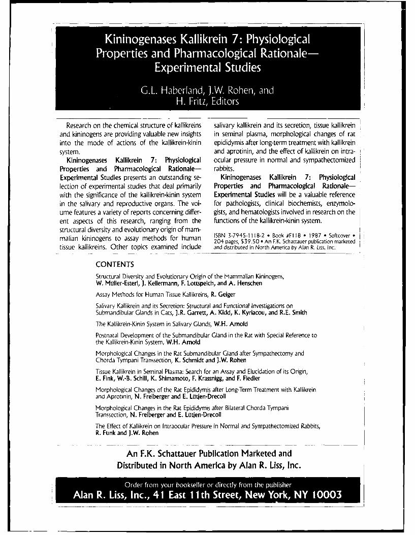
0 a
Expermal Stde
Research on the chemical structure of kallikreins salivary kallikrein and its secretion, tissue kallikreinand kininogens are providing valuable new insights in seminal plasma, morphological changes of ratinto the mode of actions of the kallikrein-kinin epididymis after long-term treatment with kallikreinsystem. and aprotinin, and the effect of kallikrein on intra-
Kininogenases Kallikrein 7: Physiological ocular pressure in normal and sympathectomizedProperties and Pharmacological Rationale- rabbits.Experimental Studies presents an outstanding se- Kininogenases Kallikrein 7: Physiologicallection of experimental studies that deal primarily Properties and Pharmacological Rationale-with the significance of the kallikrein-kinin system Experimental Studies will be a valuable referencein the salivary and reproductive organs. The vol- for pathologists, clinical biochemists, enzymolo-ume features a variety of reports concerning differ- gists, and hematologists involved in research on theent aspects of this research, ranging from the functions of the kallikrein-kinin system.structural diversity and evolutionary origin of mam-malian kininogens to assay methods for human ISBN 3-7945-1118-2 e Book #FI 18 * 1987 * Softcover
204 pages, $39.50 * An F.K. Schattauer publication marketedtissue kallikreins. Other topics examined include and distributed in North America by Alan R. Liss, Inc.
CONTENTS
Structural Diversity and Evolutionary Origin of the Mammalian Kininogens,W. Moller-Esteri, J. Kellermann, F. Lottspeich, and A. Henschen
Assay Methods for Human Tissue Kallikreins, R. Geiger
Salivary Kallikrein and its Secretion: Structural and Functional Investigations onSubmandibular Glands in Cats, J.R. Garrett, A. Kidd, K. Kyriacou, and R.E. Smith
The Kallikrein-Kinin System in Salivary Glands, W.H. Arnold
Postnatal Development of the Submandibular Gland in the Rat with Special Reference tothe Kallikrein-Kinin System, W.H. Arnold
Morphological Changes in the Rat Submandibular Gland after Sympathectomy andChorda Tympani Transsection, K. Schmidt and J.W. Rohen
Tissue Kallikrein in Seminal Plasma: Search for an Assay and Elucidation of its Origin,E. Fink, W.-B. Schill, K. Shimamoto, F. Krassnigg, and F. Fiedler
Morphological Changes of the Rat Epididymis after Long-Term Treatment with Kallikreinand Aprotinin, N. Freiberger and E. Latien-Drecoll
Morphological Changes in the Rat Epididymis after Bilateral Chorda TympaniTranssection, N. Freiberger and E. Latien-Drecoll
The Effect of Kallikrein on Intraocular Pressure in Normal and Sympathectomized Rabbits,R. Funk and J.W. Rohen
An F.K. Schattauer Publication Marketed andDistributed in North America by Alan R. Liss, Inc.
Ala R. Lis n. 1Fs hSreN wY rN 0003
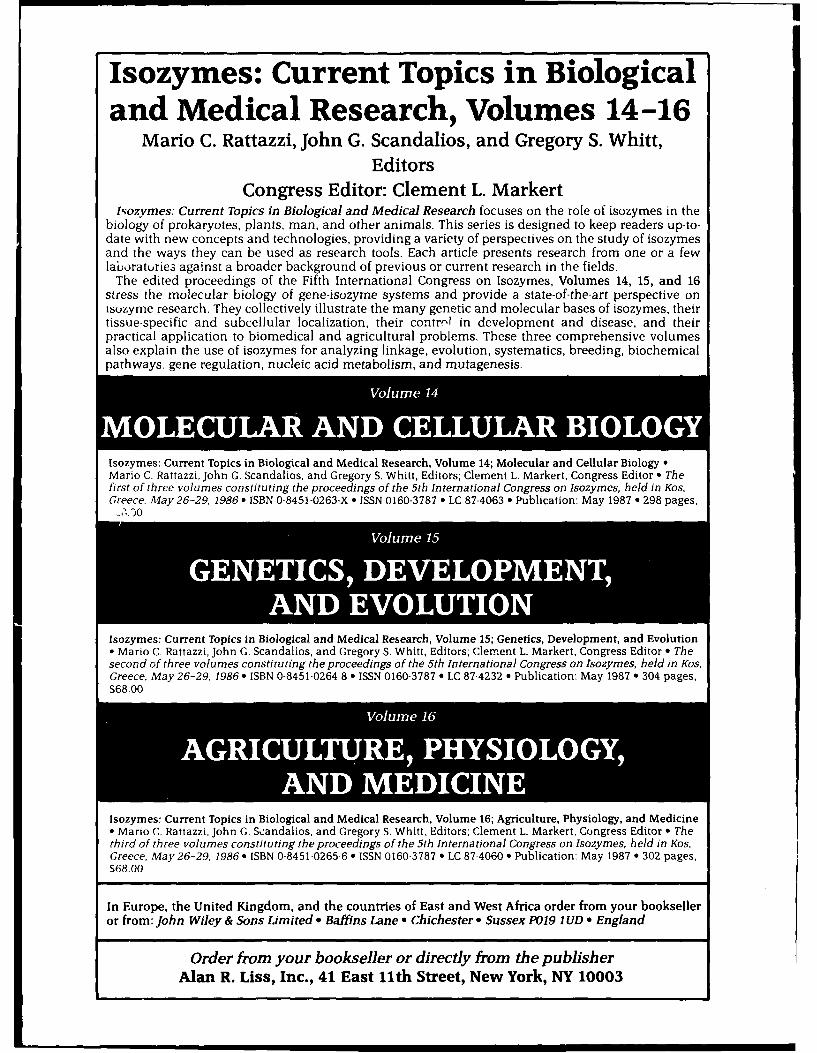
Isozymes: Current Topics in Biologicaland Medical Research, Volumes 14-16
Mario C. Rattazzi, John G. Scandalios, and Gregory S. Whitt,Editors
Congress Editor: Clement L. MarkertlNozymes: Current Topics in Biological and Medical Research focuses on the role of isozymes in the
biology of prokaryotes, plants, man, and other animals. This series is designed to keep readers up-to-date with new concepts and technologies, providing a variety of perspectives on the study of isozymesand the ways they can be used as research tools. Each article presents research from one or a fewlaboraturies against a broader background of previous or current research in the fields.
The edited proceedings of the Fifth International Congress on Isozymes, Volumes 14, 15, and 16stress the molecular biology of gene-isozyme systems and provide a state-of-the-art perspective onisuzyme research. They collectively illustrate the many genetic and molecular bases of isozymes, theirtissue-specific and subcellular localization, their contr-i in development and disease, and theirpractical application to biomedical and agricultural problems. These three comprehensive volumesalso explain the use of isozymes for analyzing linkage, evolution, systematics, breeding, biochemicalpathways. gene regulation, nucleic acid metabolism, and mutagenesis.
Isozymes: Current Topics in Biological and Medical Research, Volume 14; Molecular and Cellular Biology *Mario C. Rattazzi, John G. Scandalios, and Gregory S. Whitt, Editors; Clement L. Markert, Congress Editor 6 Theferst of three volumes constituting the proceedings of the 5th International Congress on Isozymes, held in Kos,Greece, May 26-29, 1986 * ISBN 0-8451-0263-X * ISSN 0160-3787 * LC 87-4063 * Publication: May 1987 * 298 pages,
6,.90
Isozymes: Current Topics in Biological and Medical Research, Volume 15; Genetics, Development, and EvolutionoMario C. Rattazzi, John G. Scandalios, and Gregory S. Whitt, Editors; Clement L. Markert, Congress Editor * Thesecond of three volumes constituting the proceedings of the 5th International Congress on Isozymes, held in Kos,Greece, May 26-29, 1986 * ISBN 0-8451-0264-8 9 ISSN 0160-3787 * LC 87-4232 e Publication: May 1987 * 304 pages,S68.00
Isozymes: Current Topics in Biological and medical Research, Volume 16; Agriculture, Physiology, and Medicine
* Mario C. Rattazzi, John G. Scandalios, and Gregory S. Whitt, Editors; Clement L. Markert, Congress Editor *, Thethird of three volumes constituting the proceedings of the 5th International Congress on isozymes, held in Kos,Greece, May 26-29, 1986 * ISBN 0-8451-0265-6 e, ISSN 0160-3787 e LC 87-4060 e Publication: May 1987 * 302 pages,
S68.00
In Europe, the United Kingdom, and the countries of East and West Africa order from your bookselleror from: John Wiley & Sons Limited * Baffins Lane a Chichester • Sussex P019 IUD 9 England
Order from your bookseller or directly from the publisherAlan R. Liss, Inc., 41 East 11th Street, New York, NY 10003
JOURNAL OFLEUKOCYTE
INFORMATION FOR AUTHORSBIOLOGY , ,oosBIOLOGYRevised, July 1988
Manuscripts should be concise and consistent in style, spell- thors and complete titles. Unpublished observations and per-ing, and use of abbreviations. The original plus two copies of the sonal communications should be cited parenthetically in the textmanuscript should be sent to: and not included with published references. Abbreviations of
Carleton C. Stewart, Editor-in-Chief journal anmes should follow the style of List of Journals IndexedRoswell Park Memorial Institute in Index Medicus with periods. When the names of authors are666 Elm Street used in the text and the number of authors exceeds two, the termBuffalo, NY 14263 et al. should follow the name of the first author. Note the followingThe original manuscript must be typewritten and double-spaced examples:
on one side of good quality white paper, 81/2" x 11", with one- Journal Articlesinch margins. The two copies must closely simulate the original 1. Vray, B., Seint-Gullain, M.. Leloup, R., and Hoebeke, J.in quality. The original manuscript plus both copies must include Kinetic and morphologic studies of rat macrophageoriginal figures. phagocytosis. J. Reticuloendothel. Soc. 29,307, 1981.
The paper should be organized according to the following Bookssections: 2. Fisher, R.A., Statistical Methods for Research. Philadel-
(1) Abstract phia: Lea & Febiger, p. 183, 1980.(2) Introduction; give reasons for doing the work Chapters in Books(3) Materials and Methods 3. Lipton, J.M., and Nathan, D.G. The role of T lymphocytes(4) Results in human erythropoiesis. In The Lymphocyte (Sell, K.W.,(5) Discussion and Miller, M.V., Eds.) New York: Alan R. Liss, p. 57,(6) Acknowledgments 1981.(7) References Short communications in the form of a note are acceptable
Page 1 should contain the title (Roman numerals are not ac- provided they relate to new and important findings. The format tocepted in titles of papers in a series), author(s), and affiliation(s), be used for notes is simply a summary and text without any otherA short running title, not to exceed 50 spaces, should be at the section headings. They should not exceed four printed pages,bottom of the page. In addition, the name, complete mailing including tables and figures (approximately seven to eight dou-address, and telephone number of the person to whom proofs ble-spaced typewritten pages). Notes will be published within 100are to be sent should be included. A list of three to six key words days of acceptance. To expedite the review of notes, which arenot in the title should be provided for indexing purposes. sent to reviewers by Federal Express, a $50.00 handling fee
Page 2 should contain a short abstract approximately 100-150 payable to the Society for Leukocyte Biology must accompanywords in length. the short communication. This charge pertains only to short
Footnotes are to be used only when absolutely necessary. communications.When used, they should be typed consecutively, double-spaced, Letters to the editor may be submitted, but they must beon a separate sheet of paper in order of their appearance in the concise and concerned with pertinent scientific issues to be ac-text. In all instances, arabic numerals should be used as super- ceptable for publication. The Editor-in-Chief reserves the right toscripts for footnotes appearing in the text. determine their acceptability based on the above criteria.
Legends to figures should be typed on a separate page. Label Manuscripts are reviewed by at least two experts in the field;all illustrations with authors' names, figure number, and an arrow the appropriate section editor makes the decision on acceptabil-indicating top. ity. The Editor-in-Chief reserves the right to reject any manuscript
Generally accepted abbreviations should be used whenever that is not in proper form or written in acceptable English whenpossible. No sentence should begin with an abbreviation or ara- first received at the editorial office. The editors reserve the rightbic numeral. Follow CBE Style Manual: Fifth Edition, 1978 (avail- to make minor changes in the text of a manuscript to improve itsable from the Council of Biology Editors, Inc., 9650 Rockville composition, provided the author's meaning or intent has notPike, Bethesda, MD 20814). been changed.
Figures should be numbered with arabic numerals and treated Author's alterations in the page proofs in excess of 5 percentconsecutively in the text. Photographs should be submitted as of the cost of composition will be charged to the author.glossy prints (5" x 7" or 8" x 10"). Micrographs should be planned Reprints may be ordered in multiples of 100. The author willso that they can be reproduced without reduction (63/4" x 83/4" receive an order form for reprints with the page proofs.maximum). Graphs may also be submitted in the form of glossyprints. Alternatively, they may be drawn with black India ink on ALL MANUSCRIPTS submitted to the Journal of Leukocytelight blue coordinate paper with coordinates properly labeled and Biology must be submitted solely to this journal. may not havenecessary grid lines inked in black. Graphs similarly drawn on been published in any part or form in another publication of anywhile paper are also acceptable. enpbihdi n ato or naohrpbiaino n
he cost oe ilston cpublied itype, professional or lay, and become the property of the pub-The cost of illustrations published in color will be charged to lisher. Upon acceptance of a manuscript for publication, the au-the author. A quote will be sent when the illustrations are re- thor will be requested to sign an agreement transferring copyrightceived. These illustrations must be submitted in the form of totepbihrworsrvscyig.Noulsedmeiaglosy clorprins o trnspaencesto the publisher, who reserves copyright. No published materialglossy color prints or transparencies. may be reproduced or published elsewhere without the written
Tables should be numbered consecutively with arabic numer- permission of the publisher and the author. The journal will notals in the order of their appearance in the text. Tables must be be responsible for the loss of manuscripts at any time. All state-typed double-spaced, on separate pages. A short descriptive title ments in, or omissions from, published manuscripts are the re-must be provided. If footnotes are used, they should be labeled sponsibility of the authors, who will assist the editors and thewith lowercase superscript letters. publisher by reviewing proofs before publication. All manuscripts
References should be cited in the text by numbers in brackets. accepted for publication will be subject to a charge of $50 perThe references must be alphabetized according to the surname page for all pages exceeding six printed pages. Charges for theseof the first author and number consecutively under the section pages will be invoiced separately. Authors are expected to makeheading, References. References must include names of all au- cell lines and their products available to qualified investigators.
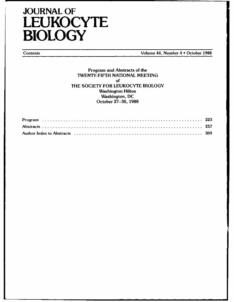
JOURNAL OF
LEUKOCYTEBIOLOGYContents Volume 44, Number 4 *October 1988
Program and Abstracts of theTWENTY-FIFTH NATIONAL MEETING
ofTHE SOCIETY FOR LEUKOCYTE BIOLOGY
Washington HiltonWashington, DC
October 27-30, 1988
Program............................................................... 223
Abstracts .............................................................. 257
Author Index to Abstracts .................................................. 309