Cytokines and chemokines in postovulatory follicle regression of domestic chicken ( Gallus gallus...
12
Available at www.sciencedirect.com journal homepage: www.elsevier.com/locate/devcompimm Cytokines and chemokines in postovulatory follicle regression of domestic chicken (Gallus gallus domesticus) N.R. Sundaresan a,c, ,1 , V.K. Saxena a,1, , K.V.H. Sastry b,1 , K. Nagarajan c , Preeti Jain a , Rani Singh a , D. Anish b,c , P.V. Ravindra c , M. Saxena c , K.A. Ahmed a a Disease Genetics and Biotechnology Laboratory, Central Avian Research Institute, Izatnagar-243122, Bareilly, India b Division of Avian Physiology and Reproduction, Central Avian Research Institute, Izatnagar-243122, Bareilly, India c Indian Veterinary Research Institute, Izatnagar-243122, Bareilly, India Received 1 September 2006; received in revised form 30 May 2007; accepted 30 May 2007 Available online 5 July 2007 KEYWORDS Cytokines; Chemokines; Apoptosis; Immune cells; Postovulatory follicle; Chicken Abstract The mechanism of postovulatory follicle (POF) regression in birds is still poorly understood. In the current study, expression of IL-1b, IL-6, GM-CSF, IFN-g, IL-2, IL-4, IL-13, chCXCLi2, chCCLi2, chCCLi4, chCCLi7, IL-10 and TGF-b2 mRNAs was estimated in regressing POF by semi-quantitative RT-PCR. In addition, the changes in immune cell population, histological and apoptotic changes were also studied in regressing POF. The expression of cytokines (IL-1b, IL-6, IL-10 and TGF-b2) and chemokines (chCXCLi2, chCCLi2, chCCLi4 and chCCLi7) was upregulated in POFs, suggesting a role for these molecules in tissue regression. The histological findings suggested a significant infiltration of immune cells, especially heterophils, lymphocytes and macrophages, into the regressing POF. The flow cytometry analysis of lymphocyte subpopulations revealed that CD3 + , CD4 + , CD8 + and Bu-1 + lymphocytes were significantly increased during this regression. The significant up-regulation of chemokines might have attracted the immune cells during POF regression. The percentage of apoptotic cells was significantly increased during the regression of POF. The up-regulation of IL-1b, IL-6, IL-10 and TGF-b2 and down-regulation of GM-CSF might have induced apoptosis during the POF regression. However, expression of IFN-g, IL-2, IL-4 and IL-13 was not significantly altered during POF regression. In conclusion, cytokines appear to play an important role in the regression of POF in chicken. Furthermore, the regression of chicken POF seems to be an inflammatory event similar to luteolysis of the mammalian corpus luteum. & 2007 Elsevier Ltd. All rights reserved. ARTICLE IN PRESS 0145-305X/$ - see front matter & 2007 Elsevier Ltd. All rights reserved. doi:10.1016/j.dci.2007.05.011 Corresponding authors. Indian Veterinary Research Institute, Izatnagar-243122, Bareilly, India. Tel.: +91 5812310216; fax: +91 581 230 1321. E-mail addresses: [email protected] (N.R. Sundaresan), [email protected] (V.K. Saxena). 1 These authors contributed equally to this work. Developmental and Comparative Immunology (2008) 32, 253–264
-
Upload
independent -
Category
Documents
-
view
0 -
download
0
Transcript of Cytokines and chemokines in postovulatory follicle regression of domestic chicken ( Gallus gallus...
ARTICLE IN PRESS
Available at www.sciencedirect.com
Developmental and Comparative Immunology (2008) 32, 253–264
0145-305X/$ - see frodoi:10.1016/j.dci.20
�Corresponding aufax: +91 581 230 1321
E-mail addresses1These authors co
journal homepage: www.elsevier.com/locate/devcompimm
Cytokines and chemokines in postovulatoryfollicle regression of domestic chicken(Gallus gallus domesticus)
N.R. Sundaresana,c,�,1, V.K. Saxenaa,1,�, K.V.H. Sastryb,1, K. Nagarajanc,Preeti Jaina, Rani Singha, D. Anishb,c, P.V. Ravindrac, M. Saxenac, K.A. Ahmeda
aDisease Genetics and Biotechnology Laboratory, Central Avian Research Institute, Izatnagar-243122, Bareilly, IndiabDivision of Avian Physiology and Reproduction, Central Avian Research Institute, Izatnagar-243122, Bareilly, IndiacIndian Veterinary Research Institute, Izatnagar-243122, Bareilly, India
Received 1 September 2006; received in revised form 30 May 2007; accepted 30 May 2007Available online 5 July 2007
KEYWORDSCytokines;Chemokines;Apoptosis;Immune cells;Postovulatory follicle;Chicken
nt matter & 200707.05.011
thors. Indian Vete.: drravicari@gmail
ntributed equally
AbstractThe mechanism of postovulatory follicle (POF) regression in birds is still poorly understood. Inthe current study, expression of IL-1b, IL-6, GM-CSF, IFN-g, IL-2, IL-4, IL-13, chCXCLi2,chCCLi2, chCCLi4, chCCLi7, IL-10 and TGF-b2 mRNAs was estimated in regressing POF bysemi-quantitative RT-PCR. In addition, the changes in immune cell population, histologicaland apoptotic changes were also studied in regressing POF. The expression of cytokines (IL-1b,IL-6, IL-10 and TGF-b2) and chemokines (chCXCLi2, chCCLi2, chCCLi4 and chCCLi7) wasupregulated in POFs, suggesting a role for these molecules in tissue regression. Thehistological findings suggested a significant infiltration of immune cells, especiallyheterophils, lymphocytes and macrophages, into the regressing POF. The flow cytometryanalysis of lymphocyte subpopulations revealed that CD3+, CD4+, CD8+ and Bu-1+ lymphocyteswere significantly increased during this regression. The significant up-regulation ofchemokines might have attracted the immune cells during POF regression. The percentageof apoptotic cells was significantly increased during the regression of POF. The up-regulationof IL-1b, IL-6, IL-10 and TGF-b2 and down-regulation of GM-CSF might have induced apoptosisduring the POF regression. However, expression of IFN-g, IL-2, IL-4 and IL-13 was notsignificantly altered during POF regression. In conclusion, cytokines appear to play animportant role in the regression of POF in chicken. Furthermore, the regression of chickenPOF seems to be an inflammatory event similar to luteolysis of the mammalian corpus luteum.& 2007 Elsevier Ltd. All rights reserved.
Elsevier Ltd. All rights reserved.
rinary Research Institute, Izatnagar-243122, Bareilly, India. Tel.: +91 581 231 0216;
.com (N.R. Sundaresan), [email protected] (V.K. Saxena).
to this work.

ARTICLE IN PRESS
N.R. Sundaresan et al.254
Healthy single-comb White Leghorn hens at 65 weeks of age
1. IntroductionOvulation is a process of rupture of the largest preovulatoryfollicle wall and the subsequent release of ovum. In birds,the ovulated follicles rapidly terminate the biosynthesis ofprogesterone and fail to form a functional corpus luteum.Instead, the residual postovulatory follicle (POF) begins toregress shortly after ovulation. This regression is almostentirely completed within 4–6 days after ovulation [1]. Theregression of POF following ovulation is important, as itinfluences oviposition and nesting behavior [2]. The regres-sion of chicken POF occurs via the process of caspase-mediated apoptosis [1,3]. During this process, a markedinfiltration of MHC-II(+) cells and macrophages occurs in thePOF [4,5]. These results suggest that the immune systemmight play an important role in the regression of POF [4,5].However, the signals and molecular mediators responsiblefor the regression of POF are unknown [1].
Most of the studies related to tissue regression have beencarried out in mammals. According to recent reviews,regression of reproductive tissue is viewed as an immune-mediated event and cytokines such as IL-1b, IL-6, IFN-g andTNF-a were identified as key mediators [6–9]. Cytokines alsoserve as mediators required for bidirectional communicationbetween the neuro-endocrine and immune systems [10]. Thesecytokines originate either from leukocytes recruited into theovary or from resident ovarian cells, such as granulosa or thecacells [7]. The roles of cytokines in the regression of mammaliancorpus luteum [8,11–13], follicles [14,15] and mammary gland[16] are well defined. However, the role of cytokines in theregression of POF in birds is yet to be established.
Functional studies of chicken cytokines suggest that theyhave similar properties as their mammalian counterparts[17]. In the chicken, inflammation is controlled by IL-1b [18]and IL-6 [19]. The Th1 cytokines include IFN-g [20] and IL-2[21]. The Th2 cytokines include IL-4 and IL-13 [22]. IL-10 [23]and transforming growth factor (TGF)-b2 [24] are anti-inflammatory cytokines. GM-CSF is a colony stimulatingfactor [22]. Chemokines are important mediators of cellmigration during inflammation and in normal leukocytetrafficking [25–27]. In chickens, CXC chemokines includechCXCLi1 [28] and chCXCLi2 [29]. CCL chemokines includechCCLi2 [27,30–32], chCCLi4 [27,30,33] and chCCLi7 [27,33].
Recently, we identified the role of avian cytokines intissue remodeling of the ovary and oviduct during inducedmolting [34]. We hypothesized that the cytokines might playa key role in the regression of POF in the chicken. Therefore,the expression profiles of cytokines and chemokines in theregressing POF of White Leghorn hens were studied.Cytokine transcripts were quantified using semi-quantitativeRT-PCR. Moreover, changes in immune cell populations,histological and apoptotic changes were also studied, toinvestigate the involvement of the immune system duringthe regression of POF.
2. Materials and methods
2.1. Experimental birds
This experiment was performed in accordance with the rulesof the Animal Ethics Monitoring Committee of the Institute.
from the same hatch and laying a regular sequence of atleast 6–10 eggs were used in the current study. All the birdswere maintained under uniform standard managementconditions with 15 h light:9 h dark. Birds were housedindividually in laying cages, provided with free access tofeed and water. Individual laying cycles were monitored bythe daily timing of oviposition.
2.2. Collection of samples
Ten birds laying more than 5 eggs in sequence weresacrificed 1–2 h before expected oviposition based on theindividual bird’s sequence of laying. The ovaries along withthe ovarian follicles were removed immediately and placedon ice. The follicles were separated from the ovary andweighed in order to identify the largest (F1) and post-ovulatory follicles {largest, recent (POF1), second-largest(POF2), third-largest (POF3), fouth-largest (POF4) and fifth-largest (POF5)}. The F1 follicle was cut open transverselyalong the stigma to drain the yolk material completely. Thefollicular membranes were washed with ice-cold sterilesaline, taking care to ensure that there was no adherentyolk material. The F1 follicle and postovulatory follicles(POF1–5) were collected without separating granulosa andtheca layers. All tissue samples were incubated separately inRNA stabilization solution (RNAlater, Ambion Inc., USA) at4 1C overnight. Then, samples were removed from RNAlaterand stored at �80 1C for 1–2 weeks as per the manufac-turer’s instructions, till the RNA isolation was performed.Replicates of follicular tissues were also collected in PBS or10% formalin saline for the analysis of apoptosis orhistological changes.
2.3. Quantification of cytokine mRNA expression
Semi-quantitative RT-PCR was chosen to estimate thetranscripts of cytokine genes. The estimation of mRNAs ofcytokine genes in F1 and POFs was carried out as describedearlier [34].
Total RNA was extracted from individual tissue samplesusing the RNAgents—Total RNA isolation system (Promega,Madison, WI, USA) according to the manufacturer’s instruc-tions. The concentration and purity of RNA preparationswere determined spectrophotometrically at A260 versus A280.Any possible traces of genomic DNA were removed bytreating 5 mg of each RNA sample with 5U of RNase-freeDNase (Biogene, CA, USA) at 37 1C for 1 h. The DNase wassubsequently inactivated by incubation at 65 1C for 10min.Each DNase-treated total RNA sample (2 mg) was reversetranscribed with suitable negative and positive controlsusing the ‘RevertAid First strand cDNA synthesis kit’ (MBIFermentas, Hanover, MD, USA), according to the manufac-turer’s instructions. The resultant cDNA was stored frozen at�20 1C till used. Negative controls were performed using allcomponents except reverse transcriptase. Total RNA fromchicken spleen was used in positive controls and forstandardizing reaction conditions.
PCR reactions were performed with equal amount of cDNAsamples from all birds, in duplicate and in separate tubes forthe amplification of various mRNA with specific primers in a
ARTICLE IN PRESS
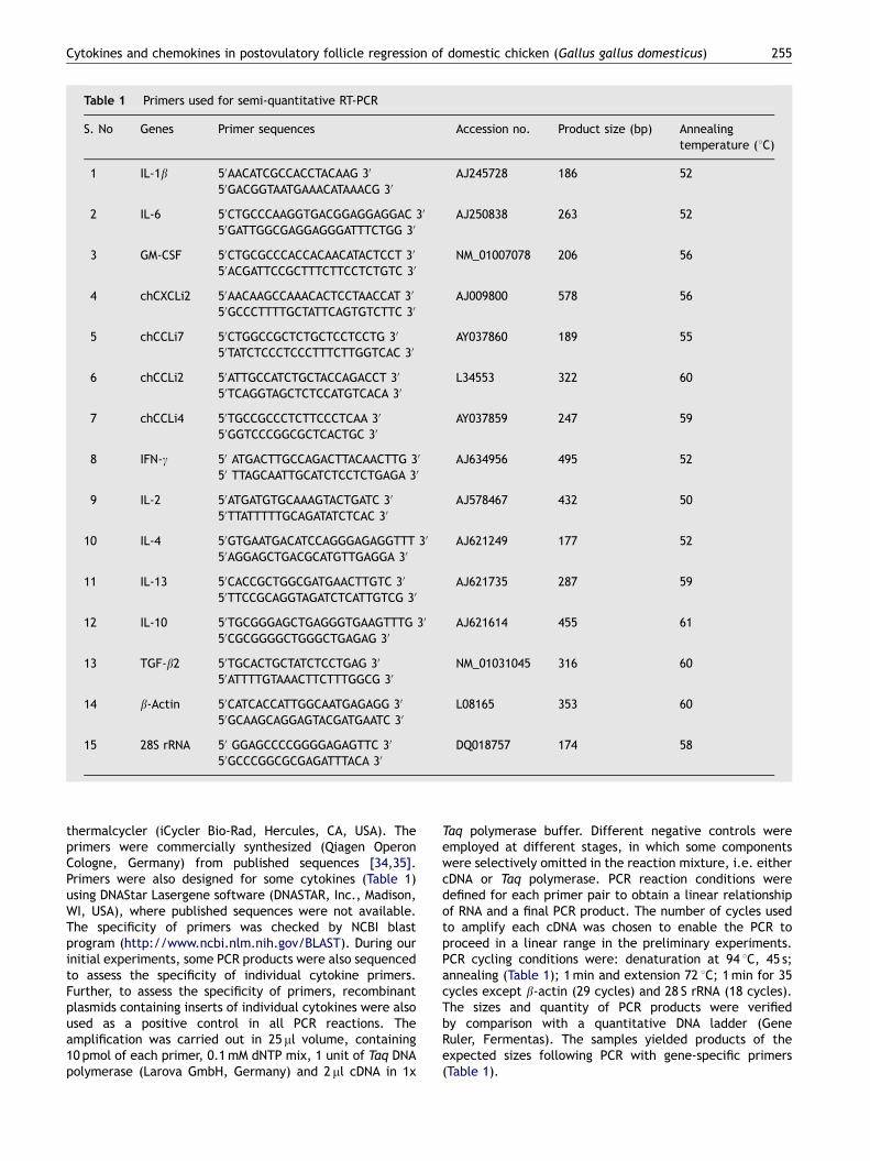
Table 1 Primers used for semi-quantitative RT-PCR
S. No Genes Primer sequences Accession no. Product size (bp) Annealingtemperature (1C)
1 IL-1b 50AACATCGCCACCTACAAG 30 AJ245728 186 5250GACGGTAATGAAACATAAACG 30
2 IL-6 50CTGCCCAAGGTGACGGAGGAGGAC 30 AJ250838 263 5250GATTGGCGAGGAGGGATTTCTGG 30
3 GM-CSF 50CTGCGCCCACCACAACATACTCCT 30 NM_01007078 206 5650ACGATTCCGCTTTCTTCCTCTGTC 30
4 chCXCLi2 50AACAAGCCAAACACTCCTAACCAT 30 AJ009800 578 5650GCCCTTTTGCTATTCAGTGTCTTC 30
5 chCCLi7 50CTGGCCGCTCTGCTCCTCCTG 30 AY037860 189 5550TATCTCCCTCCCTTTCTTGGTCAC 30
6 chCCLi2 50ATTGCCATCTGCTACCAGACCT 30 L34553 322 6050TCAGGTAGCTCTCCATGTCACA 30
7 chCCLi4 50TGCCGCCCTCTTCCCTCAA 30 AY037859 247 5950GGTCCCGGCGCTCACTGC 30
8 IFN-g 50 ATGACTTGCCAGACTTACAACTTG 30 AJ634956 495 5250 TTAGCAATTGCATCTCCTCTGAGA 30
9 IL-2 50ATGATGTGCAAAGTACTGATC 30 AJ578467 432 5050TTATTTTTGCAGATATCTCAC 30
10 IL-4 50GTGAATGACATCCAGGGAGAGGTTT 30 AJ621249 177 5250AGGAGCTGACGCATGTTGAGGA 30
11 IL-13 50CACCGCTGGCGATGAACTTGTC 30 AJ621735 287 5950TTCCGCAGGTAGATCTCATTGTCG 30
12 IL-10 50TGCGGGAGCTGAGGGTGAAGTTTG 30 AJ621614 455 6150CGCGGGGCTGGGCTGAGAG 30
13 TGF-b2 50TGCACTGCTATCTCCTGAG 30 NM_01031045 316 6050ATTTTGTAAACTTCTTTGGCG 30
14 b-Actin 50CATCACCATTGGCAATGAGAGG 30 L08165 353 6050GCAAGCAGGAGTACGATGAATC 30
15 28S rRNA 50 GGAGCCCCGGGGAGAGTTC 30 DQ018757 174 5850GCCCGGCGCGAGATTTACA 30
Cytokines and chemokines in postovulatory follicle regression of domestic chicken (Gallus gallus domesticus) 255
thermalcycler (iCycler Bio-Rad, Hercules, CA, USA). Theprimers were commercially synthesized (Qiagen OperonCologne, Germany) from published sequences [34,35].Primers were also designed for some cytokines (Table 1)using DNAStar Lasergene software (DNASTAR, Inc., Madison,WI, USA), where published sequences were not available.The specificity of primers was checked by NCBI blastprogram (http://www.ncbi.nlm.nih.gov/BLAST). During ourinitial experiments, some PCR products were also sequencedto assess the specificity of individual cytokine primers.Further, to assess the specificity of primers, recombinantplasmids containing inserts of individual cytokines were alsoused as a positive control in all PCR reactions. Theamplification was carried out in 25 ml volume, containing10 pmol of each primer, 0.1mM dNTP mix, 1 unit of Taq DNApolymerase (Larova GmbH, Germany) and 2ml cDNA in 1x
Taq polymerase buffer. Different negative controls wereemployed at different stages, in which some componentswere selectively omitted in the reaction mixture, i.e. eithercDNA or Taq polymerase. PCR reaction conditions weredefined for each primer pair to obtain a linear relationshipof RNA and a final PCR product. The number of cycles usedto amplify each cDNA was chosen to enable the PCR toproceed in a linear range in the preliminary experiments.PCR cycling conditions were: denaturation at 94 1C, 45 s;annealing (Table 1); 1min and extension 72 1C; 1min for 35cycles except b-actin (29 cycles) and 28 S rRNA (18 cycles).The sizes and quantity of PCR products were verifiedby comparison with a quantitative DNA ladder (GeneRuler, Fermentas). The samples yielded products of theexpected sizes following PCR with gene-specific primers(Table 1).

ARTICLE IN PRESS
N.R. Sundaresan et al.256
Our studies in chicken reproductive regression [34] andearlier work [35] found b-actin to be a suitable house-keeping gene during reproductive regression. However, wetested another house-keeping gene, 28S rRNA [30] in theregressing reproductive tissues. We have used SYBR green-based real-time PCR for quantifying the transcripts ofb-actin and 28S rRNA in various POFs. The delta Ct valuesfor b-actin and 28S rRNA are provided in the SupplementaryFile. The results suggested that there are no significantdifferences in b-actin and 28S rRNA expression levels duringthe regression of POF. Therefore, in the current study,b-actin was used for normalization. Relative expression wasdetermined as arbitrary units, defined as the ratio of mRNAlevel to the corresponding b-actin mRNA level aftersubtraction of background intensity [value ¼ (intensity;gene of interest—intensity; background)/(intensity; b-acti-n—intensity; background)]. Mean values of three measure-ments of each band were taken for analysis.
2.4. Quantification of apoptosis
The percentage of apoptosis during POF regression wasquantified using standard protocols [36]. Briefly, tissuesamples were trypsinized as per standard protocols to geta single-cell suspension. Cells were harvested in PBS andpelleted by centrifugation at 3000g for 10min. Cells wereresuspended in PBS containing 0.1% glucose and washedtwice using the same procedure. The supernatants of eachstep were carefully discarded without disturbing the pellet.One milliliter of ice-cold ethanol was slowly added to thecells with intermittent vortexing and allowed to fix for about18 h. Just before addition of propidium iodide solution, thefixed cells were vortexed and centrifuged at 3000g for 5min.The supernatant was discarded and 0.5ml of propidiumiodide staining solution (PBS containing 50 mg/ml of propi-dium iodide, 20 mg/ml RNase A and 0.1% glucose) was addedto each tube, vortexed and incubated for at least 30min atroom temperature. Presence or absence of apoptosis wasdetermined by the quantification of DNA hypodiploidy usingflow cytometry (BD FACSCalibur System, BD Biosciences, CA,USA). Data acquisition and analysis were performed usingCell Quest software (BD Biosciences, USA).
2.5. Analysis of DNA fragmentation in agarose gels
Apoptotic DNA was extracted from tissue samples asdescribed earlier [37], with some modifications. Tissuesamples were trypsinized as per standard protocols to geta single-cell suspension. Cells were harvested in PBS andpelleted by centrifugation at 4000 RPM for 10min. The cellpellet was treated with 500ml lysis buffer (20mM Tris–HClat pH 7.4, 10mM EDTA and 0.2% Triton X-100) and vortexedfor 10 s. The supernatant was collected by centrifuging at10,000g for 10min. The pellet was again treated with thelysis buffer as above. The supernatants of both steps werepooled and treated with 1% SDS and RNase A (1 mg/ml) andincubated at 56 1C for 2 h. The samples were then digestedwith 100 mg/ml proteinase-K for a minimum of 2 h at 37 1C.Half the volume of 10M ammonium acetate was added tothe sample and DNA precipitated with 2.5 volumes ofethanol at �20 1C overnight. The DNA was pelleted, dried
and dissolved in nuclease-free water for electrophoresis in2% agarose gels containing 1mg/ml ethidium bromide.Agarose gels were visualized in UV using a gel documentationsystem (Syngene, UK).
2.6. Histological examination
Samples collected earlier (Section 2.2) from both F1 andPOFs were cut into small pieces, dehydrated through agraded series of alcohol to xylol and embedded in paraffinwax. Wax-embedded sections (4 mm) were cut and stainedwith hematoxylin and eosin for the histological study. Thesesections were examined for histological atretic and apopto-tic changes as described earlier [38]. The same tissuesections were also examined microscopically using an oil-immersion lens at � l000 magnification for the immune cells[34]. Total numbers of heterophils, macrophages andlymphocytes were counted in 100 fields in each sectionfrom F1 and POFs of each bird. The average value wascalculated per tissue for F1 and POFs.
2.7. Flow cytometric analysis of lymphocytesubpopulations in POF during regression
Alterations in the lymphocyte subpopulations during theregression of POF were investigated as described earlier[39]. Three single comb White Leghorn hens were used forcollection of follicles as described in Section 2.2. Single-cellsuspensions of POFs were prepared as described in Section2.4. The cells were incubated overnight at 4 1C with eachmouse monoclonal antibody specific for chicken T lympho-cytes (CD3, CD4 and CD8) and B lymphocytes (Bu-1)(Serotec, USA) or isotypic controls (Santa Cruz Biotechnol-ogy, Santa Cruz, CA) from the same species (mouse). Themonoclonal antibodies specific for chicken CD4, CD8 and Bu-1a were RPE-labeled and CD3 was unlabeled. For CD4, CD8and Bu-1, following incubation, the cells were washed thricewith PBS and the fluorescence was measured in the FL-2filter of a flow cytometer (BD FACSCalibur System, BDBiosciences) with 1� 104 viable cells. For CD3, followingincubation, the cells were washed thrice with PBS, againincubated with FITC-labeled goat anti-mouse IgG secondaryantibody (Sigma) for 1 h on ice. After incubation, the cellswere washed three times with PBS and the fluorescencewas measured using the FL-1 filter with 1� 104 viable cells.Data acquisition and analysis were performed using CellQuest software (BD Biosciences, USA) as described pre-viously [39].
2.8. Statistical analysis
Analysis of normalized cytokine values, apoptosis percen-tage, number of immune cells and percentage of lympho-cyte subpopulations were carried out using one-way analysisof variance with Dunnett’s post-test using GraphPad Prismversion 5.00 for Windows, GraphPad Software, San Diego,California, USA, www.graphpad.com.

ARTICLE IN PRESS
Cytokines and chemokines in postovulatory follicle regression of domestic chicken (Gallus gallus domesticus) 257
3. Results
3.1. Cytokine mRNA expression duringpostovulatory follicle regression
Expressions of IL-1b, IL-6, GM-CSF, chCXCLi2, chCCLi2,chCCLi4, chCCLi7, IFN-g, IL-4, IL-13, IL-10 and TGF-b2mRNAs were observed in the largest preovulatory follicle(Fig. 1). However, IL-2 transcripts were not detected in anyof the samples tested (Fig. 1).
When compared with F1, a rapid increase in IL-1b and IL-6mRNA expression levels were noticed in POF1. The expres-sion levels was significantly higher up to POF5. However, theexpression of GM-CSF mRNA was downregulated significantlyduring the regression of POF (Fig. 2A). chCXCLi2 mRNAexpression was upregulated from POF1 to POF4, during theregression of POF. chCCLi7 expression was upregulated manyfolds during the POF regression. The peak expression ofchCCLi7 transcripts was noticed in POF4. The expressionof chCCLi4 mRNA was also upregulated during the regressionof the POF. The peak expression of chCCLi4 was noticed inPOF3, and was downregulated thereafter. Chicken chCCLi2mRNA expression was also upregulated during the regressionof POF (Fig. 2B). A faint expression of IFN-g was observed inF1 as well as in POFs. However, the IFN-g expression levelsrevealed no significant changes during the regression of POF.The mRNA expressions of IL-4 and IL-13 exhibited nosignificant changes during POF regression (Fig. 2C). How-ever, TGF-b2 mRNA expression was significantly upregulatedin POF1–5. The expression of IL-10 mRNA was significantlyupregulated in the initial stages of POF (POF1–2) regression(Fig. 2D).
3.2. Apoptosis during postovulatory follicleregression
In the largest preovulatory follicle, DNA fragmentation wasabsent, whereas the regressing POFs exhibited the typicalapoptotic DNA ladder pattern (Fig. 3A). The percentage ofapoptosis in the various POFs is depicted in Fig. 3B. Therewas a gradual significant increase in apoptosis during theregression of POF. Histological examination also revealed
Fig. 1 RT-PCR analysis of cytokine mRNA expression in thelargest preovulatory follicle (lane 1; F1) and postovulatoryfollicles (lane 2–6; POF1–5) in the chicken. Photographs show arepresentative example of the PCR product of cytokines orhouse-keeping genes.
the presence of apoptotic cells with fragmented nuclei andapoptotic bodies (Fig. 4F).
3.3. Histological changes
Rapid loss of granulosa cells was noticed immediately afterovulation, followed by the loss of thecal cells at a slowerrate. The granulosa layer of the POF denuded from the thecalayer and completely regressed on POF3. The theca layerexhibited vacuolar degenerative changes from POF3. OnPOF5, regression was almost complete in the theca layer.Further, we also noticed increased vascularization in POF3–5(Fig. 4).
Immune cells (macrophages, lymphocytes and hetero-phils) were localized in the theca layer of the F1preovulatory follicle, whereas they were found in both thetheca and granulosa layers in POFs (Fig. 4). The frequency ofimmune cells in POFs (POF1 and POF2) was significantlygreater than in the F1 follicle. The frequency of immunecells decreased significantly in the POFs (POF4–5) ascompared with POF1 (Fig. 5). In the regressing POFs,macrophages with multiple nuclei were observed, suggest-ing phagocytosis of apoptotic cells. Therefore, theseobservations suggest that macrophages might be involvedin the clearance of apoptotic cells during POF regression(Fig. 4E).
3.4. Alterations of lymphocyte subpopulationsduring the regression of POF
Alterations of lymphocyte subpopulations during the re-gression of POF were investigated to better understandthe nature of lymphocytes involved in the chicken POFregression. The percentages of CD3+, CD4+, CD8+ and Bu-1+
cells were significantly increased during the regression(Fig. 6).
4. Discussion
In mammals, cytokines are involved in the remodeling ofreproductive tissues such as the endometrium, corpusluteum, follicles and mammary gland. Based on mammalianreproductive regression, we chose a group of cytokines toinvestigate the regression of the POF. Cytokine expressionlevels were compared between F1 and POFs to analyze theirrole in regression of POF. In the current study, we used onlythe F1 follicle as control, but not the F3, because nosignificant differences in immune cell numbers were noticedbetween F1 and F3 [4,5]. In the current study, most of thecytokines except IL-2 were found to be expressed constitu-tively in the F1 follicle. The constituent expression ofcytokines contributes to the maintenance of physiologicalhomeostasis as well as to the innate immune system of thefemale reproductive tract of mammals [40–44]. However,their exact role needs to be elucidated in birds.
Cytokines (IL-1b and IL-6) were upregulated during POFregression. In mammals, proinflammatory cytokines activatematrix metalloproteinases such as collagenase, gelatinase,proteinases and proteoglycans to degrade the connectivetissue matrix enzymatically [45–53]. In the chicken, metal-loproteinases are the chief enzymes involved in ovary and

ARTICLE IN PRESS
Fig. 2 RT-PCR analysis of cytokine mRNA expression in the largest preovulatory follicle (F1) and postovulatory follicles (POF1–5). (A)IL-1b, IL-6 and GM-CSF, (B) chCXCLi2, chCCLi2, chCCLi4 and chCCLi7, (C) IFN-g, IL-2, IL-4 and Il-13, and (D) IL-10 and TGF-b2. Graphicsrepresent the mean of the normalized O.D. for each mRNA band obtained by densitometric analysis (mean7S.E; n ¼ 10).Normalization was done by dividing each O.D. value of an individual cytokine band by the value of the b-actin band for the samesample. *Significant differences between F1 and POF1-5 (Po0.05).
N.R. Sundaresan et al.258
oviduct regression [54]. In the present study, increasedexpression of IL-1b and IL-6 might have activated theseenzymes for regression of the POF.
In the present study, we noticed increased levels ofapoptosis during regression of the POF. These findingsconfirm earlier work [3]. In mammals, IL-1b and IL-6

ARTICLE IN PRESS
Fig. 3 Analysis of apoptosis during the regression of postovulatory follicles. (A) Ladder pattern of apoptotic DNA isolated from F1and POF1–5. (B) Percentage of cells showing apoptosis during chicken postovulatory follicle regression. There was a significantincrease of apoptosis during the regression of POF. *Significant differences between F1 and POF1–5 (Po0.05).
Cytokines and chemokines in postovulatory follicle regression of domestic chicken (Gallus gallus domesticus) 259
regulate tissue regression via apoptosis [8,55,56]. Increasedexpression of these cytokines in the POFs again suggest arole in apoptosis.
Earlier studies observed a sudden decline in the proges-terone content of POF [57]. In mammals, IL-1b and IL-6 werefound to suppress ovarian steroidogenesis by inhibiting thegonadotropin support [58–64]. These findings suggest thatthe cytokines released in the POFs might have decreasedprogesterone to induce apoptosis.
During regression of the POF, chemokines were upregu-lated. In mammals, chemokines attract neutrophils, mono-cytes, natural killer cells, eosinophils and dendritic cellsinto the inflamed tissue [65–69]. In the chicken, in general,the C and CC chemokines are involved in the recruitment ofmacrophages and lymphocytes, whereas CXC chemokinesparticipate in the recruitment of heterophils to sites ofinflammation [39]. In the present study, we also noticed
recruitment of immune cells, i.e. macrophages, heterophils,and CD3+, CD4+, CD8+ and Bu-1+ lymphocytes duringregression of the POF. Therefore, the upregulated chemo-kines might have attracted the immune cells and they mayplay a key role in the initiation of inflammation, for POFregression.
In mammals, ovarian macrophages and mast cells inaddition to theca-interstitial cells synthesize and releaseGM-CSF during ovarian cycles [70]. GM-CSF plays a sig-nificant role in the follicular development of mammals[71]. Further, GM-CSF inhibits the apoptosis of neutro-phils [72]. In the present study, we observed significantdown-regulation of GM-CSF mRNA during the regressionof POF. Therefore, we assume that the down-regulationof GM-CSF might be related to the induction of apo-ptosis of follicular granulosa and theca cells during POFregression.

ARTICLE IN PRESS
Fig. 4 Histological changes during the regression of postovulatory follicles. (A) F1 follicle showing immune cells. (B) POF1 showingpresence of immune cells in the theca layer. (C) POF3 showing presence of immune cells with apoptotic nucleus in the theca layer(arrow head). (D) POF3 theca layer showing the presence of macrophages. (E) POF3 theca layer showing multinucleatedmacrophages. (F) POF5 showing an apoptotic cell with fragmented nuclei and absence of any immune cell.
N.R. Sundaresan et al.260
In the present study, we also noticed up-regulation ofTGF-b2 mRNA during the regression of POF. In mammals,thecal cells are eliminated from atretic follicles by an activeand strictly regulated process, involving the combinedactions of TGF-a and TGF-b [73,74]. Therefore, we presumethat TGF-b2 might be involved in regression of the thecallayer of the POF in the chicken. However, the expression ofTGF-b is regulated post-transcriptionally [75]. In the presentinvestigation, we quantified only the mRNA levels of TGF-b2.
Therefore, further studies are required to confirm the roleof TGF-b in the regression of POF.
In mammals, IL-10 regulates the clearance of apoptoticcells by macrophages [76,77]. In the current study, weobserved up-regulation of IL-10 mRNA in POFs and involve-ment of macrophages in the clearance of apoptotic cellsduring the regression of POF. Therefore, we assume that theexpression of IL-10 mRNA during POF regression might berelated to the clearance of apoptotic cells by macrophages.

ARTICLE IN PRESS
Fig. 6 Subpopulations of lymphocytes during the regression of chflow cytometry using mAb against the following surface markers: (Apositive cells are given in the figures. *Significant differences betw
Fig. 5 Frequency of immune cells in the regressing postovu-latory follicles of chicken. Graphics represent the mean numberof cells (macrophages–gray bars; lymphocytes–white bars andheterophils–black bars) at different days of regression(mean7S.E.; n ¼ 10). *Significant differences between F1 andPOF1–5 (Po0.05).
Cytokines and chemokines in postovulatory follicle regression of domestic chicken (Gallus gallus domesticus) 261
In the present investigation, we found no significantchanges in expression of Th2 (IL-4 and IL-13) as well as Th1(IFN-g and IL-2) cytokines during the regression of POF. Inmammals, IFN-g plays a significant role in the regression ofreproductive tissues [8,56]. Recently, we also observedhigher levels of IFN-g mRNA expression during reproductiveremodeling of the chicken ovary and oviduct [34]. Further, inmammals, IL-4 promoted progesterone production in cul-tured luteal cells [78]. However, results of the current studysuggest that there is no significant role for IFN-g or IL-4 inchicken POF regression.
In conclusion, cytokines appear to play an important rolein the regression of POF in chicken. Moreover, the regressionof chicken POF seems to be an inflammatory event similar toluteolysis of the mammalian corpus luteum.
icken POF (mean7S.E.; n ¼ 3). Lymphocytes were analyzed by) CD3, (B) CD4, (C) CD8 and (D) Bu-1. The percentages of mAbeen F1 and POF1–5 (Po0.05).
ARTICLE IN PRESS
N.R. Sundaresan et al.262
Appendix A. Supplementary materials
Supplementary data associated with this article can befound in the online version at doi:10.1016/j.dci.2007.05.011
References
[1] Johnson AL, Bridgham JT. Caspase-mediated apoptosis in thevertebrate ovary. Reproduction 2002;124:19–27.
[2] Gilbert AB, Davidson MF, Wells JW. Role of the granulosa cellsof the postovulatory follicle of the domestic fowl in oviposi-tion. J Reprod Fertil 1978;52:227–9.
[3] Tilly JL, Kowalski KI, Johnson AL, Hsueh AJ. Involvement ofapoptosis in ovarian follicular atresia and postovulatoryregression. Endocrinology 1991;129:2799–801.
[4] Barua A, Yoshimura Y, Tamura T. Localization of macrophages inthe ovarian follicles during the follicular growth and post-ovulatory regression in chicken, Gallus domesticus. Poult Sci1998;77:1417–21.
[5] Barua A, Michiue H, Yoshimura Y. Changes in the localization ofMHC class II positive cells in hen ovarian follicles during theprocesses of follicular growth, postovulatory regression andatresia. Reproduction 2001;121:953–7.
[6] Pate JL. Involvement of immune cells in regulation of ovarianfunction. J Reprod Fertil Suppl 1995;49:365–77.
[7] Bukulmez O, Arici A. Leukocytes in ovarian function. HumReprod Update 2000;6:1–15.
[8] Neuvians TP, Schams D, Berisha B, Pfaffl MW. Involvement ofpro-inflammatory cytokines, mediators of inflammation, andbasic fibroblast growth factor in prostaglandin F2alpha-inducedluteolysis in bovine corpus luteum. Biol Reprod 2004;70:473–80.
[9] Tanikawa M, Acosta TJ, Fukui T, Murakami S, Korzekwa A,Skarzynski DJ, et al. Regulation of prostaglandin synthesisby interleukin-1alpha in bovine endometrium during theestrous cycle. Prostaglandins Other Lipid Mediat 2005;78:279–90.
[10] Felten SY, Madden KS, Bellinger DL, Kruszewska B, MoynihanJA, Felten DL. The role of the sympathetic nervous system inthe modulation of immune responses. Adv Pharmacol 1998;42:583–7.
[11] Petroff MG, Petroff BK, Pate JL. Expression of cytokinemessenger ribonucleic acids in the bovine corpus luteum.Endocrinology 1999;140:1018–21.
[12] Penny LA, Armstrong D, Bramley TA, Webb R, Collins RA,Watson ED. Immune cells and cytokine production in the bovinecorpus luteum throughout the oestrous cycle and after inducedluteolysis. J Reprod Fertil 1999;115:87–96.
[13] Krusche CA, Vloet TD, Herrler A, Black S, Beier HM. Functionaland structural regression of the rabbit corpus luteum isassociated with altered luteal immune cell phenotypes andcytokine expression patterns. Histochem Cell Biol 2002;118:479–89.
[14] Basini G, Mainardi GL, Bussolati S, Tamanini C. Steroidogenesis,proliferation and apoptosis in bovine granulosa cells: role oftumour necrosis factor-alpha and its possible signallingmechanisms. Reprod Fertil Dev 2002;14:141–50.
[15] Wu R, Van der Hoek KH, Ryan NK, Norman RJ, Robker RL.Macrophage contributions to ovarian function. Hum ReprodUpdate 2004;10:119–33.
[16] Clarkson RW, Wayland MT, Lee J, Freeman T, Watson CJ. Geneexpression profiling of mammary gland development revealsputative roles for death receptors and immune mediatorsin post-lactational regression. Breast Cancer Res 2004;6:R92–R109.
[17] Cheeseman JH, Kaiser MG, Ciraci C, Kaiser P, Lamont SJ. Breedeffect on early cytokine mRNA expression in spleen and cecumof chickens with and without Salmonella enteritidis infection.Dev Comp Immunol 2007;31:52–60.
[18] Weining KC, Sick C, Kaspers B, Staeheli P. A chicken homolog ofmammalian interleukin-1 beta: cDNA cloning and purificationof active recombinant protein. Eur J Biochem 1998;258:994–1000.
[19] Schneider K, Klaas R, Kaspers B, Staeheli P. Chicken inter-leukin-6. cDNA structure and biological properties. Eur JBiochem 2001;268:4200–6.
[20] Digby MR, Lowenthal JW. Cloning and expression of the chickeninterferon-gamma gene. J Interferon Cytokine Res 1995;15:939–45 [Sick et al., 1996].
[21] Sundick RS, Gill-Dixon C. A cloned chicken lymphokinehomologous to both mammalian IL-2 and IL-15. J Immunol1997;159:720–5.
[22] Avery S, Rothwell L, Degen WD, Schijns VE, Young J, KaufmanJ, et al. Characterization of the first nonmammalian T2cytokine gene cluster: the cluster contains functional single-copy genes for IL-3, IL-4, IL-13, and GM-CSF, a gene for IL-5that appears to be a pseudogene, and a gene encoding anothercytokine like transcript, KK34. J Interferon Cytokine Res2004;24:600–10.
[23] Rothwell L, Young JR, Zoorob R, Whittaker CA, Hesketh P,Archer A, et al. Cloning and characterization of chicken IL-10and its role in the immune response to Eimeria maxima.J Immunol 2004;173:2675–82.
[24] Burt DW, Paton IR. Molecular cloning and primary structure ofthe chicken transforming growth factor-beta 2 gene. DNA CellBiol 1991;10:723–34.
[25] DeVries ME, Kelvin AA, Xu L, Ran L, Robinson J, Kelvin DJ.Defining the origins and evolution of the chemokine/chemo-kine receptor system. J Immunol 2006;176:401–15.
[26] Wang J, Adelson DL, Yilmaz A, Sze SH, Jin Y, Zhu JJ. Genomicorganization, annotation, and ligand-receptor inferences ofchicken chemokines and chemokine receptor genes based oncomparative genomics. BMC Genomics 2005;6:45.
[27] Kaiser P, Poh TY, Rothwell L, Avery S, Balu S, Pathania US, et al.A genomic analysis of chicken cytokines and chemokines.J Interferon Cytokine Res 2005;25:467–84.
[28] Sick C, Schneider K, Staeheli P, Weining KC. Novel chicken CXCand CC chemokines. Cytokine 2000;12:181–6.
[29] Kaiser P, Hughes S, Bumstead N. The chicken 9E3/CEF4 CXCchemokine is the avian orthologue of IL8 and maps tochicken chromosome 4 syntenic with genes flanking themammalian chemokine cluster. Immunogenetics 1999;49:673–84.
[30] Hughes S, Poh TY, Bumstead N, Kaiser P. Re-evaluation of thechicken MIP family of chemokines and their receptors suggeststhat CCL5 is the prototypic MIP family chemokine, and thatdifferent species have developed different repertoires of boththe CC chemokines and their receptors. Dev Comp Immunol2007;31:72–86.
[31] Petrenko O, Ischenko I, Enrietto PJ. Isolation of a cDNAencoding a novel chicken chemokine homologous to mamma-lian macrophage inflammatory protein-1 beta. Gene 1995;160:305–6.
[32] Hughes S, Bumstead N. Mapping of the gene encoding a chickenhomologue of the mammalian chemokine SCYA4. Anim Genet1999;30:404.
[33] Hughes S, Haynes A, O’Regan M, Bumstead N. Identification,mapping, and phylogenetic analysis of three novel chicken CCchemokines. Immunogenetics 2001;53:674–83.
[34] Sundaresan NR, Anish D, Sastry KVH, Saxena VK,Mohan J, Ahmed KA. Cytokines in reproductive remodelingof molting White Leghorn hens. J Reprod Immunol 2007;73:39–50.

ARTICLE IN PRESS
Cytokines and chemokines in postovulatory follicle regression of domestic chicken (Gallus gallus domesticus) 263
[35] Monroe DG, Berger RR, Sanders MM. Tissue-protective effectsof estrogen involve regulation of caspase gene expression. MolEndocrinol 2002;16:1322–31.
[36] Coligan JE, Kruisbeck AM, Margulies DH, Shevach EM, StroberW. Current protocols in immunology. New York, NY: GreenePublishing and Wiley Interscience; 1991.
[37] Herrmann M, Lorenz HM, Voll R, Grunke M, Woith W, Kalden JR.A rapid and simple method for the isolation of apoptotic DNAfragments. Nucleic Acids Res 1994;22:5506–7.
[38] Kerr JF, Wyllie AH, Currie AR. Apoptosis: a basic biologicalphenomenon with wide-ranging implications in tissue kinetics.Br J Cancer 1972;26:239–57.
[39] Hong YH, Lillehoj HS, Lillehoj EP, Lee SH. Changes in immune-related gene expression and intestinal lymphocyte subpopula-tions following Eimeria maxima infection of chickens. VetImmunol Immunopathol 2006;114:259–72.
[40] Rutanen EM. Cytokines in reproduction. Ann Med 1993;25:343–7.
[41] Vinatier D, Dufour P, Tordjeman-Rizzi N, Prolongeau JF, Depret-Moser S, Monnier JC. Immunological aspects of ovarianfunction: role of the cytokines. Eur J Obstet Gynecol ReprodBiol 1995;63:155–68.
[42] Kayisli UA, Mahutte NG, Arici A. Uterine chemokines inreproductive physiology and pathology. Am J Reprod Immunol2002;47:213–21.
[43] Wira CR, Fahey JV. The innate immune system: gatekeeperto the female reproductive tract. Immunology 2004;111:13–5.
[44] Dimitriadis E, White CA, Jones RL, Salamonsen LA. Cytokines,chemokines and growth factors in endometrium related toimplantation. Hum Reprod Update 2005;11:613–30.
[45] Conquer JA, Kandel RA, Cruz TF. Interleukin 1 and phorbol 12-myristate 13-acetate induce collagenase and PGE2 productionthrough a PKC-independent mechanism in chondrocytes.Biochim Biophys Acta 1992;1134:1–6.
[46] Kandel RA, Petelycky M, Dinarello CA, Minden M, Pritzker KP,Cruz TF. Comparison of the effect of interleukin 6 andinterleukin 1 on collagenase and proteoglycan production bychondrocytes. J Rheumatol 1990;17:953–7.
[47] Kandel RA, Pritzker KP, Mills GB, Cruz TF. Fetal bovine seruminhibits chondrocyte collagenase production: interleukin 1reverses this effect. Biochim Biophys Acta 1990;1053:130–4.
[48] Lo YY, Conquer JA, Grinstein S, Cruz TF. Interleukin-1 betainduction of c-fos and collagenase expression in articularchondrocytes: involvement of reactive oxygen species. J CellBiochem 1991;69:19–29.
[49] Baici A, Lang A. Effect of interleukin-1 beta on the productionof cathepsin B by rabbit articular chondrocytes. FEBS Lett1990;277:93–6.
[50] Lotz M, Guerne PA. Interleukin-6 induces the synthesis of tissueinhibitor of metalloproteinases-1/erythroid potentiating activ-ity (TIMP-1/EPA). J Biol Chem 1991;266:2017–20.
[51] Shingu M, Nagai Y, Isayama T, Naono T, Nobunaga M, Nagai Y.The effects of cytokines on metalloproteinase inhibitors (TIMP)and collagenase production by human chondrocytes and TIMPproduction by synovial cells and endothelial cells. Clin ExpImmunol 1993;94:145–9.
[52] Fernandes JC, Martel-Pelletier J, Pelletier JP. The role ofcytokines in osteoarthritis pathophysiology. Biorheology2002;39:237–46.
[53] Nishimura R, Bowolaksono A, Acosta TJ, Murakami S, Pio-trowska K, Skarzynski DJ, et al. Possible role of interleukin-1 inthe regulation of bovine corpus luteum throughout the lutealphase. Biol Reprod 2004;71:1688–93.
[54] Berry WD. The physiology of induced molting. Poult Sci2003;82:971–80.
[55] Hsueh AJ, Eisenhauer K, Chun SY, Hsu SY, Billig H. Gonadal cellapoptosis. Recent Prog Horm Res 1996;51:433–55.
[56] Petroff MG, Petroff BK, Pate JL. Mechanisms of cytokine-induced death of cultured bovine luteal cells. Reproduction2001;121:753–60.
[57] Dick HR, Culbert J, Wells JW, Gilbert AB, Davidson MF. Steroidhormones in the postovulatory follicle of the domestic fowl(Gallus domesticus). J Reprod Fertil 1978;53:103–7.
[58] Bornstein SR, Rutkowski H, Vrezas I. Cytokines and steroido-genesis. Mol Cell Endocrinol 2004;215:135–41.
[59] Breard E, Benhaim A, Feral C, Leymarie P. Rabbit ovarianproduction of interleukin-6 and its potential effects ongonadotropin-induced progesterone secretion in granulosaand theca cells. J Endocrinol 1998;159:479–87.
[60] Breard E, Delarue B, Benhaim A, Feral C, Leymarie P. Inhibitionby gonadotropins of interleukin-1 production by rabbit granu-losa and theca cells: effects on gonadotropin-induced proges-terone production. Eur J Endocrinol 1998;138:328–36.
[61] Hurwitz A, Payne DW, Packman JN, Andreani CL, Resnick CE,Hernandez ER, et al. Cytokine-mediated regulation of ovarianfunction: interleukin-1 inhibits gonadotropin-induced andro-gen biosynthesis. Endocrinology 1991;129:1250–6.
[62] Best CL, Hill JA. Interleukin-1 alpha and -beta modulation ofluteinized human granulosa cell oestrogen and progesteronebiosynthesis. Hum Reprod 1995;10:3206–10.
[63] Kohen P, Castro A, Caballero-Campo P, Castro O, Vega M,Makrigiannakis A, et al. Interleukin-1beta (IL-1beta) is amodulator of human luteal cell steroidogenesis: localizationof the IL type I system in the corpus luteum. J Clin EndocrinolMetab 1999;84:4239–45.
[64] Young JE, Friedman CI, Danforth DR. Interleukin-1 betamodulates prostaglandin and progesterone production byprimate luteal cells in vitro. Biol Reprod 1997;56:663–7.
[65] Arici A, Oral E, Bukulmez O, Buradagunta S, Engin O, Olive DL.Interleukin-8 expression and modulation in human preovula-tory follicles and ovarian cells. Endocrinology 1996;137:3762–9.
[66] Chang RJ, Gougeon A, Erickson GF. Evidence for a neutrophil-interleukin-8 system in human folliculogenesis. Am J ObstetGynecol 1998;178:650–7.
[67] Belayet HM, Kanayama N, Khatun S, Asahina T, Okada Y,Kitamura K, et al. Pharmacologic doses of interleukin 8suppositories induce follicular maturation in rabbits. Cytokine2000;12:361–7.
[68] Lillard Jr JW, Singh UP, Boyaka PN, Singh S, Taub DD, McGheeJR. MIP-1alpha and MIP-1beta differentially mediate mucosaland systemic adaptive immunity. Blood 2003;101:807–14.
[69] Lillard Jr JW, Boyaka PN, Taub DD, McGhee JR. RANTESpotentiates antigen-specific mucosal immune responses.J Immunol 2001;166:162–9.
[70] Tamura K, Tamura H, Kumasaka K, Miyajima A, Suga T, Kogo H.Ovarian immune cells express granulocyte-macrophage colony-stimulating factor (GM-CSF) during follicular growth andluteinization in gonadotropin-primed immature rodents. MolCell Endocrinol 1998;142:153–63.
[71] Wang H, Wen Y, Polan ML, Boostanfar R, Feinman M, Behr B.Exogenous granulocyte-macrophage colony-stimulating factorpromotes follicular development in the newborn rat in vivo.Hum Reprod 2005;20:2749–56.
[72] Kobayashi SD, Voyich JM, Whitney AR, DeLeo FR. Spontaneousneutrophil apoptosis and regulation of cell survival bygranulocyte macrophage-colony stimulating factor. J LeukocBiol 2005;78:1408–18.
[73] Foghi A, Teerds KJ, van der Donk H, Dorrington J. Induction ofapoptosis in rat thecal/interstitial cells by transforming growthfactor alpha plus transforming growth factor beta in vitro.J Endocrinol 1997;153:169–78.
[74] Foghi A, Teerds KJ, van der Donk H, Moore NC, Dorrington J.Induction of apoptosis in thecal/interstitial cells: action oftransforming growth factor (TGF) alpha plus TGF beta on bcl-2

ARTICLE IN PRESS
N.R. Sundaresan et al.264
and interleukin-1 beta-converting enzyme. J Endocrinol 1998;157:489–94.
[75] Kim SJ, Romeo D, Yoo YD, Park K. Transforming growth factor-beta: expression in normal and pathological conditions. HormRes 1994;42:5–8.
[76] Ogden CA, Pound JD, Batth BK, Owens S, Johannessen I, Wood K,et al. Enhanced apoptotic cell clearance capacity and B cellsurvival factor production by IL-10-activated macrophages: im-plications for Burkitt’s lymphoma. J Immunol 2005;174:3015–23.
[77] Xu W, Roos A, Schlagwein N, Woltman AM, Daha MR, van KootenC. IL-10-producing macrophages preferentially clear earlyapoptotic cells. Blood 2006;107:4930–7.
[78] Hashii K, Fujiwara H, Yoshioka S, Kataoka N, Yamada S, HiranoT, et al. Peripheral blood mononuclear cells stimulateprogesterone production by luteal cells derived from pregnantand non-pregnant women: possible involvement of interleukin-4 and interleukin-10 in corpus luteum function and differentia-tion. Hum Reprod 1998;13:2738–44.